1988;48:5717-5721. Cancer Res Kurt J. Henle, Angie Stone and Sunil K. Chatterjee Glycosyltransferases in Chinese Hamster Ovary Cells Effect of Hyperthermia on Activity of Three Updated version http://cancerres.aacrjournals.org/content/48/20/5717 Access the most recent version of this article at: E-mail alerts related to this article or journal. Sign up to receive free email-alerts Subscriptions Reprints and . [email protected] Department at To order reprints of this article or to subscribe to the journal, contact the AACR Publications Permissions . [email protected] Department at To request permission to re-use all or part of this article, contact the AACR Publications Research. on October 6, 2014. © 1988 American Association for Cancer cancerres.aacrjournals.org Downloaded from Research. on October 6, 2014. © 1988 American Association for Cancer cancerres.aacrjournals.org Downloaded from

Welcome message from author
This document is posted to help you gain knowledge. Please leave a comment to let me know what you think about it! Share it to your friends and learn new things together.
Transcript
1988;48:5717-5721. Cancer Res Kurt J. Henle, Angie Stone and Sunil K. Chatterjee Glycosyltransferases in Chinese Hamster Ovary CellsEffect of Hyperthermia on Activity of Three
Updated version
http://cancerres.aacrjournals.org/content/48/20/5717
Access the most recent version of this article at:
E-mail alerts related to this article or journal.Sign up to receive free email-alerts
Subscriptions
Reprints and
To order reprints of this article or to subscribe to the journal, contact the AACR Publications
Permissions
To request permission to re-use all or part of this article, contact the AACR Publications
Research. on October 6, 2014. © 1988 American Association for Cancercancerres.aacrjournals.org Downloaded from
Research. on October 6, 2014. © 1988 American Association for Cancercancerres.aacrjournals.org Downloaded from
(CANCER RESEARCH 48, 5717-5721, October 15, 1988]
Effect of Hyperthermia on Activity of Three Glycosyltransferasesin Chinese Hamster Ovary Cells1
Kurt J. Ik-iik-, ' Angie Stone, and Sunil K. Chatterjee
Department of Medicine, University of Arkansas for Medical Sciences, Little Rock, Arkansas 72205 [K. J. H,, A. S.J; and the Department of Gynecologic Oncology,Roswell Park Memorial Institute, Buffalo, New York 14263 [S. K. C.J
ABSTRACT
We measured activities of three glycosyltransferases at various timesduring heat-induced thermotolerance development. Glycosyltransferasesare normally located in the Golgi apparatus and catalyze cellular glyco-sylation reactions. UDP-GahW-acetylglucosamine /31,4-galactosyltrans-ferase 081,4-GaIT) is known to participate in the formation di' A'-linked
glycoproteins; when compared to cell survival, 01,4-GalT activity wassignificantly more heat resistant (50% loss of activity: 80 min, 45"C) and
showed little elevation at a time when thermotolerance was fully expressed. However, /31,4-GalT activity increased twofold by 24-h postheat-ing when thermotolerance had begun to decay.
Activity of /31,4-GalT was compared with glycosyltransferase activitiesthat are considered to be specific for 0-linked glycoproteins: I 1)1'-Gal:/V-acetylgalactosamine-/31,3-galactosyltransferase (/81,3-GalT), andUDP-GalNAc:polypeptide /V-acetylgalactosaminyltransferase (Gal-NAcT). Heat-inactivation experiments with heating times up to 60 minat 45°Cfailed to reduce either activity below that of unheated control
cells. Instead both 01,3-GalT and GalNAcT activity increased approximately twofold immediately after 10 min at 45°C.Activity of /91,3-GalT
rapidly decreased with time after heating and returned to control levelsby 6-h postheating. In contrast, GalNAcT activity continued to increasewith time after 10 min at 4S°C,and was 4.5-fold above unheated controlsby 6-h postheating. GalNAcT activity returned to control levels 24- to48-h postheating. A comparison with the cellular survival responseshowed that GalNAcT activity preceded thermotolerance expression by2-4 h and also decayed more rapidly than heat resistance in thermo-tolérantcells. These data, together with other published results, suggestthat expression of thermotolerance may be associated with enhancedglycosylation of intracellular proteins.
INTRODUCTION
Hyperthermia is currently being evaluated as an adjunct toconventional cancer therapy; biochemical consequences of hy-perthermia and the cellular response to heat, however, are notyet fully understood (1). In mammalian cells, elevated temperatures in the range of 40-45°Care known to affect virtually all
cellular structures and functions, including chromatin conformation, macromolecular synthesis, the synthesis of heat shockproteins, and cellular morphology (1). One of the first observable morphological alterations upon heating is an enlargementof the Golgi apparatus, first reported by Overgaard (2) as a"rapid conspicuous hypertrophie" response. Subsequently other
investigators (3, 4) have confirmed this observation in othercell lines and under various experimental conditions.
The altered morphology of the Golgi apparatus may reflecta functional change of these organdÃes; the above studies,however, included no functional endpoint. The Golgi apparatusis the location of numerous enzymes involved both in proteinglycosylation, processing of oligosaccharides, and in lactosebiosynthesis (5). We wanted to determine the effect of hyper-
Received2/17/88;revised6/1/88;accepted7/19/88.The costs of publication of this article were defrayed in part by the payment
of page charges. This article must therefore be hereby marked advertisement inaccordance with 18 U.S.C. Section 1734 solely to indicate this fact.
1This investigation was supported by USPHS Grants CA-33405 and CA-
35689, awarded by the National Cancer Institute, DHHS.2To whom requests for reprints should be addressed, at Medical Research
151, VA Medical Center, 4300 West 7th Street. Little Rock, AR 72205.
thermia and the development of thermotolerance on Golgifunction, specifically on glycosyltransferase activity involved inN- versus 0-linked glycosylation. The latter has been proposedto play a role in the expression of thermotolerance (6).
Activity of UDP-Gal3:N-acetylglucosamine /31,4-galactosyl-transferase (/31,4-GalT: B.C. 2.4.1.38) has been utilized as amarker enzyme and/or as a functional probe of the Golgiapparatus, where it is localized in the trans cisternae (7, 8).UDP-Gal:glucopyranose /31,4-galactosyltransferase (EC2.4.1.22) and /31,4-GalT probably represent the same enzyme;by kinetic, isoelectric, and immunochemical criteria they areclosely related and occur ubiquitously (9, 10). Thus, 01,4-GalTcatalyzes the transfer of galactose from UDP-Gal to a varietyof acceptor substrates, i.e., to glucose in lactose synthesis or toGlcNAc in glycoprotein synthesis (7). Galactosyltransferasesthemselves are glycoproteins (9, 10), containing 10-15% carbohydrate (11).
Localization of another galactosyltransferase, UDP-GaliA^-acetylgalactosamine /31,3-galactosyltransferase (/81,3-GalT: EC2.4.1.74), overlaps with that of/31,4-GalT, but whereas 01,4-GalT acts on asparagine-linked oligosaccharides, /31,3-GalTappears to be specific for oligosaccharides linked through serineand/or threonine residues (11,12) and plays a major role in thesynthesis of glycoconjugates, such as mucins (12). Competitionstudies have shown that /31,4-GalT and /31,3-GalT activity isattributable to different enzymes with distinct and separatefunctions (11).
Another glycosyltransferase, UDP-GalNAc:polypeptide N-acetylgalactosaminyltransferase (GalNAcT: EC 2.4.1.41) isnormally located in the Golgi complex and transfers a-GalNAcresidues from UDP-GalNAc to specific serine or threonineresidues of polypeptide chains. Since GalNAc is generally thelinkage sugar in O-linked glycoconjugates (11-13), GalNAcTactivity is a key step in O-linked glycoprotein synthesis.
In an independent study, we have observed the appearance ofa glycoprotein with a molecular weight of 50,000 that correlatedwith the development of cellular thermotolerance in CHO cells(14). We hypothesized that glycosylation of the M, 50,000 bandmight be preceded by enhanced activity of the Golgi apparatus.This study, therefore, was designed to measure the activity of/31,4-GalT, /31,3-GalT and GalNAcT in the same cell line afterhyperthermia and during thermotolerance development.
MATERIALS AND METHODS
Details of cell culture, measurement of cell survival, and heating ofculture flasks have been reported previously (15). Measurements of01,4-GalT activity were performed as described by Verdónand Berger(9), based on labeling of ovalbumin with [l4C]UDP-Gal, its retention
and quantitation on glass fiber filters. Specifically, a CHO cell preparation was obtained by plating cells 48 h prior to the experiment at adensity of 6 x 10⁵/T-75 flask. At the beginning of an experiment, each
3The abbreviations used are: Gal, D-galactose; CHO, Chinese hamster ovary;GalNAc, ;V-acetylgalactosamine; GalNAcT, /V-acetylgalactosaminyltransferase;GalT, galactosyltransferase; GlcNAc, /V-acetylglucosamine; HSP, heat shockproteins.
5717
Research. on October 6, 2014. © 1988 American Association for Cancercancerres.aacrjournals.org Downloaded from
HYPERTHERMIA EFFECT ON GLYCOSYLTRANSFERASES IN CHO CELLS
flask typically contained 6-8 x IO6cells in exponential growth; these
were heated, cells were trypsinized, washed in buffer, and resuspendedin 0.1% Triton X-100/distilled water, pH 7.2, generally at a concentration of IO6cells/ 10 fi\. Cells were disrupted by three cycles of freeze-
thawing, unless noted otherwise (Fig. 1, inset), and kept on ice untiladdition of a total-cell lysate sample to the reaction mixture (see below).
Activities of the three glycosyltransferases were measured essentiallyunder identical conditions, using appropriate acceptors and substrates(see below). Reaction solutions were prepared fresh for each experimentand contained (final concentration in the assay mixture): 43.8 mM Nacacodylate, pH 7.35, 8.75 mM MnCl2, 47.2 mM NaCI, and the acceptorsubstrate. The cell lysate sample (10 ¿il)was mixed on ice with 35 «Iofthe reaction solution, and the assay was started by adding 5 ..I of theradioactively labeled sugar donor to the reaction mixture. After anincubation period of 1 h, 30"C, the reaction was stopped with the
addition of 1 ml ice-cold phosphotungstic acid (5% w/v). The precipitate was filtered (Whatman 934-AM, 1827-024) and washed twice with2 ml of ice-cold ethanol ( 1% v/v). Filters were dried for 15 min at roomtemperature and added to scintillation vials containing 4 ml of toluene-based scintillation fluid. Results were corrected for controls corresponding to zero time incubation at 30'C; in representative experiments wealso measured endogenous acceptor binding during l h at 30°Cwhen
the exogenous acceptor (ovalbumin, asialomucin, or apomucin) wasdeleted from the reaction mixture. The addition of either 2 mM ATPor 0.3 mM UMP (final concentration) to selected samples, designed toinhibit potential breakdown of the UDP-sugar donor by nucleotidepyrophosphatase (9), had no measurable effect on the activity of any ofthe three enzymes (data not shown). ATP or UMP, therefore, were notroutinely included in the reaction mixtures.
For 01,4-GalT measurements, the sugar donor was uridine 5'-di-phospho-D-[(/-'4C]Gal (155 mCi/mmol, Amersham) at a final concen
tration of 50 /JM, 5 fiCi/ml; the acceptor substrate was ovalbumin, 20mg per sample, equivalent to 407 ^M (9). The fraction of radioactivecounts that bound to endogenous acceptors, measured in representativeexperiments with unheated control cells after l h at 30°Cwithout
exogenous acceptor, relative to samples with exogenous acceptor, was11.4%. The zero-time incubation control with ovalbumin bound 5.8%of radioactivity. Heat effects of endogenous binding are described below(Table 1).
Addition of protease inhibitors (phenylmethylsulfonyl fluoride, 1.73mg/ml; leupeptin, 0.5 mg/ml; pepstatin, 0.1 mg/ml) to the reaction
lUMT10"
io-
10-
10-
\ AHill
0.
5- 3A- A.
1.761 IO6»Ils
1357Ko. »ICults
'I
-60
I 11 3l «O II«OO 20TinnUS'ClMinl
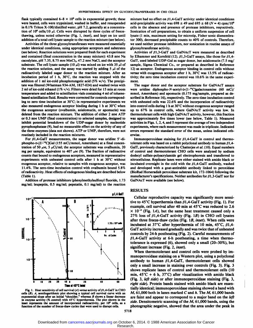
Fig. I. Heat sensitivity of cell survival (A) versus activity of/31,4-GalT in CHOcells (B). A, semilogarithmic plot showing a typical cell survival curve with anexponential slope after an initial "shoulder," whereas B shows a linear decreasein enzyme activity (% control) with 45'C hyperthermia. The plot shown in theinset represents the amount of incorporated radioactivity, per Id" cells, as afunction of the number of freeze-thaw cycles that were used to disrupt cells.
mixture had no effect on jSl,4-GalT activity: under identical conditionsacid-precipitable activity was 698 ±49 and 695 ±68 (N = 4) cpm/106
cells in the absence and presence of protease inhibitors, respectively.Sonication of cell preparations, to obtain a uniform suspension of celllysate (1 min, maximum setting for microtip. Fisher sonic dismembra-tor 300), decreased precipitable counts to 40% of controls. Therefore,we used neither protease inhibitors, nor sonication in routine assays ofglycosyltransferase activity.
Activities of 01,3-GalT and GalNAcT were measured as describedby Elhammer and Kornfeld (12). ,31,3-GalT assays, like those for 01,4-GalT, used labeled UDP-Gal as sugar donor, but asialomucin (7.5 mg/sample, Sigma Chemical Co.. or prepared as described in Reference12) as acceptor. Endogenous acceptor binding in control cells withoutversus with exogenous acceptor after 1 h, 30"C was 13.5% of radioac
tivity; the zero time incubation control was 10.6% in the same experiment.
The sugar donor and the acceptor substrate for GalNAcT assayswere uridine diphospho-Ar-acetyl-D-[l-1<C]galactosamine (60 mCi/
mmol, Amersham) and apomucin (0.175 mg/sample, prepared as described in Reference 16), respectively. The zero-time incubation controlwith unheated cells was 22.6% and the incorporation of radioactivityinto control cells during 1 h at 30"C without exogenous acceptor ranged
up to 67% in control cells, where GalNAcT activity was low. Inthermotolerant cells with high GalNAcT activity, however, this fractionwas approximately five times lower (see below, Table 1). Measuredactivities in Figs. 1, 2,4, and 5 represent the average of two independentexperiments, where each measurement was made in triplicate. Indicatederrors represent the standard error of the mean, unless indicated otherwise.
Immunoperoxidase staining for /31,4-GalT in control and thermotolerant cells was based on a rabbit polyclonal antibody to human £1,4-GalT, previously characterized by Chatterjee et al. (10). Equal numbersof control and thermotolerant CHO cells were analyzed by sodiumdodecyl sulfate-polyacrylamide gel electrophoresis and transferred tonitrocellulose. Replicate lanes were either stained with amido black orincubated overnight in the cold with the .¡1.4( inI"I antibody, washed
and developed with a goat-antirabbit antibody linked to peroxidase(lin iHad Horseradish peroxidase substrate kit, 172-1064) following themanufacturer's specifications. Neither antibodies for 01,3-GalT nor for
GalNAcT were available (see below).
RESULTS
Cellular reproductive capacity was significantly more sensitive to 45°Chyperthermia than /31,4-GalT activity (Fig. 1). Forexample, cell survival after 40 min at 45°Cwas reduced to 2.6x 10~4 (Fig. 1/1), but the same heat treatment caused only a
27% loss of 01,4-GalT activity (Fig. IB) in CHO cell lysatesafter three freeze-thaw cycles (Fig. \B, inset). When cells wereincubated at 37°Cafter hyperthermia of 10 min, 45°C,|91,4-
GalT activity increased gradually and was twice that of unheatedcontrols by 24-h postheating (Fig. 2). Careful measurements of01,4-GalT activity at 6-h postheating, a time when thermo-toléranceis expressed (6), showed only a small (20-30%), butsignificant increase (Fig. 2, inset).
When thermotolerant and control cells were probed by im-munoperoxidase staining on a Western plot, using a polyclonalantibody to human /31,4-GalT, thermotolerant cells showedonly a small increase in staining over controls (Fig. 3). Fig. 3shows replicate lanes of control and thermotolerant cells (10min, 45"C + 6 h, 37°C)after visualization with amido black
(Fig. 3, left side) or after immunoperoxidase staining (Fig. 3,right side). Protein bands stained with amido black are essentially identical; immunoperoxidase staining showed a band withM, 61,000 both in lanes marked C and h. The M, 61,000 bandsare faint and appear to correspond to a major band on the leftside. Densitometric scanning of the M, 61,000 bands, using thephotographic negative, showed that the area under the peak in
5718
Research. on October 6, 2014. © 1988 American Association for Cancercancerres.aacrjournals.org Downloaded from
HYPERTHERMIA EFFECT ON GLYCOSYLTRANSFERASES IN CHO CELLS
p1.4 GalT
U
MINunlur.i 10"'
0 6 12 18 24TinillilaftirlOiiMS'C
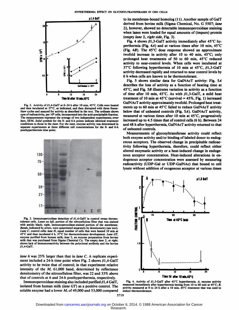
Fig. 2. Activity of/31,4-GalT at 0-24 h after 10 min, 45°C.Cells were heatedand then incubated at 37°C,as indicated, and then disrupted with three freeze-
thaw cycles and assayed for activity as described in the text. The ordinate showscpm of radioactivity, per 10' cells, incorporated into the acid-precipitable fraction.
The measurements represent the average of two independent experiments; errorbars, SEM. Solid symbols for the 0- and 6-h points indicate identical treatmentconditions to those in the inset. For the inset, enzyme activity was determined inseparate experiments at three different cell concentrations for the 0- and 6-hposthyperthermia time point.
17-
b Z GT
to its membrane-bound homolog (11). Another sample of GalTderived from bovine milk (Sigma Chemical, No. G 5507; lane2), however, showed no detectable immunoperoxidase stainingwhen lanes were loaded for equal amounts of (impure) protein(empty lane 2, right side, Fig. 3).
Fig. 4 shows j31,3-GalT activity immediately after 45°Chy-perthermia (Fig. 4A) and at various times after 10 min, 45°C(Fig. 4B). The 45°Cdose response showed an approximatetwofold increase in activity after 10 to 40 min, 45°C;onlyprolonged heat treatments of 50 to 60 min, 45°Creduced
activity to near-control levels. When cells were incubated at37°Cfollowing hyperthermia of 10 min at 45°C,/31,3-GalT
activity decreased rapidly and returned to near control levels by6 h when cells are known to be thermotolerant.
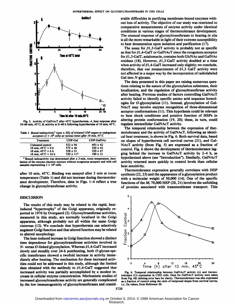
Fig. 5 shows similar data for GalNAcT activity: Fig. 5Adescribes the loss of activity as a function of heating time at45°C,and Fig. 5B illustrates variation in activity as a functionof time after 10 min, 45°C.As with /31,3-GalT, a mild heattreatment of 10 min at 45°C(survival = 45%, Fig. 1) increased
GalNAcT activity approximately twofold. Prolonged heat treatments up to 60 min at 45°Cfailed to reduce GalNAcT activity
below that of unheated controls (Fig. 5A). GalNAcT activity,measured at various times after 10 min at 45°C,progressively
increased up to 4.5 times that of control cells (6 h). Between 24and 48 h after hyperthermia, GalNAcT activity returned to thatof unheated controls.
Measurements of glycosyltransferase activity could reflectboth enzyme activity and/or binding of labeled donor to endogenous acceptors. The observed change in precipitale radioactivity following hyperthermia, therefore, could reflect eitheraltered enzymatic activity or a heat-induced change in endogenous acceptor concentration. Heat-induced alterations in endogenous acceptor concentration were assessed by measuringradioactivity (UDP-Gal or UDP-GalNAc) that bound to celllysate without addition of exogenous acceptor at various times
A
/
r>k ßUGalT
Fig. 3. Immunoperoxidase detection of 01,4-GalT in control versi«thermotolerant cells. Lanes on left, portion of the nitrocellulose filter that was stainedwith amido black; right, immunoperoxidase-stained portion of the membrane.Bands, indicated by arrow, were quantitated separately by densitometry (see text).Lane C, control cells; lane H, equal number of cells that were heated 10 min at45°Cand then incubated 6 h, 37°Cfor thermotolerance development. Lane GT,
enzyme purified from human milk; lane S, an enzyme preparation from bovinemilk that was purchased from Sigma Chemical Co. The empty lane S, at right,shows lack of immunoreactivity between the polyclonal antibody and the bovine01,4-GalT.
lane h was 25% larger than that in lane C. A replicate experiment included a 24-h time point when Fig. 2 shows /31,4-GalTactivity to be twice that of control; in that experiment relativeintensity of the M, 61,000 band, determined by reflectancedensitometry of the nitrocellulose filter, was 22 and 33% abovethat of controls at 6 and 24-h posthyperthermia, respectively.
Immunoperoxidase staining also included purified 01,4 GalT,isolated from human milk (lane GT) as a positive control. Thesoluble enzyme had a lower A/r of 49,000 and 53,000 compared
5719
B 1>•S?
iS
t ioS'iteH-
'1
I30Tim
it 45°C1•
ii50Ininl
1.1
M-
24
TimiIMliter 10«¡MS"CFig. 4. Activity of /31,3-GalT after 45°Chyperthermia. A, enzyme activity
measured immediately after hyperthermia lasting from 10 to 60 min at 45"C; B,activity measured at 0 to 24 h after a 10 min. 45'C treatment that was used to
induce thermotolerance.
Research. on October 6, 2014. © 1988 American Association for Cancercancerres.aacrjournals.org Downloaded from
HYPERTHERMIA EFFECT ON GLYCOSVLTRANSFERASES IN CHO CELLS
GalNAcT
H1*iiiiiiis
20 4060iiU£
5K432_
Ti«lUS'ClPiinl1ifl/
K/\/\i'\' \i*•/Tt/' T^'
-/ 1 ^^^•^.Õ
S-,-O
O 6 12 18 24 48ThMlkliltsrlOaiMS'C
Fig. 5. Activity of GalNAcT after 45'C hyperthermia. A, heat response after10-60 min, 45'C; B, activity at 0-48 h following hyperthermia of 10 min, 45'C.
Table 1 Bound radioactivity" (cpm ±SD) oftritiated UDP-sugars to endogenousacceptors (2 x. Iff cells) at various times after 10 min, 45'C
TreatmentUnheated
control10 min, 45'C + 0 h10 min, 45'C + 6 h10 min, 45'C + 24 hUDP-Gal522
±94575 ±46528 ±51530 ±117UDP-GalNAc493
±42520 ±43220 ±47180 ±31
" Bound radioactivity was determined after a 2-min, room temperature, incu
bation of the enzyme reaction mixture without exogenous acceptor and with cellsamples representing 2x10' cells.
after 10 min, 45°C.Binding was assayed after 2 min at roomtemperature (Table 1) and did not increase during thermotoler-ance development. Therefore, data in Figs. 1-4 reflect a truechange in glycosyltransferase activity.
érabledifficulties in purifying membrane-bound enzymes without loss of activity. The objective of our study was restricted tocomparative measurements of enzyme activity under identicalconditions at various stages of thermotolerance development.The unusual response of glycosyltransferases to heating in situis all the more remarkable in light of their extreme susceptibilityto heat denaturation upon isolation and purification (17).
The assay for 01,3-GalT activity is probably not as specificas that for /31,4-GalT or GalNAcT since the exogenous acceptorfor/31,3-GalT, asialomucin, contains both GlcNAc and GalNAcresidues (18). However, /31,3-GalT activity doubled at a timewhen activity of/31,4-GalT increased only slightly; we conclude,therefore, that our measurements of 01,3 GalT activity werenot affected in a major way by the incorporation of radiolabeledGal into /V-glycans.
The data presented in this paper are raising numerous questions relating to the nature of the glycosylation substrates, theirlocalization, and the regulation of glycosyltransferase activityafter heating. Previous studies of factors controlling GalNAcTactivity failed to identify specific amino acid sequence homol-ogies for O-glycosylation (11). Instead, glycosylation of GalNAcT may involve enzyme recognition of three-dimensionalacceptor conformations (11). This hypothesis could be relevantto heat shock conditions and putative function of HSPs inaltering protein conformation (19, 20); these, in turn, couldregulate intracellular GalNAcT activity.
The temporal relationship between the expression of thermotolerance and the activity of GalNAcT, following an identical heat treatment, is shown in Fig. 6. Both survival data, basedon slopes of hyperthermia cell survival curves (21), and GalNAcT activity (from Fig. 5) are expressed as a fraction ofcontrol. Fig. 6 shows the development of thermotolerance lagging behind the increase in GalNAcT activity by 2-4 h, ashypothesized above (see "Introduction"). Similarly, GalNAcT
activity returned more quickly to control levels than cellularheat sensitivity.
Thermotolerance expression generally correlates with HSPsynthesis (22,23) and the appearance of a glycosylation productwith a molecular weight of 50,000 (14). One of the specificfunctions of the M, 70,000 HSP (20, 21) involves the unfoldingof proteins associated with transmembrane transport. This
DISCUSSION
The results of this study may be related to the rapid, heat-induced "hypertrophy" of the Golgi apparatus, originally re
ported in 1976 by Overgaard (2). Glycosyltransferase activities,measured in this study, are normally localized in the Golgiapparatus, although probably not all within the same Golgicisternae (12). We conclude that hyperthermia can selectivelyaugment Golgi function and that altered function may be relatedto altered morphology.
The heat-induced increase in Golgi function showed a distincttime dependence for glycosyltransferase activities involved inN- versus O-linked glycosylation. Whereas /31,4-GalT increasedslowly and steadily over 24-h postheating, both O-glycan-spe-cific transferases showed a twofold increase in activity immediately after heating. The mechanism for these increased activities could not be addressed in this study, although the limiteddata obtained with the antibody to /31,4-GalT suggested thatincreased activity was partially accomplished by a modest increase in cellular enzyme concentration. Mechanistic studies ofincreased glycosyltransferase activity are generally complicatedby the low immunogenicity of glycosyltransferases and consid-
5720
Fig. 6. Temporal relationship between GalNAcT activity (A) and thermotolerance (G) expression in CHO cells. Data for GalNAcT activity were takenfrom Fig. 4ß,deleting error bars for clarity. Thermotolerance was also expressedas a fraction of control using the ratio of reciprocal slopes from survival curves,i.e.. Do values, from Reference 20.
Research. on October 6, 2014. © 1988 American Association for Cancercancerres.aacrjournals.org Downloaded from
HYPERTHERMIA EFFECT ON GLYCOSYLTRANSFERASES IN CHO CELLS
function may facilitate cellular recovery from heat shock andcontribute to enhanced cell survival of thermotolerant cells afterhyperthermia. At the same time, enhanced protein glycosylationcould protect specific intracellular sites against thermal dena-
turation and/or participate in protein redistribution. Increasedactivity of glycosyltransferase, e.g., GalNAcT, preceding theexpression of thermotolerance, would be consistent with thisconcept. Specific relationships between HSP function, elevatedglycosyltransferase activity, the appearance of the M, 50,000glycosylation product and the phenomenon of thermotolerance,however, are still obscure and remain to be elucidated.
REFERENCES
1. Hahn, G. M. Hyperthermia and Cancer. New York: Plenum Press, 1982.2. Overgaard, J. Ultrastructure of a murine mammary carcinoma exposed to
hyperthermia in vivo. Cancer Res., 36: 983-985, 1976.3. Robins, H. I., Sleeves, R. A., Clark, A. W., Martin, P. A.. Miller, K., and
Dennis, W. A. Differential sensitivity of AKR murine leukemia and normalbone marrow cells to hyperthermia. Cancer Res., 43:4951-4955, 1983.
4. Clark, A. W., Robins, H. I., Vorpahl, J. W., and Yatvin, M. B. Structuralchanges in murine cancer associated with hyperthermia and lidocaine. CancerRes., «.-1716-1723, 1983.
5. Farquhar, M. G., and Palade, G. E. The Golgi apparatus (complex)—(1954-1981)—from artifact to center stage. J. Cell Biol., 91: 77s-103s, 1981.
6. Henle, K. J. Thermotolerance in cultured mammalian cells. In: K. J. Henle(ed.), Thermotolerance, Vol. 1, pp. 13-71. Boca Raton, FL: CRC Press, Inc.,1987.
7. Strous, G. J. Golgi and secreted galactosyltransferase. CRC Crit. Rev.Biochem., 21:119-151, 1986.
8. Ram, B. P., and Munjal, D. D. Galactosyltransferases: physical, chemicaland biological aspects. CRC Crit. Rev. Biochem., 17: 257-331, 1985.
9. Verdón, B., and Berger, E. G. Galactosyltransferase. In: H. U. Bergmeyer,(ed.), Methods of Enzymatic Analysis, Vol. 4, Ed 3, pp. 374-381. Weinheim,FRG: Verlag Chemie. 1984.
10. Chatterjee, S. K., Bhattacharya, M., and Barlow, J. J. Murine monoclonalantibodies against galactosyltransferase from the ascites of ovarian cancerpatients. Cancer Res., 44: 5725-5732, 1984.
11. Schachter, H., and Roseman, S. Mammalian glycosyltransferases, Part A.In: W. J. Lennarz, (ed.). The Biochemistry of Glycoproteins and Proteogly-cans, pp. 85-127. New York: Plenum Press, 1980.
12. Elhammer, A., and Kornfeld, S. Two enzymes involved in the synthesis ofO-linkcd oligosaccharides are localized on membranes of different densitiesin mouse lymphoma BW5147 cells. J. Cell Biol., 98: 327-331, 1984.
13. Sharon, N., and Lis, H. Glycoproteins. In: The Proteins, Vol. 5, pp. 1-144.New York: Academic Press, 1982.
14. Henle, K. J., Nagle, W. A., Norris, J. S., and Moss, A. J. Enhancedglycosylation of a 50 kD protein during thermotolerance development inCHO cells. Int. J. Radiât.Biol., S3: 839-847, 1988.
15. Henle, K. J., Moss, A. J., and Nagle, W. A. Temperature-dependent inductionof thermotolerance by ethanol. Radiât.Res., 108: 327-335, 1986.
16. Hagopian, A., and Eylar, E. H. Glycoprotein biosynthesis: studies on thereceptor specificity of the polypeptidyltA'-acetylgalactosaminyl transferasefrom bovine submaxillary glands. Arch. Biochem. Biophys., 128: 422-433,1968.
17. Sadler, J. E., Beyer, T. A., Oppenheimer, C. L., Paulson, J. C., Prieels, J-P.,Rearick, J. I., and Hill, R. L. Purification of mammalian glycosyltransferases.Methods Enzymol., 83:458-514, 1982.
18. Chatterjee, S. K., Bhattacharya, M., and Barlow, J. J. Characterization ofproducts synthesized by glycosyltransferase purified from the ascites ofovarian cancer patients. J. Nati. Cancer Inst., 77: 855-862, 1986.
19. Deshaies, R. J., Koch, B. D., Werner-Washburne, M., Craig, E. A., andSchekman, R. A subfamily of stress proteins facilitates translocation ofsecretory and mitochondria! precursor polypeptides. Nature (I.mid.). 332:800-805, 1988.
20. Chirico, W. J., Waters, M. G., and Blobel, G. 70 k heat shock related proteinsstimulate protein translocation into microsomes. Nature (Lond.), 332: 805-810, 1988.
21. Henle, K. J., Bitner, A., and Dethlefsen, L. A. The induction of thermotoler-ance by multiple heat fractions in CHO cells. Cancer Res., 39: 2486-2491,1979.
22. Landry, J., Lamarche, S., and Chretien, P. Heat shock proteins: a lead to theunderstanding of cell thermotolerance. In: K. J. Henle (ed.), Thermotolerance, Vol. 1, pp. 145-173. Boca Raton, FL: CRC Press, Inc., 1987.
23. Bienz, M., and Pelman, H. R. B. Mechanisms of heat-shock gene activationin higher eukaryotes. Adv. Genet., 24: 31-72, 1987.
5721
Research. on October 6, 2014. © 1988 American Association for Cancercancerres.aacrjournals.org Downloaded from
Related Documents

![Putative Glycosyltransferases and Other Plant Golgi ... · Putative Glycosyltransferases and Other Plant Golgi Apparatus Proteins Are Revealed by LOPIT Proteomics1[W] Nino Nikolovski,](https://static.cupdf.com/doc/110x72/5beabde209d3f2ff498bfa69/putative-glycosyltransferases-and-other-plant-golgi-putative-glycosyltransferases.jpg)