1 CONTENTS INTRODUCTION ...................................................................................................................................... 4 HISTORICAL BACKGROUND ........................................................................................................................ 5 TERMINOLOGY ....................................................................................................................................... 8 IDENTIFICATION ................................................................................................................................... 13 EARLY LIFE HISTORY DESCRIPTIONS .......................................................................................................... 17 CLUPEIDAE........................................................................................................................................ 22 Sardina pilchardus (Walbaum, 1792) ............................................................................................... 22 Sprattus sprattus (Linnaeus, 1758) ................................................................................................. 26 ENGRAULIDAE ................................................................................................................................... 30 Engraulis encrasicolus (Linnaeus, 1758) .......................................................................................... 30 GONOSTOMATIDAE ............................................................................................................................. 34 Cyclothone braueri Jespersen & Tåning, 1926 .................................................................................. 34 STERNOPTYCHIDAE ............................................................................................................................ 36 Maurolicus muelleri (Gmelin, 1789) ................................................................................................. 36 STOMIIDAE ....................................................................................................................................... 38 Stomias boa (Risso, 1810) .............................................................................................................. 38 ARGENTINIDAE .................................................................................................................................. 40 Argentina sphyraena Linnaeus, 1758 ............................................................................................... 40 MYCTOPHIDAE................................................................................................................................... 42 Benthosema glaciale (Reinhardt, 1837) ........................................................................................... 42 Ceratoscopelus maderensis (Lowe, 1839) ........................................................................................ 44 Diogenichthys atlanticus (Tåning, 1928) .......................................................................................... 46 Electrona risso (Cocco, 1829) ......................................................................................................... 48 Myctophum punctatum Rafinesque, 1810 ........................................................................................ 50 Notolychnus valdiviae (Brauer, 1904) .............................................................................................. 52 PARALEPIDIDAE................................................................................................................................. 54 Magnisudis atlantica (Krøyer, 1868) ................................................................................................ 54 Paralepis coregonoides Risso, 1820 ................................................................................................. 56 ANGUILLIDAE .................................................................................................................................... 58 Anguilla anguilla (Linnaeus, 1758) ................................................................................................... 58 MURAENIDAE .................................................................................................................................... 60 Muraena helena Linnaeus, 1758 ...................................................................................................... 60 CONGRIDAE ...................................................................................................................................... 62 Conger conger (Linnaeus, 1758) ..................................................................................................... 62 OPHICHTIDAE.................................................................................................................................... 64 Ophisurus serpens (Linnaeus, 1758) ............................................................................................... 64 BELONIDAE ....................................................................................................................................... 66 Belone belone (Linnaeus, 1761) ...................................................................................................... 66 MACRORAMPHOSIDAE ........................................................................................................................ 68 Macroramphosus scolopax (Linnaeus, 1758) .................................................................................... 68 SYNGNATHIDAE ................................................................................................................................. 70 Entelurus aequoraeus (Linnaeus, 1758) ........................................................................................... 70 Hipoccampus guttulatus Cuvier, 1829.............................................................................................. 72 Hippocampus hippocampus (Linnaeus, 1758) .................................................................................. 74 Nerophis lumbriciformis (Jenyns, 1835) ........................................................................................... 76 Nerophis ophidion (Linnaeus, 1758) ................................................................................................ 78 Syngnathus abaster Risso, 1826 ..................................................................................................... 80 Syngnathus acus Linnaeus, 1758 .................................................................................................... 82 Syngnathus typhle Linnaeus, 1758 .................................................................................................. 84 MERLUCCIDAE ................................................................................................................................... 86 Merluccius merluccius Linnaeus, 1758 ............................................................................................. 86 GADIDAE .......................................................................................................................................... 88 Ciliata mustela (Linnaeus, 1758) ..................................................................................................... 88 Gadiculus argenteus Guichenot, 1850.............................................................................................. 90 Micromessistius poutassou (Risso, 1827) ......................................................................................... 92

Welcome message from author
This document is posted to help you gain knowledge. Please leave a comment to let me know what you think about it! Share it to your friends and learn new things together.
Transcript
1
CONTENTS INTRODUCTION ...................................................................................................................................... 4 HISTORICAL BACKGROUND ........................................................................................................................ 5
TERMINOLOGY ....................................................................................................................................... 8 IDENTIFICATION ................................................................................................................................... 13
EARLY LIFE HISTORY DESCRIPTIONS .......................................................................................................... 17
CLUPEIDAE ........................................................................................................................................ 22 Sardina pilchardus (Walbaum, 1792) ............................................................................................... 22
Sprattus sprattus (Linnaeus, 1758) ................................................................................................. 26 ENGRAULIDAE ................................................................................................................................... 30
Engraulis encrasicolus (Linnaeus, 1758) .......................................................................................... 30 GONOSTOMATIDAE ............................................................................................................................. 34
Cyclothone braueri Jespersen & Tåning, 1926 .................................................................................. 34
STERNOPTYCHIDAE ............................................................................................................................ 36 Maurolicus muelleri (Gmelin, 1789) ................................................................................................. 36
STOMIIDAE ....................................................................................................................................... 38 Stomias boa (Risso, 1810) .............................................................................................................. 38
ARGENTINIDAE .................................................................................................................................. 40
Argentina sphyraena Linnaeus, 1758 ............................................................................................... 40 MYCTOPHIDAE ................................................................................................................................... 42
Benthosema glaciale (Reinhardt, 1837) ........................................................................................... 42 Ceratoscopelus maderensis (Lowe, 1839) ........................................................................................ 44
Diogenichthys atlanticus (Tåning, 1928) .......................................................................................... 46
Electrona risso (Cocco, 1829) ......................................................................................................... 48 Myctophum punctatum Rafinesque, 1810 ........................................................................................ 50
Notolychnus valdiviae (Brauer, 1904) .............................................................................................. 52 PARALEPIDIDAE ................................................................................................................................. 54
Magnisudis atlantica (Krøyer, 1868) ................................................................................................ 54 Paralepis coregonoides Risso, 1820 ................................................................................................. 56
ANGUILLIDAE .................................................................................................................................... 58
Anguilla anguilla (Linnaeus, 1758) ................................................................................................... 58 MURAENIDAE .................................................................................................................................... 60
Muraena helena Linnaeus, 1758 ...................................................................................................... 60 CONGRIDAE ...................................................................................................................................... 62
Conger conger (Linnaeus, 1758) ..................................................................................................... 62
OPHICHTIDAE .................................................................................................................................... 64 Ophisurus serpens (Linnaeus, 1758) ............................................................................................... 64
BELONIDAE ....................................................................................................................................... 66 Belone belone (Linnaeus, 1761) ...................................................................................................... 66
MACRORAMPHOSIDAE ........................................................................................................................ 68 Macroramphosus scolopax (Linnaeus, 1758) .................................................................................... 68
SYNGNATHIDAE ................................................................................................................................. 70
Entelurus aequoraeus (Linnaeus, 1758) ........................................................................................... 70 Hipoccampus guttulatus Cuvier, 1829 .............................................................................................. 72
Hippocampus hippocampus (Linnaeus, 1758) .................................................................................. 74 Nerophis lumbriciformis (Jenyns, 1835) ........................................................................................... 76
Nerophis ophidion (Linnaeus, 1758) ................................................................................................ 78
Syngnathus abaster Risso, 1826 ..................................................................................................... 80 Syngnathus acus Linnaeus, 1758 .................................................................................................... 82
Syngnathus typhle Linnaeus, 1758 .................................................................................................. 84 MERLUCCIDAE ................................................................................................................................... 86
Merluccius merluccius Linnaeus, 1758 ............................................................................................. 86
GADIDAE .......................................................................................................................................... 88 Ciliata mustela (Linnaeus, 1758) ..................................................................................................... 88
Gadiculus argenteus Guichenot, 1850 .............................................................................................. 90 Micromessistius poutassou (Risso, 1827) ......................................................................................... 92
2
Pollachius pollachius (Linnaeus, 1758) ............................................................................................. 94
Trisopterus luscus (Linnaeus, 1758) ................................................................................................ 96 Trisopterus minutus (Linnaeus, 1758) ............................................................................................. 98
ZEIDAE ........................................................................................................................................... 100 Zeus faber Linnaeus, 1758 ............................................................................................................ 100
CAPROIDAE ..................................................................................................................................... 102
Caprus aper (Linnaeus, 1758) ....................................................................................................... 102 SERRANIDAE ................................................................................................................................... 104
Serranus cabrilla (Linnaeus, 1758) ................................................................................................ 104 MORONIDAE .................................................................................................................................... 106
Dicentrarchus labrax (Linnaeus, 1758) .......................................................................................... 106 CEPOLIDAE ...................................................................................................................................... 108
Cepola macrophthalma Linnaeus, 1758 ......................................................................................... 108
CARANGIDAE ................................................................................................................................... 110 Trachurus trachurus (Linnaeus, 1758) ........................................................................................... 110
MULLIDAE ....................................................................................................................................... 112 Mullus surmuletus Linnaeus, 1758 ................................................................................................. 112
SPARIDAE ....................................................................................................................................... 114
Boops boops (Linnaeus, 1758) ...................................................................................................... 114 Diplodus sargus (Linnaeus, 1758) ................................................................................................. 116
Pagellus bogaraveo (Brünnich, 1768) ............................................................................................ 118 Pagrus pagrus (Linnaeus, 1758) .................................................................................................... 120
Spondyliosoma cantharus (Linnaeus, 1758) ................................................................................... 122 CENTRACANTHIDAE .......................................................................................................................... 124
Centracanthus cirrus Rafinesque, 1810 .......................................................................................... 124
LABRIDAE ....................................................................................................................................... 126 Centrolabrus exoletus (Linnaeus, 1758) ......................................................................................... 126
Coris julis (Linnaeus, 1758) .......................................................................................................... 128 Ctenolabrus rupestris (Linnaeus, 1758) ......................................................................................... 130
Labrus bergylta Ascanius, 1767 ..................................................................................................... 132
Labrus merula Linnaeus, 1758 ...................................................................................................... 134 Labrus mixtus Linnaeus, 1758 ....................................................................................................... 136
Symphodus melops (Linnaeus, 1758) ............................................................................................ 138 AMMODYTIDAE ................................................................................................................................ 140
Ammodytes tobianus Linnaeus, 1758 ............................................................................................ 140
Gymnamodytes semisquamatus (Jourdain, 1879) ........................................................................... 142 Hyperoplus lanceolatus (Le Sauvage, 1824) ................................................................................... 144
TRACHINIDAE .................................................................................................................................. 146 Echiichthys vipera (Cuvier, 1829) .................................................................................................. 146
Trachinus draco Linnaeus, 1758 .................................................................................................... 148 SCOMBRIDAE ................................................................................................................................... 150
Scomber japonicus Houttuyn, 1782 ............................................................................................... 150
Scomber scombrus (Linnaeus, 1758) ............................................................................................. 152 GOBIIDAE ....................................................................................................................................... 154
Aphia minuta (Risso, 1810) ........................................................................................................... 154 Crystalogobius linearis (Düben, 1845) ........................................................................................... 156
Gobius cobitis Pallas, 1814 ........................................................................................................... 158
Gobius cruentatus Gmelin, 1789 ................................................................................................... 160 Gobius niger Linnaeus, 1758 ......................................................................................................... 162
Gobius paganellus Linnaeus, 1758 ................................................................................................ 164 Gobius xanthocephalus Heymer & Zander, 1992 ............................................................................ 166
Gobiusculus falvescens (Fabricius, 1779) ....................................................................................... 168 Lebetus guilleti (La Danois, 1913) ................................................................................................. 170
Lebetus scorpioides (Collet, 1874) ................................................................................................. 172
Pomatoschistus microps (Krøyer, 1838) ......................................................................................... 174 Pomatoschistus minutus (Pallas, 1770) .......................................................................................... 176
Pomatischistus pictus (Malm, 1865) .............................................................................................. 178

3
CALLIONYMIDAE .............................................................................................................................. 180
Callionymus lyra Linnaeus, 1758 ................................................................................................... 180 Callionymus maculatus Rafinesque, 1810 ...................................................................................... 182
BLENNIIDAE .................................................................................................................................... 184 Blennius ocellaris Linnaeus, 1758 .................................................................................................. 184
Coryphoblennius galerita (Linnaeus, 1758) .................................................................................... 186
Lipophrys pholis (Linnaeus, 1758) ................................................................................................. 188 Lipophrys trigloides (Valenciennes, 1836) ...................................................................................... 190
Parablennius gattorugine (Linnaeus, 1758) .................................................................................... 192 Parablennius pilicornis (Cuvier, 1829) ............................................................................................ 194
CARAPIDAE ..................................................................................................................................... 196 Carapus acus (Brünnich, 1768) ..................................................................................................... 196
MUGILIDAE ..................................................................................................................................... 198
Mugil cephalus Linnaeus, 1758 ..................................................................................................... 198 ATHERINIDAE .................................................................................................................................. 200
Atherina presbyter Linnaeus, 1758 ................................................................................................ 200 TRIGLIDAE ...................................................................................................................................... 202
Trigla lucerna Linnaeus, 1758 ....................................................................................................... 202
COTTIDAE ....................................................................................................................................... 204 Taurulus bubalis (Euphrasen, 1786) .............................................................................................. 204
LIPARIDAE ...................................................................................................................................... 206 Liparis montagui (Donovan, 1804) ................................................................................................ 206
SCOPHTHALMIDAE ............................................................................................................................ 208 Lepidorhombus boscii (Risso, 1810) .............................................................................................. 208
Psetta maxima (Linnaeus, 1758) ................................................................................................... 210
Zeugopterus punctatus (Bloch, 1787) ............................................................................................ 212 Zeugopterus regius (Bonnaterre, 1788) ......................................................................................... 214
BOTHIDAE ....................................................................................................................................... 216 Arnoglossus laterna (Walbaum, 1792) ........................................................................................... 216
PLEURONECTIDAE ............................................................................................................................ 218
Platichthys flesus (Linnaeus, 1758) ............................................................................................... 218 SOLEIDAE ....................................................................................................................................... 220
Buglossidium luteum (Risso, 1810) ................................................................................................ 220 Microchirus variegatus (Donovan, 1808) ........................................................................................ 222
Pegusa lascaris (Risso, 1810) ........................................................................................................ 224
Solea senegalensis Kaup, 1858 ..................................................................................................... 226 Solea solea (Linnaeus, 1758) ........................................................................................................ 228
GOBIESOCIDAE ................................................................................................................................ 230 Diplecogaster bimaculata (Bonnaterre, 1788) ................................................................................ 230
Lepadogaster lepadogaster (Bonnaterre, 1788) .............................................................................. 232 LOPHIIDAE ...................................................................................................................................... 234
Lophius piscatorius Linnaeus, 1758 ............................................................................................... 234
REFERENCES.................................................................................................................................... 236 ANNEX ............................................................................................................................................ 245
4
INTRODUCTION This guide is intended for the identification of the Early Life History (ELH) stages of fishes collected by
plankton nets from the marine and estuarine waters of the Iberian Peninsula (Eastern North Atlantic Ocean). The coverage area extends from latitude 34º-45º north, to longitude 6º-14º west.
The basic characteristics of the eggs and larvae of 104 species belonging to 45 families are described. The emphasis has been placed on the most diagnostic or easily observed characters in
order to facilitate comparisons between taxa.
The descriptive accounts of this guide follow the format of previous ELH guides: Fahay (1983), Moser
(1996) and Richards (2005). Nomenclature follows Eschmeyer (1998) except for more recent changes. Within families, genera are listed in alphabetical order.
Species descriptions are given only for species for which some ELH stages are known. Each species
account includes the same basic information (written information on the left hand page and figures on the facing right hand page). Written information includes meristic data (fin-ray counts in adults
and myomere counts), life history information (range, habitat, spawning season, ELH pattern), main
references and ELH descriptions (eggs and larvae). Measurements of larvae usually refer to standard lengths. Many published illustrations have been redrawn mainly to provide certain uniformity
throughout the guide. Sources of illustrations are given for every Plate.
The contents of the present guide represent the current knowledge on the development of ELH
stages of fishes occurring in coastal waters of the Iberian Peninsula. The authors have been involved, for more than 25 years, in ichthyoplankton research.

5
HISTORICAL BACKGROUND In 1865 G.O. Sars (1837-1927) was asked by the Norwegian authorities to study the biology of Gadus morhua in order to understand the fluctuations of cod fisheries in the Lofoten area. Sars carefully studied, for the first time, the complete life history of cod, from pelagic eggs and pelagic larvae to
juveniles and adults. The pioneer work of Sars stimulated interest in pelagic fish eggs and larvae.
Soon it was realized that most species of commercial interest had planktonic eggs and larvae.
Systematic sampling of fish eggs was initiated by the German planktonologist Vitor Hensen (1835-1924). Hensen devised special plankton nets to capture pelagic fish eggs in a quantitative way.
During the last two decades of the 19th century, fish eggs and early larvae were reared under
controlled conditions to determine the main characters that would permit their identification in plankton samples. This early work was mainly pursued in England, Italy and Germany. Among these
pioneers were J.T. Cunningham, E.W.L. Holt, W.C. M’Intosh, W.C. Prince, A.T. Masterman in the United Kingdom, C. Emery, L. Facciola, F. Raffaele in Italy and E. Ehrenbaum, FR. Heincke in
Germany.
Older stages of fish larvae were seldom obtained by rearing fish eggs. The improvement of plankton
nets and research vessels was a major step forward in the early 20th century. German researchers (V. Hensen, C. Apstein) were responsible for most of the early work on performance and quantification of
plankton nets. C.G.J. Petersen in Denmark designed a very effective young fish trawl.
E. Ehrenbaum (1861-1942) published a comprehensive account of these studies that became a
standard reference for the identification of early life history stages of marine fish in the North-eastern Atlantic. This book was published in two volumes, one dated 1905 and the other 1909 (Ehrenbaum,
1905-1909).
A series of papers published by C.G.J. Petersen and J. Schmidt emerged from the cooperative research undertaken by the research steamer “Thor” off Iceland and the Faeroe Islands in 1903 and
1904. Petersen described the early life history stages of flatfishes (Petersen 1904, 1906, 1909).
Schmidt dealt mainly with the genus Gadus (Schmidt, 1905, 1906, 1907). The later contributions are landmarks even by present standards. Schmidt (1905) gives an excellent description of his technique
for describing the early life history stages:
“The order of procedure has been, to begin with such older stages as were so far developed, that they could be identified from characters similar to those which mark the adult fish. Then, earlier and still earlier stages were taken and compared with the older, and the determinations were in this manner, if the material was rich enough, successfully carried down to the youngest, post-larval stages, attention being directed to certain outstanding characters whose successive development could be followed thought the development series. The method thus employed for the determination of the unknown, pelagic fish-young might be called the series-method and it stands in contrast to the hatching-method in that, instead of making certain starting-point with the egg and following its further development, it begins at the opposite end and follows the development backwards. The condition for being able to use the series-method is, that a large material has to be at disposal, but in such cases it will lead to certain results, especially if the material contains whole series of the species”. Schmidt used for the first time meristic counts especially of anal and dorsal fins to verify each series. The pigmentation pattern was also extensively used for discriminating among the described gadoid
species. J. Schmidt is also well known for his work on the European eel, especially for establishing its breeding ground (Schmidt, 1923).
The Danish researcher A.V. Tåning described a great number of larval myctophids and sternoptychids (Tåning, 1918, Jespersen and Tåning, 1926). His larval studies of myctophids preceded his major
contribution in adult taxonomy. V. Ege made very important contributions on larvae of meso- and bathypelagic species (Ege, 1918, 1930, 1953, 1957). Bertelsen (1951) was the first author to include
larval characters in the clarification of the taxonomy of ceratioid fishes.
6
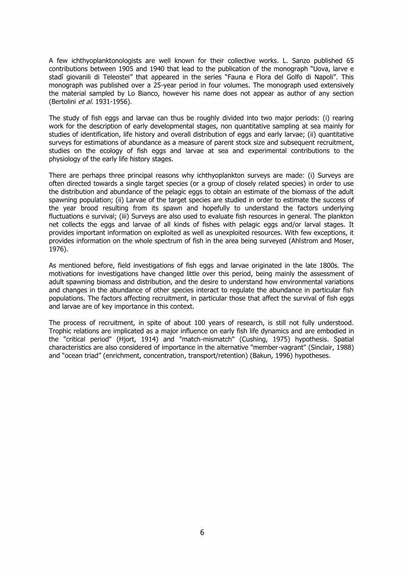
A few ichthyoplanktonologists are well known for their collective works. L. Sanzo published 65 contributions between 1905 and 1940 that lead to the publication of the monograph “Uova, larve e
stadî giovanili di Teleostei” that appeared in the series “Fauna e Flora del Golfo di Napoli”. This monograph was published over a 25-year period in four volumes. The monograph used extensively
the material sampled by Lo Bianco, however his name does not appear as author of any section
(Bertolini et al. 1931-1956).
The study of fish eggs and larvae can thus be roughly divided into two major periods: (i) rearing work for the description of early developmental stages, non quantitative sampling at sea mainly for
studies of identification, life history and overall distribution of eggs and early larvae; (ii) quantitative surveys for estimations of abundance as a measure of parent stock size and subsequent recruitment,
studies on the ecology of fish eggs and larvae at sea and experimental contributions to the
physiology of the early life history stages.
There are perhaps three principal reasons why ichthyoplankton surveys are made: (i) Surveys are often directed towards a single target species (or a group of closely related species) in order to use
the distribution and abundance of the pelagic eggs to obtain an estimate of the biomass of the adult
spawning population; (ii) Larvae of the target species are studied in order to estimate the success of the year brood resulting from its spawn and hopefully to understand the factors underlying
fluctuations e survival; (iii) Surveys are also used to evaluate fish resources in general. The plankton net collects the eggs and larvae of all kinds of fishes with pelagic eggs and/or larval stages. It
provides important information on exploited as well as unexploited resources. With few exceptions, it provides information on the whole spectrum of fish in the area being surveyed (Ahlstrom and Moser,
1976).
As mentioned before, field investigations of fish eggs and larvae originated in the late 1800s. The
motivations for investigations have changed little over this period, being mainly the assessment of adult spawning biomass and distribution, and the desire to understand how environmental variations
and changes in the abundance of other species interact to regulate the abundance in particular fish
populations. The factors affecting recruitment, in particular those that affect the survival of fish eggs and larvae are of key importance in this context.
The process of recruitment, in spite of about 100 years of research, is still not fully understood.
Trophic relations are implicated as a major influence on early fish life dynamics and are embodied in
the "critical period" (Hjort, 1914) and "match-mismatch" (Cushing, 1975) hypothesis. Spatial characteristics are also considered of importance in the alternative "member-vagrant" (Sinclair, 1988)
and “ocean triad” (enrichment, concentration, transport/retention) (Bakun, 1996) hypotheses.

7
TABLE 1
Main early contributions dealing with early life history descriptions of marine teleost fishes (adapted from Ahlstrom and Moser, 1981).
Author Date no. of papers Group Field Rearing Egg Larvae Juveniles Area
Petersen, C.G.J. 1904-1909 3 Flatfishes + - - all some E. North Atlantic
Schmidt, J. 1905-1907 6 Gadoid + - - all some E. North Atlantic
Schmidt, J. 1908-1932 44 Eels + - - all some North Atlantic-Worldwide
Eherenbaum, E. 1905-1909 book Teleosts + + some all some E. North Atlantic
Sanzo, L. 1905-1940 65 Teleosts + + some all some Mediterranean
Kyle, H.M. 1913 1 Flatfishes + - - all some Mediterranean-E. North Atlantic
Fage, L. 1918 1 Shorefishes + - - most some Mediterranean-E. North Atlantic
Tåning, A.V. 1918 1 Myctophids + - - all all Mediterranean-E. North Atlantic
Jespersen and Tåning 1919-1926 2 Sternoptychids + - some all some Mediterranean-E. North Atlantic
Delsman, H.C. 1921-1938 24 Teleosts + + some most some Indo Pacific
Hildebrand and Cable 1930-1938 3 Shore-Bay Fishes + + most all most W. North Atlantic
Uchida et al. 1958 1 Teleosts + + some all some Japan
Mito, S. 1960-1963 12 Fish eggs + + all some - Japan
Castle, P.H.J. 1959- several Eels + + some all some New Zealand-Worldwide
Dekhnik, T.V. 1973 1 Teleosts + + most most - Black Sea
Russell, FS 1976 book Teleosts + + most all - North Sea
8
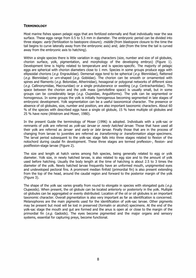
TERMINOLOGY Most marine fishes spawn pelagic eggs that are fertilized externally and float individually near the sea
surface. These eggs range from 0.5 to 5.5 mm in diameter. The embryonic period can be divided into three stages: early (fertilization to bastopore closure); middle (from blastopore closure to the time the
tail begins to curve laterally away from the embryonic axis) and; late (from the time the tail is curved
away from the embryonic axis to hatching).
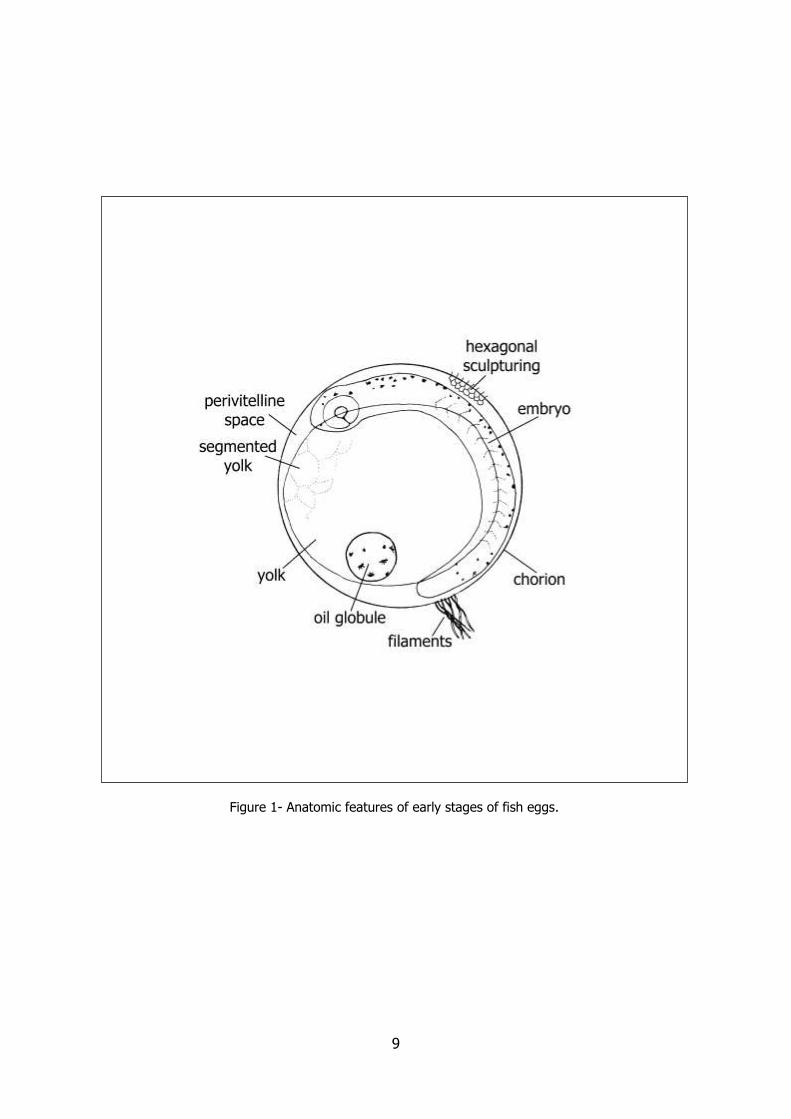
Within a single species there is little variation in egg characters (size, number and size of oil globules, chorion surface, yolk, pigmentation, and morphology of the developing embryo) (Figure 1).
Development time is highly related to temperature and is species-specific. The majority of pelagic
eggs are spherical with chorion diameters close to 1 mm. Species in some groups produce eggs with ellipsoidal chorions (e.g. Engraulidae). Demersal eggs tend to be spherical (e.g. Blenniidae), flattened
(e.g. Blenniidae) or urn-shaped (e.g. Gobiidae). The chorion can be smooth or ornamented with spines and filaments (e.g. Belonidae, Atherinidae), hexagonal or polygonal networks of different sizes
(e.g. Callinonymidae, Macrouridae) or a single protuberance or swelling (e.g. Centrachantidae). The space between the chorion and the yolk mass (perivitelline space) is usually small, but in some
groups can be considerably large (e.g. Clupeidae, Anguiliforms). The yolk can be segmented or
homogenous. In some groups the yolk is initially homogenous becoming segmented in late stages of embryonic development. Yolk segmentation can be a useful taxonomical character. The presence or
absence of oil globules, size, number and position, are also important taxonomic characters. About 60 % of the species with described eggs have a single oil globule, 15 % have multiple oil globules and
25 % have none (Ahlstrom and Moser, 1980).
In the present Guide the terminology of Moser (1996) is adopted. Individuals with a yolk-sac or
remnants of yolk are referred as yolk-sac larvae or newly hatched larvae. Those that have used all their yolk are referred as larvae and early or late larvae. Finally those that are in the process of
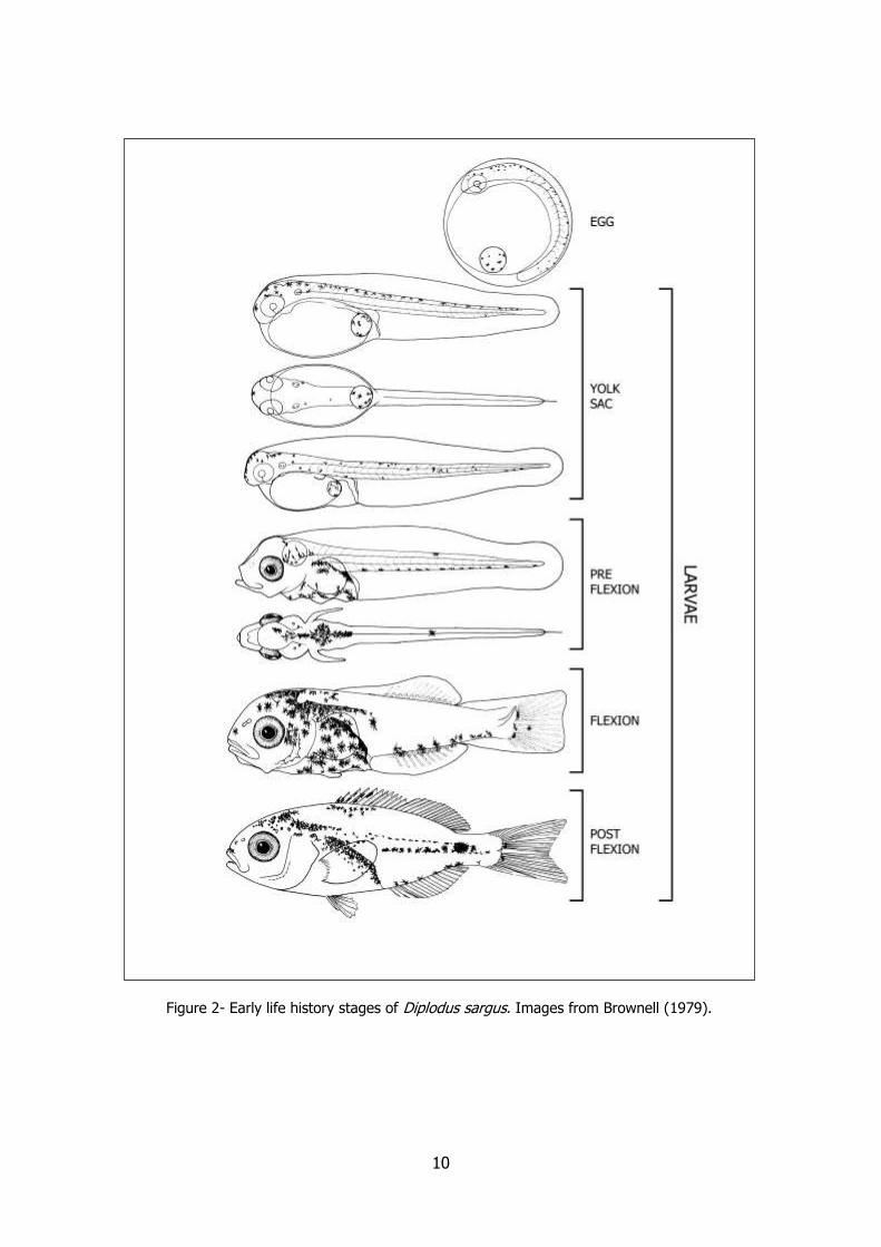
changing from larvae to juveniles are referred as transforming or transformation stage specimens. The larval period subsequent to the yolk-sac stage falls into three stages related to flexion of the
notochord during caudal fin development. These three stages are termed preflexion-, flexion- and
postflexion-stage larvae (Figure 2).
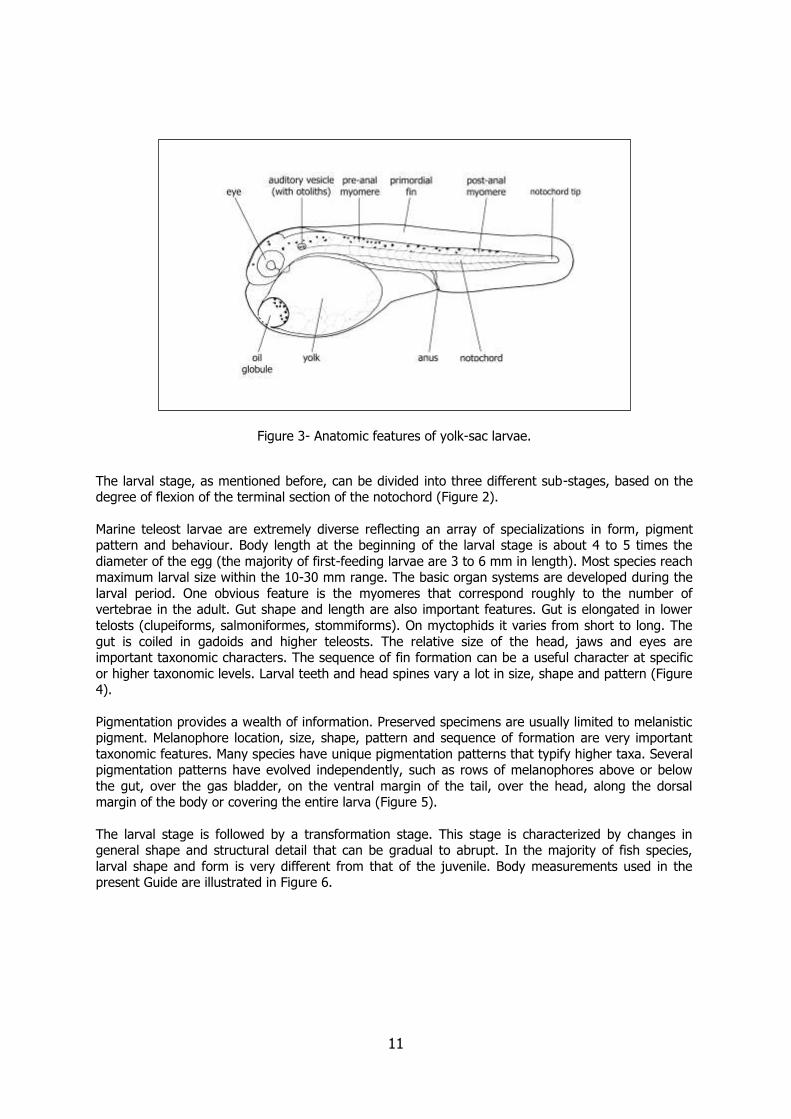
The size and length at hatch varies among fish species, being generally related to egg or yolk diameter. Yolk size, in newly hatched larvae, is also related to egg size and to the amount of yolk
used before hatching. Usually the body length at the time of hatching is about 2.5 to 3 times the
diameter of the yolk. Newly hatched larvae frequently have an unformed mouth, unpigmented eyes and undeveloped pectoral fins. A prominent median finfold (primordial fin) is also present extending
from the top of the head, around the caudal region and forward to the posterior margin of the yolk (Figure 3).
The shape of the yolk sac varies greatly from round to elongate in species with elongated guts (e.g. Clupeoids). When present, the oil globule can be located anteriorly or posteriorly in the yolk. Multiple
oil globules can be aggregated or evenly distributed. Location of the oil or oil globules is an important taxonomic character. Overall pigmentation is also very important as far as identification is concerned.
Melanophores are the main pigments used for the identification of yolk-sac larvae. Other pigments may be present but most will be lost in preserved (formalin or alcohol) specimens. At the end of the
yolk-sac stage the mouth and gut are formed and the anus is open at or close to the margin of the
primordial fin (e.g. Gadoids). The eyes become pigmented and the major organs and sensory systems, essential for capturing preys, become functional.
9
Figure 1- Anatomic features of early stages of fish eggs.
10
Figure 2- Early life history stages of Diplodus sargus. Images from Brownell (1979).
11
Figure 3- Anatomic features of yolk-sac larvae.
The larval stage, as mentioned before, can be divided into three different sub-stages, based on the degree of flexion of the terminal section of the notochord (Figure 2).
Marine teleost larvae are extremely diverse reflecting an array of specializations in form, pigment pattern and behaviour. Body length at the beginning of the larval stage is about 4 to 5 times the
diameter of the egg (the majority of first-feeding larvae are 3 to 6 mm in length). Most species reach maximum larval size within the 10-30 mm range. The basic organ systems are developed during the
larval period. One obvious feature is the myomeres that correspond roughly to the number of vertebrae in the adult. Gut shape and length are also important features. Gut is elongated in lower
telosts (clupeiforms, salmoniformes, stommiforms). On myctophids it varies from short to long. The
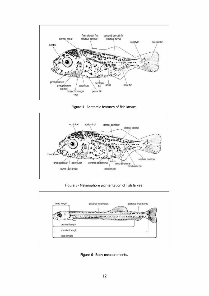
gut is coiled in gadoids and higher teleosts. The relative size of the head, jaws and eyes are important taxonomic characters. The sequence of fin formation can be a useful character at specific
or higher taxonomic levels. Larval teeth and head spines vary a lot in size, shape and pattern (Figure 4).
Pigmentation provides a wealth of information. Preserved specimens are usually limited to melanistic pigment. Melanophore location, size, shape, pattern and sequence of formation are very important
taxonomic features. Many species have unique pigmentation patterns that typify higher taxa. Several pigmentation patterns have evolved independently, such as rows of melanophores above or below
the gut, over the gas bladder, on the ventral margin of the tail, over the head, along the dorsal margin of the body or covering the entire larva (Figure 5).
The larval stage is followed by a transformation stage. This stage is characterized by changes in general shape and structural detail that can be gradual to abrupt. In the majority of fish species,
larval shape and form is very different from that of the juvenile. Body measurements used in the present Guide are illustrated in Figure 6.
12
Figure 4- Anatomic features of fish larvae.
Figure 5- Melanophore pigmentation of fish larvae.
Figure 6- Body measurements.

13
IDENTIFICATION
Identification of early stages of fishes is not an easy task. Fish eggs and larvae are usually small requiring the use of a good stereoscopic microscope and adequate lighting. In a single
ichthyoplankton sample there is usually a large variety of sizes, shapes and pigmentation patterns.
Generally dichotomous keys can not be used since most of the important taxonomic characters change dramatically over the course of the development. Very few species have distinct features that
can be recognized throughout the entire early life history. In a given area a large proportion of the fish eggs and larvae may be unknown or undescribed.
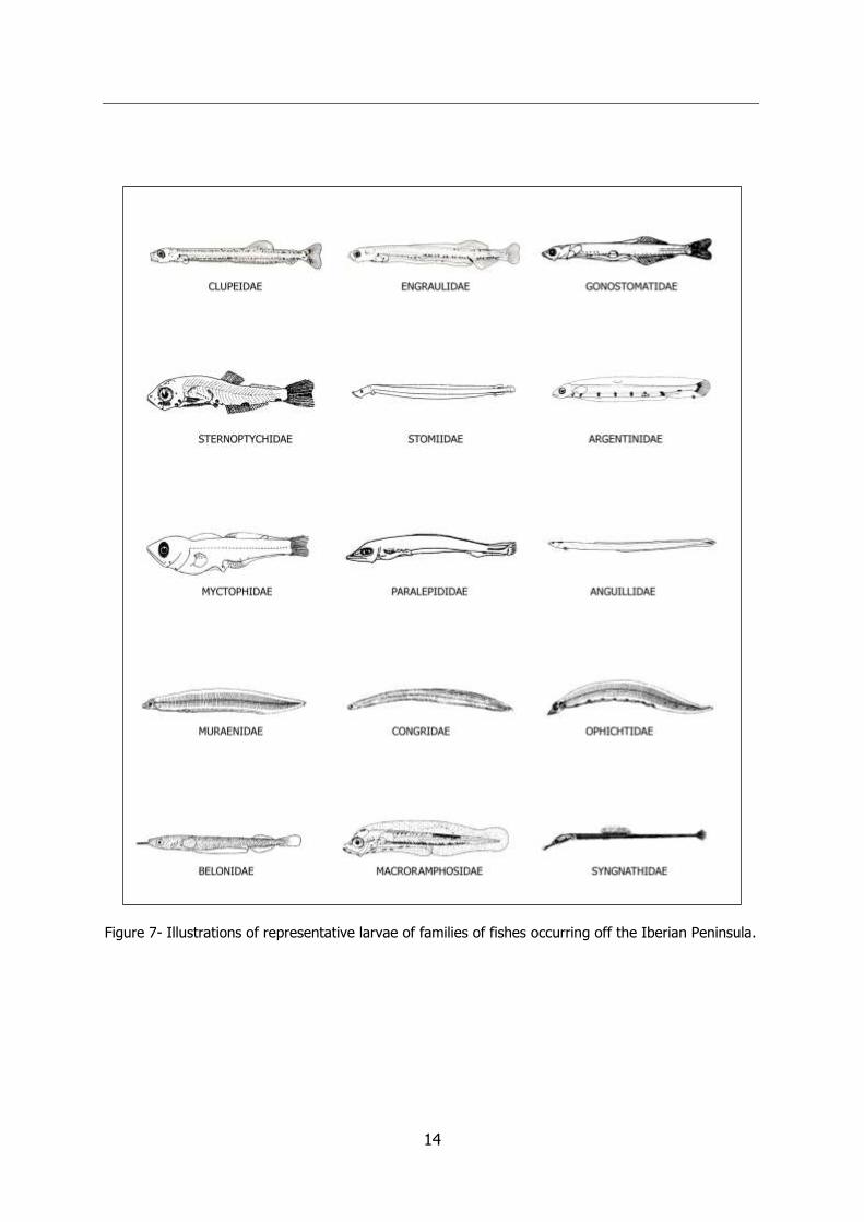
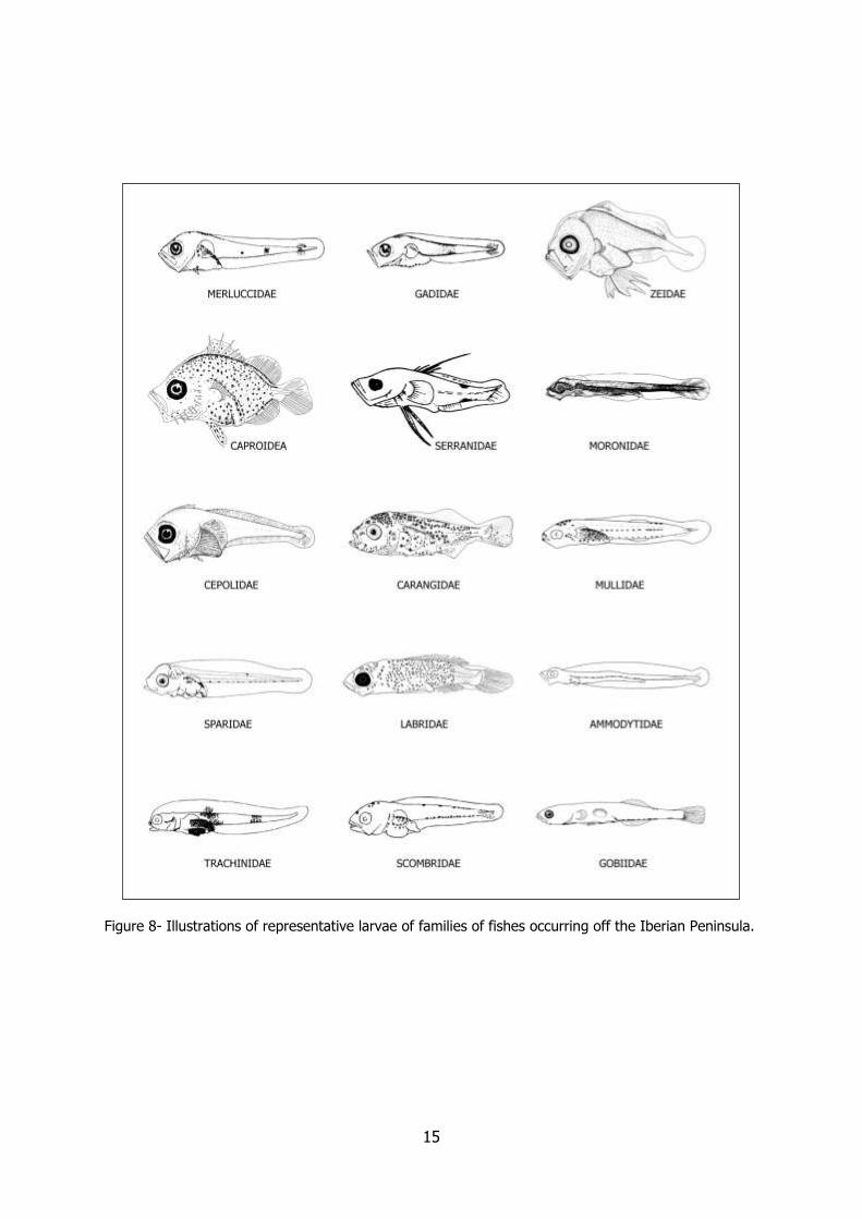
Identifying a fish larva is very different from identifying an adult specimen. Its shape, size, stage of
development, pigmentation pattern and myomere count are very important features. New ways of
manipulating data are needed to reach identification. This has been called the “look alike” system (Moser et al., 1984). It consists of a simple procedure: one should be able to identify a fish larva to
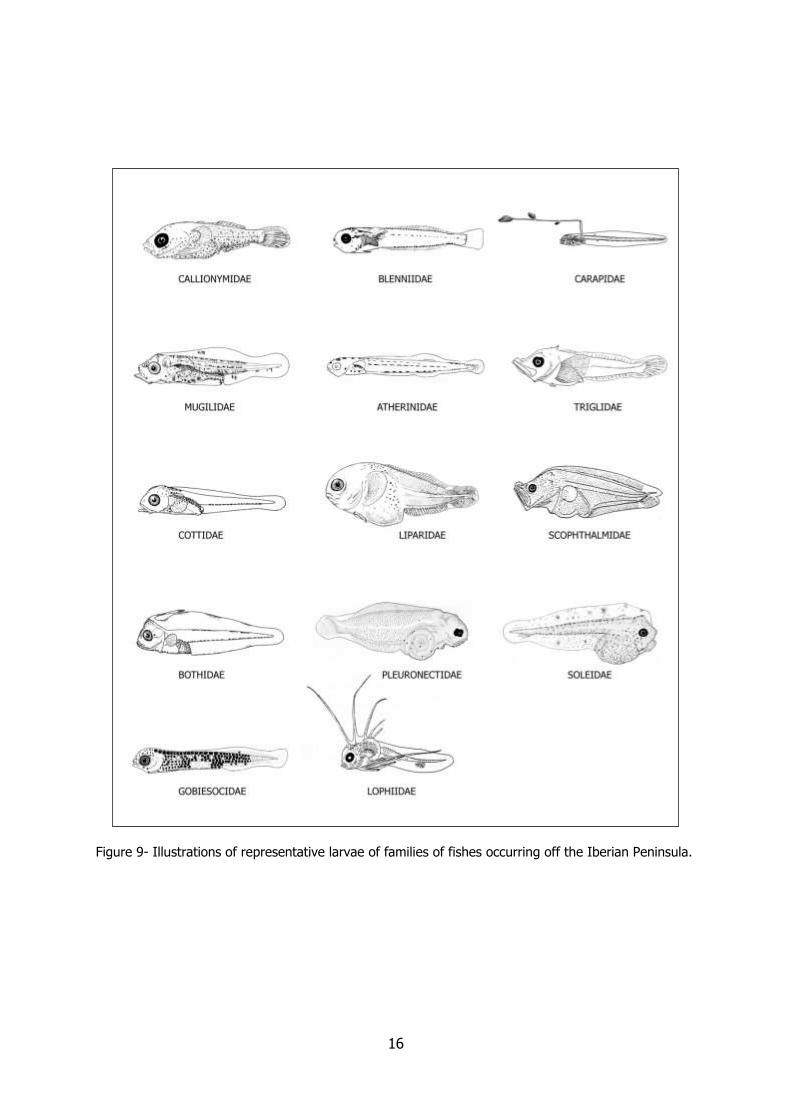
the family level by matching the characteristics of a specimen as far as general shape and striking features are concerned (Figures 7 to 9).
Meristic characters are also very important. Total myomere (vertebrae) count is presented in Table 2. The identification can be carried further by reading the information presented for each species.
TABLE 2
List of families in each interval of vertebral counts. Adapted from Aboussouan (1994).
15-23 vertebrae 31-40 vertebrae 51-68 vertebrae Callionymidae Blenniidae Ammodytidae Labridae Bothidae Argentinidae Macroramphosidae Gadidae Atherinidae
Gobiesocidae Cepolidae 24-26 vertebrae Gobiidae Clupeidae Carangidae Gonostomatidae Gadidae Centracanthidae Labridae Liparidae Gobiidae Lophiidae Merluccidae Labridae Myctophidae Paralepididae Lophiidae Pleuronectidae Scombridae Macroramphosidae Scombridae Soleidae Moronidae Scophthalmidae Syngnathidae Mugilidae Soleidae Mullidae Sternoptychidae 69-100 vertebrae Serranidae Trachinidae Belonidae Sparidae Triglidae Carapidae
Zeidae Gadidae 27-30 vertebrae Paralepididae Blenniidae 41-50 vertebrae Syngnathidae
Cottidae Argentinidae Gobiesocidae Atherinidae >105 vertebrae
Gobiidae Blenniidae Anguillidae Labridae Bothidae Carapidae Lophiidae Clupeidae Congridae Serranidae Engraulidae Muraenidae Triglidae Gadidae Syngnathidae
Gonostomatidae Merluccidae Myctophidae Scombridae Scophthalmidae Soleidae Syngnathidae Trachinidae
14
Figure 7- Illustrations of representative larvae of families of fishes occurring off the Iberian Peninsula.
15
Figure 8- Illustrations of representative larvae of families of fishes occurring off the Iberian Peninsula.
16
Figure 9- Illustrations of representative larvae of families of fishes occurring off the Iberian Peninsula.
17
EARLY LIFE HISTORY DESCRIPTIONS
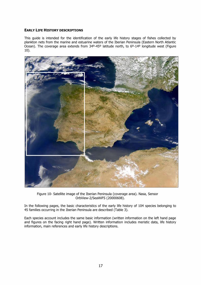
This guide is intended for the identification of the early life history stages of fishes collected by plankton nets from the marine and estuarine waters of the Iberian Peninsula (Eastern North Atlantic
Ocean). The coverage area extends from 34º-45º latitude north, to 6º-14º longitude west (Figure 10).
Figure 10- Satellite image of the Iberian Peninsula (coverage area). Nasa, Sensor
OrbView-2/SeaWiFS (20000608).
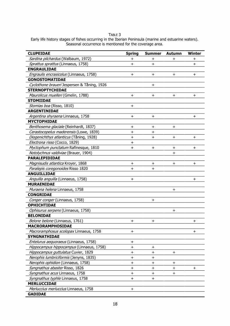
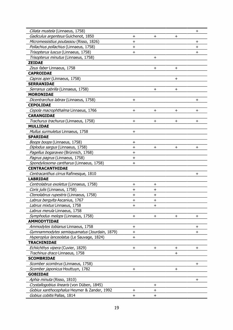
In the following pages, the basic characteristics of the early life history of 104 species belonging to 45 families occurring in the Iberian Peninsula are described (Table 3).
Each species account includes the same basic information (written information on the left hand page and figures on the facing right hand page). Written information includes meristic data, life history
information, main references and early life history descriptions.
18
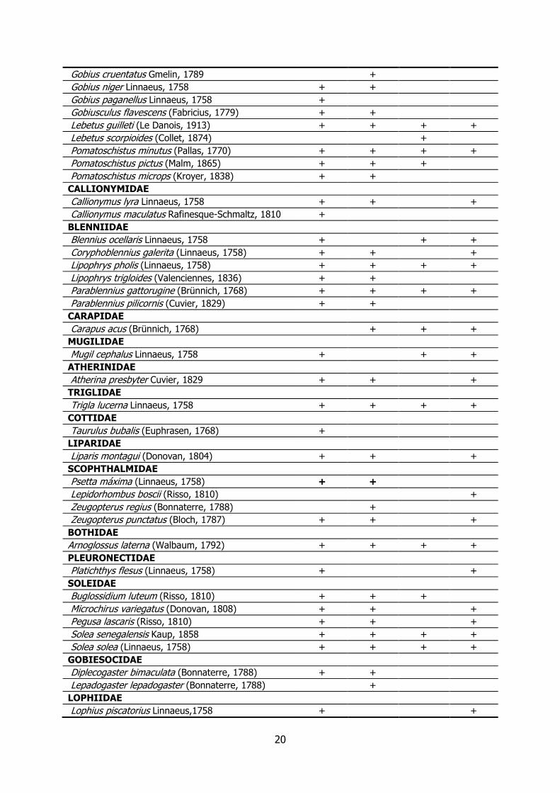
TABLE 3 Early life history stages of fishes occurring in the Iberian Peninsula (marine and estuarine waters).
Seasonal occurrence is mentioned for the coverage area.
CLUPEIDAE Spring Summer Autumn Winter
Sardina pilchardus (Walbaum, 1972) + + + +
Sprattus sprattus (Linnaeus, 1758) + + +
ENGRAULIDAE
Engraulis encrasicolus (Linnaeus, 1758) + + + +
GONOSTOMATIDAE
Cyclothone braueri Jespersen & Tåning, 1926 +
STERNOPTYCHIDAE
Maurolicus muelleri (Gmelin, 1788) + + + +
STOMIIDAE
Stomias boa (Risso, 1810) +
ARGENTINIDAE
Argentina shyraena Linnaeus, 1758 + + +
MYCTOPHIDAE
Benthosema glaciale (Reinhardt, 1837) + + +
Cerastocopelus maderensis (Lowe, 1839) + +
Diogenichthys atlanticus (Tåning, 1928) + + + +
Electrona risso (Cocco, 1829) +
Myctophum punctatum Rafinesque, 1810 + + + +
Notolychnus valdiviae (Brauer, 1904) +
PARALEPIDIDAE
Magnisudis atlantica Kroyer, 1868 + + + +
Paralepis coregonoides Risso 1820 + +
ANGUILLIDAE
Anguilla anguilla (Linnaeus, 1758) + +
MURAENIDAE
Muraena helena Linnaeus, 1758 +
CONGRIDAE
Conger conger (Linnaeus, 1758) +
OPHICHTIDAE
Ophisurus serpens (Linnaeus, 1758) +
BELONIDAE
Belone belone (Linnaeus, 1761) + + +
MACRORAMPHOSIDAE
Macroramphosus scolopax Linnaeus, 1758 + +
SYNGNATHIDAE
Entelurus aequoraeus (Linnaeus, 1758) +
Hippocampus hippocampus (Linnaeus, 1758) + +
Hippocampus guttulatus Cuvier, 1829 + + +
Nerophis lumbriciformis (Jenyns, 1835) + +
Nerophis ophidion (Linnaeus, 1758) + + +
Syngnathus abaster Risso, 1826 + + + +
Syngnathus acus Linnaeus, 1758 + + +
Syngnathus typhle Linnaeus, 1758 + +
MERLUCCIDAE
Merluccius merluccius Linnaeus, 1758 +
GADIDAE
19
Ciliata mustela (Linnaeus, 1758) +
Gadiculus argenteus Guichenot, 1850 + + +
Micromessistius poutassou (Risso, 1826) + +
Pollachius pollachius (Linnaeus, 1758) + +
Trisopterus luscus (Linnaeus, 1758) + +
Trisopterus minutus (Linnaeus, 1758) +
ZEIDAE
Zeus faber Linnaeus, 1758 + +
CAPROIDAE
Capros aper (Linnaeus, 1758) +
SERRANIDAE
Serranus cabrilla (Linnaeus, 1758) + +
MORONIDAE
Dicentrarchus labrax (Linnaeus, 1758) + +
CEPOLIDAE
Cepola macrophthalma Linnaeus, 1766 + + + +
CARANGIDAE
Trachurus trachurus (Linnaeus, 1758) + + + +
MULLIDAE
Mullus surmuletus Linnaeus, 1758 +
SPARIDAE
Boops boops (Linnaeus, 1758) +
Diplodus sargus (Linnaeus, 1758) + + + +
Pagellus bogaraveo (Brünnich, 1768) +
Pagrus pagrus (Linnaeus, 1758) +
Spondyliosoma cantharus (Linnaeus, 1758) +
CENTRACANTHIDAE
Centracanthus cirrus Rafinesque, 1810 +
LABRIDAE
Centrolabrus exoletus (Linnaeus, 1758) + +
Coris julis (Linnaeus, 1758) + +
Ctenolabrus rupestris (Linnaeus, 1758) + +
Labrus bergylta Ascanius, 1767 + +
Labrus mixtus Linnaeus, 1758 + +
Labrus merula Linnaeus, 1758
Symphodus melops (Linnaeus, 1758) + + + +
AMMODYTIDAE
Ammodytes tobianus Linnaeus, 1758 + +
Gymnammodytes semisquamatus (Jourdain, 1879) + +
Hyperoplus lanceolatus (Le Sauvage, 1824) +
TRACHINIDAE
Echiichthys vipera (Cuvier, 1829) + + + +
Trachinus draco Linnaeus, 1758 +
SCOMBRIDAE
Scomber scombrus (Linnaeus, 1758) +
Scomber japonicus Houttuyn, 1782 + +
GOBIIDAE
Aphia minuta (Risso, 1810) +
Crystallogobius linearis (von Düben, 1845) +
Gobius xanthocephalus Heymer & Zander, 1992 + +
Gobius cobitis Pallas, 1814 + +
20
Gobius cruentatus Gmelin, 1789 +
Gobius niger Linnaeus, 1758 + +
Gobius paganellus Linnaeus, 1758 +
Gobiusculus flavescens (Fabricius, 1779) + +
Lebetus guilleti (Le Danois, 1913) + + + +
Lebetus scorpioides (Collet, 1874) +
Pomatoschistus minutus (Pallas, 1770) + + + +
Pomatoschistus pictus (Malm, 1865) + + +
Pomatoschistus microps (Kroyer, 1838) + +
CALLIONYMIDAE
Callionymus lyra Linnaeus, 1758 + + +
Callionymus maculatus Rafinesque-Schmaltz, 1810 +
BLENNIIDAE
Blennius ocellaris Linnaeus, 1758 + + +
Coryphoblennius galerita (Linnaeus, 1758) + + +
Lipophrys pholis (Linnaeus, 1758) + + + +
Lipophrys trigloides (Valenciennes, 1836) + +
Parablennius gattorugine (Brünnich, 1768) + + + +
Parablennius pilicornis (Cuvier, 1829) + +
CARAPIDAE
Carapus acus (Brünnich, 1768) + + +
MUGILIDAE
Mugil cephalus Linnaeus, 1758 + + +
ATHERINIDAE
Atherina presbyter Cuvier, 1829 + + +
TRIGLIDAE
Trigla lucerna Linnaeus, 1758 + + + +
COTTIDAE
Taurulus bubalis (Euphrasen, 1768) +
LIPARIDAE
Liparis montagui (Donovan, 1804) + + +
SCOPHTHALMIDAE
Psetta máxima (Linnaeus, 1758) + +
Lepidorhombus boscii (Risso, 1810) +
Zeugopterus regius (Bonnaterre, 1788) +
Zeugopterus punctatus (Bloch, 1787) + + +
BOTHIDAE
Arnoglossus laterna (Walbaum, 1792) + + + +
PLEURONECTIDAE
Platichthys flesus (Linnaeus, 1758) + +
SOLEIDAE
Buglossidium luteum (Risso, 1810) + + +
Microchirus variegatus (Donovan, 1808) + + +
Pegusa lascaris (Risso, 1810) + + +
Solea senegalensis Kaup, 1858 + + + +
Solea solea (Linnaeus, 1758) + + + +
GOBIESOCIDAE
Diplecogaster bimaculata (Bonnaterre, 1788) + +
Lepadogaster lepadogaster (Bonnaterre, 1788) +
LOPHIIDAE
Lophius piscatorius Linnaeus,1758 + +

21
22
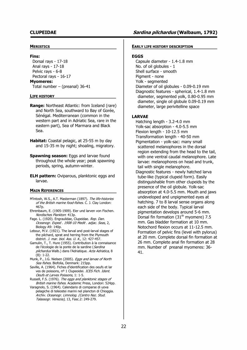
CLUPEIDAE Sardina pilchardus (Walbaum, 1792) MERISTICS
Fins:
Dorsal rays - 17-18 Anal rays - 17-18
Pelvic rays - 6-8 Pectoral rays - 16-17
Myomeres:
Total number – (preanal) 36-41
LIFE HISTORY
Range: Northeast Atlantic: from Iceland (rare) and North Sea, southward to Bay of Gorée,
Sénégal. Mediterranean (common in the
western part and in Adriatic Sea, rare in the eastern part), Sea of Marmara and Black
Sea.
Habitat: Coastal pelagic, at 25-55 m by day
and 15-35 m by night; shoaling, migratory.
Spawning season: Eggs and larvae found throughout the whole year; peak spawning
periods, spring, autumn-winter.
ELH pattern: Oviparous, planktonic eggs and
larvae.
MAIN REFERENCES
M'intosh, W.S., A.T. Masterman (1897). The life-histories of the British marine food-fishes. C. J. Clay London: 467p.
Ehrenbaum, E. (1905-1909). Eier und larven von Fischen. Nordisches Plankton: 413p.
Fage. L. (1920). Engraulidae, Clupeidae. Rep. Dan. Oceanogr. Exped . 1908-10 Medit . adjac. Seas, 2, Biology A9: 140p.
Lebour, M.V. (1921). The larval and post-larval stages of the pilchard, sprat and herring from the Plymouth district. J. mar. biol. Ass. U. K., 12: 427-457.
Gamulin, T., T. Hure (1955). Contribution à la connaisance de l'écologie de la ponte de la sardine (Sardina pilchardus Walb.) dans l'Adriatique. Acta Adriatica, 8 (8): 1-22.
Munk, P., J.G. Nielsen (2005). Eggs and larvae of North Sea fishes. Biofolia, Denmark: 215pp.
Saville, A. (1964). Fiches d'identification des oeufs et lar ves de poissons, nº 1 Clupeoidei. ICES Fich. Ident. Oeufs et Larves Poissons, 1: 1-5.
Russell, F.S. (1976). The eggs and planktonic stages of British marine fishes. Academic Press, London: 524pp.
Varagnolo, S. (1964). Calendario di comparse di uova pelagiche di teleostei marini nel plancton di Chioggia. Archiv. Oceanogr. Limnolog. (Centro Naz. Stud. Talassogr. Venezia), 13, Fasc.2: 249-279.
EARLY LIFE HISTORY DESCRIPTION
EGGS
Capsule diameter - 1.4-1.8 mm No. of oil globules - 1
Shell surface - smooth Pigment - none
Yolk - segmented
Diameter of oil globules - 0.09-0.19 mm Diagnostic features - spherical, 1.4-1.8 mm
diameter, segmented yolk, 0.80-0.95 mm diameter, single oil globule 0.09-0.19 mm
diameter, large perivitelline space
LARVAE
Hatching length - 3.2-4.0 mm Yolk-sac absorption - 4.0-5.5 mm
Flexion length - 10-12.5 mm
Transformation length - 40-50 mm Pigmentation - yolk-sac: many small
scattered melanophores in the dorsal region extending from the head to the tail,
with one ventral caudal melanophore. Late larvae: melanophores on head and trunk,
tail with single melanophore.
Diagnostic features - newly hatched larva tube-like (typical clupeid form). Easily
distinguishable from other clupeids by the presence of the oil globule. Yolk-sac
absorption at 4.0-5.5 mm. Mouth and jaws
undeveloped and unpigmented eyes at hatching. 7 to 8 larval sense organs along
each side of the body. Typical larval pigmentation develops around 5-6 mm.
Dorsal fin formation (31th myomere) 7.5 mm. Gas bladder formation at 10 mm.
Notochord flexion occurs at 11-12.5 mm.
Formation of pelvic fins (level with pylorus) at 20 mm. Complete dorsal fin formation at
26 mm. Complete anal fin formation at 28 mm. Number of preanal myomeres: 36-
41.

23
CLUPEIDAE Sardina pilchardus (Walbaum, 1792)
Plate 1- Stages of development of Sardina pilchardus eggs. Ré et al. (1988).

24
CLUPEIDAE Sardina pilchardus (Walbaum, 1792)
Plate 2- Early life history stages of Sardina pilchardus. Varagnolo (1964), Fage (1920).

25
26
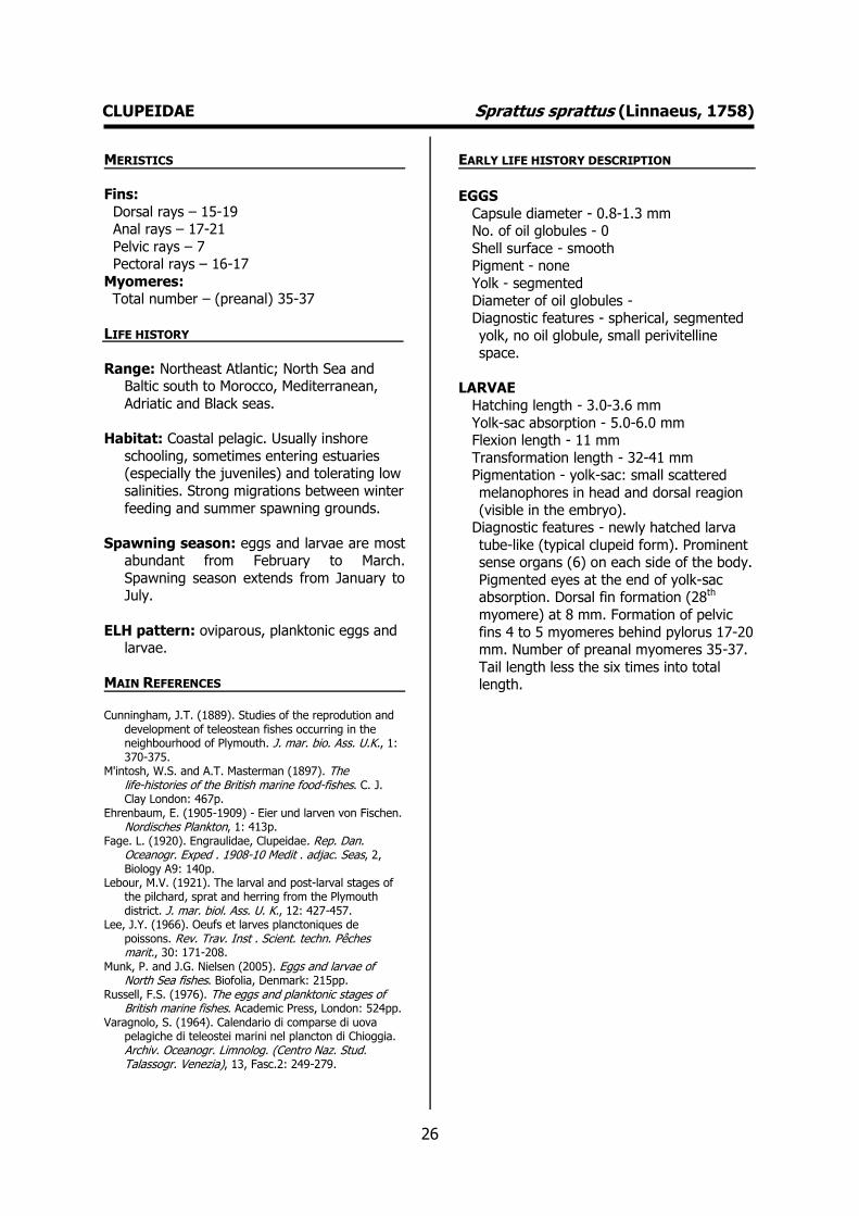
CLUPEIDAE Sprattus sprattus (Linnaeus, 1758) MERISTICS
Fins:
Dorsal rays – 15-19 Anal rays – 17-21
Pelvic rays – 7 Pectoral rays – 16-17
Myomeres: Total number – (preanal) 35-37
LIFE HISTORY
Range: Northeast Atlantic; North Sea and Baltic south to Morocco, Mediterranean,
Adriatic and Black seas.
Habitat: Coastal pelagic. Usually inshore
schooling, sometimes entering estuaries (especially the juveniles) and tolerating low
salinities. Strong migrations between winter
feeding and summer spawning grounds.
Spawning season: eggs and larvae are most abundant from February to March.
Spawning season extends from January to July.
ELH pattern: oviparous, planktonic eggs and larvae.
MAIN REFERENCES Cunningham, J.T. (1889). Studies of the reprodution and
development of teleostean fishes occurring in the neighbourhood of Plymouth. J. mar. bio. Ass. U.K., 1: 370-375.
M'intosh, W.S. and A.T. Masterman (1897). The life-histories of the British marine food-fishes. C. J. Clay London: 467p.
Ehrenbaum, E. (1905-1909) - Eier und larven von Fischen. Nordisches Plankton, 1: 413p.
Fage. L. (1920). Engraulidae, Clupeidae. Rep. Dan. Oceanogr. Exped . 1908-10 Medit . adjac. Seas, 2, Biology A9: 140p.
Lebour, M.V. (1921). The larval and post-larval stages of the pilchard, sprat and herring from the Plymouth district. J. mar. biol. Ass. U. K., 12: 427-457.
Lee, J.Y. (1966). Oeufs et larves planctoniques de poissons. Rev. Trav. Inst . Scient. techn. Pêches marit., 30: 171-208.
Munk, P. and J.G. Nielsen (2005). Eggs and larvae of North Sea fishes. Biofolia, Denmark: 215pp.
Russell, F.S. (1976). The eggs and planktonic stages of British marine fishes. Academic Press, London: 524pp.
Varagnolo, S. (1964). Calendario di comparse di uova pelagiche di teleostei marini nel plancton di Chioggia. Archiv. Oceanogr. Limnolog. (Centro Naz. Stud. Talassogr. Venezia), 13, Fasc.2: 249-279.
EARLY LIFE HISTORY DESCRIPTION
EGGS
Capsule diameter - 0.8-1.3 mm No. of oil globules - 0
Shell surface - smooth Pigment - none
Yolk - segmented
Diameter of oil globules - Diagnostic features - spherical, segmented
yolk, no oil globule, small perivitelline space.
LARVAE
Hatching length - 3.0-3.6 mm
Yolk-sac absorption - 5.0-6.0 mm Flexion length - 11 mm
Transformation length - 32-41 mm Pigmentation - yolk-sac: small scattered
melanophores in head and dorsal reagion
(visible in the embryo). Diagnostic features - newly hatched larva
tube-like (typical clupeid form). Prominent sense organs (6) on each side of the body.
Pigmented eyes at the end of yolk-sac absorption. Dorsal fin formation (28th
myomere) at 8 mm. Formation of pelvic
fins 4 to 5 myomeres behind pylorus 17-20 mm. Number of preanal myomeres 35-37.
Tail length less the six times into total length.
27
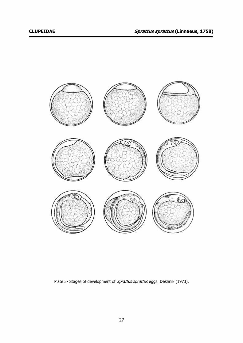
CLUPEIDAE Sprattus sprattus (Linnaeus, 1758)
Plate 3- Stages of development of Sprattus sprattus eggs. Dekhnik (1973).

28
CLUPEIDAE Sprattus sprattus (Linnaeus, 1758)
Plate 4- Early life history stages of Sprattus sprattus. Varagnolo (1964) and Russell (1976).

29
30
ENGRAULIDAE Engraulis encrasicolus (Linnaeus, 1758) MERISTICS
Fins:
Dorsal rays - 15-18 Anal rays - 16-18
Pelvic rays - 7 Pectoral rays - 15-17
Myomeres:
Total number – 44-47
LIFE HISTORY
Range: Eastern Atlantic; Bergen, Norway to South Africa. Mediterranean, Black and
Azov Seas.
Habitat: Coastal marine species, forming
large schools. Tolerates salinities of 5-41 %º and in some areas enters estuaries and
lagoons, especially during spawning. Tends
to move further north and into surface waters in summer, retreating and
descending in winter.
Spawning season: Spawns from March to November with peaks usually in summer.
ELH pattern: oviparous, planktonic eggs and larvae.
MAIN REFERENCES Raffaele, F. (1888). Le uova galleggianti e la larve dei Te-
leostei nel Golfo di Napoli. Mitt. zool. Stn Neapel, 8: 1-85.
Ehrenbaum, E. (1905-1909). Eier und larven von Fischen. Nordisches Plankton, 1: 413p.
Fage. L. (1920). Engraulidae, Clupeidae. Rep. Dan. Oceanogr. Exped . 1908-10 Medit . adjac. Seas, 2, Biology A9: 140p.
Lee, J.Y. (1966). Oeufs et larves planctoniques de poissons. Rev. Trav. Inst . Scient. techn. Pêches marit., 30: 171-208.
Munk, P. and J.G. Nielsen (2005). Eggs and larvae of North Sea fishes. Biofolia, Denmark: 215pp.
Russell, F.S. (1976). The eggs and planktonic stages of British marine fishes. Academic Press, London: 524pp.
Ré, P. (1986). Sobre a identificação dos primeiros estados larvares planctónicos de Sardina pilchardus (Walbaum, 1792) e de Engraulis encrasicolus (Linnaeus, 1758). Ciência Biológica. Ecology Systematics, 6 (1/2): 135-140.
Ré, P. (1999). Ictioplâncton estuarino da Península Ibérica (Guia de identificação dos ovos e estados larvares planctónicos), 163pp, 51 fig. Prémio do Mar, 1996. Câmara Municipal de Cascais. ISBN 972-637-065-5.
Varagnolo, S. (1964). Calendario di comparse di uova pelagiche di teleostei marini nel plancton di Chioggia. Archiv. Oceanogr. Limnolog. (Centro Naz. Stud. Talassogr. Venezia), 13, Fasc.2: 249-279.
EARLY LIFE HISTORY DESCRIPTION
EGGS
Capsule diameter - 1.2-1.9 mm x 0.5-1.2 mm
No. of oil globules - 0 Shell surface - smooth
Pigment - none
Yolk - segmented Diameter of oil globules -
Diagnostic features - Ovoid in shape. Segmented yolk, no oil globule, small
perivitelline space.
LARVAE
Hatching length - 3.0-4.0 mm Yolk-sac absorption - 5.0 mm
Flexion length - 9.0-10.0 mm Transformation length - 25 mm
Pigmentation - Early larvae: two
melanophores on abdominal wall behind pylorus, two on anal papilla, one or two in
tail region. Late larvae: Row of melanophores on top of the gut.
Diagnostic features - Head more than five times into total length, dorsal fin opposite
to anus. Pelvic fins on level with pylorus.
Late larvae: Head with characteristic adult shape. Dorsal and anal fins overlapping.

31
ENGRAULIDAE Engraulis encrasicolus (Linnaeus, 1758)
Plate 5- Stages of development of Engraulis encrasicolus eggs. Dekhnik (1973).
32
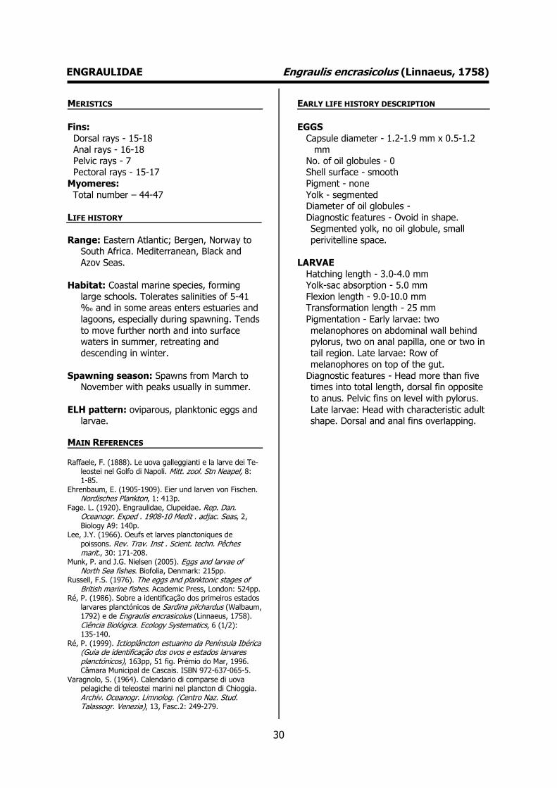
ENGRAULIDAE Engraulis encrasicolus (Linnaeus, 1758)
Plate 6- Early life history stages of Engraulis encrasicolus. Ré (unpublished data).

33
34
GONOSTOMATIDAE Cyclothone braueri Jespersen & Tåning, 1926 MERISTICS
Fins:
Dorsal rays – 13-15 Anal rays – 18-20
Pelvic rays – 6-7 Pectoral rays – 9-10
Myomeres: Total number – 29-33
LIFE HISTORY
Range: Atlantic, Indian and South Pacific: in tropical and subtropical waters, including
the Mediterranean. Also known from the
temperate North Atlantic as far as 66 ºN and in the sub-Antarctic water south of
Australia.
Habitat: Mesopelagic; depth range 10 – 2000
m. Deep-water; 67 ºN – 40 ºS, 180 ºW – 180 ºE.
Spawning season: Larvae found in spring
and summer offshore.
ELH pattern: Oviparous, planktonic larvae.
MAIN REFERENCES Jespersen, P., A.V. Tåning (1926). Mediterranean
Sternoptychidae. Rep. Dan. Oceanogr. Exp. Mediterr., 2 (A.12): 1-59.
Sanzo, L. (1931). Salmonoidei and Stomiatoidei. In Uova, larve e stadi Giovanili di Teleostei: monografia elaborata con l’uso del materiale raccolto e sweriato da Salvatore Lo Bianco. Fauna e Flora Golfo Napoli Monogr. 38 (1): 21-92.
EARLY LIFE HISTORY DESCRIPTION
EGGS: Undescribed
Capsule diameter - Unknown No. of oil globules - Unknown
Shell surface - Unknown Pigment - Unknown
Yolk - Unknown
Diameter of oil globules - Unknown Diagnostic features - Unknown
LARVAE
Hatching length – Unknown Yolk-sac absorption - Unknown
Flexion length – 4.5-5.5 mm
Transformation length – 14 mm Pigmentation – 2-3 melanophores along
gut. 11-12 pairs of evenly-spaced melanophores over anal base. Lateral
series of melanophores posterior to
pectoral fin. 14-15 melanophores on pterygiophores, 1 melanophore over
urostyle and 3 melanophores laterally before the urostyle.
Diagnostic features – Body slender, elongate, preanal length 50 % of standard
length, round eyes, pipgmented gas
bladder. Photophores form at about 11-12 mm. Larvae similar to Vinciguerria.
35
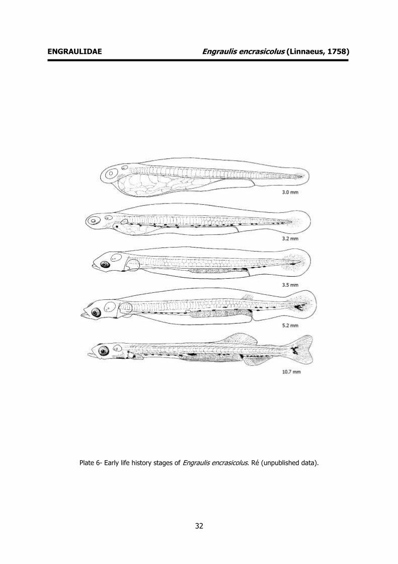
GONOSTOMATIDAE Cyclothone braueri Jespersen & Tåning, 1926
Plate 7- Early life history stages of Cyclothone braueri. Sanzo (1931).

36
STERNOPTYCHIDAE Maurolicus muelleri (Gmelin, 1789) MERISTICS
Fins:
Dorsal soft rays – 10-11 Anal soft rays – 19-22
Pelvic rays – 6-8 Pectoral rays – 17-20
Myomeres: Total number – 33-35
LIFE HISTORY
Range: Eastern Atlantic: Iceland and Norway to Senegal, including the western
Mediterranean. Western Atlantic: Gulf of
Maine to the Gulf of Mexico, Caribbean Sea and the Straits of Magellan. Southeast
Pacific: Chile. Western Pacific.
Habitat: Bathypelagic; marine; depth range 0
– 1524 m. Deep-water; 72 ºN – 55 ºS, 98 ºW – 41 ºE.
Spawning season: Eggs and larvae captured
throughout the whole year.
ELH pattern: Oviparous, planktonic eggs and
larvae.
MAIN REFERENCES Fahay, M.P. (1983). Guide to the early stages of marine
fishes occurring in the Western North Atlantic Ocean, Cape Hattaras to the southern Scotian Shelf. J. Northwest Atl. Fish. Sci., 4 : 1-423.
Olivar, P., J.-M. Fortuño (1991). Guide to the ichthyoplankton of the Southeast Atlantic (Benguela current region). Scientia Marina, 55 (1): 1-383.
Robertson, D.A. (1976). Planktonic stages of Maurolicus muelleri (Telesostei: Sternoptychidae) in New Zealand waters. N.Z. J. Mar. Freshwater Res., 10: 331-328.
EARLY LIFE HISTORY DESCRIPTION
EGGS
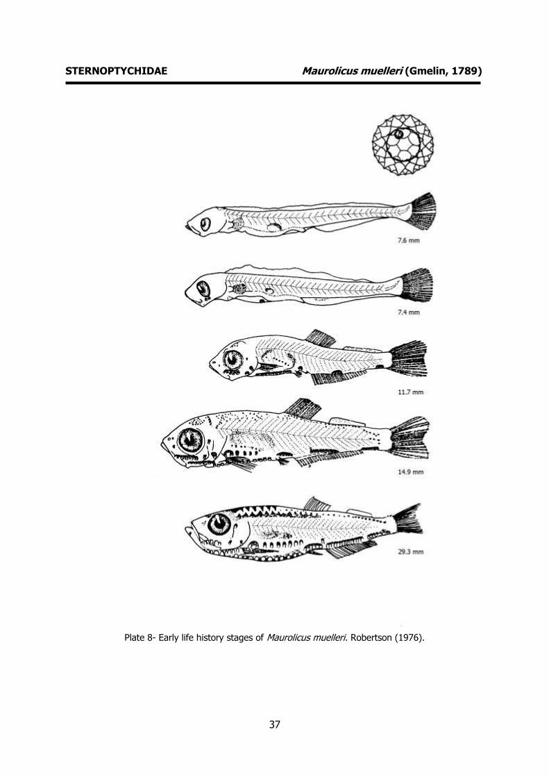
Capsule diameter – 1.60-1.84 mm No. of oil globules – 1
Shell surface – Sculptured chorion. Network of hexagonal and pentagonal polygons
Pigment - None Yolk - Segmented
Diameter of oil globules – 0.21-0.26 mm
Diagnostic features – Unique chorion ornamentation. Small periviteline space.
LARVAE
Hatching length - Unknown
Yolk-sac absorption - Unknown Flexion length – 5-7 mm
Transformation length – 11-12 mm Pigmentation – Complete absence of
melanophores up to a length of 7 mm.
Diagnostic features – Eyes vertically elliptical, rounded in late larvae. Early
appearance of photophores. Gut < 50 % of standard length. Lack of pigment except
over gas bladder. Development of photophores differs among Atlantic,
Mediterranean and Pacific late larvae.
37
STERNOPTYCHIDAE Maurolicus muelleri (Gmelin, 1789)
Plate 8- Early life history stages of Maurolicus muelleri. Robertson (1976).
38
STOMIIDAE Stomias boa (Risso, 1810) MERISTICS
Fins:
Dorsal rays – 17-21 Anal rays – 19-23
Pelvic rays - 5 Pectoral rays – 6-7
Myomeres:
Total number – 75-78
LIFE HISTORY
Range: Eastern Atlantic: western Mediterranean south to Mauritania, and
from Angola to South Africa. Southwest
Atlantic: Brazil to Argentina. Southeast Pacific: Chile. Sub-Antarctic region of the
Indian Ocean sector south to Heard Island.
Habitat: Found in deep oceanic waters to
more than 1000 m depth, may migrate to near-surface waters at night.
Spawning season: Larvae found in spring.
Rare.
ELH pattern: Oviparous, planktonic larvae.
MAIN REFERENCES Ege, V. (1918). Stomiatoidae. Rep. Danish Oceanogr. Exp.
Medit. 1908-1910, Vol II (A4): 28pp. Olivar, P., J.-M. Fortuño (1991). Guide to the
ichthyoplankton of the Southeast Atlantic (Benguela current region). Scientia Marina, 55 (1): 1-383.
Richards, W.J. (ed) (2005). Early Stages of Atlantic Fishes: An Identification Guide for the Western Central Atlantic. CRC Press Inc., U.S. Two volumes: 2640pp.
Sanzo, L. (1931). Salmonoidei and Stomiatoidei. In Uova, larve e stadi Giovanili di Teleostei: monografia elaborata con l’uso del materiale raccolto e sweriato da Salvatore Lo Bianco. Fauna e Flora Golfo Napoli Monogr.38 (1): 21-92.
EARLY LIFE HISTORY DESCRIPTION
EGGS: Undescribed Capsule diameter - Unknown No. of oil globules - Unknown
Shell surface - Unknown Pigment - Unknown
Yolk - Unknown
Diameter of oil globules - Unknown Diagnostic features - Unknown
LARVAE
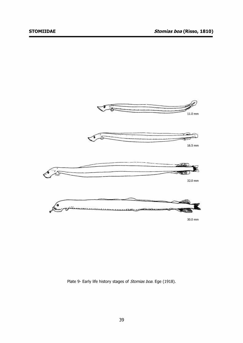
Hatching length – 3-4 mm ? Yolk-sac absorption - Unknown
Flexion length - Unknown
Transformation length – 38–41.5 mm Pigmentation – Distinct row of ventral
pigment above the gut, intermittent pigment on dorsum, anal fin and lower
caudal fin. Photophores appear around 30
mm. Diagnostic features – Larvae elongate with
long heads and prominent jaws. Elliptical eyes. Caudal fin fold in form of spatula.
Pectorals very small and pedunculate. Slightly trailing gut (until transformation).
At transformation when the barbell
appears the juveniles are shorter than the late larvae.
39
STOMIIDAE Stomias boa (Risso, 1810)
Plate 9- Early life history stages of Stomias boa. Ege (1918).
40
ARGENTINIDAE Argentina sphyraena Linnaeus, 1758 MERISTICS
Fins:
Dorsal rays – 10-12 Anal rays – 11-15
Pelvic rays – 10-12 Pectoral rays – 12-15
Myomeres: Total number – 46-55: 36 (preanal) 17-19
(postanal)
LIFE HISTORY
Range: Eastern Atlantic: northern Norway to
western Sahara including southern Iceland,
Faroe Islands, Shetlands and western Mediterranean.
Habitat: Relatively common on the
continental shelf and upper slope, probably
schools near the bottom. Depth range 50-500 m.
Spawning season: Larvae found in spring
and summer.
ELH pattern: Oviparous, planktonic eggs and larvae.
MAIN REFERENCES Ehrenbaum, E. (1905-1909). Eier und Larven von Fischen.
Nordisches Plankton, 1: 413pp. Munk, P., J.G. Nielsen (2005). Eggs and larvae of North
Sea fishes. Biofolia, Denmark: 215pp. Russell, F.S. (1976). The eggs and planktonic stages of
British marine fishes. Academic Press, London: 524pp. Schmidt, J. (1906). On the larval and post-larval
development of the argentines Argentina silus (Ascan.) and Angentina sphyraena (Linné) with some notes on Malloyus villosus (O.F. Muller). Medd. Komm. Havunders. Ser. Fiskeri 2 (4): 20pp.
Schmidt, J. (1918). Argentinidae, Microstomidae, Opisthoproctidae, Mediterranean Odontostomidae. Rep. Dan. Oceanogr. Exped. 1908-10 Medit. adiac. seas 2 Biology, A5: 1-40.
EARLY LIFE HISTORY DESCRIPTION
EGGS
Capsule diameter – 1.70-1.85 mm No. of oil globules - 1
Shell surface - smooth Pigment - Small melanophores on the
embryo, and numerous stellate melanophores on the york sac (which vary
in position and number). Advanced embryo
exhibits ventral melanophores and a caudal group on the dorsal and ventral
sides. Yolk - segmented
Diameter of oil globules – 0.37-0.47 mm
Diagnostic features – Large oil globule. Segmented yolk, small periviteline space.
LARVAE
Hatching length – 7-8 mm
Yolk-sac absorption – 9-10 mm Flexion length – 13-17 mm
Transformation length – 50 mm? Pigmentation – Large stellate melanophores
on yolk sac (anterior portion). 6 Groups of melanophores present along the dorsal
side of the gut. Postanal dorsal and anal
groups of melanophores. Pigmentation of late larvae identical.
Diagnostic features – Typical pigmentation. Seven groups of equally spaced
melanophores along the ventral contour.
The primordial fin does not disappear until the larva is over 30 mm.

41
ARGENTINIDAE Argentina sphyraena Linnaeus, 1758
Plate 10- Early life history stages of Argentina sphyraena. Schmidt (1906), Russell (1976).

42
MYCTOPHIDAE Benthosema glaciale (Reinhardt, 1837) MERISTICS
Fins:
Dorsal rays – 12-14 Anal rays – 17-19
Pelvic rays – 17-19 Pectoral rays – 11-13
Myomeres: Total number – 34-36
LIFE HISTORY
Range: Eastern Atlantic: Norway and Greenland south to Morocco, and from
Mauritania to Guinea (Mauritanian
Upwelling Region). Seasonally present from Morocco to Mauritania along the edge of
the continental shelf. Also known from the Mediterranean Sea. Western Atlantic: Baffin
Bay to northern edge of Gulf Stream.
Habitat: Mesopelagic at depths between 375-
800 m during daytime and 12-200 m during night.
Spawning season: Larvae present during
spring and summer
ELH pattern: Oviparous, planktonic larvae.
MAIN REFERENCES Fahay, M.P. (1983). Guide to the early stages of marine
fishes occurring in the Western North Atlantic Ocean, Cape Hattaras to the southern Scotian Shelf. J. Northwest Atl. Fish. Sci., 4 : 1-423.
Moser, H.G., E.H. Ahlstrom (1974). Role of larval stages in systematic investigations of marine teleosts: the Myctophidae, a case study. Fish. Bull. U.S., 72: 391-413.
Shiganova, T.A. (1977). Larvae and juvenile of lantern-fishes (Myctophidae, Pisces) of the Atlantic Ocean. Proceedings of the P.P Shirshov Institute of Oceanology, 109: 42-112.
EARLY LIFE HISTORY DESCRIPTION
EGGS: Undescribed
Capsule diameter - Unknown No. of oil globules - Unknown
Shell surface - Unknown Pigment - Unknown
Yolk - Unknown
Diameter of oil globules - Unknown Diagnostic features - Unknown
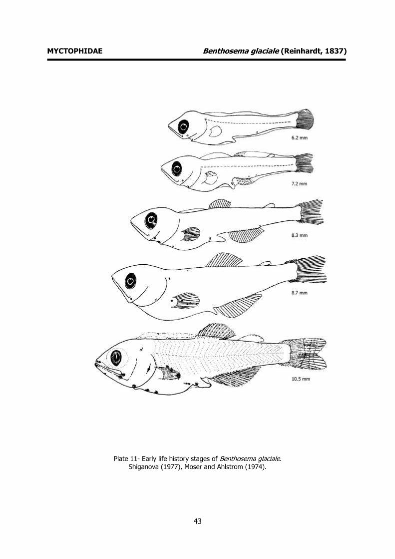
LARVAE
Hatching length - Unknown Yolk-sac absorption - Unknown
Flexion length – 5-7 mm
Transformation length – 11 mm Pigmentation – Pigmented pectoral fins.
Melanophore at posterior edge of opercula. Pigment at tips of snout and lower jaw.
Three preanal melanophores. Faint spot
over mid anal fin. Diagnostic features – General form and
pigmentation.
43
MYCTOPHIDAE Benthosema glaciale (Reinhardt, 1837)
Plate 11- Early life history stages of Benthosema glaciale.
Shiganova (1977), Moser and Ahlstrom (1974).
44
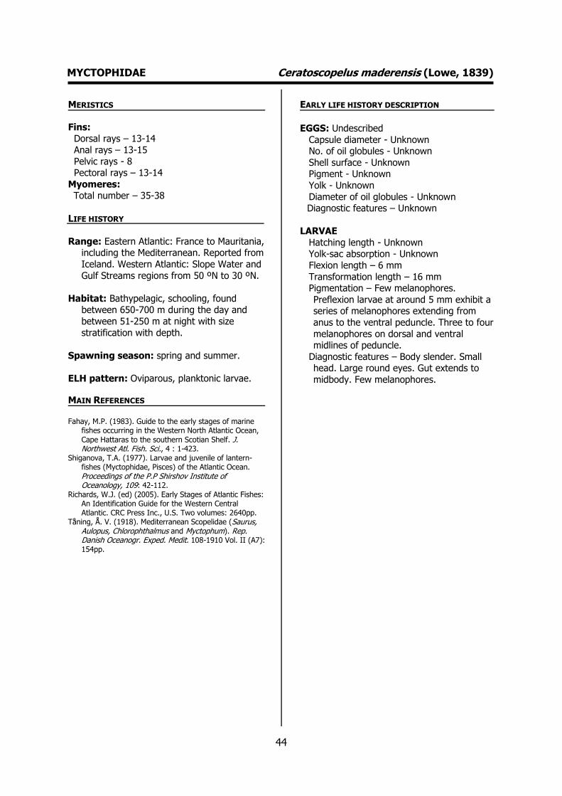
MYCTOPHIDAE Ceratoscopelus maderensis (Lowe, 1839) MERISTICS
Fins:
Dorsal rays – 13-14 Anal rays – 13-15
Pelvic rays - 8 Pectoral rays – 13-14
Myomeres: Total number – 35-38
LIFE HISTORY
Range: Eastern Atlantic: France to Mauritania, including the Mediterranean. Reported from
Iceland. Western Atlantic: Slope Water and
Gulf Streams regions from 50 ºN to 30 ºN.
Habitat: Bathypelagic, schooling, found between 650-700 m during the day and
between 51-250 m at night with size
stratification with depth.
Spawning season: spring and summer.
ELH pattern: Oviparous, planktonic larvae.
MAIN REFERENCES Fahay, M.P. (1983). Guide to the early stages of marine
fishes occurring in the Western North Atlantic Ocean, Cape Hattaras to the southern Scotian Shelf. J. Northwest Atl. Fish. Sci., 4 : 1-423.
Shiganova, T.A. (1977). Larvae and juvenile of lantern-fishes (Myctophidae, Pisces) of the Atlantic Ocean. Proceedings of the P.P Shirshov Institute of Oceanology, 109: 42-112.
Richards, W.J. (ed) (2005). Early Stages of Atlantic Fishes: An Identification Guide for the Western Central Atlantic. CRC Press Inc., U.S. Two volumes: 2640pp.
Tåning, Å. V. (1918). Mediterranean Scopelidae (Saurus, Aulopus, Chlorophthalmus and Myctophum). Rep. Danish Oceanogr. Exped. Medit. 108-1910 Vol. II (A7): 154pp.
EARLY LIFE HISTORY DESCRIPTION
EGGS: Undescribed
Capsule diameter - Unknown No. of oil globules - Unknown
Shell surface - Unknown Pigment - Unknown
Yolk - Unknown
Diameter of oil globules - Unknown Diagnostic features – Unknown
LARVAE
Hatching length - Unknown Yolk-sac absorption - Unknown
Flexion length – 6 mm
Transformation length – 16 mm Pigmentation – Few melanophores.
Preflexion larvae at around 5 mm exhibit a series of melanophores extending from
anus to the ventral peduncle. Three to four
melanophores on dorsal and ventral midlines of peduncle.
Diagnostic features – Body slender. Small head. Large round eyes. Gut extends to
midbody. Few melanophores.
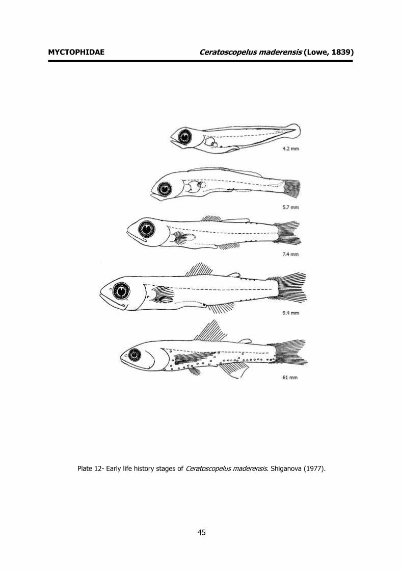
45
MYCTOPHIDAE Ceratoscopelus maderensis (Lowe, 1839)
Plate 12- Early life history stages of Ceratoscopelus maderensis. Shiganova (1977).

46
MYCTOPHIDAE Diogenichthys atlanticus (Tåning, 1928) MERISTICS
Fins:
Dorsal rays – 11-12 Anal rays – 15-16
Pelvic rays - Pectoral rays – 12-13
Myomeres: Total number – 32-35
LIFE HISTORY
Range: Atlantic Ocean: widely but unevenly distributed between 50 ºN and 48 ºS, less
abundant or absent in regions of low
productivity. Western Indian Ocean: between 22 ºS and 45 ºS. Pacific Ocean:
widespread uneven distribution between 35 ºN and 25 ºS.
Habitat: Oceanic, epipelagic to mesopelagic, found between 400-930 m during the day
and between 18-1050 m at night.
Spawning season: Spawning occurs troughout the whole year.
ELH pattern: Oviparous, planktonic larvae.
MAIN REFERENCES Fahay, M.P. (1983). Guide to the early stages of marine
fishes occurring in the Western North Atlantic Ocean, Cape Hattaras to the southern Scotian Shelf. J. Northwest Atl. Fish. Sci., 4 : 1-423.
Moser, H.G. (Ed.) (1996). The early stages of fishes in the California Current region. Calcofi Atlas no. 33: 1505pp.
Moser, H.G., E.H. Ahlstrom (1970). Development of lanterfishes (Family Myctophidae) in the California Current. Part I. Species with narrow-eyed larvae. Nat. Hist. Mus. Los Ang. Cty. Sci. Bull., 7 : 145pp.
Richards, W.J. (ed) (2005). Early Stages of Atlantic Fishes: An Identification Guide for the Western Central Atlantic. CRC Press Inc., U.S. Two volumes: 2640pp.
Shiganova, T.A. (1977). Larvae and juvenile of lantern-fishes (Myctophidae, Pisces) of the Atlantic Ocean. Proceedings of the P.P Shirshov Institute of Oceanology, 109: 42-112.
Tåning, Å. V. (1918). Mediterranean Scopelidae (Saurus, Aulopus, Chlorophthalmus and Myctophum). Rep. Danish Oceanogr. Exped. Medit. 108-1910 Vol. II (A7): 154pp.
EARLY LIFE HISTORY DESCRIPTION
EGGS: Undescribed
Capsule diameter - Unknown No. of oil globules - Unknown
Shell surface - Unknown Pigment - Unknown
Yolk - Unknown Diameter of oil globules - Unknown
Diagnostic features – Unknown
LARVAE
Hatching length - Unknown Yolk-sac absorption - Unknown
Flexion length – 6-7 mm
Transformation length – 14-15 mm Pigmentation – Four pairs of melanophores
on the lateral surfaces of the gut. Melanophores on anal rays, at base of
pectoral rays and at base of caudal rays.
Large melanophore over end of gut (side of trunk). Row of melanophores on ventral
midline of tail and along anal fin base. Diagnostic features – Eye slightly oval. Body
slender in small larvae. Barbel on lower jaw present from about 5 mm until
transformation.
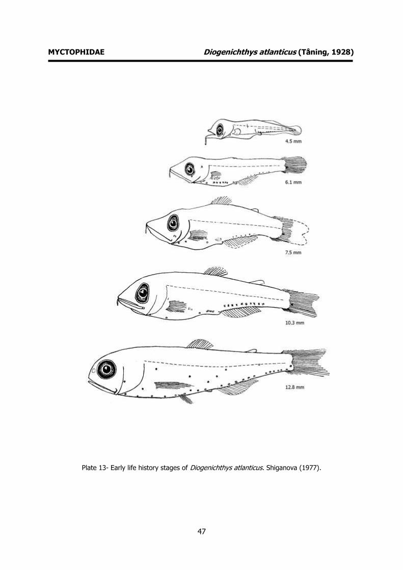
47
MYCTOPHIDAE Diogenichthys atlanticus (Tåning, 1928)
Plate 13- Early life history stages of Diogenichthys atlanticus. Shiganova (1977).
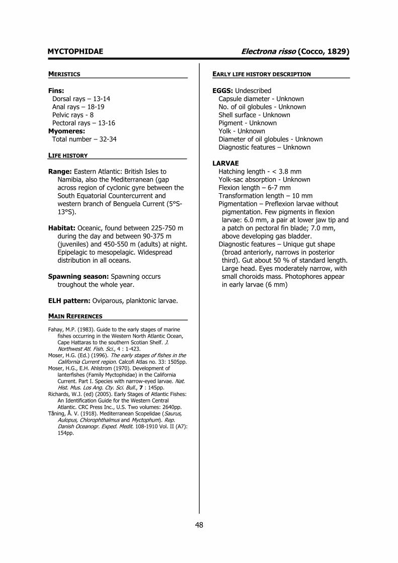
48
MYCTOPHIDAE Electrona risso (Cocco, 1829) MERISTICS
Fins:
Dorsal rays – 13-14 Anal rays – 18-19
Pelvic rays - 8 Pectoral rays – 13-16
Myomeres:
Total number – 32-34
LIFE HISTORY
Range: Eastern Atlantic: British Isles to Namibia, also the Mediterranean (gap
across region of cyclonic gyre between the
South Equatorial Countercurrent and western branch of Benguela Current (5°S-
13°S).
Habitat: Oceanic, found between 225-750 m
during the day and between 90-375 m (juveniles) and 450-550 m (adults) at night.
Epipelagic to mesopelagic. Widespread distribution in all oceans.
Spawning season: Spawning occurs
troughout the whole year.
ELH pattern: Oviparous, planktonic larvae.
MAIN REFERENCES
Fahay, M.P. (1983). Guide to the early stages of marine fishes occurring in the Western North Atlantic Ocean, Cape Hattaras to the southern Scotian Shelf. J. Northwest Atl. Fish. Sci., 4 : 1-423.
Moser, H.G. (Ed.) (1996). The early stages of fishes in the California Current region. Calcofi Atlas no. 33: 1505pp.
Moser, H.G., E.H. Ahlstrom (1970). Development of lanterfishes (Family Myctophidae) in the California Current. Part I. Species with narrow-eyed larvae. Nat. Hist. Mus. Los Ang. Cty. Sci. Bull., 7 : 145pp.
Richards, W.J. (ed) (2005). Early Stages of Atlantic Fishes: An Identification Guide for the Western Central Atlantic. CRC Press Inc., U.S. Two volumes: 2640pp.
Tåning, Å. V. (1918). Mediterranean Scopelidae (Saurus, Aulopus, Chlorophthalmus and Myctophum). Rep. Danish Oceanogr. Exped. Medit. 108-1910 Vol. II (A7): 154pp.
EARLY LIFE HISTORY DESCRIPTION
EGGS: Undescribed
Capsule diameter - Unknown No. of oil globules - Unknown
Shell surface - Unknown Pigment - Unknown
Yolk - Unknown
Diameter of oil globules - Unknown Diagnostic features – Unknown
LARVAE
Hatching length - < 3.8 mm Yolk-sac absorption - Unknown
Flexion length – 6-7 mm
Transformation length – 10 mm Pigmentation – Preflexion larvae without
pigmentation. Few pigments in flexion larvae: 6.0 mm, a pair at lower jaw tip and
a patch on pectoral fin blade; 7.0 mm,
above developing gas bladder. Diagnostic features – Unique gut shape
(broad anteriorly, narrows in posterior third). Gut about 50 % of standard length.
Large head. Eyes moderately narrow, with small choroids mass. Photophores appear
in early larvae (6 mm)

49
MYCTOPHIDAE Electrona risso (Cocco, 1829)
Plate 14- Early life history stages of Electrona risso. Moser and Ahlstrom (1970).

50
MYCTOPHIDAE Myctophum punctatum Rafinesque, 1810 MERISTICS
Fins:
Dorsal rays – 13-14 Anal rays – 20-22
Pelvic rays - 8 Pectoral rays – 13-14
Myomeres: Total number – 40-42
LIFE HISTORY
Range: Eastern Atlantic: Mauritanian Upwelling Region (15 ºN-20 ºN) as far as
30 ºN. Mediterranean Sea. Western
Atlantic: Greenland to USA.
Habitat: High-oceanic, mesopelagic. Nyctoepipelagic at the surface and down to
125 m, between 225-750 m during the day.
Spawning season: spawning occurs
troughout the whole year.
ELH pattern: Oviparous, planktonic larvae.
MAIN REFERENCES Fahay, M.P. (1983). Guide to the early stages of marine
fishes occurring in the Western North Atlantic Ocean, Cape Hattaras to the southern Scotian Shelf. J. Northwest Atl. Fish. Sci., 4 : 1-423.
Moser, H.G., E.H. Ahlstrom (1974). Role of larval stages in systematic investigations of marine teleosts: the Myctophidae, a case study. Fish. Bull. U.S., 72: 391-413.
Shiganova, T.A. (1977). Larvae and juvenile of lantern-fishes (Myctophidae, Pisces) of the Atlantic Ocean. Proceedings of the P.P Shirshov Institute of Oceanology, 109: 42-112.
EARLY LIFE HISTORY DESCRIPTION
EGGS: Undescribed
Capsule diameter - Unknown No. of oil globules - Unknown
Shell surface - Unknown Pigment - Unknown
Yolk - Unknown
Diameter of oil globules - Unknown Diagnostic features – Unknown
LARVAE
Hatching length - Unknown Yolk-sac absorption - Unknown
Flexion length – 7 mm
Transformation length – 21-22 mm Pigmentation – Characteristic pigmentation.
Preanal series of melanophores from anus to head. Rows of melanophores on upper
and lower jaws. Spots on upper edge of
opercula. Diagnostic features – Body elongate, snout
proeminent, flat and broad. Eyes narrow, stalked with choroids mass. Large pectoral
fins (fan shaped base).
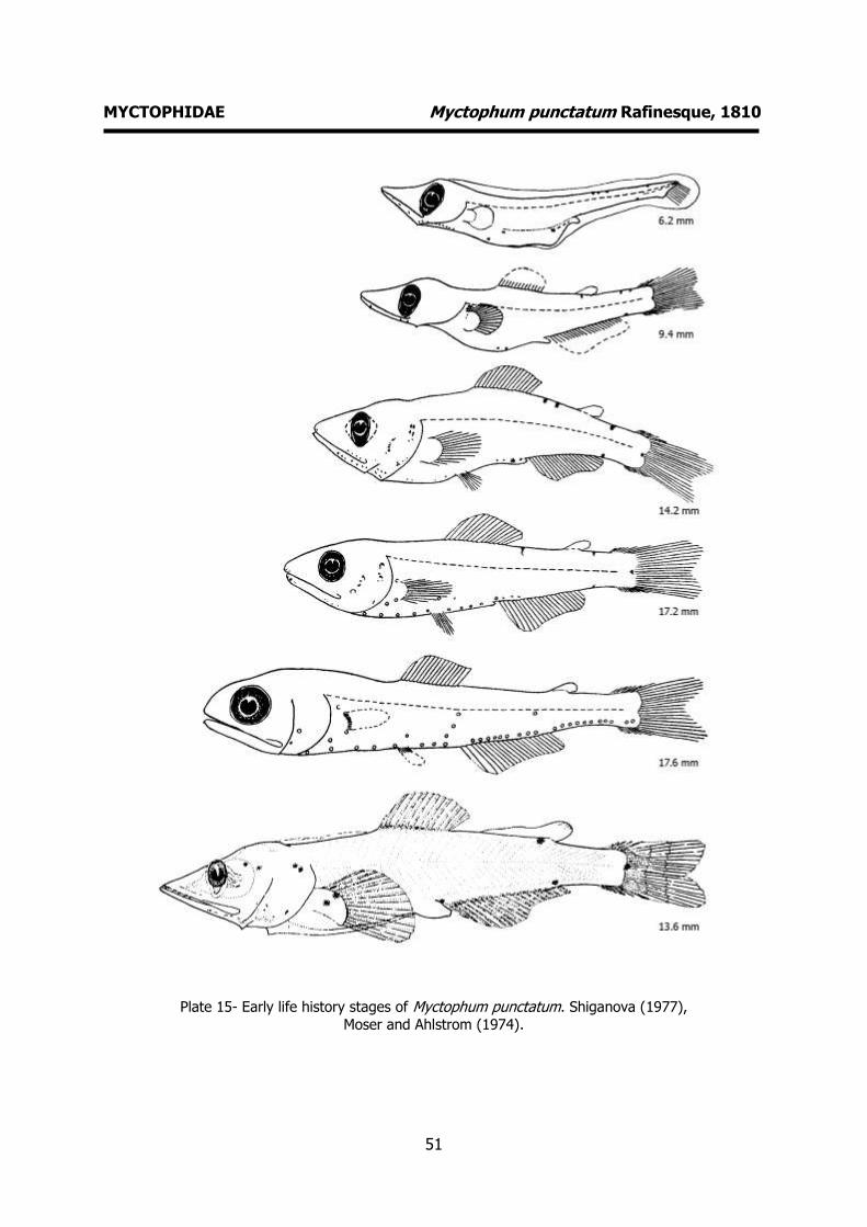
51
MYCTOPHIDAE Myctophum punctatum Rafinesque, 1810
Plate 15- Early life history stages of Myctophum punctatum. Shiganova (1977),
Moser and Ahlstrom (1974).

52
MYCTOPHIDAE Notolychnus valdiviae (Brauer, 1904) MERISTICS
Fins:
Dorsal rays – 10-12 Anal rays – 12-14
Pelvic rays - 6 Pectoral rays – 12-13
Myomeres: Total number – 27-30
LIFE HISTORY
Range: Worldwide distribution in tropical, subtropical and temperate waters. Eastern
Atlantic: west of British Isles and Bay of
Biscay to South Africa. Western Atlantic: Canada to Argentina. Western Pacific:
between 0 º and 32 ºS. Eastern Pacific: between 32 ºN and 30 ºS, but with 20 ºS
southern limit in Peruvian Transitional
Zone. Eastern Indian Ocean: between 9 º-32 ºS.
Habitat: bathypelagic depth range 25–700 m.
Spawning season: Spawning occurs
troughout the whole year.
ELH pattern: Oviparous, planktonic larvae.
MAIN REFERENCES Fahay, M.P. (1983). Guide to the early stages of marine
fishes occurring in the Western North Atlantic Ocean, Cape Hattaras to the southern Scotian Shelf. J. Northwest Atl. Fish. Sci., 4 : 1-423.
Moser, H.G. (Ed.) (1996). The early stages of fishes in the California Current region. Calcofi Atlas no. 33: 1505pp.
Moser, H.G., E.H. Ahlstrom (1974). Role of larval stages in systematic investigations of marine teleosts: the Myctophidae, a case study. Fish. Bull. U.S., 72: 391-413.
Richards, W.J. (ed) (2005). Early Stages of Atlantic Fishes: An Identification Guide for the Western Central Atlantic. CRC Press Inc., U.S. Two volumes: 2640pp.
Tåning, Å. V. (1918). Mediterranean Scopelidae (Saurus, Aulopus, Chlorophthalmus and Myctophum). Rep. Danish Oceanogr. Exped. Medit. 108-1910 Vol. II (A7): 154pp.
EARLY LIFE HISTORY DESCRIPTION
EGGS: Undescribed
Capsule diameter - Unknown No. of oil globules - Unknown
Shell surface - Unknown Pigment - Unknown
Yolk - Unknown
Diameter of oil globules - Unknown Diagnostic features – Unknown
LARVAE
Hatching length - Unknown Yolk-sac absorption - Unknown
Flexion length – 4-5 mm
Transformation length – 9-10 mm Pigmentation – Usually 2 or 3 melanophores
on lateral gut (flexion larvae). Postflexion larvae- 2-7 postanal ventral melanophores.
Diagnostic features – Characteristic
(unusual) shape of head body and gut. Eye shape varies (narrow to round). All
photophores formed after transformation. Low pelvic ray count (6).

53
MYCTOPHIDAE Notolychnus valdiviae (Brauer, 1904)
Plate 16- Early life history stages of Notolychnus valdiviae. Moser (1996).
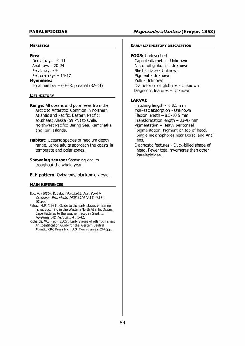
54
PARALEPIDIDAE Magnisudis atlantica (Krøyer, 1868) MERISTICS
Fins:
Dorsal rays – 9-11 Anal rays – 20-24
Pelvic rays - 9 Pectoral rays – 15-17
Myomeres:
Total number – 60-68, preanal (32-34)
LIFE HISTORY
Range: All oceans and polar seas from the Arctic to Antarctic. Common in northern
Atlantic and Pacific. Eastern Pacific:
southeast Alaska (59 ºN) to Chile. Northwest Pacific: Bering Sea, Kamchatka
and Kuril Islands.
Habitat: Oceanic species of medium depth
range. Large adults approach the coasts in temperate and polar zones.
Spawning season: Spawning occurs
troughout the whole year.
ELH pattern: Oviparous, planktonic larvae.
MAIN REFERENCES Ege, V. (1930). Sudidae (Paralepis). Rep. Danish
Oceanogr. Exp. Medit. 1908-1910, Vol II (A13): 201pp.
Fahay, M.P. (1983). Guide to the early stages of marine fishes occurring in the Western North Atlantic Ocean, Cape Hattaras to the southern Scotian Shelf. J. Northwest Atl. Fish. Sci., 4 : 1-423.
Richards, W.J. (ed) (2005). Early Stages of Atlantic Fishes: An Identification Guide for the Western Central Atlantic. CRC Press Inc., U.S. Two volumes: 2640pp.
EARLY LIFE HISTORY DESCRIPTION
EGGS: Undescribed
Capsule diameter - Unknown No. of oil globules - Unknown
Shell surface - Unknown Pigment - Unknown
Yolk - Unknown
Diameter of oil globules - Unknown Diagnostic features – Unknown
LARVAE
Hatching length - < 8.5 mm Yolk-sac absorption - Unknown
Flexion length – 8.5-10.5 mm
Transformation length – 23-47 mm Pigmentation – Heavy peritoneal
pigmentation. Pigment on top of head. Single melanophores near Dorsal and Anal
fins.
Diagnostic features - Duck-billed shape of head. Fewer total myomeres than other
Paralepididae.
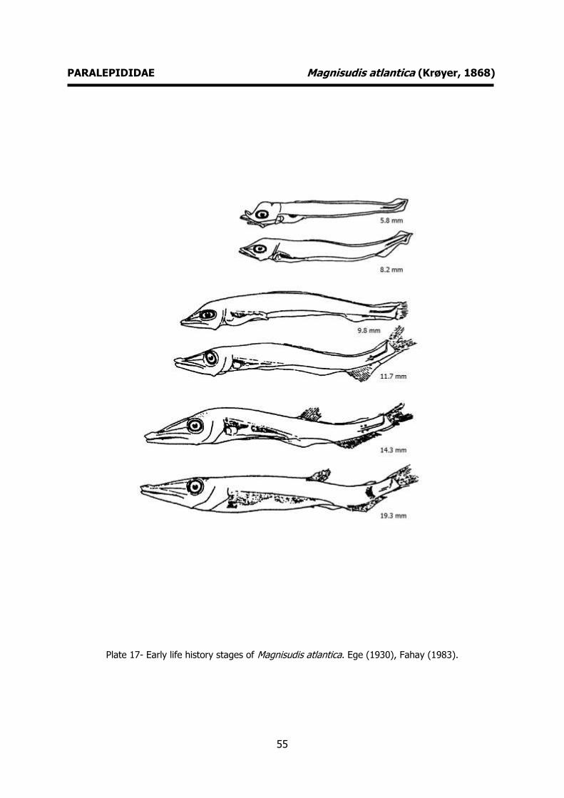
55
PARALEPIDIDAE Magnisudis atlantica (Krøyer, 1868)
Plate 17- Early life history stages of Magnisudis atlantica. Ege (1930), Fahay (1983).

56
PARALEPIDIDAE Paralepis coregonoides Risso, 1820 MERISTICS
Fins:
Dorsal rays – 8-11 Anal rays – 21-25
Pelvic rays - 9 Pectoral rays – 13-16
Myomeres: Total number – 70-74, preanal (34-36)
LIFE HISTORY
Range: Atlantic Ocean: restricted to the North Atlantic and adjacent seas.
Habitat: Depth range from 50-1000 m.
Spawning season: spring and summer.
ELH pattern: Oviparous, planktonic larvae.
MAIN REFERENCES Ege, V. (1930). Sudidae (Paralepis). Rep. Danish
Oceanogr. Exp. Medit. 1908-1910, Vol II (A13): 201pp.
Fahay, M.P. (1983). Guide to the early stages of marine fishes occurring in the Western North Atlantic Ocean, Cape Hattaras to the southern Scotian Shelf. J. Northwest Atl. Fish. Sci., 4 : 1-423.
Richards, W.J. (ed) (2005). Early Stages of Atlantic Fishes: An Identification Guide for the Western Central Atlantic. CRC Press Inc., U.S. Two volumes: 2640pp.
EARLY LIFE HISTORY DESCRIPTION
EGGS: Undescribed
Capsule diameter - Unknown No. of oil globules - Unknown
Shell surface - Unknown Pigment - Unknown
Yolk - Unknown
Diameter of oil globules - Unknown Diagnostic features – Unknown
LARVAE
Hatching length - <6 mm Yolk-sac absorption - Unknown
Flexion length – 10-15 mm
Transformation length – 25 mm Pigmentation – 1 to 2 peritoneal patches
(preflexion larvae). Long internal series of pigments above and below the notochord
(flexion larvae).
Diagnostic features – Small head, snout shorter than M. atlantica. Other Paralepis lack internal pigments above and below the notochord.
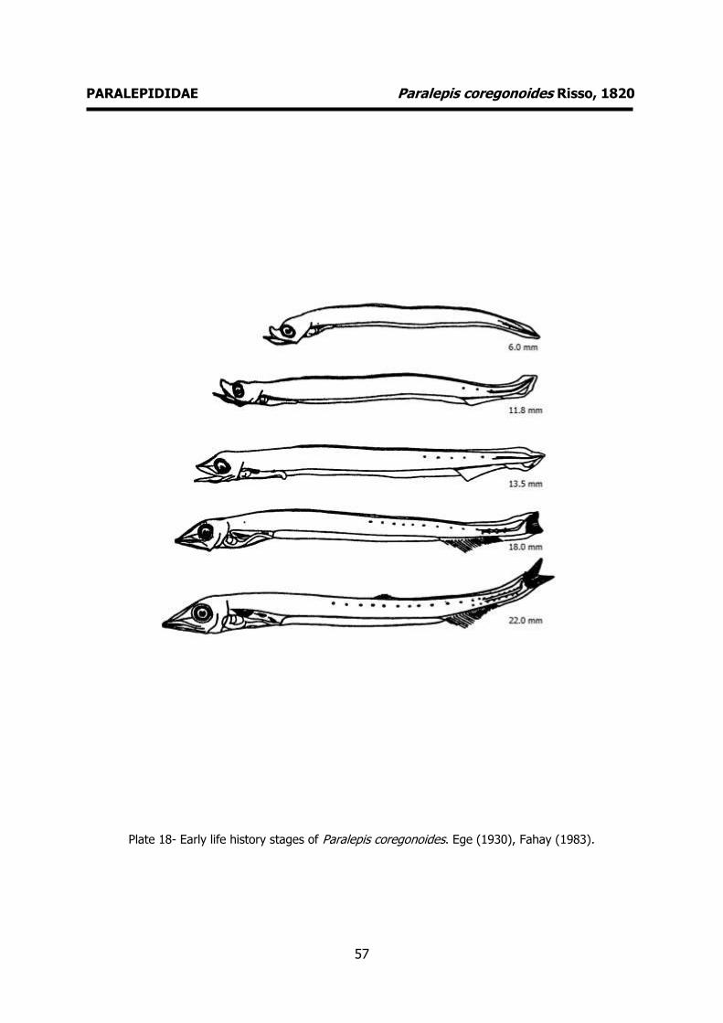
57
PARALEPIDIDAE Paralepis coregonoides Risso, 1820
Plate 18- Early life history stages of Paralepis coregonoides. Ege (1930), Fahay (1983).
58
ANGUILLIDAE Anguilla anguilla (Linnaeus, 1758) MERISTICS
Fins:
Dorsal rays - Anal rays -
Pelvic rays - Pectoral rays –
Myomeres: Total number – 110-119
LIFE HISTORY
Range: Catadromous. Migrates to the depths of the Sargasso Sea to spawn. leptocephali
larvae are transported to the coasts of
Europe by the Gulf Stream in 7 to 11 months and can last for up to 3 years.
These are transformed into elvers, enter the estuaries and colonize continental
waters.
Habitat: Atlantic Ocean: Atlantic coast from
Scandinavia to Morocco and rivers of North Atlantic, Baltic and Mediterranean seas.
Spawning season: Spawning takes place in
late winter and spring in the Sargasso Sea.
Juveniles are caught throughout the whole year inside estuaries and lagoons (mainly in
spring and summer).
ELH pattern: Oviparous, planktonic eggs and
larvae.
MAIN REFERENCES Boëtius, J. (1976). Elvers, Anguilla anguilla and Anguilla
rostrata from two Danish localities. Size, body weight, developmental stage and number of vertebrae related to time of ascent. Meddr Danm. Fisk.- og Havunders., NS: 199-220.
Garcia A.M.A., P.D. Moyano (1990). Estados juveniles de la ictiofauna en los canos de las salinas de da Bahia de Cadiz. Instituto de Ciencias Marinas de Andalucia. Consejo Superior de Investigaciones Científicas, Cadiz: 163pp.
Munk, P., J.G. Nielsen (2005). Eggs and larvae of North Sea fishes. Biofolia, Denmark: 215pp.
Schmidt, J. (1909). Remarks on the metamorphosis and distribution of the larvae of the eel (Anguilla vulgaris, Turton). Medd. Komm. Havunds., Fiskeri, 3 (3): 1-17.
Schmidt, J. (1923). Breeding places and migrations of the Eel. Nature, 3 (2776): 51-54.
EARLY LIFE HISTORY DESCRIPTION
EGGS
Capsule diameter – 0.9-1.38 mm No. of oil globules - 1
Shell surface - smooth Pigment - none
Yolk - unsegmented
Diameter of oil globules – 0.25-0.36 mm Diagnostic features – Initially many oils
globules that coalesce into one. In later stages of development, dendritic
melanophores appear over the anterior hemisphere of the oil globule.
LARVAE Hatching length – 6 mm
Yolk-sac absorption – 6 mm Flexion length -
Transformation length -
Pigmentation – Newly hatched larva unpigmented. Late leptocephalus – a few
melanophores caudal area. Diagnostic features - Newly hatched larva
with protruding teeth. Leptocephalus stage – morphology of the larva changes
dramatically (the body becomes very high
and laterally compressed). Head very small relative to body. Transparent with distinct
myomeres (110-119). Pelvic fins absent. A. anguilla can be distinguished from A. rostrata by the number of myomeres (110-
119 versus 102-111). Juveniles are caught throughout the whole year inside estuaries
and lagoons (mainly in spring and summer).

59
ANGUILLIDAE Anguilla anguilla (Linnaeus, 1758)
Plate 19- Juveniles of Anguilla anguilla. Garcia and Moyano (1990).

60
MURAENIDAE Muraena helena Linnaeus, 1758 MERISTICS
Fins:
Dorsal rays - Anal rays -
Pelvic rays - Pectoral rays –
Myomeres: Total number – 145-150 (14 mm), preanal
(76-80). Later 139-144 myomeres.
LIFE HISTORY
Range: Eastern Atlantic: south of British Isles
to Senegal, including the Mediterranean,
Azores, Madeira, Canary Islands, and Cape Verde.
Habitat: A nocturnal and territorial species
commonly lurking in holes, and writhing
snakelike through crevices, under rocks or corals.
Spawning season: summer and autumn.
ELH pattern: Oviparous, planktonic eggs and
larvae.
MAIN REFERENCES D’Ancona, U. (1931-1933). Apodes (Muraenoidei). Uova,
larve e stadi Giovanili di Teleostei: monografia elaborata con l’uso del materiale raccolto e sweriato da Salvatore Lo Bianco. Fauna e Flora Golfo Napoli Monogr.38.: 84-146.
EARLY LIFE HISTORY DESCRIPTION
EGGS
Capsule diameter – 5.0-5.5 mm No. of oil globules - 0
Shell surface - smooth Pigment - none
Yolk - segmented
Diameter of oil globules - Diagnostic features – Large periviteline
space.
LARVAE Hatching length – 10.5-11 mm
Yolk-sac absorption -
Flexion length - Transformation length -
Pigmentation – Few pigments. Diagnostic features – Typical shape. Eel
like. Tube-like gut. Teeth clearly visible.
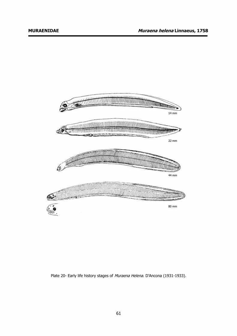
61
MURAENIDAE Muraena helena Linnaeus, 1758
Plate 20- Early life history stages of Muraena Helena. D’Ancona (1931-1933).

62
CONGRIDAE Conger conger (Linnaeus, 1758) MERISTICS
Fins:
Dorsal rays - Anal rays -
Pelvic rays - Pectoral rays –
Myomeres: Total number – 148-155
LIFE HISTORY
Range: Eastern Atlantic: Norway and Iceland to Senegal. Also in the Mediterranean and
Black Sea.
Habitat: Found on rocky and sandy bottoms.
Depth range from 0-500 m. It stays near the coast when young and moves toward
deeper waters upon reaching adulthood.
Spawning season: Spawns in summer in the
Atlantic off Portugal and in the Mediterranean.
ELH pattern: Oviparous, planktonic larvae.
MAIN REFERENCES D’Ancona, U. (1931-1933). Apodes (Muraenoidei). Uova,
larve e stadi Giovanili di Teleostei: monografia elaborata con l’uso del materiale raccolto e sweriato da Salvatore Lo Bianco. Fauna e Flora Golfo Napoli Monogr.38.: 84-146.
EARLY LIFE HISTORY DESCRIPTION
EGGS: Undescribed
Capsule diameter - Unknown No. of oil globules - Unknown
Shell surface - Unknown Pigment - Unknown
Yolk - Unknown
Diameter of oil globules - Unknown Diagnostic features – Unknown
LARVAE
Hatching length - <8 mm Yolk-sac absorption - Unknown
Flexion length -
Transformation length – 150 mm Pigmentation - No melanophores on head or
trunk. Tail with single melanophore. Peritoneal pigmentation - row of
melanophores.
Diagnostic features – Characteristic shape. Eel like. Tube-like gut. Teeth clearly
visible.
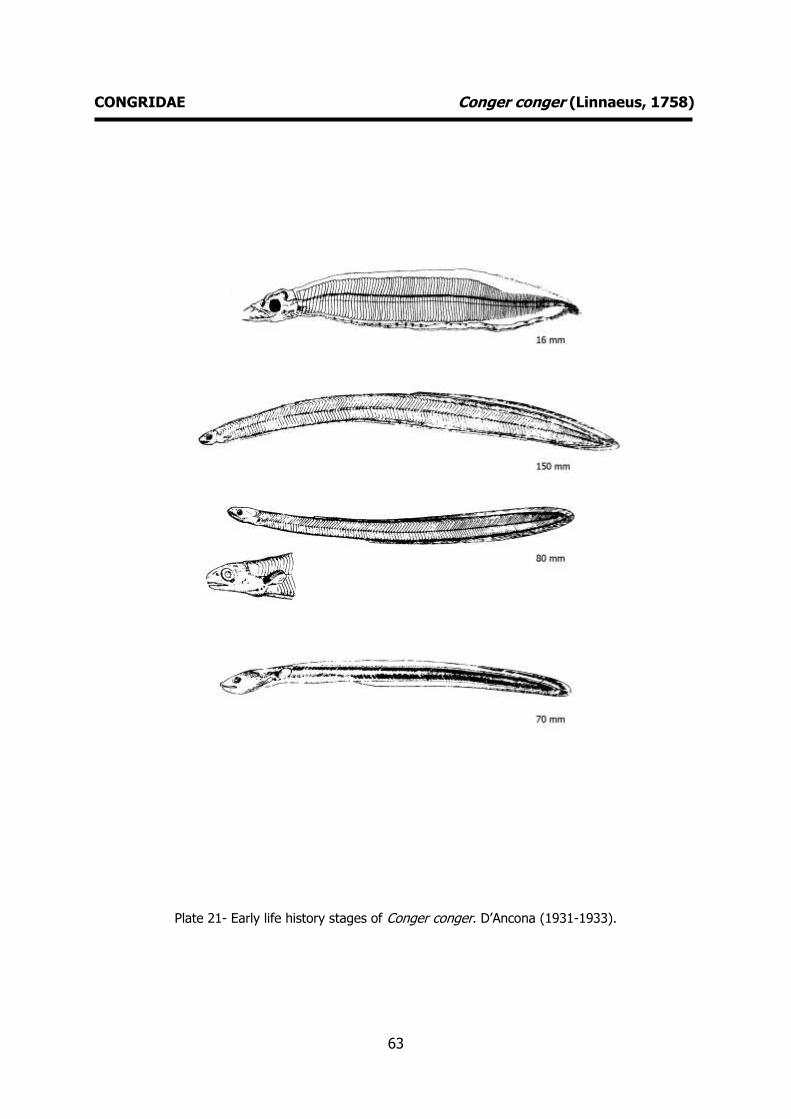
63
CONGRIDAE Conger conger (Linnaeus, 1758)
Plate 21- Early life history stages of Conger conger. D’Ancona (1931-1933).

64
OPHICHTIDAE Ophisurus serpens (Linnaeus, 1758) MERISTICS
Fins:
Dorsal rays - Anal rays -
Pelvic rays - Pectoral rays –
Myomeres: Total number – 200-208, preanal (89-94)
LIFE HISTORY
Range: Eastern Atlantic: northern coast of Iberian Peninsula to South Africa, also
Madeira and western Mediterranean.
Western Indian Ocean: southern Mozambique to South Africa. Western
Pacific: Japan and Australasia.
Habitat: reef-associated; brackish; marine;
depth range 0-300 m.
Spawning season: spring and summer.
ELH pattern: Oviparous, planktonic eggs and larvae.
MAIN REFERENCES D’Ancona, U. (1931-1933). Apodes (Muraenoidei). Uova,
larve e stadi Giovanili di Teleostei: monografia elaborata con l’uso del materiale raccolto e sweriato da Salvatore Lo Bianco. Fauna e Flora Golfo Napoli Monogr.38.: 84-146.
EARLY LIFE HISTORY DESCRIPTION
EGGS
Capsule diameter – 2.5-3.5 mm No. of oil globules – 11-28
Shell surface - smooth Pigment - none
Yolk - Segmented
Diameter of oil globules – 0.08-0.16 mm Diagnostic features – Large periviteline
space
LARVAE Hatching length - >8 mm (191 myomeres)
Yolk-sac absorption -
Flexion length - Transformation length -
Pigmentation – Larvae with 15 mm have 5 abdominal melanophores. Three postanal
ventral melanophores.
Diagnostic features - Eel like. Tube-like gut. Typical pigmentation. Elongated snout.
Upper jaw shorther than lower. Each jaw with 4 teeth.

65
OPHICHTIDAE Ophisurus serpens (Linnaeus, 1758)
Plate 22- Early life history stages of Ophisurus serpens. D’Ancona (1931-1933).

66
BELONIDAE Belone belone (Linnaeus, 1761) MERISTICS
Fins:
Dorsal rays – 16-20 Anal rays – 19-23
Pelvic rays - Pectoral rays –
Myomeres: Total number – 75-84
LIFE HISTORY
Range: Eastern Atlantic and Mediterranean Sea. Three subspecies are recognized
Belone belone belone (Linnaeus, 1761)
(Northeast Atlantic); Belone belone euxini Günther, 1866 (Black Sea and Sea of
Azov); Belone belone acus Risso, 1827 (Mediterranean Sea and adjacent parts of
Atlantic Ocean, Madeira, Canary Islands,
Azores, and south to Cape Verde; subspecies Belone belone gracilis Lowe,
1839 (France to the Canary Islands including the Mediterranean.
Habitat: Lives close to the surface and has a
migratory pattern similar to the mackerel.
Spawning season: spring and summer.
ELH pattern: Oviparous, demersal eggs and
planktonic larvae.
MAIN REFERENCES D’Ancona, U. (1931). Scomberesocidae. In Uova, larve e
stadi Giovanili di Teleostei: monografia elaborata con l’uso del materiale raccolto e sweriato da Salvatore Lo Bianco. Fauna e Flora Golfo Napoli Monogr. 38 (1): 157-164.
Dekhnik, T.V. (1973). Ichthyoplankton of the Black Sea. Naukova Dumka, Kiev: 235pp (in russian).
Ehrenbaum, E. (1905-1909). Eier und Larven von Fischen. Nordisches Plankton, 1: 413pp.
Garcia A.M.A., P.D. Moyano (1990). Estados juveniles de la ictiofauna en los canos de las salinas de da Bahia de Cadiz. Instituto de Ciencias Marinas de Andalucia. Consejo Superior de Investigaciones Científicas, Cadiz: 163pp.
M'intosh, W.S., A.T. Masterman (1897). The life-histories of the British marine food-fishes. C. J. Clay London: 467p.
Moser, H.G., W.J. Richards, D.M. Cohen, M.P. Fahay, A.W. Kendall, S.L. Richardson (eds.) (1984). Ontogeny and systematics of fishes. American Society of Ichthyologists and Herpetologists ed., Special publication Number 1: 760pp.
Russell, F.S. (1976). The eggs and planktonic stages of British marine fishes. Academic Press, London: 524pp.
EARLY LIFE HISTORY DESCRIPTION
EGGS
Capsule diameter – 2.8-3.5 mm No. of oil globules - 0
Shell surface – filamentous processes Pigment - none
Yolk - unsegmented
Diameter of oil globules - Diagnostic features – large size, 60 to 80
long filamentous processes (4-18 mm) on shell surface. Eggs can be found attached
to floating objects or demersal algae.
LARVAE
Hatching length – 9-13 mm Yolk-sac absorption -
Flexion length - Transformation length -
Pigmentation – newly hatched larva
completely covered with a large number of black and yellow chromatophores.
Melanophores arranged along myomeres in the dorsal part of the body.
Diagnostic features - lower jaw strongly projecting. Elongation of lower jaw starts
at 18 mm. Pelvic fins appear at a length of
20-24 mm

67
BELONIDAE Belone belone (Linnaeus, 1761)
Plate 23- Early life history stages of Belone belone. Dekhnik (1973), Russell (1976),
Moser et al. (1984)

68
MACRORAMPHOSIDAE Macroramphosus scolopax (Linnaeus, 1758) MERISTICS
Fins:
Dorsal rays – 6-8 (spines) 11-13 (soft) Anal rays - 18
Pelvic rays - Pectoral rays –
Myomeres: Total number – 24-25, 10 (preanal)
LIFE HISTORY
Range: Western Atlantic: off the east coast of the USA and the Greater Antilles; also
northern South America and Argentina.
Eastern Atlantic and Mediterranean. Indo-West Pacific. Mainly in temperate latitudes
between 20 º and 40 ºN.
Habitat: demersal; marine; depth range 25–
600 m.
Spawning season: spring
ELH pattern: Oviparous, planktonic eggs and larvae.
MAIN REFERENCES D’Ancona, U. (1931-1933). Macrorhamphosidae. Uova,
larve e stadi Giovanili di Teleostei: monografia elaborata con l’uso del materiale raccolto e sweriato da Salvatore Lo Bianco. Fauna e Flora Golfo Napoli Monogr.38: 299-306.
Hardy, J.D. (1978). Macrorhamphosidae. Development of fishes of the Mid-Atlantic Bight. Fish and Wildlife Service, U.S. Department of the Interior. Volume II: 377-386.
Spartà, A. (1936). Contributo alla conoscenza di uova, stadi embrionali e post-embrionali in Macrorhamphosus scolopax L.. R. Com. talassog. Ital. Mem. 225: 14pp.
EARLY LIFE HISTORY DESCRIPTION
EGGS
Capsule diameter – 1.0 mm No. of oil globules - 1
Shell surface - smooth Pigment – scattered melanophores on sides
of embryo.
Yolk - unsegmented Diameter of oil globules – 0.2 mm
Diagnostic features – Viteline membrane light amber, yolk with rose or violet halo
depending on viewing light, single oil globule.
LARVAE Hatching length – 3.0 mm
Yolk-sac absorption - Flexion length -
Transformation length -
Pigmentation – Newly-hatched larvae: melanophores concentrated in
posterodorsal region and in a continuous line along ventral surface of body from
behind the eye to caudal region. Larvae: eye completely pigmented with metallic
reflections. Melanophores concentrated on
posterodorsal surface and along ventral line of body.
Diagnostic features – Snout somewhat elongate. Laterally compressed.
Supraorbital crest prominent at 6.0 mm.
Typical pigmentation.

69
MACRORAMPHOSIDAE Macroramphosus scolopax (Linnaeus, 1758)
Plate 24- Early life history stages of Macroramphosus scolopax. Spartà (1936).
70
SYNGNATHIDAE Entelurus aequoraeus (Linnaeus, 1758) MERISTICS Fins:
Dorsal rays – 37-47
Anal rays -
Pelvic rays - Pectoral rays –
Myomeres: Total number –
LIFE HISTORY
Range: Eastern Atlantic: Iceland and Norway to Azores and also enters Baltic Sea.
Habitat: Inhabits inshore waters, among algae.
Spawning season: spring and summer.
ELH pattern: Ovoviviparous. The male carries
the eggs in a brood pouch, which is found
under the tail.
MAIN REFERENCES Ehrenbaum, E. (1905-1909). Eier und Larven von Fischen.
Nordisches Plankton, 1: 413pp. Le Danois, E. (1913). Contribution à l’Étude systématique
et biologique des poisons de la Manche Occdidentale. Ann. Inst. Océanogr., Paris, 5: 1-214.
Munk, P., J.G. Nielsen (2005). Eggs and larvae of North Sea fishes. Biofolia, Denmark: 215pp.
EARLY LIFE HISTORY DESCRIPTION
EGGS
Capsule diameter - No. of oil globules -
Shell surface - Pigment -
Yolk -
Diameter of oil globules - Diagnostic features - Pear-shaped eggs in
brood pouch (400-1000 eggs).
LARVAE Hatching length -
Yolk-sac absorption -
Flexion length - Transformation length -
Pigmentation - 12 - 13 dorsal clusters of melanophores.
Diagnostic features – Newly hatched larvae
with primordial and pectoral fins. Anus below the posterior part of the dorsal fin.
Sides of body with 90 - 95 tooth-like projections. Larval characteristics remain
until length of about 100 mm. Pectoral fins disappear when the larva settles.

71
SYNGNATHIDAE Entelurus aequoraeus (Linnaeus, 1758)
Plate 25- Early life history stages of Entelurus aequoraeus. Le Danois (1913).

72
SYNGNATHIDAE Hippocampus guttulatus Cuvier, 1829 MERISTICS
Fins:
Dorsal rays – 19-22 Anal rays - 4
Pelvic rays - Pectoral rays – 15-18
Myomeres:
Total number – 51-52
LIFE HISTORY
Range: Eastern Atlantic: British Isles to Morocco, Canary Islands, Madeira, and the
Azores including the Mediterranean.
International trade is monitored through a licensing system (CITES II, since
20040514).
Habitat: Occurs mostly in shallow inshore
waters among algae and eel grass.
Spawning season: spring and summer.
ELH pattern: Ovoviviparous. Incubation of the eggs takes place in a marsupial pouch
found in front of the tail of the males.
MAIN REFERENCES D’Ancona, U. (1931-1933). Syngnathidae. Uova, larve e
stadi Giovanili di Teleostei: monografia elaborata con l’uso del materiale raccolto e sweriato da Salvatore Lo Bianco. Fauna e Flora Golfo Napoli Monogr.38: 281-289.
Garcia A.M.A., P.D. Moyano (1990). Estados juveniles de la ictiofauna en los canos de las salinas de da Bahia de Cadiz. Instituto de Ciencias Marinas de Andalucia. Consejo Superior de Investigaciones Científicas, Cadiz: 163pp.
EARLY LIFE HISTORY DESCRIPTION
EGGS
Capsule diameter – 1.9-2.0 mm No. of oil globules -
Shell surface - Pigment -
Yolk -
Diameter of oil globules - Diagnostic features - Pear-shaped. Large
periviteline space.
LARVAE Hatching length – 10 mm
Yolk-sac absorption -
Flexion length - Transformation length -
Pigmentation – Melanophores uniformly distributed on head and body.
Diagnostic features – Eye diameter
comprised more than twice in the preorbital space. Bony crests on head.
Dorsal, anal and pectoral fins completely developed at hatching.
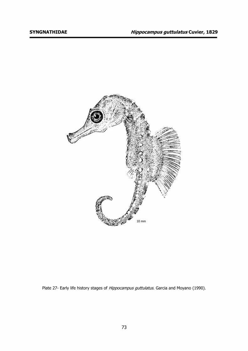
73
SYNGNATHIDAE Hippocampus guttulatus Cuvier, 1829
Plate 27- Early life history stages of Hippocampus guttulatus. Garcia and Moyano (1990).

74
SYNGNATHIDAE Hippocampus hippocampus (Linnaeus, 1758) MERISTICS
Fins:
Dorsal rays – 16-19 Anal rays - 4
Pelvic rays - Pectoral rays – 13-15
Myomeres:
Total number – 47-51
LIFE HISTORY
Range: Eastern Atlantic: Wadden Sea southward to the Gulf of Guinea, Canary
Islands and along the African coast to
Guinea. Also in the Mediterranean Sea. International trade is monitored through a
licensing system (CITES II, since 20040514).
Habitat: shallow inshore waters among algae.
Spawning season: spring and summer.
ELH pattern: Ovoviviparous. Incubation of the eggs takes place in a marsupial pouch
found in front of the tail of the males.
MAIN REFERENCES
D’Ancona, U. (1931-1933). Syngnathidae. Uova, larve e stadi Giovanili di Teleostei: monografia elaborata con l’uso del materiale raccolto e sweriato da Salvatore Lo Bianco. Fauna e Flora Golfo Napoli Monogr.38: 281-289.
Garcia A.M.A., P.D. Moyano (1990). Estados juveniles de la ictiofauna en los canos de las salinas de da Bahia de Cadiz. Instituto de Ciencias Marinas de Andalucia. Consejo Superior de Investigaciones Científicas, Cadiz: 163pp.
EARLY LIFE HISTORY DESCRIPTION
EGGS
Capsule diameter – 1.34-1.82 mm No. of oil globules – numerous oil globules
of different size. Shell surface - smooth
Pigment -
Yolk - Diameter of oil globules -
Diagnostic features - Pear-shaped. Large periviteline space.
LARVAE
Hatching length – 8 mm
Yolk-sac absorption - Flexion length -
Transformation length - Pigmentation - Melanophores on head and
trunk.
Diagnostic features – Horizontal diameter of the eye comprised ca. 1.5 times in the
preorbital space. Dorsal profile of the snout concave. Length of the lower jaw
roughly equal to the eye diameter. Prominent occipital crest. Dorsal, anal and
pectoral fins completely developed at
hatching.
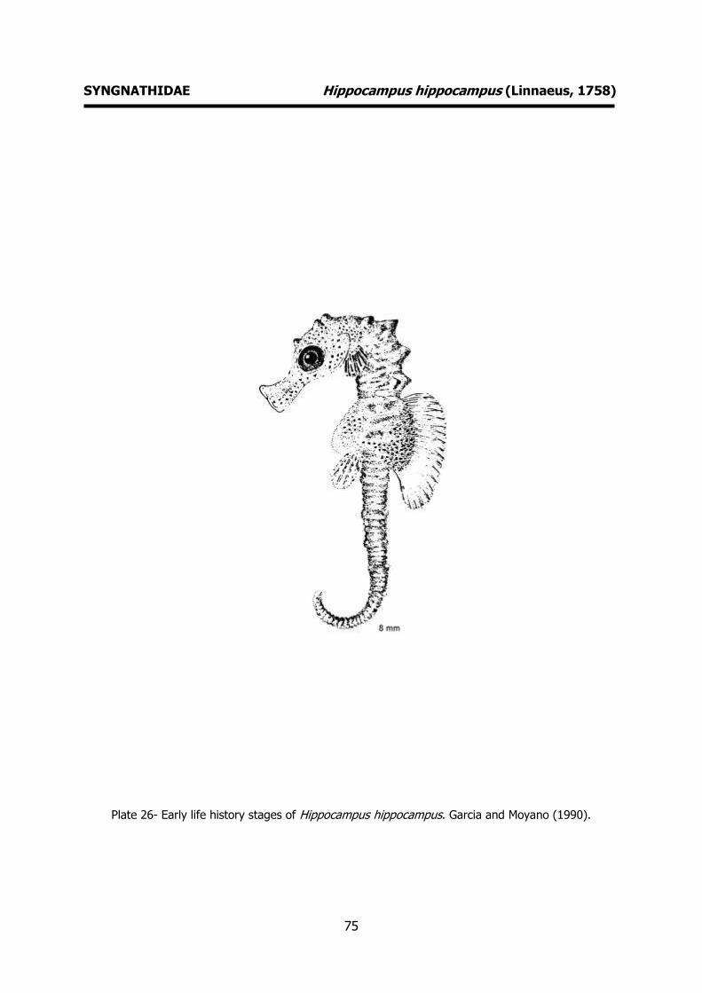
75
SYNGNATHIDAE Hippocampus hippocampus (Linnaeus, 1758)
Plate 26- Early life history stages of Hippocampus hippocampus. Garcia and Moyano (1990).
76
SYNGNATHIDAE Nerophis lumbriciformis (Jenyns, 1835) MERISTICS
Fins:
Dorsal rays – 24-28 Anal rays -
Pelvic rays - Pectoral rays –
Myomeres:
Total number –
LIFE HISTORY
Range: Eastern Atlantic: southern Norway, Kattegat and British Isles to Rio de Oro in
Western Sahara.
Habitat: Intertidal to about 30 m, among
seaweeds at high tide and underneath loose stones during ebb tide.
Spawning season: throughout the whole year with a marked reduction during
summer and autumn.
ELH pattern: Ovoviviparous. Male carries the eggs in a brood pouch, which is found
under the tail.
MAIN REFERENCES Le Danois, E. (1913). Contribution à l’Étude systématique
et biologique des poisons de la Manche Occidentale. Ann. Inst. Océanogr., Paris, 5: 1-214.
Monteiro, N., V. Almada, M.N. Vieira (2003). Early life history of the pipefish Nerophis lumbriciformis (Pisces: Syngnathidae). J. Mar. Biol. Assoc. U.K., 83 (6): 1179-1182.
Munk, P., J.G. Nielsen (2005). Eggs and larvae of North Sea fishes. Biofolia, Denmark: 215pp.
EARLY LIFE HISTORY DESCRIPTION
EGGS
Capsule diameter – 0.7-1.72 (major axis) No. of oil globules -
Shell surface – smooth, transparent Pigment -
Yolk -
Diameter of oil globules - Diagnostic features – orange yolk, variable
shape
LARVAE Hatching length – 10 mm
Yolk-sac absorption -
Flexion length - Transformation length -
Pigmentation - 12 pigmentation marks in upper trunk and tail, 4 to 5 marks in the
ventral margin of primordial fin.
Diagnostic features – Yolk-sac is completely absorbed at hatching. Primordial fin and
pectoral fins present at hatching. Duck-billed shape of head. Mouth functional.

77
SYNGNATHIDAE Nerophis lumbriciformis (Jenyns, 1835)
Plate 28- Early life history stages of Nerophis lumbriciformis. Le Danois (1913), Monteiro et al. (2003).
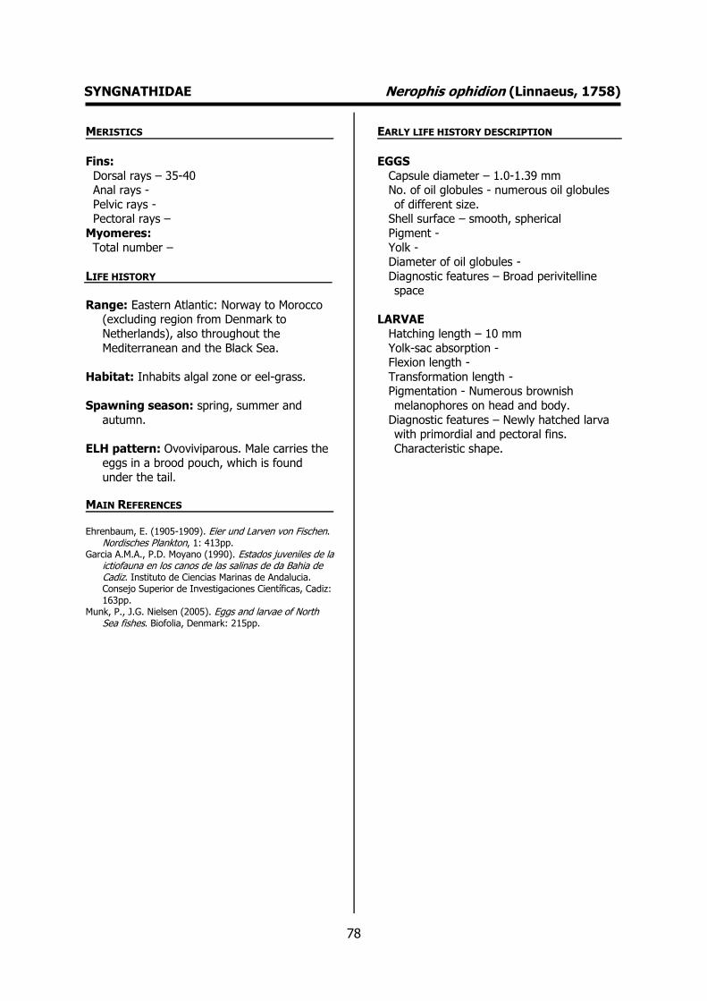
78
SYNGNATHIDAE Nerophis ophidion (Linnaeus, 1758) MERISTICS
Fins:
Dorsal rays – 35-40 Anal rays -
Pelvic rays - Pectoral rays –
Myomeres:
Total number –
LIFE HISTORY
Range: Eastern Atlantic: Norway to Morocco (excluding region from Denmark to
Netherlands), also throughout the
Mediterranean and the Black Sea.
Habitat: Inhabits algal zone or eel-grass.
Spawning season: spring, summer and
autumn.
ELH pattern: Ovoviviparous. Male carries the eggs in a brood pouch, which is found
under the tail.
MAIN REFERENCES Ehrenbaum, E. (1905-1909). Eier und Larven von Fischen.
Nordisches Plankton, 1: 413pp. Garcia A.M.A., P.D. Moyano (1990). Estados juveniles de la
ictiofauna en los canos de las salinas de da Bahia de Cadiz. Instituto de Ciencias Marinas de Andalucia. Consejo Superior de Investigaciones Científicas, Cadiz: 163pp.
Munk, P., J.G. Nielsen (2005). Eggs and larvae of North Sea fishes. Biofolia, Denmark: 215pp.
EARLY LIFE HISTORY DESCRIPTION
EGGS
Capsule diameter – 1.0-1.39 mm No. of oil globules - numerous oil globules
of different size. Shell surface – smooth, spherical
Pigment -
Yolk - Diameter of oil globules -
Diagnostic features – Broad perivitelline space
LARVAE
Hatching length – 10 mm
Yolk-sac absorption - Flexion length -
Transformation length - Pigmentation - Numerous brownish
melanophores on head and body.
Diagnostic features – Newly hatched larva with primordial and pectoral fins.
Characteristic shape.
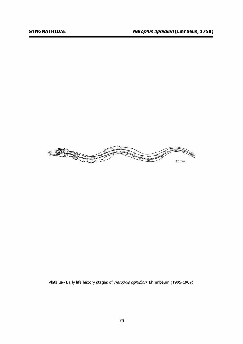
79
SYNGNATHIDAE Nerophis ophidion (Linnaeus, 1758)
Plate 29- Early life history stages of Nerophis ophidion. Ehrenbaum (1905-1909).
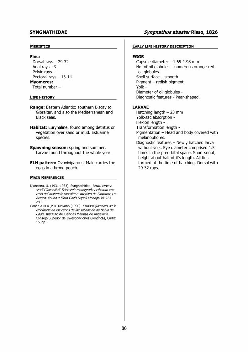
80
SYNGNATHIDAE Syngnathus abaster Risso, 1826 MERISTICS
Fins:
Dorsal rays – 29-32 Anal rays - 3
Pelvic rays – Pectoral rays – 13-14
Myomeres:
Total number –
LIFE HISTORY
Range: Eastern Atlantic: southern Biscay to Gibraltar, and also the Mediterranean and
Black seas.
Habitat: Euryhaline, found among detritus or
vegetation over sand or mud. Estuarine species.
Spawning season: spring and summer. Larvae found throughout the whole year.
ELH pattern: Ovoviviparous. Male carries the
eggs in a brood pouch.
MAIN REFERENCES
D’Ancona, U. (1931-1933). Syngnathidae. Uova, larve e stadi Giovanili di Teleostei: monografia elaborata con l’uso del materiale raccolto e sweriato da Salvatore Lo Bianco. Fauna e Flora Golfo Napoli Monogr.38: 281-289.
Garcia A.M.A.,P.D. Moyano (1990). Estados juveniles de la ictiofauna en los canos de las salinas de da Bahia de Cadiz. Instituto de Ciencias Marinas de Andalucia. Consejo Superior de Investigaciones Científicas, Cadiz: 163pp.
EARLY LIFE HISTORY DESCRIPTION
EGGS
Capsule diameter – 1.65-1.98 mm No. of oil globules – numerous orange-red
oil globules Shell surface – smooth
Pigment – redish pigment
Yolk - Diameter of oil globules -
Diagnostic features - Pear-shaped.
LARVAE Hatching length – 23 mm
Yolk-sac absorption -
Flexion length - Transformation length -
Pigmentation – Head and body covered with melanophores.
Diagnostic features – Newly hatched larva
without yolk. Eye diameter comprised 1.5 times in the preorbital space. Short snout,
height about half of it’s length. All fins formed at the time of hatching. Dorsal with
29-32 rays.
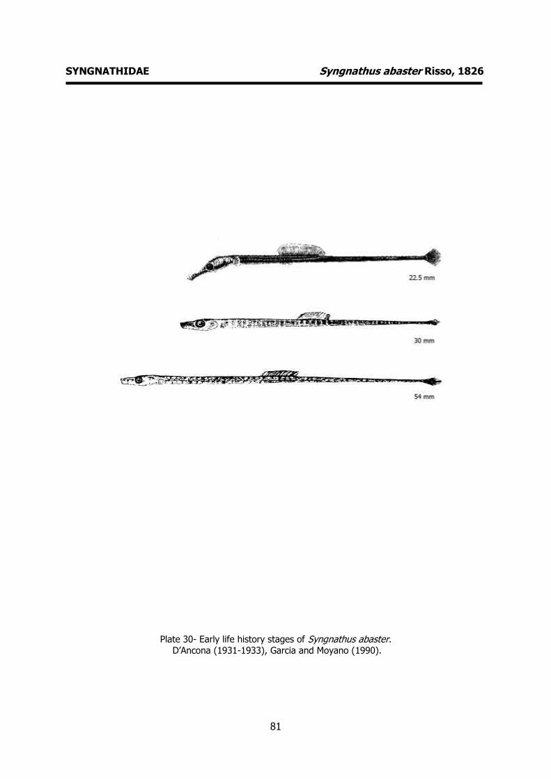
81
SYNGNATHIDAE Syngnathus abaster Risso, 1826
Plate 30- Early life history stages of Syngnathus abaster.
D’Ancona (1931-1933), Garcia and Moyano (1990).
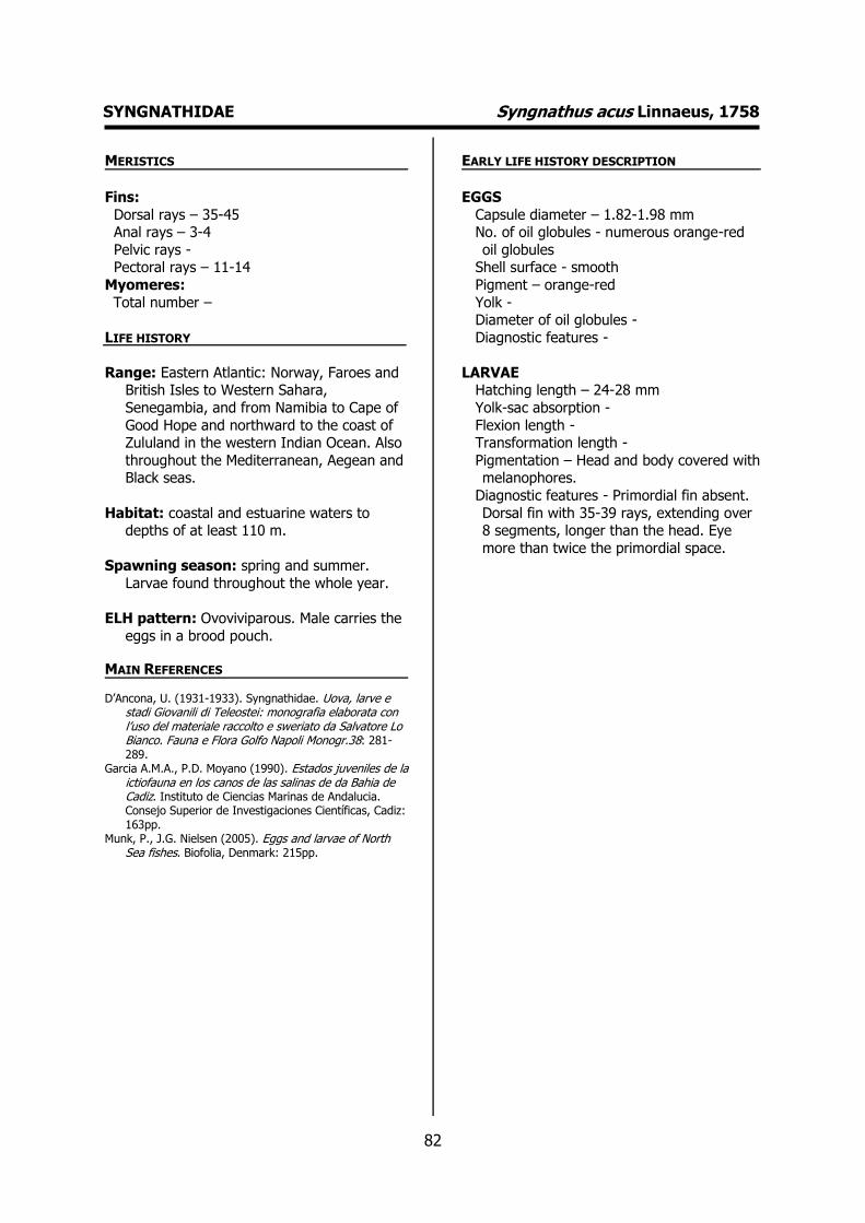
82
SYNGNATHIDAE Syngnathus acus Linnaeus, 1758 MERISTICS
Fins:
Dorsal rays – 35-45 Anal rays – 3-4
Pelvic rays - Pectoral rays – 11-14
Myomeres:
Total number –
LIFE HISTORY
Range: Eastern Atlantic: Norway, Faroes and British Isles to Western Sahara,
Senegambia, and from Namibia to Cape of
Good Hope and northward to the coast of Zululand in the western Indian Ocean. Also
throughout the Mediterranean, Aegean and Black seas.
Habitat: coastal and estuarine waters to depths of at least 110 m.
Spawning season: spring and summer.
Larvae found throughout the whole year.
ELH pattern: Ovoviviparous. Male carries the
eggs in a brood pouch.
MAIN REFERENCES
D’Ancona, U. (1931-1933). Syngnathidae. Uova, larve e stadi Giovanili di Teleostei: monografia elaborata con l’uso del materiale raccolto e sweriato da Salvatore Lo Bianco. Fauna e Flora Golfo Napoli Monogr.38: 281-289.
Garcia A.M.A., P.D. Moyano (1990). Estados juveniles de la ictiofauna en los canos de las salinas de da Bahia de Cadiz. Instituto de Ciencias Marinas de Andalucia. Consejo Superior de Investigaciones Científicas, Cadiz: 163pp.
Munk, P., J.G. Nielsen (2005). Eggs and larvae of North Sea fishes. Biofolia, Denmark: 215pp.
EARLY LIFE HISTORY DESCRIPTION
EGGS
Capsule diameter – 1.82-1.98 mm No. of oil globules - numerous orange-red
oil globules Shell surface - smooth
Pigment – orange-red
Yolk - Diameter of oil globules -
Diagnostic features -
LARVAE Hatching length – 24-28 mm
Yolk-sac absorption -
Flexion length - Transformation length -
Pigmentation – Head and body covered with melanophores.
Diagnostic features - Primordial fin absent.
Dorsal fin with 35-39 rays, extending over 8 segments, longer than the head. Eye
more than twice the primordial space.
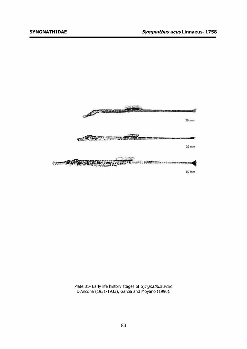
83
SYNGNATHIDAE Syngnathus acus Linnaeus, 1758
Plate 31- Early life history stages of Syngnathus acus. D’Ancona (1931-1933), Garcia and Moyano (1990).
84
SYNGNATHIDAE Syngnathus typhle Linnaeus, 1758 MERISTICS
Fins:
Dorsal rays – 34-38 Anal rays - 3
Pelvic rays - Pectoral rays – 14-17
Myomeres:
Total number –
LIFE HISTORY
Range: Eastern Atlantic: Norway, Baltic Sea and the British Isles to Morocco. Also
throughout the Mediterranean, Black Sea
and Sea of Azov.
Habitat: Usually found along the coasts and estuaries at a temperature range of 8 º to
24 ºC.
Spawning season: spring and summer.
ELH pattern: Oviparous, planktonic eggs and
larvae.
MAIN REFERENCES D’Ancona, U. (1931-1933). Syngnathidae. Uova, larve e
stadi Giovanili di Teleostei: monografia elaborata con l’uso del materiale raccolto e sweriato da Salvatore Lo Bianco. Fauna e Flora Golfo Napoli Monogr.38: 281-289.
Garcia A.M.A., P.D. Moyano (1990). Estados juveniles de la ictiofauna en los canos de las salinas de da Bahia de Cadiz. Instituto de Ciencias Marinas de Andalucia. Consejo Superior de Investigaciones Científicas, Cadiz: 163pp.
Munk, P., J.G. Nielsen (2005). Eggs and larvae of North Sea fishes. Biofolia, Denmark: 215pp.
EARLY LIFE HISTORY DESCRIPTION
EGGS
Capsule diameter – 1.98-2.42 mm No. of oil globules - numerous oil globules
Shell surface - smooth Pigment – yellowish-orange
Yolk -
Diameter of oil globules - Diagnostic features -
LARVAE
Hatching length – 20-25 mm Yolk-sac absorption -
Flexion length -
Transformation length - Pigmentation - Body and head covered with
brownish chromatophores. Melanophores on head and trunk. Tail completely
covered with melanophores.
Diagnostic features - Snout slightly elongate. Dorsal fin 34-38 rays.
85
SYNGNATHIDAE Syngnathus typhle Linnaeus, 1758
Plate 32- Early life history stages of Syngnathus typhle.
D’Ancona (1931-1933), Garcia and Moyano (1990).
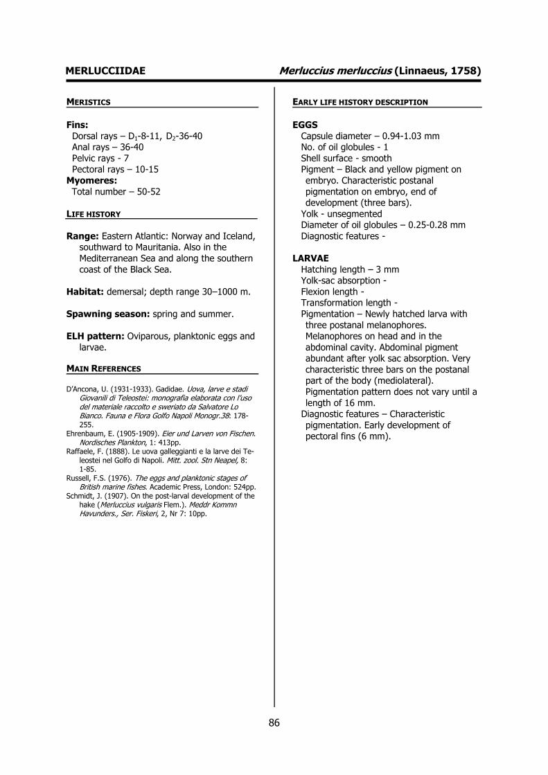
86
MERLUCCIIDAE Merluccius merluccius (Linnaeus, 1758) MERISTICS
Fins:
Dorsal rays – D1-8-11, D2-36-40 Anal rays – 36-40
Pelvic rays - 7 Pectoral rays – 10-15
Myomeres:
Total number – 50-52
LIFE HISTORY
Range: Eastern Atlantic: Norway and Iceland, southward to Mauritania. Also in the
Mediterranean Sea and along the southern
coast of the Black Sea.
Habitat: demersal; depth range 30–1000 m.
Spawning season: spring and summer.
ELH pattern: Oviparous, planktonic eggs and
larvae.
MAIN REFERENCES D’Ancona, U. (1931-1933). Gadidae. Uova, larve e stadi
Giovanili di Teleostei: monografia elaborata con l’uso del materiale raccolto e sweriato da Salvatore Lo Bianco. Fauna e Flora Golfo Napoli Monogr.38: 178-255.
Ehrenbaum, E. (1905-1909). Eier und Larven von Fischen. Nordisches Plankton, 1: 413pp.
Raffaele, F. (1888). Le uova galleggianti e la larve dei Te-leostei nel Golfo di Napoli. Mitt. zool. Stn Neapel, 8: 1-85.
Russell, F.S. (1976). The eggs and planktonic stages of British marine fishes. Academic Press, London: 524pp.
Schmidt, J. (1907). On the post-larval development of the hake (Merluccius vulgaris Flem.). Meddr Kommn Havunders., Ser. Fiskeri, 2, Nr 7: 10pp.
EARLY LIFE HISTORY DESCRIPTION
EGGS
Capsule diameter – 0.94-1.03 mm No. of oil globules - 1
Shell surface - smooth Pigment – Black and yellow pigment on
embryo. Characteristic postanal
pigmentation on embryo, end of development (three bars).
Yolk - unsegmented Diameter of oil globules – 0.25-0.28 mm
Diagnostic features -
LARVAE
Hatching length – 3 mm Yolk-sac absorption -
Flexion length - Transformation length -
Pigmentation – Newly hatched larva with
three postanal melanophores. Melanophores on head and in the
abdominal cavity. Abdominal pigment abundant after yolk sac absorption. Very
characteristic three bars on the postanal part of the body (mediolateral).
Pigmentation pattern does not vary until a
length of 16 mm. Diagnostic features – Characteristic
pigmentation. Early development of pectoral fins (6 mm).
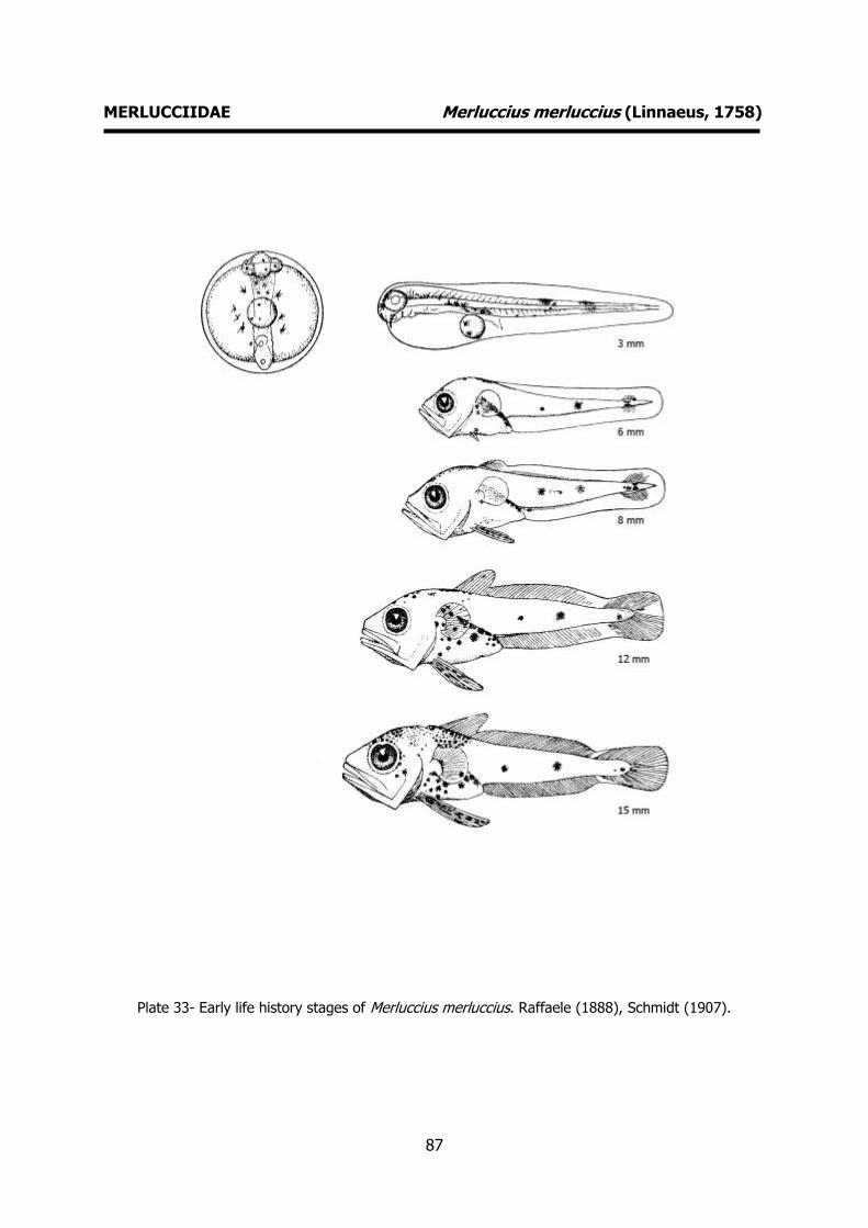
87
MERLUCCIIDAE Merluccius merluccius (Linnaeus, 1758)
Plate 33- Early life history stages of Merluccius merluccius. Raffaele (1888), Schmidt (1907).
88
GADIDAE Ciliata mustela (Linnaeus, 1758) MERISTICS
Fins:
Dorsal rays - D2-45-55 Anal rays – 40-46
Pelvic rays – 7-8 Pectoral rays – 14-16
Myomeres:
Total number –
LIFE HISTORY
Range: Northeast Atlantic: Lisbon north to Finnmark, around the British Isles, in the
Skagerrak and Kattegat and Iceland.
Habitat: demersal; depth range 0-20 m.
Spawning season: winter and spring.
ELH pattern: Oviparous, planktonic eggs and larvae.
MAIN REFERENCES Ehrenbaum, E. (1905-1909). Eier und Larven von Fischen.
Nordisches Plankton, 1: 413pp. Russell, F.S. (1976). The eggs and planktonic stages of
British marine fishes. Academic Press, London: 524pp.
EARLY LIFE HISTORY DESCRIPTION
EGGS
Capsule diameter – 0.66-0.98 mm No. of oil globules - 1
Shell surface - smooth Pigment – Small melanophores arranged in
two rows on embryo. Several
melanophores on the oil globule. Yolk - unsegmented
Diameter of oil globules – 0.12-0.16 mm Diagnostic features -
LARVAE
Hatching length – 2 mm
Yolk-sac absorption - Flexion length -
Transformation length - Pigmentation – Newly hatched larvae with
stellate melanophores on head and
peritoneum. Postanal pigmentation consists of two groups of ventral
melanophores and a few dorsal melanophores. Pelvic fins extremely
elongated and pigmented in pre-flexion larvae.
Diagnostic features – Characteristic
pigmentation. Elongated and pigmented pelvic fins.
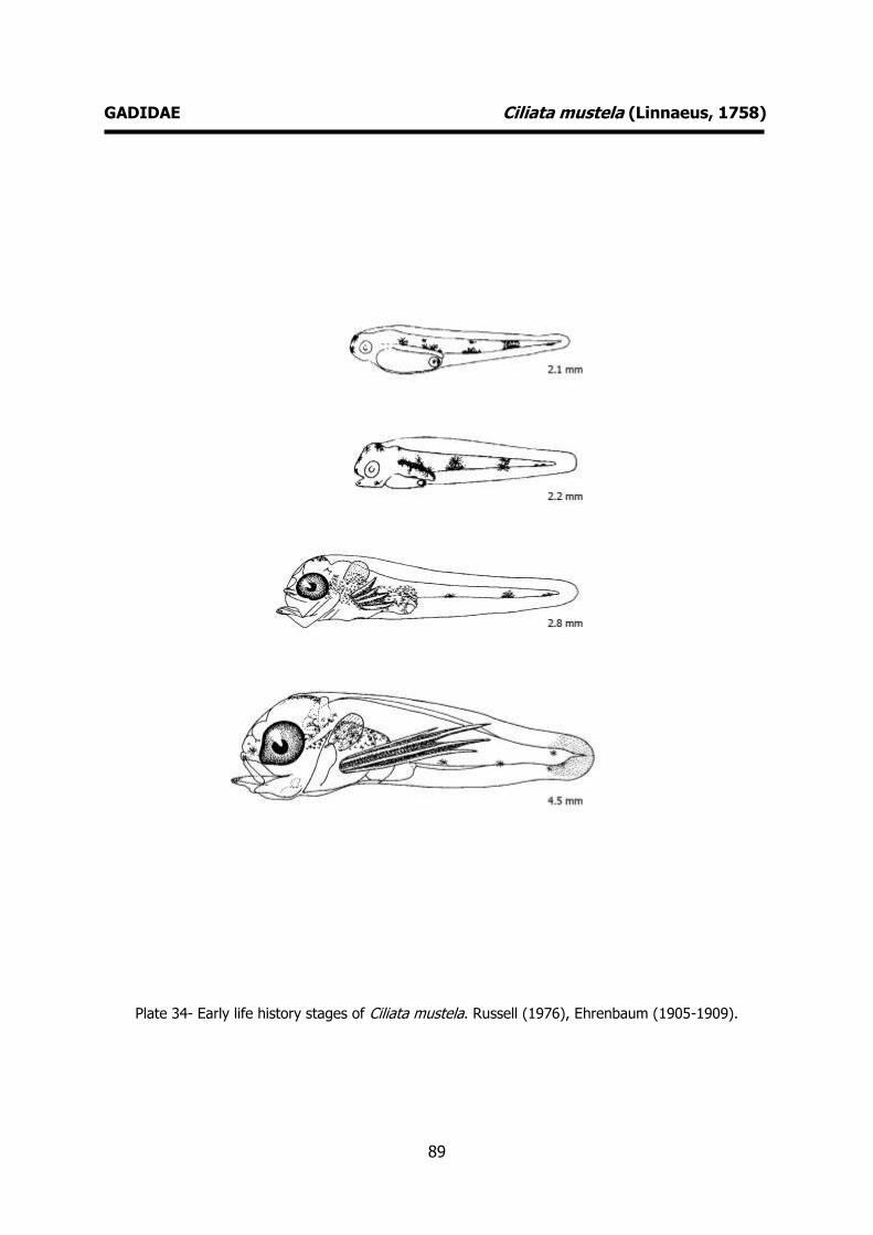
89
GADIDAE Ciliata mustela (Linnaeus, 1758)
Plate 34- Early life history stages of Ciliata mustela. Russell (1976), Ehrenbaum (1905-1909).
90
GADIDAE Gadiculus argenteus Guichenot, 1850 MERISTICS
Fins:
Dorsal rays - D1-9-13, D2-14-17, D3-15-16 Anal rays - A1-17-18, A2-15
Pelvic rays - 6 Pectoral rays – 14-15
Myomeres:
Total number –
LIFE HISTORY
Range: Northeast Atlantic: found in the western Mediterranean and in the Atlantic
around the Strait of Gibraltar and to the
south along the Moroccan coast.
Habitat: pelagic; non-migratory; marine; depth range 100 – 1000 m.
Spawning season: spring, summer and autumn.
ELH pattern: Oviparous, planktonic larvae.
MAIN REFERENCES D’Ancona, U. (1931-1933). Gadidae. Uova, larve e stadi
Giovanili di Teleostei: monografia elaborata con l’uso del materiale raccolto e sweriato da Salvatore Lo Bianco. Fauna e Flora Golfo Napoli Monogr.38: 178-255.
Ehrenbaum, E. (1905-1909). Eier und Larven von Fischen. Nordisches Plankton, 1: 413pp.
Munk, P., J.G. Nielsen (2005). Eggs and larvae of North Sea fishes. Biofolia, Denmark: 215pp.
Raffaele, F. (1888). Le uova galleggianti e la larve dei Te-leostei nel Golfo di Napoli. Mitt. zool. Stn Neapel, 8: 1-85.
Russell, F.S. (1976). The eggs and planktonic stages of British marine fishes. Academic Press, London: 524pp.
Schmidt, J. (1906). The pelagic post-larval stages of the Atlantic species of Gadus. Part II. Meddr Kommn Havunders., Ser. Fiskeri, 2, Nr 2: 18pp.
EARLY LIFE HISTORY DESCRIPTION
EGGS: Undescribed
Capsule diameter - Unknown No. of oil globules - Unknown
Shell surface - Unknown Pigment - Unknown
Yolk - Unknown
Diameter of oil globules - Unknown Diagnostic features - Unknown
LARVAE
Hatching length - Unknown Yolk-sac absorption - Unknown
Flexion length -
Transformation length - Pigmentation - One dorso-ventral cluster in
the posterior part of the tail. At 7.5 mm the pelvic fins begin to develop.
Diagnostic features – Characteristic
pigmentation.

91
GADIDAE Gadiculus argenteus Guichenot, 1850
Plate 35- Early life history stages of Gadiculus argenteus. Schmidt (1906).

92
GADIDAE Micromesistius poutassou (Risso, 1827) MERISTICS
Fins:
Dorsal rays - D1-11-14, D2-10-14, D3-20-24 Anal rays - A1-34-38, A2-20-26
Pelvic rays - 6 Pectoral rays – 20-26
Myomeres:
Total number –
LIFE HISTORY
Range: Northeast Atlantic: Barents Sea south through the eastern Norwegian Sea, around
Iceland, then in the western Mediterranean,
and south along the African coast to Cape Bojador. Northwest Atlantic: southern
Greenland and off southeast Canada and the northeastern coast of the USA.
Habitat: pelagic; depth range 150 – 3000 m.
Spawning season: winter and spring.
ELH pattern: Oviparous, planktonic eggs and larvae.
MAIN REFERENCES D’Ancona, U. (1931-1933). Gadidae. Uova, larve e stadi
Giovanili di Teleostei: monografia elaborata con l’uso del materiale raccolto e sweriato da Salvatore Lo Bianco. Fauna e Flora Golfo Napoli Monogr.38: 178-255.
Ehrenbaum, E. (1905-1909). Eier und Larven von Fischen. Nordisches Plankton, 1: 413pp.
Munk, P., J.G. Nielsen (2005). Eggs and larvae of North Sea fishes. Biofolia, Denmark: 215pp.
Russell, F.S. (1976). The eggs and planktonic stages of British marine fishes. Academic Press, London: 524pp.
Schmidt, J. (1905). The pelagic post-larval stages of the Atlantic species of Gadus. Part I. Meddr Kommn Havunders., Ser. Fiskeri, 1, Nr 4: 77pp.
Schmidt, J. (1906). The pelagic post-larval stages of the Atlantic species of Gadus. Part II. Meddr Kommn Havunders., Ser. Fiskeri, 2, Nr 2: 18pp.
EARLY LIFE HISTORY DESCRIPTION
EGGS
Capsule diameter – 1.04-1.28 mm No. of oil globules - 0
Shell surface - smooth Pigment – Two rows of melanophores
running from the snout to approximately
five sixths of the way along the trunk. Yolk - unsegmented
Diameter of oil globules - Diagnostic features -
LARVAE
Hatching length – 2.0-2.2 mm
Yolk-sac absorption - Flexion length -
Transformation length - Pigmentation – Newly hatched larvae with
the same pigment found in the embryo.
Characteristic pigmentation at 3.3 mm (dorsal and ventral bar on the trunk,
pigmented eyes and mouth open). Diagnostic features – Characteristic
pigmentation. Paired dorsal and ventral streak of melanophores. The dorsal
pigment extends further back than the
ventral. Unpigmented mediolateral region. Marked occipital pigmentation.
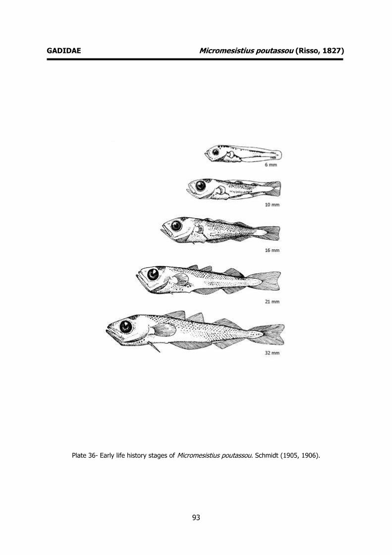
93
GADIDAE Micromesistius poutassou (Risso, 1827)
Plate 36- Early life history stages of Micromesistius poutassou. Schmidt (1905, 1906).

94
GADIDAE Pollachius pollachius (Linnaeus, 1758) MERISTICS
Fins:
Dorsal rays - D1-11-12, D2-15-21, D3-15-20 Anal rays - A1-23-31, A2-16-21
Pelvic rays - 6 Pectoral rays – 16-19
Myomeres:
Total number – 52-55
LIFE HISTORY
Range: Northeast Atlantic: Norway, the Faeroes, and Iceland to the Bay of Biscay.
Habitat: benthopelagic; depth range 0-200 m.
Spawning season: winter and spring.
ELH pattern: Oviparous, planktonic eggs and larvae.
MAIN REFERENCES
D’Ancona, U. (1931-1933). Gadidae. Uova, larve e stadi Giovanili di Teleostei: monografia elaborata con l’uso del materiale raccolto e sweriato da Salvatore Lo Bianco. Fauna e Flora Golfo Napoli Monogr.38: 178-255.
Ehrenbaum, E. (1905-1909). Eier und Larven von Fischen. Nordisches Plankton, 1: 413pp.
Munk, P., J.G. Nielsen (2005). Eggs and larvae of North Sea fishes. Biofolia, Denmark: 215pp.
Russell, F.S. (1976). The eggs and planktonic stages of British marine fishes. Academic Press, London: 524pp.
Schmidt, J. (1905). The pelagic post-larval stages of the Atlantic species of Gadus. Part I. Meddr Kommn Havunders., Ser. Fiskeri, 1, Nr 4: 77pp.
EARLY LIFE HISTORY DESCRIPTION
EGGS
Capsule diameter – 1.10-1.22 mm No. of oil globules - 0
Shell surface - smooth Pigment – Weak pigmentation on embryo.
Tail free of melanophores.
Yolk – unsegmented Diameter of oil globules -
Diagnostic features -
LARVAE Hatching length - Unknown
Yolk-sac absorption - Unknown
Flexion length - Transformation length -
Pigmentation – Pigmentation concentrated in continuous paired dorsal and ventral
contour rows of melanophores (tail region
unpigmented). Mediolateral pigmentation appears at 5 mm and becomes very
conspicuous. Body densely pigmented at a size of 15 mm.
Diagnostic features – Characteristic pigmentation. Paired dorsal and ventral
contour rows of melanophores.
Conspicuous mediolateral row.

95
GADIDAE Pollachius pollachius (Linnaeus, 1758)
Plate 37- Early life history stages of Pollachius pollachius. Schmidt (1905), Russell (1976).
96
GADIDAE Trisopterus luscus (Linnaeus, 1758) MERISTICS
Fins:
Dorsal rays - D1-12-15, D2-21-26, D3-18-22 Anal rays - A1-27-36, A2-18-22
Pelvic rays - 6 Pectoral rays – 19-20
Myomeres:
Total number –
LIFE HISTORY
Range: Eastern Atlantic: British Isles and Skagerrak to the African coast, including
offshore islands. Also in the western
Mediterranean.
Habitat: benthopelagic; brackish; marine; depth range 30-100 m.
Spawning season: winter and spring.
ELH pattern: Oviparous, planktonic eggs and larvae.
MAIN REFERENCES D’Ancona, U. (1931-1933). Gadidae. Uova, larve e stadi
Giovanili di Teleostei: monografia elaborata con l’uso del materiale raccolto e sweriato da Salvatore Lo Bianco. Fauna e Flora Golfo Napoli Monogr.38: 178-255.
Ehrenbaum, E. (1905-1909). Eier und Larven von Fischen. Nordisches Plankton, 1: 413pp.
Munk, P., J.G. Nielsen (2005). Eggs and larvae of North Sea fishes. Biofolia, Denmark: 215pp.
Russell, F.S. (1976). The eggs and planktonic stages of British marine fishes. Academic Press, London: 524pp.
Schmidt, J. (1905). The pelagic post-larval stages of the Atlantic species of Gadus. Part I. Meddr Kommn Havunders., Ser. Fiskeri, 1, Nr 4: 77pp.
Schmidt, J. (1906). The pelagic post-larval stages of the Atlantic species of Gadus. Part II. Meddr Kommn Havunders., Ser. Fiskeri, 2, Nr 2: 18pp.
EARLY LIFE HISTORY DESCRIPTION
EGGS
Capsule diameter – 0.9-1.23 mm No. of oil globules - 0
Shell surface - smooth Pigment – Many small melanophores on
dorsal side of embryo. Unpigmented tail.
Yolk - unsegmented Diameter of oil globules -
Diagnostic features -
LARVAE Hatching length – 3 mm
Yolk-sac absorption -
Flexion length - Transformation length -
Pigmentation – Newly hatched larva with scattered melanophores all over the body.
Dorsal and ventral pigmentation
conspicuous. Marked occipital pigmentation. Dorsal and ventral streaks of
melanophores evident. No mediolateral pigmentation.
Diagnostic features – Characteristic pigmentation, Well-marked dorsal and
ventral pigment streaks that end abruptly
(unpigmented tail).

97
GADIDAE Trisopterus luscus (Linnaeus, 1758)
Plate 38- Early life history stages of Trisopterus luscus. Ehrenbaum (1905-1909),
Schmidt (1905, 1906).
98
GADIDAE Trisopterus minutus (Linnaeus, 1758) MERISTICS
Fins:
Dorsal rays - D1-11-15, D2-20-26, D3-19-23 Anal rays - A1-25-31, A2-20-24
Pelvic rays - 6 Pectoral rays – 17-19
Myomeres:
Total number –
LIFE HISTORY
Range: Eastern Atlantic: Trondheim Fjord and the Faeroe Islands to Portugal and along
the Atlantic coast of Morocco; also in the
Mediterranean.
Habitat: benthopelagic; non-migratory; marine; depth range 0-400 m.
Spawning season: spring and summer
ELH pattern: Oviparous, planktonic eggs and larvae.
MAIN REFERENCES Munk, P., J.G. Nielsen (2005). Eggs and larvae of North
Sea fishes. Biofolia, Denmark: 215pp. Schmidt, J. (1905). The pelagic post-larval stages of the
Atlantic species of Gadus. Part I. Meddr Kommn Havunders., Ser. Fiskeri, 1, Nr 4: 77pp.
Russell, F.S. (1976). The eggs and planktonic stages of British marine fishes. Academic Press, London: 524pp.
Schmidt, J. (1906). The pelagic post-larval stages of the Atlantic species of Gadus. Part II. Meddr Kommn Havunders., Ser. Fiskeri, 2, Nr 2: 18pp.
EARLY LIFE HISTORY DESCRIPTION
EGGS
Capsule diameter – 0.95-1.03 mm No. of oil globules - 0
Shell surface - smooth Pigment -
Yolk - unsegmented
Diameter of oil globules - Diagnostic features -
LARVAE
Hatching length – 2.3-2.4 mm Yolk-sac absorption -
Flexion length -
Transformation length - Pigmentation – Dorsal and ventral unpaired
contour rows of large stellate melanophores (8-10) extending posteriorly
for approximately equal distances to the
base of the caudal fin area. No occipital pigmentation. Two melanophores on the
head above the eyes. A few ventral preanal melanophores. No mediolateral
pigmentation. Diagnostic features – Characteristic
pigmentation. Dorsal and ventral row of
melanophores.

99
GADIDAE Trisopterus minutus (Linnaeus, 1758)
Plate 39- Early life history stages of Trisopterus minutus. Russell (1976).
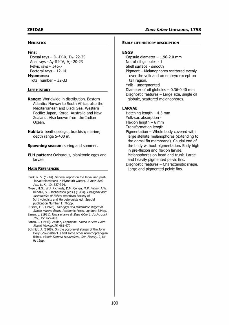
100
ZEIDAE Zeus faber Linnaeus, 1758 MERISTICS
Fins:
Dorsal rays – D1-IX-X, D2- 22-25 Anal rays - A1-III-IV, A2- 20-23
Pelvic rays – I+5-7 Pectoral rays – 12-14
Myomeres:
Total number – 32-33
LIFE HISTORY
Range: Worldwide in distribution. Eastern Atlantic: Norway to South Africa, also the
Mediterranean and Black Sea. Western
Pacific: Japan, Korea, Australia and New Zealand. Also known from the Indian
Ocean.
Habitat: benthopelagic; brackish; marine;
depth range 5-400 m.
Spawning season: spring and summer.
ELH pattern: Oviparous, planktonic eggs and larvae.
MAIN REFERENCES Clark, R. S. (1914). General report on the larval and post-
-larval teleosteans in Plymouth waters. J. mar. biol. Ass. U. K., 10: 327-394.
Moser, H.G., W.J. Richards, D.M. Cohen, M.P. Fahay, A.W. Kendall, S.L. Richardson (eds.) (1984). Ontogeny and systematics of fishes. American Society of Ichthyologists and Herpetologists ed., Special publication Number 1: 760pp.
Russell, F.S. (1976). The eggs and planktonic stages of British marine fishes. Academic Press, London: 524pp.
Sanzo, L. (1931). Uova e larve di Zeus faber L. Archo zool. Ital., 15: 475-483.
Sanzo, L. (1956). Zeidae, Caproidae. Fauna e Flora Golfo Napoli Monogr.38: 461-470.
Schmidt, J. (1908). On the post-larval stages of the John Dory (Zeus faber L.) and some other Acanthopterygian fishes. Meddr Kommn Havunders., Ser. Fiskery, 2, Nr 9: 12pp.
EARLY LIFE HISTORY DESCRIPTION
EGGS
Capsule diameter – 1.96-2.0 mm No. of oil globules - 1
Shell surface - smooth Pigment – Melanophores scattered evenly
over the yolk and on embryo except on
tail region. Yolk - unsegmented
Diameter of oil globules – 0.36-0.40 mm Diagnostic features – Large size, single oil
globule, scattered melanophores.
LARVAE
Hatching length – 4.3 mm Yolk-sac absorption -
Flexion length – 6 mm Transformation length -
Pigmentation – Whole body covered with
large stellate melanophores (extending to the dorsal fin membrane). Caudal end of
the body without pigmentation. Body high in pre-flexion and flexion larvae.
Melanophores on head and trunk. Large and heavily pigmented pelvic fins.
Diagnostic features – Characteristic shape.
Large and pigmented pelvic fins.
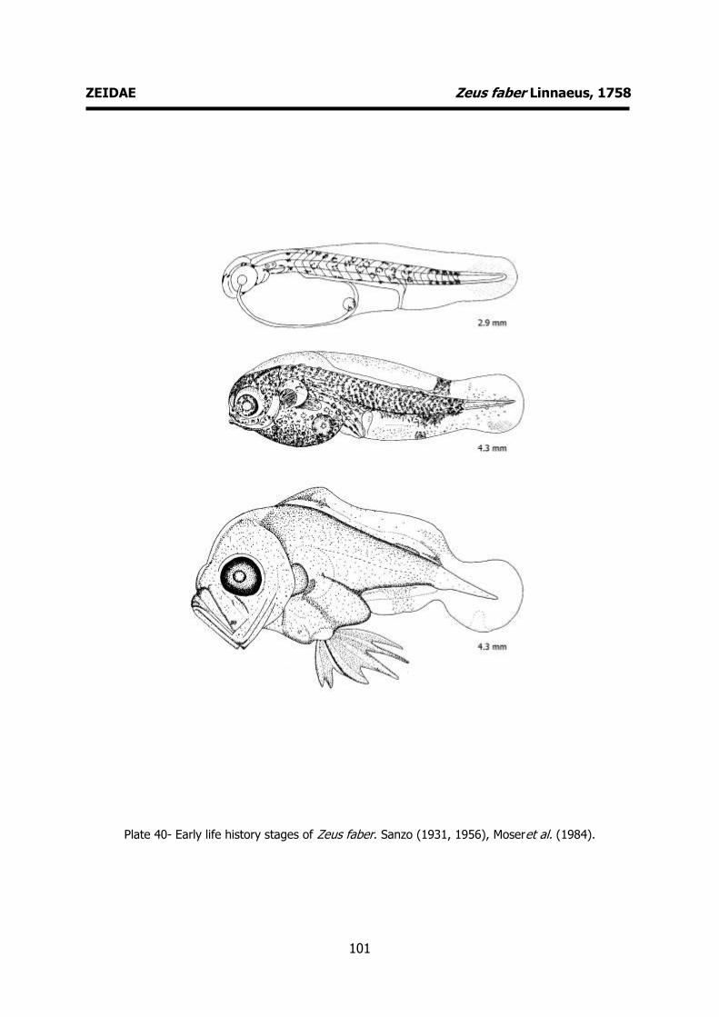
101
ZEIDAE Zeus faber Linnaeus, 1758
Plate 40- Early life history stages of Zeus faber. Sanzo (1931, 1956), Moseret al. (1984).
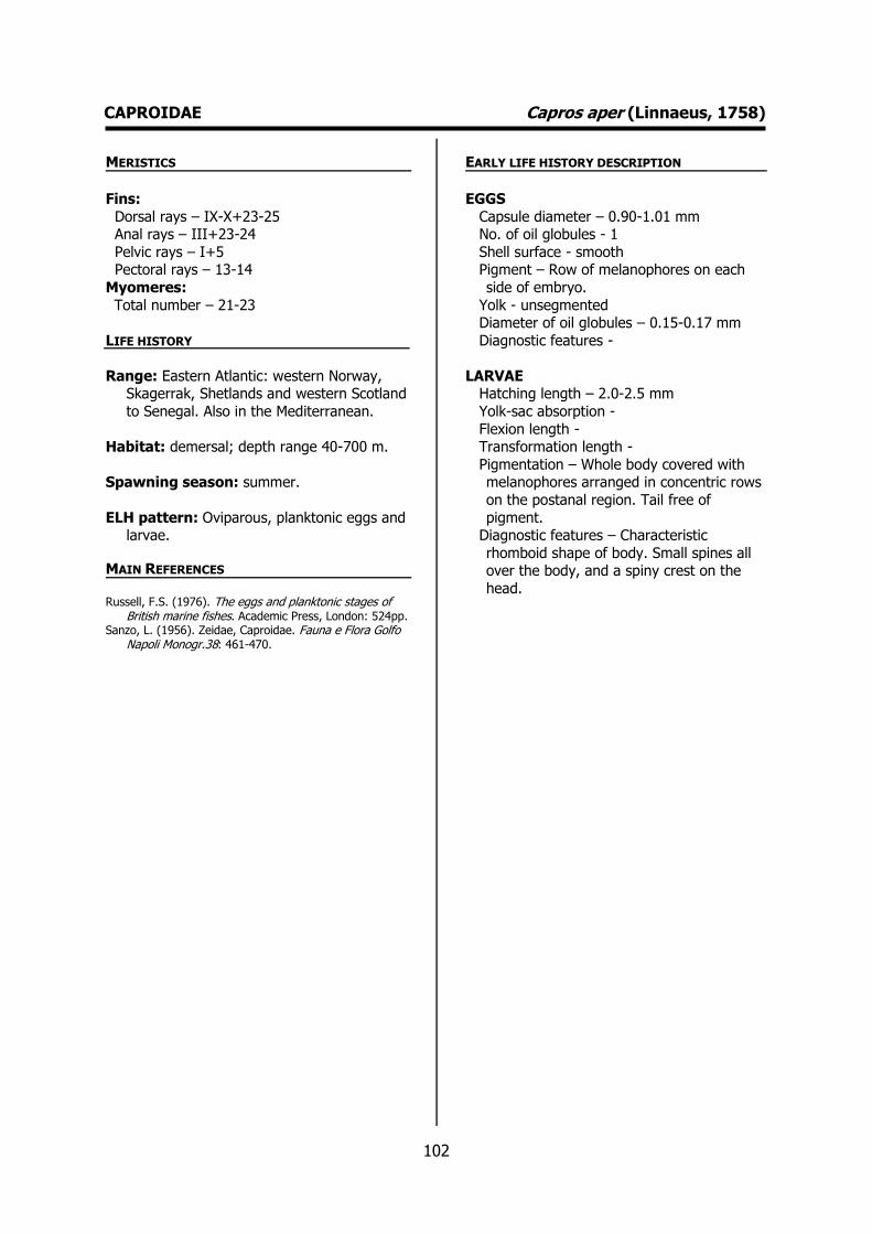
102
CAPROIDAE Capros aper (Linnaeus, 1758) MERISTICS
Fins:
Dorsal rays – IX-X+23-25 Anal rays – III+23-24
Pelvic rays – I+5 Pectoral rays – 13-14
Myomeres:
Total number – 21-23
LIFE HISTORY
Range: Eastern Atlantic: western Norway, Skagerrak, Shetlands and western Scotland
to Senegal. Also in the Mediterranean.
Habitat: demersal; depth range 40-700 m.
Spawning season: summer.
ELH pattern: Oviparous, planktonic eggs and larvae.
MAIN REFERENCES Russell, F.S. (1976). The eggs and planktonic stages of
British marine fishes. Academic Press, London: 524pp. Sanzo, L. (1956). Zeidae, Caproidae. Fauna e Flora Golfo
Napoli Monogr.38: 461-470.
EARLY LIFE HISTORY DESCRIPTION
EGGS
Capsule diameter – 0.90-1.01 mm No. of oil globules - 1
Shell surface - smooth Pigment – Row of melanophores on each
side of embryo.
Yolk - unsegmented Diameter of oil globules – 0.15-0.17 mm
Diagnostic features -
LARVAE Hatching length – 2.0-2.5 mm
Yolk-sac absorption -
Flexion length - Transformation length -
Pigmentation – Whole body covered with melanophores arranged in concentric rows
on the postanal region. Tail free of
pigment. Diagnostic features – Characteristic
rhomboid shape of body. Small spines all over the body, and a spiny crest on the
head.
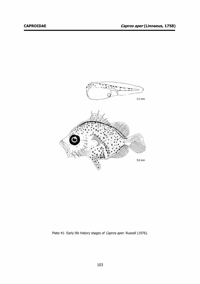
103
CAPROIDAE Capros aper (Linnaeus, 1758)
Plate 41- Early life history stages of Capros aper. Russell (1976).
104
SERRANIDAE Serranus cabrilla (Linnaeus, 1758) MERISTICS
Fins:
Dorsal rays – X+13-15 Anal rays – III+7-8
Pelvic rays – I+5 Pectoral rays – 14-15
Myomeres:
Total number – 10-19
LIFE HISTORY
Range: Eastern Atlantic: English Channel southward round the Cape of Good Hope to
Natal, South Africa, including Azores,
Madeira and the Canary Islands. Also in the Mediterranean and western Black Sea and
possibly in the Red Sea.
Habitat: demersal; marine; depth range 0-
500 m.
Spawning season: spring and summer.
ELH pattern: Oviparous, planktonic eggs and larvae.
MAIN REFERENCES Bertolini, F. (1933). Serranidae. Uova, larve e stadi
Giovanili di Teleostei: monografia elaborata con l’uso del materiale raccolto e sweriato da Salvatore Lo Bianco. Fauna e Flora Golfo Napoli Monogr.38. Publi. In 4 parts: 310-321.
Ehrenbaum, E. (1905-1909). Eier und Larven von Fischen. Nordisches Plankton, 1: 413pp.
Fage, L. (1918). Shore-fishes. Rep. Dan. Oceanogr. Exped . 1908-10 Medit . adjac. Seas, 2, Biology A3: 1-154.
Marinaro, J-Y (1971). Contribution à l'étude des oeufs et larves pèlagiques de poissons mediterranéens. V. Oeufs pèlagiques de la Baie d'Alger. Pelagos, 3 (1): 1-118.
Munk, P., J.G. Nielsen (2005). Eggs and larvae of North Sea fishes. Biofolia, Denmark: 215pp.
Raffaele, F. (1888). Le uova galleggianti e la larve dei Te-leostei nel Golfo di Napoli. Mitt. zool. Stn Neapel, 8: 1-85.
Russell, F.S. (1976). The eggs and planktonic stages of British marine fishes. Academic Press, London: 524pp.
EARLY LIFE HISTORY DESCRIPTION
EGGS
Capsule diameter – 0.85-0.97 mm No. of oil globules - 1
Shell surface - smooth Pigment - A few pale yellow
chromatophores along the sides of the
body and smaller rather inconspicuous melanophores uniformly distributed over
the body. A little yellow and black pigment over the oil globule.
Yolk - unsegmented Diameter of oil globules – 0.14-0.15 mm
Diagnostic features -
LARVAE
Hatching length – 1.8-2.4 mm Yolk-sac absorption -
Flexion length -
Transformation length - Pigmentation – Melanophores on head and
trunk. Pigmented peritoneum. One dorsal and one ventral melanophore on body
contours about the middle of the postanal region.
Diagnostic features – Newly hatched larva:
oil globule situated behind the anterior end of the yolk sac nearer to the midpoint.
Primordial fin with many stellate chromatophores. Third dorsal ray very
long. At 6.5 mm, the first 8 rays of the
dorsal fin are well developed. Elongated pelvic fins.

105
SERRANIDAE Serranus cabrilla (Linnaeus, 1758)
Plate 42- Early life history stages of Serranus cabrilla. Bertolini (1933).
106
MORONIDAE Dicentrarchus labrax (Linnaeus, 1758) MERISTICS
Fins:
Dorsal rays – D1 VIII-IX D2 I-II+11-12 Anal rays – III+10-12
Pelvic rays – I+5 Pectoral rays – 15-16
Myomeres:
Total number –
LIFE HISTORY
Range: Eastern Atlantic: Norway to Morocco, the Canary Islands and Senegal. Reported
from Iceland. Also known from the
Mediterranean and Black Sea.
Habitat: demersal; freshwater; brackish; marine; depth range 10-100 m.
Spawning season: winter and spring.
ELH pattern: Oviparous, planktonic eggs and larvae.
MAIN REFERENCES Barnabé, G., F. Boulineau-Coatanea, F. Rene (1976).
Chronologie de la morphogenese chez le loup ou bar Dicentrarchus labrax (L.) (Pisces, Serranidae) obtenu par reproduction artificielle. Aquaculture, 8: 351-363.
Bertolini, F. (1933). Serranidae. Uova, larve e stadi Giovanili di Teleostei: monografia elaborata con l’uso del materiale raccolto e sweriato da Salvatore Lo Bianco. Fauna e Flora Golfo Napoli Monogr.38. Publi. In 4 parts: 310-321.
Ehrenbaum, E. (1905-1909). Eier und Larven von Fischen. Nordisches Plankton, 1: 413pp.
Munk, P., J.G. Nielsen (2005). Eggs and larvae of North Sea fishes. Biofolia, Denmark: 215pp.
Raffaele, F. (1888) - Le uova galleggianti e la larve dei Te-leostei nel Golfo di Napoli. Mitt. zool. Stn Neapel, 8: 1-85.
Russell, F.S. (1976). The eggs and planktonic stages of British marine fishes. Academic Press, London: 524pp.
EARLY LIFE HISTORY DESCRIPTION
EGGS
Capsule diameter – 1.10-1.52 mm No. of oil globules - 1
Shell surface - smooth Pigment - Yellow and black chromatophores
on the yolk and on the large yellowish oil
globule. Yolk - unsegmented
Diameter of oil globules – 0.31-0.46 mm Diagnostic features -
LARVAE
Hatching length – 3.61-4.05 mm
Yolk-sac absorption – 5-6 mm Flexion length – 9 mm
Transformation length - Pigmentation – Newly hatched larva: Anus
situated some distance behind the yolk
sac. A row of ventral melanophores on the upper side of the alimentary canal. Late
larvae: Continous line of melanophores stretching from the snout to the base of
the caudal fin. Dorsal and lateral row of melanophores only in the posterior part of
the tail.
Diagnostic features – Typical pigmentation.
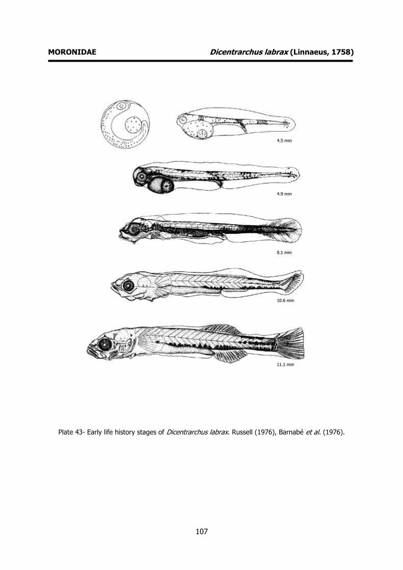
107
MORONIDAE Dicentrarchus labrax (Linnaeus, 1758)
Plate 43- Early life history stages of Dicentrarchus labrax. Russell (1976), Barnabé et al. (1976).
108
CEPOLIDAE Cepola macrophthalma Linnaeus, 1758 MERISTICS
Fins:
Dorsal rays – 67-74 Anal rays – 60-70
Pelvic rays – I+5 Pectoral rays – 12-18
Myomeres:
Total number – 15+54
LIFE HISTORY
Range: Eastern Atlantic: British Isles to north of Senegal, including the Mediterranean.
Habitat: demersal; marine; depth range 15– 400 m.
Spawning season: spawning occurs
throughout the whole year.
ELH pattern: Oviparous, planktonic eggs and
larvae.
MAIN REFERENCES Clark, R. S. (1920) . The pelagic young and early bottom
stages of Teleosteans. J. mar. biol. Ass. U. K., 12: 159-240.
Fage, L. (1918). Shore-fishes. Rep. Dan. Oceanogr. Exped . 1908-10 Medit . adjac. Seas, 2, Biology A3: 1-154.
Russell, F.S. (1976). The eggs and planktonic stages of British marine fishes. Academic Press, London: 524pp.
EARLY LIFE HISTORY DESCRIPTION
EGGS
Capsule diameter – 0.72 mm No. of oil globules - 1
Shell surface - smooth Pigment - unknown
Yolk -
Diameter of oil globules – 0.14 mm Diagnostic features -
LARVAE
Hatching length - unknown Yolk-sac absorption - unknown
Flexion length -
Transformation length - Pigmentation – Melanophores on the head,
jaw, snout and abdomen. 1 to 3 caudal melanophores.
Diagnostic features - The most
characteristic features are the occipital, supraorbital and 7-9 preopercular spines.
Row of denticles (6-7) along the lower margin of the jaw. Occipital spines (begins
as 2 spines and later changes into a toothed ridge with a longer posterior
spine). Supraorbital ridge with 4 to 5 teeth.
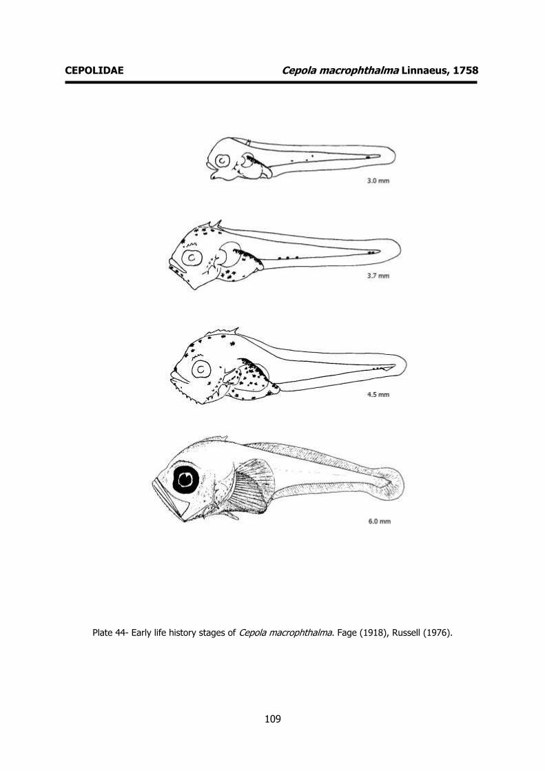
109
CEPOLIDAE Cepola macrophthalma Linnaeus, 1758
Plate 44- Early life history stages of Cepola macrophthalma. Fage (1918), Russell (1976).

110
CARANGIDAE Trachurus trachurus (Linnaeus 1758) MERISTICS
Fins:
Dorsal rays – D1 VIII, D2 I+28-34 Anal rays – A1 II, A2 I+25-34
Pelvic rays – I+5 Pectoral rays – 20-21
Myomeres:
Total number – 25
LIFE HISTORY
Range: Eastern Atlantic: Iceland to Senegal, including the Mediterranean and Marmara
seas, and the Black Sea. Also western
Atlantic, Indian, and western Pacific.
Habitat: pelagic; marine, depth range 0–1050 m.
Spawning season: spawning occurs throughout the whole year.
ELH pattern: Oviparous, planktonic eggs and
larvae.
MAIN REFERENCES Brownell, C.L. (1979). Stages in the early development of
40 marine fish species with pelagic eggs from the cape of Good Hope. Ichthyological Bulletin of the J.L.B. Smith Institute of Ichthyology, Rhodes University, Grahamstown, (40): 84pp.
Ehrenbaum, E. (1905-1909). Eier und Larven von Fischen. Nordisches Plankton, 1: 413pp.
Marinaro, J-Y (1971). Contribution à l'étude des oeufs et larves pèlagiques de poissons mediterranéens. V. Oeufs pèlagiques de la Baie d'Alger. Pelagos, 3 (1): 1-118.
Munk, P., J.G. Nielsen (2005). Eggs and larvae of North Sea fishes. Biofolia, Denmark: 215pp.
Russell, F.S. (1976). The eggs and planktonic stages of British marine fishes. Academic Press, London: 524pp.
EARLY LIFE HISTORY DESCRIPTION
EGGS
Capsule diameter – 0.76-1.04 mm No. of oil globules - 1
Shell surface - smooth Pigment - Yolk brownish-yellow and black
pigment appear along the body contours
on embryo. Yolk - segmented
Diameter of oil globules – 0.19-0.29 Diagnostic features -
LARVAE
Hatching length – 2.5 mm
Yolk-sac absorption - Flexion length -
Transformation length - Pigmentation - Numerous melanophores on
lower jaw, along abdomen and on head
and upper surface of stomach. Dorsal and ventral body contour of melanophores and
a mediolateral row. As the larva growths the number of melanophores increase
covering almost the whole body (with the exception of the caudal region).
Diagnostic features – Newly hatched larva:
anterior edge of yolk-sac slightly beyond the anterior point of head. Anus well
behind the yolk-sac. Anterior position of oil globule. Larvae 4 mm long: 4-5 opercular
spines on the outer row and about 5 on
the inner row, becoming more in the late larvae. Characteristic pigmentation.

111
CARANGIDAE Trachurus trachurus (Linnaeus 1758)
Plate 45- Early life history stages of Trachurus trachurus. Brownell (1979).
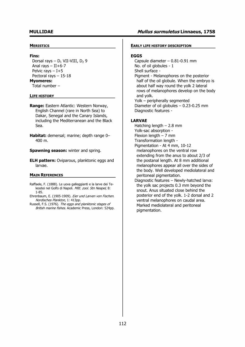
112
MULLIDAE Mullus surmuletus Linnaeus, 1758 MERISTICS
Fins:
Dorsal rays – D1 VII-VIII, D2 9 Anal rays – II+6-7
Pelvic rays – I+5 Pectoral rays – 15-18
Myomeres:
Total number –
LIFE HISTORY
Range: Eastern Atlantic: Western Norway, English Channel (rare in North Sea) to
Dakar, Senegal and the Canary Islands,
including the Mediterranean and the Black Sea.
Habitat: demersal; marine; depth range 0–
400 m.
Spawning season: winter and spring.
ELH pattern: Oviparous, planktonic eggs and
larvae.
MAIN REFERENCES Raffaele, F. (1888). Le uova galleggianti e la larve dei Te-
leostei nel Golfo di Napoli. Mitt. zool. Stn Neapel, 8: 1-85.
Ehrenbaum, E. (1905-1909). Eier und Larven von Fischen. Nordisches Plankton, 1: 413pp.
Russell, F.S. (1976). The eggs and planktonic stages of British marine fishes. Academic Press, London: 524pp.
EARLY LIFE HISTORY DESCRIPTION
EGGS
Capsule diameter – 0.81-0.91 mm No. of oil globules - 1
Shell surface - Pigment - Melanophores on the posterior
half of the oil globule. When the embryo is
about half way round the yolk 2 lateral rows of melanophores develop on the body
and yolk. Yolk – peripherally segmented
Diameter of oil globules – 0.23-0.25 mm Diagnostic features -
LARVAE Hatching length – 2.8 mm
Yolk-sac absorption - Flexion length – 7 mm
Transformation length -
Pigmentation - At 4 mm, 10-12 melanophores on the ventral row
extending from the anus to about 2/3 of the postanal length. At 8 mm additional
melanophores appear all over the sides of the body. Well developed mediolateral and
peritoneal pigmentation.
Diagnostic features – Newly-hatched larva: the yolk sac projects 0.3 mm beyond the
snout. Anus situated close behind the posterior end of the yolk. 1-2 dorsal and 2
ventral melanophores on caudal area.
Marked mediolateral and peritoneal pigmentation.

113
MULLIDAE Mullus surmuletus Linnaeus, 1758
Plate 46- Early life history stages of Mullus suyrmuletus. Russell (1976).
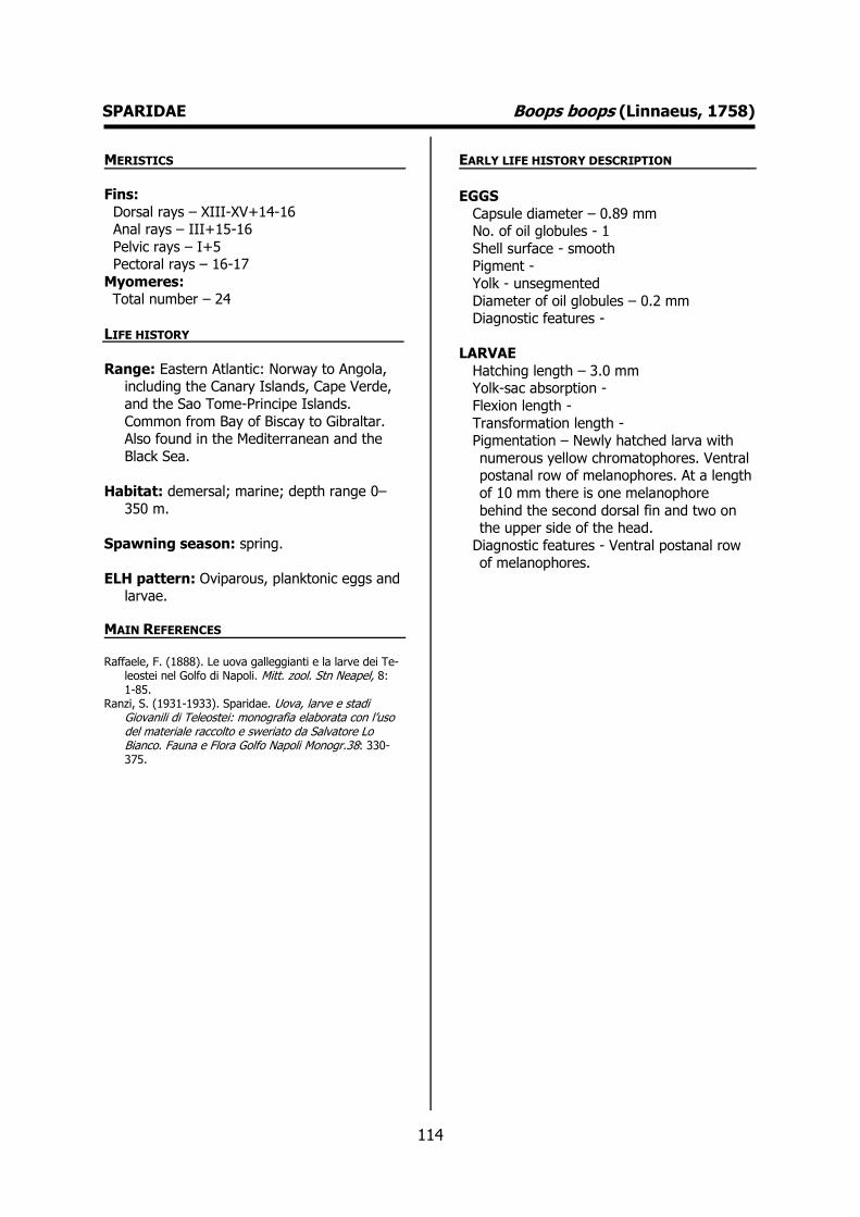
114
SPARIDAE Boops boops (Linnaeus, 1758) MERISTICS
Fins:
Dorsal rays – XIII-XV+14-16 Anal rays – III+15-16
Pelvic rays – I+5 Pectoral rays – 16-17
Myomeres: Total number – 24
LIFE HISTORY
Range: Eastern Atlantic: Norway to Angola, including the Canary Islands, Cape Verde,
and the Sao Tome-Principe Islands.
Common from Bay of Biscay to Gibraltar. Also found in the Mediterranean and the
Black Sea.
Habitat: demersal; marine; depth range 0–
350 m.
Spawning season: spring.
ELH pattern: Oviparous, planktonic eggs and larvae.
MAIN REFERENCES Raffaele, F. (1888). Le uova galleggianti e la larve dei Te-
leostei nel Golfo di Napoli. Mitt. zool. Stn Neapel, 8: 1-85.
Ranzi, S. (1931-1933). Sparidae. Uova, larve e stadi Giovanili di Teleostei: monografia elaborata con l’uso del materiale raccolto e sweriato da Salvatore Lo Bianco. Fauna e Flora Golfo Napoli Monogr.38: 330-375.
EARLY LIFE HISTORY DESCRIPTION
EGGS
Capsule diameter – 0.89 mm No. of oil globules - 1
Shell surface - smooth Pigment -
Yolk - unsegmented
Diameter of oil globules – 0.2 mm Diagnostic features -
LARVAE
Hatching length – 3.0 mm Yolk-sac absorption -
Flexion length -
Transformation length - Pigmentation – Newly hatched larva with
numerous yellow chromatophores. Ventral postanal row of melanophores. At a length
of 10 mm there is one melanophore
behind the second dorsal fin and two on the upper side of the head.
Diagnostic features - Ventral postanal row of melanophores.

115
SPARIDAE Boops boops (Linnaeus, 1758)
Plate 47- Early life history stages of Boops boops. Dekhnik (1973).

116
SPARIDAE Diplodus sargus (Linnaeus, 1758) MERISTICS
Fins:
Dorsal rays – XI-XII+12-15 Anal rays – III+12-14
Pelvic rays – I+5 Pectoral rays – 15-16
Myomeres:
Total number –
LIFE HISTORY
Range: Eastern Atlantic: Bay of Biscay and Mediterranean to South Africa.
Habitat: demersal; brackish; marine; depth range 0–50 m.
Spawning season: spawns throughout the
whole year.
ELH pattern: Oviparous, planktonic eggs and
larvae.
MAIN REFERENCES Raffaele, F. (1888). Le uova galleggianti e la larve dei Te-
leostei nel Golfo di Napoli. Mitt. zool. Stn Neapel, 8: 1-85.
Ranzi, S. (1931-1933). Sparidae. Uova, larve e stadi Giovanili di Teleostei: monografia elaborata con l’uso del materiale raccolto e sweriato da Salvatore Lo Bianco. Fauna e Flora Golfo Napoli Monogr.38: 330-375.
EARLY LIFE HISTORY DESCRIPTION
EGGS
Capsule diameter – ca. 1.0 mm No. of oil globules - 1
Shell surface - smooth Pigment – Pigment on embryo congregate
mainly in four large spots located above
and under the eyes, along the whole trunk, tail and scattered on the yolk.
Yolk - unsegmented Diameter of oil globules – 0.18-0.20 mm
Diagnostic features -
LARVAE
Hatching length – ca. 3 mm Yolk-sac absorption – ca. 4 mm
Flexion length - Transformation length -
Pigmentation – Two large dorsal and ventral
postanal melanophores (not always present). Ventral row of postanal
melanophores. Peritoneal pigmentation. Diagnostic features – Typical pigmentation.
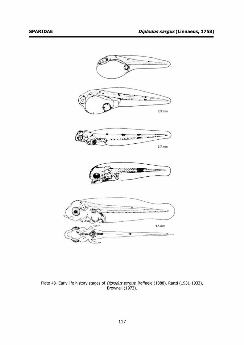
117
SPARIDAE Diplodus sargus (Linnaeus, 1758)
Plate 48- Early life history stages of Diplodus sargus. Raffaele (1888), Ranzi (1931-1933),
Brownell (1973).

118
SPARIDAE Pagellus bogaraveo (Brünnich, 1768) MERISTICS
Fins:
Dorsal rays – XII-XIII+11-13 Anal rays – III+10-12
Pelvic rays – I+5 Pectoral rays – 15-17
Myomeres:
Total number –
LIFE HISTORY
Range: Eastern Atlantic: Norway, Strait of Gibraltar to Cape Blanc in Mauritania,
Madeira, Canary Islands, and western
Mediterranean (rare beyond the Sicilian Strait). Reported from Iceland.
Habitat: marine; benthopelagic; depth range
0-700 m.
Spawning season: winter, spring and
summer.
ELH pattern: Oviparous, planktonic larvae.
MAIN REFERENCES Fage, L. (1918). Shore-fishes. Rep. Dan. Oceanogr. Exped
. 1908-10 Medit . adjac. Seas, 2, Biology A3: 1-154. Ranzi, S. (1931-1933). Sparidae. Uova, larve e stadi
Giovanili di Teleostei: monografia elaborata con l’uso del materiale raccolto e sweriato da Salvatore Lo Bianco. Fauna e Flora Golfo Napoli Monogr.38: 330-375.
EARLY LIFE HISTORY DESCRIPTION
EGGS
Capsule diameter - Unknown No. of oil globules - Unknown
Shell surface - Unknown Pigment - Unknown
Yolk - Unknown
Diameter of oil globules - Unknown Diagnostic features - Unknown
LARVAE
Hatching length - Unknown Yolk-sac absorption - Unknown
Flexion length -
Transformation length - Pigmentation – Postanal well developed
ventral and dorsal melanophore. Pigmented urostyle. Peritoneal
pigmentation.
Diagnostic features - At 10 mm operculum with two crests; the inner with 3 or 4 low
teeth; the outer with 6 teeth, the fourth of which is the longest.
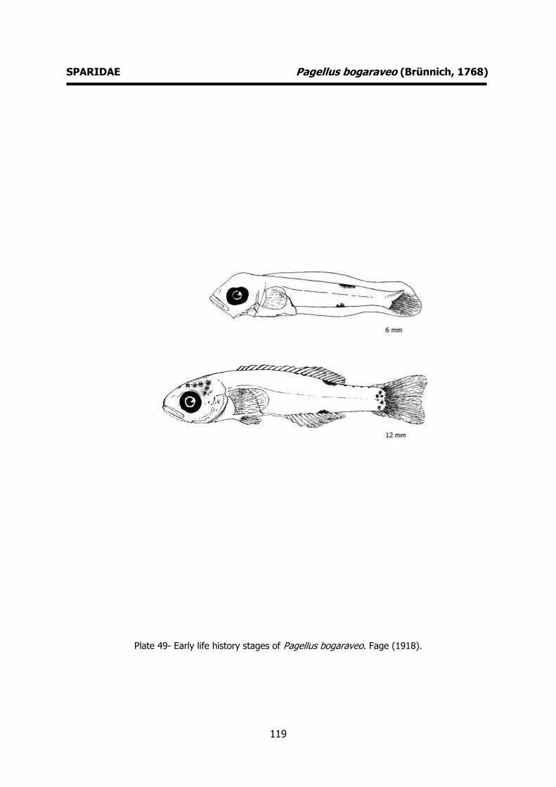
119
SPARIDAE Pagellus bogaraveo (Brünnich, 1768)
Plate 49- Early life history stages of Pagellus bogaraveo. Fage (1918).

120
SPARIDAE Pagrus pagrus (Linnaeus, 1758) MERISTICS
Fins:
Dorsal rays – XII+10-11 Anal rays – III+8-9
Pelvic rays – I+5 Pectoral rays – 15
Myomeres: Total number –
LIFE HISTORY
Range: Eastern Atlantic: Strait of Gibraltar to 15 ºN (rare southward 20 ºN), including
Madeira and the Canary Islands;
Mediterranean and northward to the British Isles. Western Atlantic: New York, USA and
northern Gulf of Mexico to Argentina, including the continental coast of the
Caribbean Sea.
Habitat: benthopelagic; marine; depth range
0-250 m.
Spawning season: spring.
ELH pattern: Oviparous, planktonic larvae.
MAIN REFERENCES Fage, L. (1918). Shore-fishes. Rep. Dan. Oceanogr. Exped.
1908-10 Medit . adjac. Seas, 2, Biology A3: 1-154. Ranzi, S. (1931-1933). Sparidae. Uova, larve e stadi
Giovanili di Teleostei: monografia elaborata con l’uso del materiale raccolto e sweriato da Salvatore Lo Bianco. Fauna e Flora Golfo Napoli Monogr.38: 330-375.
EARLY LIFE HISTORY DESCRIPTION
EGGS
Capsule diameter - Unknown No. of oil globules - 1
Shell surface - smooth Pigment - Unknown
Yolk - unsegmented
Diameter of oil globules - Unknown Diagnostic features - Unknown
LARVAE
Hatching length - Unknown Yolk-sac absorption -
Flexion length -
Transformation length - Pigmentation – Little pigmentation. Ventral
row of postanal melanophores. Peritoneal pigmentation.
Diagnostic features - Strong occipital spine.
Two opercular crests with very well-developed teeth. Few pigments.
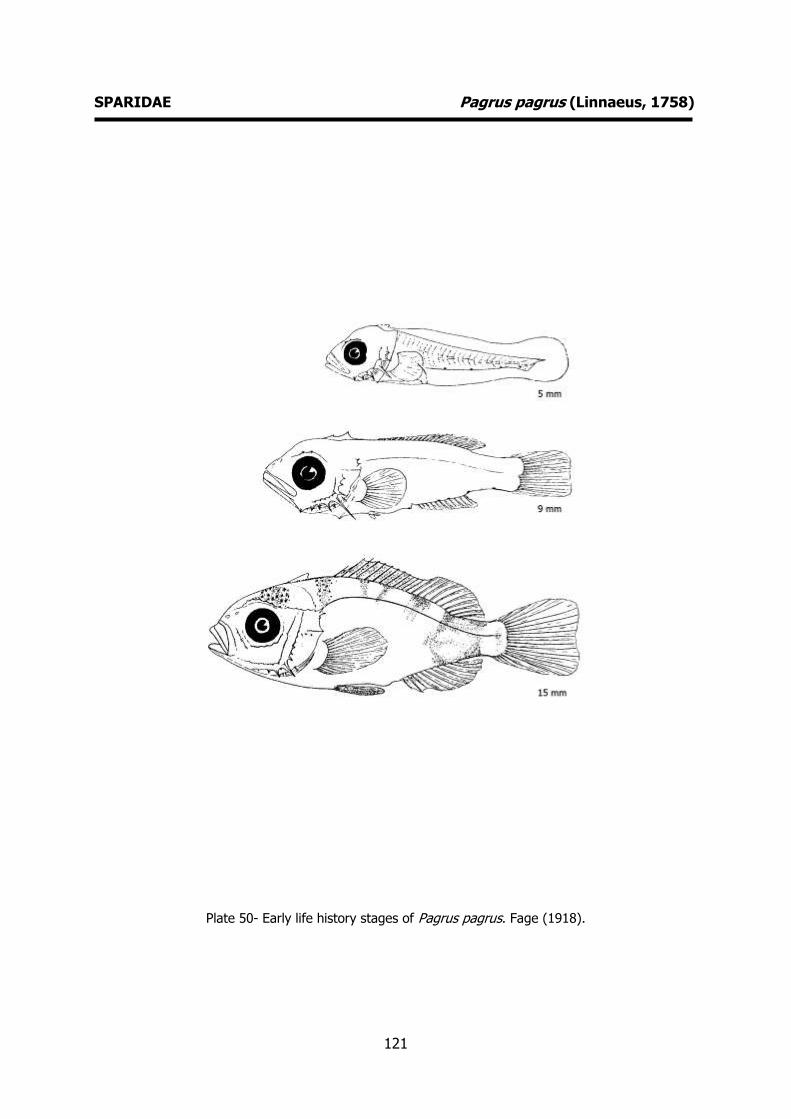
121
SPARIDAE Pagrus pagrus (Linnaeus, 1758)
Plate 50- Early life history stages of Pagrus pagrus. Fage (1918).
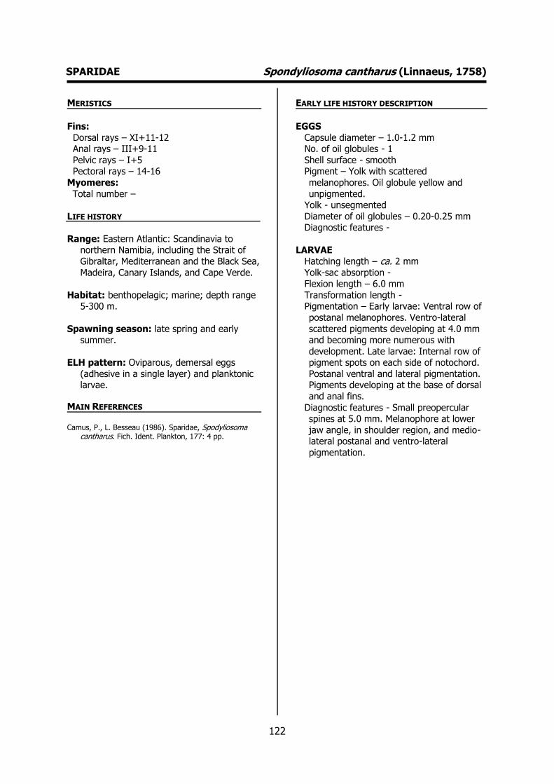
122
SPARIDAE Spondyliosoma cantharus (Linnaeus, 1758) MERISTICS
Fins:
Dorsal rays – XI+11-12 Anal rays – III+9-11
Pelvic rays – I+5 Pectoral rays – 14-16
Myomeres:
Total number –
LIFE HISTORY
Range: Eastern Atlantic: Scandinavia to northern Namibia, including the Strait of
Gibraltar, Mediterranean and the Black Sea,
Madeira, Canary Islands, and Cape Verde.
Habitat: benthopelagic; marine; depth range 5-300 m.
Spawning season: late spring and early summer.
ELH pattern: Oviparous, demersal eggs
(adhesive in a single layer) and planktonic larvae.
MAIN REFERENCES Camus, P., L. Besseau (1986). Sparidae, Spodyliosoma
cantharus. Fich. Ident. Plankton, 177: 4 pp.
EARLY LIFE HISTORY DESCRIPTION
EGGS
Capsule diameter – 1.0-1.2 mm No. of oil globules - 1
Shell surface - smooth Pigment – Yolk with scattered
melanophores. Oil globule yellow and
unpigmented. Yolk - unsegmented
Diameter of oil globules – 0.20-0.25 mm Diagnostic features -
LARVAE
Hatching length – ca. 2 mm
Yolk-sac absorption - Flexion length – 6.0 mm
Transformation length - Pigmentation – Early larvae: Ventral row of
postanal melanophores. Ventro-lateral
scattered pigments developing at 4.0 mm and becoming more numerous with
development. Late larvae: Internal row of pigment spots on each side of notochord.
Postanal ventral and lateral pigmentation. Pigments developing at the base of dorsal
and anal fins.
Diagnostic features - Small preopercular spines at 5.0 mm. Melanophore at lower
jaw angle, in shoulder region, and medio-lateral postanal and ventro-lateral
pigmentation.
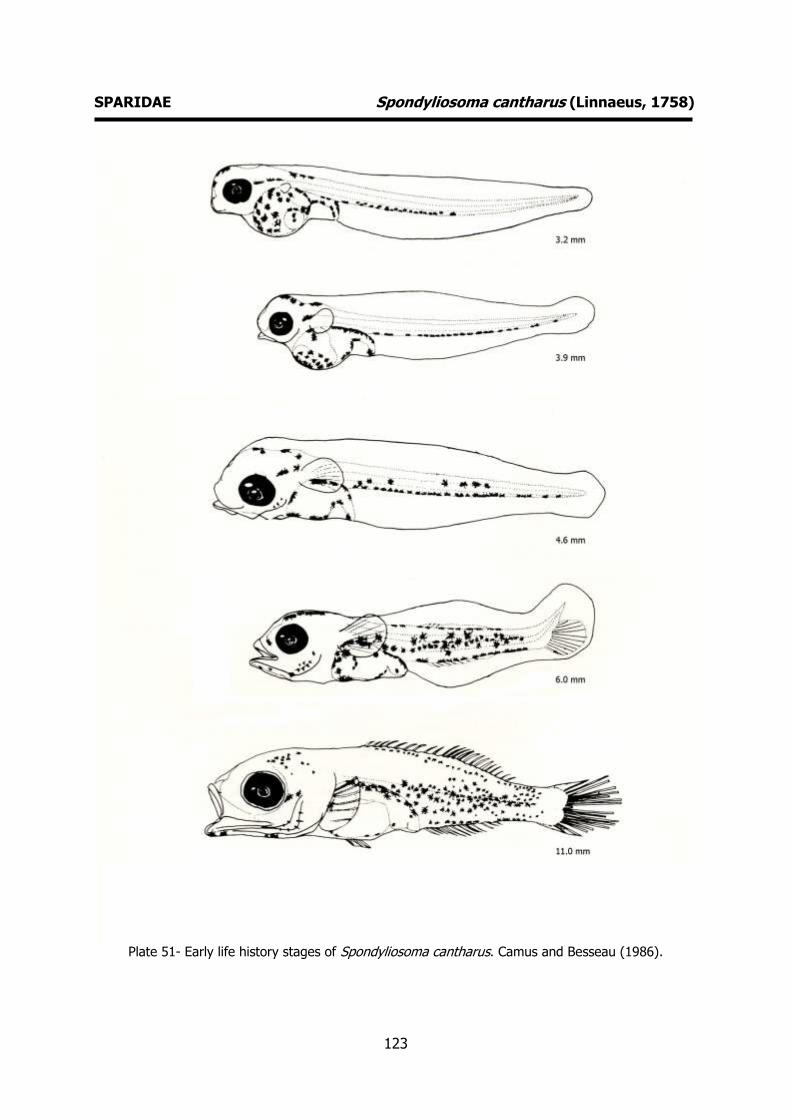
123
SPARIDAE Spondyliosoma cantharus (Linnaeus, 1758)
Plate 51- Early life history stages of Spondyliosoma cantharus. Camus and Besseau (1986).
124
CENTRACANTHIDAE Centracanthus cirrus Rafinesque, 1810 MERISTICS
Fins:
Dorsal rays – XIII+9-10 Anal rays – III+10
Pelvic rays – I+5 Pectoral rays – 15
Myomeres:
Total number –
LIFE HISTORY
Range: Eastern Atlantic: Portugal, Morocco (off south coast), Azores, Madeira, Canary
Islands and the Mediterranean. Reported
from Mauritania.
Habitat: marine; benthopelagic; depth range 0-500 m.
Spawning season: spring.
ELH pattern: Oviparous, planktonic eggs and larvae.
MAIN REFERENCES Marinaro, J-Y (1971). Contribution à l'étude des oeufs et
larves pèlagiques de poissons mediterranéens. V. Oeufs pèlagiques de la Baie d'Alger. Pelagos, 3 (1): 1-118.
Thomopoulos, A. (1954). Sur quelques oeufs planctoniques de Téléostéens de la baie de Villefranche. Bulletin de l'Institut Océanographique, 1043: 15pp.
EARLY LIFE HISTORY DESCRIPTION
EGGS
Capsule diameter – 0.84-0.97 mm No. of oil globules - 1
Shell surface - smooth Pigment - unpigmented
Yolk - unsegmented
Diameter of oil globules – 0.18-0.20 mm Diagnostic features – Spherical with a
characteristic dome.
LARVAE Hatching length – 2.3 mm
Yolk-sac absorption -
Flexion length - Transformation length -
Pigmentation - Diagnostic features – Newly hatched larva:
one oil globule posterior pigmented. Dorsal
and ventral rows of melanophores. Tail with single melanophore.

125
CENTRACANTHIDAE Centracanthus cirrus Rafinesque, 1810
Plate 52- Early life history stages of Centracanthus cirrus. Thomopoulos (1954).
126
LABRIDAE Centrolabrus exoletus (Linnaeus, 1758) MERISTICS
Fins:
Dorsal rays – XVIII-XX+6-7 Anal rays – V+7-8
Pelvic rays – I+5 Pectoral rays – 14
Myomeres:
Total number – 17+18
LIFE HISTORY
Range: Eastern Atlantic: Norway to Portugal; also eastern Greenland.
Habitat: marine; reef-associated.
Spawning season: spring and summer.
ELH pattern: Oviparous, demersal eggs and
planktonic larvae.
MAIN REFERENCES Fives, J.M. (1976) Labridae of the eastern North Atlantic.
Fiches d'Identification du Zooplancton, 149: 7pp. Ford, E. (1922b). On the Port-larvae of the Wrasses
occurring near Plymouth. J. mar. biol. Ass. U.K., 12: 693-699.
Russell, F.S. (1976). The eggs and planktonic stages of British marine fishes. Academic Press, London: 524pp.
EARLY LIFE HISTORY DESCRIPTION
EGGS: Undescribed
Capsule diameter - Unknown No. of oil globules - Unknown
Shell surface - Unknown Pigment - Unknown
Yolk - Unknown
Diameter of oil globules - Unknown Diagnostic features - Unknown
LARVAE
Hatching length – ca. 3 mm Yolk-sac absorption -
Flexion length -
Transformation length - Pigmentation – Newly hatched larva: Anal
fin membrane without melanophores. Some pigment along the posterior margin
of the posterior ventral margin of the body
and on the primordial fin. Late larvae: Paired crescent-shaped group of
melanophores on the head. Pigmentation covers only anterior half of tail. No
melanophores on the anal fin. Diagnostic features – Characteristic
pigmentation. Paired crescent-shaped
group of melanophores on the head. No melanophores on the anal fin.
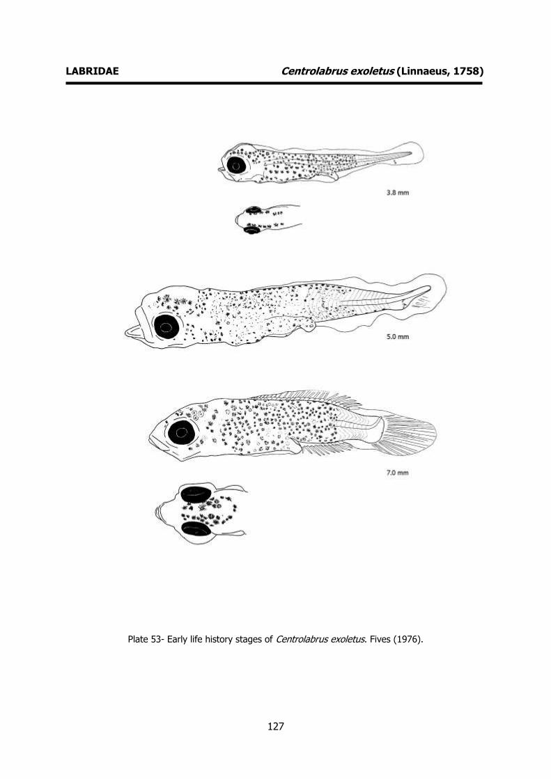
127
LABRIDAE Centrolabrus exoletus (Linnaeus, 1758)
Plate 53- Early life history stages of Centrolabrus exoletus. Fives (1976).

128
LABRIDAE Coris julis (Linnaeus, 1758) MERISTICS
Fins:
Dorsal rays – IX+12-13 Anal rays – III+11-13
Pelvic rays – I+5 Pectoral rays – 12-14
Myomeres:
Total number – 25-26
LIFE HISTORY
Range: Eastern Atlantic: Sweden to south of Cape Lopez, Gabon. Also known from the
Mediterranean Sea.
Habitat: Occurs in the littoral zone, near
rocks and eelgrass beds. Usually found between 1-60 m.
Spawning season:
ELH pattern: Oviparous, planktonic eggs and larvae.
MAIN REFERENCES Ehrenbaum, E. (1905-1909). Eier und Larven von Fischen.
Nordisches Plankton, 1: 413pp. Fage, L. (1918). Shore-fishes. Rep. Dan. Oceanogr. Exped
. 1908-10 Medit . adjac. Seas, 2, Biology A3: 1-154. Ford, E. (1922b). On the Port-larvae of the Wrasses
occurring near Plymouth. J. mar. biol. Ass. U.K., 12: 693-699.
Holt, E.W. L. (1899). Recherches sur la reproduction des poissons osseux principalement dans le Golfe de Marseille. Annls Mus. Hist. nat. Marseille, 5 Mém.2: 128p.
Marinaro, J-Y (1971). Contribution à l'étude des oeufs et larves pèlagiques de poissons mediterranéens. V. Oeufs pèlagiques de la Baie d'Alger. Pelagos, 3 (1): 1-118.
Raffaele, F. (1888) - Le uova galleggianti e la larve dei Te-leostei nel Golfo di Napoli. Mitt. zool. Stn Neapel, 8: 1-85.
Sparta, A. (1931-1956). Labridae. Uova, larve e stadi Giovanili di Teleostei: monografia elaborata con l’uso del materiale raccolto e sweriato da Salvatore Lo Bianco. Fauna e Flora Golfo Napoli Monogr.38: 576-594.
EARLY LIFE HISTORY DESCRIPTION
EGGS
Capsule diameter – 0.6-0.7 mm No. of oil globules - 1
Shell surface - smooth Pigment – yellow egg
Yolk - unsegmented
Diameter of oil globules – 0.12-0.16 mm Diagnostic features -
LARVAE
Hatching length – 2.5 mm Yolk-sac absorption -
Flexion length -
Transformation length - Pigmentation - One dorso-ventral cluster of
melanophores in the posterior half of the tail. Another dorso-ventral cluster above
the anus. Body typically slender, tail fin
convex. Diagnostic features – Newly-hatched larva:
Yolk sac projects beyond the snout. Oil globule anterior. Late larvae: Characteristic
pigmentation.
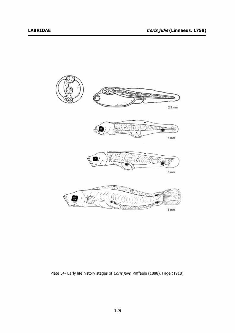
129
LABRIDAE Coris julis (Linnaeus, 1758)
Plate 54- Early life history stages of Coris julis. Raffaele (1888), Fage (1918).
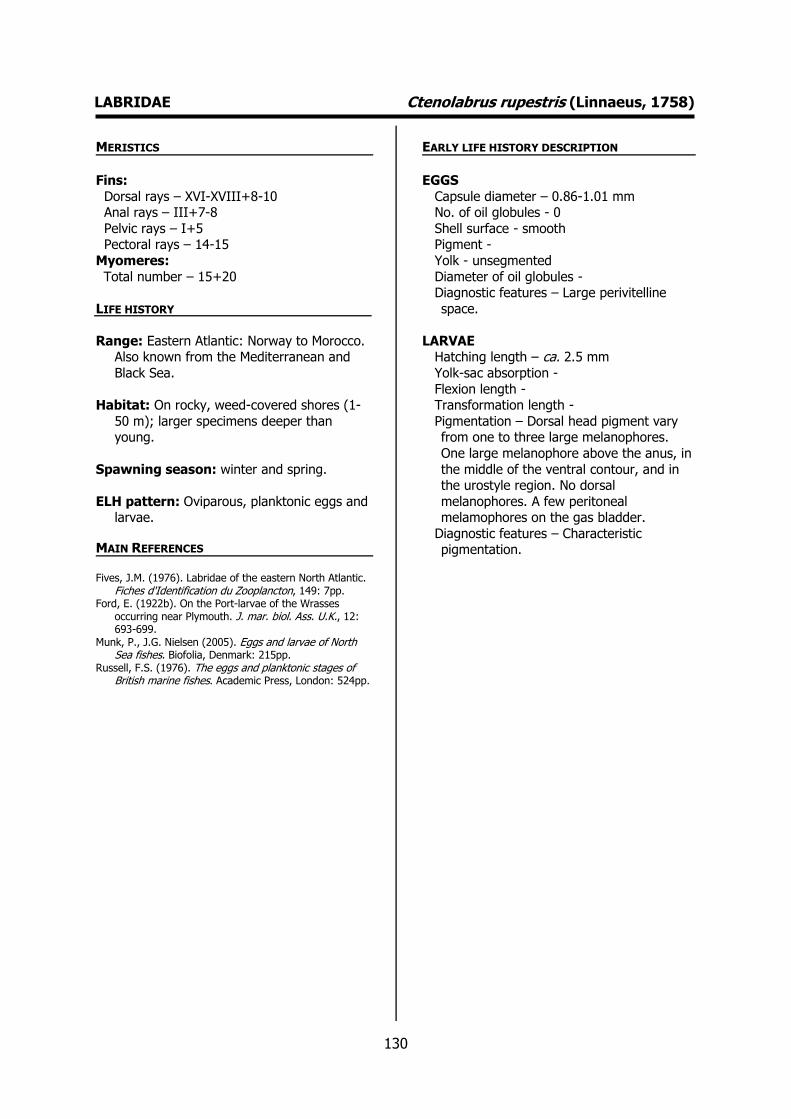
130
LABRIDAE Ctenolabrus rupestris (Linnaeus, 1758) MERISTICS
Fins:
Dorsal rays – XVI-XVIII+8-10 Anal rays – III+7-8
Pelvic rays – I+5 Pectoral rays – 14-15
Myomeres:
Total number – 15+20
LIFE HISTORY
Range: Eastern Atlantic: Norway to Morocco. Also known from the Mediterranean and
Black Sea.
Habitat: On rocky, weed-covered shores (1-
50 m); larger specimens deeper than young.
Spawning season: winter and spring.
ELH pattern: Oviparous, planktonic eggs and larvae.
MAIN REFERENCES Fives, J.M. (1976). Labridae of the eastern North Atlantic.
Fiches d'Identification du Zooplancton, 149: 7pp. Ford, E. (1922b). On the Port-larvae of the Wrasses
occurring near Plymouth. J. mar. biol. Ass. U.K., 12: 693-699.
Munk, P., J.G. Nielsen (2005). Eggs and larvae of North Sea fishes. Biofolia, Denmark: 215pp.
Russell, F.S. (1976). The eggs and planktonic stages of British marine fishes. Academic Press, London: 524pp.
EARLY LIFE HISTORY DESCRIPTION
EGGS
Capsule diameter – 0.86-1.01 mm No. of oil globules - 0
Shell surface - smooth Pigment -
Yolk - unsegmented
Diameter of oil globules - Diagnostic features – Large perivitelline
space.
LARVAE Hatching length – ca. 2.5 mm
Yolk-sac absorption -
Flexion length - Transformation length -
Pigmentation – Dorsal head pigment vary from one to three large melanophores.
One large melanophore above the anus, in
the middle of the ventral contour, and in the urostyle region. No dorsal
melanophores. A few peritoneal melamophores on the gas bladder.
Diagnostic features – Characteristic pigmentation.
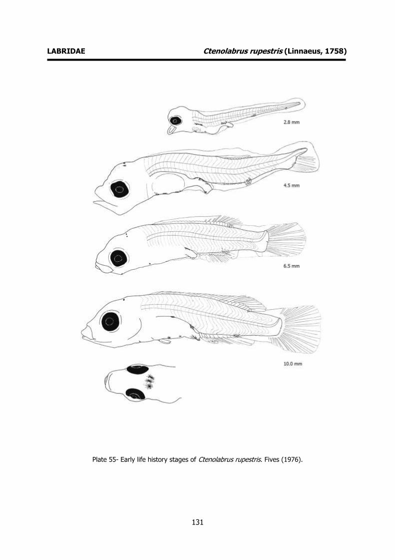
131
LABRIDAE Ctenolabrus rupestris (Linnaeus, 1758)
Plate 55- Early life history stages of Ctenolabrus rupestris. Fives (1976).
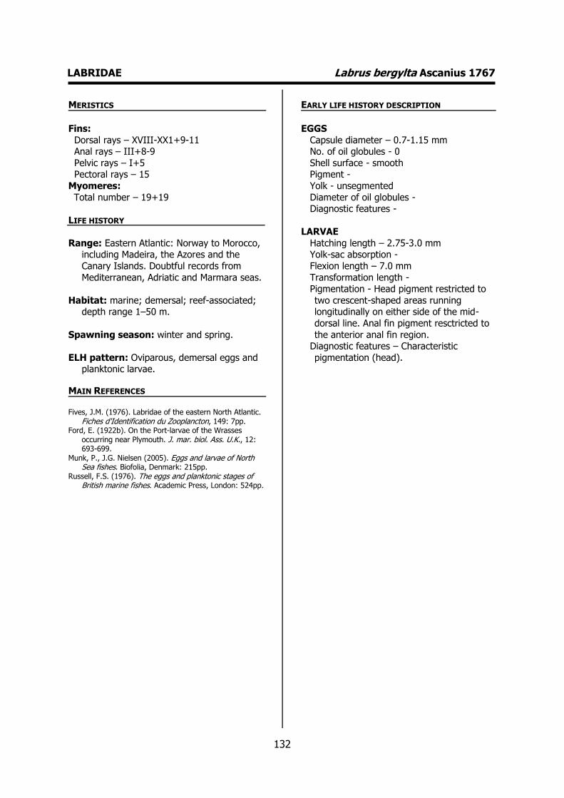
132
LABRIDAE Labrus bergylta Ascanius 1767 MERISTICS
Fins:
Dorsal rays – XVIII-XX1+9-11 Anal rays – III+8-9
Pelvic rays – I+5 Pectoral rays – 15
Myomeres:
Total number – 19+19
LIFE HISTORY
Range: Eastern Atlantic: Norway to Morocco, including Madeira, the Azores and the
Canary Islands. Doubtful records from
Mediterranean, Adriatic and Marmara seas.
Habitat: marine; demersal; reef-associated; depth range 1–50 m.
Spawning season: winter and spring.
ELH pattern: Oviparous, demersal eggs and planktonic larvae.
MAIN REFERENCES Fives, J.M. (1976). Labridae of the eastern North Atlantic.
Fiches d'Identification du Zooplancton, 149: 7pp. Ford, E. (1922b). On the Port-larvae of the Wrasses
occurring near Plymouth. J. mar. biol. Ass. U.K., 12: 693-699.
Munk, P., J.G. Nielsen (2005). Eggs and larvae of North Sea fishes. Biofolia, Denmark: 215pp.
Russell, F.S. (1976). The eggs and planktonic stages of British marine fishes. Academic Press, London: 524pp.
EARLY LIFE HISTORY DESCRIPTION
EGGS
Capsule diameter – 0.7-1.15 mm No. of oil globules - 0
Shell surface - smooth Pigment -
Yolk - unsegmented
Diameter of oil globules - Diagnostic features -
LARVAE
Hatching length – 2.75-3.0 mm Yolk-sac absorption -
Flexion length – 7.0 mm
Transformation length - Pigmentation - Head pigment restricted to
two crescent-shaped areas running longitudinally on either side of the mid-
dorsal line. Anal fin pigment resctricted to
the anterior anal fin region. Diagnostic features – Characteristic
pigmentation (head).
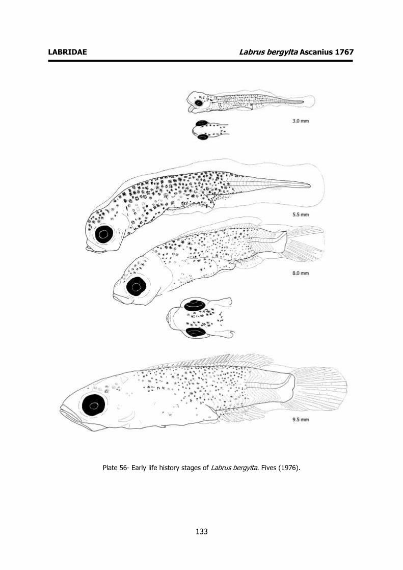
133
LABRIDAE Labrus bergylta Ascanius 1767
Plate 56- Early life history stages of Labrus bergylta. Fives (1976).

134
LABRIDAE Labrus merula Linnaeus, 1758 MERISTICS
Fins:
Dorsal rays – XVII-XIX+11-13 Anal rays – III+8-10
Pelvic rays – I+5 Pectoral rays – 14-15
Myomeres:
Total number – 38-39
LIFE HISTORY
Range: Eastern Atlantic: Portugal to Morocco and Azores. Also in the Mediterranean.
Habitat: marine; demersal; reef-associated.
Spawning season: winter and spring.
ELH pattern: Oviparous, demersal eggs and
planktonic larvae.
MAIN REFERENCES Dulčic, J., V. KozulL, M. Kraljevic, B. Skaramuca, B.
Glamuzina, P. Ré (1999). Embryonic and larval development of the labrid Labrus merula Linnaeus, 1758. J. mar. biol. Ass. U. K., 79: 327-332.
EARLY LIFE HISTORY DESCRIPTION
EGGS
Capsule diameter – 0.83-1.05 mm No. of oil globules - 0
Shell surface - Pigment -
Yolk - unsegmented
Diameter of oil globules - Diagnostic features -
LARVAE
Hatching length – 3.8 mm Yolk-sac absorption – 4.7 mm
Flexion length -
Transformation length - Pigmentation – Early larvae: Small number
of melanophores on head, without a definite crescent-shaped arrangement. No
pigment along the anal fin-fold. Late
larvae: Paired crescent-shaped groups of melanophores on head and pigmented
dorsal fin. No melanophores on caudal region, snout or lower jaw. Pigmentation
similar to Labrus bergylta. Diagnostic features – Characteristic
pigmentation (similar to Labrus bergylta).
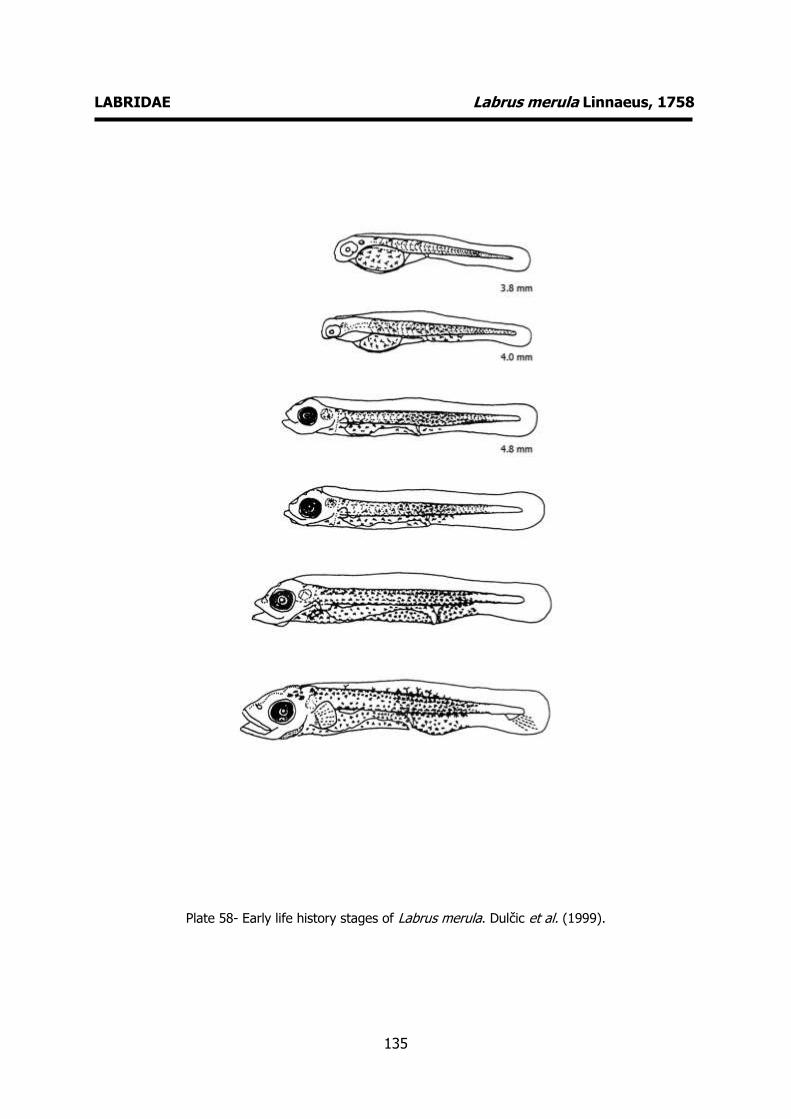
135
LABRIDAE Labrus merula Linnaeus, 1758
Plate 58- Early life history stages of Labrus merula. Dulčic et al. (1999).

136
LABRIDAE Labrus mixtus Linnaeus, 1758 MERISTICS
Fins:
Dorsal rays – XVI-XIX+11-14 Anal rays – III-9-12
Pelvic rays – I+5 Pectoral rays – 14-17
Myomeres:
Total number – 17/18+21/22
LIFE HISTORY
Range: Eastern Atlantic: Norway south to Senegal, Azores and Madeira. Also in the
Mediterranean.
Habitat: marine; demersal; reef-associated;
non-migratory; depth range 2–200 m.
Spawning season: winter and spring.
ELH pattern: Oviparous, demersal eggs and
planktonic larvae.
MAIN REFERENCES Fives, J.M. (1976). Labridae of the eastern North Atlantic.
Fiches d'Identification du Zooplancton, 149: 7pp. Ford, E. (1922b). On the Port-larvae of the Wrasses
occurring near Plymouth. J. mar. biol. Ass. U.K., 12: 693-699.
Russell, F.S. (1976). The eggs and planktonic stages of British marine fishes. Academic Press, London: 524pp.
EARLY LIFE HISTORY DESCRIPTION
EGGS: Undescribed
Capsule diameter - Unknown No. of oil globules - Unknown
Shell surface - Unknown Pigment - Unknown
Yolk - Unknown
Diameter of oil globules - Unknown Diagnostic features - Unknown
LARVAE
Hatching length - Yolk-sac absorption -
Flexion length -
Transformation length - Pigmentation – Body pigments confined to
dorsal and ventral margins of body and gut. Five larve melanophores along the
dorsal margin and three to four along the
ventral postanal margin. Some melanophores along dorsal and ventral gut
and on lower jaw. Diagnostic features – Characteristic
pigmentation.
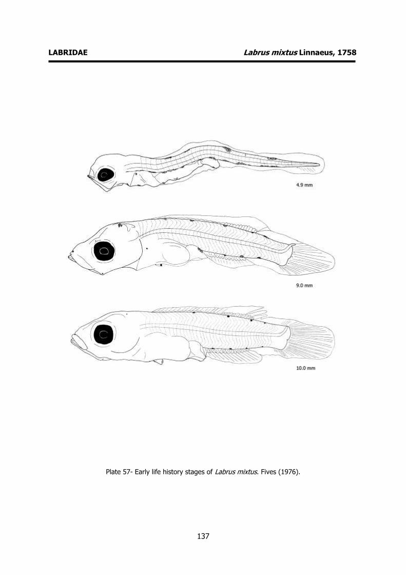
137
LABRIDAE Labrus mixtus Linnaeus, 1758
Plate 57- Early life history stages of Labrus mixtus. Fives (1976).

138
LABRIDAE Symphodus melops (Linnaeus, 1758) MERISTICS
Fins:
Dorsal rays – XIV-XVII+8-11 Anal rays – III+9-11
Pelvic rays – I+5 Pectoral rays – 14-15
Myomeres:
Total number – 14+19
LIFE HISTORY
Range: Eastern Atlantic: Norway to Morocco and the Azores. Also known from the
western Mediterranean and Adriatic seas.
Habitat: marine; demersal; reef-associated.
Spawning season: Spawns throughout thw
whole year.
ELH pattern: Oviparous, demersal eggs and
planktonic larvae.
MAIN REFERENCES Fives, J.M. (1976). Labridae of the eastern North Atlantic.
Fiches d'Identification du Zooplancton, 149: 7pp. Ford, E. (1922b). On the Port-larvae of the Wrasses
occurring near Plymouth. J. mar. biol. Ass. U.K., 12: 693-699.
Russell, F.S. (1976). The eggs and planktonic stages of British marine fishes. Academic Press, London: 524pp.
EARLY LIFE HISTORY DESCRIPTION
EGGS
Capsule diameter – 0.8-0.85 mm No. of oil globules - 0
Shell surface - smooth Pigment -
Yolk - unsegmented
Diameter of oil globules - Diagnostic features -
LARVAE
Hatching length – 2.5-3.0 mm Yolk-sac absorption -
Flexion length -
Transformation length - Pigmentation - Anterior half of tail covered
with melanophores. Melanophores along the whole length of the anal fin. No
crescent-shaped arrangement of the
melanophores on the head. Ventral lip pigment present.
Diagnostic features – Characteristic pigmentation. No crescent-shaped
arrangement of the melanophores on the head. Pigmentation of anal fin. Body
pigment less dense than Labrus bergylta and Centrolabrus exoletus.
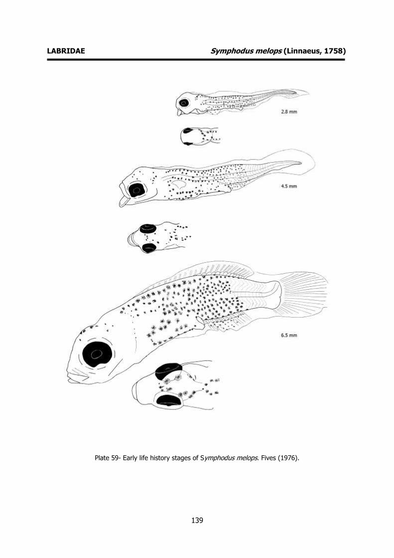
139
LABRIDAE Symphodus melops (Linnaeus, 1758)
Plate 59- Early life history stages of Symphodus melops. Fives (1976).
140
AMMODYTIDAE Ammodytes tobianus Linnaeus, 1758 MERISTICS
Fins:
Dorsal rays – 50-56 Anal rays – 25-31
Pelvic rays - Pectoral rays – 10-14
Myomeres:
Total number – 60-66
LIFE HISTORY
Range: Northeast Atlantic: Murmansk to Spain, including Iceland and the Baltic and
the Mediterranean.
Habitat: demersal; marine; brackish.
Spawning season: winter and spring.
ELH pattern: Oviparous, demersal eggs and planktonic larvae.
MAIN REFERENCES Einarsson, H. (1955). On the post-larvak stages of
Ammodytes lancea Cuvier. Acta Naturalia Islandica, II (1): 7pp.
Macer, C.T. (1967). Ammodytidae. Fiches d’identification des oeufs et larves de poissons, Nº 2: 6pp.
Munk, P., J.G. Nielsen (2005). Eggs and larvae of North Sea fishes. Biofolia, Denmark: 215pp.
Russell, F.S. (1976). The eggs and planktonic stages of British marine fishes. Academic Press, London: 524pp.
EARLY LIFE HISTORY DESCRIPTION
EGGS
Capsule diameter – 0.72-0.97 mm No. of oil globules - 1
Shell surface – smooth, ovoid Pigment -
Yolk -
Diameter of oil globules – 0.25-0.31 mm Diagnostic features -
LARVAE
Hatching length – 4 mm Yolk-sac absorption -
Flexion length -
Transformation length - Pigmentation – Dorsal body melanophores
absent or limited to 1-3 in caudal region; ventral body row of melanophores
extending from anus to base of tail, not to
tip of notochord, row of dorsal gut melanophores.
Diagnostic features - Absence of ventral gut melanophores, weak development of
caudal pigmentation and extension of dorsal body melanophore row to the level
of the anus. Late larvae very similar to
Hyperoplus immaculatus.

141
AMMODYTIDAE Ammodytes tobianus Linnaeus, 1758
Plate 60- Early life history stages of Ammodytes tobianus. Macer (1967).

142
AMMODYTIDAE Gymnammodytes semisquamatus (Jourdain, 1879) MERISTICS
Fins:
Dorsal rays – 56-59 Anal rays – 28-32
Pelvic rays - Pectoral rays – 14-15
Myomeres:
Total number –
LIFE HISTORY
Range: Northeast Atlantic: southern coast of Norway and the Shetlands to Spain,
including all coasts of the British Isles and
the North Sea, but not the Baltic.
Habitat: demersal; marine.
Spawning season: winter and spring.
ELH pattern: Oviparous, demersal eggs and
planktonic larvae.
MAIN REFERENCES Cameron, J. (1959). The larval and post-larval stages of
Gymnammodytes semisquamatus (Jourdain). J. Mar. Biol. Ass. U.K., 38: 17-25.
Ford, E. (1920). The post-larval stages of Ammodytes species captured during the cruises of S.S. “Oithona” in Plymouth waters in the year 1919. J. mar. biol. Ass. U.K., 12: 249-252.
Macer, C.T. (1967). Ammodytidae. Fiches d’identification des oeufs et larves de poissons, Nº 2: 6pp.
Munk, P., J.G. Nielsen (2005). Eggs and larvae of North Sea fishes. Biofolia, Denmark: 215pp.
Russell, F.S. (1976). The eggs and planktonic stages of British marine fishes. Academic Press, London: 524pp.
EARLY LIFE HISTORY DESCRIPTION
EGGS
Capsule diameter - Unknown No. of oil globules - 1
Shell surface - Unknown Pigment - Unknown
Yolk - Unknown
Diameter of oil globules - Unknown Diagnostic features - Unknown
LARVAE
Hatching length – ca. 3 mm Yolk-sac absorption -
Flexion length – 9 mm
Transformation length - Pigmentation – Newly hatched larvae: No
head, dorsal, caudal or ventral gut melanophores; Dorsal gut pigmentation of
about 16 melanophores, postanal ventral
pigment of about 15 melanophores, ventral margin of the primordial fin: 13
melanophores. Diagnostic features – Characteristic
pigmentation. The only Ammodytidae species to possess ventral fin-membrane
pigmentation (from hatching to 20 mm)
and denticles on pre-maxilla (7-25 mm).
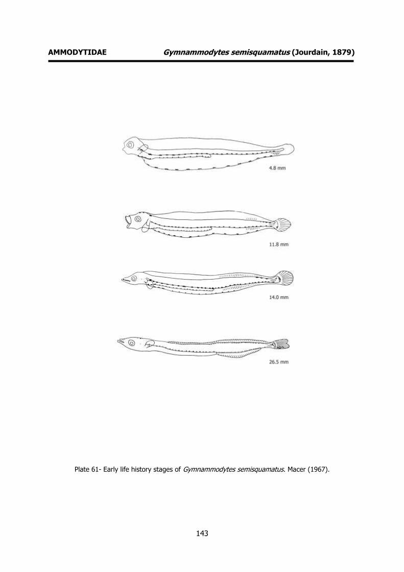
143
AMMODYTIDAE Gymnammodytes semisquamatus (Jourdain, 1879)
Plate 61- Early life history stages of Gymnammodytes semisquamatus. Macer (1967).

144
AMMODYTIDAE Hyperoplus lanceolatus (Le Sauvage, 1824) MERISTICS
Fins:
Dorsal rays – 52-61 Anal rays – 28-33
Pelvic rays - Pectoral rays – 12-14
Myomeres:
Total number – 66-69
LIFE HISTORY
Range: Northeast Atlantic: Murman and Spitzbergen to Portugal, including Iceland
and much of the Baltic.
Habitat: demersal; marine.
Spawning season: winter.
ELH pattern: Oviparous, demersal eggs and planktonic larvae.
MAIN REFERENCES Einarsson, H. (1951). The post-larval stages of Sand-eels
(Ammodytidae) in Faroe, Iceland and W-Greenland waters. Acta Naturalia Islandica, 1, Nº 7: 1-54.
Ford, E. (1920). The post-larval stages of Ammodytes species captured during the cruises of S.S. “Oithona” in Plymouth waters in the year 1919. J. mar. biol. Ass. U.K., 12: 249-252.
Macer, C.T. (1967). Ammodytidae. Fiches d’identification des oeufs et larves de poissons, Nº 2: 6pp.
Munk, P., J.G. Nielsen (2005). Eggs and larvae of North Sea fishes. Biofolia, Denmark: 215pp.
Russell, F.S. (1976). The eggs and planktonic stages of British marine fishes. Academic Press, London: 524pp.
EARLY LIFE HISTORY DESCRIPTION
EGGS
Capsule diameter - Unknown No. of oil globules - 1
Shell surface - Unknown Pigment - Unknown
Yolk - Unknown
Diameter of oil globules – 0.25-0.31 Diagnostic features - Unknown
LARVAE
Hatching length – ca. 4.5 mm Yolk-sac absorption -
Flexion length -
Transformation length - Pigmentation – Early larvae: Black
pigmentation on dorsal and postanal body rows, and dorsal and ventral gut rows. The
rectum does not reach the fin margin. Late
larvae: Early forward extension of the dorsal body row of melanophores, to the
head, and presence of heavy ventral gut melanophores. In later stages, the caudal
pigmentation is characteristic of the species.
Diagnostic features – Very heavy
pigmentation, especially in the dorsal row, which usually extends from tail to anus (8-
10 mm) and to the head (11-16 mm). Vomerine teeth visible at 20-25 mm.

145
AMMODYTIDAE Hyperoplus lanceolatus (Le Sauvage, 1824)
Plate 62- Early life history stages of Hyperoplus lancelolatus. Macer (1967).

146
TRACHINIDAE Echiichthys vipera (Cuvier, 1829) MERISTICS
Fins:
Dorsal rays – D1 V-VII, D2 21-25 Anal rays – I+24-26
Pelvic rays – I+5 Pectoral rays – 14
Myomeres:
Total number –
LIFE HISTORY
Range: Eastern Atlantic: North Sea to the Mediterranean, Morocco and Madeira.
Reported from the Canary Islands.
Habitat: demersal; marine.
Spawning season: spawns throughout the
whole year.
ELH pattern: Oviparous, planktonic eggs and
larvae.
MAIN REFERENCES
Ehrenbaum, E. (1905-1909). Eier und Larven von Fischen. Nordisches Plankton, 1: 413pp.
Holt, E.W.L. (1899. Recherches sur la reproduction des poissons osseux principalement dans le Golfe de Marseille. Annls Mus. Hist. nat. Marseille, 5 Mém.2: 128p.
Marinaro, J-Y (1971). Contribution à l'étude des oeufs et larves pèlagiques de poissons mediterranéens. V. Oeufs pèlagiques de la Baie d'Alger. Pelagos, 3 (1): 1-118.
Munk, P., J.G. Nielsen (2005). Eggs and larvae of North Sea fishes. Biofolia, Denmark: 215pp.
Padoa, E. (1933-1956). Trachinidae. Uova, larve e stadi Giovanili di Teleostei: monografia elaborata con l’uso del materiale raccolto e sweriato da Salvatore Lo Bianco. Fauna e Flora Golfo Napoli Monogr.38: 687-697.
Russell, F.S. (1976). The eggs and planktonic stages of British marine fishes. Academic Press, London: 524pp.
EARLY LIFE HISTORY DESCRIPTION
EGGS
Capsule diameter – 0.95-1.37 mm No. of oil globules – 6-30 scattered
Shell surface - smooth Pigment – yellow oil globules
Yolk - unsegmented
Diameter of oil globules – 0.024-0.12 mm Diagnostic features – pelvic fins apparent
when embryo surrounds the yolk.
LARVAE Hatching length – 3 mm
Yolk-sac absorption -
Flexion length – 6 mm Transformation length -
Pigmentation – Early larvae: pigmented eyes. Continuous dorsal and ventral
contour rows of melanophores extending
posteriorly behound half way along the postanal region. Well developed and
pigmented pelvic fins (black and yellow). Two bars of melanophores on the body
and melanophores on snout, head and peritoneum. Late larvae: One or two
ventral melanophores near the tail. At a
length of 5 mm, the dorsal fin begins to form. Preopercular and opercular spinal
armature starts to develop at an early stage. Heavy peritoneal pigmentation and
distinct row of melanophores at the base
of the caudal fin. Diagnostic features – Large pigmented
pelvic fins. Well developed preopercular and opercular spines. Characteristic shape.
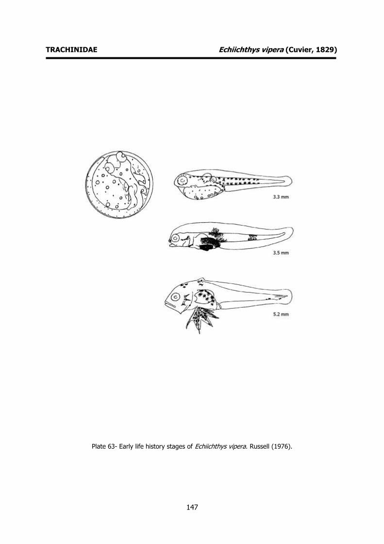
147
TRACHINIDAE Echiichthys vipera (Cuvier, 1829)
Plate 63- Early life history stages of Echiichthys vipera. Russell (1976).
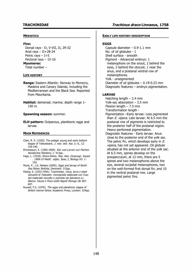
148
TRACHINIDAE Trachinus draco Linnaeus, 1758 MERISTICS
Fins:
Dorsal rays - D1 V-VII, D2 29-32 Anal rays – II+28-34
Pelvic rays – I+5 Pectoral rays – 15-16
Myomeres:
Total number –
LIFE HISTORY
Range: Eastern Atlantic: Norway to Morocco, Madeira and Canary Islands, including the
Mediterranean and the Black Sea. Reported
from Mauritania.
Habitat: demersal; marine; depth range 1-150 m.
Spawning season: summer.
ELH pattern: Oviparous, planktonic eggs and larvae.
MAIN REFERENCES Clark, R. S. (1920). The pelagic young and early bottom
stages of Teleosteans. J. mar. biol. Ass. U. K., 12: 159-240.
Ehrenbaum, E. (1905-1909). Eier und Larven von Fischen. Nordisches Plankton, 1: 413pp.
Fage, L. (1918). Shore-fishes. Rep. Dan. Oceanogr. Exped . 1908-10 Medit . adjac. Seas, 2, Biology A3: 1-154.
Munk, P., J.G. Nielsen (2005). Eggs and larvae of North Sea fishes. Biofolia, Denmark: 215pp.
Padoa, E. (1933-1956). Trachinidae. Uova, larve e stadi Giovanili di Teleostei: monografia elaborata con l’uso del materiale raccolto e sweriato da Salvatore Lo Bianco. Fauna e Flora Golfo Napoli Monogr.38: 687-697.
Russell, F.S. (1976). The eggs and planktonic stages of British marine fishes. Academic Press, London: 524pp.
EARLY LIFE HISTORY DESCRIPTION
EGGS
Capsule diameter – 0.9-1.1 mm No. of oil globules - 1
Shell surface - smooth Pigment - Advanced embryo: 1
melanophore on the snout, 2 behind the
eyes, 2 behind the otocyst, 1 near the anus, and a postanal ventral row of
melanophores. Yolk - unsegmented
Diameter of oil globules – 0.19-0.23 mm Diagnostic features – embryo pigmentation.
LARVAE Hatching length – 2.4 mm
Yolk-sac absorption – 3.5 mm Flexion length – 7.5 mm
Transformation length -
Pigmentation –Early larvae: Less pigmented than E. vipera. Late larvae: At 6.5 mm the
postanal row of pigments is restricted to the posterior half of the postanal region.
Heavy peritoneal pigmentation. Diagnostic features - Early larvae: Anus
close to the posterior end of the yolk sac.
The pelvic fin, which develops early in E. vipera, has not yet appeared. Oil globule
situated at the anterior end of the yolk sac. At 6.5 mm, spines develop on the
preoperculum, at 12 mm, there are 5
spines and two melanophores above the eye, several occipital melanophores, two
on the well-formed first dorsal fin, and 10 in the ventral postanal row. Large
pigmented pelvic fins.
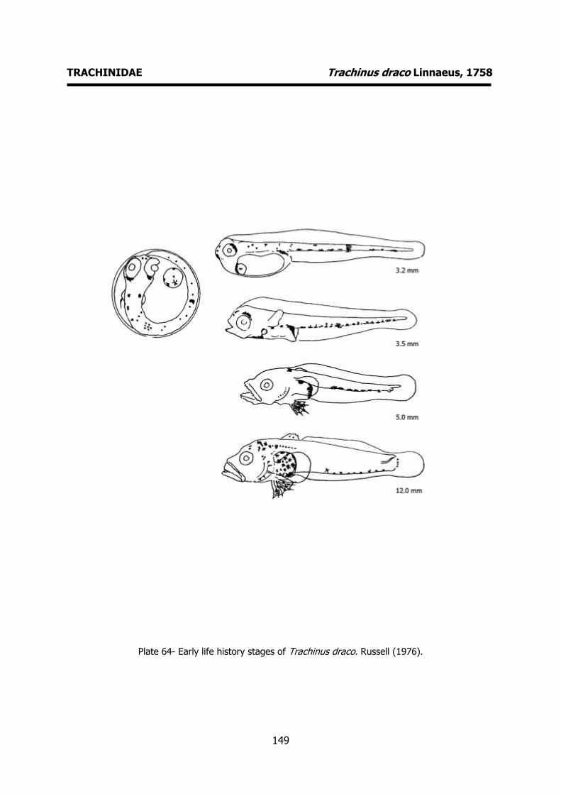
149
TRACHINIDAE Trachinus draco Linnaeus, 1758
Plate 64- Early life history stages of Trachinus draco. Russell (1976).
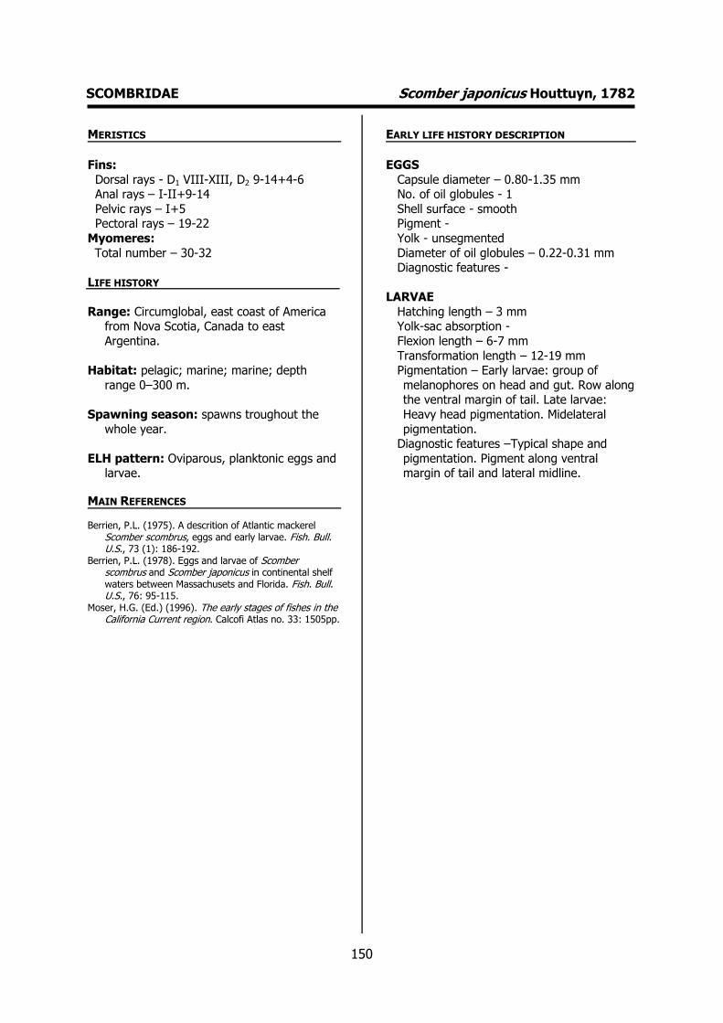
150
SCOMBRIDAE Scomber japonicus Houttuyn, 1782 MERISTICS
Fins:
Dorsal rays - D1 VIII-XIII, D2 9-14+4-6 Anal rays – I-II+9-14
Pelvic rays – I+5 Pectoral rays – 19-22
Myomeres:
Total number – 30-32
LIFE HISTORY
Range: Circumglobal, east coast of America from Nova Scotia, Canada to east
Argentina.
Habitat: pelagic; marine; marine; depth
range 0–300 m.
Spawning season: spawns troughout the
whole year.
ELH pattern: Oviparous, planktonic eggs and larvae.
MAIN REFERENCES
Berrien, P.L. (1975). A descrition of Atlantic mackerel Scomber scombrus, eggs and early larvae. Fish. Bull. U.S., 73 (1): 186-192.
Berrien, P.L. (1978). Eggs and larvae of Scomber scombrus and Scomber japonicus in continental shelf waters between Massachusets and Florida. Fish. Bull. U.S., 76: 95-115.
Moser, H.G. (Ed.) (1996). The early stages of fishes in the California Current region. Calcofi Atlas no. 33: 1505pp.
EARLY LIFE HISTORY DESCRIPTION
EGGS
Capsule diameter – 0.80-1.35 mm No. of oil globules - 1
Shell surface - smooth Pigment -
Yolk - unsegmented
Diameter of oil globules – 0.22-0.31 mm Diagnostic features -
LARVAE
Hatching length – 3 mm Yolk-sac absorption -
Flexion length – 6-7 mm
Transformation length – 12-19 mm Pigmentation – Early larvae: group of
melanophores on head and gut. Row along the ventral margin of tail. Late larvae:
Heavy head pigmentation. Midelateral
pigmentation. Diagnostic features –Typical shape and
pigmentation. Pigment along ventral margin of tail and lateral midline.
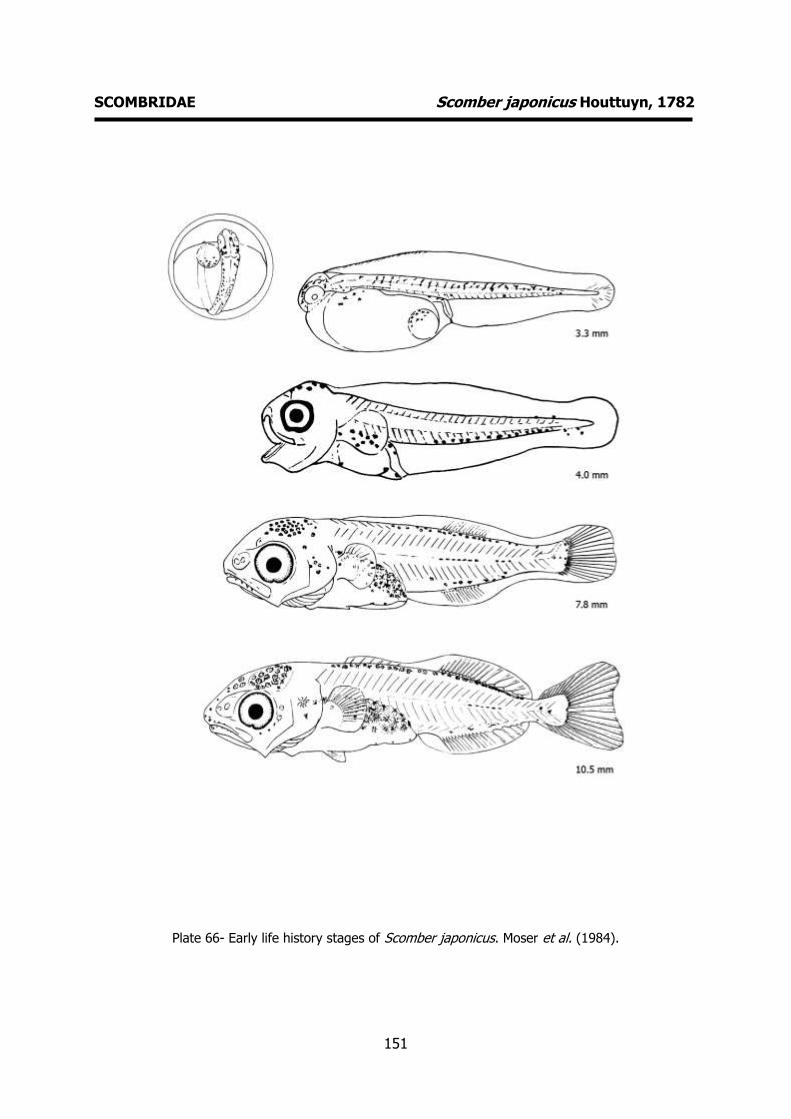
151
SCOMBRIDAE Scomber japonicus Houttuyn, 1782
Plate 66- Early life history stages of Scomber japonicus. Moser et al. (1984).
152
SCOMBRIDAE Scomber scombrus (Linnaeus, 1758) MERISTICS
Fins:
Dorsal rays - D1 VII-X, D2 13+5-6 Anal rays – I+12+5-6
Pelvic rays – I+5 Pectoral rays – 19-21
Myomeres:
Total number – 31
LIFE HISTORY
Range: Eastern Atlantic: including the southwestern Baltic Sea, the Mediterranean
and Black Sea. Western Atlantic: Labrador
to Cape Lookout.
Habitat: pelagic; marine; depth range 0–1000 m.
Spawning season: spring and summer.
ELH pattern: Oviparous, planktonic eggs and larvae.
MAIN REFERENCES Berrien, P.L. (1975). A descrition of Atlantic mackerel
Scomber scombrus, eggs and early larvae. Fish. Bull. U.S., 73 (1): 186-192.
Cunningham, J.T. (1889). Studies of the reprodution and development of teleostean fishes occurring in the neighbourhood of Plymouth. J. mar. bio. Ass. U.K., 1: 370-375.
Holt, E.W.L. (1899). Recherches sur la reproduction des poissons osseux principalement dans le Golfe de Marseille. Annls Mus. Hist. nat. Marseille, 5 Mém.2: 128p.
Ehrenbaum, E. (1905-1909). Eier und Larven von Fischen. Nordisches Plankton, 1: 413pp.
Munk, P., J.G. Nielsen (2005). Eggs and larvae of North Sea fishes. Biofolia, Denmark: 215pp.
Padoa, E. (1933-1956). Scombridae. Uova, larve e stadi Giovanili di Teleostei: monografia elaborata con l’uso del materiale raccolto e sweriato da Salvatore Lo Bianco. Fauna e Flora Golfo Napoli Monogr.38: 471-507.
Russell, F.S. (1976). The eggs and planktonic stages of British marine fishes. Academic Press, London: 524pp.
EARLY LIFE HISTORY DESCRIPTION
EGGS
Capsule diameter – 0.9-1.4 mm No. of oil globules - 1
Shell surface - smooth Pigment – yellow oil globule
Yolk - unsegmented
Diameter of oil globules – 0.28-0.35 Diagnostic features -
LARVAE
Hatching length – 3.3 mm Yolk-sac absorption – 5 mm
Flexion length – 7 mm
Transformation length - Pigmentation – Early larvae: A group of
melanophores on the head. Peritoneal black pigment occurs on the upper half of
the abdomen. Vent opens at the margin of
the finfold. Late larvae: Sharp teeth on upper and low jaws. Typical form of the
stomach and rectum. Melanophores on the head. Ventral and dorsal melanophores
start at a definite distance behind the anus. There are never any melanophores
on the sides of the body between the
dorsal and ventral contour rows. Diagnostic features – Characteristic
pigmentation. Presence of sharp teeth on upper and lower jaws that become
apparent at 5 mm.
153
SCOMBRIDAE Scomber scombrus (Linnaeus, 1758)
Plate 65- Early life history stages of Scomber scombrus. Russell (1976).

154
GOBIIDAE Aphia minuta (Risso, 1810) MERISTICS
Fins:
Dorsal rays – D1 IV-V, D2 I+11-13 Anal rays – I+13-14
Pelvic rays – I+5 Pectoral rays – 17-18
Myomeres:
Total number – 27 (26-28)
LIFE HISTORY
Range: Atlantic Ocean: Trondheim to Morocco. Also known from the
Mediterranean and Black Sea.
Habitat: demersal; marine; depth range 0-80
m.
Spawning season: summer and autumn.
ELH pattern: Oviparous, demersal eggs and
planktonic larvae.
MAIN REFERENCES Ehrenbaum, E. (1905-1909). Eier und Larven von Fischen.
Nordisches Plankton, 1: 413pp. Lebour, M.V. (1919). The young of the Gobiidae from the
neighbourhood of Plymouth. J. Mar. Biol. Ass. U.K., 12: 48-80.
Munk, P., J.G. Nielsen (2005). Eggs and larvae of North Sea fishes. Biofolia, Denmark: 215pp.
Padoa, E. (1933-1956). Gobiidae. Uova, larve e stadi Giovanili di Teleostei: monografia elaborata con l’uso del materiale raccolto e sweriato da Salvatore Lo Bianco. Fauna e Flora Golfo Napoli Monogr.38: 648-678.
Petersen, C.G.J. (1919). Our gobies (Gobiidae). From the egg to the adult stages. Reports of the Danish Biological Station, 26: 45-66.
Russell, F.S. (1976). The eggs and planktonic stages of British marine fishes. Academic Press, London: 524pp.
EARLY LIFE HISTORY DESCRIPTION
EGGS: Undescribed
Capsule diameter - Unknown No. of oil globules - Unknown
Shell surface - Unknown Pigment - Unknown
Yolk - Unknown
Diameter of oil globules - Unknown Diagnostic features - Unknown
LARVAE
Hatching length - Unknown Yolk-sac absorption - Unknown
Flexion length -
Transformation length - Pigmentation – Very few pigments.
Melanophore on angle of lower jaw. Stellate melanophores on ventral
abdomen. Row of melanophores from the
anus extending almost to caudal fin. Melanophores on base of caudal fin.
Diagnostic features - At 7 mm the pectoral fins are present as minute flaps. At 10 mm
the second dorsal fin and the anal fin are fully developed. Behind the anal fin are
about 5-6 diagonal stripes situated on the
ventral half of the body. Prominent gas bladder well back in the body. Fewer
myomeres that other Gobiids. The late larva is very similar to the adult.
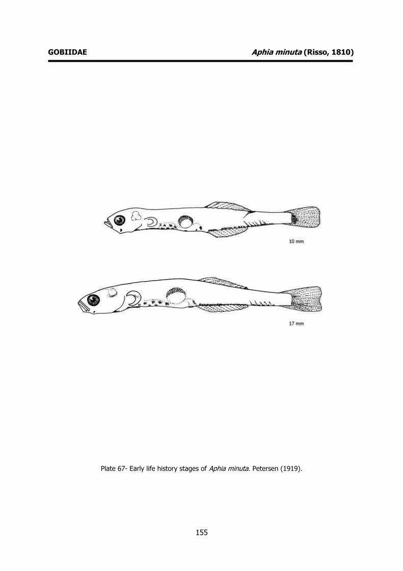
155
GOBIIDAE Aphia minuta (Risso, 1810)
Plate 67- Early life history stages of Aphia minuta. Petersen (1919).
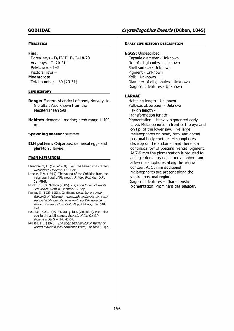
156
GOBIIDAE Crystallogobius linearis (Düben, 1845) MERISTICS
Fins:
Dorsal rays - D1 II-III, D2 I+18-20 Anal rays – I+20-21
Pelvic rays - I+5 Pectoral rays –
Myomeres:
Total number – 39 (29-31)
LIFE HISTORY
Range: Eastern Atlantic: Lofotens, Norway, to Gibraltar. Also known from the
Mediterranean Sea.
Habitat: demersal; marine; deph range 1-400
m.
Spawning season: summer.
ELH pattern: Oviparous, demersal eggs and
planktonic larvae.
MAIN REFERENCES Ehrenbaum, E. (1905-1909). Eier und Larven von Fischen.
Nordisches Plankton, 1: 413pp. Lebour, M.V. (1919). The young of the Gobiidae from the
neighbourhood of Plymouth. J. Mar. Biol. Ass. U.K., 12: 48-80.
Munk, P., J.G. Nielsen (2005). Eggs and larvae of North Sea fishes. Biofolia, Denmark: 215pp.
Padoa, E. (1933-1956). Gobiidae. Uova, larve e stadi Giovanili di Teleostei: monografia elaborata con l’uso del materiale raccolto e sweriato da Salvatore Lo Bianco. Fauna e Flora Golfo Napoli Monogr.38: 648-678.
Petersen, C.G.J. (1919). Our gobies (Gobiidae). From the egg to the adult stages. Reports of the Danish Biological Station, 26: 45-66.
Russell, F.S. (1976). The eggs and planktonic stages of British marine fishes. Academic Press, London: 524pp.
EARLY LIFE HISTORY DESCRIPTION
EGGS: Undescribed
Capsule diameter - Unknown No. of oil globules - Unknown
Shell surface - Unknown Pigment - Unknown
Yolk - Unknown
Diameter of oil globules - Unknown Diagnostic features - Unknown
LARVAE
Hatching length - Unknown Yolk-sac absorption - Unknown
Flexion length -
Transformation length - Pigmentation – Heavily pigmented early
larva. Melanophores in front of the eye and on tip of the lower jaw. Five large
melanophores on head, neck and dorsal
postanal body contour. Melanophores develop on the abdomen and there is a
continuos row of postanal ventral pigment. At 7-9 mm the pigmentation is reduced to
a single dorsal branched melanophore and a few melanophores along the ventral
contour. At 11 mm additional
melanophores are present along the ventral postanal region.
Diagnostic features – Characteristic pigmentation. Prominent gas bladder.
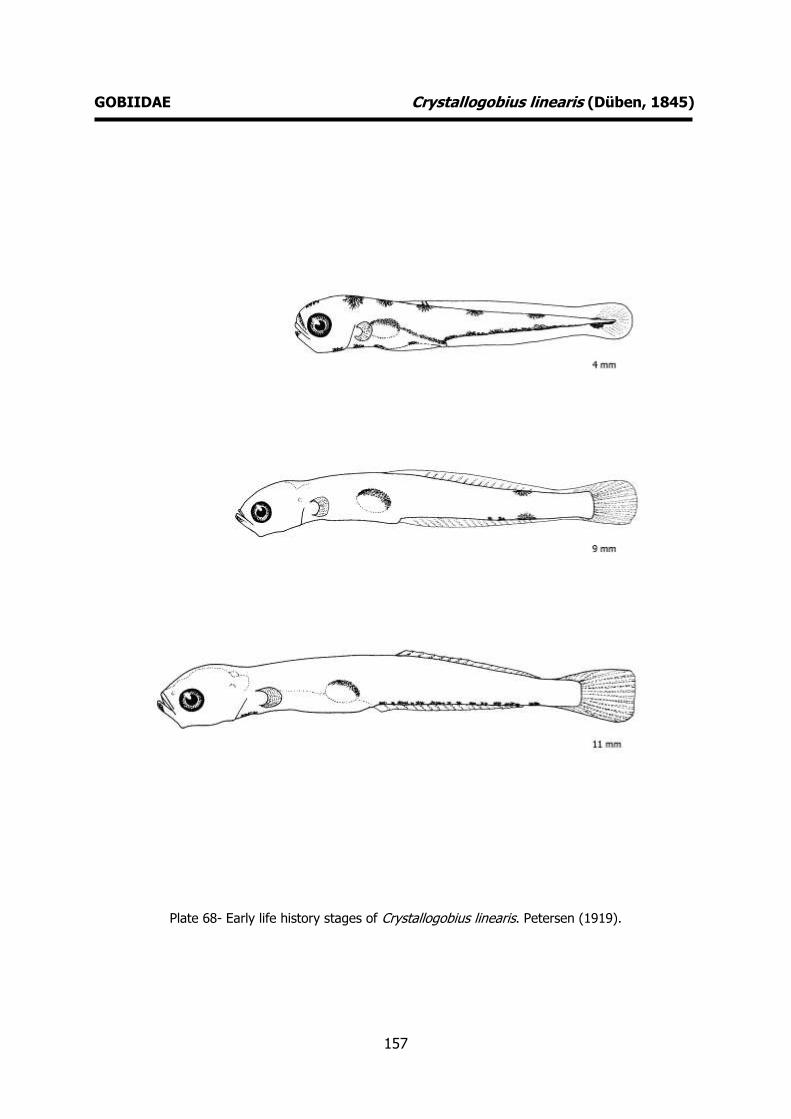
157
GOBIIDAE Crystallogobius linearis (Düben, 1845)
Plate 68- Early life history stages of Crystallogobius linearis. Petersen (1919).

158
GOBIIDAE Gobius cobitis Pallas, 1814 MERISTICS
Fins:
Dorsal rays - D1 VI, D2 I+13 Anal rays – I+10-12
Pelvic rays - I+5 Pectoral rays – 19-22
Myomeres:
Total number –
LIFE HISTORY
Range: Eastern Atlantic: southwestern England to Agadir, Morocco, throughout the
Mediterranean and Black Sea.
Habitat: demersal; marine; depth range 0-10
m.
Spawning season: spring and summer.
ELH pattern: Oviparous, demersal eggs and
planktonic larvae.
MAIN REFERENCES Gil, F., E.J. Gonçalves, C. Faria, V.C. Almada, C. Baptista,
H. Carreiro (1997). Embryonic and larval development of the giant goby Gobius cobitis (Pisces: Gobiidae). Journal of Natural History, 31: 799-804.
Padoa, E. (1933-1956). Gobiidae. Uova, larve e stadi Giovanili di Teleostei: monografia elaborata con l’uso del materiale raccolto e sweriato da Salvatore Lo Bianco. Fauna e Flora Golfo Napoli Monogr.38: 648-678.
EARLY LIFE HISTORY DESCRIPTION
EGGS
Capsule diameter – 3.59x1.18 mm No. of oil globules -
Shell surface – smooth, fusiform Pigment -
Yolk -
Diameter of oil globules - Diagnostic features -
LARVAE
Hatching length – 5.5 mm Yolk-sac absorption - 5.5-5.6 mm
Flexion length – 7.5 mm
Transformation length – 17-19 mm Pigmentation – Newly hatched larva: eyes
fully pigmented, gas bladder formed but not filled, reduced yolk. Ventral row of
melanophores extending form behind the
anus to the caudal region. Two large malnophores above the anus and three
others at the dorsal midline near the caudal peduncle. Pigmented gas bladder.
Late larva: dorsal row of melanophores extending from the head to the caudal
region.
Diagnostic features – Pigmentation.
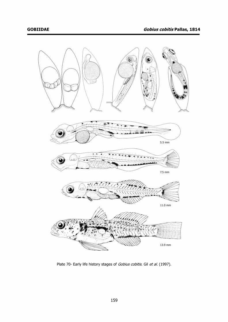
159
GOBIIDAE Gobius cobitis Pallas, 1814
Plate 70- Early life history stages of Gobius cobitis. Gil et al. (1997).
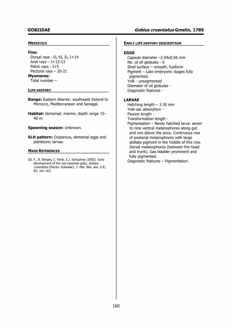
160
GOBIIDAE Gobius cruentatus Gmelin, 1789 MERISTICS
Fins:
Dorsal rays - D1 VI, D2 I+14 Anal rays – I+12-13
Pelvic rays - I+5 Pectoral rays – 20-21
Myomeres: Total number –
LIFE HISTORY
Range: Eastern Atlantic: southwest Ireland to Morocco, Mediterranean and Senegal.
Habitat: demersal; marine; depth range 15-
40 m.
Spawning season: Unknown.
ELH pattern: Oviparous, demersal eggs and
planktonic larvae.
MAIN REFERENCES Gil, F., R. Borges, C. Faria. E.J. Gonçalves (2002). Early
development of the red mouthed goby, Gobius cruentatus (Pisces: Gobiidae). J. Mar. Biol. Ass. U.K., 82: 161-163.
EARLY LIFE HISTORY DESCRIPTION
EGGS
Capsule diameter –2.04x0.56 mm No. of oil globules - 0
Shell surface – smooth, fusiform Pigment – Late embryonic stages fully
pigmented.
Yolk - unsegmented Diameter of oil globules -
Diagnostic features -
LARVAE Hatching length – 3.30 mm
Yolk-sac absorption -
Flexion length - Transformation length -
Pigmentation – Newly hatched larva: seven to nine ventral melanophores along gut
and one above the anus. Continuous row
of postanal melanophores with large stellate pigment in the middle of this row.
Dorsal melanophores (between the head and trunk). Gas bladder prominent and
fully pigmented. Diagnostic features – Pigmentation.
161
GOBIIDAE Gobius cruentatus Gmelin, 1789
Plate 71- Early life history stages of Gobius cruentatus. Gil et al. (2002).

162
GOBIIDAE Gobius niger Linnaeus, 1758 MERISTICS
Fins:
Dorsal rays - D1 V-VII, D2 I+11-13 Anal rays – I+11-12
Pelvic rays – I+5 Pectoral rays – 15-20
Myomeres:
Total number – 27-29
LIFE HISTORY
Range: Eastern Atlantic and Mediterranean Sea: throughout North Africa from Cape
Blanc, Mauritania north and eastwards to
the Suez Canal; also along the eastern Atlantic coast northwards to Trondheim
(Norway) and Baltic Sea. Also known from the Black Sea.
Habitat: demersal; marine; depth range 1-75 m.
Spawning season: spring and summer.
ELH pattern: Oviparous, demersal eggs and
planktonic larvae.
MAIN REFERENCES Lebour, M.V. (1919). The young of the Gobiidae from the
neighbourhood of Plymouth. J. Mar. Biol. Ass. U.K., 12: 48-80.
Munk, P., J.G. Nielsen (2005). Eggs and larvae of North Sea fishes. Biofolia, Denmark: 215pp.
Padoa, E. (1933-1956). Gobiidae. Uova, larve e stadi Giovanili di Teleostei: monografia elaborata con l’uso del materiale raccolto e sweriato da Salvatore Lo Bianco. Fauna e Flora Golfo Napoli Monogr.38: 648-678.
Petersen, C.G.J. (1892). On the eggs and breeding of our Gobiidae. Reports of the Danish Biological Station, 2: 1-9.
Petersen, C.G.J. (1917).On the development of of our common gobies (Gobius) from the egg to the adult stages etc.. Reports of the Danish Biological Station, 24: 5-16.
Petersen, C.G.J. (1919). Our gobies (Gobiidae). From the egg to the adult stages. Reports of the Danish Biological Station, 26: 45-66.
Russell, F.S. (1976). The eggs and planktonic stages of British marine fishes. Academic Press, London: 524pp.
EARLY LIFE HISTORY DESCRIPTION
EGGS
Capsule diameter – 1.5 mm No. of oil globules – many oil globules
Shell surface – smooth, fusiform Pigment -
Yolk -
Diameter of oil globules - Diagnostic features -
LARVAE
Hatching length – 2.5 mm Yolk-sac absorption – 4.0-4.5 mm
Flexion length -
Transformation length - Pigmentation – Newly hatched larva: no
pigment on head. Three characteristic ventral melanophores, one below the yolk
sac, one at the anus and one mid-way on
the tail. Postanal row of melanophores, with one large ramified about mid-way
along the postanal region. No postanal dorsal pigmentation. Late larvae: no
significant change in pigmentation, head and jaws with no pigmentation. Gas
bladder heavily pigmented. One
melanophore on the ventral half of the caudal fin. More advanced in development
than other related species at equivalent sizes.
Diagnostic features – Pigmentation.
Prominent gas bladder. Fewer myotomes than Pomastoschistus minutus and
Pomatoschistus microps.

163
GOBIIDAE Gobius niger Linnaeus, 1758
Plate 72- Early life history stages of Gobius niger. Petersen (1892, 1917, 1919).
164
GOBIIDAE Gobius paganellus Linnaeus, 1758 MERISTICS
Fins:
Dorsal rays - D1 VI, D2 I+13-14 Anal rays – I+10-13
Pelvic rays – I+5 Pectoral rays – 18-23
Myomeres:
Total number – 28
LIFE HISTORY
Range: Eastern Atlantic: western Scotland to Senegal. Also known from the
Mediterranean and Black Sea. Indian
Ocean: as lessepsian migrant in the Gulf of Eilat and Red Sea.
Habitat: demersal; marine; depth range 3-15
m.
Spawning season: spring and summer.
ELH pattern: Oviparous, demersal eggs and
planktonic larvae.
MAIN REFERENCES Borges, R., C. Faria, F. Gil, E.J. Gonçalves, V.C. Almada
(2003). Embryonic and larval development of Gobius paganellus (Pisces: Gobiidae). J. Mar. Biol. Ass. U.K., 83: 1151-1156.
Lebour, M.V. (1919). The young of the Gobiidae from the neighbourhood of Plymouth. J. Mar. Biol. Ass. U.K., 12: 48-80.
Padoa, E. (1933-1956). Gobiidae. Uova, larve e stadi Giovanili di Teleostei: monografia elaborata con l’uso del materiale raccolto e sweriato da Salvatore Lo Bianco. Fauna e Flora Golfo Napoli Monogr.38: 648-678.
Russell, F.S. (1976). The eggs and planktonic stages of British marine fishes. Academic Press, London: 524pp.
EARLY LIFE HISTORY DESCRIPTION
EGGS
Capsule diameter – 1.84x3.0 mm No. of oil globules -
Shell surface – smooth, fusiform Pigment -
Yolk -
Diameter of oil globules - Diagnostic features -
LARVAE
Hatching length – 4.0-4.8 mm Yolk-sac absorption – 5 mm
Flexion length -
Transformation length - Pigmentation – Newly hatched larva: no
melanophores on head, postanal dorsal and ventral melanophores. Several
melanophores above the anus and below
the gut. Gas bladder fully pigmented. Late larva: postanal dorsal melanophores
disappears (7 mm). No melanophores on head (until 11-12 mm). Row of
melanophores from throat to anus. Large melanophore above tha anus. Large
stellate melanophores appears in the
otocystic region. Diagnostic features – Pigmentation.

165
GOBIIDAE Gobius paganellus Linnaeus, 1758
Plate 73- Early life history stages of Gobius paganellus. Borges et al. (2003).
166
GOBIIDAE Gobius xanthocephalus Heymer & Zander, 1992 MERISTICS
Fins:
Dorsal rays - D1 VII, D2 14-16 Anal rays – I+14
Pelvic rays - I+5 Pectoral rays –
Myomeres:
Total number – 28
LIFE HISTORY
Range: Eastern Atlantic: northern Spain to Madeira and the Canary Islands;
Mediterranean.
Habitat: demersal; marine.
Spawning season: spring and summer.
ELH pattern: Oviparous, demersal eggs and planktonic larvae.
MAIN REFERENCES Monteiro, J., R. Borges, J. Robalo, V.C. Almada, S.
Henriques, E.J. Gonçalves (submitted). Larval development of Gobius xantochephalus and genetic validation of larval identification.
EARLY LIFE HISTORY DESCRIPTION
EGGS: Undescribed
Capsule diameter - Unknown No. of oil globules - Unknown
Shell surface - Unknown Pigment - Unknown
Yolk - Unknown
Diameter of oil globules - Unknown Diagnostic features - Unknown
LARVAE
Hatching length – 2.8 mm Yolk-sac absorption -
Flexion length – 5.0-5.5 mm
Transformation length - Pigmentation – Newly hatched larvae: No
pigmentation at the lower jaw. Lacks dorsal pigmentation. Presence of a ventral
postanal row of regularly spaced
melanophores extending form the anus to the caudal fin.
Diagnostic features – Characteristic pigmentation. Meristics.
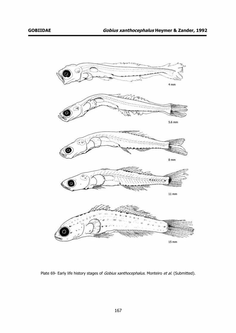
167
GOBIIDAE Gobius xanthocephalus Heymer & Zander, 1992
Plate 69- Early life history stages of Gobius xanthocephalus. Monteiro et al. (Submitted).

168
GOBIIDAE Gobiusculus flavescens (Fabricius, 1779) MERISTICS
Fins:
Dorsal rays - D1 VII-VIII, D2 I+9-10 Anal rays – I+10-11
Pelvic rays – I+5 Pectoral rays – 18-19
Myomeres:
Total number – 35-40
LIFE HISTORY
Range: Eastern Atlantic: Faeroes, Vesterålen (Norway), and western Baltic to north-west
Spain, excluding south-eastern North Sea.
Reported from Estonia. Mediterranean records, from Sicily and the Adriatic.
Habitat: demersal; marine; depth range 0-
20m.
Spawning season: spring and summer.
ELH pattern: Oviparous, demersal eggs
planktonic larvae.
MAIN REFERENCES Lebour, M.V. (1919). The young of the Gobiidae from the
neighbourhood of Plymouth. J. Mar. Biol. Ass. U.K., 12: 48-80.
Padoa, E. (1933-1956). Gobiidae. Uova, larve e stadi Giovanili di Teleostei: monografia elaborata con l’uso del materiale raccolto e sweriato da Salvatore Lo Bianco. Fauna e Flora Golfo Napoli Monogr.38: 648-678.
Petersen, C.G.J. (1892). On the eggs and breeding of our Gobiidae. Reports of the Danish Biological Station, 2: 1-9.
Petersen, C.G.J. (1919). Our gobies (Gobiidae). From the egg to the adult stages. Reports of the Danish Biological Station, 26: 45-66.
Russell, F.S. (1976). The eggs and planktonic stages of British marine fishes. Academic Press, London: 524pp.
EARLY LIFE HISTORY DESCRIPTION
EGGS
Capsule diameter – 0.8-0.6 mm No. of oil globules – reddish-brown oil
globules Shell surface - Egg pear-shaped, with round
or pointed apex
Pigment - Yolk -
Diameter of oil globules - Diagnostic features -
LARVAE
Hatching length – 2.2-2.6 mm
Yolk-sac absorption – 4.0 mm Flexion length -
Transformation length - Pigmentation – Newly hatched larva:
continuous row of ventral melanophores
from throat to anus and a ventral postanal row and one or two dosal postanal
melanophores. One melanophore, mid-way along the tail, especially prominent. Sub-
otocystic melanophore. Late larva: dorsal pigmentation disappears when the larva
reaches 5 mm. A large melanophore
develops in the otocystic region when the larva reaches 8-9 mm. Large melanophore
above the anus. Diagnostic features – Pigmentation.
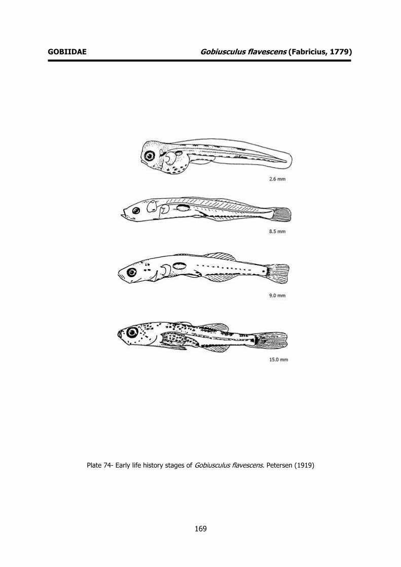
169
GOBIIDAE Gobiusculus flavescens (Fabricius, 1779)
Plate 74- Early life history stages of Gobiusculus flavescens. Petersen (1919)
170
GOBIIDAE Lebetus guilleti (Le Danois, 1913) MERISTICS
Fins:
Dorsal rays - D1 VI, D2 I+7-9 Anal rays – I+5-6
Pelvic rays – I+5 Pectoral rays – 15-17
Myomeres:
Total number – 25-26
LIFE HISTORY
Range: Eastern Atlantic: Kattegat and Belt seas to Portugal; also from Banyuls,
western Mediterranean, and Canary
Islands.
Habitat: demersal; reef-associated; depth range 0-30 m.
Spawning season: spawns throughout the whole year.
ELH pattern: Oviparous, planktonic larvae.
MAIN REFERENCES
Demir, N., F. Russell (1971). On the postlarva of the goby Lebetus. J. Mar. Biol. Ass. U.K., 51: 669-678.
Petersen, C.G.J. (1919). Our gobies (Gobiidae). From the egg to the adult stages. Reports of the Danish Biological Station, 26: 45-66.
Ré, P. (1980-1981). On the occurence of postlarval stages of Lebetus (Pisces: Gobiidae) off Portugal. Boletim da Sociedade Portuguesa de Ciências Naturais, 20: 67-69.
Russell, F.S. (1976). The eggs and planktonic stages of British marine fishes. Academic Press, London: 524pp.
EARLY LIFE HISTORY DESCRIPTION
EGGS: Undescribed
Capsule diameter - Unknown No. of oil globules - Unknown
Shell surface - Unknown Pigment - Unknown
Yolk - Unknown
Diameter of oil globules - Unknown Diagnostic features - Unknown
LARVAE
Hatching length – Unknown Yolk-sac absorption – Unknown
Flexion length -
Transformation length - Pigmentation – Body well covered with
melanophores except in the caudal region. Well marked mediolateral row of
melanophores. Row of melanophores
between the bases of the anal fin rays. Diagnostic features - Characteristic
pigmentation. Larva more pigmented than that of Lebetus scorpioides. Pelvic fins
develop quickly reaching nearly to the anus when the larva is only 5-6 mm long.

171
GOBIIDAE Lebetus guilleti (Le Danois, 1913)
Plate 75- Early life history stages of Lebetus guilleti. Russell (1976), Ré (1980-1981).

172
GOBIIDAE Lebetus scorpioides (Collett, 1874) MERISTICS
Fins:
Dorsal rays - D1 VI-VII, D2 I+9-10 Anal rays – I+7-8
Pelvic rays – I+5 Pectoral rays – 17-21
Myomeres:
Total number – 27-29
LIFE HISTORY
Range: Eastern Atlantic: southwest Iceland, the Faeroes and Hemnefjord, Norway, to
northern Bay of Biscay.
Habitat: marine; reef-associated; depth range
30-375 m.
Spawning season: spawns throughout the
whole year.
ELH pattern: Oviparous, planktonic larvae.
MAIN REFERENCES Demir, N., F. Russell (1971). On the postlarva of the goby
Lebetus. J. Mar. Biol. Ass. U.K., 51: 669-678. Fage, L. (1918). Shore-fishes. Rep. Dan. Oceanogr. Exped
. 1908-10 Medit . adjac. Seas, 2, Biology A3: 1-154. Russell, F.S. (1976). The eggs and planktonic stages of
British marine fishes. Academic Press, London: 524pp.
EARLY LIFE HISTORY DESCRIPTION
EGGS: Undescribed
Capsule diameter - Unknown No. of oil globules - Unknown
Shell surface - Unknown Pigment - Unknown
Yolk - Unknown
Diameter of oil globules - Unknown Diagnostic features - Unknown
LARVAE
Hatching length - Unknown Yolk-sac absorption - Unknown
Flexion length -
Transformation length - Pigmentation – Body covered with
pigmentation except on caudal region. Well marked mediolateral row of melanophores.
Whole larva less pigmented than L. guilleti. Diagnostic features – Pigmentation,
meristics. Pelvis fins develop much slower
than those of L. guilleti. These may not reach the anus in specimens with 8 mm.
Shape of body slender than L. guilleti.

173
GOBIIDAE Lebetus scorpioides (Collett, 1874)
Plate 76- Early life history stages of Lebetus scorpioides. Fage (1918).

174
GOBIIDAE Pomatoschistus microps (Krøyer, 1838) MERISTICS
Fins:
Dorsal rays - D1 V-VII, D2 I+8-11 Anal rays – I+8-9
Pelvic rays – I+5 Pectoral rays – 15-20
Myomeres:
Total number – 30-32
LIFE HISTORY
Range: Eastern Atlantic: Norway to Morocco, including Baltic Sea (to southern Portugal)
and western Mediterranean. Also in
Mauritania and the Canary Islands.
Habitat: demersal; freshwater; brackish; marine; depth range 0–12 m.
Spawning season: spring and summer.
ELH pattern: Oviparous, demersal eggs and planktonic larvae.
MAIN REFERENCES Lebour, M.V. (1919). The young of the Gobiidae from the
neighbourhood of Plymouth. J. Mar. Biol. Ass. U.K., 12: 48-80.
Lebour, M.V. (1920). The eggs of Gobius minutus, pictus and microps. J. Mar. Biol. Ass. U.K., 12: 253-260.
Munk, P., J.G. Nielsen (2005). Eggs and larvae of North Sea fishes. Biofolia, Denmark: 215pp.
Padoa, E. (1933-1956). Gobiidae. Uova, larve e stadi Giovanili di Teleostei: monografia elaborata con l’uso del materiale raccolto e sweriato da Salvatore Lo Bianco. Fauna e Flora Golfo Napoli Monogr.38: 648-678.
Petersen, C.G.J. (1892). On the eggs and breeding of our Gobiidae. Reports of the Danish Biological Station, 2: 1-9.
Petersen, C.G.J. (1917).On the development of of our common gobies (Gobius) from the egg to the adult stages etc.. Reports of the Danish Biological Station, 24: 5-16.
Petersen, C.G.J. (1919). Our gobies (Gobiidae). From the egg to the adult stages. Reports of the Danish Biological Station, 26: 45-66.
Russell, F.S. (1976). The eggs and planktonic stages of British marine fishes. Academic Press, London: 524pp.
EARLY LIFE HISTORY DESCRIPTION
EGGS
Capsule diameter – 0.7-1.0x0.65-0.7 mm No. of oil globules – many oil globules
Shell surface – smooth, pear-shaped with rounded apex
Pigment -
Yolk - Diameter of oil globules – up to 0.08 mm
Diagnostic features -
LARVAE Hatching length – 3.0 mm
Yolk-sac absorption – 3.5 mm
Flexion length - Transformation length -
Pigmentation – Newly hatched larva: melanophores on yolk and along the
ventral body contour. One prominent
ventral melanophore, mid-way along the tail. One to Two dorsal melanophores. Late
larva: melanophores on the otocystic region, on head and lower jaw. Pigmention
intensifies half-way along the tail and medio-lateral region.
Diagnostic features – Pigmentation.
Prominent gas bladder. Very large melanophore mid-way along the ventral
side of the tail.
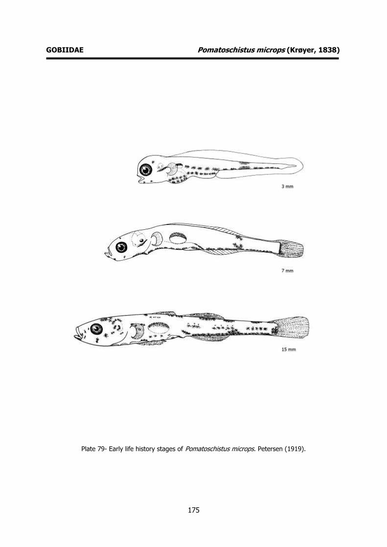
175
GOBIIDAE Pomatoschistus microps (Krøyer, 1838)
Plate 79- Early life history stages of Pomatoschistus microps. Petersen (1919).
176
GOBIIDAE Pomatoschistus minutus (Pallas, 1770) MERISTICS
Fins:
Dorsal rays - D1 VI-VII, D2 I+10-12 Anal rays – I+9-12
Pelvic rays – I+5 Pectoral rays – 18-21
Myomeres:
Total number – 32-34
LIFE HISTORY
Range: demersal; brackish; marine; depth range 4–200 m.
Habitat: Eastern Atlantic: from Norway to Spain; also Mediterranean Sea and Black
Sea.
Spawning season: spawns throughout the
whole year.
ELH pattern: Oviparous, demersal eggs and planktonic larvae.
MAIN REFERENCES Lebour, M.V. (1919). The young of the Gobiidae from the
neighbourhood of Plymouth. J. Mar. Biol. Ass. U.K., 12: 48-80.
Lebour, M.V. (1920). The eggs of Gobius minutus, pictus and microps. J. Mar. Biol. Ass. U.K., 12: 253-260.
Munk, P., J.G. Nielsen (2005). Eggs and larvae of North Sea fishes. Biofolia, Denmark: 215pp.
Padoa, E. (1933-1956). Gobiidae. Uova, larve e stadi Giovanili di Teleostei: monografia elaborata con l’uso del materiale raccolto e sweriato da Salvatore Lo Bianco. Fauna e Flora Golfo Napoli Monogr.38: 648-678.
Petersen, C.G.J. (1892). On the eggs and breeding of our Gobiidae. Reports of the Danish Biological Station, 2: 1-9.
Petersen, C.G.J. (1919). Our gobies (Gobiidae). From the egg to the adult stages. Reports of the Danish Biological Station, 26: 45-66.
Russell, F.S. (1976). The eggs and planktonic stages of British marine fishes. Academic Press, London: 524pp.
EARLY LIFE HISTORY DESCRIPTION
EGGS
Capsule diameter – 0.9-1.4x0.7-0.8 mm No. of oil globules -
Shell surface – smooth, pear-shaped Pigment -
Yolk – pigmented embryo
Diameter of oil globules - Diagnostic features -
LARVAE
Hatching length – 3.0 mm Yolk-sac absorption – 3.5 mm
Flexion length -
Transformation length - Pigmentation – Newly hatched larva:
pigmentation along the ventral and dorsal body contours. Melanophores develop in
the otocystic region, on head and on the
lower jaw. Late larva: basically the same pigmentation pattern. Pigmentation
intensifies along the tail (dorsal and ventral body contours). In postflexion larvae,
melanophores appear on head and on medio-lateral region.
Diagnostic features – Pigmentation.
Prominent gas bladder. Melanophores along the ventral side of the tail are of the
same size (the is no evidence of a large branched melanophore mid-way along the
tail).

177
GOBIIDAE Pomatoschistus minutus (Pallas, 1770)
Plate 77- Early life history stages of Pomatoschistus minutus. Petersen (1919).

178
GOBIIDAE Pomatoschistus pictus (Malm, 1865) MERISTICS
Fins:
Dorsal rays - D1 V-VI, D2 I+7-10 Anal rays – I+8-9
Pelvic rays – I+5 Pectoral rays – 16-20
Myomeres:
Total number – 30-31
LIFE HISTORY
Range: Eastern Atlantic: Norway (Trondheim fjord) to Spain and the Canary Islands.
Habitat: demersal; marine; depth range 1-55 m.
Spawning season: spawns throughout the
whole year.
ELH pattern: Oviparous, demersal eggs and
planktonic larvae.
MAIN REFERENCES Lebour, M.V. (1919). The young of the Gobiidae from the
neighbourhood of Plymouth. J. Mar. Biol. Ass. U.K., 12: 48-80.
Lebour, M.V. (1920). The eggs of Gobius minutus, pictus and microps. J. Mar. Biol. Ass. U.K., 12: 253-260.
Munk, P., J.G. Nielsen (2005). Eggs and larvae of North Sea fishes. Biofolia, Denmark: 215pp.
Petersen, C.G.J. (1917).On the development of of our common gobies (Gobius) from the egg to the adult stages etc.. Reports of the Danish Biological Station, 24: 5-16.
Petersen, C.G.J. (1919). Our gobies (Gobiidae). From the egg to the adult stages. Reports of the Danish Biological Station, 26: 45-66.
Russell, F.S. (1976). The eggs and planktonic stages of British marine fishes. Academic Press, London: 524pp.
EARLY LIFE HISTORY DESCRIPTION
EGGS
Capsule diameter – 0.75-0.8 mm long No. of oil globules – many oil globules
Shell surface – smooth, pear-shaped Pigment -
Yolk -
Diameter of oil globules - Diagnostic features -
LARVAE
Hatching length – 3.0 mm Yolk-sac absorption – 3.5 mm
Flexion length -
Transformation length - Pigmentation – Newly hatched larva: well
pigmented. Melanophores on the yolk sac and along the dorsal and ventral body
controur. Dorsal melanophores stretch
from the neck to the middle of the tail. Melanophores on head, otocystic region
and lower jaw. Late larva: dorsal pigmentation disappears being dominated
by the postanal ventral row, which has large branched melanophores from above
the anus to the caudal region. Postflexion
larva: dorsal pigmentation reappears. Three groups of medio-lateral
melanophores. Diagnostic features – Pigmentation.
Prominent gas bladder. Well pigmented
newly hatched larvae. Well developed dorsal pigmentation.
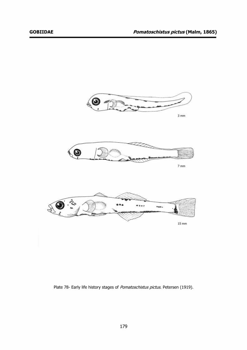
179
GOBIIDAE Pomatoschistus pictus (Malm, 1865)
Plate 78- Early life history stages of Pomatoschistus pictus. Petersen (1919).

180
CALLIONYMIDAE Callionymus lyra Linnaeus, 1758 MERISTICS
Fins:
Dorsal rays - D1 IV, D2 9 Anal rays - 9
Pelvic rays – I+5 Pectoral rays – 18-20
Myomeres:
Total number –
LIFE HISTORY
Range: Gibraltar, and Algeria, western Black Sea, Aegean and Adriatic Sea, Azores and
the Canary Islands.
Habitat: demersal; marine; depth range 5-
430 m.
Spawning season: spring and summer.
ELH pattern: Oviparous, planktonic eggs and
larvae.
MAIN REFERENCES Demir, N. (1972). The abundance and distribution of the
eggs and larvae of some teleost fishes off Plymouth in 1969 and 1970. II. The postlarvae of Callionymus. J. Mar. Biol. Ass. U.K., 52: 997-1010.
Demir, N. (1976). Callionymidae of the Northeastern Atlantic. Fiches d'Identification du Zooplancton, 148: 5pp.
Ehrenbaum, E. (1905-1909). Eier und Larven von Fischen. Nordisches Plankton, 1: 413pp.
Fage, L. (1918). Shore-fishes. Rep. Dan. Oceanogr. Exped . 1908-10 Medit . adjac. Seas, 2, Biology A3: 1-154.
Holt, E.W.L. (1899). Recherches sur la reproduction des poissons osseux principalement dans le Golfe de Marseille. Annls Mus. Hist. nat. Marseille, 5 Mém.2: 128p.
Munk, P., J.G. Nielsen (2005). Eggs and larvae of North Sea fishes. Biofolia, Denmark: 215pp.
Russell, F.S. (1976). The eggs and planktonic stages of British marine fishes. Academic Press, London: 524pp.
EARLY LIFE HISTORY DESCRIPTION
EGGS
Capsule diameter – 0.81-0.97 mm No. of oil globules - 0
Shell surface – honeycomb-like sculpturing (4, 6 or 7 sided cells).
Pigment -
Yolk – peripheral segmentation Diameter of oil globules -
Diagnostic features – Characteristic shell surface.
LARVAE
Hatching length – 1.08 mm
Yolk-sac absorption – 2.3 mm Flexion length – 4 mm
Transformation length – 10 mm Pigmentation – very strong pigmentation
distributed over most of the larva.
Mediolateral pigmentation prominent. Diagnostic features – small newly hatched
larva (ca. 2 mm). Typical shape and pigmentation. C. lyra has a greater
concentration of melanophores in the opercular and mid-brain regions compared
to Callionymus maculatus. The best
diagnostic character in late larvae is the form and development of the preopercular
spines. Larvae with more than 6 mm develop a characteristic preopercular
spine. Larger pelvic fins as compared to C. maculatus.

181
CALLIONYMIDAE Callionymus lyra Linnaeus, 1758
Plate 80- Early life history stages of Callionymus lyra. Fage (1918). Preopercular spines, Demir (1972).
182
CALLIONYMIDAE Callionymus maculatus Rafinesque, 1810 MERISTICS
Fins:
Dorsal rays - D1 IV, D2 9-10 Anal rays – 8-9
Pelvic rays – I+5 Pectoral rays – 16-20
Myomeres:
Total number –
LIFE HISTORY
Range: Eastern Atlantic: southern and western Iceland and Norway south to
Senegal. Also in the Mediterranean,
including the Adriatic and Aegean but not in the Black Sea.
Habitat: demersal; marine; depth range 45-
650 m.
Spawning season: spring and summer.
ELH pattern: Oviparous, planktonic eggs and
larvae.
MAIN REFERENCES Demir, N. (1972). The abundance and distribution of the
eggs and larvae of some teleost fishes off Plymouth in 1969 and 1970. II. The postlarvae of Callionymus. J. Mar. Biol. Ass. U.K., 52: 997-1010.
Demir, N. (1976). Callionymidae of the Northeastern Atlantic. Fiches d'Identification du Zooplancton, 148: 5pp.
Ehrenbaum, E. (1905-1909). Eier und Larven von Fischen. Nordisches Plankton, 1: 413pp.
Fage, L. (1918). Shore-fishes. Rep. Dan. Oceanogr. Exped . 1908-10 Medit . adjac. Seas, 2, Biology A3: 1-154.
Holt, E.W.L. (1899). Recherches sur la reproduction des poissons osseux principalement dans le Golfe de Marseille. Annls Mus. Hist. nat. Marseille, 5 Mém.2: 128p.
Munk, P., J.G. Nielsen (2005). Eggs and larvae of North Sea fishes. Biofolia, Denmark: 215pp.
Russell, F.S. (1976). The eggs and planktonic stages of British marine fishes. Academic Press, London: 524pp.
EARLY LIFE HISTORY DESCRIPTION
EGGS
Capsule diameter – 0.66-0.79 mm No. of oil globules - 0
Shell surface - honeycomb-like sculpturing Pigment -
Yolk – peripheral segmentation
Diameter of oil globules - Diagnostic features – Characteristic shell
surface.
LARVAE Hatching length – 2.29 mm (very similar to
C. lyra)
Yolk-sac absorption - Flexion length – 4 mm
Transformation length – 10 mm Pigmentation – heavily pigmented larva
(stronger than C. lyra in branchiostegal
and mandibular regions). Strongly pigmented pectoral fins.
Diagnostic features – Pigmentation, length of pelvic fins and preopercular spine.
Preopercular spine very distinct from that o C. lyra. Pelvic fins develop when the larva
reaches 3.0 to 3.5 mm. At 6.5 mm they
reach a point half way along the base of the pectoral fin. At comparable sizes the
pelvic fins are shorter than those of C. lyra.

183
CALLIONYMIDAE Callionymus maculatus Rafinesque, 1810
Plate 81- Early life history stages of Callionymus maculatus. Fage (1918). Preopercular spines,
Demir (1972).

184
BLENNIIDAE Blennius ocellaris Linnaeus, 1758 MERISTICS
Fins:
Dorsal rays – XI-XII+14-16 Anal rays – II+14-16
Pelvic rays – I+3 Pectoral rays – 12
Myomeres:
Total number –
LIFE HISTORY
Range: Southeast Atlantic: Atlantic coast from Morocco to the English Channel. Also
known from the Mediterranean and Black
Sea.
Habitat: demersal; marine; depth range 10-400 m.
Spawning season: spring and summer.
ELH pattern: Oviparous, demersal eggs and planktonic larvae.
MAIN REFERENCES Cunningham, J.T. (1889). Studies of the reprodution and
development of teleostean fishes occurring in the neighbourhood of Plymouth. J. mar. bio. Ass. U.K., 1: 370-375.
Ehrenbaum, E. (1905-1909). Eier und Larven von Fischen. Nordisches Plankton, 1: 413pp.
Ford, E. (1922a). On the young of Blennius ocellaris L., Blennius pholis L., and Blennius gattorugine. J. mar. biol. Ass. U.K., 12: 688-692.
Lebour, M.V. (1927). The eggs and newly hatched young of the common blennies from the Plymounth neighbourhood. J. mar. biol. Ass. U.K., 14: 647-650.
Padoa, E. (1933-1956). Benniidae. Uova, larve e stadi Giovanili di Teleostei: monografia elaborata con l’uso del materiale raccolto e sweriato da Salvatore Lo Bianco. Fauna e Flora Golfo Napoli Monogr.38: 720-745.
Russell, F.S. (1976). The eggs and planktonic stages of British marine fishes. Academic Press, London: 524pp.
EARLY LIFE HISTORY DESCRIPTION
EGGS
Capsule diameter – 1.12-1.20 mm No. of oil globules – many oil globules
Shell surface – smooth, nearly spherical Pigment -
Yolk - unsegmented
Diameter of oil globules - Diagnostic features -
LARVAE
Hatching length – 4.6 mm Yolk-sac absorption -
Flexion length -
Transformation length - Pigmentation – Newly hatched larva:
melanophores on snout, head, peritoneal area and a strongly pigmented pectoral fin
(arranged in longitudinal rows between the
fin rays). Postanal row of pigments along the posterior half of the caudal region.
Late larva: large, rounded in shape and heavily pigmented pectoral fin with rows of
large melanophores. Diagnostic features - Characteristic
pigmentation, large, round and heavily
pigmented pectoral fins.

185
BLENNIIDAE Blennius ocellaris Linnaeus, 1758
Plate 82- Early life history stages of Blennius ocellaris. Ford (1922a).
186
BLENNIIDAE Coryphoblennius galerita (Linnaeus, 1758) MERISTICS
Fins:
Dorsal rays – XIII+16-17 Anal rays – II+16-19
Pelvic rays – I+3 Pectoral rays – 12
Myomeres:
Total number –
LIFE HISTORY
Range: Eastern Atlantic: along the coasts of western England and the British Channel,
Spain, Portugal, France, Morocco, Madeira ,
Canary Islands; Mediterranean Sea, Sea of Marmara, and the Black Sea.
Habitat: demersal; marine.
Spawning season: spring and summer.
ELH pattern: Oviparous, demersal eggs and planktonic larvae.
MAIN REFERENCES Fives, J.M. (1980). An account of the eggs and
developmental stages of Montagu's blenny, Coryphoblennius galerita (L.), with notes on the reproductive behaviour of the adults. J. Mar. Biol. Ass. U.K., 60: 749-757.
Fives, J.M. (1986). Blenniidae of the North Atlantic (revised). Fich. Ident. Plancton, 172: 6pp.
Padoa, E. (1933-1956). Benniidae. Uova, larve e stadi Giovanili di Teleostei: monografia elaborata con l’uso del materiale raccolto e sweriato da Salvatore Lo Bianco. Fauna e Flora Golfo Napoli Monogr.38: 720-745.
Russell, F.S. (1976). The eggs and planktonic stages of British marine fishes. Academic Press, London: 524pp.
EARLY LIFE HISTORY DESCRIPTION
EGGS
Capsule diameter – ca. 1 mm No. of oil globules – golden-yellow oil
globules Shell surface – smooth, nearly spherical
Pigment -
Yolk - Diameter of oil globules -
Diagnostic features -
LARVAE Hatching length – 3.3 mm
Yolk-sac absorption -
Flexion length - Transformation length -
Pigmentation - Melanophores on top and back of head and on auditory capsule.
Pigmented pectoral fins. Melanophores on
top of gut and a metameric series of 24-25 melanophores along posterior ventral
margin from fifth to sixth postanal myomere to urostyle tip.
Diagnostic features – Characteristic pigmentation. Pointed pectoral fins
extending to second or third postanal body
segment.
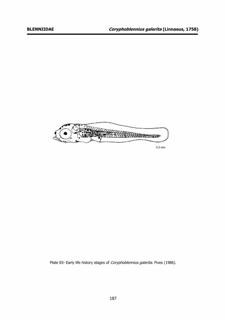
187
BLENNIIDAE Coryphoblennius galerita (Linnaeus, 1758)
Plate 83- Early life history stages of Coryphoblennius galerita. Fives (1986).

188
BLENNIIDAE Lipophrys pholis (Linnaeus, 1758) MERISTICS
Fins:
Dorsal rays – XII+18-19 Anal rays – II+19
Pelvic rays – I+3 Pectoral rays – 13
Myomeres:
Total number –
LIFE HISTORY
Range: Eastern Atlantic: southern Norway to Morocco and Madeira, including the
Mediterranean and the Balearics.
Habitat: demersal; marine; depth range 0-8
m.
Spawning season: winter and spring.
ELH pattern: Oviparous, demersal eggs and
planktonic larvae.
MAIN REFERENCES Ford, E. (1922a). On the young of Blennius ocellaris L.,
Blennius pholis L., and Blennius gattorugine. J. mar. biol. Ass. U.K., 12: 688-692.
Faria, C., R. Borges, F. Gil, V.C. Almada, E.J. Gonçalves (2002). Embryonic and larval development of Lipophrys pholis (Pisces: Blenniidae). Scentia Marina, 66 (1): 21-26.
Fives, J.M. (1986). Blenniidae of the North Atlantic (revised). Fich. Ident. Plancton, 172: 6pp.
Lebour, M.L. (1927). The eggs and newly hatched young of the common blennies from the Plymounth neighbourhood. J. mar. biol. Ass. U.K., 14: 647-650.
Munk, P., J.G. Nielsen (2005). Eggs and larvae of North Sea fishes. Biofolia, Denmark: 215pp.
Padoa, E. (1933-1956). Benniidae. Uova, larve e stadi Giovanili di Teleostei: monografia elaborata con l’uso del materiale raccolto e sweriato da Salvatore Lo Bianco. Fauna e Flora Golfo Napoli Monogr.38: 720-745.
Russell, F.S. (1976). The eggs and planktonic stages of British marine fishes. Academic Press, London: 524pp.
EARLY LIFE HISTORY DESCRIPTION
EGGS
Capsule diameter – 1.18-1.6 mm No. of oil globules – several oil globules
Shell surface – smooth, hemispherical Pigment -
Yolk - unsegmented
Diameter of oil globules - Diagnostic features -
LARVAE
Hatching length – 5 mm Yolk-sac absorption – 5.0-5.5 mm
Flexion length – 10 mm
Transformation length – 17-19 mm Pigmentation – Newly hatched larvae:
rounded and heavily pigmented pectoral fins (12 radial rows of melanophores).
Peritoneal pigmentation. Late larva:
narrow, pointed and heavily pigmented pectoral fin. A few postanal ventral
melanophores in the posterior half of the caudal region and a vertical row at the
base of the caudal fin. Notochordal row of melanophores evident anteriourly.
Diagnostic features – Heavily pigmented
pectoral fins. Shape of body and pectoral fins.

189
BLENNIIDAE Lipophrys pholis (Linnaeus, 1758)
Plate 84- Early life history stages of Lipophrys pholis. Faria et al. (2002).

190
BLENNIIDAE Lipophrys trigloides (Valenciennes, 1836) MERISTICS
Fins:
Dorsal rays – XII-16-17 Anal rays – II+18
Pelvic rays – I+3 Pectoral rays – 13
Myomeres:
Total number –
LIFE HISTORY
Range: Eastern Atlantic: Along the coasts of France (Brittany), the Iberian Peninsula,
Morocco, the Mediterranean and the Sea of
Marmara southwards to Senegal, the Canary Islands and Madeira.
Habitat: demersal; marine.
Spawning season: winter and spring.
ELH pattern: Oviparous, demersal eggs and planktonic larvae.
MAIN REFERENCES Faria, C., F. Gil, V.C. Almada (2005). Ontogenic
development of Lipophrys trigloides (Pisces: Blenniidae), with some notes of the spawning behaviour. J. Mar. Biol. Ass. U.K., 85: 185-188.
Padoa, E. (1933-1956). Benniidae. Uova, larve e stadi Giovanili di Teleostei: monografia elaborata con l’uso del materiale raccolto e sweriato da Salvatore Lo Bianco. Fauna e Flora Golfo Napoli Monogr.38: 720-745.
EARLY LIFE HISTORY DESCRIPTION
EGGS
Capsule diameter – 1.3x0.86 mm No. of oil globules -
Shell surface – smooth, hemispherical Pigment -
Yolk -
Diameter of oil globules - Diagnostic features -
LARVAE
Hatching length – 4.8 mm Yolk-sac absorption – 4.8 mm
Flexion length –
Transformation length – 16-17 mm Pigmentation - Newly hatched larva: heavily
pigmented pectoral fins. Heavy peritoneal pigmentation (8-10 radial rows of
melanophores). One or two melanophores
at base of the pectoral fin and 8-14 melanophores on the last myomeres with
1-6 near the caudal tip. Pigmentation on head, upper jaw and otocystic capsule.
Late larva: pigmentation pattern maintained with an increase in the number
and intensity of the postanal ventral
pigmentation. Diagnostic features – Pigmentation.
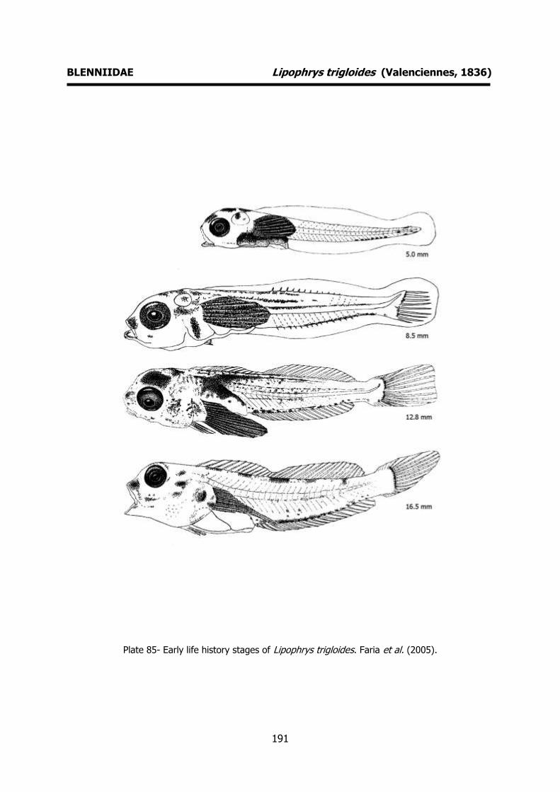
191
BLENNIIDAE Lipophrys trigloides (Valenciennes, 1836)
Plate 85- Early life history stages of Lipophrys trigloides. Faria et al. (2005).
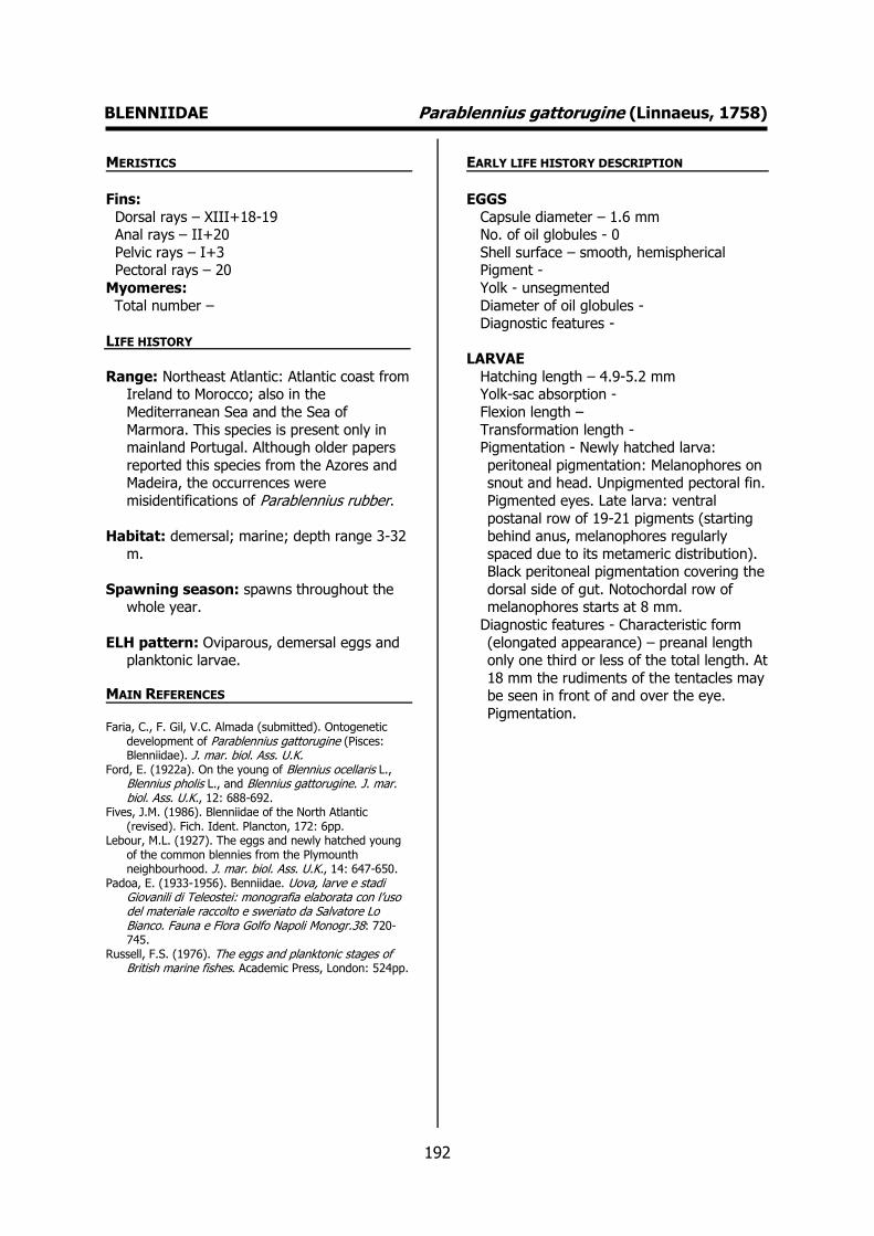
192
BLENNIIDAE Parablennius gattorugine (Linnaeus, 1758) MERISTICS
Fins:
Dorsal rays – XIII+18-19 Anal rays – II+20
Pelvic rays – I+3 Pectoral rays – 20
Myomeres:
Total number –
LIFE HISTORY
Range: Northeast Atlantic: Atlantic coast from Ireland to Morocco; also in the
Mediterranean Sea and the Sea of
Marmora. This species is present only in mainland Portugal. Although older papers
reported this species from the Azores and Madeira, the occurrences were
misidentifications of Parablennius rubber. Habitat: demersal; marine; depth range 3-32
m.
Spawning season: spawns throughout the whole year.
ELH pattern: Oviparous, demersal eggs and planktonic larvae.
MAIN REFERENCES Faria, C., F. Gil, V.C. Almada (submitted). Ontogenetic
development of Parablennius gattorugine (Pisces: Blenniidae). J. mar. biol. Ass. U.K.
Ford, E. (1922a). On the young of Blennius ocellaris L., Blennius pholis L., and Blennius gattorugine. J. mar. biol. Ass. U.K., 12: 688-692.
Fives, J.M. (1986). Blenniidae of the North Atlantic (revised). Fich. Ident. Plancton, 172: 6pp.
Lebour, M.L. (1927). The eggs and newly hatched young of the common blennies from the Plymounth neighbourhood. J. mar. biol. Ass. U.K., 14: 647-650.
Padoa, E. (1933-1956). Benniidae. Uova, larve e stadi Giovanili di Teleostei: monografia elaborata con l’uso del materiale raccolto e sweriato da Salvatore Lo Bianco. Fauna e Flora Golfo Napoli Monogr.38: 720-745.
Russell, F.S. (1976). The eggs and planktonic stages of British marine fishes. Academic Press, London: 524pp.
EARLY LIFE HISTORY DESCRIPTION
EGGS
Capsule diameter – 1.6 mm No. of oil globules - 0
Shell surface – smooth, hemispherical Pigment -
Yolk - unsegmented
Diameter of oil globules - Diagnostic features -
LARVAE
Hatching length – 4.9-5.2 mm Yolk-sac absorption -
Flexion length –
Transformation length - Pigmentation - Newly hatched larva:
peritoneal pigmentation: Melanophores on snout and head. Unpigmented pectoral fin.
Pigmented eyes. Late larva: ventral
postanal row of 19-21 pigments (starting behind anus, melanophores regularly
spaced due to its metameric distribution). Black peritoneal pigmentation covering the
dorsal side of gut. Notochordal row of melanophores starts at 8 mm.
Diagnostic features - Characteristic form
(elongated appearance) – preanal length only one third or less of the total length. At
18 mm the rudiments of the tentacles may be seen in front of and over the eye.
Pigmentation.
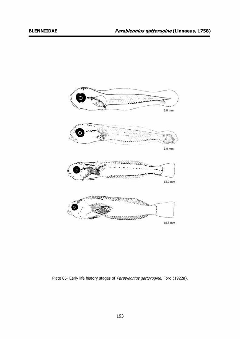
193
BLENNIIDAE Parablennius gattorugine (Linnaeus, 1758)
Plate 86- Early life history stages of Parablennius gattorugine. Ford (1922a).

194
BLENNIIDAE Parablennius pilicornis (Cuvier, 1829) MERISTICS
Fins:
Dorsal rays – XII+21 Anal rays – II+23
Pelvic rays – I+3 Pectoral rays – 14
Myomeres:
Total number – 38
LIFE HISTORY
Range: Eastern Atlantic: Spain and Portugal to Möwe Bay, Namibia. Also in the
Mediterranean along the coast from
Morocco, Algeria, Spain. Southwest Atlantic: Brazil and Patagonia, Argentina.
Western Indian Ocean: Natal to Knysna in South Africa.
Habitat: demersal; marine; depth range 0–25 m.
Spawning season: spring and summer.
ELH pattern: Oviparous, demersal eggs and
planktonic larvae.
MAIN REFERENCES
Faria, C., F. Gil, V.C. Almada (2006). Ontogenic development of Parablennius pilicornis (Pisces: Blenniidae) in controlled conditions. Scientia Marina, 70 (4): 667-671.
Olivar, P. (1986). Development and distribution of Parablennius pilicornis (Cuvier) larvae (Teleostei: Blenniidae) off Namibia. S. Afr. J. mar. Sci.¸4: 193-201.
Olivar, P., J.-M. Fortuño (1991). Guide to the ichthyoplankton of the Southeast Atlantic (Benguela current region). Scientia Marina, 55 (1): 1-383.
EARLY LIFE HISTORY DESCRIPTION
EGGS
Capsule diameter - No. of oil globules -
Shell surface - Pigment -
Yolk -
Diameter of oil globules - Diagnostic features -
LARVAE
Hatching length – 3.0-3.2 mm Yolk-sac absorption –
Flexion length – 10-11 mm
Transformation length - Pigmentation – Newly hatched larva: heavy
peritoneal pigmentation, pigmented eyes, small and unpigmented pectoral fins. One
melanophore close to the anus and a
characteristic row of postanal melanophores (ca. 30). Late larva: 6-7
pre-opercular spines visible from a length of 6 mm until metamorphosis.
Pigmentation pattern maintained during development, with an increase in the
number and intensity of melanophores at
the ventral row, on liver, head and opercula.
Diagnostic features – Characteristic pigmentation. Small preanal length (25-33
% of total length) from the earliest stages
to juveniles. Presence of pre-opercular spines from a length of 6 mm until
metamorphosis.

195
BLENNIIDAE Parablennius pilicornis (Cuvier, 1829)
Plate 87- Early life history stages of Parablennius pilicornis. Faria et al. (2006).

196
CARAPIDAE Carapus acus (Brünnich, 1768) MERISTICS
Fins:
Dorsal rays – 37-39 Anal rays - 58
Pelvic rays - Pectoral rays –
Myomeres:
Total number – 125-144 (17-18 pre-caudal)
LIFE HISTORY
Range: Eastern Atlantic: including the Mediterranean. Probably south to Ascension
Island.
Habitat: demersal; marine; depth range 0-
100 m. Adults typically live as commensals in the gut of shallow-water holothurians,
Holothuria tubulosa and Stichopus regalis. Spawning season: summer, autumn and
winter.
ELH pattern: Oviparous, planktonic eggs (in slimy egg masses) and larvae.
MAIN REFERENCES Padoa, E. (1933-1956). Carapidae. Uova, larve e stadi
Giovanili di Teleostei: monografia elaborata con l’uso del materiale raccolto e sweriato da Salvatore Lo Bianco. Fauna e Flora Golfo Napoli Monogr.38: 761-774.
Raffaele, F. (1888) - Le uova galleggianti e la larve dei Te-leostei nel Golfo di Napoli. Mitt. zool. Stn Neapel, 8: 1-85.
EARLY LIFE HISTORY DESCRIPTION
EGGS
Capsule diameter – 0.75-0.9 mm No. of oil globules - 1
Shell surface – smooth, ellipsoidal Pigment -
Yolk - unsegmented
Diameter of oil globules – 0.18-0.20 mm Diagnostic features -
LARVAE
Hatching length – 2.5 mm Yolk-sac absorption -
Flexion length -
Transformation length - Pigmentation – Newly hatched larva:
abundant pigments on the head and the trunk. Pigmented caudal region.
Diagnostic features – Characteristic larva. A
dorsal appendage (vexillum), longer than the trunk, is developed at 4 mm.

197
CARAPIDAE Carapus acus (Brünnich, 1768)
Plate 88- Early life history stages of Carapus acus. Emery (1880 in Padoa, 1933-1956).

198
MUGILIDAE Mugil cephalus Linnaeus, 1758 MERISTICS
Fins:
Dorsal rays – D1 IV, D2 I+8-9 Anal rays – III+8
Pelvic rays – I+5 Pectoral rays – 17
Myomeres:
Total number –
LIFE HISTORY
Range: Cosmopolitan in coastal waters of the tropical and subtropical zones of all seas.
Habitat: benthopelagic; catadromous; freshwater; brackish; marine; depth range
0-120 m.
Spawning season: spawns mainly in winter.
ELH pattern: Oviparous, planktonic eggs and
larvae.
MAIN REFERENCES Russell, F.S. (1976). The eggs and planktonic stages of
British marine fishes. Academic Press, London: 524pp. Vialli, M. (1933-1956). Mugilidae. Uova, larve e stadi
Giovanili di Teleostei: monografia elaborata con l’uso del materiale raccolto e sweriato da Salvatore Lo Bianco. Fauna e Flora Golfo Napoli Monogr.38: 433-457.
EARLY LIFE HISTORY DESCRIPTION
EGGS
Capsule diameter – 0.72 mm No. of oil globules - 1
Shell surface - smooth Pigment -
Yolk - unsegmented
Diameter of oil globules – 0.28 mm Diagnostic features – Large oil globule.
LARVAE
Hatching length – 2.5 mm Yolk-sac absorption -
Flexion length -
Transformation length - Pigmentation – Melanophores on head and
trunk. Tip of tail less pigmented. Anus situated about 50% of total length.
Diagnostic features – Typical pigmentation.
Shape of body.
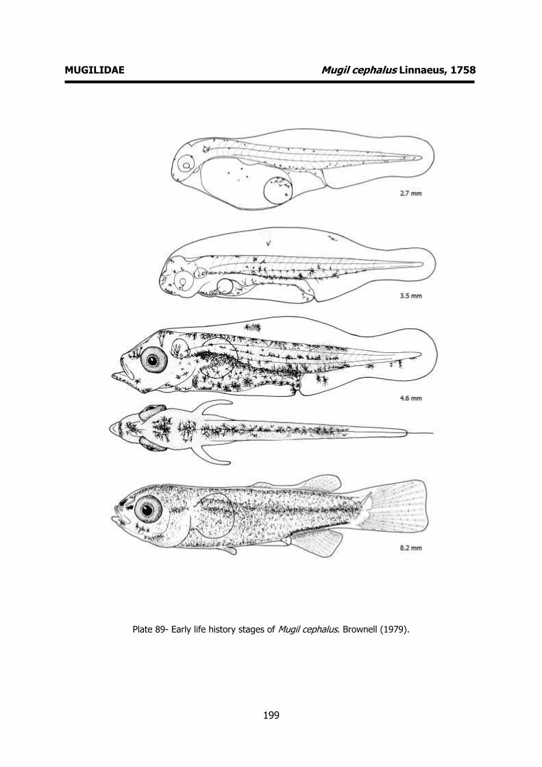
199
MUGILIDAE Mugil cephalus Linnaeus, 1758
Plate 89- Early life history stages of Mugil cephalus. Brownell (1979).

200
ATHERINIDAE Atherina presbyter Linnaeus, 1758 MERISTICS
Fins:
Dorsal rays – VII-IX+11-14 Anal rays – I+14-17
Pelvic rays – I+5 Pectoral rays – 13-15
Myomeres:
Total number – 46-52
LIFE HISTORY
Range: marine; pelagic; in coastal areas and estuaries.
Habitat: Eastern Atlantic: Kattegat (rare) and British Isles to Canary Islands, Mauritania
and Cape Verde, also western Mediterranean.
Spawning season: spring and summer.
ELH pattern: Oviparous, planktonic eggs and larvae.
MAIN REFERENCES Ehrenbaum, E. (1905-1909). Eier und Larven von Fischen.
Nordisches Plankton, 1: 413pp. Holt, E.W.L. (1899). Recherches sur la reproduction des
poissons osseux principalement dans le Golfe de Marseille. Annls Mus. Hist. nat. Marseille, 5 Mém.2: 128p.
Russell, F.S. (1976). The eggs and planktonic stages of British marine fishes. Academic Press, London: 524pp.
EARLY LIFE HISTORY DESCRIPTION
EGGS
Capsule diameter – 1.85-1.9 mm No. of oil globules – several oil globules
Shell surface – smooth, long hairy appendages. Attached to plants
Pigment -
Yolk - Diameter of oil globules -
Diagnostic features -
LARVAE Hatching length - Unknown
Yolk-sac absorption - Unknown
Flexion length - Transformation length -
Pigmentation – Three large melanophores on the crown of the head and two smaller
ones over the anterior of the eye. Well
developed peritoneal pigmentation on dorsal side of abdomen. Characteristic
mediolateral line consisting of 10-13 elongated melanophores, in some larvae
almost forming a continuous line. Two ventral contour postanal melanophores
near tail (6 mm). Continuous row of
postanal ventral melanophores from a short distance behind the anus to the tail.
Single melanophore near the posterior edge of operculum. Row of internal
notochordal melanophores (11 mm).
Diagnostic features – Body long and narrow. Anus well forward. Characteristic
pigmentation.
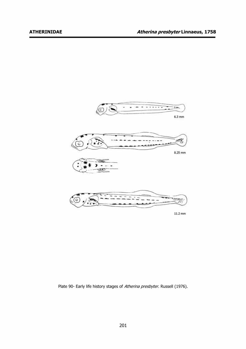
201
ATHERINIDAE Atherina presbyter Linnaeus, 1758
Plate 90- Early life history stages of Atherina presbyter. Russell (1976).

202
TRIGLIDAE Trigla lucerna Linnaeus, 1758 MERISTICS
Fins:
Dorsal rays – D1 VIII+IX-16-17 Anal rays – 14-16
Pelvic rays – I+5 Pectoral rays – 10-11+3
Myomeres:
Total number – 33-34
LIFE HISTORY
Range: Eastern Atlantic: Norway to Cape Blanc (along the African coast); not
recorded at Madeira and the Azores. Also in
the Mediterranean and Black seas.
Habitat: demersal; marine; depth range 20-318 m.
Spawning season: spring and summer.
ELH pattern: Oviparous, planktonic eggs and larvae.
MAIN REFERENCES Ehrenbaum, E. (1905-1909). Eier und Larven von Fischen.
Nordisches Plankton, 1: 413pp. Fage, L. (1918). Shore-fishes. Rep. Dan. Oceanogr. Exped
. 1908-10 Medit . adjac. Seas, 2, Biology A3: 1-154. Munk, P., J.G. Nielsen (2005). Eggs and larvae of North
Sea fishes. Biofolia, Denmark: 215pp. Padoa, E. (1933-1956). Triglidae. Uova, larve e stadi
Giovanili di Teleostei: monografia elaborata con l’uso del materiale raccolto e sweriato da Salvatore Lo Bianco. Fauna e Flora Golfo Napoli Monogr.38: 627-642.
Russell, F.S. (1976). The eggs and planktonic stages of British marine fishes. Academic Press, London: 524pp.
EARLY LIFE HISTORY DESCRIPTION
EGGS
Capsule diameter – 1.10-1-54 mm No. of oil globules - 1
Shell surface - smooth Pigment -
Yolk -
Diameter of oil globules – 0.22-0.31 mm Diagnostic features -
LARVAE
Hatching length – 3.5 mm Yolk-sac absorption -
Flexion length -
Transformation length - Pigmentation – Newly hatched larva: large
pectoral fin, melanophores distributed on larva and yolk sac. Late larva:
Melanophores on head, upper and lower
jaws. Postanal row of melanophores. Pigmented pectoral fin (radial
melanophores). Diagnostic features – Shape of body. Spines
on pre-operculum. Large and rounded pectoral fins reaching the anus. Pelvic fins
pigmented at their margins. Occipital
ridge, infra-orbital ridge, nasal and mandibular spines. Spines also develop
medio-laterally and on the bases of pectoral fins.
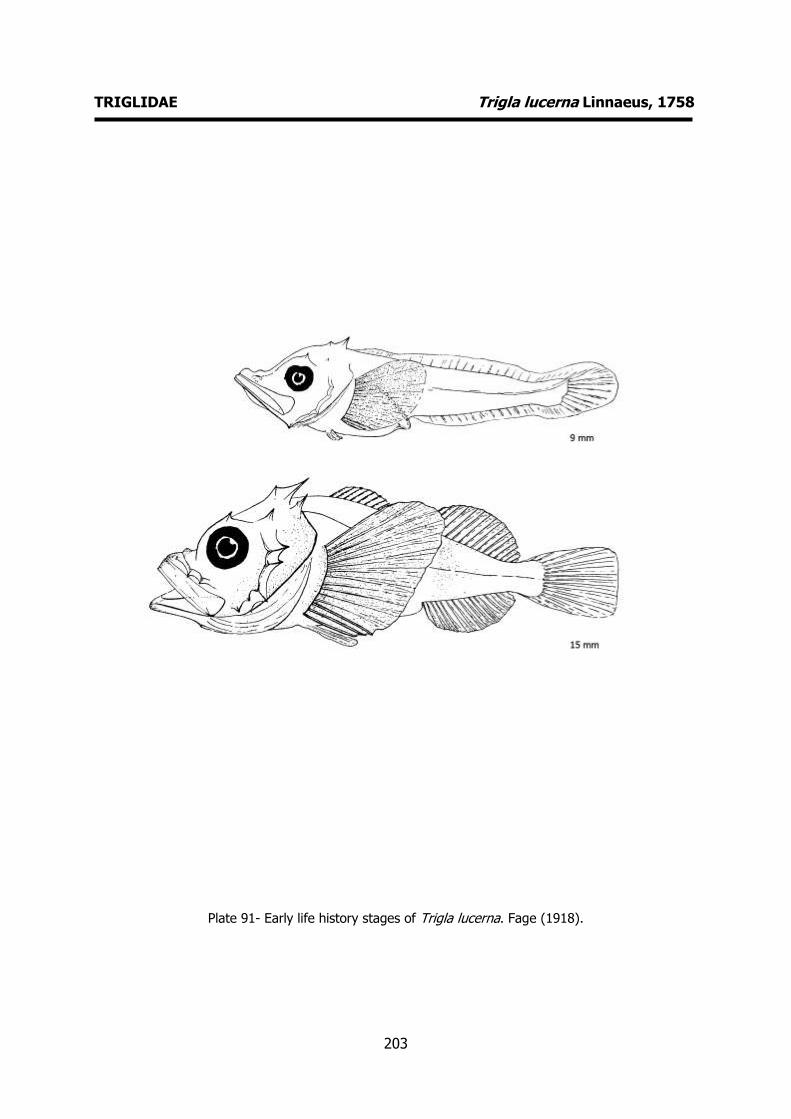
203
TRIGLIDAE Trigla lucerna Linnaeus, 1758
Plate 91- Early life history stages of Trigla lucerna. Fage (1918).
204
COTTIDAE Taurulus bubalis (Euphrasen, 1786) MERISTICS
Fins:
Dorsal rays – D1 VII-X, D2 10-14 Anal rays – 8-10
Pelvic rays – I+3 Pectoral rays – 14-16
Myomeres:
Total number – 29-33
LIFE HISTORY
Range: Eastern Atlantic: Iceland, the Shetlands, from Murmansk southward to
Portugal, also Baltic Sea northward to Gulf
of Finland and northern Mediterranean coasts eastward to Gulf of Genoa.
Habitat: demersal; brackish; marine; depth
range 0-100 m.
Spawning season: spring.
ELH pattern: Oviparous, demersal eggs and
planktonic larvae.
MAIN REFERENCES Ehrenbaum, E. (1905-1909). Eier und Larven von Fischen.
Nordisches Plankton, 1: 413pp. M'intosh, W.S., A.T. Masterman (1897). The life-histories
of the British marine food-fishes. C. J. Clay London: 467p.
Munk, P., J.G. Nielsen (2005). Eggs and larvae of North Sea fishes. Biofolia, Denmark: 215pp.
Russell, F.S. (1976). The eggs and planktonic stages of British marine fishes. Academic Press, London: 524pp.
EARLY LIFE HISTORY DESCRIPTION
EGGS
Capsule diameter – 1.5-1.8 mm No. of oil globules – several oil globules
Shell surface – smooth, spherical Pigment -
Yolk - unsegmented
Diameter of oil globules – 0.25-0.35 mm Diagnostic features -
LARVAE
Hatching length – 5.5-5.8 mm Yolk-sac absorption -
Flexion length -
Transformation length - Pigmentation – Newly hatched larva: no
melanophores on head and base of pectoral fin. Late larva: heavy peritoneal
pigmentation. Row of postanal ventral
melanophores. Late larva: little change in pigmentation. The black peritoneal
pigmentation is the most obvious feature. Usually tow melanophores at the base of
the caudal fin. Two large melanophores on each side of the throat region, and a few
melanophores on the ventral edge of the
abdomen. Diagnostic features – Heavy peritoneal
pigmentation. Characteristic shape of body. Spinal armature evident. Single
otocystic spine, four preopercular spines
and two small protuberances behind the otocyst.

205
COTTIDAE Taurulus bubalis (Euphrasen, 1786)
Plate 92- Early life history stages of Taurulus bubalis. Ehrenbaum (1905-1909).

206
LIPARIDAE Liparis montagui (Donovan, 1804) MERISTICS
Fins:
Dorsal rays – 28-30 Anal rays – 22-25
Pelvic rays - Pectoral rays –
Myomeres:
Total number –
LIFE HISTORY
Range: Northeast Atlantic: North Sea, around the British Isles, Norwegian Sea,
southwestern Barents Sea and around
southern Iceland.
Habitat: demersal; marine; depth range 0-30 m.
Spawning season: winter to summer.
ELH pattern: Oviparous, demersal eggs and planktonic larvae.
MAIN REFERENCES Ehrenbaum, E. (1905-1909). Eier und Larven von Fischen.
Nordisches Plankton, 1: 413pp. Fage, L. (1918). Shore-fishes. Rep. Dan. Oceanogr. Exped
. 1908-10 Medit . adjac. Seas, 2, Biology A3: 1-154. Munk, P., J.G. Nielsen (2005). Eggs and larvae of North
Sea fishes. Biofolia, Denmark: 215pp. Ré. P., L. Arruda, P. Salgado, (1985). On the occurence of
Liparis montagui (L.) larval and juvenile stages off the portuguese coast. Cybium, 9 (4): 407- 409.
Russell, F.S. (1976). The eggs and planktonic stages of British marine fishes. Academic Press, London: 524pp.
EARLY LIFE HISTORY DESCRIPTION
EGGS
Capsule diameter – 1.03-1.19 mm No. of oil globules - 1
Shell surface – smooth, spherical Pigment -
Yolk - unsegmented
Diameter of oil globules – 0.25-0.38 mm Diagnostic features -
LARVAE
Hatching length – 3.5 mm Yolk-sac absorption – 4.0-4.5 mm
Flexion length -
Transformation length - Pigmentation – Newly hatched larva: heavy
peritoneal pigmentation, row of postanal ventral melanophores, pigment on head
and pectoral fins. Late larva: little change
in overall pigmentation. Diagnostic features – Typical boby shape.
Heavy peritoneal pigmentation. Large pectoral fins. Pelvic fin sucker. At 7-8 mm
fin rays develop and the small pelvic fin sucker is already apparent. Large
pigmented pectoral fins. At 11-12 mm
dosal and anal fins rays fully developed. Pectoral fin reaches de anus.
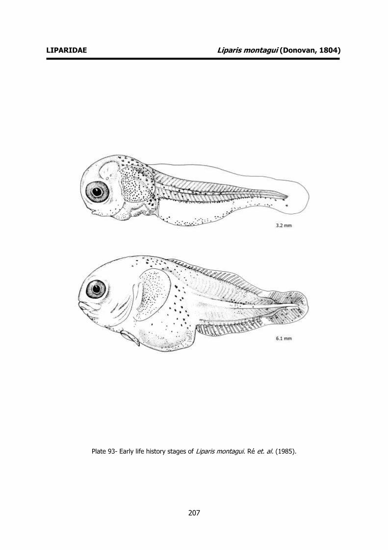
207
LIPARIDAE Liparis montagui (Donovan, 1804)
Plate 93- Early life history stages of Liparis montagui. Ré et. al. (1985).
208
SCOPHTHALMIDAE Lepidorhombus boscii (Risso, 1810) MERISTICS
Fins:
Dorsal rays – 79-86 Anal rays – 65-69
Pelvic rays - 6 Pectoral rays – 11-12
Myomeres:
Total number – 10+32
LIFE HISTORY
Range: Northeast Atlantic: British Isles south to Cape Bojador, West Sahara and the
Mediterranean.
Habitat: demersal; marine; depth range 7–
800 m.
Spawning season: winter and spring.
ELH pattern: Oviparous, planktonic larvae.
MAIN REFERENCES Petersen, C.G.J. (1909). On the larval and post-larval
stages of some Pleuronectidae (Zeugopterus, Arnoglossus, Solea). Meddr Kommn Havunders., Ser. Fiskeri, 3, Nr. 1: 1-18.
Russell, F.S. (1976). The eggs and planktonic stages of British marine fishes. Academic Press, London: 524pp.
EARLY LIFE HISTORY DESCRIPTION
EGGS: Undescribed
Capsule diameter – Unknown No. of oil globules - Unknown
Shell surface – Unknown Pigment - Unknown
Yolk - Unknown
Diameter of oil globules - Unknown Diagnostic features - Unknown
LARVAE
Hatching length - Unknown Yolk-sac absorption - Unknown
Flexion length – 9 mm
Transformation length - Pigmentation – Seven groups of
melanophores along the margin of the dorsal fin and three along the margin of
the anal fin. Three bands os melanophores
on the dorsal portion of the body, the most anterior above the anus. Three
groups on the ventral postanal region. Diagnostic features – Characteristic
pigmentation (groups of melanophores on dorsal and anal fins). Preoperculum with
many small spines behind the otocystic
region (two distint rows).

209
SCOPHTHALMIDAE Lepidorhombus boscii (Risso, 1810)
Plate 94- Early life history stages of Lepidorhombus boscii. Petersen (1909).
210
SCOPHTHALMIDAE Psetta maxima (Linnaeus, 1758) MERISTICS
Fins:
Dorsal rays – 57-71 Anal rays – 43-52
Pelvic rays - 6 Pectoral rays – 11-12
Myomeres:
Total number – 11+20
LIFE HISTORY
Range: Northeast Atlantic: throughout the Mediterranean and along the European
coasts to Arctic Circle; also found in most of
the Baltic Sea.
Habitat: demersal; brackish; marine; depth range 20-70 m.
Spawning season: spring and summer.
ELH pattern: Oviparous, planktonic eggs and larvae.
MAIN REFERENCES Ehrenbaum, E. (1905-1909). Eier und Larven von Fischen.
Nordisches Plankton, 1: 413pp. M'intosh, W.S., A.T. Masterman (1897) - The life-histories
of the British marine food-fishes. C. J. Clay London: 467p.
Munk, P., J.G. Nielsen (2005). Eggs and larvae of North Sea fishes. Biofolia, Denmark: 215pp.
Russell, F.S. (1976). The eggs and planktonic stages of British marine fishes. Academic Press, London: 524pp.
EARLY LIFE HISTORY DESCRIPTION
EGGS
Capsule diameter –0.91-1.20 mm No. of oil globules - 1
Shell surface – smooth Pigment - When the embryo is a little over
half way around the yolk, chromatophores
appear over the head, trunk and oil globule, and black fine pigment on either
side of the notochord and a few larger ones over the oil globule. Reddish
appearance. Yolk - unsegmented
Diameter of oil globules – 0.15-0.22 mm
Diagnostic features -
LARVAE Hatching length – 2.14-2.8 mm
Yolk-sac absorption -
Flexion length - Transformation length -
Pigmentation – Newly hatched larva: characteristic pigmentation. Melanophores
over head and trunk and postanal bar fromed by spreading the pigment onto the
primordial fin dorsally and ventrally. Late
larva: Identical pigmentation pattern. At about 8 mm, melanophores are scattered
over most of the dorsal and anal fins giving the larva a much darker
appearance than that of other related
species. Diagnostic features – Characteristic
pigmentation. Spiny armature appear at 4-5 mm and is very well developed at 10-11,
declining in after. Presence of a gas
baldder. Spiny armature on head region. Bony toothed crest above the posterior
margin of the eye, two rows of spines on the operculum ant two small groups on the
otocystic region.

211
SCOPHTHALMIDAE Psetta maxima (Linnaeus, 1758)
Plate 95- Early life history stages of Psetta maxima. Ehrenbaum (1905-1909).

212
SCOPHTHALMIDAE Zeugopterus punctatus (Bloch, 1787) MERISTICS
Fins: Dorsal rays – 88-102
Anal rays – 67-76 Pelvic rays - 6
Pectoral rays – 11-12
Myomeres: Total number –
LIFE HISTORY
Range: North Atlantic: Trondheim to the Bay of Biscay; including Newfoundland, Canada.
Habitat: demersal; marine.
Spawning season: winter to summer.
ELH pattern: Oviparous, planktonic eggs and larvae.
MAIN REFERENCES Ehrenbaum, E. (1905-1909). Eier und Larven von Fischen.
Nordisches Plankton, 1: 413pp. Holt, E.W.L. (1899). Recherches sur la reproduction des
poissons osseux principalement dans le Golfe de Marseille. Annls Mus. Hist. nat. Marseille, 5 Mém.2: 128p.
M'intosh, W.S., A.T. Masterman (1897) - The life-histories of the British marine food-fishes. C. J. Clay London: 467p.
Munk, P., J.G. Nielsen (2005). Eggs and larvae of North Sea fishes. Biofolia, Denmark: 215pp.
Petersen, C.G.J. (1906). On the larval and post-larval stages of some Pleuronectidae (Pleuronectes, Zeugopterus). Meddr Kommn Havunders., Ser. Fiskeri, 2, Nr. 1: 9pp.
Petersen, C.G.J. (1909). On the larval and post-larval stages of some Pleuronectidae (Zeugopterus, Arnoglossus, Solea). Meddr Kommn Havunders., Ser. Fiskeri, 3, Nr. 1: 1-18.
Russell, F.S. (1976). The eggs and planktonic stages of British marine fishes. Academic Press, London: 524pp.
EARLY LIFE HISTORY DESCRIPTION EGGS
Capsule diameter – 0.92-1.07 mm No. of oil globules - 1
Shell surface – smooth
Pigment - Yolk - unsegmented
Diameter of oil globules – 0.17-0.20 mm Diagnostic features -
LARVAE
Hatching length – 2.5-3.0 mm
Yolk-sac absorption - Flexion length -
Transformation length - Pigmentation – Characteristic pigmentation
on the primordial fin – patterns of evenly
spaced cone-shaped and latter elongated oblong groups of melanophores. Rows of
melanophores also appear on the body contour.
Diagnostic features – Characteristic pigmentation. Two large spines on the
otocystic region.
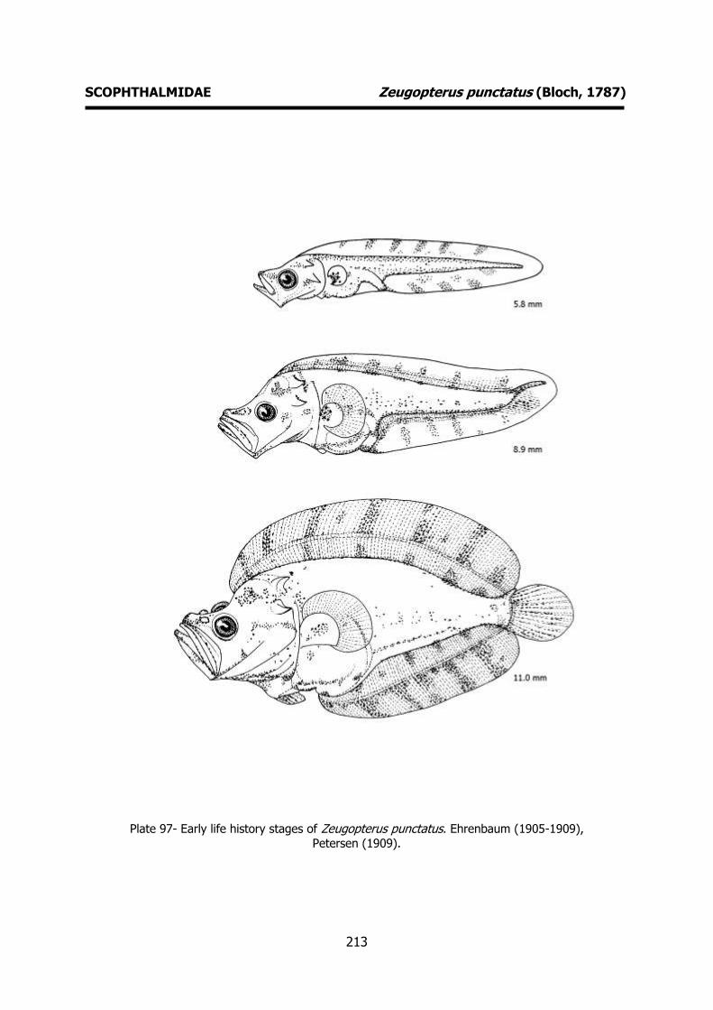
213
SCOPHTHALMIDAE Zeugopterus punctatus (Bloch, 1787)
Plate 97- Early life history stages of Zeugopterus punctatus. Ehrenbaum (1905-1909),
Petersen (1909).
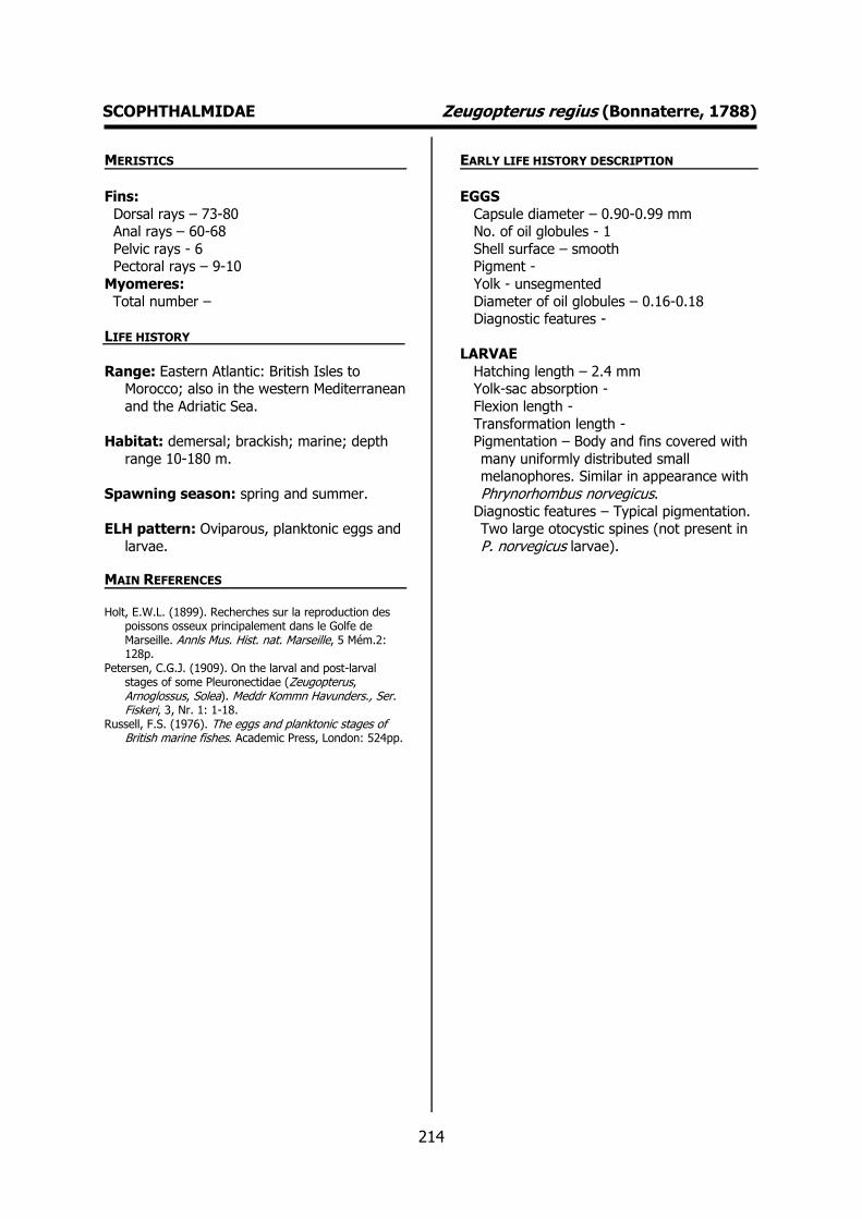
214
SCOPHTHALMIDAE Zeugopterus regius (Bonnaterre, 1788) MERISTICS
Fins:
Dorsal rays – 73-80 Anal rays – 60-68
Pelvic rays - 6 Pectoral rays – 9-10
Myomeres:
Total number –
LIFE HISTORY
Range: Eastern Atlantic: British Isles to Morocco; also in the western Mediterranean
and the Adriatic Sea.
Habitat: demersal; brackish; marine; depth
range 10-180 m.
Spawning season: spring and summer.
ELH pattern: Oviparous, planktonic eggs and
larvae.
MAIN REFERENCES Holt, E.W.L. (1899). Recherches sur la reproduction des
poissons osseux principalement dans le Golfe de Marseille. Annls Mus. Hist. nat. Marseille, 5 Mém.2: 128p.
Petersen, C.G.J. (1909). On the larval and post-larval stages of some Pleuronectidae (Zeugopterus, Arnoglossus, Solea). Meddr Kommn Havunders., Ser. Fiskeri, 3, Nr. 1: 1-18.
Russell, F.S. (1976). The eggs and planktonic stages of British marine fishes. Academic Press, London: 524pp.
EARLY LIFE HISTORY DESCRIPTION
EGGS
Capsule diameter – 0.90-0.99 mm No. of oil globules - 1
Shell surface – smooth Pigment -
Yolk - unsegmented
Diameter of oil globules – 0.16-0.18 Diagnostic features -
LARVAE
Hatching length – 2.4 mm Yolk-sac absorption -
Flexion length -
Transformation length - Pigmentation – Body and fins covered with
many uniformly distributed small melanophores. Similar in appearance with
Phrynorhombus norvegicus. Diagnostic features – Typical pigmentation.
Two large otocystic spines (not present in
P. norvegicus larvae).

215
SCOPHTHALMIDAE Zeugopterus regius (Bonnaterre, 1788)
Plate 96- Early life history stages of Phrynorhombus regius. Petersen (1909).

216
BOTHIDAE Arnoglossus laterna (Walbaum, 1792) MERISTICS
Fins:
Dorsal rays – 87-93 Anal rays – 65-74
Pelvic rays - 6 Pectoral rays – 11-12
Myomeres:
Total number –
LIFE HISTORY
Range: Eastern Atlantic: Norway to Angola. Also known from the Mediterranean and
Black Sea.
Habitat: demersal; marine; depth range 10-
200 m.
Spawning season: spawns throughout the
whole year.
ELH pattern: Oviparous, planktonic eggs and larvae.
MAIN REFERENCES Ehrenbaum, E. (1905-1909). Eier und Larven von Fischen.
Nordisches Plankton, 1: 413pp. Holt, E.W.L. (1899). Recherches sur la reproduction des
poissons osseux principalement dans le Golfe de Marseille. Annls Mus. Hist. nat. Marseille, 5 Mém.2: 128p.
Kyle, H.M. (1913). Flat-fishes (Heterosomata). Rep. Dan. Oceanogr. Exped., 1908-1910. Vol. II, Biology, A.1: 150pp.
Munk, P., J.G. Nielsen (2005). Eggs and larvae of North Sea fishes. Biofolia, Denmark: 215pp.
Padoa, E. (1933-1956). Heterosomata. Uova, larve e stadi Giovanili di Teleostei: monografia elaborata con l’uso del materiale raccolto e sweriato da Salvatore Lo Bianco. Fauna e Flora Golfo Napoli Monogr.38: 783-877.
Russell, F.S. (1976). The eggs and planktonic stages of British marine fishes. Academic Press, London: 524pp.
EARLY LIFE HISTORY DESCRIPTION
EGGS
Capsule diameter – 0.60-0.76 mm No. of oil globules - 1
Shell surface – smooth, spherical Pigment -
Yolk -
Diameter of oil globules – 0.11-0.15 mm Diagnostic features – small dimensions.
LARVAE
Hatching length – 2.5 mm Yolk-sac absorption – 2.9 mm
Flexion length -
Transformation length - Pigmentation - Few melanophores in dorsal
contour. Evenly spaced small melanophores in ventral row. Some
melanophores in the otocystic region.
Head and abdomen heavily pigmented. At 5 mm the gas blabber is apparent and
there is a large tentacle on the head. Pigmentation can vary (pale form with little
or no pigment, and well pigmented form). Diagnostic features – Shape of body. Large
tentacle on head. Spines on head near the
base of tentacle (5-6).
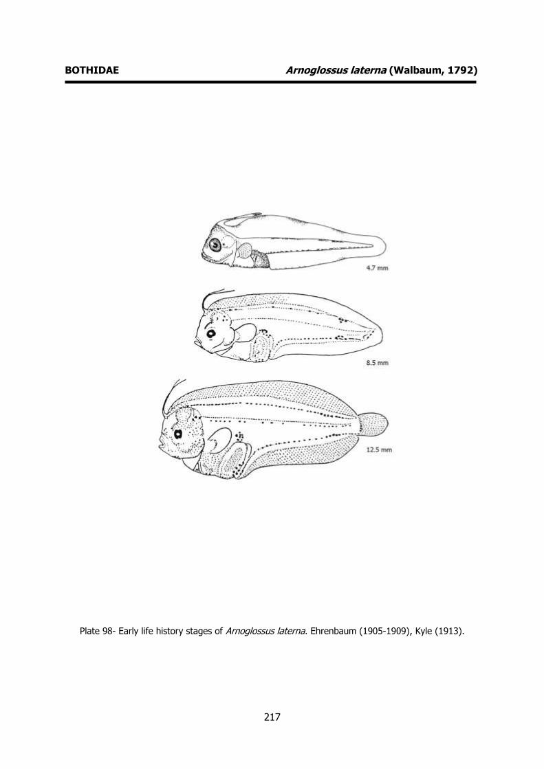
217
BOTHIDAE Arnoglossus laterna (Walbaum, 1792)
Plate 98- Early life history stages of Arnoglossus laterna. Ehrenbaum (1905-1909), Kyle (1913).
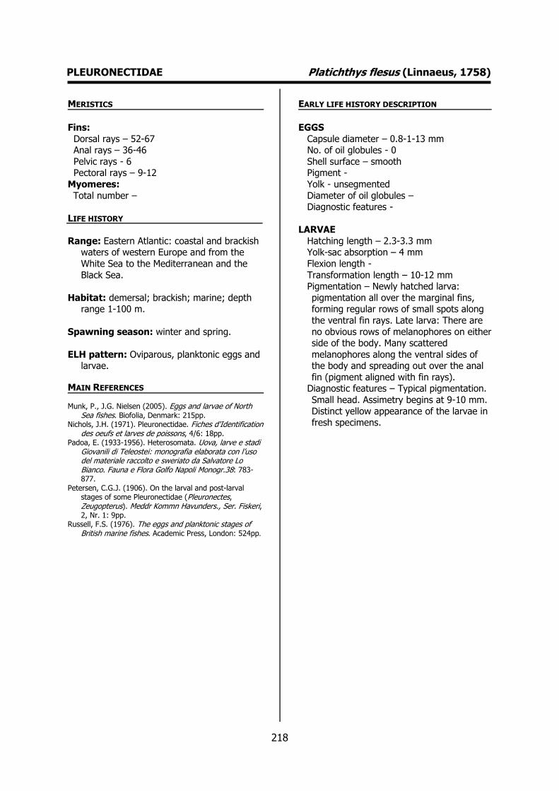
218
PLEURONECTIDAE Platichthys flesus (Linnaeus, 1758) MERISTICS
Fins:
Dorsal rays – 52-67 Anal rays – 36-46
Pelvic rays - 6 Pectoral rays – 9-12
Myomeres:
Total number –
LIFE HISTORY
Range: Eastern Atlantic: coastal and brackish waters of western Europe and from the
White Sea to the Mediterranean and the
Black Sea.
Habitat: demersal; brackish; marine; depth range 1-100 m.
Spawning season: winter and spring.
ELH pattern: Oviparous, planktonic eggs and larvae.
MAIN REFERENCES
Munk, P., J.G. Nielsen (2005). Eggs and larvae of North Sea fishes. Biofolia, Denmark: 215pp.
Nichols, J.H. (1971). Pleuronectidae. Fiches d'Identification des oeufs et larves de poissons, 4/6: 18pp.
Padoa, E. (1933-1956). Heterosomata. Uova, larve e stadi Giovanili di Teleostei: monografia elaborata con l’uso del materiale raccolto e sweriato da Salvatore Lo Bianco. Fauna e Flora Golfo Napoli Monogr.38: 783-877.
Petersen, C.G.J. (1906). On the larval and post-larval stages of some Pleuronectidae (Pleuronectes, Zeugopterus). Meddr Kommn Havunders., Ser. Fiskeri, 2, Nr. 1: 9pp.
Russell, F.S. (1976). The eggs and planktonic stages of British marine fishes. Academic Press, London: 524pp.
EARLY LIFE HISTORY DESCRIPTION
EGGS
Capsule diameter – 0.8-1-13 mm No. of oil globules - 0
Shell surface – smooth Pigment -
Yolk - unsegmented
Diameter of oil globules – Diagnostic features -
LARVAE
Hatching length – 2.3-3.3 mm Yolk-sac absorption – 4 mm
Flexion length -
Transformation length – 10-12 mm Pigmentation – Newly hatched larva:
pigmentation all over the marginal fins, forming regular rows of small spots along
the ventral fin rays. Late larva: There are
no obvious rows of melanophores on either side of the body. Many scattered
melanophores along the ventral sides of the body and spreading out over the anal
fin (pigment aligned with fin rays). Diagnostic features – Typical pigmentation.
Small head. Assimetry begins at 9-10 mm.
Distinct yellow appearance of the larvae in fresh specimens.
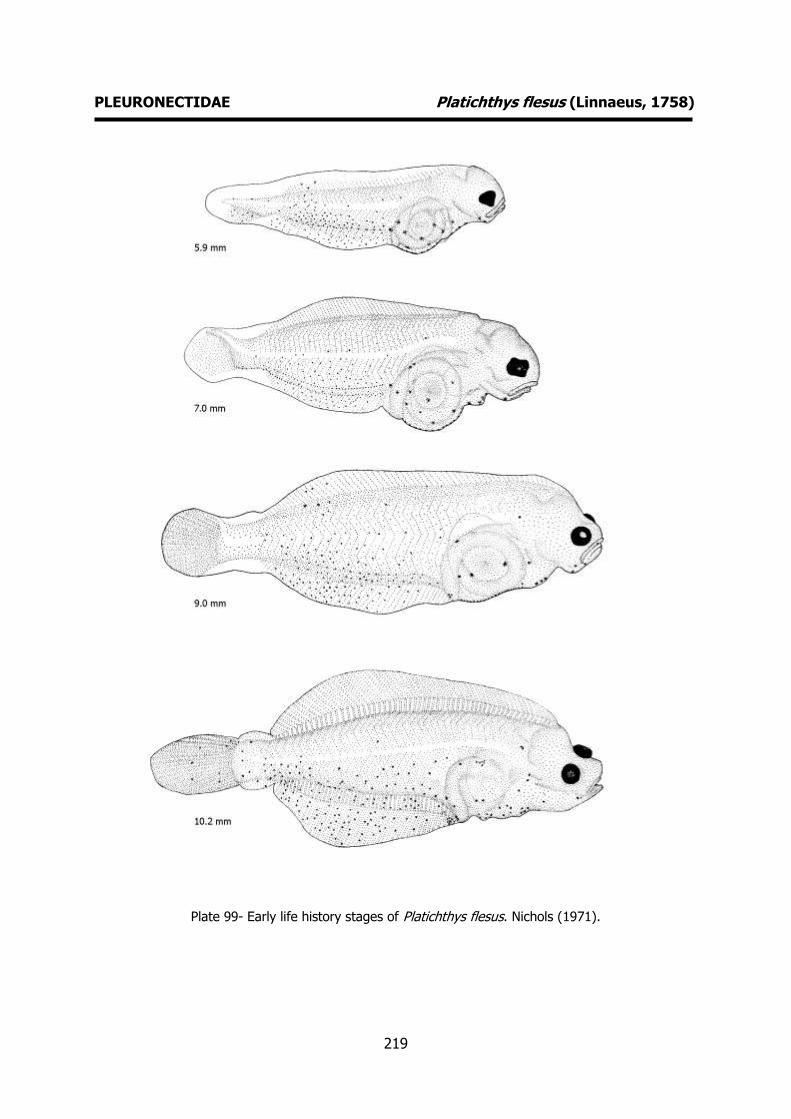
219
PLEURONECTIDAE Platichthys flesus (Linnaeus, 1758)
Plate 99- Early life history stages of Platichthys flesus. Nichols (1971).
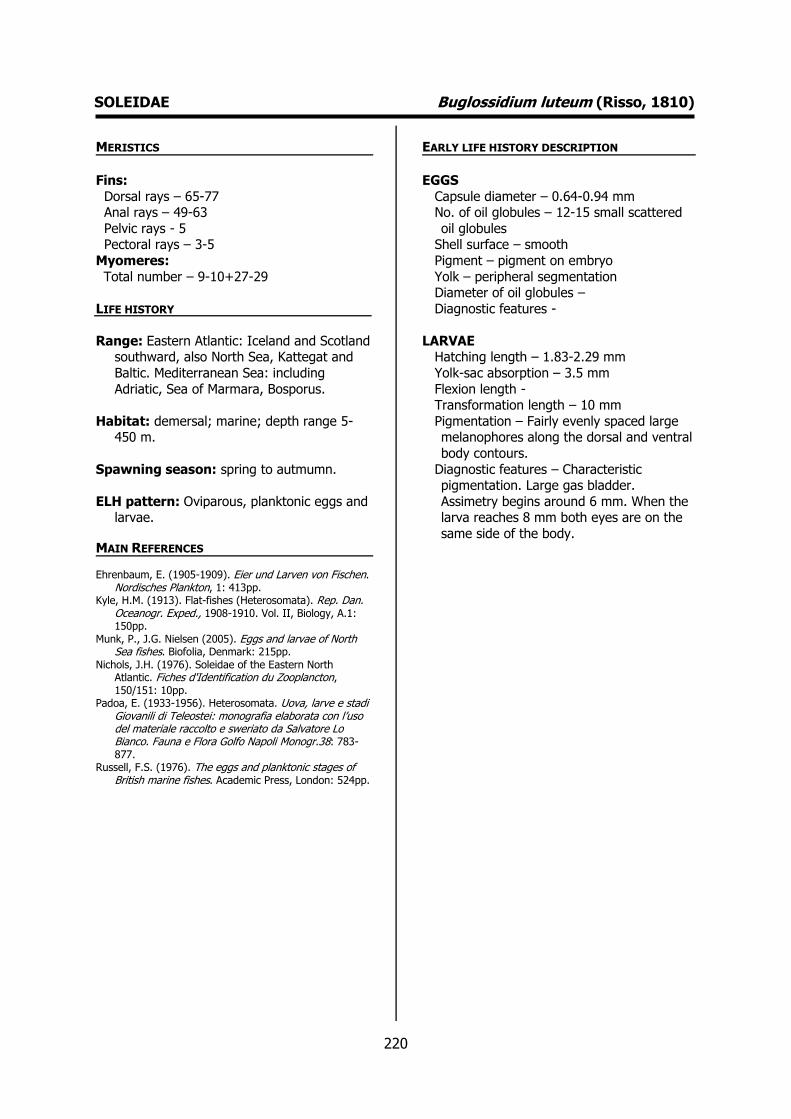
220
SOLEIDAE Buglossidium luteum (Risso, 1810) MERISTICS
Fins:
Dorsal rays – 65-77 Anal rays – 49-63
Pelvic rays - 5 Pectoral rays – 3-5
Myomeres:
Total number – 9-10+27-29
LIFE HISTORY
Range: Eastern Atlantic: Iceland and Scotland southward, also North Sea, Kattegat and
Baltic. Mediterranean Sea: including
Adriatic, Sea of Marmara, Bosporus.
Habitat: demersal; marine; depth range 5-450 m.
Spawning season: spring to autmumn.
ELH pattern: Oviparous, planktonic eggs and larvae.
MAIN REFERENCES
Ehrenbaum, E. (1905-1909). Eier und Larven von Fischen. Nordisches Plankton, 1: 413pp.
Kyle, H.M. (1913). Flat-fishes (Heterosomata). Rep. Dan. Oceanogr. Exped., 1908-1910. Vol. II, Biology, A.1: 150pp.
Munk, P., J.G. Nielsen (2005). Eggs and larvae of North Sea fishes. Biofolia, Denmark: 215pp.
Nichols, J.H. (1976). Soleidae of the Eastern North Atlantic. Fiches d'Identification du Zooplancton, 150/151: 10pp.
Padoa, E. (1933-1956). Heterosomata. Uova, larve e stadi Giovanili di Teleostei: monografia elaborata con l’uso del materiale raccolto e sweriato da Salvatore Lo Bianco. Fauna e Flora Golfo Napoli Monogr.38: 783-877.
Russell, F.S. (1976). The eggs and planktonic stages of British marine fishes. Academic Press, London: 524pp.
EARLY LIFE HISTORY DESCRIPTION
EGGS
Capsule diameter – 0.64-0.94 mm No. of oil globules – 12-15 small scattered
oil globules Shell surface – smooth
Pigment – pigment on embryo
Yolk – peripheral segmentation Diameter of oil globules –
Diagnostic features -
LARVAE Hatching length – 1.83-2.29 mm
Yolk-sac absorption – 3.5 mm
Flexion length - Transformation length – 10 mm
Pigmentation – Fairly evenly spaced large melanophores along the dorsal and ventral
body contours.
Diagnostic features – Characteristic pigmentation. Large gas bladder.
Assimetry begins around 6 mm. When the larva reaches 8 mm both eyes are on the
same side of the body.
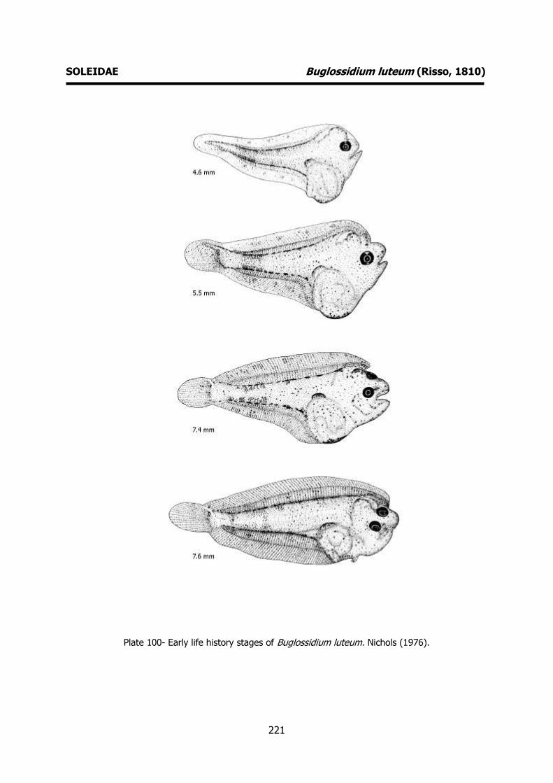
221
SOLEIDAE Buglossidium luteum (Risso, 1810)
Plate 100- Early life history stages of Buglossidium luteum. Nichols (1976).
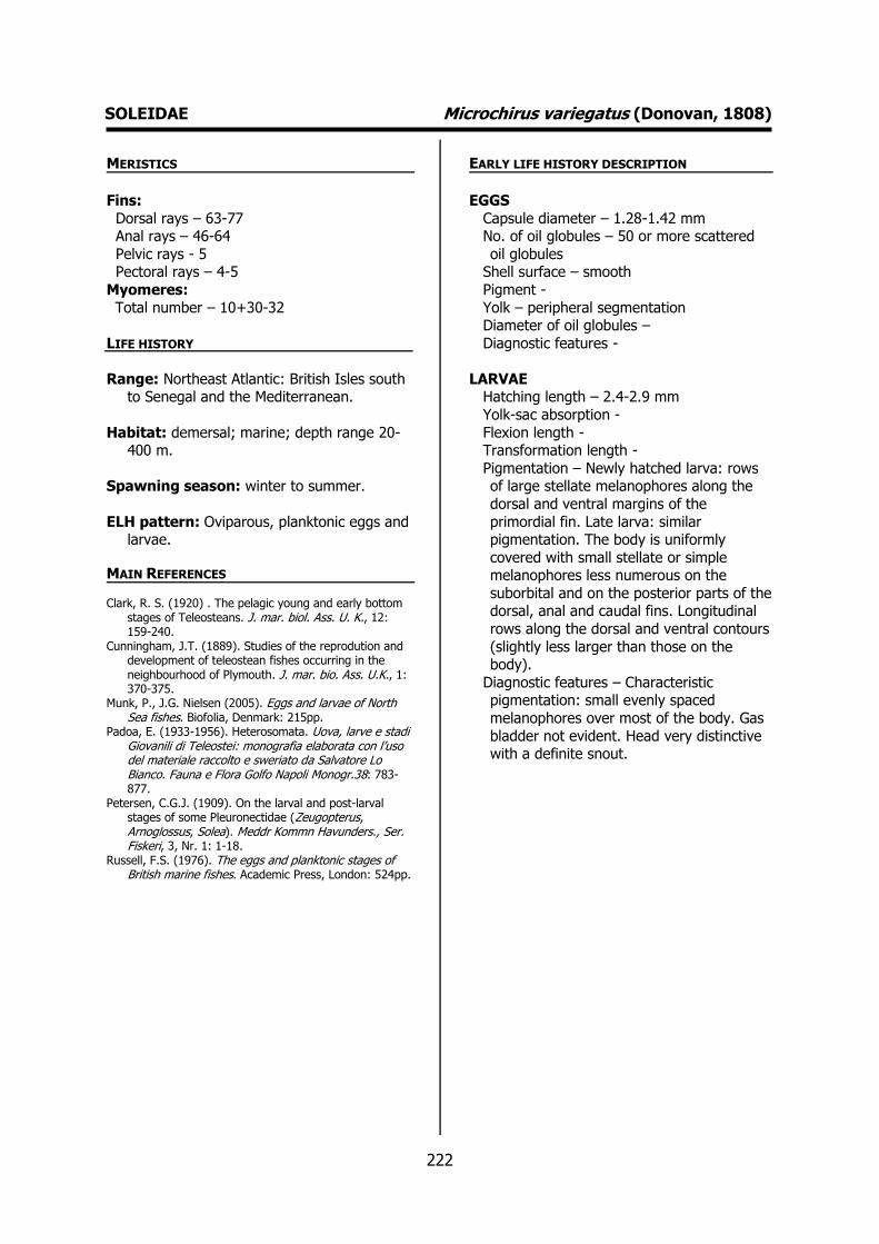
222
SOLEIDAE Microchirus variegatus (Donovan, 1808) MERISTICS
Fins:
Dorsal rays – 63-77 Anal rays – 46-64
Pelvic rays - 5 Pectoral rays – 4-5
Myomeres:
Total number – 10+30-32
LIFE HISTORY
Range: Northeast Atlantic: British Isles south to Senegal and the Mediterranean.
Habitat: demersal; marine; depth range 20-400 m.
Spawning season: winter to summer.
ELH pattern: Oviparous, planktonic eggs and larvae.
MAIN REFERENCES
Clark, R. S. (1920) . The pelagic young and early bottom stages of Teleosteans. J. mar. biol. Ass. U. K., 12: 159-240.
Cunningham, J.T. (1889). Studies of the reprodution and development of teleostean fishes occurring in the neighbourhood of Plymouth. J. mar. bio. Ass. U.K., 1: 370-375.
Munk, P., J.G. Nielsen (2005). Eggs and larvae of North Sea fishes. Biofolia, Denmark: 215pp.
Padoa, E. (1933-1956). Heterosomata. Uova, larve e stadi Giovanili di Teleostei: monografia elaborata con l’uso del materiale raccolto e sweriato da Salvatore Lo Bianco. Fauna e Flora Golfo Napoli Monogr.38: 783-877.
Petersen, C.G.J. (1909). On the larval and post-larval stages of some Pleuronectidae (Zeugopterus, Arnoglossus, Solea). Meddr Kommn Havunders., Ser. Fiskeri, 3, Nr. 1: 1-18.
Russell, F.S. (1976). The eggs and planktonic stages of British marine fishes. Academic Press, London: 524pp.
EARLY LIFE HISTORY DESCRIPTION
EGGS
Capsule diameter – 1.28-1.42 mm No. of oil globules – 50 or more scattered
oil globules Shell surface – smooth
Pigment -
Yolk – peripheral segmentation Diameter of oil globules –
Diagnostic features -
LARVAE Hatching length – 2.4-2.9 mm
Yolk-sac absorption -
Flexion length - Transformation length -
Pigmentation – Newly hatched larva: rows of large stellate melanophores along the
dorsal and ventral margins of the
primordial fin. Late larva: similar pigmentation. The body is uniformly
covered with small stellate or simple melanophores less numerous on the
suborbital and on the posterior parts of the dorsal, anal and caudal fins. Longitudinal
rows along the dorsal and ventral contours
(slightly less larger than those on the body).
Diagnostic features – Characteristic pigmentation: small evenly spaced
melanophores over most of the body. Gas
bladder not evident. Head very distinctive with a definite snout.

223
SOLEIDAE Microchirus variegatus (Donovan, 1808)
Plate 101- Early life history stages of Microchirus variegatus. Nichols (1976).

224
SOLEIDAE Pegusa lascaris (Risso, 1810) MERISTICS
Fins:
Dorsal rays – 71-90 Anal rays – 55-75
Pelvic rays - 5 Pectoral rays – 7-10
Myomeres:
Total number – 9-10+37-38
LIFE HISTORY
Range: Eastern Atlantic: northeastern to southeastern Atlantic; Gulf of Guinea; the
Mediterranean Sea and Black Sea.
Habitat: demersal; brackish; marine; depth
range 5-350 m.
Spawning season:
ELH pattern: Oviparous, planktonic eggs and
larvae.
MAIN REFERENCES Ehrenbaum, E. (1905-1909). Eier und Larven von Fischen.
Nordisches Plankton, 1: 413pp. Holt, E.W.L. (1899) - Recherches sur la reproduction des
poissons osseux principalement dans le Golfe de Marseille. Annls Mus. Hist. nat. Marseille, 5 Mém.2: 128p.
Kyle, H.M. (1913). Flat-fishes (Heterosomata). Rep. Dan. Oceanogr. Exped., 1908-1910. Vol. II, Biology, A.1: 150pp.
Padoa, E. (1933-1956). Heterosomata. Uova, larve e stadi Giovanili di Teleostei: monografia elaborata con l’uso del materiale raccolto e sweriato da Salvatore Lo Bianco. Fauna e Flora Golfo Napoli Monogr.38: 783-877.
Russell, F.S. (1976). The eggs and planktonic stages of British marine fishes. Academic Press, London: 524pp.
EARLY LIFE HISTORY DESCRIPTION
EGGS
Capsule diameter – 1.28-1.38 mm No. of oil globules – 50 or more scattered
oil globules Shell surface – smooth
Pigment -
Yolk – peripheral segmentation Diameter of oil globules –
Diagnostic features -
LARVAE Hatching length – 3.5 mm
Yolk-sac absorption -
Flexion length - Transformation length -
Pigmentation – Distinct pigmentation pattern. Large finely branched
melanophores on the head, trunk and
abdomen (largest on the gut region). Aggregations of pigment on the dorsal and
anal fin membranes towards the posterior end of the body forming a distinct anal
bar. As development proceeds the pigmentation becomes more pronounced.
Pigmented gas bladder.
Diagnostic features – Abnormal large anterior prolongation of the dorsal
primordial fin during first stages of development. Pigmentation pattern.
Pigmented gas bladder.
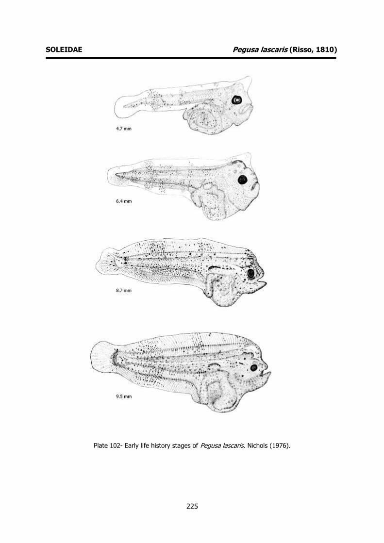
225
SOLEIDAE Pegusa lascaris (Risso, 1810)
Plate 102- Early life history stages of Pegusa lascaris. Nichols (1976).

226
SOLEIDAE Solea senegalensis Kaup, 1858 MERISTICS
Fins:
Dorsal rays – 75-95 Anal rays – 61-75
Pelvic rays - 5 Pectoral rays – 8-10
Myomeres:
Total number – 9+36-37
LIFE HISTORY
Range: Eastern Atlantic: North to southern Atlantic.
Habitat: demersal; brackish; marine; depth range 12–65 m.
Spawning season: spawns throughout the
whole year.
ELH pattern: Oviparous, planktonic eggs and
larvae.
MAIN REFERENCES Dinis, M.T. (1986). Quatre Soleidae de l’estuaire du Tage.
Reproduction et croissance. Essai d’elevage de Solea senegalenesis Kaup. Tese, Université de Bretagne Occidentale: 348 pp.
Ré, P. (1999). Ictioplâncton estuarino da Península Ibérica (Guia de identificação dos ovos e estados larvares planctónicos), 163pp, 51 fig. Prémio do Mar, 1996. Câmara Municipal de Cascais. ISBN 972-637-065-5.
EARLY LIFE HISTORY DESCRIPTION
EGGS: Undescribed
Capsule diameter – Unknown No. of oil globules - Unknown
Shell surface – Unknown Pigment - Unknown
Yolk - Unknown
Diameter of oil globules – Unknown Diagnostic features - Unknown
LARVAE
Hatching length - Unknown Yolk-sac absorption -
Flexion length -
Transformation length - Pigmentation – Newly hatched larva: heavy
pigmentation on the primordial fin, over the body and yolk sac. Late larva: similar
pigmentation pattern. Bands of pigment on
the dorsal and anal fins. Body heavily pigmented.
Diagnostic features – Protruding otocystic capsules. Overall pigmentation.

227
SOLEIDAE Solea senegalensis Kaup, 1858
Plate 103- Early life history stages of Solea senegalensis. Dinis (1986).

228
SOLEIDAE Solea solea (Linnaeus, 1758) MERISTICS
Fins:
Dorsal rays – 69-97 Anal rays – 53-79
Pelvic rays – 5-6 Pectoral rays – 7-10
Myomeres:
Total number – 9-10+40-42
LIFE HISTORY
Range: Eastern Atlantic: southward from Trondheim Fjord (including North Sea and
western Baltic) and Mediterranean Sea
(including Sea of Marmara, Bosporus and southwestern Black Sea).
Habitat: demersal; brackish; marine; depth
range 0-150 m.
Spawning season:
ELH pattern: Oviparous, planktonic eggs and
larvae.
MAIN REFERENCES Cunningham, J.T. (1889). Studies of the reprodution and
development of teleostean fishes occurring in the neighbourhood of Plymouth. J. mar. bio. Ass. U.K., 1: 370-375.
Ehrenbaum, E. (1905-1909). Eier und Larven von Fischen. Nordisches Plankton, 1: 413pp.
Munk, P., J.G. Nielsen (2005). Eggs and larvae of North Sea fishes. Biofolia, Denmark: 215pp.
Padoa, E. (1933-1956). Heterosomata. Uova, larve e stadi Giovanili di Teleostei: monografia elaborata con l’uso del materiale raccolto e sweriato da Salvatore Lo Bianco. Fauna e Flora Golfo Napoli Monogr.38: 783-877.
Russell, F.S. (1976). The eggs and planktonic stages of British marine fishes. Academic Press, London: 524pp.
EARLY LIFE HISTORY DESCRIPTION
EGGS
Capsule diameter – 1.0-1.6 mm No. of oil globules – many small aggregated
oil globules Shell surface – smooth
Pigment -
Yolk – peripheral segmentation Diameter of oil globules –
Diagnostic features -
LARVAE Hatching length – 2.5-3.75 mm
Yolk-sac absorption -
Flexion length - Transformation length -
Pigmentation – Newly hatched larva: heavy pigmentation. Rows of melanophores over
the body, yolk and primordial fin forming a
complete network, except for the extreme caudal end of the tail. Late larva: same
overall pigmentation pattern. The number of melanophores increase with
development, eventually covering the whole body.
Diagnostic features – Characteristic
pigmentation. In early larvae the primordial fin have a hood-like projection
over the head. Gas bladder not clearly visible. Pigment not in the form of bands,
but evenly spread over the whole larva.

229
SOLEIDAE Solea solea (Linnaeus, 1758)
Plate 104- Early life history stages of Solea solea. Nichols (1976).
230
GOBIESOCIDAE Diplecogaster bimaculata (Bonnaterre, 1788) MERISTICS
Fins:
Dorsal rays – 4-7 Anal rays – 4-6
Pelvic rays – I+4 Pectoral rays – 18-26
Myomeres:
Total number – 30-31
LIFE HISTORY
Range: Eastern Atlantic: Norway and the Faroes to Gibraltar including western
Mediterranean and Adriatic Sea.
Habitat: demersal; marine; depth range 18-
36 m.
Spawning season: spring and summer.
ELH pattern: Oviparous, demersal eggs and
planktonic larvae.
MAIN REFERENCES Guitel, F. (1988). Recherches sur les Lepadogasters. Arch.
Zool. exp. gén., (Ser. 2), 6: 423-647. Hefford, A. E. (1910). Notes on teleostean ova and larvae
observed at Plymouth in spring and summer. J. Mar. Biol. Ass. U.K., 9: 2-58.
Padoa, E. (1933-1956). Gobiesocidae. Uova, larve e stadi Giovanili di Teleostei: monografia elaborata con l’uso del materiale raccolto e sweriato da Salvatore Lo Bianco. Fauna e Flora Golfo Napoli Monogr.38: 774-782.
Russell, F.S. (1976). The eggs and planktonic stages of British marine fishes. Academic Press, London: 524pp.
EARLY LIFE HISTORY DESCRIPTION
EGGS
Capsule diameter – 1.37-1.54 mm No. of oil globules - 1
Shell surface – smooth, flattened, oval Pigment – Yellow oil globule, colourless yolk
Yolk - unsegmented
Diameter of oil globules – 0.24-0.28 mm Diagnostic features -
LARVAE
Hatching length – 3-4.3 mm Yolk-sac absorption – 4.8 mm
Flexion length -
Transformation length - Pigmentation - At 5.5 mm, pigment begins
to develop on top of the head. In specimens 7.5-10 mm long, the
melanophores arranged in four more or
less regular longitudinal rows on the body. Diagnostic features - About 4.8-4.9 mm
long (soon after the yolk has been reabsorbed), early larvae similar to the
newly hatched larva. At 7.0 mm, the head is noticeably flattened dorsoventrally.
Sucker appears at 8 mm and is well
developed at 10 mm length. Late larvae can be easily distinguished from L. lepadogaster (wide unpigmented dorsal area and different pigmentation of the anal
fin).

231
GOBIESOCIDAE Diplecogaster bimaculata (Bonnaterre, 1788)
Plate 105- Early life history stages of Diplecogaster bimaculata. Russell (1976)

232
GOBIESOCIDAE Lepadogaster lepadogaster (Bonnaterre, 1788) MERISTICS
Fins:
Dorsal rays – 15-20 Anal rays – 9-12
Pelvic rays – I+4 Pectoral rays – 20-22
Myomeres:
Total number –
LIFE HISTORY
Range: Eastern Atlantic: eastern and central Mediterranean westward to Monaco,
possibly in the Black Sea.
Habitat: demersal; marine.
Spawning season: summer.
ELH pattern: Oviparous, demersal eggs and planktonic larvae.
MAIN REFERENCES Guitel, F. (1988). Recherches sur les Lepadogasters. Arch.
Zool. exp. gén., (Ser. 2), 6: 423-647. Padoa, E. (1933-1956). Gobiesocidae. Uova, larve e stadi
Giovanili di Teleostei: monografia elaborata con l’uso del materiale raccolto e sweriato da Salvatore Lo Bianco. Fauna e Flora Golfo Napoli Monogr.38: 774-782.
Russell, F.S. (1976). The eggs and planktonic stages of British marine fishes. Academic Press, London: 524pp.
EARLY LIFE HISTORY DESCRIPTION
EGGS
Capsule diameter – 1.85-1.90 mm No. of oil globules - 1
Shell surface – smooth, flattened, oval Pigment –
Yolk - unsegmented
Diameter of oil globules – 0.36-0.28 mm Diagnostic features -
LARVAE Hatching length – 5 mm
Yolk-sac absorption -
Flexion length - Transformation length -
Pigmentation – Larva heavily pigmented. The top of the head and the sides of the
body are intensively pigmented.
Melanophores arranged regularly along longitudinal rows. Body pigmentation
continuous down to the ventral side of the abdomen. Snout and operculum appear to
be without melanophores. Tail and pectoral fins without pigment. Row of
melanophores on the base of the anal fin.
Diagnostic features – Characteristic pigmentation. Gas bladder invisible due to
the heavy pigmention.
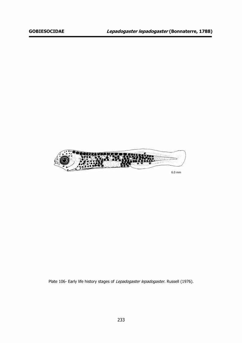
233
GOBIESOCIDAE Lepadogaster lepadogaster (Bonnaterre, 1788)
Plate 106- Early life history stages of Lepadogaster lepadogaster. Russell (1976).

234
LOPHIIDAE Lophius piscatorius Linnaeus, 1758 MERISTICS
Fins:
Dorsal rays – D1 II+I+III D2 10-12 Anal rays – 10-11
Pelvic rays – I+5 Pectoral rays – 23-28
Myomeres:
Total number –
LIFE HISTORY
Range: Eastern Atlantic: south-western Barents Sea to Strait of Gibraltar including
the Mediterranean and Black Sea. Reported
from Iceland and Mauritania. North Atlantic specimens attain larger sizes than those
collected off West Africa and they also occur in shallower depths.
Habitat: demersal; marine; depth range 20-1000 m.
Spawning season: spring.
ELH pattern: Oviparous, planktonic eggs and larvae.
MAIN REFERENCES Padoa, E. (1933-1956). Lophiidae. Uova, larve e stadi
Giovanili di Teleostei: monografia elaborata con l’uso del materiale raccolto e sweriato da Salvatore Lo Bianco. Fauna e Flora Golfo Napoli Monogr.38: 878-888.
Russell, F.S. (1976). The eggs and planktonic stages of British marine fishes. Academic Press, London: 524pp.
Tåning, Å. V. (1923). Lophius. Rep. Dan. Oceanogr. Exped . 1908-10 Medit . adjac. Seas, 2, Biology A10: 1-30.
EARLY LIFE HISTORY DESCRIPTION
EGGS
Capsule diameter – 2.3-3.1 mm No. of oil globules – one or more (up to
nine) Shell surface – ovoid in jelly matrix
Pigment – yellow, orange, amber
Yolk - unsegmented Diameter of oil globules – 0.51-0.88 mm
Diagnostic features – Eggs embedded in a gelatinous ribbon ca. 11 m long and 30 cm
wide.
LARVAE
Hatching length – 4.5 mm Yolk-sac absorption – 6.5 mm
Flexion length - Transformation length -
Pigmentation – Pigmented eyes before
hatching. Pigmentation does not seem to change during development. Black
pigmentation over the head and shoulder region (large ramified melanophores).
Melanophores are also present along the gut. Late larva: two bars of melanophores,
one situated behind the anus and the
other near the tail end. Diagnostic features – Characteristic shape.
Newly hatched larva: hump-like projection into the dorsal primordial fin corresponding
to the third ray of the fully-formed fin.
Pelvic fins are very elongated.
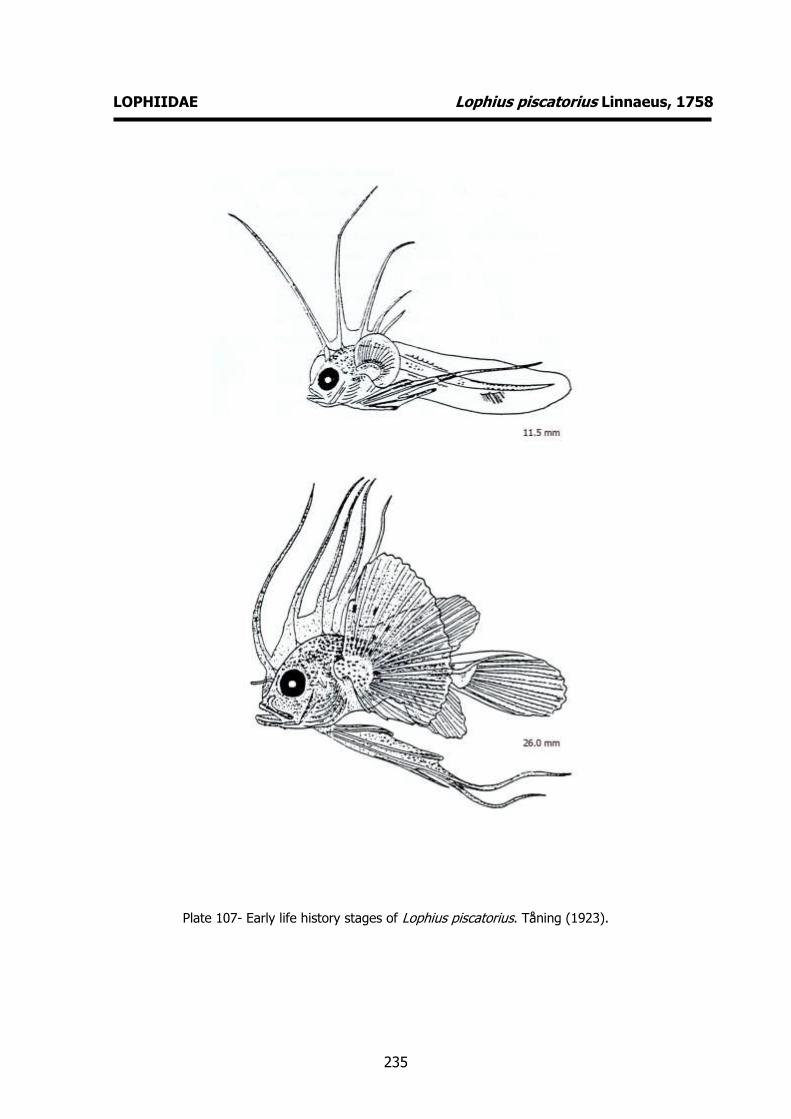
235
LOPHIIDAE Lophius piscatorius Linnaeus, 1758
Plate 107- Early life history stages of Lophius piscatorius. Tåning (1923).
236
REFERENCES Aboussouan, A. (1994). Intérêt des formulas vertébrales pour l’identification des poissons de la
mer Méditerranée. Cybium, 18 (2) : 177-197.
Ahlstrom, E. H., H. G. Moser (1980). Characters useful in identification of pelagic marine fish eggs. Calif. Coop. Oceanic Fish. Invest. Rep., 21: 121-131.
Ahlstrom, E. H., H. G. Moser (1981). Eggs and larvae of fishes and their role in systematic investigations and fisheries. Rev. Trav. Inst. Pêches mariti., 40 (3 et 4): 379-398.
Ahlstrom, E. H., H. G. Moser (1981). Systematics and development of early life history stages of
marine fishes: achivements during the past century, present status and suggestions for the future. Rapp. P.-v. Réun. Cons. int. Explor. Mer, 178: 541-547.
Bakun, A. (1996). Patterns in the Ocean: Ocean Processes and Marine Population Dynamics. La Jolla: Californa Sea Grant College System.
Barnabé, G., F. Boulineau-Coatanea, F. Rene (1976). Chronologie de la morphogenese chez le
loup ou bar Dicentrarchus labrax (L.) (Pisces, Serranidae) obtenu par reproduction
artificielle. Aquaculture, 8: 351-363.
Berrien, P.L. (1975). A description of Atlantic mackerel Scomber scombrus, eggs and early larvae. Fish. Bull. U.S., 73 (1): 186-192.
Berrien, P.L. (1978). Eggs and larvae of Scomber scombrus and Scomber japonicus in continental shelf waters between Massachusets and Florida. Fish. Bull. U.S., 76: 95-115.
Bertelson, E. (1951). The ceratioid fishes. Dana report, 39: 276pp.
Bertolini, F. (1933). Serranidae. Uova, larve e stadi Giovanili di Teleostei: monografia elaborata
con l’uso del materiale raccolto e sweriato da Salvatore Lo Bianco. Fauna e Flora Golfo Napoli Monogr.38. Publi. In 4 parts: 310-321.
Bertolini, F., U. D'Ancona, E. Padoa, G. Montalenti, S. Ranzi, L. Sanzo, A. Sparta, E. Tortonese, M. Vialli (1931-1956). Uova, larve e stadi Giovanili di Teleostei: monografia elaborata con l’uso del materiale raccolto e sweriato da Salvatore Lo Bianco. Fauna e Flora Golfo Napoli Monogr.38. Publi. In 4 parts: 1064pp, 51 pls.
Boëtius, J. (1976). Elvers, Anguilla anguilla and Anguilla rostrata from two Danish localities. Size, body weight, developmental stage and number of vertebrae related to time of ascent.
Meddr Danm. Fisk.- og Havunders., NS: 199-220.
Borges, R., C. Faria, F. Gil, E.J. Gonçalves, V.C. Almada (2003). Embryonic and larval
development of Gobius paganellus (Pisces: Gobiidae). J. Mar. Biol. Ass. U.K., 83: 1151-1156.
Brownell, C.L. (1979). Stages in the early development of 40 marine fish species with pelagic eggs from the cape of Good Hope. Ichthyological Bulletin of the J.L.B. Smith Institute of Ichthyology, Rhodes University, Grahamstown, (40): 84pp.
Cameron, J. (1959). The larval and post-larval stages of Gymnammodytes semisquamatus
(Jourdain). J. Mar. Biol. Ass. U.K., 38: 17-25.
Camus, P., L. Besseau (1986). Sparidae, Spodyliosoma cantharus. Fich. Ident. Plankton, 177: 4
pp.

237
Castle, P.H.J. (1969). An index and bibliography of eel larvae. J.L.B. Smith Inst. Of Ichthyology Spec. Publ., No. 7: 121pp.
Clark, R. S. (1920). The pelagic young and early bottom stages of Teleosteans. J. mar. biol.
Ass. U. K., 12: 159-240.
Cunningham, J.T. (1889). Studies of the reprodution and development of teleostean fishes
occurring in the neighbourhood of Plymouth. J. mar. bio. Ass. U.K., 1: 370-375.
Cushing, D.H. (1975). Marine Ecology and Ficheries. Cambridge University Press.
D’Ancona, U. (1931-1933). Apodes (Muraenoidei). Uova, larve e stadi Giovanili di Teleostei: monografia elaborata con l’uso del materiale raccolto e sweriato da Salvatore Lo Bianco. Fauna e Flora Golfo Napoli Monogr.38: 84-146.
D’Ancona, U. (1931-1933). Gadidae. Uova, larve e stadi Giovanili di Teleostei: monografia
elaborata con l’uso del materiale raccolto e sweriato da Salvatore Lo Bianco. Fauna e Flora Golfo Napoli Monogr.38: 178-255.
D’Ancona, U. (1931-1933). Syngnathidae. Uova, larve e stadi Giovanili di Teleostei: monografia elaborata con l’uso del materiale raccolto e sweriato da Salvatore Lo Bianco. Fauna e Flora Golfo Napoli Monogr.38: 281-289.
D’Ancona, U. (1931-1933). Macrorhamphosidae. Uova, larve e stadi Giovanili di Teleostei: monografia elaborata con l’uso del materiale raccolto e sweriato da Salvatore Lo Bianco. Fauna e Flora Golfo Napoli Monogr.38: 299-306.
Demir, N. (1972). The abundance and distribution of the eggs and larvae of some teleost fishes
off Plymouth in 1969 and 1970. II. The postlarvae of Callionymus. J. Mar. Biol. Ass. U.K., 52: 997-1010.
Demir, N. (1976). Callionymidae of the Northeastern Atlantic. Fiches d'Identification du Zooplancton, 148: 5pp.
Demir, N., F. Russell (1971). On the postlarva of the goby Lebetus. J. Mar. Biol. Ass. U.K.., 51:
669-678.
Dekhnik, T.V. (1973). Ichthyoplankton of the Black Sea. Naukova Dumka, Kiev: 235pp (in
russian).
Delsman, H.C. (1921-1938). Fish eggs and larvar from the Java Sea. Treubia, 24 parts, Vols 2-16.
Dinis, M.T. (1986). Quatre Soleidae de l’estuaire du Tage. Reproduction et croissance. Essai d’elevage de Solea senegalenesis Kaup. Tese, Université de Bretagne Occidentale: 348 pp.
Dulčic, J., V. KozulL, M. Kraljevic, B. Skaramuca, B. Glamuzina, P. Ré (1999). Embryonic and
larval development of the labrid Labrus merula Linnaeus, 1758. J. Mar. Biol. Ass. U.K., 79:
327-332.
Ege, V. (1918). Stomiatoidae. Rep. Danish Oceanogr. Exp. Medit. 1908-1910, Vol II (A4): 28pp.
Ege, V. (1930). Sudidae (Paralepis). Rep. Danish Oceanogr. Exp. Medit. 1908-1910, Vol II (A13): 201pp.
Ege, V. (1953). Paralepididae I (Paralepis and Lestudium). Taxonomy, Ontogeny, Phylogeny and Distribution. Dana Report, 40: 184pp.
238
Ege, V. (1957). Paralepididae II (Macroparalepis). Taxonomy, Ontogeny, Phylogeny and
Distribution. Dana Report, 43: 101pp.
Ehrenbaum, E. (1905-1909). Eier und Larven von Fischen. Nordisches Plankton: 413pp.
Einarsson, H. (1951). The post-larval stages of Sand-eels (Ammodytidae) in Faroe, Iceland and
W-Greenland waters. Acta Naturalia Islandica, 1, Nº 7: 1-54.
Einarsson, H. (1955). On the post-larvak stages of Ammodytes lancea Cuvier. Acta Naturalia
Islandica, 2, Nº 1: 7pp.
Eschmeyer, W.N. (ed.) (1998). Catalogue of fishes. Center for Biodiversity Research and Information. California Academy of Sciences. Spec. Publ. (1). 3 vols, 2905pp.
Fage, L. (1918). Shore-fishes. Rep. Dan. Oceanogr. Exped . 1908-10 Medit. adjac. Seas, 2, Biology A3: 1-154.
Fage. L. (1920). Engraulidae, Clupeidae. Rep. Dan. Oceanogr. Exped. 1908-10 Medit . adjac.
Seas, 2, Biology A9: 140p.
Fahay, M.P. (1983). Guide to the early stages of marine fishes occurring in the Western North
Atlantic Ocean, Cape Hattaras to the southern Scotian Shelf. J. Northwest Atl. Fish. Sci., 4 : 1-423.
Faria, C., R. Borges, F. Gil, V.C. Almada, E.J. Gonçalves (2002). Embryonic and larval
development of Lipophrys pholis (Pisces: Blenniidae). Scentia Marina, 66 (1): 21-26.
Faria, C., F. Gil, V.C. Almada (2005). Ontogenic development of Lipophrys trigloides (Pisces:
Blenniidae), with some notes of the spawning behaviour. J. Mar. Biol. Ass. U.K., 85: 185-188.
Faria, C., F. Gil, V.C. Almada (2006). Ontogenic development of Parablennius pilicornis (Pisces: Blenniidae) in controlled conditions. Scientia Marina, 70 (4): 667-671.
Faria, C., F. Gil, V.C. Almada (submitted). Ontogenetic development of Parablennius gattorugine
(Pisces: Blenniidae). J. mar. biol. Ass. U.K. Fives, J.M. (1976) Labridae of the eastern North Atlantic. Fich. Ident. Zooplancton, 149: 7pp.
Fives, J.M. (1980). An account of the eggs and developmental stages of Montagu's blenny,
Coryphoblennius galerita (L.), with notes on the reproductive behaviour of the adults. J. Mar. Biol. Ass. U.K., 60: 749-757.
Fives, J.M. (1986). Blenniidae of the North Atlantic (revised). Fich. Ident. Plancton, 172: 6pp.
Ford, E. (1920). The post-larval stages of Ammodytes species captured during the cruises of S.S. “Oithona” in Plymouth waters in the year 1919. J. mar. biol. Ass. U.K., 12: 249-252.
Ford, E. (1922a). On the young of Blennius ocellaris L., Blennius pholis L., and Blennius gattorugine. J. mar. biol. Ass. U.K., 12: 688-692.
Ford, E. (1922b). On the Port-larvae of the Wrasses occurring near Plymouth. J. mar. biol. Ass.
U.K., 12: 693-699.
Gamulin, T., T. Hure (1955). Contribution à la connaisance de l'écologie de la ponte de la
sardine (Sardina pilchardus Walb.) dans l'Adriatique. Acta Adriatica, 8 (8): 1-22.
Garcia A.M.A., P.D. Moyano (1990). Estados juveniles de la ictiofauna en los canos de las
239
salinas de da Bahia de Cadiz. Instituto de Ciencias Marinas de Andalucia. Consejo Superior
de Investigaciones Científicas, Cadiz: 163pp.
Gil, F., E.J. Gonçalves, C. Faria, V.C. Almada, C. Baptista, H. Carreiro (1997). Embryonic and
larval development of the giant goby Gobius cobitis (Pisces: Gobiidae). Journal of Natural History, 31: 799-804.
Gil, F., R. Borges, C. Faria. E.J. Gonçalves (2002). Early development of the red mouthed goby, Gobius cruentatus (Pisces: Gobiidae). J. Mar. Biol. Ass. U.K., 82: 161-163.
Guitel, F. (1988). Recherches sur les Lepadogasters. Arch. Zool. exp. gén., (Ser. 2), 6: 423-647.
Hardy, J.D. (1978). Macrorhamphosidae. Development of fishes of the Mid-Atlantic Bight. Fish
and Wildlife Service, U.S. Department of the Interior. Volume II: 377-386.
Hefford, A.E. (1910). Notes on teleostean ova and larvae observed at Plymouth in spring and
summer. J. Mar. Biol. Ass. U.K., 9: 2-58.
Hildebrand, S.F., L. E. Cable (1930). Development and life history of fourteen teleostean fishes
at Beauford, N.C. Fish. Bull. U.S., 46:383-488.
Hildebrand, S.F., L.E. Cable (1934). Reproduction and development of whitings or kingfishes, drums, spot, croaker and weakfishes or sea trouts familiy Scianidae of the Atlantic coast of
the United States. Fish. Bull. U.S., 48: 41-117.
Hildebrand, S.F., L.E. Cable (1938). Further notes on the development and life history of some
telesosts at Beauford, N.C. Fish. Bull. U.S., 48: 505-642.
Hjort, J. (1914). Fluctuations in the great ficheries of northern Europe viewed in the light of biological research. Rapp. P.-v. Réun. Cons. int. Explor. Mer, 20: 1-228.
Holt, E.W.L. (1899). Recherches sur la reproduction des poissons osseux principalement dans le Golfe de Marseille. Annls Mus. Hist. nat. Marseille, 5 Mém.2: 128p.
Jespersen, P., A.V. Tåning (1926). Mediterranean Sternoptychidae. Rep. Dan. Oceanogr. Exp.
Mediterr., 2 (A.12): 1-59.
Kyle, H.M. (1913). Flat-fishes (Heterosomata). Rep. Dan. Oceanogr. Exped., 1908-1910. Vol. II,
Biology, A.1: 150pp.
Le Danois, E. (1913). Contribution à l’Étude systématique et biologique des poisons de la Manche Occdidentale. Ann. Inst. Océanogr., Paris, 5: 1-214.
Lebour, M.V. (1919). The young of the Gobiidae from the neighbourhood of Plymouth. J. Mar. Biol. Ass. U.K., 12: 48-80.
Lebour, M.V. (1920). The eggs of Gobius minutus, pictus and microps. J. Mar. Biol. Ass. U.K.,
12: 253-260.
Lebour, M.V. (1921). The larval and post-larval stages of the pilchard, sprat and herring from
the Plymouth district. J. mar. biol. Ass. U. K., 12: 427-457.
Lebour, M.V. (1927). The eggs and newly hatched young of the common blennies from the Plymounth neighbourhood. J. mar. biol. Ass. U.K., 14: 647-650.
Lee, J.Y. (1966). Oeufs et larves planctoniques de poissons. Rev. Trav. Inst . Scient. techn. Pêches marit., 30: 171-208.
240
Macer, C.T. (1967). Ammodytidae. Fiches d’identification des oeufs et larves de poissons, Nº 2:
6pp.
Marinaro, J-Y (1971). Contribution à l'étude des oeufs et larves pèlagiques de poissons
mediterranéens. V. Oeufs pèlagiques de la Baie d'Alger. Pelagos, 3 (1): 1-118.
M'Intosh, W.S., A.T. Masterman (1897). The life-histories of the British marine food-fishes. C. J.
Clay London: 467p.
Mito, S. (1960). Keys to the pelagic fish eggs and hatched larvae found in the adjacents waters of Japan. Sci. Bull. Fac. Agr., Kyushu Univ., 18 (1): 71-94
Mito, S. (1961-1963). Pelagic fish eggs from Japanese water. Parts 1-2, 4-7 in Sci. Bull. Fac.
Agr., Kyushu Univ., volumes 18, 19, Parts 3, 8-10 in Jap. J. Ichthyol., vol. 11: 39-113.
Monteiro, N., V. Almada, M.N. Vieira (2003). Early life history of the pipefish Nerophis
lumbriciformis (Pisces: Syngnathidae). J. Mar. Biol. Assoc. U.K., 83 (6): 1179-1182.
Monteiro, J., R. Borges, J. Robalo, V.C. Almada, S. Henriques, E.J. Gonçalves (submitted).
Larval development of Gobius xantochephalus and genetic validation of larval identification.
Moser, H.G. (Ed.) (1996). The early stages of fishes in the California Current region. Calcofi Atlas no. 33: 1505pp.
Moser, H.G., E.H. Ahlstrom (1970). Development of lanterfishes (Family Myctophidae) in the
California Current. Part I. Species with narrow-eyed larvae. Nat. Hist. Mus. Los Ang. Cty. Sci. Bull., 7 : 145pp.
Moser, H.G., E.H. Ahlstrom (1974). Role of larval stages in systematic investigations of marine teleosts: the Myctophidae, a case study. Fish. Bull. U.S., 72: 391-413.
Moser, H.G., W.J. Richards, D.M. Cohen, M.P. Fahay, A.W. Kendall, S.L. Richardson (eds.) (1984). Ontogeny and systematics of fishes. American Society of Ichthyologists and
Herpetologists ed., Special publication Number 1: 760pp.
Munk, P., J.G. Nielsen (2005). Eggs and larvae of North Sea fishes. Biofolia, Denmark: 215pp.
Nichols, J.H. (1971). Pleuronectidae. Fiches d'Identification des oeufs et larves de poissons,
4/6: 18pp.
Nichols, J.H. (1976). Soleidae of the Eastern North Atlantic. Fiches d'Identification du Zooplancton, 150/151: 10pp.
Olivar, P. (1986). Development and distribution of Parablennius pilicornis (Cuvier) larvae (Teleostei: Blenniidae) off Namibia. S. Afr. J. mar. Sci.¸4: 193-201.
Olivar, P., J.-M. Fortuño (1991). Guide to the ichthyoplankton of the Southeast Atlantic
(Benguela current region). Scientia Marina, 55 (1): 1-383.
Padoa, E. (1933-1956). Scombridae. Uova, larve e stadi Giovanili di Teleostei: monografia
elaborata con l’uso del materiale raccolto e sweriato da Salvatore Lo Bianco. Fauna e Flora Golfo Napoli Monogr.38: 471-507.
Padoa, E. (1933-1956). Gobiidae. Uova, larve e stadi Giovanili di Teleostei: monografia
elaborata con l’uso del materiale raccolto e sweriato da Salvatore Lo Bianco. Fauna e Flora Golfo Napoli Monogr.38: 648-678.
Padoa, E. (1933-1956). Trachinidae. Uova, larve e stadi Giovanili di Teleostei: monografia
241
elaborata con l’uso del materiale raccolto e sweriato da Salvatore Lo Bianco. Fauna e Flora Golfo Napoli Monogr.38: 687-697.
Padoa, E. (1933-1956). Blenniidae. Uova, larve e stadi Giovanili di Teleostei: monografia
elaborata con l’uso del materiale raccolto e sweriato da Salvatore Lo Bianco. Fauna e Flora Golfo Napoli Monogr.38: 720-745.
Padoa, E. (1933-1956). Carapidae. Uova, larve e stadi Giovanili di Teleostei: monografia elaborata con l’uso del materiale raccolto e sweriato da Salvatore Lo Bianco. Fauna e Flora Golfo Napoli Monogr.38: 761-774.
Padoa, E. (1933-1956). Gobiesocidae. Uova, larve e stadi Giovanili di Teleostei: monografia elaborata con l’uso del materiale raccolto e sweriato da Salvatore Lo Bianco. Fauna e Flora Golfo Napoli Monogr.38: 774-782.
Padoa, E. (1933-1956). Heterosomata. Uova, larve e stadi Giovanili di Teleostei: monografia
elaborata con l’uso del materiale raccolto e sweriato da Salvatore Lo Bianco. Fauna e Flora Golfo Napoli Monogr.38: 783-877.
Padoa, E. (1933-1956). Lophiidae. Uova, larve e stadi Giovanili di Teleostei: monografia elaborata con l’uso del materiale raccolto e sweriato da Salvatore Lo Bianco. Fauna e Flora Golfo Napoli Monogr.38: 878-888.
Petersen, C.G.J. (1892). On the eggs and breeding of our Gobiidae. Reports of the Danish Biological Station, 2: 1-9.
Petersen, C.G.J. (1904). On the larval and post-larval stages of the long rough dab and the genus Pleuronectes. Meddr Kommn Havunders., Ser. Fiskeri, 1, Nr. 1: 13pp.
Petersen, C.G.J. (1906). On the larval and post-larval stages of some Pleuronectidae
(Pleuronectes, Zeugopterus). Meddr Kommn Havunders., Ser. Fiskeri, 2, Nr. 1: 9pp.
Petersen, C.G.J. (1909). On the larval and post-larval stages of some Pleuronectidae
(Zeugopterus, Arnoglossus, Solea). Meddr Kommn Havunders., Ser. Fiskeri, 3, Nr. 1: 1-18.
Petersen, C.G.J. (1917).On the development of of our common gobies (Gobius) from the egg to
the adult stages etc.. Reports of the Danish Biological Station, 24: 5-16.
Petersen, C.G.J. (1919). Our gobies (Gobiidae). From the egg to the adult stages. Reports of the Danish Biological Station, 26: 45-66.
Raffaele, F. (1888). Le uova galleggianti e la larve dei Teleostei nel Golfo di Napoli. Mitt. zool.
Stn Neapel, 8: 1-85.
Ranzi, S. (1931-1933). Sparidae. Uova, larve e stadi Giovanili di Teleostei: monografia elaborata
con l’uso del materiale raccolto e sweriato da Salvatore Lo Bianco. Fauna e Flora Golfo Napoli Monogr.38: 330-375.
Ré, P. (1980-1981). On the occurence of postlarval stages of Lebetus (Pisces: Gobiidae) off Portugal. Boletim da Sociedade Portuguesa de Ciências Naturais, 20: 67-69.
Ré, P. (1986). Sobre a identificação dos primeiros estados larvares planctónicos de Sardina
pilchardus (Walbaum, 1792) e de Engraulis encrasicolus (Linnaeus, 1758). Ciência Biológica. Ecology Systematics, 6 (1/2): 135-140.
Ré, P. (1999). Ictioplâncton estuarino da Península Ibérica (Guia de identificação dos ovos e estados larvares planctónicos), 163pp, 51 fig. Prémio do Mar, 1996. Câmara Municipal de
Cascais. ISBN 972-637-065-5.
242
Ré. P., L. Arruda, P. Salgado, (1985). On the occurence of Liparis montagui (L.) larval and
juvenile stages off the portuguese coast. Cybium, 9 (4): 407- 409.
Ré, P., A. Farinha, I. Meneses (1988). Diel spawning time of sardine, Sardina pilchardus (Walbaum, 1792) (Teleostei, Clupeidae), off Portugal. Inv. Pesq., 52 (2): 207-213.
Richards, W.J. (ed) (2005). Early Stages of Atlantic Fishes: an Identification Guide for the Western Central North Atlantic: An Identification Guide for the Western Central Atlantic. CRC
Press Inc., U.S. Two volumes: 2640pp.
Robertson, D.A. (1976). Planktonic stages of Maurolicus muelleri (Telesostei: Sternoptychidae) in New Zealand waters. N.Z. J. Mar. Freshwater Res., 10: 331-328.
Russell, F.S. (1976). The eggs and planktonic stages of British marine fishes. Academic Press, London: 524pp.
Sanzo, L. (1931). Salmonoidei and Stomiatoidei. In Uova, larve e stadi Giovanili di Teleostei:
monografia elaborata con l’uso del materiale raccolto e sweriato da Salvatore Lo Bianco. Fauna e Flora Golfo Napoli Monogr.38, (1): 21-92.
Sanzo, L. (1931). Uova e larve di Zeus faber L. Archo zool. Ital., 15: 475-483.
Sanzo, L. (1956). Zeidae, Caproidae. Uova, larve e stadi Giovanili di Teleostei: monografia elaborata con l’uso del materiale raccolto e sweriato da Salvatore Lo Bianco. Fauna e Flora Golfo Napoli Monogr.38: 461-470.
Saville, A. (1964). Fiches d'identification des oeufs et larves de poissons, nº 1 Clupeoidei. ICES
Fich. Ident. Oeufs et Larves Poissons, 1: 1-5.
Shiganova, T.A. (1977). Larvae and juvenile of lantern-fishes (Myctophidae, Pisces) of the
Atlantic Ocean. Proceedings of the P.P Shirshov Institute of Oceanology, 109: 42-112.
Schmidt, J. (1905). The pelagic post-larval stages of the Atlantic species of Gadus. Part I. Meddr Kommn Havunders., Ser. Fiskeri, 1, Nr 4: 77pp.
Schmidt, J. (1906). On the larval and post-larval development of the argentines Argentina silus (Ascan.) and Angentina sphyraena (Linné) with some notes on Malloyus villosus (O.F.
Muller). Medd. Komm. Havunders. Ser. Fiskeri 2 (4): 20pp.
Schmidt, J. (1906). The pelagic post-larval stages of the Atlantic species of Gadus. Part II. Meddr Kommn Havunders., Ser. Fiskeri, 2, Nr 2: 18pp.
Schmidt, J. (1907). On the post-larval development of the hake (Merluccius vulgaris Flem.). Meddr Kommn Havunders., Ser. Fiskeri, 2, Nr 7: 10pp.
Schmidt, J. (1923). Breeding places and migrations of the Eel. Nature, 3 (2776): 51-54.
Sinclair, M. (1988). Marine Populations: an Essay in Population Regulation and Speciation. University of Washington Press, Seattle, WA.
Spartà, A. (1936). Contributo alla conoscenza di uova, stadi embrionali e post-embrionali in
Macrorhamphosus scolopax L.. R. Com. talassog. Ital. Mem. 225: 14pp.
Spartà, A. (1931-1956). Labridae. Uova, larve e stadi Giovanili di Teleostei: monografia elaborata con l’uso del materiale raccolto e sweriato da Salvatore Lo Bianco. Fauna e Flora Golfo Napoli Monogr.38: 576-594.
243
Varagnolo, S. (1964). Calendario di comparse di uova pelagiche di teleostei marini nel plancton
di Chioggia. Archiv. Oceanogr. Limnolog. (Centro Naz. Stud. Talassogr. Venezia), 13, Fasc.2:
249-279.
Tåning, Å. V. (1918). Mediterranean Scopelidae (Saurus, Aulopus, Chlorophthalmus and Myctophum). Rep. Danish Oceanogr. Exped. Medit. 108-1910 Vol. II (A7): 154pp.
Tåning, Å. V. (1923). Lophius. Rep. Dan. Oceanogr. Exped . 1908-10 Medit . adjac. Seas, 2, Biology A10: 1-30.
Thomopoulos, A. (1954). Sur quelques oeufs planctoniques de Téléostéens de la baie de
Villefranche. Bulletin de l'Institut Océanographique, 1043: 15pp.
Uchida, K., S. Imai, S. Mito, S. Fujita, M. Ueno, Y. Shojima, T. Senda, M. Tahuku, Y. Dotu
(1958). Studies on eggs, larvae and juveniles of Japanese Fishes. Series 1. Second Lab. Fish Biol., Fish Dept. Fac of Agr., Kyushu Univ., Fukuoja, Japan: 85pp.
Vialli, M. (1933-1956). Mugilidae. Uova, larve e stadi Giovanili di Teleostei: monografia
elaborata con l’uso del materiale raccolto e sweriato da Salvatore Lo Bianco. Fauna e Flora Golfo Napoli Monogr.38: 433-457.

244

245
ANNEX

246
247
COMPARATIVE APPROACH OF EARLY LIFE HISTORY STAGES OF SOME FISHES
In this Annex, we will include comparisons between the early life stages of some fish species, which are difficult to distinguish and of some species rarely seen or overlooked off the Iberian
Peninsula. We considered important to include these comparisons to warn researchers for possible occurrences due to potential influx from the surrounding areas.
Great variation is observed in biometric data depending on habitat (Fage, 1920) so we advise to pay attention mainly to relative meristic values. Size at stage of development can be modified
by environmental factors such as temperature or food availability (Moser et al., 1984).
Detailed descriptions of reported species in our samples can be seen on the identification sheets
in the main part of this guide.
The species to compare are in three groups:
A. Clupeiformes Sardina pilchardus (Walbaum, 1792)
Sardinella aurita Valenciennes, 1847
Sardinella maderensis (Lowe, 1838) Sprattus sprattus (Linnaeus, 1758)
Engraulis encrasicolus (Linnaeus, 1758)
B. Perciformes vs Gasteroteiformes Macroramphosus scolopax (Linnaeus, 1758)
Dicentrarchus labrax (Linnaeus, 1758) Trachurus mediterraneus (Steindachner, 1868)
Trachurus picturatus (Bowdich, 1825) Trachurus trachurus (Linnaeus, 1758)
C. Perciformes
C.1. Trachurus mediterraneus (Steindachner, 1868)
Trachurus picturatus (Bowdich, 1825)
Trachurus trachurus (Linnaeus, 1758) Scomber japonicus Houttuyn, 1782
Scomber scombrus Linnaeus, 1758
C.2. Trachurus mediterraneus (Steindachner, 1868)
Trachurus picturatus (Bowdich, 1825)
Trachurus trachurus (Linnaeus, 1758) Pomatomus saltatrix (Linnaeus, 1766)
Boops boops (Linnaeus, 1758) Pagrus pagrus (Linnaeus, 1758)
248
EGGS
It is difficult to identify most fish eggs with certanty. Except for late stages, few eggs can be
identified to the species level (Moser et al., 1984).
The following features will be considered: 1. shape – spherical, ellipsoidal
2. size – vary
3. oil globule/s – presence or absence, number, size, color, position, pigmentation 4. perivitelline space – size
5. yolk – segmented or unsegmented, colour and pigmentation 6. embryo characteristics – morphologic, pigmentation patterns, body shape.
LARVAE
Larval anatomy is dynamic and, for this reason, it´s identification can be difficult and frequently must be based on a combination of characters (Moser et al., 1984).
Meristic characters are important, as counts usually do not change once established.
Vertebrae/myomere counts and fin element counts are particularly important. Nevertheless, one must be cautious because in the literature some doubts can emerge due to differences of
methodology and variable attention to detail (Moser et al., 1984).
A second set of characters includes:
1. pigmentation 2. shapes
3. spination
4. fin development patterns 5. fin placement
6. fin element development 7. eye shape (Moser et al., 1984)

249
A. Clupeiformes Clupeid and Engraulid larvae have characteristic elongated bodies, which makes identification
easy in relation to other families. On the other hand, they have many features that are similar between them besides the elongated trunk, such as numerous myomeres and rectilinear
elongated gut with the position of the anus near the tail (Fage, 1920).
Off the Iberian Peninsula, the more frequent species are Sardina pilchardus and Engraulis encrasicolus with one main spawning season (Ré et al., 1990 and Uriarte et al., 1996).
See Figure A1 to Figure A4 for comparison of different Early Life History stages.
EGGS
1) Shape a) Spherical
i) With one oil globule and large perivitelline space - Sardina pilchardus and Sardinella aurita (refered sometimes as “with additional smaller oil droplets”, Jones et al., 1978)
ii) Without oil globule and large perivitelline space – Sprattus sprattus b) Ellipsoidal, without oil globule – Engraulis encrasicolus
2) Capsule diameter a) 0.8–1.3 mm (Russell, 1976) – Sprattus sprattus b) 1.2-1.4 mm (D’Ancona, 1931) – Sardinella aurita c) 1.5-1.7 mm (D’Ancona, 1931) – Sardina pilchardus d) 1.2-1.9 mm x 0.5-1.2 mm (Russell, 1976) – Engraulis encrasicolus
3) Oil globule diameter
a) 0.16 mm (D’Ancona, 1931) – Sardina pilchardus b) 0.12 mm; smaller oil droplets, when present, 0.08-0.94 mm (Jones et al., 1978) –
Sardinella aurita Sardinella maderensis eggs have not been described in the literature.

250
Figure A1– Early life history stages of Clupeidae and Engraulidae. Figures redrawn from
D’Ancona (1931); and Conand and Fagetti (1971).
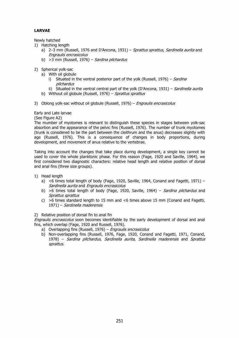
251
LARVAE
Newly hatched 1) Hatching length
a) 2–3 mm (Russell, 1976 and D’Ancona, 1931) – Sprattus sprattus, Sardinella aurita and Engraulis encrasicolus
b) >3 mm (Russell, 1976) – Sardina pilchardus 2) Spherical yolk-sac
a) With oil globule i) Situated in the ventral posterior part of the yolk (Russell, 1976) – Sardina
pilchardus ii) Situated in the ventral central part of the yolk (D’Ancona, 1931) – Sardinella aurita
b) Without oil globule (Russell, 1976) – Sprattus sprattus 3) Oblong yolk-sac without oil globule (Russell, 1976) – Engraulis encrasicolus Early and Late larvae
(See Figure A2)
The mumber of myotomes is relevant to distinguish these species in stages between yolk-sac absortion and the appearance of the pelvic fins (Russell, 1976). The number of trunk myotomes
(trunk is considered to be the part between the cleithrum and the anus) decreases slightly with age (Russell, 1976). This is a consequence of changes in body proportions, during
development, and movement of anus relative to the vertebrae.
Taking into account the changes that take place during development, a single key cannot be
used to cover the whole planktonic phase. For this reason (Fage, 1920 and Saville, 1964), we first considered two diagnostic characters: relative head length and relative position of dorsal
and anal fins (three size groups).
1) Head length
a) <6 times total length of body (Fage, 1920, Saville, 1964, Conand and Fagetti, 1971) – Sardinella aurita and Engraulis encrasicolus
b) >6 times total length of body (Fage, 1920, Saville, 1964) – Sardina pilchardus and Sprattus sprattus
c) >6 times standard length to 15 mm and <6 times above 15 mm (Conand and Fagetti,
1971) – Sardinella maderensis
2) Relative position of dorsal fin to anal fin Engraulis encrasicolus soon becomes identifiable by the early development of dorsal and anal
fins, which overlap (Fage, 1920 and Russell, 1976). a) Overlapping fins (Russell, 1976) – Engraulis encrasicolus b) Non-overlapping fins (Russell, 1976, Fage, 1920, Conand and Fagetti, 1971, Conand,
1978) – Sardina pilchardus, Sardinella aurita, Sardinella maderensis and Sprattus sprattus.
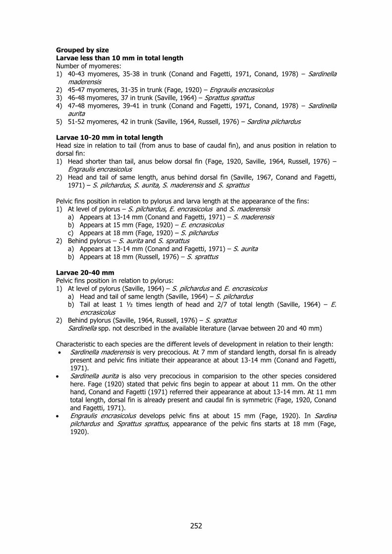
252
Grouped by size
Larvae less than 10 mm in total length
Number of myomeres: 1) 40-43 myomeres, 35-38 in trunk (Conand and Fagetti, 1971, Conand, 1978) – Sardinella
maderensis 2) 45-47 myomeres, 31-35 in trunk (Fage, 1920) – Engraulis encrasicolus 3) 46-48 myomeres, 37 in trunk (Saville, 1964) – Sprattus sprattus 4) 47-48 myomeres, 39-41 in trunk (Conand and Fagetti, 1971, Conand, 1978) – Sardinella
aurita
5) 51-52 myomeres, 42 in trunk (Saville, 1964, Russell, 1976) – Sardina pilchardus
Larvae 10-20 mm in total length Head size in relation to tail (from anus to base of caudal fin), and anus position in relation to
dorsal fin:
1) Head shorter than tail, anus below dorsal fin (Fage, 1920, Saville, 1964, Russell, 1976) – Engraulis encrasicolus
2) Head and tail of same length, anus behind dorsal fin (Saville, 1967, Conand and Fagetti, 1971) – S. pilchardus, S. aurita, S. maderensis and S. sprattus
Pelvic fins position in relation to pylorus and larva length at the appearance of the fins: 1) At level of pylorus – S. pilchardus, E. encrasicolus and S. maderensis
a) Appears at 13-14 mm (Conand and Fagetti, 1971) – S. maderensis b) Appears at 15 mm (Fage, 1920) – E. encrasicolus c) Appears at 18 mm (Fage, 1920) – S. pilchardus
2) Behind pylorus – S. aurita and S. sprattus a) Appears at 13-14 mm (Conand and Fagetti, 1971) – S. aurita
b) Appears at 18 mm (Russell, 1976) – S. sprattus
Larvae 20-40 mm Pelvic fins position in relation to pylorus:
1) At level of pylorus (Saville, 1964) – S. pilchardus and E. encrasicolus a) Head and tail of same length (Saville, 1964) – S. pilchardus b) Tail at least 1 ½ times length of head and 2/7 of total length (Saville, 1964) – E.
encrasicolus 2) Behind pylorus (Saville, 1964, Russell, 1976) – S. sprattus Sardinella spp. not described in the available literature (larvae between 20 and 40 mm)
Characteristic to each species are the different levels of development in relation to their length:
Sardinella maderensis is very precocious. At 7 mm of standard length, dorsal fin is already
present and pelvic fins initiate their appearance at about 13-14 mm (Conand and Fagetti, 1971).
Sardinella aurita is also very precocious in comparision to the other species considered
here. Fage (1920) stated that pelvic fins begin to appear at about 11 mm. On the other hand, Conand and Fagetti (1971) referred their appearance at about 13-14 mm. At 11 mm
total length, dorsal fin is already present and caudal fin is symmetric (Fage, 1920, Conand
and Fagetti, 1971). Engraulis encrasicolus develops pelvic fins at about 15 mm (Fage, 1920). In Sardina
pilchardus and Sprattus sprattus, appearance of the pelvic fins starts at 18 mm (Fage,
1920).
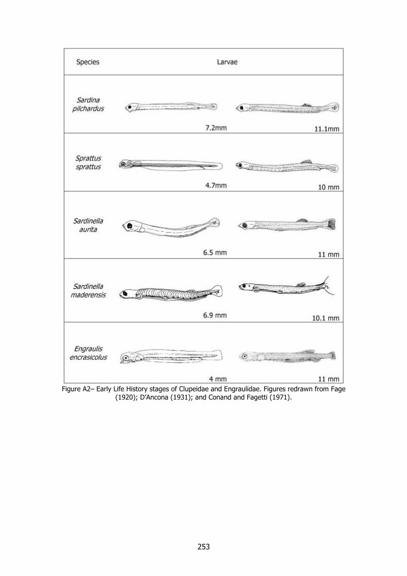
253
Figure A2– Early Life History stages of Clupeidae and Engraulidae. Figures redrawn from Fage
(1920); D’Ancona (1931); and Conand and Fagetti (1971).
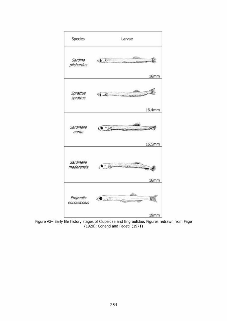
254
Figure A3– Early life history stages of Clupeidae and Engraulidae. Figures redrawn from Fage
(1920); Conand and Fagetii (1971)

255
Sardinella aurita and Sardinella maderensis features
Identification of the Early Life History stages of Sardinella aurita and Sardinella maderensis before the appearance of the pelvic fins is very difficult. Conand and Fagetti (1971) and Conand (1978) compared the larval stages of the two species. One of the main features considered was
the pigmentation of the head, ventral pigmentation, and the position of the pylorus in relation to the ventral pigments.
Larvae less than 12 mm in standard length Presence of continuous double row of pre-pyloric melanophores; caudal pigmentation in inferior
position; snout longer or similar to eye diameter; anus position after the 32nd myomere; eye diameter more than 4% of standard length
1) Pylorus at level of third-to-last pre-pyloric melanophore; anus position at the 39th – 40th myomere – Sardinella aurita (Figure A4a)
2) Pylorus sligthtly posterior in relation to the last pre-pyloric melanophore; anus position
at the 34th – 38th myomere – Sardinella maderensis (Figure A4b)
Larvae of 12-18 mm in standard length Anal fin with less than 25 rays; one double range of pre-pyloric melanophores; anus in posterior
position in relation to the 30th myomere; anal fin behind the dorsal fin extremity; presence of
head pigments 1) Head pigmentation in dome; in advanced larval stages one tiny otocystic melanophore;
absence of shoulder or occipital melanophores; pelvic fins level with the pylorus – Sardinella maderensis (Figure A5a, and c)
2) Head pigments but not in dome; pelvic fins behind the pylorus – Sardinella aurita (Figure A5b and d)

256
Clupeidae Sardinella spp.
Figure A4– Ventral pigmentation at level of the pylorus of Sardinella aurita (a); and Sardinella
maderensis (b) larvae. Redrawn from Conand (1978).
Figure A5– Head pigmentation and pelvic fins (arrows) position of Sardinella aurita, 16.5 mm
(a) and (c); and Sardinella maderensis, 16.0 mm (b) and (d). Redrawn from Conand and
Fagetti (1971).
a b
c d
a 10.0 mm
b 10.1 mm
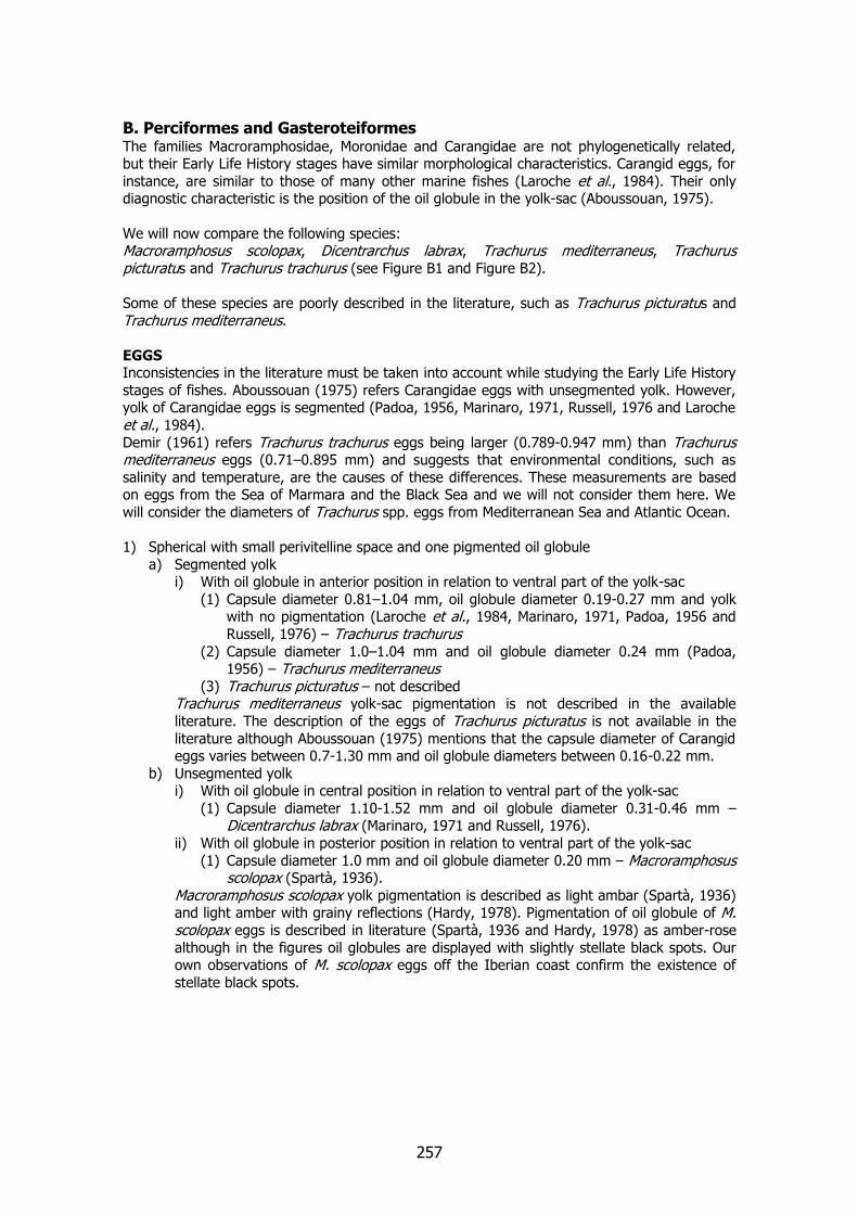
257
B. Perciformes and Gasteroteiformes The families Macroramphosidae, Moronidae and Carangidae are not phylogenetically related, but their Early Life History stages have similar morphological characteristics. Carangid eggs, for
instance, are similar to those of many other marine fishes (Laroche et al., 1984). Their only diagnostic characteristic is the position of the oil globule in the yolk-sac (Aboussouan, 1975).
We will now compare the following species: Macroramphosus scolopax, Dicentrarchus labrax, Trachurus mediterraneus, Trachurus picturatus and Trachurus trachurus (see Figure B1 and Figure B2).
Some of these species are poorly described in the literature, such as Trachurus picturatus and
Trachurus mediterraneus.
EGGS Inconsistencies in the literature must be taken into account while studying the Early Life History
stages of fishes. Aboussouan (1975) refers Carangidae eggs with unsegmented yolk. However, yolk of Carangidae eggs is segmented (Padoa, 1956, Marinaro, 1971, Russell, 1976 and Laroche
et al., 1984).
Demir (1961) refers Trachurus trachurus eggs being larger (0.789-0.947 mm) than Trachurus mediterraneus eggs (0.71–0.895 mm) and suggests that environmental conditions, such as
salinity and temperature, are the causes of these differences. These measurements are based on eggs from the Sea of Marmara and the Black Sea and we will not consider them here. We
will consider the diameters of Trachurus spp. eggs from Mediterranean Sea and Atlantic Ocean.
1) Spherical with small perivitelline space and one pigmented oil globule
a) Segmented yolk i) With oil globule in anterior position in relation to ventral part of the yolk-sac
(1) Capsule diameter 0.81–1.04 mm, oil globule diameter 0.19-0.27 mm and yolk with no pigmentation (Laroche et al., 1984, Marinaro, 1971, Padoa, 1956 and
Russell, 1976) – Trachurus trachurus (2) Capsule diameter 1.0–1.04 mm and oil globule diameter 0.24 mm (Padoa,
1956) – Trachurus mediterraneus (3) Trachurus picturatus – not described
Trachurus mediterraneus yolk-sac pigmentation is not described in the available
literature. The description of the eggs of Trachurus picturatus is not available in the
literature although Aboussouan (1975) mentions that the capsule diameter of Carangid eggs varies between 0.7-1.30 mm and oil globule diameters between 0.16-0.22 mm.
b) Unsegmented yolk i) With oil globule in central position in relation to ventral part of the yolk-sac
(1) Capsule diameter 1.10-1.52 mm and oil globule diameter 0.31-0.46 mm – Dicentrarchus labrax (Marinaro, 1971 and Russell, 1976).
ii) With oil globule in posterior position in relation to ventral part of the yolk-sac
(1) Capsule diameter 1.0 mm and oil globule diameter 0.20 mm – Macroramphosus scolopax (Spartà, 1936).
Macroramphosus scolopax yolk pigmentation is described as light ambar (Spartà, 1936) and light amber with grainy reflections (Hardy, 1978). Pigmentation of oil globule of M. scolopax eggs is described in literature (Spartà, 1936 and Hardy, 1978) as amber-rose
although in the figures oil globules are displayed with slightly stellate black spots. Our own observations of M. scolopax eggs off the Iberian coast confirm the existence of
stellate black spots.

258
Figure B1– Early Life History stages of different species of Macroramphosidae, Moronidae and
Carangidae. Figures redrawn from Spartà (1936); Bertolini (1933); Padoa (1956); and Demir
(1961).
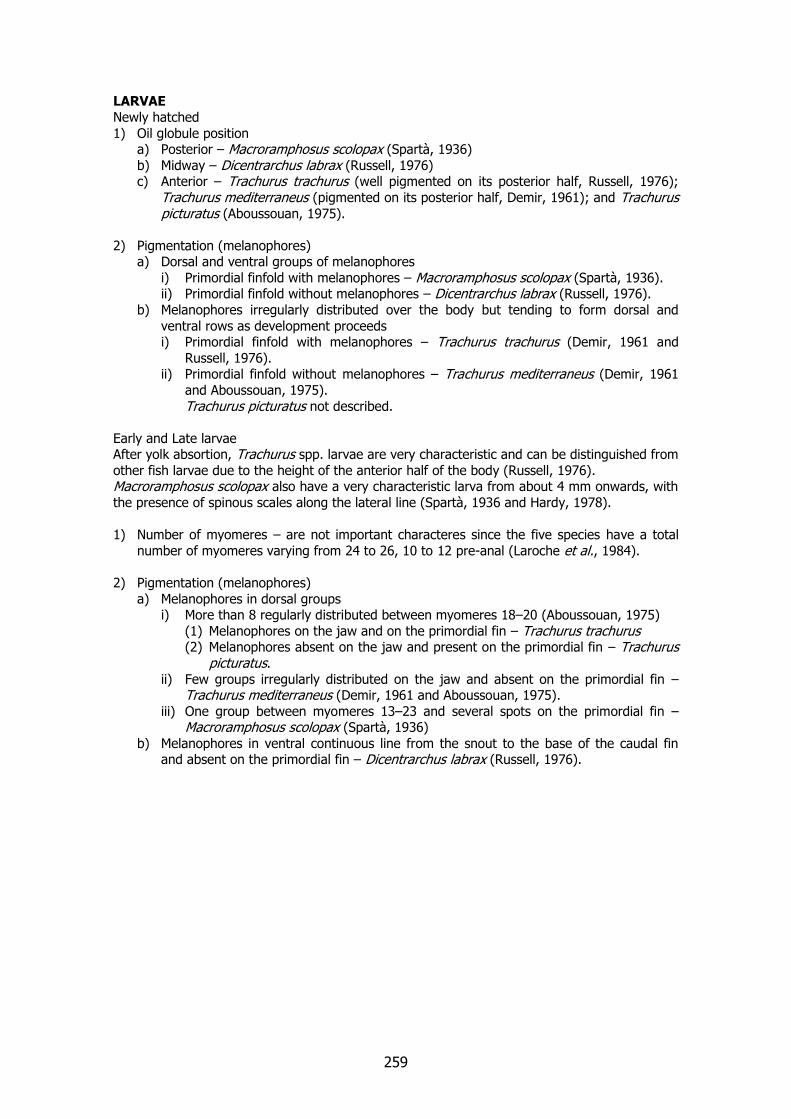
259
LARVAE
Newly hatched
1) Oil globule position a) Posterior – Macroramphosus scolopax (Spartà, 1936)
b) Midway – Dicentrarchus labrax (Russell, 1976) c) Anterior – Trachurus trachurus (well pigmented on its posterior half, Russell, 1976);
Trachurus mediterraneus (pigmented on its posterior half, Demir, 1961); and Trachurus picturatus (Aboussouan, 1975).
2) Pigmentation (melanophores) a) Dorsal and ventral groups of melanophores
i) Primordial finfold with melanophores – Macroramphosus scolopax (Spartà, 1936). ii) Primordial finfold without melanophores – Dicentrarchus labrax (Russell, 1976).
b) Melanophores irregularly distributed over the body but tending to form dorsal and
ventral rows as development proceeds i) Primordial finfold with melanophores – Trachurus trachurus (Demir, 1961 and
Russell, 1976). ii) Primordial finfold without melanophores – Trachurus mediterraneus (Demir, 1961
and Aboussouan, 1975).
Trachurus picturatus not described.
Early and Late larvae After yolk absortion, Trachurus spp. larvae are very characteristic and can be distinguished from
other fish larvae due to the height of the anterior half of the body (Russell, 1976). Macroramphosus scolopax also have a very characteristic larva from about 4 mm onwards, with
the presence of spinous scales along the lateral line (Spartà, 1936 and Hardy, 1978).
1) Number of myomeres – are not important characteres since the five species have a total
number of myomeres varying from 24 to 26, 10 to 12 pre-anal (Laroche et al., 1984).
2) Pigmentation (melanophores)
a) Melanophores in dorsal groups i) More than 8 regularly distributed between myomeres 18–20 (Aboussouan, 1975)
(1) Melanophores on the jaw and on the primordial fin – Trachurus trachurus (2) Melanophores absent on the jaw and present on the primordial fin – Trachurus
picturatus. ii) Few groups irregularly distributed on the jaw and absent on the primordial fin –
Trachurus mediterraneus (Demir, 1961 and Aboussouan, 1975).
iii) One group between myomeres 13–23 and several spots on the primordial fin – Macroramphosus scolopax (Spartà, 1936)
b) Melanophores in ventral continuous line from the snout to the base of the caudal fin and absent on the primordial fin – Dicentrarchus labrax (Russell, 1976).

260
Figure B2– Early Life History stages of different species of Macroramphosidae, Moronidae and
Carangidae. Figures redrawn from Spartà (1936); Bertolini (1933); Padoa (1956); Aboussouan (1975); and Demir (1961). Trachurus trachurus wild larva with ~6.1 mm of standard length was
sampled in 1998 off Portuguese coast.

261
3) Crests
Occipital crests are important larval features of Tarchurus spp. These crests develop on the head during the preflexion stage (Laroche et al., 1984). Aboussouan (1975) describes these
features and compares their characteristics in Trachurus trachurus and Trachurus picturatus (Figure B3).
Macroramphosus scolopax develops one supraorbital crest during the preflexion stage, as well as occipital and supraorbital crests at flexion stage (Spartà, 1936 and Hardy, 1978).
Figure B3– Occipital crests of Trachurus picturatus and Trachurus trachurus larvae of different
lenghts. Redrawn from Aboussouan (1975).

262

263
C. Perciformes C.1. Carangidae and Scombridae
The following species will be compared: Trachurus mediterraneus, Trachurus picturatus Trachurus trachurus, Scomber japonicus and Scomber scombrus (See Figure C1 and Figure C2).
EGGS
1) Spherical with small perivitelline space and with one oil globule
a) Segmented yolk and several melanophores rounding the oil globule i) Oil globule in anterior position in relation to ventral part of the yolk-sac
(1) Capsule diameter 0.81–1.04 mm; oil globule diameter 0.19–0.27 mm; (Laroche et al., 1984, Marinaro, 1971, Padoa, 1956 and Russell, 1976) yolk-sac with no
pigmentation – Trachurus trachurus (2) Capsule diameter 1.0–1.04 mm; oil globule diameter 0.24 mm (Padoa, 1956);
yolk-sac with no pigmentation – Trachurus mediterraneus (3) Trachurus picturatus. – not described
Aboussouan (1975) refers that the capsule diameter of Carangid eggs varies between
0.7-1.30 mm. We consider egg diameters of Trachurus picturatus within this range although not described specifically in the literature. As already mentioned in Section B
of this Appendix, we have not considered the results of Demir (1961) for Trachurus trachurus and Trachurus mediterraneus as far as egg diameters are concerned. We assume no pigmentation in the yolk-sac of Trachurus spp. eggs although Trachurus mediterraneus and Trachurus picturatus are not described in the literature.
b) Unsegmented yolk
i) Oil globule in posterior/central position in relation to ventral part of the yolk-sac
(a) Capsule diameter 1.0–1.38mm; oil globule diameter 0.28–0.35 mm; no pigmentation on yolk; oil globule with melanophores in its anterior half
before the caudal extremity is free from yolk (Russell, 1976 and Fritzsche, 1978) – Scomber scombrus
(b) Capsule diameter 1.06–1.14mm; oil globule diameter 0.26 mm (Olivar and Fortuño, 1991); pigments on the yolk after embryo tail reaches the head;
these melanophores migrate ventrally and spread from sides of body
forward onto the yolk-sac near the head (Fritzsche, 1978); oil globule with melanophores in its anterior half after the caudal extremity is free from
yolk (Fritzsche, 1978) – Scomber japonicus The range of oil globules diameters described for Scomber japonicus from Pacific Ocean varies between 0.25 and 0.32 mm (Fritzsche, 1978).
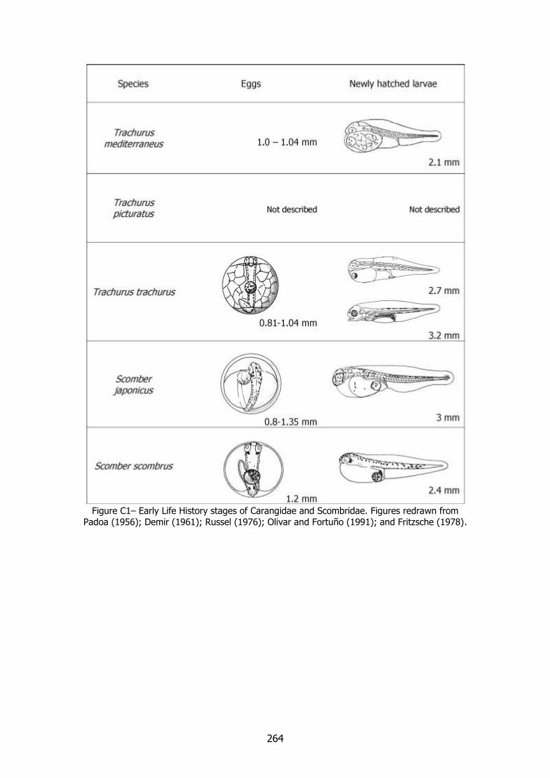
264
Figure C1– Early Life History stages of Carangidae and Scombridae. Figures redrawn from
Padoa (1956); Demir (1961); Russel (1976); Olivar and Fortuño (1991); and Fritzsche (1978).
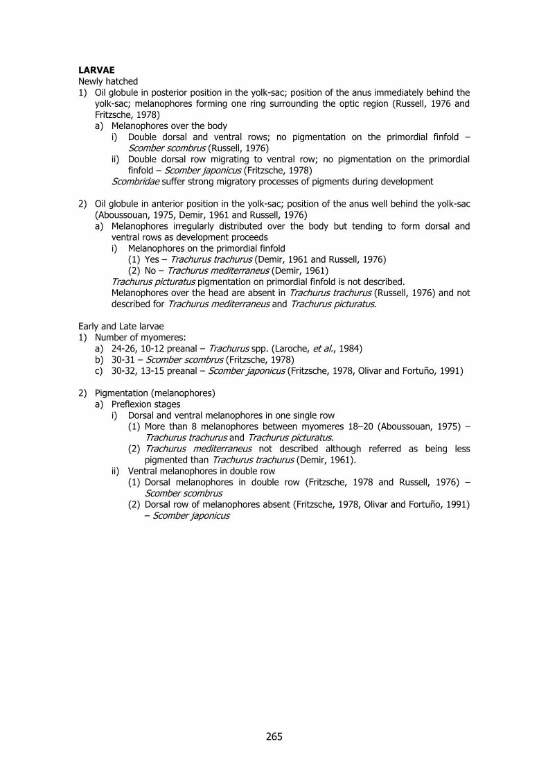
265
LARVAE
Newly hatched
1) Oil globule in posterior position in the yolk-sac; position of the anus immediately behind the yolk-sac; melanophores forming one ring surrounding the optic region (Russell, 1976 and
Fritzsche, 1978) a) Melanophores over the body
i) Double dorsal and ventral rows; no pigmentation on the primordial finfold –
Scomber scombrus (Russell, 1976) ii) Double dorsal row migrating to ventral row; no pigmentation on the primordial
finfold – Scomber japonicus (Fritzsche, 1978) Scombridae suffer strong migratory processes of pigments during development
2) Oil globule in anterior position in the yolk-sac; position of the anus well behind the yolk-sac
(Aboussouan, 1975, Demir, 1961 and Russell, 1976)
a) Melanophores irregularly distributed over the body but tending to form dorsal and ventral rows as development proceeds
i) Melanophores on the primordial finfold (1) Yes – Trachurus trachurus (Demir, 1961 and Russell, 1976)
(2) No – Trachurus mediterraneus (Demir, 1961)
Trachurus picturatus pigmentation on primordial finfold is not described. Melanophores over the head are absent in Trachurus trachurus (Russell, 1976) and not
described for Trachurus mediterraneus and Trachurus picturatus.
Early and Late larvae 1) Number of myomeres:
a) 24-26, 10-12 preanal – Trachurus spp. (Laroche, et al., 1984)
b) 30-31 – Scomber scombrus (Fritzsche, 1978) c) 30-32, 13-15 preanal – Scomber japonicus (Fritzsche, 1978, Olivar and Fortuño, 1991)
2) Pigmentation (melanophores)
a) Preflexion stages
i) Dorsal and ventral melanophores in one single row (1) More than 8 melanophores between myomeres 18–20 (Aboussouan, 1975) –
Trachurus trachurus and Trachurus picturatus. (2) Trachurus mediterraneus not described although referred as being less
pigmented than Trachurus trachurus (Demir, 1961).
ii) Ventral melanophores in double row (1) Dorsal melanophores in double row (Fritzsche, 1978 and Russell, 1976) –
Scomber scombrus (2) Dorsal row of melanophores absent (Fritzsche, 1978, Olivar and Fortuño, 1991)
– Scomber japonicus
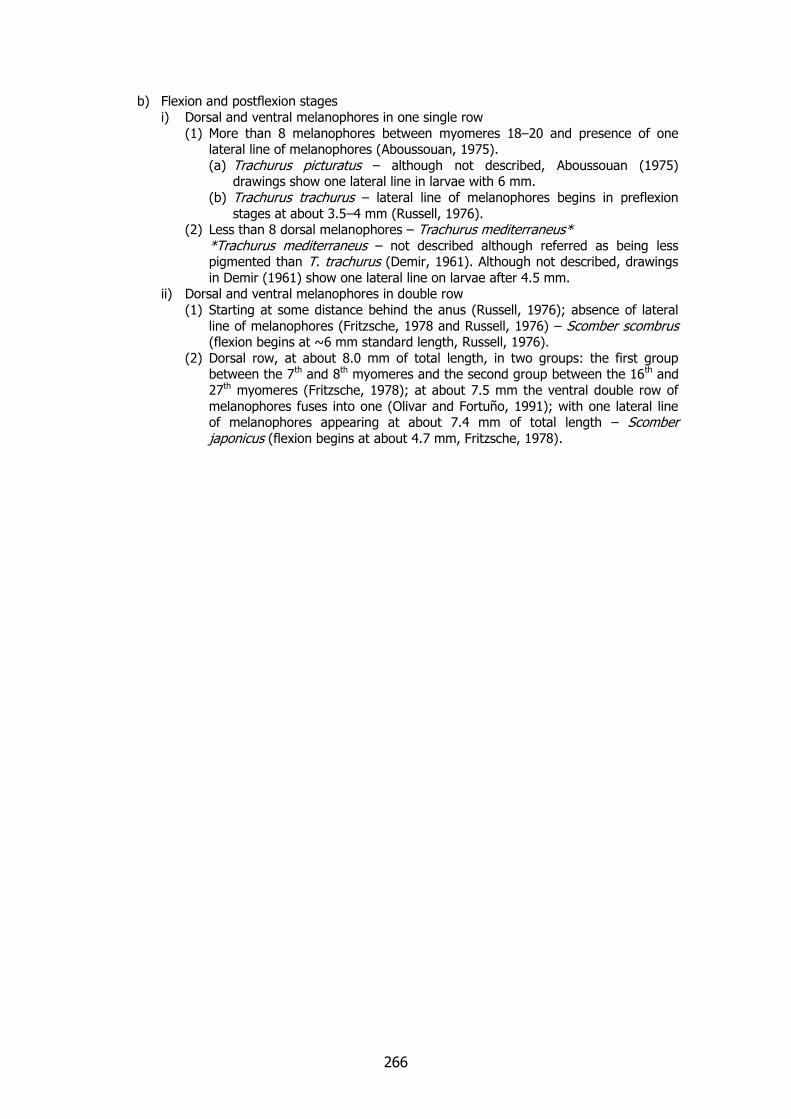
266
b) Flexion and postflexion stages
i) Dorsal and ventral melanophores in one single row
(1) More than 8 melanophores between myomeres 18–20 and presence of one lateral line of melanophores (Aboussouan, 1975).
(a) Trachurus picturatus – although not described, Aboussouan (1975) drawings show one lateral line in larvae with 6 mm.
(b) Trachurus trachurus – lateral line of melanophores begins in preflexion
stages at about 3.5–4 mm (Russell, 1976). (2) Less than 8 dorsal melanophores – Trachurus mediterraneus*
*Trachurus mediterraneus – not described although referred as being less pigmented than T. trachurus (Demir, 1961). Although not described, drawings
in Demir (1961) show one lateral line on larvae after 4.5 mm. ii) Dorsal and ventral melanophores in double row
(1) Starting at some distance behind the anus (Russell, 1976); absence of lateral
line of melanophores (Fritzsche, 1978 and Russell, 1976) – Scomber scombrus (flexion begins at ~6 mm standard length, Russell, 1976).
(2) Dorsal row, at about 8.0 mm of total length, in two groups: the first group between the 7th and 8th myomeres and the second group between the 16th and
27th myomeres (Fritzsche, 1978); at about 7.5 mm the ventral double row of
melanophores fuses into one (Olivar and Fortuño, 1991); with one lateral line of melanophores appearing at about 7.4 mm of total length – Scomber japonicus (flexion begins at about 4.7 mm, Fritzsche, 1978).
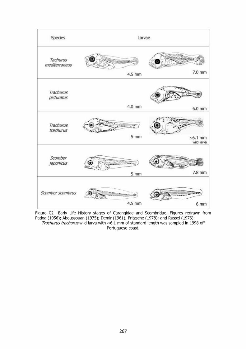
267
Figure C2– Early Life History stages of Carangidae and Scombridae. Figures redrawn from
Padoa (1956); Aboussouan (1975); Demir (1961); Fritzsche (1978); and Russel (1976). Trachurus trachurus wild larva with ~6.1 mm of standard length was sampled in 1998 off
Portuguese coast.

268
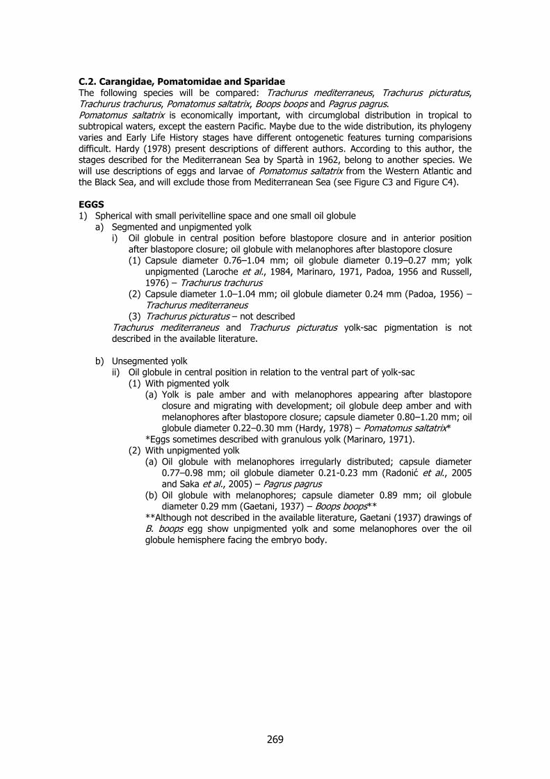
269
C.2. Carangidae, Pomatomidae and Sparidae
The following species will be compared: Trachurus mediterraneus, Trachurus picturatus, Trachurus trachurus, Pomatomus saltatrix, Boops boops and Pagrus pagrus. Pomatomus saltatrix is economically important, with circumglobal distribution in tropical to subtropical waters, except the eastern Pacific. Maybe due to the wide distribution, its phylogeny
varies and Early Life History stages have different ontogenetic features turning comparisions
difficult. Hardy (1978) present descriptions of different authors. According to this author, the stages described for the Mediterranean Sea by Spartà in 1962, belong to another species. We
will use descriptions of eggs and larvae of Pomatomus saltatrix from the Western Atlantic and the Black Sea, and will exclude those from Mediterranean Sea (see Figure C3 and Figure C4).
EGGS
1) Spherical with small perivitelline space and one small oil globule
a) Segmented and unpigmented yolk i) Oil globule in central position before blastopore closure and in anterior position
after blastopore closure; oil globule with melanophores after blastopore closure (1) Capsule diameter 0.76–1.04 mm; oil globule diameter 0.19–0.27 mm; yolk
unpigmented (Laroche et al., 1984, Marinaro, 1971, Padoa, 1956 and Russell,
1976) – Trachurus trachurus (2) Capsule diameter 1.0–1.04 mm; oil globule diameter 0.24 mm (Padoa, 1956) –
Trachurus mediterraneus (3) Trachurus picturatus – not described
Trachurus mediterraneus and Trachurus picturatus yolk-sac pigmentation is not described in the available literature.
b) Unsegmented yolk ii) Oil globule in central position in relation to the ventral part of yolk-sac
(1) With pigmented yolk (a) Yolk is pale amber and with melanophores appearing after blastopore
closure and migrating with development; oil globule deep amber and with
melanophores after blastopore closure; capsule diameter 0.80–1.20 mm; oil globule diameter 0.22–0.30 mm (Hardy, 1978) – Pomatomus saltatrix*
*Eggs sometimes described with granulous yolk (Marinaro, 1971). (2) With unpigmented yolk
(a) Oil globule with melanophores irregularly distributed; capsule diameter
0.77–0.98 mm; oil globule diameter 0.21-0.23 mm (Radonić et al., 2005 and Saka et al., 2005) – Pagrus pagrus
(b) Oil globule with melanophores; capsule diameter 0.89 mm; oil globule diameter 0.29 mm (Gaetani, 1937) – Boops boops**
**Although not described in the available literature, Gaetani (1937) drawings of B. boops egg show unpigmented yolk and some melanophores over the oil
globule hemisphere facing the embryo body.
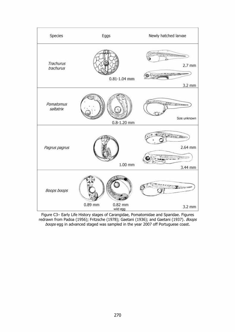
270
Figure C3– Early Life History stages of Carangidae, Pomatomidae and Sparidae. Figures
redrawn from Padoa (1956); Fritzsche (1978); Gaetani (1936); and Gaetani (1937). Boops boops egg in advanced staged was sampled in the year 2007 off Portuguese coast.
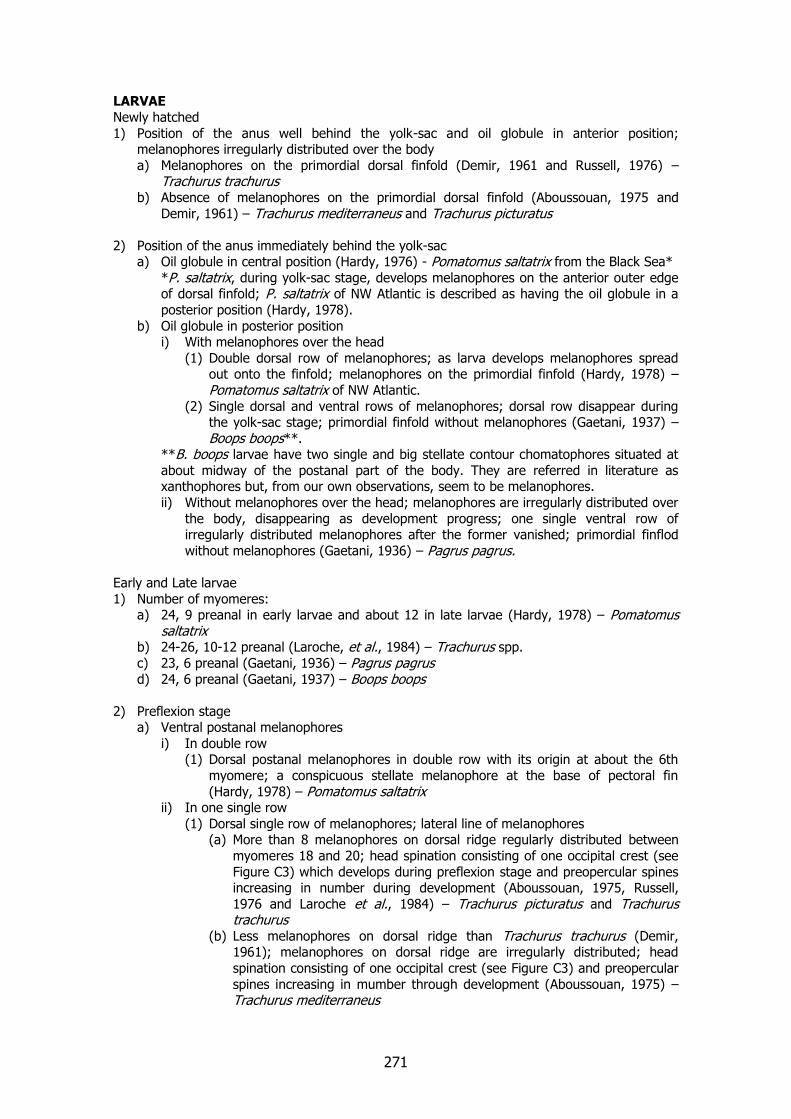
271
LARVAE
Newly hatched
1) Position of the anus well behind the yolk-sac and oil globule in anterior position; melanophores irregularly distributed over the body
a) Melanophores on the primordial dorsal finfold (Demir, 1961 and Russell, 1976) – Trachurus trachurus
b) Absence of melanophores on the primordial dorsal finfold (Aboussouan, 1975 and
Demir, 1961) – Trachurus mediterraneus and Trachurus picturatus
2) Position of the anus immediately behind the yolk-sac a) Oil globule in central position (Hardy, 1976) - Pomatomus saltatrix from the Black Sea*
*P. saltatrix, during yolk-sac stage, develops melanophores on the anterior outer edge of dorsal finfold; P. saltatrix of NW Atlantic is described as having the oil globule in a
posterior position (Hardy, 1978).
b) Oil globule in posterior position i) With melanophores over the head
(1) Double dorsal row of melanophores; as larva develops melanophores spread out onto the finfold; melanophores on the primordial finfold (Hardy, 1978) –
Pomatomus saltatrix of NW Atlantic.
(2) Single dorsal and ventral rows of melanophores; dorsal row disappear during the yolk-sac stage; primordial finfold without melanophores (Gaetani, 1937) –
Boops boops**. **B. boops larvae have two single and big stellate contour chomatophores situated at
about midway of the postanal part of the body. They are referred in literature as xanthophores but, from our own observations, seem to be melanophores.
ii) Without melanophores over the head; melanophores are irregularly distributed over
the body, disappearing as development progress; one single ventral row of irregularly distributed melanophores after the former vanished; primordial finflod
without melanophores (Gaetani, 1936) – Pagrus pagrus.
Early and Late larvae
1) Number of myomeres: a) 24, 9 preanal in early larvae and about 12 in late larvae (Hardy, 1978) – Pomatomus
saltatrix b) 24-26, 10-12 preanal (Laroche, et al., 1984) – Trachurus spp.
c) 23, 6 preanal (Gaetani, 1936) – Pagrus pagrus d) 24, 6 preanal (Gaetani, 1937) – Boops boops
2) Preflexion stage a) Ventral postanal melanophores
i) In double row (1) Dorsal postanal melanophores in double row with its origin at about the 6th
myomere; a conspicuous stellate melanophore at the base of pectoral fin
(Hardy, 1978) – Pomatomus saltatrix ii) In one single row
(1) Dorsal single row of melanophores; lateral line of melanophores (a) More than 8 melanophores on dorsal ridge regularly distributed between
myomeres 18 and 20; head spination consisting of one occipital crest (see
Figure C3) which develops during preflexion stage and preopercular spines increasing in number during development (Aboussouan, 1975, Russell,
1976 and Laroche et al., 1984) – Trachurus picturatus and Trachurus trachurus
(b) Less melanophores on dorsal ridge than Trachurus trachurus (Demir, 1961); melanophores on dorsal ridge are irregularly distributed; head
spination consisting of one occipital crest (see Figure C3) and preopercular
spines increasing in mumber through development (Aboussouan, 1975) – Trachurus mediterraneus
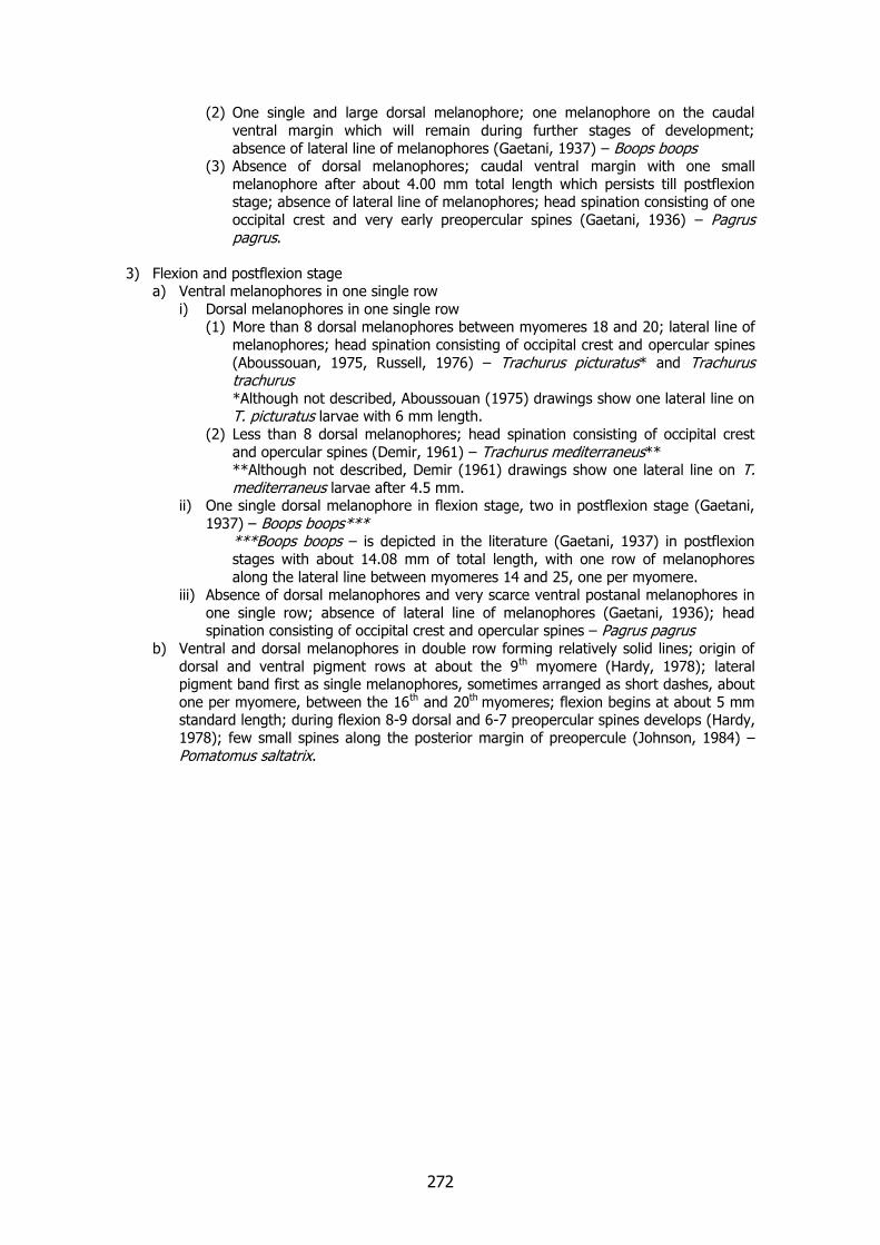
272
(2) One single and large dorsal melanophore; one melanophore on the caudal
ventral margin which will remain during further stages of development;
absence of lateral line of melanophores (Gaetani, 1937) – Boops boops (3) Absence of dorsal melanophores; caudal ventral margin with one small
melanophore after about 4.00 mm total length which persists till postflexion stage; absence of lateral line of melanophores; head spination consisting of one
occipital crest and very early preopercular spines (Gaetani, 1936) – Pagrus pagrus.
3) Flexion and postflexion stage a) Ventral melanophores in one single row
i) Dorsal melanophores in one single row (1) More than 8 dorsal melanophores between myomeres 18 and 20; lateral line of
melanophores; head spination consisting of occipital crest and opercular spines
(Aboussouan, 1975, Russell, 1976) – Trachurus picturatus* and Trachurus trachurus *Although not described, Aboussouan (1975) drawings show one lateral line on T. picturatus larvae with 6 mm length.
(2) Less than 8 dorsal melanophores; head spination consisting of occipital crest
and opercular spines (Demir, 1961) – Trachurus mediterraneus** **Although not described, Demir (1961) drawings show one lateral line on T. mediterraneus larvae after 4.5 mm.
ii) One single dorsal melanophore in flexion stage, two in postflexion stage (Gaetani,
1937) – Boops boops*** ***Boops boops – is depicted in the literature (Gaetani, 1937) in postflexion
stages with about 14.08 mm of total length, with one row of melanophores
along the lateral line between myomeres 14 and 25, one per myomere. iii) Absence of dorsal melanophores and very scarce ventral postanal melanophores in
one single row; absence of lateral line of melanophores (Gaetani, 1936); head spination consisting of occipital crest and opercular spines – Pagrus pagrus
b) Ventral and dorsal melanophores in double row forming relatively solid lines; origin of
dorsal and ventral pigment rows at about the 9th myomere (Hardy, 1978); lateral pigment band first as single melanophores, sometimes arranged as short dashes, about
one per myomere, between the 16th and 20th myomeres; flexion begins at about 5 mm standard length; during flexion 8-9 dorsal and 6-7 preopercular spines develops (Hardy,
1978); few small spines along the posterior margin of preopercule (Johnson, 1984) –
Pomatomus saltatrix.
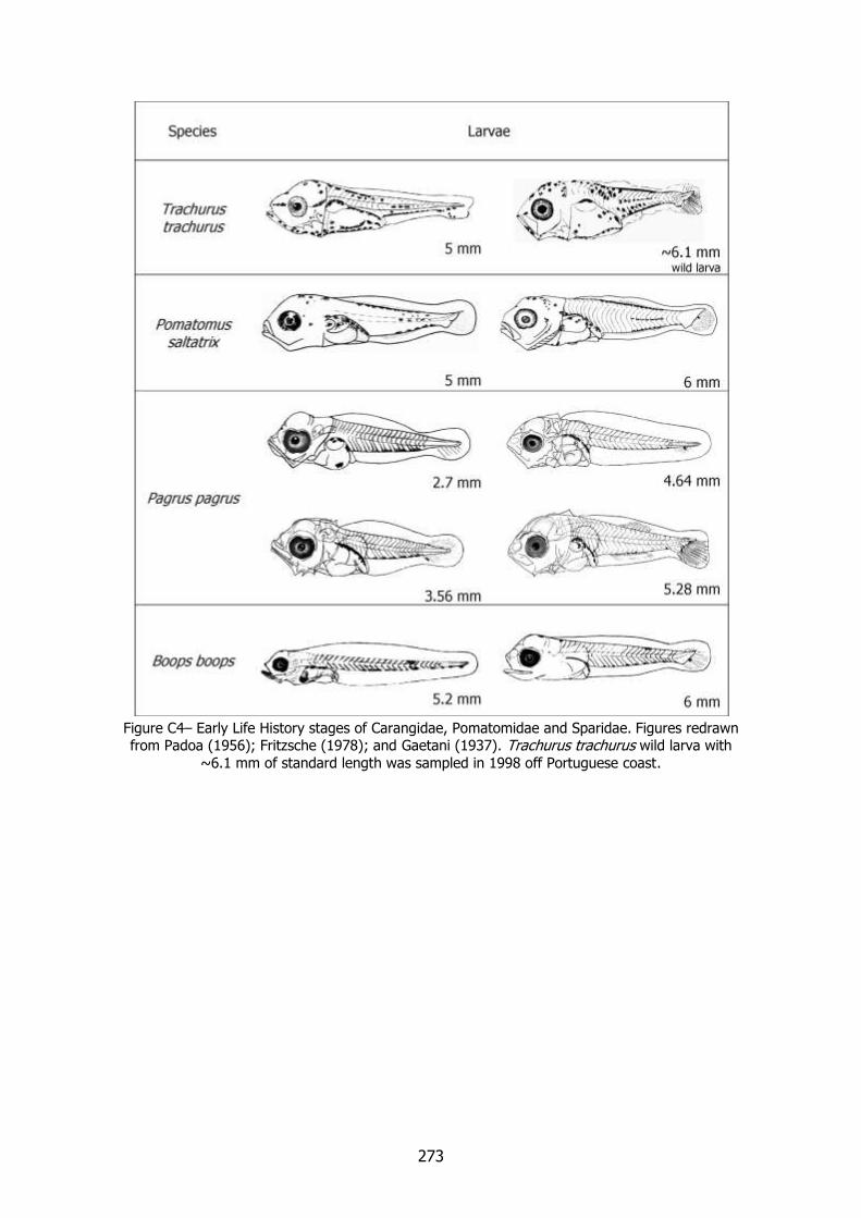
273
Figure C4– Early Life History stages of Carangidae, Pomatomidae and Sparidae. Figures redrawn from Padoa (1956); Fritzsche (1978); and Gaetani (1937). Trachurus trachurus wild larva with
~6.1 mm of standard length was sampled in 1998 off Portuguese coast.

274
275
EGGS AND LARVAE OF TWO TROPICAL FISHES OCCURING IN PORTUGUESE WATERS The distribution, ecology and spawning seasons of fish species are normaly used by
planktonologists as characteristics to aid in the difficult identification of early life stages. Environmental changes are presently globally reported (e.g. Reid et al., 2001, Stenseth et al., 2002) and must be recognised as potential causes for possible alterations to the distribution of
adult fish and of their spawning products.
Already in last decade, typically tropical species have been shown to ocur in more northerly latitudes than was previously recognised. Among these we will consider here the Red cornetfish Fistularia petimba, Lacépède 1803 (Syngnathiphormes: Fistulariidae) and monrovia doctorfish Ancanthurus monroviae, Steindachner, 1876 (Perciformes: Acanthuridae) recently reported off the Iberian waters (Cardénas et al., 1997, Bañon and Sande, 2008 and Martins, pers. comm.).
Although the Early Life History stages of these species cannot be mistaken for any other here
described, we considered their inclusion in the following pages of special interest, due to the high possibility of their occurence in plankton samples.

276
Fistulariidae Red cornetfish (Fistularia petimba, Lacépède, 1803) is found in its adult stage, in deeper waters
along continental margins and was found off Portuguese continental waters in 2008 (Martins pers. comm.) and Azores waters in 2004 (Azevedo et al., 2004).
FISTURALIDAE Fistularia petimba (Lacépède, 1803) MERISTICS Fins:
Dorsal rays – 13-15 Anal rays – 14-15
Pelvic rays – 5-6 (for the Family)
Pectoral rays – 13-18 Myomeres:
Total number – 82-86 Preanal number – 53
Postanal – 33
LIFE HISTORY
Range: Throughout tropical Atlantic, Indo-
West Pacific and Hawaii.
Habitat: Inhabits soft-bottomed inshore
waters, usually at depths over 10 meters.
Spawning season: February in Pacifc
Ocean.
ELH pattern: oviparous, planktonic eggs
and larvae.
MAIN REFERENCES Hardy, J.D. (1978). Development of Fishes of the Mid-
Atlantic Bght, Fish and Wildlife Service, U.S. Department of the Interior. Volume II: 366-373.
Moser, H.G., W.J. Richards, D.M. Cohen, M.P. Fahay, A.W. Kendall, S.L. Richardson (eds.) (1984). Ontogeny and systematics of fishes. American Society of Ichthyologists and Herpetologists ed., Special publication Number 1: 760pp.
EARLY LIFE HISTORY DESCRIPTION EGGS
Capsule diameter – 1.50-2.10 mm No. of oil globules – 0
Shell surface – smooth
Pigment – Yolk – unsegmented
Diameter of oil globules – Diagnostic features – spherical, no oil
globule, small perivitelline space.
LARVAE
Hatching length – 7.08 mm Yolk-sac absorption –
Flexion length –
Transformation length – Pigmentation – Light pigmentation. Newly
hatched larvae are pigmented along dorsal and ventral body contours, on
yolk, along ventral wall of gut near anus, and extended into ventral finfold
on posterior half of tail.
Diagnostic features – Develops small spines on body (“villosa-stage”).

277
FISTULARIIDAE Fistularia petimba, (Lacépède, 1803)
Figure D– Early Life History stages of Fistularia petimba. Redrawn from Moser, et al. (1984).
278
Acanthuridae
Monrovia doctorfish (Acanthurus monroviae, Steindachner, 1876) is a coastal demersal species
found in the mouth of rivers and coastal lagoons, on rocky and coral bottoms (Leis and Richards, 1984). Depth distribution 5-200 m. The reported range of distribution at Eastern
Atlantic tropical latitudes, varies between 30º N and 15º S (water temperatures between 20 and 25 ºC). In recent years, Scuba divers reported the presence of this species off the
Portuguese coast. Early life stages of this species are poorly described in literature.
ACANTHURIDAE Acanthurus monroviae (Steindachner, 1876)
MERISTICS
Fins:
Dorsal rays – 24-26 (adults) Anal rays – 24-26(adults)
Pelvic rays – Pectoral rays –
Myomeres: Total number – 22
Preanal number – 3-4
Postanal – 18
LIFE HISTORY
Range: Eastern Atlantic from 30º N to 15º
N
Habitat: Inhabits inshore waters on rocky and coral bottoms, in the mouth of
rivers and lagoons.
Spawning season:
ELH pattern: oviparous, pelagic eggs and
planktonic larvae.
MAIN REFERENCES Aboussouan, A. (1965). Oeufs et larves de Télèostéens
de l´Ouest africain. I. – Acanthurus monroviae Steind. Bulletin de l´I.F.A.N., T. 27, sér. A, nº 3: 1183-1187
Leis, J.M.; W.J., Richards (1984) Acanthuroidei: Development and relationships in Moser, H.G., W.J. Richards, D.M. Cohen, M.P. Fahay, A.W. Kendall, S.L. Richardson (eds.) (1984). Ontogeny and systematics of fishes. American Society of Ichthyologists and Herpetologists ed., Special publication Number 1: 760pp
EARLY LIFE HISTORY DESCRIPTION
EGGS Capsule diameter – < 1mm
No. of oil globules – 1 Shell surface –
Pigment – Yolk - unsegmented
Diameter of oil globules –
Diagnostic features –
LARVAE Hatching length – 2.5 mm
Yolk-sac absorption – unknown
Flexion length – ~4.7 mm; with scales shortly after flexion
Transformation length – ~6.0 - 7 mm Pigmentation – Gut dorsal wall heavily
pigmented. Ventral postanal body
contour with small chromatophores until caudal region. Occipital region and
supraorbital region of the head with several small chromatophores and few
large branched chromatophores. Diagnostic features – Head and trunk
remarkably deepened. Develop big
ventral and dorsal spines, early stages (1.9–2.1 mm). Develops one anal spine
~3.1 mm. Tubular snout with a little mouth and spines on its mandible.
Strong backward bending spines on
superior ridge of the head.
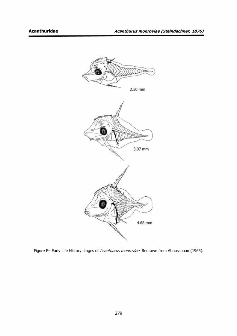
279
Acanthuridae Acanthurus monroviae (Steindachner, 1876)
Figure E– Early Life History stages of Acanthurus monroviae. Redrawn from Aboussouan (1965).

280
REFERENCES
Aboussouan, A. (1965). Oeufs et larves de Télèostéens de l´Ouest africain. I. – Acanthurus
monroviae Steind. Bulletin de l´I.F.A.N., T. 27, sér. A, nº 3: 1183-1187
Aboussouan, A. (1975). Oeufs et larves de Téléostéens de l´Ouest africain. XIII. Contribution à
l´identification des larves de Carangidae. Bulletin de l´I.F.A.N., T. 37, sér. A, nº 4: 899-938.
Azevedo, J.M.N., P.M. Raposeiro, P.M.L. Rodrigues (2004). Fisrt records of Fistularia petimba and
Diodon eydouxii for the Azores, with notes on the occurrence of three additional species. J. Fish. Biol. 65: 1180-1184
Bañon, R. and C. Sande (2008). First record of the red cornetfish Fistularia petimba
(Syngnathiformes: Fistularidae) in Galician waters: a norhernmost occurrence in the eastern
Atlantic. J. Appl. Ichthyol. 24: 106-107
Bertolini, F. (1933). Serranidae. Uova, larve e stadi Giovanili di Teleostei: monografia elaborata con l’uso del materiale raccolto e sweriato da Salvatore Lo Bianco. Fauna e Flora Golfo Napoli Monogr.38. Publi. In 4 parts: 310-321.
Cardénas, S., D.A. Berastegui and J.M. Ortiz (1997). First record of Fistularia petimba Lacépède, 1803
(Pisces, Fistularidae) off the coast of Cadiz (southern Iberian Peninsula). Bol. Inst. Esp. Oceanogr. 13: 83-86
Conand, F. (1978). Systématique des larves de Clupéidés de l´Atlantique Oriental entre 20º N et 15º
S (eaux marines et saumâtres). Cah. O.R.S.T.O.M., sér. Océanogr., vol. XVI, nº 1: 3-8
Conand, F. and E. Fagetti (1971). Description et distribuition saisonnière des larves de sardinelles des
côtes du Sénégal et de la Gambie en 1968 et 1969. Cah. O.R.S.T.O.M., sér. Océanogr., vol. IX, nº 3: 293-318
D’Ancona, U. (1931). Clupeoidei. In Uova, larve e stadi Giovanili di Teleostei: monografia elaborata con l’uso del materiale raccolto e sweriato da Salvatore Lo Bianco. Fauna e Flora Golfo Napoli Monogr. 38 (1): 1-16
Fage. L. (1920). Engraulidae, Clupeidae. Rep. Dan. Oceanogr. Exped. 1908-10 Medit . adjac. Seas, 2, Biology A9: 140p.
Fritzsche, R.A. (1978). Scombridae. Development of fishes of the Mid-Atlantic Bight. Fish and Wildlife Service, U.S. Department of the Interior. Volume V: 62-167.
Gaetani, D. (1936). Uova, sviluppo embrionale e stadi post-embrionali negli Sparidi 4. Pagrus vulgaris
C. V. Memoria R. Comitato Talassografico Italiano 226: 1-14 (1 plate).
Gaetani, D. (1937). Uova, sviluppo embrionale e stadi post-embrionali negli Sparidi 5. Box boops L.
Memoria R. Comitato Talassografico Italiano 241: 1-14 (1 plate).
Hardy, J.D. (1978). Macrorhamphosidae. Development of fishes of the Mid-Atlantic Bight. Fish and
Wildlife Service, U.S. Department of the Interior. Volume II: 377-386.
Hardy, J.D. (1978). Pomatomidae. Development of fishes of the Mid-Atlantic Bight. Fish and Wildlife Service, U.S. Department of the Interior. Volume III: 338-353.
Hurrell, J. W., R.R. Dickson, 2004: Climate variability over the North Atlantic. Marine Ecosystems and
Climate Variation - the North Atlantic, N.C. Stenseth, G. Ottersen, J.W. Hurrell, and A. Belgrano,
Eds., Oxford University Press, 15–32.

281
Jones, P.W., F.D. Martin, J.D. Hardy (1978). Clupeidae. Development of fishes of the Mid-Atlantic Bight. Fish and Wildlife Service, U.S. Department of the Interior. Volume I: 73-150.
Jonhson, G.D. (1984). Percoidei: Development and Relationships in Moser, H.G., W.J. Richards, D.M. Cohen, M.P. Fahay, A.W. Kendall, S.L. Richardson (eds.) (1984). Ontogeny and systematics of fishes. American Society of Ichthyologists and Herpetologists ed., Special publication Number 1:
760pp.
Laroche, W.A., W.F. Smith-Vaniz, S.L. Richardson (1984) Carangidae: Development in Moser, H.G., W.J. Richards, D.M. Cohen, M.P. Fahay, A.W. Kendall, S.L. Richardson (eds.) (1984). Ontogeny and systematics of fishes. American Society of Ichthyologists and Herpetologists ed., Special publication Number 1: 760pp.
Leis, J.M., W.J. Richards (1984) Acanthuroidei: Development and relationships in Moser, H.G., W.J. Richards, D.M. Cohen, M.P. Fahay, A.W. Kendall, S.L. Richardson (eds.) (1984). Ontogeny and systematics of fishes. American Society of Ichthyologists and Herpetologists ed., Special publication Nº 1: 760pp
Marinaro, J-Y (1971). Contribution à l'étude des oeufs et larves pèlagiques de poissons mediterranéens. V. Oeufs pèlagiques de la Baie d'Alger. Pelagos, 3 (1): 1-118.
Moser, H.G., W.J. Richards, D.M. Cohen, M.P. Fahay, A.W. Kendall, S.L. Richardson (eds.) (1984).
Ontogeny and systematics of fishes. American Society of Ichthyologists and Herpetologists ed., Special publication Number 1: 760pp.
Olivar, P., J.-M. Fortuño (1991). Guide to the ichthyoplankton of the Southeast Atlantic (Benguela current region). Sci. Mar., 55 (1): 1-383.
Padoa, E. (1956). Carangidae. Uova, larve e stadi Giovanili di Teleostei: monografia elaborata con
l’uso del materiale raccolto e sweriato da Salvatore Lo Bianco. Fauna e Flora Golfo Napoli Monogr.38: 548-570.
Radonić, M., A.V. López, M. Oka, E.O. Aristizábal (2005). Effect of the incubation temperature on the embryonic development and hatching time of eggs of the red porgy Pagrus pagrus (Linne, 1758)
(Pisces: Sparidae). Revista de Biologia Marina y Oceanografia 40 (2): 91 – 99.
Ré, P., R. Cabral e Silva, M.E. Cunha, A. Farinha, I. Meneses, T. Moita (1990). Sardine spawning off
Portugal. Bol. Inst. Nac. Inv. Pescas 15: 31-44
Russell, F.S. (1976). The eggs and planktonic stages of British marine fishes. Academic Press, London: 524pp.
Saka, Ş., K. Fιrat, H.O. Kamacι, E. Büke (2005). The effect of temperature on embryonic development of the red porgy (Pagrus pagrus) eggs. E.U. J. Fish. Aquat. Sci., 22 (1-2): 95-99.
Saville, A. (1964). Fiches d'identification des oeufs et larves de poissons, nº 1 Clupeoidei. ICES Fich.
Ident. Oeufs et Larves Poissons, 1: 1-5.
Spartà, A. (1936). Contributo alla conoscenza di uova, stadi embrionali e post-embrionali in
Macrorhamphosus scolopax L. R. Com. talassog. Ital. Mem. 225: 14pp.
Stenseth, N., Mysterud, A., Ottersen, G., Hurrel, J.W., Chan, K., Lima, M. (2002) Ecological effects of climate fluctuations. Science 297; 1292-1296
Tamoikine, M.Y., D. Pandaré (1994). Ichthyoplankton study in Guinean and Senegalese coastal and estuarine waters. Unesco Reports in Marine Science, 65: 70pp

282
Uriarte, A., P. Prouzet, B. Villamor (1996). Bay of Biscay and Ibero Atlantic anchovy populations and
their fisheries. Sci. Mar., 60 (Supl. 2): 237-255
Related Documents