RESEARCH ARTICLE Early Maternal Alcohol Consumption Alters Hippocampal DNA Methylation, Gene Expression and Volume in a Mouse Model Heidi Marjonen 1 , Alejandra Sierra 2 , Anna Nyman 1 , Vladimir Rogojin 3 , Olli Gröhn 2 , Anni-Maija Linden 4 , Sampsa Hautaniemi 3 , Nina Kaminen-Ahola 1 * 1 Department of Medical Genetics, Faculty of Medicine, University of Helsinki, Helsinki, Finland, 2 Department of Neurobiology, A. I. Virtanen Institute for Molecular Sciences, University of Eastern Finland, Kuopio, Finland, 3 Institute of Biomedicine & Genome-Scale Biology Research Program, Faculty of Medicine, University of Helsinki, Helsinki, Finland, 4 Institute of Biomedicine, Pharmacology, University of Helsinki, Helsinki, Finland * [email protected] Abstract The adverse effects of alcohol consumption during pregnancy are known, but the molecular events that lead to the phenotypic characteristics are unclear. To unravel the molecular mechanisms, we have used a mouse model of gestational ethanol exposure, which is based on maternal ad libitum ingestion of 10% (v/v) ethanol for the first 8 days of gestation (GD 0.5-8.5). Early neurulation takes place by the end of this period, which is equivalent to the developmental stage early in the fourth week post-fertilization in human. During this ex- posure period, dynamic epigenetic reprogramming takes place and the embryo is vulnera- ble to the effects of environmental factors. Thus, we hypothesize that early ethanol exposure disrupts the epigenetic reprogramming of the embryo, which leads to alterations in gene regulation and life-long changes in brain structure and function. Genome-wide anal- ysis of gene expression in the mouse hippocampus revealed altered expression of 23 genes and three miRNAs in ethanol-exposed, adolescent offspring at postnatal day (P) 28. We confirmed this result by using two other tissues, where three candidate genes are known to express actively. Interestingly, we found a similar trend of upregulated gene ex- pression in bone marrow and main olfactory epithelium. In addition, we observed altered DNA methylation in the CpG islands upstream of the candidate genes in the hippocampus. Our MRI study revealed asymmetry of brain structures in ethanol-exposed adult offspring (P60): we detected ethanol-induced enlargement of the left hippocampus and decreased volume of the left olfactory bulb. Our study indicates that ethanol exposure in early gestation can cause changes in DNA methylation, gene expression, and brain structure of offspring. Furthermore, the results support our hypothesis of early epigenetic origin of alcohol-induced disorders: changes in gene regulation may have already taken place in embryonic stem cells and therefore can be seen in different tissue types later in life. PLOS ONE | DOI:10.1371/journal.pone.0124931 May 13, 2015 1 / 20 a11111 OPEN ACCESS Citation: Marjonen H, Sierra A, Nyman A, Rogojin V, Gröhn O, Linden A-M, et al. (2015) Early Maternal Alcohol Consumption Alters Hippocampal DNA Methylation, Gene Expression and Volume in a Mouse Model. PLoS ONE 10(5): e0124931. doi:10.1371/journal.pone.0124931 Academic Editor: Meijia Zhang, China Agricultural University, CHINA Received: December 4, 2014 Accepted: March 8, 2015 Published: May 13, 2015 Copyright: © 2015 Marjonen et al. This is an open access article distributed under the terms of the Creative Commons Attribution License, which permits unrestricted use, distribution, and reproduction in any medium, provided the original author and source are credited. Data Availability Statement: All relevant data are within the paper and its Supporting Information files. Funding: This work was supported by the Academy of Finland (258304), Päivikki and Sakari Sohlberg Foundation, The Finnish Foundation for Alcohol Studies, Finnish Cultural Foundation, Orion-Farmos Research Foundation and Paulo Foundation to N.J. K., The Finnish Foundation for Alcohol Studies and Finnish Cultural Foundation to H.M.M., Academy of Finland (275453) and Finnish Cultural Foundation to A.S. and Biocentrum Helsinki to S.K.H. The funders had no role in study design, data collection and

Welcome message from author
This document is posted to help you gain knowledge. Please leave a comment to let me know what you think about it! Share it to your friends and learn new things together.
Transcript

RESEARCH ARTICLE
Early Maternal Alcohol Consumption AltersHippocampal DNA Methylation, GeneExpression and Volume in a Mouse ModelHeidi Marjonen1, Alejandra Sierra2, Anna Nyman1, Vladimir Rogojin3, Olli Gröhn2,Anni-Maija Linden4, Sampsa Hautaniemi3, Nina Kaminen-Ahola1*
1 Department of Medical Genetics, Faculty of Medicine, University of Helsinki, Helsinki, Finland,2 Department of Neurobiology, A. I. Virtanen Institute for Molecular Sciences, University of Eastern Finland,Kuopio, Finland, 3 Institute of Biomedicine & Genome-Scale Biology Research Program, Faculty ofMedicine, University of Helsinki, Helsinki, Finland, 4 Institute of Biomedicine, Pharmacology, University ofHelsinki, Helsinki, Finland
AbstractThe adverse effects of alcohol consumption during pregnancy are known, but the molecular
events that lead to the phenotypic characteristics are unclear. To unravel the molecular
mechanisms, we have used a mouse model of gestational ethanol exposure, which is
based on maternal ad libitum ingestion of 10% (v/v) ethanol for the first 8 days of gestation
(GD 0.5-8.5). Early neurulation takes place by the end of this period, which is equivalent to
the developmental stage early in the fourth week post-fertilization in human. During this ex-
posure period, dynamic epigenetic reprogramming takes place and the embryo is vulnera-
ble to the effects of environmental factors. Thus, we hypothesize that early ethanol
exposure disrupts the epigenetic reprogramming of the embryo, which leads to alterations
in gene regulation and life-long changes in brain structure and function. Genome-wide anal-
ysis of gene expression in the mouse hippocampus revealed altered expression of 23
genes and three miRNAs in ethanol-exposed, adolescent offspring at postnatal day (P) 28.
We confirmed this result by using two other tissues, where three candidate genes are
known to express actively. Interestingly, we found a similar trend of upregulated gene ex-
pression in bone marrow and main olfactory epithelium. In addition, we observed altered
DNA methylation in the CpG islands upstream of the candidate genes in the hippocampus.
Our MRI study revealed asymmetry of brain structures in ethanol-exposed adult offspring
(P60): we detected ethanol-induced enlargement of the left hippocampus and decreased
volume of the left olfactory bulb. Our study indicates that ethanol exposure in early gestation
can cause changes in DNA methylation, gene expression, and brain structure of offspring.
Furthermore, the results support our hypothesis of early epigenetic origin of alcohol-induced
disorders: changes in gene regulation may have already taken place in embryonic stem
cells and therefore can be seen in different tissue types later in life.
PLOS ONE | DOI:10.1371/journal.pone.0124931 May 13, 2015 1 / 20
a11111
OPEN ACCESS
Citation: Marjonen H, Sierra A, Nyman A, Rogojin V,Gröhn O, Linden A-M, et al. (2015) Early MaternalAlcohol Consumption Alters Hippocampal DNAMethylation, Gene Expression and Volume in aMouse Model. PLoS ONE 10(5): e0124931.doi:10.1371/journal.pone.0124931
Academic Editor: Meijia Zhang, China AgriculturalUniversity, CHINA
Received: December 4, 2014
Accepted: March 8, 2015
Published: May 13, 2015
Copyright: © 2015 Marjonen et al. This is an openaccess article distributed under the terms of theCreative Commons Attribution License, which permitsunrestricted use, distribution, and reproduction in anymedium, provided the original author and source arecredited.
Data Availability Statement: All relevant data arewithin the paper and its Supporting Information files.
Funding: This work was supported by the Academyof Finland (258304), Päivikki and Sakari SohlbergFoundation, The Finnish Foundation for AlcoholStudies, Finnish Cultural Foundation, Orion-FarmosResearch Foundation and Paulo Foundation to N.J.K., The Finnish Foundation for Alcohol Studies andFinnish Cultural Foundation to H.M.M., Academy ofFinland (275453) and Finnish Cultural Foundation toA.S. and Biocentrum Helsinki to S.K.H. The fundershad no role in study design, data collection and

IntroductionExposure to an adverse environment during pregnancy can harm the developing fetus andhave life-long effects on the individual’s health and wellbeing. Maternal alcohol consump-tion during pregnancy is a leading cause of nongenetic mental retardation and birth defectsin the Western world [1], [2]. It can produce fetal alcohol spectrum disorders (FASD), whichis an umbrella term for all alcohol-related neurodevelopmental disorders and birth defects.Fetal alcohol syndrome (FAS) with growth restriction, craniofacial dysmorphology, and cen-tral nervous system defects represents the most severe end of the FASD continuum. Thereare several factors contributing to the complex phenotype of alcohol-induced disorders, suchas genetic susceptibility, drinking pattern, amount of alcohol, and timing of drinking [3].Alcohol consumption during early embryogenesis, particularly the time frame aroundgastrulation when pregnancy may be unknown, has been shown to lead to a high FAS inci-dence [4], [5], [6].
There is growing evidence to support that the epigenome mediates gene-environment inter-actions [7], but the molecular mechanisms linking disorders and early life events are unclear.Previous animal studies have shown that ethanol exposure during embryonic development canaffect gene expression via epigenetic modifications, such as DNA methylation [8], [9], [10],[11] and non-coding RNAs [11]. The beginning of embryonic development is a period of highDNA synthetic rate and dynamic epigenetic reprogramming [12], [13]. This period appears tobe particularly vulnerable to the effects of environmental factors [9], [14], [15], [16], [17], [18]and disruption of these processes can have long-term effects on development [9], [19], [20].
We hypothesize that early gestational ethanol exposure alters the epigenetic reprogrammingof the embryo, which leads to alterations in gene regulation and embryonic development, andcauses life-long changes in brain structure, function, and behaviour. Previously, we have devel-oped a mouse model of early gestational ethanol exposure, based on maternal ad libitum inges-tion of 10% (v/v) ethanol between gestational days 0.5–8.5 [9]. This period encompasses pre-implantation, implantation, gastrulation, and the beginning of neurulation. This exposure isconsidered moderate and chronic, and the exposure period is developmentally equivalent tothe first three-four weeks of human pregnancy (clinical gestation age from week three to thebeginning of week six). To keep maternal stress as low as possible, we have used mouse strainC57BL/6, which has a strong drinking preference for 10% alcohol [21], [22].
Our previous study demonstrated, for the first time, that ethanol can cause permanentchanges to the phenotype of offspring by altering the epigenotype of the early embryo [9]. Wediscovered that exposure to ethanol increases the DNA-methylation and probability of tran-scriptional silencing of an epigenetically sensitive allele Agouti viable yellow (Avy) in the off-spring. The exposure also caused significant gene expression changes in their liver tissue. Thephenotype of the offspring was highly variable, but reminiscent of human FAS with craniofa-cial dysmorphology and postnatal growth restriction [9], [19]. Increased hyperlocomotion and,unexpectedly, significant improvement in spatial memory in a water maze has also been ob-served in this mouse model [20].
The aim of this study was to characterize potential changes in the epigenetic regulation ofgenes in hippocampi caused by early gestational ethanol exposure. The hippocampus is knownto be particularly vulnerable to the effects of ethanol. Previous rodent studies have shown thatprenatal exposure can reduce the number of hippocampal cells [23], [24], [25], decrease neuro-genesis [26], [27], and alter the morphology of neurons [28], [29]. We wanted to see if our ex-posure is capable of inducing changes in the DNAmethylation of other genes along with theepigenetically-sensitive Avy, leading to altered gene expression. By using a genome-wide geneexpression array we found altered expression of 23 genes and three microRNAs in the
Effects of Early Alcohol Exposure
PLOS ONE | DOI:10.1371/journal.pone.0124931 May 13, 2015 2 / 20
analysis, decisions to publish, or preparation of themanuscript.
Competing Interests: The authors have declaredthat no competing interests exist.

hippocampi of ethanol-exposed adolescent male offspring at postnatal day (P) 28. We alsofound site-specific changes in DNA methylation in three CpG islands.
To confirm our array results in hippocampus and to prove our hypothesis of early changesin gene regulation, we tested if similar changes can be detected in gene expression in other tis-sues. The epigenetic changes in the early embryo that occur prior to cell differentiation are am-plified during development by cell divisions, and thus affect numerous cells of different tissuetypes in the fully grown organism. We selected three candidate genes of which two, Olfr601andH2-M10.3, are known to be expressed actively in the main olfactory epithelium (MOE)and one, Vpreb2, in bone marrow. Interestingly, we observed significant ethanol-induced upre-gulated gene expression in two genes in these three tissues.
This chronic and moderate early gestational ethanol exposure pattern has previously dem-onstrated distinct phenotypic effects in offspring, but thus far the impact on the structures ofthe central nervous system remains unclear. To determine the effects on neuronal developmentthat ultimately lead to alterations in offspring brain structure, we performed magnetic reso-nance imaging (MRI) for adult male offspring (P60) and observed changes in the volumes ofhippocampus, olfactory bulb (OB), and ventricles. Most interestingly, we found asymmetry inthe volumes of the brain structures: left hippocampus was significantly larger and left OBsmaller in the ethanol-exposed offspring.
Materials and Methods
Ethic StatementAll the animals were handled and maintained with instructions, orders and ethical principlesof EU-directive (European Union). All animal work was approved by the Animal ExperimentBoard in Finland (ESAVI/3312/04.10.03/2011, ESAVI/976/04.10.07/2013).
Study design and animalsThe mice in this study were inbred, genetically identical, C57BL/6J Rcc (Harlan, Netherlands).The experiments were performed in two animal houses, where all environmental factors (e.g.cage type, environmental enrichment) were standardized for both experimental groups. Intotal, 18 control (123 offspring, 74 male offspring), 19 ethanol-exposed (137 offspring, 75 maleoffspring) and 19 cross-fostering control dams were used in this study. Ethanol exposure didnot significantly alter litter size (control 6.8±1.6, ethanol-exposed 7.2±1.9, mean±SD, Student’st-test p = 0.5). The females (8–10 weeks old) were caged with males and the day of pluggingwas designated gestational day (GD) 0.5. The male was removed from the cage and the waterbottle was replaced with a bottle containing 10% (v/v) ethanol. The ethanol solution waschanged and consumption was measured every 24 hours. The average daily consumption of10% ethanol during GD 0.5–8.5 was 3.2±0.6 (mean±SD) ml/mouse/day (or 12g±2.6g ethanol/kg body weight/day). It has been shown that in female mice, consumption of 10% (w/v) ethanolat 14 g ethanol/kg body weight/day produces an average peak blood alcohol level of�120mg/dl [30]. A 0.12% blood alcohol level, or approximately 0.10% like in this study, is a realistichuman exposure considering that the maximum legal blood alcohol level for driving in Organi-zation for Economic Cooperation and Development (OECD) countries varies from 0.02–0.08% [31]. Pregnant females were allowed free access to the 10% ethanol bottle and food at alltime, but water was not available during the exposure period. On the final day of exposure,(GD8.5) the ethanol bottle was replaced with a bottle of tap water. The control females dranktap water through the whole procedure.
A cross-fostering procedure was used to exclude potential alcohol-induced changes in ma-ternal behaviour or care, which could affect the offspring epigenome. The litter from the
Effects of Early Alcohol Exposure
PLOS ONE | DOI:10.1371/journal.pone.0124931 May 13, 2015 3 / 20

ethanol-exposed dam was transferred into the cage of the control dam and vice versa withinone day of birth. Cross-fostering control offspring were not used as controls in our study. Theoffspring were left with the dams until weaning at 3 weeks of age. To avoid the potential effectsof hierarchy for gene expression in the brain we housed offspring individually for a week.Four-week-old (P28) offspring were sacrificed by cervical dislocation and hippocampi, olfacto-ry bulbs, main olfactory epithelium, and bone marrow from hind-limb bones were dissected.Male mice designated for MRI were housed with male siblings for 5.5 weeks after weaning untilanaesthetized and perfused as adults (P57-P60, mainly P60).
Expression studiesGene Expression Array. For the expression array the hippocampus RNA was extracted
by using AllPrep DNA/RNA/Protein Mini Kit and miRNeasy Mini Kit (Qiagen, Valencia,CA, USA). The quality was confirmed by BioAnalyzer (Agilent RNA 600 Nano, Agilent, Ger-many) and only samples with RNA Integrity Numbers (RINs) above 9 were accepted. TheAffymetrix Mouse Exon 1.0 ST Array was used to analyse gene expression in the hippocampiof five control and five ethanol-exposed four-week-old male offspring (from two and three lit-ters, respectively).
The exon array probe expression values were normalized first with the MEAP algorithm[32], [33], [34] followed by background and noise corrections. The normalized probe signalswere transformed into gene expression values using MEAP. The analysis was done in the freelyavailable Anduril computational framework [35]. Differential gene expression analysis wasperformed with statistical significance (Student’s t-test) and fold-change. Genes with nominalp-value less than 0.05 and fold-change (log2-space) more than 1.5 were considered to bedifferentially expressed.
Quantitative real-time PCR. The hippocampus, main olfactory epithelium, and bonemarrow RNA for TaqMan procedure was extracted by AllPrep DNA/RNA/Protein Mini Kit orNucleoSpin RNA II kit (hippocampus) (Qiagen, Valencia, CA, USA and Macherey-Nagel,Düren, Germany), Allprep DNA/RNAMini Kit (MOE) (Qiagen, Valencia, CA, USA), andTRIzol Reagent (bone marrow) (Ambion, Carlsbad, CA, USA). After DNAse treatment (RQ1RNase-Free DNase, Promega, Madison, WI, USA), cDNA synthesis was performed by usingthe iScript cDNA Synthesis Kit (BIO-RAD Laboratories, Hercules, CA, USA). TaqMan wasperformed by using TaqMan Gene Expression Assays (Applied Biosystems, Foster City, CA,USA) and iTaq Universal Probes Supermix kit (BIO-RAD, Laboratories, Hercules, CA, USA).Reaction conditions were as specified by Applied Biosystems. Taqman Assays used for analysiswere Olfr601 (Mm01280848_s1), H2-M10.3 (Mm01277728_g1), and Vpreb2(Mm00785621_s1), and housekeeping gene Rps16 (Mm01617542_g1) as a reference gene forboth: MOE and bone marrow. According to our experiments and previous alcohol studies,Rps16 was a convenient reference gene for this study [36].
MOEs from 10 control and 10 ethanol-exposed offspring (males from 7 and 5 litters, respec-tively) were used in TaqMan procedures for Olfr601 and nine controls and nine ethanol-ex-posed offspring (males from 5 and 4 litters, respectively) for H2-M10.3 (Fig 1). Five controland five ethanol-exposed offspring (males from 4 and 3 litters, respectively) were used in Taq-Man for Vpreb2 in bone marrow. The qPCR was performed by using the Applied Biosystems7500 Fast Real Time PCR System (Applied Biosystems, Carlsbad, CA, USA), samples were ana-lysed in triplicates and relative values of expression of genes of interest were determined foreach sample using the ΔΔCt method [37]. One tailed Student’s t-test was used to assess differ-ences in relative gene expressions in controls and ethanol-exposed samples.
Effects of Early Alcohol Exposure
PLOS ONE | DOI:10.1371/journal.pone.0124931 May 13, 2015 4 / 20

Methylation studiesFor bisulfite sequencing, hippocampus and MOE samples were extracted using commercialkits (AllPrep DNA/RNA/Protein Mini Kit and Allprep DNA/RNAMini Kit, Qiagen, Valencia,CA, USA and NucleoSpin RNA II kit, Macherey-Nagel, Düren, Germany) and standard phe-nol-chloroform protocol. Prior to bisulphite conversion, some hippocampus samples extractedby commercial kits were cleaned by Genomic DNA Clean and Concentrator kit (Zymo Re-search, Irvine, CA, USA). MOE DNA was cleaned by Proteinase K (Macherey-Nagel, Düren,Germany) and Genomic DNA Clean and Concentrator kit (Zymo Research, Irvine, CA, USA).Sodium bisulphite conversion of hippocampus and main olfactory epithelium DNA was car-ried out using the EZ methylation kit (Zymo Research, Irvine, CA, USA). For each mouse, oneto two bisulphite conversions and 1–2 independent PCR reactions were performed. PCR prim-ers (Sigma-Aldrich, Helsinki, Finland) were designed by using the MethPrim program (The LiLab, Department of Urology, UCSF) (S1 Table) and the AmpliTaq Gold PCR kit (Applied Bio-systems, Carlsbad, CA, USA) was used according to the manufacturer´s protocol. 2 μl of bisul-phite treated DNA was used as a template in 25 μl PCR reactions. PCR conditions were: 95°C10 min; 40 cycles of 95°C 35 s, 56–60°C 35 s (depending on primers), 72°C 1 min; followed by72°C 10 min. Nested PCR with 0.5 μl -2 μl of template and 35 cycles were used for some PCRfragments (S1 Table). PCR fragments were gel-isolated with NucleoSpin Gel and PCR Clean-up kit (Macherey-Nagel, Düren, Germany) and ligated into the pGEM-T Vector system I (Pro-mega, Madison, WI, USA). Transformation was performed by using standard protocol with100 μl of DH5α competent cells (Invitrogen, Carlsbad, CA, USA) on ampicillin (100 μg/ml)LB-plates equilibrated with IPTG (Bioline, Taunton, MA, USA) and X-Gal (Promega,Madison, WI, USA). White bacterial colonies were increased in 2 ml of LB and 20 μl of ampi-cillin (100 μg/ml) overnight. Plasmids were extracted by using NucleoSpin Plasmid and Plas-mid EasyPure kits (Macherey-Nagel, Düren, Germany) and sequenced in the Institute forMolecular Medicine Finland (Helsinki, Finland).
Six CpG islands of five candidate genes differentially expressed in ethanol-exposed offspringin hippocampus expression arrays were bisulphite sequenced: Olfr110 (7 control and 7 ethanol-exposed offspring, males from 5 and 6 litters, respectively, average amount of clones per mousewas 9), Olrf601 (10+8 offspring, males from 4 and 4 litters, on average 10 clones/mouse),Vmn2r64(1) (8+8 offspring, males from 4 and 4 litters, on average 10 clones/mouse), Vmn2r64(2) (7+7 offspring, males from 5 and 6 litters, respectively, on average 9 clones/mouse), Vpreb2(6+6 offspring, males from 3 and 4 litters, respectively, on average 9 clones/mouse), andHist1h2ai (6+5 offspring, males from 4 and 3 litters, respectively, on average 7 clones/mouse).
Fig 1. Effects of gestational alcohol exposure on gene expression in main olfactory epithelium and bonemarrow.Quantitative PCR studies showedincreased expression ofOlfr601 in main olfactory epithelium (MOE) and Vpreb2 in bone marrow (*p<0.05 and ***p<0.001, respectively, one-tailed Student’st-test) of ethanol-exposed (EtOH) offspring relative to reference gene Rps16. Expression ofH2-M10.3 was not significantly changed in MOE (p = 0.35, one-tailed Student’s t-test). Each dot represents an individual four-week-old (P28) male mouse. Bars are averaged values ± SD.
doi:10.1371/journal.pone.0124931.g001
Effects of Early Alcohol Exposure
PLOS ONE | DOI:10.1371/journal.pone.0124931 May 13, 2015 5 / 20

Hist1h2ai has a long CpG island covering the whole gene-body and promoter region, fromwhich we investigated 43 CpGs (Fig 2). One CpG island of Olfr601 in MOE (5+5 offspring,males from 3 and 3 litters, on average 9 clones/mouse) was also sequenced (Fig 3F). Sequenceswere analyzed with BiQ Analyzer [38]. Any clones with lower than 90% conversion rate wereexcluded from the dataset. The information of CpG-islands was based on the data of the Na-tional Center for Biotechnology. Two additional CpG sites for Vmn2r64(2) were found (CpG9and CpG10 in Fig 4B). CpG content in the islands was about 50% or above. Non-parametricMann-Whitney’s analysis was used to calculate the difference in both, overall DNAmethylationof CpG islands and site-specific DNAmethylation between two experimental groups.
Magnetic resonance imaging (MRI)Tissue preparation. Control (n = 8 males from 4 litters) and ethanol-exposed (n = 13
males from 4 litters) mice (P60) were first anaesthetised with an overdose of 100–200 mg/kgpentobarbital (Mebunat, Orion Pharma, Espoo, Finland) i.p. In a deep anaesthesia the micewere transcardially perfused first with 1x phosphate buffered saline (PBS) for 5–10 min fol-lowed by 4% paraformaldehyde in 0.1 M PBS, pH 7.4 (5 ml/min, 4°C) for 10 min. The brainwas removed from the skull and postfixed in 4% PFA overnight. After this it was washed in0.9% NaCl for at least 24 h before MRI. Before ex vivoMRI, all the brains were immersed inperfluoropolyether (Galden HS240, Ausimont, Milano, Italy) to avoid signal from the solution.
Data acquisition and analysis. MRI experiments were carried out in a vertical 9.4 T mag-net (Oxford Instruments PLC, Abingdon, UK) interfaced to a Varian DirectDrive console(Varian Inc, Palo Alto, CA) using a quadrature volume RF-coil (diameter 20 mm, Rapid Bio-medical GmbH, Rimpar, Germany) for transmitting and receiving. Diffusion tensor imaging(DTI) data were acquired using a diffusion-weighted spin echo sequence (TR = 1.0 s and
Fig 2. Schematic structures of five candidate genes with six CpG islands. Five candidate genes with sixCpG islands were chosen for bisulphite sequencing: Vomeronasal type 2 receptor 64 (Vmn2r64, 2 CpGislands (1 and 2),Olfactory receptor genes 110 (Olfr110) and 601 (Olfr601), Pre-B lymphocyte gene 2(Vpreb2), and Histone cluster 1 H2ai (Hist1h2ai). Exons are illustrated in black and CpG islands in white. Thebisulphite sequenced part of CpG island of Hist1h2ai is illustrated in grey.
doi:10.1371/journal.pone.0124931.g002
Effects of Early Alcohol Exposure
PLOS ONE | DOI:10.1371/journal.pone.0124931 May 13, 2015 6 / 20

TE = 26 ms) using 12 diffusion weighting directions with the following parameters: δ = 4.5 ms,Δ = 17 ms and b-value = 1000 s/mm2, and one data set without diffusion weighting. The FOVof 20 x 10 x 10 mm3 was covered with a 128 x 64 x 64 points. Data were zero padded to 256 x128 x 128 points resulting in spatial resolution of 78.1 x 78.1 x 78.1 μm3. Total scan time was15 hours.
All data were corrected for eddy current distortions [39], [40] using the FMRIB Software Li-brary (FSL 4.0) software (http://www.fmrib.ox.ac.uk/fsl/). We calculated the diffusion tensor,and the eigenvectors and eigenvalues obtained from the diffusion tensor were used to createmaps of fractional anisotropy (FA) [41].
Fig 3. DNAmethylation levels of CpG islands of five candidate genes in control and ethanol-exposed offspring.DNAmethylation levels of CpGislands in Vmn2r64 [A: upstream CpG island (1) and B: CpG island in gene-body (2)],Olfr110 (C, upstream), Vpreb2 (D, in gene-body), and Olfr601 (E, ingene-body) in hippocampus (HC) andOlfr601 (F, in gene-body) in main olfactory epithelium (MOE). Each dot represents an average of methylation percentof clones in a particular CpG island from individual control or ethanol-exposed (EtOH) male offspring (P28).
doi:10.1371/journal.pone.0124931.g003
Effects of Early Alcohol Exposure
PLOS ONE | DOI:10.1371/journal.pone.0124931 May 13, 2015 7 / 20

The volumetric analysis was performed on FA maps using in-house built AEDES Matlabsoftware (http://aedes.uef.fi/) (Matlab R2012a). FA maps provided an excellent tissue contrastto outline manually anatomical brain areas selected in this study. We measured total brain vol-ume included cerebrum (olfactory bulbs (OB), basal ganglia, limbic system and cortex), thala-mus, midbrain and cerebellum. As selected brain areas, we calculated the volumes of lateralventricles, hemispheres, hippocampi, olfactory bulbs, and cerebellum. Their volumes were nor-malized to the total brain volume. Body weight, and whole brain and left/right hemispheresvolumes were used as such in the calculations. Cohen´s d was calculated to study the effect sizebetween controls and ethanol-exposed mice considering large effect size d values�0.8 or�-0.8
Fig 4. Site-specific DNAmethylation levels of six CpG islands in control and ethanol-exposed offspring (P28). Site-specific methylation levels ofVmn2r64 [A: upstream island (1) and B: CpG island in gene-body (2)],Olfr110 (C, upstream), Vpreb2 (D, in gene-body), and Olfr601 (E, in gene-body) inhippocampus (HC) andOlfr601 (F, in gene-body) in main olfactory epithelium (MOE). The methylation levels of CpG1 in Vmn2r64 (1)(A), CpG3 inOlfr110 (C),and CpG1 inOlfr601 (E) were significantly changed between the two experimental groups (*p<0.05, Mann-Whitney). Notable changes are marked bydiamonds (♦): CpG4 and CpG5 inOlfr110 (p = 0.07 and p = 0.11, Mann-Whitney, respectively). Controls are illustrated in white and ethanol-exposed offspringin grey bars.
doi:10.1371/journal.pone.0124931.g004
Effects of Early Alcohol Exposure
PLOS ONE | DOI:10.1371/journal.pone.0124931 May 13, 2015 8 / 20
[42]. Positive d values indicated increase in volume when negative d values showed decrease involume after ethanol exposure.
Results
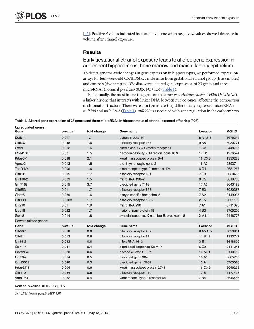
Early gestational ethanol exposure leads to altered gene expression inadolescent hippocampus, bone marrow and main olfactory epitheliumTo detect genome-wide changes in gene expression in hippocampus, we performed expressionarrays for four-week-old C57BL/6JRcc male mice from gestational ethanol group (five samples)and controls (five samples). We discovered altered gene expression of 23 genes and threemicroRNAs (nominal p-values<0.05, FC�1.5) (Table 1).
Functionally, the most interesting gene on the array was Histone cluster 1 H2ai (Hist1h2ai),a linker histone that interacts with linker DNA between nucleosomes, affecting the compactionof chromatin structure. There were also two interesting differentially expressed microRNAs:miR290 andmiR138-2 (Table 1).miR290 is associated with gene regulation in the early embryo
Table 1. Altered gene expression of 23 genes and three microRNAs in hippocampus of ethanol-exposed offspring (P28).
Upregulated genes:Gene p-value fold change Gene name Location MGI ID
Defb14 0.017 1.7 defensin beta 14 8 A1.3 8 2675345
Olfr937 0.048 1.6 olfactory receptor 937 9 A5 3030771
Cxcr1 0.012 1.6 chemokine (C-X-C motif) receptor 1 1 C3 2448715
H2-M10.3 0.03 1.5 histocompatibility 2, M region locus 10.3 17 B1 1276524
Krtap6-1 0.038 2.1 keratin associated protein 6–1 16 C3.3 1330228
Vpreb2 0.013 1.6 pre-B lymphocyte gene 2 16 A3 98937
Tas2r124 0.006 1.6 taste receptor, type 2, member 124 6 G1 2681267
Olfr601 0.005 1.7 olfactory receptor 601 7 E3 3030435
Mir138-2 0.023 1.5 microRNA 138–2 8 C5 3618733
Gm7168 0.015 3.7 predicted gene 7168 17 A2 3643198
Olfr553 0.01 1.7 olfactory receptor 553 7 E3 3030387
Obox5 0.039 1.6 oocyte specific homeobox 5 7 A2 2149035
Olfr1305 0.0003 1.7 olfactory receptor 1305 2 E5 3031139
Mir290 0.01 1.9 microRNA 290 7 A1 3711323
Mup18 0.022 1.7 major urinary protein 18 4 B3 3705220
Ssxb8 0.014 1.8 synovial sarcoma, X member B, breakpoint 8 X A1.1 2446777
Downregulated genes:
Gene p-value fold change Gene name Location MGI ID
Olfr967 0.018 0.6 olfactory receptor 967 9 A5.1; 9 3030801
Olfr51 0.012 0.6 olfactory receptor 51 11 B1.3 1333747
Mir16-2 0.032 0.6 microRNA 16–2 3 E1 3618690
C87414 0.041 0.4 expressed sequence C87414 5 E2 2141341
Hist1h2ai 0.023 0.6 histone cluster 1, H2ai 13 A3.1 2448457
Gm904 0.014 0.5 predicted gene 904 13 A5 2685750
Gm15632 0.048 0.5 predicted gene 15632 15 A1 3783076
Krtap27-1 0.004 0.6 keratin associated protein 27–1 16 C3.3 3646229
Olfr110 0.034 0.6 olfactory receptor 110 17 B1 2177493
Vmn2r64 0.032 0.4 vomeronasal type 2 receptor 64 7 B4 3646456
Nominal p-values <0.05, FC � 1.5.
doi:10.1371/journal.pone.0124931.t001
Effects of Early Alcohol Exposure
PLOS ONE | DOI:10.1371/journal.pone.0124931 May 13, 2015 9 / 20

and the maintenance of the pluripotent cell state [43], [44] andmiR138, the mature form ofmiR138-2, is associated with the size of dendritic spines in rat hippocampal neurons [45].
The group of genes with altered expression consists of a number of G-protein coupled che-mosensory receptors: olfactory receptor genes, which are actively expressed in main olfactoryepithelium (Olfr937, Olfr967, Olfr51, Olfr601, Olfr553, Olfr1305, Olfr110) and vomeronasal re-ceptor gene (Vmn2r64), which is known to be expressed in the epithelium of the vomeronasalorgan. Taste receptor, type 2,member 124 (Tas2r124) is expressed actively in taste receptor cellsin tongue and palate. Three of the up-regulated genes, defencing beta 14 (Defb14), chemokine(C-X-C motif) receptor 1 (Cxcr1) and pre-B lymphocyte gene 2 (Vpreb2), are known to be in-volved in the function or development of the immune system. H2-M10.3 belongs to the majorhistocompatibility complex class Ib molecules, but not to the family of classical class I MHCgenes, which present antigens to cytotoxic T cells.H2-M10 genes have been observed to havecomplex and nonrandom combinations of coexpressions in neurons with vomeronasal receptorgenes (Vmn2r) [46], [47]. A recent study has shown that H2-M10 genes are required for ultra-sensitive chemodetection by a subset of vomeronasal sensory neurons [48].
The expression levels of the differentially expressed genes observed in hippocampus werelow and impossible to verify by quantitative PCR (TaqMan). To verify our array results we in-vestigated if similar upregulated expression of Olfr601 and H2-M10.3 can be seen in both, hip-pocampus and MOE, and Vpreb2 in hippocampus and bone marrow. Interestingly, weobserved significant upregulated expression in two of the three genes in the tested tissues ofethanol-exposed offspring, Olfr601 in MOE and Vpreb2 in bone marrow (p = 0.04 andp<0.001, respectively, one-tailed Student’s t-test) (Fig 1). No significant difference was ob-served for H2-M10.3 expression between ethanol-exposed and control offspring in MOE(p = 0.35, one-tailed Student’s t-test).
Altered site-specific DNAmethylation in ethanol-exposed offspringTo obtain information about the epigenetic status of the array candidate genes we performedbisulphite sequencing for six CpG islands of five differentially expressed candidate genes. Twoof the candidate genes, Olfr110 and Vmn2r64, have CpG islands upstream of the genes andVmn2r64, Vpreb2, and Olfr601 have CpG islands in the gene-body (Fig 2). CpG islands of allthe genes except Hist1h2ai were highly methylated in both experimental groups (Fig 3). Theaverages of total methylation levels of all clones were reminiscent and there were no significantdifferences between ethanol-exposed offspring and controls (Fig 3). There was no difference inDNAmethylation between ethanol-exposed and control offspring in Hist1h2ai either: in bothgroups the CpG region that we investigated was hypomethylated.
In addition to overall DNA methylation of CpG islands, we calculated the CpG site-specificDNAmethylation in each CpG island (Fig 4). In ethanol-exposed offspring, we observed nota-ble hypermethylation in one CpG site in a CpG island upstream of Vmn2r64 (CpG1 p = 0.019,non-parametric Mann-Whitney) (Fig 4A), in addition to hypomethylation in one CpG site(CpG3 p = 0.023, Mann-Whitney), and hypermethylation in the next two CpG sites (CpG4p = 0.07, CpG5 p = 0.11, Mann-Whitney) in an island upstream of Olfr110 (Fig 4C). The ex-pressions of both genes were downregulated in ethanol-exposed offspring. Furthermore, wefound a significantly altered CpG site in exonic CpG island in Olfr601 (CpG1 p = 0.038, Mann-Whitney) (Fig 4E). This CpG site is hypomethylated and the gene expression is upregulated inhippocampus of ethanol-exposed offspring.
In addition to the hippocampus, we detected significantly upregulated expression of Olfr601also in MOE of ethanol-exposed offspring (Fig 1). Due to this, we also sequenced an exonicCpG island of Olfr601 in MOE to find potential similar changes in DNAmethylation caused
Effects of Early Alcohol Exposure
PLOS ONE | DOI:10.1371/journal.pone.0124931 May 13, 2015 10 / 20

by gestational ethanol-exposure. We did not observe similar significant ethanol-inducedchanges in DNA methylation in the hippocampus and MOE, suggesting that this particular ex-onic CpG site itself does not affect the regulation of Olfr601 expression (Fig 4F).
Structural brain abnormalities in ethanol-exposed offspringMRI was performed for 8 control and 13 ethanol-exposed male offspring (P60). The offspringexposed to ethanol showed a reduced body weight as compared to controls (d = -1.42) (Fig 5).
We found a small decrease in total brain volume (d = -0.29) after ethanol exposure (Fig 5).We measured the effect of ethanol exposure on the size of the hippocampus, OB, ventricles andcerebellum after normalizing their volumes to total brain volume. These regions have been themost severely affected in earlier studies overlapping our ethanol exposure period [42], [49],[50], [51]. There was a large increase in the volume of the hippocampi (d = 0.84), slightly in-creased lateral ventricular volume (d = 0.65) and a large decrease in OB (d = -0.85) in ethanol-exposed mice (Fig 5). These regions showed asymmetric changes between hemispheres: the lefthemisphere exhibited larger changes, such as a large increase in volume in the left hippocam-pus (d = 1.32) and left ventricle (d = 0.83) and a large decrease in left OB (d = -1.40) (Fig 5).We found a small decrease in the volume of both hemispheres without the contribution of thelateral ventricles (d = -0.24), however, individual hemispheres showed slight asymmetricchanges (left: d = -0.27; right: d = -0.18) (Fig 5). No differences were seen in cerebellar volumebetween controls and ethanol-exposed offspring (d = -0.02).
Examples of increase in ventricular volume are shown in Fig 6. Control mice showed smalllateral ventricular volume as compared to ethanol-exposed mice (Fig 6A and 6D), which variedbetween animals (Fig 6B,6C and 6E,6F). The most relevant volumetric findings are presentedin Fig 7. Left hippocampal volume in ethanol-exposed mice was significantly higher as com-pared to that in control mice (p<0.05, Mann-Whitney) and to the right hippocampus in the
Fig 5. Effect of ethanol exposure on brain regional volumes obtained fromMRI images, and body weight.Cohen´s d was calculated to study the effectsize between controls and ethanol-exposed adult male offspring (P60). Large effect size was considered when d values were� 0.8 (increase in volume) or�-0.8 (decrease in volume) after ethanol exposure (dashed line on d = ± 0.8).
doi:10.1371/journal.pone.0124931.g005
Effects of Early Alcohol Exposure
PLOS ONE | DOI:10.1371/journal.pone.0124931 May 13, 2015 11 / 20

same animals (p<0.001, Wilcoxon test) (Fig 7A). Left olfactory bulb volume was found to besignificantly lower as compared to that in control mice (p<0.05, Mann-Whitney) (Fig 7B). Theventricular volumes of both hemispheres appeared slightly larger than those in control mice,however, no significant differences were found (Fig 7C).
Discussion
Altered DNAmethylation and gene expression in ethanol-exposedoffspringWe carried out gene expression arrays for the hippocampus of four-week-old male offspring.According to our hypothesis, alcohol-induced alterations in the epigenome and gene expres-sion in early stage of development are permanent and can be observed later in life. Because theexpression profile of a cell and a tissue depends on its function and developmental period, it ischallenging to catch primary alterations in gene expressions later in development. Due to that,we took into consideration even very weakly expressed genes on our arrays despite the difficul-ties to confirm these alterations with other methods.
Fig 6. 3D visualization of the ventricular volumes of control and ethanol-exposedmice (P60). The toppanel shows lateral ventricles of a control (A) and two ethanol-exposed mice (B and C). Fractional anisotropymaps of the same animals are shown in the panel below (D-F). Red line outlines the left ventricle.
doi:10.1371/journal.pone.0124931.g006
Fig 7. Altered volumes of left and right hippocampi, olfactory bulbs, and lateral ventricles of ethanol-exposed offspring.Wilcoxon test was used toaccess left-right differences within the same animals (#) and Mann-Whitney test to compare control and ethanol-exposed (EtOH) mice (*). #/*p<0.05 and###p<0.001. Each dot represents an individual adult male mouse (P60). Control mice are illustrated in black and ethanol-exposed offspring in grey. Bars areaveraged values ± SD.
doi:10.1371/journal.pone.0124931.g007
Effects of Early Alcohol Exposure
PLOS ONE | DOI:10.1371/journal.pone.0124931 May 13, 2015 12 / 20

The microarrays revealed 23 candidate genes and three miRNAs: 15 of the 23 candidategenes belong to gene families that have been associated with gestational ethanol exposure inprevious mouse studies. The transcript levels of Cxcr3, Defb15, Defb30, Krtap4-7, Tas2r126,and Vmn2r54 have been reported to increase in the whole brain of C57BL/6J adult (P70) malemice that were exposed to maternal 10% ethanol consumption before fertilization, throughoutgestation, and ten days after birth (P10) [52]. In addition, Hist1h3a,Hist1h4i, andHist3h2awere all reported to downregulate in embryos that were cultured and treated with alcohol for46 hours at early neurulation (E8.25) [53]. Despite different timings and doses of ethanol expo-sure, the directions of altered expression of our candidate genes (Cxcr1, Defb14, Krtap6-1,Tas2r124, Hist1h2ai) and genes in the same families mentioned above, were similar. Only inone of the genes, Vmn2r64, the direction of change was opposite when compared with its fami-ly member Vmn2r54 in the earlier study [52]. Members of olfactory receptor (Olfr) and keratinassociated protein (Krtap) gene families were both up- and downregulated on our array, whichsuggests that the location and structure of chromatin could determine the effect of ethanol ongene expression. Another whole mouse-embryo culture study also revealed significant changesin DNA methylation in a large number of olfactory receptor genes and decreased methylationinHist1h3d [8], which made olfactory receptor genes andHist1h2ai on our expression arrayplausible candidate genes for methylation studies. In addition, Liu and colleagues observed aconsiderable amount of alcohol-induced changes in DNAmethylation on chromosome 7 [8],the most common location for our candidate genes: five of our 26 candidate genes and miR-NAs are located on this chromosome.
Hist1h2ai, the most distinctive candidate gene considering its function, had a lower expres-sion level in ethanol-exposed offspring. We examined only a part of a long CpG island, whichwas hypomethylated in both experimental groups. The expression arrays revealed seven differ-entially expressed olfactory receptor genes (Olfrs) in hippocampus of ethanol-exposed off-spring. Olfrs detect volatile chemicals leading to the initial perception of smell in the brain. Themembers of this large gene family are expressed normally in a monogenic and monoallelic fash-ion in olfactory sensory neurons in MOE. Their expression is regulated in a specific epigeneticmanner, which has been compared to phenomena like genomic imprinting or X-chromosomeinactivation. It has been suggested thatOlfrs are silenced in MOE by hallmarks of constitutiveheterochromatin and at a later stage an enzymatic activity removes these hallmarks from a sto-chastically chosen allele, allowing its transcriptional activity [54]. In addition to MOE, Olfrs areknown to also express in other tissues, such as brain, but their function remains unclear [55].
We searched for potential changes in DNAmethylation of Olfr110 and Vmn2r64. Theywere both downregulated in our hippocampus array in ethanol-exposed offspring and bothhave upstream CpG islands. Although the overall methylation level of the CpG islands wassimilar between the experimental groups, we observed ethanol-induced CpG site-specific alter-ations in four sites. These upstream regions could have a role in gene regulation, for example asenhancers. Hypermethylated sites in both regulatory regions can disturb the binding of tran-scription factors and thus slightly suppress gene expression. Although there is earlier evidenceof site-specific DNA methylation associating with decreased transcription [56], [57], functionalexperiments will be needed to clarify whether these alterations in DNAmethylation are capableof reducing the transcription of Vmn2r64 and Olfr110.
Expression array results are supported by our TaqMan study. Regardless of the difference be-tween cell types and epigenetic profiles in these three tissues, it was possible to detect significant-ly upregulated expression in two of the three tested genes. Olfr601 was upregulated in both thehippocampus andMOE of ethanol-exposed offspring. Vpreb2 was upregulated in the hippocam-pus and startlingly overexpressed in bone marrow. Vpreb2 is one of two mouse pre-B lymphocytegenes, Vpreb1 and Vpreb2, that are involved in B-cell development. A previous study has shown
Effects of Early Alcohol Exposure
PLOS ONE | DOI:10.1371/journal.pone.0124931 May 13, 2015 13 / 20

that the expression of Vpreb2 is significantly lower compared to Vpreb1 and it is not needed forthe normal development of B cells [58]. Due to the unclear function of Vpreb2, further researchis needed to clarify if this alteration is caused by alcohol-induced activation of the immune sys-tem or dysfunction of epigenetic gene regulation caused by gestational ethanol exposure.
We observed higher methylation in ethanol-exposed offspring in the exonic CpG island ofthe upregulated Vpreb2 in the hippocampus. Gene-body DNA hypermethylation has been ob-served to correlate positively with gene expression levels in previous studies [59], [60]. However,we cannot rule out hydroxymethylation, which has recently been found; especially in embryonicstem cells, adult nervous system, and bone marrow [61], [62]. It has been suggested that hydro-xymethylation has a role in DNA demethylation and enhanced gene expression level [63], [64].
Changes in brain structure caused by gestational alcohol exposureThe most interesting finding concerning brain structure in our mouse model is a trend ofasymmetry in hippocampi: enlarged left hippocampus in ethanol-exposed offspring. Our resultis similar to a human study, where hippocampal volumes in male adolescents with and withouta family history of alcoholism, prior to the initiation of alcohol use of the children themselves,was examined [65]. The participants were typically from upper-middle to upper-class familiesand they were excluded for any current or past medical, physical, or psychiatric problems [65],[66]. It is tempting to hypothesize that the hippocampal phenotype in this study is primarilycaused by early gestational alcohol exposure as in our mouse model. Interestingly, enlargementof hippocampus has been associated with longer-lasting spatial memory in birds [67], [68], andan altered shape of hippocampus with taxi driving in London [69]. Enlarged hippocampus hasbeen observed in autistic children [70], [71] and may also be related to memory function inthis disorder [71]. Furthermore, increased hippocampal cell density and improved perfor-mance in the Morris water-maze learning paradigm have been detected in the valproic acid ratmodel of autism [72]. Whether the larger left hippocampus is causing improvement in spatialmemory observed in the Morris water-maze in the mouse model we have used in this study[20] requires further research. Although numerous studies with rodents have shown that pre-natal ethanol exposure impairs offspring’s spatial memory [73], [74], [75], [76], no effects oreven subtle improvement in hippocampal-dependent learning and memory tasks have alsobeen observed [77], [78], [79], [80], [81]. In addition to dose [80], timing of ethanol exposureseems to be significant for the function of the hippocampus: acute ethanol exposure at postna-tal day 7 (P7) causes impairment in hippocampal-dependent spatial memory in adult mice,whereas there is no difference between controls and early ethanol-exposed (E8) offspring [81].
We also detected smaller OBs in the ethanol-exposed offspring. Reduced volume of OB inethanol-exposed offspring (P60), and also impaired odour discrimination, have been observedin a mouse model, in which C57BL/6J dams consume 10% ethanol solution throughout preg-nancy [42]. In this model, the exposure was initiated before fertilization and gradually de-creased after the birth. They observed a decreased number of neural precursor cells in thesubependymal zone of the lateral ventricles, which is the region of adult neural stem cells. Theyalso detected a decreased number of new cells in the OB during the first few postnatal weeks.Our study supports this result and indicates that ethanol exposure only for the first eight daysafter fertilization is capable of reducing OB volume. Smaller OBs have also been seen in GD17embryos in a study where pregnant C57BL/6J mice were administered a strong intraperitonealethanol dose twice at GD8 [49].
MRI revealed enlargement of lateral ventricles in the ethanol-exposed offspring. Similar re-sults have been seen in MRIs also in other FASD animal models [49], [50], [82], [83], [84], [85]and in clinical studies of prenatal alcohol exposure [86], [87]. Alcohol-induced facial asymmetry
Effects of Early Alcohol Exposure
PLOS ONE | DOI:10.1371/journal.pone.0124931 May 13, 2015 14 / 20

[88] and altered symmetry of brain structures [65], [89], [90] have been observed in human stud-ies. In our work, we detected more structural changes in the left side of the brain unlike in theprevious MRI study of gestational day 17 (GD17) embryos, whose dams were exposed for acuteethanol insult GD8, in early neurulation stage [49]. They detected the most marked dispropor-tional reduction of regional brain volumes on the right side and the changes were significant inOB, hippocampus, and cerebellum. No difference in the size of cerebellums was observed here,but in both studies, decreased OB volumes and increased ventricular volumes were detected.
In addition to the volumetric analysis, we performed ROI analysis of the dentate gyrus, genu,and body of the corpus callosum and anterior commissure to search microstructural alterationsdue to the ethanol exposure. No DTI parameters (fractional anisotropy, and axial, radial, andmean diffusivities) showed significant differences when compared to control and ethanol-ex-posed mice in those areas (data not shown). More detail analysis of DTI data needs further in-vestigation in white and grey matter areas to study the effect of ethanol in tissue microstructure.
ConclusionsOur study has demonstrated that early, chronic and moderate gestational ethanol exposure af-fects the development of the embryo and these early changes can be seen in altered DNAmeth-ylation, gene expression, and brain structure in later life. The ethanol exposure period in ourmouse model developmentally corresponds to the first three-four weeks of human pregnancy,a time period when women are often not aware of their pregnancy. Our results strengthen thesignificance of environmental factors in early pregnancy and support the role of the epigenomein this interaction.
All the tissues we have studied are regions of actively proliferating stem cells: neuronal pro-genitor cells in hippocampus, OB (central nervous system) and MOE (peripheral nervous sys-tem), and hematopoietic stem cells in bone marrow. This supports our hypothesis of earlyalcohol-induced epigenetic changes: relatively subtle alterations in the gene regulation couldhave occurred already in stem cells, affecting both neuronal and hematopoietic cell lines, andcan be observed in adult brain structures as well as altered gene expression in brain, bone mar-row, and MOE. Although there is an increasing amount of studies that support epigenome in-volvement in the effects of gestational ethanol exposure, the linkage between early epigeneticchanges, altered gene expression, and phenotype characteristics for FASD has not yet beenfound. In our ongoing work, we aim to reveal the causal epigenetic alterations in early embry-onic development and thus increase the understanding of the molecular mechanism behindFASD. Epigenetic changes that alter gene regulation could be considered to be biomarkers andwould offer a new tool for the challenging diagnostics of alcohol-induced developmental disor-ders. Biomarkers in blood, or other available biological samples, would indicate the severity ofdamage caused by early ethanol exposure. This would enable early diagnosis and appropriatesupport for development.
Supporting InformationS1 Table. Sequences of primers used in the study.(XLSX)
AcknowledgmentsWe thank Iiris Hovatta for teaching us brain dissections and Staffan Bohm for guiding us inmain olfactory epithelium dissection. We also thank Arttu Ahola for discussions and helpfulcomments, and Anni Moilanen and Mia Toivonen for technical assistance.
Effects of Early Alcohol Exposure
PLOS ONE | DOI:10.1371/journal.pone.0124931 May 13, 2015 15 / 20

Author ContributionsConceived and designed the experiments: HM OG NK. Performed the experiments: HM ASAN AML NK. Analyzed the data: HM AS VR SH NK. Contributed reagents/materials/analysistools: AML OG SH NK. Wrote the paper: HM AS NK.
References1. Abel EL, Hannigan JH. Maternal risk factors in fetal alcohol syndrome: provocative and permissive in-
fluences. Neurotoxicol Teratol 17: 445–462. Review. Erratum in: Neurotoxicol Teratol. 1995; 17: 689.PMID: 7565491
2. Sokol RJ, Delaney-Black V, Nordstrom B. Fetal alcohol spectrum disorder. Jama. 2003; 290: 2996–2999. PMID: 14665662
3. Maier SE, West JR. Drinking patterns and alcohol-related birth defects. Alcohol Res Health. 2001; 25:168–174. PMID: 11810954
4. Sulik KK. Critical periods for alcohol teratogenesis in mice, with special reference to the gastrulationstage of embryogenesis. Ciba found Symp. 1984; 105: 124–141. PMID: 6563984
5. Guerri C. Mechanisms involved in central nervous system dysfunctions induced by prenatal ethanol ex-posure. Neurotox. 2002; Res 4: 327–335. PMID: 12829422
6. Sulik KK. Genesis of alcohol-induced craniofacial dysmorphism. Exp Biol Med (Maywood). 2005; 230:366–375. PMID: 15956766
7. Feil R, Fraga MF. Epigenetics and the environment: emerging patterns and implications. Nat RevGenet. 2012; 13: 97–109. doi: 10.1038/nrg3142 PMID: 22215131
8. Liu Y, Balaraman Y, Wang G, Nephew KP, Zhou FC. Alcohol exposure alters DNAmethylation profilesin mouse embryos at early neurulation. Epigenetics. 2009; 4: 500–511. PMID: 20009564
9. Kaminen-Ahola N, Ahola A, Maga M, Mallitt KA, Fahey P, Cox TC, et al. Maternal ethanol consumptionalters the epigenotype and the phenotype of offspring in a mouse model. PLoS Genet 2010; 6:e1000811. doi: 10.1371/journal.pgen.1000811 PMID: 20084100
10. Zhou FC, Balaraman Y, Teng M, Liu Y, Singh RP, Nephew KP. Alcohol alters DNAmethylation patternsand inhibits neural stem cell differentiation. Alcohol Clin Exp Res. 2011; 35: 735–746. doi: 10.1111/j.1530-0277.2010.01391.x PMID: 21223309
11. Laufer BI, Mantha K, Kleiber ML, Diehl EJ, Addison SM, Singh SM. Long-lasting alterations to DNAmethylation and ncRNAs could underlie the effects of fetal alcohol exposure in mice. Dis Model Mech.2013; 6: 977–992. doi: 10.1242/dmm.010975 PMID: 23580197
12. Reik W, DeanW, Walter J. Epigenetic reprogramming in mammalian development. Science. 2001;293: 1089–1093. PMID: 11498579
13. Hemberger M, DeanW, Reik W. Epigenetic dynamics of stem cells and cell lineage commitment: dig-ging waddington's canal. Nat Rev Mol Cell Biol. 2009; 10: 526–537. doi: 10.1038/nrm2727 PMID:19603040
14. Morgan HD, Jin XL, Li A, Whitelaw E, O'Neill C. The culture of zygotes to the blastocyst stage changesthe postnatal expression of an epigentically labile allele, agouti viable yellow, in mice. Biol Reprod.2008; 79: 618–623. doi: 10.1095/biolreprod.108.068213 PMID: 18562706
15. Rivera RM, Stein P, Weaver JR, Mager J, Schultz RM, Bartolomei MS. Manipulations of mouse embry-os prior to implantation result in aberrant expression of imprinted genes on day 9.5 of development.HumMol Genet. 2008; 17: 1–14. PMID: 17901045
16. Heijmans BT, Tobi EW, Stein AD, Putter H, Blauw GJ, Susser ES, et al. Persistent epigenetic differ-ences associated with prenatal exposure to famine in humans. Proc Natl Acad Sci U S A. 2008; 105:17046–17049. doi: 10.1073/pnas.0806560105 PMID: 18955703
17. Tobi EW, Slagboom PE, van Dongen J, Kremer D, Stein AD, Putter H, et al. Prenatal famine and genet-ic variation are independently and additively associated with DNAmethylation at regulatory loci withinIGF2/H19. PLoS One. 2012; 7: e37933. doi: 10.1371/journal.pone.0037933 PMID: 22666415
18. Susiarjo M, Sasson I, Mesaros C, Bartolomei MS (2013) Bisphenol a exposure disrupts genomic im-printing in the mouse. PLoS Genet 9: e1003401. doi: 10.1371/journal.pgen.1003401 PMID: 23593014
19. Kaminen-Ahola N, Ahola A, Flatscher-Bader T, Wilkins SJ, Anderson GJ, Whitelaw E, et al. (2010)Postnatal growth restriction and gene expression changes in a mouse model of fetal alcohol syndrome.Birth Defects Res A Clin Mol Teratol 88: 818–826. doi: 10.1002/bdra.20729 PMID: 20878912
Effects of Early Alcohol Exposure
PLOS ONE | DOI:10.1371/journal.pone.0124931 May 13, 2015 16 / 20

20. Sanchez Vega MC, Chong S, Burne TH. Early gestational exposure to moderate concentrations of eth-anol alters adult behaviour in C57BL/6J mice. Behav Brain Res. 2013; 252: 326–333. doi: 10.1016/j.bbr.2013.06.003 PMID: 23756143
21. McClearn G, Rodgers D. Differences in alcohol preference among inbred strains of mice. Q J Stud Alco-hol. 1959; 20: 691–695.
22. Belknap JK, Crabbe JC, Young ER. Voluntary consumption of ethanol in 15 inbred mouse strains.Psychopharmacology (Berl). 1993; 112: 503–510. PMID: 7871064
23. Barnes DE, Walker DW. Prenatal ethanol exposure permanently reduces the number of pyramidal neu-rons in rat hippocampus. Brain Res. 1981; 227: 333–340. PMID: 7260643
24. Diaz Perez H, Espinosa Villanueva J, Machado Salas J. Behavioral and hippocampal morphologicalchanges induced by ethanol administered to pregnant rats. Ann N Y Acad Sci. 1991; 625: 300–4.PMID: 2058890
25. Miller MW. Generation of neurons in the rat dentate gyrus and hippocampus: effects of prenatal andpostnatal treatment with ethanol. Alcohol Clin Exp Res. 1995; 19: 1500–9. PMID: 8749817
26. Choi IY 1, Allan AM, Cunningham LA. Moderate fetal alcohol exposure impairs the neurogenic re-sponse to an enriched environment in adult mice. Alcohol Clin Exp Res. 2005; 29: 2053–62. PMID:16340464
27. Gil-Mohapel J, Titterness AK, Patten AR, Taylor S, Ratzlaff A, Ratzlaff T, et al. Prenatal ethanol expo-sure differentially affects hippocampal neurogenesis in the adolescent and aged brain. Neuroscience.2014; 273: 174–88. doi: 10.1016/j.neuroscience.2014.05.012 PMID: 24846617
28. West JR, Hodges CA, Black AC Jr. Prenatal exposure to ethanol alters the organization of hippocampalmossy fibers in rats. Science. 1981; 211: 957–9. PMID: 7466371
29. Abel EL, Jacobson S, Sherwin BT. In utero alcohol exposure: functional and structural brain damage.Neurobehav Toxicol Teratol. 1983; 5: 363–6. PMID: 6877477
30. Allan AM, Chynoweth J, Tyler LA, Caldwell KK. A mouse model of prenatal ethanol exposure using avoluntary drinking paradigm. 2003; Alcohol Clin Exp Res 27: 2009–16. PMID: 14691390
31. WHO (World Health Organization) World Health Report 2002: Reducing Risks, Promoting Healthy Life,World Health Organization, Geneva. PMID: 14741909
32. Brazma A, Hingamp P, Quackenbush J, Sherlock G, Spellman P, Stoeckert C, et al. Minimum informa-tion about a microarray experiment (MIAME)-toward standards for microarray data. Nat Genet. 2001;29: 365–371. PMID: 11726920
33. Dalma-Weiszhausz DD, Warrington J, Tanimoto EY, Miyada CG. The affymetrix GeneChip platform:an overview. In DNAMicroarrays, Part A: Array Platforms andWet-Bench Protocols, Methods in Enzy-mology (ed. Kimmel A. and Oliver B.),2006; 410 pp. 3–28. San Diego: Elsevier Academic Press.PMID: 16938544
34. Chen P, Lepikhova T, Hu Y, Monni O, Hautaniemi S. Comprehensive exon array data processing meth-od for quantitative analysis of alternative spliced variants. Nucleic Acids Res. 2011; 39: 123.
35. Ovaska K, Laakso M, Haapa-Paananen S, Louhimo R, Chen P, Aittomäki V, et al. Large-scale data in-tegration framework provides a comprehensive view on glioblastoma multiforme. GenomeMed. 2010;2: 65. doi: 10.1186/gm186 PMID: 20822536
36. Carnahan MN, Veazey KJ, Muller D, Tingling JD, Miranda RC, Golding MC. Identification of cell-specif-ic patterns of reference gene stability in quantitative reverse-transcriptase polymerase chain reactionstudies of embryonic, placental and neural stemmodels of prenatal ethanol exposure. Alcohol. 2013;47: 109–120. doi: 10.1016/j.alcohol.2012.12.003 PMID: 23317542
37. Livak KJ, Schmittgen TD. Analysis of relative gene expression data using real-time quantitative PCRand the 2(-delta delta C(T)) method. Methods. 2001; 25: 402–408. PMID: 11846609
38. Bock C, Reither S, Mikeska T, Paulsen M, Walter J, Lengauer T. BiQ analyzer: visualization and qualitycontrol for DNAmethylation data from bisulfite sequencing. Bioinformatics. 2005; 21: 4067–4068.PMID: 16141249
39. Jenkinson M, Smith S. A global optimisation method for robust affine registration of brain images. MedImage Anal. 2001; 5: 143–156. PMID: 11516708
40. Jenkinson M, Bannister P, Brady M, Smith S. Improved optimization for the robust and accurate linearregistration and motion correction of brain images. Neuroimage. 2001; 17: 825–841.
41. Basser PJ, Pierpaoli C. Microstructural and physiological features of tissues elucidated by quantitative-diffusion-tensor MRI. J Magn Reson B. 1996; 111: 209–219. PMID: 8661285
42. Akers KG, Kushner SA, Leslie AT, Clarke L, van der Kooy D, Lerch JP, et al. Fetal alcohol exposureleads to abnormal olfactory bulb development and impaired odor discrimination in adult mice. MolBrain. 2011; 4: 29. doi: 10.1186/1756-6606-4-29 PMID: 21736737
Effects of Early Alcohol Exposure
PLOS ONE | DOI:10.1371/journal.pone.0124931 May 13, 2015 17 / 20

43. Houbaviy HB, Murray MF, Sharp PA. Embryonic stem cell-specific MicroRNAs. Dev Cell. 2003; 5:351–358. PMID: 12919684
44. Tata PR, Tata NR, Kuhl M, Sirbu IO. Identification of a novel epigenetic regulatory region within the plur-ipotency associated microRNA cluster, EEmiRC. Nucleic Acids Res. 2011; 39: 3574–3581. doi: 10.1093/nar/gkq1344 PMID: 21247880
45. Siegel G, Obernosterer G, Fiore R, OehmenM, Bicker S, Christensen M, et al. A functional screen im-plicates microRNA-138-dependent regulation of the depalmitoylation enzyme APT1 in dendritic spinemorphogenesis. Nat Cell Biol. 2009; 11: 705–716. doi: 10.1038/ncb1876 PMID: 19465924
46. Ishii T, Hirota J, Mombaerts P. Combinatorial coexpression of neural and immune multigene families inmouse vomeronasal sensory neurons. Curr Biol. 2003; 13: 394–400. PMID: 12620187
47. Loconto J, Papes F, Chang E, Stowers L, Jones EP, Takada T, et al. Functional expression of murineV2R pheromone receptors involves selective association with the M10 and M1 families of MHC class ibmolecules. Cell. 2003; 112: 607–618. PMID: 12628182
48. Leinders-Zufall T, Ishii T, Chamero P, Hendrix P, Oboti L, Schmid A, et al. A family of nonclassical classI MHC genes contributes to ultrasensitive chemodetection by mouse vomeronasal sensory neurons. JNeurosci. 2014; 34: 5121–5133. doi: 10.1523/JNEUROSCI.0186-14.2014 PMID: 24719092
49. Parnell SE, O'Leary-Moore SK, Godin EA, Dehart DB, Johnson BW, Allan Johnson G, et al. Magneticresonancemicroscopy defines ethanol-induced brain abnormalities in prenatal mice: effects of acute in-sult on gestational day 8. Alcohol Clin Exp Res. 2009; 33: 1001–1011. doi: 10.1111/j.1530-0277.2009.00921.x PMID: 19302087
50. Godin EA, O'Leary-Moore SK, Khan AA, Parnell SE, Ament JJ, Dehart DB, et al. Magnetic resonancemicroscopy defines ethanol-induced brain abnormalities in prenatal mice: effects of acute insult on ges-tational day 7. Alcohol Clin Exp Res. 2010; 34: 98–111. doi: 10.1111/j.1530-0277.2009.01071.x PMID:19860813
51. Parnell SE, Holloway HE, Baker LK, Styner MA, Sulik KK. Dysmorphogenic effects of first trimester-equivalent ethanol exposure in mice: a magnetic resonance microscopy-based study. Alcohol Clin ExpRes. 2014; 38: 2008–2014. doi: 10.1111/acer.12464 PMID: 24931007
52. Kleiber ML, Laufer BI, Wright E, Diehl EJ, Singh SM. Long-term alterations to the brain transcriptome ina maternal voluntary consumption model of fetal alcohol spectrum disorders. Brain Res. 2012; 1458:18–33. doi: 10.1016/j.brainres.2012.04.016 PMID: 22560501
53. Zhou FC, Zhao Q, Liu Y, Goodlett CR, Liang T, McClintick JN, et al. Alteration of gene expression by al-cohol exposure at early neurulation. BMCGenomics. 2011; 12: 124. doi: 10.1186/1471-2164-12-124PMID: 21338521
54. Magklara A, Yen A, Colquitt BM, Clowney EJ, Allen W, Markenscoff-Papadimitriou E, et al. An epige-netic signature for monoallelic olfactory receptor expression. Cell. 2011; 145: 555–570. doi: 10.1016/j.cell.2011.03.040 PMID: 21529909
55. Kang N, Koo J. Olfactory receptors in non-chemosensory tissues. BMB Rep. 2012; 45: 612–622.PMID: 23186999
56. Martinowich K, Hattori D, Wu H, Fouse S, He F, Hu Y, et al. DNAmethylation-related chromatin remod-eling in activity-dependent BDNF gene regulation. Science. 2003; 302: 890–893. PMID: 14593184
57. Jones B, Chen J. Inhibition of IFN-gamma transcription by site-specific methylation during T helper celldevelopment. EMBO J. 2006; 25: 2443–2452. PMID: 16724115
58. Mundt C, Licence S, Maxwell G, Melchers F, Martensson IL. Only VpreB1, but not VpreB2, is express-ed at levels which allow normal development of B cells. Int Immunol. 2006; 18: 163–172. PMID:16361315
59. Rauch TA, Wu X, Zhong X, Riggs AD, Pfeifer GP. A human B cell methylome at 100-base pair resolu-tion. Proc Natl Acad Sci U S A. 2009; 106: 671–678. doi: 10.1073/pnas.0812399106 PMID: 19139413
60. Aran D, Toperoff G, Rosenberg M, Hellman A. Replication timing-related and gene body-specific meth-ylation of active human genes. HumMol Genet. 2011; 20: 670–680. doi: 10.1093/hmg/ddq513 PMID:21112978
61. Kriaucionis S, Heintz N. The nuclear DNA base 5-hydroxymethylcytosine is present in purkinje neuronsand the brain. Science. 2009; 324: 929–930. doi: 10.1126/science.1169786 PMID: 19372393
62. Ruzov A, Tsenkina Y, Serio A, Dudnakova T, Fletcher J, Bai Y, et al. Lineage-specific distribution ofhigh levels of genomic 5-hydroxymethylcytosine in mammalian development. Cell Res. 2011; 21:1332–1342. doi: 10.1038/cr.2011.113 PMID: 21747414
63. Tahiliani M, Koh KP, Shen Y, Pastor WA, Bandukwala H, Brudno Y, et al. Conversion of 5-methylcyto-sine to 5-hydroxymethylcytosine in mammalian DNA by MLL partner TET1. Science. 2009; 324: 930–935. doi: 10.1126/science.1170116 PMID: 19372391
Effects of Early Alcohol Exposure
PLOS ONE | DOI:10.1371/journal.pone.0124931 May 13, 2015 18 / 20

64. Guo JU, Su Y, Zhong C, Ming GL, Song H. Hydroxylation of 5-methylcytosine by TET1 promotes activeDNA demethylation in the adult brain. Cell. 2011; 145: 423–434. doi: 10.1016/j.cell.2011.03.022 PMID:21496894
65. Hanson KL, Medina KL, Nagel BJ, Spadoni AD, Gorlick A, Tapert SF. Hippocampal volumes in adoles-cents with and without a family history of alcoholism. Am J Drug Alcohol Abuse. 2010; 36: 161–167.doi: 10.3109/00952991003736397 PMID: 20465374
66. Nagel BJ, Schweinsburg AD, Phan V, Tapert SF. Reduced hippocampal volume among adolescentswith alcohol use disorders without psychiatric comorbidity. Psychiatry Res. 2005; 139: 181–190.PMID: 16054344
67. Clayton NS, Krebs JR. Hippocampal growth and attrition in birds affected by experience. Proc NatlAcad Sci U S A. 1994; 91: 7410–7414. PMID: 8052598
68. Biegler R, McGregor A, Krebs JR, Healy SD. A larger hippocampus is associated with longer-lastingspatial memory. Proc Natl Acad Sci U S A. 2001; 98: 6941–6944. PMID: 11391008
69. Maguire EA, Gadian DG, Johnsrude IS, Good CD, Ashburner J, Frackowiak RS, et al. Navigation-relat-ed structural change in the hippocampi of taxi drivers. Proc Natl Acad Sci U S A. 2000; 97: 4398–4403.PMID: 10716738
70. Sparks BF, Friedman SD, Shaw DW, Aylward EH, Echelard D, Artru AA, et al. Brain structural abnor-malities in young children with autism spectrum disorder. Neurology. 2002; 59: 184–192. PMID:12136055
71. Schumann CM, Hamstra J, Goodlin-Jones BL, Lotspeich LJ, Kwon H, Reiss AL, et al. The amygdala isenlarged in children but not adolescents with autism; the hippocampus is enlarged at all ages. J Neu-rosci. 2004; 24: 6392–6401. PMID: 15254095
72. Edalatmanesh MA, Nikfarjam H, Vafaee F, Moghadas M. Increased hippocampal cell density and en-hanced spatial memory in the valproic acid rat model of autism. Brain Res. 2013; 1526: 15–25. doi: 10.1016/j.brainres.2013.06.024 PMID: 23806776
73. Blanchard BA, Riley EP, Hannigan JH. Deficits on a spatial navigation task following prenatal exposureto ethanol. Neurotoxicol Teratol. 1987; 9:253–258. PMID: 3627089
74. Gianoulakis C. Rats exposed prenatally to alcohol exhibit impairment in spatial navigation test. BehavBrain Res. 1990; 36:217–228. PMID: 2310487
75. Richardson DP, Byrnes ML, Brien JF, Reynolds JN, Dringenberg HC. Impaired acquisition in the watermaze and hippocampal long-term potentiation after chronic prenatal ethanol exposure in the guinea-pig. Eur J Neurosci. 2002; 16:1593–1598. PMID: 12405973
76. Christie BR, Swann SE, Fox CJ, Froc D, Lieblich SE, Redila V, et al. Voluntary exercise rescues deficitsin spatial memory and long-term potentiation in prenatal ethanol-exposed male rats. Eur J Neurosci.2005; 21: 1719–1726. PMID: 15845099
77. Osborne GL, Caul WF, Fernandez K. Behavioral effects of prenatal ethanol exposure and differentialearly experience in rats. Pharmacol Biochem Behav. 1980; 12: 393–401. PMID: 7393938
78. Vorhees CV, Fernandez K. Effects of short-term prenatal alcohol exposure on maze, activity, and olfac-tory orientation performance in rats. Neurobehav Toxicol Teratol. 1986; 8:23–28. PMID: 3703092
79. Clausing P, Ferguson SA, Holson RR, Allen RR, Paule MG. Prenatal ethanol exposure in rats: long-lasting effects on learning. Neurotoxicol Teratol. 1995; 17: 545–552. PMID: 8552000
80. Cullen CL, Burne TH, Lavidis NA, Moritz KM. Low dose prenatal alcohol exposure does not impair spa-tial learning and memory in two tests in adult and aged rats. PLoS One. 2014; 9: e101482. doi: 10.1371/journal.pone.0101482 PMID: 24978807
81. Sadrian B, Lopez-Guzman M, Wilson DA, Saito M. Distinct neurobehavioral dysfunction based on thetiming of developmental binge-like alcohol exposure. Neuroscience. 2014; 280: 204–219. doi: 10.1016/j.neuroscience.2014.09.008 PMID: 25241068
82. Mattson SN, Riley EP, Jernigan TL, Garcia A, KanekoWM, Ehlers CL, et al. A decrease in the size ofthe basal ganglia following prenatal alcohol exposure: a preliminary report. Neurotoxicol Teratol. 1994;16: 283–289. PMID: 7935262
83. Zhou FC, Sari Y, Powrozek T, Goodlett CR, Li TK. Moderate alcohol exposure compromises neuraltube midline development in prenatal brain. Brain Res Dev Brain Res. 2003; 144: 43–55. PMID:12888216
84. Sakata-Haga H, Sawada K, Ohnishi T, Fukui Y. Hydrocephalus following prenatal exposure to ethanol.Acta Neuropathol. 2004; 108: 393–398. PMID: 15365720
85. O'Leary-Moore SK, Parnell SE, Godin EA, Dehart DB, Ament JJ, Khan AA, et al. Magnetic resonancemicroscopy-based analyses of the brains of normal and ethanol-exposed fetal mice. Birth Defects ResA Clin Mol Teratol. 2010; 88:953–964. doi: 10.1002/bdra.20719 PMID: 20842647
Effects of Early Alcohol Exposure
PLOS ONE | DOI:10.1371/journal.pone.0124931 May 13, 2015 19 / 20

86. Johnson VP, Swayze VW II, Sato Y, Andreasen NC. Fetal alcohol syndrome: craniofacial and centralnervous systemmanifestations. Am J Med Genet. 1996; 61: 329–339. PMID: 8834044
87. Swayze VW 2nd, Johnson VP, Hanson JW, Piven J, Sato Y, Gledd JN, et al. Magnetic resonance imag-ing of brain anomalies in fetal alcohol syndrome. Pediatrics. 1997; 99: 232–240. PMID: 9024452
88. Klingenberg CP, Wetherill L, Rogers J, Moore E, Ward R, Autti-Rämö I, et al. Prenatal Alcohol Expo-sure Alters the Patterns of Facial Asymmetry. Alcohol. 2010; 44: 649–657. doi: 10.1016/j.alcohol.2009.10.016 PMID: 20060678
89. Riikonen R, Salonen I, Partanen K and Verho S. Brain perfusion SPECT and MRI in foetal alcohol syn-drome. Dev Med Child Neurol. 1999; 41: 652–659. PMID: 10587040
90. Sowell ER, Thompson PM, Peterson BS, Mattson SN, Welcome SE, Henkenius AL, et al. Mapping cor-tical gray matter asymmetry patterns in adolescents with heavy prenatal alcohol exposure. Neuro-image. 2002; 17: 1807–1819. PMID: 12498754
Effects of Early Alcohol Exposure
PLOS ONE | DOI:10.1371/journal.pone.0124931 May 13, 2015 20 / 20
Related Documents