919 J. Exp. Med. The Rockefeller University Press • 0022-1007/98/09/919/12 $2.00 Volume 188, Number 5, September 7, 1998 919–930 http://www.jem.org Dual Signaling of the Fas Receptor: Initiation of Both Apoptotic and Necrotic Cell Death Pathways By Dominique Vercammen, Greet Brouckaert, Geertrui Denecker, Marc Van de Craen, Wim Declercq, Walter Fiers, and Peter Vandenabeele From the Department of Molecular Biology, Flanders Interuniversity Institute for Biotechnology and University of Ghent, B-9000 Ghent, Belgium Summary Murine L929 fibrosarcoma cells were transfected with the human Fas (APO-1/CD95) receptor, and the role of various caspases in Fas-mediated cell death was assessed. Proteolytic activation of procaspase-3 and -7 was shown by Western analysis. Acetyl-Tyr-Val-Ala-Asp-chlorometh- ylketone and benzyloxycarbonyl-Asp(OMe)-Glu(OMe)-Val-Asp(OMe)-fluoromethylketone, tetrapeptide inhibitors of caspase-1– and caspase-3–like proteases, respectively, failed to block Fas-induced apoptosis. Unexpectedly, the broad-spectrum caspase inhibitors benzyloxycarbo- nyl-Val-Ala-Asp(OMe)-fluoromethylketone and benzyloxycarbonyl-Asp(OMe)-fluorometh- ylketone rendered the cells even more sensitive to Fas-mediated cell death, as measured after 18 h incubation. However, when the process was followed microscopically, it became clear that anti-Fas–induced apoptosis of Fas-transfected L929 cells was blocked during the first 3 h, and subsequently the cells died by necrosis. As in tumor necrosis factor (TNF)-induced necrosis, Fas treatment led to accumulation of reactive oxygen radicals, and Fas-mediated necrosis was inhibited by the oxygen radical scavenger butylated hydroxyanisole. However, in contrast to TNF, anti-Fas did not activate the nuclear factor kB under these necrotic conditions. These re- sults demonstrate the existence of two different pathways originating from the Fas receptor, one rapidly leading to apoptosis, and, if this apoptotic pathway is blocked by caspase inhibitors, a second directing the cells to necrosis and involving oxygen radical production. Key words: Fas antigen • apoptosis • necrosis • caspases • oxygen radicals D epending on the cell type and the stimulus, a cell may die in either of two distinct ways: apoptosis or necro- sis. Necrosis is characterized by swelling of the cell and the organelles, and results in disruption of the cell membrane and in lysis (1). Release of the cytoplasmic content leads to an inflammatory response. Apoptosis can be recognized by membrane blebbing, shrinking and condensing of the cells and their organelles, internucleosomal degradation of the DNA, and disintegration of the cell, after which the fragments are phagocytosed by neighboring cells (2, 3). Apoptosis occurs at various stages in the life cycle of mul- ticellular organisms. From embryonic development through immunological regulation, as well as part of pathological situations, cell death by apoptosis is the most common way to eliminate superfluous, inappropriate, or harmful cells (4). However, inappropriate or defective apoptosis is the cause of many human diseases (5). Therefore, many studies aim to elucidate the apoptotic pathways and inter- actions between proapoptotic and antiapoptotic cellular components. Although mechanisms signaling to necrosis cannot be fully explained in molecular terms, apoptosis is generally believed to be mediated by caspase activation (6–8). The TNF receptor type 1 (TNFR-1) and Fas (APO-1/ CD95) are two cell surface receptors sharing structural and functional homology. Both are characterized by extracellu- lar repeats of cysteine-rich domains (9), but TNFR-1 and Fas also have a common intracellular motif, called the “death domain” (DD), 1 of z80 amino acids (10, 11). These 1 Abbreviations used in this paper: Ac-DEVD-amc, acetyl-Asp(OMe)- Glu(OMe)-Val-Asp(OMe)-aminomethylcoumarin; Ac-YVAD-amc, acetyl- Tyr-Val-Ala-Asp-aminomethylcoumarin; Ac-YVAD-cmk, acetyl-Tyr-Val- Ala-Asp-chloromethylketone; BHA, butylated hydroxyanisole; DD, death domain; DR, death receptor; DHR123, dihydrorhodamine, 123; FADD, Fas-associated DD protein; L929hFas, Fas-transfected L929 (cells); MORT, mediator of receptor-induced toxicity; NF-kB, nuclear factor kB; PI, propidium iodide; TRADD, TNFR-associated DD protein; TRAIL, TNF- related apoptosis–inducing ligand; zAAD-cmk, benzyloxycarbonyl-Ala-Ala- Asp-chloromethylketone; zDEVD-fmk, benzyloxycarbonyl-Asp(OMe)- Glu(OMe)-Val-Asp(OMe)-fluoromethylketone; zD-fmk, benzyloxycarbo- nyl-Asp(OMe)-fluoromethylketone; zVAD-afc, benzyloxycarbonyl-Val- Ala-Asp(OMe)-aminotrifluoromethylcoumarin; zVAD-fmk, benzyloxycar- bonyl-Val-Ala-Asp(OMe)-fluoromethylketone. on April 27, 2014 jem.rupress.org Downloaded from Published September 7, 1998

Welcome message from author
This document is posted to help you gain knowledge. Please leave a comment to let me know what you think about it! Share it to your friends and learn new things together.
Transcript
919
J. Exp. Med.
The Rockefeller University Press • 0022-1007/98/09/919/12 $2.00Volume 188, Number 5, September 7, 1998 919–930http://www.jem.org
Dual Signaling of the Fas Receptor: Initiation of Both Apoptotic and Necrotic Cell Death Pathways
By Dominique Vercammen, Greet Brouckaert, Geertrui Denecker, Marc Van de Craen, Wim Declercq, Walter Fiers,and Peter Vandenabeele
From the Department of Molecular Biology, Flanders Interuniversity Institute for Biotechnology and University of Ghent, B-9000 Ghent, Belgium
Summary
Murine L929 fibrosarcoma cells were transfected with the human Fas (APO-1/CD95) receptor,and the role of various caspases in Fas-mediated cell death was assessed. Proteolytic activationof procaspase-3 and -7 was shown by Western analysis. Acetyl-Tyr-Val-Ala-Asp-chlorometh-ylketone and benzyloxycarbonyl-Asp(OMe)-Glu(OMe)-Val-Asp(OMe)-fluoromethylketone,tetrapeptide inhibitors of caspase-1– and caspase-3–like proteases, respectively, failed to blockFas-induced apoptosis. Unexpectedly, the broad-spectrum caspase inhibitors benzyloxycarbo-nyl-Val-Ala-Asp(OMe)-fluoromethylketone and benzyloxycarbonyl-Asp(OMe)-fluorometh-ylketone rendered the cells even more sensitive to Fas-mediated cell death, as measured after 18 hincubation. However, when the process was followed microscopically, it became clear thatanti-Fas–induced apoptosis of Fas-transfected L929 cells was blocked during the first 3 h, andsubsequently the cells died by necrosis. As in tumor necrosis factor (TNF)-induced necrosis,Fas treatment led to accumulation of reactive oxygen radicals, and Fas-mediated necrosis wasinhibited by the oxygen radical scavenger butylated hydroxyanisole. However, in contrast toTNF, anti-Fas did not activate the nuclear factor
k
B under these necrotic conditions. These re-sults demonstrate the existence of two different pathways originating from the Fas receptor, onerapidly leading to apoptosis, and, if this apoptotic pathway is blocked by caspase inhibitors, asecond directing the cells to necrosis and involving oxygen radical production.
Key words: Fas antigen • apoptosis • necrosis • caspases • oxygen radicals
D
epending on the cell type and the stimulus, a cell maydie in either of two distinct ways: apoptosis or necro-
sis. Necrosis is characterized by swelling of the cell and theorganelles, and results in disruption of the cell membraneand in lysis (1). Release of the cytoplasmic content leadsto an inflammatory response. Apoptosis can be recognizedby membrane blebbing, shrinking and condensing of thecells and their organelles, internucleosomal degradation ofthe DNA, and disintegration of the cell, after which thefragments are phagocytosed by neighboring cells (2, 3).Apoptosis occurs at various stages in the life cycle of mul-ticellular organisms. From embryonic development throughimmunological regulation, as well as part of pathologicalsituations, cell death by apoptosis is the most commonway to eliminate superfluous, inappropriate, or harmfulcells (4). However, inappropriate or defective apoptosis isthe cause of many human diseases (5). Therefore, manystudies aim to elucidate the apoptotic pathways and inter-actions between proapoptotic and antiapoptotic cellularcomponents. Although mechanisms signaling to necrosiscannot be fully explained in molecular terms, apoptosis is
generally believed to be mediated by caspase activation(6–8).
The TNF receptor type 1 (TNFR-1) and Fas (APO-1/CD95) are two cell surface receptors sharing structural andfunctional homology. Both are characterized by extracellu-lar repeats of cysteine-rich domains (9), but TNFR-1 andFas also have a common intracellular motif, called the“death domain” (DD),
1
of
z
80 amino acids (10, 11). These
1
Abbreviations used in this paper:
Ac-DEVD-amc, acetyl-Asp(OMe)-Glu(OMe)-Val-Asp(OMe)-aminomethylcoumarin; Ac-YVAD-amc, acetyl-Tyr-Val-Ala-Asp-aminomethylcoumarin; Ac-YVAD-cmk, acetyl-Tyr-Val-Ala-Asp-chloromethylketone; BHA, butylated hydroxyanisole; DD, deathdomain; DR, death receptor; DHR123, dihydrorhodamine, 123; FADD,Fas-associated DD protein; L929hFas, Fas-transfected L929 (cells); MORT,mediator of receptor-induced toxicity; NF-
k
B, nuclear factor
k
B; PI,propidium iodide; TRADD, TNFR-associated DD protein; TRAIL, TNF-related apoptosis–inducing ligand; zAAD-cmk, benzyloxycarbonyl-Ala-Ala-Asp-chloromethylketone; zDEVD-fmk, benzyloxycarbonyl-Asp(OMe)-Glu(OMe)-Val-Asp(OMe)-fluoromethylketone; zD-fmk, benzyloxycarbo-nyl-Asp(OMe)-fluoromethylketone; zVAD-afc, benzyloxycarbonyl-Val-Ala-Asp(OMe)-aminotrifluoromethylcoumarin; zVAD-fmk, benzyloxycar-bonyl-Val-Ala-Asp(OMe)-fluoromethylketone.
on April 27, 2014
jem.rupress.org
Dow
nloaded from
Published September 7, 1998
920
Fas Signaling to Necrosis
DDs aggregate after ligand binding, which initiates signalingto cell death (12–14). The clustered DDs of TNFR-1 recruitTNFR-associated DD protein (TRADD), which then formsa docking site for Fas-associated DD protein (FADD)/medi-ator of receptor-induced toxicity (MORT)1 (15, 16). Afterclustering of Fas, FADD directly associates through its ownDD to that of Fas (13, 17, 18). For both TNFR-1 and Fas,FADD recruits FADD-like IL-1
b
–converting enzyme(FLICE)/MORT1-associated CED-3 homologue (MACH)/procaspase-8 by a homotypic interaction through their so-called “death effector domain” (19, 20). The COOH-termi-nal part of procaspase-8 consists of a CPP32/YAMA/caspase-3–like domain. It is presumed that recruitment of procaspase-8in the Fas complex after Fas ligand binding results in activationof procaspase-8 (21), which in turn initiates a procaspase acti-vation cascade, eventually leading to apoptosis (22–24).
Under normal conditions, Fas triggering has so far onlybeen reported to result in cell death by apoptosis. But de-pending on the cell line, activation of the TNFR-1–signal-ing pathway leads either to apoptotic or to necrotic celldeath (25, 26). Mere trimerization of the TNFR-1 DD issufficient for signaling to necrosis of L929 cells (14). Thesame intracellular domain mediates both TNF-mediatednecrosis and apoptosis. In contrast to apoptosis, TNF-medi-ated necrosis is not dependent on caspase activity in L929cells. On the contrary, inhibition of caspases sensitizes thecells to TNF cytotoxicity, and this sensitization is corre-lated with a higher production of reactive oxygen radicals(27). It has been shown previously that TNF-induced ne-crosis in L929 cells is preceded by mitochondrial radicalproduction, and scavenging of these radicals by agents suchas butylated hydroxyanisole (BHA) blocks TNF cytotoxic-ity (28). There is much evidence that reactive oxygen radi-cals are the key mediators of necrotic cell death in L929cells.
In this paper we show that when the apoptotic pathwayinduced by Fas triggering in L929 cells is blocked by acaspase inhibitor, the cells die in a necrotic way, which in-volves oxygen radicals.
Materials and Methods
Cells.
L929 cells and their TNF-resistant derivatives L929r1(29) were cultured in DMEM, supplemented with 5% newbornbovine serum and 5% FBS, penicillin (100 U/ml), streptomycin(0.1 mg/ml), and
l
-glutamine (0.03%).
Abs, Cytokines, and Reagents.
Recombinant murine TNF wasproduced in
Escherichia coli
and purified to 99% homogeneity (30).The specific activity was 1.4
3
10
8
IU/mg as determined in astandardized cytotoxicity assay on L929 cells. Anti–human FasAbs (agonistic Abs: clone CH-11; immunodetection Abs: cloneUB-2) were purchased from ImmunoTech (Marseille, France).Dihydrorhodamine 123 (DHR123; Molecular Probes, Inc., Eugene,OR) was prepared as a 5-mM stock solution in DMSO and usedat 1
m
M. Propidium iodide (PI; Becton Dickinson, FranklinLakes, NJ) was dissolved at 3 mM in PBS and used at 30
m
M.BHA was purchased from Sigma Chemical Co. (St. Louis, MO)and prepared as a 500-mM stock solution in ethanol.
The caspase peptide inhibitors benzyloxycarbonyl-Asp(OMe)-Glu(OMe)-Val-Asp(OMe)-fluoromethylketone (zDEVD-fmk), ben-zyloxycarbonyl-Val-Ala-Asp(OMe)-fluoromethylketone (zVAD-fmk), and benzyloxycarbonyl-Asp(OMe)-fluoromethylketone(zD-fmk) were purchased from Enzyme Systems Products, Inc.(Livermore, CA). Acetyl-Tyr-Val-Ala-Asp-chloromethylketone (Ac-YVAD-cmk) and benzyloxycarbonyl-Ala-Ala-Asp-chlorometh-ylketone (zAAD-cmk) were supplied by Calbiochem-Novabio-chem International (San Diego, CA).
Anticytokine response modifier A Abs were provided by Dr.D. Pickup (Duke University Medical Center, Durham, NC).Polyclonal Abs against recombinant murine caspases were pre-pared by the Centre d’Economie Rurale (Laboratoire d’Hormo-nologie Animale, Marloie, Belgium).
Plasmids and Transfections.
Human Fas cDNA was providedby Dr. S. Nagata (Osaka Bioscience Institute, Osaka, Japan), andwas inserted as an XhoI-XbaI fragment in pEF-BOS (31). pPHT,containing the hygromycin resistance gene, was used as a selec-tion vector.
Cytotoxicity Assays.
Cells were seeded on day –1 at 2
3
10
4
cells/well in 96-well plates. The next day, inhibitors and anti-Fas(clone CH-11) were added at the given concentrations. Typi-cally, cells were incubated with anti-Fas for 18 h, and cell viabil-ity was assessed using 3-(4,5-dimethylthiazol-2-yl)-2,5-diphe-nyltetrazolium bromide staining as described previously (32). Thepercentage of cell survival was calculated as follows: (
A
595/655
treated cells
2
A
595/655
medium)/(
A
595/655
untreated cells
2
A
595/655
medium)
3
100.
Measurement of Fas Expression, Oxygen Radical Accumulation, CellDeath, and DNA Histograms by Flow Fluorocytometry.
For FACS
analysis, cells were kept in suspension by seeding them the daybefore in uncoated 24-well tissue culture plates (Sarstedt, Inc.,Newton, NC).
To visualize cell membrane expression of Fas, cells werestained with anti-Fas Abs (clone UB-2) and FITC-conjugatedanti–mouse IgG (Harlan Sera-Lab Ltd., Loughborough, Leices-tershire, UK), and analyzed on a FACScalibur
flow fluorocy-tometer (Becton Dickinson) equipped with a 488 nm argon ionlaser. FITC fluorescence was detected at 525 nm.
To measure oxygen radical production, DHR123 was added atthe same time as anti-Fas to suspension cultures, and samples weretaken at different time points. PI fluorescence was detected at 610nm and served as a measure for the percentage of loss of mem-brane integrity to quantify cell death. R123 fluorescence, as a re-sult of DHR123 oxidation, was analyzed on intact PI-negativecells and detected at 525 nm. Relative R123 fluorescence is de-fined as the ratio of emitted fluorescence at a given time point toinitial fluorescence for the same condition. DNA histograms wereobtained by subjecting the samples to one freeze–thaw cycle inthe presence of PI, and analyzing PI fluorescence of the cells asdescribed above.
Confocal Microscopy.
L929 cells were seeded on the day beforemeasurements on Lab-Tek chambered coverglass (Nunc, Inc.,Naperville, IL). The next day, zVAD-fmk was added at 25
m
Mfinal concentration, followed 2 h later by the addition of anti-FasAbs (500 ng/ml) or TNF (10,000 IU/ml). Before microscopicanalysis, PI was added, and the cells were observed with a confo-cal microscope (model LSM410; Carl Zeiss, Inc., Thornwood,NY), using an excitation wavelength of 488 nm and analyzingthrough a 610-nm long-pass emission filter.
Fluorogenic Substrate Assay for Caspase Activity.
Cytosolic cellextracts were prepared by lysing the cells in a buffer containing1% NP-40, 200 mM NaCl, 20 mM Tris-HCl, pH 7.4, 10
m
g/ml
on April 27, 2014
jem.rupress.org
Dow
nloaded from
Published September 7, 1998
921
Vercammen et al.
leupeptin, aprotinin (0.27 trypsin-inhibitory U/ml), and 100
m
MPMSF. Caspase-like activities were determined by incubation ofcell lysate (containing 25
m
g total protein) with 50
m
M of the flu-orogenic substrates acetyl-Tyr-Val-Ala-Asp-aminomethylcoumarin(Ac-YVAD-amc), acetyl-Asp(OMe)-Glu(OMe)-Val-Asp(OMe)-aminomethylcoumarin (Ac-DEVD-amc), or benzyloxycarbonyl-Val-Ala-Asp(OMe)-aminotrifluoromethylcoumarin (zVAD-afc)(Peptide Institute Inc., Osaka, Japan) in 200
m
l cell-free systembuffer, comprising 10 mM Hepes, pH 7.4, 220 mM mannitol,68 mM sucrose, 2 mM NaCl, 2.5 mM KH
2
PO
4
, 0.5 mM EGTA,2 mM MgCl
2
, 5 mM pyruvate, 0.1 mM PMSF, and 1 mM dithio-threitol. The release of fluorescent aminomethylcoumarin or ami-notrifluoromethylcoumarin was measured for 1 h at 2-min intervalsby fluorometry (Cytofluor; PerSeptive Biosystems, Cambridge,MA); data are expressed as the maximal rate of increase in fluores-cence.
Western Analysis of Caspase Activation.
Cells were treated withanti-Fas for 0.5 or 2 h, or were left untreated as a control; 7.5
3
10
6
cells were then lysed in 1 ml lysis buffer (1% NP-40, 10 mMTris, pH 7.4, 10 mM NaCl, 3 mM MgCl
2
, 1 mM PMSF, 0.3 mMaprotinin, and 1 mM leupeptin). 50
m
g total protein was loadedper lane on 15% SDS-polyacrylamide gels, and after separationand blotting the different caspase fragments were detected usingpolyclonal antisera raised against recombinant murine caspasesand developed by ECL (Nycomed Amersham plc, Little Chal-font, Buckinghamshire, UK).
Measurement of Nuclear Factor
k
B Activity.
L929 cells carried areporter construct consisting of a luciferase gene under control ofthe minimal chicken conalbumin promoter preceded by threenuclear factor
k
B (NF-
k
B) sites (33). Cells were seeded on day
2
1 at 2
3
10
4
/microwell. The next day, cells were pretreatedwith different caspase inhibitors for 2 h and stimulated with TNF.After 3 h incubation, cells were lysed according to the luciferaseassay protocol of Promega, Inc. (Madison, WI); luciferin (Duch-efa Biochemie, Haarlem, The Netherlands) was added, and lu-ciferase activity was measured on a Topcount Luminometer(Packard, Meriden, CT).
Results
Generation of L929 Cells Undergoing Rapid Apoptosis afterAnti-Fas Addition.
L929 cells were transfected with thecDNA encoding human FasR under transcriptional controlof the strong human elongation factor promoter (31). Fas-transfected L929 cells (hereafter referred to as L929hFas)
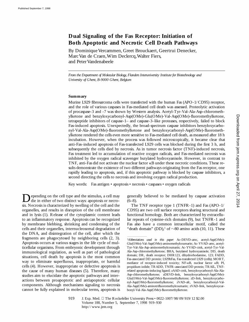
were isolated by functional screening. Several clones wereidentified that underwent rapid apoptosis after the additionof anti-Fas mAbs of the IgM isotype (clone CH-11; refer-ence 34); all of these clones exhibited high expression levelsof Fas at the cell surface. Microscopic examination of anti-Fas–treated cells showed that the onset of apoptosis, as indi-cated by membrane blebbing, already started to occur after30 min, and all cells died after 3 h as detected by flow fluo-rocytometry of PI-positive cells (Fig. 1). Inhibition of tran-scription by actinomycin D had no effect on the sensitivityof the cells or the kinetics of cell death, suggesting that in-duced protection mechanisms such as A20 (35, 36) are notrelevant in this particular cell clone, in contrast to TNF-induced cell death, which is highly sensitized and acceler-ated in the presence of actinomycin D (37, 38).
Figure 1. Rapid induction of apoptosis in L929hFas cells. Cells weretreated with 500 ng/ml of agonistic anti-Fas mAb in the absence (circles)or presence (triangles) of 1 mg/ml actinomycin D. The fraction of PI-pos-itive cells was determined in a time-course flow fluorocytometric experi-ment. Inset, Flow fluorocytometric analysis of Fas expression on Fas-trans-fected cells (solid line) and mock-transfected cells (broken line).
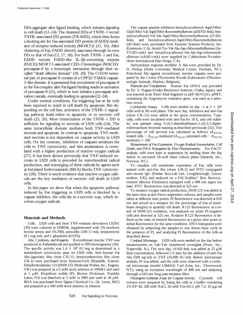
Figure 2. Determination ofcaspase activity in whole cell ly-sates of L929hFas cells. Cells weretreated with 500 ng/ml of anti-Fas Ab (A) or 10,000 IU/ml ofTNF (B) in the absence of actino-mycin D. The cleavage activity,expressed as maximal fluorescenceincrease (max. Dfl.), on Ac-DEVD-amc (filled circles), Ac-YVAD-amc(triangles), and zVAD-afc (squares)was measured as described in Ma-terials and Methods. Before lysis,the percentage of PI-positive cellswas determined by flow fluorocy-tometric analysis (open circles).
on April 27, 2014
jem.rupress.org
Dow
nloaded from
Published September 7, 1998
922
Fas Signaling to Necrosis
Fas-mediated Apoptosis in L929 Cells Is Associated with Ac-tivation of Caspase-3 and -7.
We investigated whether anti-Fas treatment of L929hFas cells resulted in induction ofcaspase activity. In a first approach, cells were stimulatedfor several time periods with anti-Fas, and total cytoplasmiclysates were incubated with fluorogenic oligopeptide caspasesubstrates. No cleavage of Ac-YVAD-amc or zVAD-afcwas observed. However, significant hydrolysis of Ac-DEVD-amc was detected (Fig. 2
A
). These results confirmthat during Fas-mediated apoptosis in L929 cells, caspase-3–like proteases are rapidly activated.
In a parallel set-up, cell viability was assessed using flowfluorocytometry to determine the percentage of PI-positivecells. Maximal Ac-DEVD-amc cleaving activity roughlycorresponded with the onset of loss of membrane integrity.As cell death progressed in time, Ac-DEVD-amc cleavageactivity decreased rapidly. Presumably, this reflects loss ofactive enzyme in the dying cells. As a control, L929hFascells were treated with TNF. TNF kills these cells by ne-crosis, and no caspase activity could be detected after TNFtreatment (Fig. 2
B
).To further identify the caspases involved in Fas-mediated
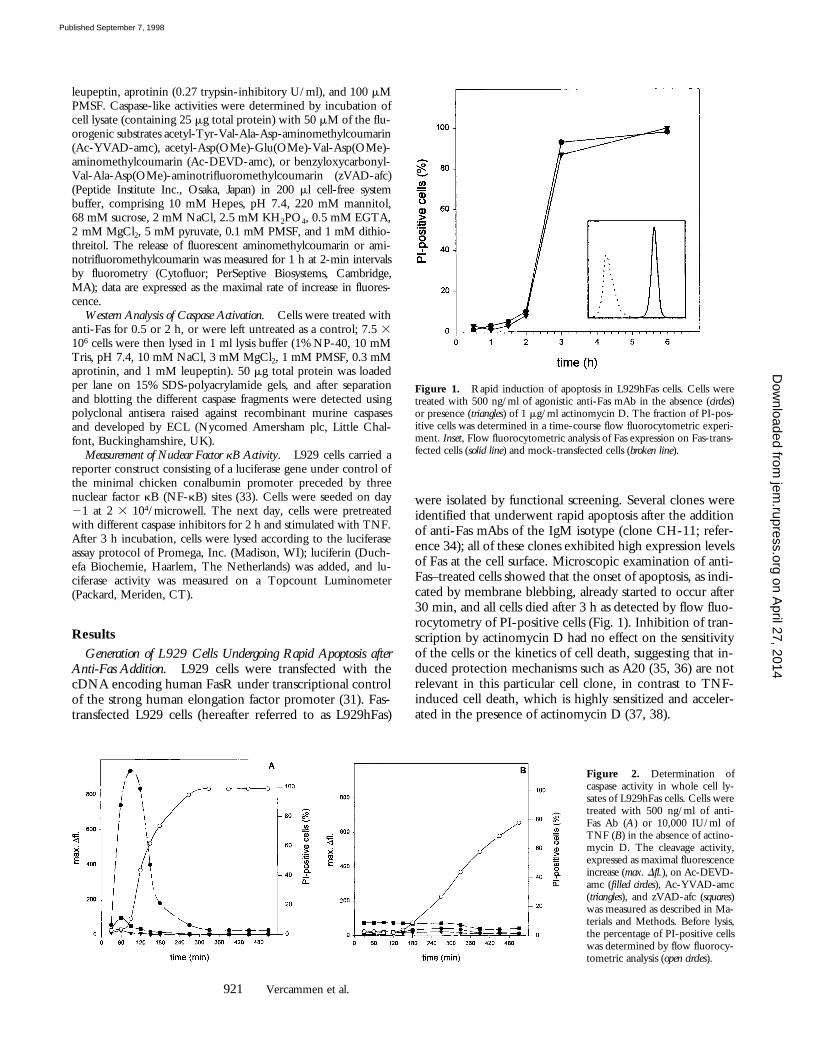
apoptosis in L929hFas cells, we subjected whole cell lysates ofFas-treated cells to SDS-PAGE, followed by Western analysisusing polyclonal Abs raised against recombinant murinecaspases. As shown in Fig. 3, cleavage of procaspases-3 and -7and formation of p17/p18 fragments (39) were observed after2 h. No cleavage fragments of procaspases-1, -2, -6, -11, or-12 were detected in the same lysates. This suggests that clus-tering of Fas very rapidly leads to activation of procaspase-3and -7. Most probably, activation of procaspase-8 leadsdirectly to activation of downstream procaspase-3 and –7. In-deed, the latter caspases are direct substrates of caspase-8 asdemonstrated previously in vitro (22, 23).
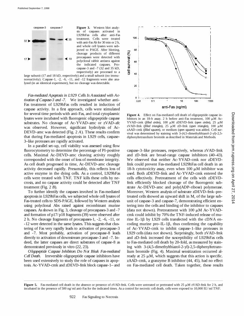
Oligopeptide Caspase Inhibitors Do Not Block Fas-mediatedCell Death.
Irreversible oligopeptide caspase inhibitors havebeen used extensively to study the role of caspases in apop-tosis. Ac-YVAD-cmk and zDEVD-fmk block caspase-1– and
caspase-3–like proteases, respectively, whereas zVAD-fmkand zD-fmk are broad-range caspase inhibitors (40–43).We observed that neither Ac-YVAD-cmk nor zDEVD-fmk could prevent Fas-mediated L929hFas cell death in an18-h cytotoxicity assay, even when 100
m
M inhibitor wasused. Both zDEVD-fmk and Ac-YVAD-cmk entered thecells effectively. Pretreatment of the cells with zDEVD-fmk efficiently blocked cleavage of the fluorogenic sub-strate Ac-DEVD-amc and poly(ADP-ribose) polymerase.Moreover, Western analysis of substrate zDEVD-fmk–pre-treated cells showed an upward shift in M
r
of the large sub-unit of caspase-3 and caspase-7, demonstrating efficient en-tering into the cells and binding of the inhibitor to caspases(data not shown). Pretreatment with 100
m
M Ac-YVAD-cmk could inhibit by 70% the TNF-induced release of mu-rine IL-1
b
by L929 cells transfected with the cDNA en-coding murine pro–IL-1
b
, thus confirming the capabilityof Ac-YVAD-cmk to inhibit caspase-1–like proteases inL929 cells (data not shown). Surprisingly, both zVAD-fmkand zD-fmk increased the susceptibility of L929hFas cellsto Fas-mediated cell death by 20-fold, as measured by stain-ing with 3-(4,5-dimethylthiazol-2-yl)-2,5-diphenyltetrazo-lium bromide (Fig. 4). Maximal sensitization occurred al-ready at 25
m
M, which suggests that this action is specific.zAAD-cmk, a granzyme B inhibitor (44, 45), had no effecton Fas-mediated cell death. Taken together, these results
Figure 3. Western blot analy-sis of caspases activated inL929hFas cells after anti-Fastreatment. Cells were treatedwith anti-Fas Ab for 30 min or 2 h,and whole cell lysates were sub-jected to PAGE. After blotting,cleavage products of differentprocaspases were detected withpolyclonal rabbit antisera againstthe indicated caspases. Pro-caspase-3 and -7 (32 and 35 kD,respectively) are processed to a
large subunit (17 and 18 kD, respectively) and a small subunit (no immu-noreactivity). Caspase-1, -2, -6, -11, and -12 fragments were also ana-lyzed (in an identical experiment), but no cleavage was detectable.
Figure 4. Effect on Fas-mediated cell death of oligopeptide caspase in-hibitors in an 18-h assay. 2 h before anti-Fas treatment, 100 mM Ac-YVAD-cmk (filled circles), 100 mM zDEVD-fmk (open circles), 25 mMzVAD-fmk (filled triangles), 25 mM zD-fmk (open triangles), 100 mMzAAD-cmk (filled squares), or medium (open squares) was added. Cell sur-vival was determined by staining with 3-(4,5-dimethylthiazol-2-yl)-2,5-diphenyltetrazolium bromide as described in Materials and Methods.
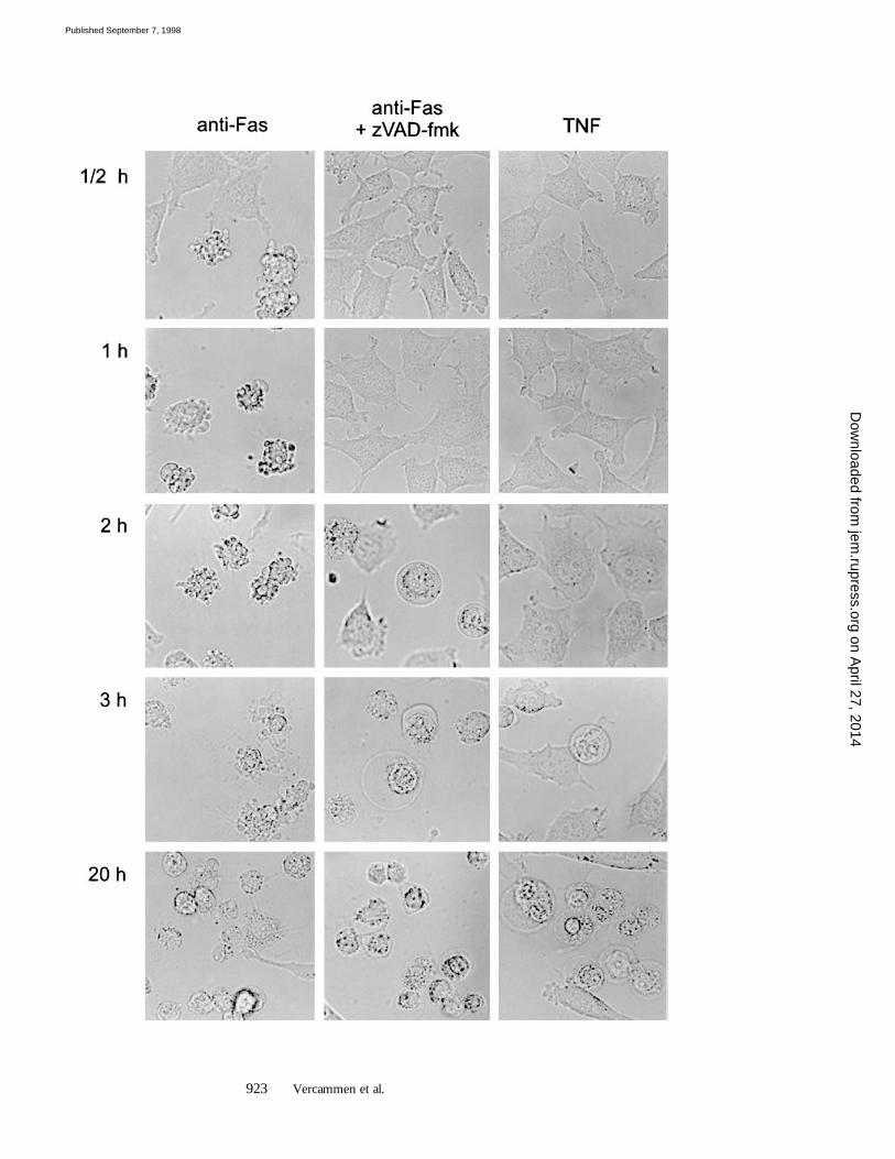
Figure 5.
Fas-mediated cell death in the absence or presence of zVAD-fmk. Cells were untreated or pretreated with 25
m
M zVAD-fmk for 2 h, andincubated in the presence of 500 ng/ml anti-Fas for the indicated times. As a control for necrotic cell death, cells were exposed to 10,000 IU/ml TNF.
on April 27, 2014
jem.rupress.org
Dow
nloaded from
Published September 7, 1998
923
Vercammen et al.
on April 27, 2014
jem.rupress.org
Dow
nloaded from
Published September 7, 1998
924
Fas Signaling to Necrosis
suggest that even when activation of caspase-3 and -7 isblocked, Fas clustering still leads to death of L929hFas cells.Moreover, the broad-range caspase inhibitors sensitized thecells
z
20-fold to anti-Fas. These results are reminiscent ofthe sensitization by caspase inhibitors observed in TNF-mediated necrosis in L929 cells (27).
zVAD-fmk and zD-fmk Treatment of L929hFas Cells Re-veals an Alternative Death Signaling Pathway Originating fromFas.
To study whether the increased sensitivity to Fas-mediated cell death in the presence of zVAD-fmk or zD-fmk reflected necrotic cell death, cells were examined mi-croscopically (Fig. 5). Cells treated for 1 h with anti-Fasalone clearly showed extensive membrane blebbing. How-ever, when zVAD-fmk was present, the cells retained thesame morphology as control cells in the absence of anti-Fas. After 2 h, anti-Fas–treated cells, without caspase inhib-itors or in the presence of Ac-YVAD-cmk or zDEVD-fmk, succumbed to apoptotic cell death. Quite surprisingly,cells incubated in the presence of zVAD-fmk began to ex-hibit an altered morphology, such as cytoplasmic swelling.After 3 h, all cells had died in a way that strikingly resem-bled TNF-mediated necrosis.
To further characterize this alternative Fas-mediated
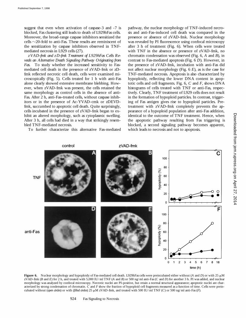
pathway, the nuclear morphology of TNF-induced necro-sis and anti-Fas–induced cell death was compared in thepresence or absence of zVAD-fmk. Nuclear morphologywas revealed by PI fluorescence using confocal microscopyafter 3 h of treatment (Fig. 6). When cells were treatedwith TNF in the absence or presence of zVAD-fmk, nochromatin condensation was observed (Fig. 6,
A
and
B
), incontrast to Fas-mediated apoptosis (Fig. 6
D
). However, inthe presence of zVAD-fmk, incubation with anti-Fas didnot affect nuclear morphology (Fig. 6
E
), as is the case forTNF-mediated necrosis. Apoptosis is also characterized byhypoploidy, reflecting the lower DNA content in apop-totic cells and cell fragments. Fig. 6,
C
and
F
, shows DNAhistograms of cells treated with TNF or anti-Fas, respec-tively. Clearly, TNF treatment of L929 cells does not resultin the formation of hypoploid particles. In contrast, trigger-ing of Fas antigen gives rise to hypoploid particles. Pre-treatment with zVAD-fmk completely prevents the ap-pearance of a hypoploid population after anti-Fas addition,identical to the outcome of TNF treatment. Hence, whenthe apoptotic pathway resulting from Fas triggering isblocked, a second signaling pathway becomes apparent,which leads to necrosis and not to apoptosis.
Figure 6. Nuclear morphology and hypoploidy of Fas-mediated cell death. L929hFas cells were preincubated either without (A and D) or with 25 mMzVAD-fmk (B and E) for 2 h, and treated with 5,000 IU/ml TNF (A and B) or 500 ng/ml anti-Fas (C and D) for another 3 h. PI was added, and nuclearmorphology was analyzed by confocal microscopy. Necrotic nuclei are PI-positive, but retain a normal structural appearance; apoptotic nuclei are char-acterized by strong condensation of chromatin. C and F show the fraction of hypoploid cell fragments measured as a function of time. Cells were prein-cubated without (open circles) or with (filled circles) 25 mM zVAD-fmk, and treated with 500 IU/ml TNF (C) or 500 ng/ml anti-Fas (F).
on April 27, 2014
jem.rupress.org
Dow
nloaded from
Published September 7, 1998
925
Vercammen et al.
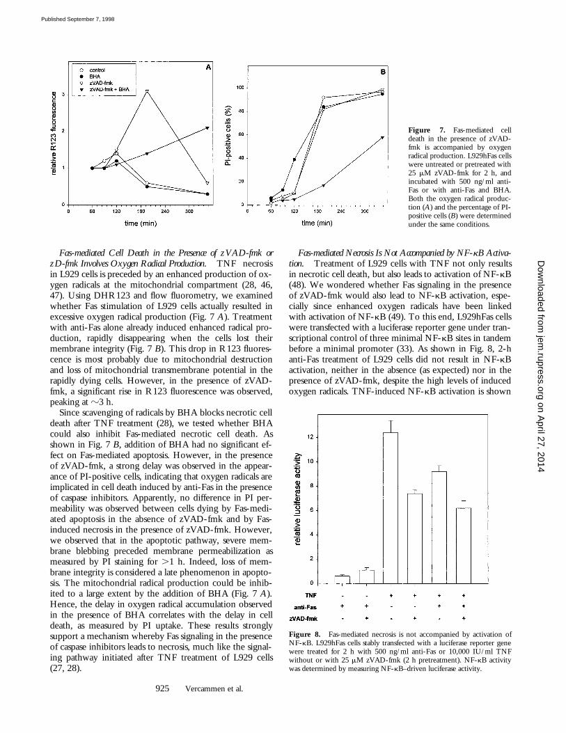
Fas-mediated Cell Death in the Presence of zVAD-fmk orzD-fmk Involves Oxygen Radical Production.
TNF necrosisin L929 cells is preceded by an enhanced production of ox-ygen radicals at the mitochondrial compartment (28, 46,47). Using DHR123 and flow fluorometry, we examinedwhether Fas stimulation of L929 cells actually resulted inexcessive oxygen radical production (Fig. 7
A
). Treatmentwith anti-Fas alone already induced enhanced radical pro-duction, rapidly disappearing when the cells lost theirmembrane integrity (Fig. 7
B
). This drop in R123 fluores-cence is most probably due to mitochondrial destructionand loss of mitochondrial transmembrane potential in therapidly dying cells. However, in the presence of zVAD-fmk, a significant rise in R123 fluorescence was observed,peaking at
z
3 h.Since scavenging of radicals by BHA blocks necrotic cell
death after TNF treatment (28), we tested whether BHAcould also inhibit Fas-mediated necrotic cell death. Asshown in Fig. 7
B
, addition of BHA had no significant ef-fect on Fas-mediated apoptosis. However, in the presenceof zVAD-fmk, a strong delay was observed in the appear-ance of PI-positive cells, indicating that oxygen radicals areimplicated in cell death induced by anti-Fas in the presenceof caspase inhibitors. Apparently, no difference in PI per-meability was observed between cells dying by Fas-medi-ated apoptosis in the absence of zVAD-fmk and by Fas-induced necrosis in the presence of zVAD-fmk. However,we observed that in the apoptotic pathway, severe mem-brane blebbing preceded membrane permeabilization asmeasured by PI staining for
.
1 h. Indeed, loss of mem-brane integrity is considered a late phenomenon in apopto-sis. The mitochondrial radical production could be inhib-ited to a large extent by the addition of BHA (Fig. 7
A
).Hence, the delay in oxygen radical accumulation observedin the presence of BHA correlates with the delay in celldeath, as measured by PI uptake. These results stronglysupport a mechanism whereby Fas signaling in the presenceof caspase inhibitors leads to necrosis, much like the signal-ing pathway initiated after TNF treatment of L929 cells(27, 28).
Fas-mediated Necrosis Is Not Accompanied by NF-
k
B Activa-tion.
Treatment of L929 cells with TNF not only resultsin necrotic cell death, but also leads to activation of NF-
k
B(48). We wondered whether Fas signaling in the presenceof zVAD-fmk would also lead to NF-
k
B activation, espe-cially since enhanced oxygen radicals have been linkedwith activation of NF-
k
B (49). To this end, L929hFas cellswere transfected with a luciferase reporter gene under tran-scriptional control of three minimal NF-
k
B sites in tandembefore a minimal promoter (33). As shown in Fig. 8, 2-hanti-Fas treatment of L929 cells did not result in NF-
k
Bactivation, neither in the absence (as expected) nor in thepresence of zVAD-fmk, despite the high levels of inducedoxygen radicals. TNF-induced NF-
k
B activation is shown
Figure 7. Fas-mediated celldeath in the presence of zVAD-fmk is accompanied by oxygenradical production. L929hFas cellswere untreated or pretreated with25 mM zVAD-fmk for 2 h, andincubated with 500 ng/ml anti-Fas or with anti-Fas and BHA.Both the oxygen radical produc-tion (A) and the percentage of PI-positive cells (B) were determinedunder the same conditions.
Figure 8. Fas-mediated necrosis is not accompanied by activation ofNF-kB. L929hFas cells stably transfected with a luciferase reporter genewere treated for 2 h with 500 ng/ml anti-Fas or 10,000 IU/ml TNFwithout or with 25 mM zVAD-fmk (2 h pretreatment). NF-kB activitywas determined by measuring NF-kB–driven luciferase activity.
on April 27, 2014
jem.rupress.org
Dow
nloaded from
Published September 7, 1998
926 Fas Signaling to Necrosis
as a control. These results indicate that signaling pathwaysleading to necrosis and to NF-kB activation are segregated,and that Fas signaling in the presence of zVAD-fmk onlyactivates the signals leading to necrotic death. Furthermore,at least in this particular cell system, a causal relationshipbetween oxygen radical production and activation of NF-kB cannot be established. Alternatively, addition of BHA,which effectively retards TNF-induced necrosis (27), doesnot influence TNF-mediated NF-kB activation (data notshown). This suggests that enhanced mitochondrial oxygenradical production is not implicated in the activation ofNF-kB. Combined treatment with TNF and anti-Fas inthe presence of zVAD-fmk, resulting in almost completecell death after 2 h, does not affect the level of NF-kB ac-tivity compared with TNF plus zVAD-fmk, excluding thepossibility that rapid cell death would mask Fas-mediatedNF-kB activation.
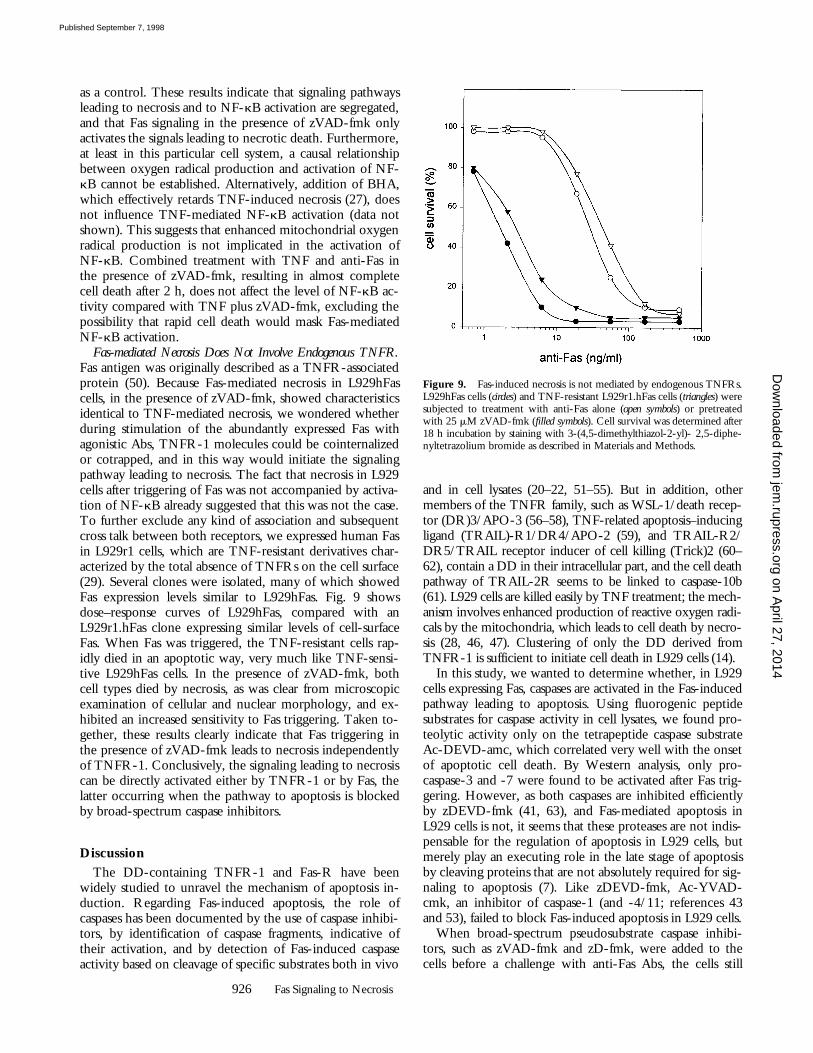
Fas-mediated Necrosis Does Not Involve Endogenous TNFR.Fas antigen was originally described as a TNFR-associatedprotein (50). Because Fas-mediated necrosis in L929hFascells, in the presence of zVAD-fmk, showed characteristicsidentical to TNF-mediated necrosis, we wondered whetherduring stimulation of the abundantly expressed Fas withagonistic Abs, TNFR-1 molecules could be cointernalizedor cotrapped, and in this way would initiate the signalingpathway leading to necrosis. The fact that necrosis in L929cells after triggering of Fas was not accompanied by activa-tion of NF-kB already suggested that this was not the case.To further exclude any kind of association and subsequentcross talk between both receptors, we expressed human Fasin L929r1 cells, which are TNF-resistant derivatives char-acterized by the total absence of TNFRs on the cell surface(29). Several clones were isolated, many of which showedFas expression levels similar to L929hFas. Fig. 9 showsdose–response curves of L929hFas, compared with anL929r1.hFas clone expressing similar levels of cell-surfaceFas. When Fas was triggered, the TNF-resistant cells rap-idly died in an apoptotic way, very much like TNF-sensi-tive L929hFas cells. In the presence of zVAD-fmk, bothcell types died by necrosis, as was clear from microscopicexamination of cellular and nuclear morphology, and ex-hibited an increased sensitivity to Fas triggering. Taken to-gether, these results clearly indicate that Fas triggering inthe presence of zVAD-fmk leads to necrosis independentlyof TNFR-1. Conclusively, the signaling leading to necrosiscan be directly activated either by TNFR-1 or by Fas, thelatter occurring when the pathway to apoptosis is blockedby broad-spectrum caspase inhibitors.
Discussion
The DD-containing TNFR-1 and Fas-R have beenwidely studied to unravel the mechanism of apoptosis in-duction. Regarding Fas-induced apoptosis, the role ofcaspases has been documented by the use of caspase inhibi-tors, by identification of caspase fragments, indicative oftheir activation, and by detection of Fas-induced caspaseactivity based on cleavage of specific substrates both in vivo
and in cell lysates (20–22, 51–55). But in addition, othermembers of the TNFR family, such as WSL-1/death recep-tor (DR)3/APO-3 (56–58), TNF-related apoptosis–inducingligand (TRAIL)-R1/DR4/APO-2 (59), and TRAIL-R2/DR5/TRAIL receptor inducer of cell killing (Trick)2 (60–62), contain a DD in their intracellular part, and the cell deathpathway of TRAIL-2R seems to be linked to caspase-10b(61). L929 cells are killed easily by TNF treatment; the mech-anism involves enhanced production of reactive oxygen radi-cals by the mitochondria, which leads to cell death by necro-sis (28, 46, 47). Clustering of only the DD derived fromTNFR-1 is sufficient to initiate cell death in L929 cells (14).
In this study, we wanted to determine whether, in L929cells expressing Fas, caspases are activated in the Fas-inducedpathway leading to apoptosis. Using fluorogenic peptidesubstrates for caspase activity in cell lysates, we found pro-teolytic activity only on the tetrapeptide caspase substrateAc-DEVD-amc, which correlated very well with the onsetof apoptotic cell death. By Western analysis, only pro-caspase-3 and -7 were found to be activated after Fas trig-gering. However, as both caspases are inhibited efficientlyby zDEVD-fmk (41, 63), and Fas-mediated apoptosis inL929 cells is not, it seems that these proteases are not indis-pensable for the regulation of apoptosis in L929 cells, butmerely play an executing role in the late stage of apoptosisby cleaving proteins that are not absolutely required for sig-naling to apoptosis (7). Like zDEVD-fmk, Ac-YVAD-cmk, an inhibitor of caspase-1 (and -4/11; references 43and 53), failed to block Fas-induced apoptosis in L929 cells.
When broad-spectrum pseudosubstrate caspase inhibi-tors, such as zVAD-fmk and zD-fmk, were added to thecells before a challenge with anti-Fas Abs, the cells still
Figure 9. Fas-induced necrosis is not mediated by endogenous TNFRs.L929hFas cells (circles) and TNF-resistant L929r1.hFas cells (triangles) weresubjected to treatment with anti-Fas alone (open symbols) or pretreatedwith 25 mM zVAD-fmk (filled symbols). Cell survival was determined after18 h incubation by staining with 3-(4,5-dimethylthiazol-2-yl)- 2,5-diphe-nyltetrazolium bromide as described in Materials and Methods.
on April 27, 2014
jem.rupress.org
Dow
nloaded from
Published September 7, 1998
927 Vercammen et al.
died. However, closer inspection of the type of cell deathrevealed that at sufficiently high concentrations of inhibi-tors, apoptosis was indeed inhibited, but now the cells un-derwent rapid necrotic cell death, as characterized by mor-phological criteria, such as swelling of the cells, the absenceof nuclear condensation, and the appearance of hypoploidparticles. Furthermore, the generation of oxygen radicalswas identified as the effective mechanism to necrosis.Therefore, it seems that at least in L929 cells, two differentpathways emerge from the Fas-R: one is caspase-depen-dent and leads to apoptosis; the other is oxygen radical–mediated, becomes evident in the presence of caspase in-hibitors, and results in necrotic cell death.
Caspases that have been reported to be inhibited byzVAD-fmk are caspase-1 (42, 43), -8 (20, 21), -3, -4/11,and very weakly caspase-7 (43). The use of the inhibitorsdoes not allow the pinpointing of caspases involved in thecell death pathways in L929 cells. Moreover, not onlyblocking of a caspase activity but also amplification of oxy-gen radical production is necessary to reveal the necroticFas pathway. In a previous study, we have shown thatTNF-driven radical production in L929 cells is indeedstrongly enhanced by zVAD-fmk and zD-fmk, and, muchmore weakly, by Ac-YVAD-cmk and zDEVD-fmk (27).Hence, an enhanced oxygen radical production, which isnegatively regulated by zVAD-fmk– and zD-fmk–sensitiveproteases, seems to make the difference between apoptoticand necrotic outcome of L929 cell death. Further studiesare required to elucidate the relation between caspase inhi-bition and enhanced radical production.
Necrosis after either TNF or Fas triggering in the pres-ence of caspase inhibitors involves the production of mito-chondrial oxygen radicals, which can be shown, for exam-ple, by the oxidation of DHR123 and the inhibition ofnecrosis by the radical scavenger BHA (28). This meansthat TNF- and Fas-mediated necrosis converge upstream ofthe mitochondria. On the other hand, Fas-mediated necro-sis is not accompanied by activation of NF-kB, as is thecase with TNF-induced necrosis. Hence, induction of oxy-gen radical production and activation of NF-kB diverge atan early point in the TNF-signaling pathway, and it isdownstream of this point that Fas- and TNF-mediated ne-crosis meet. Previous studies have shown that at least insome cells, NF-kB activation can be regulated by the redoxstate (49). However, in the present study, even though ox-ygen radical production is prominent when Fas is triggeredin the presence of caspase inhibitors, no NF-kB activationwas observed. This implies that the inability of Fas to medi-ate NF-kB activation is solely due to its inability to recruitTRADD, receptor-interacting protein (RIP), and TNFR-associating factor (TRAF)2, which constitutes the effectoraxis leading to NF-kB activation (64). The inability of Fasto recruit TRADD, RIP, and TRAF2 also implies thatthese receptor-associated molecules are not involved inFas-mediated necrosis. We propose a dual signaling path-way emerging from Fas: one is caspase-dependent, leads torapid apoptosis, and does not involve production of oxygenradicals; the second, which under normal conditions is
masked by rapid apoptosis, results in necrotic cell death andis mediated by massive mitochondrial oxygen radical pro-duction.
It has been proposed that apoptosis and necrosis mightshare common mediators such as Ca21 and caspases (65).However, at least in the case of L929 cells, clear differencesexist between the two pathways (38). The most strikingdifference is that, whereas TNF-mediated necrosis relies onthe production of oxygen radicals as an essential executingstep, Fas-initiated apoptosis does not depend on free radicalproduction. As shown in this study, the same goes for thedifference between Fas-mediated apoptosis and, in thepresence of the appropriate caspase inhibitors, Fas-mediatednecrosis. Furthermore, even at the top of the signalingpathway, divergence between apoptotic and necrotic path-ways is evident. Indeed, whereas Fas-mediated apoptosis isdependent on recruitment and activation of procaspase-8(21) or a homologous protease, as shown by the inhibitingeffect of the peptide inhibitors of caspases on apoptosis,Fas-mediated necrosis, like TNF-mediated necrosis, is not.Moreover, the broad-spectrum inhibitors zVAD-fmk andzD-fmk even potently sensitize the necrotic pathway (27).Hence, to obtain triggering of Fas antigen resulting in mi-tochondria-dependent necrosis, both inhibition of the caspase-mediated apoptotic pathway and amplification of the radi-cal-dependent necrotic pathway are necessary.
Recently, Scaffidi and co-workers (66) described the ex-istence of two Fas-signaling pathways depending on celltype. Type I cells are characterized by rapid and massive re-cruitment of FADD and procaspase-8 into the death-induc-ing signaling complex, followed 30 min later by activationof caspase-3, which eventually leads to apoptotic cell death.
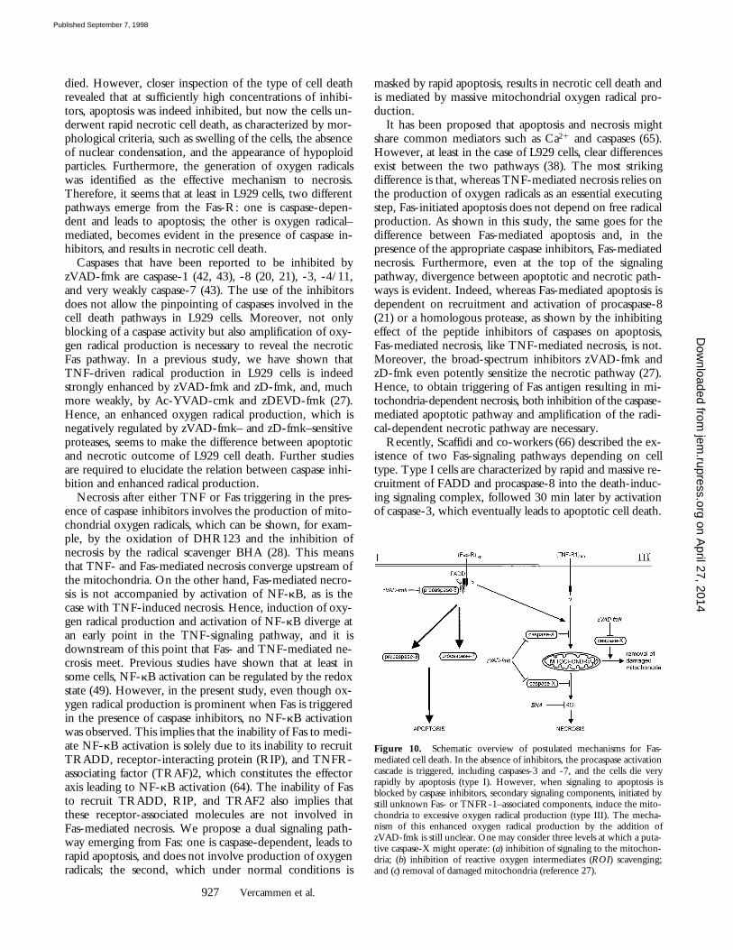
Figure 10. Schematic overview of postulated mechanisms for Fas-mediated cell death. In the absence of inhibitors, the procaspase activationcascade is triggered, including caspases-3 and -7, and the cells die veryrapidly by apoptosis (type I). However, when signaling to apoptosis isblocked by caspase inhibitors, secondary signaling components, initiated bystill unknown Fas- or TNFR-1–associated components, induce the mito-chondria to excessive oxygen radical production (type III). The mecha-nism of this enhanced oxygen radical production by the addition ofzVAD-fmk is still unclear. One may consider three levels at which a puta-tive caspase-X might operate: (a) inhibition of signaling to the mitochon-dria; (b) inhibition of reactive oxygen intermediates (ROI) scavenging;and (c) removal of damaged mitochondria (reference 27).
on April 27, 2014
jem.rupress.org
Dow
nloaded from
Published September 7, 1998
928 Fas Signaling to Necrosis
Alternatively, type II cells showed a reduced formation ofthe death-inducing signaling complex and delayed activa-tion of caspase-3 and -8. However, both cell types dis-played similar kinetics in the loss of mitochondrial trans-membrane potential and cell death. Overexpression of Bcl-2or Bcl-xL could prevent this reduction of mitochondrialtransmembrane potential in both type I and II cells, butcould rescue only type II cells from cell death. Hence, intype II cells, activation of caspases and subsequent celldeath depend on mitochondria, whereas type I signaling ismediated directly by caspase-8 and -3, the reduction of mi-tochondrial transmembrane potential being a secondary phe-nomenon. In view of this model, Fas-mediated apoptosis inL929 cells can be regarded as type I signaling, involvingrapid activation of the caspase cascade. Fas-triggered necro-sis, in the presence of caspase inhibitors, and TNF-inducednecrosis may represent an alternate form of signaling to celldeath relying on mitochondrial alterations, the latter result-ing in enhanced oxygen radical production rather than acti-
vation of caspases. A schematic overview of the proposedtype I and type III signaling components is given in Fig. 10.
The therapeutic use of caspase inhibitors for escapingunwanted apoptosis is an attractive possibility. For instance,caspases have already been assigned a role in amyotrophiclateral sclerosis (67), Alzheimer’s disease (68), and Hunting-ton’s disease (69, 70). Furthermore, neuronal damage afterhypoxic-ischemic insult (71) or fulminant liver destructionafter anti-Fas injection (72) could be diminished or pre-vented successfully by the use of peptide caspase inhibitors.However, the results presented in this paper could restrictthe therapeutic use of caspase inhibitors in that they do notnecessarily guarantee blockage of cell death in general. Inother words, necrotic cell death could at least in some celltypes become evident. Furthermore, cells dying by necro-sis, instead of apoptosis, may cause an inflammatory re-sponse. At present, it is not clear which components of thebiochemical machinery make a cell more prone to suc-cumb to necrosis.
The authors thank W. Burm, A. Meeus, and M. Van den Hemel for technical assistance, K. De Vos for con-focal imaging, and M. Vandecasteele for critical reading of the manuscript. They are indebted to Dr. S. Na-gata for providing human Fas cDNA and pEF-BOS, and to Dr. D. Pickup for supplying cytokine responsemodifier A cDNA.
Research was supported by the Interuniversitaire Attractiepolen, grant 9005097N of the Fonds voor Weten-schappelijk Onderzoek–Vlaanderen, and European Community Biomed Program grant BMH4-CT96-0300. G. Denecker is a research assistant, and P. Vandenabeele a postdoctoral researcher with the Fonds voorWetenschappelijk Onderzoek–Vlaanderen.
Address correspondence to P. Vandenabeele, Department of Molecular Biology, Flanders Interuniversity In-stitute for Biotechnology and University of Ghent, K.L. Ledeganckstraat 35, B-9000 Ghent, Belgium.Phone: 32-9-264-51-31; Fax: 32-9-264-53-48; E-mail: [email protected]
Received for publication 4 May 1998 and in revised form 10 June 1998.
References1. Grooten, J., V. Goossens, B. Vanhaesebroeck, and W. Fiers.
1993. Cell membrane permeabilization and cellular collapse,followed by loss of dehydrogenase activity: early events intumour necrosis factor-induced cytotoxicity. Cytokine. 5:546–555.
2. Kerr, J.F.R., A.H. Wyllie, and A.R. Currie. 1972. Apoptosis:a basic biological phenomenon with wide-ranging implica-tions in tissue kinetics. Br. J. Cancer. 26:239–257.
3. Wyllie, A.H., J.F.R. Kerr, and A.R. Currie. 1980. Cell death:the significance of apoptosis. Int. Rev. Cytol. 68:251–306.
4. Jacobson, M.D., M. Weil, and M.C. Raff. 1997. Pro-grammed cell death in animal development. Cell. 88:347–354.
5. Thompson, C. 1995. Apoptosis in the pathogenesis and treat-ment of disease. Science. 267:1456–1462.
6. Nicholson, D.W., and N.A. Thornberry. 1997. Caspases:killer proteases. Trends Biochem. Sci. 22:299–306.
7. Villa, P., S.H. Kaufmann, and W.C. Earnshaw. 1997.Caspases and caspase inhibitors. Trends Biochem. Sci. 22:388–393.
8. Porter, A.G., P. Ng, and R.U. Jänicke. 1997. Death sub-strates come alive. Bioessays. 19:501–507.
9. Nagata, S. 1997. Apoptosis by death factor. Cell. 88:355–365.10. Itoh, N., and S. Nagata. 1993. A novel protein domain re-
quired for apoptosis. Mutational analysis of human Fas anti-gen. J. Biol. Chem. 268:10932–10937.
11. Tartaglia, L., T. Ayres, G. Wong, and D. Goeddel. 1993. Anovel domain within the 55 kd TNF receptor signals celldeath. Cell. 74:845–853.
12. Song, H.Y., J.D. Dunbar, and D.B. Donner. 1994. Aggrega-tion of the intracellular domain of the type 1 tumor necrosisfactor receptor defined by the two-hybrid system. J. Biol.Chem. 269:22492–22495.
13. Boldin, M.P., E.E. Varfolomeev, Z. Pancer, I.L. Mett, J.H.Camonis, and D. Wallach. 1995. A novel protein that inter-acts with the death domain of Fas/APO1 contains a sequencemotif related to the death domain. J. Biol. Chem. 270:7795–7798.
14. Vandevoorde, V., G. Haegeman, and W. Fiers. 1997. In-duced expression of trimerized intracellular domains of thehuman tumor necrosis factor (TNF) p55 receptor elicits TNFeffects. J. Cell Biol. 137:1627–1638.
on April 27, 2014
jem.rupress.org
Dow
nloaded from
Published September 7, 1998
929 Vercammen et al.
15. Hsu, H., J. Xiong, and D.V. Goeddel. 1995. The TNF re-ceptor 1-associated protein TRADD signals cell death andNF-kB activation. Cell. 81:495–504.
16. Hsu, H., H.B. Shu, M.G. Pan, and D.V. Goeddel. 1996.TRADD-TRAF2 and TRADD-FADD interactions definetwo distinct TNF receptor 1 signal transduction pathways.Cell. 84:299–308.
17. Chinnaiyan, A.M., K. O’Rourke, M. Tewari, and V.M.Dixit. 1995. FADD, a novel death domain-containing pro-tein, interacts with the death domain of Fas and initiates apop-tosis. Cell. 81:505–512.
18. Kischkel, F.C., S. Hellbardt, I. Behrmann, M. Germer, M.Pawlita, P.H. Krammer, and M.E. Peter. 1995. Cytotoxicity-dependent APO-1 (Fas/CD95)-associated proteins form adeath-inducing signaling complex (DISC) with the receptor.EMBO (Eur. Mol. Biol. Organ.) J. 14:5579–5588.
19. Boldin, M.P., T.M. Goncharov, Y.V. Goltsev, and D.Wallach. 1996. Involvement of MACH, a novel MORT1/FADD-interacting protease, in Fas/APO-1- and TNF recep-tor-induced cell death. Cell. 85:803–815.
20. Muzio, M., A.M. Chinnaiyan, F.C. Kischkel, K. O’Rourke,A. Shevchenko, J. Ni, C. Scaffidi, J.D. Bretz, M. Zhang, R.Gentz, et al. 1996. FLICE, a novel FADD-homologous ICE/CED-3-like protease, is recruited to the CD95 (Fas/APO-1)death-inducing signaling complex. Cell. 85:817–827.
21. Medema, J.P., C. Scaffidi, F.C. Kischkel, A. Shevchenko, M.Mann, P.H. Krammer, and M.E. Peter. 1997. FLICE is acti-vated by association with the CD95 death-inducing signalingcomplex (DISC). EMBO (Eur. Mol. Biol. Organ.) J. 16:2794–2804.
22. Srinivasula, S.M., M. Ahmad, T. Fernandes-Alnemri, G. Lit-wack, and E.S. Alnemri. 1996. Molecular ordering of theFas-apoptotic pathway: the Fas/APO-1 protease Mch5 is aCrmA-inhibitable protease that activates multiple Ced-3/ICE-like cysteine proteases. Proc. Natl. Acad. Sci. USA. 93:14486–14491.
23. Muzio, M., G.S. Salvesen, and V.M. Dixit. 1997. FLICE in-duced apoptosis in a cell-free system. Cleavage of caspase zy-mogens. J. Biol. Chem. 272:2952–2956.
24. Wallach, D., M. Boldin, E. Varfolomeev, R. Beyaert, P.Vandenabeele, and W. Fiers. 1997. Cell death induction byreceptors of the TNF family: towards a molecular under-standing. FEBS Lett. 410:96–106.
25. Laster, S.M., J.G. Wood, and L.R. Gooding. 1988. Tumornecrosis factor can induce both apoptotic and necrotic formsof cell lysis. J. Immunol. 141:2629–2635.
26. Fiers, W., R. Beyaert, E. Boone, S. Cornelis, W. Declercq,E. Decoster, G. Denecker, B. Depuydt, D. De Valck, G. DeWilde, et al. 1995. TNF-induced intracellular signaling lead-ing to gene induction or to cytotoxicity by necrosis or by apop-tosis. J. Inflamm. 47:67–75.
27. Vercammen, D., R. Beyaert, G. Denecker, V. Goossens, G.Van Loo, W. Declercq, J. Grooten, W. Fiers, and P. Vande-nabeele. 1998. Inhibition of caspases increases the sensitivityof L929 cells to necrosis mediated by tumor necrosis factor. J.Exp. Med. 187:1477–1485.
28. Goossens, V., J. Grooten, K. De Vos, and W. Fiers. 1995.Direct evidence for tumor necrosis factor-induced mitochon-drial reactive oxygen intermediates and their involvement incytotoxicity. Proc. Natl. Acad. Sci. USA. 92:8115–8119.
29. Vanhaesebroeck, B., S. Van Bladel, A. Lenaerts, P. Suffys, R.Beyaert, R. Lucas, F. Van Roy, and W. Fiers. 1991. Twodiscrete types of tumor necrosis factor-resistant cells derived
from the same cell line. Cancer Res. 51:2469–2477.30. Fransen, L., R. Müller, A. Marmenout, J. Tavernier, J. Van
der Heyden, E. Kawashima, E. Chollet, R. Tizard, H. VanHeuverswyn, A. Van Vliet, et al. 1985. Molecular cloning ofmouse tumour necrosis factor cDNA and its eukaryotic ex-pression. Nucleic Acids Res. 13:4417–4429.
31. Mizushima, S., and S. Nagata. 1990. pEF-BOS, a powerfulmammalian expression vector. Nucleic Acids Res. 18:5322.
32. Tada, H., O. Shiho, K. Kuroshima, M. Koyama, and K.Tsukamoto. 1986. An improved colorimetric assay for inter-leukin 2. J. Immunol. Methods. 93:157–165.
33. Kimura, A., A. Israël, O. Le Bail, and P. Kourilsky. 1986.Detailed analysis of the mouse H-2Kb promoter: enhancer-like sequences and their role in the regulation of class I geneexpression. Cell. 44:261–272.
34. Itoh, N., S. Yonehara, A. Ishii, M. Yonehara, S. Mizushima,M. Sameshima, A. Hase, Y. Seto, and S. Nagata. 1991. Thepolypeptide encoded by the cDNA for human cell surfaceantigen Fas can mediate apoptosis. Cell. 66:233–243.
35. Opipari, A.W., H.M. Hu, Jr., R. Yabkowitz, and V.M.Dixit. 1992. The A20 zinc finger protein protects cells fromtumor necrosis factor cytotoxicity. J. Biol. Chem. 267:12424–12427.
36. Jäättelä, M., H. Mouritzen, F. Elling, and L. Bastholm. 1996.A20 zinc finger protein inhibits TNF and IL-1 signaling. J.Immunol. 156:1166–1173.
37. Kirstein, M., W. Fiers, and C. Baglioni. 1986. Growth inhi-bition and cytotoxicity of tumor necrosis factor in L929 cellsis enhanced by high cell density and inhibition of mRNAsynthesis. J. Immunol. 137:2277–2280.
38. Vercammen, D., P. Vandenabeele, R. Beyaert, W. Declercq,and W. Fiers. 1997. Tumour necrosis factor-induced necrosisversus anti-Fas-induced apoptosis in L929 cells. Cytokine. 9:801–808.
39. Fernandes-Alnemri, T., R.C. Armstrong, J. Krebs, S.M.Srinivasula, L. Wang, F. Bullrich, L.C. Fritz, J.A. Trapani,K.J. Tomaselli, G. Litwack, and E.S. Alnemri. 1996. In vitroactivation of CPP32 and Mch3 by Mch4, a novel humanapoptotic cysteine protease containing two FADD-like do-mains. Proc. Natl. Acad. Sci. USA. 93:7464–7469.
40. Fernandes-Alnemri, T., A. Takahashi, R. Armstrong, J.Krebs, L. Fritz, K.J. Tomaselli, L. Wang, Z. Yu, C.M.Croce, G. Salveson, et al. 1995. Mch3, a novel human apop-totic cysteine protease highly related to CPP32. Cancer Res.55:6045–6052.
41. Nicholson, D.W., A. Ali, N.A. Thornberry, J.P. Vaillan-court, C.K. Ding, M. Gallant, Y. Gareau, P.R. Griffin, M.Labelle, Y.A. Lazebnik, et al. 1995. Identification and inhibi-tion of the ICE/CED-3 protease necessary for mammalianapoptosis. Nature. 376:37–43.
42. Sarin, A., M.L. Wu, and P.A. Henkart. 1996. Different inter-leukin-1b converting enzyme (ICE) family protease require-ments for the apoptotic death of T lymphocytes triggered bydiverse stimuli. J. Exp. Med. 184:2445–2450.
43. Margolin, N., S.A. Raybuck, K.P. Wilson, W. Chen, T.Fox, Y. Gu, and D.J. Livingston. 1997. Substrate and inhibi-tor specificity of interleukin-1b-converting enzyme and re-lated caspases. J. Biol. Chem. 272:7223–7228.
44. Odake, S., C.M. Kam, L. Narasimhan, M. Poe, J.T. Blake,O. Krahenbuhl, J. Tschopp, and J.C. Powers. 1991. Humanand murine cytotoxic T lymphocyte serine proteases: subsitemapping with peptide thioester substrates and inhibition ofenzyme activity and cytolysis by isocoumarins. Biochemistry.
on April 27, 2014
jem.rupress.org
Dow
nloaded from
Published September 7, 1998
930 Fas Signaling to Necrosis
30:2217–2227.45. Shi, L., C.M. Kam, J.C. Powers, R. Aebersold, and A.H.
Greenberg. 1992. Purification of three cytotoxic lymphocytegranule serine proteases that induce apoptosis through distinctsubstrate and target cell interactions. J. Exp. Med. 176:1521–1529.
46. Schulze-Osthoff, K., A.C. Bakker, B. Vanhaesebroeck, R.Beyaert, W.A. Jacob, and W. Fiers. 1992. Cytotoxic activityof tumor necrosis factor is mediated by early damage of mito-chondrial functions. J. Biol. Chem. 267:5317–5323.
47. Schulze-Osthoff, K., P.H. Krammer, and W. Dröge. 1994.Divergent signalling via APO-1/Fas and the TNF receptor,two homologous molecules involved in physiological celldeath. EMBO (Eur. Mol. Biol. Organ.) J. 13:4587–4596.
48. Patestos, N.P., G. Haegeman, V. Vandevoorde, and W. Fiers.1993. Activation of the nuclear factor kB is not sufficient forregulation of tumor necrosis factor-induced interleukin-6gene expression. Biochimie (Paris). 75:1007–1018.
49. Müller, J.M., R.A. Rupec, and P.A. Baeuerle. 1997. Study ofgene regulation by NF-kB and AP-1 in response to reactiveoxygen intermediates. Methods. 11:301–312.
50. Yonehara, S., A. Ishii, and M. Yonehara. 1989. A cell-killingmonoclonal antibody (anti-Fas) to a cell surface antigen co-downregulated with the receptor of tumor necrosis factor. J.Exp. Med. 169:1747–1756.
51. Los, M., M. Van de Craen, L.C. Penning, H. Schenk, M.Westendorp, P.A. Baeuerle, W. Dröge, P.H. Krammer, W.Fiers, and K. Schulze-Osthoff. 1995. Requirement of anICE/CED-3 protease for Fas/APO-1-mediated apoptosis.Nature. 375:81–83.
52. Enari, M., H. Hug, and S. Nagata. 1995. Involvement of anICE-like protease in Fas-mediated apoptosis. Nature. 375:78–81.
53. Enari, M., R.V. Talanian, W.W. Wong, and S. Nagata.1996. Sequential activation of ICE-like and CPP32-like pro-teases during Fas-mediated apoptosis. Nature. 380:723–726.
54. Tewari, M., L.T. Quan, K. O’Rourke, S. Desnoyers, Z.Zeng, D.R. Beidler, G.G. Poirier, G.S. Salvesen, and V.M.Dixit. 1995. Yama/CPP32b, a mammalian homolog ofCED-3, is a CrmA-inhibitable protease that cleaves the deathsubstrate poly(ADP-ribose) polymerase. Cell. 81:801–809.
55. Kamada, S., M. Washida, J. Hasegawa, H. Kusano, Y.Funahashi, and Y. Tsujimoto. 1997. Involvement of caspase-4(-like) protease in Fas-mediated apoptotic pathway. Onco-gene. 15:285–290.
56. Kitson, J., T. Raven, Y.P. Jiang, D.V. Goeddel, K.M. Giles,K.T. Pun, C.J. Grinham, R. Brown, and S.N. Farrow. 1996.A death-domain-containing receptor that mediates apoptosis.Nature. 384:372–375.
57. Chinnaiyan, A.M., K. O’Rourke, G.L. Yu, R.H. Lyons, M.Garg, D.R. Duan, L. Xing, R. Gentz, J. Ni, and V.M. Dixit.1996. Signal transduction by DR3, a death domain-contain-ing receptor related to TNFR-1 and CD95. Science. 274:990–992.
58. Marsters, S.A., J.P. Sheridan, C.J. Donahue, R.M. Pitti, C.L.Gray, A.D. Goddard, K.D. Bauer, and A. Ashkenazi. 1996.Apo-3, a new member of the tumor necrosis factor receptorfamily, contains a death domain and activates apoptosis and
NF-kB. Curr. Biol. 6:1669–1676.59. Pan, G., K. O’Rourke, A.M. Chinnaiyan, R. Gentz, R. Eb-
ner, J. Ni, and V.M. Dixit. 1997. The receptor for the cyto-toxic ligand TRAIL. Science. 276:111–113.
60. Walczak, H., M.A. Degli-Espoti, R.S. Johnson, P.J. Smolak,J.Y. Waugh, N. Boiani, M.S. Timour, M.J. Gerhart, K.A.Schooley, C.A. Smith, et al. 1997. TRAIL-R2: a novel apop-tosis-mediating receptor for TRAIL. EMBO (Eur. Mol. Biol.Organ.) J. 16:5386–5397.
61. Pan, G., J. Ni, Y.F. Wei, G. Yu, R. Gentz, and V.M. Dixit.1997. An antagonist decoy receptor and a death domain-con-taining receptor for TRAIL. Science. 277:815–818.
62. Sheridan, J.P., S.A. Marsters, R.M. Pitti, A. Gurney, M. Sku-batch, D. Baldwin, L. Ramakrishnan, C.L. Gray, K. Baker,W.I. Wood, et al. 1997. Control of TRAIL-induced apopto-sis by a family of signaling and decoy receptors. Science. 277:818–821.
63. Lippke, J.A., Y. Gu, C. Sarnecki, P.R. Caron, and M.S. Su.1996. Identification and characterization of CPP32/Mch2homolog 1, a novel cysteine protease similar to CPP32. J.Biol. Chem. 271:1825–1828.
64. Stancovski, I., and D. Baltimore. 1997. NF-kB activation:the IkB kinase revealed? Cell. 91:299–302.
65. Leist, M., and P. Nicotera. 1997. The shape of cell death.Biochem. Biophys. Res. Commun. 236:1–9.
66. Scaffidi, C., S. Fulda, A. Srinivasan, C. Friesen, F. Li, K.J.Tomaselli, K.-M. Debatin, P.H. Krammer, and M.E. Peter.1998. Two CD95 (APO-1/Fas) signaling pathways. EMBO(Eur. Mol. Biol. Organ.) J. 17:1675–1687.
67. Friedlander, R.M., R.H. Brown, V. Gagliardini, J. Wang,and J. Yuan. 1997. Inhibition of ICE slows ALS in mice. Na-ture. 388:31.
68. Kim, T.-W., W.H. Pettingell, Y.-K. Jung, D.M. Kovacs, andR.E. Tanzi. 1997. Alternative cleavage of Alzheimer-associ-ated presenilins during apoptosis by a caspase-3 family pro-tease. Science. 277:373–376.
69. Goldberg, Y.P., D.W. Nicholson, D.M. Rasper, M.A. Kalch-man, H.B. Koide, R.K. Graham, M. Bromm, P. Kazemi-Esfarjani, N.A. Thornberry, J.P. Vaillancourt, and M.R. Hay-den. 1996. Cleavage of huntingtin by apopain, a proapoptoticcysteine protease, is modulated by the polyglutamine tract.Nat. Genet. 13:442–449.
70. Scherzinger, E., R. Lurz, M. Turmaine, L. Mangiarini, B.Hollenbach, R. Hasenbank, G.P. Bates, S.W. Davies, H. Le-hrach, and E.E. Wanker. 1997. Huntingtin-encoded poly-glutamine expansions form amyloid-like protein aggregates invitro and in vivo. Cell. 90:549–558.
71. Hara, H., R.M. Friedlander, V. Gagliardini, C. Ayata, K.Fink, Z. Huang, M. Shimizu-Sasamata, J. Yuan, and M.A.Moskowitz. 1997. Inhibition of interleukin 1b convertingenzyme family proteases reduces ischemic and excitotoxicneuronal damage. Proc. Natl. Acad. Sci. USA. 94:2007–2012.
72. Rodriguez, I., K. Matsuura, C. Ody, S. Nagata, and P. Vas-salli. 1996. Systemic injection of a tripeptide inhibits the in-tracellular activation of CPP32-like proteases in vivo andfully protects mice against Fas-mediated fulminant liver de-struction and death. J. Exp. Med. 184:2067–2072.
on April 27, 2014
jem.rupress.org
Dow
nloaded from
Published September 7, 1998
Related Documents


