Death receptor-induced apoptotic and necrotic cell death: differential role of caspases and mitochondria G Denecker 1,3 , D Vercammen 1 , M Steemans 1 , T Vanden Berghe 1 , G Brouckaert 1 , G Van Loo 1 , B Zhivotovsky 2 , W Fiers 1 , J Grooten 1 , W Declercq 1 and P Vandenabeele* ,1 1 Molecular Signaling and Cell Death Unit, Department of Molecular Biology, Flanders Interuniversity Institute for Biotechnology and Ghent University, 9000 Gent, Belgium 2 Institute of Environmental Medicine, Division of Toxicology, Karolinska Institute, 171 77 Stockholm, Sweden 3 Current address: Microbial Pathogenesis Unit, Catholic University of Louvain, 1200 Brussels, Belgium * Corresponding author: P Vandenabeele, Department of Molecular Biology, Flanders Interuniversity Institute for Biotechnology and Ghent University, K.L. Ledeganckstraat 35, B-9000 Gent, Belgium. Tel: 32-9-2648716. Fax: 32-9-2645348. E-mail: [email protected] Received 5.3.01; accepted 27.3.01 Edited by J Tschopp Abstract In L929sAhFas cells, tumor necrosis factor (TNF) leads to necrotic cell death, whereas agonistic anti-Fas antibodies elicit apoptotic cell death. Apoptosis, but not necrosis, is correlated with a rapid externalization of phosphatidylserine and the appearance of a hypoploid population. During necrosis no cytosolic and organelle-associated active caspase-3 and -7 fragments are detectable. The necrotic process does not involve proteolytic generation of truncated Bid; moreover, no mitochondrial release of cytochrome c is observed. Bcl-2 overexpression slows down the onset of necrotic cell death. In the case of apoptosis, active caspases are released to the culture supernatant, coinciding with the release of lactate dehydrogenase. Following necrosis, mainly unprocessed forms of caspases are released. Both TNF- induced necrosis and necrosis induced by anti-Fas in the presence of the caspase inhibitor benzyloxycarbonyl-Val-Ala- Asp(OMe)-fluoromethylketone are prevented by the serine protease inhibitor N-tosyl-L-phenylalanine chloromethyl- ketone and the oxygen radical scavenger butylated hydroxyanisole, while Fas-induced apoptosis is not affected. Cell Death and Differentiation (2001) 8, 829 – 840. Keywords: apoptosis; necrosis; death receptors; caspases; mitochondria Abbreviations: Ac-DEVD-amc, acetyl-Asp(OMe)Glu(OMe)-Val- Asp(OMe)-aminomethylcoumarin; Ac-YVAD-amc, acetyl-Tyr-Val- Ala-Asp-aminomethylcoumarin; AK, adenylate kinase; Apaf-1, apoptotic protease-activating factor-1; BHA, butylated hydroxyani- sole; CAF, caspase-activated factor; CMTMros, chloromethyl- tetramethylrosamine; DD, death domain; EMAP II, endothelial monocyte-activating polypeptide II; FADD, Fas-associated DD; FITC, fluorescein isothiocyanate; LDH, lactate dehydrogenase; MTT, 3-(4,5-dimethylthiazol-2-yl)-2,5-diphenyltetrazolium bromide; PI, propidium iodide; PS, phosphatidylserine; R123, rhodamine 123; ROS, reactive oxygen species; tBid, truncated Bid; TLCK, N- tosyl-L-lysine chloromethylketone; TNF, tumor necrosis factor; TNF-RI, p55 TNF receptor; TPCK, N-tosyl-L-phenylananine chloromethylketone; zVAD-fmk, benzyloxycarbonyl-Val-Ala- Asp(OMe)-fluoromethylketone; DC m , mitochondrial transmem- brane potential Introduction There are two prototype ways for a cell to die, namely apoptosis and necrosis. Apoptosis is a highly conserved process than can be induced by a variety of physiological or pathological conditions and in which caspases and mitochon- dria plays a crucial role. Except for the death domain (DD) receptors, the molecular mechanisms by which the many different proapoptotic stimuli (such as irradiation, chemother- apeutics or growth factor depletion), signal and initiate mitochondrial changes, are currently not well understood. 1,2 Clustering of Fas (CD95) by its natural ligand or by agonistic antibodies results in formation of a death-inducing signaling complex, consisting of the adapter protein Fas-associated DD (FADD) 3 and procaspase-8. 4 Dimerization of procaspase-8 is sufficient to generate mature caspase-8. 5 Apoptotic cell death is accompanied by a decrease in mitochondrial transmem- brane potential (DC m ) and release of cytochrome c from the mitochondrial intermembrane space. 6 Recently, molecular links have been identified between early procaspase-8 activation by DD receptor aggregation and mitochondrial events. It was demonstrated that cytochrome c release and decrease in DC m is mediated by caspase-8-dependent proteolysis of Bid, a proapoptotic member of the Bcl-2 family 7,8 or caspase-activated factor (CAF). 9 The C-terminal proteolytic fragment of Bid is relocalized from the cytosol to the mitochondrial outer membrane to exert its proapoptotic function. 7,8 Cytosolic cytochrome c, together with ATP/dATP, induces a conformation change in apoptotic protease- activating factor-1 (Apaf-1), allowing the latter to dimerize procaspase-9 in a so-called apoptosome complex, which results in proteolytic activation of procaspase-9. 10,11 Mature caspase-9 in turn proteolytically activates the executioner caspase-3, which is also recruited in the apoptosome complex. 12 The exact mechanism of how cytochrome c is released from the mitochondrial intermembrane space is currently unknown. Two models have been proposed. A first model states that cytochrome c is released due to swelling of the mitochondrial matrix and subsequent disruption of the mitochondrial outer membrane. This might be caused by two different mechanisms: either opening of the mitochon- Cell Death and Differentiation (2001) 8, 829 – 840 ª 2001 Nature Publishing Group All rights reserved 1350-9047/01 $15.00 www.nature.com/cdd

Welcome message from author
This document is posted to help you gain knowledge. Please leave a comment to let me know what you think about it! Share it to your friends and learn new things together.
Transcript
Death receptor-induced apoptotic and necrotic cell death:differential role of caspases and mitochondria
G Denecker1,3, D Vercammen1, M Steemans1,
T Vanden Berghe1, G Brouckaert1, G Van Loo1,
B Zhivotovsky2, W Fiers1, J Grooten1, W Declercq1 and
P Vandenabeele*,1
1 Molecular Signaling and Cell Death Unit, Department of Molecular Biology,Flanders Interuniversity Institute for Biotechnology and Ghent University, 9000Gent, Belgium
2 Institute of Environmental Medicine, Division of Toxicology, KarolinskaInstitute, 171 77 Stockholm, Sweden
3 Current address: Microbial Pathogenesis Unit, Catholic University of Louvain,1200 Brussels, Belgium
* Corresponding author: P Vandenabeele, Department of Molecular Biology,Flanders Interuniversity Institute for Biotechnology and Ghent University,K.L. Ledeganckstraat 35, B-9000 Gent, Belgium. Tel: 32-9-2648716.Fax: 32-9-2645348. E-mail: [email protected]
Received 5.3.01; accepted 27.3.01Edited by J Tschopp
AbstractIn L929sAhFas cells, tumor necrosis factor (TNF) leads tonecrotic cell death, whereas agonistic anti-Fas antibodieselicit apoptotic cell death. Apoptosis, but not necrosis, iscorrelated with a rapid externalization of phosphatidylserineand the appearance of a hypoploid population. Duringnecrosis no cytosolic and organelle-associated activecaspase-3 and -7 fragments are detectable. The necroticprocess does not involve proteolytic generation of truncatedBid; moreover, no mitochondrial release of cytochrome c isobserved. Bcl-2 overexpression slows down the onset ofnecrotic cell death. In the case of apoptosis, active caspasesare released to the culture supernatant, coinciding with therelease of lactate dehydrogenase. Following necrosis, mainlyunprocessed forms of caspases are released. Both TNF-induced necrosis and necrosis induced by anti-Fas in thepresence of the caspase inhibitor benzyloxycarbonyl-Val-Ala-Asp(OMe)-fluoromethylketone are prevented by the serineprotease inhibitor N-tosyl-L-phenylalanine chloromethyl-ketone and the oxygen radical scavenger butylatedhydroxyanisole, while Fas-induced apoptosis is notaffected. Cell Death and Differentiation (2001) 8, 829 ± 840.
Keywords: apoptosis; necrosis; death receptors; caspases;mitochondria
Abbreviations: Ac-DEVD-amc, acetyl-Asp(OMe)Glu(OMe)-Val-Asp(OMe)-aminomethylcoumarin; Ac-YVAD-amc, acetyl-Tyr-Val-Ala-Asp-aminomethylcoumarin; AK, adenylate kinase; Apaf-1,apoptotic protease-activating factor-1; BHA, butylated hydroxyani-sole; CAF, caspase-activated factor; CMTMros, chloromethyl-tetramethylrosamine; DD, death domain; EMAP II, endothelial
monocyte-activating polypeptide II; FADD, Fas-associated DD;FITC, ¯uorescein isothiocyanate; LDH, lactate dehydrogenase;MTT,3-(4,5-dimethylthiazol-2-yl)-2,5-diphenyltetrazoliumbromide;PI, propidium iodide; PS, phosphatidylserine; R123, rhodamine123; ROS, reactive oxygen species; tBid, truncated Bid; TLCK, N-tosyl-L-lysine chloromethylketone; TNF, tumor necrosis factor;TNF-RI, p55 TNF receptor; TPCK, N-tosyl-L-phenylananinechloromethylketone; zVAD-fmk, benzyloxycarbonyl-Val-Ala-Asp(OMe)-¯uoromethylketone; DCm, mitochondrial transmem-brane potential
Introduction
There are two prototype ways for a cell to die, namelyapoptosis and necrosis. Apoptosis is a highly conservedprocess than can be induced by a variety of physiological orpathological conditions and in which caspases and mitochon-dria plays a crucial role. Except for the death domain (DD)receptors, the molecular mechanisms by which the manydifferent proapoptotic stimuli (such as irradiation, chemother-apeutics or growth factor depletion), signal and initiatemitochondrial changes, are currently not well understood.1,2
Clustering of Fas (CD95) by its natural ligand or by agonisticantibodies results in formation of a death-inducing signalingcomplex, consisting of the adapter protein Fas-associated DD(FADD)3 and procaspase-8.4 Dimerization of procaspase-8 issufficient to generate mature caspase-8.5 Apoptotic cell deathis accompanied by a decrease in mitochondrial transmem-brane potential (DCm) and release of cytochrome c from themitochondrial intermembrane space.6 Recently, molecularlinks have been identified between early procaspase-8activation by DD receptor aggregation and mitochondrialevents. It was demonstrated that cytochrome c release anddecrease in DCm is mediated by caspase-8-dependentproteolysis of Bid, a proapoptotic member of the Bcl-2family7,8 or caspase-activated factor (CAF).9 The C-terminalproteolytic fragment of Bid is relocalized from the cytosol tothe mitochondrial outer membrane to exert its proapoptoticfunction.7,8 Cytosolic cytochrome c, together with ATP/dATP,induces a conformation change in apoptotic protease-activating factor-1 (Apaf-1), allowing the latter to dimerizeprocaspase-9 in a so-called apoptosome complex, whichresults in proteolytic activation of procaspase-9.10,11 Maturecaspase-9 in turn proteolytically activates the executionercaspase-3, which is also recruited in the apoptosomecomplex.12
The exact mechanism of how cytochrome c is releasedfrom the mitochondrial intermembrane space is currentlyunknown. Two models have been proposed. A first modelstates that cytochrome c is released due to swelling of themitochondrial matrix and subsequent disruption of themitochondrial outer membrane. This might be caused bytwo different mechanisms: either opening of the mitochon-
Cell Death and Differentiation (2001) 8, 829 ± 840ã 2001 Nature Publishing Group All rights reserved 1350-9047/01 $15.00
www.nature.com/cdd
drial permeability transition pore in the inner membrane13 ormitochondrial hyperpolarization.14 A second model is basedon the observation that Bcl-2 family members regulate therelease of cytochrome c by their channel-forming capacityor my modulating the activity of existing channels.15
Irrespective of the mechanism implicated, the release ofcytochrome c has two important consequences: (i)activation of a caspase cascade by interaction ofcytochrome c with Apaf-1 and procaspase-9;10,11 (ii)inhibition of the mitochondrial electron transfer chain,resulting in reduced oxidative phosphorylation, promotionof production of reactive oxygen species (ROS) and finally(during secondary necrosis) impairment of cellular ATPproduction.13
It is a generally accepted concept that apoptosis, incontrast to necrosis, does not lead to inflammation. This isonly true when apoptotic cells are rapidly removed beforethe plasma membrane integrity is lost; the cellular content isspilled into the surrounding tissue during secondarynecrosis.16 When massive apoptosis occurs or when thephagocytic activity is not sufficiently available to removedying cells, inflammation and subsequent tissue damagebecome apparent, such as in renal ischemia-reperfusioninjury.17,18 Until now, only endothelial monocyte-activatingpolypeptide II (EMAP II) has been identified as an apoptoticcell-derived molecule with proinflammatory and chemotacticproperties.19 EMAP II is generated via cleavage bycaspase-7 of the p43 subunit of the tRNA synthetasecomplex.20 Here we report that caspases-3 and -7, involvedin the execution of anti-Fas-induced apoptosis, are releasedin the cell culture supernatant during secondary necrosis.
It has recently become clear that, depending on thecellular context, either apoptotic or necrotic cell death willoccur. The intensity of the same initial insult decides theprevalence of either apoptosis or necrosis.21 ± 23 In addition,depletion of the cellular ATP content will convert an initiallyapoptotic stimulus in a necrotic one.24,25 Blocking theapoptotic pathway by caspase inhibitors or Bcl-2 over-expression in many cases results in necrotic-like celldeath.26,27 Furthermore, it was shown that enforcedoligomerization of FADD in Jurkat cells pretreated withbenzyloxycarbonyl-Val-Ala-Asp(OMe)-fluoromethylketone(zVAD-fmk) or deficient for caspase-8, resulted in necroticcell death, while inducing apoptosis in normal Jurkatcells.28 Also anti-Fas-induced apoptosis in L929sAhFascells is initially blocked by pretreatment with zVAD-fmk, buteventually reverts to necrosis.29 Thus the same cell deathstimulus can result in apoptotic or necrotic cell death,depending whether caspases are activated or not.30
Recently, it was shown that DD receptor initiated necrosisin Jurkat clones is initiated by RIP in a caspase-independent way but requiring the kinase activity ofRIP.31 Another report identified the death effector domainof FADD as the adaptor molecule initiating direct necroticsignaling in Jurkat cells.32 In L929sA cells we have foundthat the DD of FADD has strong cytotoxic activity, while inapoptotically dying cells it functions as a dominant negativemolecule.33 Less is known about the executioner programof necrotic cell death, although mitochondrial ROS30 and avariety of proteases have been implicated.34,35
In order to study possible differences in TNF- and anti-Fas-induced downstream cytotoxic necrotic and apoptoticpathways we compared a number of cell death parametersin L929sAhFas cells, viz. subcellular activation of procas-pases, proteolytic activation of Bid, mitochondrial cyto-chrome c release, protection by Bcl-2 overexpression,influence of ROS scavengers and serine proteaseinhibitors. Our results indicate a differential role ofcaspases, mitochondrial ROS and serine proteases innecrotic and apoptotic cell death pathways. Furthermore,during anti-Fas-induced apoptosis, active caspases arereleased in the culture supernatant, which might operate inextracellular proteolytic cascades.
Results
Time kinetic analysis of different cell deathparameters during anti-Fas-induced apoptosisand TNF-induced necrosis
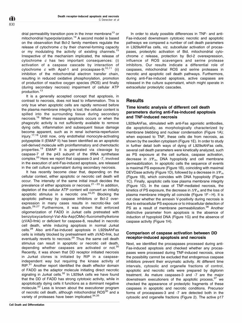
L929sAhFas, stimulated with anti-Fas agonistic antibodies,die apoptotically, as morphologically characterized bymembrane blebbing and nuclear condensation (Figure 1A);when exposed to TNF, these cells die from necrosis, asshown by the swollen cytoplasm (Figure 1E). In order to studyin further detail both ways of dying of L929sAhFas cells,several cell death parameters were kinetically analyzed, suchas PS exposure at the cell surface, caspase activation,decrease in DCm, DNA hypoploidy and cell membranepermeabilization. In apoptotic cells the sequence of eventsis maximal PS exposure (Figure 1B), coinciding with maximalDEVDase activity (Figure 1D), followed by a decrease in DCm
(Figure 1B), which coincides with DNA hypoploidy (Figure1C). Finally, apoptotic cells lose plasma membrane integrity(Figure 1D). In the case of TNF-mediated necrosis, thekinetics of PS exposure, the decrease in DCm and the loss ofplasma membrane integrity all coincide (Figure 1F ± H). It isnot clear whether the annexin V-positivity during necrosis isdue to extracellular PS exposure or to intracellular detection ofPS as a result of membrane permeabilization.36 Anotherdistinctive parameter from apoptosis is the absence ofinduction of hypoploid DNA (Figure 1G) and the absence ofcaspase activity (Figure 1H).
Comparison of caspase activation between DDreceptor-induced apoptosis and necrosis
Next, we identified the procaspases processed during anti-Fas-induced apoptosis and checked whether any procas-pases were processed during TNF-induced necrosis, sincethe possibility cannot be excluded that endogenous caspaseinhibitors prevent their enzymatic activity. At different timeintervals, cytosolic and organelle fractions of control,apoptotic and necrotic cells were prepared by digitonintreatment. As mature caspases-3 and -7 are the majordownstream executioners of the apoptotic process,37 wechecked the appearance of proteolytic fragments of thesecaspases in apoptotic and necrotic conditions. Precursorforms of procaspases-3 and -7 are detected both in thecytosolic and organelle fractions (Figure 2). The active p17
Death receptor-induced apoptosis and necrosisG Denecker et al
830
Cell Death and Differentiation
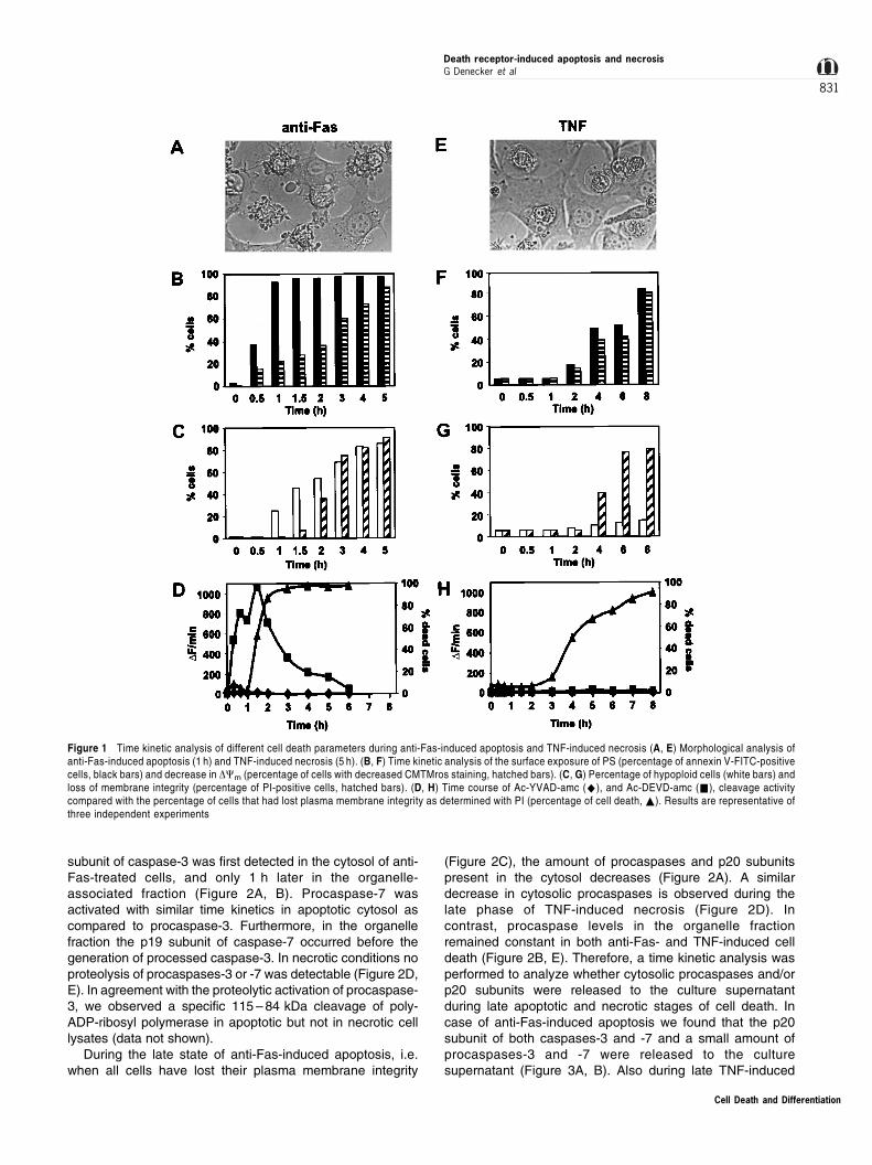
subunit of caspase-3 was first detected in the cytosol of anti-Fas-treated cells, and only 1 h later in the organelle-associated fraction (Figure 2A, B). Procaspase-7 wasactivated with similar time kinetics in apoptotic cytosol ascompared to procaspase-3. Furthermore, in the organellefraction the p19 subunit of caspase-7 occurred before thegeneration of processed caspase-3. In necrotic conditions noproteolysis of procaspases-3 or -7 was detectable (Figure 2D,E). In agreement with the proteolytic activation of procaspase-3, we observed a specific 115 ± 84 kDa cleavage of poly-ADP-ribosyl polymerase in apoptotic but not in necrotic celllysates (data not shown).
During the late state of anti-Fas-induced apoptosis, i.e.when all cells have lost their plasma membrane integrity
(Figure 2C), the amount of procaspases and p20 subunitspresent in the cytosol decreases (Figure 2A). A similardecrease in cytosolic procaspases is observed during thelate phase of TNF-induced necrosis (Figure 2D). Incontrast, procaspase levels in the organelle fractionremained constant in both anti-Fas- and TNF-induced celldeath (Figure 2B, E). Therefore, a time kinetic analysis wasperformed to analyze whether cytosolic procaspases and/orp20 subunits were released to the culture supernatantduring late apoptotic and necrotic stages of cell death. Incase of anti-Fas-induced apoptosis we found that the p20subunit of both caspases-3 and -7 and a small amount ofprocaspases-3 and -7 were released to the culturesupernatant (Figure 3A, B). Also during late TNF-induced
Figure 1 Time kinetic analysis of different cell death parameters during anti-Fas-induced apoptosis and TNF-induced necrosis (A, E) Morphological analysis ofanti-Fas-induced apoptosis (1 h) and TNF-induced necrosis (5 h). (B, F) Time kinetic analysis of the surface exposure of PS (percentage of annexin V-FITC-positivecells, black bars) and decrease in DCm (percentage of cells with decreased CMTMros staining, hatched bars). (C, G) Percentage of hypoploid cells (white bars) andloss of membrane integrity (percentage of PI-positive cells, hatched bars). (D, H) Time course of Ac-YVAD-amc (^), and Ac-DEVD-amc (&), cleavage activitycompared with the percentage of cells that had lost plasma membrane integrity as determined with PI (percentage of cell death, ~). Results are representative ofthree independent experiments
Cell Death and Differentiation
Death receptor-induced apoptosis and necrosisG Denecker et al
831
necrosis, procaspases were detected in the culture super-natant (Figure 3D, E). Next we investigated whether therelease of subunits of caspases-3 and -7 were enzymati-cally active on Ac-DEVD-amc fluorogenic substrate. As acontrol for release of cytosolic proteins and loss of plasmamembrane integrity, the amount of LDH present in theculture supernatant was determined. As shown in Figure 3,during anti-Fas-induced apoptosis a vast Ac-DEVD-amcproteolytic activity in the culture supernatant coincided withLDH activity, suggesting a passive diffusion process ofcytosolic proteins during necrosis and the secondarynecrotic phase of the apoptotic process (Figure 3C, F). Atlater time points of TNF-induced necrotic cell death somep20-like fragments of caspases-3 and -7 appeared in theculture supernatant, resulting in minor DEVDase activity(Figure 3E, F).
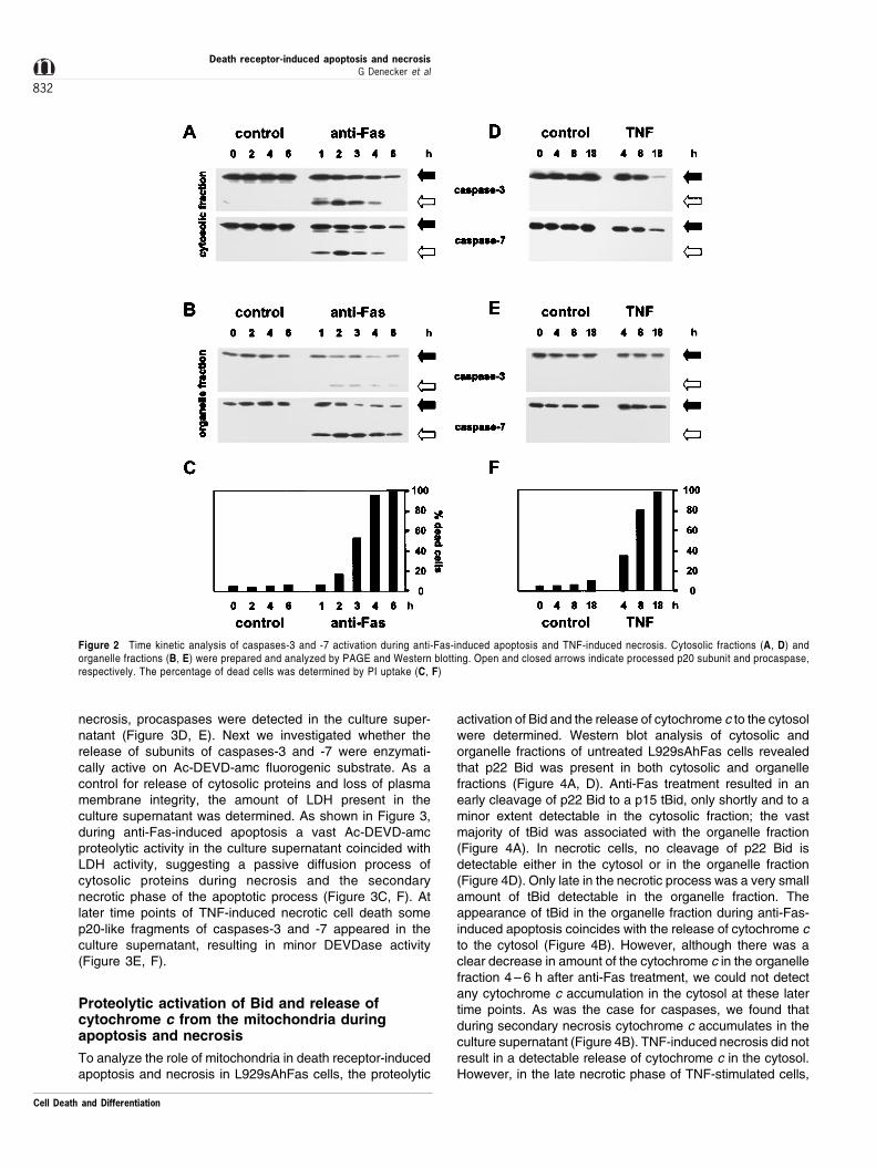
Proteolytic activation of Bid and release ofcytochrome c from the mitochondria duringapoptosis and necrosis
To analyze the role of mitochondria in death receptor-inducedapoptosis and necrosis in L929sAhFas cells, the proteolytic
activation of Bid and the release of cytochrome c to the cytosolwere determined. Western blot analysis of cytosolic andorganelle fractions of untreated L929sAhFas cells revealedthat p22 Bid was present in both cytosolic and organellefractions (Figure 4A, D). Anti-Fas treatment resulted in anearly cleavage of p22 Bid to a p15 tBid, only shortly and to aminor extent detectable in the cytosolic fraction; the vastmajority of tBid was associated with the organelle fraction(Figure 4A). In necrotic cells, no cleavage of p22 Bid isdetectable either in the cytosol or in the organelle fraction(Figure 4D). Only late in the necrotic process was a very smallamount of tBid detectable in the organelle fraction. Theappearance of tBid in the organelle fraction during anti-Fas-induced apoptosis coincides with the release of cytochrome cto the cytosol (Figure 4B). However, although there was aclear decrease in amount of the cytochrome c in the organellefraction 4 ± 6 h after anti-Fas treatment, we could not detectany cytochrome c accumulation in the cytosol at these latertime points. As was the case for caspases, we found thatduring secondary necrosis cytochrome c accumulates in theculture supernatant (Figure 4B). TNF-induced necrosis did notresult in a detectable release of cytochrome c in the cytosol.However, in the late necrotic phase of TNF-stimulated cells,
Figure 2 Time kinetic analysis of caspases-3 and -7 activation during anti-Fas-induced apoptosis and TNF-induced necrosis. Cytosolic fractions (A, D) andorganelle fractions (B, E) were prepared and analyzed by PAGE and Western blotting. Open and closed arrows indicate processed p20 subunit and procaspase,respectively. The percentage of dead cells was determined by PI uptake (C, F)
Cell Death and Differentiation
Death receptor-induced apoptosis and necrosisG Denecker et al
832

cytochrome c accumulated in the culture supernatant from themoment that cells had lost their plasma membrane integrity(Figure 4E).
As a control for the release of mitochondrial intermem-brane proteins we also analyzed the release of AK2 fromthe mitochondria. AK2 in healthy cells is confined to themitochondrial intermembrane space. AK2 is released duringetoposide-, anti-Fas- and staurosporine-induced apoptosisin Jurkat cells.38,39 Release of AK2 during anti-Fas-inducedapoptosis in L929sAhFas cells occurs later than release ofcytochrome c (Figure 4C), which might suggest thatcytochrome c is more rapidly accessible for release thanAK2. During TNF-induced necrosis, AK2 is not, or only invery small amounts, detectable in the cytosol (Figure 4F).AK2 is detectable in the supernatant of necrotically dyingcells from the moment that cells have lost their plasmamembrane integrity, as is the case for detection ofcytochrome c (data not shown). To examine whether theinner mitochondrial membrane was damaged duringnecrosis, we checked the presence of AK3. AK3 is locatedexclusively in the mitochondrial matrix.40 AK3 was
undetected in the cytosol and in the supernatant ofapoptotic or necrotic cells (data not shown). Moreover,the amount of matrix AK3 in the organelle fraction remainedunchanged, also at time points when cells had completelylost their cell membrane integrity, suggesting that the innermitochondrial membrane remains intact during bothapoptosis and necrosis.
Influence of Bcl-2 overexpression on apoptosis,necrosis, and cytochrome c release
Bcl-2 delays apoptotic cell death by different stimuli.15 Scaffidiet al.41 demonstrated the existence of two pathways of anti-Fas-induced apoptosis. Type I apoptosis is independent ofpro-apoptotic factors released by mitochondria and is notinhibited by Bcl-2. Type II apoptosis depends on the release ofproapoptotic factors from the mitochondria and is inhibited byBcl-2. It has also been reported that Bcl-2 inhibits necrotic celldeath induced by oxygen depletion, respiratory chaininhibitors, or by glutathione depletion.42 ± 44 Therefore, weanalyzed the effect of Bcl-2 overexpression on anti-Fas- and
Figure 3 Time kinetic analysis of caspases-3 and -7 processing, DEVDase activity and LDH release in culture supernatant during anti-Fas-induced apoptosis andTNF-induced necrosis. Total cell lysates were prepared and the culture supernatant was precipitated. The presence of caspase-3 and caspase-7 in lysate (A, D)and supernatant (B, E) was determined by PAGE and Western blotting. Open and closed arrows indicate processed p20 subunit and procaspase, respectively. In aparallel set-up, the presence of Ac-DEVD-amc cleavage activity in cytosol (^), and supernatant (*), as well as of LDH in supernatant (~) was determined (C,F)
Cell Death and Differentiation
Death receptor-induced apoptosis and necrosisG Denecker et al
833
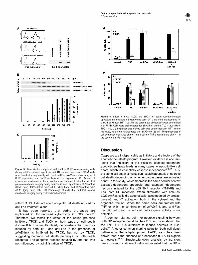
TNF-induced cell death. L929sAhFas.Bcl-2 cells weregenerated, and clones were selected based on human Bcl-2and human Fas receptor expression (Figure 5A). Thecytotoxic response of Bcl-2-transfected cells was examinedby the loss of plasma membrane integrity. Overexpression ofBcl-2 delayed consistently for about 1 h anti-Fas-inducedapoptotic cell death as compared to control L929sAhFascells, which displayed 100% secondary necrosis at 3 h(Figure 5B). TNF-induced necrosis was delayed for about4 h (Figure 5C), while 100% plasma membrane permeabiliza-tion of control cells was seen at 12 h. These results indicatethat overexpression of Bcl-2 temporarily prevents bothapoptosis and necrosis. Overexpression of Bcl-2 also delaysthe release of cytochrome c after anti-Fas treatment (Figure
5B) and allows its prolonged accumulation in the cytosol,probably due to delay in secondary necrosis.
Role of ROS and serine proteases duringapoptosis and necrosis
When L929sAhFas cells are preincubated with zVAD-fmkprior to anti-Fas treatment, caspase activity and apoptotic celldeath are completely prevented. However, pretreatment withzVAD-fmk does not prevent cell death, as cells rapidly die in atypical necrotic way.45 TNF-induced necrosis can be blockedby addition of BHA, a lipophilic oxygen radical scavenger.46
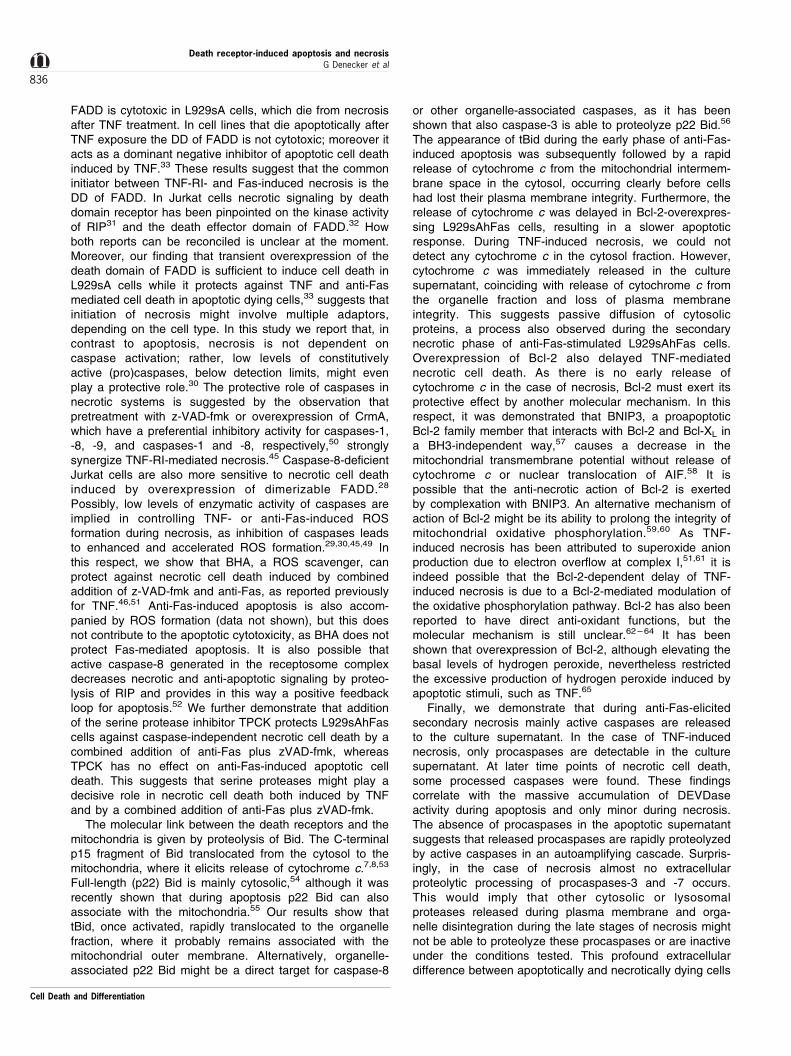
As clear from Figure 6A, necrosis induced by the combinedaddition of zVAD-fmk and anti-Fas is blocked by pretreatment
Figure 4 Time kinetic analysis of the proteolytic activation of Bid (A, D), cellular distribution of cytochrome c (B, E), AK2 and AK3 (C, F), during anti-Fas-inducedapoptosis and TNF-induced necrosis. Cytosolic and organelle fractions were prepared and analyzed by Western blotting. Closed and open arrows indicate full-length Bid and tBid, respectively. Cell death was determined by PI uptake (shown in Figure 2C and F)
Cell Death and Differentiation
Death receptor-induced apoptosis and necrosisG Denecker et al
834
with BHA. BHA did not affect apoptotic cell death induced byanti-Fas treatment alone.
It has been reported that serine proteases areimplicated in TNF-induced cytotoxicity in L929 cells.47
Therefore, we tested the effect of the serine proteaseinhibitors TPCK and TLCK on both types of cell death(Figure 6B). The results clearly demonstrate that necrosisinduced by both TNF and anti-Fas in the presence ofzVAD-fmk is inhibited by TPCK, but not by TLCK,suggesting common cell death pathways by both deathreceptors. The apoptotic process induced by anti-Fas wasnot influenced by administration of TPCK.
Discussion
Caspases are indispensable as initiators and effectors of theapoptotic cell death program. However, evidence is accumu-lating that inhibition of the classical caspase-dependentapoptotic pathway leads in many cases to necrotic-like celldeath, which is essentially caspase-independent.26,27 Thus,the same cell death stimulus can result in apoptotic or necroticcell death, depending on whether procaspases are activatedor not. In this study, we compared in the same cellular contextcaspase-dependent apoptosis and caspase-independentnecrosis initiated by the p55 TNF receptor (TNF-RI) andFas, both DD receptors. When stimulated with anti-Fas,L929sAhFas cells die apoptotically, accompanied by procas-pases-3 and -7 activation, both in the cytosol and theorganelle fraction. When the same cells are treated withTNF or with the combination of zVAD-fmk and anti-Fas,necrotic cell death is induced but caspase activity is notdetected.
A common starting point for necrotic signaling betweenboth DD receptors could be their DD, as it was shown thatthe TNF-RI DD is sufficient to induce necrosis in L929cells.48 Another common starting point for both cell deathpathways is the adapter protein FADD, as it has beenshown that in the absence of procaspase-8, FADD signalsto necrosis.28,49 Structure/function analysis by transientoverexpression in different cell lines revealed that the DD of
Figure 5 Time kinetic analysis of cell death in Bcl-2-overexpressing cellsduring anti-Fas-induced apoptosis and TNF-induced necrosis. L929sA cellswere transfected sequentially with Bcl-2 and Fas. (A) Western blot analysis ofBcl-2 expression and FACS analysis of Fas expression. (B) Amount ofcytochrome c released in the cytosol and percentage of cells that had lostplasma membrane integrity during anti-Fas-induced apoptosis in L929sAhFas(black bars), L929sAhFas.Bcl-2 cl6.2 (white bars) and L929sAhFas.Bcl-2cl6.11 (grey bars) cells. (C) Percentage of cells that had lost plasmamembrane integrity during TNF-induced necrosis
Figure 6 Effect of BHA, TLCK and TPCK on death receptor-inducedapoptosis and necrosis in L929sAhFas cells. (A) Cells were preincubated for2 h with or without BHA (100 mM); the percentage of dead cells was determinedwith PI. (B) Cells were preincubated for 2 h with or without TLCK (300 mM) orTPCK (50 mM); the percentage of dead cells was determined with MTT. Whereindicated, cells were co-pretreated with zVAD-fmk (25 mM). The percentage ofcell death was measured after 6 h in the case of TNF treatment and after 4 h inthe case of anti-Fas treatment
Cell Death and Differentiation
Death receptor-induced apoptosis and necrosisG Denecker et al
835
FADD is cytotoxic in L929sA cells, which die from necrosisafter TNF treatment. In cell lines that die apoptotically afterTNF exposure the DD of FADD is not cytotoxic; moreover itacts as a dominant negative inhibitor of apoptotic cell deathinduced by TNF.33 These results suggest that the commoninitiator between TNF-RI- and Fas-induced necrosis is theDD of FADD. In Jurkat cells necrotic signaling by deathdomain receptor has been pinpointed on the kinase activityof RIP31 and the death effector domain of FADD.32 Howboth reports can be reconciled is unclear at the moment.Moreover, our finding that transient overexpression of thedeath domain of FADD is sufficient to induce cell death inL929sA cells while it protects against TNF and anti-Fasmediated cell death in apoptotic dying cells,33 suggests thatinitiation of necrosis might involve multiple adaptors,depending on the cell type. In this study we report that, incontrast to apoptosis, necrosis is not dependent oncaspase activation; rather, low levels of constitutivelyactive (pro)caspases, below detection limits, might evenplay a protective role.30 The protective role of caspases innecrotic systems is suggested by the observation thatpretreatment with z-VAD-fmk or overexpression of CrmA,which have a preferential inhibitory activity for caspases-1,-8, -9, and caspases-1 and -8, respectively,50 stronglysynergize TNF-RI-mediated necrosis.45 Caspase-8-deficientJurkat cells are also more sensitive to necrotic cell deathinduced by overexpression of dimerizable FADD.28
Possibly, low levels of enzymatic activity of caspases areimplied in controlling TNF- or anti-Fas-induced ROSformation during necrosis, as inhibition of caspases leadsto enhanced and accelerated ROS formation.29,30,45,49 Inthis respect, we show that BHA, a ROS scavenger, canprotect against necrotic cell death induced by combinedaddition of z-VAD-fmk and anti-Fas, as reported previouslyfor TNF.46,51 Anti-Fas-induced apoptosis is also accom-panied by ROS formation (data not shown), but this doesnot contribute to the apoptotic cytotoxicity, as BHA does notprotect Fas-mediated apoptosis. It is also possible thatactive caspase-8 generated in the receptosome complexdecreases necrotic and anti-apoptotic signaling by proteo-lysis of RIP and provides in this way a positive feedbackloop for apoptosis.52 We further demonstrate that additionof the serine protease inhibitor TPCK protects L929sAhFascells against caspase-independent necrotic cell death by acombined addition of anti-Fas plus zVAD-fmk, whereasTPCK has no effect on anti-Fas-induced apoptotic celldeath. This suggests that serine proteases might play adecisive role in necrotic cell death both induced by TNFand by a combined addition of anti-Fas plus zVAD-fmk.
The molecular link between the death receptors and themitochondria is given by proteolysis of Bid. The C-terminalp15 fragment of Bid translocated from the cytosol to themitochondria, where it elicits release of cytochrome c.7,8,53
Full-length (p22) Bid is mainly cytosolic,54 although it wasrecently shown that during apoptosis p22 Bid can alsoassociate with the mitochondria.55 Our results show thattBid, once activated, rapidly translocated to the organellefraction, where it probably remains associated with themitochondrial outer membrane. Alternatively, organelle-associated p22 Bid might be a direct target for caspase-8
or other organelle-associated caspases, as it has beenshown that also caspase-3 is able to proteolyze p22 Bid.56
The appearance of tBid during the early phase of anti-Fas-induced apoptosis was subsequently followed by a rapidrelease of cytochrome c from the mitochondrial intermem-brane space in the cytosol, occurring clearly before cellshad lost their plasma membrane integrity. Furthermore, therelease of cytochrome c was delayed in Bcl-2-overexpres-sing L929sAhFas cells, resulting in a slower apoptoticresponse. During TNF-induced necrosis, we could notdetect any cytochrome c in the cytosol fraction. However,cytochrome c was immediately released in the culturesupernatant, coinciding with release of cytochrome c fromthe organelle fraction and loss of plasma membraneintegrity. This suggests passive diffusion of cytosolicproteins, a process also observed during the secondarynecrotic phase of anti-Fas-stimulated L929sAhFas cells.Overexpression of Bcl-2 also delayed TNF-mediatednecrotic cell death. As there is no early release ofcytochrome c in the case of necrosis, Bcl-2 must exert itsprotective effect by another molecular mechanism. In thisrespect, it was demonstrated that BNIP3, a proapoptoticBcl-2 family member that interacts with Bcl-2 and Bcl-XL ina BH3-independent way,57 causes a decrease in themitochondrial transmembrane potential without release ofcytochrome c or nuclear translocation of AIF.58 It ispossible that the anti-necrotic action of Bcl-2 is exertedby complexation with BNIP3. An alternative mechanism ofaction of Bcl-2 might be its ability to prolong the integrity ofmitochondrial oxidative phosphorylation.59,60 As TNF-induced necrosis has been attributed to superoxide anionproduction due to electron overflow at complex I,51,61 it isindeed possible that the Bcl-2-dependent delay of TNF-induced necrosis is due to a Bcl-2-mediated modulation ofthe oxidative phosphorylation pathway. Bcl-2 has also beenreported to have direct anti-oxidant functions, but themolecular mechanism is still unclear.62 ± 64 It has beenshown that overexpression of Bcl-2, although elevating thebasal levels of hydrogen peroxide, nevertheless restrictedthe excessive production of hydrogen peroxide induced byapoptotic stimuli, such as TNF.65
Finally, we demonstrate that during anti-Fas-elicitedsecondary necrosis mainly active caspases are releasedto the culture supernatant. In the case of TNF-inducednecrosis, only procaspases are detectable in the culturesupernatant. At later time points of necrotic cell death,some processed caspases were found. These findingscorrelate with the massive accumulation of DEVDaseactivity during apoptosis and only minor during necrosis.The absence of procaspases in the apoptotic supernatantsuggests that released procaspases are rapidly proteolyzedby active caspases in an autoamplifying cascade. Surpris-ingly, in the case of necrosis almost no extracellularproteolytic processing of procaspases-3 and -7 occurs.This would imply that other cytosolic or lysosomalproteases released during plasma membrane and orga-nelle disintegration during the late stages of necrosis mightnot be able to proteolyze these procaspases or are inactiveunder the conditions tested. This profound extracellulardifference between apoptotically and necrotically dying cells
Cell Death and Differentiation
Death receptor-induced apoptosis and necrosisG Denecker et al
836
might result in differential pericellular responses. Thephysiological implications of the release of caspases arecurrently unknown. However, one may imagine thatextracellular active caspases play a modulatory role oninflammation, be it positive or negative. This could be ofpathophysiological relevance in the case of massiveapoptosis or insufficient phagocytic capacity.
Our results demonstrate that apoptosis and necrosis,although initiated by the same DD receptors, involveseparate signaling pathways in which caspases andmitochondrial parameters are differentially implicated.Necrosis, as defined by the absence of DNA hypoploidy,cytoplasmic swelling and rapid plasma membrane permea-bilization, is essentially a caspase-independent process inwhich serine proteases and mitochondria reactive oxygenproduction play an essential role. Another distinctiveparameter is the absence of active caspase release inthe case of necrosis, which might have importantpathophysiological implications. However, many questionsremain unanswered regarding the precise molecularsignaling events leading to DD receptor-mediated necro-sis. At the moment different adaptors and domains havebeen implicated, which might reflect cellular differences.Until now, the necrotic cell death process can merely bediscussed in negative terms with the well-studied apoptoticpathways as a reference. Further research on definedmodels will be required to elucidate the necrotic cell deathpathway as clearly distinct or interrelated with the apoptoticcell death pathway and to define the executioner processesduring cell death by necrosis.
Materials and Methods
Antibodies, cytokines and reagents
Recombinant human TNF was produced in Escherichia coli andpurified to at least 99% homogeneity in the Ghent laboratory. Thespecific biological activity was 9.46107 IU/mg as determined in astandardized cytotoxicity assay on L929sA cells. Anti-human Fasantibody (clone 2R2) was purchased from Cell Diagnostica (MuÈ nster,Germany). Butylated hydroxyanisole (BHA), N-tosyl-L-phenylalaninechloromethylketone (TPCK) and N-tosyl-L-lysine chloromethylketone(TLCK) were purchased from Sigma Chemical Co. (St. Louis, MO,USA). Propidium iodide (PI; Becton Dickinson, San Jose, CA, USA)was dissolved at 3 mM in PBS and was used at 30 mM. Annexin V-fluorescein isothiocyanate (FITC) was obtained from PharMingen (SanDiego, CA, USA). The fluorescent markers chloromethyltetramethyl-rosamine (CMTMros) and rhodamine 123 (R123) were purchased fromMolecular Probes (Eugene, OR, USA), prepared as a 1 mM stocksolution in DMSO and used at 0.05 and 0.1 mM, respectively. Thecaspase peptide inhibitor zVAD-fmk was purchased from Bachem(Bubendorf, Switzerland). The caspase fluorogenic substrates acetyl-Asp(OMe)-Glu(OMe)-Val-Asp(OMe)-aminomethylcoumarin (Ac-DEVD-amc) and acetyl-Tyr-Val-Ala-Asp-aminomethylcoumarin (Ac-YVAD-amc) were obtained from Peptide Institute (Osaka, Japan).Antibodies to cytochrome c and human Bcl-2 were obtained fromPharMingen (San Diego, CA, USA) and antibodies to human/mouseBid from R&D Systems (Minneapolis, MN, USA). Rabbit polyclonalantibodies against recombinant murine caspases66 were prepared atthe Centre d'Economie Rurale (Laboratoire d'Hormonologie Animale,
Marloie, Belgium). All anti-caspase antibodies used recognized bothprocaspase and cleaved p20 subunits (our own unpublished results).Adenylate kinase (AK) 2 and AK3 antibodies were kindly provided byDr. T Noma (Department of Biochemistry, Yamaguchi UniversitySchool of Medicine, Yamaguchi, Japan).
Cell culture
L929sA is a murine fibrosarcoma cell line, derived from L929, whichwas selected for its sensitivity to the cytotoxic activity of TNF andcultured as described previously.67 L929sA was transfected with thehuman Fas receptor (L929sAhFas) as described previously.29
L929sAhFas.Bcl-2 cells were obtained by consecutive transfectionof human Bcl-2 cDNA, using neomycine as a selection marker,followed by transection of human Fas cDNA, using puromycine as aselection marker. Human Bcl-2 cDNA was kindly provided by Dr. JReed (Burnham Institute, La Jolla, CA, USA)68 and was inserted as anEcoRI ± EcoRI fragment in pCAGGS.69 Expression of the transfectedgenes was controlled by flow fluorocytometry and Western blotting ofcell lysates (anti-human Fas antibody and anti-human Bcl-2 antibody,respectively).
Induction of apoptosis or necrosis for FACSanalysis and Western blotting
For flow fluorocytometric analysis, L929sAhFas or L929sAhFas.Bcl2cells were kept in suspension by seeding them at 1.56105 cells/ml perwell the day before analysis in uncoated 24-well tissue culture plates(Sarstedt, Newton, NC, USA) in serum-containing DMEM medium. ForWestern analysis, cells were seeded at 56105 cells/ml per well theday before analysis in 6-well tissue culture plates. The next day, anti-Fas (250 ng/ml) or TNF (10 000 IU/ml) were added to the cells.
Flow ¯uorocytometric analysis of DCm, cellmembrane alterations and hypoploidy
The decrease in DCm was analyzed using the fluorogenic probeCMTMros. The exposure of phosphatidylserine (PS) at the cell surfacewas analyzed with annexin V-FITC as detailed previously.70 The lossof cell membrane integrity was determined by means of the PIexclusion method.71 The percentage of cells containing hypoploidDNA was determined by PI staining of cells after one freeze ± thawcycle to permeabilize cells, as described previously.72
Fluorogenic substrate assay for caspase activity
The fluorogenic substrate assay for caspase activity was carried outas described previously.29 Briefly, 1.56105 cells/ml were treated withTNF (10 000 IU/ml) or anti-Fas (250 ng/ml). Cells were washed in coldphosphate buffer and lysed in 200 ml NP-40 lysis buffer. Caspaseactivity was determined by incubating 25 mg of cell lysate with 50 mMAc-YVAD-amc or Ac-DEVD-ame in 200 ml cell-free system buffer. Therelease of fluorescent 7-amino-4-methylcoumarin was measured for60 min at 2-min intervals by fluorometry (excitation at 360 nm andemission at 480 nm) (Cytofluor; PerSeptive Biosystems, Cambridge,MA, USA); the maximal rate of increase in fluorescence wascalculated (DF/min).
Lactate dehydrogenase (LDH) assay
LDH measurements were performed with a Hitachi 747 automatedanalyzer (Roche Molecular Biochemicals Basel, Switzerland), based
Cell Death and Differentiation
Death receptor-induced apoptosis and necrosisG Denecker et al
837
on conversion of pyruvate to lactate and simultaneous oxidation ofNADH to NAD. The rate of decrease in NADH is directly proportional tothe LDH activity and is determined spectrophotometrically at 340 nm.
3-(4,5-dimethylthiazol-2-yl)-2,5-diphenyltetrazoliumbromide (MTT) assay
Cells were seeded the day before at 26104 cells/well in 96-wellplates. The next day inhibitors, TNF and anti-Fas were added at thegiven concentrations. Cell death was assessed at different timeintervals using MTT staining as described previously.73 Thepercentage of cell death was calculated using the equation100%6[1 ± (A595/655 treated cells ± A595/655 medium)/(A595/655 un-treated cells ± A595/655 medium)].
Western blot analysis
56105 ml cells seeded in 6-well plates in serum-free DMEMsupplemented with insulin, transferring and selenium were treatedwith or without TNF (10 000 IU/ml) or anti-Fas (250 ng/ml). Theproteins present in the culture supernatant were precipitated with 10%trichloracetic acid. Cells were washed in cold phosphate buffer andpermeabilized with 250 ml 0.02% digitonin dissolved in cell-freesystem buffer and left on ice for 1 min. This treatment allowsselective lysis of the plasma membrane without affecting the organellemembranes. After centrifugation, the supernatant and the pellet(organelle fraction) were dissolved separately in Laemmli buffer andanalyzed by 15% SDS ± PAGE and Western blotting using antibodiesto cytochrome c, Bid, AK2, AK3, caspases-3 and -7 and developed byECL-based detection (Nycomed Amersham, Little Chalfont, UK).
AcknowledgementsResearch was supported by the Interuniversitaire Attractiepolen, theFonds voor Wetenschappelijk Onderzoek-Vlaanderen (grant No.G005097N and 3G000601), and an EC-RTD grant No. QLRT-199-00739. D Vercammen is a postdoctoral researcher and G Brouckaert is apredoctoral researcher with the Fonds voor Wetenschappelijk Onder-zoek-Vlaanderen. G Denecker was supported by Kom op tegen Kanker.M Steemans and T Vanden Berghe are predoctoral researchers with theVlaams Instituut voor de Bevordering van het Wetenschappelijk-technologisch Onderzoek in de Industrie. G Van Loo is paid by theFlanders Interuniversity Institute for Biotechnology. The authors thank AMeeus and W Burm for technical assistance with tissue culture. BZhivotovsky was supported by the Swedish Cancer Society.
References
1. Sun XM, MacFarlane M, Zhuang J, Wolf BB, Green DR and Cohen GM (1999)
Distinct caspase cascades are initiated in receptor-mediated and chemical-
induced apoptosis. J. Biol. Chem. 274: 5053 ± 5060
2. Wallach D, Varfolomeev EE, Malinin NL, Goltsev YV, Kovalenko AV and Boldin
MP (1999) Tumor Necrosis Factor Receptor and Fas signaling Mechanisms.
Annu. Rev. Immunol. 17: 331 ± 367
3. Boldin MP, Varfolomeev EE, Pancer Z, Mett IL, Camonis JH and Wallach D
(1995) A novel protein that interacts with the death domain of Fas/APO1
contains a sequence motif related to the death domain. J. Biol. Chem. 270:
7795 ± 7798
4. Muzio M, Chinnaiyan AM, Kischkel FC, O'Rourke K, Shevchenko A, Ni J,
Scaffidi C, Bretz JD, Zhang M, Gentz R, Mann M, Krammer PH, Peter ME
and Dixit VM (1996) FLICE, a novel FADD-homologous ICE/CED-3-like
protease, is recruited to the CD95 (Fas/APO-1) death-inducing signaling
complex. Cell 85: 817 ± 827
5. Muzio M, Stockwell BR, Stennicke HR, Salvesen GS and Dixit VM (1998) An
induced proximity model for caspase-8 activation. J. Biol. Chem. 273: 2926 ±
2930
6. Liu X, Kim CN, Yang J, Jemmerson R and Wang X (1996) Induction of apoptotic
program in cell-free extracts: requirement for dATP and cytochrome c. Cell 86:
147 ± 157
7. Li H, Zhu H, Xu CJ and Yuan J (1998) Cleavage of BID by caspase 8 mediates the
mitochondrial damage in the Fas pathway of apoptosis. Cell 94: 491 ± 5018. Luo X, Budihardjo I, Zou H, Slaughter C and Wang X (1998) Bid, a Bcl2 interacting
protein, mediates cytochrome c release from mitochondria in response to
activation of cell surface death receptors. Cell 94: 481 ± 490
9. Steemans M, Goossens V, Van de Craen M, Van Herreweghe F,
Vancompernolle K, De Vos K, Vandenabeele P and Grooten J (1998) A
caspase-activated factor (CAF) induces mitochondrial membrane depolariza-
tion and cytochrome c release by a nonproteolytic mechanism. J. Exp. Med. 188:
2193 ± 2198
10. Zou H, Henzel WJ, Liu X, Lutschg A and Wang X (1997) Apaf-1, a human protein
homologous to C. elegans CED-4, participates in cytochrome c-dependent
activation of caspase-3 [see comments]. Cell 90: 405 ± 413
11. Srinivasula SM, Ahmad M, Fernandes-Alnemri T and Alnemri ES (1998)
Autoactivation of procaspase-9 by Apaf-1-mediated oligomerization. Mol. Cell 1:
949 ± 957
12. Hu Y, Benedict MA, Ding L and Nu nÄ ez G (1999) Role of cytochrome c and dATP/
ATP hydrolysis in Apaf-1-mediated caspase-9 activation and apoptosis. EMBO
J. 18: 3586 ± 3595
13. Bernardi P, Scorrano L, Colonna R, Petronilli V and Di Lisa F (1999) Mitochondriaand cell death. Mechanistic aspects and methodological issues. Eur. J. Biochem.
264: 687 ± 701
14. Vander Heiden MG, Chandel NS, Williamson EK, Schumacker PT and
Thompson CB (1997) Bcl-xL regulates the membrane potential and volume
homeostasis of mitochoncria [see comments]. Cell 91: 627 ± 637
15. Gross A, McDonnell JM and Korsmeyer SJ (1999) BCL-2 family members and
the mitochondria in apoptosis. Genes Dev. 13: 1899 ± 1911
16. Fadok VA, Bratton DL, Rose DM, Pearson A, Ezekewitz RA and Henson PM
(2000) A receptor for phosphatidylserine-specific clearance of apoptotic cells.
Nature 405: 85 ± 90
17. Miwa K, Asano M, Horai R, Iwakura Y, Nagata S and Suda T (1998) Caspase 1-
independent IL-1 beta release and inflammation induced by the apoptosis
inducer Fas ligand. Nat. Med. 4: 1287 ± 1292
18. Daemen VA, van't Veer C, Denecker G, Heemskerk VH, Wolfs TG, Clauss
M,Vandenabeele P and Buurman WA (1999) Inhibition of apoptosis induced by
ischemia-reperfusion prevents inflammation. J. Clin. Invest. 104: 541 ± 549
19. Knies UE, Behrensdorf HA, Mitchell CA, Deutsch U, Risau W, Drexler HCA and
Clauss M (1998) Regulation of endothelial monocyte-activating polypeptide IIrelease by apoptosis. Proc. Natl. Acad. Sci. USA 95: 12322 ± 12327
20. Behrensdorf HA, van de Craen M, Knies UE, Vandenabelle P and Clauss M
(2000) The endothelial monocyte-activating polypeptide II (EMAP II) is a
substrate for caspase-7. FEBS Lett. 466: 143 ± 147
21. Dypbukt JM, Ankarcrona M, Burkitt M, Sjoholm A, Strom K, Orrenius S and
Nicotera P (1994) Different prooxidant levels stimulate growth, trigger apoptosis,
or produce necrosis of insulin-secreting RINm5F cells. The roel of intracellular
polyamines. J. Biol. Chem. 269: 30553 ± 30560
22. Bonfoco E, Krainc D, Ankarcrona M, Nicotera P and Lipton SA (1995) Apoptosis
and necrosis: two distinct events induced, respectively, by mild and intense
insults with N-methyl-D-aspartate or nitric oxide/superoxide in cortical cell
cultures. Proc. Natl. Acad. Sci. USA 92: 7162 ± 7166
23. Vantieghem A, Assefa Z, Vandenabelle P, Declercq W, Courtois S,
Vandenheede JR, Merlevede W, de Witte P and Agostinis P (1998) Hypericin-
induced photosensitization of HeLa cells leads to apoptosis or necrosis.
Involvement of cytochrome c and procaspase-3 activation in the mechanism of
apoptosis. FEBS Lett. 440: 19 ± 24
24. Eguchi Y, Shimizu S and Tsujimoto Y (1997) Intracellular ATP levels determine
cell death fate by apoptosis or necrosis. Cancer Res 57: 1835 ± 184025. Leist M, Single B, Castoldi AF, KuÈ hnle S and Nicotera P (1997) Intracellular
adenosine triphosphate (ATP) concentration: a switch in the decision between
apoptosis and necrosis. J. Exp. Med. 185: 1481 ± 1486
26. Borner C and Monney L (1999) Apoptosis without caspases: an inefficient
molecular guillotine? Cell Death Differ. 6: 497 ± 507
Cell Death and Differentiation
Death receptor-induced apoptosis and necrosisG Denecker et al
838
27. Kitanaka C and Kuchino Y (1999) Caspase-independent programmed cell death
with necrotic morphology. Cell Death Differ. 6: 508 ± 515
28. Kawahara A, Ohsawa Y, Matsumura H, Uchiyama Y and Nagata S (1998)
Caspase-independent cell killing by Fas-associated protein with death domain.
J. Cell Biol. 143: 1353 ± 1360
29. Vercammen D, Brouckaert G, Denecker G, Van de Craen M, Declercq W,
Fiers W and Vandenabeele P (1998) Dual signaling of the Fas receptor:
initiation of both apoptotic and necrotic cell death pathways. J. Exp. Med.188: 919 ± 930
30. Fiers W, Beyaert R, Declercq W and Vandenabeele P (1999) More than one way
to die: apoptosis, necrosis and reactive oxygen damage. Oncogene 18: 7719 ±
7730
31. Holler N, Zaru R, Micheau O, Thome M, Attinger A, Valitutti S, Bodmer JL,
Schneider P, Seat B and Tschopp J (2000) Fas triggers an alternative, caspase-
8-independent cell death pathway using the kinase RIP as effector molecule.
Nat. Immunol. 1: 489 ± 495
32. Matsumura H, Shimizu Y, Ohsawa Y, Kawahara A, Uchiyama Y and Nagata S
(2000) Necrotic death pathway in Fas receptor signaling. J. Cell Biol. 151: 1247 ±
1256
33. Boone E, Vanden Berghe T, Van Loo G, De Wilde G, De Wael N, Vercammen D,
Fiers W, Haegeman G and Vandenabeele P (2000) Structure/Function analysis
of p55 tumor necrosis factor receptor and fas-associated death domain. Effect on
necrosis in L929sA cells. J. Biol. Chem. 275: 37596 ± 37603
34. Johnson DE (2000) Noncaspase proteases in apoptosis. Leukemia 14: 1695 ±
1703
35. Wang KK (2000) Calpain and caspase: can you tell the difference? Trends.Neurosci. 23: 20 ± 26
36. Martin SJ, Reutelingsperger CP, McGahon AJ, Rader JA, van Schie RC, LaFace
DM and Green DR (1995) Early redistribution of plasma membrane
phosphatidylserine is a general feature of apoptosis regardless of the initiating
stimulus: inhibition by overexpression of Bcl-2 and Abl. J. Exp. Med. 182: 1545 ±
1556
37. Earnshaw WC, Martins LM and Kaufmann SH (1999) Mammalian caspases:
Structure, Activation, Substrates, and Functions During Apoptosis. Annu. Rev.
Biochem. 68: 383 ± 424
38. Single B, Leist M and Nicotera P (1998) Simultaneous release of adenylate
kinase and cytochrome c in cell death [letter]. Cell Death Differ. 5: 1001 ±
1003
39. KoÈ hler C, Gahm A, Noma T, Nakazawa A, Orrenius S and Zhivotovsky B (1999)
Release of adenylate kinase 2 from the mitochondrial intermembrane space
during apoptosis. FEBS Lett. 447: 10 ± 12
40. Nobumoto M, Yamada M, Song S, Inouye S and Nakazawa A (1998) Mechanism
of mitochondrial import of adenylate kinase isozymes. J. Biochem. (Tokyo) 123:
128 ± 13541. Scaffidi C, Fulda S, Srinivasan A, Friesen C, Li F, Tomaselli KJ, Debatin KM,
Krammer PH and Peter ME (1998) Two CD95 (APO-1/Fas) signaling pathways.
EMBO J. 17: 1675 ± 1687
42. Kane DJ, Ord T, Anton R and Bredesen DE (1995) Expression of bcl-2 inhibits
necrotic neural cell death. J. Neurosci. Res. 40: 269 ± 275
43. Shimizu S, Eguchi Y, Kamiike W, Itoh Y, Hasegawa J, Yamabe K, Otsuki Y,
Matsuda H and Tsujimoto Y (1996) Induction of apoptosis as well as necrosis by
hypoxia and predominant prevention of apoptosis by Bcl-2 and Bcl-XL. Cancer
Res. 56: 2161 ± 2166
44. Shimizu S, Eguchi Y, Kamiike W, Waguri S, Uchiyama Y, Matsuda H and
Tsujimoto Y (1996) Retardation of chemical hypoxia-induced necrotic cell death
by Bcl-2 and ICE inhibitors: possible involvement of common mediators in
apoptotic and necrotic signal transductions. Oncogene 12: 2045 ± 2050
45. Vercammen D, Beyaert R, Denecker G, Goossens V, Van Loo G, Declercq W,
Grooten J, Fiers W and Vandenabeele P (1998) Inhibition of caspases increases
the sensitivity of L929 cells to necrosis mediated by tumor necrosis factor. J. Exp.
Med. 187: 1477 ± 1485
46. Goossens V, Grooten J, De Vos K and Fiers W (1995) Direct evidence for tumor
necrosis factor-induced mitochondrial reactive oxygen intermediates and theirinvolvement in cytotoxicity. Proc. Natl. Acad. Sci. USA 92: 8115 ± 8119
47. Suffys P, Beyaert R, Van Roy F and Fiers W (1988) Involvement of a serine
protease in tumour-necrosis-factor-mediated cytotoxicity. Eur. J. Biochem. 178:
257 ± 265
48. Vandevoorde V, Haegeman G and Fiers W (1997) Induced expression of
trimerized intracellular domains of the human tumor necrosis factor (TNF) p55
receptor elicits TNF effects. J. Cell Biol. 137: 1627 ± 1638
49. Khwaja A and Tatton L (1999) Resistance to the cytotoxic effects of tumor
necrosis factor alpha can be overcome by inhibition of a FADD/Caspase-
dependent signaling pathway. J. Biol. Chem. 274: 36817 ± 36823
50. Ekert PG, Silke J and Vaux DL (1999) Caspase inhibitors. Cell Death Differ. 6:
1081 ± 108651. Schulze-Osthoff K, Beyaert R, Vandevoorde V, Haegeman G and Fiers W (1993)
Depletion of the mitochondrial electron transport abrogates the cytotoxic and
gene-inductive effects of TNF. EMBO J. 12: 3095 ± 3104
52. Martinou JC, Desagher S and Antonsson B (2000) Cytochrome c release from
mitochondria: all or nothing. Nat. Cell. Biol. 2: E41 ± E43
53. Gross A, Yin XM, Wang K, Wei MC, Jockel J, Milliman C, Erdjument-Bromage H,
Tempst P and Korsmeyer SJ (1999) Caspase cleaved BID targets mitochondria
and is required for cytochrome c release, while BCL-XL prevents this release but
not tumor necrosis factor-R1/Fas death. J. Biol. Chem. 274: 1156 ± 1163
54. Wang K, Yin XM, Chao DT, Milliman CL and Korsmeyer SJ (1996) BID: a novel
BH3 domain-only death agonist. Genes Dev. 10: 2859 ± 2869
55. Desagher S, Osen-Sand A, Nichols A, Eskes R, Montessuit S, Lauper S,
Maundrell K, Antonsson B and Martinou JC (1999) Bid-induced conformational
change of Bax is responsible for mitochondrial cytochrome c release during
apoptosis. J. Cell Biol. 144: 891 ± 901
56. Slee EA, Keogh SA and Martin SJ (2000) Cleavage of BID during cytotoxic drug
and UV radiation-induced apoptosis occurs downstream of the point of Bcl-2
action and is catalysed by caspase-3: a potential feedback loop for amplificationof apoptosis-associated mitochondrial cytochrome c release. Cell Death Differ.
7: 556 ± 565
57. Ray R, Chen G, Vande Velde C, Cizeau J, Park JH, Reed JC, Gietz RD and
Greenberg AH (2000) BNIP3 heterodimerizes with Bcl-2/Bcl-X(L) and induces
cell death independent of a Bcl-2 homology 3 (BH3) domain at both mitochondrial
and nonmitochondrial sites. J. Biol. Chem. 275: 1439 ± 1448
58. Vande Velde C, Cizeau J, Dubik D, Alimonti J, Brown T, Israels S, Hakem R and
Greenberg AH (2000) BNIP3 and genetic control of necrosis-like cell death
through the mitochondrial permeability transition pore. Mol. Cell Biol. 20: 5454 ±
5468
59. Green DR and Reed JC (1998) Mitochondria and apoptosis. Science 281:
1309 ± 1312
60. Shimizu S, Eguchi Y, Kamiike W, Funahashi Y, Mignon A, Lacronique V,
Matsuda H and Tsujimoto Y (1998) Bcl-2 prevents apoptotic mitochondrial
dysfunction by regulating proton flux. Proc. Natl. Acad. Sci. USA 95: 1455 ±
1459
61. Goossens V, Stange G, Moens K, Pipeleers D and Grooten J (1999) Regulation
of tumor necrosis factor-induced, mitochondria- and reactive oxygen species-dependent cell death by the electron flux through the electron transport chain
complex I. Antioxidants & Redox Signaling 1: 285 ± 295
62. Hockenbery DM, Oltvai ZN, Yin XM, Milliman CL and Korsmeyer SJ (1993) Bcl-2
functions in an antioxidant pathway to prevent apoptosis. Cell 75: 241 ± 251
63. Jacobson MD and Raff MC (1995) Programmed cell death and Bcl-2 protection in
very low oxygen. Nature 374: 814 ± 816
64. Shimizu S, Eguchi Y, Kosaka H, Kamiike W, Matsuda H and Tsujimoto Y (1995)
Prevention of hypoxia-induced cell death by Bcl-2 and Bcl-xL. Nature 374: 811 ±
813
65. Esposti MD, Hatzinisiriou I, McLennan H and Ralph S (1999) Bcl-2 and
mitochondrial oxygen radicals. New approaches with reactive oxygen species-
sensitive probes. J. Biol. Chem. 274: 29831 ± 29837
66. Van de Craen M, Declercq W, Van den Brande I, Fiers W and Vandenabeele P
(1999) The proteolytic procaspase activation network: an in vitro analysis. Cell
Death Differ. 6: 1117 ± 1124
67. Vanhaesebroeck B, Decoster E, Van Ostade X, Van Bladel S, Lenaerts A, Van
Roy F and Fiers W (1992) Expression of an exogenous tumor necrosis factor
(TNF) gene in TNF-sensitive cell lines confers resistance to TNF-mediated cell
lysis. J. Immunol. 148: 2785 ± 279468. Reed JC, Cuddy M, Haldar S, Croce C, Nowell P, Makover D and Bradley K
(1990) BCL2-mediated tumorigenicity of a human T-lymphoid cell line: synergy
with MYC and inhibition by BCL2 antigsense. Proc. Natl. Acad. Sci. USA 87:
3660 ± 3664
Cell Death and Differentiation
Death receptor-induced apoptosis and necrosisG Denecker et al
839
69. Niwa H, Yamamura K and Miyazaki J (1991) Efficient selection for high-
expression transfectants with a novel eukaryotic vector. Gene 108: 193 ± 199
70. Denecker G, Dooms H, Van Loo G, Vercammen D, Grooten J, Fiers W, Declercq
W and Vandenabeele P (2000) Phosphatidyl serine exposure during apoptosis
precedes release of cytochrome c and decrease in mitochondrial transmem-
brane potential. FEBS Lett. 465: 47 ± 52
71. Nicoletti I, Migliorati G, Pagliacci MC, Grignani F and Riccardi C (1991) A rapid
and simple method for measuring thymocyte apoptosis by propidium iodidestaining and flow cytometry. J. Immunol. Methods 139: 271 ± 279
72. Vercammen D, Vandenabeele P, Beyaert R, Declercq W and Fiers W (1997)
Tumour necrosis factor-induced necrosis versus anti-Fas-induced apoptosis in
L929 cells. Cytokine 9: 801 ± 808
73. Tada H, Shiho O, Kuroshima K, Koyama M and Tsukamoto K (1986) An improved
colorimetric assay for interleukin 2. J. Immunol. Methods 93: 157 ± 165
Cell Death and Differentiation
Death receptor-induced apoptosis and necrosisG Denecker et al
840
Related Documents