Research Article 3515 Introduction Transposable elements (TEs) are essential structural and regulatory components of genomes. Their ability to transpose provides a fundamental source of genetic variation but also represents a potential threat for genome integrity. Genomes have deployed a diversity of epigenetic defensive mechanisms against TEs and their concerted action results in the global, efficient and heritable repression of mobile elements throughout generations (Aravin et al., 2007; Siomi et al., 2008; Slotkin et al., 2007). In Drosophila, epigenetic control of TEs depends on histone modifications, chromatin structure, small RNA-based transcriptional silencing and DNA methylation (Aravin et al., 2007; Josse et al., 2007; Klenov et al., 2007; Dramard et al., 2007; Phalke et al., 2009; Klattenhoff et al., 2007; Klattenhoff et al., 2009; Li et al., 2009; Malone et al., 2009). Recent literature has abundantly described the mechanisms of Piwi-interacting small RNAs (piRNAs) biogenesis, as well as their essential role for the repression of TEs in germ cells (Brennecke et al., 2007; Vagin et al., 2006; Saito et al., 2006; Aravin et al., 2007; Siomi et al., 2008; Klattenhoff and Theurkauf, 2008). Accordingly, several families of TEs are derepressed in the germline of mutants affecting the piRNA pathway (Vagin et al., 2006; Chambeyron et al., 2008; Pane et al., 2007; Lim and Kai, 2007; Chen et al., 2007; Klattenhoff et al., 2007; Klattenhoff et al., 2009; Vagin et al., 2004). Remarkably, all these mutants are viable but induce female sterility associated with a complex phenotype including defects in germline stem cell maintenance, accumulation of germline DNA damage and aberrant egg axial patterning (Klattenhoff and Theurkauf, 2008). However, it is difficult to determine the actual contribution of TE activity to their complex sterility phenotype (Klattenhoff et al., 2007; Thomson and Lin, 2009). In Drosophila, massive and deleterious TE germline mobilization is also observed in the progeny of certain intraspecific crosses. This phenomenon, known as hybrid dysgenesis, has long been recognized as a powerful experimental model for the study of TE regulation in a wild-type background (Bregliano et al., 1980). Drosophila hybrid dysgenesis systems are usually characterized by a severe gonadal atrophy in both sexes, resulting in sterility. These include the D. melanogaster P-M (P element) and H-E (hobo element) systems, as well as a hybrid dysgenesis in D. virilis, which involves several families of TEs (Kidwell and Novy, 1979; Blackman et al., 1987; Yannopoulos et al., 1987; Petrov et al., 1995; Blumenstiel and Hartl, 2005). The I-R type of hybrid dysgenesis is unique as it only occurs in females and does not result from a defective ovarian development. Instead, dysgenic females lay a normal amount of eggs but the resulting embryos fail to hatch (Picard and L’Héritier, 1971). The causative factor of this non-Mendelian female sterility is the I element, a 5.4 kb, non-LTR retrotransposon of the LINE (long interspersed nucleotidic element) superfamily of transposable elements (Bucheton et al., 1984). Most D. melanogaster strains are so-called Inducer (I) strains and contain about 10 transposition-competent but transcriptionally silenced I elements. Such functional I elements are absent from Reactive (R) strains that were established before the recent worldwide invasion of this retrotransposon in natural populations (Bucheton et al., 2002). Maternal transmission of piRNAs has been proposed to underlie the epigenetic repression of TEs revealed by Drosophila hybrid dysgenesis systems (Blumenstiel and Hartl, 2005; Brennecke et al., 2008; Chambeyron et al., 2008). In the case of the I-R system, maternal epigenetic protection is largely reduced in R strains, resulting in the expression of paternally transmitted I elements in the naive germline of dysgenic females (Brennecke et al., 2008; Chambeyron et al., 2008). I-R hybrid dysgenesis occurs when I males are crossed with R females. The female progeny of this dysgenic cross, called SF (stérilité femelle) females, usually display a strong sterility phenotype associated with derepression of I elements. In addition, the I-R syndrome is characterized by a high mutation rate as well Drosophila I-R hybrid dysgenesis is associated with catastrophic meiosis and abnormal zygote formation Guillermo A. Orsi 1 , Eric F. Joyce 2 , Pierre Couble 1 , Kim S. McKim 2 and Benjamin Loppin 1, * 1 Centre de Génétique Moléculaire et Cellulaire, CNRS UMR5534, University of Lyon, UCBL Lyon1, Villeurbanne, F-69100, France 2 Waksman Institute and Department of Genetics, Rutgers University, 190, Frelinghuysen Rd, Piscataway NJ 08854, USA *Author for correspondence ([email protected]) Accepted 7 July 2010 Journal of Cell Science 123, 3515-3524 © 2010. Published by The Company of Biologists Ltd doi:10.1242/jcs.073890 Summary The Drosophila I-R type of hybrid dysgenesis is a sterility syndrome (SF sterility) associated with the mobilization of the I retrotransposon in female germ cells. SF sterility results from a maternal-effect embryonic lethality whose origin has remained unclear since its discovery about 40 years ago. Here, we show that meiotic divisions in SF oocytes are catastrophic and systematically fail to produce a functional female pronucleus at fertilization. As a consequence, most embryos from SF females rapidly arrest their development with aneuploid or damaged nuclei, whereas others develop as non-viable, androgenetic haploid embryos. Finally, we show that, in contrast to mutants affecting the biogenesis of piRNAs, SF egg chambers do not accumulate persistent DNA double- strand breaks, suggesting that I-element activity might perturb the functional organization of meiotic chromosomes without triggering an early DNA damage response. Key words: Hybrid dysgenesis, I element, Meiotic catastrophe, Haploid embryos, Meiotic DNA damage checkpoint Journal of Cell Science

Welcome message from author
This document is posted to help you gain knowledge. Please leave a comment to let me know what you think about it! Share it to your friends and learn new things together.
Transcript

Research Article 3515
IntroductionTransposable elements (TEs) are essential structural and regulatorycomponents of genomes. Their ability to transpose provides afundamental source of genetic variation but also represents apotential threat for genome integrity. Genomes have deployed adiversity of epigenetic defensive mechanisms against TEs and theirconcerted action results in the global, efficient and heritablerepression of mobile elements throughout generations (Aravin etal., 2007; Siomi et al., 2008; Slotkin et al., 2007). In Drosophila,epigenetic control of TEs depends on histone modifications,chromatin structure, small RNA-based transcriptional silencingand DNA methylation (Aravin et al., 2007; Josse et al., 2007;Klenov et al., 2007; Dramard et al., 2007; Phalke et al., 2009;Klattenhoff et al., 2007; Klattenhoff et al., 2009; Li et al., 2009;Malone et al., 2009). Recent literature has abundantly describedthe mechanisms of Piwi-interacting small RNAs (piRNAs)biogenesis, as well as their essential role for the repression of TEsin germ cells (Brennecke et al., 2007; Vagin et al., 2006; Saito etal., 2006; Aravin et al., 2007; Siomi et al., 2008; Klattenhoff andTheurkauf, 2008). Accordingly, several families of TEs arederepressed in the germline of mutants affecting the piRNApathway (Vagin et al., 2006; Chambeyron et al., 2008; Pane et al.,2007; Lim and Kai, 2007; Chen et al., 2007; Klattenhoff et al.,2007; Klattenhoff et al., 2009; Vagin et al., 2004). Remarkably, allthese mutants are viable but induce female sterility associated witha complex phenotype including defects in germline stem cellmaintenance, accumulation of germline DNA damage and aberrantegg axial patterning (Klattenhoff and Theurkauf, 2008). However,it is difficult to determine the actual contribution of TE activity totheir complex sterility phenotype (Klattenhoff et al., 2007; Thomsonand Lin, 2009).
In Drosophila, massive and deleterious TE germline mobilizationis also observed in the progeny of certain intraspecific crosses.This phenomenon, known as hybrid dysgenesis, has long been
recognized as a powerful experimental model for the study of TEregulation in a wild-type background (Bregliano et al., 1980).Drosophila hybrid dysgenesis systems are usually characterized bya severe gonadal atrophy in both sexes, resulting in sterility. Theseinclude the D. melanogaster P-M (P element) and H-E (hoboelement) systems, as well as a hybrid dysgenesis in D. virilis,which involves several families of TEs (Kidwell and Novy, 1979;Blackman et al., 1987; Yannopoulos et al., 1987; Petrov et al.,1995; Blumenstiel and Hartl, 2005). The I-R type of hybriddysgenesis is unique as it only occurs in females and does notresult from a defective ovarian development. Instead, dysgenicfemales lay a normal amount of eggs but the resulting embryos failto hatch (Picard and L’Héritier, 1971). The causative factor of thisnon-Mendelian female sterility is the I element, a 5.4 kb, non-LTRretrotransposon of the LINE (long interspersed nucleotidic element)superfamily of transposable elements (Bucheton et al., 1984). MostD. melanogaster strains are so-called Inducer (I) strains and containabout 10 transposition-competent but transcriptionally silenced Ielements. Such functional I elements are absent from Reactive (R)strains that were established before the recent worldwide invasionof this retrotransposon in natural populations (Bucheton et al.,2002). Maternal transmission of piRNAs has been proposed tounderlie the epigenetic repression of TEs revealed by Drosophilahybrid dysgenesis systems (Blumenstiel and Hartl, 2005; Brenneckeet al., 2008; Chambeyron et al., 2008). In the case of the I-Rsystem, maternal epigenetic protection is largely reduced in Rstrains, resulting in the expression of paternally transmitted Ielements in the naive germline of dysgenic females (Brennecke etal., 2008; Chambeyron et al., 2008).
I-R hybrid dysgenesis occurs when I males are crossed with Rfemales. The female progeny of this dysgenic cross, called SF(stérilité femelle) females, usually display a strong sterilityphenotype associated with derepression of I elements. In addition,the I-R syndrome is characterized by a high mutation rate as well
Drosophila I-R hybrid dysgenesis is associated withcatastrophic meiosis and abnormal zygote formationGuillermo A. Orsi1, Eric F. Joyce2, Pierre Couble1, Kim S. McKim2 and Benjamin Loppin1,*1Centre de Génétique Moléculaire et Cellulaire, CNRS UMR5534, University of Lyon, UCBL Lyon1, Villeurbanne, F-69100, France2Waksman Institute and Department of Genetics, Rutgers University, 190, Frelinghuysen Rd, Piscataway NJ 08854, USA*Author for correspondence ([email protected])
Accepted 7 July 2010Journal of Cell Science 123, 3515-3524 © 2010. Published by The Company of Biologists Ltddoi:10.1242/jcs.073890
SummaryThe Drosophila I-R type of hybrid dysgenesis is a sterility syndrome (SF sterility) associated with the mobilization of the Iretrotransposon in female germ cells. SF sterility results from a maternal-effect embryonic lethality whose origin has remained unclearsince its discovery about 40 years ago. Here, we show that meiotic divisions in SF oocytes are catastrophic and systematically fail toproduce a functional female pronucleus at fertilization. As a consequence, most embryos from SF females rapidly arrest theirdevelopment with aneuploid or damaged nuclei, whereas others develop as non-viable, androgenetic haploid embryos. Finally, weshow that, in contrast to mutants affecting the biogenesis of piRNAs, SF egg chambers do not accumulate persistent DNA double-strand breaks, suggesting that I-element activity might perturb the functional organization of meiotic chromosomes without triggeringan early DNA damage response.
Key words: Hybrid dysgenesis, I element, Meiotic catastrophe, Haploid embryos, Meiotic DNA damage checkpoint
Jour
nal o
f Cel
l Sci
ence

as chromosomal non-disjunctions and rearrangements (Buchetonet al., 2002). In contrast to SF females, the genetically identicalRSF females obtained from the reverse cross (R males with Ifemales) show much lower expression of I elements and are fullyfertile (Picard and L’Héritier, 1971; Bucheton et al., 2002). Fromthe early work of Picard et al. (Picard et al., 1977) and Lavige(Lavige, 1986), it was established that embryos produced by SFfemales died through a strict maternal effect and frequentlypresented abnormal syncytial divisions. However, despite extensiveresearch on this system, the nature of SF sterility has remainedenigmatic since its discovery (Picard and L’Héritier, 1971).
In this paper, we have undertaken a detailed cytological studyof I-R hybrid dysgenesis to determine the origin of SF sterility. Ourstudy revealed that meiotic divisions are catastrophic in SF oocytesand eggs. This highly penetrant phenotype prevents the integrationof the full set of maternal chromosomes in the zygote, resulting innon-viable embryos. We also show that, in contrast to mutantsaffecting the biogenesis of piRNAs, SF germ cells do notaccumulate massive DNA damage during early oogenesis,suggesting that I activity perturbs the functional organization ofmeiotic chromosomes without activating the early germline DNAdamage response.
ResultsMeiotic catastrophe in eggs of SF femalesWe performed a cytological study of SF eggs and embryos tounderstand the nature of SF maternal-effect embryonic lethality.We used SF females that were not older than a week as SF sterilitydecreases progressively with age (see below). Consistent with earlycytological studies (Lavige, 1986), we observed that a majority ofsyncytial SF embryos contained catastrophic mitotic figures withisolated or broken chromosomes and asynchronously dividingnuclei of various sizes (Fig. 1). In addition, in SF embryos, weobserved that the polar body did not form the typical triploidrosette and contained many fragmented chromosomes (Fig. 1D–F).This last aspect of the phenotype suggested that meiosis wasdefective in SF eggs. We then turned to late oocytes to observe thefirst meiotic division. In Drosophila, the mature stage-14 oocyte isarrested in metaphase of meiosis I (King, 1970). To visualize theorganization of meiotic chromosomes and the first meiotic spindle,we used control and SF females expressing the fluorescentcentromeric protein EGFP-Cid (Schuh et al., 2007) or themicrotubule-associated Jupiter-GFP (Buszczak et al., 2007),respectively. In fixed control stage-14 oocytes (n30), meioticchromosomes appeared as a slightly elongated mass of chromatinwith non-exchange chromosomes occasionally separated towardsthe spindle poles (Fig. 2) (Theurkauf and Hawley, 1992). In about80% of SF oocytes (22/28), the chromatin appeared fragmentedand/or abnormally distributed into several small masses (Fig. 2).Some of these masses of chromatin were associated with an EGFP-Cid spot, whereas others were not, thus suggesting the presence offragmented chromosomes. These isolated or fragmentedchromosomes formed miniature spindle-like structures as revealedwith the Jupiter-GFP marker (Fig. 2). In the rest of the SF oocytes(6/28), the first meiotic division was apparently normal, althoughthe low resolution of meiosis I chromosomes did not allow thedetection of possible more subtle defects.
We then analyzed very early SF eggs to observe the secondmeiotic division and pronuclear formation. Strikingly, meiosis II inSF eggs was almost systematically abnormal with bridges ofchromatin connecting the separating chromatids in anaphase and
telophase (Fig. 3C,D; Table 1). This defective separation ofchromatids was followed by chromosome fragmentation and unequalsegregation of meiotic products. Notably, the loss of genetic materialin the female pronucleus was obvious at the pronuclear appositionstage. In control RSF eggs, apposed pronuclei appeared identical insize (Fig. 3E). In SF eggs, however, the female pronucleus waseither small, fragmented in several smaller nuclei or, in someinstances, did not form at all (Fig. 3F,G; data not shown). We thusconcluded that, in eggs from SF females, defective meiotic divisionscompromised the formation of a normal female pronucleus.
Embryos from SF females develop with paternalchromosomesIn Drosophila fertilized eggs, pronuclei do not fuse but insteadremain apposed during the first zygotic S phase and the paternal
3516 Journal of Cell Science 123 (20)
Fig. 1. The maternal-effect embryonic lethality associated with I-R hybriddysgenesis. (A)Crossing scheme to obtain RSF (upper) and SF (lower)females. SF females lay eggs but the resulting embryos die before hatching.Genetically identical RSF females are fully fertile. (B,C)Confocal images ofearly syncytial embryos from RSF (B) or SF (C) females stained for Tubulin(green) and DNA (red). In contrast to the normal nuclear divisions observed inRSF embryos, SF embryos contain asynchronously dividing nuclei of varioussizes and fragmented chromosomes (arrows). (D–F)In RSF embryos (D),fused polar bodies form a typical rosette of condensed chromosomes. In SFembryos (E,F), polar body organization is abnormal and many chromosomesare lost or fragmented (arrows). Scale bars: 15mm.
Jour
nal o
f Cel
l Sci
ence

and maternal sets of chromosomes enter mitosis as separate entitieswithin a common mitotic spindle (Sonnenblick, 1950). In a majorityof SF embryos at first mitosis, we observed that the spindle did notcontain the full complement of chromosomes compared with RSF
zygotes (Fig. 3H,I; Table 1). In other cases, some chromosomeswere excluded from the spindle or lagged behind in anaphase ofthe first division (Fig. 3J,K; Table 1). To determine the identity ofthese absent or abnormal chromosomes, we stained SF eggs withan antibody directed against acetylated forms of histone H4 thatpreferentially marks paternal chromatin (Loppin et al., 2005a). Weobserved that, in SF eggs, from the pronuclear apposition until theend of the first zygotic division, the damaged or late chromosomeswere systematically less-intensely stained than the unaffectedchromosomes (Fig. 4A-F). In some cases, a single haploid set ofstrongly stained chromosomes was present at the first mitosis (Fig.4E). We confirmed these observations by analyzing the progeny oftransgenic SF females expressing the recombinant histone variantH3.3-Flag, a specific marker of paternal chromosomes atfertilization (Bonnefoy et al., 2007; Orsi et al., 2009). Thisexperiment clearly confirmed the specific defective integration ofmaternal chromosomes in SF zygotes (supplementary material Fig.S1).
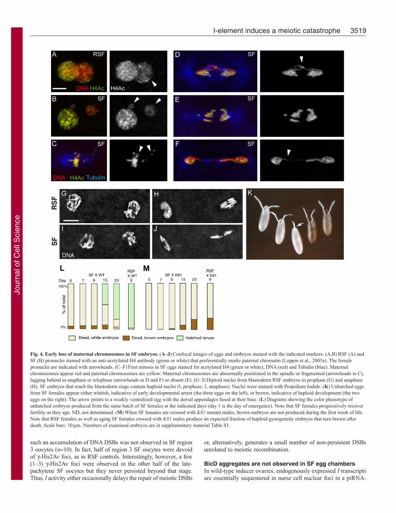
As previously reported by Picard et al. (Picard et al., 1977)and Lavige (Lavige, 1986), we observed that approximately 7%(n1134) of SF embryos died at a late developmental stage asrevealed by the fact that they turned brown after death andshowed signs of organogenesis and cuticle deposition. By contrast,the rest of the unhatched eggs remained whitish, suggesting thatthey arrested development before cellularization (Fig. 4K). In theDrosophila mutant maternal haploid (mh), paternal chromosomesare unable to divide in anaphase of the first mitosis and form achromatin bridge (Santamaria and Gans, 1980; Loppin et al.,2001). This frequently results in catastrophic early mitoses andmost embryos die after a few rounds of nuclear divisions.However, a fraction of embryos escape this early arrest and
3517I-element induces a meiotic catastrophe
Fig. 2. Meiosis I is catastrophic in oocytes from SF females. Confocalimages of meiosis I in stage-14 oocytes from females expressing the indicatedmarker. Control females are from the transgenic EGFP-Cid line and Jupiter-GFP line. SF females were obtained by crossing inducer EGFP-Cid orJupiter-GFP males with Charolles females at 25°C. At day 5 of adult life,ovaries were dissected, fixed and stained for DNA (red). Scale bar: 10mm.(Top) EGFP-tagged centromeric histone Cid (EGFP-Cid) marks centromeresin meiosis I chromosomes. In control oocytes, chromosomes show alignedcentromeres in prometaphase. In SF oocytes, chromosomes appear fragmentedor mislocalized. Centromeres are indicated with arrows. (Bottom) The controlis an anastral first meiotic spindle in prometaphase marked with microtubule-associated Jupiter-GFP. In SF oocytes, mini-spindles organize aroundmislocalized or fragmented chromosomes (arrowheads).
Fig. 3. Catastrophic meiosis and abnormal zygote formation in eggs from SF females. Confocal images of eggs and early embryos stained for Tubulin (green)and DNA (red). (A–D)Meiosis figures are shown with dorsal egg periphery at the top and the anterior end to the left. The corresponding male pronuclei are shownin insets. (A,B)RSF eggs in anaphase (A) or telophase (B) of the second meiotic division. (C,D)Meiosis II in SF eggs is catastrophic. Note the chromatin bridgesin anaphase (C) and the unequal chromosome segregation in telophase (D). Loss of genetic material in the two innermost meiotic products is obvious in D.(E)Pronuclear apposition in an RSF egg. (F,G)In SF eggs, the female pronucleus looks abnormally small (F) or fragmented into several smaller nuclei (G). Thefemale pronuclei are indicated with arrowheads. (H–K)First zygotic division. Metaphase of the first zygotic division in an RSF egg (H) containing the paternal andmaternal chromosomes. First mitosis in an SF egg with either a reduced number of chromosomes (I), with chromosomes that appear excluded from the spindle(arrowheads in J) or with lagging chromosomes in anaphase (arrowheads in K). Scale bars: 10mm.
Jour
nal o
f Cel
l Sci
ence

develop as non-viable, haploid gynogenetic embryos (Loppin etal., 2001). At the cytological level, early development of SFembryos appeared similar to mh embryos, with catastrophicsyncytial divisions forming chromatin bridges (Fig. 1C;supplementary material Fig. S2). In addition, a minority of SFembryos developed beyond the blastoderm stage and containednormal mitotic figures but the nuclei were about half the size ofcontrol diploid nuclei (Fig. 4G-J). To demonstrate that theseescaper embryos were actually haploid androgenetic embryos,we crossed SF females with males homozygous for the K81paternal effect mutation, which prevents the formation offunctional paternal chromosomes in the progeny (Fuyama, 1984;Yasuda et al., 1995; Loppin et al., 2005b). As expected, these SFfemales failed to produce any brown embryos during their firstweek, confirming that late embryos from SF females developedwith paternal chromosomes (Fig. 4L,M; supplementary materialTable S1). In conclusion, our results demonstrated that mostembryos from SF females die early with catastrophic mitoses,whereas a minority escape this early arrest as haploid androgeneticembryos.
Defective karyosome formation in SF oocytesThe meiotic phenotype observed in SF females prompted us toanalyze the structure of the oocyte nucleus during SF oogenesis.In Drosophila, female meiosis initiates in region 2A of thegermarium, at the anterior tip of each ovariole. After meioticrecombination, in later egg chambers, the oocyte nucleus enlargeswhile the condensed maternal chromosomes in prophase I ofmeiosis remain packaged within a subnuclear structure known asthe karyosome (Spradling, 1993). In stage 6–9 control oocytesstained for DNA, the karyosome appeared as a round condensedstructure within the unstained oocyte nucleus (Fig. 5A). Bystriking contrast, we observed that the karyosome wasdisorganized in a majority of SF oocytes (Fig. 5A; supplementarymaterial Fig. S2). Typically, the SF karyosomes were fragmentedand stretched along the inner side of the oocyte nuclear envelope.A remarkable and well-described feature of SF sterility is itsmodulation by age and temperature. Indeed, SF sterility is highestin young females but their fertility is progressively restored asthey age (see supplementary material Table S1) (Picard andL’Héritier, 1971). In addition, SF sterility is strongest and lastslonger at relatively cooler temperatures and fertility can betransiently restored after a heat treatment (Bucheton, 1979).Interestingly, we observed that the penetrance and severity of thekaryosome phenotype decreased with the age of SF females. Inaddition, most karyosomes were severely affected when SFfemales where placed at 18°C for 36 hours before dissection,whereas a heat treatment at 30°C dramatically suppressed thephenotype (Fig. 5B,C). Taken together, these observations suggestthat defective karyosome formation in SF oocytes results inabnormal meiotic divisions.
SF germ cells do not accumulate unrepaired DNAdouble-strand breaksIn Drosophila female germ cells, the accumulation of unrepairedDNA double-strand breaks (DSBs) can trigger the activation of awell-characterized ATR-Chk2 (Mei-41-Lok) DNA damageresponse (Ghabrial and Schupbach, 1999; Abdu et al., 2002). Inmutants that affect the repair of meiotic DNA DSBs, activationof the Chk2 checkpoint leads to a complex cellular response.This includes a specific disorganization of the karyosome and astrong egg ventralization phenotype that results from defectiveaccumulation of the signaling protein Gurken in the oocyte(Ghabrial and Schupbach, 1999; Abdu et al., 2002). Interestingly,the Chk2 checkpoint is activated in the female germline of piRNApathway mutants (Chen et al., 2007; Klattenhoff et al., 2007). Inaddition, these mutants are associated with egg patterning defectsand defective karyosome formation (supplementary material Fig.S3) (Chen et al., 2007; Klattenhoff et al., 2007). Genetic analyseshave demonstrated that, in these mutants, the checkpoint is notactivated by meiotic DSBs, thus opening the possibility that theseDNA damages could be induced by the activity of derepressedTEs (Chen et al., 2007; Klattenhoff et al., 2007; Klattenhoff etal., 2009).
As mutant stocks are generally not available in a reactivebackground, the activation of the checkpoint in SF germ cells couldnot be genetically tested. We thus examined the dorsal patterning ofSF eggs to check for indications of DNA damage response. Weobserved that a fraction of SF eggs displayed a weak ventralizationphenotype. In fact, fusion of egg dorsal appendages was onlyobserved with very young SF females not older than 3 days (Table2; Fig. 4K, arrow). Importantly, after a few days, SF females thatwere still fully sterile produced almost 100% of eggs with wild-typeappendages. By clear contrast, aub or armi mutant females produceda majority of severely ventralized eggs throughout their life (Table2). Interestingly, Van De Bor et al. (Van De Bor et al., 2005) haveshown that I and gurken (grk) transcripts compete for the same RNAlocalization machinery in SF egg chambers, resulting in defectivedorsoventral axis specification. This mechanism could indeed accountfor the ventralization of eggs produced by young SF females, wherestrong I transcription is expected to efficiently perturb grk mRNAlocalization. In conclusion, the egg patterning analysis did not supportthe hypothesis of early Chk2 checkpoint activation in SF germ cells.However, we wished to directly evaluate the impact of I-elementactivity on DNA integrity during early oogenesis. We thus stainedSF and control ovaries with antibodies against the phosphorylatedform of histone H2Av (g-His2Av), which associates with DNA DSBs(Mehrotra and McKim, 2006). In wild-type or RSF ovaries, g-His2Av foci were observed in oocytes of germarium regions 2A and2B but were no longer detected in late-pachytene oocytes in theirregion 3 egg chambers (Fig. 6). In region 3 oocytes from aub mutantfemales, late-pachytene nuclei accumulated numerous g-His2Av foci,as previously reported (Klattenhoff et al., 2007). By clear contrast,
3518 Journal of Cell Science 123 (20)
Table 1. Phenotype quantification of SF eggs and embryos
Meiosis II First zygotic division Cycle 2 –7 embryos
n Abnormal (%) n Abnormal (%) n Aneuploid (%) Haploid (%)
SF 40 97.5 56 96.4 90 82.2 11.1RSF 41 2.4 25 0 50 0 0
SF or RSF females grown at 25°C were allowed to lay eggs between days 4 and 6 after emergence (day 1). Eggs at 0–1 hours were collected, fixed and stainedfor DNA. Phenotypes of SF eggs (meiosis II and zygote) and early embryos are described in the Results. n, the total number of eggs and/or embryos analyzed.
Jour
nal o
f Cel
l Sci
ence
such an accumulation of DNA DSBs was not observed in SF region3 oocytes (n10). In fact, half of region 3 SF oocytes were devoidof g-His2Av foci, as in RSF controls. Interestingly, however, a few(1–3) g-His2Av foci were observed in the other half of the late-pachytene SF oocytes but they never persisted beyond that stage.Thus, I activity either occasionally delays the repair of meiotic DSBs
or, alternatively, generates a small number of non-persistent DSBsunrelated to meiotic recombination.
BicD aggregates are not observed in SF egg chambersIn wild-type inducer ovaries, endogenously expressed I transcriptsare essentially sequestered in nurse cell nuclear foci in a piRNA-
3519I-element induces a meiotic catastrophe
Fig. 4. Early loss of maternal chromosomes in SF embryos. (A–J)Confocal images of eggs and embryos stained with the indicated markers. (A,B)RSF (A) andSF (B) pronuclei stained with an anti-acetylated H4 antibody (green or white) that preferentially marks paternal chromatin (Loppin et al., 2005a). The femalepronuclei are indicated with arrowheads. (C–F)First mitosis in SF eggs stained for acetylated H4 (green or white), DNA (red) and Tubulin (blue). Maternalchromosomes appear red and paternal chromosomes are yellow. Maternal chromosomes are abnormally positioned in the spindle or fragmented (arrowheads in C),lagging behind in anaphase or telophase (arrowheads in D and F) or absent (E). (G–J)Diploid nuclei from blastoderm RSF embryos in prophase (G) and anaphase(H). SF embryos that reach the blastoderm stage contain haploid nuclei (I, prophase; J, anaphase). Nuclei were stained with Propidium Iodide. (K)Unhatched eggsfrom SF females appear either whitish, indicative of early developmental arrest (the three eggs on the left), or brown, indicative of haploid development (the twoeggs on the right). The arrow points to a weakly ventralized egg with the dorsal appendages fused at their base. (L)Diagrams showing the color phenotype ofunhatched embryos produced from the same batch of SF females at the indicated days (day 1 is the day of emergence). Note that SF females progressively recoverfertility as they age. ND, not determined. (M)When SF females are crossed with K81 mutant males, brown embryos are not produced during the first week of life.Note that RSF females as well as aging SF females crossed with K81 males produce an expected fraction of haploid gynogenetic embryos that turn brown afterdeath. Scale bars: 10mm. Numbers of examined embryos are in supplementary material Table S1.
Jour
nal o
f Cel
l Sci
ence

dependent manner (Chambeyron et al., 2008), whereasoverexpressed GFP-labeled I transcripts have been shown toaccumulate in cytoplasmic particles called pi-bodies that localizearound nurse cell nuclei (Lim et al., 2009). In SF egg chambers, Itranscripts are essentially transported in the oocyte (Seleme et al.,2005; Chambeyron et al., 2008). Recently, it has been shown thatlarge ribonucleoprotein (RNP) aggregates of the dynein-motormachinery form in egg chambers of piRNA biogenesis mutants(Navarro et al., 2009). Interestingly, injected I transcriptsaccumulate in these aggregates, suggesting that they could serve asdegradation sites for retrotransposon products, in the absence of
piRNA biogenesis (Navarro et al., 2009). Furthermore, these authorshave also shown that formation of these dynein aggregates waslargely dependent upon the activation of the Chk2 checkpoint.
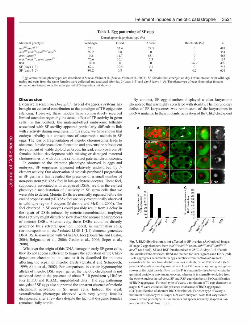
To investigate the possibility that these structures could form inSF egg chambers, we stained ovaries with anti-BicD or anti-Orbantibodies that were shown to accumulate in dynein aggregates(Navarro et al., 2009). We indeed observed aggregates in a largemajority of aub or armi mutant egg chambers. By clear contrast,however, Orb or BicD aggregates were only rarely observed in SFand RSF egg chambers (Fig. 7A,B; data not shown). We concludethat I-element activity is not sufficient to trigger the formation ofthese aggregates in dysgenic ovaries. In the course of theseexperiments, we observed that the oocyte marker BicD wasabnormally distributed in the germinal vesicle of a majority of auband armi mutant stage 6–9 egg chambers (Fig. 7A,C). Thisphenotype was fully rescued in aub mnk double-mutant females,indicating that it was dependent on checkpoint activation (Fig.7A,C). Importantly, we observed that, in SF and RSF oocytes,BicD was normally excluded from the germinal vesicle. Takentogether, these results reinforce the conclusion that SF sterility isindependent of Chk2 checkpoint activation and downstream cellularresponses.
3520 Journal of Cell Science 123 (20)
Fig. 5. Defective karyosome formation in SF oocytes. (A)Confocal imagesof stage 7–8 egg chambers dissected from RSF or SF females and stained forDNA (top). Egg chamber stages are from King (King, 1970). The oocyte is onthe right, the karyosome is indicated with an arrow. (Bottom) In oocytes fromRSF females, the karyosome appears spheric and condensed within theunstained oocyte nucleus. In SF oocytes, the karyosome is frequentlyabnormal, being slightly heterogeneous or elongated in aspect (weakphenotype) or displaying a severe distortion, fragmentation or attachment tothe nuclear envelope (strong phenotype). (B)Effect of temperature on SFkaryosome phenotype. Two-day-old RSF or SF females obtained at 25°Cusing the Charolles or JA26 reactive stocks were placed at the indicatedtemperature for 36 hours before ovary dissection and DNA staining. For eachcondition, a minimum of 40 karyosomes from stage 6–9 oocytes wereobserved and classified according to the phenotypic classes described in A.Results are shown as a percentage of all observed karyosomes. (C)Effect ofage on SF karyosome phenotype. SF females obtained at 25°C using theCharolles or JA26 reactive stocks were dissected at the indicated age andovaries were stained for DNA. Karyosome phenotype was analyzed as in B.Note that the same 25°C, 3-day-old SF female data is shown in B and C. Scalebars: 20mm.
Fig. 6. g-His2AvD distribution in the SF germline. Confocal images of wild-type (WT), aubQC42 or aubHN (aub), RSF and SF germaria stained to visualizeDNA (blue), C(3)G (green) and g-His2AvD (red). Full views of germaria withtheir anterior tip on the left are shown in the left panels. Increasedmagnifications of late-pachytene oocytes (insets) are on the right. In WT andRSF germaria, g-H2AvD foci are not detected in late-pachytene oocytes[identified by the C(3)G staining], indicating that meiotic DNA double-strandbreaks are repaired at this stage. In generally disorganized aub mutantgermaria, where oocyte determination is delayed, >10 g-His2AvD fociaccumulate in late-pachytene oocyte nuclei, shown here in an early region 3oocyte. In SF germaria, 0–3 g-His2AvD foci are observed in late-pachyteneoocytes. A total of 10 late-pachytene oocytes were examined for eachgenotype. Scale bars: 5mm.
Jour
nal o
f Cel
l Sci
ence
DiscussionExtensive research on Drosophila hybrid dysgenesis systems hasbrought an essential contribution to the paradigm of TE epigeneticsilencing. However, these models have comparatively receivedlimited attention regarding the actual effect of TE activity in germcells. In this context, the maternal-effect embryonic lethalityassociated with SF sterility appeared particularly difficult to linkwith I activity during oogenesis. In this study, we have shown thatembryo lethality is a consequence of catastrophic meiosis in SFeggs. The loss or fragmentation of meiotic chromosomes leads toabnormal female pronucleus formation and prevents the subsequentdevelopment of viable diploid embryos. Instead, embryos from SFfemales initiate development with missing or damaged maternalchromosomes or with only the set of intact paternal chromosomes.
In contrast to the dramatic phenotype observed in eggs andembryos, SF oogenesis appeared relatively undisturbed by I-element activity. Our observation of meiosis prophase I progressionin SF germaria has revealed the presence of a small number ofnon-persistent g-His2Av foci in late-pachytene oocytes. These foci,supposedly associated with unrepaired DSBs, are thus the earliestphenotypic manifestation of I activity in SF germ cells that wewere able to detect. Meiotic DSBs are normally repaired before theend of prophase and g-His2Av foci are only exceptionally observedin wild-type region 3 oocytes (Mehrotra and McKim, 2006). Thefoci observed in SF oocytes could possibly result from a delay inthe repair of DSBs induced by meiotic recombination, implyingthat I activity might disturb or slow down the normal repair processof meiotic DSBs. Alternatively, these DSBs could be directlygenerated by I retrotransposition. Indeed, in mammalian cells,retrotransposition of the I-related LINE 1 (L1) elements generatesDNA DSBs associated with g-His2AX foci (Bourc’his and Bestor,2004; Belgnaoui et al., 2006; Gasior et al., 2006; Soper et al.,2008).
Whatever the origin of this DNA damage in early SF germ cells,they do not appear sufficient to trigger the activation of the Chk2-dependent checkpoint, at least as it is described for mutantsaffecting the repair of meiotic DSBs (Ghabrial and Schupbach,1999; Abdu et al., 2002). For comparison, in certain hypomorphicalleles of meiotic DSB repair genes, the meiotic checkpoint is notactivated despite the presence of about 7–10 persistent g-His2Avfoci (E.F.J. and K.S.M., unpublished data). The egg patterninganalysis of SF eggs also supported the apparent absence of meioticcheckpoint activation in SF germ cells. Indeed, the weakventralization phenotype observed with very young femalesdisappeared after a few days despite the fact that dysgenic femalesremained fully sterile.
By contrast, SF egg chambers displayed a clear karyosomephenotype that was highly correlated with sterility. The morphologydefect of SF karyosomes was reminiscent of the karyosomes inpiRNA mutants. In these mutants, activation of the Chk2 checkpoint
3521I-element induces a meiotic catastrophe
Table 2. Egg patterning of SF eggs
Dorsal appendage phenotype (%)
Maternal genotype Wild-type Fused Absent Hatch rate (%) n
aubHN/aubQC42 23.1 52.4 24.5 0 481aubHN mnkP6/aubQC42 mnkP6 99.2 0.8 0 0 354armi1/armi72.1 0.2 11.7 88.1 0 463mnkP6/mnkP6; armi1/armi72.1 76.6 16.1 7.3 0 137RSF 100.0 0 0 96.2 498SF (days 1–3) 69.3 30.4 0.3 0 743SF (days 4–5) 99.2 0.8 0 0.2 651
Egg ventralization phenotypes are described in Staeva-Vieira et al. (Staeva-Vieira et al., 2003). SF females that emerged on day 1 were crossed with wild-typemales and eggs from the same females were collected and analyzed after day 3 (days 1–3) and day 5 (days 4–5). The phenotype of eggs from other femalesremained unchanged over the same period of 5 days (data not shown).
Fig. 7. BicD distribution is not affected in SF ovaries. (A)Confocal imagesof stage-9 egg chambers from aubHN/aubQC42 (aub), aubHN mnkP6/aubQC42
mnkP6 (aub mnk), SF and RSF females raised at 25°C. At days 3–5 of adultlife, ovaries were dissected, fixed and stained for BicD (green) and DNA (red).BicD aggregates accumulate in egg chambers from control aub mutants(arrowheads) but not from double aub mnk mutants, SF or RSF females (leftpanels). Magnification of germinal vesicles of the same stage and genotype areshown in the right panels. Note that BicD is abnormally distributed within thegerminal vesicle in aub mutant oocytes, whereas it is normally excluded fromthe oocyte nucleus in aub mnk, SF and RSF egg chambers. (B)Quantificationof BicD aggregates. For each type of ovary, a minimum of 70 egg chambers atstages 6–9 were evaluated for presence or absence of BicD aggregates.(C)Quantification of aberrant BicD distribution. For each type of ovary, aminimum of 60 oocytes at stages 6–9 were analyzed. Note that karyosomesshow a strong phenotype in aub mutants but appear normally shaped in aubmnk oocytes. Scale bars: 10mm.
Jour
nal o
f Cel
l Sci
ence

is at least partially responsible for this phenotype, in a way similarto mutants affecting the repair of meiotic breaks (Ghabrial andSchupbach, 1999; Abdu et al., 2002; Staeva-Vieira et al., 2003).Indeed, we have observed that 97% (n62) and 26% (n94) of aubmnk and mnk armi karyosomes had a morphology rescued to wild-type, respectively (Fig. 7; data not shown).
Interestingly, DNA damage accumulation and karyosome defectsin the absence of strong dorsoventral patterning defects have beendescribed for mutants that affect both meiotic DNA damage repairand checkpoint signaling, such as hus1 and brca2 (Abdu et al.,2007; Klovstad et al., 2008). Similarly, germline derepression ofTEs in the tejas mutant does not affect egg polarity (Patil and Kai,2010). We thus cannot exclude that the karyosome defect in SFoocytes could reflect a partial or late DNA damage response,which would not trigger other known hallmarks of checkpointactivation, including egg ventralization. Indeed, in SF ovaries, Itranscripts and ORF1 protein are first detected in germarium region2A but they reach their highest levels in later-stage oocytes, wherethey presumably accumulate as RNPs (Seleme et al., 1999; Selemeet al., 2005). At these stages, however, any accumulation of DNADSBs might go undetected with g-His2Av antibodies. In this model,the DNA damage response could still cause the observed karyosomedefect but would occur too late to significantly disturb Grk proteinoocyte accumulation and dorsoventral axis specification.
In the alternative possibility, accumulation of I RNPs in theoocyte could directly affect karyosome formation without inducingany DNA damage response. However, and surprisingly, I productsaccumulate in the perinuclear cytoplasm of SF oocytes and do notappear to enter the nuclear compartment at cytologically detectablelevels (Seleme et al., 1999; Seleme et al., 2005). Accordingly,GFP-tagged ORF1p remains cytoplasmic when transientlyexpressed in Drosophila cultured cells (Rashkova et al., 2002).Thus, only a minor fraction of I RNPs is expected to enter theoocyte nucleus in order to complete the retrotransposition process.This situation contrasts with the clear nuclear accumulation of L1RNPs in mael–/– mutant mouse spermatocytes associated with DNAdamage and chromosome asynapsis (Soper et al., 2008). In SFovaries, we did not detect any gross defect in the distribution ofthe SC protein C(3)G in oocytes (supplementary material Fig. S4).However, the low resolution obtained with this kind of analysis(compared with mouse spermatocytes, for instance) cannot ruleout the presence of undetected chromosome synapsis defects.
The modest effect of I activity on DNA integrity during earlySF oogenesis contrasted with the situation observed in piRNAmutants where many TEs, including I, are derepressed. However,the origin of DNA damage in piRNA pathway mutants is not clearand the involvment of TEs in generating these breaks remains tobe established (Chen et al., 2007; Klattenhoff et al., 2007;Klattenhoff and Theurkauf, 2008; Thomson and Lin, 2009). Geneticinactivation of the checkpoint does not restore the fertility ofpiRNA pathway mutant females (Table 2) (Klattenhoff et al., 2007).It thus indicates that additional, checkpoint-independent defectscause the female sterility in these mutants. Interestingly, embryosfrom aub mnk females display a specific and severe disorganizationof cleavage nuclei that could explain the observed maternal-effectlethality (Blumenstiel et al., 2008). Furthermore, in aub and spn-Emutants, the HeT-A and TART retroelements involved in telomeremaintenance are upregulated in the female germline and theirretrotransposition to broken chromosome termini is increased, withpotential consequences on chromosome stability (Savitsky et al.,2006). Finally, Piwi-family proteins are also involved in the
biogenesis or processing of piRNAs directed against the 3�UTR ofa broad set of cellular transcripts, with possible regulatory functions(Robine et al., 2009). The overall phenotype of piRNA pathwaymutants is thus expected to reflect this functional complexity, incontrast to I-R hybrid dysgenesis, where a single type of elementis activated.
The meiotic defects we observed in SF oocytes and eggs areprobably related to the chromosome rearrangements and non-disjunctions associated with I-R hybrid dysgenesis. Rearrangementsare probably generated after illegitimate homologous recombinationevents between integrating I elements (Busseau et al., 1989;Prudhommeau and Proust, 1990; Proust et al., 1992). Consideringthe fact that these chromosomal aberrations were obtained in viableprogeny from SF females, we suppose that more detrimental andfrequent rearrangements are produced when SF females are stillfully sterile. The accumulation of chromosomal rearrangements inoocyte nuclei could probably affect meiotic divisions by notablyinducing non-disjunction and chromosome fragmentation events.In this model, the progressive I repression established in aging SFfemales would reduce the probability of these events occurringuntil oocyte chromosome architecture becomes compatible withnormal meiosis.
Materials and MethodsDrosophila stocksThe w1118 standard inducer stock and the strong reactive wild-type stock Charolleswere used to set up control or dysgenic crosses, unless otherwise specified. The JA26y w reactive stock was provided by Alain Pelisson (Institute de Génétique Humaine,Montpellier, France). The EGFP-Cid stock (Schuh et al., 2007) and the Jupiter-GFPinsertion (Buszczak et al., 2007) were obtained from Stefan Heidmann (Universityof Bayreuth, Bayreuth, Germany) and from the Carnegie Protein Trap StockCollection (http://flytrap.med.yale.edu/), respectively. The mnkP6 stock was a giftfrom Tin Tin Su (Brodsky et al., 2004). The following alleles were obtainedfrom the Bloomington Drosophila Stock Center at Indiana University(http://flystocks.bio.indiana.edu/): armi1 and armi72.1 (Cook et al., 2004), aubHN andaubQC42 (Schupbach and Wieschaus, 1991), krimpf06583 (Lim and Kai, 2007) andmaelr20 (Clegg et al., 1997). The following heterozygous or hemizygous femaleswere used in this study: armi1 armi72.1 (armi), aubHN aubQC42 (aub), krimpf06583
Df(2R)Exel6063 (krimp) and maelr20 Df(3L)ED230 (mael). The K812 paternal-effectembryonic lethal mutant is a small, viable deficiency that completely removes thems(3)K81 gene (Yasuda et al., 1995). aub mnk or mnk armi double-mutant femaleswere obtained by standard crossing techniques and meiotic recombination.
Crosses and egg phenotype analysisControl and dysgenic crosses were set up at the appropriate temperature using equalnumbers of freshly emerged virgin males and females that were kept togetherthroughout the experiment. Eggs were collected on agar plates, counted and, ifnecessary, the dorsal appendage phenotype was examined by direct observationunder a stereomicroscope. Embryos were then allowed to develop for 3 days at 25°Cbefore hatching rate and brown/white phenotype determination.
Egg collection, ovary dissection and immunofluorescenceFemales that were no older than 1 week were allowed to lay eggs on agar plates inthe presence of males at 25°C. Eggs were dechorionated in bleach and fixed asdescribed (Loppin et al., 2001). Ovaries were dissected in TBST (TBS-0.15%, TritonX-100), fixed in a 1:1 mixture of heptane: 4% paraformaldehyde in PBS, rinsed inTBST and were immediately incubated with the primary antibodies as previouslydescribed (Bonnefoy et al., 2007). Antibodies and dilutions used were: anti-a-Tubulin (Sigma, T9026, 1/500); anti-H4Ac (Chemicon International, AB3062, 1/200);anti-Flag (Sigma, F3165, 1/1000); anti-H3K14Ac (Millipore, 06-911, 1/500); anti-C(3)G (kindly provided by R. S. Hawley, 1/500) (Page and Hawley, 2001); anti-g-His2AvD (1/500) (Mehrotra and McKim, 2006); and anti-BicD (DevelopmentalStudies Hybridoma Bank, 1B11-s, 1/200). DNA was stained with Propidium Iodideor Hoechst. Confocal images were acquired using either a LSM510 microscope(Carl Zeiss) or a Leica SP2 (for Fig. 5) and were processed with Adobe Photoshopsoftware.
Karyosome defect assayTo analyze the effect of temperature on karyosome phenotype, 1-day-old SF femaleswere kept at 25°C for 2 days and were then placed at 18°C, 25°C or 30°C for 36hours before ovary dissection. To analyze the effect of age, SF females that wereobtained at 25°C were aged for 3, 6 or 9 days before dissection. Stage 6–9 oocytes
3522 Journal of Cell Science 123 (20)
Jour
nal o
f Cel
l Sci
ence

stained with Propidium Iodide and H3K14Ac were observed under a confocalmicroscope and karyosomes were classified into three phenotypical categories asdescribed in Fig. 4. For each condition, a minimum of 40 karyosomes was observed.
We are grateful to Alain Pelisson, Scott Hawley, Stefan Heidmann,Tin Tin Su, the Carnegie Protein Trap Stock Collection and theBloomington Stock Center for flies and antibodies. We would like tothank Béatrice Horard, Stéphane Ronsseray, Anne Laurençon, MarionDelattre and Silke Jensen for discussions and critical reading of themanuscript, and Emmanuel Gauthier for providing some of the citedliterature. We also thank Jérôme Schmitt for technical assistance.Confocal microscopy was performed with the help of the CentreTechnologique des Microstructures. Work in the laboratory of B.L.was supported by the CNRS and by a grant from the ANR (ANR-08-BLAN-0139-01). Work in the laboratory of K.S.M. was supported bya grant from the NSF. G.A.O. is supported by a doctoral fellowshipfrom the French Ministry of Education and Research.
Supplementary material available online athttp://jcs.biologists.org/cgi/content/full/123/20/3515/DC1
ReferencesAbdu, U., Brodsky, M. and Schupbach, T. (2002). Activation of a meiotic checkpoint
during Drosophila oogenesis regulates the translation of Gurken through Chk2/Mnk.Curr. Biol. 12, 1645-1651.
Abdu, U., Klovstad, M., Butin-Israeli, V., Bakhrat, A. and Schupbach, T. (2007). Anessential role for Drosophila hus1 in somatic and meiotic DNA damage responses. J.Cell Sci. 120, 1042-1049.
Aravin, A. A., Hannon, G. J. and Brennecke, J. (2007). The Piwi-piRNA pathwayprovides an adaptive defense in the transposon arms race. Science 318, 761-764.
Belgnaoui, S. M., Gosden, R. G., Semmes, O. J. and Haoudi, A. (2006). Human LINE-1 retrotransposon induces DNA damage and apoptosis in cancer cells. Cancer Cell Int.6, 13.
Blackman, R. K., Grimaila, R., Koehler, M. M. and Gelbart, W. M. (1987). Mobilizationof hobo elements residing within the decapentaplegic gene complex: suggestion of anew hybrid dysgenesis system in Drosophila melanogaster. Cell 49, 497-505.
Blumenstiel, J. P. and Hartl, D. L. (2005). Evidence for maternally transmitted smallinterfering RNA in the repression of transposition in Drosophila virilis. Proc. Natl.Acad. Sci. USA 102, 15965-15970.
Blumenstiel, J. P., Fu, R., Theurkauf, W. E. and Hawley, R. S. (2008). Components ofthe RNAi machinery that mediate long-distance chromosomal associations aredispensable for meiotic and early somatic homolog pairing in Drosophila melanogaster.Genetics 180, 1355-1365.
Bonnefoy, E., Orsi, G. A., Couble, P. and Loppin, B. (2007). The essential role ofDrosophila HIRA for de novo assembly of paternal chromatin at fertilization. PLoSGenet. 3, 1991-2006.
Bourc’his, D. and Bestor, T. H. (2004). Meiotic catastrophe and retrotransposonreactivation in male germ cells lacking Dnmt3L. Nature 431, 96-99.
Bregliano, J. C., Picard, G., Bucheton, A., Pelisson, A., Lavige, J. M. and L’Heritier,P. (1980). Hybrid dysgenesis in Drosophila melanogaster. Science 207, 606-611.
Brennecke, J., Aravin, A. A., Stark, A., Dus, M., Kellis, M., Sachidanandam, R. andHannon, G. J. (2007). Discrete small RNA-generating loci as master regulators oftransposon activity in Drosophila. Cell 128, 1089-1103.
Brennecke, J., Malone, C. D., Aravin, A. A., Sachidanandam, R., Stark, A. andHannon, G. J. (2008). An epigenetic role for maternally inherited piRNAs in transposonsilencing. Science 322, 1387-1392.
Brodsky, M. H., Weinert, B. T., Tsang, G., Rong, Y. S., McGinnis, N. M., Golic, K. G.,Rio, D. C. and Rubin, G. M. (2004). Drosophila melanogaster MNK/Chk2 and p53regulate multiple DNA repair and apoptotic pathways following DNA damage. Mol.Cell. Biol. 24, 1219-1231.
Bucheton, A. (1979). Non-Mendelian female sterility in Drosophila melanogaster:influence of aging and thermic treatments. III. Cumulative effects induced by thesefactors. Genetics 93, 131-142.
Bucheton, A., Paro, R., Sang, H. M., Pelisson, A. and Finnegan, D. J. (1984). Themolecular basis of I-R hybrid dysgenesis in Drosophila melanogaster: identification,cloning, and properties of the I factor. Cell 38, 153-163.
Bucheton, A., Busseau, I. and Teninges, D. (2002). I Elements in Drosophilamelanogaster. In Mobile DNA II (ed. N. L. Craig) pp. 796-812. Washington, DC: ASMPress.
Busseau, I., Pelisson, A. and Bucheton, A. (1989). I elements of Drosophila melanogastergenerate specific chromosomal rearrangements during transposition. Mol. Gen. Genet.218, 222-228.
Buszczak, M., Paterno, S., Lighthouse, D., Bachman, J., Planck, J., Owen, S., Skora,A. D., Nystul, T. G., Ohlstein, B., Allen, A. et al. (2007). The carnegie protein traplibrary: a versatile tool for Drosophila developmental studies. Genetics 175, 1505-1531.
Chambeyron, S., Popkova, A., Payen-Groschene, G., Brun, C., Laouini, D., Pelisson,A. and Bucheton, A. (2008). piRNA-mediated nuclear accumulation of retrotransposontranscripts in the Drosophila female germline. Proc. Natl. Acad. Sci. USA 105, 14964-14969.
Chen, Y., Pane, A. and Schupbach, T. (2007). Cutoff and aubergine mutations result inretrotransposon upregulation and checkpoint activation in Drosophila. Curr. Biol. 17,637-642.
Clegg, N. J., Frost, D. M., Larkin, M. K., Subrahmanyan, L., Bryant, Z. and Ruohola-Baker, H. (1997). maelstrom is required for an early step in the establishment ofDrosophila oocyte polarity: posterior localization of grk mRNA. Development 124,4661-4671.
Cook, H. A., Koppetsch, B. S., Wu, J. and Theurkauf, W. E. (2004). The DrosophilaSDE3 homolog armitage is required for oskar mRNA silencing and embryonic axisspecification. Cell 116, 817-829.
Dramard, X., Heidmann, T. and Jensen, S. (2007). Natural epigenetic protection againstthe I-factor, a Drosophila LINE retrotransposon, by remnants of ancestral invasions.PLoS ONE 2, e304.
Fuyama, Y. (1984). Gynogenesis in Drosophila melanogaster. Jpn J. Genet. 59, 91-96.Gasior, S. L., Wakeman, T. P., Xu, B. and Deininger, P. L. (2006). The human LINE-1
retrotransposon creates DNA double-strand breaks. J. Mol. Biol. 357, 1383-1393.Ghabrial, A. and Schupbach, T. (1999). Activation of a meiotic checkpoint regulates
translation of Gurken during Drosophila oogenesis. Nat. Cell Biol. 1, 354-357.Ivanovska I., Khandan T., Ito T. and Orr-Weaver T. L. (2005) A histone code in
meiosis: the histone kinase, NHK-1, is required for proper chromosomal architecture inDrosophila oocytes. Genes Dev. 19, 2571-2582.
Josse, T., Teysset, L., Todeschini, A. L., Sidor, C. M., Anxolabehere, D. and Ronsseray,S. (2007). Telomeric trans-silencing: an epigenetic repression combining RNA silencingand heterochromatin formation. PLoS Genet. 3, 1633-1643.
Kidwell, M. G. and Novy, J. B. (1979). Hybrid dysgenesis in Drosophila melanogaster:sterility resulting from gonadal dysgenesis in the P-M system. Genetics 92, 1127-1140.
King, R. C. (1970). Ovarian Development in Drosophila Melanogaster. New York:Academic Press.
Klattenhoff, C. and Theurkauf, W. (2008). Biogenesis and germline functions of piRNAs.Development 135, 3-9.
Klattenhoff, C., Bratu, D. P., McGinnis-Schultz, N., Koppetsch, B. S., Cook, H. A. andTheurkauf, W. E. (2007). Drosophila rasiRNA pathway mutations disrupt embryonicaxis specification through activation of an ATR/Chk2 DNA damage response. Dev. Cell12, 45-55.
Klattenhoff, C., Xi, H., Li, C., Lee, S., Xu, J., Khurana, J. S., Zhang, F., Schultz, N.,Koppetsch, B. S., Nowosielska, A. et al. (2009). The Drosophila HP1 homolog Rhinois required for transposon silencing and piRNA production by dual-strand clusters. Cell138, 1137-1149.
Klenov, M. S., Lavrov, S. A., Stolyarenko, A. D., Ryazansky, S. S., Aravin, A. A.,Tuschl, T. and Gvozdev, V. A. (2007). Repeat-associated siRNAs cause chromatinsilencing of retrotransposons in the Drosophila melanogaster germline. Nucleic AcidsRes. 35, 5430-5438.
Klovstad, M., Abdu, U. and Schupbach, T. (2008). Drosophila brca2 is required formitotic and meiotic DNA repair and efficient activation of the meiotic recombinationcheckpoint. PLoS Genet. 4, e31.
Lavige, J. M. (1986). I-R system of hybrid dysgenesis in Drosophila melanogaster:further data on the arrest of development of the embryos from SF females. Biol. Cell56, 207-216.
Li, C., Vagin, V. V., Lee, S., Xu, J., Ma, S., Xi, H., Seitz, H., Horwich, M. D., Syrzycka,M., Honda, B. M. et al. (2009). Collapse of germline piRNAs in the absence ofArgonaute3 reveals somatic piRNAs in flies. Cell 137, 509-521.
Lim, A. K. and Kai, T. (2007). Unique germ-line organelle, nuage, functions to repressselfish genetic elements in Drosophila melanogaster. Proc. Natl. Acad. Sci. USA 104,6714-6719.
Loppin, B., Berger, F. and Couble, P. (2001). Paternal chromosome incorporation intothe zygote nucleus is controlled by maternal haploid in Drosophila. Dev. Biol. 231, 383-396.
Loppin, B., Bonnefoy, E., Anselme, C., Laurencon, A., Karr, T. L. and Couble, P.(2005a). The histone H3.3 chaperone HIRA is essential for chromatin assembly in themale pronucleus. Nature 437, 1386-1390.
Loppin, B., Lepetit, D., Dorus, S., Couble, P. and Karr, T. L. (2005b). Origin andneofunctionalization of a Drosophila paternal effect gene essential for zygote viability.Curr. Biol. 15, 87-93.
Malone, C. D., Brennecke, J., Dus, M., Stark, A., McCombie, W. R., Sachidanandam,R. and Hannon, G. J. (2009). Specialized piRNA pathways act in germline andsomatic tissues of the Drosophila ovary. Cell 137, 522-535.
Mehrotra, S. and McKim, K. S. (2006). Temporal analysis of meiotic DNA double-strand break formation and repair in Drosophila females. PLoS Genet. 2, e200.
Navarro, C., Bullock, S. and Lehmann, R. (2009). Altered dynein-dependent transportin piRNA pathway mutants. Proc. Natl. Acad. Sci. USA 106, 9691-9696.
Orsi, G. A., Couble, P. and Loppin, B. (2009). Epigenetic and replacement roles ofhistone variant H3.3 in reproduction and development. Int. J. Dev. Biol. 53, 231-243.
Page, S. L. and Hawley, R. S. (2001). c(3)G encodes a Drosophila synaptonemal complexprotein. Genes Dev. 15, 3130-3143.
Pane, A., Wehr, K. and Schupbach, T. (2007). zucchini and squash encode two putativenucleases required for rasiRNA production in the Drosophila germline. Dev. Cell 12,851-862.
Patil, V. S. and Kai, T. (2010). Repression of retroelements in Drosophila germline viapiRNA pathway by the Tudor domain protein Tejas. Curr. Biol. 20, 1-7.
Petrov, D. A., Schutzman, J. L., Hartl, D. L. and Lozovskaya, E. R. (1995). Diversetransposable elements are mobilized in hybrid dysgenesis in Drosophila virilis. Proc.Natl. Acad. Sci. USA 92, 8050-8054.
3523I-element induces a meiotic catastrophe
Jour
nal o
f Cel
l Sci
ence

Phalke, S., Nickel, O., Walluscheck, D., Hortig, F., Onorati, M. C. and Reuter, G.(2009). Retrotransposon silencing and telomere integrity in somatic cells ofDrosophila depends on the cytosine-5 methyltransferase DNMT2. Nat. Genet. 41,696-702.
Picard, G. and L’Héritier, P. (1971). A maternally inherited factor inducing sterility inDrosophila melanogaster. Drosophila inf. Serv. 46, 54.
Picard, G., Lavige, J. M., Bucheton, A. and Bregliano, J. C. (1977). Non mendelianfemale sterility in Drosophila melanogaster: physiological pattern of embryo lethality.Biol. Cell. 29, 89-98.
Proust, J., Prudhommeau, C., Ladeveze, V., Gotteland, M. and Fontyne-Branchard,M. C. (1992). I-R hybrid dysgenesis in Drosophila melanogaster. Use of in situhybridization to show the association of I factor DNA with induced sex-linked recessivelethals. Mutat. Res. 268, 265-285.
Prudhommeau, C. and Proust, J. (1990). I-R hybrid dysgenesis in Drosophilamelanogaster: nature and site specificity of induced recessive lethals. Mutat. Res. 230,135-157.
Rashkova, S., Karam, S. E. and Pardue, M. L. (2002). Element-specific localization ofDrosophila retrotransposon Gag proteins occurs in both nucleus and cytoplasm. Proc.Natl. Acad. Sci. USA 99, 3621-3626.
Robine, N., Lau, N. C., Balla, S., Jin, Z., Okamura, K., Kuramochi-Miyagawa, S.,Blower, M. D. and Lai, E. C. (2009). A broadly conserved pathway generates 3�UTR-directed primary piRNAs. Curr. Biol. 19, 2066-2076.
Saito, K., Nishida, K. M., Mori, T., Kawamura, Y., Miyoshi, K., Nagami, T., Siomi,H. and Siomi, M. C. (2006). Specific association of Piwi with rasiRNAs derived fromretrotransposon and heterochromatic regions in the Drosophila genome. Genes Dev. 20,2214-2222.
Santamaria, P. and Gans, M. (1980). Chimaeras of Drosophila melanogaster obtainedby injection of haploid nuclei. Nature 287, 143-144.
Savitsky, M., Kwon, D., Georgiev, P., Kalmykova, A. and Gvozdev, V. (2006). Telomereelongation is under the control of the RNAi-based mechanism in the Drosophilagermline. Genes Dev. 20, 345-354.
Schuh, M., Lehner, C. F. and Heidmann, S. (2007). Incorporation of DrosophilaCID/CENP-A and CENP-C into centromeres during early embryonic anaphase. Curr.Biol. 17, 237-243.
Schupbach, T. and Wieschaus, E. (1991). Female sterile mutations on the secondchromosome of Drosophila melanogaster. II. Mutations blocking oogenesis or alteringegg morphology. Genetics 129, 1119-1136.
Seleme, M. C., Busseau, I., Malinsky, S., Bucheton, A. and Teninges, D. (1999). High-frequency retrotransposition of a marked I factor in Drosophila melanogaster correlates
with a dynamic expression pattern of the ORF1 protein in the cytoplasm of oocytes.Genetics 151, 761-771.
Seleme, M. C., Disson, O., Robin, S., Brun, C., Teninges, D. and Bucheton, A. (2005).In vivo RNA localization of I factor, a non-LTR retrotransposon, requires a cis-actingsignal in ORF2 and ORF1 protein. Nucleic Acids Res. 33, 776-785.
Siomi, M. C., Saito, K. and Siomi, H. (2008). How selfish retrotransposons are silencedin Drosophila germline and somatic cells. FEBS Lett. 582, 2473-2478.
Slotkin, R. K. and Martienssen, R. (2007). Transposable elements and the epigeneticregulation of the genome. Nat. Rev. Genet. 8, 272-285.
Sonnenblick, B. P. (1950). The early embryology of Drosophila melanogaster. In Biologyof Drosophila (ed. M. Demerc), pp. 62-167. New York: John Wiley and Sons.
Soper, S. F., van der Heijden, G. W., Hardiman, T. C., Goodheart, M., Martin, S. L., deBoer, P. and Bortvin, A. (2008). Mouse maelstrom, a component of nuage, is essentialfor spermatogenesis and transposon repression in meiosis. Dev. Cell 15, 285-297.
Spradling, A. C. (1993). Developmental genetics of oogenesis. In The Development ofDrosophila melanogaster (ed. M. Bate and A. Martinez-Arias), pp. 1-70. Cold SpringHarbor, NY: Cold Spring Harbor Laboratory Press.
Staeva-Vieira, E., Yoo, S. and Lehmann, R. (2003). An essential role of DmRad51/SpnAin DNA repair and meiotic checkpoint control. EMBO J. 22, 5863-5874.
Theurkauf, W. E. and Hawley, R. S. (1992). Meiotic spindle assembly in Drosophilafemales: behavior of nonexchange chromosomes and the effects of mutations in the nodkinesin-like protein. J. Cell Biol. 116, 1167-1180.
Thomson, T. and Lin, H. (2009). The biogenesis and function of PIWI proteins andpiRNAs: progress and prospect. Annu. Rev. Cell Dev. Biol. 25, 355-376.
Vagin, V. V., Klenov, M. S., Kalmykova, A. I., Stolyarenko, A. D., Kotelnikov, R. N.and Gvozdev, V. A. (2004). The RNA interference proteins and vasa locus are involvedin the silencing of retrotransposons in the female germline of Drosophila melanogaster.RNA Biol. 1, 54-58.
Vagin, V. V., Sigova, A., Li, C., Seitz, H., Gvozdev, V. and Zamore, P. D. (2006). Adistinct small RNA pathway silences selfish genetic elements in the germline. Science313, 320-324.
Van De Bor, V., Hartswood, E., Jones, C., Finnegan, D. and Davis, I. (2005). gurkenand the I factor retrotransposon RNAs share common localization signals and machinery.Dev. Cell 9, 51-62.
Yannopoulos, G., Stamatis, N., Monastirioti, M., Hatzopoulos, P. and Louis, C. (1987).hobo is responsible for the induction of hybrid dysgenesis by strains of Drosophilamelanogaster bearing the male recombination factor 23.5MRF. Cell 49, 487-495.
Yasuda, G. K., Schubiger, G. and Wakimoto, B. T. (1995). Genetic characterization ofms (3) K81, a paternal effect gene of Drosophila melanogaster. Genetics 140, 219-229.
3524 Journal of Cell Science 123 (20)
Jour
nal o
f Cel
l Sci
ence
Related Documents