Affiliated with Columbia University College of Physicians and Surgeons and Weill Cornell Medical College ADVANCES IN EAR, NOSE, AND THROAT Dr. Lawrence Lustig Joins NewYork-Presbyterian/Columbia SEPTEMBER 2014 2 Applying Sialendoscopy to the Management of Obstructive Salivary Disease P hysicians at Weill Cornell Medical College and biomedical engineers at Cornell University have succeeded in building a facsimile of a living human ear that looks and acts like a natural ear. Researchers believe their bioengineering method will finally succeed in the search by scientists and physicians to provide normal looking “new” ears to children born with microtia. In their study published in PLoS One , the researchers demonstrated how 3-D printing and new injectable gels made of living cells can be used to fashion ears that are identical to a human ear. “I believe this will be the novel solution reconstructive Weill Cornell Researchers Fabricate Facsimile of Human Ear Lawrence R. Lustig, MD Chief, Otolaryngology – Head and Neck Surgery NewYork-Presbyterian/ Columbia University Medical Center [email protected] Michael G. Stewart, MD, MPH Chief, Otolaryngology – Head and Neck Surgery NewYork-Presbyterian/ Weill Cornell Medical Center [email protected] (continued on page 3) O n July 1, 2014, accomplished clinician and distinguished researcher Lawrence R. Lustig, MD, joined NewYork-Presbyterian/ Columbia as Otolaryngologist-in- Chief and Chair of the Department of Otolaryngology – Head and Neck Surgery at Columbia University College of Physicians and Surgeons. Dr. Lustig brings particular expertise in skull base surgery, cochlear implants, the genetics of hearing loss, cochlear gene therapy, balance disorders, and hair cell physiology. One of the nation’s leading experts in hearing loss, he has led several NIH-funded research projects examining its underlying causes. “Dr. Lustig’s extensive background in research and clinical care for hearing loss and related disorders will be pivotal to our continued provision of innovative, outstanding, and patient-centered care,” says Steven J. Corwin, MD, Chief Executive Officer of NewYork-Presbyterian. “His expertise adds significantly to the depth and breadth of our ENT program.” Dr. Lustig most recently served as Chief of the Division of Otology and Neurotology at the University of California, San Francisco, and San Francisco General Hospital. He also served as Director of the Douglas Grant Cochlear Implant Center and recently completed a study of cochlear gene therapy as a potential approach to treating children born with genetic forms of hearing loss. In follow-up studies, he and his colleagues demonstrated that virally mediated gene therapy can successfully restore the hearing phenotype in a mouse model of genetic deafness. While at UCSF, Dr. Lustig collaborated with researchers in orthopedic surgery to study cochlear bone development. Using animal models and molecular techniques applied to bone growth and development, they looked at how the material properties of bone enclosing the inner ear contribute to hearing. Their findings may help further the understanding of how metabolic abnormalities cause SAVE THE DATE 8th Annual Otolaryngology Update in New York City October 23-24, 2014 New York Athletic Club 180 Central Park South New York, NY 10019 For more information: (212) 305-3334 or [email protected] surgeons have long wished for to help children born with absence or severe deformity of the ear,” says co-lead author Jason A. Spector, MD, Director of the Laboratory for Bioregenerative Medicine and Surgery and Associate Professor of Plastic Surgery and Otolaryngology at Weill Cornell Medical College. “A bioengineered ear replacement like this would also help individuals who have lost part or all of their external ear in an accident or from cancer.” Currently replacement ears are created from materials that have a Styrofoam-like consistency or built from rib that is harvested from a young (continued on page 4) Dr. Lawrence R. Lustig

Welcome message from author
This document is posted to help you gain knowledge. Please leave a comment to let me know what you think about it! Share it to your friends and learn new things together.
Transcript

Affiliated with Columbia University College of Physicians and Surgeons and Weill Cornell Medical College
ADVANCES IN EAR, NOSE, AND THROAT
Dr. Lawrence Lustig Joins NewYork-Presbyterian/ColumbiaSEPTEMBER 2014
2 Applying Sialendoscopy to the Management of Obstructive Salivary Disease
Physicians at Weill Cornell Medical College and biomedical engineers at Cornell University
have succeeded in building a facsimile of a living human ear that looks and acts like a natural ear. Researchers believe their bioengineering method will finally succeed in the search by scientists and physicians to provide normal looking “new” ears to children born with microtia.
In their study published in PLoS One, the researchers demonstrated how 3-D printing and new injectable gels made of living cells can be used to fashion ears that are identical to a human ear. “I believe this will be the novel solution reconstructive
Weill Cornell Researchers Fabricate Facsimile of Human Ear
Lawrence R. Lustig, MDChief, Otolaryngology – Head and Neck SurgeryNewYork-Presbyterian/Columbia University Medical [email protected]
Michael G. Stewart, MD, MPHChief, Otolaryngology – Head and Neck SurgeryNewYork-Presbyterian/Weill Cornell Medical Center [email protected]
(continued on page 3)
On July 1, 2014, accomplished clinician and distinguished
researcher Lawrence R. Lustig, MD, joined NewYork-Presbyterian/Columbia as Otolaryngologist-in-Chief and Chair of the Department of Otolaryngology – Head and Neck Surgery at Columbia University College of Physicians and Surgeons.
Dr. Lustig brings particular expertise in skull base surgery, cochlear implants, the genetics of hearing loss, cochlear gene therapy, balance disorders, and hair cell physiology. One of the nation’s leading experts in hearing loss, he has led several NIH-funded research projects examining its underlying causes.
“Dr. Lustig’s extensive background in research and clinical care for hearing loss and related disorders will be pivotal to our continued provision of innovative, outstanding, and patient-centered care,” says Steven J. Corwin, MD, Chief Executive Officer of NewYork-Presbyterian. “His expertise adds significantly to the depth and breadth of our ENT program.”
Dr. Lustig most recently served as Chief of the Division of Otology and Neurotology at the University of California, San Francisco, and San Francisco General Hospital. He also served as Director of the Douglas Grant Cochlear Implant Center and recently completed a study of cochlear gene therapy as a potential approach to treating children born with genetic forms of hearing loss. In follow-up studies, he and his colleagues demonstrated that virally mediated gene therapy can successfully restore the
hearing phenotype in a mouse model of genetic deafness. While at UCSF, Dr. Lustig collaborated with researchers in orthopedic surgery to study cochlear bone development. Using animal models and molecular techniques applied to bone growth and development, they looked at how the material properties of bone enclosing the inner ear contribute to hearing. Their findings may help further the understanding of how metabolic abnormalities cause
SAVE THE DATE
8th Annual Otolaryngology Update in New York City
October 23-24, 2014
New York Athletic Club180 Central Park SouthNew York, NY 10019
For more information:(212) 305-3334 or [email protected]
surgeons have long wished for to help children born with absence or severe deformity of the ear,” says co-lead author Jason A. Spector, MD, Director of the Laboratory for Bioregenerative Medicine and Surgery and Associate Professor of Plastic Surgery and Otolaryngology at Weill Cornell Medical College. “A bioengineered ear replacement like this would also help individuals who have lost part or all of their external ear in an accident or from cancer.”
Currently replacement ears are created from materials that have a Styrofoam-like consistency or built from rib that is harvested from a young
(continued on page 4)
Dr. Lawrence R. Lustig
2
Advances in Ear, Nose, and Throat
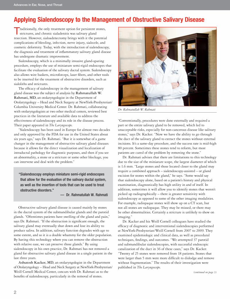
Dr. Rahmatullah W. Rahmati
Applying Sialendoscopy to the Management of Obstructive Salivary Disease
Traditionally, the only treatment option for persistent stones, strictures, and chronic sialadenitis was salivary gland
resection. However, sialoadenectomy brings with it the potential complications of bleeding, infection, nerve injury, sialocele, and cosmetic deformity. Today, with the introduction of sialendoscopy, the diagnosis and treatment of inflammatory salivary gland disease has undergone dramatic improvement.
Sialendoscopy, which is a minimally invasive gland-sparing procedure, employs the use of miniature semi-rigid endoscopes that facilitate the evaluation of the salivary ductal system. Sialendoscopy also allows wire baskets, microforceps, laser fibers, and other tools to be inserted for the treatment of obstructive disorders, such as sialoliths and strictures.
The efficacy of sialendoscopy in the management of salivary gland disease was the subject of analysis by Rahmatullah W. Rahmati, MD, an otolaryngologist in the Department of Otolaryngology – Head and Neck Surgery at NewYork-Presbyterian/Columbia University Medical Center. Dr. Rahmati, collaborating with otolaryngologists at two other medical centers, reviewed best practices in the literature and available data to address the effectiveness of sialendoscopy and its role in the disease process. Their paper appeared in The Laryngoscope.
“Sialendoscopy has been used in Europe for almost two decades and only approved by the FDA for use in the United States about six years ago,” says Dr. Rahmati. “But it is somewhat of a game changer in the management of obstructive salivary gland diseases because it allows for the direct visualization and localization of intraductal pathology for diagnostic purposes, and then if you see an abnormality, a stone or a stricture or some other blockage, you can intervene and deal with the problem.”
“Conventionally, procedures were done externally and required a part or the entire salivary gland to be removed, which led to unacceptable risks, especially for non-cancerous disease like salivary stones,” says Dr. Kacker. “Now we have the ability to go through the duct of the salivary gland to extract the stones without external incisions. It’s a same-day procedure, and the success rate is mid-high 80 percent. Sometimes these stones tend to reform, but most patients are cured of the problem by removing the stone.”
Dr. Rahmati advises that there are limitations to this technology due to the size of the miniature scope, the largest diameter of which is 1.6 mm. “Large stones and those located closer to the gland may require a combined approach – sialendoscopy-assisted – or gland excision for stones within the gland,” he says. “Some would say that sialendoscopy alone, based on a patient’s history and physical examination, diagnostically has high utility in and of itself. In addition, sometimes it will allow you to identify stones that weren’t picked up radiographically – there is greater sensitivity with sialendoscopy as opposed to some of the other imaging modalities. For example, radiopaque stones will show up on a CT scan, but not all stones are radiopaque. They may be missed, or there may be other abnormalities. Certainly a stricture is unlikely to show on imaging.”
Dr. Kacker and his Weill Cornell colleagues have studied the efficacy of diagnostic and interventional sialendoscopies performed at NewYork-Presbyterian/Weill Cornell from 2007 to 2009. They examined epidemiologic and clinical data, as well as procedural techniques, findings, and outcomes. “We attempted 37 parotid and submandibular sialendoscopies, with successful endoscopic canalization of the duct in 36 of these cases,” says Dr. Kacker. “Twenty of 25 stones were removed from 18 patients. Stones that were larger than 5 mm were more difficult to dislodge and remove without fragmentation.” The results of their investigation were published in The Laryngoscope.
(continued on page 3)
“ Sialendoscopy employs miniature semi-rigid endoscopes that allow for the evaluation of the salivary ductal system, as well as the insertion of tools that can be used to treat obstructive disorders.”
— Dr. Rahmatullah W. Rahmati
Obstructive salivary gland disease is caused mainly by stones in the ductal system of the submandibular glands and the parotid glands. “Oftentimes patients have swelling of the gland and pain,” says Dr. Rahmati. “If the obstruction is significant enough, the salivary gland may eventually shut down and lose its ability to produce saliva. In addition, salivary function degrades with age to some extent, and so it is a double whammy for the older population. By having this technology where you can remove the obstruction with relative ease, we can preserve those glands.” By using sialendoscopy in his own practice, Dr. Rahmati has not removed a gland for obstructive salivary gland disease in a single patient in the last three years.
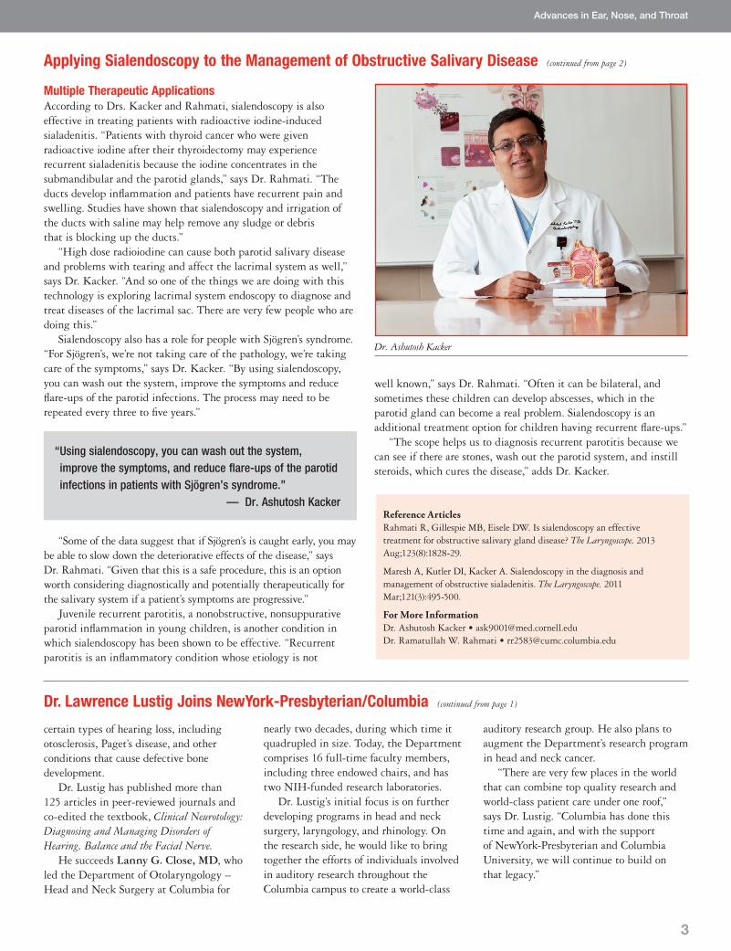
Ashutosh Kacker, MD, an otolaryngologist in the Department of Otolaryngology – Head and Neck Surgery at NewYork-Presbyterian/ Weill Cornell Medical Center, concurs with Dr. Rahmati as to the benefits of sialendoscopy, particularly in the removal of stones.
3
Multiple Therapeutic Applications According to Drs. Kacker and Rahmati, sialendoscopy is also effective in treating patients with radioactive iodine-induced sialadenitis. “Patients with thyroid cancer who were given radioactive iodine after their thyroidectomy may experience recurrent sialadenitis because the iodine concentrates in the submandibular and the parotid glands,” says Dr. Rahmati. “The ducts develop inflammation and patients have recurrent pain and swelling. Studies have shown that sialendoscopy and irrigation of the ducts with saline may help remove any sludge or debris that is blocking up the ducts.”
“High dose radioiodine can cause both parotid salivary disease and problems with tearing and affect the lacrimal system as well,” says Dr. Kacker. “And so one of the things we are doing with this technology is exploring lacrimal system endoscopy to diagnose and treat diseases of the lacrimal sac. There are very few people who are doing this.”
Sialendoscopy also has a role for people with Sjögren’s syndrome. “For Sjögren’s, we’re not taking care of the pathology, we’re taking care of the symptoms,” says Dr. Kacker. “By using sialendoscopy, you can wash out the system, improve the symptoms and reduce flare-ups of the parotid infections. The process may need to be repeated every three to five years.”
well known,” says Dr. Rahmati. “Often it can be bilateral, and sometimes these children can develop abscesses, which in the parotid gland can become a real problem. Sialendoscopy is an additional treatment option for children having recurrent flare-ups.”
“The scope helps us to diagnosis recurrent parotitis because we can see if there are stones, wash out the parotid system, and instill steroids, which cures the disease,” adds Dr. Kacker.
Advances in Ear, Nose, and Throat
certain types of hearing loss, including otosclerosis, Paget’s disease, and other conditions that cause defective bone development.
Dr. Lustig has published more than 125 articles in peer-reviewed journals and co-edited the textbook, Clinical Neurotology: Diagnosing and Managing Disorders of Hearing, Balance and the Facial Nerve.
He succeeds Lanny G. Close, MD, who led the Department of Otolaryngology – Head and Neck Surgery at Columbia for
auditory research group. He also plans to augment the Department’s research program in head and neck cancer.
“There are very few places in the world that can combine top quality research and world-class patient care under one roof,” says Dr. Lustig. “Columbia has done this time and again, and with the support of NewYork-Presbyterian and Columbia University, we will continue to build on that legacy.”
Applying Sialendoscopy to the Management of Obstructive Salivary Disease (continued from page 2)
Reference ArticlesRahmati R, Gillespie MB, Eisele DW. Is sialendoscopy an effective treatment for obstructive salivary gland disease? The Laryngoscope. 2013 Aug;123(8):1828-29.
Maresh A, Kutler DI, Kacker A. Sialendoscopy in the diagnosis and management of obstructive sialadenitis. The Laryngoscope. 2011 Mar;121(3):495-500.
For More InformationDr. Ashutosh Kacker • [email protected]. Ramatullah W. Rahmati • [email protected]
nearly two decades, during which time it quadrupled in size. Today, the Department comprises 16 full-time faculty members, including three endowed chairs, and has two NIH-funded research laboratories.
Dr. Lustig’s initial focus is on further developing programs in head and neck surgery, laryngology, and rhinology. On the research side, he would like to bring together the efforts of individuals involved in auditory research throughout the Columbia campus to create a world-class
Dr. Lawrence Lustig Joins NewYork-Presbyterian/Columbia (continued from page 1)
Dr. Ashutosh Kacker
“ Using sialendoscopy, you can wash out the system, improve the symptoms, and reduce flare-ups of the parotid infections in patients with Sjögren’s syndrome.”
— Dr. Ashutosh Kacker
“Some of the data suggest that if Sjögren’s is caught early, you may be able to slow down the deteriorative effects of the disease,” says Dr. Rahmati. “Given that this is a safe procedure, this is an option worth considering diagnostically and potentially therapeutically for the salivary system if a patient’s symptoms are progressive.”
Juvenile recurrent parotitis, a nonobstructive, nonsuppurative parotid inflammation in young children, is another condition in which sialendoscopy has been shown to be effective. “Recurrent parotitis is an inflammatory condition whose etiology is not
Advances in Ear, Nose, and Throat
NewYork-Presbyterian Hospital525 East 68th StreetNew York, NY 10065
www.nyp.org
Top Ranked Hospital in New York.Fourteen Years Running.
NON-PROFIT ORG.
US POSTAGE
PAID
STATEN ISLAND, NY
PERMIT NO. 169
Weill Cornell Researchers Fabricate Facsimile of Human Ear (continued from page 1)
patient. “This surgical option is very chal-lenging and painful for children, and the ears rarely look totally natural or perform well,” says Dr. Spector, who has been collab-orating with co-lead investigator, Lawrence J. Bonassar, PhD, Associate Chair of the Department of Biomedical Engineering at Cornell University.
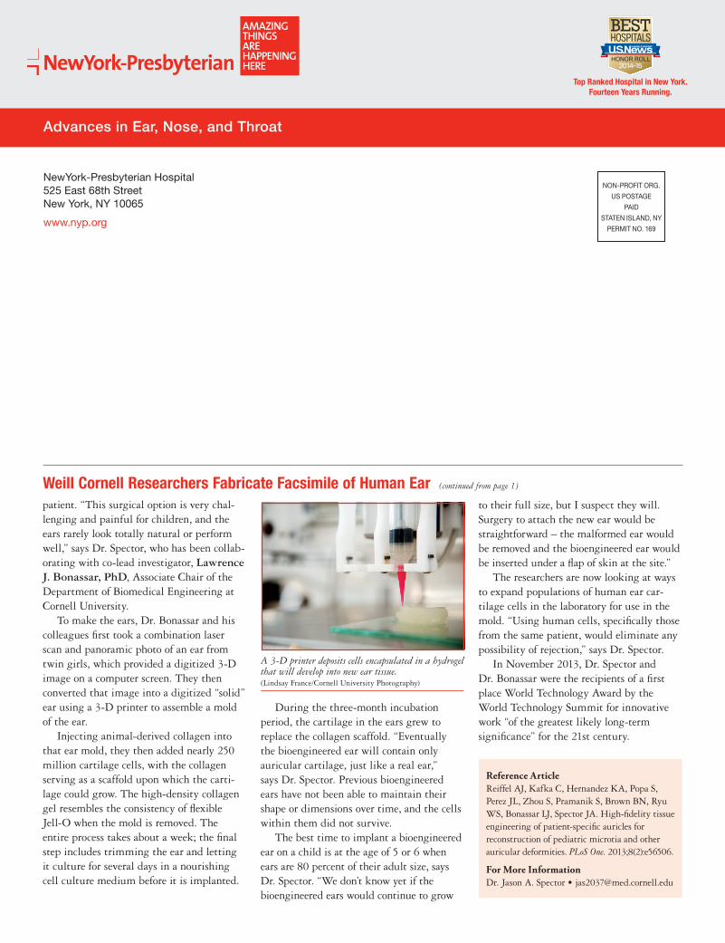
To make the ears, Dr. Bonassar and his colleagues first took a combination laser scan and panoramic photo of an ear from twin girls, which provided a digitized 3-D image on a computer screen. They then converted that image into a digitized “solid” ear using a 3-D printer to assemble a mold of the ear.
Injecting animal-derived collagen into that ear mold, they then added nearly 250 million cartilage cells, with the collagen serving as a scaffold upon which the carti-lage could grow. The high-density collagen gel resembles the consistency of flexible Jell-O when the mold is removed. The entire process takes about a week; the final step includes trimming the ear and letting it culture for several days in a nourishing cell culture medium before it is implanted.
During the three-month incubation period, the cartilage in the ears grew to replace the collagen scaffold. “Eventually the bioengineered ear will contain only auricular cartilage, just like a real ear,” says Dr. Spector. Previous bioengineered ears have not been able to maintain their shape or dimensions over time, and the cells within them did not survive.
The best time to implant a bioengineered ear on a child is at the age of 5 or 6 when ears are 80 percent of their adult size, says Dr. Spector. “We don’t know yet if the bioengineered ears would continue to grow
to their full size, but I suspect they will. Surgery to attach the new ear would be straightforward – the malformed ear would be removed and the bioengineered ear would be inserted under a flap of skin at the site.”
The researchers are now looking at ways to expand populations of human ear car-tilage cells in the laboratory for use in the mold. “Using human cells, specifically those from the same patient, would eliminate any possibility of rejection,” says Dr. Spector.
In November 2013, Dr. Spector and Dr. Bonassar were the recipients of a first place World Technology Award by the World Technology Summit for innovative work “of the greatest likely long-term significance” for the 21st century.
A 3-D printer deposits cells encapsulated in a hydrogel that will develop into new ear tissue. (Lindsay France/Cornell University Photography)
Reference ArticleReiffel AJ, Kafka C, Hernandez KA, Popa S, Perez JL, Zhou S, Pramanik S, Brown BN, Ryu WS, Bonassar LJ, Spector JA. High-fidelity tissue engineering of patient-specific auricles for reconstruction of pediatric microtia and other auricular deformities. PLoS One. 2013;8(2):e56506.
For More InformationDr. Jason A. Spector • [email protected]
Related Documents