Double-Stranded RNA-Dependent Protein Kinase Regulates the Motility of Breast Cancer Cells Mei Xu 1 , Gang Chen 1 , Siying Wang 1,2 , Mingjun Liao 1 , Jacqueline A. Frank 1 , Kimberly A. Bower 1 , Zhuo Zhang 3 , Xianglin Shi 3 , Jia Luo 1 * 1 Department of Internal Medicine, University of Kentucky College of Medicine, Lexington, Kentucky, United States of America, 2 Pathophysiological Department, School of Basic Medicine, Anhui Medical University, Hefei, Anhui, China, 3 Graduate Center for Toxicology, University of Kentucky College of Medicine, Lexington, Kentucky, United States of America Abstract Double-stranded RNA (dsRNA)-dependent protein kinase (PKR) is an interferon-induced protein kinase that plays a central role in the anti-viral process. Due to its pro-apoptotic and anti-proliferative action, there is an increased interest in PKR modulation as an anti-tumor strategy. PKR is overexpressed in breast cancer cells; however, the role of PKR in breast cancer cells is unclear. The expression/activity of PKR appears inversely related to the aggressiveness of breast cancer cells. The current study investigated the role of PKR in the motility/migration of breast cancer cells. The activation of PKR by a synthesized dsRNA (PIC) significantly decreased the motility of several breast cancer cell lines (BT474, MDA-MB231 and SKBR3). PIC inhibited cell migration and blocked cell membrane ruffling without affecting cell viability. PIC also induced the reorganization of the actin cytoskeleton and impaired the formation of lamellipodia. These effects of PIC were reversed by the pretreatment of a selective PKR inhibitor. PIC also activated p38 mitogen-activated protein kinase (MAPK) and its downstream MAPK-activated protein kinase 2 (MK2). PIC-induced activation of p38 MAPK and MK2 was attenuated by the PKR inhibitor and the PKR siRNA, but a selective p38 MAPK inhibitor (SB203580) or other MAPK inhibitors did not affect PKR activity, indicating that PKR is upstream of p38 MAPK/MK2. Cofilin is an actin severing protein and regulates membrane ruffling, lamellipodia formation and cell migration. PIC inhibited cofilin activity by enhancing its phosphorylation at Ser3. PIC activated LIM kinase 1 (LIMK1), an upstream kinase of cofilin in a p38 MAPK-dependent manner. We concluded that the activation of PKR suppressed cell motility by regulating the p38 MAPK/MK2/LIMK/cofilin pathway. Citation: Xu M, Chen G, Wang S, Liao M, Frank JA, et al. (2012) Double-Stranded RNA-Dependent Protein Kinase Regulates the Motility of Breast Cancer Cells. PLoS ONE 7(10): e47721. doi:10.1371/journal.pone.0047721 Editor: Chryso Kanthou, University of Sheffield, United Kingdom Received April 24, 2012; Accepted September 14, 2012; Published October 24, 2012 Copyright: ß 2012 Xu et al. This is an open-access article distributed under the terms of the Creative Commons Attribution License, which permits unrestricted use, distribution, and reproduction in any medium, provided the original author and source are credited. Funding: This research is supported by a NIH grant (AA017226). The funders had no role in study design, data collection and analysis, decision to publish, or preparation of the manuscript. Competing Interests: The authors have declared that no competing interests exist. * E-mail: [email protected] Introduction Double-stranded RNA (dsRNA)-dependent protein kinase (PKR) is a 551 amino acid serine/threonine protein kinase and is ubiquitously expressed in mammalian cells. PKR was initially identified as an essential element in interferon-induced anti-viral processes. PKR is activated by viral dsRNA intermediates during infection via a mechanism involving autophosphorylation. Once activated, the enzyme phosphorylates the a-subunit of protein synthesis initiation factor eIF2, thereby inhibiting translation [1]. PKR also mediates the activation of signal transduction pathways by proinflammatory stimuli, including bacterial lipopolysaccharide (LPS), tumor necrosis factor-a (TNF-a) and interleukin 1 (IL-1) [1]. PKR can be directly activated by its cellular activator PACT [2]. In addition, serum deprivation, disruption of intracellular Ca 2+ homeostasis, oxidative stress and endoplasmic reticulum (ER) stress also stimulate PKR activity [3,4]. Active PKR regulates diverse downstream substrates and signaling pathways, such as NFkB, p53, protein phosphatase 2A (PP2A), MAPK and STAT1/ STAT3 signaling [3]. PKR has been implicated in the regulation of cell proliferation, apoptosis, differentiation and transformation [5,6]. In general, activation of PKR results in the inhibition of cell proliferation or the induction of apoptosis and translational suppression; therefore, PKR is considered a ‘‘tumor suppressor’’ and considerable attention has been paid to the PKR pathway for its anti-tumor potential [3,7]. In contrast to its potential role as a tumor suppressor, PKR is over-expressed in a number of human cancers including breast cancers [8–11]. A higher level of PKR is observed in human invasive ductal breast carcinomas than surrounding normal mammary tissues [9]. In addition, much more PKR is expressed in mammary carcinoma cell lines compared to non-transformed mammary epithelial cell lines [4,11]. However, it appears the expression levels of PKR are inconsistent with its activity detected in mammary tumor cells and epithelial cells. The role of PKR in breast cancer cells is unclear. Among human breast cancer cell lines, it appears that the aggressiveness is inversely related to PKR expression/activity [8]. For example, MCF-7, a minimally invasive breast cancer cell line expresses high levels/activity of PKR, while MDA-MB231, a highly invasive breast cancer cell line, has low levels/activity of PKR [8]. We therefore hypothesized that PKR plays a role in the aggressiveness in breast cancer cells. Tumor cell motility/migration is the hallmark of invasion and an essential step in metastasis. Cell motility/migration is a complex biological process that is regulated by a myriad of molecular/cellular PLOS ONE | www.plosone.org 1 October 2012 | Volume 7 | Issue 10 | e47721

Welcome message from author
This document is posted to help you gain knowledge. Please leave a comment to let me know what you think about it! Share it to your friends and learn new things together.
Transcript
Double-Stranded RNA-Dependent Protein KinaseRegulates the Motility of Breast Cancer CellsMei Xu1, Gang Chen1, Siying Wang1,2, Mingjun Liao1, Jacqueline A. Frank1, Kimberly A. Bower1,
Zhuo Zhang3, Xianglin Shi3, Jia Luo1*
1 Department of Internal Medicine, University of Kentucky College of Medicine, Lexington, Kentucky, United States of America, 2 Pathophysiological Department, School
of Basic Medicine, Anhui Medical University, Hefei, Anhui, China, 3 Graduate Center for Toxicology, University of Kentucky College of Medicine, Lexington, Kentucky,
United States of America
Abstract
Double-stranded RNA (dsRNA)-dependent protein kinase (PKR) is an interferon-induced protein kinase that plays a centralrole in the anti-viral process. Due to its pro-apoptotic and anti-proliferative action, there is an increased interest in PKRmodulation as an anti-tumor strategy. PKR is overexpressed in breast cancer cells; however, the role of PKR in breast cancercells is unclear. The expression/activity of PKR appears inversely related to the aggressiveness of breast cancer cells. Thecurrent study investigated the role of PKR in the motility/migration of breast cancer cells. The activation of PKR by asynthesized dsRNA (PIC) significantly decreased the motility of several breast cancer cell lines (BT474, MDA-MB231 andSKBR3). PIC inhibited cell migration and blocked cell membrane ruffling without affecting cell viability. PIC also induced thereorganization of the actin cytoskeleton and impaired the formation of lamellipodia. These effects of PIC were reversed bythe pretreatment of a selective PKR inhibitor. PIC also activated p38 mitogen-activated protein kinase (MAPK) and itsdownstream MAPK-activated protein kinase 2 (MK2). PIC-induced activation of p38 MAPK and MK2 was attenuated by thePKR inhibitor and the PKR siRNA, but a selective p38 MAPK inhibitor (SB203580) or other MAPK inhibitors did not affect PKRactivity, indicating that PKR is upstream of p38 MAPK/MK2. Cofilin is an actin severing protein and regulates membraneruffling, lamellipodia formation and cell migration. PIC inhibited cofilin activity by enhancing its phosphorylation at Ser3. PICactivated LIM kinase 1 (LIMK1), an upstream kinase of cofilin in a p38 MAPK-dependent manner. We concluded that theactivation of PKR suppressed cell motility by regulating the p38 MAPK/MK2/LIMK/cofilin pathway.
Citation: Xu M, Chen G, Wang S, Liao M, Frank JA, et al. (2012) Double-Stranded RNA-Dependent Protein Kinase Regulates the Motility of Breast Cancer Cells. PLoSONE 7(10): e47721. doi:10.1371/journal.pone.0047721
Editor: Chryso Kanthou, University of Sheffield, United Kingdom
Received April 24, 2012; Accepted September 14, 2012; Published October 24, 2012
Copyright: � 2012 Xu et al. This is an open-access article distributed under the terms of the Creative Commons Attribution License, which permits unrestricteduse, distribution, and reproduction in any medium, provided the original author and source are credited.
Funding: This research is supported by a NIH grant (AA017226). The funders had no role in study design, data collection and analysis, decision to publish, orpreparation of the manuscript.
Competing Interests: The authors have declared that no competing interests exist.
* E-mail: [email protected]
Introduction
Double-stranded RNA (dsRNA)-dependent protein kinase
(PKR) is a 551 amino acid serine/threonine protein kinase and
is ubiquitously expressed in mammalian cells. PKR was initially
identified as an essential element in interferon-induced anti-viral
processes. PKR is activated by viral dsRNA intermediates during
infection via a mechanism involving autophosphorylation. Once
activated, the enzyme phosphorylates the a-subunit of protein
synthesis initiation factor eIF2, thereby inhibiting translation [1].
PKR also mediates the activation of signal transduction pathways
by proinflammatory stimuli, including bacterial lipopolysaccharide
(LPS), tumor necrosis factor-a (TNF-a) and interleukin 1 (IL-1)
[1]. PKR can be directly activated by its cellular activator PACT
[2]. In addition, serum deprivation, disruption of intracellular
Ca2+ homeostasis, oxidative stress and endoplasmic reticulum (ER)
stress also stimulate PKR activity [3,4]. Active PKR regulates
diverse downstream substrates and signaling pathways, such as
NFkB, p53, protein phosphatase 2A (PP2A), MAPK and STAT1/
STAT3 signaling [3]. PKR has been implicated in the regulation
of cell proliferation, apoptosis, differentiation and transformation
[5,6]. In general, activation of PKR results in the inhibition of cell
proliferation or the induction of apoptosis and translational
suppression; therefore, PKR is considered a ‘‘tumor suppressor’’
and considerable attention has been paid to the PKR pathway for
its anti-tumor potential [3,7].
In contrast to its potential role as a tumor suppressor, PKR is
over-expressed in a number of human cancers including breast
cancers [8–11]. A higher level of PKR is observed in human
invasive ductal breast carcinomas than surrounding normal
mammary tissues [9]. In addition, much more PKR is expressed
in mammary carcinoma cell lines compared to non-transformed
mammary epithelial cell lines [4,11]. However, it appears the
expression levels of PKR are inconsistent with its activity detected
in mammary tumor cells and epithelial cells. The role of PKR in
breast cancer cells is unclear. Among human breast cancer cell
lines, it appears that the aggressiveness is inversely related to PKR
expression/activity [8]. For example, MCF-7, a minimally
invasive breast cancer cell line expresses high levels/activity of
PKR, while MDA-MB231, a highly invasive breast cancer cell
line, has low levels/activity of PKR [8]. We therefore hypothesized
that PKR plays a role in the aggressiveness in breast cancer cells.
Tumor cell motility/migration is the hallmark of invasion and an
essential step in metastasis. Cell motility/migration is a complex
biological process that is regulated by a myriad of molecular/cellular
PLOS ONE | www.plosone.org 1 October 2012 | Volume 7 | Issue 10 | e47721

events including cytoskeleton reorganization, membrane ruffling and
lamellipodia/filopodia formation [12,13]. In this study, we investi-
gated the involvement of PKR in regulation of cell motility/
migration. We demonstrated that the activation of PKR inhibited
the motility/migration of breast cancer cells. We further showed that
activation of PKR suppressed cofilin activity. Cofilin is an F-actin
severing protein that regulates actin depolymerization, a process
required for the changes in cell shape and the formation of
lamellipodia during migration [14,15]. Cofilin plays an important
role in the invasion and metastasis of breast cancer cells [16,17]. We
further delineated a signal transduction pathway that mediated PKR
regulation of cofilin activity.
Materials and Methods
MaterialsHuman plasma fibronectin was obtained from Chemicon
International (Temecula, CA). Anti-phospho-eIF2a (Ser51) antibody
and Lipofectamine 2000 were purchased from Invitrogen Corpora-
tion (Carlsbad, CA). Antibodies for phospho-p38 MAPK, phospho-
cofilin, cofilin, phospho-LIMK, LIMK, phospho-MAPKAPK2
(MK2) and MK2 were purchased from Cell Signaling Technology
Inc. (Beverly, MA). Antibodies for PKR, GAPDH, a-actin, eIF2aand p38a were purchased from Santa Cruz Biotechnology (San
Cruz, CA). Anti-phospho-PKR antibody was obtained from Abcam
(Cambridge, MA). Phalloidin 488, Alex Fluor-labeled secondary
antibodies and Prolong Gold anti-fade reagent were obtained from
Invitrogen Molecular Probes (Eugene, OR). MTT assay kit was
purchased from Roche Molecular Biochemicals (Indianapolis, IN).
Transwells were obtained from Costar Corp. (Acton, MA).
Polyriboinosinic acid [poly I]-polyribocytidylic acid [poly C] (PIC)
was purchased from InvivoGen (San Diego, Ca). Inhibitors for PKR,
p38 MAPK, ERK and JNK were obtained from Millipore (Billerica,
MA). All other chemicals were obtained from Sigma-Aldrich (St.
Louis, MO).
Cell Culture and PIC TreatmentsMDA-MB231, BT474 and SKBR3 human breast cancer cell
lines were obtained from American Type Culture Collection
(ATCC, Manassas, VA). MDA-MB231 breast cancer cells were
grown in DMEM medium containing 10% fetal bovine serum
(FBS) and 1% antibiotics-antimycotic (Gibco, Grand island, NY)
at 37uC with 5% CO2. MDA-MB231 cells were used for most of
the experiments because they have high invasive potential and low
PKR expression/activity. BT474 cells were grown in RPMI 1640
medium containing 10% FBS, 1% antibiotics-antimycotic and
10 mg/ml insulin at 37uC with 5% CO2. SKBR3 cells were grown
in IMEM medium containing 10% FBS and 1% antibiotics-
antimycotic at 37uC with 5% CO2. PIC, a synthetic analog of
dsRNA, was used to activate PKR. For PIC treatment,
Lipofectamine 2000 was included to facilitate PIC entry into the
cells. For controls, cells were treated with Lipofectamine 2000
alone. The concentrations of PIC for this study ranged from 1–
100 ng/ml. For treatment with PKR inhibitor, cells were
pretreated with the PKR inhibitor (500 nM) for 24 hours prior
to PIC exposure. For treatment with p38 MAPK inhibitor
(SB203580), cells were pretreated with SB203580 (5 mM) for 2
hours, then exposed to PIC.
PKR siRNA TransfectionTransient transfection of PKR siRNA (San Cruz, CA) was
performed with Lipofectamine 2000 according the manufacturer’s
protocol. Briefly, MB231 cells were treated by control siRNA
(con siRNA) or PKR siRNA at a concentration of 200 nM in the
presence of Lipofectamine 2000. After 48 hours of incubation,
cells were exposed to PIC and cell signaling events or cell
migration were assayed.
Cell MigrationCell migration was analyzed using a Transwell Migration
System (Costar) as described before [18]. Briefly, cells (56104 cells)
were placed into the upper chambers (Transwells with 8.0 mm
pore size) in serum free medium in the presence or absence of PIC.
Culture medium containing 10% FBS was added into the lower
compartment of the chambers and served as a chemoattractant for
the cells. The chambers were cultured at 37uC in 5% CO2
overnight. The migrated cells were fixed in 3.7% paraformalde-
hyde and stained with 0.5% crystal violet in 2% ethanol.
Membranes were washed and the dye was eluted with 10% acetic
acid. Absorbance was measured at 595 nm using a microtiter
platereader (Beckman coulter). PIC or chemical inhibitors were
only added to the upper chambers.
Wound Healing Migration AssayThe wound healing migration assay was performed as described
previously [18]. MDA-MB231 cells were grown on 35 mm dishes
to 100% confluence and then scratched to form a wound using
sterile pipette tips. The cells were then treated with PIC (0, 1, 5 or
10 ng/ml) for 36 hours. The images were recorded using a Zeiss
Axiovert 40C photomicroscope.
MTT AssayThe MTT assay was employed to determine the number of
viable cells in culture. Briefly, the cells were plated into 96-well
plates and exposed to PIC for 24 hours. After the treatment, 10 ml
of MTT reagent was added into each well and the plates were
incubated at 37uC for 4 hours. The cultures were solubilized and
spectrophotometric absorbance was measured at 595 nm using a
microtiter platereader.
Analysis of Cell MorphologyCell adhesion to fibronectin was analyzed as described
previously [19,20]. Briefly, cells were exposed to PIC for 12
hours. After exposure, cells (56104/well) were seeded on
fibronectin (10 mg/ml)-precoated plates, allowing attachment for
3 hours at 37uC with 5% CO2. Non-adherent cells were removed
by washing with PBS. The attached cells were fixed with 3.7%
paraformaldehyde for 10 min, then washed 3 times in PBS. The
phase-contrast images were captured using a Zeiss Axiovert 40C
photomicroscope. Cells with protruded leading areas (lamellipo-
dia) were counted in ten randomly selected fields in each treatment
group. Cell spreading areas were measured using Motic Images
Plus 2.0 ML software (Motic, Canada).
Immunofluorescence MicroscopyThe procedure for immunofluorescence microscopy has been
previously described [21]. Briefly, after treatments, cells were fixed
with 3.7% paraformaldehyde for 10 min, washed 3 times in PBS
and permeabilized with 0.5% Triton X-100 for 5 min. Cells were
blocked with 5% BSA and incubated with primary antibodies for
1 hour. The concentrations of primary antibodies were: anti-cofilin,
1:100; anti-phospho-cofilin, 1:100; anti-phospho-MAPKAPK2,
1:100; and phalloidin, 1:200. After the incubation, cells were
washed and treated with Alexa Fluor-labeled secondary antibodies
and rinsed several times with PBS. Coverslips were mounted with
Prolong Gold anti-fade reagent and immunofluorescence images
were examined with an Olympus 1X81 inverted fluorescent
Activation of PKR Inhibits Cell Migration
PLOS ONE | www.plosone.org 2 October 2012 | Volume 7 | Issue 10 | e47721
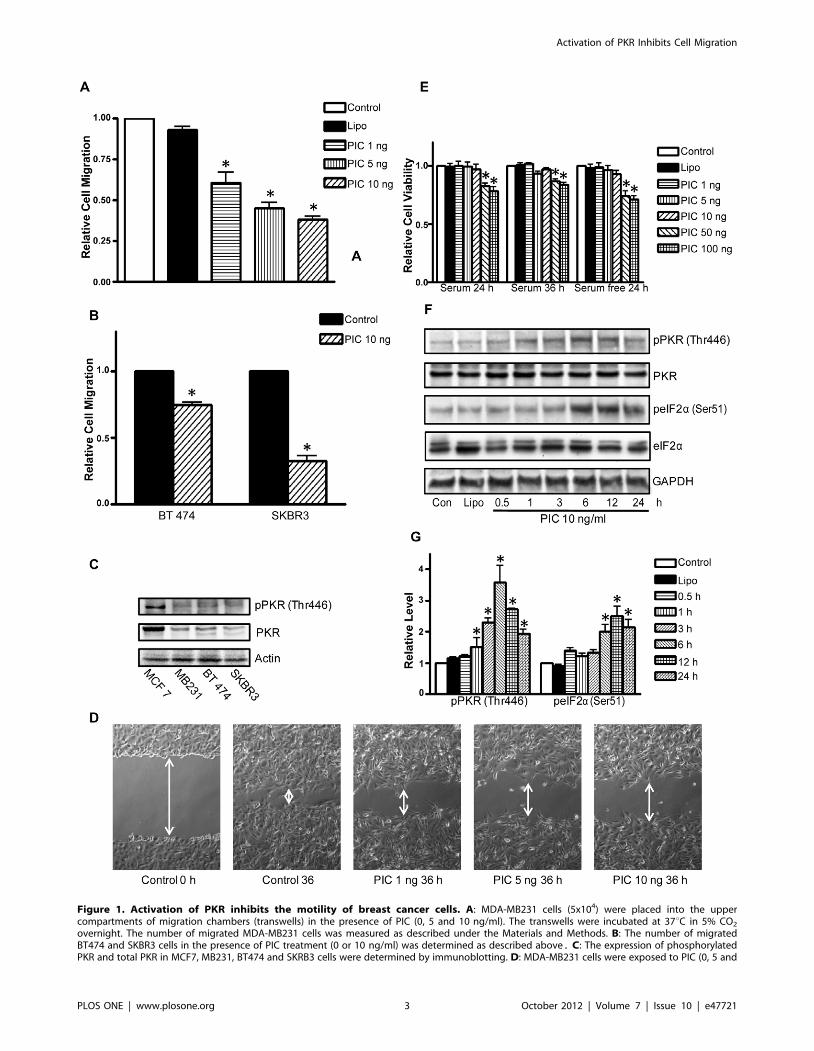
Figure 1. Activation of PKR inhibits the motility of breast cancer cells. A: MDA-MB231 cells (5x10⁴) were placed into the uppercompartments of migration chambers (transwells) in the presence of PIC (0, 5 and 10 ng/ml). The transwells were incubated at 37uC in 5% CO2
overnight. The number of migrated MDA-MB231 cells was measured as described under the Materials and Methods. B: The number of migratedBT474 and SKBR3 cells in the presence of PIC treatment (0 or 10 ng/ml) was determined as described above. C: The expression of phosphorylatedPKR and total PKR in MCF7, MB231, BT474 and SKRB3 cells were determined by immunoblotting. D: MDA-MB231 cells were exposed to PIC (0, 5 and
Activation of PKR Inhibits Cell Migration
PLOS ONE | www.plosone.org 3 October 2012 | Volume 7 | Issue 10 | e47721
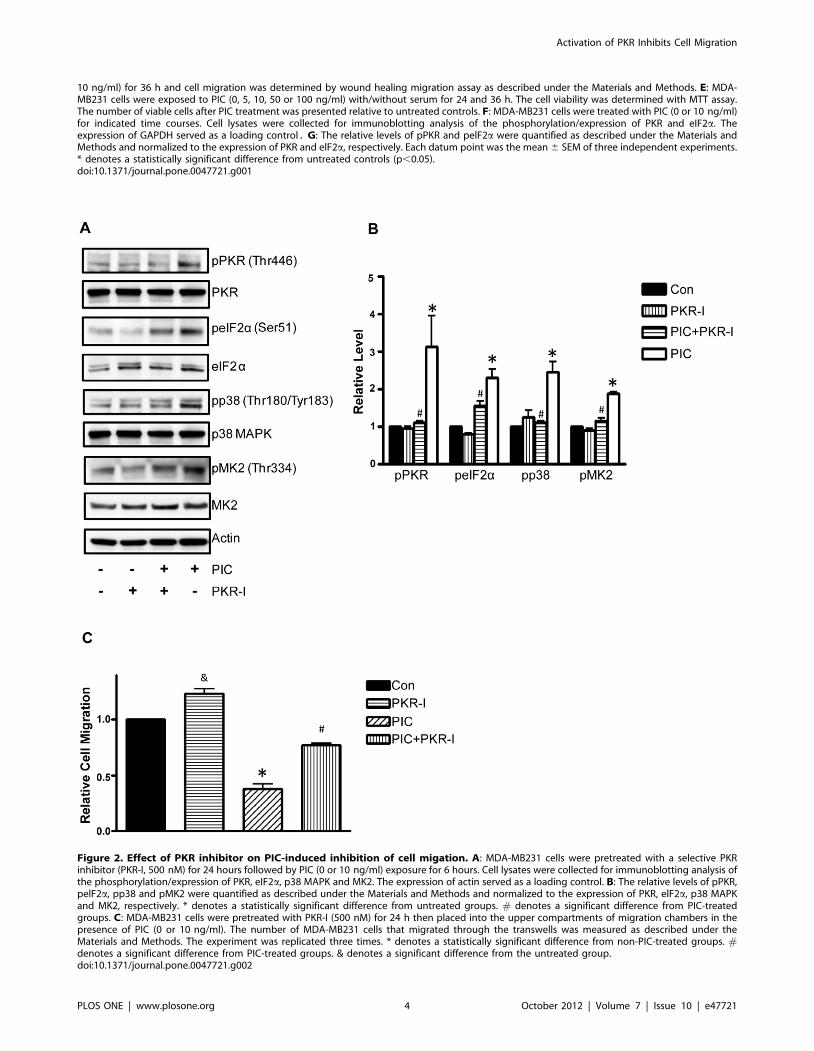
10 ng/ml) for 36 h and cell migration was determined by wound healing migration assay as described under the Materials and Methods. E: MDA-MB231 cells were exposed to PIC (0, 5, 10, 50 or 100 ng/ml) with/without serum for 24 and 36 h. The cell viability was determined with MTT assay.The number of viable cells after PIC treatment was presented relative to untreated controls. F: MDA-MB231 cells were treated with PIC (0 or 10 ng/ml)for indicated time courses. Cell lysates were collected for immunoblotting analysis of the phosphorylation/expression of PKR and eIF2a. Theexpression of GAPDH served as a loading control. G: The relative levels of pPKR and pelF2a were quantified as described under the Materials andMethods and normalized to the expression of PKR and elF2a, respectively. Each datum point was the mean 6 SEM of three independent experiments.* denotes a statistically significant difference from untreated controls (p,0.05).doi:10.1371/journal.pone.0047721.g001
Figure 2. Effect of PKR inhibitor on PIC-induced inhibition of cell migation. A: MDA-MB231 cells were pretreated with a selective PKRinhibitor (PKR-I, 500 nM) for 24 hours followed by PIC (0 or 10 ng/ml) exposure for 6 hours. Cell lysates were collected for immunoblotting analysis ofthe phosphorylation/expression of PKR, eIF2a, p38 MAPK and MK2. The expression of actin served as a loading control. B: The relative levels of pPKR,pelF2a, pp38 and pMK2 were quantified as described under the Materials and Methods and normalized to the expression of PKR, elF2a, p38 MAPKand MK2, respectively. * denotes a statistically significant difference from untreated groups. # denotes a significant difference from PIC-treatedgroups. C: MDA-MB231 cells were pretreated with PKR-I (500 nM) for 24 h then placed into the upper compartments of migration chambers in thepresence of PIC (0 or 10 ng/ml). The number of MDA-MB231 cells that migrated through the transwells was measured as described under theMaterials and Methods. The experiment was replicated three times. * denotes a statistically significant difference from non-PIC-treated groups. #denotes a significant difference from PIC-treated groups. & denotes a significant difference from the untreated group.doi:10.1371/journal.pone.0047721.g002
Activation of PKR Inhibits Cell Migration
PLOS ONE | www.plosone.org 4 October 2012 | Volume 7 | Issue 10 | e47721

Activation of PKR Inhibits Cell Migration
PLOS ONE | www.plosone.org 5 October 2012 | Volume 7 | Issue 10 | e47721

microscope with the same exposure time, detector gain and
amplifier offset.
ImmunoblottingThe procedure for immunoblotting has been previously
described [19]. Briefly, after PIC treatments, cells were lysed in
modified RIPA buffer (150 mM NaCl, 50 mM Tris, 1% NP-40,
0.25% sodium deoxycholate, 1 mM sodium vanadate, 1 mM
phenylmethanesulfonyl fluoride (PMSF), 5 mg/ml of aprotinin,
and 2 mg/ml of leupeptin). Samples were separated by centrifu-
gation at 10,000 rpm for 10 min at 4uC. Proteins were resolved in
SDS-PAGE and the separated proteins were transferred to
nitrocellulose membranes. The membranes were probed with
indicated primary antibodies, followed by the appropriate
secondary antibodies and developed by enhanced chemilumines-
cence. The images of immunoblots were documented using Gel
Logic 2200 Pro (Carestream Health, Rochester, NY). The
intensity of specific proteins was quantified using Carestream
Molecular Image Software.
Time-lapse MicroscopyThe rate of cell membrane ruffling was determined by time-
lapse microscopy. After treated with PIC for 12 hours in the
presence or absence of inhibitors, cells were trypsinized and seeded
on fibronectin precoated glass-bottom dishes in medium contain-
ing 20 mM HEPES. Cells were allowed to attach for 3 hours at
37uC with 5% CO2. Cells were maintained at 37uC and recorded
by a phase-contrast time-lapse video program using an Olympus
1X81 inverted fluorescent microscope with a 60X oil immersion
lens. The recording was performed at 10-second intervals for
10 min. Kymographs were generated from time-lapse video
images using ImageJ software (NIH) as previously described
[14]. Five or eight 1-pixel-thick lines were drawn across the cell
leading edges and the pixel intensities along each line were
combined to create the kymographs. To determine the dynamic
frequency of cell membrane ruffling, the perceived changes of
waves in 10 min on kymographs were counted manually and
presented relative to the control groups.
StatisticsDifferences among treatment groups were analyzed using analysis
of variance (ANOVA). Differences in which p was less than 0.05 were
considered statistically significant. In cases where significant
differences were detected, specific post-hoc comparisons between
treatment groups were examined with Student-Newman-Keuls tests.
Results
Activation of PKR Inhibits the Motility of Breast CancerCells
We first examined the effect of PIC on the motility/migration of
breast cancer cells. As shown in Fig. 1A, PIC suppressed the
migration of MDA-MB231 cells which was determined by
transwell assay in a concentration-dependent manner. Even at
1 ng/ml, PIC significantly inhibited the migration of MDA-
MB231 cells. Similar effects of PIC were observed in other
aggressive breast cancer cells (BT474 and SKBR3) (Fig. 1B). We
examined the expression/phosphorylation of PKR in different
breast cancer cell lines. As shown in Fig. 1C, the levels of PKR
expression/phosphorylation were much lower in more aggressive
breast cancer cell lines (MDA-MB231, BT474 and SKBR3) than
in the less aggressive line (MCF-7). PIC-mediated inhibition of cell
migration was confirmed by the wound healing assay; PIC
treatments impaired MDA-MB231 cell movement in a concen-
tration-dependent manner (Fig. 1D). The inhibition of cell motility
by PIC was not due to a decrease in cell viability because PIC up
to 10 ng/ml did not alter cell viability within 36 hours of exposure
(Fig. 1E). PIC-induced activation of PKR was verified by an
increase in the phosphorylation of PKR (Thr446) and eIF2a(Ser51), a substrate of PKR in MDA-MB231 cells (Figs. 1F and G).
To determine whether PIC inhibition of cell migration was
indeed mediated by PKR, we used a PKR inhibitor (PKR-I) to
block PKR activity. As shown in Fig. 2A, PKR-I inhibited PIC-
mediated phosphorylation of PKR and eIF2a. PKR-I blocked
PIC-induced phosphorylation of p38 MAPK and pMK2, indicat-
ing that p38 MAPK/pMK2 was downstream of PKR (Figs. 2A
and B). More importantly, PKR-I alleviated PIC-induced inhibi-
tion of cell migration (Fig. 2B). PKR-I also reduced PIC-mediated
formation of lamellipodia (data not shown). These data indicated
that activation of PKR suppressed the motility of breast cancer
cells.
Activation of PKR Impairs Lamellipodia FormationThe formation of lamellipodia at the leading edge of cells is
characteristic of cells in motion. Since PIC inhibited cell motility,
we sought to test whether PIC affected the formation of
lamellipodia. MDA-MB231 cells were pretreated with PIC for
12 hours, allowed to attach to fibronectin for 3 hours and
examined under the microscope. In the control group, the
majority of cells formed a protrusive lamella (lamellipodia)
(Figs. 3A and B); however, in the PIC-treated group, the number
of cells with lamellipodia was significantly reduced. Since dynamic
polymerization/depolymerization of the actin cytoskeleton is
required to generate the force to form lamellipodia, we sought
to determine whether PIC altered the organization of the actin
cytoskeleton. In the control group, the actin cytoskeleton was
cumulated at the lamellipodia and formed a polarized actin-rich
leading edge (Fig. 3C). In the PIC-treated group, few cells
displayed this feature. In addition, PIC also decreased cell
spreading areas (Fig. 3D). Thus, PIC inhibited the reorganization
of the actin cytoskeleton and impaired lamellipodia formation;
these alterations may underlie PIC-induced inhibition of cell
motility.
Figure 3. Activation of PKR impairs lamellipodia formation. MDA-MB231 cells were treated with PIC (0, 1, 5 or 10 ng/ml) for 12 hours, andthen equal amounts of cells were seeded on fibronectin-coated culture wells, allowing attachment for 3 hours. A: After attaching, the phase-contrastimages were captured using a Zeiss Axiovert 40C photomicroscope. The images of cells treated with PIC (0 or 10 ng/ml) are presented. Scale bar= 50 mm. B: Cells with extended leading areas (lamellipodia) were counted in ten randomly selected fields in each treatment group. The percentageof cells with lamellipodia was determined. C: MDA-MB231 cells were treated with PIC (0 or 10 ng/ml) for 12 hours. The distribution of the actincytoskeleton was detected by fluorescent staining (Alexa Fluor 488 Phalloidin) as described under the Materials and Methods. The arrow indicates thelamellipodia (the leading edge). Scale bar = 10 mm. D: MDA-MB231 cells were treated with PIC (0, 1, 5 or 10 ng/ml) for 12 hours, and then equalamounts of cells were seeded on fibronectin-coated culture wells, allowing attachment for 3 hours. Cell spreading areas were measured randomly forat least 25 cells for each treatment group. The experiment was replicated three times. Each datum point was the mean 6 SEM of three independentexperiments. * denotes a significant difference from untreated controls. # denotes a significant difference from PIC (1 ng/ml)-treated groups. 1denotes a significant difference from PIC (5 ng/ml)-treated groups (p,0.05).doi:10.1371/journal.pone.0047721.g003
Activation of PKR Inhibits Cell Migration
PLOS ONE | www.plosone.org 6 October 2012 | Volume 7 | Issue 10 | e47721

Figure 4. Activation of PKR inhibits cofilin. A: MDA-MB231 cells were treated with PIC (0 or 10 ng/ml) for indicated times. There were twocontrols; cells received no treatment (con) or cells were treated with Lipofectamine 2000 (Lipo). Cell lysates were collected for immunoblottinganalysis of the phosphorylation/expression of PKR, p38 MAPK, MK2, LIMK1 and cofilin. The expression of actin served as a loading control. B: Therelative levels of pp38, pMK2, pLIMK and pcofilin were quantified as described under the Materials and Methods and normalized to the expression ofp38 MAPK, MK2, LIMK1 and cofilin respectively. * denotes a significant difference from untreated controls. C: MDA-MB231 cells were exposed to PIC(0 or 10 ng/ml) for 6 hours, and then cells were seeded on fibronectin-coated culture wells, allowing attachment for 3 hours. The expression of cofilinand actin was detected by immunofluorescent staining as described under the Materials and Methods. Arrows indicate the lamellipodia. Scale bar= 10 mm. These experiments were replicated three times.doi:10.1371/journal.pone.0047721.g004
Activation of PKR Inhibits Cell Migration
PLOS ONE | www.plosone.org 7 October 2012 | Volume 7 | Issue 10 | e47721
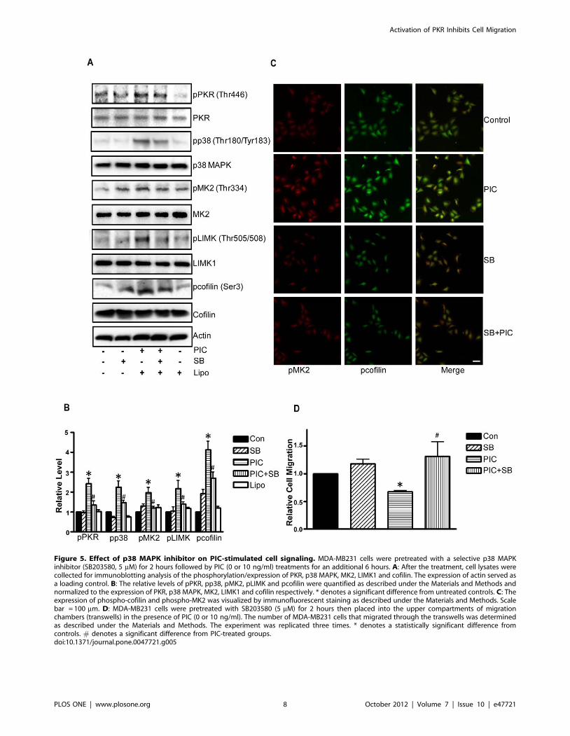
Figure 5. Effect of p38 MAPK inhibitor on PIC-stimulated cell signaling. MDA-MB231 cells were pretreated with a selective p38 MAPKinhibitor (SB203580, 5 mM) for 2 hours followed by PIC (0 or 10 ng/ml) treatments for an additional 6 hours. A: After the treatment, cell lysates werecollected for immunoblotting analysis of the phosphorylation/expression of PKR, p38 MAPK, MK2, LIMK1 and cofilin. The expression of actin served asa loading control. B: The relative levels of pPKR, pp38, pMK2, pLIMK and pcofilin were quantified as described under the Materials and Methods andnormalized to the expression of PKR, p38 MAPK, MK2, LIMK1 and cofilin respectively. * denotes a significant difference from untreated controls. C: Theexpression of phospho-cofilin and phospho-MK2 was visualized by immunofluorescent staining as described under the Materials and Methods. Scalebar = 100 mm. D: MDA-MB231 cells were pretreated with SB203580 (5 mM) for 2 hours then placed into the upper compartments of migrationchambers (transwells) in the presence of PIC (0 or 10 ng/ml). The number of MDA-MB231 cells that migrated through the transwells was determinedas described under the Materials and Methods. The experiment was replicated three times. * denotes a statistically significant difference fromcontrols. # denotes a significant difference from PIC-treated groups.doi:10.1371/journal.pone.0047721.g005
Activation of PKR Inhibits Cell Migration
PLOS ONE | www.plosone.org 8 October 2012 | Volume 7 | Issue 10 | e47721

Activation of PKR Inhibits Cofilin through p38 MAPK/MK2/LIMK Pathways
We next sought to determine the mechanisms for PIC-induced
actin cytoskeleton reorganization and decreases in lamellipodia
formation. Cofilin is an F-actin severing protein and plays an
important role in the reorganization of the actin cytoskeleton.
Cofilin activity is negatively regulated by phosphorylation at serine
3; phosphorylation of cofilin renders it unable to depolymerize F-
actin, thereby stabilizing the cytoskeleton [22]. We examined the
effect of PIC on cofilin activity. PIC increased the phosphorylation
of cofilin (Ser3) (Figs. 4A and B). PIC-induced cofilin phosphor-
ylation was confirmed by immunofluorescent staining (Fig. 5C). In
the control group, active cofilin (non-phosphorylated form) was
distributed at the leading edges of cells, i.e. lamellipodia (Fig. 4C).
In the PIC-treated group, however, no active cofilin was observed
at the leading edges. Since the localization of active cofilin at the
Figure 6. Effect of PKR siRNA on PIC-regulated cell migation. MDA-MB231 cells were transfected with control siRNA or PKR siRNA for 48 h. A:Following transfection, cell lysates were collected and the expression PKR was determined by immunoblotting. B: Following transfection, MDA-MB231 cells were placed into the upper compartments of the migration chambers in the presence of PIC (0 or 10 ng/ml). The number of MDA-MB231cells that migrated through the transwells was measured as described under the Materials and Methods. * denotes a statistically significant differencefrom controls. # denotes a significant difference from PIC-treated groups. C: Following transfection, MDA-MB231 cells were exposed to PIC (0 or10 ng/ml) for 6 hours. Cell lysates were collected for immunoblotting analysis of the phosphorylation/expression of PKR, eIF2a, p38 MAPK and MK2.The expression of tubulin served as a loading control. D: The relative levels of pPKR, pelF2a, pp38 and pMK2 were quantified as described under theMaterials and Methods and normalized to the expression of PKR, elF2a, p38 MAPK and MK2, respectively. The experiment was replicated three times.* denotes a statistically significant difference from controls. # denotes a significant difference from PIC-treated groups.doi:10.1371/journal.pone.0047721.g006
Activation of PKR Inhibits Cell Migration
PLOS ONE | www.plosone.org 9 October 2012 | Volume 7 | Issue 10 | e47721

leading edge is required for the initiation of cell movement [22],
PIC-induced inhibition of cofilin activity and redistribution of
cofilin may account for decreased lamellipodia formation and cell
movement.
We next examined the potential signal pathways that regulate
cofilin activity. LIM kinase 1 (LIMK1) is an important upstream
kinase of cofilin that regulates the phosphorylation of cofilin
[23]. LIMK activity is subjected to the regulation of p38
MAPK/MK2 pathways [24]. As shown in Fig. 4A, PIC induced
phosphorylation of PKR (Thr446), LIMK (Thr505/508), p38
MAPK (Thr180/183), MK2 (Thr334) and cofilin (Ser3). PKR
inhibitor blocked PIC-induced phosphorylation of p38 MAPK
and MK2 (Figs. 2A and B), but the p38 MAPK inhibitor
(SB203580) did not affect PKR phosphorylation (Figs. 5A and
B), suggesting that PKR is upstream of p38 MAPK and MK2.
Next, we sought to determine whether PIC-induced cofilin
phosphorylation was mediated by p38 MAPK. As shown in
Figs. 5A and B, SB203580 diminished PIC-induced phosphor-
ylation of p38 MAPK, MK2, LIMK as well as cofilin. The
finding was confirmed by immunofluorescent staining (Fig. 5C).
These results suggested that p38 MAPK was upstream of LIMK
and cofilin; PIC-mediated activation of p38 MAPK resulted in
phosphorylation of LIMK and cofilin. Furthermore, SB203580
alleviated PIC-induced inhibition of cell migration (Fig. 5D).
The involvement of PKR in p38/MK2 signaling was further
supported by the study using PKR siRNA. As shown in Fig. 6,
treatment of PKR siRNA blocked PIC-induced activation of
p38/MK2. Taken together, these results suggested the activa-
tion of PKR suppressed cofilin activity through activation of the
p38/MK2/LIMK pathway.
Activation of PKR Inhibits Cell Membrane RufflingCell membrane ruffling is a dynamic movement with rapid
irregular vacillation of protrusion and withdrawal of a cell surface
Figure 7. Activation of PKR inhibits dynamic cell membrane ruffling. A: MDA-MB231 cells were pretreated with DMSO (control), PKR-I(500 nM) or SB203580 (5 mM) for indicated hours, then followed by the treatment of PIC (10 ng/ml) for 6 hours. Top panel: Phase-contrast frameswere acquired every 10 seconds for 10 minutes on a time-lapse microscope using a 60X oil-immersion lens. Scale bar = 10 mm. Bottom panel: Thecorresponding kymographs of the movie generated along a line transecting the cell membrane on the lamellipodia are presented. B: Relative rate ofcell membrane ruffling was calculated from kymographs. The experiments were replicated five times. * denotes a statistically significant differencefrom all other groups (p,0.05).doi:10.1371/journal.pone.0047721.g007
Activation of PKR Inhibits Cell Migration
PLOS ONE | www.plosone.org 10 October 2012 | Volume 7 | Issue 10 | e47721

membrane. Cell membrane ruffling i.e. dynamic protrusion
usually appears in the leading edge of a motile cell and is
considered a dynamic parameter of cell motility [25]. An increase
in membrane ruffling is positively associated with cell movement
potential. To gain further insight into the relationship between
PKR activation and cell motility, we investigated the effect of PIC
on membrane ruffling using a time-lapse monitoring system. The
cell membrane ruffling was recorded for 10 min in 10 second
intervals. The kinetics of cell membrane ruffling was analyzed
using kymography as previously described [14]. As shown in Fig. 7,
PIC significantly decreased the rate of cell membrane ruffling.
Both PKR inhibitor and SB203580 eliminated PIC-induced
inhibition of membrane ruffling. These results further supported
the findings presented above and indicated the PKR/p38 MAPK
pathway regulated the motility of breast cancer cells.
Discussion
PKR has been implicated in anti-tumor action due to its anti-
proliferative and pro-apoptotic potential. Here, for the first time,
we demonstrate that activation of PKR inhibits the motility of
breast cancer cells. PKR activation suppresses lamellipodia
formation, cell spreading and membrane ruffling. We further
established the p38 MAPK/MK2/LIMK/cofilin signaling path-
way is involved in PKR modulation of cell migration (Fig. 8).
PKR and Cancer Cell MotilityPKR, the prototype of the eIF2a kinases, was first identified as
an anti-viral serine/threonine kinase. Activation of PKR phos-
phorylates eIF2a at Ser51, resulting in inhibition of protein
synthesis. Apart from inhibition of protein synthesis, PKR
activation can favor either the induction of cell cycle arrest or
apoptosis [10]. Therefore, there is an increasing interest to explore
PKR’s anti-tumor potential [3,7,10,26,27]. Available evidence
supports the notion that activation of PKR can inhibit tumour cell
growth and may have therapeutic benefits. It has been proposed to
develop strategies of selective PKR activation in cancer cells to kill
tumor cells [26]. PKR is over-expressed in some human cancers,
such as breast cancer, melanoma cells and colon cancer [10].
Although the implication of high levels of PKR in these cancer
cells is unclear, the high expression of PKR may offer an
opportunity to target PKR and optimize cancer cell toxicity. Our
study demonstrates that PKR activation inhibits the migration of
breast cancer cells independent of cell cycle arrest and the
induction of apoptosis, suggesting PKR activity may negatively
regulate cell mobility. This finding is consistent with the report
showing there is an inverse relationship between the aggressiveness
and the expression/activity of PKR in breast cancer cells [28,29].
The current study focuses on breast cancer cells and shows that
PKR activation impairs the migration of three aggressive breast
cancer cell lines (BT474, MDA-MB231 and SKBR3). It remains
to be determined whether the effect is general to all cell types.
Cancer metastasis consists of multiple processes, and enhanced
cancer cell motility is the hallmark of invasion and a critical step in
each metastatic process [30]. Regardless whether PKR’s effect is
cell type-specific or not, the novel finding provides a potential
approach to developing strategies of cancer therapy targeting the
PKR pathway, especially for those cancers overexpressing PKR.
Signal Pathways that Mediate PKR Inhibition on CellMotility
Cell motility initiates with cell protrusion, i.e. lamellipodia
formation which directs cell migration and is controlled by actin
remodelling. We demonstrate that PKR activation inhibits
lamellipodia formation, cell spreading and membrane ruffling,
indicating a disruption of actin cytoskeleton reorganization is a
mechanism of PKR activation-induced inhibition on cell motility.
The cooperation between cofilin and Arp2/3 is essential for this
initial step [31]. Cofilin severs actin filaments to create free barbed
ends from which new filaments are elongated by Arp2/3. The
force of actin depolymerization/polymerization on the leading
edge drives cell protrusion. The distribution of active cofilin within
leading edges of cells is a prerequisite for actin depolymerization/
polymerization which leads to cell migration. Alterations in overall
activity of cofilin have been implicated in cancer metastasis and is
directly associated with invasion and metastasis of mammary
tumors [16,17]. The activity of cofilin is regulated by phosphor-
ylation; phosphorylation of cofilin at Ser3 inactivates cofilin and
abolishes its actin-severing ability, therefore inhibiting the
formation of lamellipodia [23,32]. We demonstrate that PKR
activation stimulates cofilin phosphorylation at Ser9, suggesting
PKR activation impairs cell motility by inactivating cofilin (Fig. 8).
Phosphorylation of cofilin at Ser3 is regulated by LIM kinase
(LIMK) and testicular protein kinase [23,32]. Cofilin activity can
also be regulated by dephosphorylation which is mediated by
slingshot, chronophin and other phosphatases [17]. LIMK1 is
activated through phosphorylation at Thr508 by downstream
kinases of the Rho family GTPases, such as PAK, MRCK and
ROCK [23,33–36]. Alternatively, LIMK1 can be activated by
MAPK-activated protein kinase 2 (MK2), a substrate of p38
MAPK [24]. Overexpression of active LIMK1 inhibits the motility
of mammary cancer cells [37]. We demonstrate that PKR
activation causes the phosphorylation of p38 MAPK, MK2 and
LIMK1 (Thr508), suggesting that this pathway is involved in PKR
regulation of cofilin activity.
PKR can activate p38 MAPK through interactions with
mitogen-activated protein kinase kinase 6 (MKK6) [38–41].
Activation of p38 MAPK pathways is implicated in PKR
activation-related immune response, apoptosis and cell cycle
Figure 8. Schematic illustration of PKR-mediated cell signalingthat regulates cell migration. The activation or high expression ofPKR results in the activation of p38 MAPK/MK2 which stimulates thephosphorylation of LIMK. The activated LIMK inhibits cofilin, resulting inthe suppression of cell migration.doi:10.1371/journal.pone.0047721.g008
Activation of PKR Inhibits Cell Migration
PLOS ONE | www.plosone.org 11 October 2012 | Volume 7 | Issue 10 | e47721

arrest [40–42]. We confirm that PKR activation results in the
activation of p38 MAPK in MDA-MB231 cells because PKR
inhibitor blocks PIC-mediated p38 MAPK phosphorylation, but
a p38 MAPK inhibitor (SB203580) fails to modulate PKR
activity. Although SB203580 does not affect PKR, it inhibits
PIC-induced phosphorylation of MK2, LIMK1 and cofilin.
PIC-induced phosphorylation of MK2, LIMK1 and cofilin is
not affected by inhibitors for other MAPKs, such as ERKs and
JNKs (data not shown). Taken together, these results indicate
the PKR/p38 MAPK/MK2/LIMK1/cofilin pathway is respon-
sible for PIC-mediated inhibition of cell motility. It is unclear
whether p38 MAPK/MK2 regulates LIMK1 directly or
indirectly. MK2 may directly interact and phosphorylate/
activate LIMK1 [24]. However, it is reported that p38 MAPK
activates RhoA/ROCK which are upstream of LIMKs by
interacting with heat shock protein 27 (HSP27) in MDA-MB435
cells [43]. Kobayashi et al. confirm that p38 MAPK/MK2
activates HSP27 [24]. Therefore, it is also likely that p38
MAPK/MK2 indirectly regulates LIMK1 through the activa-
tion HSP27 and ROCK.
In summary, we demonstrate that PKR activation inhibits the
migration of breast cancer cells and establishes an underlying
signal transduction pathway that is responsible for PKR’s action.
This finding supports the notion that targeting PKR is an
attractive strategy for cancer therapy, especially for cancer cells
overexpressing PKR, such as breast cancer, melanoma and colon
cancer.
Author Contributions
Conceived and designed the experiments: JL MX ZZ XS GC. Performed
the experiments: MX ML JF KB SW. Analyzed the data: JL MX. Wrote
the paper: MX JL.
References
1. Williams BR (2001) Signal integration via PKR. Sci STKE 2001: re2. 10.1126/stke.2001.89.re2 [doi];2001/89/re2 [pii].
2. Peters GA, Dickerman B, Sen GC (2009) Biochemical analysis of PKR
activation by PACT. Biochemistry 48: 7441–7447. 10.1021/bi900433y [doi].
3. Vorburger SA, Pataer A, Swisher SG, Hunt KK (2004) Genetically targetedcancer therapy: tumor destruction by PKR activation. Am J Pharmacogenomics
4: 189–198. 436 [pii].
4. Ke ZJ, Wang X, Fan Z, Luo J (2009) Ethanol promotes thiamine deficiency-induced neuronal death: involvement of double-stranded RNA-activated protein
kinase. Alcohol Clin Exp Res 33: 1097–1103. ACER931 [pii];10.1111/j.1530-
0277.2009.00931.x [doi].
5. Balachandran S, Barber GN (2007) PKR in innate immunity, cancer, and viraloncolysis. Methods Mol Biol 383: 277–301.
6. Raven JF, Koromilas AE (2008) PERK and PKR: old kinases learn new tricks.
Cell Cycle 7: 1146–1150. 5811 [pii].
7. Jagus R, Joshi B, Barber GN (1999) PKR, apoptosis and cancer. Int J BiochemCell Biol 31: 123–138. S1357-2725(98)00136-8 [pii].
8. Savinova O, Joshi B, Jagus R (1999) Abnormal levels and minimal activity of the
dsRNA-activated protein kinase, PKR, in breast carcinoma cells. Int J BiochemCell Biol 31: 175–189. S1357-2725(98)00140-X [pii].
9. Haines GK, Cajulis R, Hayden R, Duda R, Talamonti M, et al. (1996)
Expression of the double-stranded RNA-dependent protein kinase (p68) in
human breast tissues. Tumour Biol 17: 5–12.
10. Mounir Z, Koromilas AE (2010) Uncovering the PKR pathway’s potential fortreatment of tumors. Future Oncol 6: 643–645. 10.2217/fon.10.45 [doi].
11. Nussbaum JM, Major M, Gunnery S (2003) Transcriptional upregulation of
interferon-induced protein kinase, PKR, in breast cancer. Cancer Lett 196: 207–216. S0304383503002763 [pii].
12. Vicente-Manzanares M, Horwitz AR (2011) Cell migration: an overview.
Methods Mol Biol 769: 1–24. 10.1007/978-1-61779-207-6_1 [doi].
13. Stupack DG, Cho SY, Klemke RL (2000) Molecular signaling mechanisms ofcell migration and invasion. Immunol Res 21: 83–88. IR:21:2-3:83
[pii];10.1385/IR:21:2-3:83 [doi].
14. Jovceva E, Larsen MR, Waterfield MD, Baum B, Timms JF (2007) Dynamic
cofilin phosphorylation in the control of lamellipodial actin homeostasis. J CellSci 120: 1888–1897. jcs.004366 [pii];10.1242/jcs.004366 [doi].
15. Bamburg JR (2011) Listeria monocytogenes cell invasion: a new role for cofilin
in co-ordinating actin dynamics and membrane lipids. Mol Microbiol 81: 851–854. 10.1111/j.1365–2958.2011.07759.x [doi].
16. Wang W, Mouneimne G, Sidani M, Wyckoff J, Chen X, et al. (2006) The
activity status of cofilin is directly related to invasion, intravasation, and
metastasis of mammary tumors. J Cell Biol 173: 395–404. jcb.200510115[pii];10.1083/jcb.200510115 [doi].
17. Wang W, Eddy R, Condeelis J (2007) The cofilin pathway in breast cancer
invasion and metastasis. Nat Rev Cancer 7: 429–440. nrc2148 [pii];10.1038/nrc2148 [doi].
18. Xu M, Bower KA, Wang S, Frank JA, Chen G, et al. (2010) Cyanidin-3-
glucoside inhibits ethanol-induced invasion of breast cancer cells overexpressingErbB2. Mol Cancer 9: 285. 1476-4598-9-285 [pii];10.1186/1476-4598-9-285
[doi].
19. Xu M, Bower KA, Chen G, Shi X, Dong Z, et al. (2010) Ethanol enhances the
interaction of breast cancer cells over-expressing ErbB2 with fibronectin.Alcohol Clin Exp Res 34: 751–760. ACER1147 [pii];10.1111/j.1530–
0277.2010.01147.x [doi].
20. Wang F, Nohara K, Olivera A, Thompson EW, Spiegel S (1999) Involvement offocal adhesion kinase in inhibition of motility of human breast cancer cells by
sphingosine 1-phosphate. Exp Cell Res 247: 17–28. S0014-4827(98)94327-0[pii];10.1006/excr.1998.4327 [doi].
21. Xu M, Waters CL, Hu C, Wysolmerski RB, Vincent PA, et al. (2007)
Sphingosine 1-phosphate rapidly increases endothelial barrier function inde-
pendently of VE-cadherin but requires cell spreading and Rho kinase. Am J
Physiol Cell Physiol 293: C1309–C1318. 00014.2007 [pii];10.1152/ajp-
cell.00014.2007 [doi].
22. Desmarais V, Ghosh M, Eddy R, Condeelis J (2005) Cofilin takes the lead. J Cell
Sci 118: 19–26. 118/1/19 [pii];10.1242/jcs.01631 [doi].
23. Yang N, Higuchi O, Ohashi K, Nagata K, Wada A, et al.(1998) Cofilin
phosphorylation by LIM-kinase 1 and its role in Rac-mediated actin
reorganization. Nature 393: 809–812. 10.1038/31735 [doi].
24. Kobayashi M, Nishita M, Mishima T, Ohashi K, Mizuno K (2006)
MAPKAPK-2-mediated LIM-kinase activation is critical for VEGF-induced
actin remodeling and cell migration. EMBO J 25: 713–726. 7600973
[pii];10.1038/sj.emboj.7600973 [doi].
25. Jiang WG (1995) Membrane ruffling of cancer cells: a parameter of tumour cell
motility and invasion. Eur J Surg Oncol 21: 307–309.
26. Shir A, Friedrich I, Levitzki A (2003) Tumor specific activation of PKR as a non-
toxic modality of cancer treatment. Semin Cancer Biol 13: 309–314.
27. Garcia MA, Gil J, Ventoso I, Guerra S, Domingo E, et al. (2006) Impact of
protein kinase PKR in cell biology: from antiviral to antiproliferative action.
Microbiol Mol Biol Rev 70: 1032–1060. 70/4/1032 [pii];10.1128/
MMBR.00027-06 [doi].
28. Kim SH, Forman AP, Mathews MB, Gunnery S (2000) Human breast cancer
cells contain elevated levels and activity of the protein kinase, PKR. Oncogene
19: 3086–3094. 10.1038/sj.onc.1203632 [doi].
29. Lee GW, Park HS, Kim EJ, Cho YW, Kim GT, et al. (2012) Reduction of breast
cancer cell migration via up-regulation of TASK-3 two-pore domain K(+)
channel. Acta Physiol (Oxf) 204: 513–524. 10.1111/j.1748-1716.2011.02359.x
[doi].
30. Condeelis J, Segall JE (2003) Intravital imaging of cell movement in tumours.
Nat Rev Cancer 3: 921–930. 10.1038/nrc1231 [doi];nrc1231 [pii].
31. Oser M, Condeelis J (2009) The cofilin activity cycle in lamellipodia and
invadopodia. J Cell Biochem 108: 1252–1262. 10.1002/jcb.22372 [doi].
32. Toshima J, Toshima JY, Takeuchi K, Mori R, Mizuno K (2001) Cofilin
phosphorylation and actin reorganization activities of testicular protein kinase 2
and its predominant expression in testicular Sertoli cells. J Biol Chem 276:
31449–31458. 10.1074/jbc.M102988200 [doi];M102988200 [pii].
33. Edwards DC, Sanders LC, Bokoch GM, Gill GN (1999) Activation of LIM-
kinase by Pak1 couples Rac/Cdc42 GTPase signalling to actin cytoskeletal
dynamics. Nat Cell Biol 1: 253–259. 10.1038/12963 [doi].
34. Ohashi K, Nagata K, Maekawa M, Ishizaki T, Narumiya S, et al. (2000) Rho-
associated kinase ROCK activates LIM-kinase 1 by phosphorylation at
threonine 508 within the activation loop. J Biol Chem 275: 3577–3582.
35. Bokoch GM (2003) Biology of the p21-activated kinases. Annu Rev Biochem 72:
743–781. 10.1146/annurev.biochem.72.121801.161742 [doi];121801.161742
[pii].
36. Maekawa M, Ishizaki T, Boku S, Watanabe N, Fujita A, et al. (1999) Signaling
from Rho to the actin cytoskeleton through protein kinases ROCK and LIM-
kinase. Science 285: 895–898. 7729 [pii].
37. Zebda N, Bernard O, Bailly M, Welti S, Lawrence DS, et al. (2000)
Phosphorylation of ADF/cofilin abolishes EGF-induced actin nucleation at
the leading edge and subsequent lamellipod extension. J Cell Biol 151: 1119–
1128.
38. Alisi A, Spaziani A, Anticoli S, Ghidinelli M, Balsano C (2008) PKR is a novel
functional direct player that coordinates skeletal muscle differentiation via
p38MAPK/AKT pathways. Cell Signal 20: 534–542. S0898-6568(07)00361-0
[pii];10.1016/j.cellsig.2007.11.006 [doi].
Activation of PKR Inhibits Cell Migration
PLOS ONE | www.plosone.org 12 October 2012 | Volume 7 | Issue 10 | e47721

39. Silva AM, Whitmore M, Xu Z, Jiang Z, Li X, et al. (2004) Protein kinase R
(PKR) interacts with and activates mitogen-activated protein kinase kinase 6(MKK6) in response to double-stranded RNA stimulation. J Biol Chem 279:
37670–37676. 10.1074/jbc.M406554200 [doi];M406554200 [pii].
40. Goh KC, deVeer MJ, Williams BR (2000) The protein kinase PKR is requiredfor p38 MAPK activation and the innate immune response to bacterial
endotoxin. EMBO J 19: 4292–4297. 10.1093/emboj/19.16.4292 [doi].41. Iordanov MS, Paranjape JM, Zhou A, Wong J, Williams BR, et al. (2000)
Activation of p38 mitogen-activated protein kinase and c-Jun NH(2)-terminal
kinase by double-stranded RNA and encephalomyocarditis virus: involvement of
RNase L, protein kinase R, and alternative pathways. Mol Cell Biol 20: 617–
627.42. Spaziani A, Alisi A, Sanna D, Balsano C (2006) Role of p38 MAPK and RNA-
dependent protein kinase (PKR) in hepatitis C virus core-dependent nuclear
delocalization of cyclin B1. J Biol Chem 281: 10983–10989. M512536200[pii];10.1074/jbc.M512536200 [doi].
43. Garcia MC, Ray DM, Lackford B, Rubino M, Olden K, et al. (2009)Arachidonic acid stimulates cell adhesion through a novel p38 MAPK-RhoA
signaling pathway that involves heat shock protein 27. J Biol Chem 284: 20936–
20945. M109.020271 [pii];10.1074/jbc.M109.020271 [doi].
Activation of PKR Inhibits Cell Migration
PLOS ONE | www.plosone.org 13 October 2012 | Volume 7 | Issue 10 | e47721
Related Documents