Dopamine Receptors in Human Lymphocytes: Radioligand Binding and Quantitative RT-PCR Assays Galina P. Kirillova a,* , Rebecca J. Hrutkay a , Michael R. Shurin b , Galina V. Shurin b , Irina L. Tourkova b , and Michael M. Vanyukov a,c,d aCenter for Education and Drug Abuse Research, Department of Pharmaceutical Sciences, University of Pittsburgh, Pittsburgh, PA 15261, USA bDepartment of Pathology, University of Pittsburgh Medical Center, Pittsburgh, PA 15261, USA cDepartment of Psychiatry, University of Pittsburgh, Pittsburgh, PA 15213, USA dDepartment of Human Genetics, University of Pittsburgh, Pittsburgh, PA 15261, USA Abstract Analysis of dopamine receptors (DR) in lymphocytes of the human peripheral blood mononuclear cell (PBMC) fraction is an attractive tool for evaluation of functional properties of dopaminergic function underlying variation in complex psychological/psychopathological traits. Receptor binding assays (RBA) with selective radioligands, which are widely used in CNS studies, have not produced consistent results when applied to isolated PBMC. We tested the assay conditions that could be essential for detection of DR in human PBMC and their membrane preparations. Using [ 3 H] SCH23390, a dopamine D1-like receptor antagonist, we demonstrated the presence of two binding sites in PBMC-derived membrane fraction. One of them is characterized by the K d value consistent with that reported for D5 dopamine receptors in human lymphocytes, whereas the other K d value possibly corresponds to serotonin receptor(s). Although D5 receptor binding sites in PBMC membranes could be characterized by binding assays, the low protein expression and the large volume of blood needed for membrane preparation render the binding method impracticable for individual phenotyping. In contrast, real-time RT-PCR may be used for this purpose, contingent on the relationship between DR expression in the brain and in lymphocytes. The expression of the DRD2- DRD5 genes, as detected by this method, varied widely among samples, whereas the DRD1 expression was not detected. The expression levels were comparable with those in the brain for DRD3 and DRD4, and were significantly lower for DRD2 and DRD5. Keywords DRD1; DRD2; DRD3; DRD4; DRD5; gene expression; endophenotypes; intermediate trait *Corresponding author: University of Pittsburgh, Pittsburgh, PA 15261, USA; Tel.: +1 412 648 9909, E-mail address: [email protected] (G.P. Kirillova). Publisher's Disclaimer: This is a PDF file of an unedited manuscript that has been accepted for publication. As a service to our customers we are providing this early version of the manuscript. The manuscript will undergo copyediting, typesetting, and review of the resulting proof before it is published in its final citable form. Please note that during the production process errors may be discovered which could affect the content, and all legal disclaimers that apply to the journal pertain. NIH Public Access Author Manuscript J Neurosci Methods. Author manuscript; available in PMC 2009 September 30. Published in final edited form as: J Neurosci Methods. 2008 September 30; 174(2): 272–280. doi:10.1016/j.jneumeth.2008.07.018. NIH-PA Author Manuscript NIH-PA Author Manuscript NIH-PA Author Manuscript

Welcome message from author
This document is posted to help you gain knowledge. Please leave a comment to let me know what you think about it! Share it to your friends and learn new things together.
Transcript

Dopamine Receptors in Human Lymphocytes: RadioligandBinding and Quantitative RT-PCR Assays
Galina P. Kirillovaa,*, Rebecca J. Hrutkaya, Michael R. Shurinb, Galina V. Shurinb, Irina L.Tourkovab, and Michael M. Vanyukova,c,d
aCenter for Education and Drug Abuse Research, Department of Pharmaceutical Sciences, University ofPittsburgh, Pittsburgh, PA 15261, USA
bDepartment of Pathology, University of Pittsburgh Medical Center, Pittsburgh, PA 15261, USA
cDepartment of Psychiatry, University of Pittsburgh, Pittsburgh, PA 15213, USA
dDepartment of Human Genetics, University of Pittsburgh, Pittsburgh, PA 15261, USA
AbstractAnalysis of dopamine receptors (DR) in lymphocytes of the human peripheral blood mononuclearcell (PBMC) fraction is an attractive tool for evaluation of functional properties of dopaminergicfunction underlying variation in complex psychological/psychopathological traits. Receptor bindingassays (RBA) with selective radioligands, which are widely used in CNS studies, have not producedconsistent results when applied to isolated PBMC. We tested the assay conditions that could beessential for detection of DR in human PBMC and their membrane preparations. Using [3H]SCH23390, a dopamine D1-like receptor antagonist, we demonstrated the presence of two bindingsites in PBMC-derived membrane fraction. One of them is characterized by the Kd value consistentwith that reported for D5 dopamine receptors in human lymphocytes, whereas the other Kd valuepossibly corresponds to serotonin receptor(s). Although D5 receptor binding sites in PBMCmembranes could be characterized by binding assays, the low protein expression and the large volumeof blood needed for membrane preparation render the binding method impracticable for individualphenotyping. In contrast, real-time RT-PCR may be used for this purpose, contingent on therelationship between DR expression in the brain and in lymphocytes. The expression of the DRD2-DRD5 genes, as detected by this method, varied widely among samples, whereas the DRD1expression was not detected. The expression levels were comparable with those in the brain forDRD3 and DRD4, and were significantly lower for DRD2 and DRD5.
KeywordsDRD1; DRD2; DRD3; DRD4; DRD5; gene expression; endophenotypes; intermediate trait
*Corresponding author: University of Pittsburgh, Pittsburgh, PA 15261, USA; Tel.: +1 412 648 9909, E-mail address: [email protected](G.P. Kirillova).Publisher's Disclaimer: This is a PDF file of an unedited manuscript that has been accepted for publication. As a service to our customerswe are providing this early version of the manuscript. The manuscript will undergo copyediting, typesetting, and review of the resultingproof before it is published in its final citable form. Please note that during the production process errors may be discovered which couldaffect the content, and all legal disclaimers that apply to the journal pertain.
NIH Public AccessAuthor ManuscriptJ Neurosci Methods. Author manuscript; available in PMC 2009 September 30.
Published in final edited form as:J Neurosci Methods. 2008 September 30; 174(2): 272–280. doi:10.1016/j.jneumeth.2008.07.018.
NIH
-PA Author Manuscript
NIH
-PA Author Manuscript
NIH
-PA Author Manuscript
1. IntroductionDysfunction of dopamine neurotransmission in the central nervous system (CNS) is implicatedin the development and progression of behavioral dysregulation and drug addiction (Koob andNestler, 1997; Wise, 1998; Tarter et al., 1999; Vallone et al., 2000; Maher et al., 2002).Dopamine receptors (DR) are one of the key elements of the dopaminergic system. Five typesof DR, D1 through D5, are coexpressed at different levels and in various combinations in theCNS and peripheral tissues.
Studying DR in the human CNS has obvious limitations. Determining peripheral sources ofDR, accessible in a minimally invasive way, would be advantageous for research and clinicalapplications. Human peripheral blood lymphocytes have attracted attention as a convenientcandidate for evaluation of the functional properties of DR. Molecular biological studies havedetected D3, D4 and D5 receptors mRNA in human lymphocytes (Takahashi et al., 1992;Nagai et al., 1993; Bondy et al., 1996). However, DR detection in lymphocytes by theradioligand binding method (Bondy et al., 1990) produced controversial results (Fleminger etal., 1982; Maloteaux et al., 1982, 1983; Rao et al., 1990; Vile and Strange, 1995, 1996;).Nevertheless, one group has consistently reported quantitative detection of D3, D4 and D5,but not D1 or D2 receptors on human lymphocytes (Ricci and Amenta, 1994; Ricci et al.,1995, 1997–1999; Amenta et al., 1999). Despite controversy in the results of binding studieswith lymphocytes, there have been attempts to apply the receptor binding assay (RBA) inclinical studies of schizophrenia, migraine, Alzheimer’s disease and Parkinson’s disease (LeFur et al., 1980,1983; Rotstein et al., 1983; Bondy et al., 1985; Bondy and Ackenheil, 1987;Halbach and Henning, 1989; Barbanti et al., 1999, 2000a, b). Some of these RBA findingswere supported by RT-PCR results (Nagai et al., 1996; Ilani et al., 2001; Kwak et al., 2001).
The goals of this study were to optimize the binding assay conditions for human PBMCpreparations as well as examine real-time RT-PCR technique, to use these methods in theperipheral measurement of DR expression for possible application in substance use disorder(SUD) research and other behavior-related studies. We have previously obtained pilot dataindicating association of the DRD5 gene with the risk for SUD (Vanyukov et al., 1998,2001) and antisociality (Vanyukov et al., 2000), and thus aimed at exploring the possibility ofrelating these results to the properties of the dopamine D5 receptor.
2. Materials and methods2.1. Reagents
The following reagents were used: [3H]SCH23390, 69 Ci/mmol (Amersham); (+)-Butaclamol,pepstatin A, leupeptin hydrochloride, PMFS (phenylmethanesulfonyl fluoride), EDTA,HEPES, Tris-HCl, Histopaque-1077, PEI (polyethylenimine), ascorbic acid, and NaCLphysiological solution (Sigma); HBSS and PBS buffers, and Trizol-LS reagent (Invitrogen);Ready-Safe scintillation cocktail (Beckman); BCA protein assay reagent kit (Pierce); RNEasyMini Kit (Qiagen); Superscript II (Gibco BRL); RNAsin (Promega); DNA-free Kit and humanbrain total RNA (Ambion), dNTP set (Gene Choice). Primers and fluorescence-labeled probes(assays–on–demand) for D1 (ref: Hs00265245_s1), D2 (ref: Hs00241436_m1), D3 (ref:Hs00168045_m1), D4 (ref: Hs00609526_m1), D5 (ref: Hs00361234_s1) receptors and humanGAPDH control reagents (with included forward and reverse primers and probes) were orderedfrom Applied Biosystems.
2.2. Specimen collectionBlood for PBMC isolation and optimization of binding assays was drawn from healthyvolunteers. Lymphocyte-enriched buffy coat product received from a local blood bank was
Kirillova et al. Page 2
J Neurosci Methods. Author manuscript; available in PMC 2009 September 30.
NIH
-PA Author Manuscript
NIH
-PA Author Manuscript
NIH
-PA Author Manuscript
used for PBMC membrane fraction preparation. Blood for extraction of total RNA for real-time PCR experiments was drawn from 38 adult male participants in a study of substance usedisorder etiology (Center for Education and Drug Abuse Research, CEDAR [Tarter andVanyukov, 2001]). In this group, 14 individuals were diagnosed with a DSM-III-R substanceuse disorder related to an illicit drug (SUD+), five with alcohol use disorder only, 3 with anotherpsychiatric disorder, and 16 had no psychiatric disorder (SUD−). An expanded version of theStructured Clinical Interview for DSM-III-R-outpatient version (SCID-OP) (Spitzer et al.,1987) was administered by experienced research associates to obtain psychiatric diagnoses.The expanded SCID evaluates current episode (past 6 months) and worst past (before the past6 months) episode of psychopathology. Informed consent was obtained from all participants.The study was approved by the University of Pittsburgh Institutional Review Board.
2.3. Peripheral blood mononuclear cell (PBMC) isolationPBMC were isolated from 50–100 ml of blood drawn into EDTA vacutainers. The blood wasdiluted two-fold with NaCl physiological solution (Ricci and Amenta, 1994), or HBSS/HEPESbuffer (136 mM NaCl, 4.16 mM NaHCO3, 0.34 mM Na2HPO4, 5.36 mM KCl, 0.44 mMKH2PO4, 1.25 mM CaCl2, 0.81 mM MgSO4, 22.3 mM HEPES, 1 mM EDTA, pH 7.4) (Bondyet al., 1990), or PBS (phosphate buffered saline, pH 7.4). The diluted blood (15 ml) was layeredon Histopaque-1077 reagent (15 ml) in 50-ml polypropylene centrifuge tubes and centrifugedat 22–23°C for 40 min at 400×g. Collected PBMC were washed in 30 ml of isolation solutionby three consequent centrifugation steps (15 min at 300×g), every time gently resuspendedwith a pipette tip precut to protect cells integrity. Viability of cells was tested by Trypan blueexclusion. The cell suspension was used for the assay within 20–30 min after preparation. Totest the influence of the presence of monocytes in the PBMC fraction, the binding step wasperformed with and without monocytes in the samples. Monocytes were removed by incubationof cell suspension at 37°C and 5% CO2 for an additional 60 min (Ricci and Amenta, 1994),and the non-adherent cells were used for binding assay.
To isolate PBMC from the buffy coat product, the contents of the pack (~60 ml) were diluted3-fold with the HBSS isolation buffer, and 10 ml of the suspension were layered on 15 ml ofHystopaque-1077 in 50 ml tubes. After centrifugation as described above, cells were washedthree times, and the final cell pellets (300–400×106 cells per tube) were frozen at −80°C andused later for the membrane preparation.
2.4. Membrane preparation from PBMC isolated from buffy coatThe membrane preparation procedure used in this study was modified from protocols describedelsewhere (Chiu et al., 1982; Seeman et al., 1984; Zhou et al., 1990; Hill et al., 1996). Cellsdefrosted on ice (300–400×106 cells per tube) were reconstituted in 10 ml of the ice-coldhypotonic Tris-HCl buffer (25 mM Tris-HCl, 6 mM MgCl2, 1 mM EDTA, pH 7.4) thatcontained 10 µM PMFS, 1 µM pepstatin A, 1 µM leupeptin. Cells were homogenized on icethree times for 15 sec by TissueTearor homogenizer (Biospec Products, Inc) at 30,000 rpmwith 30-second intervals between the homogenization steps. The homogenate was diluted withan additional 10 ml of the ice-cold isolation buffer and centrifuged for 30 min at 4°C at24,000×g. The residue was resuspended in 10 ml of the isolation buffer, homogenized 3 timesfor 15 sec, and centrifuged for 30 min at 39,000×g at 4°C. The final pellet was resuspended in1–2 ml of the isolation buffer to contain about 100–260 µg of membrane protein in 50 µl.Protein concentration was measured at the different steps of the membrane preparation. Theprotein content estimated for 1×10⁶ cells from PBMC sample and for isolated membranes fromthe same amount of cells was approximately 40–50 µg and 13–17 µg, respectively. Themembranes were kept on ice and used for binding assay within 20–30 min after preparation.
Kirillova et al. Page 3
J Neurosci Methods. Author manuscript; available in PMC 2009 September 30.
NIH
-PA Author Manuscript
NIH
-PA Author Manuscript
NIH
-PA Author Manuscript
2.5. Binding procedure2.5.1. Isolated PBMC—Binding assays were conducted in glass tubes in experiments wherethe unbound fraction of the ligand was separated by filtration, or in Eppendorf microcentrifugetubes when centrifugation was used to terminate binding. A single concentration of [3H]SCH23390 (0.6–0.8 nM) was used to characterize the dynamics of the ligand binding at 4°C,23°C and 37°C. A minimum of 12 concentrations of [3H]SCH23390 in the range of 0.062 – 8nM were used to generate the concentration-dependent binding curve with triplicated PBMCprobes. Whereas this compound is a highly potent antagonist for both D1 and D5 receptors(Hyttel, 1983; Bourne, 2001), we used it as a specific D5 receptor antagonist because the D1receptor had not been detected in human lymphocytes (Ricci et al., 1999). (+)-Butaclamol (1µM) was used as the displacer to determine nonspecific binding. The reaction mixturecontained 250 µl of buffer with radioligand, 250 µl of buffer with butaclamol (or withoutbutaclamol) and 250 µl of the lymphocyte suspension.
Buffers of the following composition were used:
1. Buffer 1 (170 mM Tris-HCl): 170 mM Tris-HCl, 120 mM NaCl, 5 mM KCl, 1.5 mMCaCl2, 4 mM MgCl2, 1 mM EDTA, pH 7.4;
2. Buffer 2 (50 mM Tris-HCL): 50 mM Tris-HCl, 120 mM NaCl, 5 mM KCL, 2 mMCaCl2, 1 mM MgCl2, pH 7.4;
3. Buffer 3 (HBSS/HEPES): 136 mM NaCl, 4.16 mM NaHCO3, 0.34 mM Na2HPO4,5.36 mM KCl, 0.44 mM KH2PO4, 1.25 mM CaCl2, 0.81 mM MgSO4, 22.3 mMHEPES, 1 mM EDTA, pH 7.4.
Experimental conditions, such as the cell number, buffer composition, incubation temperatureand duration of binding reaction, varied in accordance with the experimental task (see figurelegends). At the end of the assay the unbound fraction of the ligand was separated by vacuumfiltration or by brief centrifugation followed by aspiration of the supernatant.
The separation of the unbound ligand by filtration was conducted by a vacuum Millipore systemat room temperature. The GF/B filters (25 mm in diameter) were prewashed with the assaybuffer. The unbound ligand was washed out 3 times from the filters with 2 ml of ice-cold assaybuffer. Slightly air-dried filters were placed into scintillation vials with 5 ml of Ready-Safecocktail (Beckman), shaken for 16 h and counted by a Beckman LS 5500 beta-counter for 5min. Alternatively, the binding reaction was terminated by the brief centrifugation of reactiontubes at 14,000×g at 23°C. The supernatant was aspirated without disturbing the cell spot. Thepellet was resuspended in 300 µl of 1% SDS, transferred into a scintillation vial with 5 ml ofthe scintillation cocktail, and radioactivity was counted for 5 minutes.
2.5.2. PBMC membrane fraction—Membranes (50 µl) were incubated with [3H]SCH23390 in a buffer containing 120 mM NaCl, 20 mM Tris-HCl, 2 mM MgCl2, 1.4 mMascorbic acid, 10 mg/ml of BSA, pH 7.4 in 750 µl of total volume of reaction mixture. Thetemperature and time-dependent dynamics of ligand binding with the membranes wasdetermined at 4°C, 23°C and 30°C with a single concentration of the radioligand.Concentration-dependent binding was conducted with increasing ligand concentrations(0.062–30 nM) for 30 min at 23°C. Non-specific binding was determined in the presence of10 µM (+)-butaclamol. The reaction was started with addition of the membranes and wasconducted at low speed shaking. The unbound fraction of the ligand was separated by filtrationthrough GF/B filters presoaked in 0.5 % PEA (pH 7.4) for 1 h prior the filtration step. Thefilters were washed and counted as described for PBMC.
Kirillova et al. Page 4
J Neurosci Methods. Author manuscript; available in PMC 2009 September 30.
NIH
-PA Author Manuscript
NIH
-PA Author Manuscript
NIH
-PA Author Manuscript
2.6. Calculation and analysis of binding assay dataSpecific binding (SB) of [3H]SCH23390 was estimated by subtraction of the amount of ligandbound to PBMC or membranes in the presence of (+)-butaclamol (nonspecific binding, NB)from the amount of the ligand bound in the absence of butaclamol (total binding, TB). Kdvalues were determined from the specific binding curve by nonlinear regression analysis usingGraphPad Prism software (version 4).
2.7. Quantitative real-time PCR assayTotal RNA was extracted from isolated PBMC with Trizol-LS reagent according to themanufacturer’s protocol, and was purified with DNA-free Kit and RNEasy Mini Kit.Commercial human total brain RNA was used as calibrator, and GAPDH was used as thereference housekeeping gene in RT-PCR assays. Total RNA (400 ng in 100 µl) was firstreverse-transcribed into first-strand cDNA using Superscript II reverse transcriptase andrandom hexamers as primers. Amplification of the DR target gene and GAPDH was conductedwith 20 ng of cDNA in 25 µl of the reaction mixture by gene-specific primers using fluorogenicprobes and 7300 TaqMan instrument (ABI). PCR cycling parameters were: 50°C for 2 minutes,95°C for 10 minutes, followed by 40 cycles at 95°C for 15 sec, and 60°C for 1 min.
The amplification of the PCR product was expressed as the number of amplification cycles atwhich this particular product was first detected above the background (CT). The analysis ofrelative gene expression was performed by the comparative 2−ΔΔCT method (Livak andSchimittgen, 2001). The average CT from four replicates was calculated for both target andGAPDH genes. The ΔCT (CT, DR – CT, GAPDH) was determined to normalize for amounts ofRNA used in reverse transcription reactions from PBMC and brain samples. The ΔΔCT(ΔCT, PBMC – 10 ΔCT, brain) was determined to calculate target gene expression in the PBMCsample relative to the target gene expression in the brain sample. Therefore, the data arepresented as fold change ( 2−ΔΔCT ± SD) in PBMC gene expression relative to the brain.
3. Results3.1. Characterization of isolated PBMC samples used for binding assay
The recovery of lymphocytes isolated from the blood of different donors varied from0.75×106 cells/ml to 1.6×10⁶ cells/ml. The total number of PBMC isolated from 60 ml of sixspecimens of buffy coat varied from 350×106 to 800×106 cells. All techniques utilizing bloodsamples met the criteria recommended for PBMC isolation to conserve the integrity of the cellmembrane (Bondy et al., 1990), and did not exceed 2 hr. Preparation of PBMC from the buffycoat was more time-consuming (2.5–3 hr) due to the larger sample volume.
The final PBMC suspension contained less than 10% of erythrocytes, and the platelet/lymphocyte ratio was on average 8:1. Viability of the isolated cells was 96–98% regardless ofwhether NaCl, PBS, or HBSS buffers were used for the isolation procedure. However, 1 hrincubation of isolated cells at 37°C in 5% CO2, conducted to remove the adherent monocytes(Ricci et al., 1994), caused about 10% death of lymphocytes in NaCl solution, but only 2% inHBSS buffer. The recovery of nonadherent cells after incubation depended upon the labwareused for monocyte removal: 60–70% of the original cell count when regular plastic Petri disheswere used, and 45–50% when culture flasks or vacuum gas plasma treated Petri dishes wereused instead. There was no change in binding after the removal of monocytes from PBMCsample. At the end of 1 h of the binding procedure there was about 3% cell death in the reactionmixture at both 23°C and 37°C.
Kirillova et al. Page 5
J Neurosci Methods. Author manuscript; available in PMC 2009 September 30.
NIH
-PA Author Manuscript
NIH
-PA Author Manuscript
NIH
-PA Author Manuscript
3.2. The influence of assay conditions on [3H]SCH23390 binding with intact PBMCUsing a single ligand concentration, we evaluated binding of [3H]SCH23390 to PBMC underassay conditions previously described in other studies (Ricci and Amenta, 1994; Bondy et al.,1990). The methods of separation of the unbound ligand (centrifugation or filtrationprocedures), the buffer composition, and the temperature and duration of the binding stepswere among the tested conditions.
3.2.1. Specific and nonspecific binding—Figure 1 demonstrates the [3H]SCH23390binding with PBMC in 170 mM Tris-HCl buffer (assay buffer used in Ricci et al., 1994) when0.8 nM of radioligand was used for binding, and centrifugation was applied to separate unboundfraction. Under these conditions, relatively low specific binding (15–20% of total binding indifferent experiments) and a high level of non-specific association of [3H]SCH23390 wereestimated in the presence of 1 µM (+)-butaclamol. To ensure that specific binding was not anartifact, a parallel set of tubes with PBMC was preincubated in the boiling waterbath prior tothe binding assay (100% of cells were dead after 8 minutes of incubation). The total radioligandbinding to the dead cells was still high, approximately 70% of that with intact cells, and specificbinding was abolished, as indicated by the absence of the displacing effect of 1 µM (+)-butaclamol in these samples.
3.2.2. Influence of washing procedure—To simulate the washing procedure that isroutinely used in protocols with filtration to remove traces of the unbound ligand, the PBMCpellets were rinsed with fresh ice-cold assay buffer (without pellet distortion), followed bybrief centrifugation. This procedure displaced almost 70% of the ligand from the pellet afterthe first washing step (Fig. 2). Repeated washing decreased total binding to the backgroundlevel (the level of radioactivity absorbed by the tubes incubated with the ligand in the absenceof cells).
As these experiments show, a relatively low proportion of radioligand specifically bound tothe PBMC, in combination with weak association of the ligand with these sites, could impedethe detection of the radioactive signal related to specific binding. The washing procedure canrender this signal undetectable.
3.2.3. Influence of buffer composition—The assay buffer composition significantlyinfluenced binding of [3H]SCH23390 with PBMC. The presence of Tris-HCl noticeablysuppressed total as well as specific binding of the 0.8 nM radioligand with cells (Fig. 3a). Asshown in Fig. 3b, approximately 9% of the added radioligand was bound in HBSS/HEPES(buffer 3; used in the protocols of Bondy et al., 1990). The bound fraction decreasedapproximately 1.5- and 2.3-fold respectively in the buffers containing 50 mM (buffer 2) and170 mM Tris-HCl (buffer 1; as used by Ricci et al., 1994).
The specific binding was also the highest in the HBSS/HEPES buffer, and decreased 1.4 and3.5 times, respectively, when 50 mM and 170 mM Tris-HCl were used instead. However, thefractions of ligand bound to the cells specifically (SB/TB) were similar in HBSS/HEPES and50 mM Tris-HCl solutions (35 and 37% of the total bound ligand), whereas this fraction wasdecreased to 21% in the presence of 170 mM Tris-HCl. The latter observation indicates thatthe presence of 50 mM Tris-HCl affected both specific and nonspecific binding sites to a similardegree, while in the presence of 170 mM Tris-HCl specific binding sites were affected to agreater degree.
These data demonstrate that the influence of Tris-HCl on the interaction of [3H]SCH23390with specific binding sites on human PBMC could be essential for the characterization of thissites and the detecting capacity of the method.
Kirillova et al. Page 6
J Neurosci Methods. Author manuscript; available in PMC 2009 September 30.
NIH
-PA Author Manuscript
NIH
-PA Author Manuscript
NIH
-PA Author Manuscript

3.2.4. Influence of incubation temperature and assay duration—To evaluate theoptimal temperature and the duration of the binding assay, the dynamics of [3H]SCH23390binding to PBMC was tested at 4°C, 23°C and 37°C at different time intervals. Ligand bindingin HBSS/HEPES was temperature- and time-dependent and was the highest at 37°C after 60min of incubation. At all tested temperatures, however, binding was unstable and was followedby relatively rapid dissociation (Fig. 4).
3.2.5. Concentration-dependent binding—Concentration-dependent binding assayswere conducted at 37°C. The displaceable binding curve generated with increasing ligandconcentrations in the HBSS/HEPES buffer had saturable and nonsaturable components.Saturated specific binding (albeit not prominent) was reproducible when centrifugation wasused for the separation of the unbound ligand (a representative experiment is shown on Fig.5). No saturation was observed when the unbound ligand was separated by filtration, or whenthe 170 mM Tris-HCl buffer was used for assay. The average Kd (±SD) determined from threeexperiments (assayed in triplicates) was 5.2±1.6 nM.
The data presented demonstrate that the choice of assay buffer as well as centrifugation orfiltration steps may be crucial for the detection of high affinity receptor binding sites on intactPBMC.
3.3. Relative expression of DR mRNAs isolated from PBMCLow DR protein expression in lymphocytes may be a factor explaining the difficulties andvariations in the detection of DR in PBMC samples. To address this possibility, we estimatedthe relative levels of mRNA for the five DR in 38 CEDAR PBMC samples, using human brainRNA as reference. In the brain samples utilized in our study, mRNA for all five DR was present.The lowest level of expression was observed for the DRD3 mRNA. The expression of themRNA for the DRD1, DRD2, DRD4 and DRD5, relative to the DRD3 mRNA, was 151.9±19.53; 37.9±12.02; 1.3±0.53 and 48.6±14.22 respectively (mean folds±SD calculated usingthe 2−ΔΔCT method). In the PBMC samples, unlike the brain, the DRD1 mRNA was not detected(Table 1; only the total sample data are shown as no group differences were detected). Thelevels of DRD3 and DRD4 mRNA in all samples were comparable to those in the brain. Thelevels of the DRD5 were 10–200 times lower than those in the brain. The DRD2 mRNA wasdetected in 31 sample out of 38, and was 100–1000 lower than that in the brain.
Assuming correlation between the levels of DR mRNA and receptor protein, these resultssuggest that the low and variable expression of the DR may explain the inconsistent results ofbinding assays with isolated PBMC, as well as the negative results with PBMC membranes(Fleminger et al., 1982; Rao et al., 1990).
3.4. [3H]SCH23390 binding with PBMC membranesDue to the low receptor expression, the sensitivity of the binding method may be insufficientwhen applied to PBMC, because the amount of receptor protein in the cell material used forassay (generally 1.5×10⁶ cells/reaction, corresponding to ~15 µg of total protein) is relativelysmall compared to that in the brain studies, where the same method is successfully applied(Seeman et al., 1984; Hill et al., 1996: Sweet et al., 2001a,b; Hurley et al., 2001). Using thePBMC membrane fraction, which contains a higher level of total protein (130–260 µg/750 µlof reaction mixture), we were able to demonstrate that the specific binding of [3H]SCH23390was time- and temperature-dependent (Fig. 6). Binding reached its maximum in 30 minutesafter the beginning of incubation at all tested temperatures, and was the highest at 30°C (bindingat 37°C was not tested because of rapid ligand dissociation, as shown above).
Kirillova et al. Page 7
J Neurosci Methods. Author manuscript; available in PMC 2009 September 30.
NIH
-PA Author Manuscript
NIH
-PA Author Manuscript
NIH
-PA Author Manuscript

Because of rapid dissociation of the ligand from the membranes at 30°C, saturation bindingassays were conducted at room temperature. The data from a representative experiment areshown on Figure 7. The concentration-dependent specific-binding curve contained saturableand non-saturable components. The saturable component had two transient plateaus and wasfollowed by the non-saturable component up to 30 nM of the radioligand (high concentrationsare not shown). These results were reproduced on three different membrane preparations (intriplicates). Two Kd values were determined from these experiments, indicating two distinctbinding sites. The lower Kd value, 0.61±0.22 nM, was close to those reported for D5 DR fromthe binding studies with intact lymphocytes (Ricci and Amenta, 1994;Ricci et al., 1999). Thehigher Kd value, 4.3±2.2 nM, was similar to the values determined from our experiments withwhole PBMC (Fig. 5).
These data suggest that, because of low expression, the amount of receptor protein in PBMCpreparations (cells or membranes) used in binding assays is a critical factor for the detectionof high affinity binding sites related to DR.
4. DiscussionPeripheral indicators of dopaminergic and other neurobiological activity could be of greatbenefit in the validation of results of genetic studies of behavior and psychopathology,including addictions. The potential for estimating expression of dopamine receptors on humanlymphocytes and the relatively facile availability of these cells have triggered interest to theDR binding assay. Whereas the results of prior binding studies related to DR in isolated humanlymphocytes have been contradictory, we attempted to adopt the methodologies that have beenconsistently reported as resulting in reliable evaluation of DR expression in these cells (Bondyet al., 1990; Ricci and Amenta, 1994; Ricci et al., 1999).
Various conditions were reported in different studies to be essential for the success of bindingassays. These conditions include integrity of the outer membrane of isolated lymphocytes andthe presence of ascorbic acid in the assay buffer (Shaskan et al., 1984), presence of EDTA inblood collecting tubes and the concentrations of butaclamol in reaction mixture (Bondy et al.,1990), and the pretreatment of filters with PEI solution prior filtration step (Gundlach et al.,1984; Wodarz et al., 1992). Despite some deviations from the original procedure (Bondy etal., 1990), very consistent data for DR expression in lymphocytes were reported for bindingassays conducted with highly selective ligands for D1-like (Ricci & Amenta, 1994, 1999) andD2-like receptors (Ricci et al., 1995, 1997, 1998; Amenta et al., 1999). We did not, however,observe saturated specific binding and were unable to detect high-affinity binding sites for thedopamine D5 receptor in human PBMC in conditions described by these authors.Consequently, we attempted to apply the assay conditions as originally described by Bondy etal. (1990).
Since the quality of isolated lymphocytes may affect binding results, it was evaluated withdifferent assay buffers and isolation protocols. The viability of cells could not influence bindingresults because it was consistently high in all PBMC preparations, and did not differ regardlessof the buffer composition used for cell isolation. The depletion of monocytes from PBMCsamples, resulting in an increased proportion of non-adherent lymphocytes in the PBMCfraction, did not improve the results of the binding assays either. Therefore, variations in thelymphocyte preparation procedure did not affect the results of the binding assays in our study.
The choice of conditions for the binding reaction was more critical. Using buffers of differentcomposition, we demonstrated that Tris-HCl affected the association of [3H]SCH23390 withPBMC and significantly decreased both total and specific binding. We were unable to replicatesaturated specific binding in the assay buffer containing 170 mM Tris-HCl – the conditions
Kirillova et al. Page 8
J Neurosci Methods. Author manuscript; available in PMC 2009 September 30.
NIH
-PA Author Manuscript
NIH
-PA Author Manuscript
NIH
-PA Author Manuscript

under which high affinity binding sites for D5 receptor were reported by Ricci et al. (1994,1999). However, when the HBSS/HEPES buffer was used for assay, as in Bondy et al.(1990), the displaceable binding curve had both saturated and non-saturated components. TheKd estimated, however, was not consistent with that expected for the D5 DR and exceeded ~10-fold those previously reported for D5 receptors (Ricci and Amenta, 1994; Ricci et al., 1999),indicating either a decreased affinity of ligand to the D5 binding sites, or that binding sites ofa different kind were detected in our study. The presence of Tris-HCl may affect the interactionof [3H]SCH23390 with PBMC either by decreasing the ligand affinity or by preventing itsaccess to specific binding sites.
In line with prior research with ligands for D2-like receptors (Maloteaux et al., 1983; Bondyet al., 1990), our data show that methods used for the termination of the binding reaction andfor separation of the unbound radioligand (centrifugation, filtration and washing) can alsoaffect results of binding assays with [3H]SCH23390. Due to the weak association of this ligandwith PBMC, the residual radioactivity shared between specific and non-specific binding sitescould be at the edge of the method’s detecting limits. In addition, a significant amount of totalprotein (up to 30%) and a comparable percentage of receptors could be lost during washingsteps (Seeman et al., 1984). A combination of these factors could partly explain failures inprior research (Maloteaux et al., 1982, 1983; Fleminger et al., 1982) and in our binding studies.Although pretreatment of the filters with PEI was suggested as beneficial to the binding assays(Gundlach et al., 1984; Wodarz et al., 1992), no improvement was observed in our experimentswith PBMC.
The methodological factors we tested could be especially important for PBMC samples withsmall numbers of DR, and the wide range of inter-individual variability could be a possiblecause of discrepant reports. Significant variability of D2-like receptor expression was observedin lymphocyte samples isolated from different subjects (Rotstein et al., 1983; Griffiths et al.,1992; McKenna et al., 2002). A very wide range of receptor density (Bmax), was also reportedfor the D5 receptor by Ricci and colleagues: 11.2 fmol/5×106 cells (Ricci & Amenta, 1994),223 pmol/106 cells (Ricci et al., 1995), and 231 fmol/105 cells (Ricci et al., 1999). Since thecell composition of isolated PBMC preparations is heterogeneous, and DR are mainlyexpressed on T- and B-lymphocytes (Santambrogio et al., 1993; McKenna et al., 2002), theproportion of these cells and the variability of the levels of DR expressed on them may becritical for detection by the binding method.
D2-like DR expression estimated from the levels of mRNA isolated from lymphocytes wasshown to be too low to produce a sufficient amount of the protein detectable with bindingassays (Vile and Strange, 1996). Our data from RT-PCR study show that the range of expressionof DRD2, DRD3, DRD4 (as well as DRD5) mRNA in PBMC varies widely. Our data also showthat expression of the DRD3 and DRD4 mRNA in PBMC is comparable to that in the brain,whereas expression of the DRD2 and DRD5 mRNA in PBMC is significantly lower. Assumingthat the expression of proteins and related mRNA for DR are correlated, the amount oflymphocyte material used for binding assays may be critical for assay performance. A lowcontent of the membrane protein may partially explain the unsuccessful attempts to detect theD2-like DR specific binding on membranes preparations from the lymphocytes (Maloteaux etal., 1983; Fleminger et al., 1982). Although attempts to increase the amounts of lymphocytemembranes in binding assays were made, negative results were reported (Rao et al., 1990),which may be due to conditions chosen for binding reaction: high membrane content (110 mg)in relatively small volume (250 µl) of reaction mixture, and absence of protease inhibitors inthe membrane isolation medium. We were able to demonstrate time-, temperature-, andconcentration-dependent binding when similar amounts of membranes isolated with proteaseinhibitors were incubated in a larger volume of reaction mixture (750 µl).
Kirillova et al. Page 9
J Neurosci Methods. Author manuscript; available in PMC 2009 September 30.
NIH
-PA Author Manuscript
NIH
-PA Author Manuscript
NIH
-PA Author Manuscript
A novel finding of this study was the demonstration of two binding sites for [3H]SCH23390with membranes isolated from PBMC. The displaceable binding curve in our experiments hadsaturated and non-saturated components. The saturated component of the specific bindingcurve was biphasic, indicating the presence of two binding sites, one of which is consistentwith characteristics (Kd) of the D5 receptors reported for lymphocytes by Ricci et al. (1994;1999). The second binding site was characterized by higher Kd values. Whereas its nature isunclear, it is tempting to speculate about the serotonergic origin of this site. Serotonin (5-HT)receptors have been shown to be expressed in human T-lymphocytes (Aune et al., 1993;Grimaldi et al., 1998; Marazziti et al., 2001), and monocytes (Fiebich et al., 2004; Frank et al.,2001). Despite the reportedly high selectivity of [3H]SCH23390 for the D1-like DR (Hyttel,1983; Bourne, 2001), it also was shown to have a relatively high affinity to serotonin receptors.In experiments with platelets (De Keyzer et al., 1989) and cloned human D1-like and 5-HT2Ctype receptors (Millan et al., 2001), binding characteristics of [3H]SCH23390 were similar tothose observed in this study.
In summary, our data with intact PBMC show limitations of the binding assay in theidentification and characterization of dopamine D5 receptor binding sites. Although bindingassays could be applied to PBMC membranes, their preparations require large volumes ofblood. Because of that and due to the lack of assay precision, this approach is not suitable forindividual phenotyping. An alternative methodology that could be used, for instance, forfunctional studies to support findings of genetic associations with complex traits is the real-time RT-PCR approach. Although we did not detect, using this method, group differencesbetween SUD+ and SUD− individuals, this may in part be due to the lack of power: the samplewas small while the effect sizes are expected to be low. It is also possible that DR expressionin lymphocytes is not reflective of that in the brain and thus may not be informative forbehavioral traits. For instance, as our data show, the relative expression of DR in lymphocytesdoes not parallel that in the brain. Nevertheless, studies suggest that lymphocytes remain anattractive potential window to DR expression relevant to mental disorders (Biermann et al.,2007; Boneberg et al., 2006; Zvara et al., 2005). The utility of lymphocytes as the peripheralsite of DR expression will be further explored in our ongoing genetic research.
AcknowledgmentsThe authors thank Drs. Robert E. Ferrell and Robert A. Sweet and their assistants Liane Fairfull and Ruth Hentelefffor helpful discussion and practical assistance, and the CEDAR faculty and staff for their help and support. Thisresearch was supported by grants P50DA005605, R01DA019157, and K02DA018701 from the National Institute onDrug Abuse.
ReferencesAmenta F, Bronzetti E, Felici L, Ricci A, Tayebati SK. Dopamine D2-like receptors on human peripheral
blood lymphocytes: a radioligand binding assay and immunocytochemical study. J Auton Pharmacol1999;19:151–159. [PubMed: 10511471]
Aune TM, McGrath KM, Sarr T, Bombara MP, Kelley KA. Expression of 5HT1a receptors on activatedhuman T cells. Regulation of cyclic AMP levels and T cell proliferation by 5-hydroxytryptamine. JImmunol 1993;151:1175–1183. [PubMed: 8393041]
Barbanti P, Fabbrini G, Ricci A, Bruno G, Cerbo R, Bronzetti E, Amenta F, Luigi Lenzi G. Reduceddensity of dopamine D2-like receptors on peripheral blood lymphocytes in Alzheimer's disease. MechAgeing Dev 2000a;120:65–75. [PubMed: 11087905]
Barbanti P, Fabbrini G, Ricci A, Cerbo R, Bronzetti E, Caronti B, Calderaro C, Felici L, Stocchi F, MecoG, et al. Increased expression of dopamine receptors on lymphocytes in Parkinson's disease. MovDisord 1999;14:764–771. [PubMed: 10495037]
Kirillova et al. Page 10
J Neurosci Methods. Author manuscript; available in PMC 2009 September 30.
NIH
-PA Author Manuscript
NIH
-PA Author Manuscript
NIH
-PA Author Manuscript
Barbanti P, Fabbrini G, Ricci A, Pascali MP, Bronzetti E, Amenta F, Lenzi GL, Cerbo R. Migrainepatients show an increased density of dopamine D3 and D4 receptors on lymphocytes. Cephalalgia2000b;20:15–19. [PubMed: 10817442]
Biermann T, Bonsch D, Reulbach U, Kornhuber J, Bleich S. Dopamine and N-methyl-D-aspartatereceptor expression in peripheral blood of patients undergoing alcohol withdrawal. J Neural Transm2007;114:1081–1084. [PubMed: 17370105]
Bondy B, Ackenheil M. 3H-spiperone binding sites in lymphocytes as possible vulnerability marker inschizophrenia. J Psychiatr Res 1987;21:521–529. [PubMed: 3481637]
Bondy B, Ackenheil M, Elbers R, Frohler M. Binding of 3H-spiperone to human lymphocytes: abiological marker in schizophrenia? Psychiatry Res 1985;15:41–48. [PubMed: 2409568]
Bondy B, Ackenheil M, Engel RR. Methodology of 3H-spiperone binding to lymphocytes. J PsychiatrRes 1990;24:83–92. [PubMed: 1694898]
Bondy B, de Jonge S, Pander S, Primbs J, Ackenheil M. Identification of dopamine D4 receptor mRNAin circulating human lymphocytes using nested polymerase chain reaction. J Neuroimmunol1996;71:139–144. [PubMed: 8982113]
Boneberg EM, von Seydlitz E, Propster K, Watzl H, Rockstroh B, Illges H. D3 dopamine receptor mRNAis elevated in T cells of schizophrenic patients whereas D4 dopamine receptor mRNA is reduced inCD4+ -T cells. J Neuroimmunol 2006;173:180–187. [PubMed: 16376996]
Bourne JA. SCH 23390: the first selective dopamine D1-like receptor antagonist. CNS Drug Rev2001;7:399–414. [PubMed: 11830757]
Chiu S, Ferris JA, Johnson R, Mishra RK. CNS putative L-prolyl-L-leucyl-glycinamide (PLG) receptors,brain and lymphocyte dopamine receptors. Prog Neuropsychopharmacol Biol Psychiatry1982;6:365–368. [PubMed: 6298884]
De Keyser J, Walraevens H, Convents A, Ebinger G, Vauquelin G. [3H]SCH 23390 labels a novel 5-hydroxytryptamine binding site in human blood platelet membranes. Eur J Pharmacol 1989;162:437–445. [PubMed: 2568263]
Fiebich BL, Akundi RS, Seidel M, Geyer V, Haus U, Muller W, Stratz T, Candelario-Jalil E. Expressionof 5-HT3A receptors in cells of the immune system. Scand J Rheumatol Suppl 2004:9–11. [PubMed:15515405]
Fleminger S, Jenner P, Marsden CD. Are dopamine receptors present on human lymphocytes? J PharmPharmacol 1982;34:658–663. [PubMed: 6128389]
Frank MG, Johnson DR, Hendricks SE, Frank JL. Monocyte 5-HT1A receptors mediate pindobindsuppression of natural killer cell activity: modulation by catalase. Int Immunopharmacol 2001;1:247–253. [PubMed: 11360926]
Griffiths RS, Chung-a-on KO, Griffiths KD, Payne JW, Davies JI. The sequestration of. J Psychiatr Res1992;26:77–84. [PubMed: 1560411]
Grimaldi B, Sibella-Arguelles C, Bonnin A, Fillion MP, Massot O, Rousselle JC, Seznec JC, Fillion G.Functional properties of 5-HT-moduline in the immune system: a model for central nervous systeminvestigation. Ann N Y Acad Sci 1998;861:249–250. [PubMed: 9928272]
Gundlach AL, Largent BL, Snyder SH. 125I-Spiperone: a novel ligand for D2 dopamine receptors. LifeSci 1984;35:1981–1988. [PubMed: 6149442]
Halbach M, Henning U. Abnormal glucocorticoid dependent increase of spiperone binding sites onlymphocytes from schizophrenics in vitro. Pharmacopsychiatry 1989;22:169–173. [PubMed:2813506]
Hill P, Lai Y, Hnilo J, Lin CC, Karla M, Bounds S, Herz J, Mitchell R. Cloning, expression, andcomparison of the binding characteristics of the known human dopamine receptors. Adv Neurol1996;69:41–52. [PubMed: 8615159]
Hurley MJ, Mash DC, Jenner P. Dopamine D(1) receptor expression in human basal ganglia and changesin Parkinson's disease. Brain Res Mol Brain Res 2001;87:271–279. [PubMed: 11245931]
Hyttel J. SCH 23390 - the first selective dopamine D-1 antagonist. Eur J Pharmacol 1983;91:153–154.[PubMed: 6225651]
Ilani T, Ben-Shachar D, Strous RD, Mazor M, Sheinkman A, Kotler M, Fuchs S. A peripheral markerfor schizophrenia: Increased levels of D3 dopamine receptor mRNA in blood lymphocytes. Proc NatlAcad Sci U S A 2001;98:625–628. [PubMed: 11149951]
Kirillova et al. Page 11
J Neurosci Methods. Author manuscript; available in PMC 2009 September 30.
NIH
-PA Author Manuscript
NIH
-PA Author Manuscript
NIH
-PA Author Manuscript
Koob GF, Nestler EJ. The neurobiology of drug addiction. J Neuropsychiatry Clin Neurosci 1997;9:482–497. [PubMed: 9276849]
Kwak YT, Koo MS, Choi CH, Sunwoo I. Change of dopamine receptor mRNA expression in lymphocyteof schizophrenic patients. BMC Med Genet 2001;2:3. [PubMed: 11252158]
Le Fur G, Meininger V, Phan T, Gerard A, Baulac M, Uzan A. Decrease in lymphocyte spiroperidolbinding sites in Parkinsonism. Life Sci 1980;27:1587–1591. [PubMed: 7442460]
Le Fur G, Zarifian E, Phan T, Cuche H, Flamier A, Bouchami F, Burgevin MC, Loo H, Gerard A, UzanA. [3H]spiroperidol binding on lymphocytes: changes in two different groups of schizophrenicpatients and effect of neuroleptic treatment. Life Sci 1983;32:249–255. [PubMed: 6130459]
Livak KJ, Schmittgen TD. Analysis of relative gene expression data using real-time quantitative PCRand the 2(-Delta Delta C(T)) Method. Methods 2001;25:402–408. [PubMed: 11846609]
Maher BS, Marazita ML, Moss HB, Vanyukov MM. Segregation analysis of attention deficithyperactivity disorder. Am J Med Genet 1999;88:71–78. [PubMed: 10050971]
Maloteaux JM, Gossuin A, Waterkeyn C, Laduron PM. Trapping of labelled ligands in intact cells: apitfall in binding studies. Biochem Pharmacol 1983;32:2543–2548. [PubMed: 6615549]
Maloteaux JM, Waterkein C, Laduron PM. Absence of dopamine and muscarinic receptors on humanlymphocytes. Arch Int Pharmacodyn Ther 1982;258:174–176. [PubMed: 7138143]
Marazziti D, Ori M, Nardini M, Rossi A, Nardi I, Cassano GB. mRNA expression of serotonin receptorsof type 2C and 5A in human resting lymphocytes. Neuropsychobiology 2001;43:123–126. [PubMed:11287788]
McKenna F, McLaughlin PJ, Lewis BJ, Sibbring GC, Cummerson JA, Bowen-Jones D, Moots RJ.Dopamine receptor expression on human T- and B-lymphocytes, monocytes, neutrophils, eosinophilsand NK cells: a flow cytometric study. J Neuroimmunol 2002;132:34–40. [PubMed: 12417431]
Millan MJ, Newman-Tancredi A, Quentric Y, Cussac D. The "selective" dopamine D1 receptorantagonist, SCH23390, is a potent and high efficacy agonist at cloned human serotonin2C receptors.Psychopharmacology (Berl) 2001;156:58–62. [PubMed: 11465634]
Nagai Y, Ueno S, Saeki Y, Soga F, Hirano M, Yanagihara T. Decrease of the D3 dopamine receptormRNA expression in lymphocytes from patients with Parkinson's disease. Neurology 1996;46:791–795. [PubMed: 8618685]
Nagai Y, Ueno S, Saeki Y, Soga F, Yanagihara T. Expression of the D3 dopamine receptor gene and anovel variant transcript generated by alternative splicing in human peripheral blood lymphocytes.Biochem Biophys Res Commun 1993;194:368–374. [PubMed: 7916609]
Rao ML, Deister A, Roth A. Lymphocytes of healthy subjects and schizophrenic patients possess nohigh-affinity binding sites for spiroperidol. Pharmacopsychiatry 1990;23:176–181. [PubMed:2381986]
Ricci A, Amenta F. Dopamine D5 receptors in human peripheral blood lymphocytes: a radioligandbinding study. J Neuroimmunol 1994;53:1–7. [PubMed: 8051291]
Ricci A, Bronzetti E, Felici L, Greco S, Amenta F. Labeling of dopamine D3 and D4 receptor subtypesin human peripheral blood lymphocytes with [3H]7-OH-DPAT: a combined radioligand bindingassay and immunochemical study. J Neuroimmunol 1998;92:191–195. [PubMed: 9916894]
Ricci A, Bronzetti E, Felici L, Tayebati SK, Amenta F. Dopamine D4 receptor in human peripheral bloodlymphocytes: a radioligand binding assay study. Neurosci Lett 1997;229:130–134. [PubMed:9223608]
Ricci A, Bronzetti E, Mignini F, Tayebati SK, Zaccheo D, Amenta F. Dopamine D1-like receptor subtypesin human peripheral blood lymphocytes. J Neuroimmunol 1999;96:234–240. [PubMed: 10337922]
Ricci A, Veglio F, Amenta F. Radioligand binding characterization of putative dopamine D3 receptor inhuman peripheral blood lymphocytes with. J Neuroimmunol 1995;58:139–144. [PubMed: 7759603]
Rotstein E, Mishra RK, Singal DP, Barone D. Lymphocyte 3H-spiroperidol binding in schizophrenia:preliminary findings. Prog Neuropsychopharmacol Biol Psychiatry 1983;7:729–732. [PubMed:6686701]
Santambrogio L, Lipartiti M, Bruni A, Dal Toso R. Dopamine receptors on human T- and B-lymphocytes.J Neuroimmunol 1993;45:113–119. [PubMed: 8392516]
Kirillova et al. Page 12
J Neurosci Methods. Author manuscript; available in PMC 2009 September 30.
NIH
-PA Author Manuscript
NIH
-PA Author Manuscript
NIH
-PA Author Manuscript

Seeman P, Ulpian C, Wreggett KA, Wells JW. Dopamine receptor parameters detected by [3H]spiperonedepend on tissue concentration: analysis and examples. J Neurochem 1984;43:221–235. [PubMed:6726248]
Shaskan EG, Ballow M, Lederman M, Margoles SL, Melchreit R. Spiroperidol binding sites on mouselymphoid cells. Effects of ascorbic acid and psychotropic drugs. J Neuroimmunol 1984;6:59–66.[PubMed: 6200497]
Spitzer, RL.; Williams, JB.; Gibbon, M. Instruction Manual for the Structured Clinical Interview forDSM-III-R. New York: Biometrics Research Department, New York State Psychiatric Institute;1987.
Sweet RA, Hamilton RL, Healy MT, Wisniewski SR, Henteleff R, Pollock BG, Lewis DA, DeKoskyST. Alterations of striatal dopamine receptor binding in Alzheimer disease are associated with Lewybody pathology and antemortem psychosis. Arch Neurol 2001a;58:466–472. [PubMed: 11255451]
Sweet RA, Henteleff RA, Meinert KA, DeMichele MA, Kirshner MA, Sorisio DA, Pollock BG.Antipsychotic radioreceptor assay: a modification identifying selective receptor effects. Ther DrugMonit 2001b;23:421–426. [PubMed: 11477327]
Takahashi N, Nagai Y, Ueno S, Saeki Y, Yanagihara T. Human peripheral blood lymphocytes expressD5 dopamine receptor gene and transcribe the two pseudogenes. FEBS Lett 1992;314:23–25.[PubMed: 1451800]
Tarter RE, Vanyukov MM. Theoretical and operational framework for research into the etiology ofsubstance use disorder. J Child Adol Subst Abuse 2001;10:1–12.
Tarter R, Vanyukov M, Giancola P, Dawes M, Blackson T, Mezzich A, Clark DB. Etiology of early ageonset substance use disorder: a maturational perspective. Dev Psychopathol 1999;11:657–683.[PubMed: 10624720]
Vallone D, Picetti R, Borrelli E. Structure and function of dopamine receptors. Neurosci Biobehav Rev2000;24:125–132. [PubMed: 10654668]
Vanyukov MM, Maher BS, Ferrell RE, Devlin B, Marazita ML, Kirillova GP, Tarter RE. Associationbetween the dopamine receptor D5 gene and the liability to substance dependence in males: Areplication. J Child Adol Subst Abuse 2001;10:55–63.
Vanyukov MM, Moss HB, Gioio AE, Hughes HB, Kaplan BB, Tarter RE. An association between amicrosatellite polymorphism at the DRD5 gene and the liability to substance abuse: pilot study. BehavGenet 1998;28:75–82. [PubMed: 9583233]
Vanyukov MM, Moss HB, Kaplan BB, Kirillova GP, Tarter RE. Antisociality, substance dependence,and the DRD5 gene: a preliminary study. Am J Med Genet 2000;96:654–658. [PubMed: 11054773]
Vile JM, Strange PG. High-affinity binding sites for neuroleptic drugs in human peripheral bloodlymphocytes and their relation to dopamine receptors. A long-standing controversy. BiochemPharmacol 1995;49:747–753. [PubMed: 7702632]
Vile JM, Strange PG. D2-like dopamine receptors are not detectable on human peripheral bloodlymphocytes. Biol Psychiatry 1996;40:881–885. [PubMed: 8896774]
Wise RA. Drug-activation of brain reward pathways. Drug Alcohol Depend 1998;51:13–22. [PubMed:9716927]
Wodarz N, Fritze J, Kornhuber J, Riederer P. 3H-spiroperidol binding to human peripheral mononuclearcells: methodological aspects. Biol Psychiatry 1992;31:291–303. [PubMed: 1532133]
Zhou QY, Grandy DK, Thambi L, Kushner JA, Van Tol HH, Cone R, Pribnow D, Salon J, Bunzow JR,Civelli O. Cloning and expression of human and rat D1 dopamine receptors. Nature 1990;347:76–80. [PubMed: 2168520]
Zvara A, Szekeres G, Janka Z, Kelemen JZ, Cimmer C, Santha M, Puskas LG. Over-expression ofdopamine D2 receptor and inwardly rectifying potassium channel genes in drug-naive schizophrenicperipheral blood lymphocytes as potential diagnostic markers. Dis Markers 2005;21:61–69.[PubMed: 15920292]
Kirillova et al. Page 13
J Neurosci Methods. Author manuscript; available in PMC 2009 September 30.
NIH
-PA Author Manuscript
NIH
-PA Author Manuscript
NIH
-PA Author Manuscript
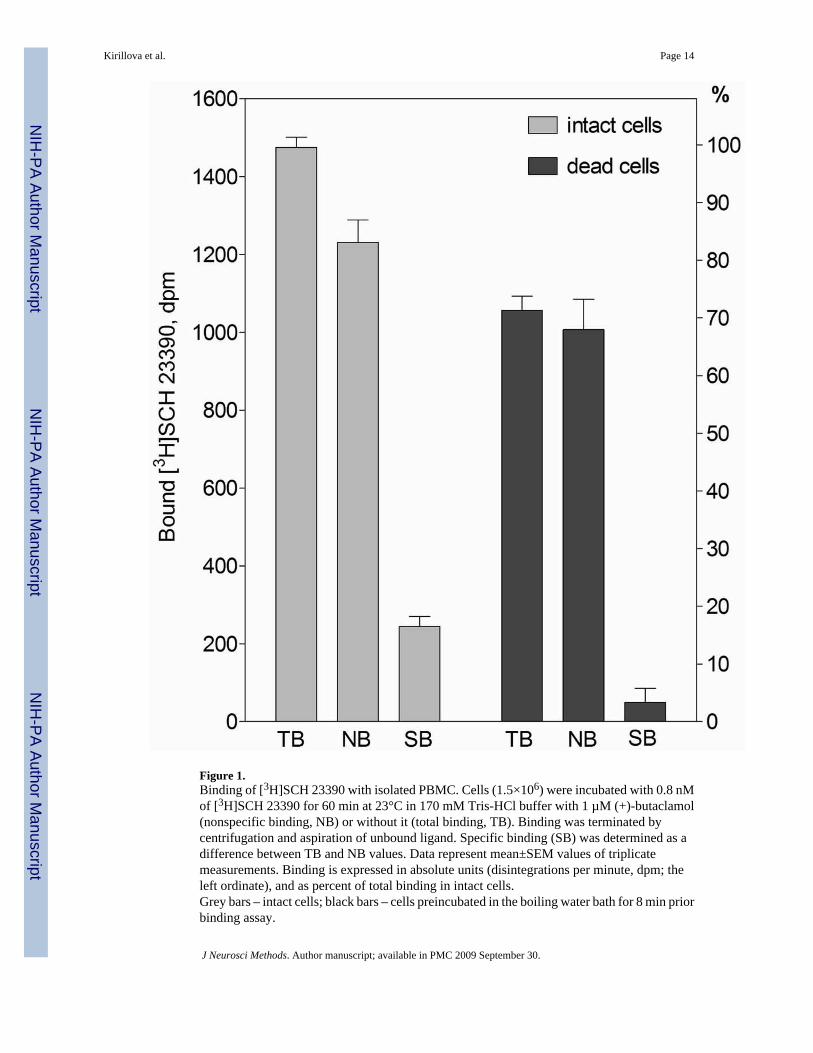
Figure 1.Binding of [3H]SCH 23390 with isolated PBMC. Cells (1.5×10⁶) were incubated with 0.8 nMof [3H]SCH 23390 for 60 min at 23°C in 170 mM Tris-HCl buffer with 1 µM (+)-butaclamol(nonspecific binding, NB) or without it (total binding, TB). Binding was terminated bycentrifugation and aspiration of unbound ligand. Specific binding (SB) was determined as adifference between TB and NB values. Data represent mean±SEM values of triplicatemeasurements. Binding is expressed in absolute units (disintegrations per minute, dpm; theleft ordinate), and as percent of total binding in intact cells.Grey bars – intact cells; black bars – cells preincubated in the boiling water bath for 8 min priorbinding assay.
Kirillova et al. Page 14
J Neurosci Methods. Author manuscript; available in PMC 2009 September 30.
NIH
-PA Author Manuscript
NIH
-PA Author Manuscript
NIH
-PA Author Manuscript
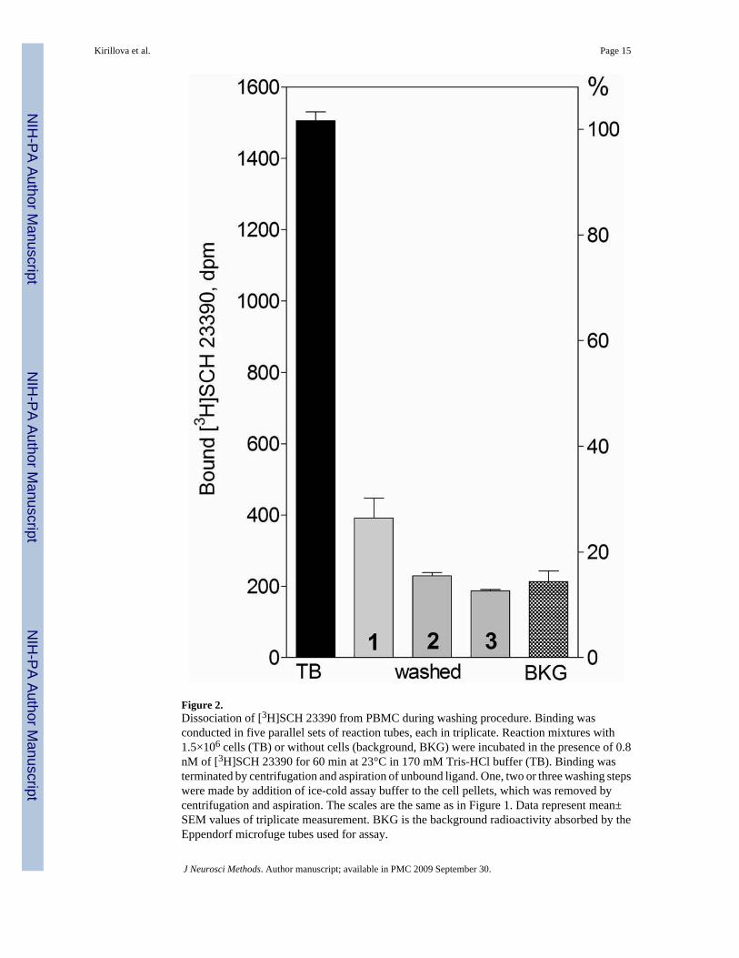
Figure 2.Dissociation of [3H]SCH 23390 from PBMC during washing procedure. Binding wasconducted in five parallel sets of reaction tubes, each in triplicate. Reaction mixtures with1.5×106 cells (TB) or without cells (background, BKG) were incubated in the presence of 0.8nM of [3H]SCH 23390 for 60 min at 23°C in 170 mM Tris-HCl buffer (TB). Binding wasterminated by centrifugation and aspiration of unbound ligand. One, two or three washing stepswere made by addition of ice-cold assay buffer to the cell pellets, which was removed bycentrifugation and aspiration. The scales are the same as in Figure 1. Data represent mean±SEM values of triplicate measurement. BKG is the background radioactivity absorbed by theEppendorf microfuge tubes used for assay.
Kirillova et al. Page 15
J Neurosci Methods. Author manuscript; available in PMC 2009 September 30.
NIH
-PA Author Manuscript
NIH
-PA Author Manuscript
NIH
-PA Author Manuscript

Kirillova et al. Page 16
J Neurosci Methods. Author manuscript; available in PMC 2009 September 30.
NIH
-PA Author Manuscript
NIH
-PA Author Manuscript
NIH
-PA Author Manuscript
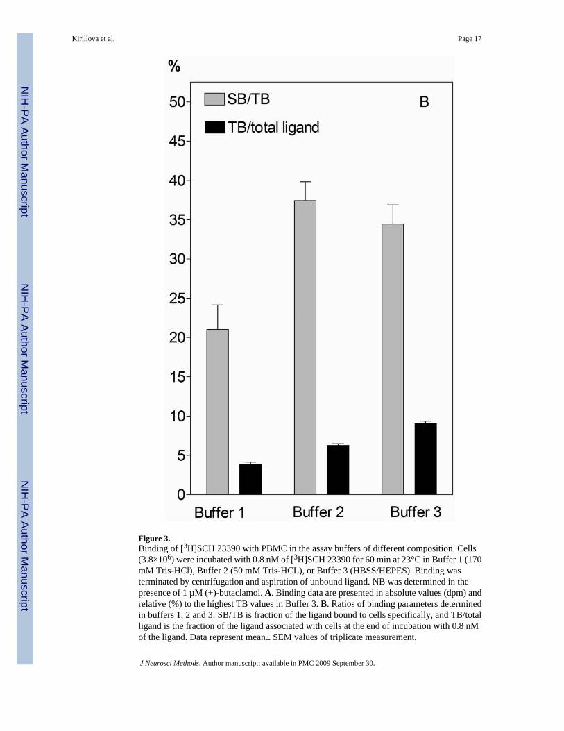
Figure 3.Binding of [3H]SCH 23390 with PBMC in the assay buffers of different composition. Cells(3.8×10⁶) were incubated with 0.8 nM of [3H]SCH 23390 for 60 min at 23°C in Buffer 1 (170mM Tris-HCl), Buffer 2 (50 mM Tris-HCL), or Buffer 3 (HBSS/HEPES). Binding wasterminated by centrifugation and aspiration of unbound ligand. NB was determined in thepresence of 1 µM (+)-butaclamol. A. Binding data are presented in absolute values (dpm) andrelative (%) to the highest TB values in Buffer 3. B. Ratios of binding parameters determinedin buffers 1, 2 and 3: SB/TB is fraction of the ligand bound to cells specifically, and TB/totalligand is the fraction of the ligand associated with cells at the end of incubation with 0.8 nMof the ligand. Data represent mean± SEM values of triplicate measurement.
Kirillova et al. Page 17
J Neurosci Methods. Author manuscript; available in PMC 2009 September 30.
NIH
-PA Author Manuscript
NIH
-PA Author Manuscript
NIH
-PA Author Manuscript
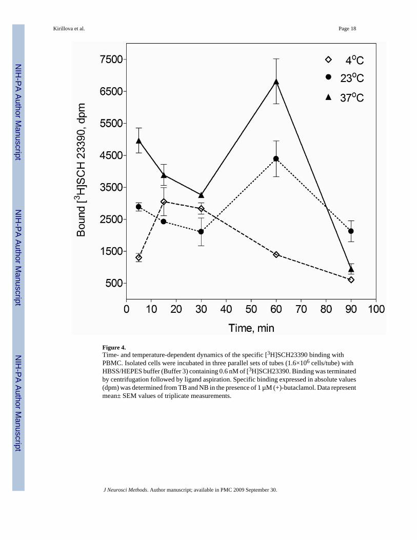
Figure 4.Time- and temperature-dependent dynamics of the specific [3H]SCH23390 binding withPBMC. Isolated cells were incubated in three parallel sets of tubes (1.6×10⁶ cells/tube) withHBSS/HEPES buffer (Buffer 3) containing 0.6 nM of [3H]SCH23390. Binding was terminatedby centrifugation followed by ligand aspiration. Specific binding expressed in absolute values(dpm) was determined from TB and NB in the presence of 1 µM (+)-butaclamol. Data representmean± SEM values of triplicate measurements.
Kirillova et al. Page 18
J Neurosci Methods. Author manuscript; available in PMC 2009 September 30.
NIH
-PA Author Manuscript
NIH
-PA Author Manuscript
NIH
-PA Author Manuscript

Figure 5.Concentration-dependent association of [3H]SCH23390 with PBMC. Cells were incubated inthe presence of increasing concentrations of [3H]SCH23390 in HBSS/HEPES buffer at 37°Cfor 60 minutes with (NB) or without (TB) 1 µM (+)-butaclamol (the concentration scale istruncated to depict binding at low ligand concentrations). Binding was terminated bycentrifugation followed by ligand aspiration. Data are expressed as the amount of ligand (fmol)bound to 2.4×10⁶ cells in reaction tubes. Data represent mean± SEM values of triplicatemeasurement.
Kirillova et al. Page 19
J Neurosci Methods. Author manuscript; available in PMC 2009 September 30.
NIH
-PA Author Manuscript
NIH
-PA Author Manuscript
NIH
-PA Author Manuscript
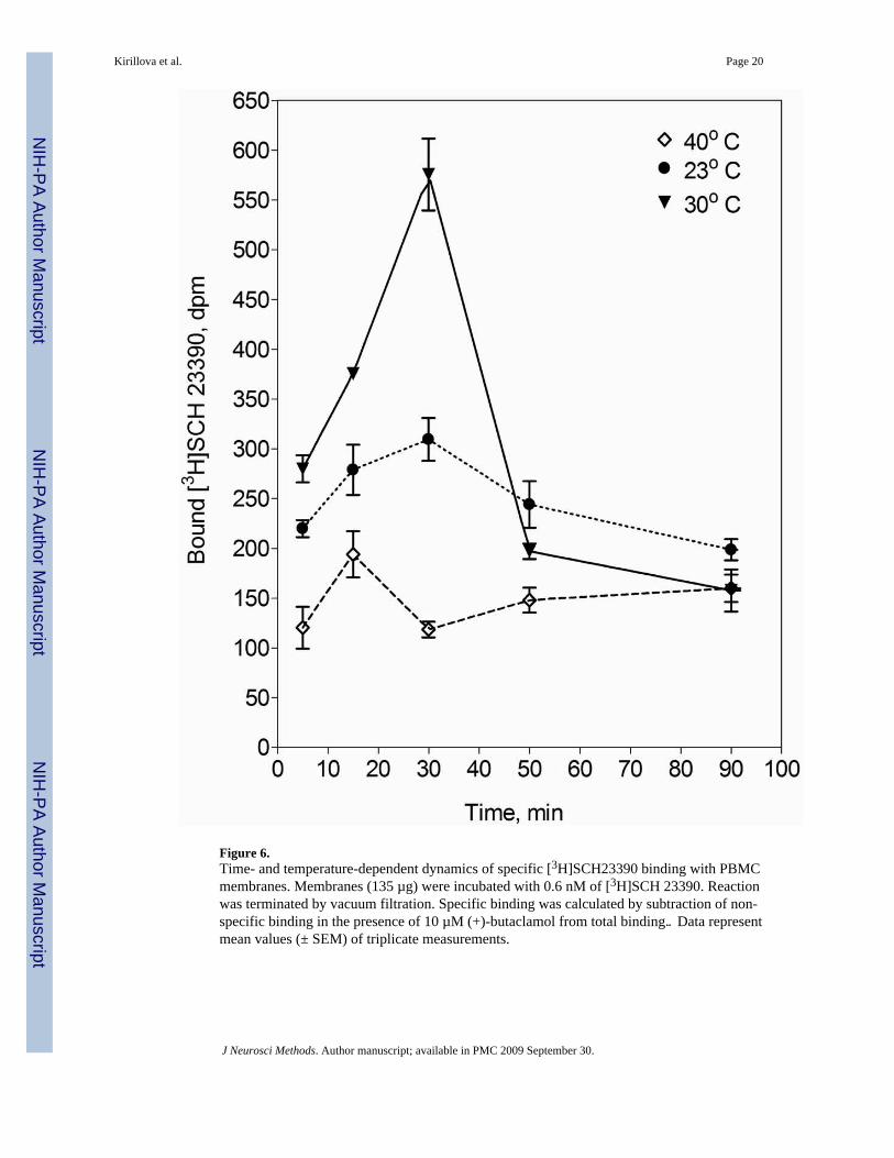
Figure 6.Time- and temperature-dependent dynamics of specific [3H]SCH23390 binding with PBMCmembranes. Membranes (135 µg) were incubated with 0.6 nM of [3H]SCH 23390. Reactionwas terminated by vacuum filtration. Specific binding was calculated by subtraction of non-specific binding in the presence of 10 µM (+)-butaclamol from total binding‥ Data representmean values (± SEM) of triplicate measurements.
Kirillova et al. Page 20
J Neurosci Methods. Author manuscript; available in PMC 2009 September 30.
NIH
-PA Author Manuscript
NIH
-PA Author Manuscript
NIH
-PA Author Manuscript
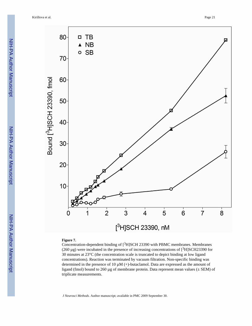
Figure 7.Concentration-dependent binding of [3H]SCH 23390 with PBMC membranes. Membranes(260 µg) were incubated in the presence of increasing concentrations of [3H]SCH23390 for30 minutes at 23°C (the concentration scale is truncated to depict binding at low ligandconcentrations). Reaction was terminated by vacuum filtration. Non-specific binding wasdetermined in the presence of 10 µM (+)-butaclamol. Data are expressed as the amount ofligand (fmol) bound to 260 µg of membrane protein. Data represent mean values (± SEM) oftriplicate measurements.
Kirillova et al. Page 21
J Neurosci Methods. Author manuscript; available in PMC 2009 September 30.
NIH
-PA Author Manuscript
NIH
-PA Author Manuscript
NIH
-PA Author Manuscript

NIH
-PA Author Manuscript
NIH
-PA Author Manuscript
NIH
-PA Author Manuscript
Kirillova et al. Page 22
Table IRelative expression of dopamine receptor genes in peripheral blood mononuclear cells (folds of expression relative tothat of the respective gene in the brain)
Expressed gene Mean SD Range
DRD1 undetected
DRD2 0.005 0.0044 0.0003–0.03
DRD3 1.65 1.142 0.21–6.66
DRD4 1.12 1.339 0.05–16.98
DRD5 0.03 0.019 0.004–0.08
J Neurosci Methods. Author manuscript; available in PMC 2009 September 30.
Related Documents