Dopamine Receptors in Human Adipocytes: Expression and Functions Dana C. Borcherding 1 , Eric R. Hugo 1 , Gila Idelman 1 , Anuradha De Silva 1 , Nathan W. Richtand 1 , Jean Loftus 2 , Nira Ben-Jonathan 1 * 1 Department of Cancer and Cell Biology, University of Cincinnati, Cincinnati, Ohio, United States of America, 2 The Christ Hospital, Cincinnati, Ohio, United States of America Abstract Introduction: Dopamine (DA) binds to five receptors (DAR), classified by their ability to increase (D1R-like) or decrease (D2R- like) cAMP. In humans, most DA circulates as dopamine sulfate (DA-S), which can be de-conjugated to bioactive DA by arylsulfatase A (ARSA). The objective was to examine expression of DAR and ARSA in human adipose tissue and determine whether DA regulates prolactin (PRL) and adipokine expression and release. Methods: DAR were analyzed by RT-PCR and Western blotting in explants, primary adipocytes and two human adipocyte cell lines, LS14 and SW872. ARSA expression and activity were determined by qPCR and enzymatic assay. PRL expression and release were determined by luciferase reporter and Nb2 bioassay. Analysis of cAMP, cGMP, leptin, adiponectin and interleukin 6 (IL-6) was done by ELISA. Activation of MAPK and PI3 kinase/Akt was determined by Western blotting. Results: DAR are variably expressed at the mRNA and protein levels in adipose tissue and adipocytes during adipogenesis. ARSA activity in adipocyte increases after differentiation. DA at nM concentrations suppresses cAMP, stimulates cGMP, and activates MAPK in adipocytes. Acting via D2R-like receptors, DA and DA-S inhibit PRL gene expression and release. Acting via D1R/D5R receptors, DA suppresses leptin and stimulates adiponectin and IL-6 release. Conclusions: This is the first report that human adipocytes express functional DAR and ARSA, suggesting a regulatory role for peripheral DA in adipose functions. We speculate that the propensity of some DAR-activating antipsychotics to increase weight and alter metabolic homeostasis is due, in part, to their direct action on adipose tissue. Citation: Borcherding DC, Hugo ER, Idelman G, De Silva A, Richtand NW, et al. (2011) Dopamine Receptors in Human Adipocytes: Expression and Functions. PLoS ONE 6(9): e25537. doi:10.1371/journal.pone.0025537 Editor: Wolfgang Meyerhof, German Institute for Human Nutrition, Germany Received May 6, 2011; Accepted September 6, 2011; Published September 26, 2011 Copyright: ß 2011 Borcherding et al. This is an open-access article distributed under the terms of the Creative Commons Attribution License, which permits unrestricted use, distribution, and reproduction in any medium, provided the original author and source are credited. Funding: This work was supported by National Institutes of Health grants S012212 and CA096613, Department of Defense BC05725, Susan G. Komen Breast Cancer Foundation grant BCRT87406, and a grant from the Elsa U. Pardee foundation (to NBJ), and a National Research Service Award (NRSA) predoctoral fellowship F31 DK761852 (to DCB). The funders had no role in study design, data collection and analysis, decision to publish, or preparation of the manuscript. Competing Interests: The authors have declared that no competing interests exist. * E-mail: [email protected] Introduction Catecholamines (CA) are synthesized from tyrosine by a sequential enzymatic conversion of dopamine (DA) to norepi- nephrine (NE) and epinephrine (EPI). DA binds to five G-protein- coupled, seven transmembrane domain receptors (DAR), classified as those that are linked to stimulation (D1R and D5R) or inhibition (D2R, D3R and D4R) of adenylate cyclase (AC) [1,2]. When coupled to Gas proteins, D1-like receptors can activate the protein kinase A (PKA), mitogen activated protein kinase (MAPK), and cGMP/PKG pathways [3–6]. The D2-like receptors are coupled to Gai/o proteins and inhibit AC, followed by the suppression of cAMP [7]. DA is a pleiotropic compound that acts as a neurotransmitter and a hormone. In the brain, DA rapidly alters electrical activity, ion channels and neurotransmitter release. In peripheral non-neuronal tissues, e.g., pituitary, kidney and blood vessels, DA acts more slowly and affects electrolyte transport, vasodilation, hormone production and cell proliferation [7,8]. DA is the primary inhibitor of pituitary prolactin (PRL) release [9]. Unique to humans, PRL is also produced in multiple extrapituitary sites, where it functions as a cytokine [10]. After discovering de novo synthesis of PRL in human adipose tissue [11,12], we examined its regulation. When adipocytes were placed in culture, PRL release increased for several days [13]. This resembled the progressive rise in PRL release from cultured pituitary cells, which is attributable to the removal of tonic inhibition by hypothalamic DA [14]. We initially ruled out DA as the inhibitor of adipose PRL because a ready source of DA to the adipocytes was not apparent, and there was no information on DAR expression in human adipose tissue, except for a single report describing a novel DAR in rat brown adipose tissue [15]. Dopamine sulfate (DA-S) is the major form of circulating DA in humans [16,17]. Sulfoconjugation is carried out in the gastroin- testinal (GI) tract by SULT1A3 sulfotransferase [16,18]. Basal serum DA-S levels at <10 nM exceeds by 5 fold the combined levels of free DA (1.5 nM), NE (1 nM) or Epi (0.2 nM). The PLoS ONE | www.plosone.org 1 September 2011 | Volume 6 | Issue 9 | e25537

Welcome message from author
This document is posted to help you gain knowledge. Please leave a comment to let me know what you think about it! Share it to your friends and learn new things together.
Transcript
Dopamine Receptors in Human Adipocytes: Expressionand FunctionsDana C. Borcherding1, Eric R. Hugo1, Gila Idelman1, Anuradha De Silva1, Nathan W. Richtand1, Jean
Loftus2, Nira Ben-Jonathan1*
1 Department of Cancer and Cell Biology, University of Cincinnati, Cincinnati, Ohio, United States of America, 2 The Christ Hospital, Cincinnati, Ohio, United States of
America
Abstract
Introduction: Dopamine (DA) binds to five receptors (DAR), classified by their ability to increase (D1R-like) or decrease (D2R-like) cAMP. In humans, most DA circulates as dopamine sulfate (DA-S), which can be de-conjugated to bioactive DA byarylsulfatase A (ARSA). The objective was to examine expression of DAR and ARSA in human adipose tissue and determinewhether DA regulates prolactin (PRL) and adipokine expression and release.
Methods: DAR were analyzed by RT-PCR and Western blotting in explants, primary adipocytes and two human adipocytecell lines, LS14 and SW872. ARSA expression and activity were determined by qPCR and enzymatic assay. PRL expression andrelease were determined by luciferase reporter and Nb2 bioassay. Analysis of cAMP, cGMP, leptin, adiponectin andinterleukin 6 (IL-6) was done by ELISA. Activation of MAPK and PI3 kinase/Akt was determined by Western blotting.
Results: DAR are variably expressed at the mRNA and protein levels in adipose tissue and adipocytes during adipogenesis.ARSA activity in adipocyte increases after differentiation. DA at nM concentrations suppresses cAMP, stimulates cGMP, andactivates MAPK in adipocytes. Acting via D2R-like receptors, DA and DA-S inhibit PRL gene expression and release. Actingvia D1R/D5R receptors, DA suppresses leptin and stimulates adiponectin and IL-6 release.
Conclusions: This is the first report that human adipocytes express functional DAR and ARSA, suggesting a regulatory rolefor peripheral DA in adipose functions. We speculate that the propensity of some DAR-activating antipsychotics to increaseweight and alter metabolic homeostasis is due, in part, to their direct action on adipose tissue.
Citation: Borcherding DC, Hugo ER, Idelman G, De Silva A, Richtand NW, et al. (2011) Dopamine Receptors in Human Adipocytes: Expression and Functions. PLoSONE 6(9): e25537. doi:10.1371/journal.pone.0025537
Editor: Wolfgang Meyerhof, German Institute for Human Nutrition, Germany
Received May 6, 2011; Accepted September 6, 2011; Published September 26, 2011
Copyright: � 2011 Borcherding et al. This is an open-access article distributed under the terms of the Creative Commons Attribution License, which permitsunrestricted use, distribution, and reproduction in any medium, provided the original author and source are credited.
Funding: This work was supported by National Institutes of Health grants S012212 and CA096613, Department of Defense BC05725, Susan G. Komen BreastCancer Foundation grant BCRT87406, and a grant from the Elsa U. Pardee foundation (to NBJ), and a National Research Service Award (NRSA) predoctoralfellowship F31 DK761852 (to DCB). The funders had no role in study design, data collection and analysis, decision to publish, or preparation of the manuscript.
Competing Interests: The authors have declared that no competing interests exist.
* E-mail: [email protected]
Introduction
Catecholamines (CA) are synthesized from tyrosine by a
sequential enzymatic conversion of dopamine (DA) to norepi-
nephrine (NE) and epinephrine (EPI). DA binds to five G-protein-
coupled, seven transmembrane domain receptors (DAR), classified
as those that are linked to stimulation (D1R and D5R) or
inhibition (D2R, D3R and D4R) of adenylate cyclase (AC) [1,2].
When coupled to Gas proteins, D1-like receptors can activate the
protein kinase A (PKA), mitogen activated protein kinase (MAPK),
and cGMP/PKG pathways [3–6]. The D2-like receptors are
coupled to Gai/o proteins and inhibit AC, followed by the
suppression of cAMP [7]. DA is a pleiotropic compound that acts
as a neurotransmitter and a hormone. In the brain, DA rapidly
alters electrical activity, ion channels and neurotransmitter release.
In peripheral non-neuronal tissues, e.g., pituitary, kidney and
blood vessels, DA acts more slowly and affects electrolyte
transport, vasodilation, hormone production and cell proliferation
[7,8].
DA is the primary inhibitor of pituitary prolactin (PRL)
release [9]. Unique to humans, PRL is also produced in multiple
extrapituitary sites, where it functions as a cytokine [10]. After
discovering de novo synthesis of PRL in human adipose tissue
[11,12], we examined its regulation. When adipocytes were
placed in culture, PRL release increased for several days [13].
This resembled the progressive rise in PRL release from
cultured pituitary cells, which is attributable to the removal of
tonic inhibition by hypothalamic DA [14]. We initially ruled out
DA as the inhibitor of adipose PRL because a ready source of
DA to the adipocytes was not apparent, and there was no
information on DAR expression in human adipose tissue, except
for a single report describing a novel DAR in rat brown adipose
tissue [15].
Dopamine sulfate (DA-S) is the major form of circulating DA in
humans [16,17]. Sulfoconjugation is carried out in the gastroin-
testinal (GI) tract by SULT1A3 sulfotransferase [16,18]. Basal
serum DA-S levels at <10 nM exceeds by 5 fold the combined
levels of free DA (1.5 nM), NE (1 nM) or Epi (0.2 nM). The
PLoS ONE | www.plosone.org 1 September 2011 | Volume 6 | Issue 9 | e25537
biologically inactive DA-S has a serum half-life of 3–4 hr,
compared to several minutes for unmodified DA [19]. However,
unlike inactivation of DA by deamination, O-methylation or
glucuronidation, sulfoconjugation is reversible, and DA-S can be
converted back to bioactive DA by arylsulfatase A (ARSA), a
releasable lysosomal enzyme [20,21]. We reasoned that if
adipocytes express DAR and possess an active ARSA, circulating
DA-S can serve as an inhibitor of adipose PRL.
Production of adipokines is a major function of adipose tissue.
Leptin is an important adipokine which is involved in body
weight regulation via its effects on food intake and energy
expenditure [22]. Adiponectin is a key adipose-derived hormone
which plays critical roles in fuel homeostasis, insulin action and
atherosclerosis [23]. Interleukin-6 (IL-6) is an inflammatory
cytokine which is produced by both macrophages and
adipocytes and whose release is increased in obesity [24]. The
production/release of these adipokines is differentially regulated
in human adipocytes by beta adrenergic receptor (b-AR)
agonists [25–28]. DA was recently reported to suppress leptin
expression and release in 3T3-L1 murine adipocytes [29], but
there is no information on comparable effects of DA on human
adipocytes.
The objective of this study was to detect expression of DAR in
human adipocytes at both the mRNA and protein levels and
confirm their functionality by examining the effects of DA on
PRL, leptin, adiponectin and IL-6 release. To this end, we
undertook a comprehensive approach and used adipose tissue
explants, primary adipocytes and two human adipocyte cell lines:
LS14 which we have cloned from a metastatic liposarcoma [30],
and SW872, a liposarcoma-derived cell line obtained from the
ATCC. Both cell lines differ with respect to gene expression and
the timing of full differentiation.
Materials and Methods
Ethics StatementWritten informed consents, approved by the Institutional
Review Board of the Christ Hospital of Cincinnati, were obtained
from all participants.
Patients, explant preparation and cell harvestingSubcutaneous (sc) adipose tissue was obtained from non-obese,
non-diabetic women (BMI,30) undergoing elective abdomino-
plasty. Adipose explants were placed in 48-well plates (80–
100 mg/well) containing DMEM:F12 with 1% ITS+ (BD
Biosciences, San Jose, CA) and incubated with the various
treatments. For cell harvesting, tissue fragments were digested
with collagenase as described [13]. After filtration and brief
centrifugation, the floating mature adipocytes (100 ml of packed
cells) were incubated in collagen-coated plates containing
DMEM:F12 with 1% ITS+ and the various treatments. The
stromal vascular cell (SVC) fraction which contains the preadipo-
cytes was sedimented, and after treatment with erythrocyte lysis
buffer, cells were cultured in DMEM:F12 with 10% FBS and
50 mg/ml Primocin (Invitrogen, San Diego, CA). After 2–3
passages, cells were frozen until used.
Cell culture and differentiationLS14 cells were cultured as described [13], while SW872 cells
were maintained in DMEM:F12 with 10% FBS. For differenti-
ation, primary sc preadipocytes, SW872 and LS14 cells were
plated in collagen-coated 24 well plates at 100,000 cells/well. Cells
were incubated in serum-free basal adipogenesis medium (BAM)
as described [13]. Adipogenesis was induced by the addition of
250 mM IBMX (BioMol, Plymouth Meeting, PA). After three
days, cells were incubated in BAM without IBMX, and cell
differentiation was monitored visually. On days 10–14, 80–90% of
either primary or cell lines, showed lipid accumulation. After
overnight incubation in DMEM/F12 plus 1% CSS, cells were
treated as described below.
Generation of stably transfected SW872 cells andluciferase assay
The full-length superdistal PRL promoter (dPRL) cloned into
the PGL3E luciferase reporter vector (Promega, San Louis
Obispo, CA) was provided by Dr. Brar, Children’s Hospital,
Cincinnati, OH. SW872 cells were co-transfected with dsRed2-N1
plasmid (Clontech, Mountain View, CA) and the dPRL/PGL3E
construct at a 1:10 ratio using Fugene 6 (Roche, Indianapolis, IN).
After 72 hr, cells were selected using 0.8 mg/ml G418 (Sigma, St
Louis, MO). Colonies were picked, expanded and the cells were
maintained thereafter in 0.08 mg/ml G418. The stably transfected
cells were induced to differentiate, serum starved, and treated as
indicated below. After lysing with Passive Lysis Buffer (Promega),
luciferase activity was determined using Microplate Luminometer
(Dynex Technologies, Chantilly, VA).
Conventional and Real-time RT-PCRRNA isolation and cDNA synthesis were done as described
[13]. Conventional PCR was performed using intron-spanning
primers (Table 1). For D1R/D5R which have no introns, DNAse
was added during RNA isolation. Additionally, samples were
evaluated for genomic DNA contamination using no reverse
transcriptase control. Cycle conditions were: 96 C for 6 min for
polymerase activation, followed by 35 cycles of 94, 57, and 72 C,
each for 45 sec. Products were resolved on a 1.5% agarose gel
containing ethidium bromide and photographed. Quantitative
real-time PCR (qPCR) was performed with SYBR Green and the
appropriate primers (Table 1). Fluorometric products were
detected with Applied Biosystem StepOnePlus instrument. Cycle
parameters were: 96 C for 6 min, followed by 45 cycles of 94 C for
15 sec, 57 C for 15 sec, and 72 C for 30 sec, and an optical read at
83.5 C for 6 sec. Product purity was confirmed by DNA melting
curve analysis and agarose gel electrophoresis. PCR efficiency was
determined using the LinRegPCR program or by cDNA dose
response curve analysis. b2-Microglobulin (B2M) was used as a
reference gene. Fold changes in gene expression were calculated
from cycle threshold and efficiency measurements as described
[13].
PRL release studiesAdipose tissue explants, mature adipocytes or differentiated
primary adipocytes, LS14 and SW872 cells were incubated with
DA, bromocriptine (a D2R agonist), isoproterenol (all from
Sigma), dopamine-4-O-sulfate (from NIMH) or raclopride, a
D2R antagonist (Tocris, Ellisvile, MO). After 24 h, conditioned
media (CM) were collected and aliquots were analyzed for PRL by
the Nb2 bioassay, which is approximately 50 times more sensitive
than RIA or ELISA for PRL [13]. Briefly, rat Nb2 lymphocytes
were serum-starved and plated in 96-well plates at 30,000 cells/
well. Cells were incubated with recombinant human PRL (Protein
Laboratories, Rehovot, Israel) in triplicate or with CM aliquots in
duplicate. After 72 h, cell viability was determined by fluorescence
using resazurin reduction. PRL concentration in CM was
calculated from a standard curve, with the lowest detectable level
of 2 pg/well. Nb2 cell proliferation was not affected by the tested
compounds.
Dopamine Receptors in Human Adipocytes
PLoS ONE | www.plosone.org 2 September 2011 | Volume 6 | Issue 9 | e25537
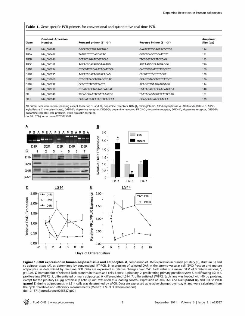
Table 1. Gene-specific PCR primers for conventional and quantitative real time PCR.
GeneGenbank AccessionNumber Forward primer (59R39) Reverse Primer (59R39)
AmplimerSize (bp)
B2M NM_004048 GGCATTCCTGAAGCTGAC GAATCTTTGGAGTACGCTGG 114
ARSA NM_000487 TATGCCTCTCACCACAC GGTCTCAGGTCCATTGTC 191
ARSB NM_000046 GCTACCAGATCCGTACAG TTCCGGTACATTCCCAG 153
ARSC NM_000351 AGCACTGATAGGGAAATGG AGCAAGGGTAAGGAGGG 216
DRD1 NM_000794 CTCCGTTTCCAAATACATTCCA CACTGTTGATTCTTTGCCCT 169
DRD2 NM_000795 AGCATCGACAGGTACACAG CTCGTTCTGGTCTGCGT 159
DRD3 NM_033660 GTGGTATACCTGGAGGTGAC GCAGTGTACCTGTCTATGCT 136
DRD4 NM_000797 CCGCTCTTCGTCTACTC ACAGGTTGAAGATGGAGG 114
DRD5 NM_000798 CTCATCTCCTACAACCAAGAC TGATAGATCTGGAACATGCGA 148
PRL NM_000948 TTCAGCGAATTCGATAAACGG TGATACAGAGGCTCATTCCAG 181
PRLR NM_000949 CGTGACTTACATAGTTCAGCCA GGAGCGTGAACCAACCA 139
All primer sets were intron-spanning except those for D1 and D5 dopamine receptors. B2M-b2 microglobulin, ARSA-arylsulfatase A, ARSB-arylsulfatase B, ARSC-arylsulfatase C (steroylsulfatase), DRD1-D1 dopamine receptor, DRD2-D2 dopamine receptor, DRD3-D3 dopamine receptor, DRD4-D4 dopamine receptor, DRD5-D5
dopamine receptor, PRL-prolactin, PRLR-prolactin receptor.doi:10.1371/journal.pone.0025537.t001
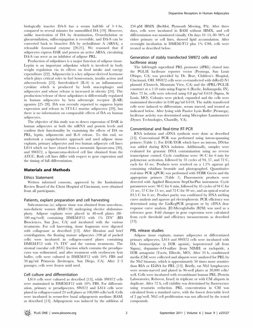
Figure 1. DAR expression in human adipose tissue and adipocytes. A, comparison of DAR expression in human pituitary (P), striatum (S) andsc adipose tissue (A), as determined by conventional RT-PCR. B, expression of selected DAR in the stromo-vascular cell (SVC) fraction and matureadipocytes, as determined by real-time PCR. Data are expressed as relative changes over SVC. Each value is a mean6SEM of 3 determinations; *,p,0.05. C, Immunoblot of selected DAR proteins in tissues and cells. Lanes: 1, pituitary; 2, proliferating primary preadipocytes; 3, proliferating LS14; 4,proliferating SW872; 5, differentiated primary adipocytes; 6, differentiated LS14; 7, differentiated SW872. Each lane was loaded with 40 mg proteins,except for the pituitary (30 mg proteins). b-actin (b-Act) was used as a loading control. Expression of D1R, D2R and D4R (panel D), and PRL vs PRLR(panel E) during adipogenesis in LS14 cells was determined by qPCR. Data are expressed as relative changes over day 0, and were calculated fromthe cycle threshold and efficiency measurements (Mean6SEM of 3 determinations).doi:10.1371/journal.pone.0025537.g001
Dopamine Receptors in Human Adipocytes
PLoS ONE | www.plosone.org 3 September 2011 | Volume 6 | Issue 9 | e25537
Western blot analysisCells were homogenized in lysis buffer and 35 mg of lysate
proteins were separated on 12% SDS gels and transferred to
nitrocellulose membranes. For DAR, validated antibodies [31–33]
against D1R (Calbiochem, San Diego, CA: 324390; 1:2,000), D2R
(Santa Cruz, CA: SC-5303; 1:250), and D4R (Calbiochem:
324405; 1:1000) were used. For signaling pathways, ERK1/2
(ab #9102) phospho ERK 1/2 (Thr202/Tyr204 ab #9101S),
Phospho Akt (Ser473, ab #9271S) and Akt (ab #9272), all from
Cell signaling (Danvers, MA) were used at 1:1000. After
incubation with horseradish peroxidase-conjugated secondary
antibodies, products were exposed to SuperSignal chemilumines-
cence reagents (Pierce, Rockford, IL) and photographed; b-actin
(Sigma, A1978;1:5000) was used as a loading control.
Arylsulfatase A ActivityARSA activity was determined by the method of Chang et al
[34] after modifications. Briefly, samples were homogenized in
0.05 M acetate buffer, pH 5.0, freeze-thawed, and centrifuged at
12,0006 g for 5 min. Lysates (15 mg) or CM (X10 concentrated)
were incubated at 37C in 0.05 M acetate buffer, pH 5.6
containing 3 mM lead acetate, and 5 mM 4-methylumbelliferyl
sulfate (Sigma), with or without 3 mM Ag+. After 30 min, the
reaction was stopped with 0.2 M glycine-carbonate buffer,
pH 10.4 and 1 mM EDTA. Fluorescence was measured at
370 nm excitation and 450 nm emission, using Gemini fluorescent
microplate reader (Molecular Devices, Sunnyvale, CA). Enzyme
activity was calculated from a standard curve.
cAMP and cGMP determinationsCells were incubated with the various treatments for 30 min
and then lysed in 0.1 M HCl. After centrifugation, the supernatant
was analyzed for cAMP and cGMP using respective ELISA kits
from Cayman Chemical Co (Ann Arbor, MI). To increase assay
sensitivity for cGMP analysis, samples were first acetylated
according to manufacturer’s instructions.
Adipokine determinationFluorescent sandwich ELISAs for human leptin, adiponectin
and IL-6 were optimized in our lab as described [35]. Briefly,
matched monoclonal ab pairs against leptin (R&D Systems,
Minneapolis MN; MAB398 for capture and BAM398 for
detection), adiponectin (R&D systems; MAB10651 for capture
and BAM1065 for detection) and IL-6 (Invitrogen, Carlsbad CA;
AHC0562 for capture and AHC0469 for detection) were used.
Plates coated with the capture ab were co-incubated with
biotinylated detection ab, antigen standards and CM from
explants or cells. Streptavidin-conjugated horseradish peroxidase
was added, followed by a fluorimetric substrate (QuantaBlue;
Thermo-Fisher, Rockford, IL). Plates were read at 325 nm
excitation and 420 nm emission.
Data AnalysisExperiments were repeated at least 3 times. Values were
expressed as means6SEM. Data were analyzed by Student’s t test
or ANOVA. P,0.05 was considered significant.
Results
Expression of DAR in adipose tissue and adipocytesConventional RT-PCR was used to compare the expression of
DAR in sc adipose tissue, striatum and pituitary. Four of the five
DAR were detected in adipose tissue, with D1R being the most
abundant (Fig. 1A). As expected, the pituitary showed high
expression of D2R, D4R and D5R, while the striatum showed
differential expression of all DAR. As determined by qPCR, the
relative expression of D1R is higher, while that of D2R is lower in
mature adipocytes than the SVC fraction, while expression of
D4R was similar in the two fractions (Fig. 1B).
Figure 2. Expression of active arylsulfatase A (ARSA) in adipose tissue and adipocytes and confirmation of DA-S bioactivity. A,comparison of mRNA levels of ARSA (A), ARSB (B) and ARSC (C) in sc adipose tissue, as determine by qPCR. Data are expressed as fold changes in geneexpression over ARSB, and were calculated from the cycle threshold and efficiency measurements. B, ARSA activity before (light bars) and after (darkbars) differentiation of LS14 and SW872 adipocytes. Specific ARSA activity was determined by subtracting enzyme activity in the presence of silvernitrate from total enzyme activity. Each value is a mean6SEM of 4 determinations; *, p,0.05. C, both DA and DA-S inhibit PRL release from sc adiposetissue explants, whereas isoproterenol (ISO), a b-AR agonist, causes stimulation. Explants were incubated with the different compounds for 24 hr andPRL concentration in CM was determined by the Nb2 bioassay. Data are expressed as % of control. Each value is a mean6SEM of 6 determinations;*, p,0.05.doi:10.1371/journal.pone.0025537.g002
Dopamine Receptors in Human Adipocytes
PLoS ONE | www.plosone.org 4 September 2011 | Volume 6 | Issue 9 | e25537
Western blotting was used to compare expression of the DAR
proteins in primary adipocytes, LS14 and SW872 cells before and
after differentiation. Fig. 1C shows that D1R, D2R and D4R
proteins were expressed at variable amounts in the pituitary
(serving as a positive control), and in the three types of adipocytes.
There was an apparent downregulation of D1R and D4R, but
upregulation of D2R, in LS14 cells after differentiation. All
receptors examined showed some evidence of isoforms.
Changes in D1R, D2R and D4R mRNA levels during
adipogenesis in LS14 cells were determined by qPCR. Fig. 1Dshows a reduction in D1R, but an increase in D2R, during the first
three days of differentiation. Expression of D4R was reduced on
day 3 and remained suppressed until day 10. PRL expression was
increased during the first six days of differentiation, followed by a
decline, whereas PRLR expression showed a significant reduction
throughout adipogenesis (Fig. 1D).
Adipocytes express an active ARSA and respond to DA-SWe used qPCR to compare the expression of arylsulfatase A
(ARSA), specific for catecholamines and cerebrosides, arylsulfatase
B (ARSB), specific for glycosaminoglycans, and arylsulfatase C
(ARSC), specific for steroids [21]. The mRNA levels of ARSA and
ARSC in sc adipose tissue were 4.5 and 2.5 fold higher,
respectively, than those of ARSB (Fig. 2A). An enzymatic assay
that measures total arylsulfatase activity was then employed. In the
presence of silver nitrate, only ARSA activity is blocked, enabling
the calculation of its activity by subtraction from total enzyme
activity. As shown in Fig. 2B, basal ARSA activity was detectable
in non-differentiated LS14 and SW872 cells, increasing 8 and 20
fold, respectively, after differentiation. ARSA activity was also
detectable in CM from differentiated adipocytes (data not shown).
To determine whether adipocytes can convert DA-S to bioactive
DA, the effects of DA and DA-S on PRL release from sc adipose
explants were compared. Fig. 2C shows a similar inhibition of
PRL release by both DA and DA-S. The b-AR agonist
isoproterenol (ISO), stimulated PRL release, showing an inverted
U-shaped dose-dependent curve.
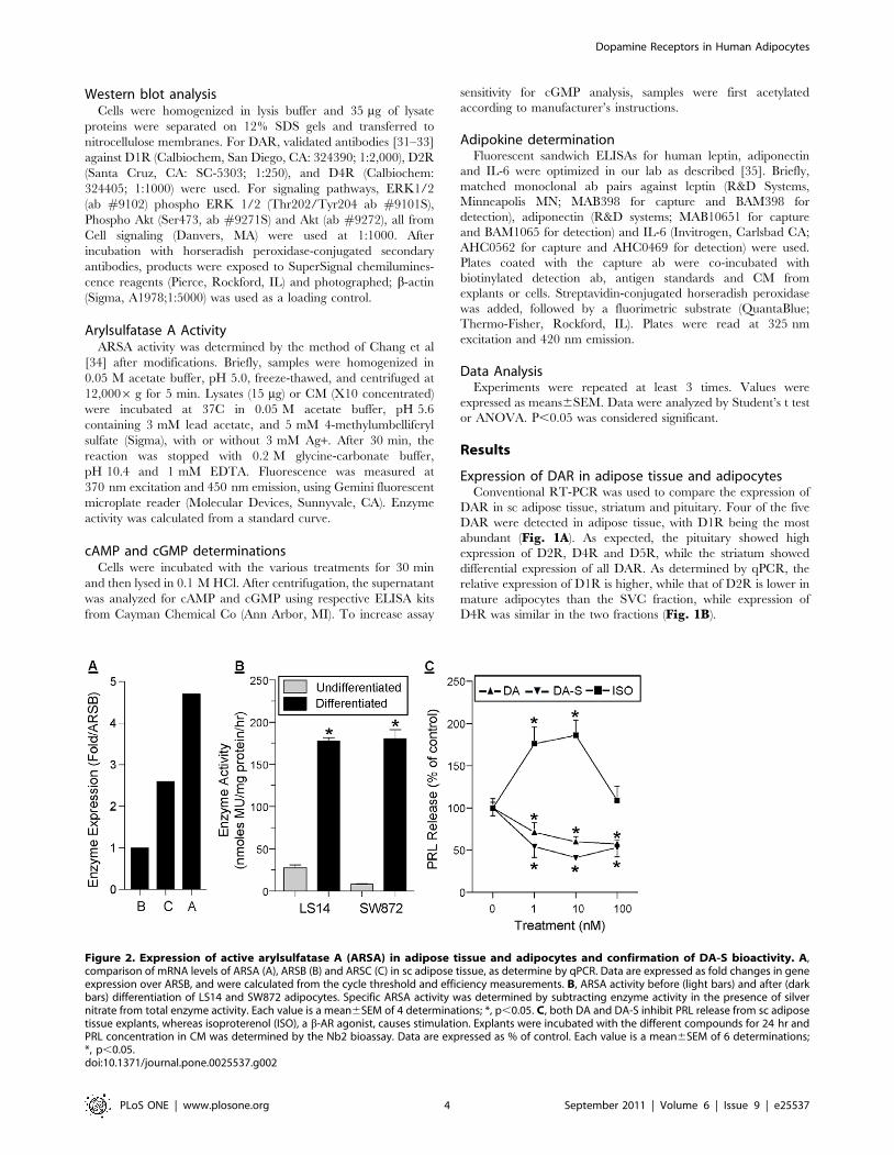
PRL release from all types of adipocytes is inhibited byboth DA and bromocriptine
We next examined whether DA directly affects PRL release by
the adipocytes. Fig. 3 shows a similar DA-induced inhibition of
PRL release from mature adipocytes (panel A), differentiated
primary preadipocytes (panel B), differentiated LS14 cells (panelC), and differentiated SW872 cells (panel D). Bromocriptine
(BRO), a specific D2R agonist, mimicked the inhibitory effect of
DA. A non-monotonic dose-dependent inhibition of PRL release
was apparent in all cases, with 1 and 10 nM, but not 100 nM, of
DA and BRO showing effective inhibition.
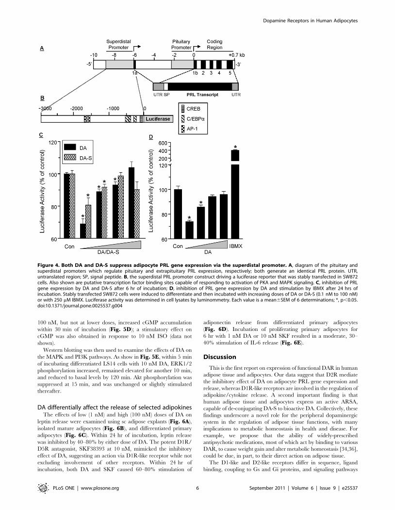
Both DA and DA-S suppress PRL gene expression via thesuperdistal promoter
A diagram of the proximal and superdistal promoters that
regulate pituitary and extrapituitary PRL, respectively, is shown in
Fig. 4A. The superdistal promoter is located 5.8 kB upstream of
the pituitary start site [10], and contains putative CREB (cAMP
response element binding protein) and c/EBP (CCAAT/enhancer
binding protein) transcription binding sites in the proximal region,
as well as two AP-1 sites in the more distal region (Fig. 4B).
SW872 cells, stably transfected with a luciferase reporter driven by
the 3000 kb superdistal promoter (Fig. 4B), were used. Within
6 hr, as little as 0.1 nM of DA or DA-S suppressed PRL
expression (Fig. 4C), an effect that was also seen after 24 hr
(Fig. 4D). As was the case with PRL release (Fig. 3), a non-linear,
dose-dependent curve was evident, suggesting activation of
stimulatory DAR at the higher DA-S/DA doses. IBMX, a
phosphodiesterase inhibitor, increased PRL expression 4 fold,
supporting the role of the cAMP system in the transcriptional
control of adipocyte PRL.
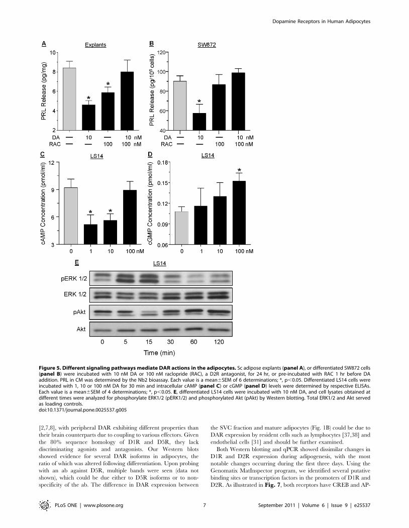
DAR signal through multiple pathways in the adipocytesTo further characterize the DAR that mediates the suppression
of PRL by DA, adipose explants (Fig. 5A) and differentiated
SW872 cells (Fig. 5B) were pretreated with 100 nM raclopride, a
selective D2R antagonist, before incubation with 10 nM DA for
24 hrs. In both cases, raclopride abrogated the inhibitory action of
DA on PRL release, indicating mediation by D2R. The ability of
DA to affect intracellular cAMP and cGMP concentrations in
LS14 cells was then examined. Within 30 min of incubation, 1 and
10 nM DA suppressed, while 100 nM DA had no effect, on
intracellular cAMP levels (Fig. 5C). On the other hand, DA at
Figure 3. DA and bromocriptine (BRO) inhibit PRL release fromdifferent types of adipocytes. A, isolated sc mature adipocytes. B,differentiated sc primary adipocytes. C, differentiated LS14 cells. D,differentiated SW872 cells. In each case, cells were incubated withdifferent doses of DA or bromocriptine (BRO) for 24 hr, and PRL in CMwas determined by the Nb2 bioassay. Each value is a mean6SEM of 6determinations; *, p,0.05.doi:10.1371/journal.pone.0025537.g003
Dopamine Receptors in Human Adipocytes
PLoS ONE | www.plosone.org 5 September 2011 | Volume 6 | Issue 9 | e25537
100 nM, but not at lower doses, increased cGMP accumulation
within 30 min of incubation (Fig. 5D); a stimulatory effect on
cGMP was also obtained in response to 10 nM ISO (data not
shown).
Western blotting was then used to examine the effects of DA on
the MAPK and PI3K pathways. As show in Fig. 5E, within 5 min
of incubating differentiated LS14 cells with 10 nM DA, ERK1/2
phosphorylation increased, remained elevated for another 10 min,
and reduced to basal levels by 120 min. Akt phosphorylation was
suppressed at 15 min, and was unchanged or slightly stimulated
thereafter.
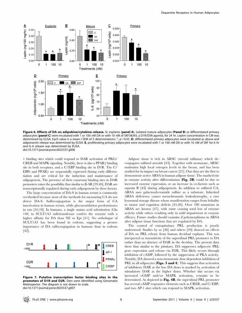
DA differentially affect the release of selected adipokinesThe effects of low (1 nM) and high (100 nM) doses of DA on
leptin release were examined using sc adipose explants (Fig. 6A),
isolated mature adipocytes (Fig. 6B), and differentiated primary
adipocytes (Fig. 6C). Within 24 hr of incubation, leptin release
was inhibited by 40–80% by either dose of DA. The potent D1R/
D5R antagonist, SKF38393 at 10 nM, mimicked the inhibitory
effect of DA, suggesting an action via D1R-like receptor while not
excluding involvement of other receptors. Within 24 hr of
incubation, both DA and SKF caused 60–80% stimulation of
adiponectin release from differentiated primary adipocytes
(Fig. 6D). Incubation of proliferating primary adipocytes for
6 hr with 1 nM DA or 10 nM SKF resulted in a moderate, 30–
40% stimulation of IL-6 release (Fig. 6E).
Discussion
This is the first report on expression of functional DAR in human
adipose tissue and adipocytes. Our data suggest that D2R mediate
the inhibitory effect of DA on adipocyte PRL gene expression and
release, whereas D1R-like receptors are involved in the regulation of
adipokine/cytokine release. A second important finding is that
human adipose tissue and adipocytes express an active ARSA,
capable of de-conjugating DA-S to bioactive DA. Collectively, these
findings underscore a novel role for the peripheral dopaminergic
system in the regulation of adipose tissue functions, with many
implications to metabolic homeostasis in health and disease. For
example, we propose that the ability of widely-prescribed
antipsychotic medications, most of which act by binding to various
DAR, to cause weight gain and alter metabolic homeostasis [34,36],
could be due, in part, to their direct action on adipose tissue.
The D1-like and D2-like receptors differ in sequence, ligand
binding, coupling to Gs and Gi proteins, and signaling pathways
Figure 4. Both DA and DA-S suppress adipocyte PRL gene expression via the superdistal promoter. A, diagram of the pituitary andsuperdistal promoters which regulate pituitary and extrapituitary PRL expression, respectively; both generate an identical PRL protein. UTR,untranslated region; SP, signal peptide. B, the superdistal PRL promoter construct driving a luciferase reporter that was stably transfected in SW872cells. Also shown are putative transcription factor binding sites capable of responding to activation of PKA and MAPK signaling. C, inhibition of PRLgene expression by DA and DA-S after 6 hr of incubation; D, inhibition of PRL gene expression by DA and stimulation by IBMX after 24 hrs ofincubation. Stably transfected SW872 cells were induced to differentiate and then incubated with increasing doses of DA or DA-S (0.1 nM to 100 nM)or with 250 mM IBMX. Luciferase activity was determined in cell lysates by luminommetry. Each value is a mean6SEM of 6 determinations; *, p,0.05.doi:10.1371/journal.pone.0025537.g004
Dopamine Receptors in Human Adipocytes
PLoS ONE | www.plosone.org 6 September 2011 | Volume 6 | Issue 9 | e25537
[2,7,8], with peripheral DAR exhibiting different properties than
their brain counterparts due to coupling to various effectors. Given
the 80% sequence homology of D1R and D5R, they lack
discriminating agonists and antagonists. Our Western blots
showed evidence for several DAR isoforms in adipocytes, the
ratio of which was altered following differentiation. Upon probing
with an ab against D5R, multiple bands were seen (data not
shown), which could be due either to D5R isoforms or to non-
specificity of the ab. The difference in DAR expression between
the SVC fraction and mature adipocytes (Fig. 1B) could be due to
DAR expression by resident cells such as lymphocytes [37,38] and
endothelial cells [31] and should be further examined.
Both Western blotting and qPCR showed dissimilar changes in
D1R and D2R expression during adipogenesis, with the most
notable changes occurring during the first three days. Using the
Genomatix MatInspector program, we identified several putative
binding sites or transcription factors in the promoters of D1R and
D2R. As illustrated in Fig. 7, both receptors have CREB and AP-
Figure 5. Different signaling pathways mediate DAR actions in the adipocytes. Sc adipose explants (panel A), or differentiated SW872 cells(panel B) were incubated with 10 nM DA or 100 nM raclopride (RAC), a D2R antagonist, for 24 hr, or pre-incubated with RAC 1 hr before DAaddition. PRL in CM was determined by the Nb2 bioassay. Each value is a mean6SEM of 6 determinations; *, p,0.05. Differentiated LS14 cells wereincubated with 1, 10 or 100 nM DA for 30 min and intracellular cAMP (panel C) or cGMP (panel D) levels were determined by respective ELISAs.Each value is a mean6SEM of 4 determinations; *, p,0.05. E, differentiated LS14 cells were incubated with 10 nM DA, and cell lysates obtained atdifferent times were analyzed for phosphorylate ERK1/2 (pERK1/2) and phosphorylated Akt (pAkt) by Western blotting. Total ERK1/2 and Akt servedas loading controls.doi:10.1371/journal.pone.0025537.g005
Dopamine Receptors in Human Adipocytes
PLoS ONE | www.plosone.org 7 September 2011 | Volume 6 | Issue 9 | e25537
1 binding sites which could respond to DAR activation of PKG/
CREB and MAPK signaling. Notably, there is also a PPARc binding
site in both receptors, and a C/EBP binding site in D1R. The C/
EBPs and PPARc are sequentially expressed during early differen-
tiation and are critical for the induction and maintenance of
adipogenesis. The presence of their consensus binding sites in DAR
promoters raises the possibility that similar to b-AR [39,40], DAR are
transcriptionally regulated during early adipogenesis by these factors.
The large concentration of DA-S in human serum is commonly
overlooked because most of the methods for measuring CA do not
detect DA-S. Sulfoconjugation is the major form of CA
inactivation in human serum, while glucuronidation predominates
in rats [16,18]. In humans, a single amino acid substitution (Glu
146) in SULT1A3 sulfotransferase confers the enzyme with a
higher affinity for DA than NE or Epi [41]. No orthologue of
SULT1A3 has been found in rodents, suggesting a greater
importance of DA sulfoconjugation in humans than in rodents
[42].
Adipose tissue is rich in ARSC (steroid sulfatase) which de-
conjugates sulfated steroids [43]. Together with aromatase, ARSC
maintains high local estrogen levels in the breast, and has been
studied for its impact on breast cancer [21]. Our data are the first to
demonstrate active ARSA in human adipose tissue. The marked rise
in enzyme activity after differentiation (Fig. 2B) could be due to
increased enzyme expression, or an increase in co-factors such as
saposin B [44] during adipogenesis. In addition to sulfated CA,
ARSA uses galactosylceramide sulfate as a substrate. Inherited
ARSA deficiency causes metachromatic leukodystrophy, a rare
lysosomal storage disease whose manifestation ranges from lethality
to motor and cognition deficits [45,46]. Over 100 mutations in
ARSA are known [47], with some causing total loss of enzyme
activity while others resulting only in mild impairment in enzyme
efficacy. Future studies should examine if polymorphisms in ARSA
alter adipose tissue functions that are regulated by DA.
The control of extrapituitary PRL production is poorly
understood. Studies by us [48] and others [49] showed no effects
of DA on PRL release from human decidual explants. This was
interpreted as insensitivity of the superdistal PRL promoter to DA
rather than an absence of DAR in the decidua. The present data
show that similar to the pituitary, DA suppresses adipocyte PRL
gene expression and release via D2R. This likely occurs through
inhibition of cAMP, followed by the suppression of PKA activity.
Notably, DA showed a non-monotonic dose dependent inhibition of
PRL in all adipocytes (Figs. 3 and 4). This suggests that activation
of inhibitory DAR at the low DA doses is masked by activation of
stimulatory DAR at the higher doses. Whether this occurs via
increased cGMP and/or MAPK activation, remains to be
determined. As depicted in Fig. 4B, the superdistal PRL promoter
has several cAMP responsive elements such as CREB, and C/EBP,
and two AP-1 sites which can respond to MAPK activation.
Figure 6. Effects of DA on adipokine/cytokine release. Sc explants (panel A), isolated mature adipocytes (Panel B) or differentiated primaryadipocytes (panel C) were incubated with 1 or 100 nM DA or with 10 nM of SKF38393, a D1R/D5R agonist, for 24 hr. Leptin concentration in CM wasdetermined by ELISA. Each value is a mean6SEM of 5 determinations; *, p,0.05. D, differentiated primary adipocytes were incubated as above andadiponectin release was determined by ELISA. E, proliferating primary adipocytes were incubated with 1 or 100 nM DA or with 10 nM of SKF for 6 hrand IL-6 release was determined by ELISA.doi:10.1371/journal.pone.0025537.g006
Figure 7. Putative transcription factor binding sites in thepromoters of D1R and D2R. Sites were identified using GenomatixMatInspector. The diagram is not drawn to scale.doi:10.1371/journal.pone.0025537.g007
Dopamine Receptors in Human Adipocytes
PLoS ONE | www.plosone.org 8 September 2011 | Volume 6 | Issue 9 | e25537
PRL has multiple roles in adipose tissue functions, including
stimulating of adipogenesis, inhibition of lipolysis and variable
effects on adipokine release (reviewed in [12]). Using visceral and
sc explants from 50 patients, we found attenuated PRL release
from sc explants from obese, but not in non-obese individuals [13].
Whether obesity is associated with alterations in ARSA activity,
DAR expression, and/or changes in the signaling pathways that
are activated by DA, is an intriguing issue which deserves further
exploration. Notably, expression of PRL and PRLR showed an
opposite pattern during early differentiation, followed by some
recovery of the PRLR but a fall for PRL (Fig. 1E). Additional
studies should examine the various factors that regulate expression
of the above genes during adipogenesis.
Adipose tissue produces numerous adipokines and cytokines
which participate in metabolic homeostasis and their dysregulation
affects food consumption, body weight, lipid metabolism and
inflammation [50,51]. Whereas the release of leptin was strongly
inhibited by DA, adiponectin and IL-6 were moderately
stimulated, suggesting differential effects of the dopaminergic
system on three important adipokines/cytokines. The ability of
SKF, a D1R/D5R agonist, to mimic the actions of DA on
adipokine release suggests involvement of D1R-like receptors. At
present, neither of our human adipocyte cell line produces
sufficient amounts of leptin or adiponectin to enable an in-depth
mechanistic studies. As was recently reported for increased
expression of leptin in 3T3-L1 adipocytes [52], we are exper-
imenting with different culture conditions of LS14 and/or SW872
cells to resolve this issue. Our attempts to verify the role of MAPK
in mediating the effects of DA on adipokine release were
complicated by the fact that incubation of adipose explants or
mature adipocytes with 5 mM U0126, a specific MEK inhibitor,
markedly suppressed leptin release (data not shown). This
suggested that another system(s), which is sensitive to MAPK
inhibition, is involved in leptin release.
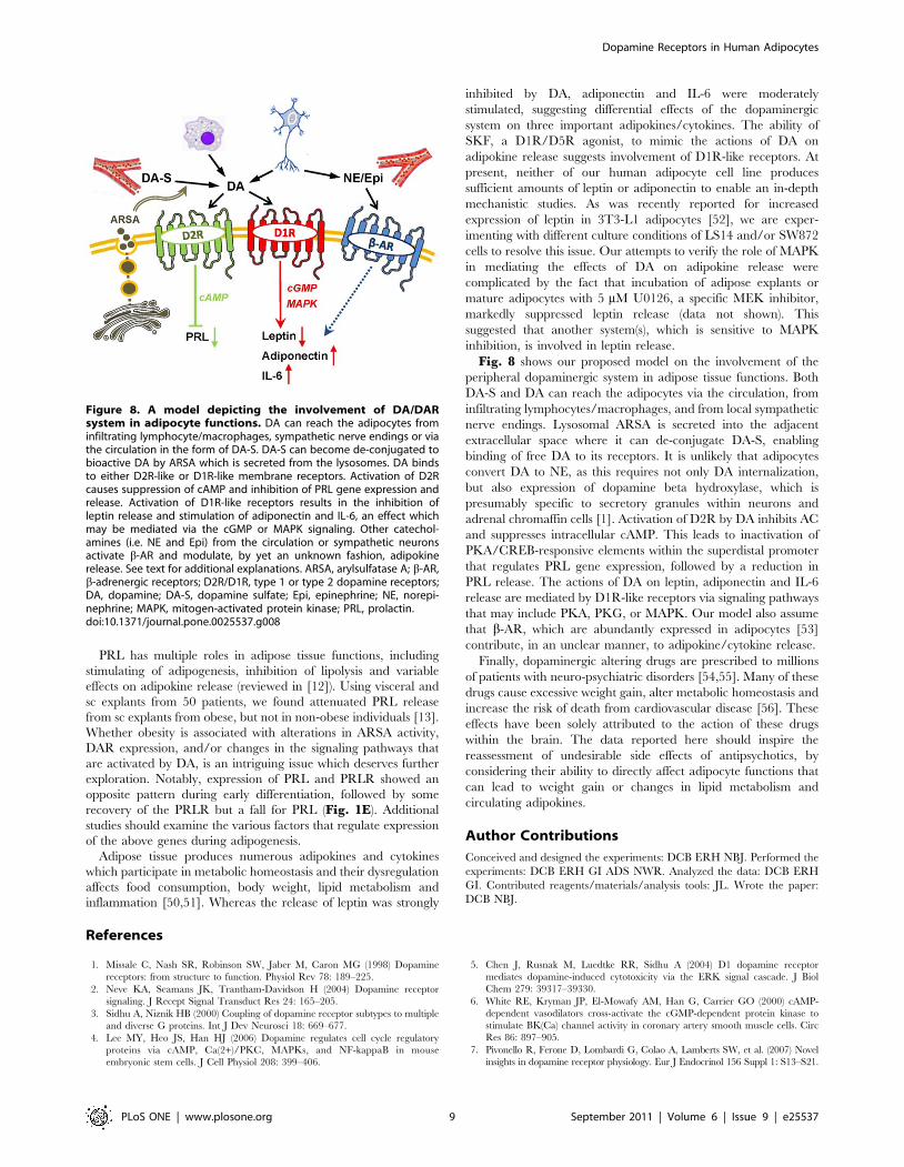
Fig. 8 shows our proposed model on the involvement of the
peripheral dopaminergic system in adipose tissue functions. Both
DA-S and DA can reach the adipocytes via the circulation, from
infiltrating lymphocytes/macrophages, and from local sympathetic
nerve endings. Lysosomal ARSA is secreted into the adjacent
extracellular space where it can de-conjugate DA-S, enabling
binding of free DA to its receptors. It is unlikely that adipocytes
convert DA to NE, as this requires not only DA internalization,
but also expression of dopamine beta hydroxylase, which is
presumably specific to secretory granules within neurons and
adrenal chromaffin cells [1]. Activation of D2R by DA inhibits AC
and suppresses intracellular cAMP. This leads to inactivation of
PKA/CREB-responsive elements within the superdistal promoter
that regulates PRL gene expression, followed by a reduction in
PRL release. The actions of DA on leptin, adiponectin and IL-6
release are mediated by D1R-like receptors via signaling pathways
that may include PKA, PKG, or MAPK. Our model also assume
that b-AR, which are abundantly expressed in adipocytes [53]
contribute, in an unclear manner, to adipokine/cytokine release.
Finally, dopaminergic altering drugs are prescribed to millions
of patients with neuro-psychiatric disorders [54,55]. Many of these
drugs cause excessive weight gain, alter metabolic homeostasis and
increase the risk of death from cardiovascular disease [56]. These
effects have been solely attributed to the action of these drugs
within the brain. The data reported here should inspire the
reassessment of undesirable side effects of antipsychotics, by
considering their ability to directly affect adipocyte functions that
can lead to weight gain or changes in lipid metabolism and
circulating adipokines.
Author Contributions
Conceived and designed the experiments: DCB ERH NBJ. Performed the
experiments: DCB ERH GI ADS NWR. Analyzed the data: DCB ERH
GI. Contributed reagents/materials/analysis tools: JL. Wrote the paper:
DCB NBJ.
References
1. Missale C, Nash SR, Robinson SW, Jaber M, Caron MG (1998) Dopamine
receptors: from structure to function. Physiol Rev 78: 189–225.
2. Neve KA, Seamans JK, Trantham-Davidson H (2004) Dopamine receptor
signaling. J Recept Signal Transduct Res 24: 165–205.
3. Sidhu A, Niznik HB (2000) Coupling of dopamine receptor subtypes to multiple
and diverse G proteins. Int J Dev Neurosci 18: 669–677.
4. Lee MY, Heo JS, Han HJ (2006) Dopamine regulates cell cycle regulatory
proteins via cAMP, Ca(2+)/PKC, MAPKs, and NF-kappaB in mouse
embryonic stem cells. J Cell Physiol 208: 399–406.
5. Chen J, Rusnak M, Luedtke RR, Sidhu A (2004) D1 dopamine receptor
mediates dopamine-induced cytotoxicity via the ERK signal cascade. J Biol
Chem 279: 39317–39330.
6. White RE, Kryman JP, El-Mowafy AM, Han G, Carrier GO (2000) cAMP-
dependent vasodilators cross-activate the cGMP-dependent protein kinase to
stimulate BK(Ca) channel activity in coronary artery smooth muscle cells. Circ
Res 86: 897–905.
7. Pivonello R, Ferone D, Lombardi G, Colao A, Lamberts SW, et al. (2007) Novel
insights in dopamine receptor physiology. Eur J Endocrinol 156 Suppl 1: S13–S21.
Figure 8. A model depicting the involvement of DA/DARsystem in adipocyte functions. DA can reach the adipocytes frominfiltrating lymphocyte/macrophages, sympathetic nerve endings or viathe circulation in the form of DA-S. DA-S can become de-conjugated tobioactive DA by ARSA which is secreted from the lysosomes. DA bindsto either D2R-like or D1R-like membrane receptors. Activation of D2Rcauses suppression of cAMP and inhibition of PRL gene expression andrelease. Activation of D1R-like receptors results in the inhibition ofleptin release and stimulation of adiponectin and IL-6, an effect whichmay be mediated via the cGMP or MAPK signaling. Other catechol-amines (i.e. NE and Epi) from the circulation or sympathetic neuronsactivate b-AR and modulate, by yet an unknown fashion, adipokinerelease. See text for additional explanations. ARSA, arylsulfatase A; b-AR,b-adrenergic receptors; D2R/D1R, type 1 or type 2 dopamine receptors;DA, dopamine; DA-S, dopamine sulfate; Epi, epinephrine; NE, norepi-nephrine; MAPK, mitogen-activated protein kinase; PRL, prolactin.doi:10.1371/journal.pone.0025537.g008
Dopamine Receptors in Human Adipocytes
PLoS ONE | www.plosone.org 9 September 2011 | Volume 6 | Issue 9 | e25537
8. Amenta F, Ricci A, Tayebati SK, Zaccheo D (2002) The peripheral
dopaminergic system: morphological analysis, functional and clinical applica-tions. Ital J Anat Embryol 107: 145–167.
9. Ben-Jonathan N, Hnasko R (2001) Dopamine as a prolactin (PRL) inhibitor.
Endocr Rev 22: 724–763.10. Ben-Jonathan N, Mershon JL, Allen DL, Steinmetz RW (1996) Extrapituitary
prolactin: distribution, regulation, functions, and clinical aspects. Endocr Rev17: 639–669.
11. Zinger M, McFarland M, Ben-Jonathan N (2003) Prolactin expression and
secretion by human breast glandular and adipose tissue explants. J ClinEndocrinol Metab 88: 689–696.
12. Brandebourg TD, Hugo ER, Ben-Jonathan N (2007) Adipocyte prolactin:regulation of release and putative functions. Diabetes,Obesity and Metabolism 9:
364–377.13. Hugo ER, Borcherding DC, Gersin KS, Loftus J, Ben-Jonathan N (2008)
Prolactin release by adipose explants, primary adipocytes, and LS14 adipocytes.
J Clin Endocrinol Metab 93: 4006–4012.14. Ben-Jonathan N (1985) Dopamine: a prolactin-inhibiting hormone. Endocr Rev
6: 564–589.15. Nisoli E, Tonello C, Memo M, Carruba MO (1992) Biochemical and functional
identification of a novel dopamine receptor subtype in rat brown adipose tissue.
Its role in modulating sympathetic stimulation-induced thermogenesis.J Pharmacol Exp Ther 263: 823–829.
16. Goldstein DS, Eisenhofer G, Kopin IJ (2003) Sources and significance of plasmalevels of catechols and their metabolites in humans. J Pharmacol Exp Ther 305:
800–811.17. Goldstein DS, Swoboda KJ, Miles JM, Coppack SW, Aneman A, et al. (1999)
Sources and physiological significance of plasma dopamine sulfate. J Clin
Endocrinol Metab 84: 2523–2531.18. Eisenhofer G, Coughtrie MW, Goldstein DS (1999) Dopamine sulphate: an
enigma resolved. Clin Exp Pharmacol Physiol Suppl 26: S41–S53.19. Eldrup E (2004) Significance and origin of DOPA, DOPAC, and dopamine-
sulphate in plasma, tissues and cerebrospinal fluid. Dan Med Bull 51: 34–62.
20. Strobel G, Werle E, Weicker H (1990) Isomer specific kinetics of dopamine beta-hydroxylase and arylsulfatase towards catecholamine sulfates. Biochem Int 20:
343–351.21. Ghosh D (2007) Human sulfatases: a structural perspective to catalysis. Cell Mol
Life Sci 64: 2013–2022.22. Ahima RS (2006) Adipose tissue as an endocrine organ. Obesity (Silver Spring)
14(Suppl 5): 242S–249S.
23. Shetty S, Kusminski CM, Scherer PE (2009) Adiponectin in health and disease:evaluation of adiponectin-targeted drug development strategies. Trends
Pharmacol Sci 30: 234–239.24. Fain JN (2006) Release of interleukins and other inflammatory cytokines by
human adipose tissue is enhanced in obesity and primarily due to the nonfat
cells. Vitam Horm 74: 443–477.25. Mohamed-Ali V, Flower L, Sethi J, Hotamisligil G, Gray R, et al. (2001) beta-
Adrenergic regulation of IL-6 release from adipose tissue: in vivo and in vitrostudies. J Clin Endocrinol Metab 86: 5864–5869.
26. Goossens GH, Jocken JW, van Baak MA, Jansen EH, Saris WH, et al. (2008)Short-term beta-adrenergic regulation of leptin, adiponectin and interleukin-6
secretion in vivo in lean and obese subjects. Diabetes Obes Metab 10:
1029–1038.27. Path G, Bornstein SR, Gurniak M, Chrousos GP, Scherbaum WA, et al. (2001)
Human breast adipocytes express interleukin-6 (IL-6) and its receptor system:increased IL-6 production by beta-adrenergic activation and effects of IL-6 on
adipocyte function. J Clin Endocrinol Metab 86: 2281–2288.
28. Fain JN, Cowan GS, Jr., Buffington C, Li J, Pouncey L, et al. (2000) Synergismbetween insulin and low concentrations of isoproterenol in the stimulation of
leptin release by cultured human adipose tissue. Metabolism 49: 804–809.29. Than A, Ye F, Xue R, Ong JW, Poh CL, et al. (2011) The crosstalks between
adipokines and catecholamines. Mol Cell Endocrinol 332: 261–270.
30. Hugo ER, Brandebourg TD, Comstock CE, Gersin KS, Sussman JJ, et al. (2006)LS14: a novel human adipocyte cell line that produces prolactin. Endocrinology
147: 306–313.31. Basu S, Sarkar C, Chakroborty D, Nagy J, Mitra RB, et al. (2004) Ablation of
peripheral dopaminergic nerves stimulates malignant tumor growth by inducingvascular permeability factor/vascular endothelial growth factor-mediated
angiogenesis. Cancer Res 64: 5551–5555.
32. Mignini F, Bronzetti E, Felici L, Ricci A, Sabbatini M, et al. (2000) Dopaminereceptor immunohistochemistry in the rat choroid plexus. J Auton Pharmacol
20: 325–332.
33. Nair VD, Sealfon SC (2003) Agonist-specific transactivation of phosphoinositide
3-kinase signaling pathway mediated by the dopamine D2 receptor. J Biol Chem278: 47053–47061.
34. Chang PL, Rosa NE, Davidson RG (1981) Differential assay of arylsulfatase A
and B activities: a sensitive method for cultured human cells. Anal Biochem 117:382–389.
35. Lapensee CR, Hugo ER, Ben-Jonathan N (2008) Insulin stimulates interleukin-6expression and release in LS14 human adipocytes through multiple signaling
pathways. Endocrinology 149: 5415–5422.
36. Maayan L, Vakhrusheva J, Correll CU (2010) Effectiveness of medications used
to attenuate antipsychotic-related weight gain and metabolic abnormalities: a
systematic review and meta-analysis. Neuropsychopharmacology 35:1520–1530.
37. Ricci A, Bronzetti E, Mignini F, Tayebati SK, Zaccheo D, et al. (1999)Dopamine D1-like receptor subtypes in human peripheral blood lymphocytes.
J Neuroimmunol 96: 234–240.
38. Ricci A, Bronzetti E, Felici L, Tayebati SK, Amenta F (1997) Dopamine D4
receptor in human peripheral blood lymphocytes: a radioligand binding assay
study. Neurosci Lett 229: 130–134.
39. Guest SJ, Hadcock JR, Watkins DC, Malbon CC (1990) Beta 1- and beta 2-
adrenergic receptor expression in differentiating 3T3-L1 cells. Independentregulation at the level of mRNA. J Biol Chem 265: 5370–5375.
40. Dixon TM, Daniel KW, Farmer SR, Collins S (2001) CCAAT/enhancer-
binding protein alpha is required for transcription of the beta 3-adrenergicreceptor gene during adipogenesis. J Biol Chem 276: 722–728.
41. Strott CA (2002) Sulfonation and molecular action. Endocr Rev 23: 703–732.
42. Dajani R, Cleasby A, Neu M, Wonacott AJ, Jhoti H, et al. (1999) X-ray crystal
structure of human dopamine sulfotransferase, SULT1A3. Molecular modelingand quantitative structure-activity relationship analysis demonstrate a molecular
basis for sulfotransferase substrate specificity. J Biol Chem 274: 37862–37868.
43. Valle LD, Toffolo V, Nardi A, Fiore C, Bernante P, et al. (2006) Tissue-specifictranscriptional initiation and activity of steroid sulfatase complementing
dehydroepiandrosterone sulfate uptake and intracrine steroid activations inhuman adipose tissue. J Endocrinol 190: 129–139.
44. Matzner U, Breiden B, Schwarzmann G, Yaghootfam A, Fluharty AL, et al.(2009) Saposin B-dependent reconstitution of arylsulfatase A activity in vitro and
in cell culture models of metachromatic leukodystrophy. J Biol Chem 284:
9372–9381.
45. Biffi A, Cesani M, Fumagalli F, Del CU, Baldoli C, et al. (2008) Metachromatic
leukodystrophy - mutation analysis provides further evidence of genotype-phenotype correlation. Clin Genet 74: 349–357.
46. Gieselmann V (2008) Metachromatic leukodystrophy: genetics, pathogenesis and
therapeutic options. Acta Paediatr Suppl 97: 15–21.
47. Regis S, Corsolini F, Stroppiano M, Cusano R, Filocamo M (2002) Contribution
of arylsulfatase A mutations located on the same allele to enzyme activityreduction and metachromatic leukodystrophy severity. Hum Genet 110:
351–355.
48. Ben-Jonathan N, Munsick RA (1980) Dopamine and prolactin in human
pregnancy. J Clin Endocrinol Metab 51: 1019–1025.
49. Golander A, Barrett J, Hurley T, Barry S, Handwerger S (1979) Failure ofbromocriptine, dopamine, and thyrotropin-releasing hormone to affect prolactin
secretion by human decidual tissue in vitro. J Clin Endocrinol Metab 49:787–789.
50. Rondinone CM (2006) Adipocyte-derived hormones, cytokines, and mediators.Endocrine 29: 81–90.
51. Maury E, Brichard SM (2010) Adipokine dysregulation, adipose tissue
inflammation and metabolic syndrome. Mol Cell Endocrinol 314: 1–16.
52. Zeigerer A, Rodeheffer MS, McGraw TE, Friedman JM (2008) Insulin regulates
leptin secretion from 3T3-L1 adipocytes by a PI 3 kinase independentmechanism. Exp Cell Res 314: 2249–2256.
53. Collins S, Surwit RS (2001) The beta-adrenergic receptors and the control of
adipose tissue metabolism and thermogenesis. Recent Prog Horm Res 56:309–328.
54. Richtand NM, Welge JA, Logue AD, Keck PE, Jr., Strakowski SM, et al. (2007)Dopamine and serotonin receptor binding and antipsychotic efficacy. Neuro-
psychopharmacology 32: 1715–1726.
55. Nasrallah HA (2008) Atypical antipsychotic-induced metabolic side effects:
insights from receptor-binding profiles. Mol Psychiatry 13: 27–35.
56. Zimmermann U, Kraus T, Himmerich H, Schuld A, Pollmacher T (2003)Epidemiology, implications and mechanisms underlying drug-induced weight
gain in psychiatric patients. J Psychiatr Res 37: 193–220.
Dopamine Receptors in Human Adipocytes
PLoS ONE | www.plosone.org 10 September 2011 | Volume 6 | Issue 9 | e25537
Related Documents