Iron deficiency decreases dopamine D 1 and D 2 receptors in rat brain By: Keith M. Erikson , Byron C. Jones, Ellen J. Hess, Qian Zhang, and John L. Beard Erikson, K.M., Jones, B.C., Hess, E.J. and Beard, J.L. (2001) Iron Deficiency Decreases dopamine D1 and D2 receptors in rat brain. Pharmacology Biochemistry and Behavior. 69(3-4):409-18. Made available courtesy of Elsevier: http://www.elsevier.com/ *** Note: Figures may be missing from this format of the document Abstract: Iron deficiency (ID) in early life is known to alter neurological development and functioning, but data regarding specific effects on dopamine biology are lacking. The objective of this study was to determine the extent of functional alterations in dopamine receptors in two dopaminergic tracts in young, growing, iron-deficient rats. Forty male and 40 female weanling Sprague–Dawley rats were fed either an iron-deficient (ID) diet or control (CN) diet for 6 weeks. ID decreased densities of D 1 and D 2 receptors in the caudate–putamen and decreased D 2 receptor densities in the nucleus accumbens. There were no apparent effects of ID on the affinities for the ligands in either receptor in several brain regions. In situ hybridization studies for both dopamine receptors revealed no significant effect of ID on mRNA expression for either receptor. Iron-deficient rats had a significantly higher ED 50 for raclopride-induced hypolocomotion in male and female rats compared to control rats of each sex. The loss of iron in the striatum due to dietary ID was significantly correlated with the decrease in D 2 receptor density; however, this relationship was not apparent in other brain regions. These experiments thus demonstrate abnormal dopamine receptor density and functioning in several brain regions that are related to brain regional iron loss. Importantly, the impact of ID on dopamine was more pronounced in males than females, demonstrating sex-related different sensitivities to nutrient deprivation. Keywords: Iron deficiency; Rats; Dopamine receptors; Sex; Brain iron Article: 1. Introduction Iron deficiency (ID) is one of the most common nutritional disorders in the world, affecting nearly 5 billion people (ACC/SCN, 1992). Symptoms of ID include lethargy, lack of concentration and decreased cognitive and attentional processes (Lozoff and Brittenham, 1986; Pollit, 1993; Walter, 1993). There is evidence showing that iron does play a role in neurobiological processes (Beard et al., 1993; Rocangliolo et al., 1998; Youdim et al., 1989); however, the neurobiological mechanisms behind iron’s role in behavior and cognition remain unknown. Whether ID early in life has persistent neurobehavioral effects is the topic of research for several investigators. For example, one question deals with whether deficits in cognition resulting from ID in infancy can be reversed by an iron-adequate diet later in life. In the rat, postweaning ID produces a decrease in brain iron content that is reversible with iron repletion (Chen et al., 1995a; Erikson et al., 1997; Pinero et al., 2000). Furthermore, ID-related decreased brain iron content has been linked to many neurological alterations, including hypomyelination (Larkin and Rao, 1990), delayed neuromaturation (Rocangliolo et al., 1998) and altered dopaminergic functioning (Nelson et al., 1997). The effects of ID on dopamine function include decreased D2 receptor density in caudate–putamen (Youdim et al., 1983) and increased extracellular dopamine levels (Beard et al., 1994; Chen et al., 1995b; Nelson et al., 1997). Other researchers have restricted the examination of ID to the undifferentiated striatum; however, we recently observed differential effects of ID between the dorsal striatum (containing the caudate and putamen) and the ventral striatum (containing the nucleus accumbens) (Erikson et al., 2000). A recent study from our laboratory demonstrated a significant decrease in DA transporter density in several brain regions with an associated altered sensitivity to cocaine (Erikson et al., 2000).

Welcome message from author
This document is posted to help you gain knowledge. Please leave a comment to let me know what you think about it! Share it to your friends and learn new things together.
Transcript
Iron deficiency decreases dopamine D1 and D2 receptors in rat brain
By: Keith M. Erikson, Byron C. Jones, Ellen J. Hess, Qian Zhang, and John L. Beard
Erikson, K.M., Jones, B.C., Hess, E.J. and Beard, J.L. (2001) Iron Deficiency Decreases dopamine D1 and D2
receptors in rat brain. Pharmacology Biochemistry and Behavior. 69(3-4):409-18.
Made available courtesy of Elsevier: http://www.elsevier.com/
*** Note: Figures may be missing from this format of the document
Abstract:
Iron deficiency (ID) in early life is known to alter neurological development and functioning, but data regarding
specific effects on dopamine biology are lacking. The objective of this study was to determine the extent of
functional alterations in dopamine receptors in two dopaminergic tracts in young, growing, iron-deficient rats.
Forty male and 40 female weanling Sprague–Dawley rats were fed either an iron-deficient (ID) diet or control
(CN) diet for 6 weeks. ID decreased densities of D1 and D2 receptors in the caudate–putamen and decreased D2
receptor densities in the nucleus accumbens. There were no apparent effects of ID on the affinities for the
ligands in either receptor in several brain regions. In situ hybridization studies for both dopamine receptors
revealed no significant effect of ID on mRNA expression for either receptor. Iron-deficient rats had a
significantly higher ED50 for raclopride-induced hypolocomotion in male and female rats compared to control
rats of each sex. The loss of iron in the striatum due to dietary ID was significantly correlated with the decrease
in D2 receptor density; however, this relationship was not apparent in other brain regions. These experiments
thus demonstrate abnormal dopamine receptor density and functioning in several brain regions that are related
to brain regional iron loss. Importantly, the impact of ID on dopamine was more pronounced in males than
females, demonstrating sex-related different sensitivities to nutrient deprivation.
Keywords: Iron deficiency; Rats; Dopamine receptors; Sex; Brain iron
Article:
1. Introduction
Iron deficiency (ID) is one of the most common nutritional disorders in the world, affecting nearly 5 billion
people (ACC/SCN, 1992). Symptoms of ID include lethargy, lack of concentration and decreased cognitive and
attentional processes (Lozoff and Brittenham, 1986; Pollit, 1993; Walter, 1993). There is evidence showing that
iron does play a role in neurobiological processes (Beard et al., 1993; Rocangliolo et al., 1998; Youdim et al.,
1989); however, the neurobiological mechanisms behind iron’s role in behavior and cognition remain unknown.
Whether ID early in life has persistent neurobehavioral effects is the topic of research for several investigators.
For example, one question deals with whether deficits in cognition resulting from ID in infancy can be reversed
by an iron-adequate diet later in life.
In the rat, postweaning ID produces a decrease in brain iron content that is reversible with iron repletion (Chen
et al., 1995a; Erikson et al., 1997; Pinero et al., 2000). Furthermore, ID-related decreased brain iron content has
been linked to many neurological alterations, including hypomyelination (Larkin and Rao, 1990), delayed
neuromaturation (Rocangliolo et al., 1998) and altered dopaminergic functioning (Nelson et al., 1997). The
effects of ID on dopamine function include decreased D2 receptor density in caudate–putamen (Youdim et al.,
1983) and increased extracellular dopamine levels (Beard et al., 1994; Chen et al., 1995b; Nelson et al., 1997).
Other researchers have restricted the examination of ID to the undifferentiated striatum; however, we recently
observed differential effects of ID between the dorsal striatum (containing the caudate and putamen) and the
ventral striatum (containing the nucleus accumbens) (Erikson et al., 2000). A recent study from our laboratory
demonstrated a significant decrease in DA transporter density in several brain regions with an associated altered
sensitivity to cocaine (Erikson et al., 2000).
Other neurotransmitter systems may be involved as well because iron is known to be involved in the synthesis
and catabolism of other monoamines. For example, ID decreases the density of serotonin transporters in mice
(Morse et al., 1999) and serotonin levels in rat brain (Youdim et al., 1989). Also, noradrenergic function is
altered (Beard et al., 1994; Chen et al., 1995b; Youdim et al., 1989). Behavioral consequences include altered
locomotor and exploratory activity (Glover and Jacobs, 1972; Hunt et al., 1994; unpublished observations from
our laboratory). It is important to note that the alterations in dopamine function result from decreased brain iron
content and not anemia per se (Ashkenazi et al., 1982; Nelson et al., 1997; Youdim et al., 1989) and are
reversible with iron therapy if the ID occurs after weaning (Nelson et al., 1997; Youdim et al., 1981).
To date, five dopamine receptors have been identified and subdivided into two groups, D2-like (D2, D3 and D4)
and D1-like (D1 and D5), based on pharmacological and biochemical characteristics (reviewed in Vallone et al.,
2000). Previous studies examining alterations in striatal dopamine function related to ID demonstrated that D2
receptor density was significantly decreased, whereas there was no effect on D1 receptor density (Youdim et al.,
1979, 1989) or on tissue concentrations of dopamine. Our own reports (Beard et al., 1994; Chen et al., 1995b;
Nelson et al., 1997) of elevations in extracellular DA in striatum of iron-deficient rats suggest a high likelihood
of changes in D1 and D2 receptor densities, although there is no clear evidence for changes in D1 receptor
density or affinity in the extant literature. We propose that a careful examination of the impact of ID in several
brain regions and on multiple dopamine receptor types is crucial to a more complete understanding of the
impact of ID.
Our overall hypothesis was that ID affects dopaminergic brain regions by decreasing both D2 and D1 receptor
densities in terminal fields of dopaminergic tracts. We also sought to elucidate the relationship between ID-
altered dopamine receptor expression and dopamine-related behaviors.
2. Materials and methods
2. 1. Animals
Male and female 21-day-old Sprague–Dawley rats (purchased from Harlan Sprague–Dawley, Indianapolis, IN)
were randomly divided into two dietary treatment groups: control (CN; 35 mg Fe/kg diet, n = 20–25 males and
females) and iron-deficient (ID; 3 mg Fe/kg diet, n = 20– 25 males and females). The design of the experiments
was established in a way as to test the effects of dietary treatment, sex of the subjects and potential interactions
of these two independent variables on dopamine function and behavior. The animals were fed iron-sufficient
and iron-deficient diets, as previously described (Erikson et al., 1997; Pinero et al., 2000), and had free access to
food and water 24 h/day. In the colony room, the lights were turned off between 1800 and 0600 h and the
temperature was maintained at 25 ± 1°C. As will be seen in the Results section, growth of iron-deficient animals
was significantly slower than that of control, but this was not due to anorexia (Beard et al., 1995). To determine
if being ―underweight‖ had a significant impact on our dependent variables of interest, we utilized frozen brains
from a nearly identical cohort of animals except for the inclusion of a group of controls who were food-
restricted (pair-fed) to match their growth rate to those of the controls.
The behavioral studies were performed between 0900 1200 h or approximately 3–6 h after the onset of the light
cycle. Animals continued to have free access to food and water the night before behavior testing. The
behavioral testing apparatus was located in a room adjacent to animal holding rooms. The Pennsylvania State
University Animal Care and Use Committee approved all of the animal procedures.
2.2. Effect of raclopride on locomotor activity
After 4 weeks of dietary treatment, the animals were tested for the behavioral effects of a potent D2 receptor
antagonist, raclopride. Locomotor activity was measured using a Digiscan Animal Activity Monitor, model
RXYZCM (Omnitech Electronics, Columbus, OH). It consisted of a set of four 40 x 40 x 30.5 cm Plexiglas
boxes with vertical and horizontal infrared sensors. The flooring was an elevated acrylic platform with equally
spaced holes (4 x 4 x 1.5 cm in diameter). A 2-day behavioral testing protocol was implemented. On Day 1, five
rats from each group (diet/sex) were injected with saline (1 ml/kg bw) 15 min prior to testing. Rats were then
injected with a dose of cocaine–HCl (Sigma Chemicals, Natick, MA) shown previously to increase their

locomotion by 50% (Larkin and Rao, 1990) and immediately placed in the center of the activity monitor. This
dose of cocaine was used to increase locomotor activity of iron-deficient rats to a level sufficient to allow us to
determine the impact of raclopride. The ED50 cocaine doses used were established in previous studies: 7.1 and
12 mg/kg bw, female and male CN, respectively; 11.2 and 17 mg/kg bw, female and male ID, respectively
(Erikson et al., 2000). The animals were left in the activity monitor for 30 min. The index of locomotor activity
was total distance traveled over 30 min in centimeters. On Day 2, the rats were injected with one of four doses,
0.1, 0.25, 0.5 or 1.0 mg/kg bw of raclopride tartrate (RBI, Natick, MA), 15 min prior to testing. The rats were
then injected with their respective ED50 cocaine dose and locomotor activity was measured as on the preceding
day. The ED50 for raclopride to decrease total distance (by 50%) was calculated using a Log–Logit
transformation of the data followed by regression analysis. Other behaviors recorded by the Omnitech activity
monitors included frequency nose-pokes in the holeboard (a putative measure of exploration), frequency of
repeated movements (related to stereotypy) and time spent in the center of the apparatus (a putative measure of
temerity). Upon completion of the behavior testing, the rats were killed by decapitation, livers removed for non-
heme iron determination (Erikson et al., 1997) and brains rapidly taken for iron analysis and ligand binding
assays. The hematological status of each rat was determined in the fresh blood drained from the trunk by
published methods (Chen et al., 1995a; Erikson et al., 1997).
2.3. Dopamine D1 receptor density determinations
Brains from rats were dissected on an ice-cold aluminum block into four regions: prefrontal cortex (PFC),
caudate– putamen (CP), nucleus accumbens (NA) and ventral mid-brain (VMB — consisting of the substantia
nigra and the ventral tegmedullary region) using well-defined landmarks (Paxinos and Watson, 1986) and
regions frozen at -80°C. Membrane fractions were prepared from frozen tissue and assays conducted using
published methods with few modifications (Morse et al., 1999). Membrane fractions were washed several times
with isolation buffer prior to their utilization in binding assays. Binding reactions were con-ducted in microtiter
plates (CoStar brand, Corning 9017) containing: tissue homogenate (25 μl), Tris buffer (50 mM Tris, 120 mM
NaCl), pH 7.4 (25 μl), 10 μl3H-SCH23390 (8 nM) and 0.1 mM Butaclamol (RBI), final concentration, for non-
specific binding. Saturation binding was performed using a nano- to micromolar range of SCH23390. Samples
were incubated for 1 h at room temperature, filtered onto GF/ B Whatman filters and the filters were placed in
scintillation vials containing 5 ml scintillation cocktail. Samples were counted on Beckman liquid scintillation
counter (LS 3801; Beckman Instruments, Irvine, CA) 24 h later. Protein concentrations were determined using
the micro-Lowry assay (P5656; Sigma).
2.4. Dopamine D2-like receptor density determinations
Radioligand binding was performed on membrane fractions prepared identically to the above procedure and
according to published protocols (Morse et al., 1999) with the following exceptions: Tris buffer (50 mM Tris,
120 mM NaCl), pH 7.8, was used and 10 μl 125
I-epidepride (D2 antagonist, 4 nM) added to all tubes. As above,
0.1 mM Butaclamol (RBI) was used to determine non-specific binding. We did not differentiate between
members of the D2 ―family‖ by using other more specific blocking agents like 7- hydroxy-N,N-di(I-propyl)-2-
aminotetralin (7-OH-DPAT) in these particular studies. Scatchard plot analyses were per-formed using a 106
range of epidipride concentrations prior to the determinations of receptor density.
2.5. Iron analysis
Total iron content of brain region homogenates was determined according to our standard laboratory method
using acid digestion and analysis with atomic absorption spectrophotometry (Erikson et al., 1997; Pinero et al.,
2000).
2.6. Analysis of transcription for D1 and D2 receptors
Six additional iron-deficient and control rats (three of each dietary treatment), treated as were the others, were
killed at PND 63 in order to conduct in situ hybridization histochemistry. Brains were rapidly removed, quickly
frozen in isopentane and dry ice and frozen at -80°C until histological preparation (Campbell and Hess, 1999).
Twenty-micrometer sagittal sections were cut using a cryostat and thaw-mounted on Superfrost Plus glass slides
(Fisher, Pittsburgh, PA). After drying, the slide-mounted sections were stored at -70°C. The assay was

performed in such a way as to always have at least one control brain slice on the slide with an iron-deficient
brain slice. Comparisons were always made between slices on the same slide to reduce variability.
The cDNA probe specific to the D1 dopamine receptor in pGEM-3Z was a generous gift from Dr. Marc Caron
(Duke University, NC). The clone for the D2 dopamine receptor was obtained from the IMAGE Consortium
(LLNL) cDNA clone ID no. 312056 in pCMV-SPORT. Hybridization assays were conducted as previously
described (Campbell and Hess, 1999).
Analysis of mean grain density was performed using the public domain NIH Image program (National
Technical Information Service, Springfield, VA). For each region, mean grain density was measured as square
pixels remaining after the images were normalized by adjusting the threshold in NIH Image. For single cell
quantitation of D2 dopamine receptor mRNA in the substantia nigra, grain density over individual cells was
counted in inverted darkfield views captured at x 20 magnification. At least 96 cells, randomly identified with a
random number algorithm, were quantitated per animal for each probe. Slices from the same regions for five of
the six animals were examined. To quantitate D1 dopamine receptor and D2 dopamine mRNA expression in the
striatum, images were captured at x 3.0 directly from autoradiographs. The density of at least 64 measurements
was assessed per animal for each probe. These densities were then compared between iron-deficient and control
slices on the same slide and data expressed as a percentage of control density.
2.7. Statistical analysis
The data were analyzed using the SAS system for Windows v. 6.12 statistical analysis package (SAS, Cary,
NC). Analysis of variance (ANOVA) with repeated-measures factors (brain regions) and between-groups
factors (Sex and Diet) was used to test for main effects of and interactions between dietary treatments and sex
for ligand binding data. Two-way ANOVA was used to test effects of dietary treatment and sex on
hematological parameters, liver non-heme iron and behavioral data. Magnitude of effect for diet and sex and
their interactions was evaluated using estimated ω2 (Hinkle et al., 1998). The Tukey HSD test was used for
multiple comparisons. Alpha level for the analyses was set at P <.05.
3. Results
3. 1. ED50 raclopride experiment
Both males and females fed the iron-deficient diets from weaning were severely iron-deficient and anemic at the
time of testing (Table 1). The females were significantly smaller than males, but there was no significant
difference in severity of anemia between sexes (Table 1). Under saline treatment, iron-deficient animals evinced
less distance traveled than control rats. Total number of repeated movements [F(1,40)=102.78, ω2 = 0.523] and
center time [F(1,40)=16.21, ω2 = 0.159] were also significantly affected by ID anemia (Table 2). Increasing
doses of raclopride resulted in the expected decreased locomotor activity in both sexes (Fig. 1). Moreover, iron
status had a profound affect on the ED50 for raclopride as did the sex of the subjects (Table 2) [ F(1,40) = 16.41
and F(1,40)=6.11]. ID affected females more dramatically than males (ED50=0.70 and 0.41 mg/kg for ID

females and ID males, respectively). There was no effect of sex on the ED50 for raclopride in the control rats
(0.20 mg/kg for both sexes).
3.2. 3H-SCH23390 and
125I-epidepride ligand binding
There was no effect of ID on the affinity of either ligand (Kd) to D1 - or D2-like receptors in the caudate–
putamen as determined by Scatchard plot analyses (D2 Kd = 0. 162 ± 0.02 nM for control caudate, Kd = 0.164 ±
0.022 nM for iron-deficient caudate; SCH23390 D1 Kd=1.52±0.38 nM for control, Kd = 1.30±0.21 for iron-
deficient caudate) We performed D2 Kd determinations in pooled samples from control animals using epidipride
in prefrontal cortex (0.98±0.028 nM), nucleus accumbens (1.26 ± 0.019 nM) and ventral mid-brain (1.65 ±
0.011 nM), but did not have sufficient material to verify that there were no significant effects of ID on D2-like
receptor affinity in those regions. D1 Kd in ventral midbrain was also unaffected by brain ID (Kd = 2.20 nM in
control compared to Kd = 1.89 nM in ID).
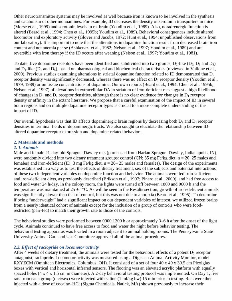
For both ligands, there were variations in receptor densities by brain region [ F(3,64) = 327.2, P <.00 1; F(3,64)
= 213.7, P<.001 (D1 and D2, respectively)] (Fig. 2). There was also a main effect of dietary treatment
[F(1,32)=5.3, P ≤ .05 (estimated ω2 =0.191)] with ID decreasing D2 receptor density (Fig. 2). ID caused a
significant decrease in both D1 and D2/D3 densities in CP [F(1,16)=5.74, P<.05 (estimated ω2=0.048;
F(1,16)=4.55, P < .05 (estimated ω2 = 0. 110)], respectively, and a significant increase in D1 and D2 densities in
prefrontal cortex [F(1,16) = 4.7, P ≤ .05 (estimated ω2 = 0.039, 0.0328); F(1,16)=5.44, P ≤ .05 (estimated ω
2 =
0.157)], respectively The nucleus accumbens and ventral midbrain were not significantly affected. Females had
a significantly higher overall D2 receptor density [F(1,32)=13.4, P ≤ .01 (estimated ω2 = 0.546)] (Fig. 2b), but
not D1 density, compared to male rats. A significant interaction between diet and sex resulted in iron-deficient
males, but not females [F(1,16)=7.6, P < .05 (estimated ω2 = 0.286)], evincing decreased D2-like receptor
density. There was no such interaction for D1 receptor density. Controlling for body weight did not remove the
significant effects of either iron status or sex of the subjects.
Because the growth of the iron-deficient rats was significantly less than that of controls, we retrospectively
examined D1 and D2 receptor density in brains of pair-fed control rats, ad libitum-fed control rats and iron-
deficient rats that had been raised similarly to the rats examined in the current report. In this much smaller
cohort of rats (n = 5-6 per treatment group, males only), we observed no significant differences in caudate–
putamen D2 receptor densities of pair-fed controls compared to ad libitum controls (1540±123 fmol/mg pair-fed
controls compared to 1374 ± 89 fmol/mg in ad libitum controls), with both control groups’ densities
substantially higher than in iron-deficient caudate–putamen (1060±107 fmol/mg). Insufficient samples and
inappropriate storage procedures precluded D1 or D2 analyses in other brain regions.
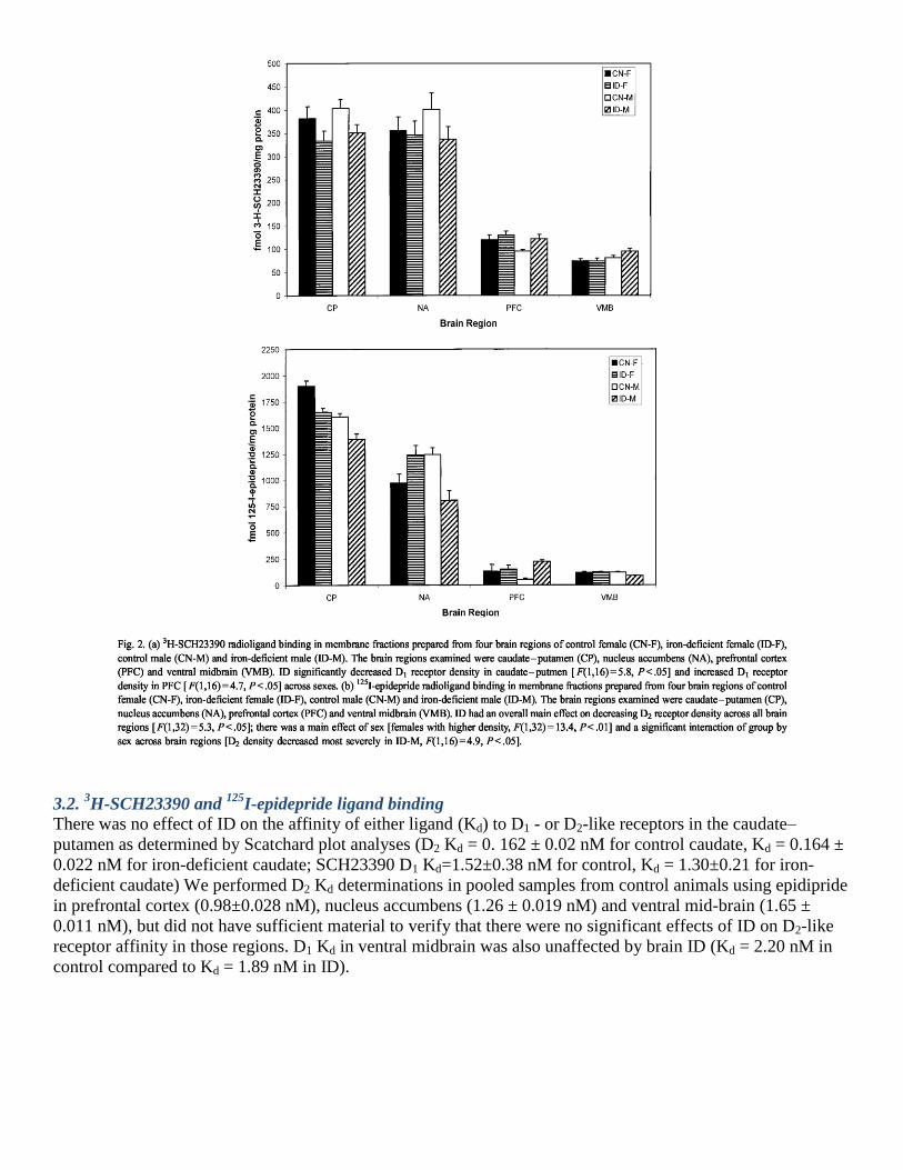
3.3. Brain region iron content
Dietary ID caused a significant decrease in brain iron content [F(1,32)=33.7, P ≤ .01 (estimated ω2 = 0.578)]
with significant effects in all four brain regions examined in this study. There was a much greater drop in brain
iron concentration in ventral midbrain (60% decrease) than in prefrontal cortex (37% decrease). In the caudate–
putamen, ID decreased iron concentration by approximately 30%, and in the nucleus accumbens, the decrease
was 20% (Fig. 3). Iron concentrations in the caudate–putamen and nucleus accumbens in females were less
affected by diet than that seen in the males.

3.4. Correlational analysis
In an attempt to determine the degree to which dopamine receptor densities are related to regional iron
concentrations, we performed a correlational analysis between iron concentration and receptor density (Fig. 4a).
In the CP, prefrontal cortex and nucleus accumbens, D2-like receptor densities showed a significant correlation
with iron (r = .91, P < .001; r = .42, r = .44, P < .05, respectively), No such correlation was found between iron
concentration and D1 receptor density.
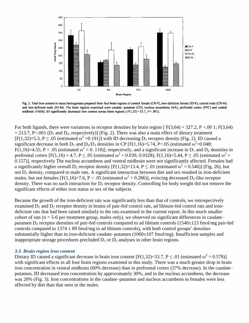
3.5. In situ hybridization histochemistry
Quantitative in situ hybridization studies focused on the substantia nigra and caudate–putamen and revealed no
significant effects of ID on mRNA levels for dopamine D1 or D2 receptors when individual cells within each of
these brain regions were examined. Brains (n = 3) from iron-deficient rats had 102%, 97% and 101% of control
mean grain density when the cDNA probe for the D1 receptor was used and 96 randomly chosen cells in each
region were counted. The densities for D2 receptor mRNA were 101 %, 100% and 96% of control densities in
the nigra.
Representative autoradiographs from coronal sections are shown in Fig. 5.
4. Discussion
These experiments demonstrate that ID alters both in vivo and in vitro functioning of dopamine receptors in the
rat brain. These alterations were brain-region-specific, and affected males more dramatically than females. This
work thus extends the previous observations that ID anemia causes a decreased D2 receptor density in caudate–
putamen (Youdim et al., 1979, 1983, 1989). Our approach of examining terminal fields of two different
dopaminergic path-ways extends the conceptual framework regarding the interaction between local ID and
brain dopamine functioning, since we now demonstrate that downregulation of the D2-like receptor is not
universal in all terminal fields of dopaminergic tracts. In addition, we demonstrate that the affinity of these
receptors in the caudate–putamen for highly specific ligands is not altered by ID. The current report is in
agreement with the previous studies that used less specific ligands to demonstrate a 15–25% drop in D2- like
receptor density in male Sprague–Dawley rats (Ben-Shachar et al., 1986; Youdim et al., 1983, 1989). We
extend those observations to now demonstrate a downregulation in the nucleus accumbens and ventral midbrain
in males. Our studies suffer a somewhat similar limitation as the earlier studies — non-specificity of the ligands
used. In this case, epidipride binds to D2 and D3 receptors, both of which are in high density in the nucleus
accumbens and the caudate– putamen. Since there are many fewer D3 receptors in the cortex, this complication
does not limit our interpretation of the data from that region where D2 density was actually higher in iron-
deficient males than controls. This finding is not in concert with the published literature that suggested a
universal negative effect of ID on DA function (Ben-Shachar et al., 1986; Youdim et al., 1979, 1983, 1989).
Finally, the less robust findings in the midbrain area may well be a result of the lack of precision of definition
and the large mass of tissue analyzed — a result that likely decreased the signal-to-noise ratio. The second
important new observation regarding ID and D2-like receptors was the importance of sex on the receptor density

response to dietary ID. Females did not respond to ID in the same manner as males, especially in the nucleus
accumbens. Estrogen clearly modifies the expression of D2 receptors (DiPaolo et al., 1988; Levesque and
DiPaolo, 1990), as well as the sensitivity of the DA transporter to cocaine (Post et al., 1987). The basis of the
significant interaction between iron status and sex of the subject is unknown at this time, as there is little
evidence that ID alters estrogen metabolism. However, the fact that iron concentrations in the nucleus
accumbens did not fall with dietary ID, in contrast to male rats, suggests to us that regional iron metabolism is
important. We do not differentiate presynaptic autoreceptors from postsynaptic receptors with the approach
taken in the current study. These autoreceptors are known to interact with the DA transporter in the regulation
of clearance of DA from the intersynaptic space (Meiergerd et al., 1993; Vallone et al., 2000). We recently
reported that ID causes a significant drop in DA transporter density in the striatum and accumbens of young rats
(Erikson et al., 2000). It is certainly possible that these two observations are related; i.e., a decreased D2
autoreceptor functioning leads to a decreased DA transporter functioning with a resultant significant elevation
in striatal DA content (Meiergerd et al., 1993). Indeed, the D2-like receptor density is significantly correlated
with DA transporter density (r = .668, P < .001).
In addition to the effects of ID anemia on D2 receptor density, we also observed alterations in D1 receptor
density. The D1 receptor density was lower in caudate–putamen, but not nucleus accumbens. This, of course, is
in contrast to lower D1 density in ID males in both brain regions. One previous study (Youdim et al., 1979)
reported no effect of ID on D1 receptor density in the caudate–putamen in male rats, but the ligand used in that
older study lacked the specificity of currently available ligands and regional iron concentrations were not
measured. It is clear from our in situ hybridization studies that D1 and D2 mRNA expression was not altered by
ID, suggesting that ID is not having an effect on gene transcription events. The inconsistent effect of ID on
regional dopamine receptor density is likely related to the regional variation in brain iron sensitivity to dietary
ID (Erikson et al., 1997; Pinero et al., 2000). In addition, there are receptor subtypes that are not differentiated
by the ligands used in the current study. For example, in prefrontal cortex, the ratio of D4 to D2/3 receptors is
much greater than in the striatum (Tarazi et al., 1997). Therefore, it is plausible that ID alters the D4 to D2/3
ratio, leading to increased 125
I-epidepride binding in the PFC in male rats.
The pharmacological impact of ID is manifest in our observation of an attenuated response to raclopride in iron-
deficient anemic rats of both sexes. Decreased motor activity has been observed previously in iron-deficient
male rats (Glover and Jacobs, 1972; Hunt et al., 1994; Youdim et al., 1981, 1989) and is confirmed in saline-
treated rats in this study. We cannot differentiate between mesolimbic and nigrostriatal contribution to
locomotion in this research, as both pathways were affected by poor iron status. Previous studies document a
highly significant drop in DA transporter density and functioning in caudate of iron-deficient rats (Erikson et
al., 2000). In addition, we know that there is an excess of DA in the extracellular space in this brain region
(Beard et al., 1994; Chen et al., 1995b; Nelson et al., 1997). Alterations in extracellular DA in other brain
regions might be expected, but is speculation at this time. Although the impact of ID on DA-related intracellular
signaling and neurotransmission efficiency is unknown, we believe that these are the next avenues for study.
One of the key activities of the D2 receptor on the presynaptic membrane is the feedback regulation of the
dopamine transporter (DAT) (Dickinson et al., 1999; Meiergerd et al., 1993). Increased agonist binding to this
receptor increases DAT activity and increases the rate of removal of DA from the synaptic space (Dickinson et
al., 1999; Meiergerd et al., 1993). Downregulation of the D2 receptor in the striatum of iron-deficient rats may
thus lead to a dysfunction of the DAT. From our raclopride experiments, it appears possible that the functional
connection between the D2 and the DAT could also be desensitized, implying that it takes significantly more
DA to stimulate the DAT. Exactly how the downregulation of the two dopamine receptors is related to the
downregulation of the dopamine transporter is the basis of ongoing investigations in our laboratory.
Our observations of sex differences in DA receptor density in rats made iron-deficient agree with our previous
studies in mice (Morse et al., 1999). Estrogen secretion is related to the density of dopamine receptors (DiPaolo
et al., 1988; Levesque and DiPaolo, 1990; Andersen et al., 1990), as well as the sensitivity of the DA transporter
to cocaine (Post et al., 1987). The current study clearly notes that the sensitivity of the brain to ID is also sex-
dependent (Morse et al., 1999; Pinero et al., 2000). Thus, brain regions that are not sensitive to loss of iron (e.g.,

nucleus accumbens) in females do not evince diminished D2 receptor densities. Correlational analysis verified
this strong association of caudate iron and caudate receptor density. This relationship did not exist for the D1
receptor and suggests additional avenues of effect of iron of receptor metabolism.
It is clear from the careful examination of the growth data in Table 1 that iron-deficient rats grew less well than
the control rats, and it is reasonable to question whether or not ―undernutrition‖ could be related to the changes
in dopamine function as opposed to ID. We, and others, have used pair-fed control animals in studies of
metabolism in mineral deficiencies and find that slower growth caused by the mineral deficiency does not
necessarily impact brain catecholamine or peripheral catecholamine metabolism (Beard et al., 1995; Halas et al.,
1982). Our brief exploration of dopamine receptor density in frozen samples in the current report supports this
conclusion. Perinatal undernutrition has been shown to alter both dopamine receptor function and behavioral
responses to drugs (Keller et al., 1990; Shen et al., 1995), but similar effects by mild postweaning under
nutrition are lacking.
5. Conclusion
These studies demonstrate strong effects of postweaning ID on both D1 and D2 receptor density and functioning
(D2). Sex of the subject was a key determinant of the receptor density variations, but not of the functional
outcomes: ID universally decreased locomotor sensitivity to attenuation by raclopride. Future studies will focus
on potential mechanisms by which local iron in the brain alters functioning of these DA receptors.
References
ACC/SCN. Second report on the world nutrition situation. Global and regional results, vol. 1. Geneva:
ACC/SCN, 1992.
Andersen SL, Rutstein M, Benzo JM, Hostetter JC, Teicher MH. Sex differences in dopamine receptor
overproduction and elimination. NeuroReport 1990;8:1495–8.
Ashkenazi R, Ben-Shachar D, Youdim MBH. Nutritional iron and dopamine binding sites in the rat brain.
Pharmacol Biochem Behav 1982; 17(Suppl 1):43–7.
Beard JL, Connor JR, Jones BC. Iron in the brain. Nutr Rev 1993;51(6): 157–70.
Beard JL, Chen Q, Connor JR, Jones BC. Altered monoamine metabolism in caudate–putamen of iron-deficient
rats. Pharmacol Biochem Behav 1994;48(3):621–4.
Beard JL, Zahn CS, Brigham DE. Growth in iron-deficient rats. Proc Soc Exp Biol Med 1995;209:65–72.
Ben-Shachar D, Ashkenazi R, Youdim MBH. Long-term consequences of early iron deficiency on
dopaminergic transmission. J Dev Neurosci 1986;4:81–8.
Campbell DB, Hess EJ. L-type calcium channels contribute to the tottering mouse dystonic episodes. Mol
Pharmacol 1999;55:23 – 3 1.
Chen Q, Connor JR, Beard JL. Brain iron, transferrin and ferritin concentrations are altered in developing iron-
deficient rats. J Nutr 1995a;125: 1529–35.
Chen Q, Beard JL, Jones BC. Abnormal brain monoamine metabolism in iron deficiency anemia. J Nutr
Biochem 1995b;6:486–93.
Dickinson SD, Sabeti J, Larson GA, Giardina K, Rubinstein M, Kelly MA, Grandy DK, Low MJ, Gerhardt GA,
Zahniser NR. Dopamine D2 receptor-deficient mice exhibit decreased dopamine transporter function but no
changes in dopamine release in dorsal striatum. J Neurochem 1999;72:148–56.
DiPaolo T, Falardeau P, Morissette M. Striatal D2 dopamine agonist binding sites fluctuate during the rat
estrous cycle. Life Sci 1988;43:665–72.
Erikson KM, Pinero DJ, Connor JR, Beard JL. Regional brain iron, ferritin and transferrin concentrations during
iron deficiency and iron repletion in developing rats. J Nutr 1997;127:2030–8.
Erikson KM, Jones BC, Beard JL. Iron deficiency alters dopamine transporter functioning in rat striatum. J Nutr
2000;130:2831–7.
Glover J, Jacobs A. Activity pattern of iron-deficient rats. Br Med 1972;2:627–8.
Halas ES, Wallwork JC, Sandstead HH. Mild zinc deficiency and under-nutrition during the prenatal and
postnatal periods in rats: effects on weight, food consumption, and brain catecholamine concentrations. J Nutr
1982;112:542–51.
Hinkle DE, Wiersma W, Jurs SG. Applied statistics for the behavioral sciences. New York: Houghton Mifflin,
1998. pp. 368–9.
Hunt JR, Zito CA, Erjavec J, Johnson L. Severe or marginal iron deficiency affects spontaneous physical
activity in rats. Am J Clin Nutr 1994;59: 413–8.
Keller EA, Molina VA, Orsingher OA. Lack of neuronal adaptive changes following chronic treatments in
perinatally undernourished rats. Pharmacol Biochem 1990;37:675–8.
Larkin EC, Rao GA. Importance of fetal and neonatal iron: adequacy for normal development of central
nervous system. In: Dobbing J, editor. Brain, behaviour and iron in the infant diet. London: Springer-Verlag,
1990. pp. 43–63.
Levesque D, DiPaolo T. Effect of the rat estrous cycle at ovariectomy on striatal D1 dopamine receptors. Brain
Res 1990;24:281–4.
Lozoff B, Brittenham GM. Behavioral aspects of iron deficiency. Prog Hematol 1986;14:23–53.
Meiergerd SM, Patterson TA, Schenk JO. D2 receptors may modulate the function of the striatal transporter for
dopamine: kinetic evidence from studies in vitro and in vivo. J Neurochem 1993;61:764–7.
Morse AC, Beard JL, Azar MR, Jones BC. Sex and genetics are important cofactors in assessing the impact of
iron deficiency on the developing mouse brain. Nutr Neurobiol 1999;2:323 – 35.
Nelson C, Erikson K, Pinero DJ, Beard JL. In vivo dopamine metabolism is altered in iron-deficient anemic
rats. J Nutr 1997;127:2282–8. Paxinos G, Watson C. The rat brain in stereotaxic coordinates. San Diego,
CA: Academic Press, 1986.
Pinero DJ, Li NQ, Connor JR, Beard JL. Variations in dietary iron alter brain iron metabolism in developing
rats. J Nutr 2000;130:254–63.
Pollit E. Iron deficiency and cognitive function. Annu Rev Nutr 1993;13: 521–38.
Post RM, Weiss SRB, Pert A, Uhde TW. Chronic cocaine administration: sensitization and kindling effects. In:
Fisher S, Raskin A, Uhlenhut EH, editors. Cocaine: clinical and biobehavioral aspects. New York: Oxford
Univ. Press, 1987. pp. 109–73.
Rocangliolo M, Garrido M, Walter T, Peirano P, Lozoff B. Evidence of altered central nervous system
development in infants with iron deficiency anemia at 6 mo: delayed maturation of auditory brainstem
responses. Am J Clin Nutr 1998;68:683 – 90.
Shen RY, Hannigan JH, Choido LA. The effects of chronic amphetamine treatment on prenatal ethanol-induced
changes in dopamine receptor function: electrophysiological findings. J Pharmacol Exp Ther 1995;274: 1054–
60.
Tarazi FI, Kula NS, Baldessarini RJ. Regional distribution of dopamine D4 receptors in rat forebrain.
NeuroReport 1997;2:41–58.
Vallone D, Picetti R, Borrelli E. Structure and function of dopamine receptors. Neurol Biobehav 2000;24:125–
32.
Walter T. Impact of iron deficiency on cognition in infancy and childhood. Eur J Clin Nutr 1993;47(5):307–16.
Youdim MBH, Green AR, Bloomfield MR, Mitchell BD, Heal DJ, Grahame-Smith DG. The effects of iron
deficiency on brain biogenic mono-amine biochemistry and function in rats. Neuropharmacology 1979;19: 259–
67.
Youdim MBH, Yehuda S, Ben-Uriah Y. Iron-deficiency-induced circadian rhythm reversal of dopaminergic-
mediated behaviours and thermoregulation in rats. Eur J Pharmacol 1981;74:295–301.
Youdim MBH, Ben-Shachar D, Ashkenazi R, Yehuda S. Brain iron and dopamine receptor function. In: Mandel
P, DeFeudis FV, editors. CNS receptors from molecular pharmacology to behaviour. New York: Raven Press,
1983. pp. 309–22.
Youdim MBH, Ben-Shachar D, Yehuda S. Putative biological mechanisms of the effect of iron deficiency on
brain biochemistry and behavior. Am J Clin Nutr 1989;50:607–17.
Related Documents


![Aldosterone and dopamine receptors in the kidney: Sites for ...Aldosterone and dopamine receptors 625 with the aldosterone receptor, when measured in vitro [21, 23] (Funder and Adam,](https://static.cupdf.com/doc/110x72/608977add019a330f10765d3/aldosterone-and-dopamine-receptors-in-the-kidney-sites-for-aldosterone-and.jpg)