I 1 Distraction osteogenesis to widen the mandible W. H. Bell, R. P. Harper, M. Gonzalez, A. M. Cherkashin, M. L. Samchukov Departments of Oral Maxillofuciul Surgery and Pharmacology and Biomedical Sciences. Bn~4or College c?f Dentistry; Texas Scottish Rite Hospital for Children, Dullas, Te.vas, USA SUMA4ARY. The purpose of this investigation was to elucidate the biology of distraction osteogenesis during mandibular widening. Midsymphyseal vertical interdental osteotomies were performed in nine Macaca mulatta monkeys. After a latency period a tooth-borne appliance was activated at a rate of 0.5 mm twice a day for 7-10 days. The appliance was then stabilized for a period of 4 or 8 weeks. The distraction gap at the inferior portion of the symphysis was bridged completely by new bony trabeculae. Bone formation in the interdental area was apparently related to the surgical technique. Newly formed bony trabeculae were oriented parallel to the direction of distraction. The location of the osteotomy site with an adequate margin of alveolar bone contiguous with the adjacent teeth was necessary for the induction of the distraction osteogenesis. Disproportional movement between superior and inferior portions of the distracted segments was noted. INTRODUCTION The principal indication for widening the mandible is absolute transverse mandibular deficiency. An excessively narrow and tapered arch form, dental crowding, tipped teeth and congenitally missing teeth are additional reasons for the use of surgery which is intended to normalize basal bone position and facilitate non-extraction orthodontic treatment. Incomplete telescopic bite in certain congenital problems (Pierre Robin, Treacher Collins, hemifacial microsomia), and combined maxillo-mandibular transverse deficiency may be additional indications. Also, patients with mandibular transverse deficiency in whom crowded teeth have been treated by extrac- tion orthodontic therapy, may be additional indi- cations. Many of these individuals may benefit from surgically assisted rapid mandibular expansion and orthodontic treatment. Transverse mandibular deficiency is commonly managed by orthodontic mechanics which might include extraction and dental compensations. The result of this approach may be unstable owing to tipping of the teeth and bending of the alveolar bone. Proffit and Ackerman’ have reported a high risk of dental relapse when compensating orthodontic ther- apy has been performed to increase the inter-canine width in the presence of a primary transverse bone deficiency. Indeed, Proffit and White2 have docu- mented the limitations and easy violation of the transverse envelope. When a skeletal or dento-alveolar deformity is so severe that the magnitude of the problem lies outside the envelope of possible correction by orthodontics alone, surgical orthodontic treatment is indicated. Osteodistraction techniques may be the key to opti- mal non-extraction management of many malocclu- sions thus maintaining and increasing the functional occlusal table. Attention to transverse deficiency is vital in planning treatment for patients who require an increase in the lateral dimensions of the mandible or maxilla. The transverse envelope of discrepancy for mandibular alterations can be addressed by sym- physeal osteotomy and gradual osteodistraction. Mandibular symphysis osteodistraction was ini- tially reported by Guerrero in 19903 and has since been used sparingly by others.4 Despite early reports of success, important questions remain unanswered: what is the biologic foundation for distraction osteo- genesis to widen the symphysis; what is the response of alveolar bone and the periodontal ligament to distraction forces; and what are the physiologic limits of hard and soft tissue expansion during mandibular widening? No previous investigations have studied the biologic basis for widening the mandible by osteodistraction following mandibular midline osteotomy between closely spaced teeth. The purpose of this investigation was to develop an animal model for widening the mandible at the symphysis in order to elucidate the biology of histo- genesis in the tissues exposed to the distraction forces: basal bone, alveolar bone, and periodontal ligament. The key hypothesis to be tested was that new alveolar bone would form in the distraction gap. MATERIALS AND METHODS This study included nine adult Macaca mulatta monk- eys. After midline osteotomy a tooth-borne expansion appliance was bonded to the mandibular dentition and further stabilized with circummandibular wires (Fig. 1). Activation of the appliance was begun at 7 and 14 days post-surgery and expanded at a rate of 0.5 mm 11

Welcome message from author
This document is posted to help you gain knowledge. Please leave a comment to let me know what you think about it! Share it to your friends and learn new things together.
Transcript
I 1
Distraction osteogenesis to widen the mandible
W. H. Bell, R. P. Harper, M. Gonzalez, A. M. Cherkashin, M. L. Samchukov Departments of Oral Maxillofuciul Surgery and Pharmacology and Biomedical Sciences. Bn~4or College c?f Dentistry; Texas Scottish Rite Hospital for Children, Dullas, Te.vas, USA
SUMA4ARY. The purpose of this investigation was to elucidate the biology of distraction osteogenesis during mandibular widening. Midsymphyseal vertical interdental osteotomies were performed in nine Macaca mulatta monkeys. After a latency period a tooth-borne appliance was activated at a rate of 0.5 mm twice a day for 7-10 days. The appliance was then stabilized for a period of 4 or 8 weeks. The distraction gap at the inferior portion of the symphysis was bridged completely by new bony trabeculae. Bone formation in the interdental area was apparently related to the surgical technique. Newly formed bony trabeculae were oriented parallel to the direction of distraction. The location of the osteotomy site with an adequate margin of alveolar bone contiguous with the adjacent teeth was necessary for the induction of the distraction osteogenesis. Disproportional movement between superior and inferior portions of the distracted segments was noted.
INTRODUCTION
The principal indication for widening the mandible is absolute transverse mandibular deficiency. An excessively narrow and tapered arch form, dental crowding, tipped teeth and congenitally missing teeth are additional reasons for the use of surgery which is intended to normalize basal bone position and facilitate non-extraction orthodontic treatment. Incomplete telescopic bite in certain congenital problems (Pierre Robin, Treacher Collins, hemifacial microsomia), and combined maxillo-mandibular transverse deficiency may be additional indications. Also, patients with mandibular transverse deficiency in whom crowded teeth have been treated by extrac- tion orthodontic therapy, may be additional indi- cations. Many of these individuals may benefit from surgically assisted rapid mandibular expansion and orthodontic treatment.
Transverse mandibular deficiency is commonly managed by orthodontic mechanics which might include extraction and dental compensations. The result of this approach may be unstable owing to tipping of the teeth and bending of the alveolar bone. Proffit and Ackerman’ have reported a high risk of dental relapse when compensating orthodontic ther- apy has been performed to increase the inter-canine width in the presence of a primary transverse bone deficiency. Indeed, Proffit and White2 have docu- mented the limitations and easy violation of the transverse envelope.
When a skeletal or dento-alveolar deformity is so severe that the magnitude of the problem lies outside the envelope of possible correction by orthodontics alone, surgical orthodontic treatment is indicated. Osteodistraction techniques may be the key to opti- mal non-extraction management of many malocclu- sions thus maintaining and increasing the functional
occlusal table. Attention to transverse deficiency is vital in planning treatment for patients who require an increase in the lateral dimensions of the mandible or maxilla. The transverse envelope of discrepancy for mandibular alterations can be addressed by sym- physeal osteotomy and gradual osteodistraction.
Mandibular symphysis osteodistraction was ini- tially reported by Guerrero in 19903 and has since been used sparingly by others.4 Despite early reports of success, important questions remain unanswered: what is the biologic foundation for distraction osteo- genesis to widen the symphysis; what is the response of alveolar bone and the periodontal ligament to distraction forces; and what are the physiologic limits of hard and soft tissue expansion during mandibular widening? No previous investigations have studied the biologic basis for widening the mandible by osteodistraction following mandibular midline osteotomy between closely spaced teeth.
The purpose of this investigation was to develop an animal model for widening the mandible at the symphysis in order to elucidate the biology of histo- genesis in the tissues exposed to the distraction forces: basal bone, alveolar bone, and periodontal ligament. The key hypothesis to be tested was that new alveolar bone would form in the distraction gap.
MATERIALS AND METHODS
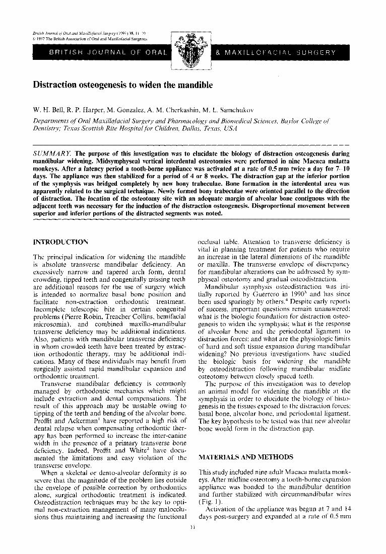
This study included nine adult Macaca mulatta monk- eys. After midline osteotomy a tooth-borne expansion appliance was bonded to the mandibular dentition and further stabilized with circummandibular wires (Fig. 1).
Activation of the appliance was begun at 7 and 14 days post-surgery and expanded at a rate of 0.5 mm
11
12 British Journal of Oral and Maxillofacial Surgery
Fig. 1 -Tooth-borne distraction device bonded to cuspid and molar teeth and further stabilized with circummandibular wires.
twice a day for 7-10 days. Postoperative healing at the mandibular symphysis was analyzed clinically and radiographically. The appliances were then stabilized with light cured acrylic in the expansion slot, for a period of 8 weeks for the first animal and 4 weeks for the others, after which the animals were sacrificed.
Surgical technique
The housing, care and experimental protocol was in accordance with guidelines established by Baylor College of Dentistry Institutional Animal Care and Research Advisory Committee. All surgical pro- cedures were accomplished under general endo- tracheal anesthesia. Each animal was anesthetized with intramuscular Ketamine HCl (10 mg/kg) and Xylazine ( 1.2 mg/kg).
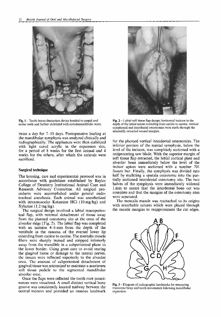
The surgical design involved a labial mucoperios- teal flap, with minimal detachment of tissue away from the planned osteotomy site at the crest of the alveolar ridge (Fig. 2). The labial flap was completed with an incision 4-6 mm from the depth of the vestibule in the mucosa of the everted lower lip extending from canine to canine. The mentalis muscle fibers were sharply incised and stripped inferiorly away from the mandible in a subperiosteal plane to the lower border. Using great care to avoid tearing the gingival tissue or damage to the mental nerves, the tissues were reflected superiorly to the alveolar crest. The amount of subperiosteal detachment of gingival tissue was minimized to maintain a maximum soft tissue pedicle to the segmented mandibular alveolar crest.
Once the flaps were reflected the tooth root promi- nences were visualized. A small distinct vertical bony groove was consistently located midway between the central incisors and provided an osseous landmark
Fig. 2 - Labial soft tissue flap design: horizontal incision in the depth of the labial sulcus extending from canine to canine, vertical symphyseal and interdental osteotomies were made through the minimally retracted wound margins.
for the planned vertical interdental osteotomies. The inferior portion of the mental symphysis, below the level of the incisors, was completely sectioned with a reciprocating saw blade. With the superior margin of soft tissue flap retracted, the labial cortical plate and alveolar bone immediately below the level of the incisor apices were sectioned with a number 701 fissure bur. Finally, the symphysis was divided into half by malleting a spatula osteotome into the par- tially sectioned interdental osteotomy site. The two halves of the symphysis were immediately widened 1 mm to assure that the interdental bone cut was complete and that the margins of the osteotomy sites were separated.
The mentalis muscle was reattached to its origins with resorbable sutures which were placed through the muscle margins to reapproximate the cut edges.
A0 An
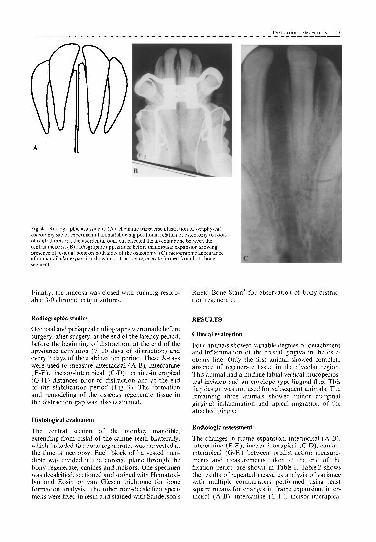
Fig. 3 -Diagram of radiographic landmarks for measuring transverse bony and tooth movements following mandibular expansion.
Distraction osteoeenesis 13
Fig. 4 - Radiographic assessment: (A) schematic transverse illustration of symphyseal osteotomy site of experimental animal showing positional relation of osteotomy to roots of central incisors; the interdental bone cut bisected the alveolar bone between the central incisors: (B) radiographic appearance before mandibular expansion showing presence of residual bone on both sides of the osteotomy; (C) radiographic appearance after mandibular expansion showing distraction regenerate formed from both bone segments.
Finally, the mucosa was closed with running resorb- able 3-O chromic catgut sutures.
Radiographic studies
Occlusal and periapical radiographs were made before surgery. after surgery, at the end of the latency period, before the beginning of distraction, at the end of the appliance activation (7-l 0 days of distraction) and every 7 days of the stabilization period. These X-rays were used to measure interincisal (A-B), intercanine (E-F), incisor-interapical (C-D), canine-interapical (G-H) distances prior to distraction and at the end of the stabilization period (Fig. 3). The formation and remodeling of the osseous regenerate tissue in the distraction gap was also evaluated.
Histological evaluation
The central section of the monkey mandible, extending from distal of the canine teeth bilaterally, which included the bone regenerate, was harvested at the time of necropsy. Each block of harvested man- dible was divided in the coronal plane through the bony regenerate, canines and incisors. One specimen was decalcified, sectioned and stained with Hematoxi- lyn and Eosin or van Gieson trichrome for bone formation analysis. The other non-decalcified speci- mens were fixed in resin and stained with Sanderson’s
Rapid Bone Stain’ for observation of bony distrac- tion regenerate.
RESULTS
Clinical evaluation
Four animals showed variable degrees of detachment and inflammation of the crestal gingiva in the oste- otomy line. Only the first animal showed complete absence of regenerate tissue in the alveolar region. This animal had a midline labial vertical mucoperios- teal incision and an envelope type lingual flap. This flap design was not used for subsequent animals. The remaining three animals showed minor marginal gingival inflammation and apical migration of the attached gingiva.
Radiologic assessment
The changes in frame expansion, interincisal (A-B), intercanine (E-F ), incisor-interapical (C-D), canine- interapical (G-H ) between predistraction measure- ments and measurements taken at the end of the fixation period are shown in Table 1. Table 2 shows the results of repeated measures analysis of variance with multiple comparisons performed using least square means for changes in frame expansion, inter- incisal (A-B), intercanine (E-F), incisor-interapical
14 British Journal of Oral and Maxillofacial Surgery
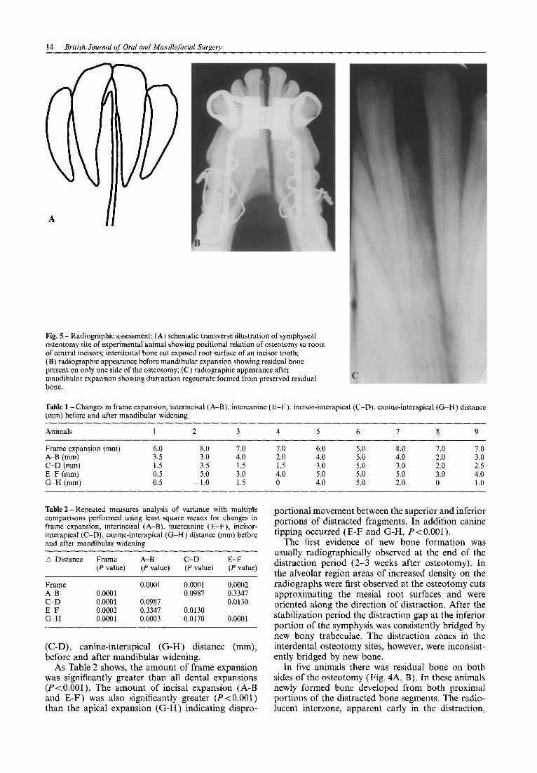
Fig. 5 - Radiographic assessment: (A) schematic transverse illustration of symphyseal osteotomy site of experimental animal showing positional relation of osteotomy to roots of central incisors; interdental bone cut exposed root surface of an incisor tooth; (B) radiographic appearance before mandibular expansion showing residual bone present on only one side of the osteotomy; (C) radiographic appearance after mandibular expansion showing distraction regenerate formed from preserved residual bone.
Table 1 - Changes in frame expansion, interincisal (A-B), intercanine (E-F), incisor-interapical (C-D), canine-interapical (G-H) distance (mm) before and after mandibular widening
Animals 1 2 3 4 5 6 1 8 9
Frame expansion (mm) 6.0 8.0 7.0 7.0 6.0 5.0 8.0 7.0 7.0 A-B (mm) 3.5 3.0 4.0 2.0 4.0 5.0 4.0 2.0 3.0 C-D (mm) 1.5 3.5 1.5 1.5 3.0 5.0 3.0 2.0 2.5 E-F (mm) 0.5 5.0 3.0 4.0 5.0 5.0 5.0 3.0 4.0 G-H (mm) 0.5 -1.0 1.5 0 4.0 5.0 2.0 0 1.0
Table 2 - Repeated measures analysis of variance with multiple comparisons performed using least square means for changes in frame expansion, interincisal (A-B), intercanine (E-F), incisor- interapical (C-D), canine-interapical (G-H) distance (mm) before and after mandibular widening
portional movement between the superior and inferior portions of distracted fragments. In addition canine tipping occurred (E-F and G-H, P<O.OOl).
D Distance
Frame A-B C-D E-F G-H
Frame (P value)
0.0001 0.0001 0.0002 0.0001
A-B (P value)
0.0001
0.0987 0.3347 0.0003
C-D (P value)
0.0001 0.0987
0.0130 0.0170
E-F (P value)
0.0002 0.3347 0.0130
0.0001
(C-D), canine-interapical (G-H) distance (mm), before and after mandibular widening.
The first evidence of new bone formation was usually radiographically observed at the end of the distraction period (2-3 weeks after osteotomy). In the alveolar region areas of increased density on the radiographs were first observed at the osteotomy cuts approximating the mesial root surfaces and were oriented along the direction of distraction. After the stabilization period the distraction gap at the inferior portion of the symphysis was consistently bridged by new bony trabeculae. The distraction zones in the interdental osteotomy sites, however, were inconsist- ently bridged by new bone.
As Table 2 shows, the amount of frame expansion In five animals there was residual bone on both was significantly greater than all dental expansions sides of the osteotomy (Fig. 4A, B). In these animals (PC 0.001). The amount of incisal expansion (A-B newly formed bone developed from both proximal and E-F) was also significantly greater (P<O.OOl) portions of the distracted bone segments. The radio- than the apical expansion (G-H) indicating dispro- lucent interzone, apparent early in the distraction,
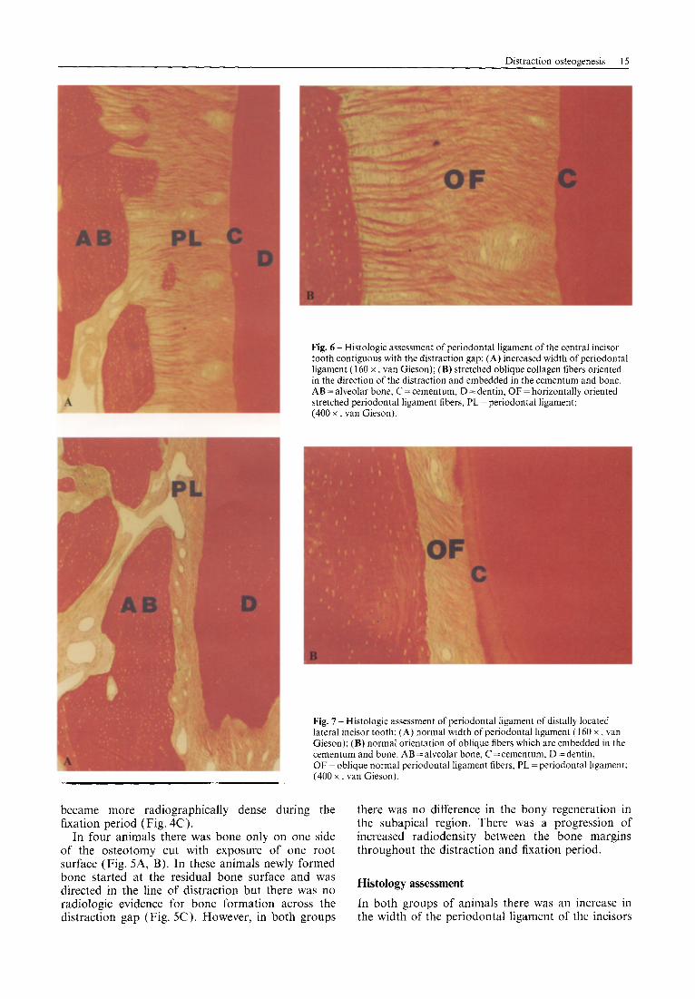
Fig. 6 -Histologic assessment of periodontal ligament of the central incisor tooth contiguous with the distraction gap: (A) increased width of periodontal ligament (160 x , van Gieson); (B) stretched oblique collagen fibers oriented in the direction of the distraction and embedded in the cementum and bone. AB = alveolar bone, C = cementum, D = dentin, OF = horizontally oriented stretched periodontal ligament fibers, PL = periodontal ligament; (400 x , van Gieson).
Fig. 7 -Histologic assessment of periodontal ligament of distally located lateral incisor tooth: (A) normal width of periodontal ligament ( 160 x , van Gieson); (B) normal orientation of oblique fibers which are embedded in the cementum and bone. AB = alveolar bone, C =cementum, D = dentin, OF=oblique normal periodontal ligament fibers, PL=periodontal ligament: (400 x . van Gieson).
became more radiographically dense during the fixation period (Fig. 4C).
In four animals there was bone only on one side of the osteotomy cut with exposure of one root surface (Fig. 5A, B). In these animals newly formed bone started at the residual bone surface -and was directed in the line of distraction but there was no radiologic evidence for bone formation across the In both groups of animals there was an increase in distraction gap (Fig. 5C). However, in both groups the width of the periodontal ligament of the incisors
there was no difference in the bony regeneration in the subapical region. There was a progression of increased radiodensity between the bone margins throughout the distraction and fixation period.
Histology assessment
16 British Journal of Oral and MaxiNofacial Surgery
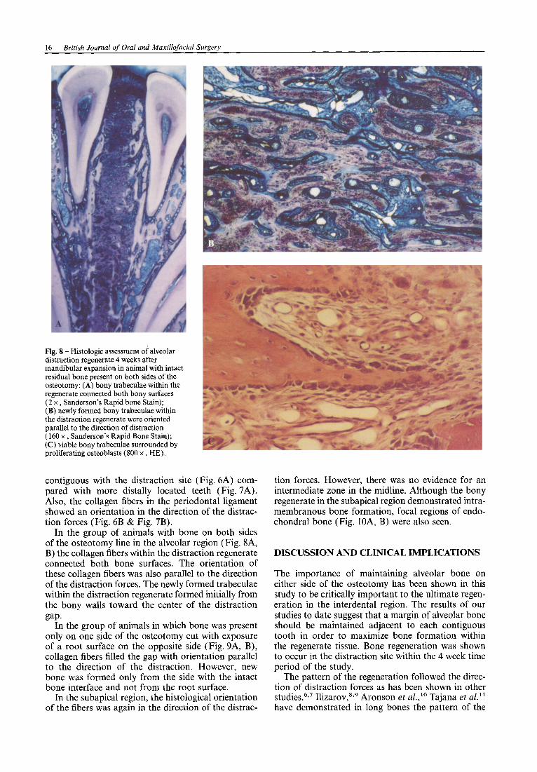
Fig. 8 - Histologic assessment of alveolar distraction regenerate 4 weeks after mandibular expansion in animal with intact residual bone present on both sides of the osteotomy: (A) bony trabeculae within the regenerate connected both bony surfaces (2 x , Sanderson’s Rapid bone Stain); (B) newly formed bony trabeculae within the distraction regenerate were oriented parallel to the direction of distraction (160 x , Sanderson’s Rapid Bone Stain); (C) viable bony trabeculae surrounded by proliferating osteoblasts (800 x , HE).
contiguous with the distraction site (Fig. 6A) com- pared with more distally located teeth (Fig. 7A). Also, the collagen fibers in the periodontal ligament showed an orientation in the direction of the distrac- tion forces (Fig. 6B & Fig. 7B).
In the group of animals with bone on both sides of the osteotomy line in the alveolar region (Fig. 8A, B) the collagen fibers within the distraction regenerate connected both bone surfaces. The orientation of these collagen fibers was also parallel to the direction of the distraction forces. The newly formed trabeculae within the distraction regenerate formed initially from the bony walls toward the center of the distraction gap.
In the group of animals in which bone was present only on one side of the osteotomy cut with exposure of a root surface on the opposite side (Fig. 9A, B), collagen fibers filled the gap with orientation parallel to the direction of the distraction. However, new bone was formed only from the side with the intact bone interface and not from the root surface.
In the subapical region, the histological orientation of the fibers was again in the direction of the distrac-
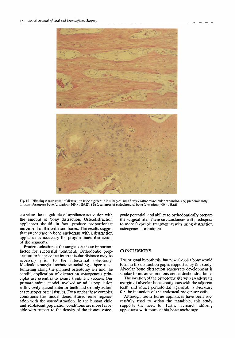
tion forces. However, there was no evidence for an intermediate zone in the midline. Although the bony regenerate in the subapical region demonstrated intra- membranous bone formation, focal regions of endo- chondral bone (Fig. lOA, B) were also seen.
DISCUSSION AND CLINICAL IMPLICATIONS
The importance of maintaining alveolar bone on either side of the osteotomy has been shown in this study to be critically important to the ultimate regen- eration in the interdental region. The results of our studies to date suggest that a margin of alveolar bone should be maintained adjacent to each contiguous tooth in order to maximize bone formation within the regenerate tissue. Bone regeneration was shown to occur in the distraction site within the 4 week time period of the study.
The pattern of the regeneration followed the direc- tion of distraction forces as has been shown in other studies.6,7 Ilizarov,839 Aronson et &lo Tajana et al.‘l have demonstrated in long bones the pattern of the
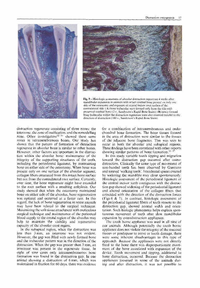
Fig. 9 - Histologic assessment of alveolar distraction regenerate 4 weeks after mandibular expansion in animals with intact residual bone present on only one side of the osteotomy and exposure of central incisor root surface of the contralateral side: (A) bone trabeculae were formed only from the side with preserved residual bone (2 x , Sanderson’s Rapid Bone Stain); (B) newly formed bony trabeculae within the distraction regenerate were also oriented parallel to the direction of distraction ( 160 X, Sanderson’s Rapid Bone Stain).
distraction regenerate consisting of three zones: the interzone, the zone of ossification, and the remodelling zone. Other investigators12-l4 showed these same zones in intramembranous bones. Our study has shown that the pattern of formation of distraction regenerate in alveolar bone is similar to other bones. However, other factors are important in the distrac- tion within the alveolar bone: maintenance of the integrity of the supporting structures of the teeth, including the periodontal ligament, by maintaining bone on either side of the osteotomy. When bone was present only on one surface of the alveolar segment, collagen fibers emanated from this intact bone surface but not from the contralateral root surface. Certainly, over time, the bone regenerate might have extended to the root surface with a resulting ankylosis. Our study showed that when the osteotomy maintained bone on either side of the alveolus, bone regeneration was optimal and occurred at a faster rate. In this regard, the lack of bone regeneration in some animals may have been related to the surgical technique. Maximizing the soft tissue attachment with meticulous surgical technique and maintenance of the periosteal blood supply to the crestal region of the alveolus may help to maintain the viability and regenerative capacity of the alveolar crest region.
In the subapical region, when the distraction was less than 3 mm, an interzone was not evident. However, the gap was filled with newly formed bone and the trabecular pattern was in the direction of the distraction. When the gap was greater than 3 mm, an interzone was present in the regenerate tissue. In eight of nine cases only intramembraneous bone formation was found in the distraction gap. In one animal showing a distraction of 6 mm, which was maintained in fixation for 60 days, there was evidence
for a combination of intramembranous and endo- chondral bone formation. The bone tissues formed in the area of distraction were similar to the tissues of the adjacent bone fragments. This was seen to occur in both the alveolar and subapical regions. These findings have been correlated with other reports showing similar patterns of bone formation.15-”
In this study variable tooth tipping and migration toward the distraction gap occurred after osteo- distraction. Clinically the same type of movement of non-banded teeth has been observed by Guerrero and termed ‘walking teeth’. Interdental spaces created by widening the mandible may close spontaneously. Histologic assessment of the periodontal ligament of the central incisor teeth contiguous with the distrac- tion gap showed widening of the periodontal ligament and altered orientation of the collagen fibers that coincided with the direction of the distraction forces (Figs 6 & 7). In contrast, histologic assessment of the periodontal ligament fibers of teeth remote to the distraction gap, showed normal width and orien- tation. Such biologic phenomena helps explain spon- taneous movement of teeth after slow mandibular expansion by osteodistraction appliances.
The tooth borne appliance was used in all nine of our animals. Although potentially the tooth borne appliance does not violate the integrity of the mucosal tissues or predispose to nerve or tooth damage. there were some inherent disadvantages to this type of approach. Because the appliances were not directly fixed to the bone there was disproportionate move- ment of the bone associated with expansion of the device. Tooth movement and tipping, additional to bone distraction, occurred. Because the distraction appliances loosened in some of the animals dur- ing and after distraction, it was not possible to
18 British Journal of Oral and Maxillofacial Surgery
Fig. 10 -Histologic assessment of distraction bone regenerate in subapical area 8 weeks after mandibular expansion: (A) predominantly intr, amembraneous bone formation (160 x , H&E); (B) focal areas of endochondral bone formation (400 x , H&E).
correlate the magnitude of appliance activation with the amount of bony distraction. Osteodistraction appliances should, in fact, produce proportionate movement of the teeth and bones. The results suggest that an increase in bone anchorage with a distraction appliance is necessary for proportionate distraction of the segments.
Prudent selection of the surgical site is an important factor for successful treatment. Orthodontic prep- aration to increase the interradicular distance may be necessary prior to the interdental osteotomy. Meticulous surgical technique including subperiosteal tunneling along the planned osteotomy site and the careful application of distraction osteogenesis prin- ciples are essential to assure treatment success. Our primate animal model involved an adult population with closely spaced anterior teeth and densely adher- ent mucoperiosteal tissues. Even under these complex conditions this model demonstrated bone regener- ation with the osteodistraction. In the human child and adolescent population conditions are more favor- able with respect to the density of the tissues, osteo-
genic potential, and ability to orthodontically prepare the surgical site. These circumstances will predispose to more favorable treatment results using distraction osteogenesis techniques.
CONCLUSIONS
The original hypothesis that new alveolar bone would form in the distraction gap is supported by this study. Alveolar bone distraction regenerate development is similar to intramembranous and endochondral bone.
The location of the osteotomy site with an adequate margin of alveolar bone contiguous with the adjacent teeth and intact periodontal ligament, is necessary for the induction of the endosteal progenitor cells.
Although tooth borne appliances have been suc- cessfully used to widen the mandible, this study supports the need for further research utilizing appliances with more stable bone anchorage.
Acknowledgements
This project was supported by grants from the Oral and Maxillofacial Surgery Foundation and the Carl and Florence E. King Foundation. We wish to acknowledge the assistance and support of Rohit C. L. Sachdeva. DDS. Richard Browne. PhD. J. ‘David Ross. MFA. Stan Richardson. Gerald Hill, and Priscilla Gillaspie.
technique in man. Characterization of Extracellular Matrix. Orthooedics 1989: 12: 515-523. Karaharju EO, Aalto K, Kahri A, rt (11. Distraction bone healing. Clin Orthop Relat Res 1993; 297: 38843. Karaharju-Suvanto T, Peltonen J, Kahri A, et (I/. Distraction osteogenesis of the mandible: an experimental study in sheep. J Oral Surg 1992; 21: 118. Karp NS, Thorne CH. McCarthy JG, Sissons HA. Bone lengthening in the craniofacial skeleton. Ann Plast Surg 1990: 24: 23 I -236.
References
I.
3.
4.
5.
6.
I.
8
9.
IO
II
Profht WR, Ackerman JL. Diagnosis and treatment planning in Orthodontics. In: Graber TM, Vanarsdall RL. Orthodontics Current Principles and Techniques. 2nd ed. Philadelphia: Mosby-Year Book, Inc., 1994: 3--95. Proffit WR, White RP. The need for surgical&orthodontic treatment. In Proffit WR, White RP. Surgical Orthodontic Treatment. 3rd ed. St Louis: Mosby--Year Book, Inc.. 1991: 2-33.
12.
13.
14.
15.
16.
17.
Waanders NA. Senunas LE. Steen H. Goulet JA, Bonadio J, Goldstein SA. Bone formation in distraction osteogenesis. Histologic and immunohistochemical findings. In: Proceedings ORS, 40th Annual Meeting 1994. D 23 1, Saleh M, Stubbs DA, Street RJ, Lang DM, Harris SC. Histologic analysis of human lengthened bone. J Pediatr Orthop Part B 1993; 2: 16-21. Delloye C, Delefortrie G. Coutelier L, Vmcent A. Bone regenerate formation in cortical bone during distraction lengthening. An experimental study. Clin Orthop Relat Res 1990; 250: 34442.
Guerrero C. Rapid mandibular expansion. Rev Venez Ortod 1990; 48: l-2. Perrott DH, Berger R, Vargervik K, Kaban LB. Use of a skeletal distraction device to widen the mandible: a case report. J Oral Surg 1993: 51: 435-439. Sanderson C. Bloebaum RD. Advances in the staining of ground section histology. Histo-Logic 1993: 23: I-3. Block MS, Daire J, Stover J, Matthews M. Changes in the inferior alveolar nerve following mandibular lengthening in the dog using distraction osteogenesis. J Oral Surg 1993: 5 I : 6522660.
The Authors
W. H. Bell DDS Professor
Costantino PD. Shybut G, Friedman CD. Pelzer HJ. Masini M, Shindo ML, Sisson GA. Segmental mandibular regeneration by distraction osteogenesis: an experimental study. Arch Otolaryngol Head Neck Surg 1990; 116: 535 545. Ilizarov GA. The principles of the Ilizarov method. Bull Hosp Joint Dis Orthop Inst 1988: 48: I. Ilizarov GA. The tension-stress effect on the genesis and growth of tissues. Part 1. The influence of stability of fixation and soft tissue preservation. Clin Orthop Relat Res 1989; 262: 249-28 I. Aronson J, Good B. Stewart C. Harrison B. Harp J. Preliminary studies of mineralization during distraction osteogenesis. Clin Orthop Relat Res 1990: 250: 43-49.
R. P. Harper DDS, FRCD(C) Assistant Professor Department of Oral Maxillofacial Surgery and Pharmacology M. Gonzalez DDS Graduate Student Biomedical Sciences Baylor College of Dentistry P.O. Box 660617 Dallas TX 75266-0677 USA A. M. Cherkashin MD Research Scientist M. L. Samchukov MD Associate Director of Ilizarov Research Texas Scottish Rite Hospital for Children 2222 Welborn Street Dallas TX 75219 USA
Correspondence and requests for offprints to W. H. Bell
Tajana GF, Morandi M, Zembo M. The structure and Paper received 1 November 1996 development of osteogenic repair tissue according to Ilizarov Accepted 7 November 1996
Related Documents