Published Ahead of Print 10 September 2012. 10.1128/MCB.00334-12. 2012, 32(22):4585. DOI: Mol. Cell. Biol. and Steven A. Brown Kramer Bert Maier, Yasuyuki Kurihara, Archa H. Fox, Achim Elzbieta Kowalska, Jürgen A. Ripperger, Christine Muheim, Loop the Circadian Transcriptional Feedback Distinct Roles of DBHS Family Members in http://mcb.asm.org/content/32/22/4585 Updated information and services can be found at: These include: SUPPLEMENTAL MATERIAL Supplemental material REFERENCES http://mcb.asm.org/content/32/22/4585#ref-list-1 at: This article cites 44 articles, 21 of which can be accessed free CONTENT ALERTS more» articles cite this article), Receive: RSS Feeds, eTOCs, free email alerts (when new http://journals.asm.org/site/misc/reprints.xhtml Information about commercial reprint orders: http://journals.asm.org/site/subscriptions/ To subscribe to to another ASM Journal go to: on February 5, 2013 by UNIV OF WESTERN AUSTRALIA M209 http://mcb.asm.org/ Downloaded from

Welcome message from author
This document is posted to help you gain knowledge. Please leave a comment to let me know what you think about it! Share it to your friends and learn new things together.
Transcript
Published Ahead of Print 10 September 2012. 10.1128/MCB.00334-12.
2012, 32(22):4585. DOI:Mol. Cell. Biol. and Steven A. Brown
KramerBert Maier, Yasuyuki Kurihara, Archa H. Fox, Achim Elzbieta Kowalska, Jürgen A. Ripperger, Christine Muheim, Loopthe Circadian Transcriptional Feedback Distinct Roles of DBHS Family Members in
http://mcb.asm.org/content/32/22/4585Updated information and services can be found at:
These include:SUPPLEMENTAL MATERIAL Supplemental material
REFERENCEShttp://mcb.asm.org/content/32/22/4585#ref-list-1at:
This article cites 44 articles, 21 of which can be accessed free
CONTENT ALERTS more»articles cite this article),
Receive: RSS Feeds, eTOCs, free email alerts (when new
http://journals.asm.org/site/misc/reprints.xhtmlInformation about commercial reprint orders: http://journals.asm.org/site/subscriptions/To subscribe to to another ASM Journal go to:
on February 5, 2013 by UN
IV OF W
ESTERN
AUSTR
ALIA M209
http://mcb.asm
.org/D
ownloaded from
Distinct Roles of DBHS Family Members in the CircadianTranscriptional Feedback Loop
Elzbieta Kowalska,a Jürgen A. Ripperger,b Christine Muheim,a Bert Maier,c Yasuyuki Kurihara,d Archa H. Fox,e Achim Kramer,c andSteven A. Browna
Institute of Pharmacology and Toxicology, University of Zurich, Zurich, Switzerlanda; Department of Biology, Unit for Biochemistry, University of Fribourg, Fribourg,Switzerlandb; Laboratory of Chronobiology, Institute of Medical Immunology, Charite Universitätsmedizin, Berlin, Germanyc; Department of Natural Environment andInformation, Yokohama National University, Hodogaya-ku, Yokohama, Japand; and Western Australian Institute for Medical Research and Centre for Medical Research,University of Western Australia, Crawley, Australiae
Factors interacting with core circadian clock components are essential to achieve transcriptional feedback necessary for meta-zoan clocks. Here, we show that all three members of the Drosophila behavior human splicing (DBHS) family of RNA-bindingproteins play a role in the mammalian circadian oscillator, abrogating or altering clock function when overexpressed or depletedin cells. Although these proteins are members of so-called nuclear paraspeckles, depletion of paraspeckles themselves via silenc-ing of the structural noncoding RNA (ncRNA) Neat1 did not affect overall clock function, suggesting that paraspeckles are notrequired for DBHS-mediated circadian effects. Instead, we show that the proteins bound to circadian promoter DNA in a fash-ion that required the PERIOD (PER) proteins and potently repressed E-box-mediated transcription but not cytomegalovirus(CMV) promoter-mediated transcription when they were exogenously recruited. Nevertheless, mice with one or both copies ofthese genes deleted show only small changes in period length or clock gene expression in vivo. Data from transient transfectionsshow that each of these proteins can either repress or activate, depending on the context. Taken together, our data suggest thatall of the DBHS family members serve overlapping or redundant roles as transcriptional cofactors at circadian clock-regulatedgenes.
The circadian oscillator governs diurnal timing for most aspectsof mammalian physiology (8). Its mechanism is cell autono-
mous and consists of interlocked feedback loops of circadian tran-scription, translation, and protein modification. In one loop, theCLOCK/NPAS2 and BMAL1/ARNTL transcriptional activatorsdrive expression of the period (Per1 and Per2) and cryptochrome(Cry1 and Cry2) gene families, whose products subsequently mul-timerize and repress their own transcription. In a second loop, thetranscriptional repressor REV-ERB!, whose transcription is alsodriven by CLOCK and BMAL1, represses the expression of Bmal1itself (8). Beyond these “dedicated” clock genes, a large number ofother factors are necessary to the circadian clock or for its regula-tion of physiology, including kinases and phosphatases, chroma-tin modifying factors, and other proteins (26, 34). We have shownpreviously that the NONO protein in mammalian cells (or itsortholog NON-A in flies) plays such a role by modulatingPERIOD (PER)-mediated transcriptional repression via un-known mechanisms (5).
NONO (also known as p54nrb in humans) has two RNA-bind-ing (RNA recognition motif [RRM]) domains and has beenshown to regulate a variety of processes outside the circadian clock(39). These include transcriptional activation and repression (17,27), pre-mRNA processing (20), and RNA transport in neurons(19). For example, NONO has been shown to regulate the tran-scriptional activation of the TORC family of growth and meta-bolic factors via recruitment of the RNA polymerase II (1). In anapparently unrelated nuclear function, it also mediates the nuclearretention of edited RNAs in nuclear paraspeckles, which arethought to be RNA holding structures (31). These structures con-tain the NONO, SFPQ, and PSPC1 proteins, as well as the scaf-folding noncoding RNA (ncRNA) Neat1 (4). Both SFPQ andPSPC1 share significant structural and functional similarities with
NONO, and for this reason all three proteins have been groupedinto the DBHS (Drosophila behavior human splicing) family ofnuclear factors. Nevertheless, to date only NONO (5) and SFPQ(11) have been implicated in the circadian clock mechanism.
Herein, we show that all three DBHS factors play importantroles in the circadian clock by binding directly to the promoter ofthe Rev-Erb! clock gene in a circadian- and PER protein-depen-dent fashion. In addition, although overexpression or silencing ofany one of them influences clock period and amplitude in cells,depletion of paraspeckles themselves has no effect on the circadianoscillator. Mice deficient for two of these proteins show circadianphenotypes, albeit less prominently than in vitro. We thereforesuggest that all three proteins play redundant roles in circadiantranscriptional modulation.
MATERIALS AND METHODSAnimal husbandry. Chimeric mice were obtained from Nono gene-trapped (Nonogt) embryonic stem (ES) cells (C57BL/6J genotype) viastandard blastocyst injection of the ES cell clone YHA266 into SV129 miceby the University of California, Davis. Individual chimeric mice werebackcrossed 4 to 10 generations against the C57BL/6J background. Thesame procedure was chosen to obtain Pspc1gt/gt and Sfpqgt/" mutant mice,
Received 13 March 2012 Returned for modification 10 April 2012Accepted 31 August 2012
Published ahead of print 10 September 2012
Address correspondence to Steven A. Brown, [email protected].
J.A.R. and C.M. contributed equally to this publication.
Supplemental material for this article may be found at http://mcb.asm.org/.
Copyright © 2012, American Society for Microbiology. All Rights Reserved.
doi:10.1128/MCB.00334-12
November 2012 Volume 32 Number 22 Molecular and Cellular Biology p. 4585–4594 mcb.asm.org 4585
on February 5, 2013 by UN
IV OF W
ESTERN
AUSTR
ALIA M209
http://mcb.asm
.org/D
ownloaded from
using ES cell clones RRS358 and BC0256, respectively. Individual chime-ric mice were backcrossed two to four generations against the C57BL/6Jbackground. All experiments were performed by comparing wild-typeand mutant littermates. Animal housing and experimental procedures arein agreement with veterinary law of the canton of Zurich.
Animal activity measurements. For period measurements of Nonogt
mice, 24 mice of each genotype were habituated to a controlled 12/12light-dark (LD) cycle in the presence of running wheels for 2 weeks andthen kept in constant dim red light for an additional 2 weeks. Data record-ing and period analysis were performed using the Clocklab software pack-age (Actimetrics). Period measurements of Pspc1gt/gt and Sfpqgt/" micewere performed identically except that 12 mice of each genotype wereused, and measurements were performed twice on each mouse. For skel-eton photoperiod measurements, the same mice were given 1 h of normalroom light at each LD transition of a normal day and otherwise kept inconstant dim red light. Running-wheel activity was measured as in periodexperiments but plotted as the sum of activities of all the mice over a 24-hday using the Clocklab software.
Plasmids. The bioluminescence reporter construct pBmal1-luc hasbeen described previously (28). Overexpression of NONO, SFPQ, andPSPC1 (tagged with the myc epitope) were achieved using the plasmidsdescribed in Kuwahara et al. (22). Plasmids expressing PER1 and PER2proteins tagged with the FLAG epitope were a gift of T. Wallach (Kramerlab, Charite Universitätsmedizin, Berlin, Germany). To create GAL4 fu-sion constructs, the same constructs were obtained as entry vectors fromNITE (the Japanese Bioresource Information Center) and recombinedinto a destination vector (Invitrogen) containing the GAL4 DNA-bindingdomain (DBD) (amino acids [aa] 1 to 93). This vector was made by clon-ing PCRed recombination sites from pEF-DEST51 (Invitrogen) intopSCT-GALVP80 (gift of W. Schaffner, University of Zurich). The Neat1overexpression vector is described in Clemson et al. (7). RNAi vectorsagainst NONO have been described previously in Brown et al. (5). Vectorstargeting SFPQ and PSPC1 were purchased from Open Biosystems (clonenumbers RRM3981-98064499 TRCN0000102241 and RMM3981-98064691 TRCN0000102470, respectively). p4xEbox-luc is described inBrown et al. (5). pGAL4-E box-luc was made by inserting five copies of amultimerized GAL4 (5#GAL4) site (cut from pFR-luc; Invitrogen) up-stream of the E boxes in p4xEbox-luc. pGAL4-CMV-luc (where CMV iscytomegalovirus) was made by inserting the same fragment the same dis-tance upstream relative to the transcription start site of the CMV pro-moter.
Primary cell isolation and culture. Primary adult dermal fibroblasts(ADFs) were taken from a 0.5-cm piece of mouse tail that was cut intoseveral small pieces by using a razor blade. Digestion occurred in 1.8 ml ofDulbecco’s modified Eagle’s medium (DMEM) containing 20% fetal bo-vine serum (FBS), 1% penicillin-streptomycin, and 1% amphotericin Bsupplemented with 0.7 unit of Liberase Blendzyme (Roche) at 37°C and5% CO2 for 8 h. After centrifugation in 1# phosphate-buffered saline(PBS), the pellet was resuspended in DMEM containing 20% FBS, 100U/ml penicillin, 100 $g/ml streptomycin, and 2.5 $g/ml amphotericin Band kept at 37°C and 5% CO2. The day after, medium was exchanged, andremaining tail pieces were removed. Another medium exchange was done3 days later. After a week the medium was exchanged for medium withoutamphotericin B. ADFs were cultured at 37°C and 5% CO2 in DMEMsupplemented with 20% FBS and 1% penicillin-streptomycin.
Transient transfections. For p4xEbox-luc reporter transfection stud-ies in NIH 3T3 cells, Lipofectamine LTX with Plus reagent (Invitrogen)was used according to the manufacturer’s instructions; cells were culti-vated in 24-well plates and transfected with a total of 850 ng of DNA, ofwhich 50 ng was the promoter luciferase reporter construct. Variousamounts of plasmid were “balanced” by the addition of pcDNA3.1 to atotal of 800 ng. Cells were harvested after 60 h by one wash with 1# PBS,and luciferase was extracted with a luciferase assay kit (Promega) andnormalized against the amount of total protein in each extract (measuredby Coomassie staining compared to a bovine serum albumin [BSA] stan-
dard curve). Transfections in primary cells were performed identically,except that twice the amount of cells was used for each reaction.
Lentiviral infections. Measurements were conducted in U2OS cellsstably transfected with a circadian Bmal1-luc reporter and then infectedwith Open Biosystems RNA interference (RNAi) lentivectors (pGIPZ), asdescribed previously (25).
Measurement of circadian bioluminescence in cultured cells. Aftertransfection or infection as described above, circadian rhythms in cellpopulations were synchronized with dexamethasone and then measuredfor 3 to 5 days via real-time luminometry in normal culture mediumlacking phenol red but supplemented with 0.2 mM luciferin and 25 mMHEPES, as described previously (28). Data were analyzed using the Lumi-Cyle analysis program (Actimetrics).
cDNA production and quantitative real-time PCR. RNA was ex-tracted as described by Xie and Rothblum (43). Five hundred nanogramsof total RNA was transcribed to cDNA with SuperScript II (Invitrogen)using random hexamer primers according to the manufacturer’s instruc-tions. For quantitative real-time PCR, 20 ng of cDNA was used, and singletranscript levels of genes were detected by TaqMan probes used with theTaqMan PCR mix protocol (Roche) and an AB7900 thermocycler. Prim-ers used for detection of NOPS transcripts were as follows: Nono, TGCGCT TCG CCT GTC A (sense), GCA GTT CGT TCG ACA GTA CTG(antisense), and FAM-AGT GCA CCC TTA CAG TCC GCA ACC TT-TAMRA (probe; FAM is 6-carboxyfluorescein, and TAMRA is 6-car-boxytetramethylrhodamine); Pspc1, GAA CTA TAC CTG GCC CACCAA T (sense), ACT GCG CC ATTA TCT GGT ATC A (antisense), andFAM-ATA TTT GCA GCT CCT TCT GGT CCC ATG-TAMRA (probe);Sfpq, TTT GAA AGA TGC AGT GAA GGT GTT (sense), CCT GCT TCACCA CCT TCT TGA (antisense), and FAM-TCC TAC TGA CAA CGACTC CTC GCC CA-TAMRA (probe). Primers for detection of circadiangenes and glyceraldehyde-3-phosphate dehydrogenase (GAPDH) can befound in Preitner et al. (32).
Protein extraction and Western blotting. For in vitro immunopre-cipitation, a 10-cm culture dish of HEK 293T cells was cotransfected with5 $g each of NONO-myc, SPFQ-myc, or PSCP-myc together with 5 $g ofPER1-FLAG or PER2-FLAG, via polyethyleneimine transfection (JetPEI;Polyplus) following the manufacturer’s instructions. The cells were har-vested 24 h later by rinsing with PBS and resuspending the sample in atotal of 100 $l of lysis buffer, as described previously for liver nuclei byLopez-Molina et al. (24). Extracts were stored in 500-$l aliquots at %80°Cuntil used. Liver nuclei were prepared by sucrose cushion centrifugationas described by Lopez-Molina et al. (24) and then extracted exactly as forcells. Western blotting was performed using standard procedures (2).Equal loading and size detection using a protein ladder were verified byPonceau-S staining of membranes prior to probing.
Immunohistochemistry. Immunohistochemistry was performed ac-cording to the protocols described at http://www.pharma.uzh.ch/research/neuromorphology/researchareas/neuromorphology/Protocols/protocol_immuno.pdf. Substrates were either brains collected in isopentane at%20°C and cryostatically sliced or cells grown on glass coverslips, rinsedwith PBS, and fixed for 5 min at room temperature in PBS– 4% parafor-maldehyde.
Antibodies. Polyclonal antibodies against NONO, SFPQ, PSPC1, andPER2 were produced from rabbits by Charles River Laboratories usingbacterially overexpressed proteins. Antibody from each serum was immu-nopurified over a column whose resin consisted of the relevant antigencovalently coupled to Affygel 10 (Bio-Rad). Anti-PSPC1 is described inFox et al. (13). For detection in coimmunoprecipitation (co-IP) experi-ments, primary anti-MYC antibody (catalog number 11667149001;Roche) was diluted at 1:2,000, primary anti-FLAG antibody (F3167;Sigma) was used at 1:2,000, primary anti-NONO antibody was used at1:2,000, primary anti-PSPC1 was used at 1:1,000, primary anti-SPFQ an-tibody was used at 1:2,000, and primary anti-PER2 antibody was used at1:1,000. The probing of the secondary antibody was done at 1:10,000 forIRDye 680 – goat anti-mouse IgG (926-32220; Licor) and 1:10,000 for
Kowalska et al.
4586 mcb.asm.org Molecular and Cellular Biology
on February 5, 2013 by UN
IV OF W
ESTERN
AUSTR
ALIA M209
http://mcb.asm
.org/D
ownloaded from
IRDye 800 – goat anti-rabbit IgG (926-33210; Licor). For immunoprecipi-tations, primary anti-cMYC antibody was diluted at 1:500, primary anti-FLAG antibody was used at 1:500, primary anti-NONO antibody for IPwas used at 1:100, primary anti-SFPQ antibody for IP was used at 1:100,primary anti-PSCP1 antibody for IP was used at 1:100, and primary anti-PER2 antibody for IP was used at 1:100.
Immunoprecipitation. Immunoprecipitation was performed usingstandard procedures with the below-mentioned adjustments (2). Extractswere precleared by incubation of the crude extracts with protein A beads(catalog number IP06; Calbiochem) and 0.1% BSA for 1 h at 4°C. Pre-cleared extract (500 $g) was bound for 2 h to antibody with co-IP buffer.The antibody-protein complex was then incubated for 1 h with protein Abeads. The beads were washed gently with co-IP buffer (without proteaseinhibitor mix) and denatured for 15 min at 65°C with 2# SDS samplebuffer containing &-mercaptoethanol. Equal amounts of IP reaction mix-tures were loaded on a 7% (for IP of overexpressed proteins in cells) or 9%(for IP of liver nuclear extracts) SDS-PAGE gel together with 1/10 of the IPamounts of precleared extract as input. The protein gel electrophoresisand blotting were performed as described in the Western blotting andimmunohistochemistry sections above.
Chromatin immunoprecipitation. Chromatin from mouse liver andtissue culture cells was obtained as described previously (35). Equalamounts of precleared chromatin were incubated overnight at 4°C with 1$l of anti-NONO antibody or anti-PER2 antibody. The capture of theDNA-protein complexes, the washing conditions, and the purification ofthe DNA fragments prior to quantitative PCR (qPCR) as well the controlantibodies have been described previously (37). The region-specific prim-er/probe pairs are listed in references 35 and 37.
Paraspeckle quantification. For paraspeckle detection, after immu-nodetection of PSPC1 as described above, cells were analyzed with anLSM710 Zeiss confocal microscope. Pictures taken were with a 40#(numerical aperture [NA], 1.3) objective, and the pinhole was kept at1 arbitrary unit (AU) or 0.8 to 0.9 $m. Nuclei were manually detectedusing ImageJ software routines (http://rsbweb.nih.gov/ij/index.html).Speckles were determined by subtracting background nucleoplasmicPSPC1 protein staining and thereafter counting remaining pixel clus-ters in nuclei. The total amount of paraspeckles per cell was estimatedby counting all pixels brighter than 140 (arbitrary units) with spot sizesbetween 0.25 to 10 $m2. Nuclei smaller than 200 pixels or 100 $m2 aswell as dividing cells were excluded. The average number of speckles
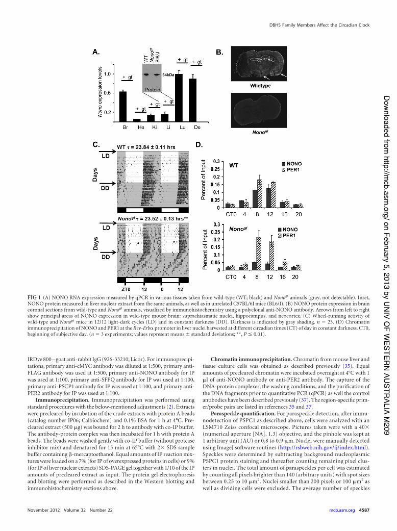
FIG 1 (A) NONO RNA expression measured by qPCR in various tissues taken from wild-type (WT; black) and Nonogt animals (gray, not detectable). Inset,NONO protein measured in liver nuclear extract from the same animals, as well as in unrelated C57BL/6J mice (BL6/J). (B) NONO protein expression in braincoronal sections from wild-type and Nonogt animals, visualized by immunohistochemistry using a polyclonal anti-NONO antibody. Arrows from left to rightshow principal areas of NONO expression in wild-type mouse brain: suprachiasmatic nuclei, hippocampus, and neocortex. (C) Wheel-running activity ofwild-type and Nonogt mice in 12/12 light-dark cycles (LD) and in constant darkness (DD). Darkness is indicated by gray shading. n ' 23. (D) Chromatinimmunoprecipitation of NONO and PER1 at the Rev-Erb! promoter in liver nuclei harvested at different circadian times (CT) of day in constant darkness. CT0,beginning of subjective day. (n ' 3 experiments; values represent means ( standard deviations; **, P ! 0.01).
DBHS Family Members Affect the Circadian Clock
November 2012 Volume 32 Number 22 mcb.asm.org 4587
on February 5, 2013 by UN
IV OF W
ESTERN
AUSTR
ALIA M209
http://mcb.asm
.org/D
ownloaded from
was normalized to the mean area and compared to the control trans-fected cells (hairpin NEAT-S).
RESULTSNONO-deficient mice show significant changes in circadian pe-riod. To better understand the function of NONO in the circadianclock and in mammalian physiology, we obtained NONO-defi-cient mice from ES cells bearing a gene trap in the intron preced-ing the Nono translational start site (see Fig. S1A and B in thesupplemental material). In wild-type mice, NONO is expressed inmost tissues including the suprachiasmatic nuclei in the brain.Nonogt mice showed no expression of Nono mRNA or protein inany of the tissues examined (Fig. 1A and B). These mice showed a20-min reduction in circadian behavioral period when under con-stant dark conditions (Fig. 1C). This reduction in period lengthwas highly significant, but it was nevertheless far less dramaticthan that in a Drosophila hypomorphic nonA strain that we ob-served previously to become arrhythmic (5). Hence, we suspectedthat in mammals the lack of Nono may be compensated by otherfactors.
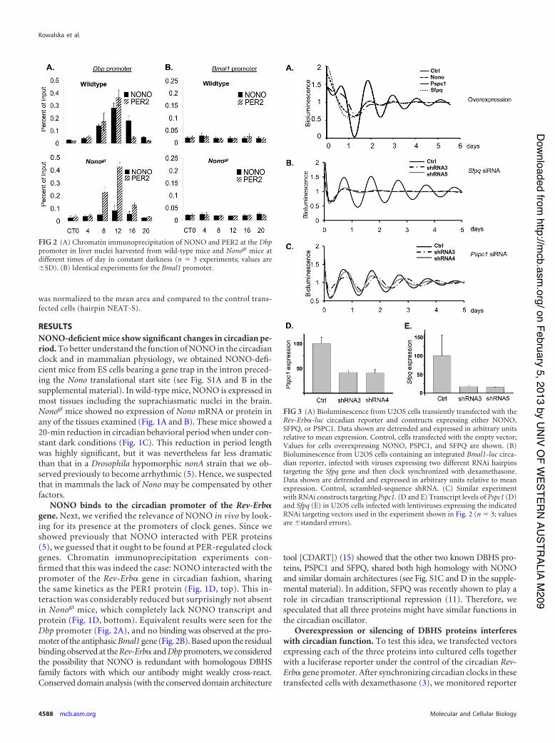
NONO binds to the circadian promoter of the Rev-Erb!gene. Next, we verified the relevance of NONO in vivo by look-ing for its presence at the promoters of clock genes. Since weshowed previously that NONO interacted with PER proteins(5), we guessed that it ought to be found at PER-regulated clockgenes. Chromatin immunoprecipitation experiments con-firmed that this was indeed the case: NONO interacted with thepromoter of the Rev-Erb! gene in circadian fashion, sharingthe same kinetics as the PER1 protein (Fig. 1D, top). This in-teraction was considerably reduced but surprisingly not absentin Nonogt mice, which completely lack NONO transcript andprotein (Fig. 1D, bottom). Equivalent results were seen for theDbp promoter (Fig. 2A), and no binding was observed at the pro-moter of the antiphasic Bmal1 gene (Fig. 2B). Based upon the residualbinding observed at the Rev-Erb! and Dbp promoters, we consideredthe possibility that NONO is redundant with homologous DBHSfamily factors with which our antibody might weakly cross-react.Conserved domain analysis (with the conserved domain architecture
tool [CDART]) (15) showed that the other two known DBHS pro-teins, PSPC1 and SFPQ, shared both high homology with NONOand similar domain architectures (see Fig. S1C and D in the supple-mental material). In addition, SFPQ was recently shown to play arole in circadian transcriptional repression (11). Therefore, wespeculated that all three proteins might have similar functions inthe circadian oscillator.
Overexpression or silencing of DBHS proteins interfereswith circadian function. To test this idea, we transfected vectorsexpressing each of the three proteins into cultured cells togetherwith a luciferase reporter under the control of the circadian Rev-Erb! gene promoter. After synchronizing circadian clocks in thesetransfected cells with dexamethasone (3), we monitored reporter
FIG 3 (A) Bioluminescence from U2OS cells transiently transfected with theRev-Erb!-luc circadian reporter and constructs expressing either NONO,SFPQ, or PSPC1. Data shown are detrended and expressed in arbitrary unitsrelative to mean expression. Control, cells transfected with the empty vector;Values for cells overexpressing NONO, PSPC1, and SFPQ are shown. (B)Bioluminescence from U2OS cells containing an integrated Bmal1-luc circa-dian reporter, infected with viruses expressing two different RNAi hairpinstargeting the Sfpq gene and then clock synchronized with dexamethasone.Data shown are detrended and expressed in arbitrary units relative to meanexpression. Control, scrambled-sequence shRNA. (C) Similar experimentwith RNAi constructs targeting Pspc1. (D and E) Transcript levels of Pspc1 (D)and Sfpq (E) in U2OS cells infected with lentiviruses expressing the indicatedRNAi targeting vectors used in the experiment shown in Fig. 2 (n ' 3; valuesare (standard errors).
FIG 2 (A) Chromatin immunoprecipitation of NONO and PER2 at the Dbppromoter in liver nuclei harvested from wild-type mice and Nonogt mice atdifferent times of day in constant darkness (n ' 3 experiments; values are(SD). (B) Identical experiments for the Bmal1 promoter.
Kowalska et al.
4588 mcb.asm.org Molecular and Cellular Biology
on February 5, 2013 by UN
IV OF W
ESTERN
AUSTR
ALIA M209
http://mcb.asm
.org/D
ownloaded from
bioluminescence in real time. Overexpression of any of the threeproteins in human U2OS fibroblasts perturbed circadian rhyth-micity (Fig. 3A).
We next undertook loss-of-function experiments based uponRNA interference (RNAi), in which U2OS human osteosarcomacells containing an integrated Bmal1-luc reporter were infected
with lentiviruses expressing short hairpin RNAs (shRNAs) target-ing Pspc1 or Sfpq. RNAi hairpins against SFPQ dampened circa-dian oscillations dramatically (Fig. 3B) similar to what was ob-served previously for NONO (5), but those against PSPC1lengthened it and somewhat dampened amplitude (Fig. 3C). Mea-surement of Sfpq and Pspc1 RNA levels in these cells showed thatthese hairpins reduced expression of Sfpq by 7-fold and of Pspc1 by2.5-fold (Fig. 3D and E).
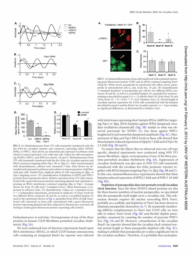
To ensure that the effects that we observed were not cell typespecific, identical experiments were conducted using NIH 3T3mouse fibroblasts. Again, overexpression of any of the three pro-teins perturbed circadian rhythmicity (Fig. 4A). Suppression ofcircadian rhythmicity was also seen in NIH 3T3 cells transientlytransfected with the circadian Rev-Erb! promoter reporter to-gether with RNAi hairpins targeting Pspc1 or Sfpq (Fig. 4B and C).In this case, immunofluorescence experiments showed that thesehairpins reduced expression of SFPQ 2-fold and of PSPC1 10-fold(Fig. 4D).
Depletion of paraspeckles does not perturb overall circadianclock function. Since the three NONO-related proteins are alsothe three known members of nuclear paraspeckles, we speculatedthat the paraspeckle itself might serve a circadian role. This sub-nuclear domain requires the nuclear noncoding RNA Neat1,probably as a scaffold, and depletion of Neat1 has been shown toeliminate paraspeckles themselves (6, 7). By transiently transfect-ing shRNAs complementary to Neat1 into U2OS cells, we wereable to reduce Neat1 levels (Fig. 4E) and thereby deplete paras-peckles, measured by counting the number of punctate PSPC1foci (Fig. 5A and B). However, cotransfection of the circadianBmal1-luc reporter showed that the circadian clock retained nor-mal period length in these paraspeckle-depleted cells (Fig. 5C),making it unlikely that paraspeckles per se play a significant role inthe circadian oscillator. Therefore, it is probable that nucleoplas-
FIG 4 (A) Bioluminescence from 3T3 cells transiently transfected with theRev-Erb!-luc circadian reporter and constructs expressing either NONO,SFPQ, or PSPC1. Data shown are detrended and expressed in arbitrary unitsrelative to mean expression. Ctrl, wild-type cells. Values for cells overexpress-ing NONO, PSPC1, and SFPQ are shown. (B and C) Bioluminescence from3T3 cells transiently transfected with the Rev-Erb!-luc circadian reporter andRNAi constructs targeting either Pspc1 (B) or Sfpq (C). After synchronizationwith dexamethasone, cultures were measured 3 days. Data shown are de-trended and expressed in arbitrary units relative to mean expression. Solid line,wild-type cells. Dashed lines, duplicate plates of cells expressing an Sfpq- orPspc1-targeting vector. (D) Quantification of depletion of SFPQ and PSPC1proteins from experiments above. Relative repression from 3T3 cells cotrans-fected with a green fluorescent protein-expressing plasmid and a plasmid ex-pressing an RNAi interference construct targeting Sfpq or Pspc1. Averagesshown are from 10 cells each ((standard errors). Mean fluorescence is ex-pressed in arbitrary units. (E) Quantification (values are (standard errors;n ' 2 independent experiments, performed in triplicate) of Neat1 levels fortwo different RNAi constructs (R and B), as well as a scrambled hairpin (S)used in the experiment shown in Fig. 4, quantified from RNA of bulk-trans-fected cells (unsorted) or from cells cotransfected with a green fluorescentprotein-expressing plasmid and then subjected to fluorescence-activated cellsorting to isolate green fluorescent protein-expressing cells (sorted).
FIG 5 (A) Immunofluorescence from cells transfected with a plasmid express-ing green fluorescent protein (GFP) and an RNAi construct targeting Neat1(Neat-R). White arrow, paraspeckle in transfected cell; yellow arrow, paras-peckle in untransfected cell; !, anti. Scale bar, 10 $m. (B) Quantification((standard deviation) of paraspeckles per cell for two different RNAi con-structs (R and B), as well as a scrambled hairpin (S), quantified by immuno-staining as described for panel A (n ' 12 cells for Neat1-R, 24 for Neat1-B, and18 for Neat1-S). *, P ) 0.05; **, P )0.01 (Student t test.) (C) Period length ofcircadian reporter expression for U2OS cells cotransfected with the hairpinsdescribed for panel A and the Bmal1-luc circadian reporter. (n ' 6 per sample;no significant differences, as determined by a Student t test).
DBHS Family Members Affect the Circadian Clock
November 2012 Volume 32 Number 22 mcb.asm.org 4589
on February 5, 2013 by UN
IV OF W
ESTERN
AUSTR
ALIA M209
http://mcb.asm
.org/D
ownloaded from
mic, non-paraspeckle-associated pools of NONO, SFPQ, andPSPC1 proteins were responsible for the circadian effects that wehave documented.
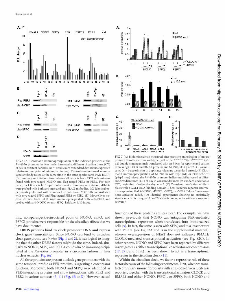
DBHS proteins bind to clock promoter DNA and repressclock gene transcription. Since NONO can bind to circadianclock gene promoters in vivo (Fig. 1 and 2), it was logical to imag-ine that the other DBHS factors might do the same. Indeed, sim-ilarly to NONO, SFPQ and PSPC1 could also be immunoprecipi-tated at the Rev-Erb! promoter in a circadian fashion in livernuclear extracts (Fig. 6A).
All three proteins are present at clock gene promoters with thesame temporal profile as PER proteins, suggesting a corepressorfunction. Moreover, both NONO and SFPQ were identified asPER-interacting proteins and show interactions with PER1 andPER2 in various contexts (5, 11) (Fig. 6B to D). However, actual
functions of these proteins are less clear. For example, we haveshown previously that NONO can antagonize PER-mediatedtranscriptional repression when transfected into immortalizedcells (5). In fact, the same is seen with SFPQ and to a lesser extentwith PSPC1 (see Fig S2A and B in the supplemental material),whereas overexpression of NEAT does not influence BMAL1/CLOCK-mediated transcriptional activation (see Fig. S2C). Inother reports, NONO and SFPQ have been reported by differentinvestigators as either transcriptional coactivators or corepressors(17, 27), and SFPQ has been shown to act as a transcriptionalrepressor in the circadian clock (11).
Within the circadian clock, we favor a repressive role of thesefactors because of the following experiments. First, when we trans-fected primary mouse fibroblasts with an E-box-driven luciferasereporter, together with the transcriptional activators CLOCK andBMAL1 and either NONO, PSPC1, or SFPQ, both NONO and
FIG 7 (A) Bioluminescence measured after transient transfection of mouseprimary fibroblasts from wild-type (wt) or per1brdm/brdm/per2brdm/brdm (p1/p2) double mutant animals transfected with an E-box-luc reporter and vectorsexpressing CLOCK and BMAL proteins and NONO, SFPQ, or PSPC1 as indi-cated (n ' 3 experiments in duplicate; values are (standard errors). (B) Chro-matin immunoprecipitation of NONO in wild-type (wt) or PER-deficient(p1/p2 mut) mice at the Rev-Erb! promoter in liver nuclei harvested at differ-ent circadian times (CT) of day in constant darkness ((standard deviations).CT0, beginning of subjective day. n ' 3. (C) Transient transfection of fibro-blasts with a GAL4 DNA-binding domain-E box-luciferase reporter and vec-tors expressing GAL4-NONO, -PSPC1, -SFPQ, or -VP16. “alone,” no exoge-nous activator added. (D) Identical experiments showing no statisticallysignificant effects using a GAL4-CMV-luciferase reporter without exogenousactivator.
FIG 6 (A) Chromatin immunoprecipitation of the indicated proteins at theRev-Erb! promoter in liver nuclei harvested at different circadian times (CT)of day in constant darkness (n ' 4; values are (standard deviations, expressedrelative to time point of minimum binding). Control reactions used an unre-lated antibody raised at the same time in the same species (anti-PAR-BZIP).(B) Immunoprecipitations from whole-cell extracts from 293T cells cotrans-fected with myc-tagged NONO and Flag-tagged PER1 or PER2. For eachpanel, the left lane is 1/10 input. Subsequent to immunoprecipitation, all blotswere probed with both anti-myc and anti-FLAG antibodies. (C) Identical ex-periments performed with whole-cell extracts from 293T cells cotransfectedwith myc-tagged SFPQ and Flag-tagged PER1 or PER2. (D) Mouse liver nu-clear extracts from CT16 were immunoprecipitated with anti-PER2 andprobed with anti-NONO or anti-SFPQ. Left lane, 1/10 input.
Kowalska et al.
4590 mcb.asm.org Molecular and Cellular Biology
on February 5, 2013 by UN
IV OF W
ESTERN
AUSTR
ALIA M209
http://mcb.asm
.org/D
ownloaded from
SFPQ repressed CLOCK-BMAL-mediated transcription from thereporter and, to a lesser extent, PSPC1, which was initially activat-ing and then repressing at higher concentrations (Fig. 7A). Whenequivalent transfections were performed using fibroblasts fromPer1brdm/brdm/Per2brdm/brdm mice that lack functional PER pro-teins and circadian clocks (45), repression was no longer observed,but instead weak activation was shown (Fig. 7A). Similarly, inPer1brdm/brdm/Per2brdm/brdm mice, no circadian immunoprecipita-tion of NONO was observed at the Rev-Erb! promoter (Fig. 7B).Therefore, PER recruits NONO and, presumably, the other familymembers too.
Second, to confirm that DBHS factors are repressors at cir-cadian promoters, we designed a hybrid GAL4 DNA-bindingdomain (GAL4DBD)-E box-luciferase reporter and fusions ofNONO, PSPC1, and SFPQ with the GAL4 DNA-binding do-main in order to enable their direct recruitment to DNA inde-pendently of PER proteins. When the GAL4DBD-E box-lu-ciferase construct was transfected into primary mousefibroblasts together with the GAL4-VP16 transcriptional acti-vator, strong activation was observed, demonstrating the func-tionality of the construct. When GAL4-NONO, -PSPC1, and-SFPQ were cotransfected into primary mouse fibroblasts to-gether with the GAL4 DNA-binding domain-E box-luciferasereporter, all three proteins strongly repressed CLOCK-BMAL-mediated transcription (Fig. 7C) though they had no statisti-cally significant effect when similarly recruited to the constitu-tively active CMV promoter (Fig. 7D).
Importance of DBHS proteins to circadian behavior. Finally,
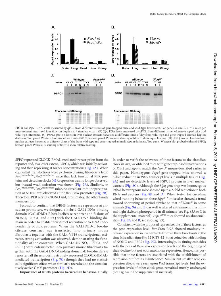
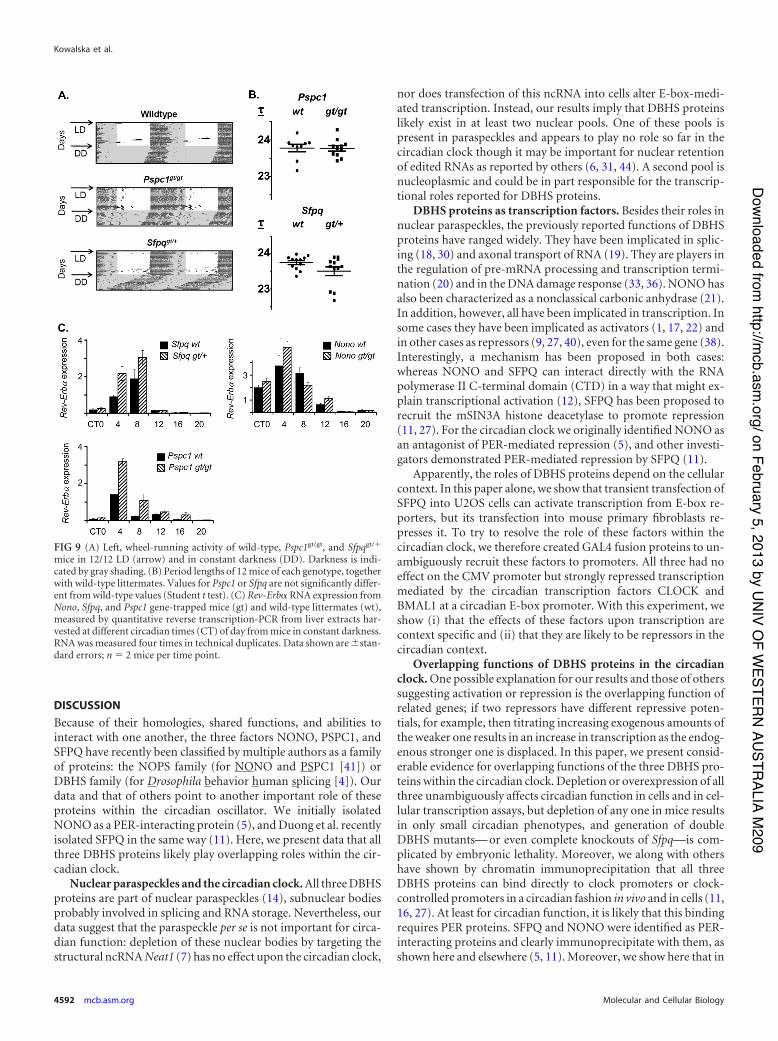
in order to verify the relevance of these factors to the circadianclock in vivo, we obtained mice with gene trap-based inactivationsof Pspc1 and Sfpq to match the Nonogt mouse described earlier inthis paper. Homozygous Pspc1-gene-trapped mice showed a5-fold reduction in Pspc1 transcript levels in multiple tissues (Fig.8A) and no detectable levels of PSPC1 protein in liver nuclearextracts (Fig. 8C). Although the Sfpq gene trap was homozygouslethal, heterozygous mice showed up to a 2-fold reduction in bothRNA and protein (Fig. 8B and D). When tested for circadianwheel-running behavior, these Sfpqgt/" mice also showed a trendtoward shortening of period similar to that of Nonogt in someanimals (Fig. 9A and B), as well as altered entrainment in a mini-mal-light skeleton photoperiod in all animals (see Fig. S3A to C inthe supplemental material). Pspc1gt/gt mice showed no abnormal-ities (Fig. 9A and B; see also Fig. S3).
Consistent with the proposed repressive role of these factors, atthe gene expression level, Rev-Erb! RNA showed modestly in-creased expression in liver extracts from all three knockouts at thetime (circadian time 8 to 12 [CT8-12]) that coincides with bindingof NONO and PER2 (Fig. 9C). Interestingly, its timing coincideswith the peak of Rev-Erb! expression levels and the beginning oftheir decline but not with maximum repression. Hence, it is pos-sible that these factors are associated with the establishment ofrepression but not its maintenance. Similar but smaller gene ex-pression effects were seen upon Per2 transcript levels, but the ex-pression levels of other clock genes remained mostly unchanged(see Fig. S4 in the supplemental material).
FIG 8 (A) Pspc1 RNA levels measured by qPCR from different tissues of gene-trapped mice and wild-type littermates. For panels A and B, n ' 2 mice permeasurement, measured four times in duplicate, (standard errors. (B) Sfpq RNA levels measured by qPCR from different tissues of gene-trapped mice andwild-type littermates. (C) PSPC1 protein levels in liver nuclear extracts harvested at different times of day from wild-type and gene-trapped animals kept indarkness. Top panel, Western blot probed with anti-PSPC1; bottom panel, Ponceau-S staining of filter to show equal loading. (D) SFPQ protein levels in livernuclear extracts harvested at different times of day from wild-type and gene-trapped animals kept in darkness. Top panel, Western blot probed with anti-SFPQ;bottom panel, Ponceau-S staining of filter to show relative loading.
DBHS Family Members Affect the Circadian Clock
November 2012 Volume 32 Number 22 mcb.asm.org 4591
on February 5, 2013 by UN
IV OF W
ESTERN
AUSTR
ALIA M209
http://mcb.asm
.org/D
ownloaded from
DISCUSSIONBecause of their homologies, shared functions, and abilities tointeract with one another, the three factors NONO, PSPC1, andSFPQ have recently been classified by multiple authors as a familyof proteins: the NOPS family (for NONO and PSPC1 [41]) orDBHS family (for Drosophila behavior human splicing [4]). Ourdata and that of others point to another important role of theseproteins within the circadian oscillator. We initially isolatedNONO as a PER-interacting protein (5), and Duong et al. recentlyisolated SFPQ in the same way (11). Here, we present data that allthree DBHS proteins likely play overlapping roles within the cir-cadian clock.
Nuclear paraspeckles and the circadian clock. All three DBHSproteins are part of nuclear paraspeckles (14), subnuclear bodiesprobably involved in splicing and RNA storage. Nevertheless, ourdata suggest that the paraspeckle per se is not important for circa-dian function: depletion of these nuclear bodies by targeting thestructural ncRNA Neat1 (7) has no effect upon the circadian clock,
nor does transfection of this ncRNA into cells alter E-box-medi-ated transcription. Instead, our results imply that DBHS proteinslikely exist in at least two nuclear pools. One of these pools ispresent in paraspeckles and appears to play no role so far in thecircadian clock though it may be important for nuclear retentionof edited RNAs as reported by others (6, 31, 44). A second pool isnucleoplasmic and could be in part responsible for the transcrip-tional roles reported for DBHS proteins.
DBHS proteins as transcription factors. Besides their roles innuclear paraspeckles, the previously reported functions of DBHSproteins have ranged widely. They have been implicated in splic-ing (18, 30) and axonal transport of RNA (19). They are players inthe regulation of pre-mRNA processing and transcription termi-nation (20) and in the DNA damage response (33, 36). NONO hasalso been characterized as a nonclassical carbonic anhydrase (21).In addition, however, all have been implicated in transcription. Insome cases they have been implicated as activators (1, 17, 22) andin other cases as repressors (9, 27, 40), even for the same gene (38).Interestingly, a mechanism has been proposed in both cases:whereas NONO and SFPQ can interact directly with the RNApolymerase II C-terminal domain (CTD) in a way that might ex-plain transcriptional activation (12), SFPQ has been proposed torecruit the mSIN3A histone deacetylase to promote repression(11, 27). For the circadian clock we originally identified NONO asan antagonist of PER-mediated repression (5), and other investi-gators demonstrated PER-mediated repression by SFPQ (11).
Apparently, the roles of DBHS proteins depend on the cellularcontext. In this paper alone, we show that transient transfection ofSFPQ into U2OS cells can activate transcription from E-box re-porters, but its transfection into mouse primary fibroblasts re-presses it. To try to resolve the role of these factors within thecircadian clock, we therefore created GAL4 fusion proteins to un-ambiguously recruit these factors to promoters. All three had noeffect on the CMV promoter but strongly repressed transcriptionmediated by the circadian transcription factors CLOCK andBMAL1 at a circadian E-box promoter. With this experiment, weshow (i) that the effects of these factors upon transcription arecontext specific and (ii) that they are likely to be repressors in thecircadian context.
Overlapping functions of DBHS proteins in the circadianclock. One possible explanation for our results and those of otherssuggesting activation or repression is the overlapping function ofrelated genes; if two repressors have different repressive poten-tials, for example, then titrating increasing exogenous amounts ofthe weaker one results in an increase in transcription as the endog-enous stronger one is displaced. In this paper, we present consid-erable evidence for overlapping functions of the three DBHS pro-teins within the circadian clock. Depletion or overexpression of allthree unambiguously affects circadian function in cells and in cel-lular transcription assays, but depletion of any one in mice resultsin only small circadian phenotypes, and generation of doubleDBHS mutants— or even complete knockouts of Sfpq—is com-plicated by embryonic lethality. Moreover, we along with othershave shown by chromatin immunoprecipitation that all threeDBHS proteins can bind directly to clock promoters or clock-controlled promoters in a circadian fashion in vivo and in cells (11,16, 27). At least for circadian function, it is likely that this bindingrequires PER proteins. SFPQ and NONO were identified as PER-interacting proteins and clearly immunoprecipitate with them, asshown here and elsewhere (5, 11). Moreover, we show here that in
FIG 9 (A) Left, wheel-running activity of wild-type, Pspc1gt/gt, and Sfpqgt/"
mice in 12/12 LD (arrow) and in constant darkness (DD). Darkness is indi-cated by gray shading. (B) Period lengths of 12 mice of each genotype, togetherwith wild-type littermates. Values for Pspc1 or Sfpq are not significantly differ-ent from wild-type values (Student t test). (C) Rev-Erb! RNA expression fromNono, Sfpq, and Pspc1 gene-trapped mice (gt) and wild-type littermates (wt),measured by quantitative reverse transcription-PCR from liver extracts har-vested at different circadian times (CT) of day from mice in constant darkness.RNA was measured four times in technical duplicates. Data shown are (stan-dard errors; n ' 2 mice per time point.
Kowalska et al.
4592 mcb.asm.org Molecular and Cellular Biology
on February 5, 2013 by UN
IV OF W
ESTERN
AUSTR
ALIA M209
http://mcb.asm
.org/D
ownloaded from
PER-deficient mice, binding of NONO to circadian promoters isno longer observed. Thus, we think it is likely that PER proteinsrecruit DBHS proteins to clock-controlled genes to control andorchestrate PER-mediated transcriptional repression. The degreeof this repression could be precisely controlled by the mix of thefactors recruited.
Functional redundancy of this family of proteins is also high-lighted by the recently published crystal structure of a NONO-PSPC1complex (29). Not only do these proteins probably form obligateheterodimers, which would suggest a role for multiple DBHS familymembers within the circadian clock, but their structure also allowsfor possible higher-order oligomers, which might provide an idealplatform for the recruitment of other factors that have been foundassociated with these factors in various contexts.
DBHS proteins as orchestrators of circadian physiology. Al-though we have shown clear roles of DBHS proteins in a cellularcontext, the circadian behavioral phenotypes of DBHS protein-deficient mice were relatively minor. As discussed above, func-tional redundancy could account for this lack of phenotype. Inaddition, however, the unique coupling of SCN cells into a net-work renders them more resilient to the effects of mutation (23).Therefore, it is also possible that more severe circadian effects ofDBHS proteins occur in peripheral tissues.
Indeed, it is likely that considerable further circadian physiol-ogy directed by DBHS proteins remains to be elucidated. Micedeficient in these factors show a spectrum of unique phenotypes,ranging from embryonic lethality (Sfpq) to neurological pheno-types (Nono). Pspc1 protein is strongly regulated in circadian fash-ion although the other two factors are not (Fig. 8 and data notshown). Moreover, the E box is a standard motif for orchestratingclock-controlled physiology (35) and directs circadian transcrip-tion at thousands of promoters (42). DBHS factor binding hasbeen observed at multiple clock-regulated promoters containingthis motif, including prolactin (16), progesterone (10), Rev-Erb!(Fig. 1B), and androgen receptor (9). Through their interactionwith PER proteins, we show here that DBHS factors play an im-portant role directly in the circadian oscillator. Binding to clockgene promoters and modulating transcriptional repression, theyregulate a portion of the transcriptional feedback which is thehallmark of metazoan circadian clocks.
ACKNOWLEDGMENTSWe thank Robert Dallmann for a critical reading of the manuscript andW. Schaffner (University of Zurich) for donation of Gal4 vectors.
This work was supported by the Swiss National Science Foundationand the University of Zurich Fonds der Akademischen Nachwuchses. Fur-ther support to S.A.B. was provided by the Neurosciences Center Zurichand the Molecular Life Sciences programs.
REFERENCES1. Amelio AL, et al. 2007. A coactivator trap identifies NONO (p54nrb) as a
component of the cAMP-signaling pathway. Proc. Natl. Acad. Sci. U. S. A.104:20314 –20319.
2. Ausubel FM, et al. 2003. Current protocols in molecular biology. J. Wiley& Sons, Inc., New York, NY.
3. Balsalobre A, et al. 2000. Resetting of circadian time in peripheral tissuesby glucocorticoid signaling. Science 289:2344 –2347.
4. Bond CS, Fox AH. 2009. Paraspeckles: nuclear bodies built on longnoncoding RNA. J. Cell Biol. 186:637– 644.
5. Brown SA, et al. 2005. PERIOD1-associated proteins modulate the neg-ative limb of the mammalian circadian oscillator. Science 308:693– 696.
6. Chen LL, Carmichael GG. 2009. Altered nuclear retention of mRNAs
containing inverted repeats in human embryonic stem cells: functionalrole of a nuclear noncoding RNA. Mol. Cell 35:467– 478.
7. Clemson CM, et al. 2009. An architectural role for a nuclear noncodingRNA: NEAT1 RNA is essential for the structure of paraspeckles. Mol. Cell33:717–726.
8. Dibner C, Schibler U, Albrecht U. 2010. The mammalian circadiantiming system: organization and coordination of central and peripheralclocks. Annu. Rev. Physiol. 72:517–549.
9. Dong X, Sweet J, Challis JR, Brown T, Lye SJ. 2007. Transcriptionalactivity of androgen receptor is modulated by two RNA splicing factors,PSF and p54nrb. Mol. Cell. Biol. 27:4863– 4875.
10. Dong X, et al. 2009. p54nrb is a transcriptional corepressor of the pro-gesterone receptor that modulates transcription of the labor-associatedgene, connexin 43 (Gja1). Mol. Endocrinol. 23:1147–1160.
11. Duong HA, Robles MS, Knutti D, Weitz CJ. 2011. A molecular mech-anism for circadian clock negative feedback. Science 332:1436 –1439.
12. Emili A, et al. 2002. Splicing and transcription-associated proteins PSF andp54nrb/nonO bind to the RNA polymerase II CTD. RNA 8:1102–1111.
13. Fox AH, Bond CS, Lamond AI. 2005. P54nrb forms a heterodimer withPSP1 that localizes to paraspeckles in an RNA-dependent manner. Mol.Biol. Cell 16:5304 –5315.
14. Fox AH, Lamond AI. 2010. Paraspeckles. Cold Spring Harb Perspect Biol.2:a000687. doi:10.1101/cshperspect.a000687.
15. Geer LY, Domrachev M, Lipman DJ, Bryant SH. 2002. CDART: proteinhomology by domain architecture. Genome Res. 12:1619 –1623.
16. Guillaumond F, et al. 2011. Chromatin remodeling as a mechanism forcircadian prolactin transcription: rhythmic NONO and SFPQ recruit-ment to HLTF. FASEB J. 25:2740 –2756.
17. Ishitani K, et al. 2003. p54nrb acts as a transcriptional coactivator foractivation function 1 of the human androgen receptor. Biochem. Biophys.Res. Commun. 306:660 – 665.
18. Kameoka S, Duque P, Konarska MM. 2004. p54(nrb) associates with the5= splice site within large transcription/splicing complexes. EMBO J. 23:1782–1791.
19. Kanai Y, Dohmae N, Hirokawa N. 2004. Kinesin transports RNA: isolationand characterization of an RNA-transporting granule. Neuron 43:513–525.
20. Kaneko S, Rozenblatt-Rosen O, Meyerson M, Manley JL. 2007. Themultifunctional protein p54nrb/PSF recruits the exonuclease XRN2 tofacilitate pre-mRNA 3= processing and transcription termination. GenesDev. 21:1779 –1789.
21. Karhumaa P, et al. 2000. Nuclear NonO/p54(nrb) protein is a nonclas-sical carbonic anhydrase. J. Biol. Chem. 275:16044 –16049.
22. Kuwahara S, et al. 2006. PSPC1, NONO, and SFPQ are expressed inmouse Sertoli cells and may function as coregulators of androgen recep-tor-mediated transcription. Biol. Reprod. 75:352–359.
23. Liu AC, et al. 2007. Intercellular coupling confers robustness againstmutations in the SCN circadian clock network. Cell 129:605– 616.
24. Lopez-Molina L, Conquet F, Dubois-Dauphin M, Schibler U. 1997. TheDBP gene is expressed according to a circadian rhythm in the suprachias-matic nucleus and influences circadian behavior. EMBO J. 16:6762– 6771.
25. Maier B, et al. 2009. A large-scale functional RNAi screen reveals a role forCK2 in the mammalian circadian clock. Genes Dev. 23:708 –718.
26. Masri S, Sassone-Corsi P. 2010. Plasticity and specificity of the circadianepigenome. Nat. Neurosci. 13:1324 –1329.
27. Mathur M, Tucker PW, Samuels HH. 2001. PSF is a novel corepressorthat mediates its effect through Sin3A and the DNA binding domain ofnuclear hormone receptors. Mol. Cell. Biol. 21:2298 –2311.
28. Nagoshi E, et al. 2004. Circadian gene expression in individual fibro-blasts: cell-autonomous and self-sustained oscillators pass time to daugh-ter cells. Cell 119:693–705.
29. Passon DM, et al. 2012. Structure of the heterodimer of human NONOand paraspeckle protein component 1 and analysis of its role in subnuclearbody formation. Proc. Natl. Acad. Sci. U. S. A. 109:4846 – 4850.
30. Patton JG, Porro EB, Galceran J, Tempst P, Nadal-Ginard B. 1993.Cloning and characterization of PSF, a novel pre-mRNA splicing factor.Genes Dev. 7:393– 406.
31. Prasanth KV, et al. 2005. Regulating gene expression through RNA nu-clear retention. Cell 123:249 –263.
32. Preitner N, et al. 2002. The orphan nuclear receptor REV-ERB! controlscircadian transcription within the positive limb of the mammalian circa-dian oscillator. Cell 110:251–260.
33. Proteau A, et al. 2005. The multifunctional nuclear protein p54nrb is
DBHS Family Members Affect the Circadian Clock
November 2012 Volume 32 Number 22 mcb.asm.org 4593
on February 5, 2013 by UN
IV OF W
ESTERN
AUSTR
ALIA M209
http://mcb.asm
.org/D
ownloaded from
multiphosphorylated in mitosis and interacts with the mitotic regulatorPin1. J. Mol. Biol. 346:1163–1172.
34. Reischl S, Kramer A. 2011. Kinases and phosphatases in the mammaliancircadian clock. FEBS Lett. 585:1393–1399.
35. Ripperger JA, Schibler U. 2006. Rhythmic CLOCK-BMAL1 binding tomultiple E-box motifs drives circadian Dbp transcription and chromatintransitions. Nat. Genet. 38:369 –374.
36. Salton M, Lerenthal Y, Wang SY, Chen DJ, Shiloh Y. 2010. Involvementof matrin 3 and SFPQ/NONO in the DNA damage response. Cell Cycle9:1568 –1576.
37. Schmutz I, Ripperger JA, Baeriswyl-Aebischer S, Albrecht U. 2010. Themammalian clock component PERIOD2 coordinates circadian output byinteraction with nuclear receptors. Genes Dev. 24:345–357.
38. Sewer MB, et al. 2002. Transcriptional activation of human CYP17 in H295Radrenocortical cells depends on complex formation among p54(nrb)/NonO,protein-associated splicing factor, and SF-1, a complex that also participatesin repression of transcription. Endocrinology 143:1280–1290.
39. Shav-Tal Y, Zipori D. 2002. PSF and p54(nrb)/NonO—multi-functionalnuclear proteins. FEBS Lett. 531:109 –114.
40. Song KS, Kim K, Chung KC, Seol JH, Yoon JH. 2008. Interaction ofSOCS3 with NonO attenuates IL-1beta-dependent MUC8 gene expres-sion. Biochem. Biophys. Res. Commun. 377:946 –951.
41. Staub E, Fiziev P, Rosenthal A, Hinzmann B. 2004. Insights into theevolution of the nucleolus by an analysis of its protein domain repertoire.Bioessays 26:567–581.
42. Ueda HR, et al. 2005. System-level identification of transcriptional cir-cuits underlying mammalian circadian clocks. Nat. Genet. 37:187–192.
43. Xie WQ, Rothblum LI. 1991. Rapid, small-scale RNA isolation fromtissue culture cells. Biotechniques 11:324, 326 –327.
44. Zhang Z, Carmichael GG. 2001. The fate of dsRNA in the nucleus: ap54(nrb)-containing complex mediates the nuclear retention of promis-cuously A-to-I edited RNAs. Cell 106:465– 475.
45. Zheng B, et al. 2001. Nonredundant roles of the mPer1 and mPer2 genesin the mammalian circadian clock. Cell 105:683– 694.
Kowalska et al.
4594 mcb.asm.org Molecular and Cellular Biology
on February 5, 2013 by UN
IV OF W
ESTERN
AUSTR
ALIA M209
http://mcb.asm
.org/D
ownloaded from
Related Documents