, 20120471, published 20 January 2014 369 2014 Phil. Trans. R. Soc. B Federico Tinarelli, Celina Garcia-Garcia, Francesco Nicassio and Valter Tucci sleep loss levels of circadian and neuronal plasticity genes following Parent-of-origin genetic background affects the transcriptional Supplementary data ml http://rstb.royalsocietypublishing.org/content/suppl/2014/01/13/rstb.2012.0471.DC1.ht "Data Supplement" References http://rstb.royalsocietypublishing.org/content/369/1637/20120471.full.html#ref-list-1 This article cites 66 articles, 14 of which can be accessed free Subject collections (437 articles) neuroscience (75 articles) genetics (502 articles) behaviour Articles on similar topics can be found in the following collections Email alerting service here right-hand corner of the article or click Receive free email alerts when new articles cite this article - sign up in the box at the top http://rstb.royalsocietypublishing.org/subscriptions go to: Phil. Trans. R. Soc. B To subscribe to on January 20, 2014 rstb.royalsocietypublishing.org Downloaded from on January 20, 2014 rstb.royalsocietypublishing.org Downloaded from

Welcome message from author
This document is posted to help you gain knowledge. Please leave a comment to let me know what you think about it! Share it to your friends and learn new things together.
Transcript
, 20120471, published 20 January 2014369 2014 Phil. Trans. R. Soc. B Federico Tinarelli, Celina Garcia-Garcia, Francesco Nicassio and Valter Tucci sleep losslevels of circadian and neuronal plasticity genes following Parent-of-origin genetic background affects the transcriptional
Supplementary data
ml http://rstb.royalsocietypublishing.org/content/suppl/2014/01/13/rstb.2012.0471.DC1.ht
"Data Supplement"
Referenceshttp://rstb.royalsocietypublishing.org/content/369/1637/20120471.full.html#ref-list-1
This article cites 66 articles, 14 of which can be accessed free
Subject collections
(437 articles)neuroscience � (75 articles)genetics �
(502 articles)behaviour � Articles on similar topics can be found in the following collections
Email alerting service hereright-hand corner of the article or click Receive free email alerts when new articles cite this article - sign up in the box at the top
http://rstb.royalsocietypublishing.org/subscriptions go to: Phil. Trans. R. Soc. BTo subscribe to
on January 20, 2014rstb.royalsocietypublishing.orgDownloaded from on January 20, 2014rstb.royalsocietypublishing.orgDownloaded from
on January 20, 2014rstb.royalsocietypublishing.orgDownloaded from
rstb.royalsocietypublishing.org
ResearchCite this article: Tinarelli F, Garcia-Garcia C,
Nicassio F, Tucci V. 2014 Parent-of-origin
genetic background affects the transcriptional
levels of circadian and neuronal plasticity
genes following sleep loss. Phil. Trans. R. Soc.
B 369: 20120471.
http://dx.doi.org/10.1098/rstb.2012.0471
One contribution of 14 to a Theme Issue
‘Timing in neurobiological processes: from
genes to behaviour’.
Subject Areas:genetics, neuroscience, behaviour
Keywords:parent-of-origin effects, sleep, gene, expression
Author for correspondence:Valter Tucci
e-mail: [email protected]
& 2014 The Author(s) Published by the Royal Society. All rights reserved.
Electronic supplementary material is available
at http://dx.doi.org/10.1098/rstb.2012.0471 or
via http://rstb.royalsocietypublishing.org.
Parent-of-origin genetic backgroundaffects the transcriptional levels ofcircadian and neuronal plasticitygenes following sleep loss
Federico Tinarelli1, Celina Garcia-Garcia1, Francesco Nicassio2 and Valter Tucci1
1Department of Neuroscience and Brain Technologies, Istituto Italiano di Tecnologia, via Morego,30, 16163 Genova, Italy2Center for Genomic Science of IIT@SEMM, Istituto Italiano di Tecnologia (IIT), 20139 Milan, Italy
Sleep homoeostasis refers to a process in which the propensity to sleep
increases as wakefulness progresses and decreases as sleep progresses. Sleep
is tightly organized around the circadian clock and is regulated by genetic
and epigenetic mechanisms. The homoeostatic response of sleep, which is
classically triggered by sleep deprivation, is generally measured as a rebound
effect of electrophysiological measures, for example delta sleep. However,
more recently, gene expression changes following sleep loss have been inves-
tigated as biomarkers of sleep homoeostasis. The genetic background of an
individual may affect this sleep-dependent gene expression phenotype. In
this study, we investigated whether parental genetic background differentially
modulates the expression of genes following sleep loss. We tested the progeny
of reciprocal crosses of AKR/J and DBA/2J mouse strains and we show a
parent-of-origin effect on the expression of circadian, sleep and neuronal plas-
ticity genes following sleep deprivation. Thus, we further explored, by in silico,
specific functions or upstream mechanisms of regulation and we observed
that several upstream mechanisms involving signalling pathways (i.e.
DICER1, PKA), growth factors (CSF3 and BDNF) and transcriptional regula-
tors (EGR2 and ELK4) may be differentially modulated by parental effects.
This is the first report showing that a behavioural manipulation (e.g. sleep
deprivation) in adult animals triggers specific gene expression responses
according to parent-of-origin genomic mechanisms. Our study suggests that
the same mechanism may be extended to other behavioural domains and
that the investigation of gene expression following experimental manipula-
tions should take seriously into account parent-of-origin effects.
1. IntroductionSleep is a genetically and epigenetically regulated phenomenon that is sub-
jected to two fundamental processes: a homoeostatic process and a circadian
process [1]. The homoeostatic process of sleep depends on previous wakeful-
ness, representing the pressure for sleep according to the time of day. The
circadian process dictates the timing of sleep; it is a self-sustained periodic
mechanism that develops with approximately 24 h, cell-autonomous, oscil-
lations. The molecular machinery that sets the circadian clock is composed of
positive and negative feedback loops, which involve transcriptional and trans-
lational core elements within the cell (reviewed in [2]). Alternative translational
and post-translational components participate in the fundamental modulatory
mechanisms that maintain circadian timing. These activities involve several epi-
genetic changes, such as histone modification, acetylation and methylation
[3,4], that modulate the dynamic on/off switches of physiological circadian
sleep–wake processes [4]. Sleep homoeostasis is biologically related to the cir-
cadian clock, and several clock gene mutations result in significant alterations to
the electrophysiological measures of sleep [5–7].
rstb.royalsocietypublishing.orgPhil.Trans.R.Soc.B
369:20120471
2
on January 20, 2014rstb.royalsocietypublishing.orgDownloaded from
Pioneering studies by Franken et al. [8] have shown that the
genetics of mouse strains influence electrophysiological
measures of sleep, for example slow oscillations in the delta
frequency range (1–4 Hz), a fundamental measure of sleep
intensity. Several studies in mice have shown that sleep depriv-
ation induces changes in gene expression and it has been
reported that the genetic backgrounds of mouse strains affect
the transcriptional changes that follow sleep loss [9]. However,
different studies have identified different classes of genes that
depend on sleep [10]. Transcriptome analysis in three mice
strains (AKR/J, C57BL/6J and DBA/2J) [9] described the
Homer1a gene as an ideal sleep-dependent target that is rapidly
and strongly induced by sleep deprivation in all strains.
In our study, we have tested for the first time whether the
effect of genetic background on sleep-dependent gene
expression is determined in a parent-of-origin manner.
Parent-of-origin effects (i.e. genomic imprinting) have been
suggested to modulate fundamental aspects of sleep. Clinical
observations of neurodevelopmental sleep disorders suggest
that genomic imprinting plays a pivotal role in the architec-
ture of both rapid eye movement (REM) and non-REM
(NREM) sleep [11–13]. Interestingly, diverse sleep deficits
occur in diseases, such as Prader–Willi syndrome (PWS)
and Angelman syndrome (AS), which are classically charac-
terized by opposing imprinting profiles. PWS is caused by
maternal duplications/paternal deletions of alleles on
chromosome 15q11–13, whereas AS is associated with
paternal duplications/maternal deletions on the same
region, 15q11–13. The former is characterized by REM
sleep abnormalities, excessive sleepiness and core tempera-
ture abnormalities [14–17], while the latter is characterized
by reductions in sleep. Sleep abnormalities associated with
the PWS/AS imprinting region may be linked to the
UBE3A gene. Indeed, the lack of the maternal allele of
Ube3a in mice results in reduced NREM sleep, deterioration
in REM sleep and an increased frequency of waking during
the dark-to-light transition [18]. Moreover, serotonin (5-HT)
2A receptors, mediating aminergic inhibition of REM-on
cells [19], are primarily expressed by maternal alleles [20].
The importance of studying the link between parental geno-
mic background and sleep has been emphasized by our
recent study in mice [21]. We have shown that loss of imprint-
ing of the maternally imprinted gene Gnas dramatically
affects REM and NREM physiology in mice [21]. To test the
hypothesis that parent-of-origin genetic background affects
the expression of specific genes, determining the presence
or the absence of a homoeostatic response to sleep loss, we
studied reciprocal crosses of two mouse strains that differ
in their homoeostatic responses to sleep deprivation: AKR/J
and DBA/2J. These two strains have distinct delta-power
profiles [8] and different gene expression responses [9,22]
after sleep deprivation. While AKR/J mice exhibit dramatic
increases in delta power after 6 h of sleep deprivation,
DBA/2J mice present a milder response following the same
deprivation protocol [8]. Furthermore, AKR/J mice show a
greater increase in mRNA levels of core circadian clock
genes, such as Bmal1, Clock, Cry1, Cry2, Per1 and Per2, after
6 h of sleep deprivation than do DBA/2J mice [22].
The rationale for our study involves the phenotypic
expression patterns of reciprocal heterozygous F1 mice.
A parent-of-origin effect would lead to a differential pheno-
type (i.e. different gene expression) between two reciprocal
F1s (hereafter referred to as F1 and F1r). For the purpose of
this study, we screened a large list of genes that are involved
in circadian, sleep, genomic imprinting and neuronal plas-
ticity regulation in the prefrontal cortex (PFC), as this brain
area has been closely linked to sleep function in mammals
[23,24]. Remarkably, we detected a sleep-dependent modu-
lation of certain genes that depends on an individual’s
parental background. This proves, for the first time, that
parent-of-origin effects regulate specific sleep-dependent
genetic mechanisms.
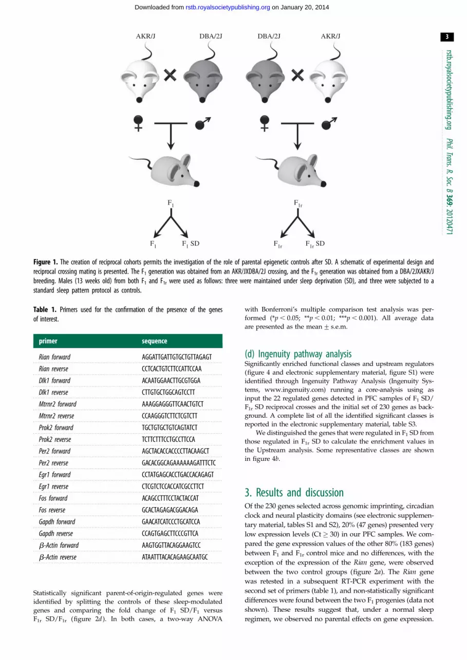
2. Material and methods(a) Animals and proceduresThe initial AKR/J mouse strain was obtained from Jackson Lab-
oratories (Bar Harbour, USA) and the DBA/2J strain was
obtained from Charles River (Wilmington, USA). The mice
were kept in an IIT (Istituto Italiano di Tecnologia) animal facility
and bred in reciprocal crosses to obtain two different experimen-
tal cohorts, AKR/JxDBA/2J F1 mice (the maternal strain is
reported first) and DBA/2JxAKR/J F1r mice. Each cohort
included a total of six males (13 weeks old) that were equally
subdivided into a sleep-deprived (SD) group and a control
group (figure 1). All mice were group-housed a week before
the experiment, with food and water ad libitum, under a 12 L :
12 D cycle (lights on from 7.00 to 19.00). On the day of the experi-
ment, SD mice underwent 6 h of sleep deprivation starting at
7.00. At 13.00, SD was interrupted, and the mice were left undis-
turbed for 1 h before they were sacrificed and their PFC tissue
was collected. Tissue from the control group was collected at
the same time, but control group mice were not subjected to
sleep deprivation. All sleep experiments were conducted in the
home-cage environment and all procedures were performed
under the guidance of the Italian Policy (licence number 039,
expires on 15 June 2015).
(b) Quantitative real-time PCRTotal RNA was extracted from approximately 1 g of snap-frozen
PFC using the Rneasy Microarray tissue mini kit (Qiagen,
Hilden, Germany). RNA samples were quantified with an
ND1000 Nanodrop spectrophotometer (Thermo Fisher Scientific,
Waltham, MA, USA). Reverse transcription of 1 mg of RNA was
performed using the RT2 First Strand Kit (Qiagen, Hilden,
Germany) according to the manufacturer’s instructions.
RT-qPCR was conducted using a custom RT2 Profiler PCR
array for 234 imprinted, circadian and epigenetic-related genes,
based on a 384-well plate format developed by Sabioscience
Qiagen technical service (Carlsbad, USA; electronic supplemen-
tary material, tables S1 and S2). Reconfirmation experiments
for the genes of interest were performed using a different set of
primers (table 1). RT-qPCR was performed on a ViiA 7
Real-time System machine (Applied Biosystem, Foster City, CA,
USA) using the following conditions: 10 min at 958C, 40 cycles
of denaturation at 958C for 15 s and an annealing and extension
step at 608C for 1 min. Each sample was run to obtain average Ct
values according to the manufacturer’s specifications. All
samples were normalized against a panel of four different house-
keeping genes: Gapdh, GusB, b-actin and B2M. Expression levels
relative to these housekeeping genes were determined by the
calculation of DCt, and the data are expressed as 22DDCt, where
DDCt is the difference between the SD and not-SD cohorts.
(c) Statistical analysisStatistically significant gene expression differences between SD
and not-SD mice were visualized by pooling together the two
normal sleep cohorts as a control group (F1 and F1r) (figure 2c).
AKR/J
F1 F1r
F1 F1 SD F1r F1r SD
DBA/2J DBA/2J AKR/J
Figure 1. The creation of reciprocal cohorts permits the investigation of the role of parental epigenetic controls after SD. A schematic of experimental design andreciprocal crossing mating is presented. The F1 generation was obtained from an AKR/JXDBA/2J crossing, and the F1r generation was obtained from a DBA/2JXAKR/Jbreeding. Males (13 weeks old) from both F1 and F1r were used as follows: three were maintained under sleep deprivation (SD), and three were subjected to astandard sleep pattern protocol as controls.
Table 1. Primers used for the confirmation of the presence of the genesof interest.
primer sequence
Rian forward AGGATTGATTGTGCTGTTAGAGT
Rian reverse CCTCACTGTCTTCCATTCCAA
Dlk1 forward ACAATGGAACTTGCGTGGA
Dlk1 reverse CTTGTGCTGGCAGTCCTT
Mtrnr2 forward AAAGGAGGGTTCAACTGTCT
Mtrnr2 reverse CCAAGGGTCTTCTCGTCTT
Prok2 forward TGCTGTGCTGTCAGTATCT
Prok2 reverse TCTTCTTTCCTGCCTTCCA
Per2 forward AGCTACACCACCCCTTACAAGCT
Per2 reverse GACACGGCAGAAAAAAGATTTCTC
Egr1 forward CCTATGAGCACCTGACCACAGAGT
Egr1 reverse CTCGTCTCCACCATCGCCTTCT
Fos forward ACAGCCTTTCCTACTACCAT
Fos reverse GCACTAGAGACGGACAGA
Gapdh forward GAACATCATCCCTGCATCCA
Gapdh reverse CCAGTGAGCTTCCCGTTCA
b-Actin forward AAGTGGTTACAGGAAGTCC
b-Actin reverse ATAATTTACACAGAAGCAATGC
rstb.royalsocietypublishing.orgPhil.Trans.R.Soc.B
369:20120471
3
on January 20, 2014rstb.royalsocietypublishing.orgDownloaded from
Statistically significant parent-of-origin-regulated genes were
identified by splitting the controls of these sleep-modulated
genes and comparing the fold change of F1 SD/F1 versus
F1r SD/F1r (figure 2d ). In both cases, a two-way ANOVA
with Bonferroni’s multiple comparison test analysis was per-
formed (*p , 0.05; **p , 0.01; ***p , 0.001). All average data
are presented as the mean+ s.e.m.
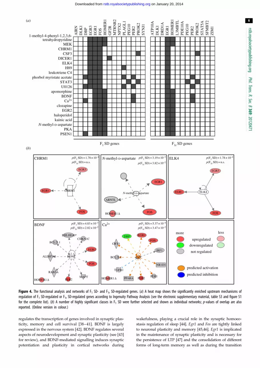
(d) Ingenuity pathway analysisSignificantly enriched functional classes and upstream regulators
(figure 4 and electronic supplementary material, figure S1) were
identified through Ingenuity Pathway Analysis (Ingenuity Sys-
tems, www.ingenuity.com) running a core-analysis using as
input the 22 regulated genes detected in PFC samples of F1 SD/
F1r SD reciprocal crosses and the initial set of 230 genes as back-
ground. A complete list of all the identified significant classes is
reported in the electronic supplementary material, table S3.
We distinguished the genes that were regulated in F1 SD from
those regulated in F1r SD to calculate the enrichment values in
the Upstream analysis. Some representative classes are shown
in figure 4b.
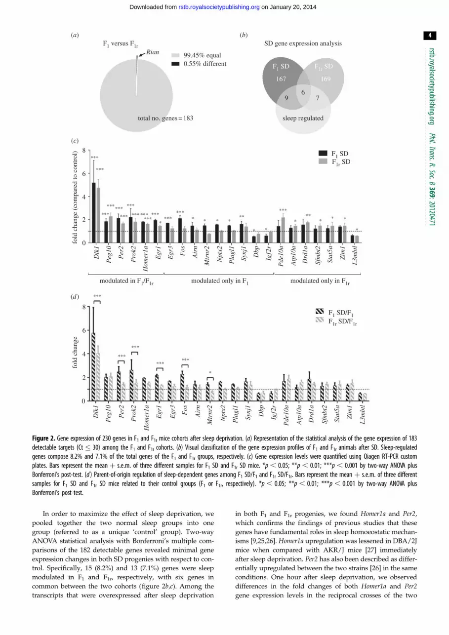
3. Results and discussionOf the 230 genes selected across genomic imprinting, circadian
clock and neural plasticity domains (see electronic supplemen-
tary material, tables S1 and S2), 20% (47 genes) presented very
low expression levels (Ct� 30) in our PFC samples. We com-
pared the gene expression values of the other 80% (183 genes)
between F1 and F1r control mice and no differences, with the
exception of the expression of the Rian gene, were observed
between the two control groups (figure 2a). The Rian gene
was retested in a subsequent RT-PCR experiment with the
second set of primers (table 1), and non-statistically significant
differences were found between the two F1 progenies (data not
shown). These results suggest that, under a normal sleep
regimen, we observed no parental effects on gene expression.
1
F1 versus F1r
99.45% equal0.55% different
total no. genes = 183
8
fold
cha
nge
(com
pare
d to
con
trol
)fo
ld c
hang
e
F1 SDF1r SD
F1 SD/F1F1r SD/F1r
***
***
***
***
******
***
*
*********
***
*** *** *** ******
** * * *
* *
*****
***
* * *
*
******
6
modulated in F1/F1r modulated only in F1 modulated only in F1r
4
2
0
8
6
4
2
0
Dlk
1
Peg
10
Per
2
Pro
k2
Hom
er1a
Egr
1
Egr
3
Fos
Air
n
Mtr
nr2
Npt
x2
Pla
gl1
Synj
1
Dbp
Igf2
r
Pde
10a
Atp
10a
Drd
1a
Sfm
bt2
Stat
5a
Zim
1
L3m
btl
Dlk
1
Peg
10
Per
2
Pro
k2
Hom
er1a
Egr
1
Egr
3
Fos
Air
n
Mtr
nr2
Npt
x2
Pla
gl1
Synj
1
Dbp
Igf2
r
Pde
10a
Atp
10a
Drd
1a
Sfm
bt2
Stat
5a
Zim
1
L3m
btl
RianSD gene expression analysis
(a)
(c)
(d )
(b)
167 169
F1 SD F1r SD
69 7
sleep regulated
Figure 2. Gene expression of 230 genes in F1 and F1r mice cohorts after sleep deprivation. (a) Representation of the statistical analysis of the gene expression of 183detectable targets (Ct � 30) among the F1 and F1r cohorts. (b) Visual classification of the gene expression profiles of F1 and F1r animals after SD. Sleep-regulatedgenes compose 8.2% and 7.1% of the total genes of the F1 and F1r groups, respectively. (c) Gene expression levels were quantified using Qiagen RT-PCR customplates. Bars represent the mean þ s.e.m. of three different samples for F1 SD and F1r SD mice. *p , 0.05; **p , 0.01; ***p , 0.001 by two-way ANOVA plusBonferroni’s post-test. (d ) Parent-of-origin regulation of sleep-dependent genes among F1 SD/F1 and F1r SD/F1r. Bars represent the mean þ s.e.m. of three differentsamples for F1 SD and F1r SD mice related to their control groups (F1 or F1r, respectively). *p , 0.05; **p , 0.01; ***p , 0.001 by two-way ANOVA plusBonferroni’s post-test.
rstb.royalsocietypublishing.orgPhil.Trans.R.Soc.B
369:20120471
4
on January 20, 2014rstb.royalsocietypublishing.orgDownloaded from
In order to maximize the effect of sleep deprivation, we
pooled together the two normal sleep groups into one
group (referred to as a unique ‘control’ group). Two-way
ANOVA statistical analysis with Bonferroni’s multiple com-
parisons of the 182 detectable genes revealed minimal gene
expression changes in both SD progenies with respect to con-
trol. Specifically, 15 (8.2%) and 13 (7.1%) genes were sleep
modulated in F1 and F1r, respectively, with six genes in
common between the two cohorts (figure 2b,c). Among the
transcripts that were overexpressed after sleep deprivation
in both F1 and F1r progenies, we found Homer1a and Per2,
which confirms the findings of previous studies that these
genes have fundamental roles in sleep homoeostatic mechan-
isms [9,25,26]. Homer1a upregulation was lessened in DBA/2J
mice when compared with AKR/J mice [27] immediately
after sleep deprivation. Per2 has also been described as differ-
entially upregulated between the two strains [26] in the same
conditions. One hour after sleep deprivation, we observed
differences in the fold changes of both Homer1a and Per2gene expression levels in the reciprocal crosses of the two
7 n.s.
n.s.
***
**
F1 SD/F1F1r SD/F1r6
5
4
3
fold
cha
nge
2
1
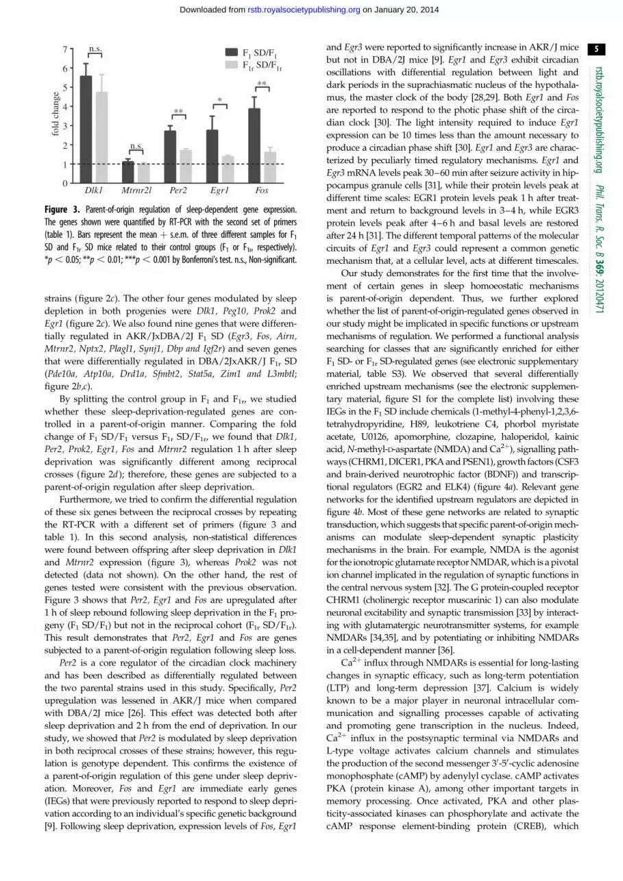
Dlk1 Mtrnr2l Per2 Egr1 Fos0
Figure 3. Parent-of-origin regulation of sleep-dependent gene expression.The genes shown were quantified by RT-PCR with the second set of primers(table 1). Bars represent the mean þ s.e.m. of three different samples for F1
SD and F1r SD mice related to their control groups (F1 or F1r, respectively).*p , 0.05; **p , 0.01; ***p , 0.001 by Bonferroni’s test. n.s., Non-significant.
rstb.royalsocietypublishing.orgPhil.Trans.R.Soc.B
369:20120471
5
on January 20, 2014rstb.royalsocietypublishing.orgDownloaded from
strains (figure 2c). The other four genes modulated by sleep
depletion in both progenies were Dlk1, Peg10, Prok2 and
Egr1 (figure 2c). We also found nine genes that were differen-
tially regulated in AKR/JxDBA/2J F1 SD (Egr3, Fos, Airn,Mtrnr2, Nptx2, Plagl1, Synj1, Dbp and Igf2r) and seven genes
that were differentially regulated in DBA/2JxAKR/J F1r SD
(Pde10a, Atp10a, Drd1a, Sfmbt2, Stat5a, Zim1 and L3mbtl;figure 2b,c).
By splitting the control group in F1 and F1r, we studied
whether these sleep-deprivation-regulated genes are con-
trolled in a parent-of-origin manner. Comparing the fold
change of F1 SD/F1 versus F1r SD/F1r, we found that Dlk1,Per2, Prok2, Egr1, Fos and Mtrnr2 regulation 1 h after sleep
deprivation was significantly different among reciprocal
crosses (figure 2d ); therefore, these genes are subjected to a
parent-of-origin regulation after sleep deprivation.
Furthermore, we tried to confirm the differential regulation
of these six genes between the reciprocal crosses by repeating
the RT-PCR with a different set of primers (figure 3 and
table 1). In this second analysis, non-statistical differences
were found between offspring after sleep deprivation in Dlk1and Mtrnr2 expression (figure 3), whereas Prok2 was not
detected (data not shown). On the other hand, the rest of
genes tested were consistent with the previous observation.
Figure 3 shows that Per2, Egr1 and Fos are upregulated after
1 h of sleep rebound following sleep deprivation in the F1 pro-
geny (F1 SD/F1) but not in the reciprocal cohort (F1r SD/F1r).
This result demonstrates that Per2, Egr1 and Fos are genes
subjected to a parent-of-origin regulation following sleep loss.
Per2 is a core regulator of the circadian clock machinery
and has been described as differentially regulated between
the two parental strains used in this study. Specifically, Per2upregulation was lessened in AKR/J mice when compared
with DBA/2J mice [26]. This effect was detected both after
sleep deprivation and 2 h from the end of deprivation. In our
study, we showed that Per2 is modulated by sleep deprivation
in both reciprocal crosses of these strains; however, this regu-
lation is genotype dependent. This confirms the existence of
a parent-of-origin regulation of this gene under sleep depriv-
ation. Moreover, Fos and Egr1 are immediate early genes
(IEGs) that were previously reported to respond to sleep depri-
vation according to an individual’s specific genetic background
[9]. Following sleep deprivation, expression levels of Fos, Egr1
and Egr3 were reported to significantly increase in AKR/J mice
but not in DBA/2J mice [9]. Egr1 and Egr3 exhibit circadian
oscillations with differential regulation between light and
dark periods in the suprachiasmatic nucleus of the hypothala-
mus, the master clock of the body [28,29]. Both Egr1 and Fosare reported to respond to the photic phase shift of the circa-
dian clock [30]. The light intensity required to induce Egr1expression can be 10 times less than the amount necessary to
produce a circadian phase shift [30]. Egr1 and Egr3 are charac-
terized by peculiarly timed regulatory mechanisms. Egr1 and
Egr3 mRNA levels peak 30–60 min after seizure activity in hip-
pocampus granule cells [31], while their protein levels peak at
different time scales: EGR1 protein levels peak 1 h after treat-
ment and return to background levels in 3–4 h, while EGR3
protein levels peak after 4–6 h and basal levels are restored
after 24 h [31]. The different temporal patterns of the molecular
circuits of Egr1 and Egr3 could represent a common genetic
mechanism that, at a cellular level, acts at different timescales.
Our study demonstrates for the first time that the involve-
ment of certain genes in sleep homoeostatic mechanisms
is parent-of-origin dependent. Thus, we further explored
whether the list of parent-of-origin-regulated genes observed in
our study might be implicated in specific functions or upstream
mechanisms of regulation. We performed a functional analysis
searching for classes that are significantly enriched for either
F1 SD- or F1r SD-regulated genes (see electronic supplementary
material, table S3). We observed that several differentially
enriched upstream mechanisms (see the electronic supplemen-
tary material, figure S1 for the complete list) involving these
IEGs in the F1 SD include chemicals (1-methyl-4-phenyl-1,2,3,6-
tetrahydropyridine, H89, leukotriene C4, phorbol myristate
acetate, U0126, apomorphine, clozapine, haloperidol, kainic
acid, N-methyl-D-aspartate (NMDA) and Ca2þ), signalling path-
ways (CHRM1, DICER1, PKA and PSEN1), growth factors (CSF3
and brain-derived neurotrophic factor (BDNF)) and transcrip-
tional regulators (EGR2 and ELK4) (figure 4a). Relevant gene
networks for the identified upstream regulators are depicted in
figure 4b. Most of these gene networks are related to synaptic
transduction, which suggests that specific parent-of-origin mech-
anisms can modulate sleep-dependent synaptic plasticity
mechanisms in the brain. For example, NMDA is the agonist
for the ionotropic glutamate receptor NMDAR, which is a pivotal
ion channel implicated in the regulation of synaptic functions in
the central nervous system [32]. The G protein-coupled receptor
CHRM1 (cholinergic receptor muscarinic 1) can also modulate
neuronal excitability and synaptic transmission [33] by interact-
ing with glutamatergic neurotransmitter systems, for example
NMDARs [34,35], and by potentiating or inhibiting NMDARs
in a cell-dependent manner [36].
Ca2þ influx through NMDARs is essential for long-lasting
changes in synaptic efficacy, such as long-term potentiation
(LTP) and long-term depression [37]. Calcium is widely
known to be a major player in neuronal intracellular com-
munication and signalling processes capable of activating
and promoting gene transcription in the nucleus. Indeed,
Ca2þ influx in the postsynaptic terminal via NMDARs and
L-type voltage activates calcium channels and stimulates
the production of the second messenger 30-50-cyclic adenosine
monophosphate (cAMP) by adenylyl cyclase. cAMP activates
PKA (protein kinase A), among other important targets in
memory processing. Once activated, PKA and other plas-
ticity-associated kinases can phosphorylate and activate the
cAMP response element-binding protein (CREB), which
SYN
J1
AIR
N
DL
K1
EG
R3
EG
R1
FOS
HO
ME
R1
DB
P
PER
2
PLA
GL
1 PE
G10
PRO
K2
GF2
R
MT
RN
R2
NPT
X2
EG
R1
HO
ME
R1
PER
2 PE
G10
PRO
K2
DL
K1
DR
D1A
AT
P10A
STA
T5A
L3M
BT
L
PDE
10A
ZIM
1 SF
MB
T2
1-methyl-4-phenyl-1,2,3,6-tetrahydropyridine
MEK CHRM1
CSF3 DICER1
ELK4 H89
leukotriene C4 phorbol myristate acetate
STAT3 U0126
apomorphine BDNF
Ca2+
clozapine EGR2
haloperidol kainic acid
N-methyl-D-aspartate PKA
PSEN1
F1 SD genes F1r SD genes
(a)
(b)
CHRM1 p(F1 SD) = 1.78 × 10⁻⁴
p(F1r SD) = n.s. ELK4 p(F1 SD) = 1.78 × 10⁻⁴
p(F1r SD) = n.s.
Ca2+ p(F1 SD) = 5.37 × 10⁻⁴
p(F1r SD) = 3.47 × 10⁻² BDNF p(F1 SD) = 4.83 × 10⁻³
p(F1r SD) = 2.82 × 10⁻²
upregulated
not regulated
predicted activation
predicted inhibition
downregulated
more less
N-methyl-D-aspartate p(F1 SD) = 3.19 × 10⁻³
p(F1r SD) = 3.82 × 10⁻²
EGR3
EGR1
EGR1
FOS
DBPCDKN1C
BHLHE40*
BCL2L1
ALDH7A1
RAB3A
HOMER1D
BDNF
EGR1
EGR1
Ca2+
FOS
IRF1
FOS
MAPTPER2
NR1D1
PER1PER2PPARA
TGFB1
BCL2L1
CRY1
HOMER1D
HOMER1D FOS
EGR3
EGR1
FOS
CHRM1
ARNTL
N-methyl-D-aspartate ELK4
Figure 4. The functional analysis and networks of F1 SD- and F1r SD-regulated genes. (a) A heat map shows the significantly enriched upstream mechanisms ofregulation of F1 SD-regulated or F1r SD-regulated genes according to Ingenuity Pathway Analysis (see the electronic supplementary material, table S3 and figure S1for the complete list). (b) A number of highly significant classes in F1 SD were further selected and shown as individual networks; p-values of overlap are alsoreported. (Online version in colour.)
rstb.royalsocietypublishing.orgPhil.Trans.R.Soc.B
369:20120471
6
on January 20, 2014rstb.royalsocietypublishing.orgDownloaded from
regulates the transcription of genes involved in synaptic plas-
ticity, memory and cell survival [38–41]. BDNF is largely
expressed in the nervous system [42]. BDNF regulates several
aspects of neurodevelopment and synaptic plasticity (see [43]
for review), and BDNF-mediated signalling induces synaptic
potentiation and plasticity in cortical networks during
wakefulness, playing a crucial role in the synaptic homoeo-
stasis regulation of sleep [44]. Egr1 and Fos are tightly linked
to neuronal plasticity and memory [45,46]. Egr1 is implicated
in the maintenance of synaptic plasticity and is necessary for
the persistence of LTP [47] and the consolidation of different
forms of long-term memory as well as during the transition
rstb.royalsocietypublishing.orgPhil.Trans.R.Soc.B
369:20120471
7
on January 20, 2014rstb.royalsocietypublishing.orgDownloaded from
from short- to long-term memories [48,49]. Interestingly, sleep
is significantly involved in the regulation of brain plasticity
and cognition (see [50] for review). A series of studies have
shown that PKA and CREB signalling pathways promote
wakefulness [51]. The same results were obtained in mice lack-
ing two of the three isoforms of CREB: the loss of alpha and
delta causes a reduction in CREB activity and a reduction in
wakefulness during the light-off period, with respect to
controls [52–54].
Although our study focused only on few genes, our func-
tional analysis identified specific domains such as behaviour,
neurological diseases and cell death and survival as enriched
processes involving these IEGs (see electronic supplementary
material, table S3). Our study suggests that the role of sleep in
neuroprotection can be determined by parental epigenetic
mechanisms. Indeed, Homer1a is implicated in intracellular
calcium homoeostasis and sleep restorative mechanisms [9],
and Egr1 and Fos are involved in molecular neuroprotective
responses, for example those triggered by ischaemia [55].
Altogether, these data indicate that sleep restriction activates
molecular pathways associated with the preservation of
neuronal integrity [56].
In our study, we concentrated on the PFC, one of the
main targets of the restorative effects of sleep on cognition
[57]. The PFC is pivotal in coordinating high-level cogni-
tive processes, such as response inhibition, higher order
attention processes, working memory and episodic learning
memory [58–62]. Different phases of sleep were reported to
be associated with specific activation and deactivation
modes of PFC regions [63,64]. Moreover, the disruption or
the alteration of normal sleep–wake cycles or circadian
rhythms delayed the time required by the PFC to achieve
the attention levels of other brain cortical regions [65]. In
addition, sleep deprivation alters the neuronal functionality
and gene expression profile in the PFC [66–68]. The evidence
that neuronal plasticity genes (and possibly many neuronal
plasticity pathways) are differently regulated in the PFC
according to parent-of-origin mechanisms casts a new light
on the epigenetic regulation of these genes in sleep and
sleep-related functions.
Acknowledgements. We thank Raquel Garcia Garcia for graphical sup-port. We also thank Glenda Lassi for reading and discussion of themanuscript, Riccardo Navone and Daniela Cantatore for assistancewith the management of the mouse colonies.
References
1. Borbely AA. 1982 A two process model of sleepregulation. Hum. Neurobiol. 1, 195 – 204.
2. Tucci V. 2012 Sleep, circadian rhythms, and intervaltiming: evolutionary strategies to time information.Front. Integr. Neurosci. 5, 92. (doi:10.3389/fnint.2011.00092)
3. Aguilar-Arnal L, Sassone-Corsi P. 2013 The circadianepigenome: how metabolism talks to chromatinremodeling. Curr. Opin. Cell Biol. 25, 170 – 176.(doi:10.1016/j.ceb.2013.01.003)
4. Borrelli E, Nestler EJ, Allis CD, Sassone-Corsi P. 2008Decoding the epigenetic language of neuronalplasticity. Neuron 60, 961 – 974. (doi:10.1016/j.neuron.2008.10.012)
5. Laposky A, Easton A, Dugovic C, Walisser J, BradfieldC, Turek F. 2005 Deletion of the mammaliancircadian clock gene BMAL1/Mop3 alters baselinesleep architecture and the response to sleepdeprivation. Sleep 28, 395 – 409.
6. Wisor JP, O’Hara BF, Terao A, Selby CP, Kilduff TS,Sancar A, Edgar DM, Franken P. 2002 A role forcryptochromes in sleep regulation. BMC Neurosci. 3,20. (doi:10.1186/1471-2202-3-20)
7. Naylor E, Bergmann BM, Krauski K, Zee PC,Takahashi JS, Vitaterna MH, Turek RW. 2000 Thecircadian clock mutation alters sleep homeostasis inthe mouse. J. Neurosci. 20, 8138 – 8143.
8. Franken P, Chollet D, Tafti M. 2001 The homeostaticregulation of sleep need is under genetic control.J. Neurosci. 21, 2610 – 2621.
9. Maret S et al. 2007 Homer1a is a core brainmolecular correlate of sleep loss. Proc. Natl Acad.Sci. USA 104, 20 090 – 20 905. (doi:10.1073/pnas.0710131104)
10. Mackiewicz M, Zimmerman JE, Shockley KR,Churchill GA, Pack AI. 2009 What are microarrays
teaching us about sleep? Trends Mol. Med. 15,79 – 87. (doi:10.1016/j.molmed.2008.12.002)
11. McNamara P. 2004 Genomic imprinting andneurodevelopmental disorders of sleep. Sleep Hypn.6, 82 – 90.
12. McNamara P, Dowdall J, Auerbach S. 2002 REMsleep, early experience, and the development ofreproductive strategies. Hum. Nat. 13, 405 – 435.(doi:10.1007/s12110-002-1001-x)
13. Tucci V, Nolan PM. 2009 Toward an understandingof the function of sleep: new insights from mousegenetics. In Evolution of sleep phylogeneticfunctional perspectives (eds P McNamara, RA Barton,CL Nunn), pp. 218 – 237. Cambridge, UK: CambridgeUniversity Press.
14. Hertz G et al. 1993 Sleep and breathing patterns inpatients with Prader Willi syndrome (PWS): effectsof age and gender. Sleep 16, 366 – 371.
15. Vela-Bueno A et al. 1984 Sleep in the Prader – Willisyndrome. Clinical and polygraphic findings. Arch.Neurol. 41, 294 – 296. (doi:10.1001/archneur.1984.04050150072020)
16. Vgontzas AN, Bixler EO, Kales A, Centurione A,Rogan PK, Mascari M, Vela-Bueno A. 1996 Daytimesleepiness and REM abnormalities in Prader – Willisyndrome: evidence of generalized hypoarousal.Int. J. Neurosci. 87, 127 – 139. (doi:10.3109/00207459609070832)
17. Vgontzas AN et al. 1996 Relationship of sleepabnormalities to patient genotypes in Prader – Willisyndrome. Am. J. Med. Genet. 67, 478 – 482. (doi:10.1002/(SICI)1096-8628(19960920)67:5,478::AID-AJMG7.3.0.CO;2-G)
18. Colas D, Wagstaff J, Fort P, Salvert D, Sarda N.2005 Sleep disturbances in Ube3a maternal-deficient mice modeling Angelman syndrome.
Neurobiol. Dis. 20, 471 – 478. (doi:10.1016/j.nbd.2005.04.003)
19. Amici R, Sanford LD, Kearney K, McInerney B, RossRJ, Horner RL, Morrison AR. 2004 A serotonergic(5-HT2) receptor mechanism in the laterodorsaltegmental nucleus participates in regulating thepattern of rapid-eye-movement sleep occurrence inthe rat. Brain Res. 996, 9 – 18. (doi:10.1016/j.brainres.2003.09.026)
20. Kato MV et al. 1996 Genomic imprinting of thehuman serotonin-receptor (HTR2) gene involved indevelopment of retinoblastoma. Am. J. Hum. Genet.59, 1084 – 1090.
21. Lassi G et al. 2012 Loss of Gnas imprintingdifferentially affects REM/NREM sleep and cognitionin mice. PLoS Genet. 8, e1002706. (doi:10.1371/journal.pgen.1002706)
22. Wisor JP, Pasumarthi RK, Gerashchenko D,Thompson CL, Pathak S, Sancar A, Franken P,Lein ES, Kilduff TS. 2008 Sleep deprivationeffects on circadian clock gene expression inthe cerebral cortex parallel electroencephalographicdifferences among mouse strains. J. Neurosci.28, 7193 – 7201. (doi:10.1523/JNEUROSCI.1150-08.2008)
23. Krueger JM, Rector DM, Roy S, Van Dongen HPA,Belenky G, Panksepp J. 2008 Sleep as afundamental property of neuronal assemblies. Nat.Rev. Neurosci. 9, 910 – 919. (doi:10.1038/nrn2521)
24. Tononi G, Cirelli C. 2006 Sleep function and synaptichomeostasis. Sleep Med. Rev. 10, 49 – 62. (doi:10.1016/j.smrv.2005.05.002)
25. Mackiewicz M et al. 2007 Macromoleculebiosynthesis: a key function of sleep. Physiol.Genomics 31, 441 – 457. (doi:10.1152/physiolgenomics.00275.2006)

rstb.royalsocietypublishing.orgPhil.Trans.R.Soc.B
369:20120471
8
on January 20, 2014rstb.royalsocietypublishing.orgDownloaded from
26. Franken P, Thomason R, Heller HC, O’Hara BF. 2007A non-circadian role for clock-genes in sleephomeostasis: a strain comparison. BMC Neurosci. 8,87. (doi:10.1186/1471-2202-8-87)
27. Andretic R, Franken P, Tafti M. 2008 Genetics ofsleep. Annu. Rev. Genet. 42, 361 – 388. (doi:10.1146/annurev.genet.42.110807.091541)
28. Lin JT, Kornhauser JM, Singh NP, Mayo KE,Takahashi JS. 1997 Visual sensitivities of nur77(NGFI-B) and zif268 (NGFI-A) induction in thesuprachiasmatic nucleus are dissociated from c-fosinduction and behavioral phase-shifting responses.Brain Res. Mol. Brain Res. 46, 303 – 310. (doi:10.1016/S0169-328X(97)00005-3)
29. Morris ME et al. 1998 A screen for genes induced inthe suprachiasmatic nucleus by light. Science 279,1544 – 1547. (doi:10.1126/science.279.5356.1544)
30. O’Donovan KJ, Tourtellotte WG, Millbrandt J,Baraban JM. 1999 The EGR family of transcription-regulatory factors: progress at the interface ofmolecular and systems neuroscience. TrendsNeurosci. 22, 167 – 173. (doi:10.1016/S0166-2236(98)01343-5)
31. O’Donovan KJ, Wilkens EP, Baraban JM. 1998Sequential expression of Egr-1 and Egr-3 inhippocampal granule cells following electroconvulsivestimulation. J. Neurochem. 70, 1241– 1248. (doi:10.1046/j.1471-4159.1998.70031241.x)
32. Lau CG, Zukin RS. 2007 NMDA receptor trafficking insynaptic plasticity and neuropsychiatric disorders.Nat. Rev. Neurosci. 8, 413 – 426. (doi:10.1038/nrn2153)
33. Picciotto MR, Higley MJ, Mineur YS. 2012Acetylcholine as a neuromodulator: cholinergicsignaling shapes nervous system function andbehavior. Neuron 76, 116 – 129. (doi:10.1016/j.neuron.2012.08.036)
34. Lu WY, Lu W-Y, Xiong Z-G, Lei S, Orser BA, Dudek E,Browning MD. 1999 G-protein-coupled receptors act viaprotein kinase C and Src to regulate NMDA receptors.Nat. Neurosci. 2, 331 – 338. (doi:10.1038/7243)
35. Marino MJ, Rouse ST, Levey AI, Potter LT, Conn PJ.1998 Activation of the genetically defined m1muscarinic receptor potentiates N-methyl-D-aspartate (NMDA) receptor currents in hippocampalpyramidal cells. Proc. Natl Acad. Sci. USA 95,11 465 – 11 470. (doi:10.1073/pnas.95.19.11465)
36. Grishin AA, Benquet P, Gerber U. 2005 Muscarinicreceptor stimulation reduces NMDA responses inCA3 hippocampal pyramidal cells via Ca2þ-dependent activation of tyrosine phosphatase.Neuropharmacology 49, 328 – 337. (doi:10.1016/j.neuropharm.2005.03.019)
37. Collingridge GL, Isaac JT, Wang YT. 2004 Receptortrafficking and synaptic plasticity. Nat. Rev. Neurosci.5, 952 – 962. (doi:10.1038/nrn1556)
38. Cohen S, Greenberg ME. 2008 Communicationbetween the synapse and the nucleus in neuronaldevelopment, plasticity, and disease. Annu. Rev. CellDev. Biol. 24, 183 – 209. (doi:10.1146/annurev.cellbio.24.110707.175235)
39. Greer PL, Greenberg ME. 2008 From synapse tonucleus: calcium-dependent gene transcription in the
control of synapse development and function. Neuron59, 846 – 860. (doi:10.1016/j.neuron.2008.09.002)
40. Saha RN, Dudek SM. 2008 Action potentials: to thenucleus and beyond. Exp. Biol. Med. 233, 385 – 393.(doi:10.3181/0709-MR-241)
41. Wiegert JS, Bading H. 2011 Activity-dependentcalcium signaling and ERK-MAP kinases in neurons:a link to structural plasticity of the nucleus andgene transcription regulation. Cell Calcium 49,296 – 305. (doi:10.1016/j.ceca.2010.11.009)
42. West AE, Chen EG, Dalva MB, Dolmetsch RE,Kornhauser JM, Shaywitz AJ, Takasu MA, Tao X,Greenberg ME. 2001 Calcium regulation of neuronalgene expression. Proc. Natl Acad. Sci. USA 98,11 024 – 11 031. (doi:10.1073/pnas.191352298)
43. Park H, Poo MM. 2013 Neurotrophin regulation ofneural circuit development and function. Nat. Rev.Neurosci. 14, 7 – 23. (doi:10.1038/nrn3379)
44. Tononi G, Cirelli C. 2003 Sleep and synaptichomeostasis: a hypothesis. Brain Res. Bull. 62,143 – 150. (doi:10.1016/j.brainresbull.2003.09.004)
45. Guzowski JF, Setlow B, Wagner EK, McGaugh JL.2001 Experience-dependent gene expression in therat hippocampus after spatial learning: acomparison of the immediate-early genes Arc, c-fos,and zif268. J. Neurosci. 21, 5089 – 5098.
46. Poirier R et al. 2008 Distinct functions of Egr genefamily members in cognitive processes. Front.Neurosci. 2, 47 – 55. (doi:10.3389/neuro.01.002.2008)
47. Jones MW et al. 2001 A requirement for theimmediate early gene Zif268 in the expression oflate LTP and long-term memories. Nat. Neurosci. 4,289 – 296. (doi:10.1038/85138)
48. Bozon B, Davis S, Laroche S. 2002 Regulatedtranscription of the immediate-early gene Zif268:mechanisms and gene dosage-dependent functionin synaptic plasticity and memory formation.Hippocampus 12, 570 – 577. (doi:10.1002/hipo.10100)
49. Bozon B, Kelly A, Josselyn SA, Silva AJ, Davis S,Laroche S. 2003 MAPK, CREB and zif268 are allrequired for the consolidation of recognitionmemory. Phil. Trans. R. Soc. Lond. B 358, 805 – 814.(doi:10.1098/rstb.2002.1224)
50. Wang G, Grone B, Colas D, Appelbaum L, MourrainP. 2011 Synaptic plasticity in sleep: learning,homeostasis and disease. Trends Neurosci. 34,452 – 463. (doi:10.1016/j.tins.2011.07.005)
51. Hendricks JC, Williams JA, Panckeri K, Kirk D, TelloM, Yin JC-P, Sehgal A. 2001 A non-circadian role forcAMP signaling and CREB activity in Drosophila resthomeostasis. Nat. Neurosci. 4, 1108 – 1115. (doi:10.1038/nn743)
52. Hummler E, Cole TJ, Blendy JA, Ganss R, Aguzzi A,Schmid W, Beermann F, Schutz G. 1994 Targetedmutation of the CREB gene: compensation withinthe CREB/ATF family of transcription factors. Proc.Natl Acad. Sci. USA 91, 5647 – 5651. (doi:10.1073/pnas.91.12.5647)
53. Graves LA et al. 2003 Genetic evidence for a role ofCREB in sustained cortical arousal. J. Neurophysiol.90, 1152 – 1159. (doi:10.1152/jn.00882.2002)
54. Graves L et al. 2002 Behavioral analysis of CREBalphadelta mutation on a B6/129 F1 hybridbackground. Hippocampus 12, 18 – 26. (doi:10.1002/hipo.10003)
55. Kamphuis W et al. 2007 Global gene expressionprofiling of ischemic preconditioning in the ratretina. Mol. Vis. 13, 1020 – 1030.
56. Mongrain V et al. 2010 Separating the contributionof glucocorticoids and wakefulness to the molecularand electrophysiological correlates of sleephomeostasis. Sleep 33, 1147 – 1157.
57. Maquet P. 1995 Sleep function(s) and cerebralmetabolism. Behav. Brain Res. 69, 75 – 83. (doi:10.1016/0166-4328(95)00017-N)
58. Couyoumdjian A, Sdoia S, Tempesta D, Curcio G,Rastellini E, De Gennaro L, Ferrara M. 2010 Theeffects of sleep and sleep deprivation on task-switching performance. J. Sleep Res. 19, 64 – 70.(doi:10.1111/j.1365-2869.2009.00774.x)
59. Harrison Y, Horne JA. 1998 Sleep loss impairs shortand novel language tasks having a prefrontal focus.J. Sleep Res. 7, 95 – 100. (doi:10.1046/j.1365-2869.1998.00104.x)
60. Killgore WD, Kahn-Greene ET, Lipizzi EL, NewmanRA, Kamimori GH, Balkin TJ. 2008 Sleep deprivationreduces perceived emotional intelligence andconstructive thinking skills. Sleep Med. 9, 517 – 526.(doi:10.1016/j.sleep.2007.07.003)
61. Granon S, Floresco S. 2009 Functional neuroanatomyof flexible behaviors in mice and rats.Endophenotypes of psychiatric and neurodegenerativedisorders in rodent models, pp. 83 – 103. Kerala:Transworld Research Network.
62. Dalley JW, Cardinal RN, Robbins TW. 2004 Prefrontalexecutive and cognitive functions in rodents: neuraland neurochemical substrates. Neurosci. Biobehav. Rev.28, 771– 784. (doi:10.1016/j.neubiorev.2004.09.006)
63. Maquet P, Dive D, Salmon E, Sadzot B, Franco G,Poirrier R, von Frenckell R, Franck G. 1990 Cerebralglucose utilization during sleep-wake cycle in mandetermined by positron emission tomography and[18F]2-fluoro-2-deoxy-D-glucose method. Brain Res.513, 136 – 143. (doi:10.1016/0006-8993(90)91099-3)
64. Maquet P et al. 2000 Experience-dependent changesin cerebral activation during human REM sleep. Nat.Neurosci. 3, 831– 836. (doi:10.1038/77744)
65. Nofzinger EA, Mintun MA, Wiseman M, Kupfer DJ,Moore RY. 1997 Forebrain activation in REM sleep:an FDG PET study. Brain Res. 770, 192 – 201.(doi:10.1016/S0006-8993(97)00807-X)
66. Winters BD, Huang YH, Dong Y, Krueger JM. 2011Sleep loss alters synaptic and intrinsic neuronalproperties in mouse prefrontal cortex. Brain Res.1420, 1 – 7. (doi:10.1016/j.brainres.2011.08.078)
67. Liu ZW, Faraguna U, Cirelli C, Tononi G, Gao X-B.2010 Direct evidence for wake-related increases andsleep-related decreases in synaptic strength inrodent cortex. J. Neurosci. 30, 8671 – 8675. (doi:10.1523/JNEUROSCI.1409-10.2010)
68. Cirelli C, Faraguna U, Tononi G. 2006 Changes inbrain gene expression after long-term sleepdeprivation. J. Neurochem. 98, 1632 – 1645. (doi:10.1111/j.1471-4159.2006.04058.x)
Related Documents