Welcome message from author
This document is posted to help you gain knowledge. Please leave a comment to let me know what you think about it! Share it to your friends and learn new things together.
Transcript

BLBK186-Key April 28, 2009 13:49
ii

BLBK186-Key April 28, 2009 13:49
Practical Hemostasis and Thrombosis
i

BLBK186-Key April 28, 2009 13:49
ii
BLBK186-Key April 28, 2009 13:49
Practical Hemostasisand ThrombosisEDITED BY
Nigel Key, MB, ChB, FRCPHarold R. Roberts Distinguished Professor of MedicineDirector, Hemophilia Treatment CenterThe University of North Carolina at Chapel HillDivision of Hematology & OncologyChapel Hill, North Carolina, USA
Michael Makris, MDDirector, Sheffield Haemophilia and Thrombosis CentreRoyal Hallamshire HospitalSheffield, UK
Denise O’Shaughnessy, DPhil, FRCP, FRCPathConsultant Haematologist and Senior Medical Advisor (Blood Policy)Department of HealthLondon, UK
David Lillicrap, MD, FRCPCProfessor, Department of Pathology and Molecular MedicineRichardson Laboratory, Queen’s UniversityKingston, Ontario, Canada
SECOND EDITION
FOREWORD BY HAROLD R. ROBERTS, MD, FACP
Sarah Graham Kenan Distinguished ProfessorMedicine and PathologyUniversity of North Carolina at Chapel HillChapel Hill, North Carolina, USA
A John Wiley & Sons, Ltd., Publication
iii
BLBK186-Key April 28, 2009 13:49
This edition first published 2009, C© 2005, 2009 by Blackwell Publishing Ltd
Blackwell Publishing was acquired by John Wiley & Sons in February 2007.Blackwell’s publishing program has been merged with Wiley’s global Scientific, Technical andMedical business to form Wiley-Blackwell.
Registered office: John Wiley & Sons Ltd, The Atrium, Southern Gate, Chichester, West Sussex,PO19 8SQ, UK
Editorial offices: 9600 Garsington Road, Oxford, OX4 2DQ, UKThe Atrium, Southern Gate, Chichester, West Sussex, PO19 8SQ, UK111 River Street, Hoboken, NJ 07030-5774, USA
For details of our global editorial offices, for customer services and for information about how toapply for permission to reuse the copyright material in this book please see our website atwww.wiley.com/wiley-blackwell.
The right of the author to be identified as the author of this work has been asserted inaccordance with the Copyright, Designs and Patents Act 1988.
All rights reserved. No part of this publication may be reproduced, stored in a retrieval system,or transmitted, in any form or by any means, electronic, mechanical, photocopying, recording orotherwise, except as permitted by the UK Copyright, Designs and Patents Act 1988, without theprior permission of the publisher.
Wiley also publishes its books in a variety of electronic formats. Some content that appears inprint may not be available in electronic books.
Designations used by companies to distinguish their products are often claimed as trademarks.All brand names and product names used in this book are trade names, service marks,trademarks or registered trademarks of their respective owners. The publisher is not associatedwith any product or vendor mentioned in this book. This publication is designed to provideaccurate and authoritative information in regard to the subject matter covered. It is sold on theunderstanding that the publisher is not engaged in rendering professional services. Ifprofessional advice or other expert assistance is required, the services of a competentprofessional should be sought.
The contents of this work are intended to further general scientific research, understanding, anddiscussion only and are not intended and should not be relied upon as recommending orpromoting a specific method, diagnosis, or treatment by physicians for any particular patient.The publisher and the author make no representations or warranties with respect to theaccuracy or completeness of the contents of this work and specifically disclaim all warranties,including without limitation any implied warranties of fitness for a particular purpose. In viewof ongoing research, equipment modifications, changes in governmental regulations, and theconstant flow of information relating to the use of medicines, equipment, and devices, thereader is urged to review and evaluate the information provided in the package insert orinstructions for each medicine, equipment, or device for, among other things, any changes inthe instructions or indication of usage and for added warnings and precautions. Readers shouldconsult with a specialist where appropriate. The fact that an organization or Website is referredto in this work as a citation and/or a potential source of further information does not mean thatthe author or the publisher endorses the information the organization or Website may provideor recommendations it may make. Further, readers should be aware that Internet Websiteslisted in this work may have changed or disappeared between when this work was written andwhen it is read. No warranty may be created or extended by any promotional statements for thiswork. Neither the publisher nor the author shall be liable for any damages arising herefrom.
Library of Congress Cataloging-in-Publication Data
Practical hemostasis and thrombosis. – 2nd ed. / edited by Nigel Key . . . [et al.] ; foreword byHarold R. Roberts.
p. ; cm.Includes bibliographical references and index.ISBN 978-1-4051-8460-1
1. Blood coagulation disorders. 2. Thrombosis. 3. Hemostasis. I. Key, Nigel, 1956–[DNLM: 1. Hemostasis–physiology. 2. Blood Coagulation Disorders. 3. Hemorrhagic
Disorders. 4. Thromboembolism. 5. Thrombosis. WH 310 P895 2009]RC647.C55P734 2009616.1′57–dc22
2008052785
ISBN: 978-1-4051-8460-1
A catalogue record for this book is available from the British Library.
Set in 8.75/12 pt Meridien by Aptara R© Inc., New Delhi, IndiaPrinted and bound in Singapore
1 2009
iv
BLBK186-Key April 28, 2009 13:49
Contents
Contributors, vii
Foreword, xi
Harold R. Roberts
1 Basic principles underlying coagulation, 1
Dougald M. Monroe
2 Laboratory tests of hemostasis, 7
Steven Kitchen and Michael Makris
3 Laboratory evaluation and thrombophilia, 17
Rajiv K. Pruthi and John A. Heit
4 Molecular diagnostic approaches to hemostasis, 25
Paula James and David Lillicrap
5 Tests of platelet function, 37
Paul Harrison
6 Evaluation of the bleeding patient, 48
Alice Ma
7 Hemophilia A and B, 61
Rhona M. Maclean and Michael Makris
8 Von Willebrand disease, 73
Giancarlo Castaman, Alberto Tosetto, and
Francesco Rodeghiero
9 The rarer inherited coagulation disorders, 88
Paula Bolton-Maggs and Jonathan Wilde
10 Quantitative platelet disorders, 96
Jeremy D. Robertson, Victor S. Blanchette, and
Walter H.A. Kahr
11 Qualitative platelet disorders, 115
Marco Cattaneo
12 Disseminated intravascular coagulation and other
microangiopathies, 123
Raj S. Kasthuri and Nigel S. Key
13 Venous thromboembolism, 135
Lori-Ann Linkins and Clive Kearon
14 Myeloproliferative neoplasms: Essential
thrombocythemia, polycythemia vera, and
primary myelofibrosis, 147
Ayalew Tefferi
15 Arterial thrombosis, 157
Gordon D.O. Lowe and R. Campbell Tait
16 Anticoagulation, 164
Gualtiero Palareti and Benilde Cosmi
17 Antiphospholipid syndrome, 177
Henry G. Watson and Beverley J. Robertson
18 Cardiology, 185
Jeffrey S. Berger and Richard C. Becker
19 Cardiothoracic surgery, 194
Denise O’Shaughnessy and Ravi Gill
20 Neurology, 209
Natalie Aucutt-Walter, Valerie Jewells,
and David Y. Huang
21 Hepatology, 218
Raj K. Patel and Roopen Arya
22 Nephrology, 227
Stephanie Perry and Thomas L. Ortel
23 Oncology, 235
Anna Falanga and Marina Marchetti
v

BLBK186-Key April 28, 2009 13:49
Contents
24 Obstetrics, contraception, and estrogen
replacement, 247
Isobel D. Walker
25 Pediatrics, 258
Mary E. Bauman and M. Patricia Massicotte
26 Intensive and critical care, 271
Beverley J. Hunt
27 Transfusion, 287
Adrian Copplestone
Appendix 1 Reference ranges, 297
Steven Kitchen and Michael Makris
Index 305
Colour plate section follows pp. 114
vi

BLBK186-Key April 28, 2009 13:49
Contributors
Roopen Arya MA, PhD, FRCPath, FRCPConsultant Hematologist
Department of Hematological Medicine
King’s College HospitalLondon, UK
Natalie Aucutt-Walter, MDVascular Neurology Fellow
Department of Neurology
University of North Carolina HospitalsChapel Hill, North Carolina, USA
Mary E. Bauman, RN, BA, MN, NPNurse Practitioner
Pediatric Thrombosis
Stollery Children’s HospitalUniversity of Alberta
Edmonton, Alberta, Canada
Richard C. Becker, MDProfessor of Medicine
Divisions of Cardiology and HematologyDuke University School of Medicine
Director, Cardiovascular Thrombosis CenterDuke Clinical Research Institute
Durham, North Carolina, USA
Jeffrey S. Berger, MD, MSCardiovascular FellowDuke Clinical Research Institute
Duke University Medical Center
Durham, North Carolina, USA
Victor S. Blanchette, MD, MA, MB, MRCS, LRCP, DCH,
MRCP, FRCPC, FRCP ChiefDivision of Hematology/Oncology
Hospital for Sick ChildrenProfessor of Pediatrics
University of TorontoToronto, Ontario, Canada
Paula Bolton-Maggs, DM, FRCP, FRCPath, FRCPCHConsultant HaematologistManchester Royal Infirmary
Honorary Senior LecturerUniversity of Manchester
Manchester, UK
Giancarlo CastamanDepartment of Cell Therapy and Hematology
Hemophilia and Thrombosis CenterSan Bortolo Hospital
Vicenza, Italy
Marco Cattaneo, MDProfessorUnit of Hematology and Thrombosis
Ospedale San PaoloDepartment of Medicine, Surgery and Dentistry
University of Milan
Milan, Italy
Adrian Copplestone, FRCP, FRCPathConsultant Haematologist
Derriford Hospital, PlymouthHonorary Reader in Haematology
Peninsula Medical School
Plymouth, UK
Benilde Cosmi, MD, PhDDepartment of Angiology and BloodCoagulation “Marino Golinelli”
University Hospital S. Orsola-MalpighiBologna, Italy
Anna Falanga, MD, PhDThrombosis and Hemostasis CenterDepartment of Hematology-Oncology
Ospedali Riuniti di Bergamo
Bergamo, Italy
vii

BLBK186-Key April 28, 2009 13:49
Contributors
Ravi GillConsultant AnesthetistSouthampton University Hospitals Trust
Tremona Rd Southampton
London, UK
Paul Harrison, BSc, PhD, FRCPathClinical Scientist
Oxford Haemophilia & Thrombosis Centre
Churchill HospitalOxford, UK
John A. Heit, MDProfessor of Medicine
Mayo Clinic College of MedicineDirector, Mayo Clinic Special Coagulation
Laboratories and ClinicDivisions of Cardiovascular Diseases, Hematology,
Hematopathology & Laboratory Genetics
Departments of Internal Medicine and LaboratoryMedicine and Pathology
Mayo ClinicRochester, Minnesota, USA
David Y. Huang, MD, PhDAssistant Professor of NeurologyDepartment of Neurology
University of North Carolina Hospitals
Chapel Hill, North Carolina, USA
Beverley J. Hunt, FRCP, FRCPath, MDProfessor of Thrombosis & HaemostasisKing’s College, London
Consultant in Departments of Haematology,Pathology & Rheumatology
Guy’s and St. Thomas’ Trust
London, UK
Paula James, MD, FRCPCAssociate Professor
Department of Medicine
Queen’s UniversityKingston, Ontario, Canada
Valerie Jewells, DOAssistant Professor of Radiology
Department of Radiology
University of North Carolina HospitalsChapel Hill, North Carolina, USA
Walter H.A. Kahr, MD, PhD, FRCPCAssistant Professor of Pediatrics
University of Toronto
Division of Hematology/OncologyThe Hospital for Sick Children
Toronto, Ontario, Canada
Raj S. Kasthuri, MDFellow in Hematology and Oncology
Division of Hematology and Oncology and TransplantationUniversity of Minnesota Medical School
Minneapolis, Minnesota, USA
Clive Kearon, MB, MRCPI, FRCPC, PhDProfessor of Medicine
McMaster University
Hamilton, Ontario, Canada
Nigel S. Key, MB, ChB, FRCPHarold R. Roberts Distinguished Professor of Mediciine
Director, Hemophilia Treatment CenterThe University of North Carolina at Chapel Hill
Division of Hematology/Oncology
Chapel Hill, North Carolina, USA
Steven Kitchen, BSc, PhDClinical Scientist
Division of CoagulationRoyal Hallamshire Hospital
Sheffield, UK
David Lillicrap, MD, FRCPCProfessorDepartment of Pathology and Molecular Medicine
Richardson Laboratory
Queen’s UniversityKingston, Ontario, Canada
Lori-Ann Linkins, MD, MSc(Epid), FRCPCAssistant ProfessorDepartment of Medicine
McMaster University
Hamilton, Ontario, Canada
Gordon D.O. Lowe, MD, FRCP, FFPHProfessor of Vascular MedicineUniversity of Glasgow
Royal Infirmary
Glasgow, UK
Alice D. Ma, MDAssociate Professor of Medicine
Department of MedicineDivision of Hematology/Oncology
University of North Carolina School of Medicine
Chapel Hill, North Carolina, USA
Rhona M. Maclean, MRCP, MRCPathConsultant Hematologist
Sheffield Hemophilia and Thrombosis CentreRoyal Hallamshire Hospital
Sheffield, UK
viii

BLBK186-Key April 28, 2009 13:49
Contributors
Michael Makris, MDDirectorSheffield Haemophilia and Thrombosis Centre
Royal Hallamshire HospitalSheffield, UK
Masina Marchetti, MScThrombosis and Hemostasis Center
Department of Hematology-Oncology
Ospedali Riuniti di BergamoBergamo, Italy
M. Patricia Massicotte, MSc, MD, FRCPC, MHScPeter Olley ChairPediatric Thrombosis Program
Stollery Children’s HospitalUniversity of Alberta
Edmonton, Alberta, Canada
Dougald M. Monroe, PhDAssociate Professor of MedicineUniversity of North Carolina at Chapel Hill
School of MedicineDivision of Hematology/Oncology
Chapel Hill, North Carolina, USA
Thomas L. Ortel, MD, PhDProfessor of Medicine and Pathology
Director, Duke Hemostasis and Thrombosis Center
Director, Clinical Coagulation LaboratoryDivision of Hematology
Department of Medicine
Duke University Medical CenterDurham, North Carolina, USA
Denise O’Shaughnessy, DPhil, FRCP, FRCPathConsultant Haemotologist and
Senior Medical Advisor (Blood Policy)
Department of Health
London, UK
Gualtiero PalaretiDepartment of Angiology and Blood
Coagulation “Marino Golinelli”University Hospital S. Orsola-Malpighi
Bologna, Italy
Raj K. Patel, MD, MRCP, FRCPathConsultant HematologistDepartment of Hematological Medicine
King’s College Hospital
London, UK
Stephanie Perry, MDDivision of Hematology
Department of Medicine
Duke University Medical CenterDurham, North Carolina, USA
Rajiv K. Pruthi, MBBSAssistant Professor of Medicine
Mayo Clinic College of MedicineDirector, Mayo Comprehensive Hemophilia Center
Co-Director, Special Coagulation Laboratories and ClinicDivisions of Hematology, Hematopathology and
Laboratory Genetics
Departments of Internal Medicine and Laboratory Medicineand Pathology
Mayo Clinic
Rochester, Minnesota, USA
Beverley J. Robertson, BSc, MB ChB, MRCP, FRCPathConsultant Haematologist
Department of HaematologyAberdeen Royal Infirmary
Aberdeen, UK
Jeremy D. Robertson, MBBS, FRCPA, FRACPConsultant HematologistDepartment of Hematology
Royal Children’s HospitalQueensland, Australia
Francesco RodeghieroDirectorDepartment of Cell Therapy and Hematology
Hemophilia and Thrombosis Center
San Bortolo HospitalVicenza, Italy
R. Campbell Tait, MB ChB, FRCP, FRCPathConsultant Haematologist
Department of Haematology
Glasgow Royal InfirmaryGlasgow, UK
Ayalew Tefferi, MDDivision of Hematology
Mayo ClinicRochester, Minnesota, USA
Alberto TosettoDepartment of Cell Therapy and HematologyHemophilia and Thrombosis Center
San Bortolo Hospital
Vicenza, Italy
Isobel D. Walker, MD, MPhil, FRCP (Ed), FRCP (Glas),
FRCPathConsultant Haematologist
Department of HaematologyGlasgow Royal Infirmary
Glasgow, UK
ix

BLBK186-Key April 28, 2009 13:49
Contributors
Henry G. Watson, MD, FRCP, FRCPathConsultant HaematologistDepartment of Haematology
Aberdeen Royal Infirmary
Aberdeen, UK
Jonathan Wilde, MA, MD, FRCP, FRCPathConsultant Hematologist
University Hospital Birmingham NHS Trust
Birmingham, UK
x
BLBK186-Key April 28, 2009 13:49
Foreword
There are many texts describing the blood clotting
mechanism and the hemorrhagic and thrombotic
problems related to it. Unfortunately, there are very
few succinct, thorough, and practical textbooks on
the subject. Many of the current texts are heavy,
extremely detailed, and not readily available for
quick and easy reference for questions related to
thrombosis and hemorrhage. Thus, a more conve-
nient yet complete textbook on this important topic
is needed. Fortunately, the second edition of Practical
Hemostasis and Thrombosis edited by Drs. Key, Makris,
O’Shaughnessy, and Lillicrap is a welcome addition
to the subject of blood coagulation and its disorders.
This book is a handy, readable resource not only for
hematologists but also for clinicians, medical interns,
residents, and medical students. It is concise and
succinct but covers all the information necessary
to understand the clotting mechanism as well as
how to prevent, diagnose, and treat bleeding and
clotting disorders. The book covers the clinical aspects
of both hemorrhage and thrombosis, including an
in-depth description of platelet abnormalities and
disseminated intravascular coagulation. In addition,
there is an excellent section describing hemorrhagic
and thrombotic problems in obstetrics, gynecology,
surgery, hepatology, and transfusion medicine. There
is also a helpful section devoted to laboratory and
molecular biological tests needed for the diagnosis of
bleeding and clotting disorders.
This is a practical, up-to-date, small textbook that
contains all the important advances made since the
first edition was published in 2005. I found this book
to be very helpful, and I predict that it will be a
handy and convenient reference book for all who
need to look up information on patients who have
suffered excessive hemorrhage or thromboembolic
complications.
Harold R. Roberts, MD, FACP
Sarah Graham Kenan, Distinguished Professor
Medicine and Pathology
University of North Carolina at Chapel Hill
xi

BLBK186-Key April 28, 2009 13:49
xii

BLBK186-Key April 28, 2009 13:49
xiii

BLBK186-Key April 28, 2009 13:49
xiv
BLBK186-Key April 11, 2009 12:50
1 Basic principles underlyingcoagulationDougald M. Monroe
This chapter will discuss coagulation in the context of
a hemostatic response to a break in the vasculature.
Coagulation is the process that leads to fibrin forma-
tion; this process involves controlled interactions be-
tween protein coagulation factors. Hemostasis is coag-
ulation that occurs in a physiological (as opposed to
pathological) setting and results in sealing a break in
the vasculature. This process has a number of compo-
nents, including adhesion and activation of platelets
coupled with ordered reactions of the protein coag-
ulation factors. Hemostasis is essential to protect the
integrity of the vasculature. Thrombosis is coagulation
in a pathological (as opposed to physiological) set-
ting that leads to localized intravascular clotting and
potentially occlusion of a vessel. There is an over-
lap between the components involved in hemostasis
and thrombosis, but there is also evidence to suggest
that the processes of hemostasis and thrombosis have
significant differences. There are also data to suggest
that different vascular settings (arterial, venous, tumor
microcirculation) may proceed to thrombosis by dif-
ferent mechanisms. Exploitation of these differences
could lead to therapeutic agents that selectively tar-
get thrombosis without interfering significantly with
hemostasis. Other chapters of this book will discuss
some of the mechanisms behind thrombosis.
Healthy vasculature
Intact vasculature has a number of active mechanisms
to maintain coagulation in a quiescent state. Healthy
endothelium expresses ecto-ADPase (CD39) and pro-
duces prostacyclin (PGI2) and nitric oxide (NO); all of
these tend to block platelet adhesion to and activation
by healthy endothelium [1]. Healthy endothelium also
has active anticoagulant mechanisms, some of which
will be discussed below. There is evidence that the vas-
culature is not identical through all parts of the body
[2]. Further, it appears that there can be alterations in
the vasculature in response to changes in the extracel-
lular environment. These changes can locally alter the
ability of endothelium to maintain a quiescent state.
Even though healthy vasculature maintains a qui-
escent state, there is evidence to support the idea that
there is ongoing, low-level activation of coagulation
factors [3]. This ongoing activation of coagulation fac-
tors is sometimes termed “idling” and may play a role
in preparing for a rapid coagulation response to injury.
Part of the evidence for idling comes from the obser-
vation that the activation peptides of factors IX and
X can be detected in the plasma of healthy individ-
uals. Because levels of the factor X activation pep-
tide are significantly reduced in factor VII deficiency
but unchanged in hemophilia, the factor VIIa complex
with tissue factor is implicated as the key player in this
idling process.
Tissue factor is present in a number of tissues
throughout the body [4]. Immunohistochemical stud-
ies show that tissue factor is present at high levels in
the brain, lung, and heart. Only low levels of tissue
factor are detected in skeletal muscle, joints, spleen,
and liver. In addition to being distributed in tissues,
tissue factor is expressed on vascular smooth muscle
cells and on the pericytes that surround blood ves-
sels. This concentration of tissue factor around the
vasculature has been referred to as a hemostatic en-
velope. Endothelial cells in vivo do not express tis-
sue factor, except possibly during invasion by cancer
cells. Also, there is evidence to suggest that tissue fac-
tor may be present on microparticles in the circulation.
The nature and function of this circulating tissue factor
is being actively researched by a number of groups.
The information to date suggests that this tissue factor
1
BLBK186-Key April 11, 2009 12:50
CHAPTER 1
accumulates in pathological thrombi. Further, there
is general agreement in these studies that circulating
tissue factor levels are extremely low in healthy in-
dividuals. Limited data suggest that tissue factor does
not incorporate into hemostatic plugs [5], unlike the
accumulation of tissue factor seen in thrombosis; and
so, the model of hemostasis described in this chapter
does not include a role for circulating tissue factor in
hemostasis.
Given the location of tissue factor, it seems plausi-
ble that the processes associated with idling may not
be intravascular but may rather occur in the extravas-
cular space. At least two mechanisms are known that
can concentrate plasma coagulation factors around
the vasculature (Plate 1.1). Coagulation proteins en-
ter the extravascular space in proportion to their size;
small proteins readily get into the extravascular space,
whereas large proteins do not seem to reach the ex-
travasculature [6]. Because tissue factor binds factor
VII so tightly, it can trap factor VII that moves into
the extravascular space. This means that blood vessels
already have factor VII(a) bound [7]. Also, factor IX
binds tightly and specifically to the extracellular ma-
trix protein collagen IV; this results in factor IX be-
ing concentrated around blood vessels [8]. A role for
this collagen IV-bound factor IX in hemostasis is sug-
gested by the observation that mice expressing a factor
IX that cannot bind collagen IV have a mild bleeding
tendency.
Initiation
A break in the vasculature exposes extracellular ma-
trix to blood and initiates the coagulation process
(Plate 1.2). Platelets adhere at the site of injury
through a number of specific interactions [9]. The
plasma protein von Willebrand factor (VWF) can bind
to exposed collagen and, under flow, undergoes a con-
formational change such that it binds tightly to the
abundant platelet receptor glycoprotein Ib. This lo-
calization of platelets to the extracellular matrix pro-
motes collagen interaction with platelet glycoprotein
VI. Binding of collagen to glycoprotein VI triggers a
signaling cascade that results in activation of platelet
integrins. Activated integrins mediate tight binding of
platelets to extracellular matrix. This process adheres
platelets to the site of injury.
In addition to platelet processes, plasma concentra-
tions of factors IX and X are brought to the preformed
factor VIIa/tissue factor complexes at the site of in-
jury. Factor VIIa/tissue factor activates both factor IX
and factor X; the activated proteins play distinct roles
in the ensuing reactions. Factor IXa moves into asso-
ciation with platelets, where it plays a role in the later
stages of hemostasis. Factor Xa forms a complex with
factor Va to convert a small amount of prothrombin
to thrombin. The source of factor Va for this reaction
is likely protein released from the alpha granules of
collagen adherent platelets [10]. Platelet factor V is
released in a partially active form and does not re-
quire further activation to promote thrombin gener-
ation [10]. Thrombin formed on pericytes and in the
extravascular space can promote local fibrin formation
but is not sufficient to provide for hemostasis through-
out the wound area.
The factor VIIa/tissue factor complexes are, over
time, inhibited by tissue factor pathway inhibitor
(TFPI). TFPI participates in a ternary complex with fac-
tor Xa and factor VIIa bound to tissue factor.
Deficiencies of tissue factor have not been seen in
humans, and a knockout of the tissue factor gene in
mouse models leads to embryonic lethality. Factor VII
deficiency is associated with a bleeding phenotype,
and many patients with �1% factor VII activity have
spontaneous, severe bleeding.
Amplification
The thrombin formed in the initiation phase acts as
an amplifier by acting on platelets and proteins to
facilitate platelet-driven thrombin generation (Plate
1.3). Thrombin has a tight specific interaction with
platelet glycoprotein Ib [11]. When bound to gly-
coprotein Ib, thrombin undergoes a conformational
change that alters the activity of the protein and may
protect it from inhibition. This conformational change
enhances the ability of thrombin to cleave either of the
two platelet protease-activated receptors (PARs). PARs
are members of the seven transmembrane domain G-
coupled family of proteins [12]. Cleavage of a PAR
creates a new amino terminal, which can fold back
on itself and bind to a receptor site in the transmem-
brane domain. This intramolecular binding initiates a
signaling cascade. In platelets, cleavage of PAR1 leads
2
BLBK186-Key April 11, 2009 12:50
Basic principles underlying coagulation
to signaling that results in platelet activation. This pro-
cess is initiated after exposure of platelets to very small
amounts of thrombin.
Platelet activation leads to numerous signifi-
cant changes. Platelets undergo cytoskeletal changes
leading to a shape change. There are regulated
changes in the platelet membrane such that expres-
sion of phosphatidylserine on the outer leaflet of
the platelets is significantly enhanced [13]. Phos-
phatidylserine induces allosteric changes in the proco-
agulant complexes that significantly increase their ac-
tivity. Platelets degranulate, releasing the contents of
both alpha granules and dense granules. Dense gran-
ule contents, especially released-ADP, participate in a
positive feedback loop either on the same platelet or
on nearby platelets to further promote platelet acti-
vation. Among the alpha granule contents released
when platelets are activated is partially activated
factor V.
In addition to its action on platelet receptors, throm-
bin can also activate procoagulant cofactors. Platelet
factor V or plasma factor V bound to platelets is acti-
vated by thrombin cleavage to release the B domain.
VWF, in addition to participating in platelet adhesion,
acts as a carrier of factor VIII. It seems reasonable that
VWF bound to glycoprotein Ib might bring factor VIII
into proximity of thrombin, also bound to glycopro-
tein Ib. Thrombin cleavage releases factor VIII from
VWF as well as activating factor VIII. So the ampli-
fication phase results in activated platelets that have
cofactors Va and VIIIa bound to the surface.
Some schemes of coagulation do not describe ampli-
fication as a separate step. But work from the Maas-
trich group, which was expanded on by Dale and
colleagues, shows that platelets can be activated to dif-
ferent levels of procoagulant activity [13,14]. This sug-
gests that in vivo the procoagulant activity of platelets
may be modulated by local conditions. It also sug-
gests that aspects of platelet activation could be tar-
geted to reduce thrombin generation in pathological
settings. So, amplification is included in this model as a
discrete step.
Propagation
The activated platelet with activated cofactors is
primed for a burst of thrombin generation (Plate 1.4).
Factor IXa formed during the initiation phase binds to
activated platelets. One component of this binding is a
saturable, specific, reversible site independent of fac-
tor VIIIa [15], and the other component of this bind-
ing is factor VIIIa. The factor IXa/VIIIa complex ac-
tivates factor X on the platelet surface. This platelet
surface-generated factor Xa can move directly into a
complex with platelet surface factor Va. In the pres-
ence of prothrombin, this factor Xa is protected from
inhibition by antithrombin or TFPI. Recent data sug-
gest that these factor Xa/Va complexes are very sta-
ble for even extended times and, in the presence of a
new supply of prothrombin, can immediately act to
promote thrombin generation [16]. Platelet surface-
generated factor Xa plays a different role than factor
X activated by factor VIIa/tissue factor. Because of the
rapid inhibition by TFPI of factor Xa that is not in a
complex, it is likely that factor X generated by fac-
tor VIIa/tissue factor cannot reach the platelet surface.
This conclusion is supported by the observation that,
in hemophilia, when platelet factor Xa generation is
absent or severely defective, the clot is very poor even
though factor VIIa/tissue factor activity is normal and
fibrin deposition can be observed at the margins of
hemophilic wounds [17].
The burst of thrombin during the propagation phase
leads to cleavage of fibrinopeptides from fibrinogen.
Cleavage of these fibrinopeptides exposes new bind-
ing sites that fit with complementary sites on other
fibrin molecules [18]. These interactions lead to fib-
rin molecules assembling in long, branched chains an-
chored at the platelet receptor glycoprotein IIb/IIIa.
This process stabilizes the initial platelet plug into a
consolidated fibrin plug. The nature and stability of the
fibrin plug appear to depend on the rate of thrombin
generation during the propagation phase [19].
In addition to its role in cleaving fibrinopeptides,
thrombin generation participates in a positive feed-
back loop by activating factor XI on the platelet sur-
face [20]; this factor XIa can activate factor IXa to en-
hance factor Xa generation. And the high levels of
thrombin generated during the burst phase can cleave
PAR4. Signaling downstream from PAR4 contributes
to platelet shape changes that might be important in
stabilization of the hemostatic plug. Finally, high levels
of thrombin generated during the propagation phase
bind to fibrin and, when bound, are protected from in-
hibition by antithrombin. This fibrin-bound thrombin
3
BLBK186-Key April 11, 2009 12:50
CHAPTER 1
provides an important role in maintaining hemostasis.
Disruption of a plug brings fibrinogen into contact
with the bound thrombin, where fibrin formation can
be initiated immediately without the need for throm-
bin generation. One aspect of the bleeding associated
with hemophilia may be both the initial poor structure
of the fibrin plug and the lack of bound thrombin to
stabilize the plug.
Deficiencies of proteins in the propagation phase
are associated with bleeding. X chromosome-linked
hemophilia in males is associated with deficiencies in
factors VIII and IX (hemophilia A and B, respectively).
Because both genes are located on the X chromosome,
the hemophilic phenotype results from a single-gene
defect in males. Bleeding risk in hemophilia A and B
is linked to factor level. Factor XI deficiency is also as-
sociated with bleeding risk. However, bleeding in fac-
tor XI deficiency shows a somewhat weak association
with factor level [21]. The proposed model is consis-
tent with this observation in that factor XI is not pri-
mary to the pathway leading to thrombin generation,
but rather contributes through the positive feedback
loop to boost thrombin generation.
Localization
A hemostatic plug should, by definition, seal the break
in the vasculature but not continue platelet accumu-
lation and thrombin generation to the point that the
entire vessel is occluded. Thrombin released from a
platelet plug into flowing blood is swept downstream.
At plasma concentrations of antithrombin, the ex-
pected half-life of thrombin in blood is well under a
minute. Also, factor Xa, either released into the blood
or generated on healthy endothelium, is rapidly in-
hibited by TFPI in solution or TFPIβ, which is associ-
ated with the endothelial cell surface through a glyco-
sylphosphatidylinositol linkage [22].
Healthy endothelial cells, in addition to the mech-
anisms described above for blocking platelet activa-
tion, have active mechanisms to downregulate throm-
bin generation [23]. Thrombin on the platelet surface
participates in a positive feedback loop that promotes
additional thrombin generation. By contrast, throm-
bin on healthy endothelium participates in a negative
feedback loop that blocks additional thrombin genera-
tion (Plate 1.5).
Thrombin that reaches an endothelial cell binds
to thrombomodulin. This binding causes a confor-
mational change in thrombin such that it can no
longer cleave fibrinogen. Thrombin bound to throm-
bomodulin is rapidly inhibited by protein C inhibitor
[24]. This thrombin/inhibitor complex rapidly dissoci-
ates so that thrombomodulin can again bind throm-
bin, and thrombin bound to thrombomodulin can
rapidly activate protein C. The endothelial cell protein
C receptor enhances protein C activation by throm-
bin/thrombomodulin. Activated protein C, in coordi-
nation with protein S, inactivates factors Va and VIIIa.
The net result is that thrombin generation is confined
by healthy endothelium to a site of injury. Deficien-
cies of protein C or S, or defects that prevent cleavage
and inactivation of factor V (factor V Leiden), allow
for the spread of thrombi into the vasculature and are
associated with venous thrombosis.
Coagulation assays
The two most common assays in the clinical coagu-
lation laboratory are the Prothrombin Time (PT) and
Activated Partial Thromboplastin Time (APTT). In the
PT assay, a large excess of thromboplastin (tissue fac-
tor) is added to plasma. There is rapid activation of
factor X, leading to thrombin generation and clot for-
mation. The assay is sensitive to deficiencies of factors
VII, X, V, and prothrombin, but not factors XI, IX, or
VIII. Thus, the PT evaluates the factors involved in the
initiation phase (Plate 1.2).
Because the PT does not assess factors VIII or IX
(the factors that are deficient in hemophilia A and B,
respectively), the APTT assay was developed to diag-
nose hemophilia and monitor therapy. The original
APTT used a dilution of thromboplastin, but kaolin
was substituted in 1961 [25], resulting in a simple, re-
producible, reliable assay (that no longer has a throm-
boplastin component). The current APTT takes advan-
tage of the ability of factor XII and high molecular
weight kininogen, even though they are not involved
in physiological hemostasis, to be activated by a neg-
atively charged surface. With this initiator, the clot-
ting reaction proceeds through, and is sensitive to de-
ficiencies of, factors XI, IX, VIII, X, V, and prothrom-
bin. Thus, the APTT assays the factors involved in the
platelet surface propagation phase (Plate 1.4).
4
BLBK186-Key April 11, 2009 12:50
Basic principles underlying coagulation
Summary
This model of hemostasis views the process as hav-
ing three overlapping phases: initiation, amplifica-
tion, and propagation. The hemostatic plug is local-
ized to the area of injury by healthy endothelium,
which has active processes to downregulate throm-
bin generation. It is important to focus on the cellu-
lar location of the steps rather than the proteins in-
volved. The protein factors overlap between the steps,
but, for example, thrombin bound to platelet surface
glycoprotein Ib plays a different role than thrombin
bound to endothelial cell thrombomodulin. So, each
of the cellular steps must contribute for the overall
process to result in a coordinated hemostatic plug. A
defect in initiation means that the coagulation reac-
tions will not be started. Tissue factor deficiency is
lethal in animals models, and factor VII deficiency is
associated with bleeding. Platelet adhesion or activa-
tion defects, such as Scott Syndrome, are associated
with bleeding. Hemophilia is a defect of factor X acti-
vation on the platelet surface during the propagation
phase. Factor X activation by factor VIIa/tissue factor
during initiation cannot substitute for the platelet sur-
face reactions. Factor Xa is confined to the tissue factor
bearing surface, where it is formed because, when re-
leased from the surface, it is rapidly inhibited by TFPI
and antithrombin. So, for normal hemostasis, a fac-
tor X-activating complex must be formed on activated
platelets. The localization process confines platelet de-
position and fibrin formation to keep the clot from
expanding over healthy endothelium. This is consis-
tent with the observation that defects in antithrom-
bin, TFPI, and proteins C and S are associated with
thrombosis. The tie between this model and the stan-
dard coagulation assays is that the PT and APTT assess
the initiation and propagation phases, respectively.
References
1 Jin RC, Voetsch B, Loscalzo J. Endogenous mecha-
nisms of inhibition of platelet function. Microcirculation
2005;12:247–58.
2 Aird WC. Vascular bed-specific thrombosis. J Thromb
Haemost 2007;5(Suppl 1):283–91.
3 Bauer KA, Mannucci PM, Gringeri A, et al. Factor IXa-
factor VIIIa-cell surface complex does not contribute to
the basal activation of the coagulation mechanism in
vivo. Blood 1992;79:2039–47.
4 Drake TA, Morrissey JH, Edgington TS. Selective cellu-
lar expression of tissue factor in human tissues. Impli-
cations for disorders of hemostasis and thrombosis. Am
J Pathol 1989;134:1087–97.
5 Hoffman M, Whinna HC, Monroe DM. Circulating tis-
sue factor accumulates in thrombi, but not in hemo-
static plugs. J Thromb Haemost 2006;4:2092–3.
6 Miller GJ, Howarth DJ, Attfield JC, et al. Haemostatic
factors in human peripheral afferent lymph. Thromb
Haemost 2000;83:427–32.
7 Hoffman M, Colina CM, McDonald AG, et al. Tis-
sue factor around dermal vessels has bound factor VII
in the absence of injury. J Thromb Haemost 2007;5:
1403–8.
8 Gui T, Lin H, Jin D, et al. Circulating and binding char-
acteristics of wild-type factor IX and certain Gla domain
mutants in vivo. Blood 2002;100:153–8.
9 Varga-Szabo D, Pleines I, Nieswandt B. Cell adhesion
mechanisms in platelets. Arterioscler Thromb Vasc Biol
2008;28:403–12.
10 Monkovic DD, Tracy PB. Functional characterization
of human platelet-released factor V and its activation
by factor Xa and thrombin. J Biol Chem 1990;265:
17132–40.
11 De Marco L, Mazzucato M, Masotti A, et al. Localization
and characterization of an alpha-thrombin-binding
site on platelet glycoprotein Ib alpha. J Biol Chem
1994;269:6478–84.
12 Coughlin SR. Protease-activated receptors in hemosta-
sis, thrombosis and vascular biology. J Thromb Haemost
2005;3:1800–14.
13 Bevers EM, Comfurius P, Zwaal RF. Changes in
membrane phospholipid distribution during platelet
activation. Biochim Biophys Acta 1983;736:57–66.
14 Dale GL. Coated-platelets: an emerging component of
the procoagulant response. J Thromb Haemost 2005;3:
2185–92.
15 Ahmad SS, Rawala-Sheikh R, Walsh PN. Comparative
interactions of factor IX and factor IXa with human
platelets. J Biol Chem 1989;264:3244–51.
16 Orfeo T, Brummel-Ziedins KE, Gissel M, et al. The
nature of the stable blood clot procoagulant activities.
J Biol Chem 2008;283:9776–86.
17 Sixma JJ, van den Berg A. The haemostatic plug in
haemophilia A: a morphological study of haemostatic
plug formation in bleeding time skin wounds of
patients with severe haemophilia A. Br J Haematol
1984;58:741–53.
18 Lord ST. Fibrinogen and fibrin: scaffold proteins in
hemostasis. Curr Opin Hematol 2007;14:236–41.
5
BLBK186-Key April 11, 2009 12:50
CHAPTER 1
19 Wolberg AS. Thrombin generation and fibrin clot
structure. Blood Rev 2007;21:131–42.
20 Oliver JA, Monroe DM, Roberts HR, et al. Thrombin
activates factor XI on activated platelets in the absence
of factor XII. Arterioscler Thromb Vasc Biol 1999;19:
170–7.
21 Seligsohn U. Factor XI in haemostasis and thrombosis:
past, present and future. Thromb Haemost 2007;98:84–9.
22 Piro O, Broze GJ. Comparison of cell-surface TFPIalpha
and beta. J Thromb Haemost 2005;3:2677–83.
23 Esmon CT. The protein C pathway. Chest 2003;124:
26S–32S.
24 Rezaie AR, Cooper ST, Church FC, et al. Protein
C inhibitor is a potent inhibitor of the thrombin-
thrombomodulin complex. J Biol Chem 1995;270:
25336–9.
25 Proctor RR, Rapaport SI. The partial thromboplastin
time with kaolin. A simple screening test for first stage
plasma clotting factor deficiencies. Am J Clin Pathol
1961;36:212–19.
6
BLBK186-Key April 24, 2009 13:30
2 Laboratory tests of hemostasisSteven Kitchen and Michael Makris
Introduction
In the laboratory investigation of hemostasis, the re-
sults of clotting tests can be affected by the collection
and processing of blood samples and by the selection,
design, quality control, and interpretation of screening
tests and specific assays. Such effects can have impor-
tant diagnostic and therapeutic implications.
Sample collection and processing
CollectionFor normal screening tests, venous blood should be
collected gently but rapidly using a syringe or an evac-
uated collection system, when possible, from veins in
the elbow. Application of a tourniquet to facilitate col-
lection does not normally affect the results of most
tests for bleeding disorders, although prolonged appli-
cation must be avoided and the tourniquet should be
applied just before sample collection.
Tests of fibrinolysisMinimal stasis should be used because venous stasis
causes local release of fibrinolytic components into
the vein. The needle should not be more than 21
gauge (for infants, a 22- or 23-gauge needle may be
necessary).
Venous cathetersCollection through peripheral venous catheters or
nonheparinized central venous catheters can be suc-
cessful for prothrombin time (PT) and activated par-
tial thromboplastin time (APTT) testing, but is best
avoided; if used, sufficient blood must be discarded to
prevent contamination or dilution by fluids from the
line (typically 5–10 mL of blood from adults).
Mixing with anticoagulantIf there is any delay between collection and mixing
with anticoagulant, or delay in filling of the collection
system, the blood must be discarded because of possi-
ble activation of coagulation. Once blood and antico-
agulant are mixed, the container should be sealed and
mixed by gentle inversion five times, even for evac-
uated collection systems.Vigorous shaking should be
avoided.
Any difficulty in venepuncture can affect the re-
sults obtained, particularly for tests of platelet func-
tion. Prior to analysis, the sample should be visually
inspected and discarded if there is evidence of clotting
or hemolysis. Partially clotted blood is typically asso-
ciated with a dramatic false shortening of the APTT
together with the loss of fibrinogen.
Anticoagulant and sample fillingThe recommended anticoagulant for collection of
blood for investigations of blood clotting is normally
trisodium citrate. Different strengths of trisodium cit-
rate have been employed but:� A strength of 0.105–0.109 mol/L has been recom-
mended for blood used for coagulation testing in gen-
eral, including factor assays. One volume of anticoag-
ulant is mixed with nine volumes of blood, and the fill
volume must be at least 90% of the target volume for
some test systems to give accurate results.� Although 0.129 mol/L trisodium citrate has been
considered acceptable in the past, this is not currently
recommended. Samples collected into 0.129 mol/L
may be more affected by underfilling than samples col-
lected into the 0.109 mol/L strength.
7
BLBK186-Key April 24, 2009 13:30
CHAPTER 2
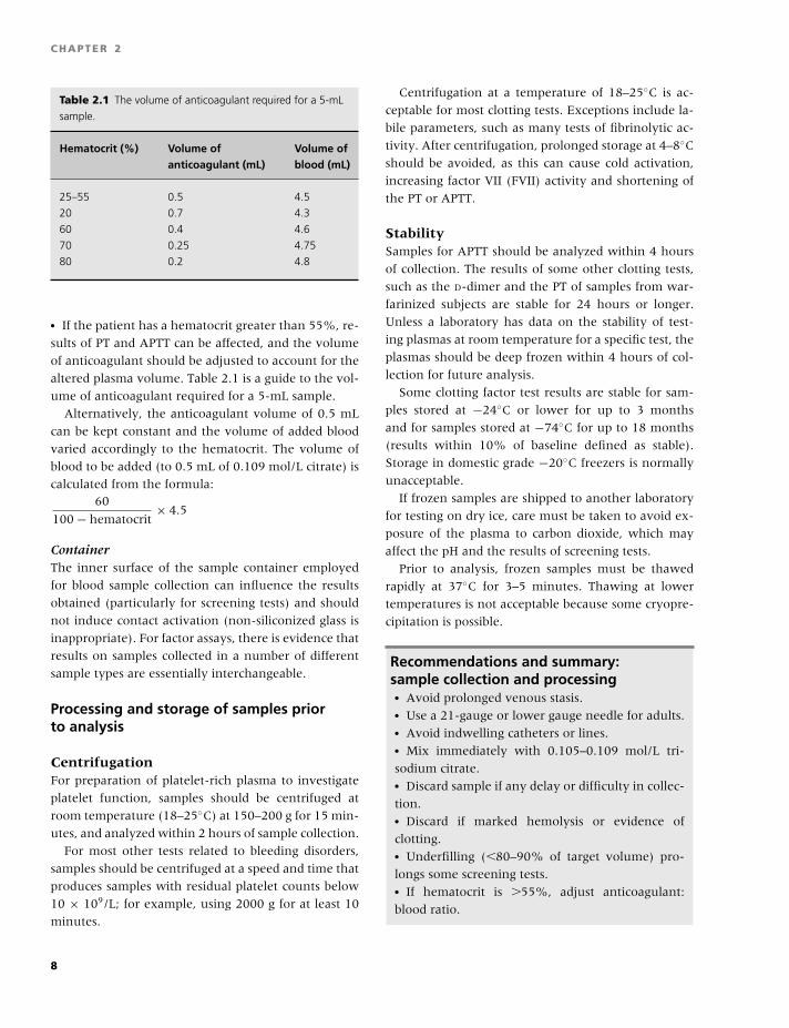
Table 2.1 The volume of anticoagulant required for a 5-mL
sample.
Hematocrit (%) Volume of Volume ofanticoagulant (mL) blood (mL)
25–55 0.5 4.5
20 0.7 4.3
60 0.4 4.6
70 0.25 4.75
80 0.2 4.8
� If the patient has a hematocrit greater than 55%, re-
sults of PT and APTT can be affected, and the volume
of anticoagulant should be adjusted to account for the
altered plasma volume. Table 2.1 is a guide to the vol-
ume of anticoagulant required for a 5-mL sample.
Alternatively, the anticoagulant volume of 0.5 mL
can be kept constant and the volume of added blood
varied accordingly to the hematocrit. The volume of
blood to be added (to 0.5 mL of 0.109 mol/L citrate) is
calculated from the formula:60
100 − hematocrit× 4.5
ContainerThe inner surface of the sample container employed
for blood sample collection can influence the results
obtained (particularly for screening tests) and should
not induce contact activation (non-siliconized glass is
inappropriate). For factor assays, there is evidence that
results on samples collected in a number of different
sample types are essentially interchangeable.
Processing and storage of samples priorto analysis
CentrifugationFor preparation of platelet-rich plasma to investigate
platelet function, samples should be centrifuged at
room temperature (18–25◦C) at 150–200 g for 15 min-
utes, and analyzed within 2 hours of sample collection.
For most other tests related to bleeding disorders,
samples should be centrifuged at a speed and time that
produces samples with residual platelet counts below
10 × 109/L; for example, using 2000 g for at least 10
minutes.
Centrifugation at a temperature of 18–25◦C is ac-
ceptable for most clotting tests. Exceptions include la-
bile parameters, such as many tests of fibrinolytic ac-
tivity. After centrifugation, prolonged storage at 4–8◦C
should be avoided, as this can cause cold activation,
increasing factor VII (FVII) activity and shortening of
the PT or APTT.
StabilitySamples for APTT should be analyzed within 4 hours
of collection. The results of some other clotting tests,
such as the D-dimer and the PT of samples from war-
farinized subjects are stable for 24 hours or longer.
Unless a laboratory has data on the stability of test-
ing plasmas at room temperature for a specific test, the
plasmas should be deep frozen within 4 hours of col-
lection for future analysis.
Some clotting factor test results are stable for sam-
ples stored at −24◦C or lower for up to 3 months
and for samples stored at −74◦C for up to 18 months
(results within 10% of baseline defined as stable).
Storage in domestic grade −20◦C freezers is normally
unacceptable.
If frozen samples are shipped to another laboratory
for testing on dry ice, care must be taken to avoid ex-
posure of the plasma to carbon dioxide, which may
affect the pH and the results of screening tests.
Prior to analysis, frozen samples must be thawed
rapidly at 37◦C for 3–5 minutes. Thawing at lower
temperatures is not acceptable because some cryopre-
cipitation is possible.
Recommendations and summary:sample collection and processing� Avoid prolonged venous stasis.� Use a 21-gauge or lower gauge needle for adults.� Avoid indwelling catheters or lines.� Mix immediately with 0.105–0.109 mol/L tri-
sodium citrate.� Discard sample if any delay or difficulty in collec-
tion.� Discard if marked hemolysis or evidence of
clotting.� Underfilling (�80–90% of target volume) pro-
longs some screening tests.� If hematocrit is �55%, adjust anticoagulant:
blood ratio.
8
BLBK186-Key April 24, 2009 13:30
Laboratory tests of hemostasis
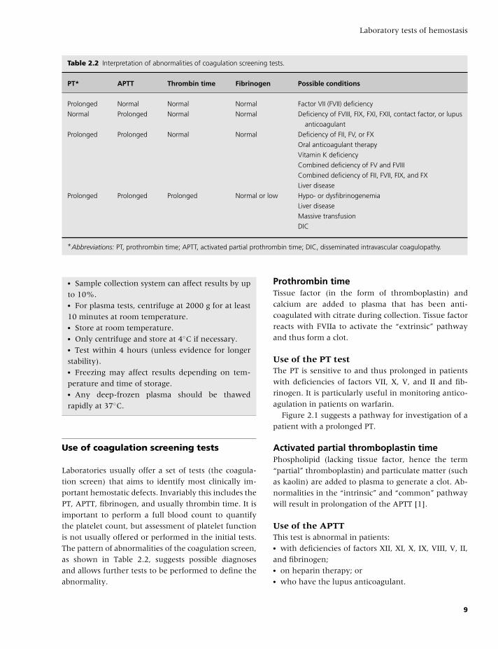
Table 2.2 Interpretation of abnormalities of coagulation screening tests.
PT* APTT Thrombin time Fibrinogen Possible conditions
Prolonged Normal Normal Normal Factor VII (FVII) deficiency
Normal Prolonged Normal Normal Deficiency of FVIII, FIX, FXI, FXII, contact factor, or lupus
anticoagulant
Prolonged Prolonged Normal Normal Deficiency of FII, FV, or FX
Oral anticoagulant therapy
Vitamin K deficiency
Combined deficiency of FV and FVIII
Combined deficiency of FII, FVII, FIX, and FX
Liver disease
Prolonged Prolonged Prolonged Normal or low Hypo- or dysfibrinogenemia
Liver disease
Massive transfusion
DIC
*Abbreviations: PT, prothrombin time; APTT, activated partial prothrombin time; DIC, disseminated intravascular coagulopathy.
� Sample collection system can affect results by up
to 10%.� For plasma tests, centrifuge at 2000 g for at least
10 minutes at room temperature.� Store at room temperature.� Only centrifuge and store at 4◦C if necessary.� Test within 4 hours (unless evidence for longer
stability).� Freezing may affect results depending on tem-
perature and time of storage.� Any deep-frozen plasma should be thawed
rapidly at 37◦C.
Use of coagulation screening tests
Laboratories usually offer a set of tests (the coagula-
tion screen) that aims to identify most clinically im-
portant hemostatic defects. Invariably this includes the
PT, APTT, fibrinogen, and usually thrombin time. It is
important to perform a full blood count to quantify
the platelet count, but assessment of platelet function
is not usually offered or performed in the initial tests.
The pattern of abnormalities of the coagulation screen,
as shown in Table 2.2, suggests possible diagnoses
and allows further tests to be performed to define the
abnormality.
Prothrombin timeTissue factor (in the form of thromboplastin) and
calcium are added to plasma that has been anti-
coagulated with citrate during collection. Tissue factor
reacts with FVIIa to activate the “extrinsic” pathway
and thus form a clot.
Use of the PT testThe PT is sensitive to and thus prolonged in patients
with deficiencies of factors VII, X, V, and II and fib-
rinogen. It is particularly useful in monitoring antico-
agulation in patients on warfarin.
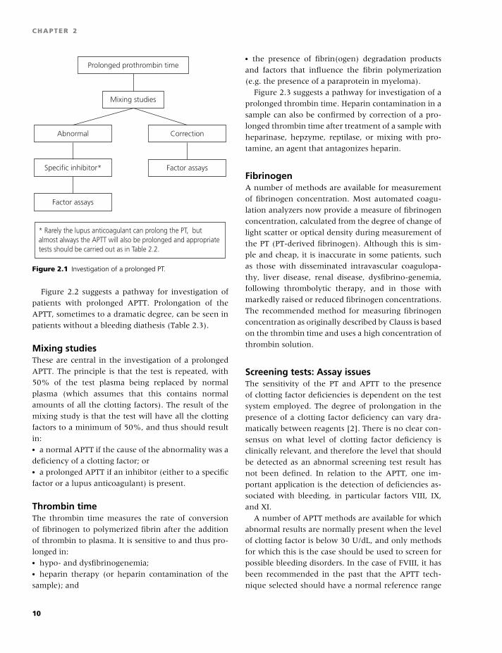
Figure 2.1 suggests a pathway for investigation of a
patient with a prolonged PT.
Activated partial thromboplastin timePhospholipid (lacking tissue factor, hence the term
“partial” thromboplastin) and particulate matter (such
as kaolin) are added to plasma to generate a clot. Ab-
normalities in the “intrinsic” and “common” pathway
will result in prolongation of the APTT [1].
Use of the APTTThis test is abnormal in patients:� with deficiencies of factors XII, XI, X, IX, VIII, V, II,
and fibrinogen;� on heparin therapy; or� who have the lupus anticoagulant.
9
BLBK186-Key April 24, 2009 13:30
CHAPTER 2
Prolonged prothrombin time
Mixing studies
Abnormal
Specific inhibitor*
Factor assays
* Rarely the lupus anticoagulant can prolong the PT, butalmost always the APTT will also be prolonged and appropriate tests should be carried out as in Table 2.2.
Correction
Factor assays
Figure 2.1 Investigation of a prolonged PT.
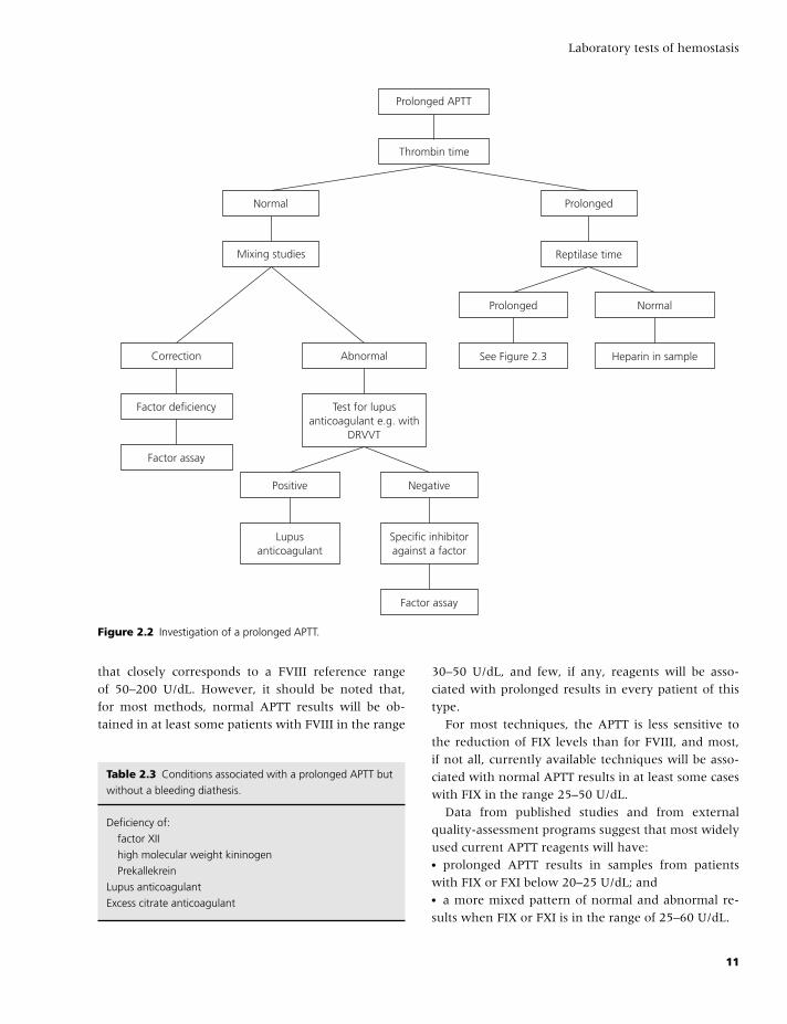
Figure 2.2 suggests a pathway for investigation of
patients with prolonged APTT. Prolongation of the
APTT, sometimes to a dramatic degree, can be seen in
patients without a bleeding diathesis (Table 2.3).
Mixing studiesThese are central in the investigation of a prolonged
APTT. The principle is that the test is repeated, with
50% of the test plasma being replaced by normal
plasma (which assumes that this contains normal
amounts of all the clotting factors). The result of the
mixing study is that the test will have all the clotting
factors to a minimum of 50%, and thus should result
in:� a normal APTT if the cause of the abnormality was a
deficiency of a clotting factor; or� a prolonged APTT if an inhibitor (either to a specific
factor or a lupus anticoagulant) is present.
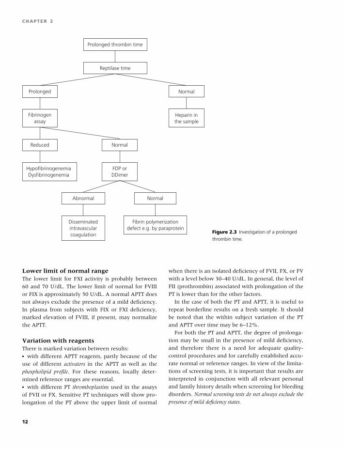
Thrombin timeThe thrombin time measures the rate of conversion
of fibrinogen to polymerized fibrin after the addition
of thrombin to plasma. It is sensitive to and thus pro-
longed in:� hypo- and dysfibrinogenemia;� heparin therapy (or heparin contamination of the
sample); and
� the presence of fibrin(ogen) degradation products
and factors that influence the fibrin polymerization
(e.g. the presence of a paraprotein in myeloma).
Figure 2.3 suggests a pathway for investigation of a
prolonged thrombin time. Heparin contamination in a
sample can also be confirmed by correction of a pro-
longed thrombin time after treatment of a sample with
heparinase, hepzyme, reptilase, or mixing with pro-
tamine, an agent that antagonizes heparin.
FibrinogenA number of methods are available for measurement
of fibrinogen concentration. Most automated coagu-
lation analyzers now provide a measure of fibrinogen
concentration, calculated from the degree of change of
light scatter or optical density during measurement of
the PT (PT-derived fibrinogen). Although this is sim-
ple and cheap, it is inaccurate in some patients, such
as those with disseminated intravascular coagulopa-
thy, liver disease, renal disease, dysfibrino-genemia,
following thrombolytic therapy, and in those with
markedly raised or reduced fibrinogen concentrations.
The recommended method for measuring fibrinogen
concentration as originally described by Clauss is based
on the thrombin time and uses a high concentration of
thrombin solution.
Screening tests: Assay issuesThe sensitivity of the PT and APTT to the presence
of clotting factor deficiencies is dependent on the test
system employed. The degree of prolongation in the
presence of a clotting factor deficiency can vary dra-
matically between reagents [2]. There is no clear con-
sensus on what level of clotting factor deficiency is
clinically relevant, and therefore the level that should
be detected as an abnormal screening test result has
not been defined. In relation to the APTT, one im-
portant application is the detection of deficiencies as-
sociated with bleeding, in particular factors VIII, IX,
and XI.
A number of APTT methods are available for which
abnormal results are normally present when the level
of clotting factor is below 30 U/dL, and only methods
for which this is the case should be used to screen for
possible bleeding disorders. In the case of FVIII, it has
been recommended in the past that the APTT tech-
nique selected should have a normal reference range
10
BLBK186-Key April 24, 2009 13:30
Laboratory tests of hemostasis
Prolonged APTT
Thrombin time
Normal
Mixing studies
Prolonged
Reptilase time
Correction
Factor deficiency
Factor assay
Abnormal
Test for lupusanticoagulant e.g. with
DRVVT
Prolonged
See Figure 2.3
Normal
Heparin in sample
Positive
Factor assay
Negative
Lupusanticoagulant
Specific inhibitoragainst a factor
Figure 2.2 Investigation of a prolonged APTT.
that closely corresponds to a FVIII reference range
of 50–200 U/dL. However, it should be noted that,
for most methods, normal APTT results will be ob-
tained in at least some patients with FVIII in the range
Table 2.3 Conditions associated with a prolonged APTT but
without a bleeding diathesis.
Deficiency of:
factor XII
high molecular weight kininogen
Prekallekrein
Lupus anticoagulant
Excess citrate anticoagulant
30–50 U/dL, and few, if any, reagents will be asso-
ciated with prolonged results in every patient of this
type.
For most techniques, the APTT is less sensitive to
the reduction of FIX levels than for FVIII, and most,
if not all, currently available techniques will be asso-
ciated with normal APTT results in at least some cases
with FIX in the range 25–50 U/dL.
Data from published studies and from external
quality-assessment programs suggest that most widely
used current APTT reagents will have:� prolonged APTT results in samples from patients
with FIX or FXI below 20–25 U/dL; and� a more mixed pattern of normal and abnormal re-
sults when FIX or FXI is in the range of 25–60 U/dL.
11
BLBK186-Key April 24, 2009 13:30
CHAPTER 2
Prolonged thrombin time
Reptilase time
Prolonged Normal
Fibrinogenassay
Heparin inthe sample
Reduced
HypofibrinogenemiaDysfibrinogenemia
Normal
FDP orDDimer
Abnormal
Disseminatedintravascularcoagulation
Normal
Fibrin polymerizationdefect e.g. by paraprotein
Figure 2.3 Investigation of a prolonged
thrombin time.
Lower limit of normal rangeThe lower limit for FXI activity is probably between
60 and 70 U/dL. The lower limit of normal for FVIII
or FIX is approximately 50 U/dL. A normal APTT does
not always exclude the presence of a mild deficiency.
In plasma from subjects with FIX or FXI deficiency,
marked elevation of FVIII, if present, may normalize
the APTT.
Variation with reagentsThere is marked variation between results:� with different APTT reagents, partly because of the
use of different activators in the APTT as well as the
phospholipid profile. For these reasons, locally deter-
mined reference ranges are essential.� with different PT thromboplastins used in the assays
of FVII or FX. Sensitive PT techniques will show pro-
longation of the PT above the upper limit of normal
when there is an isolated deficiency of FVII, FX, or FV
with a level below 30–40 U/dL. In general, the level of
FII (prothrombin) associated with prolongation of the
PT is lower than for the other factors.
In the case of both the PT and APTT, it is useful to
repeat borderline results on a fresh sample. It should
be noted that the within subject variation of the PT
and APTT over time may be 6–12%.
For both the PT and APTT, the degree of prolonga-
tion may be small in the presence of mild deficiency,
and therefore there is a need for adequate quality-
control procedures and for carefully established accu-
rate normal or reference ranges. In view of the limita-
tions of screening tests, it is important that results are
interpreted in conjunction with all relevant personal
and family history details when screening for bleeding
disorders. Normal screening tests do not always exclude the
presence of mild deficiency states.
12
BLBK186-Key April 24, 2009 13:30
Laboratory tests of hemostasis
Recommendations and summary:Screening tests� PT and APTT methods vary in sensitivity to factor
deficiency.� Mild deficiency may be associated with normal
PT or APTT.� For bleeding disorders, select a method for which
APTT is normally prolonged when FVIII, FIX, or
FXI is 30 IU/dL or less.� Elevated FVIII may normalize APTT in mild FIX
or FXI deficiency.� Assessments of APTT sensitivity should employ
samples from patients.
Clotting factor assay design
One-stage assaysFor many years, the most commonly performed assays
for clotting factors have been one-stage clotting assays
based on:� the APTT in the case of factors VIII, IX, or XI; or� the PT in the case of factors II, V, VII, or X.
There are a number of general features of the de-
sign of one-stage clotting assays that are necessary to
ensure accurate, reliable, and valid results. In factor
assays, the principle depends on the ability of a sam-
ple containing the factor under investigation to correct
or shorten the delayed clotting of a plasma completely
deficient in that factor. Such deficient plasmas must
contain less than 1 U/dL of the clotting factor under
investigation and normal levels of all other relevant
clotting factors.
It is important that the clotting time measured by
the APTT or PT depends directly on the amount of fac-
tor present in the mixture of deficient and reference
or test plasma. For example, in a FVIII assay, the level
of FVIII must be rate-limiting in relation to the clotting
time obtained. This requires dilution of a reference or
standard plasma of known concentration. Preparation
of several different dilutions of the reference plasma
allows construction of a calibration curve in which
the clotting time response depends on the dose
(concentration) of factor present. At lower plasma
dilutions or higher factor concentrations, the factor
under investigation may not be rate-limiting, and the
assay is no longer specific and therefore invalid. It may
be necessary to extend the calibration curve by testing
additional dilutions when analyzing test plasmas with
concentrations below 10 U/dL. At very low concentra-
tions of an individual factor (�1–2 U/dL), the clotting
time of the deficient plasma may not be even partially
corrected by addition of the test plasma dilution. Dilu-
tions are selected so that there is a linear relationship
between concentration (logarithmic scale) and the
response in clotting time (logarithmic or linear scale).
The reference curve should be prepared using at
least three different dilutions, and a calibration curve
should be included each time the assay is performed
unless there is clear evidence that the responses are
so reproducible that a calibration curve can be stored
for use on other occasions. The reference plasma
should be calibrated by a route traceable back to WHO
international standards where these are available. Test
plasmas should be analyzed by using three dilutions
so that it is possible to confirm that the dose–response
curve of the test plasma is linear and parallel to the
dose–response curve of the reference plasma. It is
not acceptable to test a single test dilution because
this reduces the accuracy substantially and may lead
to major underestimation of the true concentration
when inhibitors are present. If a dose–response
curve of a test plasma is not parallel to the reference
curve, and the presence of an inhibitor (such as an
antiphospholipid antibody) is confirmed or suspected,
then the estimate of activity obtained from the highest
test plasma dilution is likely to be closest to the real
concentration; but, it should be noted that the criteria
for a valid assay cannot be met and results must be
interpreted with caution. In the case of one-stage,
APTT-based assays, the interference by antiphospho-
lipid antibodies is frequently dependent on the APTT
reagent used and its phospholipid content. Some APTT
reagents, such as Actin FS, contain a high phospho-
lipid concentration, and this type of reagent is much
less affected by these antibodies and is particularly
suitable for use in factor assays in such cases.
Recommendations and summary:Factor assays� Assays should be calibrated with reference plas-
mas traceable back to WHO standards where
available.
13
BLBK186-Key April 24, 2009 13:30
CHAPTER 2
� Deficient plasmas must have �1 U/dL of the clot-
ting factor being assayed and normal levels of other
relevant factors.� No less than three dilutions of test plasmas
should be tested.� A valid assay requires test and calibration lines to
be parallel.� Interference by antiphospholipid antibodies can
be minimized by use of an APTT reagent with a
high phospholipid content.
Thrombophilia testing
This section addresses some laboratory aspects of
testing for heritable thrombophilia: protein C (PC),
protein S (PS), antithrombin (AT), activated protein
C resistance (APC-R), FV Leiden (FVL), and the pro-
thrombin 20210A allele [3,4].
Sample collection, processing, and assayFor thrombophilia testing, as for other coagulation
tests:� A citrate concentration of 0.105–0.109 mol/L should
be used for sample collection, because citrate strength
may affect results, at least for APC-R testing.� Centrifugation should be as for other coagulation
tests described above.� Residual platelets in plasma following centrifugation
can also affect results of APC-R tests, and plasmas
should be centrifuged as described above, separated,
and recentrifuged a second time to ensure maximum
removal of platelets. (Such a procedure is not neces-
sary for AT, PC, or PS testing but can be used for con-
venience without adverse effects if the same plasma
is to be used for these investigations in addition to
APC-R.)� Such double-centrifuged plasma can then be stored
deep frozen prior to analysis for at least 6 months
for clotting PS activity and at least 18 months for PC
and AT.� In general, activity assays are preferable to antigen
assays because antigen assays will be normal in some
patients with type 2 defects where a normal concen-
tration of a defective protein is present.
In the case of PS, this is complicated by the problems
associated with interference by FVL in many different
activity assays and can lead to important underestima-
tion of the true level, with misdiagnosis a possibility.
At present, the standardization of PS activity assays
is poor in that results of different assays may differ
substantially even in normal subjects. For these rea-
sons, PS activity assays must be used with caution.
FVL can also cause underestimation of the true PC
level in clotting assays. A chromogenic PC assay may
be used to avoid this problem, or alternatively the PC
clotting assay can be modified to include predilution
of test sample 1 in 4 in PC-deficient plasma to restore
specificity. A similar procedure can be employed to im-
prove performance of clotting PS assays in the pres-
ence of FVL.
Clotting assays of PC and PS may also be influenced
adversely by elevated FVIII, causing underestimation.
The presence of the lupus anticoagulant may be asso-
ciated with falsely high results, with the possibility of
a false normal result in the presence of deficiency.
When assaying PC, PS, and AT, calibration curves
should include a minimum of three dilutions, and, in
general, the most precise test results will be obtained
if a calibration curve is prepared with each group of
patient samples. As for other tests of hemostasis, it is
important to use a reference plasma traceable back to
WHO standards, which are available for AT, PC, and
PS.
Testing for APC-R is largely based on the APTT in
the presence and absence of APC, and therefore many
of the variables that affect the APTT will in turn in-
fluence APC-R test results. These include the presence
of heparin or lupus anticoagulant by prolonging clot-
ting times, or elevated FVIII, which shortens clotting
times and manifest as acquired APC-R. The original
APC-R test also requires normal levels of clotting fac-
tors, including FII and FX, which are reduced by war-
farin therapy. Valid APC-R testing as originally used
requires a normal PT and APTT.
There is evidence that standardization of results
obtained by the original assay can be improved by
calculation of the normalized APC-R ratio (test APC
ratio divided by APC ratio of a pooled normal plasma
tested in the same batch of tests). The test can be sig-
nificantly improved by predilution of test plasma in
FV-deficient plasma, making the test 100% sensitive
to the presence of FVL. This modification also makes
the test specific for FVL, and will be associated with
normal results where APC resistance in the classic as-
say is not a consequence of FVL. This must be borne
in mind when interpreting results. In some versions of
14
BLBK186-Key April 24, 2009 13:30
Laboratory tests of hemostasis
the test, there is clear separation between results ob-
tained in heterozygotes and homozygotes; but, even
for such assays, confirmation by genetic testing may be
necessary because it is important to identify homozy-
gotes with certainty.
When genetic testing for the FVL or prothrombin
alleles is undertaken, there are fewer relevant prean-
alytical variables than for phenotypic tests on plasma.
Whole blood samples are stable for several weeks, at
least for some of the genotyping methods.
Because of the many differences between results of
apparently similar assays in thrombophilia testing, it is
particularly important to establish locally a reference
or normal range (as discussed in Appendix 1).
Recommendations and summary:thrombophilia tests� Double centrifugation is required for APC-R test-
ing.� Presence of FVL may cause significant underesti-
mation of clotting PC or PS activity.� Results of PS activity assays are highly dependent
on reagents used.� Elevated FVIII or lupus anticoagulant can inter-
fere with PC or PS clotting assays.� Results of AT assays may depend on the enzyme
used in the assay.� APC-R with FV-deficient plasma dilution is the
most sensitive and specific for FVL.� Genetic testing for FVL or prothrombin allele
may not be error free.
Quality assurance
All laboratory tests of blood coagulation require care-
ful application of quality-assurance procedures to en-
sure reliability of results. Quality assurance is used to
describe all the measures that are taken to ensure the
reliability of laboratory testing and reporting. This in-
cludes the choice of test, the collection of a valid sam-
ple from the patient, analysis of the specimen, and the
recording of results in a timely and accurate manner,
through to interpretation of the results, where appro-
priate, and communication of these results to the re-
ferring clinicians.
Internal quality control (IQC) and external quality
assessment (EQA) are complementary components of
a laboratory quality-assurance program. Quality assur-
ance is required to check that the results of labora-
tory investigations are reliable enough to be released
to assist clinical decision-making, monitoring of ther-
apy, and diagnosis of hemostatic abnormalities.
Internal quality controlIQC is used to establish whether a series of techniques
and procedures are performing consistently over a pe-
riod of time (precision). It is therefore deployed to en-
sure day-to-day laboratory consistency. It is important
to recognize that a precise technique is not necessarily
accurate; accuracy being a measure of the closeness of
an estimated value to the true value.
IQC procedures should be applied in a way that
ensures immediate and constant control of result gen-
eration. Within a laboratory setting, the quality of
results obtained is influenced by maintenance of an
upto-date manual of standard operational procedures;
use of reliable reagents and reference materials; selec-
tion of automation and adequate maintenance; ade-
quate records and reporting system for results; and an
appropriate complement of suitably trained personnel.
For screening tests, it is important to include reg-
ular and frequent testing of quality-control material,
which should include a normal material and at least
one level of abnormal sample. For batch analysis,
a quality-control sample can be included with each
batch. For continuous processing systems, the fre-
quency of quality-control testing must be tailored to
the work pattern and should be adjusted until the
frequency of repeat patient testing resulting from the
limits of the quality control studies is at a minimum.
For many random access coagulometers, performing
screening tests, this could typically be every 2 hours
of continuous work or every 30–40 samples. For fac-
tor assays and parameters typically tested in batches,
a quality-control sample should be included with each
group of tests. Patient results should only be released if
quality-control results remain within acceptable target
limits. It is frequently useful to include IQC material at
different critical levels of abnormality.
External quality assessmentEQA is used to identify the degree of agreement be-
tween one laboratory’s results and those obtained by
other centers, which can be used as a measure of
accuracy. The main function of EQA is proficiency
testing of individual laboratory testing, but larger
15
BLBK186-Key April 24, 2009 13:30
CHAPTER 2
programs provide information concerning the relative
performance of analytical procedures, including the
method principle, reagents, and instruments. As a gen-
eral principle, all centers undertaking investigations
of hemostasis should participate in an accredited EQA
program for all tests where available.
Recommendations and summary:quality control� Quality-control samples should be analyzed reg-
ularly and frequently for screening tests and with
each group of factor assays.� Centers should participate in accredited EQA
programs for all tests where available.
References
1 Koepke JA. Partial thromboplastin time test: proposed
performance guidelines. ICSH Panel on the PTT. Thromb
Haemost 1986;55:143–4.
2 Lawrie AS, Kitchen S, Purdy G, Mackie IJ, Preston FE,
Machin SJ. Assessment of actin FS and actin FSL sen-
sitivity to specific clotting factor deficiencies. Clin Lab
Haematol 1998;20:179–86.
3 Jennings I, Cooper P. Screening for thrombophilia:
a laboratory perspective. Br J Biomed Sci 2003;60:
39–51.
4 Walker ID, Greaves M, Preston FE. Investigation and
management of heritable thrombophilia. Br J Haematol
2001;114:512–18.
16
BLBK186-Key April 15, 2009 9:36
3 Laboratory evaluationand thrombophiliaRajiv K. Pruthi and John A. Heit
Overview
Venous thromboembolism (VTE) is a prototype of a
multifactorial disease model in which interaction of
genetic and environmental risk factors (termed throm-
bophilia) predispose to VTE. Patients who develop a
VTE are considered to have thrombophilia; however,
this term should not be considered to be a disease,
but a risk factor for (venous or arterial) thrombosis,
thus it is important to note that presence of throm-
bophilia in an individual is not absolutely predictive
of thrombosis. The most common clinical presenta-
tion of thrombophilia is VTE; other presentations are
listed in Table 3.1. The currently recognized inher-
ited and acquired thrombophilias (Tables 3.2 and 3.3)
predispose to VTE; however, selected conditions (lu-
pus anticoagulants and hyperhomocysteinemia) may
also predispose to arterial thrombosis. The presence
of thrombophilia determines a patient’s risk for ini-
tial and subsequent (recurrent) VTE, which influences
(primary and secondary) VTE prevention strategies.
Assessment for presence ofthrombophilia
Clinical assessmentAssessment of presence of thrombophilia is not solely
confined to laboratory testing but begins with a de-
tailed history and physical examination. Detailed in-
quiry into symptoms and signs of acquired risk factors
(coexisting diseases, medication exposure, and clini-
cal circumstances) that are associated with thrombo-
sis (Tables 3.2–3.4) are an important part of the ini-
tial evaluation as is a complete physical examination.
In addition to judicious laboratory testing appropriate
for the patient’s age and symptoms, objective confir-
mation of venous thromboembolism is critical prior to
embarking on extensive laboratory testing for throm-
bophilia. Because indiscriminate, extensive testing for
occult cancer in patients presenting with idiopathic
VTE has not clearly been shown to improve cancer-
related survival, such an evaluation should be con-
fined to age-appropriate cancer screening and further
evaluation of patient symptoms and signs.
Laboratory testingCurrently, there is no single laboratory global assay
that will ‘screen’ for the presence of thrombophilia.
Thus, laboratory testing can be broadly categorized
into (1) general diagnostic testing, (2) specialized co-
agulation testing, and (3) ancillary testing for disorders
known to predispose to thrombotic disorders.
General diagnostic testingAll patients with objectively confirmed VTE should
have the following tests prior to initiation of antico-
agulant therapy: complete blood count (CBC), tests
of kidney and liver function (these tests are primar-
ily to assess for safety of anticoagulation with hep-
arin and warfarin); obtaining baseline prothrombin
time (PT) and activated partial thromboplastin time
(APTT) tests aid in optimal monitoring of anticoagu-
lation (Table 3.5).
Specialized coagulation testingSpecial Coagulation testing consists of a battery of
complex (protein and DNA-based) thrombophilia as-
says to detect presence of an inherited or acquired
thrombophilia. As discussed below, multiple pre-
analytical conditions affect results of these assays (e.g.
anticoagulants, acute thrombosis, liver disease, etc.),
17
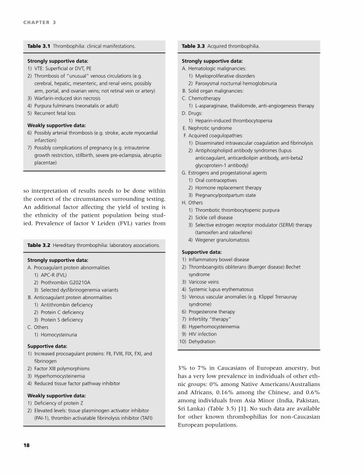
BLBK186-Key April 15, 2009 9:36
CHAPTER 3
Table 3.1 Thrombophilia: clinical manifestations.
Strongly supportive data:1) VTE: Superficial or DVT, PE
2) Thrombosis of “unusual” venous circulations (e.g.
cerebral, hepatic, mesenteric, and renal veins; possibly
arm, portal, and ovarian veins; not retinal vein or artery)
3) Warfarin-induced skin necrosis
4) Purpura fulminans (neonatalis or adult)
5) Recurrent fetal loss
Weakly supportive data:6) Possibly arterial thrombosis (e.g. stroke, acute myocardial
infarction)
7) Possibly complications of pregnancy (e.g. intrauterine
growth restriction, stillbirth, severe pre-eclampsia, abruptio
placentae)
so interpretation of results needs to be done within
the context of the circumstances surrounding testing.
An additional factor affecting the yield of testing is
the ethnicity of the patient population being stud-
ied. Prevalence of factor V Leiden (FVL) varies from
Table 3.2 Hereditary thrombophilia: laboratory associations.
Strongly supportive data:A. Procoagulant protein abnormalities
1) APC-R (FVL)
2) Prothrombin G20210A
3) Selected dysfibrinogenemia variants
B. Anticoagulant protein abnormalities
1) Antithrombin deficiency
2) Protein C deficiency
3) Protein S deficiency
C. Others
1) Homocysteinuria
Supportive data:1) Increased procoagulant proteins: FII, FVIII, FIX, FXI, and
fibrinogen
2) Factor XIII polymorphisms
3) Hyperhomocysteinemia
4) Reduced tissue factor pathway inhibitor
Weakly supportive data:1) Deficiency of protein Z
2) Elevated levels: tissue plasminogen activator inhibitor
(PAI-1), thrombin activatable fibrinolysis inhibitor (TAFI)
Table 3.3 Acquired thrombophilia.
Strongly supportive data:A. Hematologic malignancies:
1) Myeloproliferative disorders
2) Paroxysmal nocturnal hemoglobinuria
B. Solid organ malignancies:
C. Chemotherapy
1) L-asparaginase, thalidomide, anti-angiogenesis therapy
D. Drugs:
1) Heparin-induced thrombocytopenia
E. Nephrotic syndrome
F. Acquired coagulopathies:
1) Disseminated intravascular coagulation and fibrinolysis
2) Antiphospholipid antibody syndromes (lupus
anticoagulant, anticardiolipin antibody, anti-beta2
glycoprotein-1 antibody)
G. Estrogens and progestational agents
1) Oral contraceptives
2) Hormone replacement therapy
3) Pregnancy/postpartum state
H. Others
1) Thrombotic thrombocytopenic purpura
2) Sickle cell disease
3) Selective estrogen receptor modulator (SERM) therapy
(tamoxifen and raloxifene)
4) Wegener granulomatosis
Supportive data:1) Inflammatory bowel disease
2) Thromboangiitis obliterans (Buerger disease) Bechet
syndrome
3) Varicose veins
4) Systemic lupus erythematosus
5) Venous vascular anomalies (e.g. Klippel Trenaunay
syndrome)
6) Progesterone therapy
7) Infertility “therapy”
8) Hyperhomocysteinemia
9) HIV infection
10) Dehydration
3% to 7% in Caucasians of European ancestry, but
has a very low prevalence in individuals of other eth-
nic groups: 0% among Native Americans/Australians
and Africans, 0.16% among the Chinese, and 0.6%
among individuals from Asia Minor (India, Pakistan,
Sri Lanka) (Table 3.5) [1]. No such data are available
for other known thrombophilias for non-Caucasian
European populations.
18
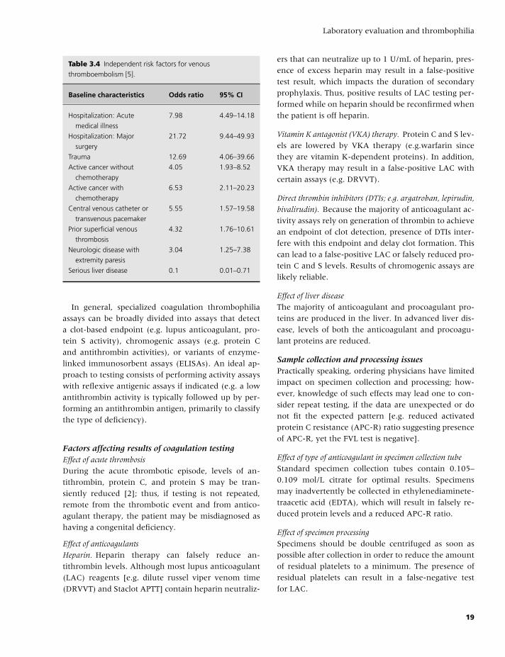
BLBK186-Key April 15, 2009 9:36
Laboratory evaluation and thrombophilia
Table 3.4 Independent risk factors for venous
thromboembolism [5].
Baseline characteristics Odds ratio 95% CI
Hospitalization: Acute
medical illness
7.98 4.49–14.18
Hospitalization: Major
surgery
21.72 9.44–49.93
Trauma 12.69 4.06–39.66
Active cancer without
chemotherapy
4.05 1.93–8.52
Active cancer with
chemotherapy
6.53 2.11–20.23
Central venous catheter or
transvenous pacemaker
5.55 1.57–19.58
Prior superficial venous
thrombosis
4.32 1.76–10.61
Neurologic disease with
extremity paresis
3.04 1.25–7.38
Serious liver disease 0.1 0.01–0.71
In general, specialized coagulation thrombophilia
assays can be broadly divided into assays that detect
a clot-based endpoint (e.g. lupus anticoagulant, pro-
tein S activity), chromogenic assays (e.g. protein C
and antithrombin activities), or variants of enzyme-
linked immunosorbent assays (ELISAs). An ideal ap-
proach to testing consists of performing activity assays
with reflexive antigenic assays if indicated (e.g. a low
antithrombin activity is typically followed up by per-
forming an antithrombin antigen, primarily to classify
the type of deficiency).
Factors affecting results of coagulation testingEffect of acute thrombosis
During the acute thrombotic episode, levels of an-
tithrombin, protein C, and protein S may be tran-
siently reduced [2]; thus, if testing is not repeated,
remote from the thrombotic event and from antico-
agulant therapy, the patient may be misdiagnosed as
having a congenital deficiency.
Effect of anticoagulants
Heparin. Heparin therapy can falsely reduce an-
tithrombin levels. Although most lupus anticoagulant
(LAC) reagents [e.g. dilute russel viper venom time
(DRVVT) and Staclot APTT] contain heparin neutraliz-
ers that can neutralize up to 1 U/mL of heparin, pres-
ence of excess heparin may result in a false-positive
test result, which impacts the duration of secondary
prophylaxis. Thus, positive results of LAC testing per-
formed while on heparin should be reconfirmed when
the patient is off heparin.
Vitamin K antagonist (VKA) therapy. Protein C and S lev-
els are lowered by VKA therapy (e.g.warfarin since
they are vitamin K-dependent proteins). In addition,
VKA therapy may result in a false-positive LAC with
certain assays (e.g. DRVVT).
Direct thrombin inhibitors (DTIs; e.g. argatroban, lepirudin,
bivalirudin). Because the majority of anticoagulant ac-
tivity assays rely on generation of thrombin to achieve
an endpoint of clot detection, presence of DTIs inter-
fere with this endpoint and delay clot formation. This
can lead to a false-positive LAC or falsely reduced pro-
tein C and S levels. Results of chromogenic assays are
likely reliable.
Effect of liver disease
The majority of anticoagulant and procoagulant pro-
teins are produced in the liver. In advanced liver dis-
ease, levels of both the anticoagulant and procoagu-
lant proteins are reduced.
Sample collection and processing issuesPractically speaking, ordering physicians have limited
impact on specimen collection and processing; how-
ever, knowledge of such effects may lead one to con-
sider repeat testing, if the data are unexpected or do
not fit the expected pattern [e.g. reduced activated
protein C resistance (APC-R) ratio suggesting presence
of APC-R, yet the FVL test is negative].
Effect of type of anticoagulant in specimen collection tube
Standard specimen collection tubes contain 0.105–
0.109 mol/L citrate for optimal results. Specimens
may inadvertently be collected in ethylenediaminete-
traacetic acid (EDTA), which will result in falsely re-
duced protein levels and a reduced APC-R ratio.
Effect of specimen processing
Specimens should be double centrifuged as soon as
possible after collection in order to reduce the amount
of residual platelets to a minimum. The presence of
residual platelets can result in a false-negative test
for LAC.
19
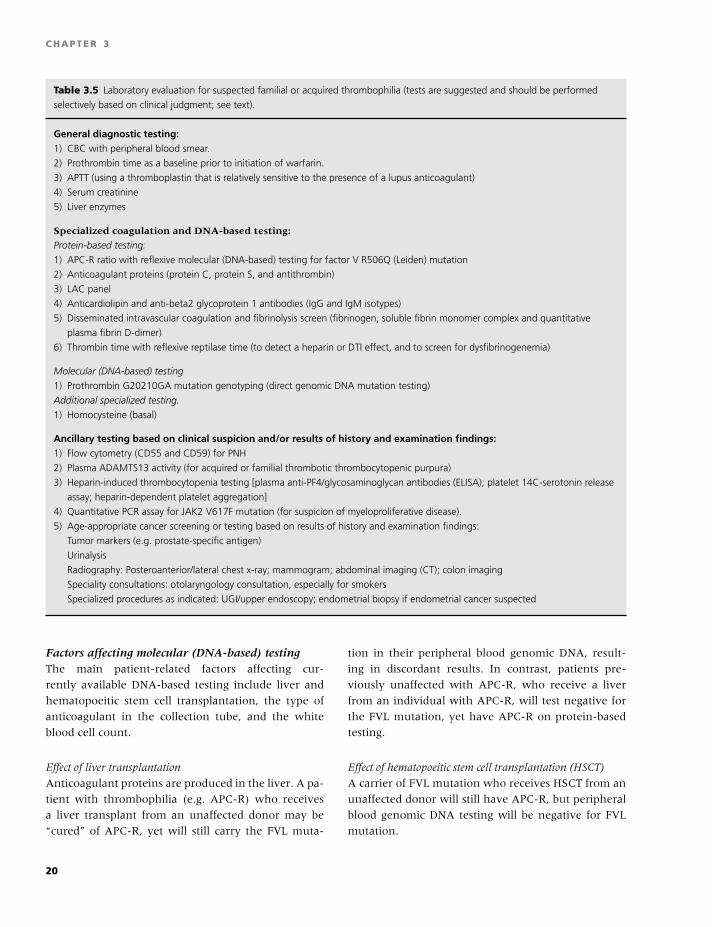
BLBK186-Key April 15, 2009 9:36
CHAPTER 3
Table 3.5 Laboratory evaluation for suspected familial or acquired thrombophilia (tests are suggested and should be performed
selectively based on clinical judgment; see text).
General diagnostic testing:1) CBC with peripheral blood smear.
2) Prothrombin time as a baseline prior to initiation of warfarin.
3) APTT (using a thromboplastin that is relatively sensitive to the presence of a lupus anticoagulant)
4) Serum creatinine
5) Liver enzymes
Specialized coagulation and DNA-based testing:
Protein-based testing:
1) APC-R ratio with reflexive molecular (DNA-based) testing for factor V R506Q (Leiden) mutation
2) Anticoagulant proteins (protein C, protein S, and antithrombin)
3) LAC panel
4) Anticardiolipin and anti-beta2 glycoprotein 1 antibodies (IgG and IgM isotypes)
5) Disseminated intravascular coagulation and fibrinolysis screen (fibrinogen, soluble fibrin monomer complex and quantitative
plasma fibrin D-dimer)
6) Thrombin time with reflexive reptilase time (to detect a heparin or DTI effect, and to screen for dysfibrinogenemia)
Molecular (DNA-based) testing
1) Prothrombin G20210GA mutation genotyping (direct genomic DNA mutation testing)
Additional specialized testing.
1) Homocysteine (basal)
Ancillary testing based on clinical suspicion and/or results of history and examination findings:1) Flow cytometry (CD55 and CD59) for PNH
2) Plasma ADAMTS13 activity (for acquired or familial thrombotic thrombocytopenic purpura)
3) Heparin-induced thrombocytopenia testing [plasma anti-PF4/glycosaminoglycan antibodies (ELISA); platelet 14C-serotonin release
assay; heparin-dependent platelet aggregation]
4) Quantitative PCR assay for JAK2 V617F mutation (for suspicion of myeloproliferative disease).
5) Age-appropriate cancer screening or testing based on results of history and examination findings:
Tumor markers (e.g. prostate-specific antigen)
Urinalysis
Radiography: Posteroanterior/lateral chest x-ray; mammogram; abdominal imaging (CT); colon imaging
Speciality consultations: otolaryngology consultation, especially for smokers
Specialized procedures as indicated: UGI/upper endoscopy; endometrial biopsy if endometrial cancer suspected
Factors affecting molecular (DNA-based) testingThe main patient-related factors affecting cur-
rently available DNA-based testing include liver and
hematopoeitic stem cell transplantation, the type of
anticoagulant in the collection tube, and the white
blood cell count.
Effect of liver transplantation
Anticoagulant proteins are produced in the liver. A pa-
tient with thrombophilia (e.g. APC-R) who receives
a liver transplant from an unaffected donor may be
“cured” of APC-R, yet will still carry the FVL muta-
tion in their peripheral blood genomic DNA, result-
ing in discordant results. In contrast, patients pre-
viously unaffected with APC-R, who receive a liver
from an individual with APC-R, will test negative for
the FVL mutation, yet have APC-R on protein-based
testing.
Effect of hematopoeitic stem cell transplantation (HSCT)
A carrier of FVL mutation who receives HSCT from an
unaffected donor will still have APC-R, but peripheral
blood genomic DNA testing will be negative for FVL
mutation.
20
BLBK186-Key April 15, 2009 9:36
Laboratory evaluation and thrombophilia
Effect of patient white blood cell count
Because the large majority of testing is performed on
sample from peripheral blood leukocytes, leucopenia
caused by intrinsic hematologic disorders, or as a result
of chemotherapy, may make it technically difficult to
perform the assays.
Type of anticoagulant in the collection tube
In general, peripheral blood for DNA-based testing
is collected in acid-citrate-dextrose (ACD) or EDTA.
Heparin interferes with the polymerase chain reaction
(PCR)-based testing.
Ancillary testingAdditional testing to detect disorders known to pre-
dispose to VTE should be pursued if clinically in-
dicated. Flow cytometry for CD55 and CD59 is in-
dicated in patients with evidence of intravascular
hemolysis with or without pancytopenia for detec-
tion of paroxysmal nocturnal hemoglobinuria (PNH).
Assays for ADAMTS-13 in patients with microangio-
pathic hemolytic anemia, thrombocytopenia, with or
without neurological symptoms, fever, and renal in-
sufficiency detect thrombotic thrombocytopenic pur-
pura. In patients exposed to heparin, testing should
be done for the heparin-induced thrombocytope-
nia (HIT) antibody using either a functional assay
(serotonin release assay, heparin-dependent platelet
aggregation) or ELISA. Patients with evidence of
erythrocytosis, thrombocytosis, or mesenteric or por-
tal venous thrombosis should be evaluated for myelo-
proliferative disease; testing consists of assessment for
the JAK-2 V617F mutation, which can be performed
on peripheral blood or bone marrow aspiration/biopsy
specimens. At this time, routine in-depth testing for a
malignancy is discouraged; however, age-appropriate
cancer screening and symptom/signs-directed testing
for case detection should be pursued (see Table 3.5).
Management of patientswith thrombophilia
Primary preventionThere are at least 300,000 first-lifetime cases of VTE
per year in the United States (US), and, given the ag-
ing population, the incidence is expected to rise. Be-
cause 25% of patients experience sudden death as
the initial presentation of pulmonary embolism (PE),
mortality is also expected to rise.Thus, primary pre-
vention of VTE in the hospitalized patient is imper-
ative. Currently, VTE prophylaxis recommendations
for patients hospitalized for surgery or medical illness
are based solely on the presence or absence of clini-
cal predictors of thrombosis (Tables 3.2 and 3.3) [3].
Although VTE is a multifactorial disease in which in-
herited thrombophilias interact with clinical risk fac-
tors to compound the risk of incident VTE, routine
thrombophilia testing with the intent of tailoring a
prophylaxis regimen for an inherited thrombophilia is
not recommended. However, given the increased risk
of symptomatic VTE after high-risk surgery, patients
with known thrombophilia should be considered for a
longer duration (e.g. out-of-hospital) of prophylaxis.
Acute therapyThe aims of anticoagulation for acute VTE include pre-
vention of extension or embolism of an acute throm-
bosis. Except for selected circumstances described be-
low, acute management of VTE in patients with
familial or acquired thrombophilia should be no differ-
ent than in those with no identifiable thrombophilia.
This involves initial intravenous unfractionated hep-
arin (UFH), low-molecular-weight heparin (LMWH)
or fondaparinux, and the simultaneous initiation of
warfarin with an overlap until a therapeutic interna-
tional normalized ration (INR) is achieved [4].
Antithrombin deficiencySome patients with antithrombin (AT) deficiency may
be relatively heparin-resistant as defined by an appar-
ently subtherapeutic APTT despite high doses of UFH
(�35,000 U of UFH per 24 hours). Supplemental AT
concentrate could be considered in these patients.
Hereditary protein C deficiencyProtein C has a short half-life of approximately 6 hours
and thus rapidly declines upon initiation of warfarin,
whereas the decline in factor II levels is slower (4 to
5 days). Without therapeutic heparin (UFH or LMWH)
overlap, this transitional period results in a “hyperco-
agulable” state and puts patients at risk for warfarin-
induced skin necrosis or progression of the thrombo-
sis. To reduce this risk, warfarin should be started only
after therapeutic heparinization, at a low initial dose
(e.g. 2 mg) that is slowly increased.
21
BLBK186-Key April 15, 2009 9:36
CHAPTER 3
Lupus anticoagulantTherapeutic monitoring of UFH is based on the APTT;
however, presence of an LAC typically prolongs the
baseline APTT, precluding accurate UFH monitoring,
hence the importance of measuring a baseline APTT
prior to initiation of UFH. In this situation, UFH can
be monitored with an anti-Xa assay (heparin assay);
alternatively, administration of weight-based LMWH
may be considered.
Secondary prophylaxisIn general, a first-lifetime VTE occurring in association
with a transient clinical risk factor does not warrant
secondary prophylaxis. However, in patients with
idiopathic VTE, those with identifiable thrombophilia,
or a clinical risk factor known to predict recurrence,
consideration of secondary prophlaxis is reasonable
[4]. Currently, VTE is viewed as a chronic disease
with episodic recurrence, with up to 30% of patients
experiencing a recurrence over 10 years [5] and with
the majority of recurrences occurring within 6 to
12 months after discontinuation of anticoagulation.
Thus, the aim of secondary prophylaxis is to prevent
recurrent VTE. The decision regarding duration of
secondary prophylaxis is complex, and the risks
(based on clinical predictors and thrombophilia) and
consequences of VTE recurrence need to be balanced
with the risk of anticoagulant-related bleeding and
patient preference. The hazards of incident and recur-
rent VTE based on the presence of clinical predictors
and thrombophilia are shown in Tables 3.4 and 3.6.
Secondary prophylaxis based on clinicalpredictorsSecondary prophylaxis may be considered for idio-
pathic, recurrent, or life-threatening VTE (e.g. hemo-
dynamically significant PE phlegmasia with threat-
ened venous gangrene, or purpura fulminans). Other
factors predictive of high risk of recurrence include ac-
tive cancer, chronic neurologic disease with extrem-
ity paresis, and persistent residual deep vein thrombo-
sis (DVT). Additional factors influencing the decision
on secondary prophylaxis include the site of incident
event and patient comorbidities. Although the site of
incident event (e.g. DVT alone vs. PE) is not a predic-
tor of recurrence, those that experience a recurrence
are more likely do so in the same vascular territory
as the incident event. Given that the 7-day patient
fatality rate is significantly higher for recurrent PE
(34%) compared with recurrent DVT alone (4%), sec-
ondary prophylaxis should be considered for incident
PE, especially for patients with reduced cardiopul-
monary functional reserve (e.g. congestive heart fail-
ure, chronic obstructive pulmonary disease, etc.).
Note that a family history of VTE is not predictive
of an increased risk for VTE recurrence and should
not influence the decision regarding secondary pro-
phylaxis.
Secondary prophylaxis based on thepresence of thrombophiliaSecondary prophylaxis is reasonable in selected
thrombophilias that are predictive of a high-
recurrence risk, including a persistent LAC, high-titer
IgG or IgM antiphospholipid antibody (anti-cardiolipin
and/or anti-beta2 glycoprotein I antibodies), congen-
ital anticoagulant deficiencies (antithrombin, protein
C, or protein S), heterozygous carriers for more than
one familial thrombophilia (e.g. heterozygous for the
FVL and prothrombin G20210A mutations), or ho-
mozygous carriers of FVL.
Other predictors of increased risk of recurrence
include significant hyperhomocysteinemia, increased
factor VIII and factor IX activities, decreased tissue-
factor pathway inhibitor activity, and a persistently in-
creased D-dimer measured at least 1 month after stop-
ping warfarin therapy independent of residual venous
obstruction.
The risk of recurrence among isolated heterozygous
carriers for either the FVL or Prothrombin G20201A
mutations is relatively low and insufficient to warrant
secondary prophylaxis after a first-lifetime thrombotic
event in the absence of other independent predictors
of recurrence [6].
The risks of recurrent VTE must be weighed against
the risks of anticoagulant-related bleeding. Predictors
of hemorrhagic complications include age (1.5-fold
for every 10-year increase in age), associated malig-
nancy (2-fold increased risk) [7], patient’s functional
status (increased risk associated with falls), prior an-
ticoagulation experience (prior hemorrhage and his-
tory of widely fluctuating INR), poor compliance,
prior gastrointestinal bleeding or stroke, recent my-
ocardial infarction, anemia (hematocrit �30%), im-
paired renal function (serum creatinine �1.5 mg/dL),
impaired liver function, and thrombocytopenia. Risks
22
BLBK186-Key April 15, 2009 9:36
Laboratory evaluation and thrombophilia
of hemorrhage can be reduced with optimal man-
agement of warfarin by anticoagulation management
services or patient self-testing or self-management.
With appropriate patient selection and management,
the risk of major bleeding can be reduced to about
1% per year.
Given that risk of recurrent VTE decreases over time
and risk of anticoagulant-related hemorrhage varies,
the need for secondary prophylaxis should be contin-
ually reevaluated at appropriate intervals (e.g. annu-
ally). It is inappropriate to recommend “lifelong” or
“indefinite” anticoagulation therapy.
Controversial aspects of thrombophiliatesting
Who should be tested?At the present time, population screening for throm-
bophilia is not indicated. Populations typically con-
sidered for testing include symptomatic patients with
a first apparently idiopathic VTE, those with recur-
rent VTE, venous thrombosis in an unusual vascu-
lar territory (e.g. cerebral, hepatic, mesenteric, or re-
nal vein thrombosis), neonatal purpura fulminans,
and warfarin-induced skin necrosis. The presence of
two or more of these characteristics may increase the
yield of finding one or more coexisting thrombophilic
traits, thus a complete thrombophilia profile is recom-
mended.
Currently, populations in whom testing is contro-
versial include patients with a first VTE associated
with a known temporary risk factor, asymptomatic
family members of symptomatic patients with known
thrombophilia, or individuals at increased risk for VTE
(e.g. prior to pregnancy, oral contraception or estro-
gen therapy, high-risk surgery, or chemotherapy with
angiogenesis inhibitors). A selective approach (e.g.
APC-R/FVL, prothrombin G20210A mutation) is rea-
sonable for first-degree relatives with known throm-
bophilia.
Timing of thrombophilia testingGiven the effects of acute thrombosis, heparin and
warfarin on the results of thrombophilia testing, and
a lack of significant impact on the acute management
of VTE, it is reasonable to delay thrombophilia testing
until completion of the appropriate duration of an-
ticoagulation. For situations in which interruption of
warfarin therapy is felt to be unsafe (e.g. possible LAC
based on a prolonged baseline APTT), LMWH can be
substituted for warfarin with the test sample being ob-
tained prior to administration of the morning LMWH
dose. However, the effect of warfarin on protein S lev-
els may not resolve for 4 to 6 weeks. Any abnormal
result should be confirmed with repeat testing and/or
by testing symptomatic relatives.
Counseling issues related tothrombophilia testing
Because thrombophilia testing involves assessment of
genetic risk factors, it is imperative that patients re-
ceive appropriate pre- and posttest counseling, with
a detailed balanced discussion on the pros and cons of
testing. Points to cover include the impact of finding of
a genetic thrombophilic trait in the patient (including
potential impact on the personal health, life insurabil-
ity and employment, stigmatization, and mental an-
guish) and the impact on family members, especially
the possibility of uncovering nonpaternity. A discus-
sion on the impact of results of testing on the overall
management of increased risk of thrombosis and the
risk of adverse pregnancy outcomes (in women of re-
productive age) should be undertaken.
Providing estimates of the absolute risk of VTE is
generally more useful than providing relative risk esti-
mates. As an example, the relative risk of VTE among
women on estrogenic oral contraceptives who are
heterozygous FVL carriers is increased about 30-fold;
however, the VTE incidence is only about 300 per
100,000 woman-years, or about 0.3% per woman-
year (Table 3.6). These absolute risk estimates will
vary with age; for example, the incidence of VTE is 123
per 100,000 woman-years among peri-menopausal
women (50 to 54 years), which increases exponen-
tially with age. Among FVL carriers of perimenopausal
age, the relative risk of VTE associated with hormone
replacement therapy (HRT) may be increased 7- to
15-fold; although the relative risk for VTE is less for
HRT than estrogenic oral contraceptives, the absolute
risk is substantially higher (approximately 900–1800
per 100,000 woman-years, or approximately l–2% per
woman-year).
23
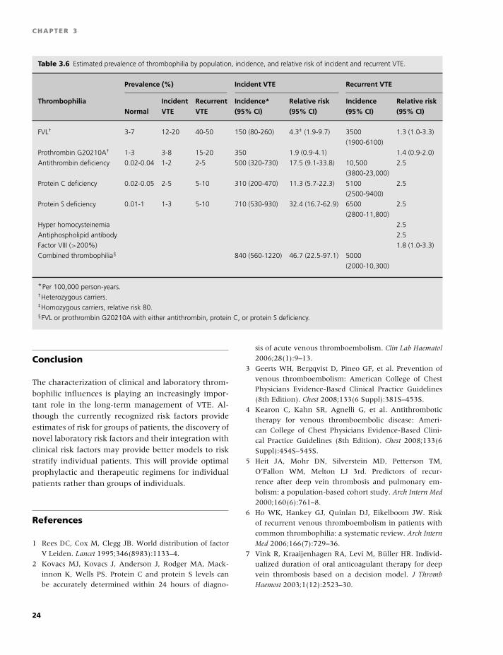
BLBK186-Key April 15, 2009 9:36
CHAPTER 3
Table 3.6 Estimated prevalence of thrombophilia by population, incidence, and relative risk of incident and recurrent VTE.
Prevalence (%) Incident VTE Recurrent VTE
Thrombophilia Incident Recurrent Incidence* Relative risk Incidence Relative riskNormal VTE VTE (95% CI) (95% CI) (95% CI) (95% CI)
FVL† 3-7 12-20 40-50 150 (80-260) 4.3‡ (1.9-9.7) 3500 1.3 (1.0-3.3)
(1900-6100)
Prothrombin G20210A† 1-3 3-8 15-20 350 1.9 (0.9-4.1) 1.4 (0.9-2.0)
Antithrombin deficiency 0.02-0.04 1-2 2-5 500 (320-730) 17.5 (9.1-33.8) 10,500 2.5
(3800-23,000)
Protein C deficiency 0.02-0.05 2-5 5-10 310 (200-470) 11.3 (5.7-22.3) 5100 2.5
(2500-9400)
Protein S deficiency 0.01-1 1-3 5-10 710 (530-930) 32.4 (16.7-62.9) 6500 2.5
(2800-11,800)
Hyper homocysteinemia 2.5
Antiphospholipid antibody 2.5
Factor VIII (>200%) 1.8 (1.0-3.3)
Combined thrombophilia§ 840 (560-1220) 46.7 (22.5-97.1) 5000
(2000-10,300)
*Per 100,000 person-years.†Heterozygous carriers.‡Homozygous carriers, relative risk 80.§FVL or prothrombin G20210A with either antithrombin, protein C, or protein S deficiency.
Conclusion
The characterization of clinical and laboratory throm-
bophilic influences is playing an increasingly impor-
tant role in the long-term management of VTE. Al-
though the currently recognized risk factors provide
estimates of risk for groups of patients, the discovery of
novel laboratory risk factors and their integration with
clinical risk factors may provide better models to risk
stratify individual patients. This will provide optimal
prophylactic and therapeutic regimens for individual
patients rather than groups of individuals.
References
1 Rees DC, Cox M, Clegg JB. World distribution of factor
V Leiden. Lancet 1995;346(8983):1133–4.
2 Kovacs MJ, Kovacs J, Anderson J, Rodger MA, Mack-
innon K, Wells PS. Protein C and protein S levels can
be accurately determined within 24 hours of diagno-
sis of acute venous thromboembolism. Clin Lab Haematol
2006;28(1):9–13.
3 Geerts WH, Bergqvist D, Pineo GF, et al. Prevention of
venous thromboembolism: American College of Chest
Physicians Evidence-Based Clinical Practice Guidelines
(8th Edition). Chest 2008;133(6 Suppl):381S–453S.
4 Kearon C, Kahn SR, Agnelli G, et al. Antithrombotic
therapy for venous thromboembolic disease: Ameri-
can College of Chest Physicians Evidence-Based Clini-
cal Practice Guidelines (8th Edition). Chest 2008;133(6
Suppl):454S–545S.
5 Heit JA, Mohr DN, Silverstein MD, Petterson TM,
O’Fallon WM, Melton LJ 3rd. Predictors of recur-
rence after deep vein thrombosis and pulmonary em-
bolism: a population-based cohort study. Arch Intern Med
2000;160(6):761–8.
6 Ho WK, Hankey GJ, Quinlan DJ, Eikelboom JW. Risk
of recurrent venous thromboembolism in patients with
common thrombophilia: a systematic review. Arch Intern
Med 2006;166(7):729–36.
7 Vink R, Kraaijenhagen RA, Levi M, Buller HR. Individ-
ualized duration of oral anticoagulant therapy for deep
vein thrombosis based on a decision model. J Thromb
Haemost 2003;1(12):2523–30.
24
BLBK186-Key April 24, 2009 7:33
4 Molecular diagnostic approachesto hemostasisPaula James and David Lillicrap
Introduction
The first coagulation factor gene, factor IX, was cloned
and characterized in 1982, and since that time, pro-
gressive advances have been made in the use of molec-
ular genetic strategies to assist in the diagnosis of coag-
ulation disorders. This chapter summarizes the current
state of molecular diagnostics for the more common
hemostatic conditions, with a discussion of both hem-
orrhagic and thrombotic problems for which genetic
tests are now available.
It is important to emphasize that, for most hemo-
static conditions encountered in clinical practice, the
initial diagnostic test of choice will still be one that
is performed in a routine hemostasis laboratory. For
example, the diagnosis of hemophilia A will still, in
the vast majority of cases, be made using a factor VIII
clotting assay. The role of molecular genetic testing for
this condition will be to assist in genetic counseling
and to provide predictive information relating to cer-
tain aspects of clinical management. To date, the num-
ber of conditions for which the initial diagnostic strat-
egy demands a genetic test is small. One such example
is the test for the prothrombin 20210 thrombophilic
variant.
A second, general issue that merits brief discus-
sion concerns the appropriate venue for molecular
genetic testing for hemostatic disorders. The success-
ful implementation of a molecular diagnostic ser-
vice for hemostatic conditions requires access to ap-
propriate expertise and technology, and these tests
cannot readily be added to the repertoire of a rou-
tine clinical coagulation laboratory. Increasingly, op-
timal molecular genetic testing approaches incorpo-
rate methodologies that require access to expensive
equipment that will not be found in a hemostasis
laboratory. However, genetic testing for hemostatic
problems can easily be incorporated into a general
molecular diagnostic facility, although the involve-
ment of personnel with an additional interest in
the phenotypic aspects of clotting is undoubtedly
beneficial for optimizing testing strategies and test
interpretation.
Molecular diagnostics ofbleeding disorders
Molecular genetic testing for the hemophilias have
been available since the cloning of the factor VIII and
IX genes in 1984 and 1982, respectively [1,2]. Since
then, with the cloning of all the known coagulation
factor genes, molecular characterization of the rare in-
herited bleeding disorders has also been possible.
Hemophilia A
To date, all inherited cases of isolated factor VIII defi-
ciency have been linked to mutations of the factor VIII
locus, which is located at the telomeric end of the long
arm of the X chromosome (Xq28) and encompasses
186 kilobases (kb) of genomic DNA (Fig. 4.1). The
large size of the gene, which contains 26 exons, was
originally a challenge for the development of molec-
ular diagnostic testing. Two diagnostic strategies can
be used to investigate this condition: an indirect test
of transmission of the hemophilic FVIII gene (poly-
morphism linkage) and direct detection of the disease-
causing mutation.
25
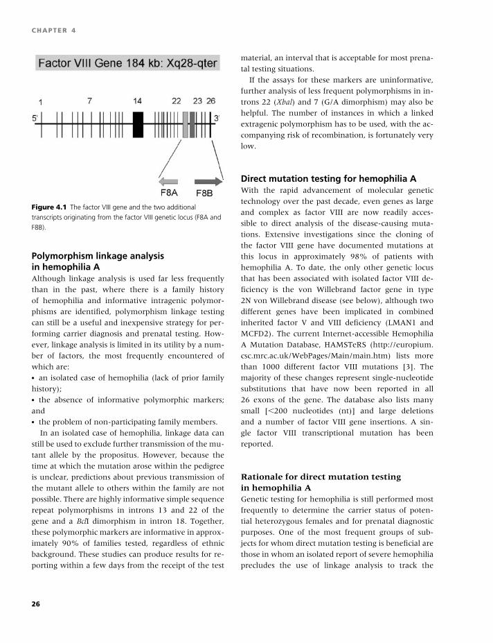
BLBK186-Key April 24, 2009 7:33
CHAPTER 4
Figure 4.1 The factor VIII gene and the two additional
transcripts originating from the factor VIII genetic locus (F8A and
F8B).
Polymorphism linkage analysisin hemophilia AAlthough linkage analysis is used far less frequently
than in the past, where there is a family history
of hemophilia and informative intragenic polymor-
phisms are identified, polymorphism linkage testing
can still be a useful and inexpensive strategy for per-
forming carrier diagnosis and prenatal testing. How-
ever, linkage analysis is limited in its utility by a num-
ber of factors, the most frequently encountered of
which are:� an isolated case of hemophilia (lack of prior family
history);� the absence of informative polymorphic markers;
and� the problem of non-participating family members.
In an isolated case of hemophilia, linkage data can
still be used to exclude further transmission of the mu-
tant allele by the propositus. However, because the
time at which the mutation arose within the pedigree
is unclear, predictions about previous transmission of
the mutant allele to others within the family are not
possible. There are highly informative simple sequence
repeat polymorphisms in introns 13 and 22 of the
gene and a BclI dimorphism in intron 18. Together,
these polymorphic markers are informative in approx-
imately 90% of families tested, regardless of ethnic
background. These studies can produce results for re-
porting within a few days from the receipt of the test
material, an interval that is acceptable for most prena-
tal testing situations.
If the assays for these markers are uninformative,
further analysis of less frequent polymorphisms in in-
trons 22 (Xbal) and 7 (G/A dimorphism) may also be
helpful. The number of instances in which a linked
extragenic polymorphism has to be used, with the ac-
companying risk of recombination, is fortunately very
low.
Direct mutation testing for hemophilia AWith the rapid advancement of molecular genetic
technology over the past decade, even genes as large
and complex as factor VIII are now readily acces-
sible to direct analysis of the disease-causing muta-
tions. Extensive investigations since the cloning of
the factor VIII gene have documented mutations at
this locus in approximately 98% of patients with
hemophilia A. To date, the only other genetic locus
that has been associated with isolated factor VIII de-
ficiency is the von Willebrand factor gene in type
2N von Willebrand disease (see below), although two
different genes have been implicated in combined
inherited factor V and VIII deficiency (LMAN1 and
MCFD2). The current Internet-accessible Hemophilia
A Mutation Database, HAMSTeRS (http://europium.
csc.mrc.ac.uk/WebPages/Main/main.htm) lists more
than 1000 different factor VIII mutations [3]. The
majority of these changes represent single-nucleotide
substitutions that have now been reported in all
26 exons of the gene. The database also lists many
small [�200 nucleotides (nt)] and large deletions
and a number of factor VIII gene insertions. A sin-
gle factor VIII transcriptional mutation has been
reported.
Rationale for direct mutation testingin hemophilia AGenetic testing for hemophilia is still performed most
frequently to determine the carrier status of poten-
tial heterozygous females and for prenatal diagnostic
purposes. One of the most frequent groups of sub-
jects for whom direct mutation testing is beneficial are
those in whom an isolated report of severe hemophilia
precludes the use of linkage analysis to track the
26
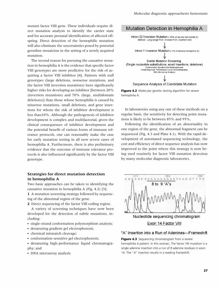
BLBK186-Key April 24, 2009 7:33
Molecular diagnostic approachesto hemostasis
mutant factor VIII gene. These individuals require di-
rect mutation analysis to identify the carrier state
and for accurate prenatal identification of affected off-
spring. Direct detection of the hemophilic mutation
will also eliminate the uncertainties posed by potential
germline mosaicism in the setting of a newly acquired
mutation.
The second reason for pursuing the causative muta-
tion in hemophilia A is the evidence that specific factor
VIII genotypes are more predictive for the risk of ac-
quiring a factor VIII inhibitor [4]. Patients with null
genotypes (large deletions, nonsense mutations, and
the factor VIII inversion mutations) have significantly
higher risks for developing an inhibitor [between 20%
(inversion mutations) and 70% (large, multidomain
deletions)] than those whose hemophilia is caused by
missense mutations, small deletions, and gene inser-
tions for whom the risk of inhibitor development is
less than10%. Although the pathogenesis of inhibitor
development is complex and multifactorial, given the
clinical consequences of inhibitor development and
the potential benefit of various forms of immune tol-
erance protocols, one can reasonably make the case
for early mutation testing in all new severe cases of
hemophilia A. Furthermore, there is also preliminary
evidence that the outcome of immune tolerance pro-
tocols is also influenced significantly by the factor VIII
genotype.
Strategies for direct mutation detectionin hemophilia ATwo basic approaches can be taken to identifying the
causative mutation in hemophilia A (Fig. 4.2) [5]:
1 A mutation screening strategy followed by sequenc-
ing of the abnormal region of the gene.
2 Direct sequencing of the factor VIII coding region.
A variety of screening techniques have now been
developed for the detection of subtle mutations, in-
cluding:� single-strand conformation polymorphism analysis;� denaturing gradient gel electrophoresis;� chemical mismatch cleavage;� conformation-sensitive gel electrophoresis;� denaturing high-performance liquid chromatogra-
phy; and� DNA microarray analysis
Figure 4.2 Molecular genetic testing algorithm for severe
hemophilia A.
In laboratories using any one of these methods on a
regular basis, the sensitivity for detecting point muta-
tions is likely to be between 85% and 95%.
Following the identification of an abnormality in
one region of the gene, the abnormal fragment can be
sequenced (Fig. 4.3 and Plate 4.1). With the rapid de-
velopment of automated sequencing technology, the
cost and efficiency of direct sequence analysis has now
improved to the point where this strategy is now be-
ing used routinely for factor VIII mutation detection
by many molecular diagnostic laboratories.
Figure 4.3 Sequencing chromatogram from a severe
hemophilia A patient. In this woman, The factor VIII mutation is a
single adenine insertion into a run of 8 adenine residues in exon
14. The “A” insertion results in a reading frameshift.
27
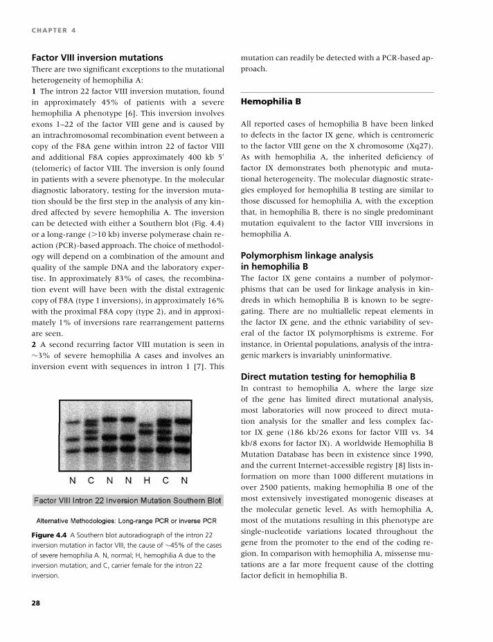
BLBK186-Key April 24, 2009 7:33
CHAPTER 4
Factor VIII inversion mutationsThere are two significant exceptions to the mutational
heterogeneity of hemophilia A:
1 The intron 22 factor VIII inversion mutation, found
in approximately 45% of patients with a severe
hemophilia A phenotype [6]. This inversion involves
exons 1–22 of the factor VIII gene and is caused by
an intrachromosomal recombination event between a
copy of the F8A gene within intron 22 of factor VIII
and additional F8A copies approximately 400 kb 5′
(telomeric) of factor VIII. The inversion is only found
in patients with a severe phenotype. In the molecular
diagnostic laboratory, testing for the inversion muta-
tion should be the first step in the analysis of any kin-
dred affected by severe hemophilia A. The inversion
can be detected with either a Southern blot (Fig. 4.4)
or a long-range (�10 kb) inverse polymerase chain re-
action (PCR)-based approach. The choice of methodol-
ogy will depend on a combination of the amount and
quality of the sample DNA and the laboratory exper-
tise. In approximately 83% of cases, the recombina-
tion event will have been with the distal extragenic
copy of F8A (type 1 inversions), in approximately 16%
with the proximal F8A copy (type 2), and in approxi-
mately 1% of inversions rare rearrangement patterns
are seen.
2 A second recurring factor VIII mutation is seen in
∼3% of severe hemophilia A cases and involves an
inversion event with sequences in intron 1 [7]. This
Figure 4.4 A Southern blot autoradiograph of the intron 22
inversion mutation in factor VIII, the cause of ∼45% of the cases
of severe hemophilia A. N, normal; H, hemophilia A due to the
inversion mutation; and C, carrier female for the intron 22
inversion.
mutation can readily be detected with a PCR-based ap-
proach.
Hemophilia B
All reported cases of hemophilia B have been linked
to defects in the factor IX gene, which is centromeric
to the factor VIII gene on the X chromosome (Xq27).
As with hemophilia A, the inherited deficiency of
factor IX demonstrates both phenotypic and muta-
tional heterogeneity. The molecular diagnostic strate-
gies employed for hemophilia B testing are similar to
those discussed for hemophilia A, with the exception
that, in hemophilia B, there is no single predominant
mutation equivalent to the factor VIII inversions in
hemophilia A.
Polymorphism linkage analysisin hemophilia BThe factor IX gene contains a number of polymor-
phisms that can be used for linkage analysis in kin-
dreds in which hemophilia B is known to be segre-
gating. There are no multiallelic repeat elements in
the factor IX gene, and the ethnic variability of sev-
eral of the factor IX polymorphisms is extreme. For
instance, in Oriental populations, analysis of the intra-
genic markers is invariably uninformative.
Direct mutation testing for hemophilia BIn contrast to hemophilia A, where the large size
of the gene has limited direct mutational analysis,
most laboratories will now proceed to direct muta-
tion analysis for the smaller and less complex fac-
tor IX gene (186 kb/26 exons for factor VIII vs. 34
kb/8 exons for factor IX). A worldwide Hemophilia B
Mutation Database has been in existence since 1990,
and the current Internet-accessible registry [8] lists in-
formation on more than 1000 different mutations in
over 2500 patients, making hemophilia B one of the
most extensively investigated monogenic diseases at
the molecular genetic level. As with hemophilia A,
most of the mutations resulting in this phenotype are
single-nucleotide variations located throughout the
gene from the promoter to the end of the coding re-
gion. In comparison with hemophilia A, missense mu-
tations are a far more frequent cause of the clotting
factor deficit in hemophilia B.
28
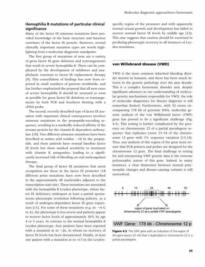
BLBK186-Key April 24, 2009 7:33
Molecular diagnostic approachesto hemostasis
Hemophilia B mutations of particular clinicalsignificanceMany of the factor IX missense mutations have pro-
vided knowledge of the basic structure and function
correlates of the factor IX protein. However, several
clinically important mutation types are worth high-
lighting from a molecular diagnostic standpoint.
The first group of mutations of note are a variety
of gross factor IX gene deletions and rearrangements
that result in severe hemophilia B. These can be com-
plicated by the development of inhibitors and ana-
phylactic reactions to factor IX replacement therapy
[9]. This constellation of findings has now been re-
ported in small numbers of patients worldwide, and
has further emphasized the proposal that all new cases
of severe hemophilia B should be screened as soon
as possible for gross factor IX deletions or rearrange-
ments by both PCR and Southern blotting with a
cDNA probe.
The second, recently described type of factor IX mu-
tation with important clinical consequences involves
missense mutations in the propeptide-encoding se-
quence, resulting in a markedly reduced affinity of the
mutant protein for the vitamin K-dependent carboxy-
lase [10]. Two different missense mutations have been
described at amino acid residue −10 in the propep-
tide, and these patients have normal baseline factor
IX levels but show marked sensitivity to treatment
with vitamin K antagonists, leading to a signifi-
cantly increased risk of bleeding on oral anticoagulant
therapy.
The final group of factor IX mutations that merit
recognition are those in the factor IX promoter (18
different point mutations have now been described
in the approximately 40 nucleotides adjacent to the
transcription start site). These mutations are associated
with the hemophilia B Leyden phenotype, where fac-
tor IX deficiency undergoes at least a partial sponta-
neous phenotypic resolution following puberty, as a
result of androgen-dependent factor IX gene expres-
sion [11]. For some of these mutations (e.g. nt ∼6 G
to A), the phenotype is less severe and patients appear
to recover factor levels of approximately 30% by age
4 or 5 years. In contrast to the normal hemophilia B
Leyden phenotype, four patients have been reported
with a mutation at nt ∼26, in whom no recovery of
factor IX levels has been documented. Finally, at least
one patient with a mutation at nt +13 in the Leyden-
specific region of the promoter and with apparently
normal sexual growth and development has failed to
recover normal factor IX levels by middle age [12].
This case suggests that caution should be exercised in
predicting phenotypic recovery in all instances of Ley-
den mutations.
von Willebrand disease (VWD)
VWD is the most common inherited bleeding disor-
der known in humans, and there has been much in-
terest in the genetic pathology over the past decade.
This is a complex hemostatic disorder and, despite
significant advances in our understanding of molecu-
lar genetic mechanisms responsible for VWD, the role
of molecular diagnostics for disease diagnosis is still
somewhat limited. Furthermore, with 52 exons en-
compassing 178 kb of genomic DNA, molecular ge-
netic analysis of the von Willebrand factor (VWF)
gene has proved to be a significant challenge (Fig.
4.5). This testing is further complicated by the pres-
ence on chromosome 22 of a partial pseudogene se-
quence that replicates exons 23–34 of the chromo-
some 12 gene with 3% sequence variation [13,14].
Thus, any analysis of this region of the gene must en-
sure that PCR primers and probes are designed for the
chromosome 12 gene. The final challenge in testing
for and interpreting VWF genetic data is the extreme
polymorphic nature of this gene. Indeed, in many
instances, a clear distinction between neutral poly-
morphic changes and disease-causing variants is still
unresolved.
Figure 4.5 The VWF gene with an indication of the region of
the gene (exons 23–34) that is duplicated on chromosome 22 in a
partial pseudogene.
29
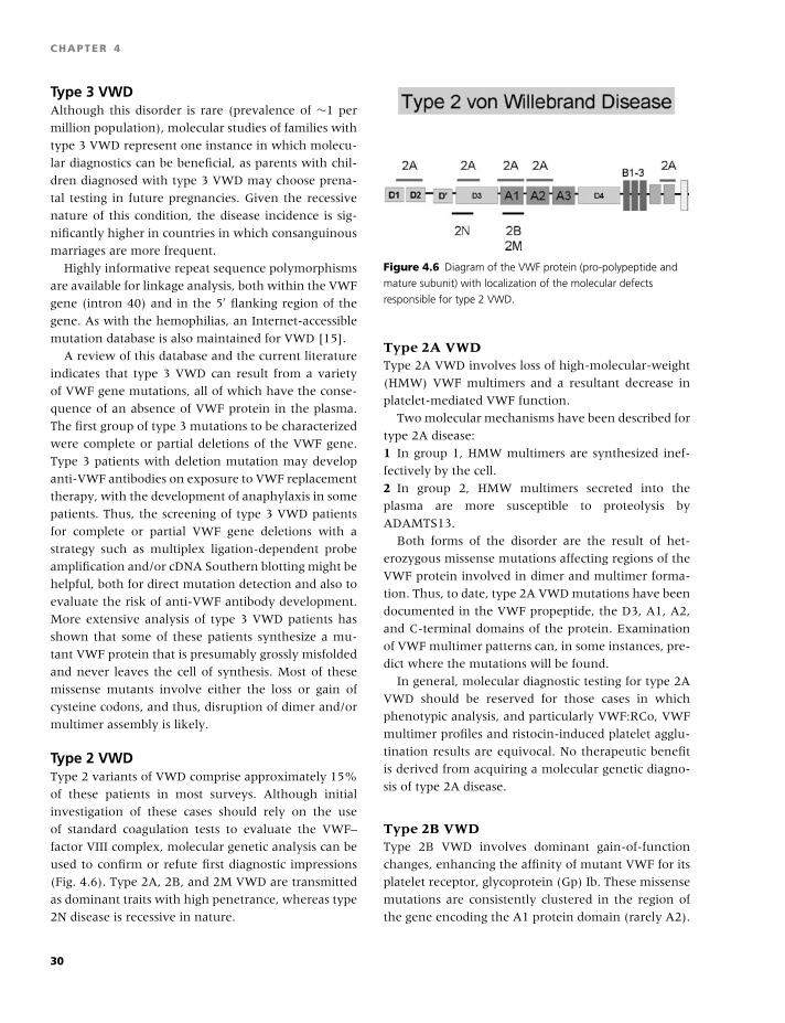
BLBK186-Key April 24, 2009 7:33
CHAPTER 4
Type 3 VWDAlthough this disorder is rare (prevalence of ∼1 per
million population), molecular studies of families with
type 3 VWD represent one instance in which molecu-
lar diagnostics can be beneficial, as parents with chil-
dren diagnosed with type 3 VWD may choose prena-
tal testing in future pregnancies. Given the recessive
nature of this condition, the disease incidence is sig-
nificantly higher in countries in which consanguinous
marriages are more frequent.
Highly informative repeat sequence polymorphisms
are available for linkage analysis, both within the VWF
gene (intron 40) and in the 5′ flanking region of the
gene. As with the hemophilias, an Internet-accessible
mutation database is also maintained for VWD [15].
A review of this database and the current literature
indicates that type 3 VWD can result from a variety
of VWF gene mutations, all of which have the conse-
quence of an absence of VWF protein in the plasma.
The first group of type 3 mutations to be characterized
were complete or partial deletions of the VWF gene.
Type 3 patients with deletion mutation may develop
anti-VWF antibodies on exposure to VWF replacement
therapy, with the development of anaphylaxis in some
patients. Thus, the screening of type 3 VWD patients
for complete or partial VWF gene deletions with a
strategy such as multiplex ligation-dependent probe
amplification and/or cDNA Southern blotting might be
helpful, both for direct mutation detection and also to
evaluate the risk of anti-VWF antibody development.
More extensive analysis of type 3 VWD patients has
shown that some of these patients synthesize a mu-
tant VWF protein that is presumably grossly misfolded
and never leaves the cell of synthesis. Most of these
missense mutants involve either the loss or gain of
cysteine codons, and thus, disruption of dimer and/or
multimer assembly is likely.
Type 2 VWDType 2 variants of VWD comprise approximately 15%
of these patients in most surveys. Although initial
investigation of these cases should rely on the use
of standard coagulation tests to evaluate the VWF–
factor VIII complex, molecular genetic analysis can be
used to confirm or refute first diagnostic impressions
(Fig. 4.6). Type 2A, 2B, and 2M VWD are transmitted
as dominant traits with high penetrance, whereas type
2N disease is recessive in nature.
Figure 4.6 Diagram of the VWF protein (pro-polypeptide and
mature subunit) with localization of the molecular defects
responsible for type 2 VWD.
Type 2A VWDType 2A VWD involves loss of high-molecular-weight
(HMW) VWF multimers and a resultant decrease in
platelet-mediated VWF function.
Two molecular mechanisms have been described for
type 2A disease:
1 In group 1, HMW multimers are synthesized inef-
fectively by the cell.
2 In group 2, HMW multimers secreted into the
plasma are more susceptible to proteolysis by
ADAMTS13.
Both forms of the disorder are the result of het-
erozygous missense mutations affecting regions of the
VWF protein involved in dimer and multimer forma-
tion. Thus, to date, type 2A VWD mutations have been
documented in the VWF propeptide, the D3, A1, A2,
and C-terminal domains of the protein. Examination
of VWF multimer patterns can, in some instances, pre-
dict where the mutations will be found.
In general, molecular diagnostic testing for type 2A
VWD should be reserved for those cases in which
phenotypic analysis, and particularly VWF:RCo, VWF
multimer profiles and ristocin-induced platelet agglu-
tination results are equivocal. No therapeutic benefit
is derived from acquiring a molecular genetic diagno-
sis of type 2A disease.
Type 2B VWDType 2B VWD involves dominant gain-of-function
changes, enhancing the affinity of mutant VWF for its
platelet receptor, glycoprotein (Gp) Ib. These missense
mutations are consistently clustered in the region of
the gene encoding the A1 protein domain (rarely A2).
30
BLBK186-Key April 24, 2009 7:33
Molecular diagnostic approachesto hemostasis
Direct sequencing of exon 28 sequences can provide
molecular genetic confirmation of the type 2B pheno-
type. This region of the VWF gene is duplicated, with
sequence variation, in the partial pseudogene on chro-
mosome 22, and thus choice of amplification primers
should take this fact into consideration. Ninety per-
cent of type 2B VWD cases are caused by the missense
mutations R1308C, R1306C, V1316M, and R1341Q.
Given the localized nature of type 2B mutations,
molecular genetic confirmation of the phenotypic di-
agnosis is easily achieved through examination of
exon 28 PCR products.
Type 2B VWD demonstrates hemostatic test results
that are very similar to those seen in the dominantly
inherited platelet disorder, platelet-type VWD. Molec-
ular genetic analysis offers a definitive approach to dif-
ferentiating between these two conditions (see below).
Type 2M VWDThe type 2M VWD variant has reduced platelet-
mediated VWF function with normal VWF multimers.
Here again, as with type 2B disease, the molecular
pathology represents a variety of missense mutations
localized to exon 28, the A1 domain-coding region
of the gene. Type 2M disease is essentially the loss-
of-function equivalent of type 2B VWD, with the A1
substitutions resulting in disruption of the interac-
tion with platelet GpIb. As with type 2B disease, ge-
netic confirmation of the type 2M phenotype can be
achieved through exon 28 sequencing.
Type 2N VWDType 2N VWD is a recessively inherited trait. This con-
dition should be considered in the differential diag-
nosis of mild–moderate isolated factor VIII deficiency
and can easily be confused with mild hemophilia A.
Phenotypic testing involves a direct assessment of the
FVIII binding potential of VWF using a microtiter
plate-based assay. The most efficient molecular genetic
approach to confirm a diagnosis of type 2N disease
is to sequence the PCR products amplified from ex-
ons 18–25 of the VWF gene, the region encoding the
N-terminal D’/D3 factor VIII binding domains of VWF.
In patients with type 2N VWD, this analysis will show
either homozygous or compound heterozygous mis-
sense mutations affecting the factor VIII binding do-
main of the protein. In addition, coinheritance of a
type 2N allele with a severe type 1 or type 3 null allele
will also result in this phenotype. The R854Q missense
mutation is the most frequent type 2N variant, result-
ing in factor VIII levels around 20%. Levels of factor
VIII of approximately 10% are seen with some of the
other mutations, such as R816W and T791M.
Type 1 VWDDespite being the most prevalent form of the disorder,
representing approximately 75% of all VWD cases, the
molecular pathogenesis of type 1 VWD remains the
least well understood. Diagnosis can often be difficult
and is influenced by a variety of factors, including the
temporal variability of VWF levels and the ABO blood
group of patients, accounting for approximately 30%
of the variability in VWF levels, with blood group O
subjects having the lowest levels. Another significant
complicating factor in attempting to address the ge-
netic basis for type 1 VWD is the marked variability
in penetrance and expression of the phenotype within
families, which makes the use of classic linkage anal-
ysis problematic. Therefore, much of the knowledge
gained in this area has relied on labor-intensive strate-
gies, such as direct sequencing of genomic DNA.
The recent completion of 3 population-based studies
of the molecular genetic pathology of type 1 VWD has
provided information from 300 patients with this di-
agnosis [16–18]. The findings from these studies have
been similar and demonstrate the following:� The type 1 VWD phenotype is linked to the VWF
gene in approximately 60% of families.� Candidate VWF gene mutations can be found in ap-
proximately 65% of type 1 VWD patients.� More than 100 different candidate VWF gene muta-
tions have been identified.� Approximately 65% of the candidate VWF muta-
tions are missense substitutions.� Candidate VWF gene mutations are found through-
out the VWF locus from the 5’ flanking region to the
C-terminal domain of the protein.� In approximately 15% of patients, more than a sin-
gle candidate VWF mutation is present.
An analysis of the mutations found in the three
population studies has also shown that certain can-
didate mutations are recurrent. This group of muta-
tions includes Y1584C (found in between 8% and
25% of the type 1 VWD population), R924Q, R1205H,
R1315C, R1374H, and R854Q. Suffice it to say that,
31
BLBK186-Key April 24, 2009 7:33
CHAPTER 4
even with these common variants, the understanding
of pathogenic mechanisms is incomplete.
The information derived from these initial molec-
ular surveys of type 1 VWD indicate that, in addi-
tion to incomplete penetrance and variable expres-
sivity, the genetics of this complex trait is further
complicated by mutational and locus heterogeneity.
Whereas most type 1 cases with plasma VWF levels
�30% will demonstrate candidate VWF mutations,
patients with mild VWF deficiency (30–50%) are more
likely to have a phenotype in which contributions
from several loci (including the ABO blood group lo-
cus) are playing an important pathogenic role. To date,
the identity of these additional genetic modifiers is
unknown.
Given the size and complexity of the VWF gene and
the problems of mutational and locus heterogeneity,
the application of molecular genetic analysis to the di-
agnosis of type 1 VWD is not currently warranted. This
situation may change with further advances in tech-
nology and the potential identification of key genetic
modifiers.
Less common inherited coagulationfactor deficiencies
As the genes for all of the procoagulant proteins
have been cloned and characterized, molecular ge-
netic testing is feasible for the inherited deficiency of
any of these factors. However, the diagnosis of these
disorders (factor XI and X deficiencies and others)
remains firmly based in the clinical hemostasis labo-
ratory through the performance of biological clotting
assays.
Although specific research laboratories may be in-
terested in determining the disease-causing mutations
in these families, primarily as a means to assist in
structure and function analysis, the performance of
these tests for diagnostic purposes is not usual. An
exception is the documentation of mutations in the
LMAN1 and MCFD2 genes in patients with inherited
combined factor V and VIII deficiencies. Most cases of
this rare disorder are caused by one of several recur-
ring point mutations in these intermediate compart-
ment processing proteins; thus, documentation of one
of these mutations would definitively establish an oth-
erwise unusual diagnosis.
Inherited platelet disorders
As with the less common coagulation factor deficien-
cies, the diagnosis of inherited platelet disorders is
predominantly by phenotypic analysis. Standard mor-
phology, platelet aggregation studies, and an evalua-
tion of platelet receptor density will usually establish
or exclude a diagnosis of Bernard–Soulier syndrome
or Glanzmann’s thrombasthenia, the two most fre-
quently encountered, but nevertheless rare, recessive
inherited platelet disorders [19].
In unusual instances, knowledge of the causative
mutation in these patients could be useful, perhaps
for prenatal testing. In the Bernard–Soulier syndrome,
a heterogeneous mutational pattern has been docu-
mented, with both homozygous and compound het-
erozygous mutations identified in the genes encoding
Gp Ib�, Ib�, and IX [19]. A variety of different muta-
tions has been found at these loci, including deletions,
frameshifts, and nonsense and missense changes. To
date, no Bernard–Soulier mutations have been identi-
fied in the GpV gene.
In Glanzmann’s thrombasthenia, a similarly var-
ied pattern of mutations has been documented in the
genes for Gp IIb and IIIa.
As alluded to above, the standard coagulation stud-
ies in platelet-type (pseudo) VWD (PT-VWD) are very
similar to those encountered in patients with type 2B
VWD. Here, clarification of the diagnosis most effec-
tively involves molecular genetic analysis of exon 28
of the VWF gene (for type 2B VWD missense mu-
tations) and the GpIb� gene (for PT-VWD) [20]. In
PT-VWD, heterozygous dominant missense mutations
can be found in the GpIb� gene, which have been
shown through the analysis of recombinant mutant
protein to possess an increased binding affinity for the
A1 domain of VWF. One partial deletion mutation in
GpIb� has also been identified as being causative for
PT-VWD.
Molecular diagnostics forthrombotic disease
Although an inherited tendency for excessive bleed-
ing can often be ascribed to single gene abnormalities,
there is ample evidence to suggest that, in contrast,
32
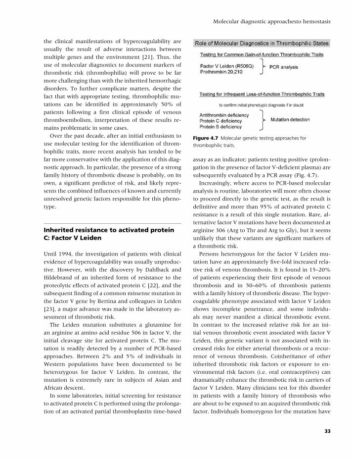
BLBK186-Key April 24, 2009 7:33
Molecular diagnostic approachesto hemostasis
the clinical manifestations of hypercoagulability are
usually the result of adverse interactions between
multiple genes and the environment [21]. Thus, the
use of molecular diagnostics to document markers of
thrombotic risk (thrombophilia) will prove to be far
more challenging than with the inherited hemorrhagic
disorders. To further complicate matters, despite the
fact that with appropriate testing, thrombophilic mu-
tations can be identified in approximately 50% of
patients following a first clinical episode of venous
thromboembolism, interpretation of these results re-
mains problematic in some cases.
Over the past decade, after an initial enthusiasm to
use molecular testing for the identification of throm-
bophilic traits, more recent analysis has tended to be
far more conservative with the application of this diag-
nostic approach. In particular, the presence of a strong
family history of thrombotic disease is probably, on its
own, a significant predictor of risk, and likely repre-
sents the combined influences of known and currently
unresolved genetic factors responsible for this pheno-
type.
Inherited resistance to activated proteinC: Factor V Leiden
Until 1994, the investigation of patients with clinical
evidence of hypercoagulability was usually unproduc-
tive. However, with the discovery by Dahlback and
Hildebrand of an inherited form of resistance to the
proteolytic effects of activated protein C [22], and the
subsequent finding of a common missense mutation in
the factor V gene by Bertina and colleagues in Leiden
[23], a major advance was made in the laboratory as-
sessment of thrombotic risk.
The Leiden mutation substitutes a glutamine for
an arginine at amino acid residue 506 in factor V, the
initial cleavage site for activated protein C. The mu-
tation is readily detected by a number of PCR-based
approaches. Between 2% and 5% of individuals in
Western populations have been documented to be
heterozygous for factor V Leiden. In contrast, the
mutation is extremely rare in subjects of Asian and
African descent.
In some laboratories, initial screening for resistance
to activated protein C is performed using the prolonga-
tion of an activated partial thromboplastin time-based
Figure 4.7 Molecular genetic testing approaches for
thrombophilic traits.
assay as an indicator; patients testing positive (prolon-
gation in the presence of factor V-deficient plasma) are
subsequently evaluated by a PCR assay (Fig. 4.7).
Increasingly, where access to PCR-based molecular
analysis is routine, laboratories will more often choose
to proceed directly to the genetic test, as the result is
definitive and more than 95% of activated protein C
resistance is a result of this single mutation. Rare, al-
ternative factor V mutations have been documented at
arginine 306 (Arg to Thr and Arg to Gly), but it seems
unlikely that these variants are significant markers of
a thrombotic risk.
Persons heterozygous for the factor V Leiden mu-
tation have an approximately five-fold increased rela-
tive risk of venous thrombosis. It is found in 15–20%
of patients experiencing their first episode of venous
thrombosis and in 50–60% of thrombosis patients
with a family history of thrombotic disease. The hyper-
coagulable phenotype associated with factor V Leiden
shows incomplete penetrance, and some individu-
als may never manifest a clinical thrombotic event.
In contrast to the increased relative risk for an ini-
tial venous thrombotic event associated with factor V
Leiden, this genetic variant is not associated with in-
creased risks for either arterial thrombosis or a recur-
rence of venous thrombosis. Coinheritance of other
inherited thrombotic risk factors or exposure to en-
vironmental risk factors (i.e. oral contraceptives) can
dramatically enhance the thrombotic risk in carriers of
factor V Leiden. Many clinicians test for this disorder
in patients with a family history of thrombosis who
are about to be exposed to an acquired thrombotic risk
factor. Individuals homozygous for the mutation have
33
BLBK186-Key April 24, 2009 7:33
CHAPTER 4
a 70-fold enhanced relative risk of venous thrombo-
sis, indicating that this phenotype is transmitted as a
codominant trait.
Prothrombin 20210 3′ non-codingsequence variant
In 1996, Poort and colleagues described an association
between a G to A nucleotide polymorphism at position
20210 in the 3′ untranslated region (UTR) of the pro-
thrombin gene, increased plasma levels of prothrom-
bin, and an enhanced risk for venous thrombosis [24].
This polymorphic nucleotide substitution is at the very
end of the 3′ UTR and exerts its effect on prothrombin
levels in the heterozygous state. Although the plasma
levels of prothrombin in subjects heterozygous for this
polymorphism are higher on average than those in in-
dividuals with a normal prothrombin genotype, levels
are usually still within the normal range. As a con-
sequence, this polymorphism can only be evaluated
by genetic testing, which is achieved by a PCR-based
assay, most often now involving a form of real-time
quantitative assay.
As with the factor V Leiden genotype, the preva-
lence of the prothrombin 20210 G to A variant in
the general population is relatively high at 1–5%. This
variant is also rare in persons of Asian and African de-
scent. The heterozygous state is associated with a two-
to four-fold increase in the relative risk for venous
thrombosis. There is no influence on venous throm-
botic recurrence or arterial thrombosis.
Thermolabile C677T5,10-methylene-tetrahydrofolatereductase variant
The third, high-prevalence genetic variant that was
initially thought to be associated with an increased
thrombotic risk is the C to T variant at nucleotide
677 (an alanine to valine substitution) in the 5,10-
methylene-tetrahydrofolate reductase (MTHFR) gene.
This genotype results in expression of an enzyme with
increased thermolability. Homozygosity for the variant
is associated with hyperhomocysteinemia, particularly
in the presence of folate deficiency. In many popula-
tions (southern Europeans and Hispanic Americans),
approximately 10% of subjects are homozygous for
the C677T variant, a sequence change that can easily
be detected by a PCR-based strategy. After further ex-
tended analysis, in contrast to the factor V Leiden and
prothrombin 20210 variants, the role of the MTHFR
C677T polymorphism as an independent risk factor for
venous thromboembolism appears minor.
Deficiencies of antithrombin, protein C,and protein S
Deficiencies of the major anticoagulant proteins an-
tithrombin, protein C, and protein S have long been
known to represent individual risk factors for the de-
velopment of venous thromboembolism. The protein
deficiencies manifest thrombotic phenotypes in the
heterozygous state, but penetrance and expression of
the phenotype are extremely variable and relate to
both the individual protein deficiency (antithrombin
deficiency being the most severe condition) and
the specific molecular defect. Homozygosity for an-
tithrombin and protein C deficiencies results in the
severe neonatal thrombotic condition, pupura fulmi-
nans.
Diagnosis of these three disorders relies on stan-
dard functional tests or immunoassays that should be
performed in the diagnostic hemostasis laboratory. All
three of the deficiency states are associated with sig-
nificant mutational heterogeneity, and routine molec-
ular diagnostic investigation of these mutations is not
warranted. However, these are lifelong diagnoses, and
if any doubt exists about the phenotypic test results,
confirmation of the diagnosis by genetic testing should
be considered (Fig. 4.7).
The role of genetic testing in the clinicalmanagement of oral anticoagulation
Studies in the past couple of years have now indicated
that individual anticoagulant responses to the vitamin
K antagonist, Coumadin (Warfarin), can be predicted
to some extent through the analysis of genotypes
for two proteins involved in the metabolism of this
drug: cytochrome P-450 2C9 (CYP2C9) and vitamin K
epoxide-reductase complex 1 (VKORC1). Analysis of
polymorphic haplotypes of the CYP2C9 and VKORC1
34
BLBK186-Key April 24, 2009 7:33
Molecular diagnostic approachesto hemostasis
genes by PCR testing has been shown to be helpful
in identifying patients who may be especially sensitive
to oral anticoagulant administration [25]. This appears
to be particularly the case for VKORC1 analysis dur-
ing the initiation phase of oral anticoagulation. Thus,
rapidly reported VKORC1 genotyping may prove to
be a useful adjunctive testing strategy to international
normalized ratio testing in this clinical situation; and,
indeed, in the United States, the Food and Drug Ad-
ministration have recommended that this laboratory-
monitoring approach be added to the drug insert in-
formation.
The future for diagnosticmolecular hemostasis
With the completion of the Human Genome Project
and the ongoing analysis of complex genetic traits
through the performance of genome-wide association
studies, additional information pertaining to genetic
influences on hemostasis is likely to be derived in the
next few years. This fact, along with further advances
in genetic methodologies, including more accessible
microarray-based testing approaches may well provide
further opportunities for the application of molecu-
lar diagnostic testing in the area of clinical hemostasis.
However, as has already been witnessed with throm-
bophilic testing, initial enthusiasm for test adoption
will need to be tempered by formal evidence of clinical
benefit deriving from the tests. Indeed there is a signif-
icant possibility that the major genetic influences on
most hemostatic phenotypes have already been identi-
fied and that any new associations are unlikely to play
a clinically important role. An area where this possibil-
ity may well be tested in the next decade is that of ge-
netic risk factors for arterial thrombosis. To date, very
little benefit can be derived from genetic testing for
this phenotype, and it may well be that the combined
genetic and environmental background of this condi-
tion will be too complex for the useful application of a
genetic testing strategy.
References
1 Gitschier J, Wood WI, Goralka TM, et al. Char-
acterization of the human factor VIII gene. Nature
1984;312:326–30.
2 Kurachi K, Davie EW. Isolation and characterization of
a cDNA coding for human factor IX. Proc Natl Acad Sci
USA 1982;79:6461–4.
3 HAMSTeRS: The Haemophilia A Mutation, Struc-
ture, Test and Resource Site. 2008. http://europium.
csc.mrc.ac.uk/WebPages/Main/main.htm.
4 Goodeve AC, Peake IR. The molecular basis of
hemophilia A: genotype-phenotype relationships
and inhibitor development. Semin Thromb Hemost
2003;29:23–30.
5 Keeney S, Mitchell M, Goodeve A. The molecular
analysis of haemophilia A: a guideline from the UK
haemophilia centre doctors’ organization haemophilia
genetics laboratory network. Haemophilia 2005;11:
387–97.
6 Lakich D, Kazazian HH Jr, Antonarakis SE, Gitschier J.
Inversions disrupting the factor VIII gene are a common
cause of severe haemophilia A. Nat Genet 1993;5:236–
41.
7 Bagnall RD, Waseem N, Green PM, Giannelli F. Re-
current inversion breaking intron 1 of the factor VIII
gene is a frequent cause of severe hemophilia A. Blood
2002;99:168–74.
8 Haemophilia B Mutation Database: Version 13. 2008.
http://www.kcl.ac.uk/ip/petergreen/haemBdatabase.
html.
9 Thorland EC, Drost JB, Lusher JM, et al. Ana-
phylactic response to factor IX replacement ther-
apy in haemophilia B patients: complete gene dele-
tions confer the highest risk. Haemophilia 1999;5:
101–5.
10 Oldenburg J, Quenzel EM, Harbrecht U, et al. Missense
mutations at ALA-10 in the factor IX propeptide: an in-
significant variant in normal life but a decisive cause of
bleeding during oral anticoagulant therapy. Br J Haema-
tol 1997;98:240–4.
11 Picketts DJ, Mueller CR, Lillicrap D. Transcriptional
control of the factor IX gene: analysis of five cis-
acting elements and the deleterious effects of natu-
rally occurring hemophilia B Leyden mutations. Blood
1994;84:2992–3000.
12 James PD, Stakiw J, Leggo J, Walker I, Lillicrap D. A
case of non-resolving hemophilia B Leyden in a 42-
year-old male (F9 promoter + 13 A�G). J Thromb
Haemost 2008;6:885–6.
13 Mancuso DJ, Tuley EA, Westfield LA, et al. Structure of
the gene for human von Willebrand factor. J Biol Chem
1989;264:19514–27.
14 Mancuso DJ, Tuley EA, Westfield LA, et al. Human
von Willebrand factor gene and pseudogene: structural
analysis and differentiation by polymerase chain reac-
tion. Biochemistry 1991;30:253–69.
35
BLBK186-Key April 24, 2009 7:33
CHAPTER 4
15 ISTH SSC VWF Database. 2008. http://www.vwf.
group.shef.ac.uk/.
16 Goodeve A, Eikenboom J, Castaman G, et al. Pheno-
type and genotype of a cohort of families historically
diagnosed with type 1 von Willebrand disease in the
European study, Molecular and Clinical Markers for the
Diagnosis and Management of Type 1 von Willebrand
Disease (MCMDM-1VWD). Blood 2007;109:112–21.
17 James PD, Notley C, Hegadorn C, et al. The mutational
spectrum of type 1 von Willebrand disease: Results
from a Canadian cohort study. Blood 2007;109:145–54.
18 Cumming A, Grundy P, Keeney S, et al. An investiga-
tion of the von Willebrand factor genotype in UK pa-
tients diagnosed to have type 1 von Willebrand disease.
Thromb Haemost 2006;96:630–41.
19 Nurden P, Nurden AT. Congenital disorders asso-
ciated with platelet dysfunctions. Thromb Haemost
2008;99:253–63.
20 Othman M, Lillicrap D. Distinguishing between
non-identical twins: platelet type andtype 2B
von Willebrand disease. Br J Haematol 2007;138:
665–6.
21 Reitsma PH, Rosendaal FR. Past and future of genetic
research in thrombosis. J Thromb Haemost 2007;5(Suppl
1):264–9.
22 Dahlback B, Hildebrand B. Inherited resistance to ac-
tivated protein C is corrected by anticoagulant cofactor
activity found to be a property of factor V. Proc Natl Acad
Sci USA 1994;91:1396–400.
23 Bertina RM, Koeleman BP, Koster T, et al. Mutation in
blood coagulation factor V associated with resistance to
activated protein C. Nature 1994;369:64–7.
24 Poort SR, Rosendaal FR, Reitsma PH, Bertina RM. A
common genetic variation in the 3’-untranslated region
of the prothrombin gene is associated with elevated
plasma prothrombin levels and an increase in venous
thrombosis. Blood 1996;88:3698–703.
25 Schwarz UI, Ritchie MD, Bradford Y, et al. Genetic de-
terminants of response to warfarin during initial anti-
coagulation. N Engl J Med 2008;358:999–1008.
36
BLBK186-Key April 24, 2009 9:18
5 Tests of platelet functionPaul Harrison
Structure of platelets
Human blood platelets are small, anucleated cells that
play a critical role in hemostasis and thrombosis. Hu-
man platelets normally circulate for approximately 10
days, constantly surveying the integrity of the vessel
wall. Normal human platelets are small and discoid in
shape (0.5 × 3.0 µm), have a mean volume of 7–11
fL, and normally circulate in relatively high numbers
between 150 and 400 × 109/L.
A cross-section of a typical discoid platelet is shown
in Fig. 5.1.
Function
Their small disc shape enables the platelets to be
marginated toward the edge of vessels so that the
majority circulate adjacent to the vascular endothe-
lial cells that line all blood vessels. Upon detection of
vessel wall damage, they undergo rapid but controlled
adhesion, activation, and aggregation to form a hemo-
static plug and thus rapidly prevent blood loss.
Endothelial cells produce a number of potent anti-
platelet substances (e.g. nitric oxide, prostacyclin, and
CD39) that normally inhibit vessel wall–platelet in-
teractions. Vessel wall damage exposes highly adhe-
sive substrates [e.g. P selectin, Von Willebrand factor
(VWF), collagen, and many other extracellular matrix
components], which result in a sequence of stepwise
events resulting in the formation of a hemostatic plug
(Plate 5.1):� Initial adhesion, transient rolling of platelets along
the vessel wall, and slowing of the cells. Consequently,
platelets are more likely to undergo stable adhesion.
� Stable adhesion through additional receptor–ligand
interactions.� Platelet activation (if there is more extensive damage
or stimuli-promoting platelet activation).� Platelet aggregation (Plate 5.1).� Generation of platelet procoagulant activity and sta-
bilization of the hemostatic plug.� Clot retraction.
The platelets interact with and sense the environ-
ment through many types of surface receptors (major
receptors and their ligands are summarized in Table
5.1). The net balance between activating or inhibitory
stimuli thus controls whether platelets continue to cir-
culate, begin to reversibly interact with the vessel wall,
or become irreversibly adherent to either the vessel
wall or each other.
During adhesion, platelets become activated
through signal transduction pathways, which mediate
shape change, degranulation, and spreading upon
areas of exposed subendothelium. Activated platelets
recruit additional platelets into the growing platelet
aggregate or thrombus via a number of positive
feedback pathways, including release of dense gran-
ular adenosine diphosphate (ADP) and generation
of thromboxane. Activated platelets also express
negatively charged phospholipids on their surface,
facilitating the local generation of high amounts of
thrombin, which not only further activates other
platelets, but also stabilizes the platelet plug through
fibrin formation. In this manner, platelets rapidly
seal any areas of vessel wall damage and provide a
catalytic surface for coagulation to occur, resulting in
the formation of a stable hemostatic plug.
Thrombosis is usually the consequence of inap-
propriate activation of platelets, especially in re-
gions of abnormal vessel wall lesions or damage (e.g.
atherosclerotic plaques). The high shear stress that
37
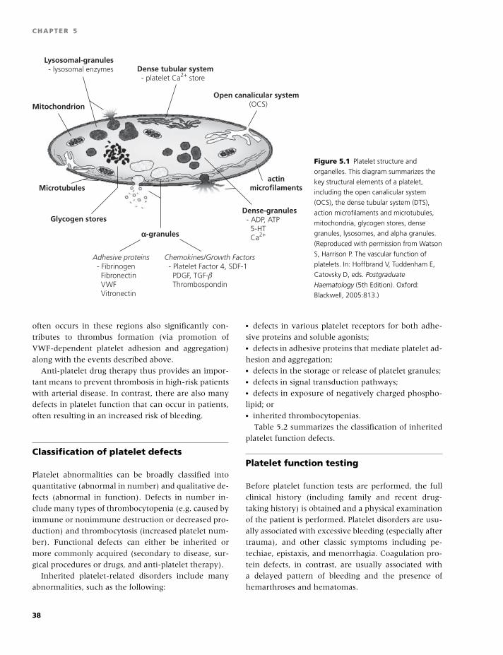
BLBK186-Key April 24, 2009 9:18
CHAPTER 5
Lysosomal-granules - lysosomal enzymes
Adhesive proteins - Fibrinogen Fibronectin VWF Vitronectin
Chemokines/Growth Factors - Platelet Factor 4, SDF-1 PDGF, TGF-β Thrombospondin
Open canalicular system (OCS)
Dense tubular system - platelet Ca2+ store
Dense-granules - ADP, ATP 5-HT Ca2+
Mitochondrion
Glycogen stores
αα-granules
Microtubulesactin
microfilaments
Figure 5.1 Platelet structure and
organelles. This diagram summarizes the
key structural elements of a platelet,
including the open canalicular system
(OCS), the dense tubular system (DTS),
action microfilaments and microtubules,
mitochondria, glycogen stores, dense
granules, lysosomes, and alpha granules.
(Reproduced with permission from Watson
S, Harrison P. The vascular function of
platelets. In: Hoffbrand V, Tuddenham E,
Catovsky D, eds. Postgraduate
Haematology (5th Edition). Oxford:
Blackwell, 2005:813.)
often occurs in these regions also significantly con-
tributes to thrombus formation (via promotion of
VWF-dependent platelet adhesion and aggregation)
along with the events described above.
Anti-platelet drug therapy thus provides an impor-
tant means to prevent thrombosis in high-risk patients
with arterial disease. In contrast, there are also many
defects in platelet function that can occur in patients,
often resulting in an increased risk of bleeding.
Classification of platelet defects
Platelet abnormalities can be broadly classified into
quantitative (abnormal in number) and qualitative de-
fects (abnormal in function). Defects in number in-
clude many types of thrombocytopenia (e.g. caused by
immune or nonimmune destruction or decreased pro-
duction) and thrombocytosis (increased platelet num-
ber). Functional defects can either be inherited or
more commonly acquired (secondary to disease, sur-
gical procedures or drugs, and anti-platelet therapy).
Inherited platelet-related disorders include many
abnormalities, such as the following:
� defects in various platelet receptors for both adhe-
sive proteins and soluble agonists;� defects in adhesive proteins that mediate platelet ad-
hesion and aggregation;� defects in the storage or release of platelet granules;� defects in signal transduction pathways;� defects in exposure of negatively charged phospho-
lipid; or� inherited thrombocytopenias.
Table 5.2 summarizes the classification of inherited
platelet function defects.
Platelet function testing
Before platelet function tests are performed, the full
clinical history (including family and recent drug-
taking history) is obtained and a physical examination
of the patient is performed. Platelet disorders are usu-
ally associated with excessive bleeding (especially after
trauma), and other classic symptoms including pe-
techiae, epistaxis, and menorrhagia. Coagulation pro-
tein defects, in contrast, are usually associated with
a delayed pattern of bleeding and the presence of
hemarthroses and hematomas.
38
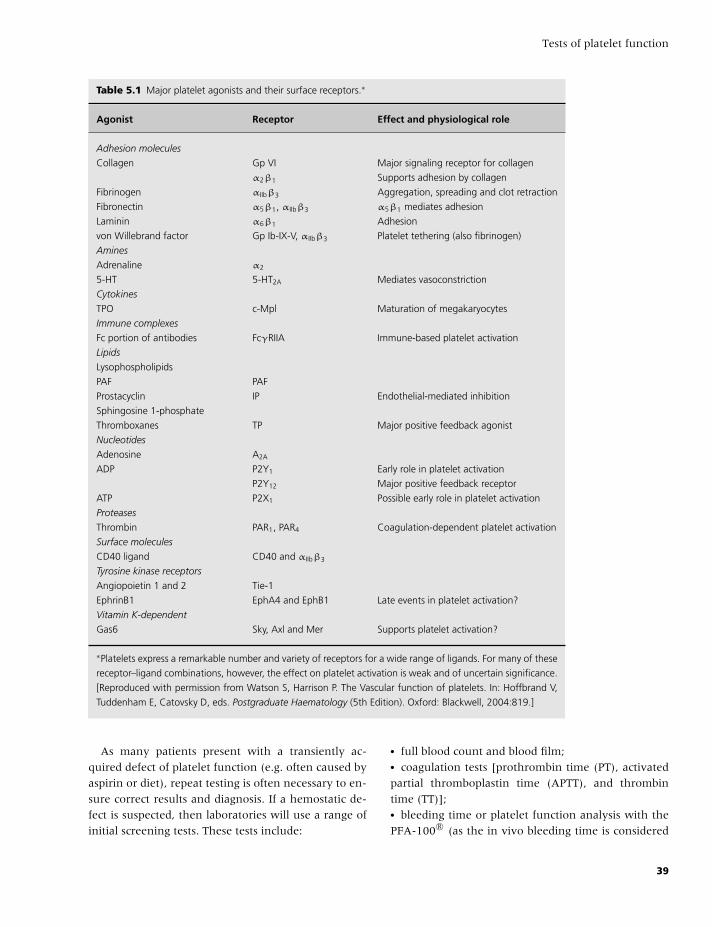
BLBK186-Key April 24, 2009 9:18
Tests of platelet function
Table 5.1 Major platelet agonists and their surface receptors.∗
Agonist Receptor Effect and physiological role
Adhesion molecules
Collagen Gp VI Major signaling receptor for collagen
α2β1 Supports adhesion by collagen
Fibrinogen αIIbβ3 Aggregation, spreading and clot retraction
Fibronectin α5β1, αIIbβ3 α5β1 mediates adhesion
Laminin α6β1 Adhesion
von Willebrand factor Gp Ib-IX-V, αIIbβ3 Platelet tethering (also fibrinogen)
Amines
Adrenaline α2
5-HT 5-HT2A Mediates vasoconstriction
Cytokines
TPO c-Mpl Maturation of megakaryocytes
Immune complexes
Fc portion of antibodies FcγRIIA Immune-based platelet activation
Lipids
Lysophospholipids
PAF PAF
Prostacyclin IP Endothelial-mediated inhibition
Sphingosine 1-phosphate
Thromboxanes TP Major positive feedback agonist
Nucleotides
Adenosine A2A
ADP P2Y1 Early role in platelet activation
P2Y12 Major positive feedback receptor
ATP P2X1 Possible early role in platelet activation
Proteases
Thrombin PAR1, PAR4 Coagulation-dependent platelet activation
Surface molecules
CD40 ligand CD40 and αIIbβ3
Tyrosine kinase receptors
Angiopoietin 1 and 2 Tie-1
EphrinB1 EphA4 and EphB1 Late events in platelet activation?
Vitamin K-dependent
Gas6 Sky, Axl and Mer Supports platelet activation?
∗Platelets express a remarkable number and variety of receptors for a wide range of ligands. For many of these
receptor–ligand combinations, however, the effect on platelet activation is weak and of uncertain significance.
[Reproduced with permission from Watson S, Harrison P. The Vascular function of platelets. In: Hoffbrand V,
Tuddenham E, Catovsky D, eds. Postgraduate Haematology (5th Edition). Oxford: Blackwell, 2004:819.]
As many patients present with a transiently ac-
quired defect of platelet function (e.g. often caused by
aspirin or diet), repeat testing is often necessary to en-
sure correct results and diagnosis. If a hemostatic de-
fect is suspected, then laboratories will use a range of
initial screening tests. These tests include:
� full blood count and blood film;� coagulation tests [prothrombin time (PT), activated
partial thromboplastin time (APTT), and thrombin
time (TT)];� bleeding time or platelet function analysis with the
PFA-100 R© (as the in vivo bleeding time is considered
39
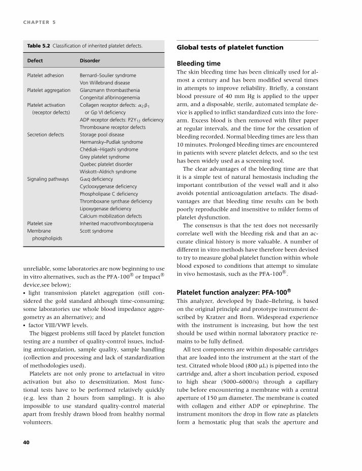
BLBK186-Key April 24, 2009 9:18
CHAPTER 5
Table 5.2 Classification of inherited platelet defects.
Defect Disorder
Platelet adhesion Bernard–Soulier syndrome
Von Willebrand disease
Platelet aggregation Glanzmann thrombasthenia
Congenital afibrinogenemia
Platelet activation Collagen receptor defects: α2β1
(receptor defects) or Gp VI deficiency
ADP receptor defects: P2Y12 deficiency
Thromboxane receptor defects
Secretion defects Storage pool disease
Hermansky–Pudlak syndrome
Chediak–Higashi syndrome
Grey platelet syndrome
Quebec platelet disorder
Wiskott–Aldrich syndrome
Signaling pathways Gαq deficiency
Cyclooxygenase deficiency
Phospholipase C deficiency
Thromboxane synthase deficiency
Lipoxygenase deficiency
Calcium mobilization defects
Platelet size Inherited macrothrombocytopenia
Membrane Scott syndrome
phospholipids
unreliable, some laboratories are now beginning to use
in vitro alternatives, such as the PFA-100 R© or Impact R©
device,see below);� light transmission platelet aggregation (still con-
sidered the gold standard although time-consuming;
some laboratories use whole blood impedance aggre-
gometry as an alternative); and� factor VIII/VWF levels.
The biggest problems still faced by platelet function
testing are a number of quality-control issues, includ-
ing anticoagulation, sample quality, sample handling
(collection and processing and lack of standardization
of methodologies used).
Platelets are not only prone to artefactual in vitro
activation but also to desensitization. Most func-
tional tests have to be performed relatively quickly
(e.g. less than 2 hours from sampling). It is also
impossible to use standard quality-control material
apart from freshly drawn blood from healthy normal
volunteers.
Global tests of platelet function
Bleeding timeThe skin bleeding time has been clinically used for al-
most a century and has been modified several times
in attempts to improve reliability. Briefly, a constant
blood pressure of 40 mm Hg is applied to the upper
arm, and a disposable, sterile, automated template de-
vice is applied to inflict standardized cuts into the fore-
arm. Excess blood is then removed with filter paper
at regular intervals, and the time for the cessation of
bleeding recorded. Normal bleeding times are less than
10 minutes. Prolonged bleeding times are encountered
in patients with severe platelet defects, and so the test
has been widely used as a screening tool.
The clear advantages of the bleeding time are that
it is a simple test of natural hemostasis including the
important contribution of the vessel wall and it also
avoids potential anticoagulation artefacts. The disad-
vantages are that bleeding time results can be both
poorly reproducible and insensitive to milder forms of
platelet dysfunction.
The consensus is that the test does not necessarily
correlate well with the bleeding risk and that an ac-
curate clinical history is more valuable. A number of
different in vitro methods have therefore been devised
to try to measure global platelet function within whole
blood exposed to conditions that attempt to simulate
in vivo hemostasis, such as the PFA-100 R©.
Platelet function analyzer: PFA-100 R©
This analyzer, developed by Dade–Behring, is based
on the original principle and prototype instrument de-
scribed by Kratzer and Born. Widespread experience
with the instrument is increasing, but how the test
should be used within normal laboratory practice re-
mains to be fully defined.
All test components are within disposable cartridges
that are loaded into the instrument at the start of the
test. Citrated whole blood (800 µL) is pipetted into the
cartridge and, after a short incubation period, exposed
to high shear (5000–6000/s) through a capillary
tube before encountering a membrane with a central
aperture of 150 µm diameter. The membrane is coated
with collagen and either ADP or epinephrine. The
instrument monitors the drop in flow rate as platelets
form a hemostatic plug that seals the aperture and
40
BLBK186-Key April 24, 2009 9:18
Tests of platelet function
stops blood flow. This parameter is recorded as the clo-
sure time (CT). The maximal value obtainable is 300 s.
To ensure optimal PFA-100 R© performance and data
interpretation, there are a number of quality-control
procedures and good practice guidelines that need to
be kept in mind:� Mandatory daily instrument checks; PFA-100 R© self-
test should always be performed.� Ensuring the quality of blood sampling.� Ensuring consistency in anticoagulation, 3.8%
(0.129 mol/L) or 3.2% (0.105 mol/L) buffered
trisodium citrate.� Checking for cartridge batch variation.� Testing within 4 hours of sampling.� Always perform a full blood count to help interpret
the results.� A control group within each laboratory setting
should be established. These individuals should ide-
ally exhibit CTs within the middle of the established
laboratory reference range.� Each laboratory should also ideally establish their
own reference ranges on both cartridges using normal
volunteers from their institution.
Typical normal ranges obtained with 3.8%
trisodium citrate are 58–151 s for collagen/ADP
and 94–202 s for collagen/epinephrine. With 3.2%
trisodium citrate, typical ranges are 55–112 s for
collagen/ADP and 79–164 s for collagen/epinephrine
(Oxford Hemophilia and Thrombosis Centre, unpub-
lished results).
Within-sample coefficients of variation (CVs) have
been reported as approximately 10%, which, although
acceptable for a platelet function test, may obviously
cause problems with values obtained close to upper
normal range cut-off values.
The advantages of the test are that it is simple, rapid,
and does not require specialist training (apart from
training in the manipulation of blood samples). It is
a potential screening tool for assessing patients with
many types of platelet abnormality. Within a typical
population of patients tested, the overall negative pre-
dictive value of the test can be high (more than 90%),
although the test is clearly not 100% sensitive to all
platelet defects. The test is particularly useful in pedi-
atric settings where the availability of blood is often
a limiting factor, particularly for potentially assessing
platelet function in nonaccidental injury cases. Given
the high shear conditions to which platelets are ex-
posed during the test, it is not surprising that the test
is highly VWF-dependent and is useful not only for
detecting VWD, but also for monitoring therapy, par-
ticularly with DDAVP. The instrument thus provides
laboratories with a limited but optional screening tool
that gives rapid and reliable data with a high negative
predictive value.
A number of studies suggest that the PFA-100 R© is
a potential in vitro replacement of the bleeding time.
The disadvantages are that, like the in vivo bleed-
ing time, the test is sensitive to both the platelet
count and hematocrit, and it is therefore crucial that
a full blood count is performed to help interpret ab-
normal results. The test is usually insensitive to co-
agulation protein defects, including afibrinogenemia,
hemophilia A and B, and other clotting factors. False-
negative results are sometimes obtained; for example,
in patients with storage pool disease, primary secretion
defects, Hermansky–Pudlak syndrome, type 1 VWD,
and the Quebec platelet disorder. Diagnosis of these
disorders could therefore be missed if relying on the
PFA-100 R© as a screening test alone. In patients with
apparently normal platelet function, the instrument
has also been shown to occasionally give false-positive
results, which then require further detailed testing.
Many studies are also in progress to assess whether
the PFA-100 R© can reliably predict either thrombotic
or bleeding complications in different patient groups.
As more interlaboratory experience is gathered, even-
tually it should be feasible to define the exact role(s)
for this instrument in routine laboratory testing. A re-
cent ISTH SSC document by Hayward and colleagues
(2006) provides a useful up-to-date consensus review
of the utility of the test.
Impact R© Cone and Plate(let) AnalyzerThe cone and plate(let) analyzer originally developed
by Varon and colleagues monitors platelet adhesion
and aggregation to a plate coated with collagen or
extracellular matrix under high shear conditions of
1800 s−1. In the commercial version of the device, the
Impact R©, a plastic plate is used instead. The test is now
fully automated, simple to operate, uses a small quan-
tity of citrated whole blood (0.12 mL), and displays
results within 6 minutes. The instrument contains a
microscope and performs staining and image analy-
sis of platelets that have adhered and aggregated on
the plate. The software permits storage of the images
41
BLBK186-Key April 24, 2009 9:18
CHAPTER 5
from each analysis and records a number of parame-
ters, including surface coverage, average size, and dis-
tribution histogram of the adhered platelets. There is
also a research version of the instrument, called the
Impact-R R©, that requires some of the steps to be man-
ually performed and facilitates adjustment of the shear
rate. To ensure optimal Impact R© performance and data
interpretation, many of the quality-control procedures
and good practice guidelines used for the PFA-100 R©
also apply. Typical normal ranges are 7.8–19% for
Surface Coverage and 35–70 µm2 for aggregate size
within 3.2% citrated blood, and typical CVs are �15%
(Oxford Hamophilia and Thrombosis Centre, unpub-
lished results).
Preliminary data suggest that the test can also po-
tentially be used for the screening of platelet defects
and VWD and monitoring anti-platelet therapy. The
test is dependent on the platelet count and hemat-
ocrit, and a full blood count should always be per-
formed. As the test also measures platelet adhesion to
polystyrene, it also important to be aware that fibrino-
gen is also an important variable within the test. Be-
cause the commercial test has only been available for
a relatively short time, widespread experience is still
limited at present. The overall sensitivity and speci-
ficity of the Impact R© as a screening test remains to be
fully evaluated.
Diagnostic tests
Light transmission platelet aggregometryIn the 1960s, the invention of platelet aggregometry
revolutionized the analysis of platelet function within
routine laboratory testing. Still regarded as the “gold
standard,” it is the most widely used platelet function
test. Citrated platelet-rich plasma (PRP) is normally
stirred under conditions of low shear within an incu-
bated cuvette (37◦C between a light source and a pho-
tocell. Anticoagulated whole blood may also be used
in some commercial multichannel impedance-based
aggregometers, such as the Chronolog or Multiplate
aggregometers. These have the significant advantage
that the blood does not require further processing for
analysis.
The addition of different dosages of a panel of ago-
nists triggers platelet activation, shape change, and pri-
mary and secondary aggregation events that increase
light transmission over time, and this is recorded on
the aggregation trace (Fig. 5.2). By using a panel of ag-
onists at differing concentrations, it is possible to detect
a number of classic platelet defects. Modern instru-
ments usually offer multichannel capability and com-
puter analysis and storage of data, although samples
and reagents still have to be prepared manually.
A fully automated and near patient testing aggre-
gation system called the VerifyNow R© device (Acc-
umetrics) is now available solely for the monitor-
ing of the three major classes of anti-platelet drugs
(e.g. Gp IIb/IIIa inhibitors, aspirin, and P2Y12 in-
hibitors/antagonists) using specific cartridges.
The light transmission method is as follows:� Citrated PRP (prepared by centrifugation at 150–
200 g for 10 minutes; non-adjusted count as this in-
troduces an artefact) is added to cuvette at 37◦C and
preincubated for 5 minutes.� The PRP is stirred at a recommended speed (e.g.
1000 rpm using a magnetic stir bar) to allow platelets
to come in contact with each other.� 100% transmission is set with autologous platelet-
poor plasma (PPP) (prepared by centrifugation at
1500 g for 20 minutes).� 0% transmission is set with PRP and a stable baseline
established before addition of agonist.� Agonist is added (up to 10% total volume).� Aggregation is recorded (10–15 minutes).� Calculate percentage aggregation (maximum or
final) and slope (rate of aggregation).� Hemolyzed or very lipemic samples may interfere
with light transmission.� Thrombocytopenic samples are also unsuitable for
analysis.
A typical panel of agonists (stored in frozen aliquots)
are:� ADP (0.1–20 µmol/L).� Epinephrine (1.0–10 µmol/L).� Collagen (1–5 µg/mL), usually mediates a steep ag-
gregation curve but after a characteristic lag phase of
more than 1 minute.� Arachidonic acid (1.0–2.0 mmol/L).� Ristocetin (0.5–1.5 mg/mL) is not strictly an agonist
but stimulates platelet agglutination through binding
of plasma VWF to Gp Ib and therefore will also give
abnormal results in VWD; usually used at a single low
and high dose.� Thrombin (0.1–0.5 IU/mL).
42
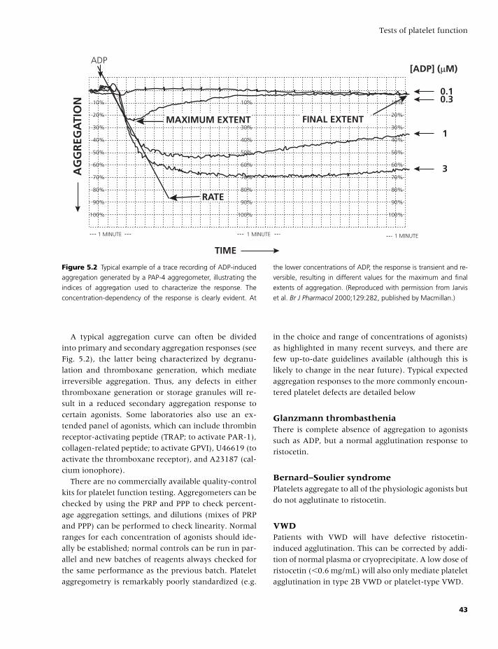
BLBK186-Key April 24, 2009 9:18
Tests of platelet function
10%
20%
30%
40%
50%
60%
70%
80%
90%
100%
ADP
MAXIMUM EXTENT FINAL EXTENT
[ADP] (µM)
0.10.3
1
3
RATE
1 MINUTE 1 MINUTE 1 MINUTE
10%
20%
30%
40%
50%
60%
70%
80%
90%
100%
10%
20%
30%
40%
50%
60%
70%
80%
90%
100%
AG
GR
EGA
TIO
N
TIME
Figure 5.2 Typical example of a trace recording of ADP-induced
aggregation generated by a PAP-4 aggregometer, illustrating the
indices of aggregation used to characterize the response. The
concentration-dependency of the response is clearly evident. At
the lower concentrations of ADP, the response is transient and re-
versible, resulting in different values for the maximum and final
extents of aggregation. (Reproduced with permission from Jarvis
et al. Br J Pharmacol 2000;129:282, published by Macmillan.)
A typical aggregation curve can often be divided
into primary and secondary aggregation responses (see
Fig. 5.2), the latter being characterized by degranu-
lation and thromboxane generation, which mediate
irreversible aggregation. Thus, any defects in either
thromboxane generation or storage granules will re-
sult in a reduced secondary aggregation response to
certain agonists. Some laboratories also use an ex-
tended panel of agonists, which can include thrombin
receptor-activating peptide (TRAP; to activate PAR-1),
collagen-related peptide; to activate GPVI), U46619 (to
activate the thromboxane receptor), and A23187 (cal-
cium ionophore).
There are no commercially available quality-control
kits for platelet function testing. Aggregometers can be
checked by using the PRP and PPP to check percent-
age aggregation settings, and dilutions (mixes of PRP
and PPP) can be performed to check linearity. Normal
ranges for each concentration of agonists should ide-
ally be established; normal controls can be run in par-
allel and new batches of reagents always checked for
the same performance as the previous batch. Platelet
aggregometry is remarkably poorly standardized (e.g.
in the choice and range of concentrations of agonists)
as highlighted in many recent surveys, and there are
few up-to-date guidelines available (although this is
likely to change in the near future). Typical expected
aggregation responses to the more commonly encoun-
tered platelet defects are detailed below
Glanzmann thrombastheniaThere is complete absence of aggregation to agonists
such as ADP, but a normal agglutination response to
ristocetin.
Bernard–Soulier syndromePlatelets aggregate to all of the physiologic agonists but
do not agglutinate to ristocetin.
VWDPatients with VWD will have defective ristocetin-
induced agglutination. This can be corrected by addi-
tion of normal plasma or cryoprecipitate. A low dose of
ristocetin (�0.6 mg/mL) will also only mediate platelet
agglutination in type 2B VWD or platelet-type VWD.
43
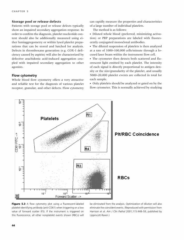
BLBK186-Key April 24, 2009 9:18
CHAPTER 5
Storage pool or release defectsPatients with storage pool or release defects typically
show an impaired secondary aggregation response. In
order to confirm the diagnosis, platelet nucleotide con-
tent should also be additionally measured using ei-
ther lumiaggregometry or within lysed platelet prepa-
rations that can be stored and batched for analysis.
Defects in thromboxane generation (e.g. COX-1 defi-
ciency caused by aspirin) will also be characterized by
defective arachidonic acid-induced aggregation cou-
pled with impaired secondary aggregation to other
agonists.
Flow cytometryWhole blood flow cytometry offers a very attractive
and reliable test for the diagnosis of various platelet
receptor, granular, and other defects. Flow cytometry
can rapidly measure the properties and characteristics
of a large number of individual platelets.
The method is as follows:� Diluted whole blood (preferred, minimizing activa-
tion) or PRP preparations are labeled with fluores-
cently conjugated monoclonal antibodies.� The diluted suspension of platelets is then analyzed
at a rate of 1000–100,000 cells/minute through a fo-
cused laser beam within the instrument flow cell.� The cytometer then detects both scattered and flu-
orescent light emitted by each platelet. The intensity
of each signal is directly proportional to antigen den-
sity or the size/granularity of the platelet, and usually
5000–20,000 platelet events are collected in total for
each sample.� Only platelets should be analyzed or gated on by the
flow cytometer. This is normally achieved by studying
Figure 5.3 A flow cytometry plot using a fluorescent-labeled
platelet-identifying antibody (anti-CD61) when triggering on a low
value of forward scatter (FS). If the instrument is triggered on
this fluorescence, all other nonplatelet events shown (RBCs) will
be eliminated from the analysis. Optimization of dilution will also
eliminate the coincident events. (Reproduced with permission from
Harrison et al. Am J Clin Pathol 2001;115:448–59, published by
Lippincott-Raven.)
44
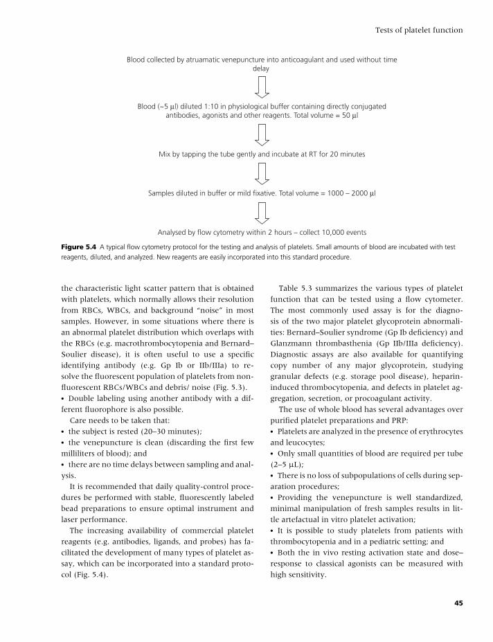
BLBK186-Key April 24, 2009 9:18
Tests of platelet function
Blood collected by atruamatic venepuncture into anticoagulant and used without timedelay
Blood (~5 µl) diluted 1:10 in physiological buffer containing directly conjugatedantibodies, agonists and other reagents. Total volume = 50 µl
Mix by tapping the tube gently and incubate at RT for 20 minutes
Samples diluted in buffer or mild fixative. Total volume = 1000 – 2000 µl
Analysed by flow cytometry within 2 hours – collect 10,000 events
Figure 5.4 A typical flow cytometry protocol for the testing and analysis of platelets. Small amounts of blood are incubated with test
reagents, diluted, and analyzed. New reagents are easily incorporated into this standard procedure.
the characteristic light scatter pattern that is obtained
with platelets, which normally allows their resolution
from RBCs, WBCs, and background “noise” in most
samples. However, in some situations where there is
an abnormal platelet distribution which overlaps with
the RBCs (e.g. macrothrombocytopenia and Bernard–
Soulier disease), it is often useful to use a specific
identifying antibody (e.g. Gp Ib or IIb/IIIa) to re-
solve the fluorescent population of platelets from non-
fluorescent RBCs/WBCs and debris/ noise (Fig. 5.3).� Double labeling using another antibody with a dif-
ferent fluorophore is also possible.
Care needs to be taken that:� the subject is rested (20–30 minutes);� the venepuncture is clean (discarding the first few
milliliters of blood); and� there are no time delays between sampling and anal-
ysis.
It is recommended that daily quality-control proce-
dures be performed with stable, fluorescently labeled
bead preparations to ensure optimal instrument and
laser performance.
The increasing availability of commercial platelet
reagents (e.g. antibodies, ligands, and probes) has fa-
cilitated the development of many types of platelet as-
say, which can be incorporated into a standard proto-
col (Fig. 5.4).
Table 5.3 summarizes the various types of platelet
function that can be tested using a flow cytometer.
The most commonly used assay is for the diagno-
sis of the two major platelet glycoprotein abnormali-
ties: Bernard–Soulier syndrome (Gp Ib deficiency) and
Glanzmann thrombasthenia (Gp IIb/IIIa deficiency).
Diagnostic assays are also available for quantifying
copy number of any major glycoprotein, studying
granular defects (e.g. storage pool disease), heparin-
induced thrombocytopenia, and defects in platelet ag-
gregation, secretion, or procoagulant activity.
The use of whole blood has several advantages over
purified platelet preparations and PRP:� Platelets are analyzed in the presence of erythrocytes
and leucocytes;� Only small quantities of blood are required per tube
(2–5 µL);� There is no loss of subpopulations of cells during sep-
aration procedures;� Providing the venepuncture is well standardized,
minimal manipulation of fresh samples results in lit-
tle artefactual in vitro platelet activation;� It is possible to study platelets from patients with
thrombocytopenia and in a pediatric setting; and� Both the in vivo resting activation state and dose–
response to classical agonists can be measured with
high sensitivity.
45
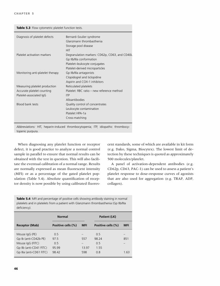
BLBK186-Key April 24, 2009 9:18
CHAPTER 5
Table 5.3 Flow cytometric platelet function tests.
Diagnosis of platelet defects Bernard–Soulier syndrome
Glanzmann thrombasthenia
Storage pool disease
HIT
Platelet activation markers Degranulation markers: CD62p, CD63, and CD40L
Gp IIb/IIIa conformation
Platelet–leukocyte conjugates
Platelet-derived microparticles
Monitoring anti-platelet therapy Gp IIb/IIIa antagonists
Clopidogrel and ticlopidine
Aspirin and COX-1 inhibitors
Measuring platelet production Reticulated platelets
Accurate platelet counting Platelet: RBC ratio – new reference method
Platelet-associated IgG ITP
Alloantibodies
Blood bank tests Quality control of concentrates
Leukocyte contamination
Platelet HPA-1a
Cross-matching
Abbreviations: HIT, heparin-induced thrombocytopenia; ITP, idiopathic thrombocy-
topenic purpura.
When diagnosing any platelet function or receptor
defect, it is good practice to analyze a normal control
sample in parallel to ensure that normal results can be
obtained with the test in question. This will also facili-
tate the eventual calibration of a normal range. Results
are normally expressed as mean fluorescent intensity
(MFI) or as a percentage of the gated platelet pop-
ulation (Table 5.4). Absolute quantification of recep-
tor density is now possible by using calibrated fluores-
cent standards, some of which are available in kit form
(e.g. Dako, Sigma, Biocytex). The lowest limit of de-
tection by these techniques is quoted as approximately
500 molecules/platelet.
A panel of activation-dependent antibodies (e.g.
CD62p, CD63, PAC-1) can be used to assess a patient’s
platelet response to dose–response curves of agonists
that are also used for aggregation (e.g. TRAP, ADP,
collagen).
Table 5.4 MFI and percentage of positive cells showing antibody staining in normal
platelets and in platelets from a patient with Glanzmann thrombasthenia (Gp IIb/IIIa
deficiency).
Normal Patient (LK)
Receptor (Mab) Positive cells (%) MFI Positive cells (%) MFI
Mouse IgG (PE) 0.5 – 0.5 –
Gp Ib (anti-CD42b PE) 97.5 557 98.24 851
Mouse IgG (FITC) 0.5 – 0.5 –
Gp IIb (anti-CD41 FITC) 95.99 13.97 1.55
Gp IIIa (anti-CD61 FITC) 98.42 598 0.8 1.63
46
BLBK186-Key April 24, 2009 9:18
Tests of platelet function
Further reading
BCSH Haemostasis and Thrombosis Task Force. Guidelines
on platelet function testing. J Clin Pathol 1988;41:1322–
30.
Bolton-Maggs PH, Chalmers EA, Collins PW, et al. A review
of inherited platelet disorders with guidelines for their
management on behalf of the UKHCDO. Br J Haematol
2006;135(5):603–33.
Fressinaud E, Veyradier A, Truchaud F, et al. Screening for
von Willebrand disease with a new analyzer using high
shear stress: a study of 60 cases. Blood 1998;91:1325–31.
Gresele P, Fuster V, Lopez H, Page C, Vermylen J, eds.
Platelets in Hematologic and Cardiovascular Disorders. Cam-
bridge: Cambridge University Press, 2008.
Harrison P. Platelet function analysis. Blood Rev 2005;
19:111–23.
Hayward CP, Harrison P, Cattaneo M, Ortel TL, Rao AK.
The Platelet Physiology Subcommittee of the Scientific
and Standardization Committee of the International So-
ciety of Thrombosis and Haemostasis. J Thromb Haemost
2006;4(2):212–9.
Hayward CP, Rao AK, Catteneo M. Congenital platelet
disorders: an overview of their mechanisms, diagnos-
tic evaluation and treatment. Haemophilia 2006;12(Suppl
3):128–36.
Hayward CP. Diagnostic approach to platelet function dis-
orders. Transfus Apher Sci 2008;38:65–76.
Hayward CP, Eikelboom J. Platelet function testing:
quality assurance. Semin Thromb Hemost 2007;33:273–
82.
Jilma B. Platelet function analyzer (PFA-100): a tool to
quantify congenital or acquired platelet dysfunction. J
Lab Clin Med 2001;138:152–63.
Linnemann B, Schwonberg J, Mani H, Prochnow S,
Lindhoff-Last E. Standardization of light transmittance
aggregometry for monitoring antiplatelet therapy: an ad-
justment for platelet count is not necessary. J Thromb
Haemost 2008;6(4):677–83.
Michelson AD. Flow cytometry: a clinical test of platelet
function. Blood 1996;87:4925–36.
Michelson AD. Platelets (2nd edition). New York: Academic
Press, 2007.
Ruggeri ZM. Platelets in atherothrombosis. Nat Med
2002;8:1227–34.
Schmitz G, Rothe G, Ruf A, et al. European Working Group
on Clinical Cell Analysis: Consensus protocol for the flow
cytometric characterization of platelet function. Thromb
Haemost 1998;79:885–96.
Zhou L, Schmaier AH. Platelet aggregation testing in
platelet-rich plasma: description of procedures with the
aim to develop standards in the field. Am J Clin Pathol
2005;123(2):172–83.
47
BLBK186-Key April 11, 2009 12:54
6 Evaluation of the bleeding patientAlice Ma
Introduction
Few evaluations in hematology provoke as much di-
agnostic uncertainty as that of the patient with a
suspected bleeding diathesis. The evaluation, includ-
ing history, physical examination, and laboratory test-
ing, is aimed at determining the likelihood that the
patient has an underlying hemorrhagic disorder, as
well as the treatment of future bleeding episodes. The
evaluation is fraught with diagnostic uncertainty, be-
cause many historical features are shared by indi-
viduals without bleeding diatheses, laboratory studies
may have a significant false-positive rate, and external
pressures (such as insurance coverage) may limit the
diagnostic testing available to the patient and physi-
cian. This chapter will attempt to present a systematic
approach to the individual with a suspected bleeding
disorder.
The bleeding history
A detailed history of bleeding episodes, including a
family history, is critical in elucidating whether a
bleeding diathesis is present. To that end, questions
are aimed at determining the likelihood of a bleeding
disorder being present as well the type of the putative
bleeding diathesis (is this a disorder of primary or sec-
ondary hemostasis?) and inheritance pattern.
The history should include an orderly description
of bleeding during infancy and childhood, including
umbilical stump bleeding (characteristic of FXIII defi-
ciency), bleeding with circumcision (characteristically
seen in boys with severe hemophilia A or B), bleeding
with loss of deciduous teeth, and bleeding with child-
hood trauma and surgeries. Bleeding with dental pro-
cedures, including wisdom tooth removal, should be
explored. Questions such as “Did you have to go back
for stitches? Did you awaken with a pillow covered
with blood?” are more specific than “Did you bleed
with tooth removal?” Patients with milder bleeding
disorders may only bleed with procedures involving
mucosal surfaces, due to the high levels of fibrinolytic
activity at these sites. Epistaxis may be a presenting
symptom of von Willebrand disease (VWD) or hered-
itary hemorrhagic telangiectasia (HHT), and is espe-
cially notable if it does not stop with pressure and
requires either cautery or a visit to the emergency
department.
Other bleeding episodes, whether spontaneous or
provoked, should be elucidated. Bleeding into muscles
and joints is characteristic of disorders of plasma clot-
ting factors, whereas mucosal bleeding is seen more
in disorders of primary hemostasis. Easy bruisability
is a complaint voiced by many patients without un-
derlying bleeding disorders, but certain historical fea-
tures are worth noting. The new onset of bruising can
herald a new thrombocytopenic disorder, such as idio-
pathic thrombocytopenic purpura or acute leukemia,
or can point to acquired hemophilia. Bruising that
only occurs over the hands and forearms suggests the
presence of senile purpura.
Each individual surgical procedure undergone by
the patient should be explored in depth. The details of
bleeding, including timing (immediate or delayed), the
need for transfusion, comments by the surgeon con-
cerning the characteristics of the bleeding, any known
anatomic sources of bleeding, etc., can shed immense
light on the bleeding diathesis. Immediate bleeding
may be more characteristic of a disorder of primary
hemostasis, whereas delayed bleeding is seen more in
patients with deficiencies in plasma clotting factors.
Bleeding in patients with an underlying hemorrhagic
48
BLBK186-Key April 11, 2009 12:54
Evaluation of the bleeding patient
condition is typically described as “diffuse oozing,”
without the readily identifiable bleeding source seen
with a surgical mishap, such as a severed vessel. If a
woman has bled with some procedures but not others,
she should be asked whether she was on oral contra-
ceptive pills (OCPs) or hormone replacement therapy
(HRT) during the procedures in which she had good
hemostasis, because OCPs and HRT can increase levels
of von Willebrand factor (VWF), leading to normaliza-
tion of hemostasis.
Women should be carefully questioned about their
menstrual history. Duration and severity of flow are
more important than the presence or severity of
cramping. “How were your periods?” is likely to yield
data insufficient to distinguish whether a bleeding
diathesis is truly present or not. Although menorrha-
gia is medically defined as loss of more than 80 cc
of blood per menstrual cycle, few if any women are
capable of determining this with any degree of preci-
sion. Pad or tampon usage is imprecise as well, because
the number of sanitary products used may vary with
the degree of fastidiousness of each patient. To that
end, pictorial assessments of blood loss (depicting pads
or tampons with varying degrees of saturation) have
been devised, with scores given for numbers of prod-
ucts used and their saturation. Scores have been cor-
related with the likelihood of an underlying bleeding
disorder and have been found to have a sensitivity and
specificity of approximately 85% [1,2]. An underlying
bleeding disorder is found in between 10% and 30%
of women who present for evaluation of menorrhagia
[3–5].
Historical features correlated with a higher likeli-
hood of an underlying bleeding disorder being found
include:� nighttime “flooding”;� passage of clots larger than a quarter;� duration longer than 8 days; and� the development of iron deficiency [6].
Whereas bleeding during pregnancy is less com-
mon in women with VWD and other bleeding dis-
orders, postpartum hemorrhage is less rarely seen.
This usually occurs 24–48 hours after delivery and
can be markedly prolonged by weeks to months. En-
dometriosis and hemorrhagic ovarian cysts are seen
with increased frequency in women with VWD [7].
A family history of bleeding should be carefully
sought out. This may require several visits to fully doc-
ument as familial memories are probed. A family his-
tory of bleeding with surgical procedures, bleeding re-
quiring transfusions, and menorrhagia leading to hys-
terectomy at a young age should be queried. However,
a negative family history does not rule out a congen-
ital bleeding disorder. Approximately one-third of all
cases of hemophilia A arise from spontaneous muta-
tions [8]. Many of the rare coagulation disorders, in-
cluding deficiencies of factors II, V, VII, X, Glanzmann
thrombasthenia, and VWD type 2N, among others, are
inherited in an autosomal recessive fashion, and other
family members may be entirely asymptomatic.
Certain medications and herbal and dietary supple-
ments increase the risk of bleeding. The use of these
agents may precipitate a hemorrhage in those with
milder bleeding disorders. The use of aspirin and non-
steroidal anti-inflammatory agents impairs primary
hemostasis, and their use should be avoided prior
to surgery or prior to evaluation of the hemostatic
system. Their inclusion in over-the-counter products
seems ubiquitous, and careful attention to cold and flu
remedy use is warranted. In some locations, aspirin-
containing remedies are given names (such as Goody
powders or BC powders) that disguise their content,
and they are not viewed as medications. The use of
these medications will likely not be volunteered and
must be specifically queried.
The physical examination
The physical examination is an integral part of any di-
agnostic evaluation and may provide useful clues to
the etiology of the patient’s bleeding.� Examining the skin may reveal petechiae, indicating
thrombocytopenia, or the characteristic ecchymoses
and lax skin seen with senile purpura. Patients with
scurvy have characteristic perifollicular hemorrhages
and “corkscrew hairs.” Telangiectasia around the lips
or on the fingertips may signal the presence of hered-
itary hemorrhagic telangiectasia syndrome. Bruising
should be examined for:� Their pattern and age; if they are all the same color
and lividity, they may have all occured simultane-
ously.� Is the pattern of distribution indicative of self-
infliction, seen sometimes in patients with Mun-
chausen’s syndrome?
49
BLBK186-Key April 11, 2009 12:54
CHAPTER 6
Oculocutaneous albinism is associated with several
platelet disorders, including the Hermansky-Pudlak
and Chediak-Higashi syndromes.� Splenomegaly can be associated with thrombocy-
topenia and may indicate underlying cirrhosis. Other
stigmata of liver disease, such as spider angiomata, gy-
necomastia, asterixis, and jaundice, also suggest that
the patient may have liver coagulopathy.� Joint hypermobility and skin hyper-elasticity may be
found in Ehlers-Danlos syndrome, although not all pa-
tients with this disorder manifest the skin findings.� A harsh systolic murmur may indicate severe aor-
tic stenosis, which can cause an acquired type 2 VWD,
with associated gastrointestinal bleeding from arteri-
ovenous malformations [9].� An enlarged tongue, carpal tunnel syndrome, and
peri-orbital purpura may point to amyloidosis, which
is associated with an acquired deficiency of many
clotting proteins, including factors V and X, VWF,
�2-antiplasmin, and plasminogen activator inhibitor
1. [10,11]
Laboratory evaluation
Introduction to coagulation laboratorytestingAlthough the history and physical examination can in-
crease suspicion for the presence of a bleeding disor-
der, laboratory confirmation is required for precise di-
agnosis and treatment.
A negative bleeding history can be seen in individu-
als with mild bleeding disorders who have never been
hemostatically challenged. Moreover, acquired disor-
ders, such as acquired hemophilia, can present with
no prior history of bleeding. On the other hand, labo-
ratory evaluation should be guided by the history and
physical examination. When used in this fashion, lab-
oratory studies are most useful. A detailed description
of each laboratory test can be found elsewhere in this
book.
Clinicians must be aware that laboratory tests are
affected by “pre-analytic variables.” That is, prepara-
tion, handling, and sample characteristics will affect
test results. The majority of coagulation studies are
done on plasma samples isolated from blood antico-
agulated with citrate.
� Tubes that are underfilled will have too much cit-
rate for the plasma volume collected, and results may
be erroneous. The ratio of citrate to plasma will also be
altered in patients with a hematocrit value that is too
high. In this case, too much of the blood volume is oc-
cupied by red cells, and the plasma volume is reduced.� Samples can be contaminated with heparin when
drawn from heparinized lines or from dialysis
catheters.� Samples should be processed as rapidly as possible
to avoid: high temperatures, which can activate the
clotting factors; contact with platelets, which can ad-
sorb antiphospholipid antibodies; and prolonged con-
tact with glass tubes, which can activate the contact
factors.� Tests of platelet function are altered by the method
of collection. Drawing blood with vacutainer tubes or
with needles of too small a gauge will cause shear
stress and may activate platelets.
It is also important to note that there is no currently
available test that serves as a screening test of global
hemostasis. No test can include or exclude the pres-
ence of an underlying bleeding disorder. The bleeding
time does not predict bleeding, as its name might sug-
gest [12]. Screening tests may point to the presence
of a factor deficiency or a defect in primary hemosta-
sis, though more precise diagnoses will require more
detailed testing. Finally, some patients and families
have multiple abnormalities in their hemostatic sys-
tems, and finding a single abnormality should not halt
the clinical evaluation if the laboratory abnormality
fails to explain the entire clinical picture. For exam-
ple, VWD has been reported in families with classical
hemophilia A and B [13].
The prothombin time (PT) and the activatedpartial thromboplastin time (APTT)The PT and the APTT are assays performed on citrated
plasma that require enzymatic generation of thrombin
on a phospholipid surface. Prolongation of the PT and
the APTT can be seen in individuals with either de-
ficiencies of, or inhibitors to, plasma clotting factors,
although not all patients with prolongations of these
assays will have bleeding diatheses.
The PT is designed to test components of the ex-
trinsic and common pathways, including factors VII,
V, X, II, and fibrinogen. It measures the time needed
for formation of an insoluble fibrin clot once citrated
50
BLBK186-Key April 11, 2009 12:54
Evaluation of the bleeding patient
plasma has been recalcified and thromboplastin has
been added. Because thromboplastin from various
sources and different lots can affect the rates of clotting
reactions, the International Normalized Ratio (INR)
was developed to avoid some of this variability in PT
measurement. Each batch of thromboplastin reagent
has assigned to it a numerical International Sensitiv-
ity Index (ISI) value. The INR is determined by the
formula:
INR = (PTpatient/PTnormal mean)ISI
The INR is most properly used to measure anticoag-
ulation in patients on vitamin K antagonists and is
less predictive of bleeding in patients with liver dis-
ease. The INR can be inaccurate in patients with lupus
anticoagulants that are strong enough to affect the PT.
The APTT tests the integrity of the intrinsic and
the common clotting pathways, including factors XII,
XI, IX, VIII, X, V, II, fibrinogen, high-molecular-
weight kininogen, and prekallikrein. The reagents
are described as a “partial thromboplastin” because
hemophilic plasma gave a prolonged clotting that was
not seen in assays such as the PT, which used “com-
plete thromboplastins” [14]. Citrated plasma is re-
calcified, and phospholipids (to provide a scaffold for
the clotting reactions) and an activator of the intrinsic
system (such as kaolin, celite, or silica) are added. The
reagents used show variable sensitivities to inhibitors
such as lupus anticoagulants and heparin, and normal
ranges will vary from laboratory to laboratory. APTT
values that are vastly different from one lab to another
should prompt suspicion of a lupus inhibitor.
The thrombin clotting time (TCT or TT) andreptilase time (RT)The TCT measures the time needed for clot formation
once thrombin is added to citrated plasma.Thrombin
enzymatically cleaves fibrinopeptides A and B from
the alpha and beta chains of fibrinogen, allowing for
polymerization into fibrin. The TCT is prolonged in the
presence of any thrombin inhibitor, such as heparin,
lepirudin, or argatroban. Low levels of fibrinogen or
structurally abnormal molecules (dysfibrinogenemias)
also lead to TCT prolongation. Elevated levels of fib-
rinogen or fibrin degradation products can also pro-
long the assay by serving as nonspecific inhibitors of
the reaction. Patients with paraproteins can have a
prolonged TCT because of the inhibitory effect of the
paraprotein on fibrin polymerization.
Reptilase is a snake venom from Bothrops atrox
that also enzymatically cleaves fibrinogen. Reptilase
cleaves only fibrinopeptide A from the alpha chain of
fibrinogen, but fibrin polymerization still occurs. The
RT is not affected by heparin but may be more sensi-
tive to the presence of a dysfibrinogenemia.
Mixing studiesMixing studies are used to evaluate prolongations of
the APTT (less commonly the PT or the TCT) and are
useful in making the distinction between an inhibitor
and a clotting factor deficiency. The patient’s plasma
is mixed 1:1 with normal control plasma, and the as-
say is repeated (with or without prolonged incubation
at 37oC). Correction of the clotting test signifies factor
deficiency, because the normal plasma will supply the
deficient factor. Incomplete correction of the clotting
test after mixing suggests the presence of an inhibitor;
an inhibitor will prolong clotting in normal plasma,
just as it does in the patient plasma. Incomplete cor-
rection can sometimes be seen with other nonspecific
inhibitors, such as a lupus inhibitor, elevated fibrin
split products, or a paraprotein. Less commonly, de-
ficiencies of multiple clotting factors can lead to in-
complete correction of the mixing study, because the
mixing study was designed to correct deficiency of a
single factor.
Specific clotting factor assaysAssays measuring the activity of specific clotting fac-
tors are done using a variant of the mixing study, in
which patient plasma is mixed at different dilutions
with reference plasma known to be deficient in the
clotting factor of interest. Thus, the only source of
the specific clotting factor will be the patient’s plasma.
The appropriate clotting assay (either PT or APTT) is
performed, and the values are plotted against a stan-
dard curve to determine the factor activity in the sam-
ple. Ordering these assays should be guided by the
clinical scenario and the results of screening assays.
The Bethesda assayThe Bethesda assay quantifies the strength of in-
hibitors to factor VIII and is used to detect and follow
the clinical course of these inhibitors. Patient plasma
is mixed and incubated with serial dilutions of normal
51
BLBK186-Key April 11, 2009 12:54
CHAPTER 6
control plasma, and the residual activity of FVIII is
measured. The assay is controlled for normal decay of
FVIII by performing the assay in tandem using control
plasma diluted in buffer or in FVIII-deficient plasma
(this is the Nijmegen modification). One Bethesda unit
is the amount of antibody that inactivates 0.5 U of
FVIII in normal plasma after incubation for 2 hours
at 37oC. This assay can be adapted to test for inhibitors
to other factors, such as FIX.
Assays for fibrinogenFibrinogen can be measured in a number of different
ways. Clottable fibrinogen is measured by using a vari-
ant of the thrombin time, in which thrombin is added
to citrated plasma. Either the rate at which clotting
occurs is measured (Clauss method) or the total de-
gree of clotting is assayed (Ellis method). Immunologic
methods are used to determine the total amount of
fibrinogen protein. Fibrinogen immunoelectrophore-
sis can be used to detect abnormal fibrinogen species.
Factor XIIIFactor XIII is activated by thrombin and serves to
crosslink monomeric fibrin strands. Deficiency of FXIII
leads to a severe bleeding diathesis but cannot be
detected by standard clot-based assays (PT, APTT, or
TCT). A simple assay to detect FXIII deficiency is based
on the ability of a fibrin clot to resist lysis in a variety
of solutions: either 5M urea, 1% chloracetic acid, or
2% acetic acid. Clots that dissolve in any of these so-
lutions within 24 hours suggest a deficiency of FXIII.
More specific functional assays as well as immunologic
assays are available from reference laboratories.
Testing for VWDVWF can be assessed by using either immunologic
methods for detection of antigen or functional meth-
ods for detection of activity. Activity levels are assayed
by determining either the ristocetin cofactor activity
or the collagen-binding activity. Multimeric analysis
requires electrophoresis in a denaturing agarose gel,
followed by immunoblotting.
Assessment of the fibrinolytic systemDisorders of the fibrinolytic system, either congenital
or acquired, can be associated with increased bleeding.
The bleeding may be delayed, because a normal clot is
formed at the time of injury, but breaks down more
quickly than normal. Hyperfibrinolysis can be seen in
conditions such as:� envenomations;� acute promyelocytic leukemia;� overdoses of fibrinolytic agents;� prostate and other uroepithelial cancers; and� disseminated intravascular coagulation.
Fibrinolysis is typically assayed by measuring lev-
els of fibrinogen and levels of breakdown products
formed by lysing fibrin clot. Fibrin degradation prod-
ucts or fibrin split products are assayed by latex agglu-
tination using polyclonal antibodies directed against
fibrinopeptides D and E. Because this assay does not
distinguish between breakdown products of fibrin and
those of fibrinogen, it is not specific for disseminated
intravascular coagulation (DIC) versus primary fib-
rinogenolysis. The D-dimer assay, however, is specific
for breakdown products of cross-linked fibrin and uses
a variety of immunologic techniques. Globally, hyper-
fibrinolysis can be assayed by use of the euglobulin
clot lysis time (ECLT). Citrated plasma is treated to
precipitate the euglobulin fraction, which contains fib-
rinogen and activators of plasminogen, as well as a
portion of fibrinolytic inhibitors such as plasminogen
activator inhibitor-1 (PAI-1). The euglobulin fraction
is redissolved and the fibrinogen is clotted. Clot ly-
sis time is then measured. Hyperfibrinolysis produces
shortening of the ECLT. There are specific assays for
inhibitors of the fibrinolytic system, including PAI-1
and �2-antiplasmin. Deficiencies of these proteins can
be either congenital or acquired and can be the cause
of rare bleeding conditions.
Tests of platelet functionThis is an area that is reviewed elsewhere in greater
detail in this book and is fraught with controversy
[15,16]. Tests are poorly standardized and poorly re-
producible. No test definitively assays all aspects of
platelet function, and normal tests do not exclude a
defect in platelet function.
The bleeding timeThe bleeding time is an assay performed by making a
small incision of standard size and depth on the fore-
arm with a sphygmomanometer inflated to a pressure
of 40 mm Hg on the upper arm. Blood is blotted away
at standard intervals with a filter paper, and the time
for bleeding cessation is measured. By blotting away
52
BLBK186-Key April 11, 2009 12:54
Evaluation of the bleeding patient
excess blood, primary hemostasis, rather than fibrin
formation, is tested. The bleeding time will be pro-
longed in cases of:� platelet dysfunction;� von Willebrand disease;� thrombocytopenia;� severe anemia; and� disorders of vascular contractility.
From a technical standpoint, it is affected by operator
experience, cold exposure, vigorous exercise, anxiety,
direction of the incision, and excessive wiping of the
skin. Mild disorders of primary hemostasis may not,
however, produce an abnormal bleeding time, making
it less useful as a screening test.
Platelet Function Analyzer-100 (PFA-100)The PFA-100 is another screening test for disorders
of primary hemostasis and is performed on whole
citrated blood, rather than on the skin of the pa-
tient. Citrated whole blood is aspirated through an
aperture in a cartridge, where it contacts a mem-
brane impregnated with a mixture of either collagen
and epinephrine (Col/Epi) or collagen and adenosine
diphosphate (Col/ADP). Contact with these agonists
leads to platelet adhesion, aggregation, and activation,
culminating in occlusion of the aperture and cessa-
tion of blood flow [17]. The time for aperture closure
is known as the closure time (CT) and will be pro-
longed in patients with hematocrits below 30% and
platelet counts below 100 × 109/L. The CTs are reli-
ably prolonged in cases of severe platelet dysfunction
and VWD. Milder cases of platelet dysfunction and
mild type 1 VWD may not prolong the CT. Prolon-
gation of the CT with Col/Epi but not Col/ADP should
lead one to suspect aspirin ingestion or another defect
in the thromboxane signaling pathway.
Platelet aggregation testingPlatelet-rich plasma is isolated from citrated blood,
and platelet aggregation is tested in an aggregometer
after exposure to a variety of platelet agonists. Ex-
ogenous platelet agonists include (but are not limited
to) thrombin, collagen, epinephrine, arachidonic acid,
ADP, the thromboxane receptor agonist U46619, and
ristocetin. Platelet-aggregation tracings in response to
weak agonists, such as epinephrine and low doses of
ADP, show a primary wave of aggregation followed
by a secondary wave once secretion of ADP within
platelet dense granules has occurred. Stronger ago-
nists, such as thrombin and collagen, generally pro-
duce a single deep primary wave of aggregation be-
cause they do not require secretion. Platelets must be
prepared freshly, and should be drawn with needles
no smaller than 19–21 gauge, and into a syringe and
not a vacutainer, in order to prevent platelet activa-
tion before the assay. When preparing PRP, red cell
contamination should be avoided, because lysed red
cells release ADP and lead to pre-activation of platelets
[18].
Lumiaggregometry directly measures release of ade-
nine nucleotides via bioluminescence, along with the
extent of aggregation. It can be performed on whole
blood or platelet-rich plasma. ADP released from
dense granules is converted to ATP, which then re-
acts with luciferin, generating adenyl-luciferin, which
becomes oxidized and emits light. Whole blood aggre-
gometry measures the increase in impedance across
electrodes placed in anticoagulated blood as they
become accreted with activated platelets. Although
whole blood aggregometry uses a smaller volume of
blood and is therefore better suited for pediatric pa-
tients, it is not sensitive to secretion and therefore does
not distinguish between primary and secondary waves
of aggregation.
Platelets from patients with Glanzmann’s throm-
basthenia will not aggregate to any of the rou-
tinely used agonists but will agglutinate to ristocetin,
whereas platelets from patients with Bernard-Soulier
syndrome show the opposite findings. Patients with
storage pool disease (SPD) have deficient secretion and
may therefore fail to show a secondary wave of aggre-
gation to weaker platelet agonists.
Electron microscopyUltrastructural analysis of platelets can help diagnose
mild bleeding disorders due to SPD. Certain patients
with mild SPD can have completely negative evalua-
tion, including BT, PFA-100, and platelet aggregome-
try, but show abnormalities in granule number when
evaluated by electron microscopy (EM) [19]. Addi-
tional disorders that can be diagnosed by EM include:� Hermansky-Pudlak syndrome,� May-Hegglin anomaly,� Epstein syndrome,� Fechtner syndrome, and� Sebastian syndrome [20].
53
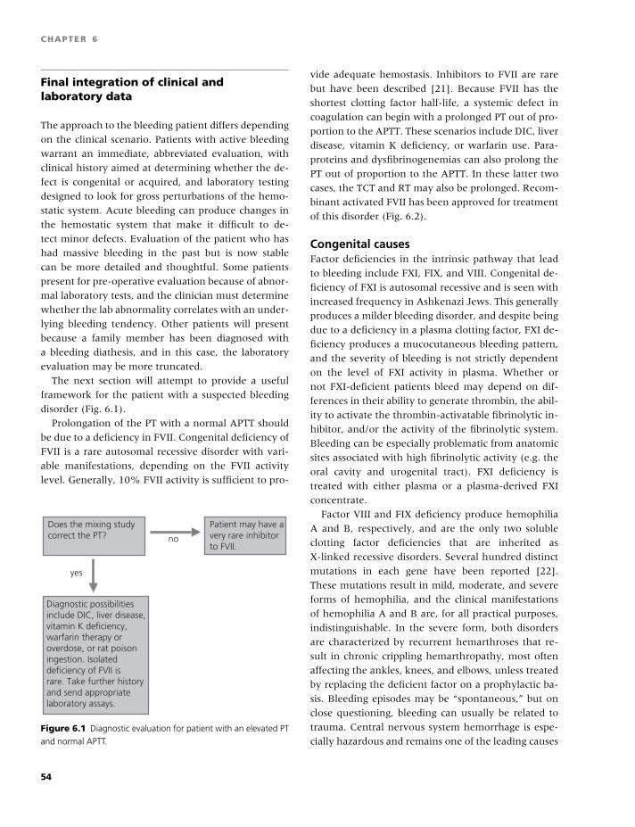
BLBK186-Key April 11, 2009 12:54
CHAPTER 6
Final integration of clinical andlaboratory data
The approach to the bleeding patient differs depending
on the clinical scenario. Patients with active bleeding
warrant an immediate, abbreviated evaluation, with
clinical history aimed at determining whether the de-
fect is congenital or acquired, and laboratory testing
designed to look for gross perturbations of the hemo-
static system. Acute bleeding can produce changes in
the hemostatic system that make it difficult to de-
tect minor defects. Evaluation of the patient who has
had massive bleeding in the past but is now stable
can be more detailed and thoughtful. Some patients
present for pre-operative evaluation because of abnor-
mal laboratory tests, and the clinician must determine
whether the lab abnormality correlates with an under-
lying bleeding tendency. Other patients will present
because a family member has been diagnosed with
a bleeding diathesis, and in this case, the laboratory
evaluation may be more truncated.
The next section will attempt to provide a useful
framework for the patient with a suspected bleeding
disorder (Fig. 6.1).
Prolongation of the PT with a normal APTT should
be due to a deficiency in FVII. Congenital deficiency of
FVII is a rare autosomal recessive disorder with vari-
able manifestations, depending on the FVII activity
level. Generally, 10% FVII activity is sufficient to pro-
no
yes
Does the mixing studycorrect the PT?
Patient may have avery rare inhibitorto FVII.
Diagnostic possibilitiesinclude DIC, liver disease,vitamin K deficiency,warfarin therapy oroverdose, or rat poisoningestion. Isolateddeficiency of FVII is rare. Take further historyand send appropriatelaboratory assays.
Figure 6.1 Diagnostic evaluation for patient with an elevated PT
and normal APTT.
vide adequate hemostasis. Inhibitors to FVII are rare
but have been described [21]. Because FVII has the
shortest clotting factor half-life, a systemic defect in
coagulation can begin with a prolonged PT out of pro-
portion to the APTT. These scenarios include DIC, liver
disease, vitamin K deficiency, or warfarin use. Para-
proteins and dysfibrinogenemias can also prolong the
PT out of proportion to the APTT. In these latter two
cases, the TCT and RT may also be prolonged. Recom-
binant activated FVII has been approved for treatment
of this disorder (Fig. 6.2).
Congenital causesFactor deficiencies in the intrinsic pathway that lead
to bleeding include FXI, FIX, and VIII. Congenital de-
ficiency of FXI is autosomal recessive and is seen with
increased frequency in Ashkenazi Jews. This generally
produces a milder bleeding disorder, and despite being
due to a deficiency in a plasma clotting factor, FXI de-
ficiency produces a mucocutaneous bleeding pattern,
and the severity of bleeding is not strictly dependent
on the level of FXI activity in plasma. Whether or
not FXI-deficient patients bleed may depend on dif-
ferences in their ability to generate thrombin, the abil-
ity to activate the thrombin-activatable fibrinolytic in-
hibitor, and/or the activity of the fibrinolytic system.
Bleeding can be especially problematic from anatomic
sites associated with high fibrinolytic activity (e.g. the
oral cavity and urogenital tract). FXI deficiency is
treated with either plasma or a plasma-derived FXI
concentrate.
Factor VIII and FIX deficiency produce hemophilia
A and B, respectively, and are the only two soluble
clotting factor deficiencies that are inherited as
X-linked recessive disorders. Several hundred distinct
mutations in each gene have been reported [22].
These mutations result in mild, moderate, and severe
forms of hemophilia, and the clinical manifestations
of hemophilia A and B are, for all practical purposes,
indistinguishable. In the severe form, both disorders
are characterized by recurrent hemarthroses that re-
sult in chronic crippling hemarthropathy, most often
affecting the ankles, knees, and elbows, unless treated
by replacing the deficient factor on a prophylactic ba-
sis. Bleeding episodes may be “spontaneous,” but on
close questioning, bleeding can usually be related to
trauma. Central nervous system hemorrhage is espe-
cially hazardous and remains one of the leading causes
54
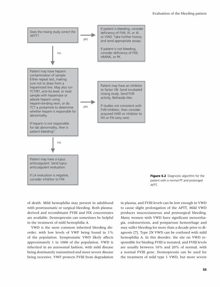
BLBK186-Key April 11, 2009 12:54
Evaluation of the bleeding patient
Does the mixing study correct the APTT?
Patient may have heparincontamination of sample. Either repeat test, makingsure not to draw from aheparinized line. May also runTCT/RT, anti-Xa level, or treatsample with heparinase oradsorb heparin usingheparin-binding resin, or doTCT ± protamine to determinewhether heparin is responsible forabnormality.
If heparin is not responsiblefor lab abnormality, then ispatient bleeding?
yes
yes
Patient may have a lupusanticoagulant. Send lupusanticoagulant evaluation.
If LA evaluation is negative,consider inhibitor to FXII
Patient may have an inhibitorto factor VIII. Send incubatedmixing study. Send FVIIIactivity, Bethesda titer.
If studies not consistent withFVIII inhibitor, then consideracquired VWD or inhibitor toFIX or FXI (very rare).
If patient is bleeding, considerdeficiency of FVIII, IX, or XI, or VWD. Take further history and send appropriate assays.
If patient is not bleeding,consider deficiency of FXII,HMWK, or PK.
no
no
Figure 6.2 Diagnostic algorithm for the
patient with a normal PT and prolonged
APTT.
of death. Mild hemophilia may present in adulthood
with posttraumatic or surgical bleeding. Both plasma-
derived and recombinant FVIII and FIX concentrates
are available. Desmopressin can sometimes be helpful
in the treatment of mild hemophilia A.
VWD is the most common inherited bleeding dis-
order, with low levels of VWF being found in 1%
of the population. Symptomatic VWD likely affects
approximately 1 in 1000 of the population. VWD is
inherited in an autosomal fashion, with mild disease
being dominantly transmitted and more severe disease
being recessive. VWF protects FVIII from degradation
in plasma, and FVIII levels can be low enough in VWD
to cause slight prolongation of the APTT. Mild VWD
produces mucocutaneous and postsurgical bleeding.
Many women with VWD have significant menorrha-
gia, endometriosis, and postpartum hemorrhage and
may suffer bleeding for more than a decade prior to di-
agnosis [7]. Type 2N VWD can be confused with mild
hemophilia A. In this disorder, the site on VWD re-
sponsible for binding FVIII is mutated, and FVIII levels
are usually between 10% and 20% of normal, with
a normal FVIII gene. Desmopressin can be used for
the treatment of mild type 1 VWD, but more severe
55
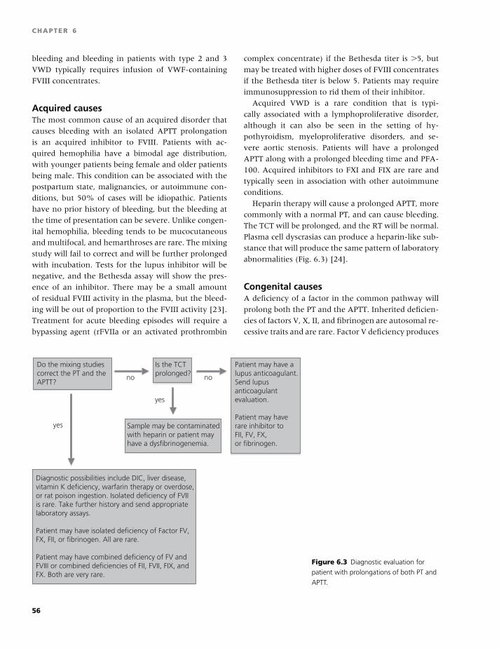
BLBK186-Key April 11, 2009 12:54
CHAPTER 6
bleeding and bleeding in patients with type 2 and 3
VWD typically requires infusion of VWF-containing
FVIII concentrates.
Acquired causesThe most common cause of an acquired disorder that
causes bleeding with an isolated APTT prolongation
is an acquired inhibitor to FVIII. Patients with ac-
quired hemophilia have a bimodal age distribution,
with younger patients being female and older patients
being male. This condition can be associated with the
postpartum state, malignancies, or autoimmune con-
ditions, but 50% of cases will be idiopathic. Patients
have no prior history of bleeding, but the bleeding at
the time of presentation can be severe. Unlike congen-
ital hemophilia, bleeding tends to be mucocutaneous
and multifocal, and hemarthroses are rare. The mixing
study will fail to correct and will be further prolonged
with incubation. Tests for the lupus inhibitor will be
negative, and the Bethesda assay will show the pres-
ence of an inhibitor. There may be a small amount
of residual FVIII activity in the plasma, but the bleed-
ing will be out of proportion to the FVIII activity [23].
Treatment for acute bleeding episodes will require a
bypassing agent (rFVIIa or an activated prothrombin
complex concentrate) if the Bethesda titer is �5, but
may be treated with higher doses of FVIII concentrates
if the Bethesda titer is below 5. Patients may require
immunosuppression to rid them of their inhibitor.
Acquired VWD is a rare condition that is typi-
cally associated with a lymphoproliferative disorder,
although it can also be seen in the setting of hy-
pothyroidism, myeloproliferative disorders, and se-
vere aortic stenosis. Patients will have a prolonged
APTT along with a prolonged bleeding time and PFA-
100. Acquired inhibitors to FXI and FIX are rare and
typically seen in association with other autoimmune
conditions.
Heparin therapy will cause a prolonged APTT, more
commonly with a normal PT, and can cause bleeding.
The TCT will be prolonged, and the RT will be normal.
Plasma cell dyscrasias can produce a heparin-like sub-
stance that will produce the same pattern of laboratory
abnormalities (Fig. 6.3) [24].
Congenital causesA deficiency of a factor in the common pathway will
prolong both the PT and the APTT. Inherited deficien-
cies of factors V, X, II, and fibrinogen are autosomal re-
cessive traits and are rare. Factor V deficiency produces
Do the mixing studiescorrect the PT and theAPTT?
no
Is the TCTprolonged?
yes
no
Patient may have alupus anticoagulant.Send lupusanticoagulantevaluation.
Patient may haverare inhibitor toFII, FV, FX,or fibrinogen.
yes
Sample may be contaminatedwith heparin or patient mayhave a dysfibrinogenemia.
Diagnostic possibilities include DIC, liver disease,vitamin K deficiency, warfarin therapy or overdose,or rat poison ingestion. Isolated deficiency of FVIIis rare. Take further history and send appropriatelaboratory assays.
Patient may have isolated deficiency of Factor FV,FX, FII, or fibrinogen. All are rare.
Patient may have combined deficiency of FV andFVIII or combined deficiencies of FII, FVII, FIX, andFX. Both are very rare.
Figure 6.3 Diagnostic evaluation for
patient with prolongations of both PT and
APTT.
56
BLBK186-Key April 11, 2009 12:54
Evaluation of the bleeding patient
a bleeding disorder that is less severe than hemophilia
A or B, even when FV levels are �1%. Bleeding times
may be prolonged due to lack of platelet FV, which is
reported to account for 20% of the FV in the body. It
is treated with fresh frozen plasma. Factor X deficiency
can be mild, moderate, or severe, with severe defi-
ciency producing bleeding similar to that seen in clas-
sical hemophilia. Patients with bleeding can be treated
with prothrombin complex concentrates (PCCs), and
FX levels should not be raised above 50% to avoid
thromboembolic complications. Inherited prothrom-
bin deficiency is very rare and can also be treated with
PCCs.
Fibrinogen gene mutations lead either to absence
of fibrinogen (afibrinogenemia) or to production of
a defective molecule (dysfibrinogenemia). Afibrino-
genemia is very rare, leading to a severe bleeding
disorder manifested by bleeding after trauma into sub-
cutaneous and deeper tissues that may result in dissec-
tion. Bleeding from the umbilical stump is frequent.
In addition to prolongation of the PT and the APTT,
these patients also show a prolonged TCT and RT. The
bleeding time is also prolonged due to the absence
of fibrinogen in the platelet alpha granule. Treatment
consists of transfusing cryoprecipitate to raise the fib-
rinogen level to the range of around 100 mg/dL. The
majority of patients with dysfibrinogenemia are het-
erozygous for the disorder and show no evidence of
either a hemorrhagic or a thrombotic state. Other dys-
fibrinogenemias, however, are associated with bleed-
ing episodes, and a few may be associated with ve-
nous or arterial thrombosis. Bleeding patients should
be treated with infusions of fibrinogen concetrates or
cryoprecipitate.
Combined deficiency of multiple clotting factors can
also be inherited, the most common conditions be-
ing combined deficiency of factors V and VIII and a
combined deficiency of the vitamin K-dependent fac-
tors (prothrombin and factors VII, IX, X, and proteins
C and S) [25,26]. A combined deficiency of factors V
and VIII is inherited in an autosomal recessive fashion
and is due to defects in one of two genes: the LMAN1
gene and a newly discovered gene called the “multiple
clotting factor deficiency 2 (MCFD2) gene [26]. The
products of both genes play an important role in the
transport of factors V and VIII from the endoplasmic
reticulum to the Golgi apparatus and are necessary for
normal secretion of these factors. The disorder results
in a mild to moderate bleeding tendency with factor
V and VIII levels ranging from 5% to 30% of normal.
When both the PT and PTT are prolonged, and either
factor V or VIII is found to be decreased, the combined
deficiency should be suspected. Factor VIII is easily
replaced using factor VIII concentrates, but the only
readily available factor V replacement is fresh frozen
plasma, which is limited in its ability to normalize the
factor V level. In some cases, plasma exchange is nec-
essary to raise the factor V to hemostatic levels.
Combined deficiencies of the vitamin K-dependent
factors can be due to defects in either the gene for
vitamin K-dependent carboxylase or the gene for vita-
min K epoxide reductase [27]. This is an autosomal re-
cessive disorder that may be associated with deficien-
cies of prothrombin, factors VII, IX, and X, as well as
proteins C and S [26]. In this syndrome, both the PT
and PTT are prolonged, and assays for the individual
factors that influence these tests are necessary. Large
doses of vitamin K may partially correct the heredi-
tary defect in some but not all cases. Some bleeding
episodes will require replacement with PCCs.
Acquired causesInhibitors to factor V are typically seen in patients
who have undergone re-do vascular or cardiac
surgery and are provoked by use of bovine thrombin.
This hemostatic agent is contaminated with a small
amount of bovine FV, and antibodies to bovine FV
will cross-react with human FV. This condition may
be self-limited, but bleeding can be treated with
platelets, because platelet FV may be less susceptible
to inhibitors in plasma.
Prothrombin antibodies can co-exist with the lupus
inhibitor, and these inhibitors increase clearance of
FII, causing an acquired deficiency, rather than neu-
tralizing prothrombin function. Thus, the mixing stud-
ies for the PT will be normal.
Factor X deficiency can be seen in conjunction with
amyloidosis, because the FX is adsorbed onto the amy-
loid protein. This can cause a severe hemorrhagic dis-
order that has been reported to respond to splenec-
tomy. This condition will also produce a mixing study
that normalizes the PT and the APTT.
Combined factor deficiencies can be seen in con-
ditions such as vitamin K deficiency, disseminated
intravascular coagulation, and severe liver disease.
Severe liver disease can also lead to an acquired
57
BLBK186-Key April 11, 2009 12:54
CHAPTER 6
dysfibrinogenemia, which can produce a prolonged
TCT and RT.
Anticoagulants such as heparin and coumadin can
cause prolongation of both the PT and the APTT, espe-
cially when given in excess. Direct thrombin inhibitors
such as lepirudin and argatroban will prolong the PT,
the APTT, and the TCT.
Patients with bleeding, but normalPT and APTT
Congenital causesFactor XIII deficiency is a rare autosomal recessive dis-
order that presents with severe bleeding. Prolonged
bleeding from the umbilical stump is common, as is
spontaneous intracranial hemorrhage. Treatment re-
lies on cryoprecipitate, although FXIII concentrates are
in clinical trials.
Congenital disorders of platelets include thrombo-
cytopenic disorders, disorders of platelet surface gly-
coproteins, signaling pathway disorders, and storage
pool and secretion disorders. They typically show
prolongation of the bleeding time and the PFA-100.
Platelet aggregation may show a typical pattern, but
milder disorders may have normal platelet aggrega-
tion tracings. Mild thrombocytopathies may be missed
by the bleeding time, the PFA-100, and platelet ag-
gregation testing, and may require more special-
ized testing, such as flow cytometry or electron
microscopy.
Congenital deficiencies of fibrinolytic inhibitors
such as �2-antiplasmin and PAI-1 have been reported,
and bleeding is typically delayed. The euglobulin lysis
time can be shortened, and assays for these proteins
can be performed but may not be helpful in the defi-
ciency state, due to assay limitations.
VWD can present with normal APTT values, espe-
cially if the FVIII activity level is above 40–50%. Type
1 VWD is a quantitative deficit of VWF, and all mul-
timeric forms are present. Mild type 1 VWD may be
missed by the bleeding time and the PFA-100, mak-
ing measurement of VWF antigen and activity levels
necessary for proper diagnosis. Additionally, levels of
VWF fluctuate in response to estrogens, stress, exer-
cise, inflammation, and bleeding; and repeated assays
are often required to make the diagnosis.
Hereditary HHT is an autosomal dominant disorder
that is associated with arteriovenous malformations of
the small vessels of the skin, oropharynx, lungs, gas-
trointestinal tract, and other tissues. The syndrome is
often suspected by the presence of epistaxis, gastroin-
testinal bleeding, telangiectasia on the lips and finger-
tips, and iron deficiency anemia. Although bleeding
does not occur at birth, it may begin in childhood,
and by age 16, the majority of patients will experience
hemorrhagic symptoms.
Ehlers-Danlos syndrome (EDS) is characterized by
easy bruising and hemorrhage from ruptured blood
vessels and is due to one of several genetic defects
[28]. The classic EDS causing joint hypermobility and
hyperextensibility of the skin may be associated with
bruising but is not likely to result in massive bleed-
ing. The vascular type IV EDS is the most likely to re-
sult in significant bruising and is due to a defect in
type III collagen resulting from defects in the COL3A1
gene. In this type of EDS, bruising can be very ex-
tensive and vascular rupture can result in death. The
skin may be thin and wrinkled, but hyperextensibility
of the skin is not common. The bruising is sufficient
to make one suspect a platelet disorder, but tests of
platelet and coagulant function are normal. Diagno-
sis is dependent on demonstration of the genetic ab-
normality or the demonstration of abnormal type III
collagen.
Acquired causesMany drugs and herbs cause platelet dysfunction, and
their use needs to be questioned extensively. Uremia,
myeloproliferative disorders, and cardiac bypass will
also cause a thrombocytopathy.
Amyloidosis has been reported in conjunction with
acquired deficiencies of �2-antiplasmin and PAI-1. In-
hibitors to FXIII have been reported and are rare.
Patients without bleeding history, but withabnormal coagulation testingWhen doing pre-operative evaluations of these pa-
tients, it is important to recognize that many patients
with mild bleeding disorders may have no known his-
tory of bleeding. Some may recall mild bleeding symp-
toms when carefully questioned, whereas some may
not have had sufficient challenges to their hemostatic
systems. Thus, some lab evaluation is required, de-
pending on the severity of the surgery that is being
planned.
58
BLBK186-Key April 11, 2009 12:54
Evaluation of the bleeding patient
Congenital causesDeficiencies of high-molecular-weight kininogen,
prekallikrein, and FXII will produce marked prolon-
gation of the APTT without conferring an increased
risk of abnormal bleeding. Some patients with FXI de-
ficiency may have no bleeding symptoms, despite low
levels of FXI. Patients with mild deficiency of FVII may
also have no bleeding symptoms. Additionally, certain
mutations in the FVII molecule affect its interaction
with bovine but not human thromboplastin, and these
mutations are not associated with clinical manifesta-
tions. Lastly, most dyfibrinogenemias are also associ-
ated with no clinical symptoms.
Acquired causesLupus anticoagulants prolong the APTT, and the
mixing study fails to correct. Unless associated with
acquired hypoprothrombinemia, lupus inhibitors con-
fer no increased risk of bleeding. The majority of ac-
quired FV antibodies are also asymptomatic and are
self-limited. Although patients with severe liver dis-
ease may have a prolonged PT/INR, their bleeding
symptoms may vary. These patients may clot and they
are not “auto-anticoagulated,” because they are defi-
cient in many anticoagulant proteins as well.
Conclusion
Evaluation of the bleeding patient requires a careful
history and physical examination. Laboratory workup
should be tailored to the clinical presentation and the
pretest probability of finding an underlying bleeding
diathesis. Many of the laboratory tests are best con-
ducted at a tertiary center with expertise in hemosta-
sis. Accurate diagnosis allows for rational, intelligent
treatment and prophylaxis of bleeding.
References
1 Higham JM, O’Brien PM, Shaw RW. Assessment of
menstrual blood loss using a pictorial chart. Br J Obstet
Gynaecol 1990;97(8):734–9.
2 Rodeghiero F, Kadir RA, Tosetto A, James PD. Rele-
vance of quantitative assessment of bleeding in haem-
orrhagic disorders. Haemophilia 2008;14(Suppl 3):68–
75.
3 Philipp CS, Faiz A, Dowling N, et al. Age and the preva-
lence of bleeding disorders in women with menorrha-
gia. Obstet Gynecol 2005;105(1):61–6.
4 Trasi SA, Pathare AV, Shetty SD, Ghosh K, Salvi V, Mo-
hanty D. The spectrum of bleeding disorders in women
with menorrhagia: a report from Western India. Ann
Hematol 2005;84(5):339–42.
5 Woo YL, White B, Corbally R, et al. von Willebrand’s
disease: an important cause of dysfunctional uterine
bleeding. Blood Coagul Fibrinolysis 2002;13(2): 89–93.
6 Lee CA. Women and inherited bleeding disorders: men-
strual issues. Semin Hematol 1999;36(3 Suppl 4): 21–7.
7 James AH. More than menorrhagia: a review of the
obstetric and gynaecological manifestations of bleeding
disorders. Haemophilia 2005;11(4):295–307.
8 Gitschier J. Molecular genetics of hemophilia A. Schweiz
Med Wochenschr 1989;119(39):1329–31.
9 Sucker C. The Heyde syndrome: proposal for a unifying
concept explaining the association of aortic valve steno-
sis, gastrointestinal angiodysplasia and bleeding. Int J
Cardiol 2007;115(1):77–8.
10 Sucker C, Hetzel GR, Grabensee B, Stockschlaeder
M, Scharf RE. Amyloidosis and bleeding: patho-
physiology, diagnosis, and therapy. Am J Kidney Dis
2006;47(6):947–55.
11 Mumford AD, O’Donnell J, Gillmore JD, Manning
RA, Hawkins PN, Laffan M. Bleeding symptoms and
coagulation abnormalities in 337 patients with AL-
amyloidosis. Br J Haematol 2000;110(2):454–60.
12 Lind SE. The bleeding time does not predict surgical
bleeding. Blood 1991;77(12):2547–52.
13 O’Brien SH, Ritchey AK, Ragni MV. Combined clotting
factor deficiencies: experience at a single hemophilia
treatment center. Haemophilia 2007;13(1):26–9.
14 Langdell RD, Wagner RH, Brinkhous KM. Effect of an-
tihemophilic factor on one-stage clotting tests; a pre-
sumptive test for hemophilia and a simple one-stage
antihemophilic factor assy procedure. J Lab Clin Med
1953;41(4):637–47.
15 Shah U, Ma AD. Tests of platelet function. Curr Opin
Hematol 2007;14(5):432–7.
16 Michelson AD, Frelinger AL 3rd, Furman MI. Cur-
rent options in platelet function testing. Am J Cardiol
2006;98(10A):4N–10N.
17 Favaloro EJ. Clinical application of the PFA-100. Curr
Opin Hematol 2002;9(5):407–15.
18 Zhou L, Schmaier AH. Platelet aggregation testing in
platelet-rich plasma: description of procedures with the
aim to develop standards in the field. Am J Clin Pathol
2005;123(2):172–83.
19 Lorez HP, Richards JG, Da Prada M, et al. Storage
pool disease: comparative fluorescence microscopical,
59
BLBK186-Key April 11, 2009 12:54
CHAPTER 6
cytochemical and biochemical studies on amine-storing
organelles of human blood platelets. Br J Haematol
1979;43(2):297–305.
20 White JG. Use of the electron microscope for di-
agnosis of platelet disorders. Semin Thromb Hemost
1998;24(2):163–8.
21 Mullighan CG, Rischbieth A, Duncan EM, Lloyd JV.
Acquired isolated factor VII deficiency associated with
severe bleeding and successful treatment with re-
combinant FVIIa (NovoSeven). Blood Coagul Fibrinolysis
2004;15(4):347–51.
22 Stenson PD, Ball EV, Mort M, et al. Human Gene
Mutation Database (HGMD): 2003 update. Hum Mutat
2003;21(6):577–81.
23 Ma AD, Carrizosa D. Acquired factor VIII inhibitors:
pathophysiology and treatment. Hematol Am Soc Hematol
Educ Program 2006:432–7.
24 Torjemane L, Guermazi S, Ladeb S, et al. Heparin-like
anticoagulant associated with multiple myeloma and
neutralized with protamine sulfate. Blood Coagul Fibri-
nolysis 2007;18(3):279–81.
25 McMillan C, Roberts H. Congenital combined defi-
ciency of coagulation factors II, VII, IX and X. N Engl
J Med 1966;274:1313–5.
26 Zhang B, Ginsburg D. Familial multiple coagulation fac-
tor deficiencies: new biologic insight from rare genetic
bleeding disorders. J Thromb Haemost 2004;2(9):1564–
72.
27 Li T, Chang CY, Jin DY, Lin PJ, Khvorova A, Stafford
DW. Identification of the gene for vitamin K epoxide
reductase. Nature 2004;427(6974):541–4.
28 De Paepe A, Malfait F. Bleeding and bruising in patients
with Ehlers-Danlos syndrome and other collagen vas-
cular disorders. Br J Haematol 2004;127(5):491–500.
60
BLBK186-Key April 24, 2009 14:43
7 Hemophilia A and BRhona M. Maclean and Michael Makris
Introduction
Hemophilia A and B are bleeding disorders inherited
in an X-linked recessive fashion, caused by deficiencies
in factor VIII (FVIII) and factor IX (FIX), respectively.
The first description of hemophilia is thought to be a
passage describing bleeding following circumcision in
the Babylonian Talmud of the 2nd century AD, “It was
taught by the Tana’im: If she circumcised her first son
and he died, and a second son and he died, she must
not circumcise a third one.”
It was initially thought that hemophilia was caused
by abnormalities of the vascular system, and it was
not until the late 1800s and early 1900s that a defi-
ciency of a component of the blood was thought to be
responsible.
All racial groups are equally affected by hemophilia
with an incidence of 1 in 5000 live male births
for hemophilia A, and 1 in 30,000 live male births
for hemophilia B. The clinical symptoms and signs
of these two disorders are identical in presentation,
and specific clotting factor assays are required to dis-
tinguish them. With modern management and the
ready availability of clotting factors, children with
hemophilia today can look forward to a normal life
expectancy [1].
Factor VIII gene and protein
In the two decades since the FVIII protein was first
purified (1983) and the gene cloned (1982–4), ad-
vances in molecular biology and protein biochemistry
have led to a greatly improved understanding of the
structure and function of both the FVIII gene and the
protein. The crystal structure of FVIII was recently
published [2].
The FVIII gene (F8) is situated in the most dis-
tal band of the long arm of the X chromosome at
Xq28, spans 186,000 base pairs (bp) of DNA, con-
tains 26 exons, and is transcribed from the telomeric
to centromeric direction to produce a mature mRNA
of approximately 9 kb. The precursor protein (2351
amino acids) is predominantly synthesized in hepa-
tocytes and has a molecular weight of approximately
293,000 Da.
After cleavage of the secretory leader sequence, the
FVIII protein has a mature sequence of 2332 amino
acids with the domain structure A1-a1-A2-a2-B-a3-
A3-C1-C2. The domain structure of FVIII is very sim-
ilar to that of coagulation factor V, and its A do-
mains are homologous with ceruloplasmin. As the
FVIII protein is very susceptible to proteolysis after
secretion, the majority of circulating FVIII comprises
heavy chains (the A1 and A2 domains with variable
lengths of the B domain) noncovalently linked to light
chains (A3, C1, and C2 domains). The B domain is un-
necessary for FVIII procoagulant activity. FVIII exerts
its procoagulant activity by accelerating the activation
of coagulation factor X by factor IXa. FVIII circulates
bound to and is stabilised by von Willebrand factor
(VWF), with a ratio of approximately 1 molecule of
FVIII to 50 molecules of VWF.
Mutations in F8There are many F8 gene defects listed on the
online hemophilia A mutation database (http://
europium.csc.mrc.ac.uk). These can be categorised
as (1) gross gene rearrangements, (2) insertions or
deletions of genetic sequence, or (3) single base
61
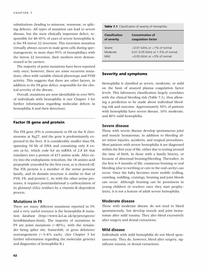
BLBK186-Key April 24, 2009 14:43
CHAPTER 7
substitutions (leading to missense, nonsense, or splic-
ing defects). All types of mutation can lead to severe
disease, but the most clinically important defect, re-
sponsible for 40–45% of cases of severe hemophilia A,
is the F8 intron 22 inversion. This inversion mutation
virtually always occurs in male germ cells during sper-
matogenesis; in more than 95% of hemophiliacs with
the intron 22 inversion, their mothers were demon-
strated to be carriers.
The majority of point mutations have been reported
only once; however, there are some recurrent muta-
tions, often with variable clinical phenotype and FVIII
activity. This suggests that there are other factors, in
addition to the F8 gene defect, responsible for the clin-
ical severity of the disease.
Overall, mutations are now identifiable in over 90%
of individuals with hemophilia A (see Chapter 3 for
further information regarding molecular defects in
hemophilia A and their detection).
Factor IX gene and protein
The FIX gene (F9) is centromeric to F8 on the X chro-
mosome at Xq27, and the gene is predominantly ex-
pressed in the liver. It is considerably smaller than F8,
spanning 34 kb of DNA and containing only 8 ex-
ons (a–h), which code for an mRNA of 2.8 kb that
translates into a protein of 415 amino acids. After en-
try into the endoplasmic reticulum, the 18-amino-acid
prepeptide (encoded by the first exon, a) is cleaved off.
The FIX protein is a member of the serine protease
family, and its domain structure is similar to that of
FVII, FX, and protein C. As with the other serine pro-
teases, it requires posttranslational γ-carboxylation of
its glutamyl (Glu) residues by a vitamin-K-dependent
process.
Mutations in F9There are many different mutations reported in F9,
and a very useful resource is the hemophilia B muta-
tion database (http://www.kcl.ac.uk/ip/petergreen/
hemBdatabase.html). The majority of mutations in
F9 are point mutations (∼80%), with the remain-
der being splice site, frameshift, or gross deletions/
rearrangements (∼3–4% each). (See Chapter 3 for
further information regarding the molecular genetics
and diagnostics of hemophilia B.)
Table 7.1 Classification of severity of hemophilia.
Classification Concentration ofof severity coagulation factor
Severe <0.01 IU/mL or <1% of normal
Moderate 0.01–0.05 IU/mL or 1–5% of normal
Mild >0.05 IU/mL or >5% of normal
Severity and symptoms
Hemophilia is classified as severe, moderate, or mild
on the basis of assayed plasma coagulation factor
levels. This laboratory classification largely correlates
with the clinical bleeding risk (Table 7.1), thus allow-
ing a prediction to be made about individual bleed-
ing risk and outcome. Approximately 50% of patients
with hemophilia have severe disease, 10% moderate,
and 40% mild hemophilia.
Severe diseaseThose with severe disease develop spontaneous joint
and muscle hematomas, in addition to bleeding af-
ter minor injuries, accidents, and surgical procedures.
Most patients with severe hemophilia A are diagnosed
within the first year of life, either due to testing around
the time of birth, in those with a family history, or
because of abnormal bruising/bleeding. Thereafter, in
the first 6–9 months of life, cutaneous bruising or oral
bleeding (due to teething or cuts in the oral cavity) can
occur. Once the baby becomes more mobile (rolling,
crawling, toddling, cruising), bruising and joint bleeds
can occur. Although bruising can be prominent in
young children (it resolves once they start prophy-
laxis), it is not a feature of adult severe hemophilia.
Moderate diseaseThose with moderate disease do not tend to bleed
spontaneously, but develop muscle and joint hema-
tomas after mild trauma. They also bleed excessively
after surgery and dental extractions.
Mild diseaseIndividuals with mild hemophilia do not bleed spon-
taneously. They do, however, bleed after surgery, sig-
nificant trauma, or dental extractions.
62
BLBK186-Key April 24, 2009 14:43
Hemophilia A and B
Inheritance
Both hemophilia A and B are X-linked recessive in-
herited disorders and therefore affect males almost ex-
clusively. It is not uncommon, however, for carrier fe-
males to have reductions in FVIII or FIX levels to the
extent that they may experience menorrhagia or will
require treatment prior to any invasive procedure or
following major trauma.
Where the female is a carrier, there is a 50:50
chance that a son will be affected by hemophilia or
that a daughter will be a carrier. When the children
are from a hemophilic male and a normal female, all
sons will be unaffected, but all daughters will be obli-
gate carriers.
Approximately one-third of cases of hemophilia are
‘sporadic,’ that is, due to the occurrence of a new mu-
tation, with no family history of the disease.
Mosaicism occurs when a proportion of the cells of
the body contain a mutation, whereas the majority do
not. Gonadal mosaicim, in which the mutation is con-
fined to the gonadal tissue, has been reported in both
hemophilia A and B [3]. Should gonadal mosaicim be
present, the risk of passing on the disease to any fu-
ture children will be higher than the risk in the gen-
eral population. Care must therefore be taken when
counseling women who do not appear to be carriers,
yet have a child with hemophilia.
Females with markedly reducedFVIII/IX levels
This is possible in the following rare circumstances:� With extreme lyonization of F8 or F9 in hemophilia
carriers (resulting in most of the expression deriving
from the hemophilic X chromosome); rarely carriers
can have levels �10%.� If there is hemizygosity of the X chromosome [e.g.
in Turner (XO) syndrome].� A female can be affected if she is the offspring of a
hemophilic male and a carrier female.� In females with the Normandy variant of von
Willebrand disease (Type 2N VWD; FVIII deficiency
only).� In females with acquired hemophilia due to autoan-
tibody development.
Carrier testing
All females who are obligate or possible carriers of
hemophilia should be offered genetic counseling to
provide them with the information necessary to make
informed reproductive choices and for the optimal
management of their pregnancies. The majority of
individuals with hemophilia A and B now have an
identifiable mutation. If the mutation within the fam-
ily is known, it is straightforward to screen the po-
tential carrier and confirm the carrier status in ob-
ligate carriers. If the mutation is not known, then
linkage analysis using informative genetic polymor-
phisms is usually successful (if sufficient family mem-
bers are available for testing). If neither of these
approaches is suitable, then direct mutation detec-
tion can be performed (see Chapter 3). All carri-
ers of hemophilia A or B should have their factor
VIII/IX levels checked to evaluate their personal risk of
bleeding.
Prenatal diagnosis
Although the treatment of hemophilia has greatly im-
proved over the last 10–20 years, many carriers of
hemophilia (often those who have grown up with
a family member who had complications of the dis-
ease, such as inhibitors or viral infections) will re-
quest prenatal diagnosis. Chorionic villus sampling
is the most widely used method of prenatal diag-
nosis, and can be performed at 10–12 weeks’ ges-
tation, allowing for first trimester termination if de-
sired. Alternatively, amniocentesis can be performed
at 16 weeks. The risks of these procedures are low in
experienced centers, with a miscarriage rate of 0.5–
1%. Fetoscopy to allow for fetal blood sampling is
rarely performed as it can only be performed after 20
weeks’ gestation and has a higher risk of fetal death
(1–6%). Following the discovery of fetal DNA in ma-
ternal blood, PCR-based techniques have been devel-
oped to detect specific Y-chromosomal sequences in
maternal blood samples. Although not yet available
in many centers, it is now possible to determine the
sex of a fetus from as early as 7 weeks’ gestation
[4].
63
BLBK186-Key April 24, 2009 14:43
CHAPTER 7
Embryo selection: PreimplantationGenetic Diagnosis
Preimplantation Genetic Diagnosis involves the ge-
netic testing of an embryo prior to implantation and
before pregnancy occurs. It is used in conjunction with
in vitro fertilization, and only embryos found to be
free of a specific genetic disorder are transferred into a
woman for pregnancy. The advantage of this approach
is that the trauma of termination of pregnancy can be
avoided [5].
Delivery of an at-risk pregnancy
All carriers of hemophilia should have an ultrasound
scan at around 20 weeks’ gestation to identify the
fetal sex (if other prenatal diagnostic tests have not
been performed). Should the baby be male, then care
should be taken to minimize the risk of bleeding at de-
livery; for example, vacuum (ventouse) extraction, ro-
tational forceps, and invasive monitoring techniques,
including placement of scalp electrodes, should be
avoided. The mode of delivery should be for obstet-
ric reasons and need not be by cesarean mode. The
choice between vaginal and cesarean delivery is hotly
debated.
A cord sample should be sent from all male infants
born to known carriers for FVIII/IX estimation. Vi-
tamin K should be given orally until it is definitely
known that the baby is not affected by hemophilia.
Making the diagnosis
Immediately following the birth of a male infant to
a known carrier of hemophilia, the following tests
should be performed on the cord blood:� Prothrombin time (PT);� Activated partial thromboplastin time (APTT);� Fibrinogen level;� FVIII or FIX activity; and� Where there is no family history, if the FVIII level
is low, VWF assays for antigen and activity should be
performed.
The APTT of an affected infant will usually be
prolonged when compared with a gestation-specific
normal range. FVIII levels in infants are comparable
with those of adults, allowing for an accurate diag-
nosis. Although FIX levels in infants are considerably
lower than those in adults, if the FIX level is less than
1%, a diagnosis of severe hemophilia B can be made.
All neonates given a diagnosis of hemophilia on test-
ing a cord blood sample should have this confirmed
on a venous blood sample. Those with equivocal re-
sults should have a repeated test at 6 months of age.
Approximately one-third of individuals with
hemophilia have no family history of a bleeding disor-
der. A diagnosis of hemophilia should be suspected if
a child has a history of excessive bruising or bleeding
or presents with a swollen painful joint or muscle
hematoma.
The majority of children with moderate or severe
hemophilia will present by 4–5 years of age. Where
there is no family history, it is important to exclude the
diagnosis of VWD, as the Normandy variant of VWD is
phenotypically identical to mild/moderate hemophilia
A (although with autosomal inheritance). If this is
suspected, a VWF–FVIII binding assay or mutation
analysis of exons 18–25 of the VWF gene should
be undertaken to establish the correct diagnosis (see
Chapter 6).
The neonate with hemophilia
The neonatal period is defined as the first 28 days
after delivery, irrespective of gestation. Most bleed-
ing episodes in neonates with hemophilia are due
to birth trauma. It has been estimated that 3.5–4%
of neonates with severe hemophilia have intracra-
nial hemorrhages, most associated with the presence
of an extracranial hemorrhage, the risk being greater
if the delivery was traumatic/vacuum-assisted [6]. As
yet there is no consensus as to whether routine cra-
nial ultrasound should be performed after delivery in
neonates known to have hemophilia, or whether pro-
phylactic factor concentrate should be given after de-
livery. Most clinicians would give prophylactic coagu-
lation factor concentrate if the delivery was traumatic,
instrumental, or in the presence of prematurity.
Bleeding episodes in the neonate with hemophilia
occurring in the first week of birth are usually due to
heel pricks performed for blood sampling, intramuscu-
lar injections of vitamin K, or after circumcision.
64
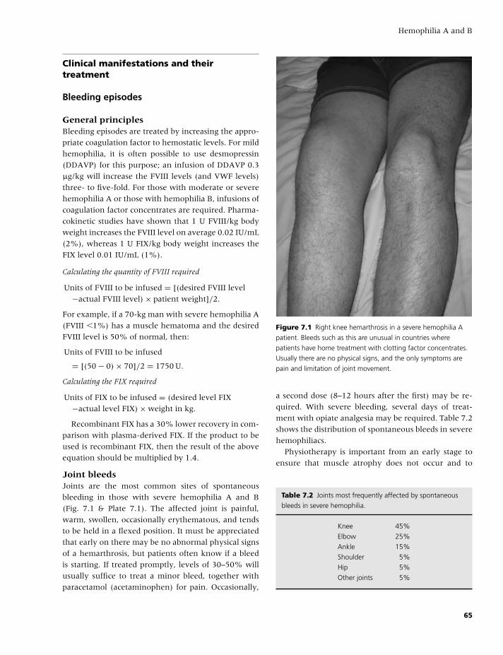
BLBK186-Key April 24, 2009 14:43
Hemophilia A and B
Clinical manifestations and theirtreatment
Bleeding episodes
General principlesBleeding episodes are treated by increasing the appro-
priate coagulation factor to hemostatic levels. For mild
hemophilia, it is often possible to use desmopressin
(DDAVP) for this purpose; an infusion of DDAVP 0.3
µg/kg will increase the FVIII levels (and VWF levels)
three- to five-fold. For those with moderate or severe
hemophilia A or those with hemophilia B, infusions of
coagulation factor concentrates are required. Pharma-
cokinetic studies have shown that 1 U FVIII/kg body
weight increases the FVIII level on average 0.02 IU/mL
(2%), whereas 1 U FIX/kg body weight increases the
FIX level 0.01 IU/mL (1%).
Calculating the quantity of FVIII required
Units of FVIII to be infused = [(desired FVIII level
−actual FVIII level) × patient weight]/2.
For example, if a 70-kg man with severe hemophilia A
(FVIII �1%) has a muscle hematoma and the desired
FVIII level is 50% of normal, then:
Units of FVIII to be infused
= [(50 − 0) × 70]/2 = 1750 U.
Calculating the FIX required
Units of FIX to be infused = (desired level FIX
−actual level FIX) × weight in kg.
Recombinant FIX has a 30% lower recovery in com-
parison with plasma-derived FIX. If the product to be
used is recombinant FIX, then the result of the above
equation should be multiplied by 1.4.
Joint bleedsJoints are the most common sites of spontaneous
bleeding in those with severe hemophilia A and B
(Fig. 7.1 & Plate 7.1). The affected joint is painful,
warm, swollen, occasionally erythematous, and tends
to be held in a flexed position. It must be appreciated
that early on there may be no abnormal physical signs
of a hemarthrosis, but patients often know if a bleed
is starting. If treated promptly, levels of 30–50% will
usually suffice to treat a minor bleed, together with
paracetamol (acetaminophen) for pain. Occasionally,
Figure 7.1 Right knee hemarthrosis in a severe hemophilia A
patient. Bleeds such as this are unusual in countries where
patients have home treatment with clotting factor concentrates.
Usually there are no physical signs, and the only symptoms are
pain and limitation of joint movement.
a second dose (8–12 hours after the first) may be re-
quired. With severe bleeding, several days of treat-
ment with opiate analgesia may be required. Table 7.2
shows the distribution of spontaneous bleeds in severe
hemophiliacs.
Physiotherapy is important from an early stage to
ensure that muscle atrophy does not occur and to
Table 7.2 Joints most frequently affected by spontaneous
bleeds in severe hemophilia.
Knee 45%
Elbow 25%
Ankle 15%
Shoulder 5%
Hip 5%
Other joints 5%
65
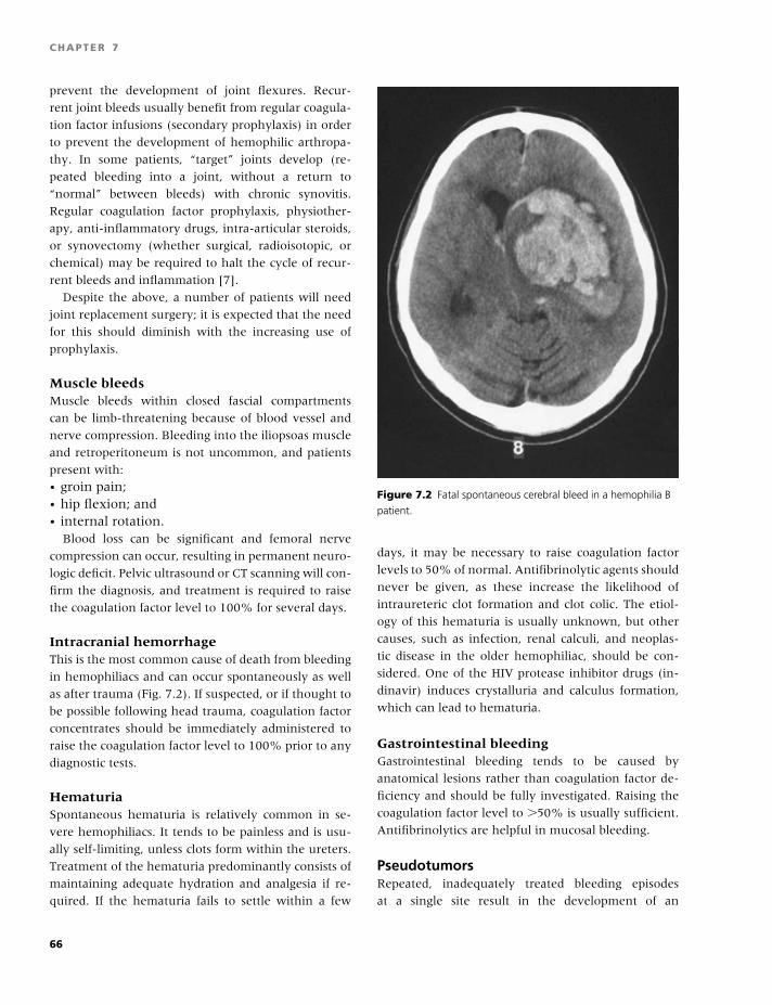
BLBK186-Key April 24, 2009 14:43
CHAPTER 7
prevent the development of joint flexures. Recur-
rent joint bleeds usually benefit from regular coagula-
tion factor infusions (secondary prophylaxis) in order
to prevent the development of hemophilic arthropa-
thy. In some patients, “target” joints develop (re-
peated bleeding into a joint, without a return to
“normal” between bleeds) with chronic synovitis.
Regular coagulation factor prophylaxis, physiother-
apy, anti-inflammatory drugs, intra-articular steroids,
or synovectomy (whether surgical, radioisotopic, or
chemical) may be required to halt the cycle of recur-
rent bleeds and inflammation [7].
Despite the above, a number of patients will need
joint replacement surgery; it is expected that the need
for this should diminish with the increasing use of
prophylaxis.
Muscle bleedsMuscle bleeds within closed fascial compartments
can be limb-threatening because of blood vessel and
nerve compression. Bleeding into the iliopsoas muscle
and retroperitoneum is not uncommon, and patients
present with:� groin pain;� hip flexion; and� internal rotation.
Blood loss can be significant and femoral nerve
compression can occur, resulting in permanent neuro-
logic deficit. Pelvic ultrasound or CT scanning will con-
firm the diagnosis, and treatment is required to raise
the coagulation factor level to 100% for several days.
Intracranial hemorrhageThis is the most common cause of death from bleeding
in hemophiliacs and can occur spontaneously as well
as after trauma (Fig. 7.2). If suspected, or if thought to
be possible following head trauma, coagulation factor
concentrates should be immediately administered to
raise the coagulation factor level to 100% prior to any
diagnostic tests.
HematuriaSpontaneous hematuria is relatively common in se-
vere hemophiliacs. It tends to be painless and is usu-
ally self-limiting, unless clots form within the ureters.
Treatment of the hematuria predominantly consists of
maintaining adequate hydration and analgesia if re-
quired. If the hematuria fails to settle within a few
Figure 7.2 Fatal spontaneous cerebral bleed in a hemophilia B
patient.
days, it may be necessary to raise coagulation factor
levels to 50% of normal. Antifibrinolytic agents should
never be given, as these increase the likelihood of
intraureteric clot formation and clot colic. The etiol-
ogy of this hematuria is usually unknown, but other
causes, such as infection, renal calculi, and neoplas-
tic disease in the older hemophiliac, should be con-
sidered. One of the HIV protease inhibitor drugs (in-
dinavir) induces crystalluria and calculus formation,
which can lead to hematuria.
Gastrointestinal bleedingGastrointestinal bleeding tends to be caused by
anatomical lesions rather than coagulation factor de-
ficiency and should be fully investigated. Raising the
coagulation factor level to �50% is usually sufficient.
Antifibrinolytics are helpful in mucosal bleeding.
PseudotumorsRepeated, inadequately treated bleeding episodes
at a single site result in the development of an
66
BLBK186-Key April 24, 2009 14:43
Hemophilia A and B
encapsulated hematoma. This progressively enlarges,
erodes, and invades surrounding structures, hence the
name pseudotumour (Plate 7.2). Surgical removal is
difficult and is associated with a significant morbid-
ity/mortality. These are now rare in countries with
ready availability of clotting factor concentrates [8].
Dental treatmentMinor dental work (scaling and polishing) can be per-
formed without factor replacement, but inferior den-
tal nerve blocks or extractions require factor concen-
trates or desmopressin administered as appropriate.
Antifibrinolytic agents (such as tranexamic acid as a
mouthwash) should be provided for 3–5 days after any
dental extractions.
SurgeryFor major surgical procedures, coagulation factor lev-
els should be maintained at 50–100% for 7–10 days
to ensure adequate hemostasis and wound healing.
This can be achieved either by bolus injections, with
an initial bolus dose to bring the factor level to 100%
followed by once daily FIX or twice daily FVIII injec-
tions, or by continuous infusion after the initial bolus
dose, as guided by coagulation factor assays.
Continuous infusions have the advantage of:� eliminating the “peaks and troughs” seen with
bolus factor administration;� less factor concentrate consumption for the
same procedure;� less cost; and� more convenient for staff to administer.
One disadvantage is that these infusions tend to
cause venous irritation, but this can be reduced by an
infusion of saline in tandem through the same can-
nula. Intramuscular injections and nonsteroidal anti-
inflammatory drugs should be avoided.
Primary prophylaxisPrimary prophylaxis was first introduced in Sweden
(by Professor Inga Marie Nilsson) in the late 1950s
and early 1960s. The rationale was that moderate
hemophiliacs do not have spontaneous hemarthroses,
and they also have significantly less joint arthropathy
compared with those with coagulation factor levels of
�1% [9].
It has since been shown that converting a severe
hemophiliac to one with moderate disease by regular
infusions of coagulation factor concentrate reduces the
number of spontaneous joint bleeds, reduces the re-
sulting joint damage [10], and is now recommended
for all children with severe disease.
In the UK, prophylaxis tends to be introduced af-
ter one or two spontaneous joint bleeds, and the dose
and frequency of administration is titrated to prevent
spontaneous bleeding events. FVIII (20–40 IU/kg) is
given ideally three times weekly (or alternate days) by
intravenous infusion, whereas FIX (25–40 IU/kg) usu-
ally only needs to be given twice weekly.
Initially, prophylaxis is given by staff based at the
hemophilia center, while training the parents (and
later the child) to take over this role. In many chil-
dren, it is possible to manage with peripheral venous
access, but in some, it is necessary to use central ve-
nous access devices (e.g. Port-A-Caths). Some centers
found that the use of play therapists significantly in-
creased the proportion of children managing with pe-
ripheral venous access. More recently, internal arte-
riovenous fistulae in the forearm, such as those used
for hemodialysis, have been used for venous access be-
cause of complications of infection and thrombosis as-
sociated with central venous access devices.
Treatment
Clotting factor replacementA landmark in the treatment of patients with bleeding
disorders was the introduction of fresh frozen plasma
in the 1940s, which, because it contained all clotting
factors, could be used to treat all clotting factor de-
ficiencies. Over the last 70 years, the number of dif-
ferent products, as well as their purity, has increased
significantly; and in the last 15–20 years, molecular
technology has produced both FVIII and FIX as recom-
binant proteins [11].
Plasma-derived concentratesHuman plasma-derived concentrates are made from
pools, with each containing up to 30,000 plasma dona-
tions. Table 7.3 lists currently available concentrates.
Transfusion-transmitted infection was the major
potential complication of plasma-derived clotting fac-
tor concentrates. Because of this, all plasma-derived
concentrates undergo viral inactivation by at least
one, and preferably two, different viral inactivation
67
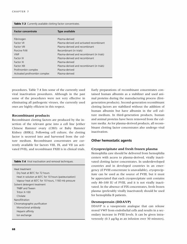
BLBK186-Key April 24, 2009 14:43
CHAPTER 7
Table 7.3 Currently available clotting factor concentrates.
Factor concentrate Type available
Fibrinogen Plasma-derived
Factor VII Plasma-derived and activated recombinant
Factor VIII Plasma-derived and recombinant
Porcine FVIII Recombinant (in trials)
VWF Plasma-derived and recombinant (in trials)
Factor IX Plasma-derived and recombinant
Factor XI Plasma-derived
Factor XIII Plasma-derived and recombinant (in trials)
Prothrombin complex Plasma-derived
Activated prothrombin complex Plasma-derived
procedures. Table 7.4 lists some of the currently used
viral inactivation procedures. Although in the past
some of the procedures were not very effective in
eliminating all pathogenic viruses, the currently used
ones are highly efficient in this respect.
Recombinant productsRecombinant clotting factors are produced by the in-
sertion of the relevant gene into a cell line [either
Chinese Hamster ovary (CHO) or Baby Hamster
Kidney (BHK)]. Following cell culture, the clotting
factor is secreted into and harvested from the cul-
ture medium. Recombinant concentrates are cur-
rently available for factors VIII, IX, and VII (as acti-
vated FVII), and recombinant FXIII is in clinical trials.
Table 7.4 Viral inactivation and removal techniques.
Heat treatment
Dry heat at 80˚C for 72 hours
Heat in solution at 60˚C for 10 hours (pasteurization)
Vapour heat at 60˚C for 10 hours, 1160 mb pressure
Solvent detergent treatment
TNBP and Tween
Triton X-100
Cholate
Nanofiltration
Chromatographic purification
Monoclonal antibody
Heparin affinity
Ion exchange
Early preparations of recombinant concentrates con-
tained human albumin as a stabilizer and used ani-
mal proteins during the manufacturing process (first-
generation products). Second-generation recombinant
clotting factors are stabilized without the addition of
human albumin but have albumin in the cell cul-
ture medium. In third-generation products, human
and animal proteins have been removed from the cul-
ture media. As for plasma-derived products, all recom-
binant clotting factor concentrates also undergo viral
inactivation.
Other hemostatic agents
Cryoprecipitate and fresh frozen plasmaHemophilia care should be delivered from hemophilia
centers with access to plasma-derived, virally inacti-
vated clotting factor concentrates. In underdeveloped
countries and in developed countries in an emer-
gency (if FVIII concentrate is unavailable), cryoprecip-
itate can be used as the source of FVIII, but it must
be appreciated that each cryoprecipitate unit contains
only 80–100 IU of FVIII, and it is not virally inacti-
vated. In the absence of FIX concentrates, fresh frozen
plasma (preferably virally inactivated) should be used
for hemophilia B patients.
Desmopressin (DDAVP)DDAVP is a vasopressin analogue that can release
stored VWF from endothelial cells and results in a sec-
ondary increase in FVIII levels. It can be given intra-
venously (0.3 µg/kg as an infusion over 30 minutes),
68
BLBK186-Key April 24, 2009 14:43
Hemophilia A and B
subcutaneously (0.3 µg/kg), or intranasally. It is use-
ful in the management of mild hemophilia A, type 1
VWD and some patients with platelet function de-
fects. DDAVP administration can be repeated over a
short period, but efficacy will then decrease because
of tachyphylaxis. However, a few days later, the en-
dothelial stores are replenished, and original efficacy
is reestablished.
Common adverse effects include a mild headache,
flushing, and fluid retention, so patients should be ad-
vised to reduce their fluid intake in the subsequent
12–24 hours. Because of the problem with fluid reten-
tion, DDAVP should be avoided in children under the
age of 2 years.
Tranexamic acidTranexamic acid is an antifibrinolytic agent that can be
given orally or intravenously. It is very useful where
there is mucosal bleeding and should be routinely ad-
ministered to hemophiliacs having dental extractions.
Complications of treatment
Despite the success of concentrate treatment, a num-
ber of complications occur; these are summarized in
Table 7.5 [12].
Inhibitor developmentAllo-antibodies develop in up to 30% of children with
severe hemophilia A who receive treatment with FVIII
concentrate. Although uncommon, they can also oc-
cur in patients with mild or moderate hemophilia A af-
ter treatment with factor VIII; in some instances, cross-
reacting with autologous factor VIII. They are rare in
hemophilia B patients (�3%). These antibodies (in-
hibitors) are more likely to develop:
Table 7.5 Complications of clotting factor therapy.
Allo-antibody formation – inhibitor development
Infections
HIV
Hepatitis A, B, C, D, G
Parvovirus B19
vCJD
Immune modulation
Thrombosis
Anaphylaxis
� before the age of 5 years;� within the first 50 treatment days;� in those of African descent;� where there is a family history of inhibitor de-
velopment; or� in patients with FVIII/IX gene deletions.
They are usually suspected when a previously ef-
fective treatment is no longer sufficient to achieve
hemostasis. The prolonged APTT does not normalize
in vitro after the addition of normal plasma, and con-
firmation is made with the Bethesda assay [13].
The treatment of acute bleeding in hemophiliacs
with inhibitors is difficult and expensive. It depends
on the level of the inhibitor and whether it is a low-
or a high-responding one.
High-responding patients develop a rapidly increas-
ing antibody level each time they are exposed to hu-
man FVIII. The two main types of treatment of acute
bleeding in these patients are:
1 Activated prothrombin complex concentrates, such
as FEIBA; and
2 Recombinant FVIIa (NovoSeven).
A recent comparative study found that the two
products are equally effective, but some patients re-
spond better to one versus the other [14]. Other
than through clinical response, there is currently
no reliable widely used method to monitor treat-
ment with these products in the laboratory; although
global assays, such as thrombin generation, thromboe-
lastometry, and thromboelastography, are showing
promise.
Porcine FVIII concentrate is also useful in patients
without a cross-reacting antibody to porcine FVIII. Al-
though currently not widely available, studies of re-
combinant porcine FVIII are underway.
In every patient with an inhibitor, the possibility of
elimination through immune tolerance should be con-
sidered. There are three immune tolerance protocols
available:
1 High-dose protocol: administers FVIII daily;
2 Low-dose protocol: alternate daily administration; or
3 Malmo protocol: FVIII is combined with intravenous
immunoglobulin, cyclophosphamide, and immunoad-
sorption or plasmapheresis.
The reported success rates from small series are
30–80%. Once an inhibitor has been eliminated,
the chance of it recurring is 15%. An international
immune tolerance induction study in patients with
69
BLBK186-Key April 24, 2009 14:43
CHAPTER 7
severe hemophilia A and inhibitors is underway com-
paring low- and high-dose immune tolerance regi-
mens (http://www.itistudy.com). Rituximab (a mon-
oclonal anti-CD20 antibody) treatment has been tried
in some hemophilia patients with inhibitors with vary-
ing success [15].
InfectionsThe viral inactivation of concentrates introduced
in 1985 was highly effective in eliminating most
transfusion-transmitted viruses. The risk of infection
in patients treated prior to 1985 was 25–70% for HIV,
100% for hepatitis C, and 50% for hepatitis B.
Human immunodeficiency virus (HIV)The transmission of HIV infections by plasma-derived
concentrates in the early 1980s has had a devastat-
ing effect in the lives of hemophiliacs. Approximately
two-thirds of the HIV-infected hemophiliacs have now
died, but in those still alive, the use of highly active an-
tiretroviral therapy (HAART) has allowed near normal
existence with immune reconstitution and a dramati-
cally reduced mortality.
Hepatitis C� 15% of patients infected cleared the virus naturally
(antibody-positive but PCR-negative).� 85% were chronically infected (persistence more
than 6 months).� Approximately 20–30% of infected patients have
evidence of cirrhosis.� 5–10% have developed liver failure or hepatocellu-
lar carcinoma.
Factors accelerating liver disease progression in-
clude:� time since infection;� older age at infection;� HIV coinfection; and� higher alcohol consumption.
Treatment with pegylated interferon and ribavirin
achieves cure of hepatitis C in 30–40% of those in-
fected with HCV genotype 1 and 70% of those infected
with genotype 2 or 3.
Hepatitis BApproximately 50% of hemophiliacs treated with
pooled plasma products prior to viral inactivation were
infected with hepatitis B virus, but most cleared the
virus spontaneously; less than 5% of these patients
show active chronic hepatitis B virus infection. All
non-immune and non-infected hemophiliacs should
be vaccinated against this virus.
Parvovirus B19This causes fifth disease in childhood, and most adults
show evidence of past infection. Although the disease
itself is relatively minor, its importance lies in the fact
that the virus is resistant to all currently used viral in-
activation techniques. The implication of this is that
unknown viruses can theoretically be transmitted by
all currently available plasma-derived clotting factor
concentrates, and this is one of the main reasons for
the introduction of recombinant concentrates in coun-
tries where alternative “safe” plasma-derived concen-
trates exist.
Variant Creutzfeldt–Jakob disease (vCJD)vCJD is a prion disease that is the human equiva-
lent of the bovine spongiform encephalopathy, which
was endemic in the British cow population in the
late 1980s and early 1990s. vCJD can be transmitted
through transfusion of fresh cellular components. A
significant number of UK hemophiliacs have been ex-
posed to plasma from donors who subsequently devel-
oped vCJD. Although no hemophiliac has ever devel-
oped clinical vCJD, in February 2009 it was reported
that vCJD related priors were identified in the spleen
of a hemophiliac who died from an unrelated cause.
This patient was treated with FVIII prepared from a
donor who subsequently developed vCJD.
Immune modulationIn vitro, it is possible to show that concentrates exert
an immunosuppressive effect. This has been observed
and reported in hemophiliacs, but this phenomenon
could have been a result of the chronic hepatitis C af-
fecting the hemophiliacs studied.
ThrombosisThrombosis is a rare complication that was well recog-
nized when prothrombin complex concentrates were
used to cover surgery in patients with hemophilia
B, prior to the addition of antithrombin and hep-
arin to the product. It is still seen in patients treated
70
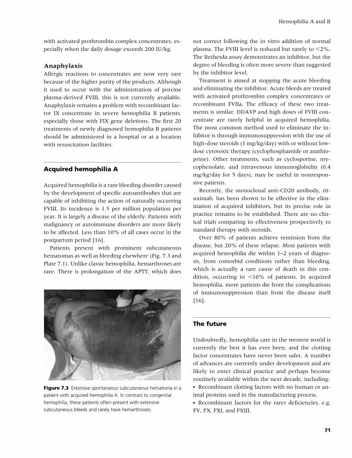
BLBK186-Key April 24, 2009 14:43
Hemophilia A and B
with activated prothrombin complex concentrates, es-
pecially when the daily dosage exceeds 200 IU/kg.
AnaphylaxisAllergic reactions to concentrates are now very rare
because of the higher purity of the products. Although
it used to occur with the administration of porcine
plasma-derived FVIII, this is not currently available.
Anaphylaxis remains a problem with recombinant fac-
tor IX concentrate in severe hemophilia B patients,
especially those with FIX gene deletions. The first 20
treatments of newly diagnosed hemophilia B patients
should be administered in a hospital or at a location
with resuscitation facilities.
Acquired hemophilia A
Acquired hemophilia is a rare bleeding disorder caused
by the development of specific autoantibodies that are
capable of inhibiting the action of naturally occurring
FVIII. Its incidence is 1.5 per million population per
year. It is largely a disease of the elderly. Patients with
malignancy or autoimmune disorders are more likely
to be affected. Less than 10% of all cases occur in the
postpartum period [16].
Patients present with prominent subcutaneous
hematomas as well as bleeding elsewhere (Fig. 7.3 and
Plate 7.1). Unlike classic hemophilia, hemarthroses are
rare. There is prolongation of the APTT, which does
Figure 7.3 Extensive spontaneous subcutaneous hematoma in a
patient with acquired hemophilia A. In contrast to congenital
hemophilia, these patients often present with extensive
subcutaneous bleeds and rarely have hemarthroses.
not correct following the in vitro addition of normal
plasma. The FVIII level is reduced but rarely to �2%.
The Bethesda assay demonstrates an inhibitor, but the
degree of bleeding is often more severe than suggested
by the inhibitor level.
Treatment is aimed at stopping the acute bleeding
and eliminating the inhibitor. Acute bleeds are treated
with activated prothrombin complex concentrates or
recombinant FVIIa. The efficacy of these two treat-
ments is similar. DDAVP and high doses of FVIII con-
centrate are rarely helpful in acquired hemophilia.
The most common method used to eliminate the in-
hibitor is through immunosuppression with the use of
high-dose steroids (1 mg/kg/day) with or without low-
dose cytotoxic therapy (cyclophosphamide or azathio-
prine). Other treatments, such as cyclosporine, my-
cophenolate, and intravenous immunoglobulin (0.4
mg/kg/day for 5 days), may be useful in nonrespon-
sive patients.
Recently, the monoclonal anti-CD20 antibody, rit-
uximab, has been shown to be effective in the elim-
ination of acquired inhibitors, but its precise role in
practice remains to be established. There are no clin-
ical trials comparing its effectiveness prospectively to
standard therapy with steroids.
Over 80% of patients achieve remission from the
disease, but 20% of these relapse. Most patients with
acquired hemophilia die within 1–2 years of diagno-
sis, from comorbid conditions rather than bleeding,
which is actually a rare cause of death in this con-
dition, occurring in �10% of patients. In acquired
hemophilia, more patients die from the complications
of immunosuppression than from the disease itself
[16].
The future
Undoubtedly, hemophilia care in the western world is
currently the best it has ever been, and the clotting
factor concentrates have never been safer. A number
of advances are currently under development and are
likely to enter clinical practice and perhaps become
routinely available within the next decade, including:� Recombinant clotting factors with no human or an-
imal proteins used in the manufacturing process.� Recombinant factors for the rarer deficiencies, e.g.
FV, FX, FXI, and FXIII.
71
BLBK186-Key April 24, 2009 14:43
CHAPTER 7
� Modified recombinant clotting factors with longer
half-lives.� Recombinant porcine FVIII for use in inhibitor
patients.� Recombinant VWF concentrate.� Agents to be coadministered with DDAVP to im-
prove its efficacy (e.g. interleukin 11).� Embryo selection in hemophilia carriers to exclude
implantation of affected embryos.� Gene therapy where a normal FVIII or FIX gene is
introduced in patients with hemophilia.
References
1 Mannucci PM, Tuddenham EG. The hemophilias:
from royal genes to gene therapy. N Engl J Med
2001;344:1773–9.
2 Shen BW, Spiegel PC, Chang CH, et al. The tertiary
structure and domain organization of coagulation FVIII.
Blood 2008;111:1240–7.
3 Leuer M, Oldenburg J, Lavergne JM, et al. Somatic mo-
saicism in hemophilia A: a fairly common event. Am J
Hum Genet 2001;69(1):75–87.
4 Lee CA, Chi C, Pavord SA, et al. The obstetric and
gynaecological management of women with inherited
bleeding disorders: review with guidelines produced by
a taskforce of UK Haemophilia Centre Doctors Organi-
zation. Haemophilia 2006;12:301–36.
5 Michaelides K, Tuddenham EG, Turner C, Lavender B,
Lavery SA. Liver birth following the first mutation spe-
cific preimplantation genetic diagnosis for haemophilia
A. Thromb Haemost 2006;95:373–9.
6 Ljung RC. Intracranial haemorrhage in haemophilia A
and B. Br J Haematol 2008;140(4):378–84.
7 Llinas A. The role of synovectomy in the manage-
ment of a target joint. Haemophilia 2008;14(Suppl 3):
177–80.
8 Rodriguez-Merchan EC. Haemophilic cysts (pseudotu-
mours). Haemophilia 2002;8:393–401.
9 Ljung R. Paediatric care of the child with hemophilia.
Haemophilia 2002;8:178–82.
10 Manco-Johnson MJ, Abshire TC, Shapiro AD, et al.
Prophylaxis versus episodic treatment to prevent joint
disease in boys with severe hemophilia. N Engl J Med
2007;357:535–44.
11 Keeling D, Tait R, Makris M. Guideline on the selection
and use of therapeutic products to treat haemophilia
and other hereditary bleeding disorders. Haemophilia
2008;14:671–84.
12 Mannucci PM. Hemophilia and related bleeding disor-
ders: a story of dismay and success. Hematology (Am Soc
Hematol Educ Program) 2002:1–9.
13 Hay CR, Brown S, Collins PW, Keeling DM, Liesner
R. The diagnosis and management of factor VIII and
IX inhibitors: a guideline from the UK Haemophilia
Centre Doctors’ Organization. Br J Haematol 2006;133:
591–605.
14 Astermark J, Donfield DM, DiMichele DM, et al. A ran-
domised comparison of bypassing agents in hemophilia
complicated by an inhibitor: the FEIBA Novo-
Seven comparative (FENOC) study. Blood 2007;109:
546–57.
15 Franchini M, Mengoli C, Lippi G, et al. Immune tol-
erance with rituximab in congenital haemophilia with
inhibitors: a systematic literature review based on indi-
vidual patients’ analysis. Haemophilia 2008;14:903–12.
16 Collins PW, Hirsch S, Baglin TP, et al. Acquired
haemophilia A in the United Kingdom: a 2 year
national surveillance study of the United Kingdom
Haemophilia Centre Doctors’ Organisation. Blood
2007;109:1870–7.
72
BLBK186-Key April 11, 2009 12:55
8 Von Willebrand diseaseGiancarlo Castaman, Alberto Tosetto, and Francesco Rodeghiero
Introduction: the von Willebrand factor
Von Willebrand disease (VWD) is caused by a defi-
ciency and/or abnormality of von Willebrand factor
(VWF) and represents the most frequent inherited
bleeding disorder [1]. VWF is synthesized by endothe-
lial cells and megakaryocytes. Its gene includes about
178 kilobases with 52 exons and is located at chro-
mosome 12p13.2. A non-coding homologous pseudo-
gene has been identified in chromosome 22, which
spans the gene sequence from exon 23 to 34 [2]. The
primary product of the VWF gene is a 2813-amino-
acid protein comprising a signal peptide of 22 amino
acids (also called pre-peptide), a large pro-peptide of
741 amino acids (also called pro-peptide), and a ma-
ture VWF subunit of 2050 amino acids. Four types
of repeated molecular domains (D1, D2, D′, D3, A1,
A2, A3, D4, B, C1, C2) of cDNA are responsible
for the different binding functions of the molecule.
The building block of VWF multimers is a dimer
made by two single-chain pro-VWF molecules, joined
through disulphide bonds within their C-terminal re-
gion. This reaction occurs after the cleavage of the
signal peptide and the subsequent translocation and
glycosylation of the precursor molecules into the en-
doplasmic reticulum. The pro-VWF dimers are then
transported to the Golgi apparatus, where, after fur-
ther post-translational modifications, including pro-
cessing of high mannose oligosaccharides, they are
polymerized into very large molecules up to a molec-
ular weight of 20,000 × 103 through disulphide bonds
connecting the two N-terminal ends of each dimer.
After polymerization, pro-VWF multimers move to
the trans-Golgi network, where the pro-peptide (also
called VWAgII), is cleaved off by a paired amino acid-
cleaving enzyme (PACE or furin), and remains, at
least within the cell, noncovalently associated with
VWF [3].
VWF is secreted from the cell via a constitutive and
a regulated pathway. The latter is used for a rapid
stimuli-induced release (e.g. by desmopressin through
its binding to vasopressin V2 receptor of endothe-
lial cells) from specialized storage organelles of en-
dothelial cells known as Weibel-Palade bodies. Only
Weibel-Palade bodies or α-granules in platelets con-
tain fully processed and functional VWF with “unusu-
ally large” multimers. These large multimers are usu-
ally not found in the circulation. Indeed, a specific
plasma protease acts on VWF multimers released from
the cell, cleaving the VWF subunit at the bond be-
tween Tyr 1605 and Met 1606 (Tyr 842 and Met 843 of
the mature subunit), thus creating the full spectrum of
circulating VWF species, ranging from the single dimer
to multimers made of up to 20 dimers in each VWF
multimer [4].
In addition to endothelial cells, megakaryocytes,
and platelets, VWF is present in the subendothelial
matrix, where it is bound through specific regions in
its A1 and A3 domains to different types of collagen.
Physiological role of VWF
VWF is essential for platelet–subendothelium adhe-
sion and platelet-to-platelet cohesion and aggregation
in vessels with elevated shear stress [5]. This function
is partially explored in vivo by measuring the bleed-
ing time. Adhesion is promoted by the interaction of
a region of the A1 domain of VWF with platelet GpIb.
It is thought that high shear stress is able to activate
the A1 domain of the collagen-bound VWF by stretch-
ing VWF multimers into a filamentous form. The in-
teraction between GPIb and VWF can be mimicked
73
BLBK186-Key April 11, 2009 12:55
CHAPTER 8
by the addition of the antibiotic ristocetin, which pro-
motes the binding of VWF to GPIb present on fresh
or formalin-fixed platelet suspensions. Aggregation of
platelets within the growing hemostatic plug is pro-
moted by the interaction with a second receptor on
platelets, the GPIIb-IIIa (or integrin αIIbβ3), which af-
ter activation binds to VWF and fibrinogen, recruiting
more platelets into a stable plug. Both of these bind-
ing activities of VWF are highest in the largest VWF
multimers.
VWF is the carrier of factor VIII (FVIII) in plasma.
VWF protects FVIII from proteolytic degradation, pro-
longing its half-life in the circulation and efficiently
localizing it at the site of vascular injury [6]. Each
monomer of VWF has one binding domain, located in
the first 272 amino acids of the mature subunit (D′
domain), that is able to bind one FVIII molecule. In
vivo, however, only 1–2% of available monomers are
occupied by FVIII. This explains why high-molecular-
weight multimers are not essential for the carrier
function of FVIII, although one would expect that
molecules of the highest molecular weight should be
most effective in localizing FVIII at the site of vascular
injury. In any case, any change in plasma VWF level is
usually associated with a concordant change in FVIII
plasma concentration.
Classification of VWD
The current nomenclature of the factor VIII/VWF
complex, as recommended by the International So-
ciety on Thrombosis and Hemostasis, is summarized
in Table 8.1 [7]. The current revised classification of
Table 8.1 Recommended nomenclature of FVIII/VWF
complex.
Factor VIIIProtein VIII
Antigen VIII:Ag
Function VIII:C
Von Willebrand factorMature protein VWF
Antigen VWF:Ag
Ristocetin cofactor activity VWF:RCo
Collagen binding capacity VWF:CB
FVIII binding capacity VWF:FVIIIB
Table 8.2 Classification of VWD (modified from Sadler
et al. [7]).
Quantitative deficiency of VWF� Type 1: Partial quantitative deficiency of VWF� Type 3: Virtually complete deficiency of VWF
Qualitative deficiency of VWF� Type 2: Qualitative deficiency of VWF
– Type 2A: Qualitative variants with decreased
platelet-dependent function associated with the absence
of high- and intermediate-molecular-weight VWF
multimers
– Type 2B: Qualitative variants with increased affinity for
platelet GPIb, with the absence of HMW VWF multimers
– Type 2M: Qualitative variants with decreased
platelet-dependent function not caused by the absence
of HMW VWF multimers
– Type 2N: Qualitative variants with markedly decreased
affinity for FVIII
VWD identifies two major categories, characterized by
quantitative (type 1 and 3) or qualitative (type 2) VWF
defects (Table 8.2). Partial quantitative deficiency of
VWF in plasma and/or platelets identifies type 1 VWD,
whereas type 3 VWD is characterized by total absence
or trace amounts of VWF in plasma and platelets. Type
1 is easily distinguished from type 3 by its milder VWF
deficiency (usually in the range of 20–40%), the au-
tosomal dominant inheritance pattern, and the pres-
ence of milder bleeding symptoms. Among type 2
variants, four subtypes have been identified reflect-
ing different pathophysiological mechanisms. Classical
type 2A is characterized by the absence of high- and
intermediate-molecular-weight (HMW) multimers of
VWF in plasma. Type 2B is characterized by an in-
creased affinity of VWF for platelet GpIb, causing re-
moval of HMW multimers from plasma. As a conse-
quence, ristocetin-induced platelet aggregation (RIPA)
in platelet-rich plasma from these patients occurs at
low ristocetin concentrations. The identification of
variants with decreased platelet-dependent function
and the presence of normal multimers on gel elec-
trophoresis have required the addition of a new sub-
type, called 2M. Type 2N (Normandy) shows a full
array of multimers because the defect lies in the N-
terminal region of the VWF, where the binding do-
main for FVIII resides. This subtype is phenotypically
identified only by tests exploiting FVIII–VWF binding.
74
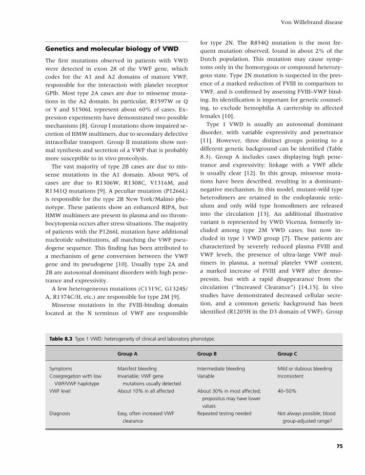
BLBK186-Key April 11, 2009 12:55
Von Willebrand disease
Genetics and molecular biology of VWD
The first mutations observed in patients with VWD
were detected in exon 28 of the VWF gene, which
codes for the A1 and A2 domains of mature VWF,
responsible for the interaction with platelet receptor
GPIb. Most type 2A cases are due to missense muta-
tions in the A2 domain. In particular, R1597W or Q
or Y and S1506L represent about 60% of cases. Ex-
pression experiments have demonstrated two possible
mechanisms [8]. Group I mutations show impaired se-
cretion of HMW multimers, due to secondary defective
intracellular transport. Group II mutations show nor-
mal synthesis and secretion of a VWF that is probably
more susceptible to in vivo proteolysis.
The vast majority of type 2B cases are due to mis-
sense mutations in the A1 domain. About 90% of
cases are due to R1306W, R1308C, V1316M, and
R1341Q mutations [9]. A peculiar mutation (P1266L)
is responsible for the type 2B New York/Malmo phe-
notype. These patients show an enhanced RIPA, but
HMW multimers are present in plasma and no throm-
bocytopenia occurs after stress situations. The majority
of patients with the P1266L mutation have additional
nucleotide substitutions, all matching the VWF pseu-
dogene sequence. This finding has been attributed to
a mechanism of gene conversion between the VWF
gene and its pseudogene [10]. Usually type 2A and
2B are autosomal dominant disorders with high pene-
trance and expressivity.
A few heterogeneous mutations (C1315C, G1324S/
A, R1374C/H, etc.) are responsible for type 2M [9].
Missense mutations in the FVIII-binding domain
located at the N terminus of VWF are responsible
Table 8.3 Type 1 VWD: heterogeneity of clinical and laboratory phenotype.
Group A Group B Group C
Symptoms Manifest bleeding Intermediate bleeding Mild or dubious bleeding
Cosegregation with low
VWF/VWF haplotype
Invariable; VWF gene
mutations usually detected
Variable Inconsistent
VWF level About 10% in all affected About 30% in most affected;
propositus may have lower
values
40–50%
Diagnosis Easy, often increased VWF
clearance
Repeated testing needed Not always possible; blood
group-adjusted range?
for type 2N. The R854Q mutation is the most fre-
quent mutation observed, found in about 2% of the
Dutch population. This mutation may cause symp-
toms only in the homozygous or compound heterozy-
gous state. Type 2N mutation is suspected in the pres-
ence of a marked reduction of FVIII in comparison to
VWF, and is confirmed by assessing FVIII–VWF bind-
ing. Its identification is important for genetic counsel-
ing, to exclude hemophilia A carriership in affected
females [10].
Type 1 VWD is usually an autosomal dominant
disorder, with variable expressivity and penetrance
[11]. However, three distinct groups pointing to a
different genetic background can be identified (Table
8.3). Group A includes cases displaying high pene-
trance and expressivity: linkage with a VWF allele
is usually clear [12]. In this group, missense muta-
tions have been described, resulting in a dominant-
negative mechanism. In this model, mutant-wild type
heterodimers are retained in the endoplasmic retic-
ulum and only wild type homodimers are released
into the circulation [13]. An additional illustrative
variant is represented by VWD Vicenza, formerly in-
cluded among type 2M VWD cases, but now in-
cluded in type 1 VWD group [7]. These patients are
characterized by severely reduced plasma FVIII and
VWF levels, the presence of ultra-large VWF mul-
timers in plasma, a normal platelet VWF content,
a marked increase of FVIII and VWF after desmo-
pressin, but with a rapid disappearance from the
circulation (“Increased Clearance”) [14,15]. In vivo
studies have demonstrated decreased cellular secre-
tion, and a common genetic background has been
identified (R1205H in the D3 domain of VWF). Group
75
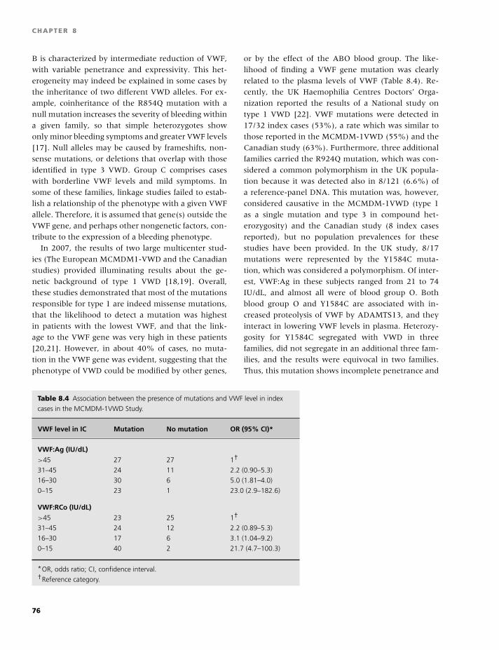
BLBK186-Key April 11, 2009 12:55
CHAPTER 8
B is characterized by intermediate reduction of VWF,
with variable penetrance and expressivity. This het-
erogeneity may indeed be explained in some cases by
the inheritance of two different VWD alleles. For ex-
ample, coinheritance of the R854Q mutation with a
null mutation increases the severity of bleeding within
a given family, so that simple heterozygotes show
only minor bleeding symptoms and greater VWF levels
[17]. Null alleles may be caused by frameshifts, non-
sense mutations, or deletions that overlap with those
identified in type 3 VWD. Group C comprises cases
with borderline VWF levels and mild symptoms. In
some of these families, linkage studies failed to estab-
lish a relationship of the phenotype with a given VWF
allele. Therefore, it is assumed that gene(s) outside the
VWF gene, and perhaps other nongenetic factors, con-
tribute to the expression of a bleeding phenotype.
In 2007, the results of two large multicenter stud-
ies (The European MCMDM1-VWD and the Canadian
studies) provided illuminating results about the ge-
netic background of type 1 VWD [18,19]. Overall,
these studies demonstrated that most of the mutations
responsible for type 1 are indeed missense mutations,
that the likelihood to detect a mutation was highest
in patients with the lowest VWF, and that the link-
age to the VWF gene was very high in these patients
[20,21]. However, in about 40% of cases, no muta-
tion in the VWF gene was evident, suggesting that the
phenotype of VWD could be modified by other genes,
Table 8.4 Association between the presence of mutations and VWF level in index
cases in the MCMDM-1VWD Study.
VWF level in IC Mutation No mutation OR (95% CI)*
VWF:Ag (IU/dL)
>45 27 27 1†
31–45 24 11 2.2 (0.90–5.3)
16–30 30 6 5.0 (1.81–4.0)
0–15 23 1 23.0 (2.9–182.6)
VWF:RCo (IU/dL)
>45 23 25 1†
31–45 24 12 2.2 (0.89–5.3)
16–30 17 6 3.1 (1.04–9.2)
0–15 40 2 21.7 (4.7–100.3)
*OR, odds ratio; CI, confidence interval.†Reference category.
or by the effect of the ABO blood group. The like-
lihood of finding a VWF gene mutation was clearly
related to the plasma levels of VWF (Table 8.4). Re-
cently, the UK Haemophilia Centres Doctors’ Orga-
nization reported the results of a National study on
type 1 VWD [22]. VWF mutations were detected in
17/32 index cases (53%), a rate which was similar to
those reported in the MCMDM-1VWD (55%) and the
Canadian study (63%). Furthermore, three additional
families carried the R924Q mutation, which was con-
sidered a common polymorphism in the UK popula-
tion because it was detected also in 8/121 (6.6%) of
a reference-panel DNA. This mutation was, however,
considered causative in the MCMDM-1VWD (type 1
as a single mutation and type 3 in compound het-
erozygosity) and the Canadian study (8 index cases
reported), but no population prevalences for these
studies have been provided. In the UK study, 8/17
mutations were represented by the Y1584C muta-
tion, which was considered a polymorphism. Of inter-
est, VWF:Ag in these subjects ranged from 21 to 74
IU/dL, and almost all were of blood group O. Both
blood group O and Y1584C are associated with in-
creased proteolysis of VWF by ADAMTS13, and they
interact in lowering VWF levels in plasma. Heterozy-
gosity for Y1584C segregated with VWD in three
families, did not segregate in an additional three fam-
ilies, and the results were equivocal in two families.
Thus, this mutation shows incomplete penetrance and
76
BLBK186-Key April 11, 2009 12:55
Von Willebrand disease
does not consistently segregate with VWD. As pre-
viously demonstrated in the Canadian study [19], a
founder effect is also likely to occur in the UK fam-
ilies. The Y1584C mutation alone was identified in
10 index cases [22], and in compound heterozygosity
in an additional 3 cases of the MCMDM-1VWD cohort
(8% of the whole index cases), with the same wide
range of VWF levels [18]. Notwithstanding these gray
areas that still require further studies, great progress
has been made in elucidating the molecular bases of a
large proportion of patients with type 1 VWD.
About 60% of the variation in VWF plasma is due
to genetic factors, with VWF level 25–35% lower in
type-O subjects than in non-O individuals [23]. Blood
group plays a major role in subjects with VWF levels
at the lower end of the normal range, in whom heri-
tability is less predictable.
In type 3 VWD, in addition to mechanisms shared
with some type 1 cases (see above), partial or total
gene deletions have also been reported [24]. Notably,
homozygosity for gene deletion may be associated
with the appearance of neutralizing antibodies against
VWF, which may render replacement therapy ineffec-
tive and stimulate anaphylactic reaction upon treat-
ment. In general, mutations may be scattered over
the entire gene, but some mutations (e.g. 2680delC
or R2535X) are particularly recurrent in Northern
Europe. Several stop codon mutations, either in ho-
mozygotes or compound heterozygotes, have also
been reported.
Prevalence and frequency of subtypesof VWD
Until the late 1980s, estimates of the prevalence of
VWD were based on the number of patients registered
at specialized centers, with figures ranging from 4 to
10 cases/100,000 inhabitants. It is generally assumed
that the number of persons with symptomatic VWD,
requiring specific treatment, is at least 100 per million.
A few studies estimated the prevalence of VWD by
screening small populations using formal, standard-
ized criteria. A prevalence approaching 1% has been
demonstrated, without ethnic differences [25]. How-
ever, the large majority of cases diagnosed by pop-
ulation studies appear to have a mild disease, and
most of these subjects were never referred for detailed
hemostatic evaluation. It remains unknown what pro-
portion of these cases is the effect of a gene(s) out-
side the VWF gene influencing the circulating level of
VWF [26].
About 70% of VWD cases appear to have type 1
by Center series. These estimates are obviously biased
because it is expected that many type 1 cases with-
out major symptoms are not referred for evaluation,
whereas almost all severe type 3 cases are followed
at a specialized center. Indeed, recent results from the
MCMDM-1 VWD study demonstrated by an accurate
VWF multimeric evaluation that many of the patients
previously identified as type 1 VWD had subtle multi-
meric abnormalities that suggested type 2 VWD [27].
However, for most of them, this evidence did not af-
fect their treatment because they showed complete re-
sponse to desmopressin administration.
In contrast to the above-reported percentages, al-
most all cases were represented by type 1 in popula-
tion studies.
Clinical manifestations
Clinical expression of VWD is usually mild in type
1, with increasing severity in type 2 and type 3.
However, in some families, variable severity of bleed-
ing manifestations is evident, underlying the different
molecular basis responsible for the diverse phenotypes
of this disorder and its variable penetrance. In gen-
eral, the severity of bleeding correlates with the de-
gree of the reduction of FVIII:C, but not with the mag-
nitude of BT prolongation or with ABO blood type of
the patient. Mucocutaneous bleeding (epistaxis, men-
orrhagia, easy bruising) is a typical, prominent man-
ifestation of the disease and may affect the quality
of life. VWD may be highly prevalent in patients
with isolated menorrhagia. Females with VWD may
require treatment with antifibrinolytics, iron supple-
mentation, or an estroprogestinic pill to control heavy
menses. Bleeding after dental extraction is the most
frequent postoperative bleeding manifestation. Be-
cause FVIII:C is usually only mildly reduced, manifes-
tations of a severe coagulation defect (hemarthrosis,
deep muscle hematoma) are rarely observed in type
1 VWD and are mainly posttraumatic. On the con-
trary, in type 3 VWD, the severity of bleeding may
sometimes be similar to that of hemophilia. Bleeding
77
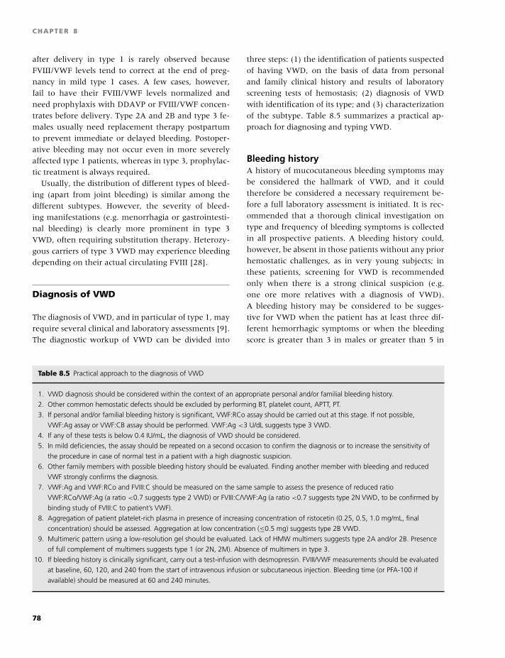
BLBK186-Key April 11, 2009 12:55
CHAPTER 8
after delivery in type 1 is rarely observed because
FVIII/VWF levels tend to correct at the end of preg-
nancy in mild type 1 cases. A few cases, however,
fail to have their FVIII/VWF levels normalized and
need prophylaxis with DDAVP or FVIII/VWF concen-
trates before delivery. Type 2A and 2B and type 3 fe-
males usually need replacement therapy postpartum
to prevent immediate or delayed bleeding. Postoper-
ative bleeding may not occur even in more severely
affected type 1 patients, whereas in type 3, prophylac-
tic treatment is always required.
Usually, the distribution of different types of bleed-
ing (apart from joint bleeding) is similar among the
different subtypes. However, the severity of bleed-
ing manifestations (e.g. menorrhagia or gastrointesti-
nal bleeding) is clearly more prominent in type 3
VWD, often requiring substitution therapy. Heterozy-
gous carriers of type 3 VWD may experience bleeding
depending on their actual circulating FVIII [28].
Diagnosis of VWD
The diagnosis of VWD, and in particular of type 1, may
require several clinical and laboratory assessments [9].
The diagnostic workup of VWD can be divided into
three steps: (1) the identification of patients suspected
of having VWD, on the basis of data from personal
and family clinical history and results of laboratory
screening tests of hemostasis; (2) diagnosis of VWD
with identification of its type; and (3) characterization
of the subtype. Table 8.5 summarizes a practical ap-
proach for diagnosing and typing VWD.
Bleeding historyA history of mucocutaneous bleeding symptoms may
be considered the hallmark of VWD, and it could
therefore be considered a necessary requirement be-
fore a full laboratory assessment is initiated. It is rec-
ommended that a thorough clinical investigation on
type and frequency of bleeding symptoms is collected
in all prospective patients. A bleeding history could,
however, be absent in those patients without any prior
hemostatic challenges, as in very young subjects; in
these patients, screening for VWD is recommended
only when there is a strong clinical suspicion (e.g.
one ore more relatives with a diagnosis of VWD).
A bleeding history may be considered to be sugges-
tive for VWD when the patient has at least three dif-
ferent hemorrhagic symptoms or when the bleeding
score is greater than 3 in males or greater than 5 in
Table 8.5 Practical approach to the diagnosis of VWD
1. VWD diagnosis should be considered within the context of an appropriate personal and/or familial bleeding history.
2. Other common hemostatic defects should be excluded by performing BT, platelet count, APTT, PT.
3. If personal and/or familial bleeding history is significant, VWF:RCo assay should be carried out at this stage. If not possible,
VWF:Ag assay or VWF:CB assay should be performed. VWF:Ag <3 U/dL suggests type 3 VWD.
4. If any of these tests is below 0.4 IU/mL, the diagnosis of VWD should be considered.
5. In mild deficiencies, the assay should be repeated on a second occasion to confirm the diagnosis or to increase the sensitivity of
the procedure in case of normal test in a patient with a high diagnostic suspicion.
6. Other family members with possible bleeding history should be evaluated. Finding another member with bleeding and reduced
VWF strongly confirms the diagnosis.
7. VWF:Ag and VWF:RCo and FVIII:C should be measured on the same sample to assess the presence of reduced ratio
VWF:RCo/VWF:Ag (a ratio <0.7 suggests type 2 VWD) or FVIII:C/VWF:Ag (a ratio <0.7 suggests type 2N VWD, to be confirmed by
binding study of FVIII:C to patient’s VWF).
8. Aggregation of patient platelet-rich plasma in presence of increasing concentration of ristocetin (0.25, 0.5, 1.0 mg/mL, final
concentration) should be assessed. Aggregation at low concentration (≤0.5 mg) suggests type 2B VWD.
9. Multimeric pattern using a low-resolution gel should be evaluated. Lack of HMW multimers suggests type 2A and/or 2B. Presence
of full complement of multimers suggests type 1 (or 2N, 2M). Absence of multimers in type 3.
10. If bleeding history is clinically significant, carry out a test-infusion with desmopressin. FVIII/VWF measurements should be evaluated
at baseline, 60, 120, and 240 from the start of intravenous infusion or subcutaneous injection. Bleeding time (or PFA-100 if
available) should be measured at 60 and 240 minutes.
78
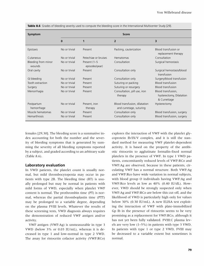
BLBK186-Key April 11, 2009 12:55
Von Willebrand disease
Table 8.6 Grades of bleeding severity used to compute the bleeding score in the International Multicenter Study [29].
Symptom Score
0 1 2 3
Epistaxis No or trivial Present Packing, cauterization Blood transfusion or
replacement therapy
Cutaneous No or trivial Petechiae or bruises Hematomas Consultation
Bleeding from minor
wounds
No or trivial Present (1–5
episodes/year)
Consultation Surgical hemostasis
Oral cavity No or trivial Present Consultation only Surgical hemostasis/blood
transfusion
GI bleeding No or trivial Present Consultation only Surgery/blood transfusion
Tooth extraction No or trivial Present Suturing or packing Blood transfusion
Surgery No or trivial Present Suturing or resurgery Blood transfusion
Menorrhagia No or trivial Present Consultation, pill use, iron
therapy
Blood transfusion,
hysterectomy, Dilatation
& Currettage
Postpartum
hemorrhage
No or trivial Present, iron
therapy
Blood transfusion, dilatation
and curretage, suturing
Hysterectomy
Muscle hematomas No or trivial Present Consultation only Blood transfusion, surgery
Hemarthrosis No or trivial Present Consultation only Blood transfusion, surgery
females [29,30]. The bleeding score is a summative in-
dex accounting for both the number and the sever-
ity of bleeding symptoms that is generated by sum-
ming the severity of all bleeding symptoms reported
by a subject, and graded according to an arbitrary scale
(Table 8.6).
Laboratory evaluationIn VWD patients, the platelet count is usually nor-
mal, but mild thrombocytopenia may occur in pa-
tients with type 2B. The bleeding time (BT) is usu-
ally prolonged but may be normal in patients with
mild forms of VWD, especially when platelet VWF
content is normal. The prothrombin time (PT) is nor-
mal, whereas the partial thromboplastin time (PTT)
may be prolonged to a variable degree, depending
on the plasma FVIII levels. Whatever the results of
these screening tests, VWD diagnosis always requires
the demonstration of reduced VWF antigen and/or
activity.
VWF antigen (VWF:Ag) is unmeasurable in type 3
VWD (below 3% or 0.03 IU/mL), whereas it is de-
creased in type 1 and low–normal in type 2 VWD.
The assay for ristocetin cofactor activity (VWF:RCo)
explores the interaction of VWF with the platelet gly-
coprotein Ib/IX/V complex, and it is still the stan-
dard method for measuring VWF platelet-dependent
activity. It is based on the property of the antibi-
otic ristocetin to agglutinate formalin-fixed normal
platelets in the presence of VWF. In type 1 VWD pa-
tients, concomitantly reduced levels of VWF:RCo and
VWF:Ag are observed, because in these patients, cir-
culating VWF has a normal structure. Both VWF:Ag
and VWF:Rco have wide variation in normal subjects,
with blood group O individuals having VWF:Ag and
VWF:Rco levels as low as 40% (0.40 IU/dL). How-
ever, VWD should be strongly suspected only when
VWF:Ag and VWF:RCo are below this cut-off, and the
likelihood of VWD is particularly high only for values
below 30% (0.30 IU/mL). A new ELISA test exploit-
ing the interaction of VWF with plate-immobilized
Gp Ib in the presence of ristocetin seems to be very
promising as a replacement for VWF:RCo, although it
has not yet been fully validated. FVIII:C plasma lev-
els are very low (1–5%) in patients with type 3 VWD.
In patients with type 1 or type 2 VWD, FVIII may
be decreased to a variable extent but sometimes is
normal.
79
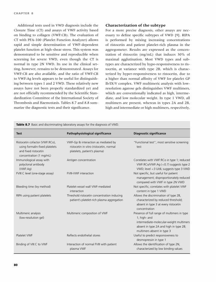
BLBK186-Key April 11, 2009 12:55
CHAPTER 8
Additional tests used in VWD diagnosis include the
Closure Time (CT) and assays of VWF activity based
on binding to collagen (VWF:CB). The evaluation of
CT with PFA-100 (Platelet Function Analyzer) allows
rapid and simple determination of VWF-dependent
platelet function at high-shear stress. This system was
demonstrated to be sensitive and reproducible when
screening for severe VWD, even though the CT is
normal in type 2N VWD. Its use in the clinical set-
ting, however, remains to be demonstrated. Assays for
VWF:CB are also available, and the ratio of VWF:CB
to VWF:Ag levels appears to be useful for distinguish-
ing between types 1 and 2 VWD. These relatively new
assays have not been properly standardized yet and
are not officially recommended by the Scientific Stan-
dardization Committee of the International Society of
Thrombosis and Haemostasis. Tables 8.7 and 8.8 sum-
marize the diagnostic tests and their significance.
Characterization of the subtypeFor a more precise diagnosis, other assays are nec-
essary to define specific subtypes of VWD [9]. RIPA
is performed by mixing increasing concentrations
of ristocetin and patient platelet-rich plasma in the
aggregometer. Results are expressed as the concen-
tration of ristocetin (mg/mL) that induces 30% of
maximal agglutination. Most VWD types and sub-
types are characterized by hypo-responsiveness to ris-
tocetin, at variance with type 2B, which is charac-
terized by hyper-responsiveness to ristocetin, due to
a higher than normal affinity of VWF for platelet GP
Ib/IX/V complex. VWF multimeric analysis with low-
resolution agarose gels distinguishes VWF multimers,
which are conventionally indicated as high, interme-
diate, and low molecular weight. In type 1 VWD, all
multimers are present, whereas in types 2A and 2B,
high and intermediate or high multimers, respectively,
Table 8.7 Basic and discriminating laboratory assays for the diagnosis of VWD.
Test Pathophysiological significance Diagnostic significance
Ristocetin cofactor (VWF:RCo),
using formalin-fixed platelets
and fixed ristocetin
concentration (1 mg/mL)
VWF-Gp Ib interaction as mediated by
ristocetin in vitro (ristocetin, normal
platelets, patient’s plasma)
“Functional test”; most sensitive screening
test
Immunological assay with
polyclonal antibody
(VWF:Ag)
Antigen concentration Correlates with VWF:RCo in type 1; reduced
VWF:RCo/VWF:Ag (<0.7) suggests type 2
VWD; level <3 U/dL suggests type 3 VWD
FVIII:C level (one-stage assay) FVIII-VWF interaction Not specific, but useful for patient
management; disproportionately reduced
compared with VWF in type 2N VWD
Bleeding time (Ivy method) Platelet-vessel wall VWF-mediated
interaction
Not specific; correlates with platelet VWF
content in type 1 VWD
RIPA using patient platelets Threshold ristocetin concentration inducing
patient’s platelet-rich plasma aggregation
Allows the discrimination of type 2B,
characterized by reduced threshold;
absent in type 3 at every ristocetin
concentration
Multimeric analysis
(low-resolution gel)
Multimeric composition of VWF Presence of full range of multimers in type
1; high- and
intermediate-molecular-weight multimers
absent in type 2A and high in type 2B;
multimers absent in type 3
Platelet VWF Reflects endothelial stores Useful to predict responsiveness to
desmopressin in type 1
Binding of VIII:C to VWF Interaction of normal FVIII with patient
plasma VWF
Allows the identification of type 2N,
characterized by low binding values
80
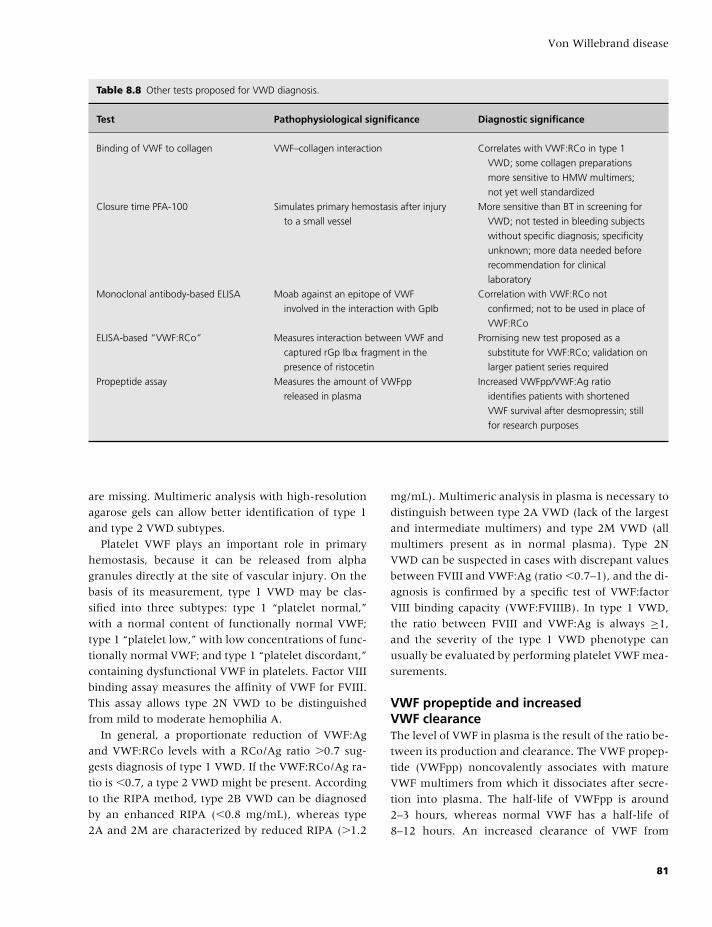
BLBK186-Key April 11, 2009 12:55
Von Willebrand disease
Table 8.8 Other tests proposed for VWD diagnosis.
Test Pathophysiological significance Diagnostic significance
Binding of VWF to collagen VWF–collagen interaction Correlates with VWF:RCo in type 1
VWD; some collagen preparations
more sensitive to HMW multimers;
not yet well standardized
Closure time PFA-100 Simulates primary hemostasis after injury
to a small vessel
More sensitive than BT in screening for
VWD; not tested in bleeding subjects
without specific diagnosis; specificity
unknown; more data needed before
recommendation for clinical
laboratory
Monoclonal antibody-based ELISA Moab against an epitope of VWF
involved in the interaction with GpIb
Correlation with VWF:RCo not
confirmed; not to be used in place of
VWF:RCo
ELISA-based “VWF:RCo” Measures interaction between VWF and
captured rGp Ibα fragment in the
presence of ristocetin
Promising new test proposed as a
substitute for VWF:RCo; validation on
larger patient series required
Propeptide assay Measures the amount of VWFpp
released in plasma
Increased VWFpp/VWF:Ag ratio
identifies patients with shortened
VWF survival after desmopressin; still
for research purposes
are missing. Multimeric analysis with high-resolution
agarose gels can allow better identification of type 1
and type 2 VWD subtypes.
Platelet VWF plays an important role in primary
hemostasis, because it can be released from alpha
granules directly at the site of vascular injury. On the
basis of its measurement, type 1 VWD may be clas-
sified into three subtypes: type 1 “platelet normal,”
with a normal content of functionally normal VWF;
type 1 “platelet low,” with low concentrations of func-
tionally normal VWF; and type 1 “platelet discordant,”
containing dysfunctional VWF in platelets. Factor VIII
binding assay measures the affinity of VWF for FVIII.
This assay allows type 2N VWD to be distinguished
from mild to moderate hemophilia A.
In general, a proportionate reduction of VWF:Ag
and VWF:RCo levels with a RCo/Ag ratio �0.7 sug-
gests diagnosis of type 1 VWD. If the VWF:RCo/Ag ra-
tio is �0.7, a type 2 VWD might be present. According
to the RIPA method, type 2B VWD can be diagnosed
by an enhanced RIPA (�0.8 mg/mL), whereas type
2A and 2M are characterized by reduced RIPA (�1.2
mg/mL). Multimeric analysis in plasma is necessary to
distinguish between type 2A VWD (lack of the largest
and intermediate multimers) and type 2M VWD (all
multimers present as in normal plasma). Type 2N
VWD can be suspected in cases with discrepant values
between FVIII and VWF:Ag (ratio �0.7–1), and the di-
agnosis is confirmed by a specific test of VWF:factor
VIII binding capacity (VWF:FVIIIB). In type 1 VWD,
the ratio between FVIII and VWF:Ag is always ≥1,
and the severity of the type 1 VWD phenotype can
usually be evaluated by performing platelet VWF mea-
surements.
VWF propeptide and increasedVWF clearanceThe level of VWF in plasma is the result of the ratio be-
tween its production and clearance. The VWF propep-
tide (VWFpp) noncovalently associates with mature
VWF multimers from which it dissociates after secre-
tion into plasma. The half-life of VWFpp is around
2–3 hours, whereas normal VWF has a half-life of
8–12 hours. An increased clearance of VWF from
81
BLBK186-Key April 11, 2009 12:55
CHAPTER 8
plasma has been reported as a novel mechanism for
type 1 VWD. Patients with R1205H (VWD Vicenza)
show a shortened VWF survival after desmopressin
(1–2 hours only), in contrast to the VWFpp half-
life, which is normal [16]. Thus, an increased ratio
of steady-state plasma VWFpp to VWF:Ag has been
demonstrated to identify patients with increased VWF
clearance. Typically, they show a severe VWF reduc-
tion at baseline and a marked but short-lived VWF
increase after desmopressin. In addition to R1205H,
other mutations have been convincingly associated
with increased clearance (C1130F, W1144G, S2179F)
[31,32]. Thus, the measurement of VWFpp in plasma
by an ELISA could help to identify the pathopysiolog-
ical mechanism responsible for low VWF in a given
patient, predicting his/her response to desmopressin.
The assay is still used for research purposes, but it is
likely that it could soon be widely available to all labs
dealing with the diagnosis of VWD.
Management of patients with VWD
Desmopressin and transfusional therapy with blood
products represent the two treatments of choice in
VWD [33]. Other forms of treatment can be consid-
ered as adjunctive or alternative to these two modali-
ties.
DesmopressinDesmopressin (1-deamino-8-D-arginine vasopressin;
DDAVP) is a synthetic analog of vasopressin origi-
nally designed for the treatment of diabetes insipidus.
DDAVP increases FVIII and VWF plasma concentra-
tions without relevant side effects when administered
to healthy volunteers or patients with mild hemophilia
A and VWD. DDAVP has become widely used for the
treatment of these diseases. It is relatively inexpen-
sive and carries no risk of transmitting blood-borne
viruses. DDAVP is usually administered intravenously
at a dose of 0.3 µg/kg diluted in 50–100 mL saline in-
fused over 30 minutes. The drug is also available in
concentrated form for subcutaneous or intranasal ad-
ministration, which can be convenient for home treat-
ment. This treatment increases plasma FVIII/VWF 3 to
5 times above basal levels within 30–60 minutes. In
general, high FVIII/VWF concentrations last for 6–8
hours. Because the responses in a given patient and
within his/her family are consistent on different occa-
sions, a test dose of DDAVP administered at the time
of diagnosis helps to establish the individual response
pattern and will permit planning future treatment. In-
fusions can be repeated every 12–24 hours depending
on the type and severity of the bleeding episode. How-
ever, most patients treated repeatedly with DDAVP be-
come less responsive to therapy.
Side effects of DDAVP may include mild tachycar-
dia, headache, and flushing. These symptoms are at-
tributed to the vasomotor effects of the drug and can
often be attenuated by slowing the rate of infusion.
Hyponatremia and volume overload due to the an-
tidiuretic effects of DDAVP are relatively rare com-
plications. A few cases have been described, mostly
in young children who received closely repeated in-
fusions. Even though no thrombotic episodes have
been reported in VWD patients treated with DDAVP,
this drug should be used with caution in elderly pa-
tients with atherosclerotic disease, because a few cases
of myocardial infarction and stroke have occurred in
hemophiliacs and uremic patients given DDAVP.
Patients with type 1 VWD, especially those who
have normal VWF in storage sites (type 1, “platelet
normal”), are the best candidates for DDAVP treat-
ment. In these patients, FVIII, VWF, and the BT are
usually corrected within 30 minutes and remain nor-
mal for 6–8 hours. Response to DDAVP is assessed
at least after 1 hour (peak) following the infusion
and is defined as an increase of at least three-fold
over baseline levels of FVIII:C and VWF:RCo, reach-
ing plasma levels of at least 30 U/dL. FVIII:C and
VWF:RCo plasma levels should also be assessed at 4
hours post-DDAVP infusion to assess the pattern of
clearance of these moieties and to identify patients
with increased clearance who are possible candidates
for alternative treatments [32,33].
In other VWD subtypes, responsiveness to DDAVP is
variable. In type 2A, FVIII levels are usually increased
by DDAVP, but the BT is shortened in only a minority
of cases. Desmopressin is best avoided in type 2B, be-
cause of the transient appearance of thrombocytope-
nia. However, there have been reports on the clin-
ical usefulness of DDAVP in some 2B cases. In any
case, platelet count should be checked during test in-
fusion to unravel possible nonclassical type 2B cases
with thrombocytopenia occurring after infusion. In
type 2N, relatively high levels of FVIII are observed
82
BLBK186-Key April 11, 2009 12:55
Von Willebrand disease
following DDAVP, but released FVIII circulates for a
shorter time period in patient plasma because the sta-
bilizing effect of VWF is impaired. Patients with type 3
VWD are usually unresponsive to DDAVP, although
in some patients, an increase of FVIII:C to effective
hemostatic levels may occur, despite no change in the
BT [34].
Other nontransfusional therapies for VWDTwo other types of nontransfusional therapies are
used in the management of VWD: antifibrinolytic
amino acids and estrogens. Antifibrinolytic amino
acids are synthetic drugs that interfere with the lysis
of newly formed clots by saturating the binding sites
on plasminogen, thereby preventing its attachment
to fibrin and making plasminogen unavailable within
the forming clot. Epsilon aminocaproic acid (50 mg/kg
four times a day) and tranexamic acid (15–25 mg/kg
three times a day) are the most frequently used an-
tifibrinolytic amino acids. Both medications can be ad-
ministered orally, intravenously, or topically and are
useful alone or as adjuncts in the management of
oral cavity bleeding, epistaxis, gastrointestinal bleed-
ing, and menorrhagia. They carry a potential risk of
thrombosis in patients with an underlying prothrom-
botic state. They are also contraindicated in the man-
agement of urinary tract bleeding. Estrogens increase
plasma VWF levels, but the response is quite vari-
able and unpredictable, so they are not widely used
for therapeutic purposes. It is common clinical expe-
rience that the continued use of oral contraceptives is
very useful in reducing the severity of menorrhagia in
women with VWD, even in those with type 3, despite
the fact that FVIII/VWF levels are not modified.
Transfusional therapiesTransfusional therapy with blood products contain-
ing FVIII/VWF is currently the treatment of choice
in patients who are unresponsive to DDAVP [33].
Cryoprecipitate has been the mainstay of VWD ther-
apy for many years. However, at present, its role re-
mains significant only in the emerging countries, and
it should preferably be prepared from virus-inactivated
plasma using simple physical methods, such as methy-
lene blue inactivation. In Western countries, virus-
inactivated concentrates, originally developed for the
treatment of hemophilia A, are the treatment of choice
for VWD patients unresponsive to DDAVP. Concen-
trates obtained by immunoaffinity chromatography
on monoclonal antibodies (FVIII �2000 IU/mg) con-
tain very small amounts of VWF and are there-
fore unsuitable for VWD management. Recently, a
chromatography-purified concentrate particularly rich
in VWF and with a very low content of FVIII has
also been produced, and it has been called very-high-
purity VWF concentrate. This concentrate was effec-
tive when tested in a small cohort of type 3 VWD
cases, but it is not yet available in North America. The
very low content in FVIII of this concentrate neces-
sitates the infusion of a single supplemental dose of
purified FVIII concentrate for the treatment of acute
bleeding episodes and for emergency surgeries to en-
sure hemostasis. Thereafter, infused VWF stabilizes
endogenously synthesized FVIII with normalization of
FVIII levels after 6–8 hours, so that no further infu-
sion of FVIII containing concentrates is necessary. The
dosages of concentrates recommended for the control
of bleeding episodes are summarized in Table 8.9. Be-
cause commercially available intermediate and high-
purity FVIII/VWF concentrates contain large amounts
of FVIII and VWF, high post-infusion levels of these
moieties are consistently obtained. Moreover, there is
a sustained rise in FVIII lasting for up to 24 hours,
higher than predicted from the doses infused. This pat-
tern is due to the stabilizing effect of exogenous VWF
on endogenous FVIII, which is synthesized at a normal
rate in these patients. The cumulation of exogenous
FVIII infused with the concentrates together with that
endogenously synthesized and stabilized by infused
VWF causes very high FVIII levels when multiple infu-
sions are given for severe bleeding episodes or to cover
major surgery. Recently, episodes of deep vein throm-
bosis have been reported in patients with VWD re-
ceiving repeated infusions of FVIII/VWF concentrates
for maintaining clinical hemostasis, especially follow-
ing surgery.
These FVIII/VWF products are not always effec-
tive in correcting the BT [35]. No concentrate con-
tains a completely functional VWF, as tested in vitro
by evaluating the multimeric pattern, because VWF
proteolysis occurs during purification due to the ac-
tion of platelet and leukocyte proteases contaminat-
ing the plasma used for fractionation. Despite their
limited and inconsistent effect on the BT, FVIII/VWF
concentrates are successfully used for the treatment
of VWD patients unresponsive to DDAVP, especially
83
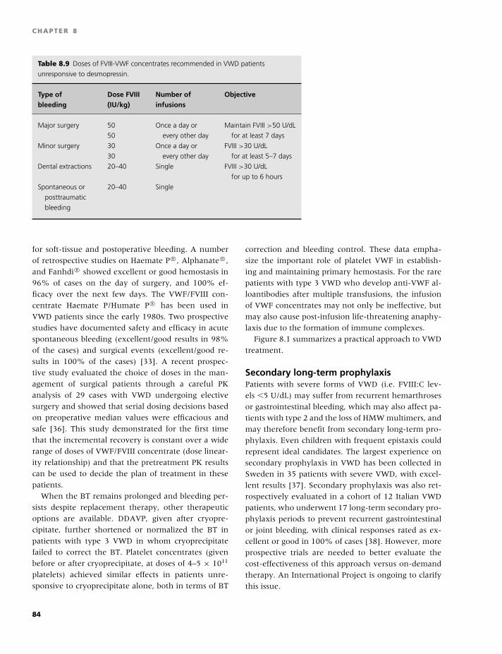
BLBK186-Key April 11, 2009 12:55
CHAPTER 8
Table 8.9 Doses of FVIII-VWF concentrates recommended in VWD patients
unresponsive to desmopressin.
Type of Dose FVIII Number of Objectivebleeding (IU/kg) infusions
Major surgery 50 Once a day or Maintain FVIII >50 U/dL
50 every other day for at least 7 days
Minor surgery 30 Once a day or FVIII >30 U/dL
30 every other day for at least 5–7 days
Dental extractions 20–40 Single FVIII >30 U/dL
for up to 6 hours
Spontaneous or 20–40 Single
posttraumatic
bleeding
for soft-tissue and postoperative bleeding. A number
of retrospective studies on Haemate P R©, Alphanate R©,
and Fanhdi R© showed excellent or good hemostasis in
96% of cases on the day of surgery, and 100% ef-
ficacy over the next few days. The VWF/FVIII con-
centrate Haemate P/Humate P R© has been used in
VWD patients since the early 1980s. Two prospective
studies have documented safety and efficacy in acute
spontaneous bleeding (excellent/good results in 98%
of the cases) and surgical events (excellent/good re-
sults in 100% of the cases) [33]. A recent prospec-
tive study evaluated the choice of doses in the man-
agement of surgical patients through a careful PK
analysis of 29 cases with VWD undergoing elective
surgery and showed that serial dosing decisions based
on preoperative median values were efficacious and
safe [36]. This study demonstrated for the first time
that the incremental recovery is constant over a wide
range of doses of VWF/FVIII concentrate (dose linear-
ity relationship) and that the pretreatment PK results
can be used to decide the plan of treatment in these
patients.
When the BT remains prolonged and bleeding per-
sists despite replacement therapy, other therapeutic
options are available. DDAVP, given after cryopre-
cipitate, further shortened or normalized the BT in
patients with type 3 VWD in whom cryoprecipitate
failed to correct the BT. Platelet concentrates (given
before or after cryoprecipitate, at doses of 4–5 × 10¹¹
platelets) achieved similar effects in patients unre-
sponsive to cryoprecipitate alone, both in terms of BT
correction and bleeding control. These data empha-
size the important role of platelet VWF in establish-
ing and maintaining primary hemostasis. For the rare
patients with type 3 VWD who develop anti-VWF al-
loantibodies after multiple transfusions, the infusion
of VWF concentrates may not only be ineffective, but
may also cause post-infusion life-threatening anaphy-
laxis due to the formation of immune complexes.
Figure 8.1 summarizes a practical approach to VWD
treatment.
Secondary long-term prophylaxisPatients with severe forms of VWD (i.e. FVIII:C lev-
els �5 U/dL) may suffer from recurrent hemarthroses
or gastrointestinal bleeding, which may also affect pa-
tients with type 2 and the loss of HMW multimers, and
may therefore benefit from secondary long-term pro-
phylaxis. Even children with frequent epistaxis could
represent ideal candidates. The largest experience on
secondary prophylaxis in VWD has been collected in
Sweden in 35 patients with severe VWD, with excel-
lent results [37]. Secondary prophylaxis was also ret-
rospectively evaluated in a cohort of 12 Italian VWD
patients, who underwent 17 long-term secondary pro-
phylaxis periods to prevent recurrent gastrointestinal
or joint bleeding, with clinical responses rated as ex-
cellent or good in 100% of cases [38]. However, more
prospective trials are needed to better evaluate the
cost-effectiveness of this approach versus on-demand
therapy. An International Project is ongoing to clarify
this issue.
84
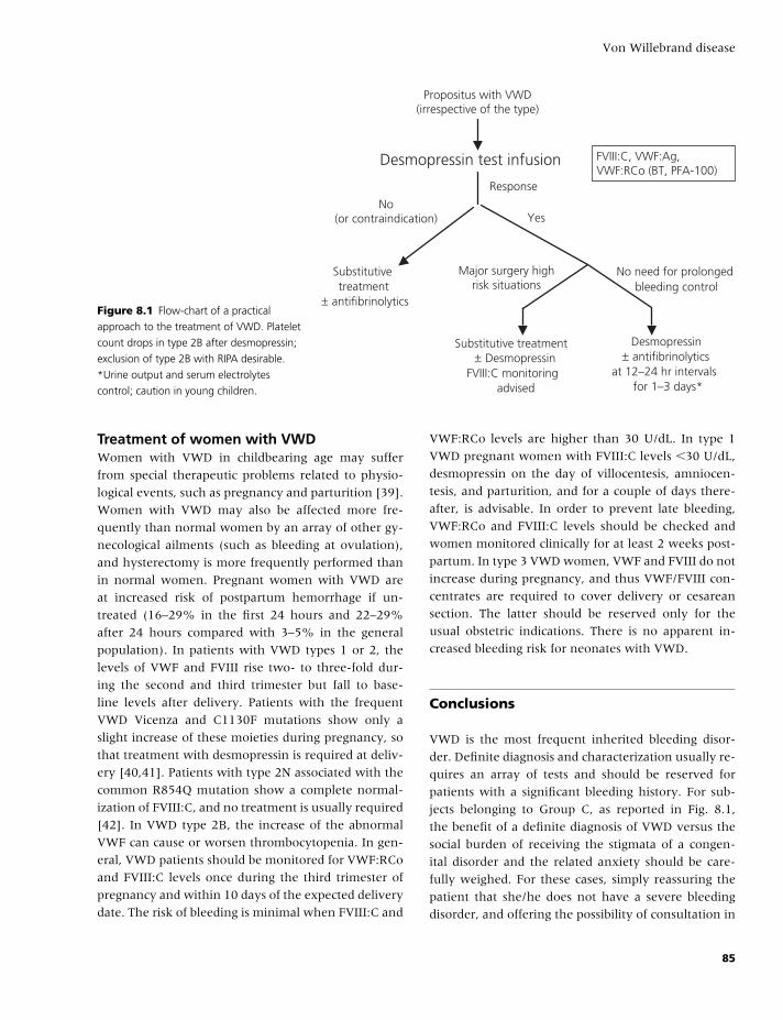
BLBK186-Key April 11, 2009 12:55
Von Willebrand disease
Propositus with VWD(irrespective of the type)
Desmopressin test infusion
Response
No(or contraindication) Yes
Substitutivetreatment
± antifibrinolytics
Major surgery highrisk situations
No need for prolongedbleeding control
Substitutive treatment± Desmopressin
FVIII:C monitoringadvised
Desmopressin± antifibrinolytics
at 12–24 hr intervalsfor 1–3 days*
FVIII:C, VWF:Ag,VWF:RCo (BT, PFA-100)
Figure 8.1 Flow-chart of a practical
approach to the treatment of VWD. Platelet
count drops in type 2B after desmopressin;
exclusion of type 2B with RIPA desirable.
*Urine output and serum electrolytes
control; caution in young children.
Treatment of women with VWDWomen with VWD in childbearing age may suffer
from special therapeutic problems related to physio-
logical events, such as pregnancy and parturition [39].
Women with VWD may also be affected more fre-
quently than normal women by an array of other gy-
necological ailments (such as bleeding at ovulation),
and hysterectomy is more frequently performed than
in normal women. Pregnant women with VWD are
at increased risk of postpartum hemorrhage if un-
treated (16–29% in the first 24 hours and 22–29%
after 24 hours compared with 3–5% in the general
population). In patients with VWD types 1 or 2, the
levels of VWF and FVIII rise two- to three-fold dur-
ing the second and third trimester but fall to base-
line levels after delivery. Patients with the frequent
VWD Vicenza and C1130F mutations show only a
slight increase of these moieties during pregnancy, so
that treatment with desmopressin is required at deliv-
ery [40,41]. Patients with type 2N associated with the
common R854Q mutation show a complete normal-
ization of FVIII:C, and no treatment is usually required
[42]. In VWD type 2B, the increase of the abnormal
VWF can cause or worsen thrombocytopenia. In gen-
eral, VWD patients should be monitored for VWF:RCo
and FVIII:C levels once during the third trimester of
pregnancy and within 10 days of the expected delivery
date. The risk of bleeding is minimal when FVIII:C and
VWF:RCo levels are higher than 30 U/dL. In type 1
VWD pregnant women with FVIII:C levels �30 U/dL,
desmopressin on the day of villocentesis, amniocen-
tesis, and parturition, and for a couple of days there-
after, is advisable. In order to prevent late bleeding,
VWF:RCo and FVIII:C levels should be checked and
women monitored clinically for at least 2 weeks post-
partum. In type 3 VWD women, VWF and FVIII do not
increase during pregnancy, and thus VWF/FVIII con-
centrates are required to cover delivery or cesarean
section. The latter should be reserved only for the
usual obstetric indications. There is no apparent in-
creased bleeding risk for neonates with VWD.
Conclusions
VWD is the most frequent inherited bleeding disor-
der. Definite diagnosis and characterization usually re-
quires an array of tests and should be reserved for
patients with a significant bleeding history. For sub-
jects belonging to Group C, as reported in Fig. 8.1,
the benefit of a definite diagnosis of VWD versus the
social burden of receiving the stigmata of a congen-
ital disorder and the related anxiety should be care-
fully weighed. For these cases, simply reassuring the
patient that she/he does not have a severe bleeding
disorder, and offering the possibility of consultation in
85
BLBK186-Key April 11, 2009 12:55
CHAPTER 8
case of need, is the preferred choice. Today, several
safe and effective therapeutic options are easily avail-
able to prevent or control bleeding episodes, which
rarely persistently affect the quality of life.
References
1 Rodeghiero F, Castaman G, Dini E. Epidemiological in-
vestigation of the prevalence of von Willebrand’s dis-
ease. Blood 1987;69:454–9.
2 Mancuso DJ, Tuley EA, Westfield LA, et al. Human
von Willebrand factor gene and pseudogene: structural
analysis and differentiation by polymerase chain reac-
tion. Biochemistry 1991; 30:253–69.
3 Wagner DD. Cell biology of von Willebrand factor.
Annu Rev Cell Biol 1990;6:217–46.
4 Furlan M, Robles R, Lammle B. Partial purification
and characterization of a protease from human plasma
cleaving von Willebrand factor to fragments produced
by in vivo proteolysis. Blood 1996;87:4223–34.
5 Ruggeri ZM. Structure of von Willebrand factor and its
function in platelet adhesion and thrombus formation.
Clin Haematol 2001;14:257–79.
6 Vlot AJ, Koppelman SJ, Bouma BN, Sixma JJ. Fac-
tor VIII and von Willebrand Factor. Thromb Haemost
1998;79:456–65.
7 Sadler JE, Budde U, Eikenboom JC, et al. Update on the
pathophysiology and classification of von Willebrand
disease: a report of the Subcommittee on von Wille-
brand Factor. J Thromb Haemost 2006;4:2103–14.
8 Lyons SE, Bruck ME, Bowie EJW, Ginsburg D. Im-
paired cellular transport produced by a subset of type
IIA von Willebrand disease mutations. J Biol Chem
1992;267:4424–30.
9 Castaman G, Federici AB, Rodeghiero F, Mannucci PM.
Von Willebrand’s disease in the year 2003: towards the
complete identification of gene defects for correct diag-
nosis and treatment. Haematologica 2003;88:94–108.
10 Baronciani L, Federici AB, Castaman G, Punzo M,
Mannucci PM. Prevalence of type 2b ‘Malmo/New
York’ von Willebrand disease in Italy: the role of von
Willebrand factor gene conversion. J Thromb Haemost
2008;6:887–90.
11 Rodeghiero F, Castaman G. Congenital von Wille-
brand disease type I: definition, phenotypes, clinical
and laboratory assessment. Best Pract Res Clin Haematol
2001;14:321–35.
12 Castaman G, Eikenboom JCJ, Missiaglia E, Rodeghiero
F. Autosomal dominant type 1 von Willebrand disease
due to G3639T mutation (C1130F) in exon 26 of von
Willebrand factor gene: description of five Italian fam-
ilies and evidence for a founder effect. Br J Haematol
2000;108:876–9.
13 Eikenboom JC, Matsushita T, Reitsma PH, et al. Domi-
nant type 1 von Willebrand disease caused by mutated
cysteine residues in the D3 domain of von Willebrand
factor. Blood 1996;88:2433–41.
14 Mannucci PM, Lombardi R, Castaman G, et al. von
Willebrand disease “Vicenza” with larger-than-normal
(supranormal) von Willebrand factor multimers. Blood
1988;71:65–70.
15 Schneppenheim R, Federici AB, Budde U, et al. Von
Willebrand disease type 2 M “Vicenza” in Italian and
German patients: identification of the first candidate
mutation (G3864A; R1205H) in 8 families. Thromb
Haemost 2000;83:136–40.
16 Casonato A, Pontara E, Sartorello F, et al. Reduced von
Willebrand factor survival in type Vicenza von Wille-
brand disease. Blood 2002;99:180–4.
17 Eikenboom JCJ, Reitsma PH, Peerlinck KMJ, et al. Re-
cessive inheritance of von Willebrand’s disease type I.
Lancet 1993;341:982–6.
18 Goodeve A, Eikenboom J, Castaman G, et al. Pheno-
type and genotype of a cohort of families historically
diagnosed with type 1 von Willebrand disease in the
European study, Molecular and Clinical Markers for the
Diagnosis and Management of Type 1 von Willebrand
Disease (MCMDM-1VWD). Blood 2007;109:112–21.
19 James PD, Notley C, Hegadorn C, et al. The mutational
spectrum of type 1 von Willebrand disease: Results
from a Canadian cohort study. Blood 2007;109:145–54.
20 Eikenboom J, Van Marion V, Putter H, et al. Linkage
analysis in families diagnosed with type 1 von Wille-
brand disease in the European study, molecular and
clinical markers for the diagnosis and management of
type 1 VWD. J Thromb Haemost 2006;4:774–82.
21 James PD, Paterson AD, Notley C, et al. Genetic link-
age and association analysis in type 1 von Willebrand
disease: results from the Canadian type 1 VWD study. J
Thromb Haemost 2006;4:783–92.
22 Cumming A, Grundy P, Keeney S, et al. An investiga-
tion of the von Willebrand factor genotype in UK pa-
tients diagnosed to have type 1 von Willebrand disease.
Thromb Haemost 2006;96:630–41.
23 Mohlke KL, Ginsburg D. von Willebrand disease and
quantitative variation in von Willebrand factor. J Lab
Clin Med 1997;130:252–61.
24 Eikenboom JCJ. Congenital von Willebrand disease
type 3: clinical manifestations, pathophysiology and
molecular biology. Clin Haematol 2001;14:365–79.
25 Rodeghiero F, Castaman G. von Willebrand disease:
epidemiology. In: Lee CA, Berntorp EE, Hoots WK,
86
BLBK186-Key April 11, 2009 12:55
Von Willebrand disease
eds. Textbook of Hemophilia. Oxford: Blackwell, 2005;
265–71.
26 Castaman G, Eikenboom JCJ, Bertina R, Rodeghiero
F. Inconsistency of association between type 1 von
Willebrand disease phenotype and genotype in fami-
lies identified in an epidemiologic investigation. Thromb
Haemostas 1999;82:1065–70.
27 Budde U, Schneppenheim R, Eikenboom J, et al. De-
tailed von Willebrand factor multimer analysis in pa-
tients with von Willebrand disease in the European
study, molecular and clinical markers for the diagno-
sis and management of type 1 von Willebrand disease
(MCMDM-1VWD). J Thromb Haemost 2008;6:762–71.
28 Castaman G, Rodeghiero F, Tosetto A, et al. Hemor-
rhagic symptoms and bleeding risk in obligatory carri-
ers of type 3 von Willebrand disease: an International,
multicenter study. J Thromb Haemost 2006;4:2164–9.
29 Rodeghiero F, Castaman G, Tosetto A, et al. The dis-
criminant power of bleeding history for the diagnosis of
type 1 von Willebrand disease: an international, multi-
center study. J Thromb Haemost 2005;3:2619–26.
30 Tosetto A, Rodeghiero F, Castaman G, et al. Impact of
plasma von Willebrand factor levels in the diagnosis
of type 1 von Willebrand disease: results from a mul-
ticenter European study (MCMDM-1VWD). J Thromb
Haemost 2007;5:715–21.
31 Castaman G, Lethagen S, Federici AB, et al. Response
to desmopressin is influenced by the genotype and phe-
notype in type 1 von Willebrand disease (VWD): re-
sults from the European Study MCMDM-1VWD. Blood
2008;111:3531–9.
32 Haberichter SL, Castaman G, Budde U, et al. Identifi-
cation of type 1 von Willebrand disease patients with
reduced von Willebrand factor survival by assay of the
VWF propeptide in the European study: molecular and
clinical markers for the diagnosis and management of
type 1 VWD (MCMDM-1VWD). Blood 2008;111:4979–
85.
33 Rodeghiero F, Castaman G. Treatment of von Wille-
brand disease. Semin Hematol 2005;42:29–35.
34 Castaman G, Lattuada A, Mannucci PM, et al. Factor
VIII:C increases after desmopressin in a subgroup of pa-
tients with autosomal recessive von Willebrand disease.
Br J Haematol 1995;89:147–51.
35 Mannucci PM, Tenconi PM, Castaman G, Rodeghiero
F. Comparison of four virus-inactivated plasma con-
centrates for treatment of severe von Willebrand dis-
ease: a cross-over randomized trial. Blood 1992;79:
3130–7.
36 Lethagen S, Kyrle PA, Castaman G, et al. von Wille-
brand factor/factor VIII concentrate (Haemate P) dosing
based on pharmacokinetics: a prospective multicenter
trial in elective surgery. J Thromb Haemost 2007;5:1420–
30.
37 Berntorp E, Petrini P. Long-term prophylaxis in
von Willebrand disease. Blood Coagul Fibrinolysis
2005;16:S23–6.
38 Federici AB, Castaman G, Franchini M, et al. Clinical
use of Haemate P in inherited von Willebrand disease:
a cohort study on 100 Italian patients. Haematologica
2007;92:944–51.
39 Kouides PA. Females with von Willebrand disease: 72
years as the silent majority. Haemophilia 1998;4:665–
76.
40 Castaman G, Eikenboom JCJ, Contri A, Rodeghiero
F. Pregnancy in women with type 1 von Willebrand
disease caused by heterozygosity for von Willebrand
factor mutation C1130F. Thromb Haemostas 2000;84:
351–2.
41 Castaman G, Federici AB, Bernardi M, Moroni B,
Bertoncello K, Rodeghiero F. Factor VIII and von Wille-
brand factor changes after desmopressin and during
pregnancy in type 2M von Willebrand disease Vi-
cenza: a prospective study comparing patients with sin-
gle (R1205H) and double (R1205H-M740I) defect. J
Thromb Haemost 2006;4:357–60.
42 Castaman G, Bertoncello K, Bernardi M, Rodeghiero
F. Pregnancy and delivery in patients with homozygous
or heterozygous R854Q type 2N von Willebrand dis-
ease. J Thromb Haemost 2005;3:391–2.
87
BLBK186-Key April 11, 2009 12:56
9 The rarer inherited coagulationdisordersPaula Bolton-Maggs and Jonathan Wilde
Introduction
The inherited coagulation disorders hemophilia A and
B (described in Chapter 7) and von Willebrand dis-
ease (described in Chapter 8) are well characterized.
However, inherited abnormalities of all the other co-
agulation factors have been recognized but are not so
well known. All are inherited autosomally and gen-
erally, with the exception of factor XI, are associated
with few or no symptoms in heterozygote individuals.
Most of the factor deficiencies are caused by abnor-
malities in the gene encoding for the particular factor.
There are three interesting exceptions.
1 Combined FV and FVIII deficiency is caused by a
defect in a protein involved in processing of proteins
within the hepatic cells.
2 Combined deficiency of the vitamin K-dependent
factors is a disorder caused by mutations in genes
encoding enzymes involved in vitamin K-dependent
carboxylation.
3 A third syndrome has recently been described
where FVII and FX are both affected by abnormali-
ties (deletions or translocations) in chromosome 13,
where both genes are located, and usually associated
with other abnormalities, such as mental retardation,
microcephaly, cleft palate [1].
As all these disorders are rare (Table 9.1), most
hematologists and pediatricians will have limited ex-
perience, and it is essential that the affected individu-
als are registered with a hemophilia center.
The annual report from the UKHCDO national
database [2] (data for 2006) shows that factor XI
deficiency (9%) is more common than hemophilia
B (7%), demonstrating that this should perhaps no
longer be considered a “rare” bleeding disorder. The
other disorders to be considered in this chapter are all
rare, making up a total of 6% of patients in the UK
register. The World Federation of Hemophilia (WFH)
performs annual global surveys via the national pa-
tient organizations in about 100 countries. Since 2004,
the survey reports some information about the rare
disorders and confirms the variation in distribution in
different parts of the world, with higher prevalence
of these disorders in countries where consanguineous
marriage is common. The global surveys can be viewed
on the WFH Web site (http://www.wfh.org).
Rare bleeding disorders have certain features in
common that can be considered together.
Genetics
These disorders are autosomal recessive conditions
and most commonly occur in individuals whose par-
ents are related, so therefore are much more common
in ethnic groups in which consanguineous marriage is
customary, such as in many Asian and Arabic commu-
nities. Factor XI deficiency (not recessive as symptoms
occur in a proportion of heterozygotes) is particularly
common in Ashkenazi Jews.
Clinical features
As autosomal disorders, both males and females are
affected; menorrhagia is a common feature of all these
disorders, and many are associated with hemorrhage
related to childbirth. Bleeding at ovulation or from
corpus luteum cysts is also reported and can be very
severe [3].
Severely deficient infants with these disorders (ex-
cept factor XI) are particularly at risk for intracranial
hemorrhage (ICH) and need to be identified quickly
88
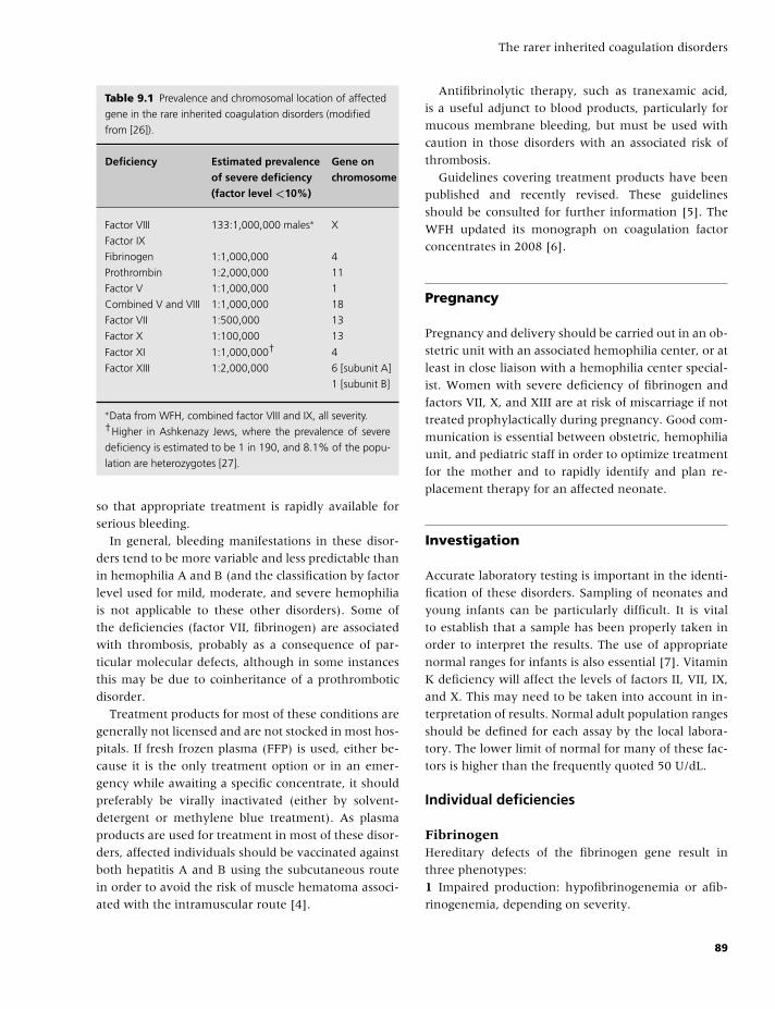
BLBK186-Key April 11, 2009 12:56
The rarer inherited coagulation disorders
Table 9.1 Prevalence and chromosomal location of affected
gene in the rare inherited coagulation disorders (modified
from [26]).
Deficiency Estimated prevalence Gene onof severe deficiency chromosome(factor level <10%)
Factor VIII 133:1,000,000 males∗ X
Factor IX
Fibrinogen 1:1,000,000 4
Prothrombin 1:2,000,000 11
Factor V 1:1,000,000 1
Combined V and VIII 1:1,000,000 18
Factor VII 1:500,000 13
Factor X 1:100,000 13
Factor XI 1:1,000,000† 4
Factor XIII 1:2,000,000 6 [subunit A]
1 [subunit B]
∗Data from WFH, combined factor VIII and IX, all severity.†Higher in Ashkenazy Jews, where the prevalence of severe
deficiency is estimated to be 1 in 190, and 8.1% of the popu-
lation are heterozygotes [27].
so that appropriate treatment is rapidly available for
serious bleeding.
In general, bleeding manifestations in these disor-
ders tend to be more variable and less predictable than
in hemophilia A and B (and the classification by factor
level used for mild, moderate, and severe hemophilia
is not applicable to these other disorders). Some of
the deficiencies (factor VII, fibrinogen) are associated
with thrombosis, probably as a consequence of par-
ticular molecular defects, although in some instances
this may be due to coinheritance of a prothrombotic
disorder.
Treatment products for most of these conditions are
generally not licensed and are not stocked in most hos-
pitals. If fresh frozen plasma (FFP) is used, either be-
cause it is the only treatment option or in an emer-
gency while awaiting a specific concentrate, it should
preferably be virally inactivated (either by solvent-
detergent or methylene blue treatment). As plasma
products are used for treatment in most of these disor-
ders, affected individuals should be vaccinated against
both hepatitis A and B using the subcutaneous route
in order to avoid the risk of muscle hematoma associ-
ated with the intramuscular route [4].
Antifibrinolytic therapy, such as tranexamic acid,
is a useful adjunct to blood products, particularly for
mucous membrane bleeding, but must be used with
caution in those disorders with an associated risk of
thrombosis.
Guidelines covering treatment products have been
published and recently revised. These guidelines
should be consulted for further information [5]. The
WFH updated its monograph on coagulation factor
concentrates in 2008 [6].
Pregnancy
Pregnancy and delivery should be carried out in an ob-
stetric unit with an associated hemophilia center, or at
least in close liaison with a hemophilia center special-
ist. Women with severe deficiency of fibrinogen and
factors VII, X, and XIII are at risk of miscarriage if not
treated prophylactically during pregnancy. Good com-
munication is essential between obstetric, hemophilia
unit, and pediatric staff in order to optimize treatment
for the mother and to rapidly identify and plan re-
placement therapy for an affected neonate.
Investigation
Accurate laboratory testing is important in the identi-
fication of these disorders. Sampling of neonates and
young infants can be particularly difficult. It is vital
to establish that a sample has been properly taken in
order to interpret the results. The use of appropriate
normal ranges for infants is also essential [7]. Vitamin
K deficiency will affect the levels of factors II, VII, IX,
and X. This may need to be taken into account in in-
terpretation of results. Normal adult population ranges
should be defined for each assay by the local labora-
tory. The lower limit of normal for many of these fac-
tors is higher than the frequently quoted 50 U/dL.
Individual deficiencies
FibrinogenHereditary defects of the fibrinogen gene result in
three phenotypes:
1 Impaired production: hypofibrinogenemia or afib-
rinogenemia, depending on severity.
89
BLBK186-Key April 11, 2009 12:56
CHAPTER 9
2 Synthesis of abnormally structured molecules: dys-
fibrinogenemia.
3 Reduced production of an abnormal molecule: rare
(hypodysfibrinogenemia).
AfibrinogenemiaThis defect is associated with a bleeding tendency,
although variable, and people with severe defi-
ciency may have infrequent bleeding, whereas others
have marked mucosal and intramuscular bleeding.
Neonates may present with umbilical cord bleeding,
and they may have ICH. Wound healing may be im-
paired. Women are at risk of recurrent miscarriage
and both ante- and postpartum hemorrhage. Paradox-
ically, thrombosis has also been reported in severe de-
ficiency not in relation to therapy or other provok-
ing events. Individuals with hypofibrinogenemia are
also at risk of bleeding with less severe manifesta-
tions, such as bleeding after surgery rather than spon-
taneous events. The diagnosis of afibrinogenemia de-
pends on demonstrating absence of fibrinogen by both
functional and antigenic assays.
DysfibrinogenemiaThis is a collection of disorders with variable clini-
cal features (over 300 variants have been described).
About 25% of patients have a mild bleeding disorder.
In roughly another 25%, the specific molecular defects
are associated with thrombosis [8]. The diagnosis may
be difficult, although generally there is a significant
discordancy between fibrinogen antigen and activity
values. Family studies may be extremely informative,
as many dysfibrinogenemias are inherited in an au-
tosomal dominant manner. The personal and family
history of bleeding and thrombosis will help in guid-
ing management.
TreatmentFibrinogen concentrates are available in some coun-
tries [6]. These are preferred to cryoprecipitate as
they are treated to reduce risks of viral transmission.
The half-life of fibrinogen is 3–5 days, and a level of
more than 0.5 g/L is associated with a reduced risk of
bleeding.
ProthrombinProthrombin deficiency is extremely rare. It has
recently been reviewed in detail [9]. Complete
deficiency is not recorded and is probably incompat-
ible with life (analogous to the situation in prothrom-
bin “knockout” mice). The two phenotypes are:� quantitative (hypoprothrombinemia); and� qualitative (dysprothrombinemia).
Individuals with hypoprothrombinemia may suffer
from joint and muscle bleeds and also mucosal bleed-
ing. It is notable that about 70% of patients with pro-
thrombin disorders are of Latin country origin. Het-
erozygotes may be missed as the prothrombin time
may be normal.
Treatment can be given with three-factor (II, IX, X)
or four-factor (II, VII, IX, X) concentrates (prothrom-
bin complex concentrates, originally developed for FIX
deficiency). Prothrombin has a long half-life, so that
treatment may be given every 2–3 days.
Factor V deficiencyFactor V deficiency presents in childhood with bruis-
ing and mucous membrane bleeding. Infants with
severe deficiency are at risk of ICH, which may occur
antenatally. Reported cases appear to have a high risk
of inhibitor development associated with replacement
therapy. Affected children should also have a factor
VIII assay performed to exclude combined deficiency
(see below).
Treatment is with FFP. Large volumes may be re-
quired leading to a risk of fluid overload. The min-
imum level of FV required for hemostasis is at least
15 U/dL.
Combined deficiency of factors V and VIIIThis interesting disorder is caused by defects in the
gene for a protein responsible for intracellular trans-
port (LMAN1) [10,11]. Levels of both factors are
most commonly between 5 and 20 IU/dL. Sponta-
neous bleeding is relatively uncommon; bleeding after
surgery is a risk. Parents have normal levels of both
factors.
Treatment is with both factor VIII concentrate (prin-
ciples as for hemophilia A) and FFP (as for FV
deficiency).
Factor VII deficiencyThis is the most common of the rare disorders
(excluding FXI). It has recently been reviewed in
detail [9]. People with mild deficiency (heterozygotes)
do not usually have a bleeding problem. Generally,
90
BLBK186-Key April 11, 2009 12:56
The rarer inherited coagulation disorders
bleeding is confined to individuals with very low levels
(�2 IU/dL), but the correlation of level with bleeding
is not close; that is, some individuals with very
low levels do not bleed, whereas those with higher
levels do.
Mucous membrane bleeding is particularly com-
mon. Menorrhagia is common in women. Thrombo-
sis has also been reported. Neonates with severe defi-
ciency are at risk of ICH [12]. The molecular defects
are heterogeneous. It is also important to note that
the the factor VII level can vary depending on the
source of the thromboplastin used in the laboratory
assay and may be related to particular molecular ab-
normalities (e.g. factor VII Padua). People formerly di-
agnosed as FVII-deficient using rabbit brain thrombo-
plastin have been shown to have normal FVIIC levels
with recombinant human thromboplastin [13]. Giro-
lami notes that different mutations may give similar
phenotypes, and conversely, patients with the same
mutation may have different phenotypes, thus the
genotype–phenotype relationships are complex [9].
The recommended treatment is with recombinant
activated factor VII (rVIIa) or with a plasma-derived
concentrate. The half-life of factor VII is particularly
short (6 hours), but despite this, prophylaxis (where
indicated) one to three times a week may be sufficient.
Factor X deficiencySevere factor X deficiency (FX �1 IU/dL) is associated
with a significant risk of ICH in the first weeks of life.
Umbilical stump bleeding also occurs. Mucosal hem-
orrhage is a particular feature, with severe epistaxis
being common at any level of deficiency. Menorrhagia
occurs in half of affected females. Severe arthropathy
may occur as a result of recurrent joint bleeds. Mild
deficiency is defined by FX levels of 6–10 IU/dL; these
individuals are often diagnosed incidentally but may
experience easy bruising or menorrhagia. A number
of clinical variants have been described, and assay by
more than one method is recommended in order not
to miss some variants [13–15].
Antifibrinolytic medication is particularly useful for
mucous membrane bleeding. Factor X is present in
prothrombin complex concentrates, which are there-
fore the recommended treatment. The half-life of fac-
tor X is 20–40 hours. Caution is required because of
the known prothrombotic properties of these concen-
trates. Therefore, factor X levels should be monitored.
In those children with recurrent joint bleeds, prophy-
laxis has been successfully admitted either every third
day, or once a week. Experience with FFP suggests
that, in severe deficiency, an FX level of 20–35 IU/dL is
sufficient for hemostasis postoperatively in severe de-
ficiency, but it is likely that levels lower than this (e.g.
down to 5 IU/dL) may be sufficient.
Factor XI deficiencyThe role of factor XI in the coagulation mechanism is
debated; there is some evidence that factor XI is physi-
ologically activated by traces of thrombin and serves to
potentiate the propagatory pathway once coagulation
has been initiated via the tissue factor pathway. How-
ever, this view has been challenged [16]. Bleeding risk
may be more related to increased fibrinolysis because
of the reduction in generation of the thrombin activat-
able fibrinolysis inhibitor secondary to a low factor XI.
FXI is a serine protease that is unique in being a dimer.
Although factor XI deficiency is particularly common
in Ashkenazi Jews, it is found in all ethnic groups. The
mutations in Jewish patients are restricted with two
being particularly common [17]. Overall, the preva-
lence of severe deficiency is 1 in 1 million, but mild de-
ficiency is much more common. In the UK, mild factor
XI deficiency is currently being reported more often
than hemophilia B. This is partly because current acti-
vated partial thromboplastin time (APTT) reagents are
sensitive to mild FXI deficiency and there is a greater
readiness to investigate these mildly prolonged APTT
levels.
Factor XI deficiency is unlike most of the other rare
coagulation disorders in that heterozygotes may have
a significant bleeding tendency that is poorly predicted
by the factor XI level [18]. Spontaneous bleeding is
extremely rare, even in those with undetectable FXI
levels; bleeding is provoked by injury and surgery, par-
ticularly in areas of high fibrinolytic activity (mouth,
nose, and genitourinary tract). Women with both se-
vere and mild deficiency may suffer menorrhagia and
bleeding in relation to childbirth. The bleeding ten-
dency varies within both a family and an individual
at different times. This may be related to mild varia-
tion in other factors, such as von Willebrand factor.
These factors make the management of surgery in FXI
deficiency more complicated. Babies with severe defi-
ciency do not bleed spontaneously (ICH and other se-
rious bleeding is not reported). Male babies are at risk
91
BLBK186-Key April 11, 2009 12:56
CHAPTER 9
of excessive bleeding at circumcision. UK guidelines
recommend that the factor XI level should be checked,
and if less than 10 IU/dL at birth, circumcision should
be delayed and the level checked at 6 months. If still
less than 10 IU/dL, the procedure should be performed
in hospital with FFP or concentrate cover (see be-
low), and the religious requirements discussed with
the family. If the level is more than 10 IU/dL, tranex-
amic acid alone can be given.
Oral antifibrinolytic therapy is very useful for the
management of mucosal bleeding (menorrhagia) and
is sufficient for the management of dental extractions,
even in people with severe deficiency. The manage-
ment of other types of surgery depends, to some ex-
tent, on whether it is in an area of high fibrinolytic
activity (such as tonsillectomy) when factor XI re-
placement is indicated, as opposed to other types of
surgery (e.g. herniorrhaphy) where replacement ther-
apy may be more parsimonious [19] .
Two factor XI concentrates are available, but both
have been associated with thrombotic events in some
individuals, particularly those with additional risk fac-
tors, such as older age, the presence of cardiovascular
disease, or malignancy. Because of this, antifibrinolytic
drugs should not accompany them, and peak levels of
more than 100 IU/dL should be avoided. FFP can be
used, but in people with severe deficiency it is difficult
to produce a sufficient rise (to about 20 to 30 IU/dL)
without the risk of fluid overload.
The management of subjects with heterozygous de-
ficiency and a bleeding history (FXI of about 20–60
IU/dL) is more difficult and is dependent on the bleed-
ing history of the individual patient, the presence or
absence of associated factor deficiencies, and the na-
ture of the hemostatic challenge.
Inhibitors can develop in severe deficiency [20]; ac-
tivated recombinant factor VII (rVIIa) has been used
successfully. It may also be useful in patients without
inhibitors but has been associated with thrombosis in
this setting.
Factor XIII deficiencyFactor XIII cross-links and stabilizes fibrin. Severe de-
ficiency, with undetectable factor XIII, is associated
with:� a serious bleeding disorder, usually presenting in
infancy;� bleeding from the umbilical stump in 80%;� ICH;
� joint and muscle bleeds;� miscarriages and bleeding after delivery or surgery;
and� delayed wound healing.
For these reasons, usually once severe deficiency
is detected, an individual is treated with prophylaxis
for life. Individuals with levels of 1–4 U/dL are also
likely to have bleeding symptoms, and rarely bleed-
ing is reported in people with levels above 5 U/dL.
For a review of factor XIII deficiency, see Anwar and
Miloszewski [21]. There is some data emerging sug-
gesting a bleeding diathesis in some heterozygous in-
dividuals.
The diagnosis is suspected when the coagulation
screen is normal. Clot solubility in urea or acetic acid
will be abnormal, and the defect is confirmed by a
factor XIII assay. Inconsistent results in the screening
tests were noted in the UK NEQAS exercises [22]. Be-
cause this is not a routine in most laboratories, it is
advisable to send the sample to a specialist center.
Plasma-derived concentrates are the treatment of
choice but a recombinant FXIII concentrate is in clini-
cal trials. Factor XIII has a long half-life of 7–10 days,
and in practice, dosing at 4–6 weekly intervals has
proved effective. It is suggested that levels of 4–10
U/dL are sufficient to prevent hemorrhage.
In the emergency situation, for example, when pre-
sented with an infant with a serious bleeding diathe-
sis, once blood has been taken for testing, either FFP
or cryoprecipitate is effective treatment.
Combined deficiencies of the vitaminK-dependent factors: II, VII, IX, and XCombined deficiency of all the vitamin K-dependent
factors is a rare but important bleeding disorder to
recognize. By 2008, only 29 cases from 24 families
had been reported. The inheritance is autosomal re-
cessive and is caused by defective function of either
gamma-carboxylase or vitamin K 2-3 epoxide reduc-
tase. Mucocutaneous and postsurgery-related bleed-
ing has been reported. Severe cases may present with
ICH or umbilical cord bleeding in infancy [23,24]. The
clinical picture and response to vitamin K is variable,
some responding to low-dose oral vitamin K and oth-
ers nonresponsive even to high-dose intravenous re-
placement. In those nonresponsive to vitamin K, pro-
thrombin complex concentrates are the product of
choice. Levels of the factors range from less than 1
to 50 IU/dL. Some individuals have associated skeletal
92
BLBK186-Key April 11, 2009 12:56
The rarer inherited coagulation disorders
abnormalities (probably related to abnormalities in
bone vitamin K-dependent proteins, such as osteocal-
cin). Genetic defects have been reported in the en-
zymes associated with vitamin K metabolism (e.g. in
γ-glutamyl carboxylase).
Illustrative case histories
Case 1A 13-month-old infant presented with a 2-cm diam-
eter swelling on his head and a swollen thigh caused
by running into a door 2 weeks previously. He was
thought to be suffering from nonaccidental injury
and was admitted. Ultrasound examination confirmed
a muscle hematoma. Coagulation screening demon-
strated a prolonged prothrombin time (PT) of 25.4
seconds (NR 11.5–15) and APTT of 80 seconds (NR
27–39). His factor X level was �1 U/dL. Both par-
ents (who were unrelated) had prolonged coagulation
tests and low factor X levels. Both were asymptomatic.
He was treated for the acute bleed with an intermedi-
ate purity factor IX (prothrombin complex) concen-
trate with monitoring of factor X levels. Over the next
3 years, he had repeated muscle and joint bleeds and
is now being treated with once weekly prophylaxis.
His concentrate dose is determined by regular dose–
response and half-life analysis.
CommentThis case illustrates a picture similar to severe
hemophilia A. Nonaccidental injury is unfortunately
more common than bleeding disorders, so that, unless
appropriate investigations are undertaken, diagnosis
may be delayed or missed.
Case 2A baby boy developed massive bilateral cephalhe-
matomas 24 hours after spontaneous vaginal delivery.
He was otherwise well with normal, unrelated par-
ents. Blood tests showed a profound anemia (Hb
7.0 g/dL) and incoagulable blood with undetectable
fibrinogen. Liver disease was excluded and he was not
septic. Cranial ultrasound confirmed that there was
no evidence of ICH. Both parents and both maternal
grandmothers were noted to have low fibrinogen lev-
els and prolonged thrombin times. He was transfused
with red cells and treated with regular cryoprecipitate
until fibrinogen concentrate could be obtained. He
was treated prophylactically, requiring a central
venous access device, but by 9 months of age, was
noted to have subclavian vein thrombosis related to
this. MR scanning demonstrated extensive thrombosis
of the upper body venous system. It was not possible
to determine whether therapy had contributed to
the thrombotic risk. Prophylaxis was stopped for 5
months, during which time he had several bruises
and was treated for minor bumps to the head, but had
no serious bleeding. When he began to walk and fall,
his mother was anxious for regular prophylaxis to be
resumed. It is unclear whether this is necessary in the
long term.
CommentIn the absence of mutation detection, it was impossible
to be sure that this child did not have compound het-
erozygosity for hypo- and dysfibrinogenemia, which
might have increased his risk of thrombosis. Muta-
tion detection can be helpful in predicting the clinical
picture in fibrinogen disorders, and will probably also
prove useful in factor VII and X deficiency where the
clinical picture can be variable.
Case 3A 12-year-old girl was admitted after a heavy third
menstrual period. She had been bleeding for 10 days,
fainted at school, and on admission was found to have
severe anemia with Hb 6.0 g/dL. Coagulation testing
demonstrated a normal APTT and a PT of 41 seconds.
Her factor VII level was 2.2 IU/dL (2.2%). She had
been adopted and had no other bleeding problems; she
had not bled excessively after being bitten by a dog,
requiring open reduction of a fracture of the forearm,
nor after being knocked down by a car. Once her peri-
ods had become established and controlled with hor-
mone therapy, she did not have any other bleeding
problems, and by the age of 18, had defaulted from
follow-up.
CommentThis case illustrates that individuals with severe factor
VII deficiency may have very few problems and con-
trasts with the next case.
Case 4An Asian baby with parents who were first cousins
was delivered by cesarean section. He was noted to
have nasal bleeds twice on day 3 and a bloodstained
93
BLBK186-Key April 11, 2009 12:56
CHAPTER 9
discharge from the umbilical cord on day 5. He was
admitted with irritability on day 18 and collapsed on
admission with Hb 8 g/dL, PT 32 seconds, APTT 39 sec-
onds. CT scanning of the head showed ICH in the pos-
terior fossa. His FVII level was 4 IU/dL. He was treated
initially with FFP (which did not shorten the PT) un-
til a FVII concentrate was available. He was treated
symptomatically over this acute event. However, fur-
ther episodes of ICH occurred over the next 2 months,
leading to cerebral atrophy and predictable develop-
mental delay. He was started on prophylaxis twice a
week at the age of 6 months via a venous access de-
vice. At 4.5 years, he had a mental age of 2.5, epilepsy,
no speech, and no vision on the R side as a conse-
quence of his previous ICHs. At the age of 5.5 years, he
was noted to have severe iron deficiency (Hb 6.9 g/dL,
MCV 59), common in children of Asian origin (di-
etary), compounded by developmental problems and
his bleeding disorder.
CommentWhere ICH occurs in relation to a severe congenital
factor deficiency, it needs to be recognized and treated
early and intensively to try to avoid long-term devel-
opmental problems. Iron deficiency is very common in
the Asian community due to dietary deficiency.
Case 5A Pakistani child with related parents was referred at
the age of 1 year. She had easy bruising and bleeding
from minor cuts, which lasted several hours. Her PT
was 45 seconds and the APTT was 92 seconds. Factor V
was �1 U/dL. At the age of 15 and 18 months, she had
recurrent mouth bleeds from trauma associated with
walking and was treated prophylactically twice weekly
with FFP. At 2 years, she had a retroperitoneal hem-
orrhage. At 3 years, there were concerns about her
neurological development, and at 5.5 years, imaging
supported the occurrence of a possible ICH in the past.
At 3 years, there was evidence of a factor V inhibitor
and regular FFP infusions were stopped. She had re-
current muscle bleeds leading to shortening and wast-
ing, and the necessity for tendon-lengthening surgery
at the age of 7 years, by which time her inhibitor had
disappeared. She continued to have recurrent muscle
and joint bleeds treated symptomatically with FFP in-
fusions (the inhibitor having resolved). Menarche oc-
curred at age 13, and her periods have not been heavy.
CommentFactor V deficiency is difficult to manage and may be
associated with the development of inhibitors, as in
this case.
Conclusion
The rare coagulation disorders may present with se-
rious and life-threatening bleeding. Prompt investi-
gation and recognition of these disorders is essential
so that the appropriate treatment can be instigated.
ICH is a serious risk in many of these disorders and
may have catastrophic consequences. Hematologists
need to work closely with pediatricians to recognize
these disorders. In communities where consanguinity
is common, there needs to be a heightened aware-
ness of the risk of these potentially serious bleeding
disorders. Mutation analysis can be very helpful, as it
offers the potential for subsequent antenatal diagnosis
in families with severe bleeding disorders.
Acknowledgments
This chapter is based on guidelines published by mem-
bers of the Rare Haemostatic Disorders Working Party
of the United Kingdom Haemophilia Centre Doctors’
Organization [25].
References
1 Girolami A, Ruzzon E, Tezza F, Scandellari R, Scapin
M, Scarparo P. Congenital FX deficiency combined
with other clotting defects or with other abnormali-
ties: a critical evaluation of the literature. Haemophilia
2008;14(2):323–8.
2 UKHCDO. National Haemophilia Database: Annual Re-
port for 2007, annual returns for 2006. Manchester,
UKHCDO, 2007.
3 Gupta N, Dadhwal V, Deka D, Jain SK, Mittal S. Corpus
luteum hemorrhage: rare complication of congenital
and acquired coagulation abnormalities. J Obstet Gy-
naecol Res 2007;33(3):376–80.
4 Makris M, Conlon CP, Watson HG. Immuniza-
tion of patients with bleeding disorders. Haemophilia
2003;9(5):541–6.
94
BLBK186-Key April 11, 2009 12:56
The rarer inherited coagulation disorders
5 UKHCDO. Guidelines on the selection and use of
therapeutic products to treat haemophilia and other
hereditary bleeding disorders. Haemophilia 2003;9(1):
1–23.
6 Brooker M. Registry of Clotting Factor Concentrates.
Montreal: WFH, April 2008.
7 Williams MD, Chalmers EA, Gibson BE. The investi-
gation and management of neonatal haemostasis and
thrombosis. Br J Haematol 2002;119(2):295–309.
8 Haverkate F, Samama M. Familial dysfibrinogene-
mia and thrombophilia. Report on a study of the
SSC Subcommittee on Fibrinogen. Thromb Haemost
1995;73(1):151–61.
9 Girolami A, Scandellari R, Scapin M, Vettore S.
Congenital bleeding disorders of the vitamin K-
dependent clotting factors. Vitam Horm 2008;78:281–
374.
10 D’Ambrosio R, Santacroce R, Di Perna P, Sarno M,
Romondia A, Margaglione M. A new case of com-
bined factor V and factor VIII deficiency further sug-
gests that the LMAN1 M1T mutation is a frequent cause
in Italian patients. Blood Coagul Fibrinolysis 2007;18(2):
203–4.
11 Ginsburg D, Nichols WC, Zivelin A, Kaufman RJ,
Seligsohn U. Combined factors V and VIII deficiency–
the solution. Haemophilia 1998;4(4):677–82.
12 Perry DJ. Factor VII deficiency. Br J Haematol
2002;118(3):689–700.
13 Bolton-Maggs PH, Hay CR, Shanks D, Mitchell MJ,
McVey JH. The importance of tissue factor source in the
management of Factor VII deficiency. Thromb Haemost
2007;97(1):151–2.
14 Peyvandi F, Mannucci PM, Lak M, et al. Congeni-
tal factor X deficiency: spectrum of bleeding symp-
toms in 32 Iranian patients. Br J Haematol 1998;102(2):
626–8.
15 Uprichard J, Perry DJ. Factor X deficiency. Blood Rev
2002;16(2):97–110.
16 Pedicord DL, Seiffert D, Blat Y. Feedback activation of
factor XI by thrombin does not occur in plasma. Proc
Natl Acad Sci U S A 2007;104(31):12855–60.
17 Hancock JF, Wieland K, Pugh RE, et al. A molec-
ular genetic study of factor XI deficiency. Blood
1991;77(9):1942–8.
18 Bolton-Maggs PH, Patterson DA, Wensley RT,
Tuddenham EG. Definition of the bleeding ten-
dency in factor XI-deficient kindreds: a clinical and
laboratory study. Thromb Haemost 1995;73(2):194–202.
19 Salomon O, Steinberg DM, Seligshon U. Variable
bleeding manifestations characterize different types of
surgery in patients with severe factor XI deficiency
enabling parsimonious use of replacement therapy.
Haemophilia 2006;12(5):490–3.
20 Salomon O, Zivelin A, Livnat T, et al. Prevalence,
causes, and characterization of factor XI inhibitors
in patients with inherited factor XI deficiency. Blood
2003;101(12):4783–8.
21 Anwar R, Miloszewski KJ. Factor XIII deficiency. Br J
Haematol 1999;107(3):468–84.
22 Jennings I, Kitchen S, Woods T, Preston F. Problems
relating to the laboratory diagnosis of FXIII deficiency:
A UK NEQAS study. J Thromb Haemost 2003;1(12):
2603–8.
23 Brenner B. Hereditary deficiency of vitamin K-
dependent coagulation factors. Thromb Haemost 2000;
84(6):935–6.
24 Oldenburg J, von Brederlow B, Fregin A, et al. Con-
genital deficiency of vitamin K dependent coagulation
factors in two families presents as a genetic defect of the
vitamin K-epoxide-reductase-complex. Thromb Haemost
2000;84(6):937–41.
25 Bolton-Maggs PH, Perry DJ, Chalmers EA, et al.
The rare coagulation disorders–review with guide-
lines for management from the United Kingdom
Haemophilia Centre Doctors’ Organisation. Haemophilia
2004;10(5):593–628.
26 Peyvandi F, Duga S, Akhavan S, Mannucci PM.
Rare coagulation deficiencies. Haemophilia 2002;8(3):
308–21.
27 Seligsohn U, Peretz H. Molecular genetics aspects of
factor XI deficiency and Glanzmann thrombasthenia.
Haemostasis 1994;24(2):81–5.
95
BLBK186-Key May 4, 2009 9:30
10 Quantitative platelet disordersJeremy D. Robertson, Victor S. Blanchette, and Walter H.A. Kahr
Introduction
Thrombocytopenia is defined as a platelet count of less
than 150 × 109/L (the normal range is 150–400 ×109/L). Increased bleeding purely as a result of a re-
duction of platelets does not usually occur until the
count drops below 50 × 109/L. Platelet counts of less
than 20 × 109/L increase the risk of life-threatening
bleeding (e.g. central nervous system or gastrointesti-
nal). However, several prospective studies have re-
vealed that hemorrhagic risk was similar using a 10 ×109/L or 20 × 109/L threshold for prophylactic platelet
transfusions, suggesting that life-threatening bleeding
increases significantly only when the platelet count
drops below 10 × 109/L [1]. Furthermore, the bleed-
ing risk at any given platelet count is partly dependent
on the underlying etiology.
The differential diagnosis of thrombocytopenia
varies with the age of onset, severity, clinical fea-
tures, and presence or absence of other hematologic
abnormalities. For example, the most probable cause
of thrombocytopenia in a newborn infant is different
from that of an older child or adult, or that of a
pregnant woman. Ranking of the most likely causes
of a low platelet count will also depend on whether
the patient is systemically well or not. This chapter
focuses on a practical approach in the assessment and
management of inherited and acquired quantitative
platelet disorders. Qualitative platelet disorders are
covered in Chapter 11.
Platelet production
Platelets are shed from megakaryocytes through
the action of thrombopoietin (TPO) and other cy-
tokines to a lesser extent, which collectively stim-
ulate pluripotent hematopoietic stem cells to form
mature megakaryocytes in bone marrow [2]. The
TPO receptor (c-Mpl, TPO-R) is expressed on the
surface of megakaryocytes, platelets, and primitive
(pluripotent) stem cells and mediates its action via
a signal transduction pathway similar to that of
erythropoietin [3]. Thrombopoietin is synthesized
predominantly in the liver and released into cir-
culation at a constant rate, where it is largely
cleared by binding to TPO-R on platelets. TPO
levels are increased up to 20-fold in bone mar-
row failure states, are only slightly elevated in im-
mune thrombocytopenia (ITP), and are low in liver
failure.
The average life span of human platelets is 7–10
days. Older platelets are removed from circulation
by reticulo-endothelial cells, although little is known
about the mechanisms through which these senes-
cent platelets are identified. A daily turnover of ap-
proximately 40 × 109 platelets/L blood is required to
maintain a constant platelet count. Aspirin (ASA) in-
hibits platelets irreversibly; thus, at least 7 days are
required to remove ASA-exposed platelets from the
circulation.
Newly formed platelets are thought to be more
functional in hemostasis than older platelets.
However, some antibodies observed in neonatal
alloimmune thrombocytopenia (NAIT) may impede
platelet function of young platelets by inactivating
interaction with the fibrinogen receptor glycopro-
tein (GP) IIb/IIIa. The higher incidence of serious
bleeding in NAIT compared with ITP (antibody
binding to GPIIb/IIIa and other platelet surface
receptors) at equivalent low platelet counts sug-
gests that the function of platelets in ITP is not
impaired.
96
BLBK186-Key May 4, 2009 9:30
Quantitative platelet disorders
Mechanisms of thrombocytopeniain children and adults
Thrombocytopenia can be classified according to
whether it is explained by increased platelet seques-
tration, the presence of decreased platelet production,
or accelerated platelet destruction (Table 10.1). In ad-
dition, dilutional thrombocytopenia following mas-
sive transfusion is a common iatrogenic mechanism
in which platelet concentration is reduced but total
platelet mass is preserved. Artefactual (false) throm-
bocytopenia is another important consideration in
the initial diagnostic evaluation, particularly when
an asymptomatic individual is unexpectedly found to
have a severely reduced platelet count. This most often
results from anticoagulant-dependent platelet clump-
ing ex vivo (pseudothrombocytopenia) or the forma-
tion of small clots in the specimen tube following trau-
matic collection (e.g. heelprick collection in neonates).
A diagnostic strategy for evaluating thrombocytopenia
in a “well” child or adult is shown in Fig. 10.1.
Platelet sequestration
In healthy individuals, splenic pooling (sequestra-
tion) accounts for approximately one-third of the total
platelet mass, but may be as high as 90% in individuals
with massive splenomegaly. The platelet count does
not always correlate directly with splenic size, and the
underlying mechanisms of platelet trapping within the
extravascular splenic pool remain poorly understood.
Preferential diversion of platelets through the splenic
cords (by virtue of their small size) as well as binding
to receptors on splenic macrophages may play a role
in pathophysiology.
Decreased platelet production
Platelets originate from megakaryocytes in the bone
marrow. Megakaryoctes protrude extensions (pro-
platelets) into blood vessels, where flowing blood
shear forces facilitate platelet shedding into the cir-
culation [4]. Reduction of total megakaryocyte mass
or functional impairment results in underproduc-
tion of platelets and subsequent thrombocytopenia.
Table 10.1 Causes of thrombocytopenia in children and
adults.
Increased platelet sequestrationHypersplenism
Decreased platelet productionAplastic anemia (idiopathic or drug-induced)
Myelodysplastic syndrome
Marrow infiltrative process
Infection (bacterial; viral: HIV, CMV, HCV)
Osteopetrosis
Nutritional deficiencies (iron, folate, vitamin B12)
Drug or radiation-induced (see Table 10.3)
Hereditary platelet disorders (see Table 10.2)
Increased platelet destructionImmune-mediated thrombocytopenias
Acute and chronic ITP
Autoimmune diseases with ITP (SLE, Evans syndrome,
autoimmune lymphoproliferative disorders, lymphoma,
antiphospholipid antibody syndrome)
Infection-related (viral, bacterial, fungal, protozoan)
Alloimmune (e.g. NAIT)
Post-transfusion purpura
Drug-induced (immune or nonimmune)
Nonimmune-mediated thrombocytopenias
Disseminated intravascular coagulation
Kasabach–Merritt syndrome
Thrombotic thrombocytopenic purpura
Hemolytic uremic syndrome
Catheters, prostheses, cardiopulmonary bypass
Familial hemophagocytic lymphohistiocytosis
Hereditary platelet disorders (see Table 10.2)
MiscellaneousLiver disease, renal disease, thyroid disease
Massive transfusions, exchange transfusions, extracorporeal
circulation
Allogeneic bone marrow transplantation, graft-versus-host
disease
Heat or cold injury
Abbreviations: HIV, human immunodeficiency virus; CMV,
cytomegalovirus; HCV, hepatitis C virus; SLE, systemic lupus
erythematosus.
Drug-associated marrow suppression is the most com-
mon cause; however, some agents preferentially affect
megakaryocytes (see below). Excess alcohol is also di-
rectly toxic to megakaryocytes, and thrombocytope-
nia in this setting may be exacerbated by other fac-
tors, such as nutritional deficiencies and chronic liver
97
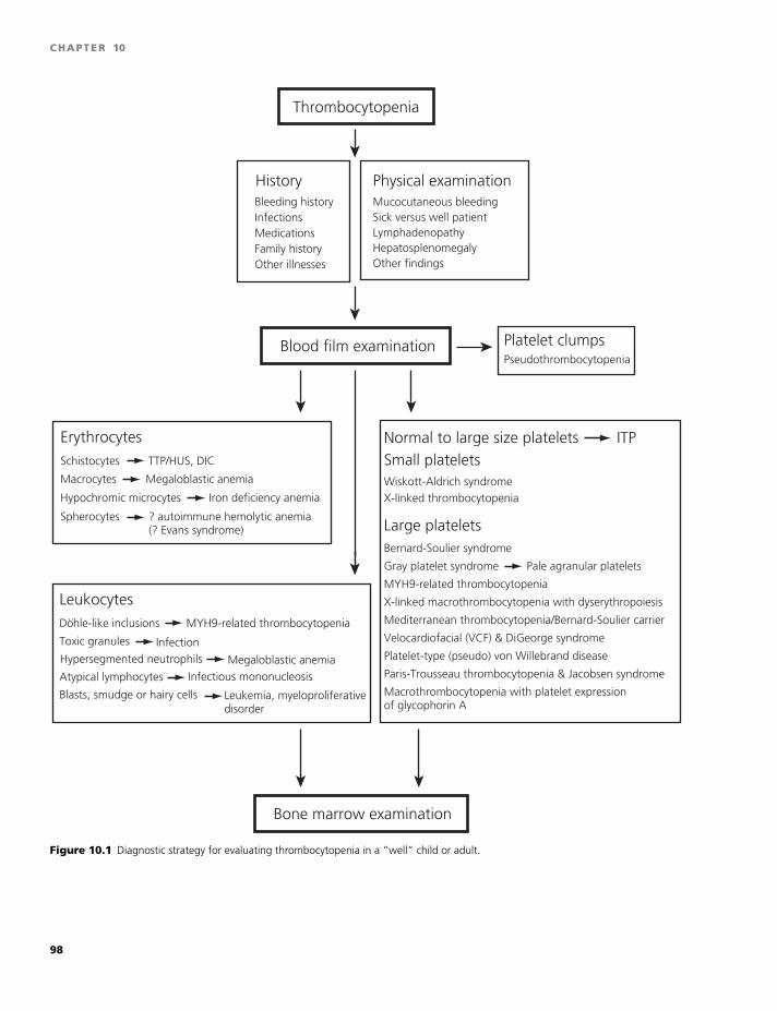
BLBK186-Key May 4, 2009 9:30
CHAPTER 10
Bone marrow examination
Medications
Thrombocytopenia
HistoryBleeding history
Family historyOther illnesses
Lymphadenopathy
Physical examinationMucocutaneous bleeding
Hepatosplenomegaly Other findings
Sick versus well patientInfections
Blood film examinationPseudothrombocytopenia
Platelet clumps
Small plateletsNormal to large size platelets ITP
Wiskott-Aldrich syndromeX-linked thrombocytopenia
Large plateletsBernard-Soulier syndrome
Gray platelet syndrome
MYH9-related thrombocytopenia
X-linked macrothrombocytopenia with dyserythropoiesis
Mediterranean thrombocytopenia/Bernard-Soulier carrier
Velocardiofacial (VCF) & DiGeorge syndrome
Platelet-type (pseudo) von Willebrand disease
Paris-Trousseau thrombocytopenia & Jacobsen syndrome
Macrothrombocytopenia with platelet expression of glycophorin A
ErythrocytesSchistocytes
Macrocytes
TTP/HUS, DIC
Megaloblastic anemia
Spherocytes ? autoimmune hemolytic anemia(? Evans syndrome)
Leukocytes
Toxic granules
MYH9-related thrombocytopenia
Infection
Atypical lymphocytes Infectious mononucleosis
Dohle-like inclusions..
Blasts, smudge or hairy cells Leukemia, myeloproliferativedisorder
Hypersegmented neutrophils Megaloblastic anemia
Hypochromic microcytes Iron deficiency anemia
Pale agranular platelets
Figure 10.1 Diagnostic strategy for evaluating thrombocytopenia in a “well” child or adult.
98
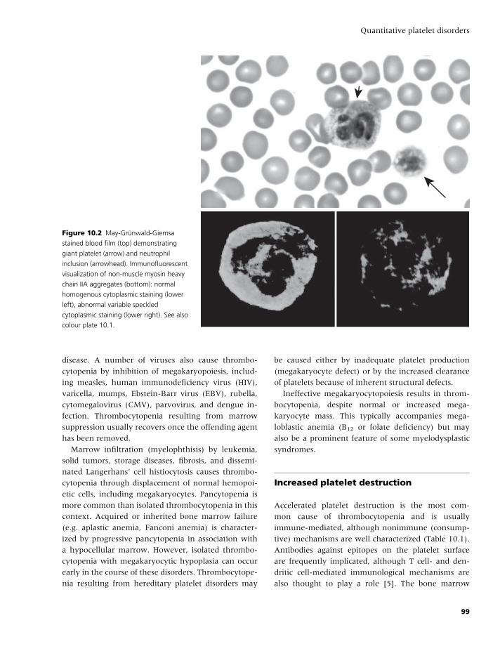
BLBK186-Key May 4, 2009 9:30
Quantitative platelet disorders
Figure 10.2 May-Grunwald-Giemsa
stained blood film (top) demonstrating
giant platelet (arrow) and neutrophil
inclusion (arrowhead). Immunofluorescent
visualization of non-muscle myosin heavy
chain IIA aggregates (bottom): normal
homogenous cytoplasmic staining (lower
left), abnormal variable speckled
cytoplasmic staining (lower right). See also
colour plate 10.1.
disease. A number of viruses also cause thrombo-
cytopenia by inhibition of megakaryopoiesis, includ-
ing measles, human immunodeficiency virus (HIV),
varicella, mumps, Ebstein-Barr virus (EBV), rubella,
cytomegalovirus (CMV), parvovirus, and dengue in-
fection. Thrombocytopenia resulting from marrow
suppression usually recovers once the offending agent
has been removed.
Marrow infiltration (myelophthisis) by leukemia,
solid tumors, storage diseases, fibrosis, and dissemi-
nated Langerhans’ cell histiocytosis causes thrombo-
cytopenia through displacement of normal hemopoi-
etic cells, including megakaryocytes. Pancytopenia is
more common than isolated thrombocytopenia in this
context. Acquired or inherited bone marrow failure
(e.g. aplastic anemia, Fanconi anemia) is character-
ized by progressive pancytopenia in association with
a hypocellular marrow. However, isolated thrombo-
cytopenia with megakaryocytic hypoplasia can occur
early in the course of these disorders. Thrombocytope-
nia resulting from hereditary platelet disorders may
be caused either by inadequate platelet production
(megakaryocyte defect) or by the increased clearance
of platelets because of inherent structural defects.
Ineffective megakaryocytopoiesis results in throm-
bocytopenia, despite normal or increased mega-
karyocyte mass. This typically accompanies mega-
loblastic anemia (B12 or folate deficiency) but may
also be a prominent feature of some myelodysplastic
syndromes.
Increased platelet destruction
Accelerated platelet destruction is the most com-
mon cause of thrombocytopenia and is usually
immune-mediated, although nonimmune (consump-
tive) mechanisms are well characterized (Table 10.1).
Antibodies against epitopes on the platelet surface
are frequently implicated, although T cell- and den-
dritic cell-mediated immunological mechanisms are
also thought to play a role [5]. The bone marrow
99
BLBK186-Key May 4, 2009 9:30
CHAPTER 10
in destructive thrombocytopenias typically reveals
megakaryocytic hyperplasia, although ITP is some-
times accompanied by suboptimal megakaryopoiesis
for reasons that are still being elucidated (see below).
Patient history
Immediate (rather than delayed) bleeding is typical of
thrombocytopenia, similar to other disorders of pri-
mary hemostasis, including platelet function defects
and von Willebrand disease (VWD). Distinctive fea-
tures include:� petechiae,� mucocutaneous bleeding,� epistaxis, and� menorrhagia.
Conversely, hemarthroses and intramuscular hema-
tomas are rare in contrast to defects of secondary
hemostasis, such as hemophilia. A careful history as-
sessing the response to trauma, surgical challenges
(including circumcision, dental extraction, and tonsil-
lectomy), menses, and postpartum hemorrhage can be
useful in defining the presence of a primary hemo-
static defect.
Bleeding since birth or early childhood is suggestive
of an inherited condition, whereas symptoms in older
patients are more likely to be caused by an acquired
defect.
Family history
A family history of bleeding and thrombocytopenia
suggests an inherited condition. Table 10.2 lists some
hereditary thrombocytopenias and their mode of in-
heritance [6,7]. A preponderance to autoimmune dis-
ease, including ITP, is also observed in some families.
A diagnosis of NAIT may be foreshadowed by a his-
tory of a previous child (sibling or cousin) affected by
intracranial hemorrhage (ICH) or thrombocytopenia
during the neonatal period.
Medication history
A careful medication history is important because
many drugs can cause thrombocytopenia, as shown
in Table 10.3 [8,9]. Platelet-inhibiting drugs, such as
ASA and other nonsteroidal anti-inflammatory drugs
(NSAIDs), ticlopidine, clopidogrel, dipyridamole, and
GPIIb/IIIa antagonists (abciximab, tirofiban, eptifi-
batide), should also be identified when evaluating a
thrombocytopenic patient, as these agents may ex-
acerbate the bleeding. Particular attention should be
paid to whether the patient is receiving heparin (in-
cluding exposure to heparin in line flushes) because
heparin-induced thrombocytopenia (HIT; see below)
needs to be excluded. Nonprescription (e.g. herbal)
medications should also be documented, as they may
contribute to thrombocytopenia and/or platelet dys-
function. For instance, patients and physicians may
not be aware that tonic water (as in “gin and tonic”)
contains quinine, an extract from cinchona tree bark
that can be associated with thrombocytopenia.
Medical history
InfectionOne of the most common causes of thrombocytope-
nia is infection. Infectious causes of thrombocytopenia
include HIV, hepatitis C virus (HCV), influenza, vari-
cella zoster virus, rubella virus, EBV, CMV, hantavirus,
mycoplasma, mycobacteria, malaria, trypanosomiasis,
Rickettsiae, and Ehrlichiae. Patients at risk for HIV and
HCV infection, such as intravenous drug users and
individuals who practice high-risk sexual activities
(e.g. unprotected sex with multiple partners) war-
rant particular attention, as virus-associated thrombo-
cytopenia is common in these diseases (see below).
Transient thrombocytopenia may be observed in chil-
dren receiving live viral vaccines, although a direct
causative role has not been established. Helicobac-
ter pylori infection can be associated with ITP, and
antimicrobial therapy may improve platelet counts in
these patients. Infection-associated hemolytic uremic
syndrome (HUS) caused by Escherichia coli (serotype
O157:H7), Shigella, Salmonella, and Campylobacter
jejuni can follow an acute diarrheal illness and is char-
acterized by thrombocytopenia with schistocytes seen
on a blood film. Meningococcemia should always be
considered in any unwell child found to have throm-
bocytopenia. Severe sepsis resulting from bacterial
infection leading to disseminated intravascular co-
agulation (DIC) results in thrombocytopenia and
100
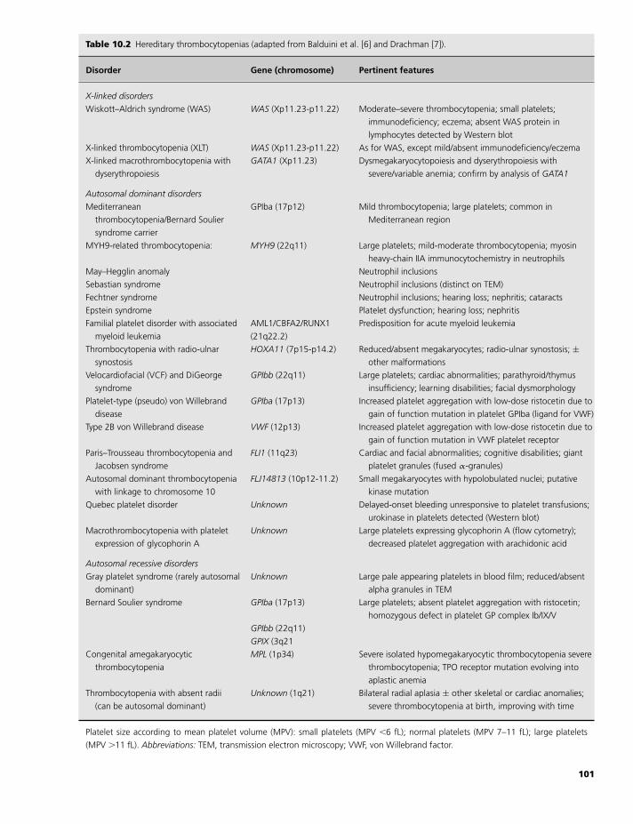
BLBK186-Key May 4, 2009 9:30
Quantitative platelet disordersTable 10.2 Hereditary thrombocytopenias (adapted from Balduini et al. [6] and Drachman [7]).
Disorder Gene (chromosome) Pertinent features
X-linked disorders
Wiskott–Aldrich syndrome (WAS) WAS (Xp11.23-p11.22) Moderate–severe thrombocytopenia; small platelets;
immunodeficiency; eczema; absent WAS protein in
lymphocytes detected by Western blot
X-linked thrombocytopenia (XLT) WAS (Xp11.23-p11.22) As for WAS, except mild/absent immunodeficiency/eczema
X-linked macrothrombocytopenia with
dyserythropoiesis
GATA1 (Xp11.23) Dysmegakaryocytopoiesis and dyserythropoiesis with
severe/variable anemia; confirm by analysis of GATA1
Autosomal dominant disorders
Mediterranean
thrombocytopenia/Bernard Soulier
syndrome carrier
GPIba (17p12) Mild thrombocytopenia; large platelets; common in
Mediterranean region
MYH9-related thrombocytopenia: MYH9 (22q11) Large platelets; mild-moderate thrombocytopenia; myosin
heavy-chain IIA immunocytochemistry in neutrophils
May–Hegglin anomaly Neutrophil inclusions
Sebastian syndrome Neutrophil inclusions (distinct on TEM)
Fechtner syndrome Neutrophil inclusions; hearing loss; nephritis; cataracts
Epstein syndrome Platelet dysfunction; hearing loss; nephritis
Familial platelet disorder with associated
myeloid leukemia
AML1/CBFA2/RUNX1
(21q22.2)
Predisposition for acute myeloid leukemia
Thrombocytopenia with radio-ulnar
synostosis
HOXA11 (7p15-p14.2) Reduced/absent megakaryocytes; radio-ulnar synostosis; ±other malformations
Velocardiofacial (VCF) and DiGeorge
syndrome
GPIbb (22q11) Large platelets; cardiac abnormalities; parathyroid/thymus
insufficiency; learning disabilities; facial dysmorphology
Platelet-type (pseudo) von Willebrand
disease
GPIba (17p13) Increased platelet aggregation with low-dose ristocetin due to
gain of function mutation in platelet GPIba (ligand for VWF)
Type 2B von Willebrand disease VWF (12p13) Increased platelet aggregation with low-dose ristocetin due to
gain of function mutation in VWF platelet receptor
Paris–Trousseau thrombocytopenia and
Jacobsen syndrome
FLI1 (11q23) Cardiac and facial abnormalities; cognitive disabilities; giant
platelet granules (fused α-granules)
Autosomal dominant thrombocytopenia
with linkage to chromosome 10
FLJ14813 (10p12-11.2) Small megakaryocytes with hypolobulated nuclei; putative
kinase mutation
Quebec platelet disorder Unknown Delayed-onset bleeding unresponsive to platelet transfusions;
urokinase in platelets detected (Western blot)
Macrothrombocytopenia with platelet
expression of glycophorin A
Unknown Large platelets expressing glycophorin A (flow cytometry);
decreased platelet aggregation with arachidonic acid
Autosomal recessive disorders
Gray platelet syndrome (rarely autosomal
dominant)
Unknown Large pale appearing platelets in blood film; reduced/absent
alpha granules in TEM
Bernard Soulier syndrome GPIba (17p13) Large platelets; absent platelet aggregation with ristocetin;
homozygous defect in platelet GP complex Ib/IX/V
GPIbb (22q11)
GPIX (3q21
Congenital amegakaryocytic
thrombocytopenia
MPL (1p34) Severe isolated hypomegakaryocytic thrombocytopenia severe
thrombocytopenia; TPO receptor mutation evolving into
aplastic anemia
Thrombocytopenia with absent radii
(can be autosomal dominant)
Unknown (1q21) Bilateral radial aplasia ± other skeletal or cardiac anomalies;
severe thrombocytopenia at birth, improving with time
Platelet size according to mean platelet volume (MPV): small platelets (MPV �6 fL); normal platelets (MPV 7–11 fL); large platelets
(MPV �11 fL). Abbreviations: TEM, transmission electron microscopy; VWF, von Willebrand factor.
101
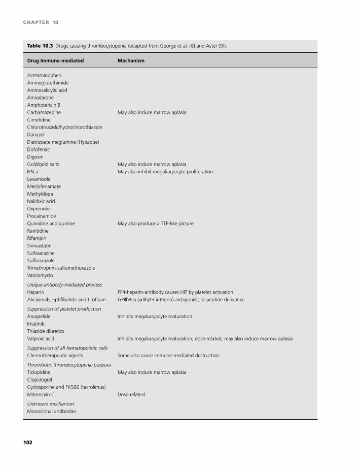
BLBK186-Key May 4, 2009 9:30
CHAPTER 10
Table 10.3 Drugs causing thrombocytopenia (adapted from George et al. [8] and Aster [9]).
Drug Immune-mediated Mechanism
Acetaminophen
Aminoglutethimide
Aminosalicylic acid
Amiodarone
Amphotericin B
Carbamazepine May also induce marrow aplasia
Cimetidine
Chlorothiazide/hydrochlorothiazide
Danazol
Diatrizoate meglumine (Hypaque)
Diclofenac
Digoxin
Gold/gold salts May also induce marrow aplasia
IFN-a May also inhibit megakaryocyte proliferation
Levamisole
Meclofenamate
Methyldopa
Nalidixic acid
Oxprenolol
Procainamide
Quinidine and quinine May also produce a TTP-like picture
Ranitidine
Rifampin
Simvastatin
Sulfasalazine
Sulfisoxazole
Trimethoprim-sulfamethoxazole
Vancomycin
Unique antibody-mediated process
Heparin PF4-heparin-antibody causes HIT by platelet activation
Abciximab, eptifibatide and tirofiban GPIIb/IIIa (αIIbβ3 integrin) antagonist; or peptide derivative
Suppression of platelet production
Anagrelide Inhibits megakaryocyte maturation
Imatinib
Thiazide diuretics
Valproic acid Inhibits megakaryocyte maturation; dose-related; may also induce marrow aplasia
Suppression of all hematopoietic cells
Chemotherapeutic agents Some also cause immune-mediated destruction
Thrombotic thrombocytopenic purpura
Ticlopidine May also induce marrow aplasia
Clopidogrel
Cyclosporine and FK506 (tacrolimus)
Mitomcyin C Dose-related
Unknown mechanism
Monoclonal antibodies
102
BLBK186-Key May 4, 2009 9:30
Quantitative platelet disorders
associated coagulopathy (see below). It should also be
highlighted that numerous infections may be associ-
ated with petechiae and/or purpura in the absence of
thrombocytopenia.
Systemic diseasesSystemic diseases involving the bone marrow, such as
aplastic anemia, myelofibrosis, leukemia, lymphoma,
or metastatic cancers infiltrating the bone marrow, of-
ten result in thrombocytopenia, but this is often man-
ifested as a pancytopenia involving all three cell types:
platelets, erythrocytes, and leukocytes.
Other systemic illnesses, such as renal and liver dis-
ease, not only affect platelet function but also are often
accompanied by mild to moderate thrombocytopenia.
Other causesPoor nutritional intake, such as can occur in the el-
derly or alcoholics, may result in decreased intake of
folate, resulting in megaloblastic anemia and throm-
bocytopenia. In addition, excessive alcohol intake has
direct inhibitory effects on platelet production.
Pregnancy is commonly associated with throm-
bocytopenia (in approximately 5–10% of pregnant
women), usually appearing during the third trimester;
however, in this situation, alternative etiologies need
to be considered (see below).
Transfusion historyPrevious transfusions may place an individual at risk
of developing post-transfusion purpura, in which se-
vere thrombocytopenia can appear 7–14 days af-
ter the transfusion of a blood product. Transfusion-
associated infection, such as HIV, HCV, CMV, West
Nile virus, or malaria, may also be complicated by
thrombocytopenia.
Physical examination
The clinical appearance of the patient is of paramount
importance in the assessment of the thrombocytopenic
patient and provides the first clue as to the likely eti-
ology. A sick patient in the intensive care unit may
have a number of possible contributing factors, includ-
ing severe sepsis, DIC, drug-induced post-transfusion
purpura, massive blood transfusion, and systemic ill-
ness. In contrast, a well patient with newly diag-
nosed isolated thrombocytopenia may have an inher-
ited thrombocytopenia, ITP, or, in a neonate, auto- or
alloimmune thrombocytopenia.
Certain hereditary thrombocytopenias are accom-
panied by typical physical findings, such as skeletal ab-
normalities, facial dysmorphologies, hearing deficien-
cies, and cataracts, as described in Table 10.2. Evidence
of an enlarged spleen or other systemic findings (e.g.
fever, jaundice, adenopathy, cachexia) can be helpful
in deciding whether an underlying illness is the likely
cause for the thrombocytopenia.
Petechiae consisting of small (�2 mm), red, flat,
discrete lesions, occurring most frequently in the
dependent areas on the ankles and feet, represent
extravasated red cells from capillaries and are the hall-
mark of a primary hemostatic disorder.They are non-
tender and do not blanch under pressure. Purpura
(�1 cm) and ecchymoses (�1 cm) represent larger ar-
eas of bleeding, and when observed in mucous mem-
branes, such as the oropharynx, are described as “wet
purpura.” These findings are in contrast to delayed
bleeding into joints or muscle, which suggest a coagu-
lation disorder rather than a platelet or von Willebrand
factor (VWF) problem.
Laboratory evaluation
Blood filmThe importance of examining a blood film in a pa-
tient with newly diagnosed thrombocytopenia cannot
be overemphasized. For example:� Visualization of schistocytes (RBC fragments) could
be indicative of thrombotic thrombocytopenic pur-
pura/hemolytic uremic syndrome (TTP/HUS) or DIC.� Evidence of platelet clumps would suggest pseu-
dothrombocytopenia.� Megathrombocytes with Dohle-like inclusions in
neutrophils could be indicative of MYH9-related dis-
eases (Figure 10.2 & Plate 10.1 top, arrow and arrow-
head, respectively).� Pale agranular-appearing platelets could represent
gray platelet syndrome.� Blasts suggest the diagnosis of leukemia or a myelo-
proliferative disorder.� Macrocytes with hypersegmented neutrophils sug-
gest megaloblastic anemia.� Toxic granulation suggests infection.
103
BLBK186-Key May 4, 2009 9:30
CHAPTER 10
� Spherocytes and polychromasia may be observed in
Evan’s syndrome (coexisting autoimmune hemolytic
anemia and ITP).
Pseudothrombocytopenia resulting from EDTA-
induced platelet clumping may be overcome by ob-
taining a film from a drop of blood smeared directly
onto a slide or by collection of blood into citrate or
heparin anticoagulants.
Mean platelet volume andreticulated plateletsFor inherited causes of thrombocytopenia, a useful di-
agnostic algorithm is based on the platelet size or mean
platelet volume [6]. One caveat of this approach is
that not all automated counters are able to detect very
small platelets (e.g. as in Wiskott–Aldrich syndrome)
or very large platelets (e.g. as in Bernard Soulier syn-
drome), resulting in underestimation of the platelet
count. This emphasizes the importance of examining
the blood film.
Platelets with a higher RNA content are believed to
represent younger (“immature”) cells, and it has been
postulated that an increase in the relative proportion
of young platelets is indicative of increased platelet
turnover, akin to the reticulocytosis that occurs during
marrow recovery or hemolytic anemia. Many modern
hematology analyzers use a flow cytometer in com-
bination with a dye that binds to RNA within cells
to provide a direct estimate of the immature platelet
fraction (“reticulated platelets”). The use of this tech-
nology has not yet been clearly defined; however,
the parameter may be useful in predicting platelet
recovery following chemotherapy, and also in the
initial diagnostic evaluation of the thrombocytopenic
patient [10].
Bone marrow examinationFor a typical presentation of ITP, a bone marrow ex-
amination is not required in patients under the age of
60 years. However, a bone marrow aspirate and biopsy
is recommended in:� patients before undertaking therapeutic splenec-
tomy;� those with additional cytopenias;� patients with lassitude, protracted fever, or bone or
joint pain;� patients with lymphadenopathy and/or organo-
megaly;
� those with unexplained macrocytosis or dysplastic
features on blood film; and� patients with suboptimal response to treatment.
Specialized platelet function tests(see Chapter 5)Specialized tests that may be indicated in the evalua-
tion of specific hereditary thrombocytopenias include:� platelet aggregation;� flow cytometry using antibodies labeling GPIb (for
Bernard Soulier syndrome);� platelet electron microscopy (for gray platelet syn-
drome);� specialized immunocytochemistry (for MYH9-
related diseases, Figure 10.2 & Plate 10.1);� Western blot for protein analyses (for Quebec
platelet disorder); and� DNA analysis (for confirmatory testing when genetic
basis is known).
Specific conditions
Immune thrombocytopenic pupuraITP is probably the most common immune destructive
thrombocytopenia in children and adults, occurring in
approximately 1 in 20,000 persons/year [11]. ITP is
an autoimmune disorder in which autoantibodies are
produced against platelet surface glycoproteins, result-
ing in increased clearance of platelets from the cir-
culation. In children, the condition is typically acute,
and spontaneous resolution is common. Conversely,
in adults, it is frequently a chronic disorder with an
insidious onset, often diagnosed incidentally when a
blood count is performed for other reasons [12]. There
is a modest female predominance in adults, whereas
young boys and girls are affected equally. Although
the etiology of ITP is poorly understood, infections ap-
pear to play a role because, in the case of childhood
ITP, the onset is often preceded by a viral infection.
Cases in which an underlying disease cannot be iden-
tified are classified as primary ITP. However 5–10% of
adult patients who initially present with ITP will sub-
sequently be diagnosed with an underlying systemic
autoimmune disease. Secondary causes of ITP are ob-
served in patients with:� systemic lupus erythematosus (SLE);� antiphospholipid antibody syndrome;
104
BLBK186-Key May 4, 2009 9:30
Quantitative platelet disorders
� immune deficiency syndromes;� chronic infections (e.g. HIV, HCV, Helicobacter pylori);� lymphoproliferative disorders; and� neoplasia-associated immune thrombocytopenia.
DiagnosisThe diagnosis of primary ITP is predominantly one
of exclusion and is suggested by the presence of
isolated thrombocytopenia in an otherwise well pa-
tient in the absence of other causes. The patient
may present with evidence of mucocutaneous bleed-
ing or after a routine complete blood count (CBC)
in an asymptomatic individual. A thorough history
and physical examination combined with careful re-
view of a CBC and blood film is sufficient for diagno-
sis in most cases. Underlying systemic diseases, drug-
induced thrombocytopenias, as well as hereditary
thrombocytopenias (e.g. positive family history, ab-
normal blood film) should be ruled out. Bone marrow
aspirate and biopsy are indicated if the clinical fea-
tures are atypical, or if additional abnormalities are
noted on the CBC or film. In older individuals, iso-
lated thrombocytopenia may be the initial presenting
feature of myelodysplasia or malignancy, and bone
marrow biopsy is recommended in adults over 60
years of age even when the findings appear typical
of ITP. Platelet autoantibody assays are not sensitive
nor specific enough to be clinically useful, and should
not be relied on for diagnosis. Interestingly, plasma
TPO levels are usually normal or mildly elevated in
ITP but are greatly elevated in amegakaryocytic states
(e.g. congenital amegakaryocytic thrombocytopenia,
bone marrow suppression, and aplastic anemia), and
therefore, may be diagnostically useful if available.
However, measurement of TPO is currently limited to
the research setting.
Principles of managementIn adults, treatment is generally indicated if the
platelet count is below 20 × 109/L or below 50 ×109/L in the presence of significant bleeding or ad-
ditional risk factors for bleeding. Those with higher
counts can be merely observed, as the bleeding risk
is low and early treatment does not modify the course
of the disease. Platelet-inhibiting drugs, such as ASA
and other NSAIDs, should be avoided. Initial treat-
ment of ITP consists of glucocorticoids (prednisone 1
mg/kg/day p.o.), IV immunoglobulin (IVIG 1 g/kg),
or IV anti-D (50–75 �g/kg)in Rh(D)-positive patients
with intact spleens. With major bleeding episodes, or
if the platelet count is less than 10 × 109/L, gluco-
corticoids can be given together with either IVIG or
IV anti-D. In the presence of ICH, platelet transfusions
are also indicated.
The natural course of acute ITP in children is that
most will recover completely within a few weeks with-
out any treatment. The major concern is ICH, which
can occur when platelets fall below 20 × 109/L but
usually only when they fall below 10 × 109/L. The
incidence of ICH in ITP is estimated to be between
0.2% and 1%. If the child presents with wet pur-
pura (extensive mucocutaneous bleeding) or evidence
of major bleeding and/or platelet counts below 20 ×109/L, then oral prednisone (3–4 mg/kg/day for 3–4
days), IV methylprednisolone (5–30 mg/kg/day),IVIG
(0.8–1 g/kg), or IV anti-D (50–75 �g/kg) in Rh(D)-
positive children are all efficacious regimens, although
the response to IVIG is generally more rapid. In adults,
platelet transfusions are reserved for life-threatening
bleeding and ICH.
Relapsed and chronic ITPAround 70% of patients will respond to initial ther-
apy with corticosteroids or immunoglobulin, although
in adults, the effect is most often transient or re-
quires repeated doses to maintain response. In con-
trast, only 25% of children will relapse, and late
spontaneous remission is well-recognized even in this
subgroup. Chronic ITP is generally defined as persis-
tence of thrombocytopenia severe enough to warrant
therapy more than 6 months after initial diagnosis.
Splenectomy is the currently accepted second-line ap-
proach for adults who fail to respond to first-line
therapy or experience unacceptable side effects from
repeated steroid exposure, and 60–70% will have
a durable response to this procedure. In children,
splenectomy is generally deferred as long as possible,
due to the higher life-long risk of post-splenectomy
sepsis and the greater chance of remission compared
with adults. Numerous therapeutic regimens have
been described for those patients who fail to respond
or relapse after splenectomy, although evidence for ef-
ficacy is mostly limited to case series. Examples in-
clude pulsed corticosteroids, danazol, dapsone, aza-
thioprine, cyclosporine, mycophenolate, vincristine,
and cyclophosphamide.
105
BLBK186-Key May 4, 2009 9:30
CHAPTER 10
Recent attention has focused on novel therapeutic
approaches to chronic ITP, either as salvage following
failed splenectomy or to avoid splenectomy alto-
gether. Rituximab, a monoclonal anti-CD20 chimeric
antibody, has shown promise in this context, with
sustained responses and minimal reported toxicity
in 40–60% of patients receiving a lymphoma-based
regimen of 375 mg/m2 weekly for 4 doses [13]. It
should be highlighted that rituximab has not been
licensed for use in this setting, and patients must be
counseled accordingly. Furthermore the potent B cell
suppression that follows rituximab may increase the
risk of viral infection or reactivation, and this must
be balanced against the risk of bacterial infection after
splenectomy.
Other research has focused on the potential role
of TPO-R agonists. Although ITP is predominantly a
condition of increased platelet destruction, TPO lev-
els in ITP are not elevated in proportion to the sever-
ity of thrombocytopenia, possibly due to faster TPO
clearance resulting from rapid platelet turnover. First-
generation TPO-R agonists, recombinant forms of hu-
man TPO, were withdrawn from development in 1998
after thrombocytopenia due to anti-TPO antibodies
was observed in some healthy volunteers receiving
such agents. There are numerous second-generation
TPO-R agonists currently in development, and these
appear to be nonimmunogenic, well-tolerated, and ef-
fective at increasing platelet count. However, at this
stage, only two agents have undergone phase 3 trials,
both in chronic ITP, namely romiplostim (AMG-531)
and eltrombopag (SB-497115). The reader is referred
to a recent review by Kuter for an overview of these
new agents [3].
Evan’s syndromeThe combination of ITP with autoimmune hemolytic
anemia in the absence of an underlying cause is re-
ferred to as Evan’s syndrome, and has a pathogene-
sis and clinical course distinct from that of classic ITP
[14]. More than half of these patients also have au-
toimmune neutropenia. Response to standard ther-
apy is often poorly sustained, and multiple relapses
with significant long-term morbidity is typical. Spe-
cific disorders that mimic Evan’s syndrome must be
excluded, as the management of these conditions is
different. These include autoimmune lymphoprolifer-
ative syndrome (ALPS), chronic variable immunod-
eficiency (CVID), and systemic autoimmune disease
(e.g. SLE).
Drug-induced thrombocytopeniaDrug-induced thrombocytopenia is common and
probably under-recognized, either because a platelet
count is not measured or because thrombocytopenia
is attributed to other factors. There are a large num-
ber of agents known to cause thrombocytopenia, and
most of these can be broadly divided into the following
categories:� drugs that cause predictable dose-dependent mar-
row suppression;� drugs that cause idiosyncratic marrow aplasia;� drugs that specifically inhibit megakaryopoiesis;� drugs that trigger immune destruction of platelets;� drugs that cause a TTP-like condition; and� drugs that induce platelet aggregation.
Chemotherapeutic agents used for malignancy
or potent immunosuppression often cause dose-
dependent thrombocytopenia as a result of generalized
bone marrow suppression, although some of these
agents can also induce immune-mediated platelet de-
struction. Although the mechanisms are poorly un-
derstood, a number of drugs have been implicated in
aplastic anemia, including anticonvulsants, NSAIDs,
sulfonamides, and gold salts. Some drugs known to
specifically inhibit megakaryopoiesis are listed in Table
10.3. Anagrelide, used in the treatment of thrombo-
cythemia in patients with myeloproliferative diseases,
can cause severe thrombocytopenia. Valproic acid,
commonly used in seizure disorders and for psychiatric
patients, has been associated with dose-dependent
thrombocytopenia resulting from direct megakary-
ocyte suppression. Thiazide diuretics also have a mild
inhibitory effect on megakaryocytes.
Many drugs have been implicated in producing a
TTP-like condition, with thrombocytopenia, hemoly-
sis, and varying degrees of neurological or renal dys-
function, although for most agents, this appears to
be an exceedingly rare event [15]. The pathogene-
sis is poorly understood, although direct or immune-
mediated endothelial injury may be an important
trigger. Drugs for which a causal association seems
likely include mitomycin C (dose-related), calcienurin
inhibitors, such as cyclosporine and tacrolimus (1–
3% incidence), and the thienopyridine derivitives
ticlopidine (�0.1% incidence) and clopidogrel (rare).
106
BLBK186-Key May 4, 2009 9:30
Quantitative platelet disorders
Interestingly, quinine can also cause TTP, although it is
better known for inducing antibody-mediated platelet
destruction.
Drug-induced antibody-mediated platelet destruc-
tion is the most common mechanism of iatrogenic
thrombocytopenia [9]. There are several mechanisms
of drug-induced ITP, although the “quinine-type” ac-
counts for the majority:� Drugs that bind to platelet glycoproteins forming
a “compound epitope” include penicillin, quinidine,
quinine, and sulfonamide. The antibody binding to
such platelets is dependent on the presence of the of-
fending drug.� Gold salts and procainamide, on the other hand, can
induce true autoantibodies, which subsequently can
bind to platelets in the absence of the original offend-
ing drug.� Antiplatelet agents such as tirofiban, eptifibatide,
and abciximab, which specifically target the GPIIb/IIIa
(αIIbβ3 integrin) receptor on platelets, cause throm-
bocytopenia in 1–5% of cardiac patients via antibody-
mediated processes.
Diagnosis of drug-induced ITP requires a high in-
dex of suspicion, as systemic illness or other coexis-
tent factors may confuse the clinical picture. Onset of
thrombocytopenia within 5–7 days of commencing a
new drug is an important clue. Withdrawal of the of-
fending agent leads to resolution of thrombocytopenia
in most cases, although rarely IVIG, steroids, or more
aggressive management may be indicated. Inadver-
tent rechallenge with the causative drug may induce
rapid and severe thrombocytopenia and should be
avoided.
Heparin-induced thrombocytopeniaHIT differs from other thrombocytopenias in that it is
a hypercoagulable state rather than a bleeding condi-
tion, manifesting as venous and/or arterial thrombosis
[16]. This iatrogenic disorder is discussed in more de-
tail in Chapter 26.
Pregnancy-associated thrombocytopenia(see also Chapter 24)There is a physiological decline in platelet count
during the course of normal pregnancy, most pro-
nounced in the third trimester, although only 5–10%
of women will become thrombocytopenic [17]. Mild
thrombocytopenia (100–149 × 109/L) is common and
of no clinical significance; however, lower platelet
counts require further evaluation. The most common
causes of pregnancy-associated thrombocytopenia
include:� Gestational thrombocytopenia (incidental or benign
thrombocytopenia of pregnancy): accounts for ap-
proximately 75% of cases.� Preeclampsia ± HELLP (hemolysis, elevated liver en-
zymes, low platelets): accounts for approximately 20%
of cases.� ITP ± SLE: accounts for approximately 4% of cases.
Gestational thrombocytopenia is a diagnosis of ex-
clusion, but in 95% of cases, it manifests as mild
thrombocytopenia in an asymptomatic pregnant pa-
tient with a previously normal platelet count. This
is a benign condition that requires no treatment. Al-
though more severe thrombocytopenia can occasion-
aly be seen, counts below 70 × 109/L should raise
strong suspicion of an alternative diagnosis. Similarly,
the finding of thrombocytopenia in early pregnancy
is more suggestive of ITP or a preexisting condition.
Thrombocytopenia develops in approximately 20%
of patients with preeclampsia, and there is an in-
verse relationship between platelet count and sever-
ity of disease. The HELLP syndrome can be a serious
complication, associated with up to 20% fetal mor-
tality. Thrombocytopenia associated with preeclamp-
sia and HELLP syndrome improves following deliv-
ery, whereas that observed in the primary microan-
giopathic hemolytic anemias, TTP and HUS, does not.
These conditions may sometimes be difficult to discern
from preeclampsia or HELLP syndrome in a pregnant
woman, and plasma exchange may be required despite
an uncertain diagnosis (see below). DIC complicates a
small proportion of cases, and a coagulation screen is
an important component of the diagnostic work-up.
ITP occurs in 1–2 per 1000 pregnancies and represents
approximately 3–5% of the causes of thrombocytope-
nia during pregnancy. A pregnant patient with ITP
can be treated with IVIG (1 g/kg prepregnant weight)
and/or prednisone (1 mg/kg prepregnant weight) in
the acute setting to raise the platelet count to above
10 × 109/L. Measurement of platelet count in the
newborn is important, as 5–10% of infants born to
mothers with ITP will have significant thrombocy-
topenia (�50 × 109/L). The management of ITP dur-
ing pregnancy is discussed in more detail in a recent
review [18].
107
BLBK186-Key May 4, 2009 9:30
CHAPTER 10
Post-transfusion purpuraPost-transfusion purpura (PTP) is a rare disorder,
which usually manisfests as severe thrombocytope-
nia 7–14 days following transfusion of a blood prod-
uct. It is caused by the formation of high-titre alloan-
tibodies against platelet glycoproteins and represents
an anamnestic immune response in a patient previ-
ously sensitized through antigen exposure in preg-
nancy and/or transfusion. The antibodies are most
commonly directed against the platelet alloantigen
HPA-1a epitope (also known as PLA1 or Zwa), where
the platelet GPIIIa contains a leucine at position 33.
Polymorphisms of GPIIIa result in alloantigen HPA-1a
and alloantigen HPA-1b (PLA2 or Zwb; proline at posi-
tion 33 of GPIIIa), which occur at a frequency of ap-
proximately 86% and 14% in caucasians, respectively.
Classically, the affected patient is a homozygous HPA-
1b middle-aged multiparous woman; however, the
condition also occurs in men and nulliparous women.
The alloantibodies paradoxically cause destruction of
autologous as well as transfused platelets through
poorly understood mechanisms. PTP has been esti-
mated to occur following 1/50–100,000 transfusions,
although this may represent an underestimate as the
diagnosis may be overshadowed by coexisting factors,
such as heparin exposure or sepsis. Interestingly, in
countries in which universal leukodepletion is prac-
ticed, a striking reduction in the incidence of PTP has
been noted, presumably due to the fact that the pro-
cess removes platelets from red cell concentrates [19].
Diagnosis of PTP requires a strong index of suspicion,
followed by demonstration of high-titre HPA alloan-
tibodies in the transfusion recipient. The observation
of a decline in platelet count below baseline following
a platelet transfusion can be an important clue to
differentiate this condition from platelet refractori-
ness, which is multifactorial and far more common.
Treatment of PTP consists of IVIG, corticosteroids, or
plasmapheresis. Platelet transfusions are contraindi-
cated except in rare circumstances, when HPA-1a-
negative platelets may be used for life-threatening
bleeding complications.
HIV-associated thrombocytopeniaThere are multiple factors that contribute to the
thrombocytopenia frequently associated with HIV, in-
cluding immune mechanisms and defective platelet
production [20]. The immune-mediated platelet de-
struction in HIV is indistinguishable from ITP with
respect to increased destruction of antibody-coated
platelets and the response to prednisone, IVIG, and
splenectomy. It differs from classic ITP with respect to:� male predominance;� markedly elevated platelet-associated IgG, IgM, and
complement C3, C4;� presence of circulating immune complexes; and� antibody-mediated peroxide lysis of platelets.
Treatment with antiretroviral therapy tends to im-
prove the defective thrombopoiesis in HIV-infected
patients. TTP is also found more frequently in HIV-
infected patients.
HCV-associated thrombocytopeniaThrombocytopenia is frequently associated with
chronic liver disease, and the severity correlates with
the extent of hepatocellular damage and fibrosis [21].
There are numerous contributing factors, including
the underlying cause of liver disease itself (e.g. al-
cohol is directly toxic to megakaryocytes), associ-
ated portal hypertension with splenic sequestration of
platelets, and reduction in TPO synthesis. Liver disease
is discussed in greater detail in Chapter 21. However,
HCV-associated thrombocytopenia warrants particular
mention for several reasons:� Thrombocytopenia in HCV frequently occurs in the
absence of clinical or radiological features to suggest
portal hypertension;� There is an approximately 20-fold increase in the in-
cidence of ITP in patients with HCV, and treatment of
ITP with steroids may promote viremia;� The presence of thrombocytopenia in HCV is an
adverse prognostic factor and may limit options for
therapy, as antiviral agents such as interferon-α can
further reduce platelet count; and� Results from a recent phase 2 study using eltrom-
bopag in HCV-associated thrombocytopenia suggest
that TPO-R agonists may provide sufficient increase in
platelet count to permit initiation of antiviral therapy
in this condition [3].
MicroangiopathiesNonimmune destructive thrombocytopenias include
DIC, Kasabach–Merritt syndrome, TTP, HUS, and
other conditions listed in Table 10.1. These disor-
ders share the common pathophysiological endpoint
of platelet trapping and thrombus formation in the
108
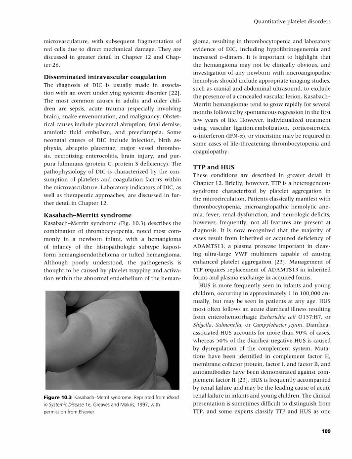
BLBK186-Key May 4, 2009 9:30
Quantitative platelet disorders
microvasculature, with subsequent fragmentation of
red cells due to direct mechanical damage. They are
discussed in greater detail in Chapter 12 and Chap-
ter 26.
Disseminated intravascular coagulationThe diagnosis of DIC is usually made in associa-
tion with an overt underlying systemic disorder [22].
The most common causes in adults and older chil-
dren are sepsis, acute trauma (especially involving
brain), snake envenomation, and malignancy. Obstet-
rical causes include placental abruption, fetal demise,
amniotic fluid embolism, and preeclampsia. Some
neonatal causes of DIC include infection, birth as-
phyxia, abruptio placentae, major vessel thrombo-
sis, necrotizing enterocolitis, brain injury, and pur-
pura fulminans (protein C, protein S deficiency). The
pathophysiology of DIC is characterized by the con-
sumption of platelets and coagulation factors within
the microvasculature. Laboratory indicators of DIC, as
well as therapeutic approaches, are discussed in fur-
ther detail in Chapter 12.
Kasabach–Merritt syndromeKasabach–Merritt syndrome (Fig. 10.3) describes the
combination of thrombocytopenia, noted most com-
monly in a newborn infant, with a hemangioma
of infancy of the histopathologic subtype kaposi-
form hemangioendothelioma or tufted hemangioma.
Although poorly understood, the pathogenesis is
thought to be caused by platelet trapping and activa-
tion within the abnormal endothelium of the heman-
Figure 10.3 Kasabach–Merrit syndrome. Reprinted from Blood
in Systemic Disease 1e, Greaves and Makris, 1997, with
permission from Elsevier.
gioma, resulting in thrombocytopenia and laboratory
evidence of DIC, including hypofibrinogenemia and
increased D-dimers. It is important to highlight that
the hemangioma may not be clinically obvious, and
investigation of any newborn with microangiopathic
hemolysis should include appropriate imaging studies,
such as cranial and abdominal ultrasound, to exclude
the presence of a concealed vascular lesion. Kasabach–
Merritt hemangiomas tend to grow rapidly for several
months followed by spontaneous regression in the first
few years of life. However, individualized treatment
using vascular ligation,embolization, corticosteroids,
�-interferon (IFN-�), or vincristine may be required in
some cases of life-threatening thrombocytopenia and
coagulopathy.
TTP and HUSThese conditions are described in greater detail in
Chapter 12. Briefly, however, TTP is a heterogeneous
syndrome characterized by platelet aggregation in
the microcirculation. Patients classically manifest with
thrombocytopenia, microangiopathic hemolytic ane-
mia, fever, renal dysfunction, and neurologic deficits;
however, frequently, not all features are present at
diagnosis. It is now recognized that the majority of
cases result from inherited or acquired deficiency of
ADAMTS13, a plasma protease important in cleav-
ing ultra-large VWF multimers capable of causing
enhanced platelet aggregation [23]. Management of
TTP requires replacement of ADAMTS13 in inherited
forms and plasma exchange in acquired forms.
HUS is more frequently seen in infants and young
children, occurring in approximately 1 in 100,000 an-
nually, but may be seen in patients at any age. HUS
most often follows an acute diarrheal illness resulting
from enterohemorrhagic Escherichia coli O157:H7, or
Shigella, Salmonella, or Campylobacter jejuni. Diarrhea-
associated HUS accounts for more than 90% of cases,
whereas 50% of the diarrhea-negative HUS is caused
by dysregulation of the complement system. Muta-
tions have been identified in complement factor H,
membrane cofactor protein, factor I, and factor B, and
autoantibodies have been demonstrated against com-
plement factor H [23]. HUS is frequently accompanied
by renal failure and may be the leading cause of acute
renal failure in infants and young children. The clinical
presentation is sometimes difficult to distinguish from
TTP, and some experts classify TTP and HUS as one
109
BLBK186-Key May 4, 2009 9:30
CHAPTER 10
disease entity, TTP/HUS. However children with HUS
resulting from E. coli O157:H7 infection, as well as HUS
caused by defects in the complement system, tend to
have normal plasma levels of ADAMTS13, suggesting
distinct microangiopathic mechanisms. Most impor-
tantly, the treatment of diarrhoea-associated HUS in
children is distinct from adults in that supportive care
is the mainstay in children. In addition, once iden-
tified, complement-deficient children can be treated
with plasma transfusions. Transfusion may also be
required for symptomatic anemia as well as dialysis
when necessary. Antimotility agents may worsen the
clinical manifestations of infectious HUS, whereas the
role of antibiotics is unresolved. Because of the poten-
tially high morbidity and mortality in TTP/HUS and
the often difficult clinical distinction between TTP
and HUS, children with atypical HUS, familial HUS,
and all adults with HUS should be treated with plasma
exchange.
HypersplenismWhen splenomegaly results in cytopenias and com-
pensatory bone marrow hyperplasia, the term “hy-
persplenism” is appropriate, although bone marrow
biopsy in this context is most often performed to ex-
clude hematologic malignancy (e.g. leukemia, lym-
phoma, myeloproliferative disease) rather than to con-
firm hypersplenism per se. Numerous conditions are
associated with splenomegaly and hypersplenism, in-
cluding portal hypertension secondary to liver dis-
ease or portal vein thrombosis, hematological malig-
nancies, chronic hemolytic anemias, storage disorders,
leishmaniasis, and malaria. The clinical picture is usu-
ally dominated by the underlying disease rather than
symptomatic pancytopenia, although in the presence
of massive splenomegaly, thrombocytopenia can be
severe, and increments following platelet transfusion
are poor. Intervention is rarely indicated for manage-
ment of thrombocytopenia alone, although improve-
ment in counts usually follows splenectomy or splenic
embolization. In the setting of portal hypertension,
surgical procedures that redirect or bypass the portal
circulation can reduce the risk of bleeding associated
with thrombocytopenia and esophageal varices.
Thrombocytopenia in the newborn infantThrombocytopenia is the most common hematologi-
cal abnormality observed during the noenatal period,
with an estimated incidence of 1–2% in healthy term
infants. However only 1 in 10 of these cases will have
a platelet count below 50 × 109/L. In contrast, throm-
bocytopenia affects up to 30% of infants admitted to
NICU, and 1 in 5 of these will be severe. The differen-
tial diagnosis is broad. However, relatively few condi-
tions account for the majority of cases, and many of
the rare disorders can be readily identified on the basis
of associated clinical and/or laboratory features [24]. A
useful approach in determining the etiology of throm-
bocytopenia in a newborn is to differentiate based on
the timing of onset and clinical condition of the in-
fant (Table 10.4). Early-onset thrombocytopenia (�72
hours of age) is most often mild to moderate in sever-
ity, and frequently relates to placental insufficiency
(e.g. preeclampsia, IUGR). In the absence of an iden-
tifiable precipitant, NAIT should always be considered
when early-onset thrombocytopenia is detected in a
well neonate (see below). Severe early-onset throm-
bocytopenia (�50 × 109/L) in a sick newborn com-
monly results from perinatal infection (e.g. group B
Streptococcus) or asphyxia (e.g. meconium aspiration
syndrome). Late-onset thrombocytopenia (�72 hours
of age) is most often due to bacterial or fungal sepsis
and/or necrotizing enterocolitis and is frequently se-
vere in this setting.
Most thrombocytopenia in the neonatal period is
self-limited or resolves with treatment of the un-
derlying condition. Treatment of severe nonimmune-
mediated thrombocytopenia in neonates consists of
platelet transfusion according to transfusion threshold
guidelines reviewed by Roberts and colleagues [24].
Neonatal alloimmune thrombocytopeniaNAIT is the most likely cause of thrombocytopenia in
the well-appearing full-term infant. The overall inci-
dence of NAIT is estimated to be 1 in 1000–2000 live
births, although severe NAIT is somewhat less fre-
quent (around 1 in 5000 births). NAIT also occurs
in preterm infants, although the diagnosis may fre-
quently be overshadowed by other contributing fac-
tors often present in this group. The importance of
recognition and accurate diagnosis lies not only in the
immediate management of the affected infant, but also
in the approach to future pregnancies of the affected
mother.
In NAIT, the destruction of fetal or neona-
tal platelets results from transplacental passage of
110
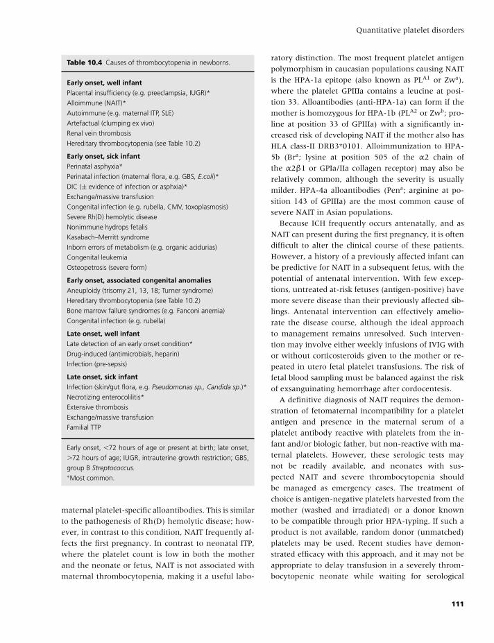
BLBK186-Key May 4, 2009 9:30
Quantitative platelet disorders
Table 10.4 Causes of thrombocytopenia in newborns.
Early onset, well infantPlacental insufficiency (e.g. preeclampsia, IUGR)*
Alloimmune (NAIT)*
Autoimmune (e.g. maternal ITP, SLE)
Artefactual (clumping ex vivo)
Renal vein thrombosis
Hereditary thrombocytopenia (see Table 10.2)
Early onset, sick infantPerinatal asphyxia*
Perinatal infection (maternal flora, e.g. GBS, E.coli)*
DIC (± evidence of infection or asphxia)*
Exchange/massive transfusion
Congenital infection (e.g. rubella, CMV, toxoplasmosis)
Severe Rh(D) hemolytic disease
Nonimmune hydrops fetalis
Kasabach–Merritt syndrome
Inborn errors of metabolism (e.g. organic acidurias)
Congenital leukemia
Osteopetrosis (severe form)
Early onset, associated congenital anomaliesAneuploidy (trisomy 21, 13, 18; Turner syndrome)
Hereditary thrombocytopenia (see Table 10.2)
Bone marrow failure syndromes (e.g. Fanconi anemia)
Congenital infection (e.g. rubella)
Late onset, well infantLate detection of an early onset condition*
Drug-induced (antimicrobials, heparin)
Infection (pre-sepsis)
Late onset, sick infantInfection (skin/gut flora, e.g. Pseudomonas sp., Candida sp.)*
Necrotizing enterocolilitis*
Extensive thrombosis
Exchange/massive transfusion
Familial TTP
Early onset, �72 hours of age or present at birth; late onset,
�72 hours of age; IUGR, intrauterine growth restriction; GBS,
group B Streptococcus.∗Most common.
maternal platelet-specific alloantibodies. This is similar
to the pathogenesis of Rh(D) hemolytic disease; how-
ever, in contrast to this condition, NAIT frequently af-
fects the first pregnancy. In contrast to neonatal ITP,
where the platelet count is low in both the mother
and the neonate or fetus, NAIT is not associated with
maternal thrombocytopenia, making it a useful labo-
ratory distinction. The most frequent platelet antigen
polymorphism in caucasian populations causing NAIT
is the HPA-1a epitope (also known as PLA1 or Zwa),
where the platelet GPIIIa contains a leucine at posi-
tion 33. Alloantibodies (anti-HPA-1a) can form if the
mother is homozygous for HPA-1b (PLA2 or Zwb; pro-
line at position 33 of GPIIIa) with a significantly in-
creased risk of developing NAIT if the mother also has
HLA class-II DRB3*0101. Alloimmunization to HPA-
5b (Bra; lysine at position 505 of the α2 chain of
the α2β1 or GPIa/IIa collagen receptor) may also be
relatively common, although the severity is usually
milder. HPA-4a alloantibodies (Pena; arginine at po-
sition 143 of GPIIIa) are the most common cause of
severe NAIT in Asian populations.
Because ICH frequently occurs antenatally, and as
NAIT can present during the first pregnancy, it is often
difficult to alter the clinical course of these patients.
However, a history of a previously affected infant can
be predictive for NAIT in a subsequent fetus, with the
potential of antenatal intervention. With few excep-
tions, untreated at-risk fetuses (antigen-positive) have
more severe disease than their previously affected sib-
lings. Antenatal intervention can effectively amelio-
rate the disease course, although the ideal approach
to management remains unresolved. Such interven-
tion may involve either weekly infusions of IVIG with
or without corticosteroids given to the mother or re-
peated in utero fetal platelet transfusions. The risk of
fetal blood sampling must be balanced against the risk
of exsanguinating hemorrhage after cordocentesis.
A definitive diagnosis of NAIT requires the demon-
stration of fetomaternal incompatibility for a platelet
antigen and presence in the maternal serum of a
platelet antibody reactive with platelets from the in-
fant and/or biologic father, but non-reactive with ma-
ternal platelets. However, these serologic tests may
not be readily available, and neonates with sus-
pected NAIT and severe thrombocytopenia should
be managed as emergency cases. The treatment of
choice is antigen-negative platelets harvested from the
mother (washed and irradiated) or a donor known
to be compatible through prior HPA-typing. If such a
product is not available, random donor (unmatched)
platelets may be used. Recent studies have demon-
strated efficacy with this approach, and it may not be
appropriate to delay transfusion in a severely throm-
bocytopenic neonate while waiting for serological
111
BLBK186-Key May 4, 2009 9:30
CHAPTER 10
confirmation and/or antigen-matched units. If the
platelet increment following transfusion is subopti-
mal, a trial of high-dose IVIG (1 g/kg for 2 days) is
warranted. In extreme situations, plasma exchange
should be considered, although in practice this is rarely
necessary. Corticosteriods are not beneficial in this
clinical setting, in contrast to neonatal autoimmune
thrombocytopenia (secondary to maternal ITP), where
first-line therapy for severely thrombocytopenic ba-
bies consists of high-dose IVIG with or without added
corticosteriods.
Inherited thrombocytopeniaAlthough inherited thrombocytopenias are rare, re-
cent progress has been made in determining the
molecular defects, thus improving our understand-
ing of normal megakaryopoesis and platelet function,
as well as providing new diagnostic avenues. Several
conditions are highlighted in this chapter (see also
Table 10.2); however, a detailed overview of inher-
ited thrombocytopenia is beyond the scope of this
text, and the reader is referred to some recent reviews
for additional information [25–27]. Treatment modal-
ities depend on the severity of the bleeding diathe-
sis, and include desmopressin (DDAVP), tranexamic
acid, platelet transfusion, and, during life-threatening
bleeding episodes, recombinant factor VIIa.
Congenital thrombocytopenia withmegakaryocytic hypoplasiaThese disorders typically present in the newborn pe-
riod with isolated severe thrombocytopenia, often
in association with significant bleeding. Examination
of the bone marrow demonstrates marked reduc-
tion or complete absence of megakaryocytes. Con-
genital amegakaryocytic thrombocytopenia (CAMT)
is an autosomal recessive disorder due to muta-
tions in the c-mpl gene (TPO-R), and ultimately pro-
gresses to complete bone marrow failure in later life.
Thrombocytopenia-absent radius syndrome (TAR) is
the combination of bilateral radial aplasia with con-
genital thrombocytopenia, frequently associated with
other skeletal or cardiac defects. The inheritance is
variable, and the condition is often sporadic; how-
ever, a microdeletion in the region of chromosome
1q21 appears to be contributory. The thrombocytope-
nia improves with age, and TAR should be differen-
tiated from Fanconi anemia, in which the thumbs
are hypoplastic, and thrombocytopenia with radio-
ulnar synostosis, in which the bones of the forearm
are fused. Multiple congenital anomalies in associa-
tion with thrombocytopenia should also raise suspi-
cion of aneuploidy (particularly trisomy 13, 18, or 21)
and congenital infection (e.g. CMV or rubella).
Autosomal dominantmacrothrombocytopeniaSeveral disorders are characterized by autosomal dom-
inant inheritance of large platelets in association with
mild to moderate thrombocytopenia and a variable de-
gree of platelet dysfunction. The molecular basis has
been determined for many of these, and abnormali-
ties in the MYH9 gene (encoding a non-muscle myosin
heavy chain IIA) comprise the majority. MHY9-related
disease incorporates several overlapping syndromes
that were previously separated on the basis of pres-
ence or absence of Dohle-like inclusions in neu-
trophils (May-Grunwald-Giemsa stained blood films,
Figure 10.2 & Plate 10.1, top) and non-hematological
abnormalities. The recognition that these inclusions
consist of misfolded (aggregated) non-muscle myosin
heavy chain IIA has allowed sensitive immunofloures-
cent visualization of aggregates in blood films, sup-
porting the diagnosis (Figure 10.2 & Plate 10.1, bot-
tom). Significant bleeding is rare in MYH9-related
thrombocytopenia; however, long-term follow-up
of these patients is important, as a significant propor-
tion may develop sensorineural hearing loss, nephri-
tis, and/or cataracts in later childhood or early adult
life. Bernard Soulier syndrome (BSS) is an autoso-
mal recessive disorder in which giant platelets are
found in association with moderate thrombocytope-
nia and severe platelet dysfunction. This condition is
discussed in Chapter 11. However, it should be high-
lighted that heterozygous carriers of BSS mutations
may have macrothrombocytopenia, and this accounts
for many of the cases previously included under the
term “benign Mediterranean macrothrombocytope-
nia.” Similarly, macrothrombocytopenia observed in
some patients with DiGeorge syndrome results from
a hemizygous 22q11 microdeletion that incorporates
one of the BSS genes. Autosomal dominant thrombo-
cytopenia is also observed in type 2B and “platelet-
type” VWD, discussed further in Chapter 8 and Chap-
ter 11, respectively.
112
BLBK186-Key May 4, 2009 9:30
Quantitative platelet disorders
Sex-linked thrombocytopeniaFamilial thrombocytopenia affecting only the male
members of a pedigree should raise suspicion of an X-
linked disorder. Despite presentation with thrombocy-
topenia in early infancy and failure to respond to stan-
dard therapy, these boys are frequently misdiagnosed
with ITP, highlighting the importance of a thorough
family history. Wiskott-Aldrich syndrome (WAS) is
characterized by small platelets, eczema, and a variable
degree of immune deficiency depending on the nature
of the underlying gene defect. Isolated microthrom-
bocytopenia due to point mutations in the WAS gene
has previously been referred to as “X-linked thrombo-
cytopenia” (XLT). Interestingly, the thrombocytopenia
may improve following splenectomy in WAS/XLT, al-
though the only curative option for the immune defi-
ciency is hemopoietic stem cell transplantation. Mu-
tations in the GATA-1 gene underlie a number of
primary hematologic disorders, including XLT with
dyserythropoiesis. However, the platelets in this disor-
der are large, thereby differentiating it from WAS/XLT.
ThrombocytosisThrombocytosis is defined as a platelet count of greater
than 400 × 109/L. Reactive thrombocytosis (RT; sec-
ondary thrombocytosis) is much more frequent (90%
of cases) than primary thrombocytosis (PT) in both
children and adults [28]. A variety of clinical condi-
tions can lead to RT, including:� infection,� malignancy,� blood loss,� inflammation,� rebound thrombocytosis,� tissue damage, and� splenectomy.
PT may be a result of rare cases of familial throm-
bocytosis caused by an autosomal dominant gain-
of-function mutation in the TPO gene, resulting in
overproduction of TPO. More commonly, PT is caused
by clonal proliferation of megakaryocyte precursors
seen in essential thrombocythemia, polycythemia
vera, chronic myelogenous leukemia, myelofibrosis,
and myelodysplastic syndrome (observed least fre-
quently). The evaluation and treatment approach to
myeloproliferative disorders, as well as their differen-
tiation from reactive disorders, are discussed in detail
in Chapter 14.
Acknowledgment
Jeremy Robertson was the 2007/2008 recipient of the
Baxter BioScience Fellowship in Hemostasis at The
Hospital for Sick Children, Toronto, Canada.
References
1 Slichter SJ. Relationship between platelet count and
bleeding risk in thrombocytopenic patients. Trans Med
Rev 2004;18(3):153–67.
2 Kaushansky K. Thrombopoietin: the primary regulator
of platelet production. Blood 1995;86:419–31.
3 Kuter DJ. New thrombopoietic growth factors. Blood
2007;109:4607–16.
4 Junt T, Schulze H, Chen Z, et al. Dynamic visualiza-
tion of thrombopoiesis within bone marrow. Science
2007;317:1767–70.
5 Chong BH, Ho S-J. Autoimmune thrombocytopenia. J
Thromb Haemost 2005;3:1763–72.
6 Balduini CL, Cattaneo M, Fabris F, et al. Inherited
thrombocytopenias: a proposed diagnostic algorithm
from the Italian Gruppo di Studio delle Piastrine.
Haematologica 2003;88:582–92.
7 Drachman JG. Inherited thrombocytopenia: when
a low platelet count does not mean ITP. Blood
2004;103:390–8.
8 George JN, Raskob GE, Shah SR, et al. Drug-induced
thrombocytopenia: a systematic review of published
case reports. Ann Intern Med 1998;129:886–90.
9 Aster RH, Bougie DW. Drug-induced immune throm-
bocytopenia. N Engl J Med 2007;357:580–7.
10 Salvagno GL, Montagnana M, Degan M, et al. Evalu-
ation of platelet turnover by flow cytometry. Platelets
2006;17(3):170–7.
11 Gernsheimer T. Epidemiology and pathophysiology of
immune thrombocytopenic purpura. Eur J Haematol
2008;80(Suppl 69):3–8.
12 Cines DB, Blanchette VS. Immune thrombocytopenic
purpura. N Engl J Med 2002;346:995–1008.
13 Garvey B. Rituximab in the treatment of autoimmune
haematological disorders. Br J Haematol 2008;141:149–
69.
14 Norton A, Roberts I. Management of Evans syndrome.
Br J Haematol 2005;132:125–37.
15 Dlott JS, Danielson CFM, Blue-Hnidy, et al. Drug-
induced thrombotic thrombocytopenic purpura/
hemolytic uremic syndrome: a concise review. Ther
Apher Dial 2004;8(2):102–11.
113
BLBK186-Key May 4, 2009 9:30
CHAPTER 10
16 Chong BH. Heparin-induced thrombocytopenia. J
Thromb Haemost 2003;1:1471–8.
17 McCrae KR. Thrombocytopenia in pregnancy: differ-
ential diagnosis, pathogenesis, and management. Blood
Rev 2003;17:7–14.
18 Gernsheimer T, McCrae KR. Immune thrombocy-
topenic purpura in pregnancy. Curr Opin Hematol
2007;14:574–80.
19 Williamson LM, Stainsby D, Jones H, et al. The im-
pact of universal leukodepletion of the blood supply on
hemovigilance reports of posttransfusion purpura and
transfusion-associated graft-versus-host disease. Trans-
fusion 2007;47:1455–67.
20 Liebman HA, Stasi R. Secondary immune thrombocy-
topenic purpura. Curr Opin Hematol 2007;14:557–73.
21 Weksler BB. The pathophysiology of thrombocytopenia
in hepatitis C virus infection and chronic liver disease.
Aliment Pharmacol Ther 2007;26(Suppl 1):13–19.
22 Levi M. Disseminated intravascular coagulation. Crit
Care Med 2007;35:2191–5.
23 Zheng XL, Sadler JE. Pathogenesis of thrombotic mi-
croangiopathies. Annu Rev Pathol 2008;3:249–57.
24 Roberts I, Stanworth S, Murray NA. Thrombocytopenia
in the neonate. Blood Rev 2008;22:173–86.
25 Geddis AE, Kaushansky K. Inherited thrombocytope-
nias: toward a molecular understanding of disor-
ders of platelet production. Curr Opin Pediatr 2004;16:
15–22.
26 Nurden AT, Nurden P. Inherited disorders of platelets:
an update. Curr Opin Hematol 2006;13:157–62.
27 Nurden P, Nurden AT. Congenital disorders asso-
ciated with platelet dysfunctions. Thromb Haemost
2008;99:253–63.
28 Vannucchi AM, Barbui T. Thrombocytosis and throm-
bosis. Hematology (Am Soc Hematol Educ Program) 2007;
363–70.
114
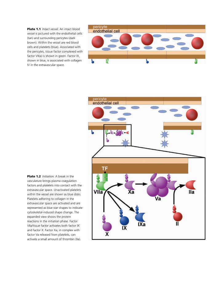
BLBK186-Key May 4, 2009 9:51
Plate 1.1 Intact vessel. An intact blood
vessel is pictured with the endothelial cells
(tan) and surrounding pericytes (dark
brown). Within the vessel are red blood
cells and platelets (blue). Associated with
the pericytes, tissue factor complexed with
factor VII(a) is shown in green. Factor IX,
shown in blue, is associated with collagen
IV in the extravascular space.
Plate 1.2 Initiation. A break in the
vasculature brings plasma coagulation
factors and platelets into contact with the
extravascular space. Unactivated platelets
within the vessel are shown as blue disks.
Platelets adhering to collagen in the
extravascular space are activated and are
represented as blue star shapes to indicate
cytoskeletal-induced shape change. The
expanded view shows the protein
reactions in the initiation phase. Factor
VIIa/tissue factor activates both factor IX
and factor X. Factor Xa, in complex with
factor Va released from platelets, can
activate a small amount of thrombin (IIa).
1
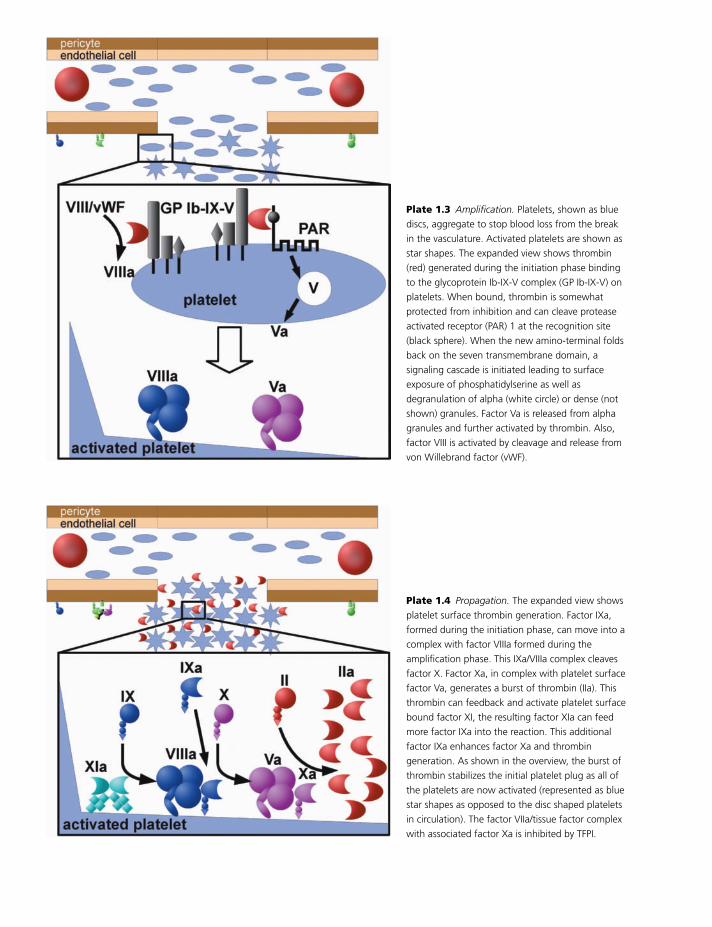
BLBK186-Key May 4, 2009 9:51
Plate 1.3 Amplification. Platelets, shown as blue
discs, aggregate to stop blood loss from the break
in the vasculature. Activated platelets are shown as
star shapes. The expanded view shows thrombin
(red) generated during the initiation phase binding
to the glycoprotein Ib-IX-V complex (GP Ib-IX-V) on
platelets. When bound, thrombin is somewhat
protected from inhibition and can cleave protease
activated receptor (PAR) 1 at the recognition site
(black sphere). When the new amino-terminal folds
back on the seven transmembrane domain, a
signaling cascade is initiated leading to surface
exposure of phosphatidylserine as well as
degranulation of alpha (white circle) or dense (not
shown) granules. Factor Va is released from alpha
granules and further activated by thrombin. Also,
factor VIII is activated by cleavage and release from
von Willebrand factor (vWF).
Plate 1.4 Propagation. The expanded view shows
platelet surface thrombin generation. Factor IXa,
formed during the initiation phase, can move into a
complex with factor VIIIa formed during the
amplification phase. This IXa/VIIIa complex cleaves
factor X. Factor Xa, in complex with platelet surface
factor Va, generates a burst of thrombin (IIa). This
thrombin can feedback and activate platelet surface
bound factor XI, the resulting factor XIa can feed
more factor IXa into the reaction. This additional
factor IXa enhances factor Xa and thrombin
generation. As shown in the overview, the burst of
thrombin stabilizes the initial platelet plug as all of
the platelets are now activated (represented as blue
star shapes as opposed to the disc shaped platelets
in circulation). The factor VIIa/tissue factor complex
with associated factor Xa is inhibited by TFPI.
2
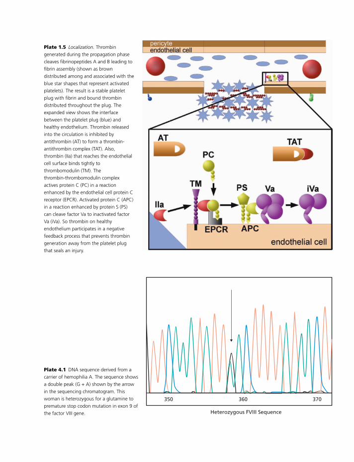
BLBK186-Key May 4, 2009 9:51
Plate 1.5 Localization. Thrombin
generated during the propagation phase
cleaves fibrinopeptides A and B leading to
fibrin assembly (shown as brown
distributed among and associated with the
blue star shapes that represent activated
platelets). The result is a stable platelet
plug with fibrin and bound thrombin
distributed throughout the plug. The
expanded view shows the interface
between the platelet plug (blue) and
healthy endothelium. Thrombin released
into the circulation is inhibited by
antithrombin (AT) to form a thrombin-
antithrombin complex (TAT). Also,
thrombin (IIa) that reaches the endothelial
cell surface binds tightly to
thrombomodulin (TM). The
thrombin-thrombomodulin complex
actives protein C (PC) in a reaction
enhanced by the endothelial cell protein C
receptor (EPCR). Activated protein C (APC)
in a reaction enhanced by protein S (PS)
can cleave factor Va to inactivated factor
Va (iVa). So thrombin on healthy
endothelium participates in a negative
feedback process that prevents thrombin
generation away from the platelet plug
that seals an injury.
370360350
Heterozygous FVIII Sequence
Plate 4.1 DNA sequence derived from a
carrier of hemophilia A. The sequence shows
a double peak (G + A) shown by the arrow
in the sequencing chromatogram. This
woman is heterozygous for a glutamine to
premature stop codon mutation in exon 9 of
the factor VIII gene.
3
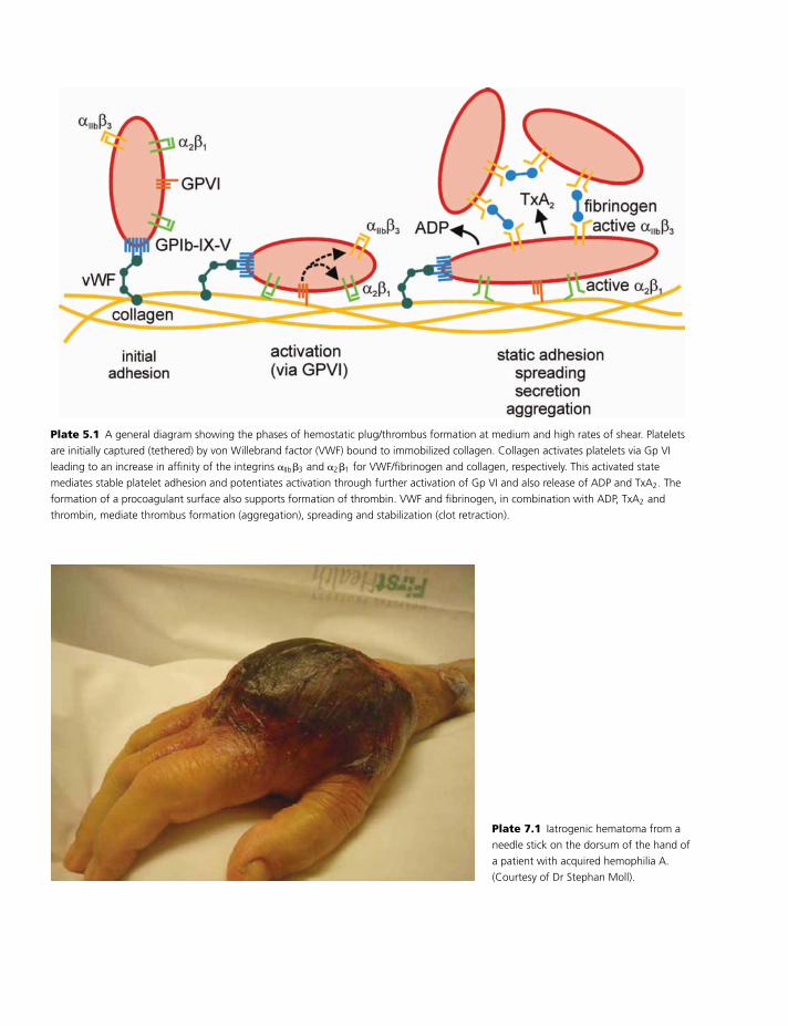
BLBK186-Key May 4, 2009 9:51
Plate 5.1 A general diagram showing the phases of hemostatic plug/thrombus formation at medium and high rates of shear. Platelets
are initially captured (tethered) by von Willebrand factor (VWF) bound to immobilized collagen. Collagen activates platelets via Gp VI
leading to an increase in affinity of the integrins �IIb�3 and �2�1 for VWF/fibrinogen and collagen, respectively. This activated state
mediates stable platelet adhesion and potentiates activation through further activation of Gp VI and also release of ADP and TxA2. The
formation of a procoagulant surface also supports formation of thrombin. VWF and fibrinogen, in combination with ADP, TxA2 and
thrombin, mediate thrombus formation (aggregation), spreading and stabilization (clot retraction).
Plate 7.1 Iatrogenic hematoma from a
needle stick on the dorsum of the hand of
a patient with acquired hemophilia A.
(Courtesy of Dr Stephan Moll).
4
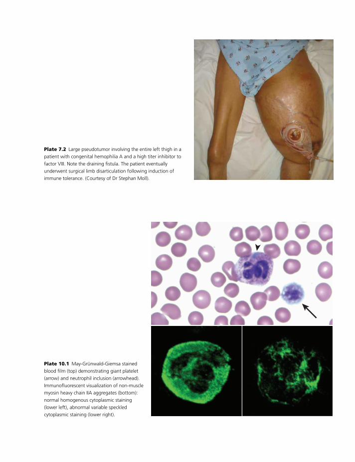
BLBK186-Key May 4, 2009 9:51
Plate 7.2 Large pseudotumor involving the entire left thigh in a
patient with congenital hemophilia A and a high titer inhibitor to
factor VIII. Note the draining fistula. The patient eventually
underwent surgical limb disarticulation following induction of
immune tolerance. (Courtesy of Dr Stephan Moll).
Plate 10.1 May-Grunwald-Giemsa stained
blood film (top) demonstrating giant platelet
(arrow) and neutrophil inclusion (arrowhead).
Immunofluorescent visualization of non-muscle
myosin heavy chain IIA aggregates (bottom):
normal homogenous cytoplasmic staining
(lower left), abnormal variable speckled
cytoplasmic staining (lower right).
5
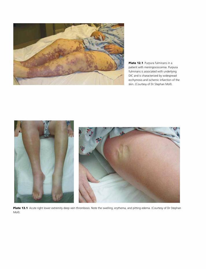
BLBK186-Key May 4, 2009 9:51
Plate 12.1 Purpura fulminans in a
patient with meningococcemia. Purpura
fulminans is associated with underlying
DIC and is characterized by widespread
ecchymosis and ischemic infarction of the
skin. (Courtesy of Dr Stephan Moll).
Plate 13.1 Acute right lower extremity deep vein thrombosis. Note the swelling, erythema, and pitting edema. (Courtesy of Dr Stephan
Moll).
6
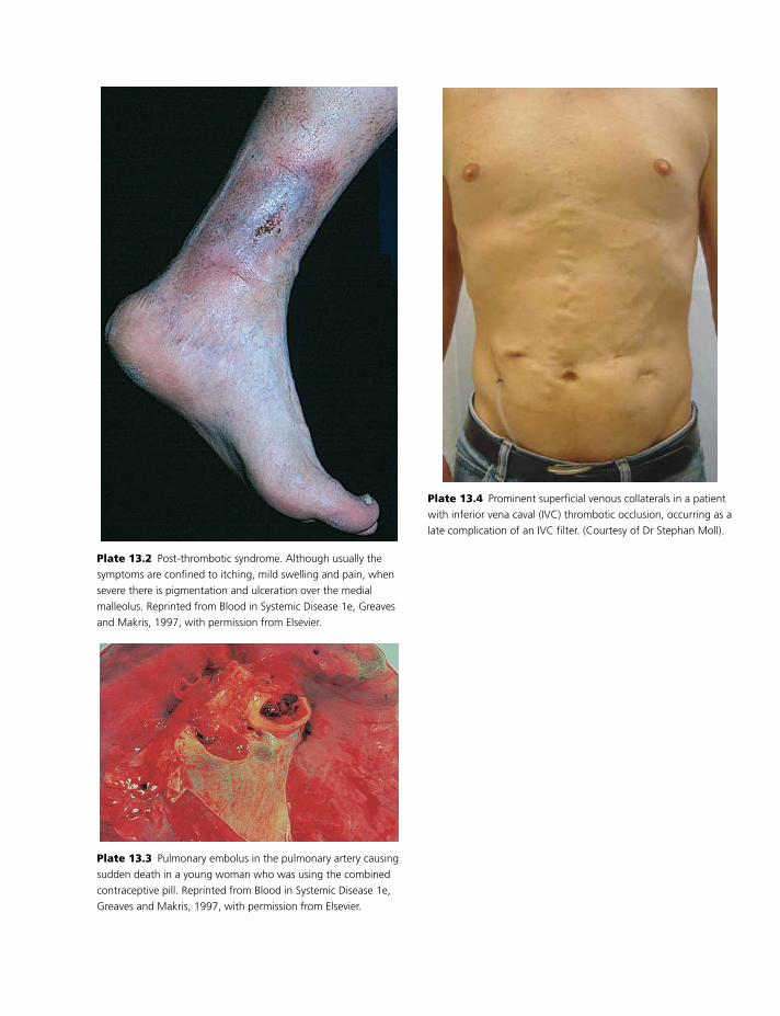
BLBK186-Key May 4, 2009 9:51
Plate 13.2 Post-thrombotic syndrome. Although usually the
symptoms are confined to itching, mild swelling and pain, when
severe there is pigmentation and ulceration over the medial
malleolus. Reprinted from Blood in Systemic Disease 1e, Greaves
and Makris, 1997, with permission from Elsevier.
Plate 13.3 Pulmonary embolus in the pulmonary artery causing
sudden death in a young woman who was using the combined
contraceptive pill. Reprinted from Blood in Systemic Disease 1e,
Greaves and Makris, 1997, with permission from Elsevier.
Plate 13.4 Prominent superficial venous collaterals in a patient
with inferior vena caval (IVC) thrombotic occlusion, occurring as a
late complication of an IVC filter. (Courtesy of Dr Stephan Moll).
7
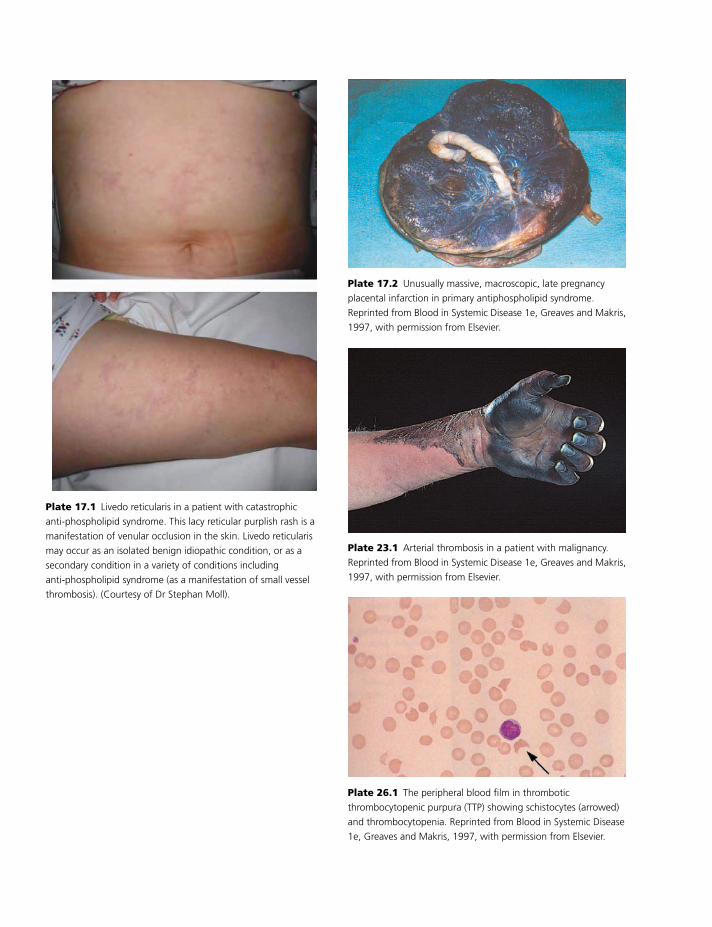
BLBK186-Key May 4, 2009 9:51
Plate 17.1 Livedo reticularis in a patient with catastrophic
anti-phospholipid syndrome. This lacy reticular purplish rash is a
manifestation of venular occlusion in the skin. Livedo reticularis
may occur as an isolated benign idiopathic condition, or as a
secondary condition in a variety of conditions including
anti-phospholipid syndrome (as a manifestation of small vessel
thrombosis). (Courtesy of Dr Stephan Moll).
Plate 17.2 Unusually massive, macroscopic, late pregnancy
placental infarction in primary antiphospholipid syndrome.
Reprinted from Blood in Systemic Disease 1e, Greaves and Makris,
1997, with permission from Elsevier.
Plate 23.1 Arterial thrombosis in a patient with malignancy.
Reprinted from Blood in Systemic Disease 1e, Greaves and Makris,
1997, with permission from Elsevier.
Plate 26.1 The peripheral blood film in thrombotic
thrombocytopenic purpura (TTP) showing schistocytes (arrowed)
and thrombocytopenia. Reprinted from Blood in Systemic Disease
1e, Greaves and Makris, 1997, with permission from Elsevier.
8
BLBK186-Key April 11, 2009 12:57
11 Qualitative platelet disordersMarco Cattaneo
Introduction
Abnormalities of platelet function are associated with
a heightened risk for bleeding, proving that platelets
have an important role in hemostasis. Typically, pa-
tients with platelet disorders have mucocutaneous
bleeding of variable severity and excessive hemor-
rhage after surgery or trauma.
In this chapter, the main inherited and acquired
qualitative platelet defects are reviewed. Abnormal-
ities of platelet function resulting from defects of
plasma proteins (e.g. von Willebrand disease, afibrino-
genemia) will not be considered here, as they are dis-
cussed in Chapters 8 and 9.
Inherited qualitative platelet defects
Inherited disorders of platelet function are generally
classified according to the functions or responses that
are abnormal. However, because platelet functions are
intimately related, a clear distinction between dis-
orders of platelet adhesion, aggregation, activation,
secretion, and procoagulant activity is, in many in-
stances, problematic. For this reason, a classification
of the inherited disorders of platelet function is pro-
posed based on abnormalities of platelet components
that share common characteristics (Table 11.1):� platelet receptors for adhesive proteins;� platelet receptors for soluble agonists;� platelet granules;� signal-transduction pathways;� procoagulant phospholipids; and� miscellaneous disorders (less well characterized).
Abnormalities of the platelet receptorsfor adhesive proteins
Gp Ib/V/IX complex (VWF binding site)The Bernard–Soulier syndrome (BSS) is characterized
by:� autosomal recessive inheritance (with one exception
of autosomal dominant inheritance);� prolonged bleeding time;� thrombocytopenia;� giant platelets (often not detected on automatic
counters);� decreased platelet survival; and� lack of platelet agglutination with ristocetin.
The lack of ristocetin-induced agglutination is not
corrected by the addition of normal plasma. The
platelet responses to physiologic agonists are normal,
with the exception of low concentrations of thrombin,
because Gp Ib� (one of the two components of Gp Ib)
has a critical role in the platelet aggregatory, secretory,
and procoagulant responses to thrombin.
Bleeding events, which may be very severe in
homozygous BSS, can be controlled by platelet trans-
fusion. Most heterozygotes do not have a bleeding
diathesis but are the most common cause of macro-
thrombocytopenia in some parts of the world [1].
BSS is caused by defects in the genes for Gp Ib�,
Ib�, or IX, but not Gp V. The molecular defects that
are responsible for BSS (frameshifts, deletions, point
mutations) are summarized at the following Web site:
http://www.bernard-soulier.org/mutations.
Platelet-type, or pseudo, von Willebrand disease
(VWD) is not caused by defects of VWF, but by a
gain-of-function phenotype of the platelet Gp Ib� [2].
115
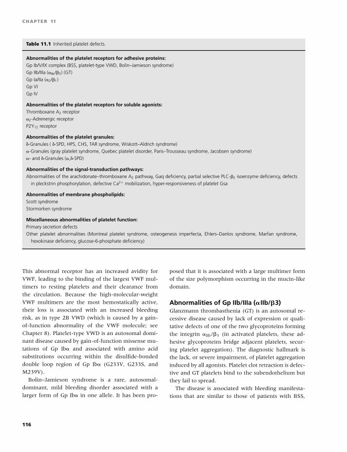
BLBK186-Key April 11, 2009 12:57
CHAPTER 11
Table 11.1 Inherited platelet defects.
Abnormalities of the platelet receptors for adhesive proteins:Gp Ib/V/IX complex (BSS, platelet-type VWD, Bolin–Jamieson syndrome)
Gp IIb/IIIa (�IIb/�3) (GT)
Gp Ia/IIa (�2/�1)
Gp VI
Gp IV
Abnormalities of the platelet receptors for soluble agonists:Thromboxane A2 receptor
�2-Adrenergic receptor
P2Y12 receptor
Abnormalities of the platelet granules:�-Granules ( �-SPD, HPS, CHS, TAR syndrome, Wiskott–Aldrich syndrome)
�-Granules (gray platelet syndrome, Quebec platelet disorder, Paris–Trousseau syndrome, Jacobsen syndrome)
�- and �-Granules (�,�-SPD)
Abnormalities of the signal-transduction pathways:Abnormalities of the arachidonate–thromboxane A2 pathway, Gaq deficiency, partial selective PLC-�2 isoenzyme deficiency, defects
in pleckstrin phosphorylation, defective Ca2+ mobilization, hyper-responsiveness of platelet Gsa
Abnormalities of membrane phospholipids:Scott syndrome
Stormorken syndrome
Miscellaneous abnormalities of platelet function:Primary secretion defects
Other platelet abnormalities (Montreal platelet syndrome, osteogenesis imperfecta, Ehlers–Danlos syndrome, Marfan syndrome,
hexokinase deficiency, glucose-6-phosphate deficiency)
This abnormal receptor has an increased avidity for
VWF, leading to the binding of the largest VWF mul-
timers to resting platelets and their clearance from
the circulation. Because the high-molecular-weight
VWF multimers are the most hemostatically active,
their loss is associated with an increased bleeding
risk, as in type 2B VWD (which is caused by a gain-
of-function abnormality of the VWF molecule; see
Chapter 8). Platelet-type VWD is an autosomal domi-
nant disease caused by gain-of-function missense mu-
tations of Gp Ib� and associated with amino acid
substitutions occurring within the disulfide-bonded
double loop region of Gp Ib� (G233V, G233S, and
M239V).
Bolin–Jamieson syndrome is a rare, autosomal-
dominant, mild bleeding disorder associated with a
larger form of Gp Ib� in one allele. It has been pro-
posed that it is associated with a large multimer form
of the size polymorphism occurring in the mucin-like
domain.
Abnormalities of Gp IIb/IIIa (� IIb/�3)Glanzmann thrombasthenia (GT) is an autosomal re-
cessive disease caused by lack of expression or quali-
tative defects of one of the two glycoproteins forming
the integrin �IIb/�3 (in activated platelets, these ad-
hesive glycoproteins bridge adjacent platelets, secur-
ing platelet aggregation). The diagnostic hallmark is
the lack, or severe impairment, of platelet aggregation
induced by all agonists. Platelet clot retraction is defec-
tive and GT platelets bind to the subendothelium but
they fail to spread.
The disease is associated with bleeding manifesta-
tions that are similar to those of patients with BSS,
116
BLBK186-Key April 11, 2009 12:57
Qualitative platelet disorders
although of less severity [3]. GT patients are grouped
into three types, according to the severity of �IIb/�3
deficiency on their platelet membranes:� Type I patients: �5% (characterized by lack of fibrino-
gen in platelet �-granules);� Type II patients: 10–20%; and� Type III (variant) patients: 50–100%.
The GT defect is caused by mutations or deletions in
the genes encoding one of the two glycoproteins form-
ing the �IIb/�3 integrin. In GT caused by mutations in
the �3 integrin, the levels of the platelet vitronectin re-
ceptor (�v/�3) are also decreased, but the phenotype
of these patients is no different from that of the other
GT patients.
Abnormalities of Gp Ia/IIa (�2/�1)Two patients with mild bleeding disorders associated
with deficient expression of the platelet receptor for
collagen Gp Ia/IIa (�2/�1) and selective impairment of
platelet responses to collagen have been described [4].
Their platelet defect spontaneously recovered after the
menopause, suggesting that �2/�1 expression is under
hormonal control.
Abnormalities of Gp VIA selective defect of collagen-induced platelet aggrega-
tion was also described in another mild bleeding disor-
der, characterized by the deficiency of the platelet Gp
VI, a member of the immunoglobulin superfamily of
receptors, which mediates platelet activation by colla-
gen. The molecular defects that are responsible for the
platelet abnormality have not been characterized in
the patients described so far [5]. The possibility should
be explored that the molecular abnormality lies in the
gene encoding for the Fc� receptor, which is the sig-
naling subunit of Gp VI.
Abnormalities of Gp IVGp IV binds collagen, thrombospondin and probably
other proteins. Its physiological role is unclear, be-
cause its deficiency, common in healthy individuals
from Japan and other East Asian populations, is not
associated with an abnormal phenotype.
Abnormalities of the platelet receptorsfor soluble agonists
Thromboxane A2 receptorIn 1993, a patient with a mild bleeding disorder was
described whose platelets had a defective response to
the TxA2 analog U46619, albeit having a normal num-
ber of TxA2 binding sites and normal equilibrium dis-
sociation rate constants. Despite the normal number of
TxA2 receptors, the TxA2-induced IP3 formation, Ca2
mobilization and GTPase activity were abnormal, sug-
gesting that the abnormality in these platelets was im-
paired coupling between the TxA2 receptor, G protein,
and PLC [6]. This patient was subsequently found to
have an Arg 60 to Leu mutation in the first cytoplas-
mic loop of the TxA2 receptor, affecting both isoforms
of the receptor.
�2-Adrenergic receptorsSubjects with a selective impairment of platelet re-
sponse to epinephrine, a decreased number of the
platelet �2-adrenergic receptors, and mildly prolonged
bleeding times have been described. However, the re-
lationship between this defect and bleeding manifes-
tations still needs to be defined.
P2Y12 receptor for ADPHuman platelets express three distinct P2 receptors
stimulated by adenosine nucleotides:� P2X1;� P2Y1 receptor for ADP with a role in the initiation of
platelet activation; and� P2Y12 receptor for ADP essential for a sustained, full
aggregation response to ADP.
The concurrent activation of both P2Y receptors is
necessary for full platelet aggregation induced by ADP.
P2Y12 also mediates the potentiation of platelet secre-
tion by ADP and the stabilization of thrombin-induced
platelet aggregates.
Only patients with congenital defects of the platelet
P2Y12 receptors have been described. The first patient
(V.R. described in 1992 by Cattaneo et al. [7]) had
a life-long history of excessive bleeding, a prolonged
bleeding time, and abnormalities of platelet aggrega-
tion similar to those observed in patients with de-
fects of platelet secretion (reversible aggregation in
117
BLBK186-Key April 11, 2009 12:57
CHAPTER 11
response to weak agonists and impaired aggregation in
response to low concentrations of collagen or throm-
bin), except that the aggregation response to ADP was
severely impaired.
Other measures of platelet function found in this
patient were:� No inhibition by ADP of PGE1-stimulated platelet
adenylyl cyclase;� Normal shape change and normal (or mildly re-
duced) mobilization of cytoplasmic ionized calcium in-
duced by ADP; and� Presence of approximately 30% of the normal num-
ber of platelet binding sites for [33P]2MeSADP or
[]3H[]ADP;
Three additional patients with very similar charac-
teristics were later described. All of these patients dis-
played base pair deletions in the P2Y12 gene, shifting
the reading frame for several residues before introduc-
ing a premature stop codon, causing an early trunca-
tion of the protein.
A fifth patient (A.C.) with a congenital bleeding
disorder associated with abnormal P2Y12-mediated
platelet responses to ADP has more recently been
characterized. The platelet phenotype is very similar to
that of patients with P2Y12 deficiency, except that the
number and affinity of []33P[]-2MeSADP binding sites
was normal [8]. Analysis of the patient’s P2Y12 gene
revealed, in one allele, a G-to-A transition changing
the codon for Arg 256 in the sixth transmembrane do-
main to Gln, and, in the other allele, a C-to-T transi-
tion changing the codon for Arg 265 in the third extra-
cellular loop to Trp. Neither mutation interfered with
receptor surface expression, but both altered function,
suggesting that the structural integrity of these regions
corresponding to the extracytoplasmic end of TM 6
and EL 3 is necessary for the normal function of this G
protein-coupled receptor.
The study of the children of patient M.G. and pa-
tient A.C. allowed the characterization of patients with
a heterozygous P2Y12 defect whose platelets do not se-
crete normal amounts of ATP after stimulation with
different agonists. This secretion defect was not caused
by impaired production of thromboxane A2 or low
concentrations of platelet granule contents, and is
therefore very similar to that described in patients with
an ill-defined group of congenital defects of platelet
secretion, sometimes referred to by the general term
“primary secretion defect” (PSD; see below), which
is the most common congenital disorder of platelet
function.
P2Y12 deficiency is probably much more common
than currently recognized; it is therefore important
to emphasize that this condition should be suspected
when ADP, even at relatively high concentrations
(10 �M or higher), induces a slight and rapidly re-
versible aggregation that is preceded by normal shape
change. The confirmatory diagnostic test is based
on the ability of ADP to inhibit the platelet adeny-
lyl cyclase after its stimulation by prostaglandins or
forskolin.
Abnormalities of the platelet granules
Abnormalities of the �-granules (�-storagepool deficiency)The term �-storage pool deficiency (�-SPD) defines a
congenital abnormality of platelets characterized by
deficiency of dense granules in megakaryocytes and
platelets [9]. It may present as an isolated platelet
function defect or associate with a variety of congeni-
tal disorders. Between 10% and 18% of patients with
congenital abnormalities of platelet function have
SPD. The inheritance is autosomal recessive in some
families but autosomal dominant in others.
�-SPD is characterized by:� a bleeding diathesis of variable degree;� mildly to moderately prolonged skin bleeding time,
inversely related to the amount of ADP or serotonin
contained in the granules;� abnormal platelet secretion induced by several
platelet agonists;� impaired platelet aggregation in 75% of cases (only
33% have aggregation tracings typical for a platelet se-
cretion defect); and� decreased levels of �-granule constituents: ATP and
ADP, serotonin, calcium, and pyrophosphate.
Lumiaggregometry, which measures platelet aggre-
gation and secretion simultaneously, may prove a
more accurate technique than platelet aggregometry
for diagnosing patients with �-SPD and, more gener-
ally, with platelet secretion defects.
Hermansky–Pudlak syndrome (HPS) and Chediak–
Higashi syndrome (CHS) are rare syndromic forms
of �-SPD. HPS is an autosomal recessive disease
of subcellular organelles of many tissues involving
118
BLBK186-Key April 11, 2009 12:57
Qualitative platelet disorders
abnormalities of melanosomes, platelet �-granules,
and lysosomes. It is characterized by tyrosinase-
positive oculocutaneous albinism, a bleeding diathesis
resulting from �-SPD, and ceroid-lipofuscin lysosomal
storage disease. HPS can arise from mutations in dif-
ferent genetic loci [9].
CHS is a lethal disorder (death usually in the first
decade of life) with:� autosomal recessive inheritance;� variable degrees of oculocutaneous albinism;� very large peroxidase-positive cytoplasmic granules
in a variety of hematopoietic (neutrophils) and non-
hematopoietic cells;� easy bruisability as a result of �-SPD;� recurrent infections, associated with neutropenia,
impaired chemotaxis, and bactericidal activity; and� abnormal natural killer (NK) cell function.
Two types of hereditary thrombocytopenia may be
associated with �-SPD:
1 Thrombocytopenia and absent radii syndrome
(TAR); and
2 Wiskott–Aldrich syndrome.
Abnormalities of the �-granulesGray platelet syndrome (GPS) derives its name from
the gray appearance of the patient’s platelets in pe-
ripheral blood smears as a consequence of the rarity
of platelet granules. The inheritance pattern seems to
be autosomal recessive, although in a single family, it
seemed to be autosomal dominant.
Affected patients have a lifelong history of mucocu-
taneous bleeding, which may vary from mild to mod-
erate in severity, and prolonged bleeding time [10].
They have mild thrombocytopenia with abnormally
large platelets and isolated reduction of the platelet
�-granule content. Mild to moderate myelofibrosis has
been described in some (hypothetically ascribed to
the action of cytokines released by the hypogranular
platelets and megakaryocytes in the bone marrow).
The basic defect in GPS is probably defective targeting
and packaging of endogenously synthesized proteins
in �-granules.
The Quebec platelet disorder is an autosomal domi-
nant qualitative platelet abnormality, characterized by:� severe posttraumatic bleeding complications unre-
sponsive to platelet transfusion;� abnormal proteolysis of �-granule proteins;� severe deficiency of platelet factor V;
� deficiency of multimerin;� reduced to normal platelet counts; and� markedly decreased platelet aggregation induced by
epinephrine.
Multimerin, one of the largest proteins found in
the human body, is present in platelet �-granules and
in endothelial cell Weibel–Palade bodies. It binds fac-
tor V and its activated form, factor Va. Its deficiency
in patients with the Quebec platelet disorder is prob-
ably responsible for the defect in platelet factor V,
which is likely to be degraded by abnormally regulated
platelet proteases, notably urokinase plasminogen ac-
tivator [11].
Jacobsen or Paris–Trousseau syndrome is a rare syn-
drome that is associated with:� a mild hemorrhagic diathesis;� congenital thrombocytopenia with normal platelet
life span;� increased number of marrow megakaryocytes
(many presenting with signs of abnormal maturation
and intramedullary lysis); and� a deletion of the distal part of one chromosome
11 [del(11)q23.3→qter] has been found in affected
patients.
Abnormalities of the �- and �-granules�,�-SPD is characterized by deficiencies of both �- and
�-granules. The clinical picture and the platelet aggre-
gation abnormalities are similar to those of patients
with GPS or �-SPD.
Abnormalities of the signal-transductionpathways
Congenital abnormalities of the arachidonate–
thromboxane A2 pathway, involving the liberation of
arachidonic acid from membrane phospholipids, de-
fects of cyclo-oxygenase, or thromboxane synthetase,
are associated with platelet function defects and mild
bleeding [12]. Other congenital abnormalities of
the platelet signal-transduction pathways have been
described involving:� G-proteins (G�q deficiency);� phosphatidylinositol metabolism (partial selective
PLC-�2 isozyme deficiency); or� defects in pleckstrin phosphorylation and hyper-
responsiveness of platelet Gs�.
119
BLBK186-Key April 11, 2009 12:57
CHAPTER 11
Abnormalities of membranephospholipids
Scott syndrome is a rare bleeding disorder associated
with the maintenance of the asymmetry of the lipid
bilayer in the membranes of blood cells, including
platelets leading to reduced thrombin generation and
defective wound healing. The cause of the defect is still
unclear.
In Stormorken syndrome, resting, unstimulated
platelets from patients with this syndrome display
a full procoagulant activity. Therefore, this condi-
tion represents the exact opposite in terms of platelet
membrane function to the Scott syndrome; yet, sur-
prisingly, it is also associated with a bleeding ten-
dency. Platelets from patients with this condition re-
spond normally to all agonists, with the exception of
collagen.
Miscellaneous abnormalities ofplatelet function
Primary secretion defectsThe term primary secretion defect was probably used
for the first time by Weiss, to indicate all those ill-
defined abnormalities of platelet secretion not associ-
ated with platelet granule deficiencies. The term was
later used to indicate the platelet secretion defects not
associated with platelet granule deficiencies and ab-
normalities of the arachidonate pathway, or all the ab-
normalities of platelet function associated with defects
of signal transduction [13].
With the progression of our knowledge of platelet
pathophysiology, this heterogeneous group, which
brings together the majority of patients with congeni-
tal disorders of platelet function, will become progres-
sively smaller, losing those patients with better de-
fined biochemical abnormalities responsible for their
platelet secretion defect. An example is heterozygous
P2Y12 deficiency state, which was included in this
group of disorders until its biochemical abnormality
was identified.
Other platelet abnormalitiesSpontaneous platelet aggregation and decreased re-
sponses to thrombin are observed in patients with
the Montreal platelet syndrome, a rare and poorly
characterized congenital thrombocytopenia with large
platelets [14].
Platelet function abnormalities have also been re-
ported in osteogenesis imperfecta, Ehlers–Danlos syn-
drome, Marfan syndrome, hexokinase deficiency. and
glucose-6-phosphate deficiency [15].
Acquired platelet defects
Platelet function can be impaired in several hemato-
logic and non-hematologic conditions and by medica-
tions (Table 11.2) [16].
Uremia
The bleeding time (BT) may be severely prolonged
in patients with uremia, but it can be corrected by
increasing the hematocrit with RBC transfusions or
with erythropoietin, suggesting that, in many in-
stances, the defective primary hemostasis in uremia
is a consequence of anemia. (It is known that RBCs
normally facilitate the platelet interaction with the
vessel wall.)
However, correction of the hematocrit fails to
correct the BT in some patients, suggesting that
other factors impair platelet–vessel wall interaction
in this condition. Abnormalities of interaction of
adhesive glycoproteins with their platelet receptors,
Table 11.2 Acquired platelet defects.
Medications affecting platelet function
Uremia
Dysproteinemias
Acute leukemias and myelodysplastic syndromes
Cardiopulmonary bypass
Liver disease
Antiplatelet antibodies
Myeloproliferative disorders
Essential thrombocythemia
Polycythemia vera
Chronic myelogenous leukemia
Agnogenic myeloid metaplasia
120
BLBK186-Key April 11, 2009 12:57
Qualitative platelet disorders
defective platelet activation, and platelet procoagulant
activity have been described. Both dialyzable and non-
dialyzable substances may be responsible.
Myeloproliferative disordersFunctional and biochemical abnormalities of platelets
from patients with myeloproliferative disorders in-
clude:� decreased release of arachidonic acid from mem-
brane phospholipids;� reduced conversion of arachidonic acid to its active
metabolites;� reduced responsiveness to TxA2;� deficiency of platelet granules;� deficiency of the �2/�1 integrin; and� decreased number of �2-adrenergic receptors.
Other factors, in addition to platelet functional de-
fects, contribute to the bleeding diathesis of these
patients, including increased whole blood viscosity
and thrombocytosis [17].
Cardiopulmonary bypass
Cardiopulmonary bypass causes transient thrombocy-
topenia and platelet function defects, which contribute
to the increased bleeding risk of these patients. Platelet
function defects associated with extracorporeal circu-
lation include:� defective aggregation,� platelet granule deficiencies,� abnormal interaction with VWF, and� generation of platelet-derived microparticles.
These abnormalities result from platelet activation
and fragmentation, hypothermia, contact with the
blood–air interface, and exposure to traces of platelet
agonists such as thrombin, ADP, and plasmin.
Table 11.3 Drugs affecting platelet function.
NSAIDs:Aspirin, indomethacin, ibuprofen, sulindac, naproxen, phenylbutazone
Thienopyridines:Ticlopidine, clopidogrel, thromboxane A2 receptor
Gp IIb/IIIa antagonists:Abciximab, eptifibatide, tirofiban
Drugs that increase the platelet cAMP or cGMP levels:Prostacyclin, iloprost, dipyridamole, theophylline, nitric oxide, nitric oxide donors
Anticoagulants and fibrinolytic agents:Heparin, streptokinase, tPA, urokinase
Cardiovascular drugs:Nitroglycerin, isosorbide dinitrate, propranolol, frusemide, calcium-channel blockers, quinidine, ACE inhibitors, verapamil, diltiazem
Volume expanders:Dextran, hydroxyethyl starch
Psychotropic drugs, anesthetics:Imipramine, amitriptyline, nortriptyline, chlorpromazine, promethazine, fluphenazine, trifluoperazine, haloperidol, halothane,
dibucaine, tetracaine, butacaine, nepercaine, procaine plaquenil
Chemotherapeutic agents:Mitomycin, daunorubicin, BCNU
Miscellaneous drugs:Antihistamines, radiographic contrast agents, clofibrate
Abbreviations: ACE, angiotensin-converting enzyme; cGMP, cyclic guanosine 3′,5′-monophosphate; CAMP, cyclic adenosine 3′,5′-monophosphate.
121
BLBK186-Key April 11, 2009 12:57
CHAPTER 11
MedicationsMany drugs affect platelet function (Table 11.3),
sometimes causing a prolongation of the BT. In some
instances, the inhibition of platelet function is the tar-
get of the drug, as in the case of antiplatelet agents
that are given to reduce the risk of cardiovascular or
cerebrovascular accidents. In other cases, the induced
abnormalities of platelet function are to be considered
side effects of the drug, which are in most instances
without obvious clinical consequences.
Liver disease
Chronic liver disease is associated with a prolongation
of the BT disproportionate to the degree of throm-
bocytopenia that usually complicates this condition.
Whether the described defects are caused by intrinsic
or extrinsic abnormalities of the platelets is unclear.
Therapy
Platelet transfusions should be used only in severe
bleeding episodes, which are usually seen in patients
with BSS or, less frequently, GT. Recombinant fac-
tor VIIa is a good, albeit expensive, alternative to
platelet transfusions [18]. Antifibrinolytic agents, such
as aprotinin and tranexamic acid, or the vasopressin
analog desmopressin (DDAVP) should be used in
all other circumstances, because they are relatively
cheap, do not cause platelet refractoriness, and are not
associated with the risk of transmitting blood-borne
viral diseases [19].
References
1 Pham A, Wang J. Bernard-Soulier syndrome: an
inherited platelet disorder. Arch Pathol Lab Med
2007;131(12):1834–6.
2 Budde U. Diagnosis of von Willebrand disease subtypes:
implications for treatment. Haemophilia 2008;14(Suppl
5):27–38.
3 Nair S, Ghosh K, Kulkarni B, Shetty S, Mohanty
D. Glanzmann’s thrombasthenia: updated. Platelets
2002;13(7):387–93.
4 Moroi M, Jung SM. Platelet receptors for collagen.
Thromb Haemost 1997;78(1):439–44.
5 Arthur JF, Dunkley S, Andrews RK. Platelet gly-
coprotein VI-related clinical defects. Br J Haematol
2007;139(3):363–72.
6 Higuchi W, Fuse I, Hattori A, Aizawa Y. Mutations of
the platelet thromboxane A2 (TXA2) receptor in pa-
tients characterized by the absence of TXA2-induced
platelet aggregation despite normal TXA2 binding ac-
tivity. Thromb Haemost 1999;82(5):1528–31.
7 Cattaneo M, Lecchi A, Randi AM, McGregor JL,
Mannucci PM. Identification of a new congenital defect
of platelet function characterized by severe impairment
of platelet responses to adenosine diphosphate. Blood
1992;80(11):2787–96.
8 Cattaneo M, Gachet C. ADP receptors and clini-
cal bleeding disorders. Arterioscler Thromb Vasc Biol
1999;19(10):2281–5.
9 Nurden P, Nurden AT. Congenital disorders asso-
ciated with platelet dysfunctions. Thromb Haemost
2008;99(2):253–63.
10 Nurden AT, Nurden P. The gray platelet syn-
drome: clinical spectrum of the disease. Blood Rev
2007;21(1):21–36.
11 Diamandis M, Veljkovic DK, Maurer-Spurej E, Rivard
GE, Hayward CP. Quebec platelet disorder: features,
pathogenesis and treatment. Blood Coagul Fibrinolysis
2008;19(2):109–19.
12 Rao AK. Congenital Platelet Signal Transduction Defects.
Cambridge: Cambridge University Press, 2002.
13 Cattaneo M. Congenital Disorders of Platelet Secretion.
Cambridge: Cambridge University Press, 2002.
14 Balduini CL, Cattaneo M, Fabris F, et al. Inherited
thrombocytopenias: a proposed diagnostic algorithm
from the Italian Gruppo di Studio delle Piastrine.
Haematologica 2003;88(5):582–92.
15 Cattaneo M. Inherited platelet-based bleeding disor-
ders. J Thromb Haemost 2003;1(7):1628–36.
16 Bennett J. Acquired Platelet Function Defects. Cambridge:
Cambridge University Press, 2002.
17 Elliott MA, Tefferi A. Thrombosis and haemorrhage in
polycythaemia vera and essential thrombocythaemia.
Br J Haematol 2005;128(3):275–90.
18 Poon M-C. Factor VIIa. Orlando, FL: Academic Press,
2003.
19 Mannucci PM, Levi M. Prevention and treatment of
major blood loss. N Engl J Med 2007;356(22):2301–11.
122
BLBK186-Key May 22, 2009 15:4
12 Disseminated intravascularcoagulation and othermicroangiopathiesRaj S. Kasthuri and Nigel S. Key
Disseminated intravascular coagulation
Disseminated intravascular coagulation (DIC) is an ac-
quired clinicopathologic syndrome characterized by
chaotic activation of the coagulation system, resulting
in widespread intravascular deposition of fibrin-rich
thrombi. DIC is not itself a disease state, but rather
is a secondary manifestation of some other under-
lying disorder. Depending on the underlying cause
and rapidity of the process, the clinical spectrum may
range from subclinical laboratory abnormalities (com-
pensated DIC or non-overt DIC) to multiorgan failure,
metabolic derangement, hemodynamic instability,
widespread bleeding, and death.
The following definition of DIC has been proposed
by the DIC Scientific and Standardization Commit-
tee of the International Society on Thrombosis and
Hemostasis (ISTH): “DIC is an acquired syndrome
characterized by the intravascular activation of coag-
ulation with loss of localization arising from different
causes. It can originate from and cause damage to the
microvasculature, which if sufficiently severe, can pro-
duce organ dysfunction” [1].
Synonyms for DIC in the medical literature include
the defibrination syndrome, consumption coagulopa-
thy, generalized intravascular coagulation, throm-
bohemorrhagic phenomenon, and disseminated in-
travascular fibrin formation.
Etiology
A broad range of pathological conditions (the most im-
portant of which are listed in Table 12.1) may trig-
ger DIC. Sepsis syndromes are among the most fre-
quently encountered causes. Although the highest risk
is seen with Gram-negative bacterial infections, Gram-
positive infections as well as nonbacterial infections
can also be associated with DIC. Complications of
pregnancy and malignancy are other common causes
of DIC in clinical practice.
Pathogenesis
The pathogenesis of DIC involves simultaneous dys-
regulation of several homeostatic mechanisms (Fig.
12.1). These can be broadly divided into:
1 excessive activation of coagulation;
2 downregulation of physiologic anticoagulant path-
ways; and
3 inhibition of fibrinolysis.
Dysfunction of the vascular endothelium, a vast and
pervasive organ, is prominent as both a cause and
a consequence of these processes. The net result is
widespread generation of thrombin and conversion
of circulating fibrinogen to insoluble fibrin thrombi,
aggravated by the relative inability of the fibrinolytic
mechanism to remove intravascular fibrin.
Obstruction of small and medium-sized vessels
caused by intravascular fibrin deposition may lead to
(multiple) organ dysfunction, especially affecting the
kidneys, brain, lung, liver, and heart. The widespread
activation of coagulation leads to consumption of clot-
ting factors and platelets, a process that is aggravated
by simultaneous impaired hepatic production of these
factors. Thus, abnormal prolongation of coagulation
screening tests, thrombocytopenia, and a seemingly
paradoxical bleeding tendency may occur in some pa-
tients with more advanced forms of DIC.
The passage of erythrocytes through the fibrin
meshwork in the microvascular circulation may lead
to red cell fragmentation. This microangiopathic
123
BLBK186-Key May 22, 2009 15:4
CHAPTER 12
Table 12.1 Conditions associated with disseminated
intravascular coagulation (DIC).
Infection:� Sepsis syndromes (Gram-positive and Gram-negative
bacteria)� Viral infections (e.g. dengue, Ebola)� Other (e.g. ricketsial, malarial infections)
Trauma/tissue damage:� Head injury� Pancreatitis� Fat embolism� Any other serious tissue damage (crush or penetrating
injury)
Malignancy:� Solid tumors� Acute leukemias (especially AML-M3)� Chronic leukemias (CMML)
Obstetric complications:� Abruptio placentae� Amniotic fluid embolism� Eclampsia and preeclampsia� Retained dead fetus
Vascular disorders:� Giant hemangiomas (Kasabach–Merritt syndrome)� Other vascular malformations� Large aortic aneurysm
Severe allergic/toxic reactions:� Toxic shock syndrome� Snake, spider venoms
Severe immunologic reactions:� Acute hemolytic transfusion reactions� Heparin-induced thrombocytopenia, type II
hemolytic anemia (MAHA) is much less common in
DIC than in the group of disorders known as the
“thrombotic microangiopathies,” where it is, in fact,
a sine qua non.
Excessive activation of coagulationAlthough coagulation may be initiated in vitro by
both the intrinsic (contact) and extrinsic (tissue factor)
pathways, the tissue factor pathway is the primary ini-
tiator of coagulation in vivo [2]. Unlike most other sol-
uble clotting factors circulating in plasma, tissue factor
(TF) is a cell-bound transmembrane protein. By virtue
of its predominant extravascular location, TF is nor-
mally present on cells that are relatively inaccessible
to blood clotting factors in the absence of vessel injury,
such as smooth muscle cells and fibroblasts. However,
the systemic response to infection and injury results
in the synthesis and release of pro-inflammatory cy-
tokines, such as tumor necrosis factor TNF-�, inter-
leukin 1 (IL-1), and IL-6, which trigger TF synthesis by
monocytes and endothelial cells (Fig. 12.1) [2]. With
other forms of DIC, it is likely that additional stimuli
capable of activating and/or propagating coagulation
(such as fat, brain lipids, cancer procoagulant protein,
or amniotic fluid) are released into the circulation.
Downregulation of physiologicalanticoagulant pathwaysDIC is associated with an acquired deficiency of natu-
rally occurring anticoagulants, particularly antithrom-
bin (III) and protein C. Plasma levels are decreased
secondary to consumption and increased enzymatic
degradation by activated neutrophils [3]. Endothe-
lial dysfunction adversely affects the protein C/protein
S/thrombomodulin pathway in other ways also. The
same proinflammatory cytokines that up-regulate TF
synthesis simultaneously down-regulate endothelial
synthesis of the cofactors thrombomodulin and en-
dothelial cell protein C receptor [4]. The end result is
decreased conversion of protein C to activated protein
C on the endothelial cell surface.
Inhibition of fibrinolysisThe role of the fibrinolytic system is to generate plas-
min on fibrin surfaces, in an effort to restore vascular
patency via enzymatic digestion of fibrin strands. In
many forms of DIC, fibrinolysis is actively suppressed
because of elevated levels of plasminogen activator
inhibitor type 1 (PAI-1) [5]. PAI-1 inhibits the plas-
minogen activators tissue plasminogen activator and
urokinase, preventing the generation of plasmin from
plasminogen. Thus, by failing to clear intravascular
fibrin thrombi, the inhibition of fibrinolysis by PAI-1
also contributes to the net procoagulant state and end-
organ hypoperfusion in DIC.
Clinical manifestations
As predicted from the complex underlying patho-
physiological derangements, patients with DIC may
124
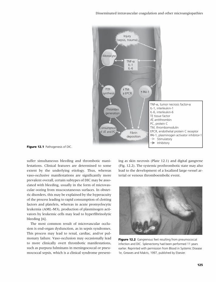
BLBK186-Key May 22, 2009 15:4
Disseminated intravascular coagulation and other microangiopathies
TNF-α, tumor necrosis factor-αIL-1, interleukin-1IL-6, interleukin-6TF, tissue factorAT, antithrombinPC, protein CTM, thrombomodulinEPCR, endothelial protein C receptorPAI-1, plasminogen activator inhibitor-1 Stimulatory Inhibitory
Injury(sepsis, trauma)
Monocyte
TNF-αIL-1IL-6
TFsynthesis
TMEPCR PAI-1
Thrombingeneration
Consumptionof AT and PC Fibrin
deposition
Figure 12.1 Pathogenesis of DIC.
suffer simultaneous bleeding and thrombotic mani-
festations. Clinical features are determined to some
extent by the underlying etiology. Thus, whereas
vaso-occlusive manifestations are significantly more
prevalent overall, certain subtypes of DIC may be asso-
ciated with bleeding, usually in the form of microvas-
cular oozing from mucocutaneous surfaces. In obstet-
ric disorders, this may be explained by the hyperacuity
of the process leading to rapid consumption of clotting
factors and platelets, whereas in acute promyelocytic
leukemia (AML-M3), production of plasminogen acti-
vators by leukemic cells may lead to hyperfibrinolytic
bleeding [6].
The most common result of microvascular occlu-
sion is end-organ dysfunction, as in sepsis syndromes.
This process may lead to renal, cardiac, and/or pul-
monary failure. Vaso-occlusion may occasionally lead
to more clinically overt thrombotic manifestations,
such as purpura fulminans in meningococcal or pneu-
mococcal sepsis, which is a clinical syndrome present-
ing as skin necrosis (Plate 12.1) and digital gangrene
(Fig. 12.2). The systemic prothrombotic state may also
lead to the development of a localized large-vessel ar-
terial or venous thromboembolic event.
Figure 12.2 Gangrenous feet resulting from pneumococcal
infection and DIC. Splenectomy had been performed 11 years
earlier. Reprinted with permission from Blood in Systemic Disease
1e, Greaves and Makris, 1997, published by Elsevier.
125
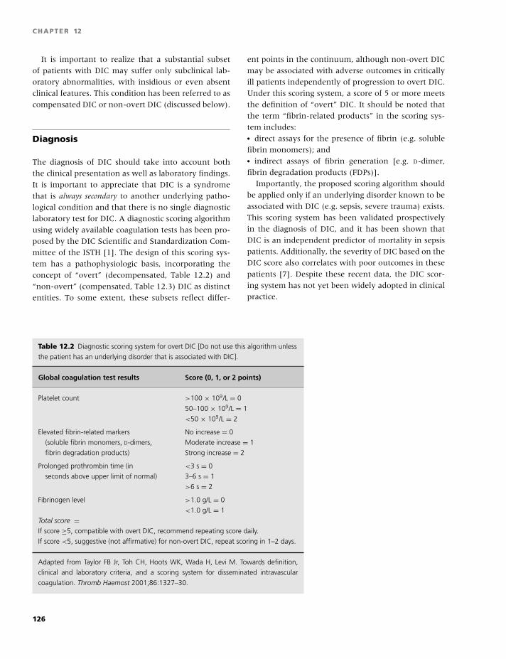
BLBK186-Key May 22, 2009 15:4
CHAPTER 12
It is important to realize that a substantial subset
of patients with DIC may suffer only subclinical lab-
oratory abnormalities, with insidious or even absent
clinical features. This condition has been referred to as
compensated DIC or non-overt DIC (discussed below).
Diagnosis
The diagnosis of DIC should take into account both
the clinical presentation as well as laboratory findings.
It is important to appreciate that DIC is a syndrome
that is always secondary to another underlying patho-
logical condition and that there is no single diagnostic
laboratory test for DIC. A diagnostic scoring algorithm
using widely available coagulation tests has been pro-
posed by the DIC Scientific and Standardization Com-
mittee of the ISTH [1]. The design of this scoring sys-
tem has a pathophysiologic basis, incorporating the
concept of “overt” (decompensated, Table 12.2) and
“non-overt” (compensated, Table 12.3) DIC as distinct
entities. To some extent, these subsets reflect differ-
ent points in the continuum, although non-overt DIC
may be associated with adverse outcomes in critically
ill patients independently of progression to overt DIC.
Under this scoring system, a score of 5 or more meets
the definition of “overt” DIC. It should be noted that
the term “fibrin-related products” in the scoring sys-
tem includes:� direct assays for the presence of fibrin (e.g. soluble
fibrin monomers); and� indirect assays of fibrin generation [e.g. D-dimer,
fibrin degradation products (FDPs)].
Importantly, the proposed scoring algorithm should
be applied only if an underlying disorder known to be
associated with DIC (e.g. sepsis, severe trauma) exists.
This scoring system has been validated prospectively
in the diagnosis of DIC, and it has been shown that
DIC is an independent predictor of mortality in sepsis
patients. Additionally, the severity of DIC based on the
DIC score also correlates with poor outcomes in these
patients [7]. Despite these recent data, the DIC scor-
ing system has not yet been widely adopted in clinical
practice.
Table 12.2 Diagnostic scoring system for overt DIC [Do not use this algorithm unless
the patient has an underlying disorder that is associated with DIC].
Global coagulation test results Score (0, 1, or 2 points)
Platelet count >100 × 109/L = 0
50–100 × 109/L = 1
<50 × 109/L = 2
Elevated fibrin-related markers No increase = 0
(soluble fibrin monomers, D-dimers, Moderate increase = 1
fibrin degradation products) Strong increase = 2
Prolonged prothrombin time (in <3 s = 0
seconds above upper limit of normal) 3–6 s = 1
>6 s = 2
Fibrinogen level >1.0 g/L = 0
<1.0 g/L = 1
Total score =If score ≥5, compatible with overt DIC, recommend repeating score daily.
If score <5, suggestive (not affirmative) for non-overt DIC, repeat scoring in 1–2 days.
Adapted from Taylor FB Jr, Toh CH, Hoots WK, Wada H, Levi M. Towards definition,
clinical and laboratory criteria, and a scoring system for disseminated intravascular
coagulation. Thromb Haemost 2001;86:1327–30.
126
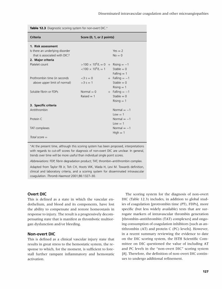
BLBK186-Key May 22, 2009 15:4
Disseminated intravascular coagulation and other microangiopathies
Table 12.3 Diagnostic scoring system for non-overt DIC.∗
Criteria Score (0, 1, or 2 points)
1. Risk assessmentIs there an underlying disorder Yes = 2
that is associated with DIC? No = 0
2. Major criteriaPlatelet count >100 × 109/L = 0 + Rising = −1
<100 × 109/L = 1 Stable = 0
Falling = 1
Prothrombin time (in seconds <3 s = 0 + Falling = −1
above upper limit of normal) >3 s = 1 Stable = 0
Rising = 1
Soluble fibrin or FDPs Normal = 0 + Falling = −1
Raised = 1 Stable = 0
Rising = 1
3. Specific criteriaAntithrombin Normal = −1
Low = 1
Protein C Normal = −1
Low = 1
TAT complexes Normal = −1
High = 1
Total score =
∗At the present time, although this scoring system has been proposed, interpretations
with regards to cut-off scores for diagnosis of non-overt DIC are unclear. In general,
trends over time will be more useful than individual single point scores.
Abbreviations: FDP, fibrin degradation product; TAT, thrombin–antithrombin complex.
Adapted from Taylor FB Jr, Toh CH, Hoots WK, Wada H, Levi M. Towards definition,
clinical and laboratory criteria, and a scoring system for disseminated intravascular
coagulation. Thromb Haemost 2001;86:1327–30.
Overt DICThis is defined as a state in which the vascular en-
dothelium, and blood and its components, have lost
the ability to compensate and restore homeostasis in
response to injury. The result is a progressively decom-
pensating state that is manifest as thrombotic multior-
gan dysfunction and/or bleeding.
Non-overt DICThis is defined as a clinical vascular injury state that
results in great stress to the hemostatic system, the re-
sponse to which, for the moment, is sufficient to fore-
stall further rampant inflammatory and hemostatic
activation.
The scoring system for the diagnosis of non-overt
DIC (Table 12.3) includes, in addition to global stud-
ies of coagulation [protrombin time (PT), FDPs], more
specific (but less widely available) tests that are sur-
rogate markers of intravascular thrombin generation
[thrombin–antithrombin (TAT) complexes] and ongo-
ing consumption of coagulation inhibitors [such as an-
tithrombin (AT) and protein C (PC) levels]. However,
in a recent summary reviewing the evidence to date
on the DIC scoring system, the ISTH Scientific Com-
mittee on DIC questioned the value of including AT
and PC levels in the “non-overt DIC” scoring system
[8]. Therefore, the definition of non-overt DIC contin-
ues to undergo additional refinement.
127

BLBK186-Key May 22, 2009 15:4
CHAPTER 12
Although perceived as a classic finding, a low
plasma fibrinogen level is not a sensitive marker of
DIC [9]. In fact, high plasma fibrinogen levels are
much more frequently encountered. Fibrinogen levels
are probably influenced more by the degree of activa-
tion of secondary fibrino(geno)lysis than the degree of
consumption during thrombus formation.
Treatment
The development of DIC in patients with sepsis or
trauma has been shown to be independently asso-
ciated with increased morbidity and mortality. Thus,
prompt and at times preemptive therapy becomes im-
portant in these patients.
Managing the underlying diseaseThe mainstay of treatment in patients with DIC is
management of the underlying disease. The reversibil-
ity of DIC depends to a large degree on the under-
lying cause. Delivery of the fetus and placenta may
promptly restore homeostasis in patients with obstetric
DIC. Eradication of infection with antibiotics and/or
surgery may not necessarily have the same rapid effect
in sepsis syndromes, possibly because of established
widespread endothelial injury.
Supportive care and blood productsGood supportive care in the management of patients
with DIC includes adequate hemodynamic support to
maintain perfusion and appropriate supportive trans-
fusion of blood products. Given the mechanisms in-
volved in the development of DIC, there is always
the theoretical fear of “fueling the fire” with trans-
fused blood cells and plasma products, although the
evidence that this occurs in practice is underwhelm-
ing. To complicate matters further, there are no con-
sensus guidelines for optimal transfusion management
of these patients.
Treatment of patients with DIC who are actively
bleeding or at high risk for bleeding should include
platelet transfusions, fresh frozen plasma, cryoprecipi-
tate, and packed red cells as needed. Patients requiring
invasive procedures should be covered peri-procedure
with plasma and platelet transfusions as needed. Rea-
sonable transfusion goals in these circumstances are
platelet counts �50 × 109/L, fibrinogen �1.0 g/L, and
maintenance of PT and activated partial thromboplas-
tin time (APTT) as close to the normal range as pos-
sible. There is no role for the prophylactic adminis-
tration of blood products in patients with DIC. The
approach to these patients should be individualized
based on their clinical and laboratory manifestations.
Systemic anticoagulationOn the basis of the pathophysiology of DIC, an ar-
gument may be made for the use of systemic hep-
arin anticoagulation. Although the literature remains
divided about this approach, the few available con-
trolled trials have failed to demonstrate a clear benefit
[10]. The routine use of heparin in DIC not associated
with a clinical thrombotic event is generally discour-
aged given the demonstrated risk of bleeding compli-
cations in these patients. There is some consensus that
treatment is indicated for those with a documented
thromboembolic event or extensive deposition of fib-
rin leading to acral ischemia or purpura fulminans. In
the case of large-vessel thromboembolic events, full
therapeutic doses of unfractionated heparin are indi-
cated, whereas in microvascular occlusive syndromes,
lower doses (e.g. 500–800 U/hour) may be preferable.
Low-molecular-weight heparin has been successfully
used as an alternative to unfractionated heparin in
some studies. The role of direct thrombin inhibitors
(such as hirudin or argatroban) in DIC also remains
to be established in controlled trials. Although these
agents might theoretically be more effective than hep-
arins, they also carry a higher risk of bleeding.
Antifibrinolytic therapyBecause fibrinolysis is generally down-regulated con-
comitant with excessive fibrin formation in DIC,
treatment with antifibrinolytic agents (such as
ε-aminocaproic acid or tranexamic acid) is generally
contraindicated. There may be exceptions to the rule,
such as patients with acute promyelocytic leukemia
who may develop a form of DIC characterized by hy-
perfibrinolytic bleeding that may result in intracranial
hemorrhage. In this instance, judicious use of antifib-
rinolytics has proven effective [11].
Specific inhibitors of coagulationIn view of the depletion of natural anticoagulants
during DIC, it is logical to suppose that replacement
128
BLBK186-Key May 22, 2009 15:4
Disseminated intravascular coagulation and other microangiopathies
therapy using one or more of the missing natural an-
ticoagulants is warranted.
Several preliminary trials with antithrombin,
mainly in patients with sepsis, demonstrated some
improvement in the duration of DIC and resolution
of laboratory abnormalities. However, a significant
benefit in mortality could not be demonstrated in
a large, randomized controlled study of sepsis (the
KyberSept Trial) [12]. Although a post hoc subgroup
analysis in this trial suggested a benefit with the use
of AT without concomitant heparin in a subset of
patients with DIC [13], the role of AT therapy in
patients with DIC remains unclear at this time.
On the other hand, a large, randomized, controlled
trial (the PROWESS Study) using recombinant acti-
vated PC (Drotrecogin alfa,activated) to treat patients
with sepsis did demonstrate improved survival com-
pared with placebo [14]. This effect was probably me-
diated not only by an antithrombotic effect, but also
by anti-inflammatory and profibrinolytic effects of this
agent. However, excess bleeding was seen in patients
treated with activated PC, which inactivates factors Va
and VIIIa. Therefore, caution is required in patients
with severe thrombocytopenia (�30 × 109/L) or oth-
erwise at high risk of bleeding. This pivotal phase III
study has been further dissected using extensive sub-
group analyses, which have suggested a greater bene-
fit for rhAPC in patients with severe sepsis (≥2 organs
affected, APACHE II score �25) and those patients
with sepsis with coexisting DIC [15]. Additionally, pa-
tients that received concomitant therapy with rhAPC
and heparin tended to have higher mortality rates.
These issues have been specifically addressed in subse-
quent clinical trials evaluating the role of rhAPC in less
severe sepsis (APACHE II score �25, the ADDRESS
study), which showed no benefit to the use of rhAPC;
and the concomitant use of heparin and rhAPC in pa-
tients with severe sepsis (the XPRESS study), which
failed to demonstrate significant differences with use
of heparin [16,17]. The role of activated PC in the
treatment of other forms of DIC has not been ade-
quately evaluated.
Thrombotic microangiopathies
The thrombotic microangiopathies are a group of re-
lated disorders characterized by widespread microvas-
Table 12.4 Underlying etiologies of thrombotic
microangiopathies.
Thrombotic thrombocytopenic purpura:� Familial (ADAMTS-13 deficiency)� Acquired
◦ Idiopathic
◦ Drug-related (quinine, ticlopidine)
Hemolytic uremic syndrome:� Familial (including factor H deficiency)� Acquired
Secondary thrombotic microangiopathies:� Malignancy� Malignant hypertension� Transplantation
◦ Stem cell transplantation
◦ Solid organ transplantation� Pregnancy-related
◦ Preeclampsia
◦ HELLP syndrome� Collagen vascular disease
◦ Scleroderma renal crisis
◦ Systemic lupus erythematosus
◦ Antiphospholipid antibody syndrome
cular occlusion by platelet-rich aggregates. The accel-
erated consumption of platelets results in thrombocy-
topenia. Red cell fragmentation occurs secondary to
turbulent blood flow in areas of the microcirculation
obstructed by platelet-rich thrombi. Peripheral blood
smear examination reveals the presence of fragmented
red cells (schistocytes or helmet cells) associated with
elevated serum lactate dehydrogenase levels, a con-
dition known as microangiopathic hemolytic anemia
(MAHA) [18].
A number of syndromes are included under the
rubric of thrombotic microangiopathies (Table 12.4),
and a clear distinction between them at the time of
presentation may be difficult or impossible. This is es-
pecially true of thrombotic thrombocytopenic purpura
(TTP) and the hemolytic uremic syndrome (HUS).
The classic clinical pentad in TTP includes:� thrombocytopenia,� MAHA,� renal failure,� neurologic abnormalities, and� fever,
but frequently not all features are present.
129
BLBK186-Key May 22, 2009 15:4
CHAPTER 12
“Classic” HUS is characterized by:� MAHA,� thrombocytopenia, and� prominent renal failure, following an acute diarrheal
illness.
The clinical presentation of many of the sec-
ondary thrombotic microangiopathies may also be in-
distinguishable on initial evaluation. The diagnostic
dilemma is further compounded by the urgent re-
quirement for plasma exchange in a subset of these
patients (discussed below). Therefore, it is appropri-
ate to use the generic diagnosis of “TTP/HUS” in pa-
tients presenting with thrombocytopenia and MAHA
in the absence of a clinically apparent cause or DIC.
Plasma exchange should then be initiated while fur-
ther evaluation to rule out an alternative diagnosis
continues [19].
Pathophysiology
Although the clinical manifestations of these syn-
dromes show considerable overlap, pathogeneses
(where understood) of some of the individual entities
may differ considerably.
Thrombotic thrombocytopenic purpuraUnlike the case with DIC, microvascular thrombi
are relatively fibrin-poor, but are enriched in von
Willebrand factor (VWF) and platelets. Microvascu-
lar platelet deposition in TTP is secondary to en-
dothelial secretion of unusually large VWF multimers.
Under normal conditions, these unusually large mul-
timers (which are particularly “sticky” for platelets)
are prevented from entering the circulation by an en-
zyme that cleaves VWF. Predominantly synthesized in
the liver, this metalloprotease enzyme is known as
ADAMTS-13 (a disintegrin-like and metalloprotease
with thrombospondin repeats) [20]. A qualitative or
quantitative defect of ADAMTS-13 allows the un-
usually large multimers of VWF to remain anchored
to endothelial cells, resulting in widespread platelet
adherence, microvascular obstruction, and end-organ
dysfunction.
Studies have demonstrated that many (but appar-
ently not all) patients with definite TTP have �5% ac-
tivity of ADAMTS-13 in their plasma. In the familial
form of TTP, affected individuals are usually homozy-
gous or doubly heterozygous for mutations in the gene
for ADAMTS-13, located on chromosome 9. In the
idiopathic acquired form of TTP, immunoglobulin G
(IgG) antibodies against the enzyme have been de-
tected, suggesting an autoimmune etiology [21]. More
modest reductions in ADAMTS-13 enzyme activity in
plasma (5–50%) may be found in liver disease, malig-
nancy, inflammation, pregnancy, and in the neonatal
period.
Hemolytic uremic syndromeMicrovascular platelet thrombus formation in the clas-
sic form of HUS is believed to be toxin-induced. Specif-
ically, prodromal infection of the gastrointestinal tract
by verotoxin-producing Escherichia coli O157:H7, or
certain other serotypes of E. coli or Shigella dysenteriae,
is characteristic of this disorder. These toxins, which
gain access to blood via the colonic circulation, ul-
timately target cerebral and glomerular epithelium,
mesangial cells and tubular epithelium in the kidneys,
and vascular endothelium. In these locations, vero-
toxins mediate cytokine release, endothelial activation
and injury, and direct activation of platelets. The re-
lease of platelet adhesogens from damaged endothe-
lium results in microvascular platelet thrombi forma-
tion and renal injury.
The small subset of individuals with familial HUS
tend to have more severe disease and a greater risk
of recurrence. Some of these patients are deficient in
complement factor H, which inactivates C3b, a prod-
uct of the alternate complement pathway. The ab-
sence of this regulatory mechanism can lead to au-
toantibody or immune complex-mediated glomerular
injury, with platelet activation, increase in local en-
dothelial procoagulant properties, and ultimately mi-
crovascular thrombus formation.
Other thrombotic microangiopathiesTiclopidine, clopidogrel, and (particularly) quinine ap-
pear to cause thrombotic microangiopathy through
an antibody-mediated mechanism. The pathogenesis
of many of the other secondary thrombotic microan-
giopathies listed in Table 12.4 remains poorly under-
stood.
130
BLBK186-Key May 22, 2009 15:4
Disseminated intravascular coagulation and other microangiopathies
Differential diagnosis
Faced with a thrombocytopenic patient with MAHA,
DIC should be ruled out by review of the history (to
rule out an underlying disorder associated with DIC)
and by confirmation that screening studies of coagu-
lation, such as the PT, APTT, and fibrinogen level, are
normal. The direct antiglobulin (Coombs) test should
be obtained to screen for immune-mediated hemol-
ysis. Stool cultures are indicated if there has been a
preceding diarrheal illness. A careful review of drug
exposures, particularly for drugs such as quinine, is es-
sential.
In a pregnant patient—particularly one in the lat-
ter stages or in the immediate postpartum period—it
may be very difficult to distinguish TTP/HUS (which
requires urgent plasma exchange) from preeclamp-
sia with or without the associated HELLP (hemolysis,
elevated liver enzymes and low-platelets) syndrome.
In general, these syndromes resolve promptly with de-
livery, whereas TTP/HUS may persist.
The utility of a low-plasma ADAMTS-13 activity re-
mains uncertain. The presence of a very low level
(�5%) is probably diagnostic but not necessarily ex-
clusive to TTP, and laboratory demonstration of an
inhibitory antibody to the enzyme helps to identify
an autoimmune etiology. However, measuring en-
zyme levels is unlikely to be useful in making the
diagnosis in the acute setting for the following rea-
sons. First, most centers do not have the capability for
real-time ADAMTS-13 testing, and there is a signifi-
cant lag time before test results are available. Second,
roughly a quarter of patients with idiopathic TTP do
not have ADAMTS-13 deficiency. Finally, and most
important, patients with TTP respond well to plasma
exchange regardless of underlying ADAMTS-13 defi-
ciency [22]. However, measurement of ADAMTS-13
activity may provide valuable information in aiding
long-term management of these patients as discussed
below.
Clinical manifestations
The distinction between TTP and HUS, when possi-
ble, is based on the presence of significant renal fail-
ure and preceding history. Thus, in the classic (en-
demic) form of acquired HUS, which is most com-
mon in children less than 5 years of age, a bloody
diarrhea resulting from E. coli or S. dysenteriae is a
prodromal hallmark. In the epidemic form of HUS,
which may occur after eating infected meat or dairy
products, approximately 10–30% of infected individ-
uals develop the full-blown syndrome. The use of
antimotility agents after an E. coli infection may in-
crease the risk of HUS. Recurrence of this type of HUS
is uncommon. Patients with familial forms of HUS
(the Upshaw–Schulman syndrome) tend to present
early in childhood and frequently have a relapsing
clinical course that may progress to end-stage renal
disease.
Classic TTP occurs much more frequently in adult-
hood and is frequently associated with neurologic
dysfunction, which characteristically manifests as
transient focal (e.g. dysphasia) or non-focal (e.g. con-
fusion, seizure) symptoms. Neurologic symptoms may
be the first sign of relapse in a patient with a pre-
vious history of TTP. The risk of recurrence in the
idiopathic acquired form of TTP is in the range of
10–30%, with most (but not all) events occurring
within the first year. Patients with familial forms
of TTP may present later in life than those with
familial HUS.
The thrombotic microangiopathy related to
chemotherapy, cyclosporine, transplantation, or total
body irradiation tends to occur weeks to months
following exposure to these agents. The diagnostic
criteria for thrombotic microangiopathy associated
with hematopoietic stem cell transplantation have
recently been addressed by a consensus conference,
which opted for stringent criteria to avoid misdiag-
nosis [23]. It was proposed that all of the following
criteria should be met: (1) �4% schistocytes on
peripheral blood smear; (2) de novo, prolonged, or
progressive thrombocytopenia (platelet count �50 ×109/L or 50% or greater reduction from previous
counts); (3) sudden and persistent increase in lactate
dehydrogenase (LDH); (4) decrease in hemoglobin
concentration or increased transfusion requirement;
and (5) decrease in serum haptoglobin. In this dis-
order, the outcome is frequently very poor, renal
manifestations are prominent, and plasma exchange
does not appear to be efficacious.
131
BLBK186-Key May 22, 2009 15:4
CHAPTER 12
Treatment
In the era prior to plasma exchange, the mortality
rate of TTP approached 100%. Since the institution of
plasma exchange, TTP has become a curable disease.
Thus, prompt diagnosis is essential. The diagnostic cri-
teria have therefore become less stringent, and, as al-
ready described, all patients with thrombocytopenia
and MAHA who do not have another explanation for
these findings—whether or not they have renal man-
ifestations, fever, or neurological dysfunction—should
be suspected of having TTP/HUS and treated as such.
Plasma exchangeThe most important treatment modality in these pa-
tients is plasma exchange, which is superior to plasma
infusion [24]. A single plasma volume exchange re-
placing with fresh frozen or cryosupernatant plasma
should be performed daily along with monitoring of
platelet counts, serum LDH, and periodic review of the
peripheral smear.
Neurologic symptoms generally resolve rapidly fol-
lowing institution of plasma exchange. Measures of
ongoing hemolysis, such as the LDH, may also im-
prove promptly with therapy, although the anemia
may persist and occasionally may require supportive
transfusions. The recovery from renal failure may be
unpredictable and often slow and incomplete, such
that some patients may need prolonged dialysis. The
platelet count is the most reliable marker of disease
activity on which to base treatment decisions. An im-
provement reflects resolution, whereas worsening of
thrombocytopenia at any point in the course of the
disease may reflect an exacerbation and the need for
more aggressive therapy. In those patients who fail to
demonstrate an initial response, more intense therapy,
such as greater volumes of plasma exchanged once or
even twice daily, is indicated [19].
Plasma exchange is most beneficial in patients with
TTP/HUS who fall into the “idiopathic acquired,”
“pregnancy-related,” and “drug-related” categories. Its
benefit is unclear in other forms of thrombotic mi-
croangiopathy, such as that associated with stem cell
transplantation (which may be more related to the
use of cyclosporine A as well as graft-versus-host
disease), total body irradiation, and cytomegalovirus
infection.
In responsive patients, there are no set criteria to
guide the optimal duration of treatment. Once the
platelet count normalizes, a decision can be made to
discontinue plasma exchange. A fall in the platelet
count may occur within the first 1–2 weeks, reflect-
ing disease exacerbation, and plasma exchange then
needs to be reinstituted. One approach has been to de-
crease the frequency of plasma exchanges rather than
to abruptly discontinue. Ultimately, however, discon-
tinuing plasma exchange is the only way to evaluate
whether hematological remission has been achieved.
There is still debate as to whether plasma exchange
is indicated in patients with postinfectious HUS. The
vast majority of disease in young children will re-
solve with supportive care alone, but plasmapheresis
is probably indicated and useful in affected adults.
ImmunosuppressionIn many centers, glucocorticoids are used as an ad-
junct to initial plasma exchange, but there are few
data to support this practice. Certainly, it is reasonable
to consider glucocorticoids in patients who are refrac-
tory to plasma exchange. Other immunosuppressive
modalities, such as vincristine, have also been reported
to be of value in refractory cases. Particular reference
must be made here to the use of rituximab, the mon-
oclonal antibody against CD20. Rituximab has been
used in patients with relapsing or refractory TTP with
encouraging results in small series of patients with
recovery of ADAMTS-13 levels and achievement of
durable remissions [25]. The beneft of combining rit-
uximab and plasma exchange in the treatment of pa-
tients with TTP will be evaluated in a phase III ran-
domized trial in the United States in the near future.
Role of ADAMTS-13 activityMeasurement of ADAMTS-13 activity and search for
an autoantibody to the enzyme is often performed at
the time that TTP/HUS is suspected. Because there is
no consensus as to the most robust assay methodol-
ogy, no specific recommendation can be made. How-
ever, at the time of writing, our understanding of the
role of ADAMTS deficiency in the diagnosis and man-
agement of TTP/HUS can be briefly summarized as
follows:� A severe deficiency of ADAMTS13 activity (�5% of
pooled normal plasma) is relatively specific for TTP;
132
BLBK186-Key May 22, 2009 15:4
Disseminated intravascular coagulation and other microangiopathies
� Only about 70% of patients with a firm diagnosis
of acute idiopathic TTP have a severe deficiency in
ADAMTS13;� The response to plasma exchange appears to be sim-
ilar in patients with or without severe ADAMTS13
deficiency;� Secondary forms of thrombotic microangiopathy
(such as that associated with hematopoietic stem
cell therapy) are associated with normal levels of
ADAMTS13 activity; and� Patients with TTP associated with deficiency of
ADAMTS-13 are at a greater risk for relapse compared
to those without.
The major implication of these observations is that
ADAMTS13 activity should not be used to decide
which patients with clinically diagnosed acute idio-
pathic TTP/HUS are (or rather, are not) candidates
for plasma exchange. On the other hand, those indi-
viduals who are subsequently proven to have severe
ADAMTS13 activity caused by an autoantibody to
the enzyme should be more carefully observed when
in remission because of their higher risk of relapse;
indeed, in this situation, a falling plasma level of
ADAMTS13 may herald the onset of a relapse. It re-
mains to be demonstrated whether these patients are
more suitable candidates for more aggressive immuno-
suppression, as might reasonably be expected.
Other treatmentsIn patients with multiple relapses, splenectomy during
hematologic remission may favorably alter the disease
course.
In the context of ADAMTS-13 deficiency, episodes
of familial TTP have been reversed or prevented by the
infusion of fresh frozen or cryosupernatant plasma.
These products contain the metalloprotease enzyme,
and in this subset of congenitally deficient patients,
plasmapheresis can therefore be avoided.
Patients with TTP/HUS rarely experience bleeding,
despite sometimes very significant thrombocytopenia.
Routine prophylactic platelet transfusion is contraindi-
cated, because of fear that it may precipitate further
vaso-occlusive phenomena. However, it is occasion-
ally necessary to administer platelets to a patient with
one of these syndromes who is actively bleeding.
The use of antimicrobial agents in HUS increases the
release of Shiga toxin from the organism and could
paradoxically increase the risk of HUS. They are there-
fore not recommended.
The sequence of the ADAMTS-13 metalloprotease
has now been determined, and gene therapy for treat-
ment of patients with familial forms of the disease may
become a reality in the relatively near future.
References
1 Taylor FB Jr, Toh CH, Hoots WK, Wada H, Levi M.
Towards definition, clinical and laboratory criteria, and
a scoring system for disseminated intravascular coagu-
lation. Thromb Haemost 2001;86(5):1327–30.
2 Mackman N, Tilley RE, Key NS. Role of the extrinsic
pathway of blood coagulation in hemostasis and throm-
bosis. Arterioscler Thromb Vasc Biol 2007;27(8):1687–93.
3 Levi M, de Jonge E, van der Poll T. Rationale for
restoration of physiological anticoagulant pathways in
patients with sepsis and disseminated intravascular co-
agulation. Crit Care Med 2001;29(7 Suppl):S90–4.
4 Levi M. Disseminated intravascular coagulation. Crit
Care Med 2007;35(9):2191–5.
5 Biemond BJ, Levi M, Ten Cate H, et al. Plasminogen
activator and plasminogen activator inhibitor I release
during experimental endotoxaemia in chimpanzees:
effect of interventions in the cytokine and coagulation
cascades. Clin Sci (Lond) 1995;88(5):587–94.
6 Falanga A. Mechanisms of hypercoagulation in
malignancy and during chemotherapy. Haemostasis
1998;28(Suppl 3):50–60.
7 Angstwurm MW, Dempfle CE, Spannagl M. New
disseminated intravascular coagulation score: A use-
ful tool to predict mortality in comparison with
Acute Physiology and Chronic Health Evaluation II
and Logistic Organ Dysfunction scores. Crit Care Med
2006;34(2):314–20.
8 Toh CH, Hoots WK. The scoring system of the Scien-
tific and Standardisation Committee on Disseminated
Intravascular Coagulation of the International Society
on Thrombosis and Haemostasis: a 5-year overview. J
Thromb Haemost 2007;5(3):604–6.
9 Levi M, de Jonge E, van der Poll T, ten Cate H. Dis-
seminated intravascular coagulation. Thromb Haemost
1999;82(2):695–705.
10 Feinstein DI. Diagnosis and management of dissem-
inated intravascular coagulation: the role of heparin
therapy. Blood 1982;60(2):284–7.
11 Schwartz BS, Williams EC, Conlan MG, Mosher DF.
Epsilon-aminocaproic acid in the treatment of pa-
tients with acute promyelocytic leukemia and acquired
133
BLBK186-Key May 22, 2009 15:4
CHAPTER 12
alpha-2-plasmin inhibitor deficiency. Ann Intern Med
1986;105(6):873–7.
12 Warren BL, Eid A, Singer P, et al. Caring for the
critically ill patient. High-dose antithrombin III in
severe sepsis: a randomized controlled trial. JAMA
2001;286(15):1869–78.
13 Kienast J, Juers M, Wiedermann CJ, et al. Treatment
effects of high-dose antithrombin without concomitant
heparin in patients with severe sepsis with or with-
out disseminated intravascular coagulation. J Thromb
Haemost 2006;4(1):90–7.
14 Bernard GR, Vincent JL, Laterre PF, et al. Efficacy and
safety of recombinant human activated protein C for
severe sepsis. N Engl J Med 2001;344(10):699–709.
15 Ely EW, Laterre PF, Angus DC, et al. Drotrecogin alfa
(activated) administration across clinically important
subgroups of patients with severe sepsis. Crit Care Med
2003;31(1):12–19.
16 Abraham E, Laterre PF, Garg R, et al. Drotrecogin alfa
(activated) for adults with severe sepsis and a low risk
of death. N Engl J Med 2005;353(13):1332–41.
17 Levi M, Levy M, Williams MD, et al. Prophylactic
heparin in patients with severe sepsis treated with
drotrecogin alfa (activated). Am J Respir Crit Care Med
2007;176(5):483–90.
18 Moake JL. Thrombotic microangiopathies. N Engl J Med
2002;347(8):589–600.
19 George JN. How I treat patients with thrombotic throm-
bocytopenic purpura-hemolytic uremic syndrome.
Blood 2000;96(4):1223–9.
20 Tsai HM. Physiologic cleavage of von Willebrand fac-
tor by a plasma protease is dependent on its confor-
mation and requires calcium ion. Blood 1996;87(10):
4235–44.
21 Furlan M, Robles R, Galbusera M, et al. von Wille-
brand factor-cleaving protease in thrombotic throm-
bocytopenic purpura and the hemolytic-uremic syn-
drome. N Engl J Med 1998;339(22):1578–84.
22 Sadler JE, Moake JL, Miyata T, George JN. Recent
advances in thrombotic thrombocytopenic purpura.
Hematology Am Soc Hematol Educ Program 2004:407–23.
23 Ruutu T, Barosi G, Benjamin RJ, et al. Diagnos-
tic criteria for hematopoietic stem cell transplant-
associated microangiopathy: results of a consensus pro-
cess by an International Working Group. Haematologica
2007;92(1):95–100.
24 Rock GA, Shumak KH, Buskard NA, et al. Com-
parison of plasma exchange with plasma infusion in
the treatment of thrombotic thrombocytopenic pur-
pura. Canadian Apheresis Study Group. N Engl J Med
1991;325(6):393–7.
25 Garvey B. Rituximab in the treatment of au-
toimmune haematological disorders. Br J Haematol
2008;141(2):149–69.
134
BLBK186-Key May 4, 2009 9:50
13 Venous thromboembolismLori-Ann Linkins and Clive Kearon
Pathogenesis of venousthromboembolism
Virchow was the first to identify stasis, vessel wall in-
jury, and hypercoagulability as the pathogenic triad
responsible for thrombosis. This classification of risk
factors for venous thromboembolism (VTE) remains
valuable. A summary of risk factors for VTE is given
in Table 13.1.
Venous stasisThe importance of venous stasis as a risk factor for
VTE is demonstrated by the fact that most deep vein
thrombi (DVTs) associated with stroke affect the par-
alyzed leg and most DVT associated with pregnancy
affect the left leg, the iliac veins of which are prone to
extrinsic compression by the pregnant uterus and the
right common iliac artery.
Vessel damageVenous endothelial damage, as a consequence of acci-
dental injury, manipulation during surgery (e.g. hip
replacement), or iatrogenic injury, is an important
risk factor for VTE. Hence, three-quarters of proximal
DVTs that complicate hip surgery occur in the oper-
ated leg, and thrombosis is common with indwelling
venous catheters.
HypercoagulabilityA complex balance of naturally occurring coagulation
and fibrinolytic factors, and their inhibitors, serve
to maintain blood fluidity and hemostasis. Inherited
or acquired changes in this balance predispose to
thrombosis.
Inherited predisposition to VTEThe most important inherited biochemical disorders
that are associated with VTE result from:
� defects in the naturally occurring inhibitors of co-
agulation: deficiencies of antithrombin, protein C, or
protein S; and� resistance to activated protein C, which is caused by
the factor V Leiden mutation in the majority of cases.
The first three of these disorders are rare in the gen-
eral population (combined prevalence of �1%), have
a combined prevalence of approximately 5% in pa-
tients with a first episode of VTE, and are associated
with a 10- to 40-fold increase in the risk of VTE [1].
The factor V Leiden mutation is common, occurring in
approximately 5% of Caucasions and approximately
20% of patients with a first episode of VTE (i.e. an ap-
proximate 4-fold increase in VTE risk).
A mutation in the 3′ untranslated region of the pro-
thrombin gene (G20210A), which is associated with
an approximately 25% increase in prothrombin levels,
occurs in about 2% of Caucasians and approximately
5% of those with a first episode of VTE (i.e. an approx-
imate 2.5-fold increase in risk).
Elevated levels of a number of coagulation factors
(I, II, VIII, IX, XI) are associated with thrombosis in a
“dose-dependent” manner. It is probable that such el-
evations are often inherited, with strong evidence for
this in the case of factor VIII. Abnormalities of the fib-
rinolytic system have questionable importance as risk
factors for VTE.
Acquired predisposition to VTEAcquired hypercoagulable states include estrogen
therapy, antiphospholipid antibodies (anticardi-
olipin antibodies and/or lupus anticoagulants),
systemic lupus erythematosus, malignancy, com-
bination chemotherapy, and surgery [2]. Patients
who develop heparin-induced thrombocytopenia
(HIT) also have a very high risk of developing ar-
terial and venous thromboembolism [3]. Finally,
135
BLBK186-Key May 4, 2009 9:50
CHAPTER 13
Table 13.1 Risk factors for VTE.
Patient factors:Previous VTE*
Age over 40
Pregnancy, purpureum
Obesity
Inherited hypercoagulable state
Underlying condition and acquired factors:Malignancy*
Estrogen therapy
Cancer chemotherapy
Paralysis*
Prolonged immobility
Major trauma*
Lower limb injuries*
Heparin-induced thrombocytopenia
Antiphospholipid antibodies
Lower limb orthopedic surgery*
Surgery requiring general anesthesia >30 minutes
Combinations of factors have at least an additive effect on
the risk of VTE.
*Common major risk factors for VTE.
hyperhomocysteinemia, whether due to hereditary or
acquired causes, is also a risk factor for VTE.
Prevalence and natural history of VTE
VTE is rare before the age of 16 years, likely because
the immature coagulation system is resistant to throm-
bosis. However, the risk of VTE increases exponen-
tially with advancing age (i.e. 1.9-fold per decade), ris-
ing from an annual incidence of approximately 30 in
100,000 at 40 years, to 90 in 100,000 at 60 years, and
260 in 100,000 at 80 years. Clinically important com-
ponents of the natural history of VTE are summarized
in Table 13.2.
Management of VTE
Diagnosis of VTEObjective testing for DVT and pulmonary embolism
(PE) is essential because clinical assessment alone is
unreliable. Failure to diagnose VTE is associated with
a high mortality, whereas inappropriate anticoagula-
Table 13.2 Natural history of VTE.
� VTE usually starts in the calf veins.� About 80% of symptomatic DVTs are proximal.� Two-thirds of asymptomatic DVT detected postoperatively by
screening venography are confined to the distal (calf) veins.� About 20% of symptomatic isolated calf DVTs subsequently
extend to the proximal veins, usually within a week of
presentation.� PE usually arises from proximal DVT.� 70% of patients with symptomatic proximal DVT have
asymptomatic PE (high probability lung scans in 40%), and
vice versa.� Only one-quarter of patients with symptomatic PE have
symptoms or signs of DVT.� 50% of untreated symptomatic proximal DVTs are expected
to cause symptomatic PE.� 10% of symptomatic PE are rapidly fatal.� 30% of untreated symptomatic non-fatal PE will have a
fatal recurrence.
tion can lead to serious complications, including fatal
hemorrhage.
Diagnosis of DVTThe clinical features of DVT include localized swelling,
erythema, tenderness, and distal edema (Plate 13.1).
However, these features are nonspecific, and approx-
imately 85% of ambulatory patients with suspected
DVT will have another cause for their symptoms. The
differential diagnosis for DVT includes:� cellulitis;� ruptured Baker cyst;� muscle tear, muscle cramps, muscle hematoma;� external venous compression;� superficial thrombophlebitis; and� post-thrombotic syndrome (see Plate 13.2).
VenographyVenography is the reference standard test for the diag-
nosis of DVT. It has advantages over other tests in that
it is capable of detecting both proximal vein throm-
bosis and isolated calf vein thrombosis. However, the
disadvantages are that it:� is invasive, expensive, and requires technical exper-
tise; and� exposes patients to the risks associated with contrast
media, including the potential for an allergic reaction
or renal impairment.
136
BLBK186-Key May 4, 2009 9:50
Venous thromboembolism
Table 13.3 Test results that confirm or exclude DVT.
Diagnostic for first DVT
Venography: Intraluminal filling defect
Venous ultrasound: Noncompressible proximal veins at two
or more of the common femoral, popliteal, and calf
trifurcation sites
Excludes first DVT
Venography: All deep veins seen, and no intraluminal filling
defects
D-dimer: Normal test which has a very high sensitivity
(i.e. ≥98%) and at least a moderate specificity (i.e. ≥40%)
Venous ultrasound: Fully compressible proximal veins and
(a) low clinical suspicion for DVT at presentation; (b) normal
D-dimer test which has a moderately high sensitivity
(i.e. ≥85%) and specificity (i.e. ≥70%) at presentation; or
(c) normal serial testing (at 7 days)
Low clinical suspicion for DVT at presentation and a normal
D-dimer test which has moderately high sensitivity
(i.e. ≥85%) and specificity (i.e. ≥70%) at presentation
Diagnostic for recurrent DVT
Venography: Intraluminal filling defect
Venous ultrasound: (a) A new noncompressible common
femoral or popliteal vein segment or (b) 4.0 mm increase
in diameter of the common femoral or popliteal vein
during compression compared to a previous recent test
Excludes Recurrent DVT
Venogram: All deep veins seen and no intraluminal filling
defects
Venous ultrasound: Normal or ≤1mm increase in diameter of
the common femoral or popliteal veins on venous
ultrasound compared to a previous test, and remains
normal (no progression of venous ultrasound) at 2 and 7 days
D-dimer: Normal test which has a very high sensitivity
(i.e. ≥98%) and at least a moderate specificity (i.e. ≥40%)
For these reasons, noninvasive tests such as ve-
nous ultrasonography and D-dimer testing, alone or
in combination with clinical assessment, have largely
replaced venography [4]. A summary of the test re-
sults that effectively confirm or exclude DVT is given
in Table 13.3.
Clinical assessmentAlthough clinical assessment cannot unequivocally
confirm or exclude DVT, clinical evaluation using em-
piric assessment or a structured clinical model (Ta-
ble 13.4) can stratify patients as having:
� Low probability of DVT (prevalence of DVT approx-
imately 5%);� Moderate probability of DVT (prevalence of DVT
approximately 25%); or� High probability of DVT (prevalence of DVT approx-
imately 60%) [5].
Such categorization is useful in guiding the perfor-
mance and interpretation of objective testing [6].
Compression venous ultrasonographyThis is the noninvasive method of choice for diag-
nosing DVT. The common femoral vein, superficial
femoral vein, popliteal vein, and proximal deep calf
veins are imaged in real time and compressed with the
transducer probe. Inability to compress the vein fully
is diagnostic of venous thrombosis.
Venous ultrasonography is highly accurate for the
detection of proximal vein thrombosis with a sensitiv-
ity of approximately 97%, specificity of approximately
94%, and negative predictive value of approximately
98% in symptomatic patients. If DVT cannot be ex-
cluded by a normal proximal venous ultrasound in
combination with other results (e.g. low clinical prob-
ability or normal D-dimer), a follow-up ultrasound is
performed after 1 week to check for extending calf
vein thrombosis (present in approximately 2% of pa-
tients). If the second ultrasound is normal, the risk
of symptomatic VTE during the next 6 months is less
than 2%.
The accuracy of venous ultrasonography is substan-
tially lower if its findings are discordant with the clini-
cal assessment [7] and/or if abnormalities are confined
to short segments of the deep veins. Ideally, these pa-
tients should have a venogram because the result of
the venogram will differ from the venous ultrasound
in approximately 25% of these cases. If venography
is not available, additional testing (e.g. D-dimer, serial
venous ultrasonography) may help to clarify the diag-
nosis and avoid inappropriate anticoagulant therapy.
Venous ultrasonography of the calf veins is more
difficult to perform (e.g. sensitivity 70%), and its value
is controversial. Some investigators have proposed
that a single complete compression ultrasound that in-
cludes examination of the calf veins should be used
to exclude DVT. Studies using this method have re-
ported an incidence of VTE of 0.5% during 3 months
follow-up after a negative examination, establishing
that a negative venous ultrasound that includes the
137
BLBK186-Key May 4, 2009 9:50
CHAPTER 13
Table 13.4 Wells’ Model for determining clinical suspicion of DVT (adapted from Wells et al., N Engl J Med 2003;349:1227–35).
Variables Points
Active cancer (treatment ongoing or within previous 6 months or palliative) 1
Paralysis, paresis or recent plaster immobilization of the lower extremities 1
Bedridden >3 days or major surgery within 4 weeks 1
Localized tenderness along the distribution of the deep venous system 1
Entire leg swollen 1
Calf swelling 3 cm > asymptomatic side (measured 10 cm below tibial tuberosity) 1
Pitting edema confined to the symptomatic leg 1
Collateral dilated superficial veins (non-varicose) 1
Previously documented DVT 1
Alternative diagnosis as likely or more likely than DVT −2
Pretest probability calculated as follows:Total Points
DVT likely ≥2
DVT unlikely 0 or 1
Note: In patients with symptoms in both legs, the more symptomatic leg is used.
calf veins excludes VTE [8]. However, this method has
the potential to diagnose calf DVT that would have
spontaneously lysed without treatment and to yield
false-positive results, thereby exposing patients to the
risk of bleeding due to anticoagulant therapy without
clear benefit.
D-dimer blood testingD-dimer is formed when cross-linked fibrin is broken
down by plasmin, and levels are usually elevated with
DVT and/or PE. Normal levels can help to exclude
VTE, but elevated D-dimer levels are nonspecific and
have low positive predictive value [9,10].
D-dimer assays differ markedly in their diagnostic
properties for VTE. A normal result with a very sen-
sitive D-dimer assay (i.e. sensitivity of approximately
98%) excludes VTE on its own [i.e. it has a high neg-
ative predictive value (NPV)]. However, very sensi-
tive D-dimer tests have low specificity (approximately
40%), which limits their use because of high false-
positive rates. In order to exclude DVT and/or PE, a
normal result with a less sensitive D-dimer assay (i.e.
approximately 85%) needs to be combined with either
a low clinical probability or another objective test that
has a high NPV, but is nondiagnostic on its own (e.g.,
negative venous ultrasound of the proximal veins; Ta-
ble 13.3) [11]. As less sensitive D-dimer assays are
more specific (approximately 70%), they yield fewer
false-positive results.
Specificity of D-dimer decreases with aging and
with comorbid illness, such as cancer. Consequently,
D-dimer testing may have limited value as a diagnos-
tic test for VTE in hospitalized patients (more false-
positive results) and is unhelpful in the early postop-
erative period.
Computed tomographic (CT) venographyand magnetic resonance (MR)venographyCT venography and MR venography have the poten-
tial to diagnose DVT in settings where the accuracy
of compression ultrasonography is limited (e.g. iso-
lated pelvic DVT, asymptomatic patients). The sensi-
tivity and specificity of CT venography compared with
compression ultrasonography for detecting all DVT has
been reported between 89% and 100%, and 94%
and 100%, respectively [12]. However, given the cost,
exposure to radiation, and limited availability of CT
venography, this modality currently plays a limited
role in the diagnosis of DVT. A meta-analysis of studies
comparing MR venography with conventional venog-
raphy reported a pooled sensitivity of 92% and speci-
ficity of 95% of MR venography for proximal DVT
[13]. As with CT venography, cost and availability will
138
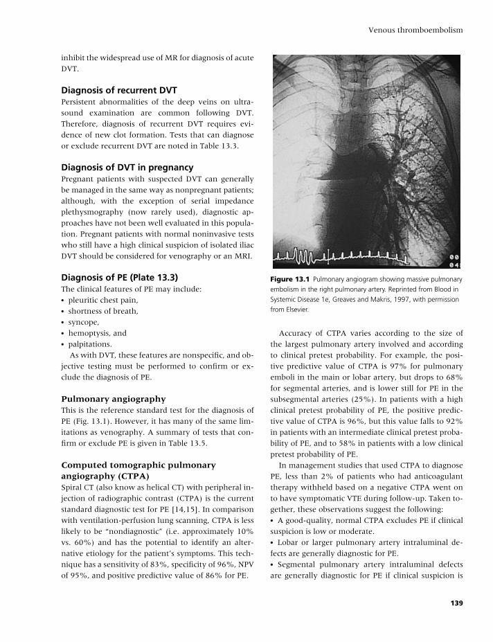
BLBK186-Key May 4, 2009 9:50
Venous thromboembolism
inhibit the widespread use of MR for diagnosis of acute
DVT.
Diagnosis of recurrent DVTPersistent abnormalities of the deep veins on ultra-
sound examination are common following DVT.
Therefore, diagnosis of recurrent DVT requires evi-
dence of new clot formation. Tests that can diagnose
or exclude recurrent DVT are noted in Table 13.3.
Diagnosis of DVT in pregnancyPregnant patients with suspected DVT can generally
be managed in the same way as nonpregnant patients;
although, with the exception of serial impedance
plethysmography (now rarely used), diagnostic ap-
proaches have not been well evaluated in this popula-
tion. Pregnant patients with normal noninvasive tests
who still have a high clinical suspicion of isolated iliac
DVT should be considered for venography or an MRI.
Diagnosis of PE (Plate 13.3)The clinical features of PE may include:� pleuritic chest pain,� shortness of breath,� syncope,� hemoptysis, and� palpitations.
As with DVT, these features are nonspecific, and ob-
jective testing must be performed to confirm or ex-
clude the diagnosis of PE.
Pulmonary angiographyThis is the reference standard test for the diagnosis of
PE (Fig. 13.1). However, it has many of the same lim-
itations as venography. A summary of tests that con-
firm or exclude PE is given in Table 13.5.
Computed tomographic pulmonaryangiography (CTPA)Spiral CT (also know as helical CT) with peripheral in-
jection of radiographic contrast (CTPA) is the current
standard diagnostic test for PE [14,15]. In comparison
with ventilation-perfusion lung scanning, CTPA is less
likely to be “nondiagnostic” (i.e. approximately 10%
vs. 60%) and has the potential to identify an alter-
native etiology for the patient’s symptoms. This tech-
nique has a sensitivity of 83%, specificity of 96%, NPV
of 95%, and positive predictive value of 86% for PE.
Figure 13.1 Pulmonary angiogram showing massive pulmonary
embolism in the right pulmonary artery. Reprinted from Blood in
Systemic Disease 1e, Greaves and Makris, 1997, with permission
from Elsevier.
Accuracy of CTPA varies according to the size of
the largest pulmonary artery involved and according
to clinical pretest probability. For example, the posi-
tive predictive value of CTPA is 97% for pulmonary
emboli in the main or lobar artery, but drops to 68%
for segmental arteries, and is lower still for PE in the
subsegmental arteries (25%). In patients with a high
clinical pretest probability of PE, the positive predic-
tive value of CTPA is 96%, but this value falls to 92%
in patients with an intermediate clinical pretest proba-
bility of PE, and to 58% in patients with a low clinical
pretest probability of PE.
In management studies that used CTPA to diagnose
PE, less than 2% of patients who had anticoagulant
therapy withheld based on a negative CTPA went on
to have symptomatic VTE during follow-up. Taken to-
gether, these observations suggest the following:� A good-quality, normal CTPA excludes PE if clinical
suspicion is low or moderate.� Lobar or larger pulmonary artery intraluminal de-
fects are generally diagnostic for PE.� Segmental pulmonary artery intraluminal defects
are generally diagnostic for PE if clinical suspicion is
139
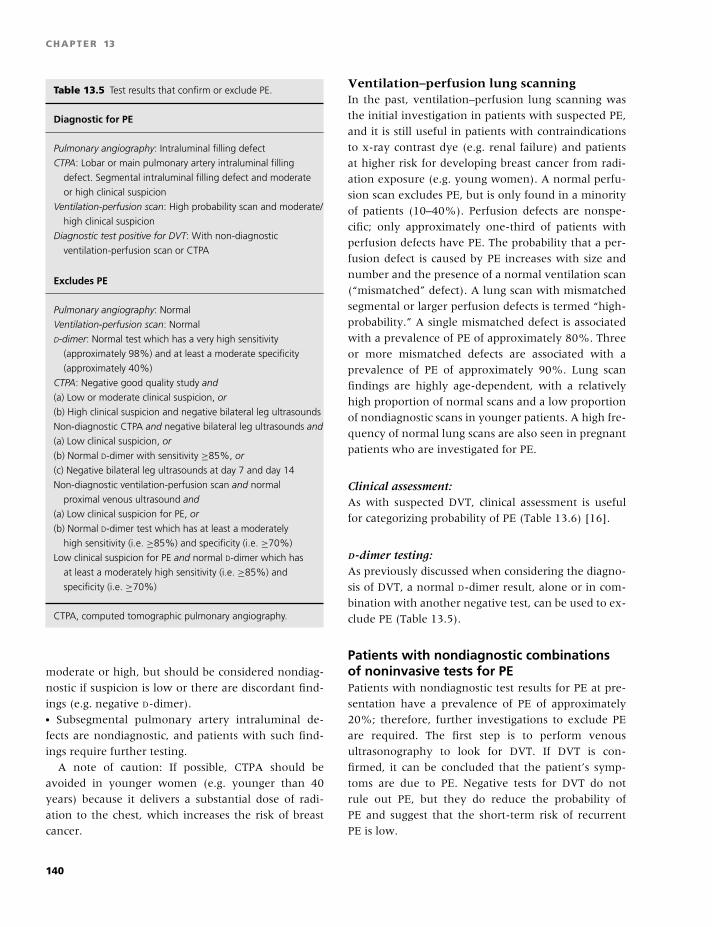
BLBK186-Key May 4, 2009 9:50
CHAPTER 13
Table 13.5 Test results that confirm or exclude PE.
Diagnostic for PE
Pulmonary angiography: Intraluminal filling defect
CTPA: Lobar or main pulmonary artery intraluminal filling
defect. Segmental intraluminal filling defect and moderate
or high clinical suspicion
Ventilation-perfusion scan: High probability scan and moderate/
high clinical suspicion
Diagnostic test positive for DVT: With non-diagnostic
ventilation-perfusion scan or CTPA
Excludes PE
Pulmonary angiography: Normal
Ventilation-perfusion scan: Normal
D-dimer: Normal test which has a very high sensitivity
(approximately 98%) and at least a moderate specificity
(approximately 40%)
CTPA: Negative good quality study and
(a) Low or moderate clinical suspicion, or
(b) High clinical suspicion and negative bilateral leg ultrasounds
Non-diagnostic CTPA and negative bilateral leg ultrasounds and
(a) Low clinical suspicion, or
(b) Normal D-dimer with sensitivity ≥85%, or
(c) Negative bilateral leg ultrasounds at day 7 and day 14
Non-diagnostic ventilation-perfusion scan and normal
proximal venous ultrasound and
(a) Low clinical suspicion for PE, or
(b) Normal D-dimer test which has at least a moderately
high sensitivity (i.e. ≥85%) and specificity (i.e. ≥70%)
Low clinical suspicion for PE and normal D-dimer which has
at least a moderately high sensitivity (i.e. ≥85%) and
specificity (i.e. ≥70%)
CTPA, computed tomographic pulmonary angiography.
moderate or high, but should be considered nondiag-
nostic if suspicion is low or there are discordant find-
ings (e.g. negative D-dimer).� Subsegmental pulmonary artery intraluminal de-
fects are nondiagnostic, and patients with such find-
ings require further testing.
A note of caution: If possible, CTPA should be
avoided in younger women (e.g. younger than 40
years) because it delivers a substantial dose of radi-
ation to the chest, which increases the risk of breast
cancer.
Ventilation–perfusion lung scanningIn the past, ventilation–perfusion lung scanning was
the initial investigation in patients with suspected PE,
and it is still useful in patients with contraindications
to x-ray contrast dye (e.g. renal failure) and patients
at higher risk for developing breast cancer from radi-
ation exposure (e.g. young women). A normal perfu-
sion scan excludes PE, but is only found in a minority
of patients (10–40%). Perfusion defects are nonspe-
cific; only approximately one-third of patients with
perfusion defects have PE. The probability that a per-
fusion defect is caused by PE increases with size and
number and the presence of a normal ventilation scan
(“mismatched” defect). A lung scan with mismatched
segmental or larger perfusion defects is termed “high-
probability.” A single mismatched defect is associated
with a prevalence of PE of approximately 80%. Three
or more mismatched defects are associated with a
prevalence of PE of approximately 90%. Lung scan
findings are highly age-dependent, with a relatively
high proportion of normal scans and a low proportion
of nondiagnostic scans in younger patients. A high fre-
quency of normal lung scans are also seen in pregnant
patients who are investigated for PE.
Clinical assessment:As with suspected DVT, clinical assessment is useful
for categorizing probability of PE (Table 13.6) [16].
D-dimer testing:As previously discussed when considering the diagno-
sis of DVT, a normal D-dimer result, alone or in com-
bination with another negative test, can be used to ex-
clude PE (Table 13.5).
Patients with nondiagnostic combinationsof noninvasive tests for PEPatients with nondiagnostic test results for PE at pre-
sentation have a prevalence of PE of approximately
20%; therefore, further investigations to exclude PE
are required. The first step is to perform venous
ultrasonography to look for DVT. If DVT is con-
firmed, it can be concluded that the patient’s symp-
toms are due to PE. Negative tests for DVT do not
rule out PE, but they do reduce the probability of
PE and suggest that the short-term risk of recurrent
PE is low.
140
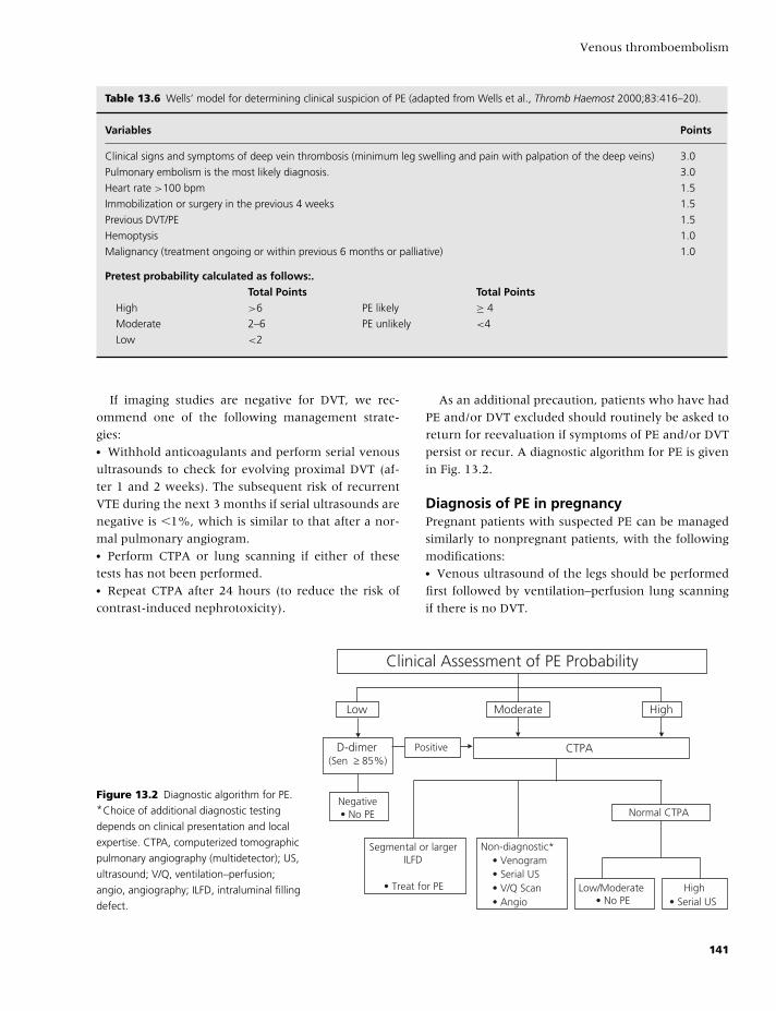
BLBK186-Key May 4, 2009 9:50
Venous thromboembolism
Table 13.6 Wells’ model for determining clinical suspicion of PE (adapted from Wells et al., Thromb Haemost 2000;83:416–20).
Variables Points
Clinical signs and symptoms of deep vein thrombosis (minimum leg swelling and pain with palpation of the deep veins) 3.0
Pulmonary embolism is the most likely diagnosis. 3.0
Heart rate >100 bpm 1.5
Immobilization or surgery in the previous 4 weeks 1.5
Previous DVT/PE 1.5
Hemoptysis 1.0
Malignancy (treatment ongoing or within previous 6 months or palliative) 1.0
Pretest probability calculated as follows:.Total Points Total Points
High >6 PE likely ≥ 4
Moderate 2–6 PE unlikely <4
Low <2
If imaging studies are negative for DVT, we rec-
ommend one of the following management strate-
gies:� Withhold anticoagulants and perform serial venous
ultrasounds to check for evolving proximal DVT (af-
ter 1 and 2 weeks). The subsequent risk of recurrent
VTE during the next 3 months if serial ultrasounds are
negative is �1%, which is similar to that after a nor-
mal pulmonary angiogram.� Perform CTPA or lung scanning if either of these
tests has not been performed.� Repeat CTPA after 24 hours (to reduce the risk of
contrast-induced nephrotoxicity).
As an additional precaution, patients who have had
PE and/or DVT excluded should routinely be asked to
return for reevaluation if symptoms of PE and/or DVT
persist or recur. A diagnostic algorithm for PE is given
in Fig. 13.2.
Diagnosis of PE in pregnancyPregnant patients with suspected PE can be managed
similarly to nonpregnant patients, with the following
modifications:� Venous ultrasound of the legs should be performed
first followed by ventilation–perfusion lung scanning
if there is no DVT.
Clinical Assessment of PE Probability
Low Moderate
D-dimer(Sen ≥ 85%)
Negative• No PE
CTPAPositive
High
Segmental or largerILFD
• Treat for PE
Non-diagnostic*• Venogram• Serial US• V/Q Scan• Angio
Normal CTPA
Low/Moderate• No PE
High• Serial US
Figure 13.2 Diagnostic algorithm for PE.*Choice of additional diagnostic testing
depends on clinical presentation and local
expertise. CTPA, computerized tomographic
pulmonary angiography (multidetector); US,
ultrasound; V/Q, ventilation–perfusion;
angio, angiography; ILFD, intraluminal filling
defect.
141
BLBK186-Key May 4, 2009 9:50
CHAPTER 13
� The amount of radioisotope used for the perfusion
scan should be reduced and the duration of scanning
extended.� If pulmonary angiography is performed, the brachial
approach with abdominal screening is preferred.� The use of CTPA in pregnancy is discouraged, pri-
marily because of radiation exposure to the mother.
Treatment of VTE
Initiation of anticoagulant therapywith heparinHeparin is a highly sulfated glycosoaminoglycan that
produces its anticoagulant effect by binding to an-
tithrombin, markedly accelerating the ability of this
naturally occurring anticoagulant to inactivate throm-
bin, activated factor X (factor Xa), and activated fac-
tor IX (factor IXa). At therapeutic concentrations,
heparin has a half-life of approximately 60 minutes.
Heparin binds to a number of plasma proteins, a phe-
nomenon that reduces its anticoagulant effect by lim-
iting its accessibility to antithrombin. The concen-
tration of heparin-binding proteins increases during
illness, which contributes to the variability in antico-
agulant response in patients with thromboembolism.
Because of this variability, response to intravenous
heparin should be monitored with the activated par-
tial thromboplastin time (APTT) [17].
Many trials have established that weight-adjusted
low-molecular-weight heparin (LMWH) is as safe and
effective as adjusted-dose heparin for the treatment
of acute VTE. LMWHs are derived from standard,
commercial-grade heparin by chemical depolymeriza-
tion to yield fragments approximately one-third the
size of heparin. Depolymerization of heparin results
in less binding to heparin-binding proteins and, con-
sequently, improved bioavailability. LMWH therefore
has a more predictable anticoagulant response than
heparin, which reduces the need for laboratory moni-
toring. Additional advantages of LMWH are that it can
be used to treat patients without hospital admission
and need only be injected subcutaneously once daily.
Other potential side effects include HIT and osteo-
porosis. These complications occur less frequently in
patients receiving LMWH. Patients with HIT, with or
without associated thrombosis, can be treated with
danaparoid, hirudin, or argatroban.
Current clinical practice is to treat patients with
acute VTE for a minimum of 5 days with: (1) intra-
venous heparin in a regimen of at least 30,000 IU/day
or 18 IU/kg/hour adjusted to achieve an APTT ratio
of 1.5 to 2.5; (2) LMWH at a weight-adjusted dose
of either approximately 100 IU/kg every 12 hours or
approximately 150–200 IU/kg once daily; (3) subcuta-
neous heparin administered twice daily, either moni-
tored (initial dose of 17,500 U twice daily or a weight-
adjusted dose of 250 U/kg twice daily, with dose
adjustment to achieve an APTT ratio of 1.5 to 2.5
six hours after injection) or unmonitored (initial dose
of 333 U/kg followed by a twice daily dose of 250
U/kg); or (4) fondaparinux 5.0 mg (2.5 mg if �50 kg;
7.5 mg if �100 kg) once daily by subcutaneous injec-
tion [18]. This initial treatment is usually overlapped
with a course of oral anticoagulants.
Long-term therapy with oral anticoagulantsVitamin K antagonists (e.g. warfarin) are coumarin
compounds that produce their anticoagulant effect
through the production of hemostatically defective,
vitamin K-dependent coagulant proteins (prothrom-
bin, factor VII, factor IX, and factor X). The dose
of warfarin must be monitored closely because the
anticoagulant response is influenced by interactions
with other medications and changes in diet [19]. The
international normalized ratio (INR) replaced the pro-
thrombin time (PT) for monitoring oral anticoagu-
lant therapy in the 1970s because, unlike the PT, the
INR takes into account differences in the responsive-
ness of thromboplastins to oral anticoagulants. The
target INR for treatment of acute VTE is 2.0–3.0.
Oral anticoagulants are typically started on day 1 or
2 of treatment of acute VTE and continued for a length
of time determined on an individual basis (discussed
below). Long-term treatment with LMWH (50–75%
of acute treatment dose) has also been shown to be
effective in treating VTE.
Duration of anticoagulant therapyThe optimal duration of anticoagulant therapy is de-
termined by both patient- and disease-related factors.
The most important factors are outlined below.
Major transient risk factorsThese include recent surgery (within 3 months
of surgery with general anesthesia), plaster cast
142
BLBK186-Key May 4, 2009 9:50
Venous thromboembolism
immobilization of a leg, and hospitalization. The risk
of recurrence after stopping anticoagulant therapy is
low, approximately 3% in the first year after stopping
anticoagulant therapy and 10% in the first 5 years.
Three months of anticoagulant therapy is considered
adequate for patients with VTE secondary to these risk
factors.
Minor transient risk factorsThese include estrogen therapy, prolonged travel (i.e.
�10 hours), pregnancy, less marked leg injuries, and
immobilization. The risk of recurrence after stopping
anticoagulant therapy is expected to be higher than
in those patients with a major transient risk factor,
but lower than those patients with an unprovoked
VTE (e.g. approximately 5% in the first year). Three
months of anticoagulant therapy is also considered ad-
equate in this setting.
Unprovoked VTEThe risk of recurrent VTE, after 6 months or more of
treatment, when anticoagulant therapy is stopped fol-
lowing an unprovoked VTE is approximately 10% in
the first year and approximately 30% after 5 years.
Given the persistent risk of recurrence, and the greater
than 90% risk reduction with oral anticoagulants tar-
geted at an INR of 2.5, long-term anticoagulation is
the preferred option for patients who have a low risk
of bleeding. The rationale for long-term anticoagula-
tion is even stronger for patients with unprovoked PE.
As patients with isolated calf DVT have half the risk of
recurrence of those with proximal DVT, 3 months of
anticoagulant therapy is considered adequate.
Active malignancyPatients with cancer who have VTE are three-fold
more likely to have recurrent VTE than patients who
do not have cancer. The patients at highest risk of re-
currence (e.g. patients with metastatic disease, poor
mobility, or ongoing chemotherapy) should be con-
sidered for indefinite anticoagulant therapy. One large
randomized trial has shown that extended duration
LMWH (minimum of 6 months) reduces the risk of
recurrent VTE in patients with malignancy by approx-
imately 50% compared with conventional anticoagu-
lant treatment (LMWH for 5–7 days followed by oral
anticoagulant therapy) [20].
Hypercoagulable statesPatients who have an antiphospholipid antibody (e.g.,
anticardiolipin antibodies and/or lupus anticoagulant)
have a higher risk of recurrence and should receive in-
definite anticoagulant therapy. Patients heterozygous
for factor V Leiden or the G20210A prothrombin gene
mutation do not appear to have a clinically impor-
tant increased risk for recurrence. The implications for
duration of treatment of other abnormalities, such as
homozygous factor V Leiden, double heterozygous for
factor V Leiden and the G20210A prothrombin gene
mutation, as well as elevated levels of clotting factors
VIII, IX, XI, and homocysteine, and deficiencies of pro-
tein C, protein S, and antithrombin, are uncertain.
PE versus DVTPatients who present with PE appear to have the same
risk of recurrent VTE as those who present with proxi-
mal DVT. However, patients who initially present with
a symptomatic PE are three times more likely to have a
PE as their recurrent VTE event (approximately 60%)
than patients who initially present with a symptomatic
DVT (approximately 20%). Consequently, the case fa-
tality of recurrent VTE in patients who initially pre-
sented with a PE is expected to be two-fold higher
(10%) after a PE than after an initial DVT (5%).
Other potential indicators for increased riskof recurrent VTEPatients who experience a second episode of VTE
have an increased risk of recurrence (RR 1.5), and
indefinite anticoagulant therapy is recommended if
both episodes were unprovoked. Male gender, pres-
ence of residual DVT on ultrasound examination, and
an elevated D-dimer level after stopping anticoagulant
therapy may all be associated with an increased risk
of recurrent VTE, but the implication of these factors
for duration of anticoagulant therapy is currently
uncertain.
Risk of bleeding on anticoagulant therapyThe risk of bleeding on anticoagulants differs markedly
among patients, depending on the prevalence of
risk factors (e.g. advanced age, previous bleeding or
stroke, renal failure, anemia, antiplatelet therapy, ma-
lignancy, poor anticoagulant control) [21]. A meta-
analysis in patients who were considered average risk
for bleeding and received oral anticoagulant therapy
143
BLBK186-Key May 4, 2009 9:50
CHAPTER 13
for VTE for 3 months (at a target INR range of 2.0–3.0)
demonstrated a case fatality of major bleeding of 9%
[22]. Consequently, the case fatality with an episode of
major bleeding appears to be similar to the case fatal-
ity of recurrent VTE after an initial PE, and twice that
of a recurrence after an initial DVT. Based on these
observations, for a patient to be considered for long-
term anticoagulant therapy, the estimated risk of re-
currence off anticoagulant therapy needs to be greater
than the risk of major bleeding on anticoagulant
therapy.
Thrombolytic therapy
Systemic thrombolytic therapy accelerates the rate of
resolution of PE, which can be life-saving for patients
with hemodynamic compromise (i.e. severe hypoten-
sion and/or hypoxia). However, this benefit comes at
the cost of about a two- to four-fold increase in the
frequency of major bleeding, and a five- to ten-fold
increase in intracranial bleeding [23]. One trial con-
ducted in patients with submassive PE demonstrated
a significant reduction in the combined endpoint of
in-hospital death and clinical deterioration, requir-
ing escalation of treatment for patients who received
thrombolysis in addition to heparin in comparison
with patients who received heparin alone. The groups
did not differ in all-cause mortality, recurrent PE, or
major bleeding. Whether thrombolytic therapy de-
creases the incidence of pulmonary hypertension or
recurrences in the long term is yet to be determined.
Similarly, thrombolytic therapy may reduce the risk of
the post-thrombotic syndrome following DVT, but this
does not appear to justify its associated risks. Catheter-
based treatments of DVT (e.g., thrombolytic therapy
combined with mechanical removal of thrombus) may
be more rapidly effective and associated with a lower
risk of bleeding, but require further evaluation before
they can be recommended.
When thrombolysis is indicated, regimens that are
given within 2 hours or less, such as 100 mg rt-PA
over 2 hours, appear preferable.
Major contraindications to thrombolytic therapy
include:� active internal bleeding,� stroke within the past 3 months, and� intracranial disease.
Relative contraindications include:� major surgery within the past 10 days,� recent organ biopsy,� recent puncture of a noncompressible vessel,� recent gastrointestinal bleeding,� liver or renal disease,� severe arterial hypertension, and� severe diabetic retinopathy.
Surgical treatment
Pulmonary endarterectomy is beneficial in selected
patients with thromboembolic pulmonary hyperten-
sion. Urgent pulmonary embolectomy is reserved for
patients with shock whose blood pressure cannot
be maintained despite administration of thrombolytic
therapy or those with an absolute contraindication to
thrombolytic therapy.
Inferior vena caval filtersA randomized trial demonstrated that a filter, as an
adjunct to anticoagulation in patients with proximal
DVT, reduced the rate of PE (asymptomatic and symp-
tomatic) from 4.5% to 1.0% during the 12 days fol-
lowing insertion, with a suggestion of fewer fatal
episodes (0% vs. 2%) [24]. However, after 2 years, pa-
tients with a filter had a significantly higher rate of re-
current DVT (21% vs. 12%) and only a nonstatistically
significant reduction in the frequency of symptomatic
PE (3% vs. 6%). After 8 years of follow up, there was
a reduction in PE, an increase in DVT, and no differ-
ence in DVT and PE combined. This study supports the
use of vena caval filters to prevent PE in patients with
acute DVT and/or PE who cannot be anticoagulated
(i.e. bleeding) but does not support more liberal use of
filters. Patients should receive a course of anticoagu-
lation if this subsequently becomes safe, which should
be continued for the same duration as if the patient
did not have a vena caval filter in situ. A rare late
complication of IVC filters is extensive IVC thrombosis
(Plate 13.4).
Treatment of VTE during pregnancyHeparin and LMWH do not cross the placenta and are
safe for the fetus, whereas oral anticoagulants cross
the placenta and can cause fetal bleeding and mal-
formations. Therefore, pregnant women with acute
VTE should be treated with therapeutic doses of
144
BLBK186-Key May 4, 2009 9:50
Venous thromboembolism
subcutaneous heparin or LMWH throughout preg-
nancy. Care should be taken to avoid delivery while
the mother is therapeutically anticoagulated; one
management approach involves stopping subcuta-
neous heparin 24 hours prior to induction of labor and
switching to intravenous heparin if there is a high risk
of embolism. After delivery, warfarin, which is safe for
infants of nursing mothers, should be given (with ini-
tial heparin overlap) for 6 weeks and until a minimum
of 3 months of treatment has been completed.
Prevention of VTE
VTE prophylaxis following surgerySurgical patients can be stratified according to their
risk factors for VTE into low-, moderate-, and high-
risk categories [25].
Low riskThis category includes patients under 40 years of age
who undergo uncomplicated surgery and have no ad-
ditional risk factors. The rate of asymptomatic prox-
imal DVT detected by surveillance bilateral venogra-
phy is 0.4%, and the rate of symptomatic PE and fatal
PE is 0.2% and �0.01%, respectively. Recommended
VTE prophylaxis in this group is limited to early
mobilization.
Moderate riskThis category includes patients over 40 years of age
who undergo prolonged and/or complicated surgery
or have additional minor risk factors. The rate of
asymptomatic proximal DVT is 5%, and the rate of
symptomatic PE and fatal PE is 2% and 0.5%, respec-
tively. Recommended VTE prophylaxis in this group
includes unfractionated heparin (5000 U/day preop-
eratively, and two to three times daily postopera-
tively), LMWH (approximately 3000 U/day), or grad-
uated compression stockings alone or in combination
with pharmacologic methods.
High riskThis category includes patients who undergo major
surgery for malignancy, hip or knee surgery, or those
who have a history of previous VTE. The rate of
asymptomatic proximal DVT is 15%, and the rate of
symptomatic PE and fatal PE is 5% and 1%, respec-
tively. Recommended VTE prophylaxis in this group
includes LMWH (4000 to 6000 U/day, as a single or di-
vided dose); warfarin (usually started postoperatively
and adjusted to achieve an INR of 2.0–3.0); or fonda-
parinux (2.5 mg once daily, usually started postopera-
tively) or intermittent pneumatic compression devices
alone or in combination with other methods of pro-
phylaxis. Mechanical methods of prophylaxis should
be used in patients who have a moderate or high risk
of VTE if anticoagulants are contraindicated (e.g. neu-
rosurgical patients).
Pharmacologic agents for VTE prophylaxisin orthopedic surgeryMeta-analyses support the finding that LMWH is more
effective than heparin following orthopedic surgery
and is associated with a similar frequency of bleeding.
Warfarin (target INR 2–3 for approximately 7–10 days)
is less effective than LMWH at preventing DVTs that
are detected by venography soon after surgery, but
appears to be similarly effective at preventing symp-
tomatic VTE over a 3-month period. An additional 3
or 4 weeks of LMWH after hospital discharge further
reduces the frequency of symptomatic VTE after or-
thopedic surgery (from 3.3% to 1.3%). There is ev-
idence that aspirin reduces the risk of postoperative
VTE by one-third. However, as warfarin and LMWH
are expected to be more effective (at least a two-
thirds reduction in VTE), aspirin alone is not recom-
mended during the initial postoperative period. Fon-
daparinux has been shown to be more effective than
LMWH following major orthopedic surgery but may
cause marginally more bleeding.
VTE prophylaxis in medical patientsPrimary prophylaxis with anticoagulants and/or me-
chanical methods should be used in hospitalized pa-
tients who have a moderate or high risk of VTE. In
recent years, three large, randomized, controlled tri-
als have shown that LMWH (enoxaparin 40 mg or
dalteparin 5000 IU subcutaneously once daily for 10
days) and fondaparinux (2.5 mg once daily) reduce
the rate of VTE by about 50% (range 45–63%) com-
pared with placebo in acutely ill medical patients.
References
1 Kearon C, Crowther M, Hirsh J. Management of pa-
tients with hereditary hypercoagulable disorders. Annu
Rev Med 2000;51:169–85.
145
BLBK186-Key May 4, 2009 9:50
CHAPTER 13
2 Anderson FA Jr, Spencer FA. Risk factors for venous
thromboembolism. Circulation 2003;107:I9–16.
3 Warkentin TE, Greinacher A, Koster A, Lincoff AM.
Treatment and prevention of heparin-induced throm-
bocytopenia: American College of Chest Physicians
Evidence-Based Clinical Practice Guidelines (8th Edi-
tion). Chest 2008;133:340S–80S.
4 Kearon C, Julian JA, Newman TE, Ginsberg JS. Nonin-
vasive diagnosis of deep venous thrombosis. McMaster
Diagnostic Imaging Practice Guidelines Initiative. Ann
Intern Med 1998;128:663–77.
5 Wells PS, Anderson DR, Rodger M, et al. Evaluation of
D-dimer in the diagnosis of suspected deep-vein throm-
bosis. N Engl J Med 2003;349:1227–35.
6 Wells PS, Owen C, Doucette S, Fergusson D, Tran H.
Does this patient have deep vein thrombosis? JAMA
2006;295:199–207.
7 Wheeler HB, Hirsh J, Wells P, Anderson FA Jr. Diag-
nostic tests for deep vein thrombosis. Clinical useful-
ness depends on probability of disease. Arch Intern Med
1994;154:1921–8.
8 Stevens SM, Elliott CG, Chan KJ, Egger MJ,
Ahmed KM. Withholding anticoagulation after a neg-
ative result on duplex ultrasonography for suspected
symptomatic deep venous thrombosis. Ann Intern Med
2004;140:985–91.
9 Kelly J, Rudd A, Lewis RR, Hunt BJ. Plasma D-dimers in
the diagnosis of venous thromboembolism. Arch Intern
Med 2002;162:747–56.
10 Stein PD, Hull RD, Patel KC, et al. D-dimer for
the exclusion of acute venous thrombosis and pul-
monary embolism: a systematic review. Ann Intern Med
2004;140:589–602.
11 Fancher TL, White RH, Kravitz RL. Combined use of
rapid D-dimer testing and estimation of clinical proba-
bility in the diagnosis of deep vein thrombosis: system-
atic review. Br Med J 2004;329:821.
12 Kanne JP, Lalani TA. Role of computed tomogra-
phy and magnetic resonance imaging for deep ve-
nous thrombosis and pulmonary embolism. Circulation
2004;109:I15–21.
13 Sampson FC, Goodacre SW, Thomas SM, van Beek EJ.
The accuracy of MRI in diagnosis of suspected deep vein
thrombosis: systematic review and meta-analysis. Eur
Radiol 2007;17:175–81.
14 Stein PD, Fowler SE, Goodman LR, et al. Multidetector
computed tomography for acute pulmonary embolism.
N Engl J Med 2006;354:2317–27.
15 Roy PM, Colombet I, Durieux P, Chatellier G, Sors
H, Meyer G. Systematic review and meta-analysis of
strategies for the diagnosis of suspected pulmonary em-
bolism. Br Med J 2005;331:259.
16 Wells PS, Anderson DR, Rodger M, et al. Excluding
pulmonary embolism at the bedside without diagnostic
imaging: management of patients with suspected pul-
monary embolism presenting to the emergency depart-
ment by using a simple clinical model and d-dimer. Ann
Intern Med 2001;135:98–107.
17 Hirsh J, Bauer KA, Donati MB, Gould M, Samama MM,
Weitz JI. Parenteral anticoagulants: American College
of Chest Physicians Evidence-Based Clinical Practice
Guidelines (8th Edition). Chest 2008;133:141S–59S.
18 Kearon C, Kahn SR, Agnelli G, Goldhaber S, Raskob
GE, Comerota AJ. Antithrombotic therapy for venous
thromboembolic disease: American College of Chest
Physicians Evidence-Based Clinical Practice Guidelines
(8th Edition). Chest 2008;133:454S–545S.
19 Ansell J, Hirsh J, Hylek E, Jacobson A, Crowther M,
Palareti G. Pharmacology and management of the vi-
tamin K antagonists: American College of Chest Physi-
cians Evidence-Based Clinical Practice Guidelines (8th
Edition). Chest 2008;133:160S–98S.
20 Lee AY, Levine MN, Baker RI, Bowden C, Kakkar AK,
Prins M, et al. Low-molecular-weight heparin versus
a coumarin for the prevention of recurrent venous
thromboembolism in patients with cancer. N Engl J Med
2003;349:146–53.
21 Schulman S, Beyth RJ, Kearon C, Levine MN. Hem-
orrhagic complications of anticoagulant and throm-
bolytic treatment: American College of Chest Physi-
cians Evidence-Based Clinical Practice Guidelines (8th
Edition). Chest 2008;133:257S–98S.
22 Linkins LA, Choi PT, Douketis JD. Clinical impact of
bleeding in patients taking oral anticoagulant therapy
for venous thromboembolism: a meta-analysis. Ann In-
tern Med 2003;139:893–900.
23 Agnelli G, Becattini C, Kirschstein T. Thrombolysis vs
heparin in the treatment of pulmonary embolism: a
clinical outcome-based meta-analysis. Arch Intern Med
2002;162:2537–41.
24 Decousus H, Leizorovicz A, Parent F, et al. A clin-
ical trial of vena caval filters in the prevention of
pulmonary embolism in patients with proximal deep-
vein thrombosis. Prevention du Risque d’Embolie Pul-
monaire par Interruption Cave Study Group. N Engl J
Med 1998;338:409–15.
25 Geerts WH, Bergqvist D, Pineo GF, et al. Prevention of
venous thromboembolism: American College of Chest
Physicians Evidence-Based Clinical Practice Guidelines
(8th Edition). Chest 2008;133:381S–453S.
146
BLBK186-Key April 11, 2009 12:59
14 Myeloproliferative neoplasms:Essential thrombocythemia,polycythemia vera, and primarymyelofibrosisAyalew Tefferi
Introduction
Myeloproliferative neoplasms (MPN) are a cate-
gory of chronic myeloid malignancies that includes
chronic myelogenous leukemia (CML), polycythemia
vera (PV), essential thrombocythemia (ET), primary
myelofibrosis (PMF), and other less known clinico-
pathologic entities [1]. From a pathogenetic stand-
point, all members of the MPN arise out of an acquired
oncogenic mutation that occurs at the stem cell level.
Therefore, the MPN are often referred to as clonal
stem cell diseases. However, at present, the disease-
causing mutation is known only for CML and involves
the cytoplasmic tyrosine kinase ABL (BCR-ABL). In
early 2005, an activating Janus kinase 2 mutation
(JAK2V617F) was discovered in PV, ET, and PMF [2].
In 2006 and 2007, additional JAK2 (exon 12 muta-
tions) and MPL (thrombopoietin receptor) mutations
were described in these diseases [2]. Current estimates
of JAK2 and MPL mutational frequencies in PV are
100% and 0%, for ET 50% and 5%, and for PMF 70%
and 10%, respectively.
Clinical presentation
Annual incidence rates for ET, PV, and PMF are esti-
mated at 2.5, 1.0, and 0.5, respectively. Prevalence fig-
ures are much higher, especially in cases of ET and PV
because of their relatively good prognosis. The median
age at diagnosis for all of these three MPN is approxi-
mately 60 years. In general, clinical manifestations are
similar in ET and PV but different than those in PMF.
Essential thrombocythemia andpolycythemia veraMost patients with ET or PV are asymptomatic at
presentation. Approximately one-third of patients
present with microvascular symptoms: headaches,
lightheadedness, visual symptoms such as blurring
and scotomata, palpitations, chest pain, erythrome-
lalgia, and distal paresthesias. Erythromelalgia is the
most dramatic vasomotor symptom, characterized by
erythema, warmth, and pain in distal extremities.
Other non-life-threatening complications in ET and
PV include constitutional symptoms, pruritus that is
often provoked by water contact, superficial throm-
bophlebitis, minor mucocutaneous bleeding, and in-
creased propensity for first trimester miscarriage. At
least two-thirds of PV patients have splenomegaly at
diagnosis.
PV and ET are associated with an increased risk
of thrombosis and bleeding. Table 14.1 (at diagnosis)
[3–14] and Table 14.2 (during follow-up) [3–15]
present incidence figures of “major” thrombotic events
in a selected series of large studies in PV and ET. Major
thrombosis at diagnosis ranges from 9.7% to 29.4%
for ET and 34% to 38.6% for PV; the corresponding
figures for major thrombosis during follow-up are 8%
to 30.7% for ET and 8.1% to 19% for PV.
In general, arterial events (strokes, transient is-
chemic attacks, myocardial infarctions, angina pec-
toris, peripheral artery occlusions) are more prevalent
than venous events (pulmonary embolism, deep vein
thrombosis, hepatic/portal/mesenteric vein thrombo-
sis, sagittal sinus thrombosis, retinal vein thrombosis)
in ET or PV. For example, in one recent study of 470
patients with either ET or PV, who experienced first
147
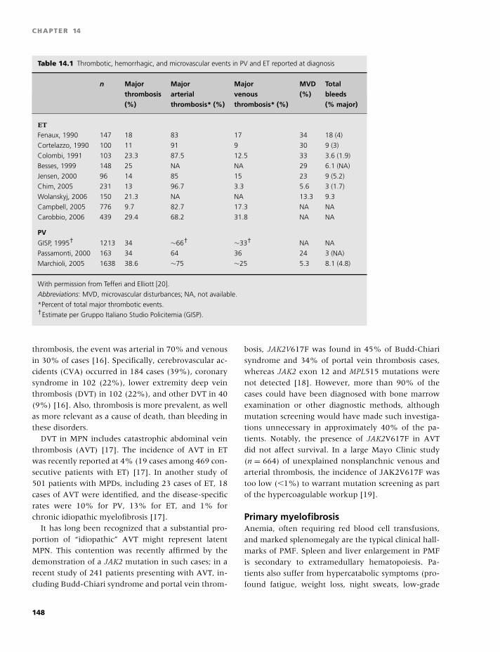
BLBK186-Key April 11, 2009 12:59
CHAPTER 14
Table 14.1 Thrombotic, hemorrhagic, and microvascular events in PV and ET reported at diagnosis
n Major Major Major MVD Totalthrombosis arterial venous (%) bleeds(%) thrombosis* (%) thrombosis* (%) (% major)
ET
Fenaux, 1990 147 18 83 17 34 18 (4)
Cortelazzo, 1990 100 11 91 9 30 9 (3)
Colombi, 1991 103 23.3 87.5 12.5 33 3.6 (1.9)
Besses, 1999 148 25 NA NA 29 6.1 (NA)
Jensen, 2000 96 14 85 15 23 9 (5.2)
Chim, 2005 231 13 96.7 3.3 5.6 3 (1.7)
Wolanskyj, 2006 150 21.3 NA NA 13.3 9.3
Campbell, 2005 776 9.7 82.7 17.3 NA NA
Carobbio, 2006 439 29.4 68.2 31.8 NA NA
PV
GISP, 1995† 1213 34 ∼66† ∼33† NA NA
Passamonti, 2000 163 34 64 36 24 3 (NA)
Marchioli, 2005 1638 38.6 ∼75 ∼25 5.3 8.1 (4.8)
With permission from Tefferi and Elliott [20].
Abbreviations: MVD, microvascular disturbances; NA, not available.
*Percent of total major thrombotic events.†Estimate per Gruppo Italiano Studio Policitemia (GISP).
thrombosis, the event was arterial in 70% and venous
in 30% of cases [16]. Specifically, cerebrovascular ac-
cidents (CVA) occurred in 184 cases (39%), coronary
syndrome in 102 (22%), lower extremity deep vein
thrombosis (DVT) in 102 (22%), and other DVT in 40
(9%) [16]. Also, thrombosis is more prevalent, as well
as more relevant as a cause of death, than bleeding in
these disorders.
DVT in MPN includes catastrophic abdominal vein
thrombosis (AVT) [17]. The incidence of AVT in ET
was recently reported at 4% (19 cases among 469 con-
secutive patients with ET) [17]. In another study of
501 patients with MPDs, including 23 cases of ET, 18
cases of AVT were identified, and the disease-specific
rates were 10% for PV, 13% for ET, and 1% for
chronic idiopathic myelofibrosis [17].
It has long been recognized that a substantial pro-
portion of “idiopathic” AVT might represent latent
MPN. This contention was recently affirmed by the
demonstration of a JAK2 mutation in such cases; in a
recent study of 241 patients presenting with AVT, in-
cluding Budd-Chiari syndrome and portal vein throm-
bosis, JAK2V617F was found in 45% of Budd-Chiari
syndrome and 34% of portal vein thrombosis cases,
whereas JAK2 exon 12 and MPL515 mutations were
not detected [18]. However, more than 90% of the
cases could have been diagnosed with bone marrow
examination or other diagnostic methods, although
mutation screening would have made such investiga-
tions unnecessary in approximately 40% of the pa-
tients. Notably, the presence of JAK2V617F in AVT
did not affect survival. In a large Mayo Clinic study
(n = 664) of unexplained nonsplanchnic venous and
arterial thrombosis, the incidence of JAK2V617F was
too low (�1%) to warrant mutation screening as part
of the hypercoagulable workup [19].
Primary myelofibrosisAnemia, often requiring red blood cell transfusions,
and marked splenomegaly are the typical clinical hall-
marks of PMF. Spleen and liver enlargement in PMF
is secondary to extramedullary hematopoiesis. Pa-
tients also suffer from hypercatabolic symptoms (pro-
found fatigue, weight loss, night sweats, low-grade
148
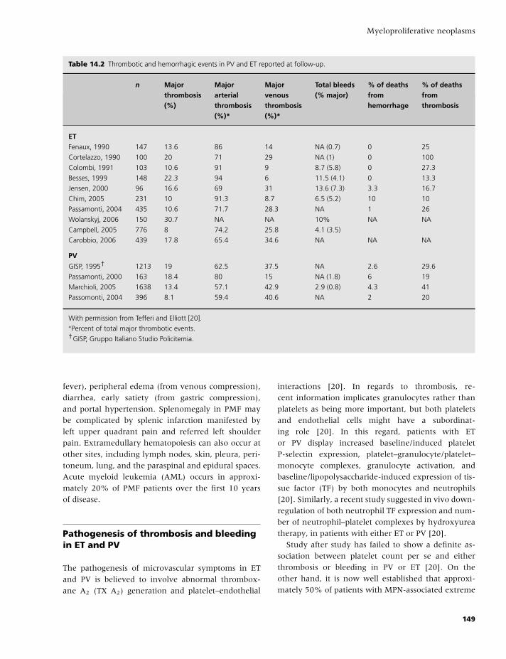
BLBK186-Key April 11, 2009 12:59
Myeloproliferative neoplasms
Table 14.2 Thrombotic and hemorrhagic events in PV and ET reported at follow-up.
n Major Major Major Total bleeds % of deaths % of deathsthrombosis arterial venous (% major) from from(%) thrombosis thrombosis hemorrhage thrombosis
(%)* (%)*
ETFenaux, 1990 147 13.6 86 14 NA (0.7) 0 25
Cortelazzo, 1990 100 20 71 29 NA (1) 0 100
Colombi, 1991 103 10.6 91 9 8.7 (5.8) 0 27.3
Besses, 1999 148 22.3 94 6 11.5 (4.1) 0 13.3
Jensen, 2000 96 16.6 69 31 13.6 (7.3) 3.3 16.7
Chim, 2005 231 10 91.3 8.7 6.5 (5.2) 10 10
Passamonti, 2004 435 10.6 71.7 28.3 NA 1 26
Wolanskyj, 2006 150 30.7 NA NA 10% NA NA
Campbell, 2005 776 8 74.2 25.8 4.1 (3.5)
Carobbio, 2006 439 17.8 65.4 34.6 NA NA NA
PV
GISP, 1995† 1213 19 62.5 37.5 NA 2.6 29.6
Passamonti, 2000 163 18.4 80 15 NA (1.8) 6 19
Marchioli, 2005 1638 13.4 57.1 42.9 2.9 (0.8) 4.3 41
Passomonti, 2004 396 8.1 59.4 40.6 NA 2 20
With permission from Tefferi and Elliott [20].∗Percent of total major thrombotic events.†GISP, Gruppo Italiano Studio Policitemia.
fever), peripheral edema (from venous compression),
diarrhea, early satiety (from gastric compression),
and portal hypertension. Splenomegaly in PMF may
be complicated by splenic infarction manifested by
left upper quadrant pain and referred left shoulder
pain. Extramedullary hematopoiesis can also occur at
other sites, including lymph nodes, skin, pleura, peri-
toneum, lung, and the paraspinal and epidural spaces.
Acute myeloid leukemia (AML) occurs in approxi-
mately 20% of PMF patients over the first 10 years
of disease.
Pathogenesis of thrombosis and bleedingin ET and PV
The pathogenesis of microvascular symptoms in ET
and PV is believed to involve abnormal thrombox-
ane A2 (TX A2) generation and platelet–endothelial
interactions [20]. In regards to thrombosis, re-
cent information implicates granulocytes rather than
platelets as being more important, but both platelets
and endothelial cells might have a subordinat-
ing role [20]. In this regard, patients with ET
or PV display increased baseline/induced platelet
P-selectin expression, platelet–granulocyte/platelet–
monocyte complexes, granulocyte activation, and
baseline/lipopolysaccharide-induced expression of tis-
sue factor (TF) by both monocytes and neutrophils
[20]. Similarly, a recent study suggested in vivo down-
regulation of both neutrophil TF expression and num-
ber of neutrophil–platelet complexes by hydroxyurea
therapy, in patients with either ET or PV [20].
Study after study has failed to show a definite as-
sociation between platelet count per se and either
thrombosis or bleeding in PV or ET [20]. On the
other hand, it is now well established that approxi-
mately 50% of patients with MPN-associated extreme
149
BLBK186-Key April 11, 2009 12:59
CHAPTER 14
thrombocytosis display laboratory evidence of ac-
quired von Willebrand syndrome (AVWS) whose ori-
gin might involve a platelet count-dependent in-
creased proteolysis of high-molecular-weight von
Willebrand protein [21]. However, the degree of the
abnormality is seldom clinically relevant, (i.e. associ-
ated with bleeding or a ristocetin cofactor activity of
�30%) [21].
Other qualitative platelet defects in ET are believed
to play a minor role in disease-associated hemorrhage
and include defects in epinephrine-, collagen-, and
ADP-induced platelet aggregation, decreased ATP se-
cretion, and acquired storage pool deficiency that re-
sults from abnormal in vivo platelet activation. Spon-
taneous platelet aggregation is another characteristic
finding in MPN, but it has no apparent clinical rele-
vance.
Diagnosis
Table 14.3 outlines the current WHO diagnostic cri-
teria for PV, ET, and PMF [1]. Figures 14.1–14.3
provide WHO-based diagnostic algorithms for these
diseases [1]. Virtually all patients with PV carry a JAK2
mutation. Therefore, peripheral blood JAK2V617F
screening is currently the preferred initial test for eval-
uating a patient with suspected PV (Fig. 14.1). The
concomitant determination of serum erythropoietin
(Epo) level is encouraged in order to minimize the
consequences of false-positive or false-negative molec-
ular test results. Mutation screening for an exon 12
JAK2 mutation and bone marrow examination should
be considered in a JAK2V617F-negative patient who
displays subnormal serum Epo levels (Fig. 14.1). Be-
cause JAK2V617F also occurs in approximately 50%
of patients with either ET or PMF, it is reasonable to
include mutation screening in the diagnostic work-up
of both thrombocytosis (Fig. 14.2) and bone marrow
fibrosis (Fig. 14.3).
Prognosis
Median survival in both ET and PV exceeds 15 years,
and the 10-year risk of developing either myelofibrosis
(MF; �4% and �10%, respectively) or AML (;2% and
�6%, respectively) is relatively low. Compared with
both PV and ET, PMF has a significantly worse prog-
nosis with a median survival of 6 years and 10-year
risk of AML estimated at 20%.
Several studies have identified advanced age
(�60 years) and thrombosis history as risk factors for
thrombosis in both PV and ET (Table 14.4) [20]. In ET,
two recent, large, single-institution studies (n = 322
and n = 439, respectively) [9,14] confirmed the pro-
thrombotic effect of advanced age (≥60 years) and
history of thrombosis, although the latter association
was significant in regards to arterial but not venous
events in one of the two studies [9]. In addition, both
studies identified leukocytosis (≥15 × 109/L in one
study [9] and �8.7 × 10⁹/L in the other [14]), but nei-
ther thrombocytosis nor the presence of JAK2V617F,
as an additional independent risk factor for thrombo-
sis. Similarly, the presence of cardiovascular risk fac-
tors did not modify thrombosis risk in one of the two
studies [9], as well as in another recent study [16].
In PV, a series of reports from the European Col-
laboration on Low-Dose Aspirin in Polycythemia Vera
(ECLAP) group have addressed multiple clinical issues,
including thrombotic complications. In their most re-
cent report (n = 1638), Landolfi and colleagues, on
behalf of ECLAP, confirmed the strong association
between advanced age and thrombosis and, in ad-
dition, identified leukocytosis (�15 × 109/L as op-
posed to ≤10 × 109/L) as an independent predictor
of myocardial infarction [20]. History of arterial or ve-
nous events predicted recurrence of a similar vascular
event. In contrast, neither the platelet count nor the
hematocrit level affected thrombosis risk. Similarly,
controlled prospective studies are needed to clarify the
prognostic relevance of hereditary and acquired causes
of thrombophilia, pattern of X chromosome inactiva-
tion in granulocyte-derived DNA (i.e. monoclonal vs.
polyclonal), and altered PRV-1, platelet Mpl, or EEC
expression [20].
Current evidence is inconclusive regarding the
prognostic relevance of JAK2 or MPL mutations in
MPNs. In ET, overall or leukemia-free survival does
not appear to be affected by either the presence of
JAK2V617F or its allele burden. The impact on the
risk of thrombosis or fibrotic transformation is less
clear [20]. Equally unclear is the prognostic relevance
of JAK2V617F allele burden in PV where a higher
mutant allele burden is implicated by some but not
by others as an adverse prognostic factor for fibrotic
150
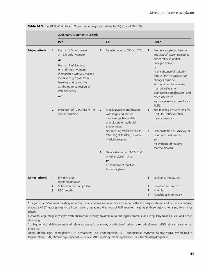
BLBK186-Key April 11, 2009 12:59
Myeloproliferative neoplasms
Table 14.3 The 2008 World Health Organization diagnostic criteria for PV, ET, and PMF [29].
2008 WHO Diagnostic Criteria
PV* ET* PMF*
Major criteria 1 Hgb > 18.5 g/dL (men)
> 16.5 g/dL (women)
or
Hgb > 17 g/dL (men),
or > 15 g/dL (women)
if associated with a sustained
increase of ≥2 g/dL from
baseline that cannot be
attributed to correction of
iron deficiency
or‡
1 Platelet count ≥ 450 × 109/L 1 Megakaryocyte proliferation
and atypia† accompanied by
either reticulin and/or
collagen fibrosis.
orIn the absence of reticulin
fibrosis, the megakaryocyte
changes must be
accompanied by increased
marrow cellularity,
granulocytic proliferation, and
often decreased
erythropoiesis (i.e. pre-fibrotic
PMF).
2 Presence of JAK2V617F or
similar mutation
2 Megakaryocyte proliferation
with large and mature
morphology. No or little
granulocyte or erythroid
proliferation.
2 Not meeting WHO criteria for
CML, PV, MDS, or other
myeloid neoplasm
3 Not meeting WHO criteria for
CML, PV, PMF, MDS, or other
myeloid neoplasm
3 Demonstration of JAK2V617F
or other clonal marker
orno evidence of reactive
marrow fibrosis
4 Demonstration of JAK2V617F
or other clonal marker
orno evidence of reactive
thrombocytosis
Minor criteria 1 BM trilineage
myeloproliferation
1 Leukoerythroblastosis
2 Subnormal serum Epo level 2 Increased serum LDH
3 EEC growth 3 Anemia
4 Palpable splenomegaly
*Diagnosis of PV requires meeting either both major criteria and one minor criterion or the first major criterion and two minor criteria;
diagnosis of ET requires meeting all four major criteria; and diagnosis of PMF requires meeting all three major criteria and two minor
criteria.
†Small to large megakaryocytes with aberrant nuclear/cytoplasmic ratio and hyperchromatic and irregularly folded nuclei and dense
clustering.‡or Hgb or Hct �99th percentile of reference range for age, sex, or altitude of residence or red cell mass �25% above mean normal
predicted.
Abbreviations: Hgb, hemoglobin; Hct, hematocrit; Epo, erythropoietin; EEC, endogenous erythroid colony; WHO, World Health
Organization; CML, chronic myelogenous leukemia; MDS, myelodysplastic syndrome; LDH, lactate dehydrogenase.
151
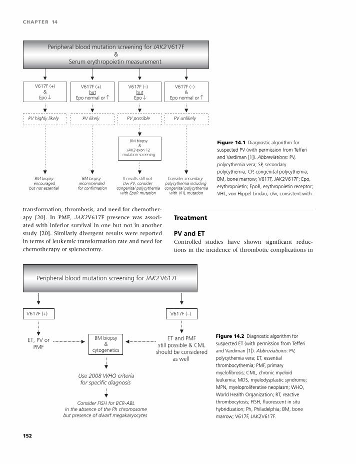
BLBK186-Key April 11, 2009 12:59
CHAPTER 14
Peripheral blood mutation screening for JAK2 V617F&
Serum erythropoietin measurement
V617F (+) but
Epo normal or ↑
V617F (+)&
Epo ↓
V617F (–) but
Epo ↓
V617F (–) &
Epo normal or ↑
PV unlikely PV possiblePV likely PV highly likely
If results still not c/w PV, consider
congenital polycythemia with EpoR mutation
BM biopsy encouraged
but not essential
BM biopsy recommendedfor confirmation
BM biopsy &
JAK2 exon 12 mutation screening
Consider secondary polycythemia including congenital polycythemia
with VHL mutation
Figure 14.1 Diagnostic algorithm for
suspected PV (with permission from Tefferi
and Vardiman [1]). Abbreviations: PV,
polycythemia vera; SP, secondary
polycythemia; CP, congenital polycythemia;
BM, bone marrow; V617F, JAK2V617F; Epo,
erythropoietin; EpoR, erythropoietin receptor;
VHL, von Hippel-Lindau; c/w, consistent with.
transformation, thrombosis, and need for chemother-
apy [20]. In PMF, JAK2V617F presence was associ-
ated with inferior survival in one but not in another
study [20]. Similarly divergent results were reported
in terms of leukemic transformation rate and need for
chemotherapy or splenectomy.
Treatment
PV and ETControlled studies have shown significant reduc-
tions in the incidence of thrombotic complications in
Peripheral blood mutation screening for JAK2 V617F
V617F (+) V617F (–)
ET, PV orPMF
Use 2008 WHO criteriafor specific diagnosis
ET and PMFstill possible & CML
should be consideredas well
BM biopsy&
cytogenetics
Consider FISH for BCR-ABLin the absence of the Ph chromosome
but presence of dwarf megakaryocytes
Figure 14.2 Diagnostic algorithm for
suspected ET (with permission from Tefferi
and Vardiman [1]). Abbreviatioins: PV,
polycythemia vera; ET, essential
thrombocythemia; PMF, primary
myelofibrosis; CML, chronic myeloid
leukemia; MDS, myelodysplastic syndrome;
MPN, myeloproliferative neoplasm; WHO,
World Health Organization; RT, reactive
thrombocytosis; FISH, fluorescent in situ
hybridization; Ph, Philadelphia; BM, bone
marrow; V617F, JAK2V617F.
152
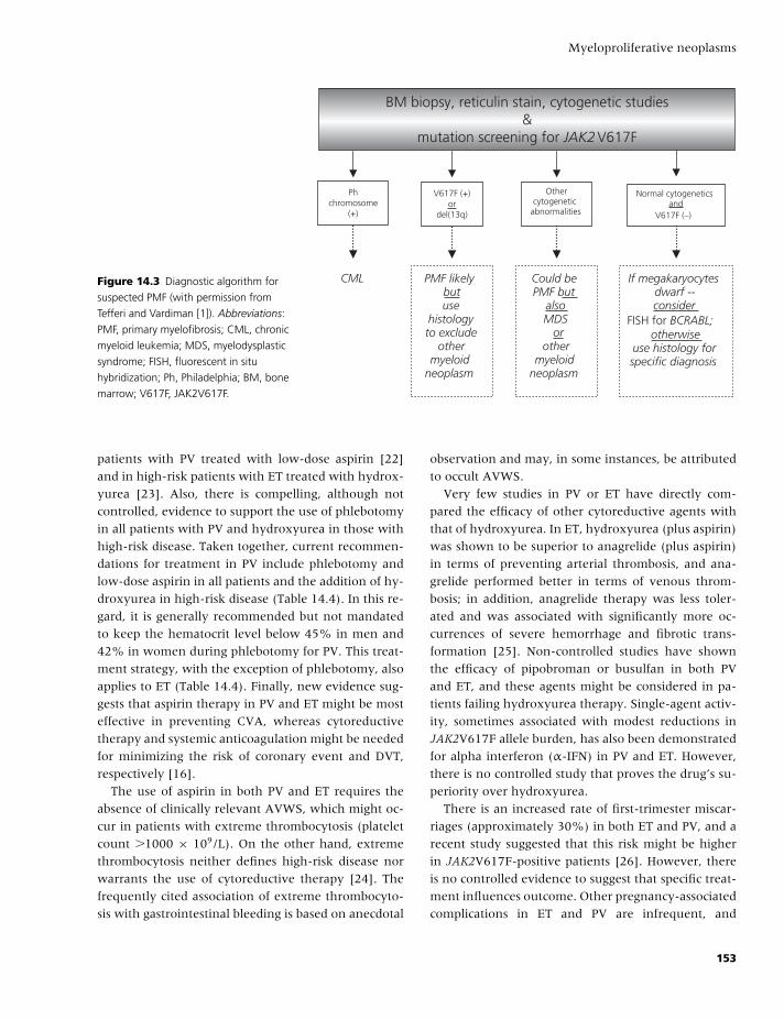
BLBK186-Key April 11, 2009 12:59
Myeloproliferative neoplasms
BM biopsy, reticulin stain, cytogenetic studies&
mutation screening for JAK2 V617F
V617F (+)or
del(13q)
Phchromosome
(+)
Normal cytogeneticsand
V617F (–)
If megakaryocytesdwarf -- consider
FISH for BCRABL;otherwise
use histology forspecific diagnosis
PMF likelybutuse
histologyto exclude
othermyeloid
neoplasm
CML
Othercytogenetic
abnormalities
Could be PMF but
alsoMDS or
othermyeloid
neoplasm
Figure 14.3 Diagnostic algorithm for
suspected PMF (with permission from
Tefferi and Vardiman [1]). Abbreviations:
PMF, primary myelofibrosis; CML, chronic
myeloid leukemia; MDS, myelodysplastic
syndrome; FISH, fluorescent in situ
hybridization; Ph, Philadelphia; BM, bone
marrow; V617F, JAK2V617F.
patients with PV treated with low-dose aspirin [22]
and in high-risk patients with ET treated with hydrox-
yurea [23]. Also, there is compelling, although not
controlled, evidence to support the use of phlebotomy
in all patients with PV and hydroxyurea in those with
high-risk disease. Taken together, current recommen-
dations for treatment in PV include phlebotomy and
low-dose aspirin in all patients and the addition of hy-
droxyurea in high-risk disease (Table 14.4). In this re-
gard, it is generally recommended but not mandated
to keep the hematocrit level below 45% in men and
42% in women during phlebotomy for PV. This treat-
ment strategy, with the exception of phlebotomy, also
applies to ET (Table 14.4). Finally, new evidence sug-
gests that aspirin therapy in PV and ET might be most
effective in preventing CVA, whereas cytoreductive
therapy and systemic anticoagulation might be needed
for minimizing the risk of coronary event and DVT,
respectively [16].
The use of aspirin in both PV and ET requires the
absence of clinically relevant AVWS, which might oc-
cur in patients with extreme thrombocytosis (platelet
count �1000 × 109/L). On the other hand, extreme
thrombocytosis neither defines high-risk disease nor
warrants the use of cytoreductive therapy [24]. The
frequently cited association of extreme thrombocyto-
sis with gastrointestinal bleeding is based on anecdotal
observation and may, in some instances, be attributed
to occult AVWS.
Very few studies in PV or ET have directly com-
pared the efficacy of other cytoreductive agents with
that of hydroxyurea. In ET, hydroxyurea (plus aspirin)
was shown to be superior to anagrelide (plus aspirin)
in terms of preventing arterial thrombosis, and ana-
grelide performed better in terms of venous throm-
bosis; in addition, anagrelide therapy was less toler-
ated and was associated with significantly more oc-
currences of severe hemorrhage and fibrotic trans-
formation [25]. Non-controlled studies have shown
the efficacy of pipobroman or busulfan in both PV
and ET, and these agents might be considered in pa-
tients failing hydroxyurea therapy. Single-agent activ-
ity, sometimes associated with modest reductions in
JAK2V617F allele burden, has also been demonstrated
for alpha interferon (α-IFN) in PV and ET. However,
there is no controlled study that proves the drug’s su-
periority over hydroxyurea.
There is an increased rate of first-trimester miscar-
riages (approximately 30%) in both ET and PV, and a
recent study suggested that this risk might be higher
in JAK2V617F-positive patients [26]. However, there
is no controlled evidence to suggest that specific treat-
ment influences outcome. Other pregnancy-associated
complications in ET and PV are infrequent, and
153
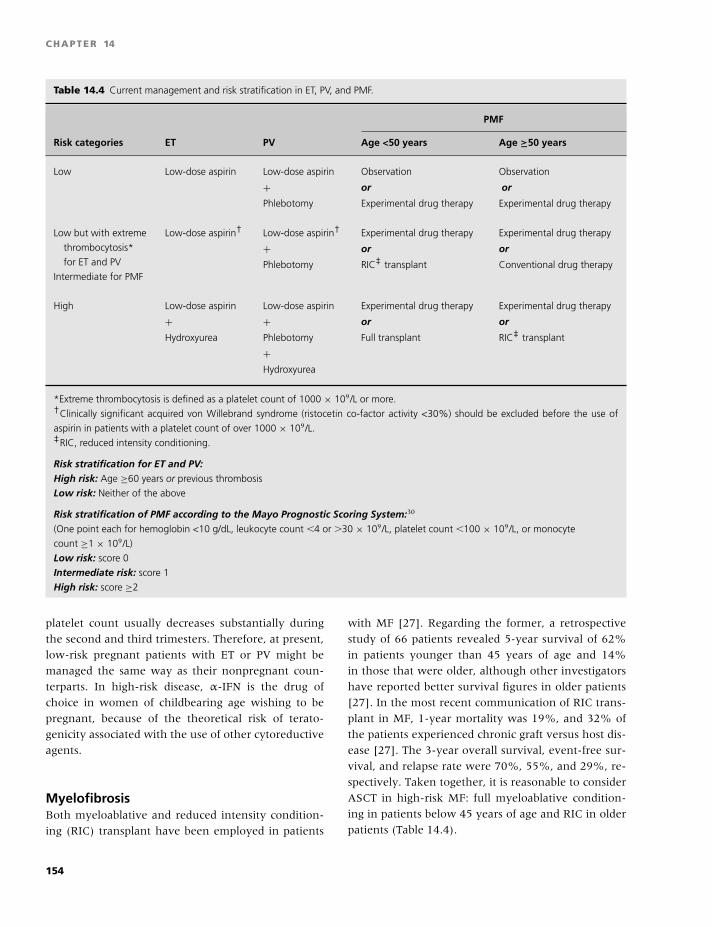
BLBK186-Key April 11, 2009 12:59
CHAPTER 14
Table 14.4 Current management and risk stratification in ET, PV, and PMF.
PMF
Risk categories ET PV Age <50 years Age ≥50 years
Low Low-dose aspirin Low-dose aspirin
+Phlebotomy
Observation
or
Experimental drug therapy
Observation
or
Experimental drug therapy
Low but with extreme
thrombocytosis*
for ET and PV
Intermediate for PMF
Low-dose aspirin† Low-dose aspirin†
+Phlebotomy
Experimental drug therapy
or
RIC‡ transplant
Experimental drug therapy
or
Conventional drug therapy
High Low-dose aspirin
+Hydroxyurea
Low-dose aspirin
+Phlebotomy
+Hydroxyurea
Experimental drug therapy
or
Full transplant
Experimental drug therapy
or
RIC‡ transplant
*Extreme thrombocytosis is defined as a platelet count of 1000 × 109/L or more.†Clinically significant acquired von Willebrand syndrome (ristocetin co-factor activity <30%) should be excluded before the use of
aspirin in patients with a platelet count of over 1000 × 109/L.‡RIC, reduced intensity conditioning.
Risk stratification for ET and PV:High risk: Age ≥60 years or previous thrombosis
Low risk: Neither of the above
Risk stratification of PMF according to the Mayo Prognostic Scoring System:30
(One point each for hemoglobin <10 g/dL, leukocyte count �4 or �30 × 109/L, platelet count �100 × 109/L, or monocyte
count ≥1 × 10⁹/L)
Low risk: score 0
Intermediate risk: score 1
High risk: score ≥2
platelet count usually decreases substantially during
the second and third trimesters. Therefore, at present,
low-risk pregnant patients with ET or PV might be
managed the same way as their nonpregnant coun-
terparts. In high-risk disease, α-IFN is the drug of
choice in women of childbearing age wishing to be
pregnant, because of the theoretical risk of terato-
genicity associated with the use of other cytoreductive
agents.
MyelofibrosisBoth myeloablative and reduced intensity condition-
ing (RIC) transplant have been employed in patients
with MF [27]. Regarding the former, a retrospective
study of 66 patients revealed 5-year survival of 62%
in patients younger than 45 years of age and 14%
in those that were older, although other investigators
have reported better survival figures in older patients
[27]. In the most recent communication of RIC trans-
plant in MF, 1-year mortality was 19%, and 32% of
the patients experienced chronic graft versus host dis-
ease [27]. The 3-year overall survival, event-free sur-
vival, and relapse rate were 70%, 55%, and 29%, re-
spectively. Taken together, it is reasonable to consider
ASCT in high-risk MF: full myeloablative condition-
ing in patients below 45 years of age and RIC in older
patients (Table 14.4).
154
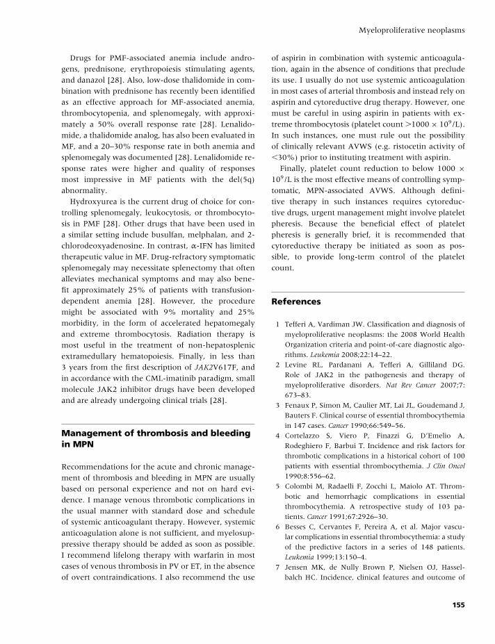
BLBK186-Key April 11, 2009 12:59
Myeloproliferative neoplasms
Drugs for PMF-associated anemia include andro-
gens, prednisone, erythropoiesis stimulating agents,
and danazol [28]. Also, low-dose thalidomide in com-
bination with prednisone has recently been identified
as an effective approach for MF-associated anemia,
thrombocytopenia, and splenomegaly, with approxi-
mately a 50% overall response rate [28]. Lenalido-
mide, a thalidomide analog, has also been evaluated in
MF, and a 20–30% response rate in both anemia and
splenomegaly was documented [28]. Lenalidomide re-
sponse rates were higher and quality of responses
most impressive in MF patients with the del(5q)
abnormality.
Hydroxyurea is the current drug of choice for con-
trolling splenomegaly, leukocytosis, or thrombocyto-
sis in PMF [28]. Other drugs that have been used in
a similar setting include busulfan, melphalan, and 2-
chlorodeoxyadenosine. In contrast, α-IFN has limited
therapeutic value in MF. Drug-refractory symptomatic
splenomegaly may necessitate splenectomy that often
alleviates mechanical symptoms and may also bene-
fit approximately 25% of patients with transfusion-
dependent anemia [28]. However, the procedure
might be associated with 9% mortality and 25%
morbidity, in the form of accelerated hepatomegaly
and extreme thrombocytosis. Radiation therapy is
most useful in the treatment of non-hepatosplenic
extramedullary hematopoiesis. Finally, in less than
3 years from the first description of JAK2V617F, and
in accordance with the CML-imatinib paradigm, small
molecule JAK2 inhibitor drugs have been developed
and are already undergoing clinical trials [28].
Management of thrombosis and bleedingin MPN
Recommendations for the acute and chronic manage-
ment of thrombosis and bleeding in MPN are usually
based on personal experience and not on hard evi-
dence. I manage venous thrombotic complications in
the usual manner with standard dose and schedule
of systemic anticoagulant therapy. However, systemic
anticoagulation alone is not sufficient, and myelosup-
pressive therapy should be added as soon as possible.
I recommend lifelong therapy with warfarin in most
cases of venous thrombosis in PV or ET, in the absence
of overt contraindications. I also recommend the use
of aspirin in combination with systemic anticoagula-
tion, again in the absence of conditions that preclude
its use. I usually do not use systemic anticoagulation
in most cases of arterial thrombosis and instead rely on
aspirin and cytoreductive drug therapy. However, one
must be careful in using aspirin in patients with ex-
treme thrombocytosis (platelet count �1000 × 109/L).
In such instances, one must rule out the possibility
of clinically relevant AVWS (e.g. ristocetin activity of
�30%) prior to instituting treatment with aspirin.
Finally, platelet count reduction to below 1000 ×109/L is the most effective means of controlling symp-
tomatic, MPN-associated AVWS. Although defini-
tive therapy in such instances requires cytoreduc-
tive drugs, urgent management might involve platelet
pheresis. Because the beneficial effect of platelet
pheresis is generally brief, it is recommended that
cytoreductive therapy be initiated as soon as pos-
sible, to provide long-term control of the platelet
count.
References
1 Tefferi A, Vardiman JW. Classification and diagnosis of
myeloproliferative neoplasms: the 2008 World Health
Organization criteria and point-of-care diagnostic algo-
rithms. Leukemia 2008;22:14–22.
2 Levine RL, Pardanani A, Tefferi A, Gilliland DG.
Role of JAK2 in the pathogenesis and therapy of
myeloproliferative disorders. Nat Rev Cancer 2007;7:
673–83.
3 Fenaux P, Simon M, Caulier MT, Lai JL, Goudemand J,
Bauters F. Clinical course of essential thrombocythemia
in 147 cases. Cancer 1990;66:549–56.
4 Cortelazzo S, Viero P, Finazzi G, D’Emelio A,
Rodeghiero F, Barbui T. Incidence and risk factors for
thrombotic complications in a historical cohort of 100
patients with essential thrombocythemia. J Clin Oncol
1990;8:556–62.
5 Colombi M, Radaelli F, Zocchi L, Maiolo AT. Throm-
botic and hemorrhagic complications in essential
thrombocythemia. A retrospective study of 103 pa-
tients. Cancer 1991;67:2926–30.
6 Besses C, Cervantes F, Pereira A, et al. Major vascu-
lar complications in essential thrombocythemia: a study
of the predictive factors in a series of 148 patients.
Leukemia 1999;13:150–4.
7 Jensen MK, de Nully Brown P, Nielsen OJ, Hassel-
balch HC. Incidence, clinical features and outcome of
155
BLBK186-Key April 11, 2009 12:59
CHAPTER 14
essential thrombocythaemia in a well defined geo-
graphical area. Eur J Haematol 2000;65:132–9.
8 Chim CS, Kwong YL, Lie AK, et al. Long-term outcome
of 231 patients with essential thrombocythemia: prog-
nostic factors for thrombosis, bleeding, myelofibrosis,
and leukemia. Arch Intern Med 2005;165:2651–8.
9 Wolanskyj AP, Schwager SM, McClure RF, Larson DR,
Tefferi A. Essential thrombocythemia beyond the first
decade: life expectancy, long-term complication rates,
and prognostic factors. Mayo Clin Proc 2006;81:159–66.
10 Campbell PJ, Scott LM, Buck G, et al. Definition of
subtypes of essential thrombocythaemia and relation
to polycythaemia vera based on JAK2 V617F mu-
tation status: a prospective study. Lancet 2005;366:
1945–53.
11 Polycythemia vera: the natural history of 1213 pa-
tients followed for 20 years. Gruppo Italiano Studio
Policitemia. Ann Intern Med 1995;123:656–64.
12 Passamonti F, Brusamolino E, Lazzarino M, et al. Effi-
cacy of pipobroman in the treatment of polycythemia
vera: long-term results in 163 patients. Haematologica
2000;85:1011–8.
13 Marchioli R, Finazzi G, Landolfi R, et al. Vascular and
neoplastic risk in a large cohort of patients with poly-
cythemia vera. J Clin Oncol 2005;23:2224–32.
14 Carobbio A, Finazzi G, Guerini V, et al. Leukocytosis
is a risk factor for thrombosis in essential thrombo-
cythemia: interaction with treatment, standard risk fac-
tors and Jak2 mutation status. Blood 2007;109:2310–3.
15 Passamonti F, Rumi E, Pungolino E, et al. Life ex-
pectancy and prognostic factors for survival in pa-
tients with polycythemia vera and essential thrombo-
cythemia. Am J Med 2004;117:755–61.
16 De Stefano V, Za T, Rossi E, et al. Recurrent throm-
bosis in patients with polycythemia vera or essential
thrombocythemia: efficacy of treatment in prevent-
ing rethrombosis in different clinical settings. Blood
2006;108:Abstract #119.
17 Gangat N, Wolanskyj AP, Tefferi A. Abdominal vein
thrombosis in essential thrombocythemia: prevalence,
clinical correlates, and prognostic implications. Eur J
Haematol 2006;77:327–33.
18 Kiladjian JJ, Cervantes F, Leebeek FW, et al. The impact
of JAK2 and MPL mutations on diagnosis and prognosis
of splanchnic vein thrombosis: a report on 241 cases.
Blood 2008;111:4922–9.
19 Pardanani A, Lasho TL, Hussein K, et al. JAK2V617F
mutation screening as part of the hypercoagulable
work-up in the absence of splanchnic venous throm-
bosis or overt myeloproliferative neoplasm: assessment
of value in a series of 664 consecutive patients. Mayo
Clin Proc 2008;83:457–9.
20 Tefferi A, Elliott M. Thrombosis in myeloproliferative
disorders: prevalence, prognostic factors, and the role
of leukocytes and JAK2V617F. Semin Thromb Hemost
2007;33:313–20.
21 Fabris F, Casonato A, Grazia del Ben M, De Marco L,
Girolami A. Abnormalities of von Willebrand factor in
myeloproliferative disease: a relationship with bleeding
diathesis. Br J Haematol 1986;63:75–83.
22 Landolfi R, Marchioli R, Kutti J, et al. Efficacy and
safety of low-dose aspirin in polycythemia vera. N Engl
J Med 2004;350:114–24.
23 Cortelazzo S, Finazzi G, Ruggeri M, et al. Hydroxyurea
for patients with essential thrombocythemia and a high
risk of thrombosis. N Engl J Med 1995;332:1132–6.
24 Tefferi A, Gangat N, Wolanskyj AP. Management of
extreme thrombocytosis in otherwise low-risk essen-
tial thrombocythemia; does number matter? Blood
2006;108:2493–4.
25 Harrison CN, Campbell PJ, Buck G, et al. Hydroxyurea
compared with anagrelide in high-risk essential throm-
bocythemia. N Engl J Med 2005;353:33–45.
26 Passamonti F, Randi ML, Rumi E, et al. Increased risk
of pregnancy complications in patients with essential
thrombocythemia carrying the JAK2 (617V�F) muta-
tion. Blood 2007;110:485–9.
27 Kroger N, Mesa RA. Choosing between stem cell ther-
apy and drugs in myelofibrosis. Leukemia 2008;22:474–
86.
28 Tefferi A. Essential thrombocythemia, polycythemia
vera, and myelofibrosis: current management and
the prospect of targeted therapy. Am J Hematol
2008;83:491–7.
156
BLBK186-Key May 22, 2009 13:48
15 Arterial thrombosisGordon D.O. Lowe and R. Campbell Tait
Introduction
Arterial thrombosis is a common cause of hospital
admission, death, and disability in developed coun-
tries (and increasingly in developing nations because
of global epidemics of smoking, obesity, and dia-
betes). It usually follows spontaneous rupture of an
atherosclerotic plaque, and may:� be clinically silent;� contribute to atherosclerotic progression resulting in
coronary stenosis and stable angina, or lower limb
artery stenosis and claudication;� be present as acute ischemia in the heart (acute
coronary syndromes: unstable angina, myocardial in-
farction), brain (transient cerebral ischemic attack or
stroke), or limb (acute limb ischemia).
There is now good evidence that patients with
acute ischemic syndromes have lower morbidity and
mortality if they are promptly diagnosed, admitted
as soon as possible to specialist acute units (coronary
care, acute stroke, or peripheral vascular), undergo
risk stratification, and receive appropriate treatment.
This includes antithrombotic drugs (e.g. aspirin,
heparin) and consideration of thrombolysis,
thrombectomy, angioplasty, or vascular reconstruc-
tion in the acute phase and early, multidisciplinary
rehabilitation.
Traditional risk factors (Table 15.1) remain the most
important markers for arterial disease and together ac-
count for up to 90% of population attributable risk
[1–3]. In patients with nonvalvular atrial fibrillation,
the risk of stroke can be estimated by a variety of scor-
ing systems, of which the CHADS2 index [4], devel-
oped from an amalgamation of the Atrial Fibrillation
Investigators and Stroke Prevention in Atrial Fibrilla-
tion schemes, is the most widely used and validated
(Table 15.2).
Primary and secondary prevention of arterial
thrombosis is everybody’s business. All health care
professionals, including hematologists, should take
the opportunity to encourage their patients to ad-
just their lifestyles (when appropriate) and to consider
pharmacologic prevention in all high-risk patients
and in all with clinical evidence of arterial disease
(Table 15.3).
Hematologists are commonly asked to develop or
revise local hospital or area guidelines for investi-
gations in thrombosis and antithrombotic therapies
and their monitoring. In addition, they are often re-
ferred patients with arterial thrombosis that is pre-
mature, recurrent, or which occurs at multiple or
unusual sites. Such referrals have increased in re-
cent years, probably because general practitioners and
physicians expect that (as with venous thromboem-
bolism) hematologists may define underlying throm-
bophilias that may require specific management. This
review therefore focuses on appropriate hematological
investigation of patients with arterial thrombosis and
appropriate antithrombotic therapy in various patient
groups.
Evidence in this field is changing rapidly; hence,
hematologists should keep up-to-date with system-
atic reviews and evidence-based national guide-
lines, such as those produced by the British Society
for Haematology/British Committee for Standards in
Haematology (http://www.bcshguidelines.com), the
Scottish Intercollegiate Guidelines Network (SIGN;
http://www.sign.ac.uk), and the National Institute for
Health and Clinical Excellence (NICE).
157
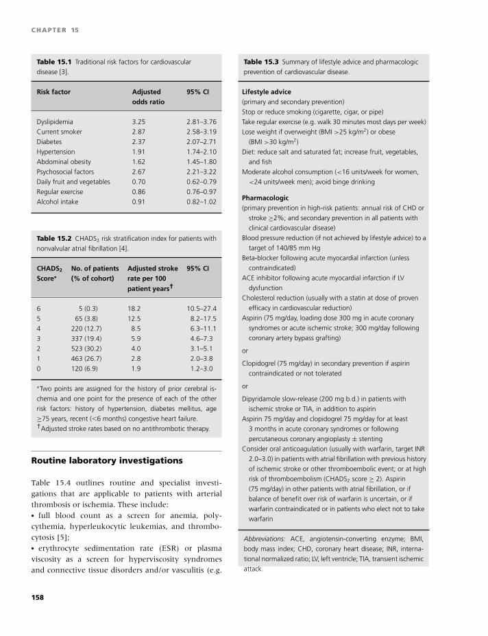
BLBK186-Key May 22, 2009 13:48
CHAPTER 15
Table 15.1 Traditional risk factors for cardiovascular
disease [3].
Risk factor Adjusted 95% CIodds ratio
Dyslipidemia 3.25 2.81–3.76
Current smoker 2.87 2.58–3.19
Diabetes 2.37 2.07–2.71
Hypertension 1.91 1.74–2.10
Abdominal obesity 1.62 1.45–1.80
Psychosocial factors 2.67 2.21–3.22
Daily fruit and vegetables 0.70 0.62–0.79
Regular exercise 0.86 0.76–0.97
Alcohol intake 0.91 0.82–1.02
Table 15.2 CHADS2 risk stratification index for patients with
nonvalvular atrial fibrillation [4].
CHADS2 No. of patients Adjusted stroke 95% CIScore∗ (% of cohort) rate per 100
patient years†
6 5 (0.3) 18.2 10.5–27.4
5 65 (3.8) 12.5 8.2–17.5
4 220 (12.7) 8.5 6.3–11.1
3 337 (19.4) 5.9 4.6–7.3
2 523 (30.2) 4.0 3.1–5.1
1 463 (26.7) 2.8 2.0–3.8
0 120 (6.9) 1.9 1.2–3.0
∗Two points are assigned for the history of prior cerebral is-
chemia and one point for the presence of each of the other
risk factors: history of hypertension, diabetes mellitus, age
≥75 years, recent (�6 months) congestive heart failure.†Adjusted stroke rates based on no antithrombotic therapy.
Routine laboratory investigations
Table 15.4 outlines routine and specialist investi-
gations that are applicable to patients with arterial
thrombosis or ischemia. These include:� full blood count as a screen for anemia, poly-
cythemia, hyperleukocytic leukemias, and thrombo-
cytosis [5];� erythrocyte sedimentation rate (ESR) or plasma
viscosity as a screen for hyperviscosity syndromes
and connective tissue disorders and/or vasculitis (e.g.
Table 15.3 Summary of lifestyle advice and pharmacologic
prevention of cardiovascular disease.
Lifestyle advice(primary and secondary prevention)
Stop or reduce smoking (cigarette, cigar, or pipe)
Take regular exercise (e.g. walk 30 minutes most days per week)
Lose weight if overweight (BMI >25 kg/m2) or obese
(BMI >30 kg/m2)
Diet: reduce salt and saturated fat; increase fruit, vegetables,
and fish
Moderate alcohol consumption (<16 units/week for women,
<24 units/week men); avoid binge drinking
Pharmacologic(primary prevention in high-risk patients: annual risk of CHD or
stroke ≥2%; and secondary prevention in all patients with
clinical cardiovascular disease)
Blood pressure reduction (if not achieved by lifestyle advice) to a
target of 140/85 mm Hg
Beta-blocker following acute myocardial infarction (unless
contraindicated)
ACE inhibitor following acute myocardial infarction if LV
dysfunction
Cholesterol reduction (usually with a statin at dose of proven
efficacy in cardiovascular reduction)
Aspirin (75 mg/day, loading dose 300 mg in acute coronary
syndromes or acute ischemic stroke; 300 mg/day following
coronary artery bypass grafting)
or
Clopidogrel (75 mg/day) in secondary prevention if aspirin
contraindicated or not tolerated
or
Dipyridamole slow-release (200 mg b.d.) in patients with
ischemic stroke or TIA, in addition to aspirin
Aspirin 75 mg/day and clopidogrel 75 mg/day for at least
3 months in acute coronary syndromes or following
percutaneous coronary angioplasty ± stenting
Consider oral anticoagulation (usually with warfarin, target INR
2.0–3.0) in patients with atrial fibrillation with previous history
of ischemic stroke or other thromboembolic event; or at high
risk of thromboembolism (CHADS2 score ≥ 2). Aspirin
(75 mg/day) in other patients with atrial fibrillation, or if
balance of benefit over risk of warfarin is uncertain, or if
warfarin contraindicated or in patients who elect not to take
warfarin
Abbreviations: ACE, angiotensin-converting enzyme; BMI,
body mass index; CHD, coronary heart disease; INR, interna-
tional normalized ratio; LV, left ventricle; TIA, transient ischemic
attack.
158
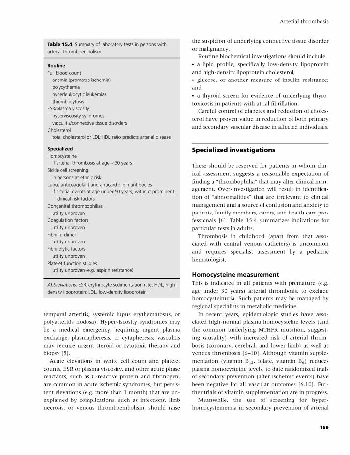
BLBK186-Key May 22, 2009 13:48
Arterial thrombosis
Table 15.4 Summary of laboratory tests in persons with
arterial thromboembolism.
RoutineFull blood count
anemia (promotes ischemia)
polycythemia
hyperleukocytic leukemias
thrombocytosis
ESR/plasma viscosity
hyperviscosity syndromes
vasculitis/connective tissue disorders
Cholesterol
total cholesterol or LDL:HDL ratio predicts arterial disease
SpecializedHomocysteine
if arterial thrombosis at age <30 years
Sickle cell screening
in persons at ethnic risk
Lupus anticoagulant and anticardiolipin antibodies
if arterial events at age under 50 years, without prominent
clinical risk factors
Congenital thrombophilias
utility unproven
Coagulation factors
utility unproven
Fibrin D-dimer
utility unproven
Fibrinolytic factors
utility unproven
Platelet function studies
utility unproven (e.g. aspirin resistance)
Abbreviations: ESR, erythrocyte sedimentation rate; HDL, high-
density lipoprotein; LDL, low-density lipoprotein.
temporal arteritis, systemic lupus erythematosus, or
polyarteritis nodosa). Hyperviscosity syndromes may
be a medical emergency, requiring urgent plasma
exchange, plasmapheresis, or cytapheresis; vasculitis
may require urgent steroid or cytotoxic therapy and
biopsy [5].
Acute elevations in white cell count and platelet
counts, ESR or plasma viscosity, and other acute phase
reactants, such as C-reactive protein and fibrinogen,
are common in acute ischemic syndromes; but persis-
tent elevations (e.g. more than 1 month) that are un-
explained by complications, such as infections, limb
necrosis, or venous thromboembolism, should raise
the suspicion of underlying connective tissue disorder
or malignancy.
Routine biochemical investigations should include:� a lipid profile, specifically low-density lipoprotein
and high-density lipoprotein cholesterol;� glucose, or another measure of insulin resistance;
and� a thyroid screen for evidence of underlying thyro-
toxicosis in patients with atrial fibrillation.
Careful control of diabetes and reduction of choles-
terol have proven value in reduction of both primary
and secondary vascular disease in affected individuals.
Specialized investigations
These should be reserved for patients in whom clin-
ical assessment suggests a reasonable expectation of
finding a “thrombophilia” that may alter clinical man-
agement. Over-investigation will result in identifica-
tion of “abnormalities” that are irrelevant to clinical
management and a source of confusion and anxiety to
patients, family members, carers, and health care pro-
fessionals [6]. Table 15.4 summarizes indications for
particular tests in adults.
Thrombosis in childhood (apart from that asso-
ciated with central venous catheters) is uncommon
and requires specialist assessment by a pediatric
hematologist.
Homocysteine measurementThis is indicated in all patients with premature (e.g.
age under 30 years) arterial thrombosis, to exclude
homocysteinuria. Such patients may be managed by
regional specialists in metabolic medicine.
In recent years, epidemiologic studies have asso-
ciated high-normal plasma homocysteine levels (and
the common underlying MTHFR mutation, suggest-
ing causality) with increased risk of arterial throm-
bosis (coronary, cerebral, and lower limb) as well as
venous thrombosis [6–10]. Although vitamin supple-
mentation (vitamin B12, folate, vitamin B6) reduces
plasma homocysteine levels, to date randomized trials
of secondary prevention (after ischemic events) have
been negative for all vascular outcomes [6,10]. Fur-
ther trials of vitamin supplementation are in progress.
Meanwhile, the use of screening for hyper-
homocysteinemia in secondary prevention of arterial
159
BLBK186-Key May 22, 2009 13:48
CHAPTER 15
thrombosis in patients aged over 30 years is un-
proven. If high homocysteine levels are found, folate
supplementation is reasonable because it is cheap and
nontoxic (provided vitamin B12 levels are normal).
It may be that folate supplementation of cereals (as
practiced in the USA) or the folate component of
a “polypill” (folic acid, antihypertensives, a statin,
and possibly aspirin) [11] may be the most clinically
effective and cost-effective strategy to reduce car-
diovascular risk if homocysteine is shown to have a
causal role in arterial (or venous) thrombosis.
Sickle cell screeningThis may be appropriate in persons at ethnic risk, al-
though in practice, a diagnosis of sickle cell disease
(SCD) will usually have been made long before adult-
hood. Large- and small-vessel arterial thromboses are
responsible for the protean manifestations of SCD.
Sickle erythrocytes appear to induce a hypercoagula-
ble state through a variety of mechanisms as assessed
by increased platelet activation, increased thrombin
generation, and decreased levels of anticoagulant pro-
teins. However, measurement of such parameters has
no proven use in the management of SCD, and clinical
studies of long-term antiplatelet agents and anticoagu-
lants have yet to show any beneficial effect on the inci-
dence of vaso-occlusive events. Furthermore, it seems
likely that the hypercoagulability is a secondary phe-
nomenon to the sickling process, because treatment
with hydroxyurea (which increases HbF levels and re-
duces sickling) is associated with a reduction in mea-
sures of hypercoagulability [12].
Screening for lupus anticoagulant andanticardiolipin antibodiesThis is appropriate in all patients with premature (e.g.
age under 50 years) cerebral or limb thrombosis or is-
chemia, and in other indications [13]. Management
of the antiphospholipid syndrome is considered in
Chapter 17.
Screening for congenital thrombophiliasThe factor V Leiden and prothrombin G20210A
mutations show modest but statistically significant as-
sociations with coronary heart disease (CHD), stroke,
and peripheral arterial events, especially in younger
persons (age under 55 years) and in women [9,14].
These findings may be relevant to the increases in risk
of coronary and stroke events during pregnancy or
with use of combined oral contraceptives or oral hor-
mone replacement therapy (each of which increases
resistance to activated protein C).
There is little evidence that other congenital throm-
bophilias are associated with increased risk of arterial
disease, and the clinical use of screening for such ab-
normalities in patients with arterial thrombosis is at
present unproven [15]. Furthermore, there is no ev-
idence that secondary prevention with oral anticoag-
ulants in such patients is more effective than routine
antithrombotic prevention with aspirin (Table 15.3).
Ischemic stroke is often associated with a right-to-
left cardiac shunt (e.g. patent foramen ovale, atrial
septal defect) in younger patients, suggesting the pos-
sibility of “paradoxical” cerebral arterial embolism
from venous thrombosis. Whether such an event is as-
sociated with thrombophilias is unknown, as are the
relative benefits and risks of prophylaxis with aspirin,
oral anticoagulants, or shunt closure [16].
Coagulation factorsPlasma fibrinogen is associated with CHD, stroke, and
peripheral arterial events; the risks increase by 30–
40% per 1-g/L increase [17]. Although there are sev-
eral plausible biologic mechanisms through which in-
creased circulating fibrinogen levels might promote
such risk (atherogenic, thrombogenic, and rheolog-
ical through increased plasma and blood viscosity),
the lack of association of functional genetic polymor-
phisms with risk of CHD argues against causality. The
association of fibrinogen with arterial risk may there-
fore be coincidental (because of mutual associations
with multiple risk factors) or consequential (reverse
causality, resulting from effects of atherosclerosis on
plasma fibrinogen). The clinical use of plasma fibrino-
gen assessment in management of arterial thrombosis
is unproven [6].
Von Willebrand factor (VWF) is weakly associated
with risk of CHD [18]; there are few reported studies
of functional polymorphisms.
Carriers of hemophilia A (or B) who have plasma
levels of factor VIII or factor IX, which on average
are 50% lower than female noncarriers, have an ap-
proximately 35% lower risk of CHD. Together with
the 80% lower risk of CHD in male hemophiliacs
compared with male non-hemophiliacs, these findings
suggest that increasing levels of factor VIII (or factor
160
BLBK186-Key May 22, 2009 13:48
Arterial thrombosis
IX) increase the risks of arterial thrombosis, as well as
of venous thrombosis [19].
The clinical use of assessment of plasma levels of
VWF, factor VIII, or factor IX (or other clotting factors)
in management of arterial thrombosis is unproven.
Coagulation activation markersPlasma fibrin D-dimer levels are associated with in-
creased risks of incident CHD and stroke, includ-
ing studies of patients with atrial fibrillation [20–22].
Although D-dimer levels might therefore be useful in
prediction of stroke in atrial fibrillation, and hence in
stratifying choice of antithrombotic therapies, further
management studies are required.
Fibrinolytic testsCirculating levels of tissue plasminogen activator anti-
gen, but not of plasminogen activator inhibitor type
1, are associated with increased risk of CHD in pop-
ulation studies. This association is markedly reduced
after adjustment for associated CHD risk factors (obe-
sity and other markers of insulin resistance) [23]. The
clinical use of plasma components of the fibrinolytic
system in management of arterial thrombosis is un-
proven.
Platelet function testsPlatelet aggregation studies and measures of platelet
activation are not useful in prediction of arterial
thrombosis. Although there is increasing evidence that
aspirin resistance (defined as a laboratory measure
of the failure of aspirin to inhibit platelet synthesis
of thromboxane A2, platelet aggregation, or the skin
bleeding time) is associated with increased risk of re-
current cardiovascular events, further work is required
to define the place of such laboratory measures in clin-
ical practice [24].
Treatment
Primary and secondary prevention therapies for all
patients with cardiovascular disease primarily involve
antiplatelet agents and are summarised in Table 15.3.
There have been recent advances in acute man-
agement of myocardial infarction and other acute
coronary syndromes using aditional anticoagulant
(low-molecular-weight heparins or fondaparinux) or
antiplatelet (specific platelet glycoprotein IIb/IIIa
inhibitors) agents [25]; this is discussed in more detail
in Chapter 18. In patients with recurrent events de-
spite aspirin, possible empirical approaches are to add
a second antiplatelet agent, to increase the dose of as-
pirin, or to change to oral anticoagulant therapy (after
considering the increased bleeding risk and the logisti-
cal problems of long-term anticoagulant monitoring).
Combination therapy with vitamin Kantagonists and antiplatelet agentsAn increasingly problematic issue in clinical practice
has been determining the risk:benefit ratios for com-
bination treatment, either in patients already receiving
a vitamin K antagonist (VKA) who develop an indica-
tion for aspirin (e.g. a patient being treated for recent
DVT who then suffers an acute coronary syndrome),
or a patient on aspirin who develops an indication
for a VKA (e.g. a patient with previous myocardiol
infarction developing atrial fibrillation). Management
of such patients has to be individualized, consider-
ing the patient’s thrombotic and bleeding risks. How-
ever, there is evidence in the literature that can inform
decisions:� In patients with atrial fibrillation, the combination
of aspirin + clopidogrel is inferior to VKA (INR 2-3)
in terms of stroke prevention, and is associated with
similar bleeding rates [26].� In patients with atrial fibrillation, the addition of as-
pirin (to VKA) is associated with a higher risk of major
bleeding [27].� In patients with peripheral arterial disease treated
with aspirin, the addition of VKA does not reduce the
cardiovascular event rate, but does increase the rate of
life-threatening bleeding [28].� In patients with stable coronary artery disease, a
VKA is as effective as aspirin at reducing the risk of
further ischemic events, albeit with an increased risk
of major bleeding. Aspirin plus VKA is not significantly
better than VKA alone [29].� In high thrombotic risk patients with prosthetic
heart valves (e.g. metal prosthetic valves, tissue pros-
thesis plus atrial fibrillation, or previous stroke), the
benefit of adding aspirin to VKA, in terms of greater
reduction in cardiovascular thrombotic events, out-
weighs the increased risk of major bleeding [30].
Therefore it would seem reasonable to treat with
VKA alone in stable coronary artery disease patients
who have an indication for VKA (e.g. atrial fibrillation
161
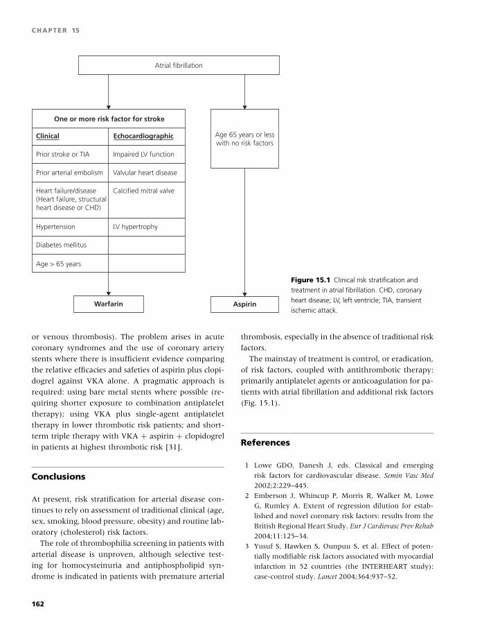
BLBK186-Key May 22, 2009 13:48
CHAPTER 15
Age 65 years or lesswith no risk factors
Atrial fibrillation
Prior stroke or TIA Impaired LV function
Prior arterial embolism Valvular heart disease
Heart failure/disease Calcified mitral valve(Heart failure, structuralheart disease or CHD)
Hypertension LV hypertrophy
Diabetes mellitus
Age > 65 years
AspirinWarfarin
One or more risk factor for stroke
Clinical Echocardiographic
Figure 15.1 Clinical risk stratification and
treatment in atrial fibrillation. CHD, coronary
heart disease; LV, left ventricle; TIA, transient
ischemic attack.
or venous thrombosis). The problem arises in acute
coronary syndromes and the use of coronary artery
stents where there is insufficient evidence comparing
the relative efficacies and safeties of aspirin plus clopi-
dogrel against VKA alone. A pragmatic approach is
required: using bare metal stents where possible (re-
quiring shorter exposure to combination antiplatelet
therapy); using VKA plus single-agent antiplatelet
therapy in lower thrombotic risk patients; and short-
term triple therapy with VKA + aspirin + clopidogrel
in patients at highest thrombotic risk [31].
Conclusions
At present, risk stratification for arterial disease con-
tinues to rely on assessment of traditional clinical (age,
sex, smoking, blood pressure, obesity) and routine lab-
oratory (cholesterol) risk factors.
The role of thrombophilia screening in patients with
arterial disease is unproven, although selective test-
ing for homocysteinuria and antiphospholipid syn-
drome is indicated in patients with premature arterial
thrombosis, especially in the absence of traditional risk
factors.
The mainstay of treatment is control, or eradication,
of risk factors, coupled with antithrombotic therapy:
primarily antiplatelet agents or anticoagulation for pa-
tients with atrial fibrillation and additional risk factors
(Fig. 15.1).
References
1 Lowe GDO, Danesh J, eds. Classical and emerging
risk factors for cardiovascular disease. Semin Vasc Med
2002;2:229–445.
2 Emberson J, Whincup P, Morris R, Walker M, Lowe
G, Rumley A. Extent of regression dilution for estab-
lished and novel coronary risk factors: results from the
British Regional Heart Study. Eur J Cardiovasc Prev Rehab
2004;11:125–34.
3 Yusuf S, Hawken S, Ounpuu S, et al. Effect of poten-
tially modifiable risk factors associated with myocardial
infarction in 52 countries (the INTERHEART study):
case-control study. Lancet 2004;364:937–52.
162
BLBK186-Key May 22, 2009 13:48
Arterial thrombosis
4 Gage BF, Waterman AD, Shannon W, Boechler M,
Rich MW, Radford MJ. Validation of clinical classifi-
cation schemes for predicting stroke: results from the
national registry of atrial fibrillation. JAMA 2001;285:
2864–70.
5 Lowe GDO, ed. Blood rheology and hyperviscosity syn-
dromes. Baillieres Clin Haematol 1987;3:597–867.
6 Lowe GDO. Can haematological tests predict cardio-
vascular risk? The 2005 Kettle Lecture. Br J Haematol
2006;133:232–50.
7 Homocysteine Studies Collaboration. Homocysteine
and risk of ischemic heart disease and stroke: a meta-
analysis. JAMA 2002;288:2015–22.
8 Klerk M, Verhoef P, Clarke R, et al. MTHFR 677C-
T polymorphism and risk of coronary heart disease: a
meta-analysis. JAMA 2002;288:2023–31.
9 Kim RJ, Becker RC. Association between factor V
Leiden, prothrombin mutation G20120A, and MTHFR
C677T mutations and events of the arterial circulatory
system: a meta-analysis of published studies. Am Heart
J 2003;146:948–57.
10 Toole JF, Malinow MR, Chambless LE, et al. Lowering
homocysteine in patients with ischemic stroke to pre-
vent recurrent stroke, myocardial infarction, and death.
JAMA 2004;291:565–75.
11 Wald NJ, Law MR. A strategy to reduce cardiovascular
disease by more than 80%. Br Med J 2003;326:1419.
12 Ataga KI, Orringer EP. Hypercoagulability in sickle
cell disease: a curious paradox. Am J Med 2003;115:
721–8.
13 Greaves M, Cohen H, Machin SJ, Mackie I, on be-
half of the British Committee for Standards in Haema-
tology. Guidelines on the investigation and manage-
ment of the antiphospholipid syndrome. Br J Haematol
2000;109:704–15.
14 Ye Z, Liu EHC, Higgins JPT, et al. Seven haemo-
static gene polymorphisms in coronary disease: meta-
analysis of 66 155 cases and 91 307 controls. Lancet
2006;367:651–8.
15 Walker ID, Greaves M, Preston FE, on behalf of the
Haemostasis and Thrombosis Task Force, British Com-
mittee for Standards in Haematology. Guideline: Inves-
tigation and management of heritable thrombophilia.
Br J Haematol 2001;114:512–28.
16 Wu LA, We LA, Lidar DA, et al. Patent foramen ovale
in cryptogenic stroke: current understanding and man-
agement options. Arch Intern Med 2004;164:950–6.
17 Danesh J, Lewington S, Thompson SG, Lowe GDO,
Collins R (writing committee). Fibrinogen Studies
Collaboration. Plasma fibrinogen level and the risk
of major cardiovascular diseases and non-vascular
mortality: an individual participant meta-analysis.
JAMA 2005;294:1799–1809.
18 Danesh J, Wheeler JG, Hirshfield GM, et al. C-reactive
protein and other circulating markers of inflammation
in the prediction of coronary heart disease. N Engl J Med
2004;350:1387–97.
19 Sramek A, Kriek M, Rosendaal FR. Decreased mor-
tality of ischaemic heart disease among carriers of
haemophlia. Lancet 2003;362:351–4.
20 Danesh J, Whincup P, Walker M, et al. Fibrin
D-dimer and coronary heart disease: prospective study
and meta-analysis. Circulation 2001;103:2323–7.
21 Vene N, Mavri A, Kosmelj K, Stegnar M. High D-
dimer levels predict cardiovascular events in patients
with chronic atrial fibrillation during oral anticoagulant
therapy. Thromb Haemost 2003;90:1163–72.
22 Lowe GDO. Fibrin D-dimer and risk of cardiovascular
events? Semin Vasc Med 2005;5:387–398.
23 Lowe GDO, Danesh J, Lewington S, et al. Tissue plas-
minogen activator antigen and coronary heart dis-
ease: prospective study and meta-analysis. Eur Heart J
2004;25:252–9.
24 Hankey GJ, Eikelboom JW. Aspirin resistance. Br Med J
2004;328:477–9.
25 Scottish Intercollegiate Guidelines Network (SIGN).
Acute Coronary Syndromes. A national clinical guide-
line (SIGN93). Edinburgh: SIGN2007. Available from:
http://www.sign.ac.uk.
26 Connolly S, Yusuf S, Camm J, et al. Clopidogrel plus
aspirin versus oral anticoagulation for atrial fibrillation
in the Atrial fibrillation Clopidogrel Trial with Irbesar-
tan for prevention of Vascular Events (ACTIVE W): a
randomised controlled trial. Lancet 2006;367:1903–12.
27 Douketis JD, Arneklev K, Goldhaber SZ, Spandor-
fer J, Halperin F, Horrow J. Comparison of bleed-
ing in patients with nonvalvular atrial fibrillation
treated with ximelagatran or warfarin. Arch Intern Med
2006;166:853–9.
28 The Warfarin Antiplatelet Vascular Evaluation Trial In-
vestigators. Oral anticoagulant and antiplatelet ther-
apy and peripheral arterial disease. N Engl J Med
2007;357:217–27.
29 Hurlen M, Abdelnoor MPH, Smith P, Erikssen J, Arne-
sen H. Warfarin, aspirin, or both after myocardial in-
farction. N Engl J Med 2002;347:969–74.
30 Little SH, Massel DR. Antiplatelet and anticoagula-
tion for patients with prosthetic heart valves. Cochrane
Database Syst Rev 2003;4:CD003464.
31 Eikelboom JW, Hirsh J. Combined antiplatelet and
anticoagulant therapy: clinical benefits and risks.
J Thromb Haemost 2007;5(Suppl 1):255–63.
163
BLBK186-Key April 15, 2009 14:11
16 AnticoagulationGualtiero Palareti and Benilde Cosmi
Introduction
Administration of coumarin drugs, also called vitamin
K antagonists (VKAs), has been the mainstay of anti-
coagulation for more than 50 years. A great and in-
creasing number of subjects receive this treatment all
over the world because it has been shown to be effec-
tive on the basis of randomized clinical trials in many
clinical conditions.
New anticoagulant drugs, based on a completely dif-
ferent mecchanism of action, are being introduced.
Candidate for prolonged treatment, some of them are,
at the moment, in advanced phases of clinical experi-
mentation and are expected to be available for current
clinical use in the near future.
Indications for anticoagulant treatment
A number of clinical trials provided evidence that an-
ticoagulation with VKAs is indicated in several con-
ditions as a formidable tool for primary or secondary
prevention of thrombotic complications [1]. Although
properly designed clinical trials are still lacking, anti-
coagulation with VKAs is widely accepted in several
other conditions (see Table 16.1).
For the majority of indications, a moderate anti-
coagulant effect [international normalized ratio (INR)
2.0–3.0] is effective [2]. No adequate studies have
been conducted on the efficacy of oral anticoagu-
lants (OACs) for the secondary prevention in ischemic
cerebrovascular disease, in retinal vein thrombosis, or
in peripheral arterial disease. In the latter condition,
VKAs are indicated in patients with venous infrain-
guinal bypasses at high risk of occlusion.
Contraindications for treatment
A reliable laboratory, an expert clinician, and a com-
pliant patient are three essential components for ap-
propriate therapy with VKAs. Before starting oral
anticoagulation, patients should be carefully evalu-
ated for compliance, absolute contraindications, and
conditions with a higher risk of complications (see
Table 16.1).
VKAs cross the placental barrier and can produce
both bleeding and a teratogenic effect in the fe-
tus (embryopathy with nasal hypoplasia and stippled
epiphyses in the first trimester and central nervous
system abnormalities at any time during preg-
nancy). They could be considered relatively safe af-
ter the first trimester up to 36 weeks of gestation.
In the last 6 weeks, exposure to VKAs could increase
the risk of bleeding at the time of delivery [3]. Nursing
mothers can be treated with VKAs, as warfarin does
not induce an anticoagulant effect in the breastfed
infant.
Major bleeding is an absolute contraindication to
VKAs for at least 1 month after the event.
Relative medical contraindications to VKAs are se-
vere hepatic or renal insufficiency (which increase the
risk of bleeding), severe hypertension, severe heart
failure, esophageal varices, bleeding diathesis, recent
central nervous system (CNS) surgery or trauma,
recent hepatic or renal biopsy, active peptic ulcer,
bacterial endocarditis, pericarditis, recent CNS hem-
orrhage, chronic bowel inflammatory disease, menor-
rhagia, thyrotoxicosis, and cerebral aneurysms.
Conditions of noncompliance of the patient, such
as psychiatric disorders, dementia, and chronic alco-
holism, can also be considered relative contraindica-
tions for VKAs.
164
BLBK186-Key April 15, 2009 14:11
Anticoagulation
Table 16.1 Indications and contraindications (absolute or relative) for anticoagulant treatment with VKAs.
Proven indications - Primary and secondary prevention of venous thromboembolism
- Prevention of systemic embolism in atrial fibrillation or in patients with tissue or mechanical heart valves
- Prevention of stroke or death in patients with acute myocardial infarction
- Prevention of acute myocardial re-infarction in men at high risk
Other accepted indications Prevention of thrombotic complications in high-risk patients with:
- prosthetic heart valves
- mitral stenosis
- systemic embolism of unknown etiology
- intraventricular thrombosis
- dilated cardiomyopathy
Absolute contraindications - Pregnancy between the 6th and 12th week
- Major bleeding (within 30 days)
Relative contraindications All the conditions that increase the risk of bleeding or of insufficient quality of treatment
- severe hepatic or renal insufficiency
- severe uncontrolled hypertension
- severe heart failure
- bleeding diathesis
- recent central venous system surgery or trauma
- active peptic ulcer or bowel inflammatory disease
- bacterial endocarditis or pericarditis
- tendency to fall
- chronic alcoholism
- poor compliance
- psychiatric disorders or dementia (if not supported by family or social services)
Oral anticoagulant drugs: clinicalpharmacology and genetic control
VKAs are 4-hydroxycoumarin compounds, which
were developed in the 1940s–1950s and introduced
in the treatment of thrombotic disorders in the 1950s.
Warfarin, acenocoumarol, and phenprocoumon are
the compounds currently in clinical use. Warfarin is
the most prescribed anticoagulant worldwide.
Warfarin is administered as a racemic compound
containing equal amounts of the R and S enantiomers.
The S enantiomer is three to five times as potent as
the R enantiomer and accounts for about 60–70%
of the anticoagulation effect of the racemic compound.
The metabolism of (S)-warfarin is almost exclusively
mediated by the activity of the enzyme CYP2C9,
which accounts for ∼85% of its catabolism. Several
genetic polymorphisms of CYP2C9 have been identi-
fied, and two of them—CYP2C9*2 and CYP2C9*3—
relatively frequent among Caucasians, are clinically
relevant because subjects carrier of these variants are
expected to metabolize (S)-warfarin in a lower rate
than carriers of the wild-type allele (CYP2C9*1) and
require lower warfarin doses [4].
In 2004, the gene encoding for vitamin K epox-
ide reductase subunit 1 (VKORC1) was identified [5].
Subsequently, numerous polymorphisms were found
to induce a different sensitivity, highly increased or re-
duced, of the enzyme to action of VKAs [6].
VKAs exert their effect by interfering with the vi-
tamin K-dependent hepatic synthesis of coagulation
factors II, VII, IX, and X as well as the coagulation
inhibitors proteins C and S. Vitamin K-dependent
posttranslational carboxylation is critical for co-
agulation factors to acquire the calcium-mediated
ability to bind to negatively charged phospholipid
surfaces [7].
165
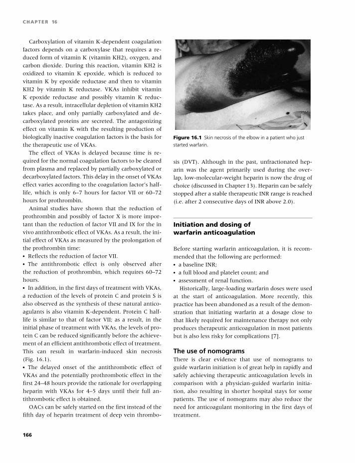
BLBK186-Key April 15, 2009 14:11
CHAPTER 16
Carboxylation of vitamin K-dependent coagulation
factors depends on a carboxylase that requires a re-
duced form of vitamin K (vitamin KH2), oxygen, and
carbon dioxide. During this reaction, vitamin KH2 is
oxidized to vitamin K epoxide, which is reduced to
vitamin K by epoxide reductase and then to vitamin
KH2 by vitamin K reductase. VKAs inhibit vitamin
K epoxide reductase and possibly vitamin K reduc-
tase. As a result, intracellular depletion of vitamin KH2
takes place, and only partially carboxylated and de-
carboxylated proteins are secreted. The antagonizing
effect on vitamin K with the resulting production of
biologically inactive coagulation factors is the basis for
the therapeutic use of VKAs.
The effect of VKAs is delayed because time is re-
quired for the normal coagulation factors to be cleared
from plasma and replaced by partially carboxylated or
decarboxylated factors. This delay in the onset of VKAs
effect varies according to the coagulation factor’s half-
life, which is only 6–7 hours for factor VII or 60–72
hours for prothrombin.
Animal studies have shown that the reduction of
prothrombin and possibly of factor X is more impor-
tant than the reduction of factor VII and IX for the in
vivo antithrombotic effect of VKAs. As a result, the ini-
tial effect of VKAs as measured by the prolongation of
the prothrombin time:� Reflects the reduction of factor VII.� The antithrombotic effect is only observed after
the reduction of prothrombin, which requires 60–72
hours.� In addition, in the first days of treatment with VKAs,
a reduction of the levels of protein C and protein S is
also observed as the synthesis of these natural antico-
agulants is also vitamin K-dependent. Protein C half-
life is similar to that of factor VII; as a result, in the
initial phase of treatment with VKAs, the levels of pro-
tein C can be reduced significantly before the achieve-
ment of an efficient antithrombotic effect of treatment.
This can result in warfarin-induced skin necrosis
(Fig. 16.1).� The delayed onset of the antithrombotic effect of
VKAs and the potentially prothrombotic effect in the
first 24–48 hours provide the rationale for overlapping
heparin with VKAs for 4–5 days until their full an-
tithrombotic effect is obtained.
OACs can be safely started on the first instead of the
fifth day of heparin treatment of deep vein thrombo-
Figure 16.1 Skin necrosis of the elbow in a patient who just
started warfarin.
sis (DVT). Although in the past, unfractionated hep-
arin was the agent primarily used during the over-
lap, low-molecular-weight heparin is now the drug of
choice (discussed in Chapter 13). Heparin can be safely
stopped after a stable therapeutic INR range is reached
(i.e. after 2 consecutive days of INR above 2.0).
Initiation and dosing ofwarfarin anticoagulation
Before starting warfarin anticoagulation, it is recom-
mended that the following are performed:� a baseline INR;� a full blood and platelet count; and� assessment of renal function.
Historically, large-loading warfarin doses were used
at the start of anticoagulation. More recently, this
practice has been abandoned as a result of the demon-
stration that initiating warfarin at a dosage close to
that likely required for maintenance therapy not only
produces therapeutic anticoagulation in most patients
but is also less risky for complications [7].
The use of nomogramsThere is clear evidence that use of nomograms to
guide warfarin initiation is of great help in rapidly and
safely achieving therapeutic anticoagulation levels in
comparison with a physician-guided warfarin initia-
tion, also resulting in shorter hospital stays for some
patients. The use of nomograms may also reduce the
need for anticoagulant monitoring in the first days of
treatment.
166
BLBK186-Key April 15, 2009 14:11
Anticoagulation
A variety of nomograms for the initial days of war-
farin therapy have been devised. Nomogram use re-
quires that baseline INR is normal or near normal (not
more than 1.4). A nomogram to start warfarin anti-
coagulation in children with thrombosis has also been
proposed, with an initial dosage of 0.2 mg/kg. One of
the first and most widely adopted nomograms to be
used for adult patients was that proposed by Fennerty
and colleagues, using 10-mg loading doses [8]. More
recently, nomograms suggesting that warfarin be ini-
tiated with a 5-mg dose were proposed, with subse-
quent doses determined by the INR response, which
can be checked on the third or fourth day [9].
Advantages of the 5-mg doseIt has been shown that the rate of lowering of pro-
thrombin levels was similar when warfarin was started
with either a 5- or 10-mg loading dose. However, the
larger loading dose produced a more rapid reduction
in protein C levels and a higher frequency in over-
anticoagulation (INR �3.0). A smaller loading dose of
warfarin might therefore be less likely to produce a po-
tentially prothrombotic effect in the first 24–48 hours
of treatment.
Advantages of the 10-mg doseIn contrast to the data suggesting that a low initial
warfarin dose is effective and safe, some authors have
reported that higher initial doses are better [10]. Pa-
tients who receive a 10-mg initial dose of warfarin
achieve a therapeutic INR earlier than patients ini-
tially treated with 5 mg, and more patients (83%) in
the 10-mg group achieve a therapeutic INR by day 5,
compared with the 5-mg group (46%). Also, fewer
INR assessments are performed in the 10-mg group.
There were no significant differences between the two
groups in recurrent events or major bleeding. The au-
thors concluded that 10-mg warfarin initiation nomo-
gram is superior to the 5-mg nomogram because it al-
lows more rapid achievement of a therapeutic INR.
Disadvantages of the 5-mg doseIt has recently been shown that starting anticoagula-
tion with 5 mg warfarin in patients with DVT, entirely
treated out of hospital, caused a prolongation of low-
molecular-weight heparin treatment likely caused by
a reduced number of INR determinations in outpa-
tients. It has been suggested that either more frequent
INR determination should be performed or higher ini-
tial dose of warfarin should be adopted in patients
younger than 60 years.
Varying dose because of age or diagnosisSome authors have demonstrated that the initial doses
of warfarin should be different according to the age
of patients because a reduced dose is required in the
elderly.
Patients starting oral anticoagulation after heart
valve replacement are more sensitive to warfarin than
nonsurgical patients, and initial warfarin doses lower
than 5 mg are indicated in some.
Guidance during anticoagulationThe effects of VKAs are highly variable both within
and between individuals. Even though the average
daily dose of warfarin is approximately 5 mg, indi-
vidual patients may require much larger or smaller
doses (the daily dose may range between 0.5 and 60
mg). Furthermore, OACs have a narrow therapeutic
window, and over- or underdosage can result in over-
anticoagulation, with increased risk of hemorrhage, or
under-anticoagulation, with increased risk of throm-
bosis, respectively.
The quality of monitoring anticoagulated patients is
certainly an important factor influencing the risk of
bleeding or thrombotic complications. Guiding VKA
therapy requires some skill and practice [7]. Tech-
niques to reduce the risk of inappropriate warfarin
regimens include:� warfarin regimen nomograms;� computer-generated warfarin regimens; and� dedicated anticoagulation clinics.
Several nomograms have been proposed to help
warfarin regimens either during the induction phase
or during the stabilized phase of anticoagulation.
Some nomograms were specifically designed to guide
warfarin treatment in some particular types of pa-
tients, such as in post-orthopedic surgery patients or
in post-partum women.
Computer-guided dosingEvidence is now available that computer-guided dos-
ing is effective in helping doctors to prescribe thera-
peutic regimens, both during long-term maintenance
and in the early, highly unstable phase of treat-
ment. The use of computer-guided dosing increases
167
BLBK186-Key April 15, 2009 14:11
CHAPTER 16
the amount of time spent in the therapeutic range,
compared with exclusive management by doctors.
The safety and effectiveness of the computer-assisted
dosage versus manual dosage on bleeding and throm-
botic events was assessed in a randomized study con-
ducted at 32 European centers with established inter-
est in oral anticoagulation in 13 European countries
(European Action on Anticoagulation) [11]. So far,
this is the largest trial performed on oral anticoagula-
tion, in which 6503 patients were randomized to med-
ical staff and 6716 to computer-assisted dosage. Clin-
ical events with computer dosage were lower (5.5%
vs. 6% in the manual dosage arm), although the dif-
ference did not reach statistical significance. “Time in
target INR range” was significantly improved by com-
puter assistance compared with medical staff dosage at
the majority of centers. This is the first study showing
an advantage of computer dosage on clinical events,
and it also provides an update and accurate description
of event rate in centers dedicated to anticoagulation in
Europe.
Dedicated anticoagulation clinicsIt is a general experience, confirmed by some stud-
ies, that the quality of anticoagulation control is higher
and the rate of bleeding lower when patients are mon-
itored by dedicated anticoagulation clinics [12]. In the
dedicated clinic, the specialized training and experi-
ence of medical and paramedical staff, proper patient
education, and the use of computer programs help to
ensure optimization of anticoagulant therapy.
Patients with highly unstableresponse to VKAsSome patients may have a highly unstable response
to VKAs, although universally accepted criteria for in-
stability of response to VKAs are lacking. Some cri-
teria have been proposed [13]: (1) less than 50% of
INR results within the intended therapeutic range,
with the other INR results both above or below the
range, and/or (2) weekly dose changes (at least 15%
of the previously prescribed coumarin weekly dose) in
at least 40% of visits during the previous 4 months.
There are data indicating that instability is more fre-
quently associated with working status (people who
work vs. pensioners), use of acenocoumarol, and dis-
tribution of CYP2C9. Instability is more frequent in
patients with insufficient score at Abbreviated Men-
tal Test administered to assess the degree of atten-
tion, and with lack of awareness of reasons for VKAs
and of the mechanisms and possible side effects of
VKAs [14].
Patient education and knowledge of VKAs and its
management is a primary determinant of the qual-
ity of anticoagulation control, and appropriate edu-
cation and information of patients is one of the most
important tasks of anticoagulation clinics [15,16]. The
distribution of patient education brochures at the be-
ginning of anticoagulant treatment, often written in
terms that are beyond the comprehension of many pa-
tients, may not be sufficient, and further education by
personal interview should be considered for unstable
patients [17].
Fluctuations in dietary vitamin K intake are also
known to lead to changes in the INR. Additionally,
patients with fluctuating INRs have a lower oral vi-
tamin K intake than patients with stable INRs. Some
patients on OACs have fluctuating INRs that cannot
be explained by changes in concomitant medications,
intercurrent illnesses, or obvious dietary changes.
Supplementation with oral vitamin K in such pa-
tients is sometimes used in clinical practice, and some
studies have shown that vitamin K supplementa-
tion (500 µg) daily decreases INR variability. INR de-
creased 2–7 days after vitamin K was initiated, and it
took 2–35 days for INRs to return to the therapeutic
range [18].
Complications of anticoagulationwith VKAs: Bleeding
Bleeding is the most important complication and is a
major concern for both physicians and patients, limit-
ing the more widespread use of oral anticoagulation.
The risk of bleeding on VKAs in prospective studies
has been reported to be:� 0.1–1.0% patient-years of treatment for fatal epi-
sodes;� 0.5–6.5% for major episodes; and� 6.2–21.8% for minor bleeding.
Differences in the adopted classification of bleed-
ing events (see Table 16.2) and in the composi-
tion of the cohorts studied may explain the wide
range of bleeding rates reported in clinical studies. Al-
though the criteria for major bleeding were different in
168
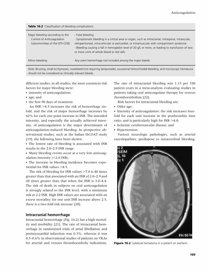
BLBK186-Key April 15, 2009 14:11
Anticoagulation
Table 16.2 Classification of bleeding complications.
Major bleeding (according to the
Control of Anticoagulation
Subcommittee of the ISTH [29])
- Fatal bleeding
- Symptomatic bleeding in a critical area or organ, such as intracranial, intraspinal, intraocular,
retroperitoneal, intra-articular or pericardial, or intramuscular with compartment syndrome
- Bleeding causing a fall in hemoglobin level of 20 g/L or more, or leading to transfusion of two
or more units of whole blood or red cells
Minor bleeding Any overt hemorrhage not included among the major bleeds
Note: Bruising, small ecchymoses, nosebleed (not requiring tamponade), occasional hemorrhoidal bleeding, and microscopic hematuria
should not be considered as clinically relevant bleeds.
different studies, in all studies, the most consistent risk
factors for major bleeding were:� intensity of anticoagulation;� age; and� the first 90 days of treatment.
An INR �4.5 increases the risk of hemorrhage six-
fold, and the risk of major hemorrhage increases by
42% for each one point increase in INR. The intended
intensity, and especially the actually achieved inten-
sity, of anticoagulation is the major determinant of
anticoagulation-induced bleeding. In prospective ob-
servational studies, such as the Italian ISCOAT study
[19], the following have been shown:� The lowest rate of bleeding is associated with INR
results in the 2.0–2.9 INR range.� Many bleeding events occur at a very low anticoag-
ulation intensity (�2.0 INR).� The increase in bleeding incidence becomes expo-
nential for INR values �4.5.
The risk of bleeding for INR values �7.0 is 40 times
greater than that associated with an INR of 2.0–2.9 and
20 times greater than that when the INR is 3.0–4.4.
The risk of death in subjects on oral anticoagulation
is strongly related to the INR level, with a minimum
risk at 2.2 INR. High INR values are associated with an
excess mortality: for one unit INR increase above 2.5,
there is a two-fold risk increase [20].
Intracranial hemorrhageIntracranial hemorrhage (Fig. 16.2) has a high mortal-
ity and morbidity [21]. The rate of intracranial hem-
orrhage in randomized trials of atrial fibrillation and
postmyocardial infarction was 0.3%, whereas it was
0.5–0.6% in observational studies of patients on VKAs
for arterial and venous thromboembolic indications.
The rate of intracranial bleeding was 1.15 per 100
patient-years in a meta-analysis evaluating studies in
patients taking oral anticoagulant therapy for venous
thromboembolism [22].
Risk factors for intracranial bleeding are:� Older age;� Intensity of anticoagulation: the risk increases four-
fold for each unit increase in the prothrombin time
ratio, and is particularly high for INR �4.0;� Ischemic cerebrovascular disease; and� Hypertension.
Various neurologic pathologies, such as arterial
vasculopathies, predispose to intracerebral bleeding.
Figure 16.2 Subdural hematoma in a patient on warfarin.
169
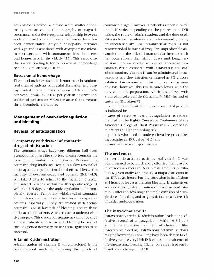
BLBK186-Key April 15, 2009 14:11
CHAPTER 16
Leukoaraiosis defines a diffuse white matter abnor-
mality seen on computed tomography or magnetic
resonance, and a dose–response relationship between
such abnormality and intracranial hemorrhage has
been demonstrated. Amyloid angiopathy increases
with age and is associated with asymptomatic micro-
hemorrhages and with spontaneous lobar intracere-
bral hemorrhage in the elderly [23]. This vasculopa-
thy is a contributing factor to intracranial hemorrhage
related to oral anticoagulation.
Extracranial hemorrhageThe rate of major extracranial hemorrhage in random-
ized trials of patients with atrial fibrillation and post-
myocardial infarction was between 0.4% and 1.4%
per year. It was 0.9–2.0% per year in observational
studies of patients on VKAs for arterial and venous
thromboembolic indications.
Management of over-anticoagulationand bleeding
Reversal of anticoagulation
Temporary withdrawal of coumarindrug administrationThe coumarin drugs have very different half-lives:
acenocoumarol has the shortest, phenprocoumon the
longest, and warfarin is in between. Discontinuing
coumarin drug intake will result in a slow reversal of
anticoagulation, proportional to their half-lives. The
majority of over-anticoagulated patients (INR �4.5)
will take 3 days to return to the therapeutic range.
For subjects already within the therapeutic range, it
will take 3–5 days for the anticoagulation to be com-
pletely reversed. Temporary withdrawal of coumarin
administration alone is useful in over-anticoagulated
patients, especially if they are treated with aceno-
coumarol, are at low risk of bleeding, and in those
anticoagulated patients who are due to undergo elec-
tive surgery. This option for treatment cannot be used
alone in patients who are actively bleeding because of
the long period necessary for the anticoagulation to be
reversed.
Vitamin K administrationAdministration of vitamin K (phytonadione) is the
recommended mode of reversing the effects of
coumarin drugs. However, a patient’s response to vi-
tamin K varies, depending on the pretreatment INR
value, the route of administration, and the dose used.
Vitamin K can be administered intravenously, orally,
or subcutaneously. The intramuscular route is not
recommended because of irregular, unpredictable ab-
sorption and the risk of intramuscular hematoma. It
has been shown that higher doses and longer re-
version times are needed with subcutaneous admin-
istration when compared with intravenous and oral
administration. Vitamin K can be administered intra-
venously as a slow injection or infused in 5% glucose
solution. Intravenous administration can cause ana-
phylaxis; however, this risk is much lower with the
new vitamin K preparation, which is stabilized with
a mixed micelle vehicle (Konakion R© MM) instead of
castor oil (Konakion R©).
Vitamin K administration in anticoagulated patients
is indicated in:� cases of excessive over-anticoagulation, as recom-
mended by the Eighth Consensus Conference of the
American College of Chest Physicians [7], especially
in patients at higher bleeding risk;� patients who need to undergo invasive procedures
that require an INR value �1.5; and� cases with active major bleeding.
The oral routeIn over-anticoagulated patients, oral vitamin K was
demonstrated to be much more effective than placebo
in correcting excessive INRs. Small amounts of vita-
min K given orally can produce a major correction in
the INR at 24 hours, but the correction is insufficient
at 4 hours or for cases of major bleeding. In patients on
acenocoumarol, administration of low-dose oral vita-
min K offers no advantage to simple omission of a sin-
gle dose of the drug and may result in an excessive risk
of under-anticoagulation.
The intravenous routeIntravenous vitamin K administration leads to an ef-
fective reversal of anticoagulation within 6–8 hours
and is therefore the treatment of choice in life-
threatening bleeding. Intravenous vitamin K doses
ranging between 0.1 and 3 mg have been shown to ef-
fectively reduce very high INR values in the absence of
life-threatening bleeding. Higher doses may frequently
result in subtherapeutic INR.
170
BLBK186-Key April 15, 2009 14:11
Anticoagulation
Fresh frozen plasmaAdministration of fresh frozen plasma (FFP) is in-
tended to correct the deficiency of factors II, VII,
IX, and X resulting from the effect of coumarin
drugs. The recommended FFP dose for warfarin re-
versal is 15 mL/kg body weight. However, several
factors should be considered that limit the value
of FFP as the best replacement treatment in this
indication [24]:� Large FFP volumes (e.g. approximately 1000 mL
for an adult weighing 70 kg) are needed to be given
rapidly to replace vitamin K-dependent factors, and
this may be harmful, especially in patients with com-
promised cardiovascular conditions.� FFP may not be virally inactivated and therefore a
potential risk of viral infection cannot be excluded.� The time needed to prepare the plasma, which is
stored frozen at −20◦C, is usually a cause of delay
before transfusion.� It has been shown that administration of the recom-
mended dose of FFP fails to significantly correct the
coumarin-induced coagulopathy, especially for persis-
tently low factor IX levels.
Prothrombin complex concentratesConcentrates of factors II, VII, IX, and X, called pro-
thrombin complex concentrates (PCCs), are available
and are highly effective in replacing clotting factors
that are deficient in anticoagulated patients [25]. A
dose of 30 U/kg is usually effective. The precise op-
timal dose of PCC remains to be defined, but the fol-
lowing have been suggested:� 25 U/kg for patients with an INR of 2.0–3.9; and� 35 U/kg for an INR of �4.0.
Potential adverse effects of PCC administration are
viral infection and thrombogenicity. Despite all the
precautions taken (selection of donors as well as spe-
cific viral inactivation procedures), an extremely small
risk of viral infection can still persist. Thrombotic
events have been reported after PCC transfusion in an-
ticoagulated patients; however, this risk is also small,
especially in preparations with added antithrombin
and heparin. The potential risks of PCC indicate that
their use should be reserved for patients with major
bleeding, especially to those with intracranial hemor-
rhage in whom an immediate correction of the coagu-
lopathy is highly recommended.
Clinical management ofover-anticoagulation and bleedingAn unexpected condition of over-anticoagulation is
not a rare finding during treatment with VKAs. The
incidence rate of an INR �6.0 was found to be as high
as 7.8 in 10,000 treatment days in prevalent users and
22.5 in 10,000 treatment days in incident users. Be-
cause it is known that the risk of bleeding increases
sharply in association with very high INR values, it is
desirable for a patient to spend as little time as possible
in a condition of over-anticoagulation. In these cases,
the clinical management should be as follows:� Patients with very high INR values (�7.0), or with
more moderately high INR values but at high risk of
bleeding, should receive 1–2 mg vitamin K orally; the
INR should be measured the following day and oral
vitamin K given again if necessary.� In patients with an INR of 4.5–6.9, and in those
treated with acenocoumarol whatever the INR, with-
holding the coumarin drug for 1–2 days followed by a
reduction of the weekly dose is usually sufficient.� All patients with minor bleeding and an INR over
the therapeutic range should receive intravenous vita-
min K, which will reduce the high INR values within
6–8 hours.� In cases with major, although not life-threatening,
bleeding, a complete reversal of anticoagulation with
intravenous vitamin K is advisable.� A complete and rapid reversal of anticoagulation is
recommended in patients with life-threatening bleed-
ing. PCC infusion will completely correct the coagu-
lopathy within 5–10 minutes. A dose of 5–10 mg vita-
min K should also be administered intravenously.� Please note the lack of recommendation to use FFP.
Management of patients treatedwith VKAs who require surgeryor invasive procedures
There are no universally accepted guidelines for
the management of anticoagulated patients requiring
surgery or invasive procedures. Clear indications are
lacking because of different patients, procedures, an-
ticoagulant regimens, event definition and duration
of follow-up, as well as the absence of randomized
clinical trials in this setting. However, the increasing
171
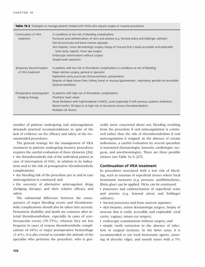
BLBK186-Key April 15, 2009 14:11
CHAPTER 16
Table 16.3 Strategies to manage patients treated with VKAs who require surgery or invasive procedures.
Continuation of VKA
treatment
In conditions at low risk of bleeding complications:
Punctures and catheterization of veins and arteries (e.g. femoral artery and Seldinger catheter)
Sternal punctures and bone marrow aspirates
Skin biopsies, minor dermatologic surgery, biopsy of mucosa that is easily accessible and explorable
(oral cavity, vagina), minor eye surgery
Endoscopic examinations without surgery
Simple tooth extraction
Temporary discontinuation
of VKA treatment
In patients with low risk of thrombotic complications in conditions at risk of bleeding:
Major elective surgery, general or specialist
Explorative cavity punctures (thoracocentesis, paracentesis)
Biopsies of deep tissues (liver, kidney, bone) or mucosa (gastroenteric, respiratory, genital) not accessible
Epidural anesthesia
Perioperative anticoagulant
bridging therapy
In patients with high risk of thrombotic complications:
Prosthetic heart valves
Atrial fibrillation with high/moderate CHADS2 score (especially if with previous systemic embolism)
Recent (within 30 days) or at high risk of recurrence venous thromboembolism
Multiple risk factors
number of patients undergoing oral anticoagulation
demands practical recommendations in spite of the
lack of evidence on the efficacy and safety of the rec-
ommended procedures.
The general strategy for the management of VKA
treatment in patients undergoing invasive procedures
requires the careful evaluation of three elements [26]:� the thromboembolic risk of the individual patient in
case of interruption of OAC, in relation to its indica-
tions and to the risk of postoperative thromboembolic
complications;� the bleeding risk of the procedure per se and in case
anticoagulation is continued; and� the necessity of alternative anticoagulant drugs
(bridging therapy) and their relative efficacy and
safety.
The substantial difference between the conse-
quences of major bleeding events and thromboem-
bolic complications should also be taken into account.
Permanent disability and death are common after ar-
terial thromboembolism, especially in cases of cere-
brovascular events (70–75%), whereas they are less
frequent in cases of venous thromboembolic compli-
cations (4–10%) or major postoperative hemorrhage
(1–6%). It is also crucial to consider the attitude of the
specialist who performs the procedure, who is gen-
erally more concerned about any bleeding resulting
from the procedure if oral anticoagulation is contin-
ued rather than the risk of thromboembolism if oral
anticoagulation is stopped. In the absence of certain
indications, a careful evaluation by several specialists
is warranted (hematologist, internist, cardiologist, sur-
geon, and anesthesiologist). There are three possible
choices (see Table 16.3) [27].
Continuation of VKA treatmentIn procedures associated with a low risk of bleed-
ing, such as traumas of superficial tissues where local
hemostatic measures (e.g. pressure, antifibrinolytics,
fibrin glue) can be applied, VKAs can be continued:� punctures and catheterization of superficial veins
and arteries (e.g. femoral artery and Seldinger
catheter);� Sternal punctures and bone marrow aspirates;� skin biopsies, minor dermatologic surgery, biopsy of
mucosa that is easily accessible and explorable (oral
cavity, vagina), minor eye surgery;� endoscopic examinations without surgery; and� simple tooth extraction in the absence of infec-
tion or surgical incisions. In the latter cases, it is
recommended to use local hemostatic agents, sutur-
ing of alveolar edges, and mouth rinses with a 5%
172
BLBK186-Key April 15, 2009 14:11
Anticoagulation
tranexamic acid solution, 4–5 minutes every 6 hours
for 5–6 days, combined with antibiotic therapy.
In these cases, it is it is advisable to lower the INR
to approximately 2.0 to decrease the hemorrhagic risk
without an increase in the thromboembolic risk. If
the expected risk of bleeding is higher (e.g. multiple
teeth extractions in the presence of infection, closed
biopsy, endo-ocular surgery, or cataract with retrob-
ulbar anesthetic) and the risk of thromboembolism is
not high (in most cases, excluding patients with pros-
thetic heart valves or cardiac endocavitary thrombo-
sis), VKAs can be temporarily reduced, aiming at INR
values between 1.5 and 2.
Patients being treated with VKAs should be told to
avoid, whenever possible, intramuscular injections so
to avoid the risk of hematomas (especially if the pa-
tient needs many injections).
Temporary discontinuation ofVKA treatmentThis is recommended in conditions associated with a
significant risk of bleeding (such as cases of trauma to
deep tissues not easily accessible to local hemostatic
measures; see Table 16.3) in patients with non-high
risk of thrombotic complications.
Perioperative bridging therapyThis strategy is indicated in patients who are at high
risk of thrombotic complications (see Table 16.3). In
these patients, the goal is to minimize risk by reducing
the duration of the bridging therapy to the minimum
and by administering bridging therapy for the duration
of subtherapeutic INR. If the procedure is elective, no
immediate reversal of VKAs is required and VKAs can
be discontinued 3–4 days before the procedure (in case
of therapeutic INR), as the INR is expected to fall to
subtherapeutic values in 3–4 days. The bridging ther-
apy can be commenced 60 hours after the last warfarin
dose (third morning after last evening dose). The INR
should be measured the day before surgery to deter-
mine whether it is below 1.5–1.7. If not, 1 mg vitamin
K can be given orally and the INR repeated on the day
of surgery.
The bridging therapy can be conducted with unfrac-
tionated heparin (subcutaneous or intravenous) when
the INR falls below 2.0. Bridging therapy can be started
with prophylactic unfractionated heparin (5000 U ev-
ery 8–12 hours subcutaneously). Those at very high
risk of thromboembolic complications (previous sys-
temic embolism in atrial fibrillation, prosthetic heart
valves, multiple risk factors) can also be given bridg-
ing therapy by administering adjusted dose heparin
(subcutaneous in outpatients or by continuous in-
travenous infusion in case of hospital admission) to
maintain an APTT value equal to 1.5–2 times the nor-
mal value of control.
Bridging therapy can also be administered with low-
molecular-weight heparin subcutaneously as out- or
inpatient for 2–3 days preoperatively, using doses rec-
ommended for prophylaxis or, in patients at very high
risk of thrombosis, therapeutic doses once (150–200
U/kg) or twice (100 U/kg) daily.
Drug administration immediately prior to surgery
must be avoided in these cases:� Subcutaneous unfractionated heparin should be dis-
continued 12 hours before surgery.� Intravenous heparin should be discontinued 6 hours
before surgery.� Low-molecular-weight heparin should be discontin-
ued no less than 8–10 hours at prophylaxis dose or
18 hours preoperatively with treatment doses, with an
additional 6-hour interval in case of planned neuroax-
ial anesthesia.
In venous thromboembolism, the risk of recurrence
is the highest in the first month after the acute event
(40%). As a result, invasive procedures should be de-
ferred, if possible, for at least 1 month and preferably
3 months after the acute event. If surgery is necessary
within 2 weeks from an acute event, patients should
have an inferior vena cava filter inserted preopera-
tively or intraoperatively.
Postoperative managementof anticoagulationVKAs may be resumed only after evaluating each case
very carefully as a function of the time needed for tis-
sues to heal and in the absence of bleeding compli-
cations. Intravenous heparin should be resumed after
12 hours postoperatively at a rate of no more than
18 U/kg, and it has the advantage of rapid elimination
if discontinued and of neutralization with protamine.
If subcutaneous low-molecular-weight heparin is pre-
ferred, twice daily doses are recommended, started 24
hours postoperatively and only after hemostasis has
been achieved. In patients with a very high bleed-
ing risk (e.g. after neurosurgery or prostatectomy),
173
BLBK186-Key April 15, 2009 14:11
CHAPTER 16
heparin is resumed only after clinical evaluation and
in general after at least 48–72 hours. VKAs can be re-
sumed postoperatively as soon as the patients can take
solid foods, overlapping with heparin until an INR �2
is obtained for two consecutive days. In case of emer-
gency surgery, oral anticoagulation must be reversed
as soon as possible.
Spinal or epidural anesthesiaRegional anesthesia in association with perioperative
prophylaxis or heparin therapy is safe and efficacious
with an adequate selection of patient and anesthesio-
logic technique. There are no controlled studies eval-
uating the risk of spinal hematoma in the course of
therapy or with heparin prophylaxis.
The following can be suggested with intravenous
(IV) or subcutaneous (SC) unfractionated heparin:� Perform the spinal puncture or the positioning of the
catheter at least 1 hour before starting heparin IV, or
more than 4 hours after the suspension of the heparin
IV and after the administration of the heparin SC.� Maintain APTT value not more than 1.5 times the
control value.� Remove the catheter only after normalization of the
APTT.
The following are suggestions for the use of prophy-
lactic dose low-molecular-weight heparins:� Perform spinal puncture or positioning of the
catheter 10–12 hours after the last dose;� Remove the catheter at least 10–12 hours after the
last dose, and administer the successive dose at least 2
hours after removal.
In any case it must be remembered:� Do not administer drugs that interfere with the
hemostasis.� Defer the operation in the presence of a bloodstained
spinal tap.� Constant patient surveillance is essential for the on-
set of signs or symptoms of medullary compression
(sphinteric alterations, progression of paresthesia, and
limb weakness).� In the case of spinal hematoma, emergency decom-
pressive laminectomy is mandatory (�6 hours from
the onset of the symptoms).
Cataract surgeryWith modern techniques, which rely on limited inci-
sion of the cornea (nonvascularized tissue), the risk of
bleeding from surgery itself is practically null. Possible
bleeding complications are linked to the type of anes-
thesia. Cases have been reported of retro- and peribul-
bar hematomas in patients on VKAs following retro-
and peribulbar anesthesia. Despite the lack of exact
data on the incidence of these complications, it should
be borne in mind that retro- and peribulbar anesthe-
sia requires normal blood hemostasis and hence dis-
continuation of VKAs, so it should be contraindicated
in patients on VKAs in whom the thrombotic risk
following a suspension of treatment is high. In con-
trast, cataract surgery can be performed without anti-
coagulant suspension in all those subjects in whom a
topical or general anesthesia can be used. Evaluating
the risk–benefit and cost–benefit ratios of the differ-
ent options (e.g. surgery without VKAs suspension vs.
surgery with retrobulbar anesthesia and VKAs suspen-
sion) should be carried out in each patient on the basis
of a general consideration of the risk factors (throm-
botic and hemorrhagic).
New anticoagulants
The complexities of oral anticoagulation treatment
have prompted the search for improved anticoagulants
[28]. The ideal anticoagulant should be effective, with
minimal complications/side effects and convenient ad-
ministration (i.e. oral for outpatients), with rapid ab-
sorption and fast on- and offset action, predictable
pharmacokinetics, no interactions with food or drugs,
no need of coagulation monitoring, and availability of
an antidote.
New anticoagulants have been developed that target
a single coagulation factor and have predictable dose–
response relationships. These include direct throm-
bin inhibitors and factor Xa inhibitors. Two parenteral
direct thrombin inhibitors, lepirudin and argatroban,
have FDA approval for the management of heparin-
induced thrombocytopenia (HIT). Bivalirudin is a par-
enteral direct thrombin inhibitor that is licensed for
patients undergoing percutaneous coronary interven-
tions and for those with HIT who require percuta-
neous coronary interventions. Ximelagatran, an oral
prodrug of the direct thrombin inhibitor melagatran,
showed efficacy in the prevention and treatment of
venous thromboembolism as well as stroke preven-
tion in patients with atrial fibrillation. However, due
174
BLBK186-Key April 15, 2009 14:11
Anticoagulation
to nonhematologic safety concerns, it did not receive
FDA approval in the US. Fondaparinux is a syn-
thetic pentasaccharide, which is highly selective for
antithrombin with exclusive anti-Xa activity, with a
predictable dose response due to the absence of as-
pecific binding to plasma proteins, and with virtually
absent risk of HIT. The effectiveness of fondaparinux
has been shown in phase III studies in the prophylaxis
and treatment of venous thromboembolism, and it is
now available for clinical use in Europe. Idraparinux
is a modified pentasaccaride with a long half-life,
which can be administered by injection once weekly.
In Phase III studies, idraparinux has been shown to
be as effective as warfarin in the treatment of ve-
nous thromboembolism, albeit with a higher risk of
bleeding.
An oral direct thrombin inhibitor, dabigatran etexi-
late, has been recently approved for clinical use in Eu-
rope for the prophylaxis of venous thromboembolism
in major orthopedic surgery. Dabigatran etexilate has
a molecular weight of 628 Da, with a half-life of 14–17
hours and time to peak level of 2 hours, with preva-
lent renal excretion. Dabigatran etexilate can be ad-
ministered orally without laboratory monitoring at a
dose of 110 mg at 1–4 hours after surgery and then
at full dose of 220 mg once daily for 10 days after
knee prosthesis and 28–35 days after hip surgery; the
dose should be reduced at 150 mg once daily in case of
age greater than 75 years and in case of chronic renal
failure. Phase III studies are ongoing in the treatment
of venous thromboembolism with the aim of replac-
ing warfarin. Two oral direct factor Xa inhibitors, ri-
varoxaban and apixaban, are undergoing evaluation
in phase III studies in the prevention and treatment of
venous thromboembolism. In spite of the potential ad-
vantages of the newer oral anticoagulant drugs, their
main limitation is the lack of an antidote.
References
1 Baglin TP, Keeling DM, Watson HG. Guidelines on oral
anticoagulation (Warfarin): third edition – 2005 up-
date. Br J Haematol 2006;132:277–85.
2 Hirsh J, Dalen JE, Anderson DR, et al. Oral anticoag-
ulants: Mechanism of action, clinical effectiveness, and
optimal therapeutic range. Chest 2001;119:8S–21S.
3 Chan WS, Anand S, Ginsberg JS. Anticoagulation
of pregnant women with mechanical heart valves: a
systematic review of the literature. Arch Intern Med
2000;160:191–6.
4 Margaglione M, Colaizzo D, D’Andrea G, et al. Ge-
netic modulation of oral anticoagulation with warfarin.
Thromb Haemost 2000;84:775–8.
5 Rost S, Fregin A, Ivaskevicius V, et al. Mutations in
VKORC1 cause warfarin resistance and multiple coag-
ulation factor deficiency type 2. Nature 2004;427:537–
41.
6 Rieder MJ, Reiner AP, Gage BF, et al. Effect of VKORC1
haplotypes on transcriptional regulation and warfarin
dose. N Engl J Med 2005;352:2285–93.
7 Ansell J, Hirsh J, Hylek E, et al. Pharmacology and
management of the vitamin K antagonists. Chest
2008;133:160S–198S.
8 Fennerty A, Dolben J, Thomas P, et al. Flexible induc-
tion dose regimen for warfarin and prediction of main-
tenance dose. Br Med J 1984;288:1268–70.
9 Crowther MA, Ginsberg JB, Kearon C, et al. A random-
ized trial comparing 5-mg and 10-mg warfarin loading
doses. Arch Intern Med 1999;159:46–8.
10 Kovacs MJ, Rodger M, Anderson DR, et al. Comparison
of 10-mg and 5-mg warfarin initiation nomograms to-
gether with low-molecular-weight heparin for outpa-
tient treatment of acute venous thromboembolism. A
randomized, double-blind, controlled trial. Ann Intern
Med 2003;138:714–9.
11 Poller L, Keown M, Ibrahim S, et al. An international
multicenter randomized study of computer-assisted
oral anticoagulant dosage vs. medical staff dosage.
J Thromb Haemost 2008;6:935–43.
12 Chiquette E, Amato MG, Bussey HI. Comparison of an
anticoagulation clinic with usual medical care: antico-
agulation control, patient outcomes, and health care
costs. Arch Intern Med 1998;158:1641–7.
13 Vandermeer FJM, Briet E, Vandenbroucke JP, et al. The
role of compliance as a cause of instability in oral anti-
coagulant therapy. Br J Haematol 1997;98:893–900.
14 Palareti G, Legnani C, Guazzaloca G, et al. Risks factors
for highly unstable response to oral anticoagulation: a
case-control study. Br J Haematol 2005;129:72–8.
15 Barcellona D, Contu P, Marongiu F. Patient edu-
cation and oral anticoagulant therapy. Haematologica
2002;87:1081–6.
16 Tang EO, Lai CS, Lee KK, et al. Relationship between
patients’ warfarin knowledge and anticoagulation con-
trol. Ann Pharmacother 2003;37:34–9.
17 Estrada CA, Hryniewicz MM, Higgs VB, et al. Antico-
agulant patient information material is written at high
readability levels. Stroke 2000;31:2966–70.
175
BLBK186-Key April 15, 2009 14:11
CHAPTER 16
18 Ford SK, Misita CP, Shilliday BB, et al. Prospective
study of supplemental vitamin K therapy in patients on
oral anticoagulants with unstable international normal-
ized ratios. J Thromb Thrombolysis 2007;24:23–7.
19 Palareti G, Leali N, Coccheri S, et al. Bleeding complica-
tions of oral anticoagulant treatment: an inception- co-
hort, prospective collaborative study (ISCOAT). Italian
Study on Complications of Oral Anticoagulant Therapy.
Lancet 1996;348:423–8.
20 Oden A, Fahlen M. Oral anticoagulation and risk
of death: a medical record linkage study. Br Med J
2002;325:1073–5.
21 Fang MC, Go AS, Chang YC, et al. Death and disability
from warfarin-associated intracranial and extracranial
hemorrhages. Am J Med 2007;120:700–5.
22 Linkins LA, Choi PT, Douketis JD. Clinical impact of
bleeding in patients taking oral anticoagulant therapy
for venous thromboembolism: a meta-analysis. Ann In-
tern Med 2003;139:893–900.
23 Rosand J, Hylek EM, Odonnell HC, et al. Warfarin-
associated hemorrhage and cerebral amyloid an-
giopathy: a genetic and pathologic study. Neurology
2000;55:947–51.
24 Makris M, Watson HG. The management of coumarin-
induced over-anticoagulation. Br J Haematol 2001;114:
271–80.
25 Hellstern P, Halbmayer WM, Kohler M, et al. Prothrom-
bin complex concentrates: Indications, contraindica-
tions, and risks: a task force summary. Thromb Res
1999;95:S3–S6.
26 Kearon C, Hirsh J. Current concepts: management of
anticoagulation before and after elective surgery. N Engl
J Med 1997;336:1506–11.
27 Kearon C. Management of anticoagulation in pa-
tients requiring invasive procedures. Semin Vasc Med
2003;3:285–93.
28 Bates SM, Weitz JI. The status of new anticoagulants.
Br J Haematol 2006;134:3–19.
29 Schulman S, Kearon C. Definition of major bleeding
in clinical investigations of antihemostatic medicinal
products in non-surgical patients. J Thromb Haemost
2005;3:692–4.
176
BLBK186-Key April 24, 2009 7:56
17 Antiphospholipid syndromeHenry G. Watson and Beverley J. Robertson
Introduction
The antiphospholipid syndrome (APS) is an acquired
prothrombotic or thrombophilic state that is also as-
sociated with adverse outcome of pregnancy. An as-
sociation of antiphospholipid antibodies with a vari-
ety of disorders has been made since the first report
in patients with systemic lupus erythematosus (SLE),
and the clinicopathological criteria for the diagnosis
of APS have been agreed internationally [1]. In spite
of this, our understanding of the pathogenesis of the
condition is limited, particularly with respect to the
complications of pregnancy for which there is no com-
pelling evidence of an ischemic pathogenesis. Because
the manifestations of the APS are common in the
population, differentiation between those individuals
with and without the syndrome is heavily dependent
on laboratory assays to detect persistent antiphospho-
lipid antibodies. The laboratory-based diagnosis, how-
ever, is a subject of serious concern with disappoint-
ing quality assurance data for all tests. This is very
important because the diagnosis of APS changes clin-
ical management significantly in those affected. For
example, anticoagulation following a first episode of
venous thromboembolism should probably be pro-
longed in those with APS, whereas it is appropri-
ate in other groups of patients to consider periods of
3–6 months only. However, whereas there are good
data to inform on the management of venous throm-
boembolism, the same is not true for arterial throm-
bosis. Finally, there are conflicting views on the
treatment of women with adverse pregnancy outcome
attributable to APS.
Definition of APS
The APS describes a clinicopathologic entity. APS is
an acquired prothrombotic state that probably has
an immune-mediated pathogenesis, and its diagno-
sis requires the coexistence of clinical manifestations
(thrombosis or adverse pregnancy outcome) with lab-
oratory evidence of antiphospholipid antibodies.
The Sapporo diagnostic criteria for APS were re-
vised in 2005 by an International Consensus Panel
(Table 17.1) [2]. A variety of other clinical abnor-
malities, which are frequently observed in association
with antiphospholipid antibodies, are not included
in the internationally agreed definition of APS. The
most common of these are thrombocytopenia and
livedo reticularis, which are frequently found in pa-
tients who do not otherwise fulfil the criteria for APS
(Table 17.2). Identification of these other associated
conditions should lead to consideration of a diagnosis
of APS. It is not clear whether thrombosis is implicated
in the pathogenesis of these conditions, and the role of
antithrombotic medicines is even less clear.
Clinical features of APS
The main clinical presentation of APS is either with
thrombosis or pregnancy failure. However, the con-
dition is heterogeneous (as are the implicated an-
tibodies), and most individuals do not suffer from
all the clinical features of the syndrome. Interest-
ingly, although a combination of venous and arterial
thrombotic events may predate the development of a
177

BLBK186-Key April 24, 2009 7:56
CHAPTER 17
Table 17.1 Diagnostic criteria for APS.
Clinical criteria
Thrombosis
Venous, arterial, or small-vessel thrombosis involving any organ
or tissue
Pregnancy
Unexplained death of a morphologically normal fetus at or
after 10 weeks’ gestation
Three or more consecutive unexplained abortions before 10
weeks’ gestation
Severe pre-eclampsia or placental insufficiency before 34
weeks’ gestation
Laboratory criteria
LA
IgG or IgM anticardiolipin antibodies at moderate or high titer
IgG or IgM anti-β2–glycoprotein 1 antibodies in titer >99th
centile
Note: To fulfill the diagnosis of APS, there must be at least
one clinical and one laboratory criterion present. The detection
of antiphospholipid antibodies must have been performed on
two occasions at least 12 weeks apart.
history of pregnancy failure in some women, espe-
cially those with SLE, most women who present with
adverse pregnancy outcome tend to have this as a sole
manifestation.
Table 17.2 Some conditions associated with
antiphospholipid antibodies but not included in the definition
of APS.
Thrombocytopenia
Livedo reticularis
Allograft failure
Transverse myelopathy
Chorea
Multifocal central nervous system syndrome resembling multi-
ple sclerosis
Skin necrosis
Pulmonary hypertension
Sensorineural deafness
Cardiac valve disease (vegetations, valve thickening and dys-
function)
Nephropathy (small-vessel vasculopathy)
ThrombosisThrombosis may involve both the arterial and the ve-
nous systems. The most common presentation is with
lower limb deep vein thrombosis, sometimes with
clinically significant pulmonary embolus. Other sites
for venous thrombosis such as cerebral vein, axillary
and subclavian vein, and intra-abdominal veins, in-
cluding the portal, hepatic, and mesenteric veins, are
less common but well recognized in APS. Patients
tend to be young individuals with unprovoked venous
thromboembolism, thrombosis at unusual sites, and
an absence of a family history of thrombophilia.
Stroke and transient cerebral ischemia are the most
common presentations of arterial thrombosis in APS.
Myocardial infarction appears rare, although the rea-
son for this is not clear. Embolic thrombus from sterile
endocarditis and cardiac valve vegetations are also de-
scribed.
Microvascular thrombosis is uncommon but is de-
scribed in the extremely rare “catastrophic antiphos-
pholipid syndrome” that presents with multiorgan fail-
ure and which usually progresses unabated in spite of
all forms of therapy (Plate 17.1).
Pregnancy failureThis is now the most common presentation that results
in a diagnosis of APS being made. This is in part be-
cause of the wish (and pressure) to investigate women
who are distressed by this presentation. Recurrent
early fetal loss is most commonly seen, although oth-
erwise unexplained fetal death after the first trimester
and severe pre-eclampsia before 34 weeks are also rec-
ognized features.
The emotive nature of these cases may result in
the inappropriate investigation of women with only
one or two early miscarriages, which can result in a
chance finding of an innocent antiphospholipid an-
tibody. Having detected antiphospholipid antibodies
in these women who do not fulfill the APS criteria
[3], clinicians find it difficult to withhold treatment,
resulting in some women spending the whole of sub-
sequent pregnancies on aspirin and low-molecular-
weight heparin (LMWH), based on very little
evidence.
Most proponents of this approach argue that
waiting for a third early loss in these women is
inappropriate and add that the therapy has so few side
178
BLBK186-Key April 24, 2009 7:56
Antiphospholipid syndrome
effects that this is not an issue. However, side effects,
although few, are seen and the costs of clinic time
and drugs are significant. This practice also converts
normal women into patients for the duration of their
pregnancy while skewing the perception of benefit for
intervention.
Antiphospholipid antibodies
These are a heterogeneous group of antibodies, which
are detected because of their capacity to react with
phospholipid either in phospholipid-dependent coag-
ulation assays in the case of a lupus anticoagulant
(LA) or bound to enzyme-linked immunosorbent as-
say (ELISA) plates in the case of anticardiolipin and
anti-β2-glycoprotein 1 antibodies.
The earliest descriptions of antiphospholipid anti-
bodies were in individuals with SLE who had false-
positive tests for syphilis. Further investigation of
these patients indicated that they had circulating anti-
bodies that were capable of binding to the negatively
charged phospholipid, cardiolipin. This gave rise to the
nomenclature anticardiolipin antibodies.
About the same time, it was noted that some sub-
jects with SLE had prolonged blood-clotting times in
in vitro test systems but had no evidence of a bleed-
ing diathesis. The prolonged clotting in phospholipid-
dependent tests could not be reversed by addition of
normal plasma, indicating the presence of an inhibitor,
the so-called lupus anticoagulant.
Paradoxically, the presence of the LA was asso-
ciated with an increased risk of thrombosis in pa-
tients with SLE, and when it became apparent that
the presence of either of these antibodies was associ-
ated with an increased thrombosis risk, the concept of
an acquired prothrombotic or thrombophilic state was
proposed.
Antiphospholipid antibodies with features of APS
may be found either in isolation as a primary antiphos-
pholipid syndrome or in association with SLE and other
autoimmune conditions, such as Sjogren syndrome as
a secondary antiphospholipid syndrome.
Although they are called antiphospholipid antibod-
ies, it is now clear that the antigenic targets for
most of these antibodies are not phospholipid per se,
but instead are proteins that bind to phospholipid
(Table 17.3). The best known of these is β2-glyco-
Table 17.3 Antigenic targets of antiphospholipid antibodies.
β2-glycoprotein 1
Prothrombin
Protein C
Protein S
Annexin V
Factors XI and XII
protein 1, a circulating protein of unknown function
which avidly binds negatively charged phospholipid.
β2-Glycoprotein 1 is considered the most important
antigenic target for antiphospholipid antibodies. The
molecule has five domains, and antibodies against do-
main 1 have been shown to be the pathogenic an-
tibodies that cause the LA effect and associate most
strongly with thrombosis. It has also been shown
that the binding of β2-glycoprotein 1 to phospholipid
causes a conformational change in the molecule and
the exposure of “cryptic epitopes.” This may, in part,
explain the formation of autoantibodies [4].
Other antigen targets for antiphospholipid antibod-
ies include prothrombin, factor XI, proteins C and S,
and annexin V, all proteins involved in hemostatic
pathways that might be relevant in explaining the
thrombotic complications associated with these anti-
bodies. In response to these findings, ELISA assays that
have β2-glycoprotein 1 and prothrombin as antigen
are now commercially available.
Despite this knowledge, the pathogenesis of throm-
bosis and pregnancy failure in APS remains unclear.
Laboratory findings combined with the outcome of
clinical studies indicate that the pathological man-
ifestations of APS are caused by a prothrombotic
state with little evidence that overt histological in-
flammation contributes significantly to the process.
No single mechanism has been shown to under-
lie the prothrombotic tendency, and this is perhaps
not surprising given the varied sites of thrombosis
and the range of target antigens for antiphospholipid
antibodies.
Whether antiphospholipid antibodies are indeed di-
rectly pathogenic is debated. IgG from serum of pa-
tients with APS has been shown to be pathogenic in
animal models of thrombosis and pregnancy loss. Lab-
oratory experiments have assessed the effects of an-
tiphospholipid antibodies on many of the processes
179
BLBK186-Key April 24, 2009 7:56
CHAPTER 17
involved in hemostasis, thrombosis, inflammation,
and fibrinolysis.
There are data to support that antiphospholipid
antibodies may induce tissue factor expression by
monocytes, inhibit the function of the natural anti-
coagulants activated protein C and protein S, induce
endothelial cell apoptosis and activation, and induce
platelet activation by binding via the Fc receptor. All,
none, or, more likely, a combination of these mecha-
nisms may contribute to the disease process [4]. Al-
though the criteria for diagnosis of APS state that
histological evidence of inflammation excludes the di-
agnosis, there is increasing experimental evidence that
antiphospholipid antibodies may induce an inflam-
matory state. Up-regulation of adhesion molecules,
such as VCAM-1 and E-selectin, and secretion of
interleukin-6 has been observed in endotheial cells in-
cubated with antiphospholipid antibodies. Increased
leukocyte adhesion to endothelium with associated
release of tissue factor could perceivably be involved
in the pathogenesis of the condition.
The pathogenesis of pregnancy failure in APS is
even more difficult to explain. Knowledge of the pos-
sible prothrombotic mechanisms has led to the infer-
ence that placental ischemia is the main mechanism
resulting in pregnancy failure in APS (Plate 17.2).
The evidence from clinical studies suggesting im-
proved outcome in patients treated with antithrom-
botic medicines, such as heparin and aspirin, is felt by
many to support this hypothesis. However, overt pla-
cental ischemia is rare, and the observation that the
most common manifestation of APS in pregnancy is
miscarriage before 10 weeks (i.e. prior to development
of the placental circulation) suggests that other mech-
anisms must contribute. Complement activation by
antiphospholipid antibodies has been linked to early
pregnancy loss, and antiphospholipid antibodies have
been shown to inhibit trophoblastic proliferation and
spiral artery invasion in vitro. Interestingly, these ef-
fects may be inhibited by heparin, which suggests that
at least part of any benefit for heparin may relate to
an anti-complement effect and/or improved implan-
tation.
Other work has suggested that antiphospholipid an-
tibodies may act by displacing the natural anticoagu-
lant annexin V from endothelial cell surfaces, resulting
in a procoagulant state. However, as normal expres-
sion of annexin V has been demonstrated in affected
pregnancies, the importance of these observations re-
mains unclear.
Diagnosis of APS
APS is a clinicopathologic entity that depends on the
identification of a clinical diagnosis combined with
demonstration of appropriate antiphospholipid anti-
bodies (Table 17.1). Although criteria for diagnosis
have been internationally agreed, the diagnosis of APS
is still complicated by two main problems:� Many antiphospholipid antibodies are nonpatholog-
ical and are not associated with APS.� The standardization of assays for LA and immuno-
logically detectable antiphospholipid antibodies, such
as anticardiolipin antibodies and β2-glycoprotein 1 an-
tibodies, is unfortunately very poor.
Transient and nonpathologicalantiphospholipid antibodies
Both LAs and anticardiolipin antibodies, alone or to-
gether, are found in a significant number of normal
subjects. Like the finding of a positive direct antiglobu-
lin test in approximately 1 in 10,000 blood donors, the
finding is of little consequence to the individual, but it
does generate further investigation and anxiety in the
patient if handled badly. One common source of this
type of scenario is in the recruitment of healthy ward
and laboratory personnel as normal controls. On some
occasions, the antiphospholipid antibody is transient,
but persistent high-titer anticardiolipin antibodies and
strong positive LAs are not uncommon. Some series
report the finding of antiphospholipid antibodies, most
often anticardiolipin, in up to 5% of normal subjects.
Perhaps the most common cause of transient an-
tiphospholipid antibodies is infection (Table 17.4).
This is mostly seen following viral infection but may
complicate bacterial and parasitic infections also. Al-
though these antibodies are not typically associated
with significant disease, purpura fulminans resulting
from acquired protein S deficiency due to antiphos-
pholipid antibodies, is a well-documented complica-
tion of varicella infection in children. Some infections,
such as HIV, hepatitis C, leprosy, syphilis, leptospiro-
sis, leishmaniasis, and malaria, are associated with
180
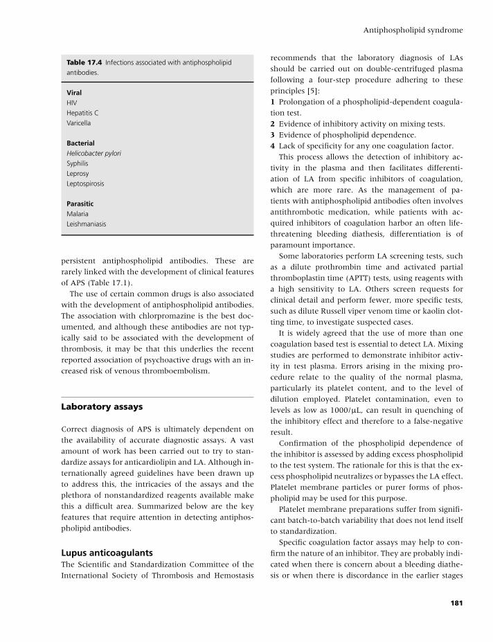
BLBK186-Key April 24, 2009 7:56
Antiphospholipid syndrome
Table 17.4 Infections associated with antiphospholipid
antibodies.
ViralHIV
Hepatitis C
Varicella
BacterialHelicobacter pylori
Syphilis
Leprosy
Leptospirosis
ParasiticMalaria
Leishmaniasis
persistent antiphospholipid antibodies. These are
rarely linked with the development of clinical features
of APS (Table 17.1).
The use of certain common drugs is also associated
with the development of antiphospholipid antibodies.
The association with chlorpromazine is the best doc-
umented, and although these antibodies are not typ-
ically said to be associated with the development of
thrombosis, it may be that this underlies the recent
reported association of psychoactive drugs with an in-
creased risk of venous thromboembolism.
Laboratory assays
Correct diagnosis of APS is ultimately dependent on
the availability of accurate diagnostic assays. A vast
amount of work has been carried out to try to stan-
dardize assays for anticardiolipin and LA. Although in-
ternationally agreed guidelines have been drawn up
to address this, the intricacies of the assays and the
plethora of nonstandardized reagents available make
this a difficult area. Summarized below are the key
features that require attention in detecting antiphos-
pholipid antibodies.
Lupus anticoagulantsThe Scientific and Standardization Committee of the
International Society of Thrombosis and Hemostasis
recommends that the laboratory diagnosis of LAs
should be carried out on double-centrifuged plasma
following a four-step procedure adhering to these
principles [5]:
1 Prolongation of a phospholipid-dependent coagula-
tion test.
2 Evidence of inhibitory activity on mixing tests.
3 Evidence of phospholipid dependence.
4 Lack of specificity for any one coagulation factor.
This process allows the detection of inhibitory ac-
tivity in the plasma and then facilitates differenti-
ation of LA from specific inhibitors of coagulation,
which are more rare. As the management of pa-
tients with antiphospholipid antibodies often involves
antithrombotic medication, while patients with ac-
quired inhibitors of coagulation harbor an often life-
threatening bleeding diathesis, differentiation is of
paramount importance.
Some laboratories perform LA screening tests, such
as a dilute prothrombin time and activated partial
thromboplastin time (APTT) tests, using reagents with
a high sensitivity to LA. Others screen requests for
clinical detail and perform fewer, more specific tests,
such as dilute Russell viper venom time or kaolin clot-
ting time, to investigate suspected cases.
It is widely agreed that the use of more than one
coagulation based test is essential to detect LA. Mixing
studies are performed to demonstrate inhibitor activ-
ity in test plasma. Errors arising in the mixing pro-
cedure relate to the quality of the normal plasma,
particularly its platelet content, and to the level of
dilution employed. Platelet contamination, even to
levels as low as 1000/µL, can result in quenching of
the inhibitory effect and therefore to a false-negative
result.
Confirmation of the phospholipid dependence of
the inhibitor is assessed by adding excess phospholipid
to the test system. The rationale for this is that the ex-
cess phospholipid neutralizes or bypasses the LA effect.
Platelet membrane particles or purer forms of phos-
pholipid may be used for this purpose.
Platelet membrane preparations suffer from signifi-
cant batch-to-batch variability that does not lend itself
to standardization.
Specific coagulation factor assays may help to con-
firm the nature of an inhibitor. They are probably indi-
cated when there is concern about a bleeding diathe-
sis or when there is discordance in the earlier stages
181
BLBK186-Key April 24, 2009 7:56
CHAPTER 17
of detecting a LA. Simultaneous reduction of more
than one coagulation factor may indicate the presence
of a LA.
When using this method to detect a LA, fac-
tor assays should be performed at numerous plasma
dilutions. Unlike the situation where a specific coagu-
lation factor inhibitor is present, the apparent coagu-
lation factor activity rises with greater dilution in the
presence of a LA; that is, the assay curves are non-
parallel. The results from these assays may vary with
different reagents.
Anticardiolipin assaysA great deal of effort has gone toward producing new,
more specific assays to measure anti-β2- glycoprotein
1 and anti-prothrombin activity in the hope that this
would improve diagnostic accuracy. A direct anti-β2-
glycoprotein 1 antibody ELISA should theoretically be
more specific than traditional standard assays, which
use cardiolipin-coated plates with β2-glycoprotein 1 as
a cofactor for antibody binding. In addition, an inter-
national standard for units to measure anticardiolipin
antibodies and terminology for the reporting of results
has been developed. However, in spite of this, prob-
lems in measuring and interpreting anticardiolipin and
anti-β2-glycoprotein 1 assays persist.
Significant numbers of patients have low-titer an-
ticardiolipin antibodies, and in an attempt to address
this and to try to more clearly delineate pathological
antibodies, the diagnostic criteria state that, to fulfil a
diagnosis of APS, patients must have moderate or high
titers of antibody (�40 GPL or MPL). However, this
does not resolve all clinical scenarios. Furthermore, al-
though assay performance has been improved by these
changes, interassay comparability is still poor, and it
appears that the new specific assays may be no more
sensitive for the diagnosis of APS than standard anti-
cardiolipin and LA tests.
Quality assuranceQuality assurance is a major issue for laboratories at-
tempting to identify and quantify antiphospholipid
antibodies. Although national and international stan-
dards and guidelines have been prepared (and are
adhered to), recent quality assurance exercises still
indicate that there are major problems.
A European Concerted Action on Thrombophilia
survey indicated that plasma containing a 10 Bethesda
unit inhibitor of factor VIII was wrongly identified as
a LA in approximately 20% of 128 participating labo-
ratories.
Likewise, a quality assurance exercise report on de-
tection of anticardiolipin antibodies indicated an inter-
laboratory coefficient of variation of more than 50%
in 74% of tests performed, leaving the authors to con-
clude that, in the majority of cases, the laboratories
could not decide whether a sample was positive or
negative.
Practical approach to diagnosis
A physician must be aware of these limitations when
making a diagnosis of APS. From published literature,
it has been shown that some antiphospholipid anti-
bodies correlate better with thrombotic risk than oth-
ers. LAs are stronger risk factors for thrombosis than
anticardiolipin antibodies, and IgG anticardiolipin an-
tibodies are more significant than IgM. The correla-
tion between anti-β2-glycoprotein 1 antibodies and
thrombosis and pregnancy morbidity is still debated,
and their clinical value in this context has not been
clearly established. As a rule, high-titer antibodies
have shown a better correlation with thrombosis than
low titer, and positivity for more than one antibody
adds to the significance.
Finally, testing for APS should be considered only
when there is a reasonable clinical suspicion of the
diagnosis and when the results will impact on manage-
ment, for example, in a young patient with no risk fac-
tors for stroke or in unprovoked venous thromboem-
bolism. Unselected screening for antiphospholipid
antibodies in all patients with thrombotic events is in-
appropriate and will result in false-positive tests and
misdiagnosis. Interpretation of results should always
be individualized.
Management of APS
ThrombosisThe initial management of venous or arterial thrombo-
sis in a patient with APS is, on the whole, no different
to the management applied to similar cases without
APS. Instead of discussing these in detail, it is more
182
BLBK186-Key April 24, 2009 7:56
Antiphospholipid syndrome
relevant to discuss issues that are specific to manage-
ment of patients with APS. These relate to:� choice of antithrombotic medication;� intensity and duration of anticoagulation; and� monitoring of anticoagulants.
Patients presenting with a first episode of unpro-
voked deep vein thrombosis or pulmonary embolus
have a risk of thrombosis recurrence after discontin-
uation of anticoagulation of approximately 10% per
annum, which seems to plateau after 3 years. Based
on these data and on considering the risks of life-
threatening or fatal hemorrhage associated with war-
farin at 1% and 0.25–0.5%, respectively, most physi-
cians treat these cases with warfarin for 6 months
at an international normalized ratio (INR) target of
2.5. In contrast, the reported rates of recurrence of
thrombosis in patients with APS is as high as 30–
50% per annum, and as a result, many physicians
offer long-term anticoagulation after a first unpro-
voked event in these cases. This change in manage-
ment has major ramifications for the patient, and this
emphasises the implications of making a diagnosis of
APS in this clincal scenario.
Previous retrospective data suggested that an INR
target of 3.5 provided better thromboprophylaxis than
a target of 2.5, but recent prospective data from
Crowther and colleagues [6] indicate that, certainly
for prevention of recurrence of venous thromboem-
bolism, an INR target of 2.5 is optimal.
In all of these cases, the likely benefit and risk to the
patient has to be considered, and additional risk factors
for bleeding on anticoagulants, such as increasing age,
anemia, previous stroke, and history of gastrointesti-
nal bleeding diabetes mellitus, and renal impairment
have to be considered.
For patients who present with arterial thrombo-
sis as a manifestation of APS, there are differences
in approach to management. In the UK, in patients
in whom there is no source of cardioembolic stroke,
such as valvular heart disease or atrial fibrillation, it
is usual to offer antiplatelet therapy with aspirin or
dipyridamole to patients with ischemic stroke or tran-
sient cerebral ischemia. The Antiphospholipid Anti-
bodies and Stroke Study found no benefit of warfarin
over aspirin in prevention of recurrent stroke in pa-
tients positive for antiphospholipid antibodies at the
time of stroke [7]. However, this study did not test for
persistence of antibody and found a higher than ex-
pected incidence of antibodies in the elderly control
cohort, suggesting a high detection rate of transient or
clinically insignificant antibodies. Antiplatelet therapy
alone seems to be effective for patients with low-titer
or transient antiphospholipid antibodies, but is prob-
ably not appropriate for patients with clearly defined
APS (i.e. persistently posistive LA or high-titer anti-
cardiolipin antibodies). These patients should be con-
sidered for warfarin and, in the absence of good data
to indicate a benefit for more intense anticoagulation,
should maintain an INR target of 2.5 [6].
The final issue for consideration is of the effect of
LAs on monitoring of anticoagulation. Although the
use of unfractionated heparin has been largely su-
perseded by LMWHs, where the former is still used,
there may be problems monitoring the APTT. Solu-
tions to this are to use an APTT reagent that is not
sensitive to LA (e.g. Actin FS) or to use a throm-
bin time or anti-Xa assay for heparin monitoring.
Few LAs produce significant prolongation of the pro-
thrombin time. If this does occur, using a low ISI
thromboplastin, which has been locally calibrated
prior to use, can usually circumvent it. The recent
trend toward the use of point-of-care devices for
the GP-based or home-based management of patients
with antiphospholipid antibodies has highlighted is-
sues of significant discrepancy between laboratory-
based and point-of-care assays. In such cases, where
point-of-care testing is desirable, appropriate collabo-
ration with a local laboratory is required to ensure that
safe monitoring can be performed.
Pregnancy failurePregnancy failure is often the only manifestation of
disease in patients with primary APS, and although
the evidence for a prothrombotic state is still not over-
whelming, the main focus of therapy over the past
10 years has been to assess the effects of antithrom-
botic medication. Studies of immunosuppression us-
ing prednisolone have shown deleterious effects on
pregnancy outcome for both mother and child. Intra-
venous immunoglobulin has been shown to have no
benefit over aspirin and LMWH in a randomized trial.
As such, the mainstay of therapy for those women
who fulfill the criteria for APS consists of intensive
antenatal care combined with aspirin with or without
low-dose heparin.
183
BLBK186-Key April 24, 2009 7:56
CHAPTER 17
Since 1997, when Rai and colleagues published a
significant study of intervention in APS, there has
been a trend toward the combined use of low-dose as-
pirin and heparin in pregnant women with APS [8].
However, two further studies at least challenge the va-
lidity of these conclusions:� In a randomized study of 98 subjects, the outcomes
for aspirin alone were the same as for aspirin and hep-
arin in combination (pregnancy failure rates 28% vs.
22%) [9].� A second study reported similar outcomes for pa-
tients randomized to supportive care only or to aspirin
and supportive care (pregnancy failure rates 15% vs.
20%) [10].
In comparison with these data, the earlier studies,
which purported to demonstrate a benefit for com-
bined heparin and aspirin, reported pregnancy fail-
ure rates for the aspirin only groups of approximately
60%.
In clinical practice, it may be difficult to convince
women with a history of pregnancy loss of the validity
of treating only those who fulfil the diagnostic criteria,
and for those who present with an Internet search in
hand, discouraging heparin may be difficult.
References
1 Wilson WA, Gharavi AE, Koike T, et al. International
consensus statement on preliminary classification cri-
teria for definite antiphospholipid syndrome: report of
an international workshop. Arthritis Rheum 1999;42:
1309–11.
2 Miyakis S, Lockshin AD, Atsumi T, et al. International
consensus statement on an update of the classification
criteria for definite antiphospholipid syndrome (APS).
J Thromb Haemost 2006;4:295–306.
3 Creagh MD, Malia RG, Cooper SM, et al. Screening
for lupus anticoagulant and anticardiolipin antibodies
in women with fetal loss. J Clin Pathol 1991;44:45–7.
4 Urbanus RT, Derksen RHMW, de Groot PG. Cur-
rent insight into the diagnosis and pathophysiology
of the antiphospholipid syndrome. Blood Rev 2008;22:
93–105.
5 Brandt JT, Triplett DA, Alving B, et al. Criteria for
the diagnosis of lupus anticoagulants: an update.
On behalf of the Subcommittee on lupus anticoag-
ulant/antiphospholipid antibody of the scientific and
standardization committee of the ISTH. Thromb Haemost
1995;74:1185–90.
6 Crowther MA, Ginsberg JS, Julian J, et al. A compar-
ison of two intensities of warfarin for the prevention
of recurrent thrombosis in patients with the antiphos-
pholipid antibody syndrome. N Engl J Med 2003;349:
1133–8.
7 Levine SR, Brey RL, Tilley BC, et al. Antiphospholipid
antibodies and subsequent thrombo-occlusive events
in patients with ischemic stroke. JAMA 2004;291:
576–84.
8 Rai R, Cohen H, Dave M, Regan L. Randomized con-
trolled trial of aspirin and aspirin plus heparin in preg-
nant women with recurrent miscarriage associated with
phospholipid antibodies (or antiphospholipid antibod-
ies). Br Med J 1997;314:253–7.
9 Farquharson RG, Quenby S, Greaves M. Antiphos-
pholipid syndrome in pregnancy a randomized con-
trolled trial of treatment. Obstet Gynecol 2002;100:
408–13.
10 Pattison NS, Chamley LW, Birdsall M, et al. Does
aspirin have a role in improving pregnancy out-
come for women with the antiphospholipid syndrome?
A randomized controlled trial. Am J Obstet Gynecol
2000;183:1008–12.
184
BLBK186-Key April 24, 2009 14:42
18 CardiologyJeffrey S. Berger and Richard C. Becker
Introduction
A balance between thrombosis, as the determin-
ing substrate for clinical phenotypes in coronary
atherothrombosis, and excess bleeding (a well-known
adverse effect from antithrombotic therapy) is a fun-
damental paradigm for practicing clinicians.
This chapter summarizes the pathobiological mech-
anisms of coronary atherothrombosis, including, as
a platform for understanding pharmacotherapies and
evidence-based treatment strategies, its development,
natural history, and the clinical expression of dis-
ease. The classic model establishes platelets, and their
complex interactions with inflammatory cells, acti-
vated endothelial cells, smooth muscle cells, apoptotic
cells, oxidized low-density lipoprotein (LDL) choles-
terol, and coagulation proteins, at the epicenter of ini-
tiating events.
Over time, atherosclerotic plaques may either
progress to the point of coronary luminal narrowing or
lose intrinsic architectural stability, predisposing them
to rupture. Rupture of a vulnerable plaque is a poten-
tially catastrophic event that serves as a sudden stim-
ulus for blood flow and myocardial tissue perfusion-
limiting thrombosis.
Pharmacotherapies directed against platelets and
one or more coagulation proteins have advanced the
care of patients with and those at risk for coronary
atherothrombosis, owing to their well-documented
ability to prevent clinical events, including myocar-
dial infarction, reinfarction, and, in some instances,
cardiovascular death. The development of direct,
selective, and targeted therapies in conjunction with
a better understanding of their inherent pharmacoki-
netic and pharmacodynamic properties will foster safe
and effective treatments.
Pathophysiology of thrombosis
Progressive atherosclerosis is the primary mediator
for development of thrombotic complications, such as
acute coronary syndromes.
Atherogenesis begins in early childhood with the
development of fatty streaks involving endothelial
cells, vascular smooth muscle cells, and inflammatory
cells and platelets [1].
Inflammatory mediators promote endothelial dys-
function and damage, stimulating accumulation and
oxidation of LDL cholesterol within the artery wall [2].
Increased expression of cellular adhesion molecules
leads to monocyte and platelet recruitment and sub-
sequent migration of monocytes into the arterial wall,
where they differentiate into macrophages.
The proinflammatory state stimulates migration
and proliferation of smooth muscle cells and the
accumulation of intracellular lipid deposits and/or ex-
tracellular lipids by macrophages, and thereby the
transition to lipid-laden foam cells during fatty streak
formation [3].
As these lesions expand, more smooth muscle cells
migrate into the arterial wall; and deposition of ex-
tracellular matrix macromolecules, such as collagen
and elastin, accompanies cellular accumulation and
proliferation, leading to atherosclerotic plaque forma-
tion [4].
A mature plaque contains a core of lipid droplets,
foam cells, and smooth muscle cells within a collagen-
rich matrix.
In the setting of ongoing inflammation, there is
a shift toward apoptosis and matrix degradation,
leading to accumulation of necrotic material within
the atheroma. Both inflammation and accelerated
degradation of the matrix promote thinning of the
185
BLBK186-Key April 24, 2009 14:42
CHAPTER 18
protective fibrous cap surrounding the atherosclerotic
plaque, thereby increasing the likelihood of plaque
rupture [4], exposing the thrombogenic core to the
circulating blood pool, stimulating platelet activation,
and thrombus formation.
The most drastic complication occurs when plaque
rupture causes arterial occlusion, leading to myocar-
dial infarction in the heart, ischemic stroke in the
brain, or critical ischemia in peripheral tissues.
In cases where plaque rupture and thrombosis do
not lead to arterial occlusion, the thrombotic response
plays an important role in the progression of athero-
sclerosis. Repetitive nonocclusive plaque rupture,
thrombosis, and fibrotic healing accelerate progressive
luminal narrowing and increase smooth muscle prolif-
eration within the atheroma. The healing process after
plaque rupture and thrombosis restores the integrity of
the injured intima, re-endothelialization, and thus an
increase in lesion size [2].
Acute coronary syndromes
The term acute coronary syndrome (ACS) has evolved
as a useful description of the spectrum of patients
presenting with angina pectoris caused by myocardial
infarction or unstable angina. The underlying patho-
logical mechanism for the development of ACS is a
vulnerable atherosclerotic plaque with either plaque
rupture or plaque ulceration leading to thrombosis.
Rupture or ulceration of the atherosclerotic plaque
exposes the subendothelial matrix to formed ele-
ments of circulating blood, leading to activation of
platelets, thrombin generation, and ultimately throm-
bus generation.
Completely occlusive
The dynamic process of plaque rupture may evolve to
a completely occlusive thrombus with ST elevation on
the electrocardiogram, known as an ST elevation my-
ocardial infarction (STEMI), or new left bundle branch
block (LBBB). If left untreated, such occlusive thrombi
lead to a large zone of necrosis involving the full or
nearly full thickness of the ventricular wall.
Less obstructive thrombi
These typically produce ST segment depression or T
wave changes on the electrocardiogram. If prolonged,
this may result in the release of cardiac enzymes and
may be diagnosed as non-ST elevation myocardial in-
farction (NSTEMI).
Unstable angina
Less prolonged and/or less flow-limiting thrombi may
not cause release of cardiac enzymes and is therefore
called unstable angina.
Therapies for ACS
The ACS spectrum follows a common pathophysiolog-
ical substrate and is useful for developing therapeutic
strategies.
Fibrinolytic therapyThe goal of fibrinolytic therapy is rapid restoration of
flow in an occluded vessel achieved by accelerating
fibrinolysis of a coronary arterial thrombus. Mecha-
nistically, fibrinolytic drugs accelerate the conversion
of plasminogen to plasmin, a serine protease that de-
grades the insoluble fibrin clot matrix.
Large, placebo-controlled clinical trials have con-
sistently demonstrated improved ventricular function,
decreased infarct size, and reduced mortality in pa-
tients receiving fibrinolytic therapy within 6 and poten-
tially upto 12 hours of the onset of STEMI. Several agents
are available and approved for use in STEMI [5].
Complications of fibrinolytic therapyThe most serious is intracerebral hemorrhage, which
occurs in between 0.5% and 1.0% of patients. Major
risk factors for intracranial hemorrhage include:� age greater than 75,� hypertension,� low body weight,� female gender, and� coagulopathy.
186
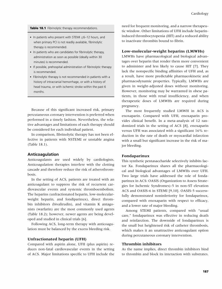
BLBK186-Key April 24, 2009 14:42
Cardiology
Table 18.1 Fibrinolytic therapy recommendations.
� In patients who present with STEMI ≤6–12 hours, and
when primary PCI is not readily available, fibrinolytic
therapy is recommended.� In patients who are candidates for fibrinolytic therapy,
administration as soon as possible (ideally within 30
minutes) is recommended.� If possible, prehospital administration of fibrinolytic therapy
is recommended.� Fibrinolytic therapy is not recommended in patients with a
history of intracranial hemorrhage, or with a history of
head trauma, or with ischemic stroke within the past 6
months.
Because of this significant increased risk, primary
percutaneous coronary intervention is preferred when
performed in a timely fashion. Nevertheless, the rela-
tive advantages and limitations of each therapy should
be considered for each individual patient.
In comparison, fibrinolytic therapy has not been ef-
fective in patients with NSTEMI or unstable angina
(Table 18.1).
AnticoagulationAnticoagulants are used widely by cardiologists.
Anticoagulation therapies interfere with the clotting
cascade and therefore reduce the risk of atherothrom-
bosis.
In the setting of ACS, patients are treated with an
anticoagulant to suppress the risk of recurrent car-
diovascular events and systemic thromboembolism.
The heparins (unfractionated heparin, low-molecular-
weight heparin, and fondiparinux), direct throm-
bin inhibitors (bivalirudin), and vitamin K antago-
nists (warfarin) are the most commonly used agents
(Table 18.2); however, newer agents are being devel-
oped and studied in clinical trials [6].
Following ACS, long-term therapy with anticoagu-
lation must be balanced by the excess bleeding risk.
Unfractionated heparin (UFH)Compared with aspirin alone, UFH (plus aspirin) re-
duces non-fatal cardiovascular events in the setting
of ACS. Major limitations specific to UFH include the
need for frequent monitoring, and a narrow therapeu-
tic window. Other limitations of UFH include heparin-
induced thrombocytopenia (HIT) and a reduced ability
to inactivate thrombin bound to fibrin.
Low-molecular-weight heparins (LMWHs)LMWHs have pharmacological and biological advan-
tages over heparin that render them more convenient
to administer and less likely to cause HIT [7]. They
lack the nonspecific binding affinities of UFH and, as
a result, have more predictable pharmacokinetic and
pharmacodynamic properties. Typically, LMWHs are
given in weight-adjusted doses without monitoring.
However, monitoring may be warranted in obese pa-
tients, in those with renal insufficiency, and when
therapeutic doses of LMWHs are required during
pregnancy.
The most frequently studied LMWH in ACS is
enoxaparin. Compared with UFH, enoxaparin pro-
vides clinical benefit. In a meta-analysis of 12 ran-
domized trials in the setting of ACS [8], enoxaparin
versus UFH was associated with a significant 16% re-
duction in the rate of death or myocardial infarction
with a small but significant increase in the risk of ma-
jor bleeding.
FondaparinuxThis synthetic pentasaccharide selectively inhibits fac-
tor Xa. Fondaparinux shares all the pharmacologi-
cal and biological advantages of LMWHs over UFH.
Two large trials have addressed the role of fonda-
parinux in ACS: OASIS (Organization to Assess Strate-
gies for Ischemic Syndromes)-5 in non-ST elevation
ACS and OASIS-6 in STEMI [9,10]. OASIS-5 success-
fully demonstrated noninferiority for fondaparinux,
compared with enoxaparin with respect to efficacy,
and a lower rate of major bleeding.
Among STEMI patients, compared with “usual
care,” fondaparinux was effective in reducing death
and reinfarction. The downside of fondaparinux is
the small but heightened risk of catheter thrombosis,
which makes it an unattractive anticoagulant option
during percutaneous coronary intervention.
Thrombin inhibitorsAs the name implies, direct thrombin inhibitors bind
to thrombin and block its interaction with substrates.
187
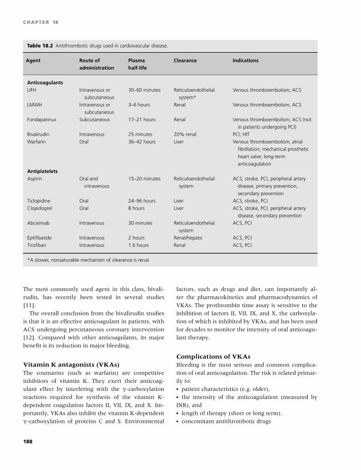
BLBK186-Key April 24, 2009 14:42
CHAPTER 18
Table 18.2 Antithrombotic drugs used in cardiovascular disease.
Agent Route of Plasma Clearance Indicationsadministration half-life
AnticoagulantsUFH Intravenous or
subcutaneous
30–60 minutes Reticuloendothelial
system*
Venous thromboembolism; ACS
LMWH Intravenous or
subcutaneous
3–6 hours Renal Venous thromboembolism; ACS
Fondaparinux Subcutaneous 17–21 hours Renal Venous thromboembolism; ACS (not
in patients undergoing PCI)
Bivalirudin Intravenous 25 minutes 20% renal PCI; HIT
Warfarin Oral 36–42 hours Liver Venous thromboembolism; atrial
fibrillation; mechanical prosthetic
heart valve; long-term
anticoagulation
AntiplateletsAspirin Oral and
intravenous
15–20 minutes Reticuloendothelial
system
ACS, stroke, PCI, peripheral artery
disease, primary prevention,
secondary prevention
Ticlopidine Oral 24–96 hours Liver ACS, stroke, PCI
Clopidogrel Oral 8 hours Liver ACS, stroke, PCI, peripheral artery
disease, secondary prevention
Abciximab Intravenous 30 minutes Reticuloendothelial
system
ACS, PCI
Eptifibatide Intravenous 2 hours Renal/hepatic ACS, PCI
Tirofiban Intravenous 1.6 hours Renal ACS, PCI
*A slower, nonsaturable mechanism of clearance is renal.
The most commonly used agent in this class, bivali-
rudin, has recently been tested in several studies
[11].
The overall conclusion from the bivalirudin studies
is that it is an effective anticoagulant in patients, with
ACS undergoing percutaneous coronary intervention
[12]. Compared with other anticoagulants, its major
benefit is its reduction in major bleeding.
Vitamin K antagonists (VKAs)The coumarins (such as warfarin) are competitive
inhibitors of vitamin K. They exert their anticoag-
ulant effect by interfering with the γ-carboxylation
reactions required for synthesis of the vitamin K-
dependent coagulation factors II, VII, IX, and X. Im-
portantly, VKAs also inhibit the vitamin K-dependent
γ-carboxylation of proteins C and S. Environmental
factors, such as drugs and diet, can importantly al-
ter the pharmacokinetics and pharmacodynamics of
VKAs. The prothrombin time assay is sensitive to the
inhibition of factors II, VII, IX, and X, the carboxyla-
tion of which is inhibited by VKAs, and has been used
for decades to monitor the intensity of oral anticoagu-
lant therapy.
Complications of VKAsBleeding is the most serious and common complica-
tion of oral anticoagulation. The risk is related primar-
ily to:� patient characteristics (e.g. older),� the intensity of the anticoagulation (measured by
INR), and� length of therapy (short or long term).� concomitant antithrombotic drugs
188
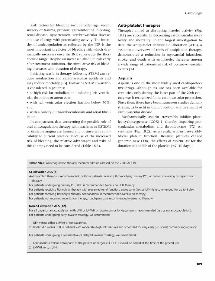
BLBK186-Key April 24, 2009 14:42
Cardiology
Risk factors for bleeding include older age, recent
surgery or trauma, previous gastrointestinal bleeding,
renal disease, hypertension, cerebrovascular disease,
and use of drugs with potentiating activity. The inten-
sity of anticoagulation as reflected by the INR is the
most important predictor of bleeding risk which dra-
matically increases once the INR supersedes the ther-
apeutic range. Despite an increased absolute risk early
after treatment initiation, the cumulative risk of bleed-
ing increases with duration of therapy.
Initiating warfarin therapy following STEMI can re-
duce reinfarction and cerebrovascular accidents and
may reduce mortality [13]. Following STEMI, warfarin
is considered in patients:� at high risk for embolization, including left ventric-
ular thrombus or aneurysm;� with left ventricular ejection fraction below 30%;
and� with a history of thromboembolism and atrial fibril-
lation.
In comparison, data concerning the possible role of
oral anticoagulation therapy with warfarin in NSTEMI
or unstable angina are limited and of uncertain appli-
cability to current practice. Because of the increased
risk of bleeding, the relative advantages and risks of
this therapy need to be considered (Table 18.3).
Anti-platelet therapiesTherapies aimed at disrupting platelet activity (Fig.
18.1) are successful in decreasing cardiovascular mor-
bidity and mortality. In the largest investigation to
date, the Antiplatelet Trialists’ Collaboration (ATC), a
systematic overview of trials of antiplatelet therapy,
demonstrated a reduction in myocardial infarction,
stroke, and death with antiplatelet therapies among
a wide range of patients at risk of occlusive vascular
events [14].
AspirinAspirin is one of the most widely used cardioprotec-
tive drugs. Although its use has been available for
centuries, only during the latter part of the 20th cen-
tury was it recognized for its cardiovascular protection.
Since then, there have been numerous studies demon-
strating its benefit in the prevention and treatment of
cardiovascular disease.
Mechanistically, aspirin irreversibly inhibits plate-
let cyclooxygenase (COX)-1, thereby impairing pro-
staglandin metabolism and thromboxane (TX) A2
synthesis (Fig. 18.2). As a result, aspirin irreversibly
blocks platelet function. Because platelets cannot
generate new COX, the effects of aspirin last for the
duration of the life of the platelet (≈7–10 days).
Table 18.3 Anticoagulation therapy recommendations (based on the 2008 ACCP).
ST elevation ACS [5]Antithrombin therapy is recommended for those patients receiving thrombolysis, primary PCI, or patients receiving no reperfusion
therapy.
For patients undergoing primary PCI, UFH is recommended (versus no UFH therapy).
For patients receiving fibrinolytic therapy with preserved renal function, enoxaparin (versus UFH) is recommended for up to 8 days.
For patients receiving fibrinolytic therapy, fondaparinux is recommended (versus no therapy).
For patients not receiving reperfusion therapy, fondaparinux is recommended (versus no therapy).
Non-ST elevation ACS [12]For all patients, anticoagulation with UFH or LMWH or bivalirudin or fondaparinux is recommended (versus no anticoagulation).
For patients undergoing early invasive strategy, we recommend:
1. UFH versus either LMWH or fondaparinux.
2. Bivalirudin versus UFH in patients with moderate–high risk features and scheduled for very early (<6 hours) coronary angiography.
For patients undergoing a conservative or delayed invasive strategy, we recommend:
1. Fondaparinux versus enoxaparin (if the patient undergoes PCI, UFH should be added at the time of the procedure).
2. LMWH versus UFH.
189
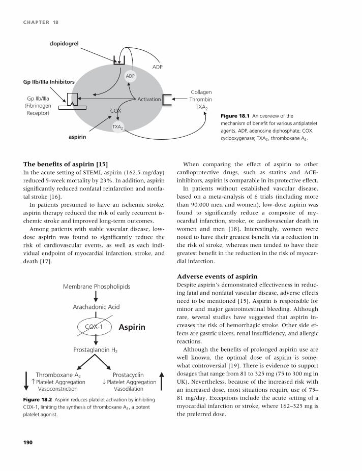
BLBK186-Key April 24, 2009 14:42
CHAPTER 18
CollagenThrombin
TXA2
ADP
(FibrinogenReceptor)
clopidogrel
TXA2
ADP
Gp IIb/IIIa Activation
COX
aspirin
Gp IIb/IIIa Inhibitors
Figure 18.1 An overview of the
mechanism of benefit for various antiplatelet
agents. ADP, adenosine diphosphate; COX,
cyclooxygenase; TXA2, thromboxane A2.
The benefits of aspirin [15]In the acute setting of STEMI, aspirin (162.5 mg/day)
reduced 5-week mortality by 23%. In addition, aspirin
significantly reduced nonfatal reinfarction and nonfa-
tal stroke [16].
In patients presumed to have an ischemic stroke,
aspirin therapy reduced the risk of early recurrent is-
chemic stroke and improved long-term outcomes.
Among patients with stable vascular disease, low-
dose aspirin was found to significantly reduce the
risk of cardiovascular events, as well as each indi-
vidual endpoint of myocardial infarction, stroke, and
death [17].
Membrane Phospholipids
Arachadonic Acid
Prostaglandin H2
COX-1
Thromboxane A2
↑ Platelet AggregationVasoconstriction
Prostacyclin↓ Platelet Aggregation
Vasodilation
Aspirin
Figure 18.2 Aspirin reduces platelet activation by inhibiting
COX-1, limiting the synthesis of thromboxane A2, a potent
platelet agonist.
When comparing the effect of aspirin to other
cardioprotective drugs, such as statins and ACE-
inhibitors, aspirin is comparable in its protective effect.
In patients without established vascular disease,
based on a meta-analysis of 6 trials (including more
than 90,000 men and women), low-dose aspirin was
found to significantly reduce a composite of my-
ocardial infarction, stroke, or cardiovascular death in
women and men [18]. Interestingly, women were
noted to have their greatest benefit via a reduction in
the risk of stroke, whereas men tended to have their
greatest benefit in the reduction in the risk of myocar-
dial infarction.
Adverse events of aspirinDespite aspirin’s demonstrated effectiveness in reduc-
ing fatal and nonfatal vascular disease, adverse effects
need to be mentioned [15]. Aspirin is responsible for
minor and major gastrointestinal bleeding. Although
rare, several studies have suggested that aspirin in-
creases the risk of hemorrhagic stroke. Other side ef-
fects are gastric ulcers, renal insufficiency, and allergic
reactions.
Although the benefits of prolonged aspirin use are
well known, the optimal dose of aspirin is some-
what controversial [19]. There is evidence to support
dosages that range from 81 to 325 mg (75 to 300 mg in
UK). Nevertheless, because of the increased risk with
an increased dose, most situations require use of 75–
81 mg/day. Exceptions include the acute setting of a
myocardial infarction or stroke, where 162–325 mg is
the preferred dose.
190
BLBK186-Key April 24, 2009 14:42
Cardiology
Table 18.4 Antiplatelet therapy recommendations (based on the 2008 ACCP).
ST elevation ACSFor all patients, we recommend aspirin (162–325 mg) versus no aspirin at initial evaluation, and this should be followed indefinitely
(75–162 mg/day).
For all patients, we recommend clopidogrel in addition to aspirin.
In patients undergoing primary PCI, an initial loading dose of at least 300 mg should be used.
In patients receiving fibrinolytic agents or conservative therapy, 300 mg should be used in patients <75 years (75 mg should be
used in patients >75):
GPIIb/IIIa inhibitors should not be used with fibrinolytic therapy,
For patients undergoing primary PCI, we recommend the use of abciximab.
Non-ST elevation ACSFor all patients without a clear allergy to aspirin, we recommend aspirin (162–325 mg) at initial evaluation, and this should be
followed indefinitely (75–162 mg/day).
For all patients with an aspirin allergy, we recommend immediate treatment with clopidogrel (300 mg load) followed by 75 mg daily.
For patients at moderate or greater risk and who will undergo an early invasive strategy, we recommend:
1. Early treatment with clopidogrel or a small-molecule IV GPIIb/IIIa inhibitor,
2. Both early clopidogrel or a small-molecule IV GPIIb/IIIa inhibitor.
For patients at moderate or greater risk and who will undergo a conservative or a delayed invasive strategy, we recommend:
1. Early treatment with clopidogrel,
2. Both early clopidogrel or a small-molecule IV GPIIb/IIIa inhibitor.
For patients who undergo PCI, we recommend both clopidogrel and an IV GPIIb/IIIa inhibitor
ThienopyridinesThe thienopyridines ticlopidine and clopidogrel in-
hibit adenosine diphosphate (ADP) receptor-mediated
platelet activation; they are more potent platelet in-
hibitors than aspirin. Because ticlopidine has been
associated with thrombocytopenic purpura and neu-
tropenia, clopidogrel has emerged as the drug of
choice [20].
In randomized trials, clopidogrel has been shown to
reduce cardiovascular events in the treatment of car-
diovascular conditions [15]. This includes the use of
clopidogrel as:� adjunct therapy in the acute management of STEMI,� the invasive and conservative management of ACS
without ST-segment elevation,� as adjunct therapy following percutaneous coronary
intervention (PCI), and� as lone therapy in the secondary prevention of
atherosclerotic heart disease [15].
Recent data suggest that clopidogrel, in addition to
aspirin for primary prevention, is of no additional ben-
efit and only increases the risk of bleeding.
Glycoprotein IIb/IIIa inhibitorsActivation of the platelet-surface glycoprotein (GP)
IIb/IIIa receptor is the final common pathway in the
process leading to platelet aggregation and, eventually,
thrombus formation. The intravenous GPIIb/IIIa rece-
ptor inhibitors have been established as effective ther-
apy for the reduction of ischemic events when used in
both the management of ACS and as adjunctive ther-
apy during PCI [21,22]. Because of its increased po-
tency as an antiplatelet agent, bleeding is a major side
effect that needs to be considered with its use.
Novel antiplatelet therapiesImportant limitations, including response variabil-
ity, irreversible inhibitory effects, and length of time
191
BLBK186-Key April 24, 2009 14:42
CHAPTER 18
required for maximal platelet inhibition, exist among
the current antiplatelet agents, and therefore, a press-
ing need for the development of improved antiplatelet
agents exists. Three ADP receptor antagonists cur-
rently under investigation (prasugrel, AZD6140, and
cangrelor) in clinical trials for the treatment of ACS ap-
pear promising [23]. Prasugrel is a new thienopyridine
compound with a much faster onset of action than
clopidogrel. In the recently reported TRITON trial [24],
prasugrel therapy was associated with significantly re-
duced rates of ischemic events, but with an increased
risk of major bleeding, including fatal bleeding. This
study validated the hypothesis that greater degrees
of adenosine diphosphate-mediated platelet inhibition
are associated with a greater suppression of clinical is-
chemic events. Two direct and reversible P2Y antago-
nists [15], cangrelor, which can only be given intra-
venously, and AZD6140, which can be given orally,
have rapid onset and reversal of platelet inhibition,
which make them attractive alternatives to thienopy-
ridines, especially when rapid inhibition of platelet ag-
gregation or its quick reversal is required (Table 18.4).
Combination therapy of VKA andantiplatelet therapyAlthough mechanistically sound, the combination of
VKAs and antiplatelet therapy has not been convinc-
ingly shown to have a favorable benefit/risk profile
for the long-term management of coronary heart dis-
ease patients. Aspirin increases the risk of warfarin-
associated bleeding. The size of this increased risk de-
pends on the intensity of anticoagulation as well as on
the daily dose of aspirin.
A number of trials have evaluated the efficacy of
warfarin plus aspirin versus aspirin alone following
ACS [25]. A large meta-analysis that evaluated 14 tri-
als involving over 25,000 patients found no significant
difference in the overall risk of myocardial infarction,
stroke, or all-cause mortality, but increased the risk of
major bleeding. However, a combined strategy of as-
pirin plus warfarin at INR values of 2–3 was superior
to aspirin alone in preventing major adverse events.
Importantly, the applicability of these results is not
known in patients treated with aspirin plus clopido-
grel, the currently recommended regimen in most pa-
tients following ACS.
References
1 Fuster V, Stein B, Ambrose JA, Badimon L, Badi-
mon JJ, Chesebro JH. Atherosclerotic plaque rupture
and thrombosis. Evolving concepts. Circulation 1990;82:
II47–59.
2 Croce K, Libby P. Intertwining of thrombosis and
inflammation in atherosclerosis. Curr Opin Hematol
2007;14:55–61.
3 Libby P. Multiple mechanisms of thrombosis complicat-
ing atherosclerotic plaques. Clin Cardiol 2000;23(Suppl
6):VI-3–7.
4 Virmani R, Burke AP, Farb A, Kolodgie FD. Pathol-
ogy of the vulnerable plaque. J Am Coll Cardiol 2006;47:
C13–18.
5 Goodman SG, Menon V, Cannon C, Steg G, Ohman
EM, Harrington RA. Aute ST-Segment Elevation My-
ocardial Infarction: American College of Chest Physi-
cians evience-based clinical practice guidelines (8th edi-
tion). Chest 2008;133:708S–75S.
6 Hirsh J, Bauer K, Donati M, Gould M, Samama M,
Weitz J. Parenteral anticoagulants: American College of
Chest Physicians evience-based clinical practice guide-
lines (8th edition). Chest 2008;133:141S–59S.
7 Weitz JI. Low-molecular-weight heparins. N Engl J Med
1997;337:688–98.
8 Petersen JL, Mahaffey KW, Hasselblad V, et al. Efficacy
and bleeding complications among patients random-
ized to enoxaparin or unfractionated heparin for an-
tithrombin therapy in non-ST-Segment elevation acute
coronary syndromes: a systematic overview. JAMA
2004;292:89–96.
9 Fifth Organization to Assess Strategies in Acute Is-
chemic Syndromes I, Yusuf S, Mehta SR, et al. Compar-
ison of fondaparinux and enoxaparin in acute coronary
syndromes. N Engl J Med 2006;354:1464–76.
10 Yusuf S, Mehta SR, Chrolavicius S, et al. Effects of
fondaparinux on mortality and reinfarction in patients
with acute ST-segment elevation myocardial infarc-
tion: the OASIS-6 randomized trial. JAMA 2006;295:
1519–30.
11 Direct Thrombin Inhibitor Trialists’ Collaborative G. Di-
rect thrombin inhibitors in acute coronary syndromes:
principal results of a meta-analysis based on individual
patients’ data. Lancet 2002;359:294–302.
12 Harrington RA, Becker RC, Cannon C, et al. Antithrom-
botic therapy for non-ST-segment elevation acute coro-
nary syndromes: American College of Chest Physicians
evience-based clinical practice guidelines (8th edition).
Chest 2008;133:670S–707S.
192
BLBK186-Key April 24, 2009 14:42
Cardiology
13 Smith P, Arnesen H, Holme I. The effect of warfarin on
mortality and reinfarction after myocardial infarction.
N Engl J Med 1990;323:147–52.
14 Antithrombotic Trialists C. Collaborative meta-analysis
of randomised trials of antiplatelet therapy for preven-
tion of death, myocardial infarction, and stroke in high
risk patients. Br Med J 2002;324:71–86.
15 Patrono C, Baigent C, Hirsh J, Roth G. Antiplatelet
drugs: American College of Chest Physicians evience-
based clinical practice guidelines (8th edition). Chest
2008;133:199S–233S.
16 Anonymous. Randomised trial of intravenous streptok-
inase, oral aspirin, both, or neither among 17,187 cases
of suspected acute myocardial infarction: ISIS-2. ISIS-2
(Second International Study of Infarct Survival) Collab-
orative Group. Lancet 1988;2:349–60.
17 Berger JS, Brown DL, Becker RC. Low-dose aspirin
in patients with stable cardiovascular disease: a meta-
analysis. Am J Med 2008;121:43–9.
18 Berger JS, Roncaglioni MC, Avanzini F, Pangrazzi I,
Tognoni G, Brown DL. Aspirin for the primary preven-
tion of cardiovascular events in women and men: a sex-
specific meta-analysis of randomized controlled trials.
JAMA 2006;295:306–13.
19 Campbell CL, Smyth S, Montalescot G, Steinhubl SR.
Aspirin dose for the prevention of cardiovascular dis-
ease: a systematic review. JAMA 2007;297:2018–24.
20 Quinn MJ, Fitzgerald DJ. Ticlopidine and clopidogrel.
Circulation 1999;100:1667–72.
21 Anderson JL, Adams CD, Antman EM, et al. ACC/AHA
2007 guidelines for the management of patients with
unstable angina/non ST-elevation myocardial infarc-
tion: a report of the American College of Cardiology/
American Heart Association Task Force on Practice
Guidelines (Writing Committee to Revise the 2002
Guidelines for the Management of Patients With Unsta-
ble Angina/Non ST-Elevation Myocardial Infarction):
developed in collaboration with the American College
of Emergency Physicians, the Society for Cardiovascu-
lar Angiography and Interventions, and the Society of
Thoracic Surgeons: endorsed by the American Associ-
ation of Cardiovascular and Pulmonary Rehabilitation
and the Society for Academic Emergency Medicine. Cir-
culation 2007;116:e148–304.
22 King SB 3rd, Smith SC Jr, Hirshfeld JW Jr, et al. 2007
focused update of the ACC/AHA/SCAI 2005 guideline
update for percutaneous coronary intervention: a re-
port of the American College of Cardiology/American
Heart Association Task Force on Practice guidelines.
J Am Coll Cardiol 2008;51:172–209.
23 Weitz JI, Hirsh J, Samama M. New antithrombotic
drugs: American College of Chest Physicians evience-
based clinical practice guidelines (8th edition). Chest
2008;133:234S–56S.
24 Wiviott SD, Braunwald E, McCabe CH, et al. Prasugrel
versus clopidogrel in patients with acute coronary syn-
dromes. N Engl J Med 2007;357:2001–15.
25 Andreotti F, Testa L, Biondi-Zoccai GG, Crea F. As-
pirin plus warfarin compared to aspirin alone after
acute coronary syndromes: an updated and compre-
hensive meta-analysis of 25,307 patients. Eur Heart J
2006;27:519–26.
193
BLBK186-Key April 24, 2009 14:39
19 Cardiothoracic surgeryDenise O’Shaughnessy and Ravi Gill
The importance for surgeons to understand the nor-
mal hemostatic mechanisms cannot be overempha-
sized. Hemostasis is a balance protecting the integrity
of the vascular system after tissue injury and main-
taining the fluidity of blood. Excessive bleeding can be
due to surgical causes, a derangement of hemostasis,
or, more often, a combination of both, of which car-
diothoracic surgery is a prime example [1].
As the incidence of heart disease continues to rise,
the consequent demand for coronary artery bypass
surgery also increases: with 400,000 in USA, over
100,000 in Europe, and 30,000 in UK per annum.
Most of these procedures, together with major heart
surgery on congenital defects and valvular heart dis-
ease, are performed on beating hearts supported by
cardiopulmonary bypass (CPB).
During surgery on the heart, it is common to stop
the heart to make it easier to suture the bypass grafts
onto the coronary arteries, which are only 1.5 mm
in diameter. During this time, the function of the
heart and lungs is taken over by a heart–lung or CPB
machine.
The heart can be stopped using several different
methods. In general, a mixture of potassium and
magnesium with some other chemicals is infused into
the coronary arteries. This mixture can be carried in
either blood (preferred) or a clear saline-like solu-
tion. These solutions are called cardioplegia and are
referred to as either blood or crystalloid cardioplegia,
respectively. The heart can also be stopped electrically,
and this is referred to as cardiac arrest with ventricular
fibrillation.
In conventional coronary artery bypass grafting
(CABG), operations are performed after cardioplegic
arrest. The pericardium is usually opened longitudi-
nally to allow unrestricted access to underlying heart
and proximal great vessels. The pericardium is usu-
ally left open. A second incision in the posterior peri-
cardium allows drainage through chest tube.
Cardiac surgery without CPB
CABG can now be performed with or without CPB.
These minimally invasive procedures restore healthy
blood flow to the heart without having to stop the
beating heart [2].
It was thought that off-pump coronary artery by-
pass (OPCAB) would have a lower risk of complica-
tions, such as stroke, acute lung injury, renal dysfunc-
tion, neurocognitive outcome, and tranfusion rates.
Although observational data have supported this, ran-
domized clinical trials have proved disappointing.
Performing surgery on a beating heart is technically
more difficult than working on a heart that has been
stopped with the help of the heart–lung machine. In
addition, the stress on the heart during the procedure
may lead to more heart muscle damage, lower blood
pressure, irregular heartbeat, and potentially, brain in-
jury if blood flow to the brain is reduced for too long
during surgery. In some cases (usually �10%), it is
necessary to convert to conventional CABG methods
on an emergency basis.
Currently there are three methods used:
Minimally invasive direct coronary arterybypass (MIDCAB)This procedure is for patients with blockage(s) in the
arteries on the front of the heart [the left anterior de-
scending (LAD) artery and its branches]. A small inci-
sion is made on the patient’s left chest to expose the
heart. After muscles in the area are pushed apart and
a small part of the front of the rib (costal cartilage) is
removed, the surgeon temporarily closes off the artery
194
BLBK186-Key April 24, 2009 14:39
Cardiothoracic surgery
that lies underneath and frees its lower end. An
opening is made in the pericardium, and a device is
attached to the heart to reduce its movement. Finally,
the surgeon connects the artery below the blockage
to the LAD artery or one of its branches. The procure
takes 2–3 hours.
Unfortunately, due to the limited size of the inci-
sion, the procedure is limited to only a few patients
who have a blockage in one or two coronary arteries
located on the front side of the heart, whether healthy
or considered too high-risk for conventional bypass
surgery or balloon angioplasty. However, for younger
patients, for those who have small coronary arteries
and need several bypasses, or for those whose heart
will not tolerate being manipulated during the proce-
dure, it may be preferable to use the traditional CABG
technique.
Off-pump coronary artery bypass (OPCAB)During this procedure, the chest is opened and grafts
harvested conventionally. Like the MIDCAB proce-
dure, a device is used to restrict movement of parts of
the heart so that the surgeon can operate on it while it
is still beating. The surgeon can repair four to five ves-
sels on the beating heart during the same procedure.
This procedure also takes 3–4 hours.
OPCAB has grown significantly because of its ad-
vantages over other procedures, such as fewer blood
transfusions, possible decreased risk of stroke, shorter
stay in the hospital, and return to normal activities
more rapidly. OPCAB is suitable for patients with
poor heart function (very low ejection fraction), se-
vere lung disease (chronic obstructive pulmonary dis-
ease, COPD, and emphysema), and acute or chronic
kidney disease. It is also suitable for those at high risk
for stroke or for those who have a calcified aorta.
Robot-assisted coronary arterybypass (RACAB)RACAB is the latest advance in heart surgery. Sur-
geons use a robot to perform the bypass. The breast-
bone does not need to be split open at all. Surgeons
do not have direct contact with the patient, perform-
ing the operation while watching a videoscreen. As the
technology becomes more advanced, the surgeon may
perform coronary bypass from a distant site (i.e. from
another room or another geographical location).
Trainee beating heart surgeonUntil a surgeon has performed up to 50 OPCAB proce-
dures, he/she is advised to avoid: cardiomegaly, small
or diffusely diseased vessels, hemodynamically unsta-
ble patients, critical left main disease, recent myocar-
dial infarction, or severe left ventricular dysfunction
[left ventricular ejection fraction (LVEF) �35%].
Expert beating heart surgeonWith experience, OPCAB can be performed safely in
the vast majority of cases (�90%). However, it is not
advisable to perform OPCAB if multiple unfavorable
characteristics are present (e.g. cardiomegaly in a pa-
tient with LVEF 25% and small targets).
Anticoagulation used
The heparin dose (1–1.5 mg/kg) is one-third of the
standard dose for CPB. The target activated clotting
time (ACT) is �300 seconds. The ACT should be
checked every 30 minutes with heparin supplemented
as needed. Heparin reversal is not mandatory; some
centers administer one-half the calculated protamine
dose.
Cardiac surgery without CPB versuscardiac surgery with CPB
In a series of 17,401 isolated CABGs performed in Dal-
las, Texas, 7283 (41.9%) were OPCABs and 10,118
(58.1%) were conventional coronary artery bypass
with CPB [3]. Factors determining selection of patients
for OPCAB included female gender, pre-existing re-
nal failure, and reoperations. Operative mortality was
2.8%.
Published data from the UK cardiac database in the
financial years 2002–2003 (n = 56,065) demonstrated
that �20% of CABG were performed off-pump, most
being performed at 5 leading hospitals: St Marys,
London (40%), Harefield Hospital, Sutton and Bris-
tol Royal Infirmary (38%), Manchester, and Liverpool
(30%).
Cardiopulmonary bypass
In 2002, 80% of all CABG surgery was performed on
CPB; the figure now, in 2008, is still over 70%.
195
BLBK186-Key April 24, 2009 14:39
CHAPTER 19
During CPB, blood is drained from the right atrium
and returned to the aorta, creating a bloodless field
for the cardiothoracic surgeon. This is achieved by ad-
ministrating high doses of heparin to anticoagulate pa-
tients (monitored using the ACT or anti-Xa levels),
and residual heparin is reversed by protamine at the
end of surgery.
The process of CPB:� activates fibrinolysis,� disturbs platelet function,� often reduces the platelet count, and� reduces the concentration of clotting factors.
Reduction in volume of the CPB circuit and im-
provements in operative techniques, together with
cell salvage and the use of antifibrinolytic drugs,
have reduced the need for transfusion. Recent “near
patient” coagulation testing devices have enabled
much of this progress and include the Haemoscope
Thrombelastograph R© (TEG R©).
Bleeding is usually manifest postoperatively, after
protamine reversal of heparin, and shed into the medi-
astinal and pleural drains. There are two main causes
of peri-operative bleeding:� Surgical, due to failure to secure hemostasis at the
operative site.� Nonsurgical, due to failure of hemostatic pathways,
and principally due to:
1 The procedure itself, in this case CPB (the circuit
and its effect on hemostasis);
2 Incomplete reversal of heparin by protamine;
3 Antiplatelet drugs (Aspirin, Clopidrogel, IIb/IIIa in-
hibitors);
4 A pre-existing bleeding disorder (e.g. hemophilia,
von Willebrand disease); or
5 Oral anticoagulation that has not been reversed
completely.
Critical rates of blood loss are 500 mL in the first
hour, 800 mL at 2 hours, 900 mL at 3 hours, 1000 mL
at 4 hours, and 1200 mL by 5 hours.
The CPB circuit
Bigelow showed in dogs that circulatory arrest (CA)
was possible, allowing simple operations without cir-
culatory support, but only for 15 minutes. Originally
invented by Gibbon in the 1930s, the pump oxygena-
tor only worked successfully in the 1950s. Even then,
only one in four cases survived and 14–25 L of fresh
blood prime was required. At the same time, Lillehei
connected a patient to a volunteer donor (parent). He
drained the blood from the superior vena cava (SVC)
of the patient, and pumped this blood into the femoral
vein of the donor. Blood was then returned from the
femoral artery of the donor to the carotid artery of the
patient. Forty-five patients (mostly children) had op-
erations. The 63% survival, despite no reliable ventila-
tors, blood gas or electrolyte analysis, pacemakers, or
defibrillators, was remarkable. However, this was not
a long-term solution.
The Gibbon Mayo pump in 1955 had bubble oxy-
genators, high-flow total cardiopulmonary support,
but still required 10–14 U fresh blood prime. Adapta-
tions over the next 60 years have reduced adult prime
volumes to 1.5–2.5 L crystalloid and pediatric prime
volumes to 400–1000 mL prime including some blood
(depending on the size of the child), such that on by-
pass the hematocrit will not fall below 20%.
A representation of current CPB is shown in
Fig. 19.1.
Hemostasis in CPBHemostasis is a dynamic and extremely complex pro-
cess, involving many interactive factors.These include
coagulation and fibrinolytic proteins, activators, in-
hibitors, and cellular elements (e.g. platelet cytoskele-
ton, cytoplasmic granules, and cell surfaces), as de-
scribed in Chapter 1.
In order to measure any degree of hemostatic im-
balance, we need to have the ability to measure the
net product of the interactions, which is the three-
dimensional clot matrix. Once the coagulation cascade
is activated, thrombin is formed.� Thrombin will cleave soluble fibrinogen into fib-
rin monomers, which polymerize to form protofibril
strands and then undergo linear extension, branching,
and lateral association, leading to the formation of a
three-dimensional matrix of fibrin.� This matrix is given rigidity by the anchoring platelet
network, thus allowing resistance to shear. Platelet
glycoprotein receptors (GPIIb/IIIa) bind the polymer-
ized fibrin network to the actin cytoskeleton of the
platelet. Actin is a muscle protein that has the abil-
ity to transmit contractility force, which is the major
contributor to clot strength.
196
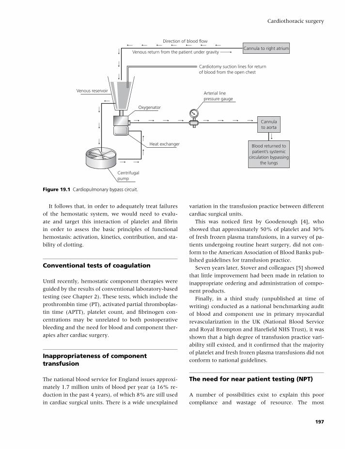
BLBK186-Key April 24, 2009 14:39
Cardiothoracic surgery
Venous return from the patient under gravity
Cardiotomy suction lines for returnof blood from the open chest
Arterial linepressure gauge
Oxygenator
Venous reservoir
Heat exchanger
Centrifugalpump
Direction of blood flow
Cannulato aorta
Cannula to right atrium
Blood returned topatient’s systemic
circulation bypassingthe lungs
Figure 19.1 Cardiopulmonary bypass circuit.
It follows that, in order to adequately treat failures
of the hemostatic system, we would need to evalu-
ate and target this interaction of platelet and fibrin
in order to assess the basic principles of functional
hemostasis: activation, kinetics, contribution, and sta-
bility of clotting.
Conventional tests of coagulation
Until recently, hemostatic component therapies were
guided by the results of conventional laboratory-based
testing (see Chapter 2). These tests, which include the
prothrombin time (PT), activated partial thromboplas-
tin time (APTT), platelet count, and fibrinogen con-
centrations may be unrelated to both postoperative
bleeding and the need for blood and component ther-
apies after cardiac surgery.
Inappropriateness of componenttransfusion
The national blood service for England issues approxi-
mately 1.7 million units of blood per year (a 16% re-
duction in the past 4 years), of which 8% are still used
in cardiac surgical units. There is a wide unexplained
variation in the transfusion practice between different
cardiac surgical units.
This was noticed first by Goodenough [4], who
showed that approximately 50% of platelet and 30%
of fresh frozen plasma transfusions, in a survey of pa-
tients undergoing routine heart surgery, did not con-
form to the American Association of Blood Banks pub-
lished guidelines for transfusion practice.
Seven years later, Stover and colleagues [5] showed
that little improvement had been made in relation to
inappropriate ordering and administration of compo-
nent products.
Finally, in a third study (unpublished at time of
writing) conducted as a national benchmarking audit
of blood and component use in primary myocardial
revascularization in the UK (National Blood Service
and Royal Brompton and Harefield NHS Trust), it was
shown that a high degree of transfusion practice vari-
ability still existed, and it confirmed that the majority
of platelet and fresh frozen plasma transfusions did not
conform to national guidelines.
The need for near patient testing (NPT)
A number of possibilities exist to explain this poor
compliance and wastage of resource. The most
197
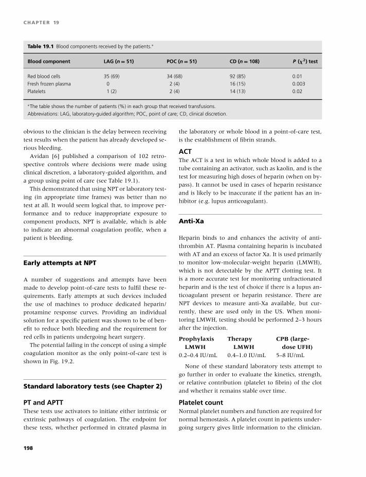
BLBK186-Key April 24, 2009 14:39
CHAPTER 19
Table 19.1 Blood components received by the patients.∗
Blood component LAG (n = 51) POC (n = 51) CD (n = 108) P (�2) test
Red blood cells 35 (69) 34 (68) 92 (85) 0.01
Fresh frozen plasma 0 2 (4) 16 (15) 0.003
Platelets 1 (2) 2 (4) 14 (13) 0.02
∗The table shows the number of patients (%) in each group that received transfusions.
Abbreviations: LAG, laboratory-guided algorithm; POC, point of care; CD, clinical discretion.
obvious to the clinician is the delay between receiving
test results when the patient has already developed se-
rious bleeding.
Avidan [6] published a comparison of 102 retro-
spective controls where decisions were made using
clinical discretion, a laboratory-guided algorithm, and
a group using point of care (see Table 19.1).
This demonstrated that using NPT or laboratory test-
ing (in appropriate time frames) was better than no
test at all. It would seem logical that, to improve per-
formance and to reduce inappropriate exposure to
component products, NPT is available, which is able
to indicate an abnormal coagulation profile, when a
patient is bleeding.
Early attempts at NPT
A number of suggestions and attempts have been
made to develop point-of-care tests to fulfil these re-
quirements. Early attempts at such devices included
the use of machines to produce dedicated heparin/
protamine response curves. Providing an individual
solution for a specific patient was shown to be of ben-
efit to reduce both bleeding and the requirement for
red cells in patients undergoing heart surgery.
The potential failing in the concept of using a simple
coagulation monitor as the only point-of-care test is
shown in Fig. 19.2.
Standard laboratory tests (see Chapter 2)
PT and APTTThese tests use activators to initiate either intrinsic or
extrinsic pathways of coagulation. The endpoint for
these tests, whether performed in citrated plasma in
the laboratory or whole blood in a point-of-care test,
is the establishment of fibrin strands.
ACTThe ACT is a test in which whole blood is added to a
tube containing an activator, such as kaolin, and is the
test for measuring high doses of heparin (when on by-
pass). It cannot be used in cases of heparin resistance
and is likely to be inaccurate if the patient has an in-
hibitor (e.g. lupus anticoagulant).
Anti-Xa
Heparin binds to and enhances the activity of anti-
thrombin AT. Plasma containing heparin is incubated
with AT and an excess of factor Xa. It is used primarily
to monitor low-molecular-weight heparin (LMWH),
which is not detectable by the APTT clotting test. It
is a more accurate test for monitoring unfractionated
heparin and is the test of choice if there is a lupus an-
ticoagulant present or heparin resistance. There are
NPT devices to measure anti-Xa available, but cur-
rently, these are used only in the US. When moni-
toring LMWH, testing should be performed 2–3 hours
after the injection.
Prophylaxis Therapy CPB (large-LMWH LMWH dose UFH)
0.2–0.4 IU/mL 0.4–1.0 IU/mL 5–8 IU/mL
None of these standard laboratory tests attempt to
go further in order to evaluate the kinetics, strength,
or relative contribution (platelet to fibrin) of the clot
and whether it remains stable over time.
Platelet countNormal platelet numbers and function are required for
normal hemostasis. A platelet count in patients under-
going surgery gives little information to the clinician.
198
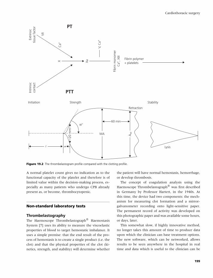
BLBK186-Key April 24, 2009 14:39
Cardiothoracic surgery
Initiation Strength Stability
Retraction
Lysisr
α°
20 m
m
k
60 min
MA A60
PT
PTT
Fibrin polymer+ plateletsX X
a
V, C
a+
Ca+
VII
Extr
insi
ctis
sue
fact
orIn
trin
sic
cont
act
Fibr
in m
onom
erC
a+ , X
III
Figure 19.2 The thrombelastogram profile compared with the clotting profile.
A normal platelet count gives no indication as to the
functional capacity of the platelet and therefore is of
limited value within the decision-making process, es-
pecially as many patients who undergo CPB already
present as, or become, thrombocytopenic.
Non-standard laboratory tests
ThrombelastographyThe Haemoscope Thrombelastograph R© Haemostasis
System [7] uses its ability to measure the viscoelastic
properties of blood to target hemostatic imbalance. It
uses a simple premise: that the end result of the pro-
cess of hemostasis is to create a single product (i.e. the
clot) and that the physical properties of the clot (ki-
netics, strength, and stability) will determine whether
the patient will have normal hemostasis, hemorrhage,
or develop thrombosis.
The concept of coagulation analysis using the
Haemoscope Thrombelastograph R© was first described
in Germany by Professor Hartert, in the 1940s. At
this time, the device had two components: the mech-
anism for measuring clot formation and a mirror-
galvanometer recording onto light-sensitive paper.
The permanent record of activity was developed on
this photographic paper and was available some hours,
or days, later.
This somewhat slow, if highly innovative method,
no longer takes this amount of time to produce data
upon which the clinician can base treatment options.
The new software, which can be networked, allows
results to be seen anywhere in the hospital in real
time and data which is useful to the clinician can be
199
BLBK186-Key April 24, 2009 14:39
CHAPTER 19
obtained within 10–15 minutes. A rigorous quality as-
surance program protects the validity of these results.
However, despite these advancements, the principle of
producing a trace that identifies a number of variables
related to functional disturbances in the hemostatic
system is still key to Thrombelastographic analysis.
Coagulation analysis: definitions ofcoagulation parameters using theThrombelastograph
R = reaction time� Time from sample placement into the cup until the
tracing amplitude reaches 2 mm.� This represents the rate of initial fibrin formation
and is related functionally to plasma clotting factors
and circulating inhibitor activity (i.e. PT and APTT).� Prolongation of the R-value may be a result of coag-
ulation factor deficiencies, anticoagulation (heparin),
or severe hypofibrinogenemia.� A reduced R-value may be present in hypercoagula-
bility syndromes.
K = clot formation time� Measured from R time to the point where the am-
plitude of the tracing reaches 20 mm.� The coagulation time represents the time taken for
a fixed degree of viscoelasticity to be achieved by the
forming clot, as a result of fibrin build-up and cross-
linking.� It is affected by the activity of the clotting factors,
fibrinogen, and platelets.
Alpha angle (α)� This is a line tangent from the point at which clot
formation begins to the peak of the curve. It denotes
speed at which solid clot forms.� Decreased values may occur with hypofibrinogene-
mia and thrombocytopenia affecting platelet function.
Maximum amplitude/G (MA/G)� This is the greatest amplitude of the TEG R© trace and
is a reflection of the absolute strength of the clot. It is
a direct function of the maximum dynamic properties
of the interaction of fibrin and platelets via GP IIb/IIIa,
and has been correlated to platelet aggregometry.� Platelet abnormalities, whether qualitative or, if se-
vere enough, quantitative, substantially disturb the
maximum ampitude (MA). There is a significant, al-
beit complex, relationship between the MA of the
TEG R© trace and the platelet count. A significant
relationship with the MA value and the aggregation
responses to collagen and ADP has also been reported.
Clot lysis� This can be expressed in a number of ways.� Normal clot will retract with time, and thus, a cer-
tain amount of narrowing of the MA can be expected.� The LY30 measures the amount of reduction in
maximum amplitude at 30 minutes and may be pre-
dicted before this using the estimated percent ly-
sis (EPL) parameter. These parameters represent lysis
rather than retraction of the clot.� Both measures reflect an abnormal decrease in am-
plitude as a function of time and reflect loss of clot
integrity as a result of lysis.
A stylized thrombelastography trace is shown in
Fig. 19.2 together with the standard clotting profile.
It is easy to recognise that thrombelastography can
provide information on clot kinetics, strength, and
stability, which are not available with conventional
laboratory-based testing.
A unique attribute of thrombelastography is its abil-
ity to define previously unrecognized changes in cer-
tain clinical scenarios. It can be observed that the trace
develops more vigorously and produces a more stable
and strong clot in certain prothrombotic clinical con-
ditions.
HypercoaguabilityIncreased alpha angle and MA associated with a
shorter R-value can be defined as hypercoagulability.
This is an increasing focus of attention in many fields,
largely due to the recognition of genetic determinants
of increased likelihood of developing a thrombotic dis-
ease process, such as stroke, coronary artery, or deep
vein thrombosis.
Development of inhibitor and activatorreagents for the HaemoscopeTEG R© System
A now common development of thrombelastography
is to use commercially available activator and inhibitor
reagent technology to define specific parts of the coag-
ulation system.
200
BLBK186-Key April 24, 2009 14:39
Cardiothoracic surgery
KaolinThe effects of contact activation (equivalent to APTT)
are assessed using kaolin.
Tissue factorAn equivalent to the PT is performed by the addition
of tissue factor.
HeparinaseThis enzyme is known to convert unfractionated hep-
arin to a relatively inactive form. It therefore allows
the use of thrombelastography to look at the underly-
ing clotting in a patient who is fully heparinized, such
as on CPB. It also reverses the effect of some LMWHs.
PlateletMapping R© (ADP andarachidonic acid)As more pharmacological interventions using platelet
inhibitor agents are becoming evident, thrombelastog-
raphy technology has addressed the issue by using
reagent technology to assess the impact of such inter-
ventions. Two new assays have been developed using
ADP and arachidonic acid agonists, generating mod-
ified MA values that measure the degree of inhibi-
tion caused by these antiplatelet agents (see “Extended
Uses of TEG”).
Functional FibrinogenThis reagent contains a monoclonal antibody to the
glycoprotein IIb/IIIa receptors on the platelet surface.
When it is added to the system, it inhibits the platelet
component (80%) of the clot strength shown in the
MA, revealing the functional fibrinogen element of
the clot (normally 20%). The results have been cor-
related to the laboratory gold standard Clauss method
of fibrinogen determination and are generated auto-
matically by the software.
RapidTEGTM
This contains both tissue factor and kaolin to fully acti-
vate the sample and produces an ACT correlated result
in seconds. It also allows for a quicker determination
of the MA of a patient.
Thrombelastography-based transfusionalgorithms
Some centers, such as Mount Sinai (New York),
Southampton (UK), and Harefield Hospital (UK), have
incorporated the TEG R© system into their transfusion
algorithms.
The Mount Sinai protocolThe measurements used were partly TEG-based
(celite, with and without heparinase), in conjunction
with platelet count and fibrinogen concentration from
the laboratory [8].� If the R-value in the non-heparinase sample was
greater than twice that found in the heparinase sam-
ple, then the patient was given supplementary pro-
tamine.� If the platelet count was �100,000 and the MA was
�45 mm, then platelets were administered.� Fresh frozen plasma (FFP) was given if the celite ac-
tivated R-value, 10 minutes post protamine adminis-
tration, was �20 mm.� Low fibrinogen was treated with cryoprecipitate.� Episilon aminocoproic acid (amicar) was given in
the event of excess lysis.
Using this protocol, they showed significant reduc-
tions in the use of hemostatic products compared with
their more conventional transfusion protocol.
The Harefield protocolThe concept of a TEG R©-derived algorithm was taken
a stage further, with measurements taken during the
bypass phase in order to predict the need for compo-
nent products [9]. A study was conducted in 60 pa-
tients who were considered to have a higher than av-
erage risk of bleeding and thus the need for hemostatic
products, but were not given aprotinin or tranexemic
acid. They were randomly allocated to have products
ordered and administered based on either a TEG R©-
derived decision tree or the clinicians’ discretion after
the return of conventional laboratory-based testing re-
sults.
Results for the TEG R© trace were available, on av-
erage, 70–90 minutes before conventional tests of
coagulation, fibrinogen, and platelet count. This was
considered significant in terms of logistical appropri-
ateness. The TEG R©-guided group also showed a 50%
reduction in the number of patients given hemostatic
products, with a reduction in the use of FFP from
a total of 16 to 5 U in transfused patients. The use
of platelet concentrates was reduced, with only one
patient receiving a single platelet pool in the TEG R©-
guided group.
201
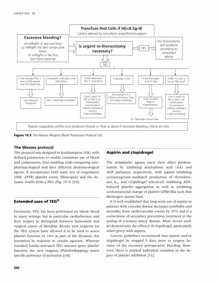
BLBK186-Key April 24, 2009 14:39
CHAPTER 19
Transfuse Red Cells if Hb<8.5g/dl
Is urgent re-thoracotomynecessary?
Excessive bleeding?(unless advised by consultant anaesthelist/surgeon)
NO
YES
For thoracotomyand productsaccording toconsultant
advice
Give 2 units* ofFFP or 500 in of
ProthrombinConcentrate if
volume overload aconcern
[*afm for 8ml/kg]
Give 2 units* ofFFP or 500 in of
ProthrombinConcentrate if
volume overload aconcern
[*afm for 8ml/kg]
>4 ml/kg/hr in any one hour>2 ml/kg/hr for two consecutive
hours>5 ml/kg/hr in the first
four hours post-op
Give 50mg ofProtamine
If the standard TEG ‘r’time is 50% greaterthan the heparinase
If platelets <100,000 or theMA<50nm
If the heparinaseTEG ‘r’ time>9min
If the EPL>7.5%
Give aprotinin 0.5millions units bolux, then
0.5 million units/hourGive 2 pooled
bags ofcryoprecipitate
If the Fibrinogenlevel<1.5g/L
Or’ Fibrinogen concentrate
If INR>1.5 with anormal TEG result
Give 1 adult bag of plateless
Repeat coagulation profile once products infused ⇒ Treat as above if excessive bleeding criteria are met
Figure 19.3 The Wessex Allogenic Blood Transfusion Protocol [10].
The Wessex protocolThis protocol was designed in Southampton (UK) with
defined parameters to enable consistent use of blood
and components, thus enabling trials comparing non-
pharmacological and later different pharmacological
agents. It incorporates both static test of coagulation
(INR, APTR, platelet count, fibrinogen) and the dy-
namic results from a TEG (Fig. 19.3) [10].
Extended uses of TEG R©
Previously, TEG has been performed on whole blood
in many settings, but in particular cardiothoracic and
liver surgery to distinguish between hemostatic and
surgical causes of bleeding. Recent new reagents for
the TEG system have allowed it to be used to assess
platelet function ex vivo as part of the dynamic clot
formation in response to certain agonists. Whereas
standard kaolin-activated TEG assesses gross platelet
function, the new reagents (PlateletMapping) assess
specific pathways of activation [10].
Aspirin and clopidrogel
The antiplatelet agents exert their affect predom-
inately by inhibiting arachadonic acid (AA) and
ADP pathways, respectively, with aspirin inhibiting
cyclooxygenase-mediated production of thrombox-
ane A2, and Clopidrogel selectively inhibiting ADP-
induced platelet aggregation as well as inhibiting
conformational change of platelet GPIIb/IIIa such that
fibrinogen cannot bind.
It is well established that long-term use of aspirin in
patients with vascular disease decreases morbidity and
mortality from cardiovascular events by 25% and is a
cornerstone of secondary prevention treatment in the
setting of coronary artery disease. More recent stud-
ies demonstrate the efficacy of clopidrogel, particularly
when given with aspirin.
Current guidelines recommend that aspirin and/or
clopidrogel be stopped 5 days prior to surgery be-
cause of the excessive perioperative bleeding. How-
ever, there is marked individual variation in the de-
gree of platelet inhibition [11].
202
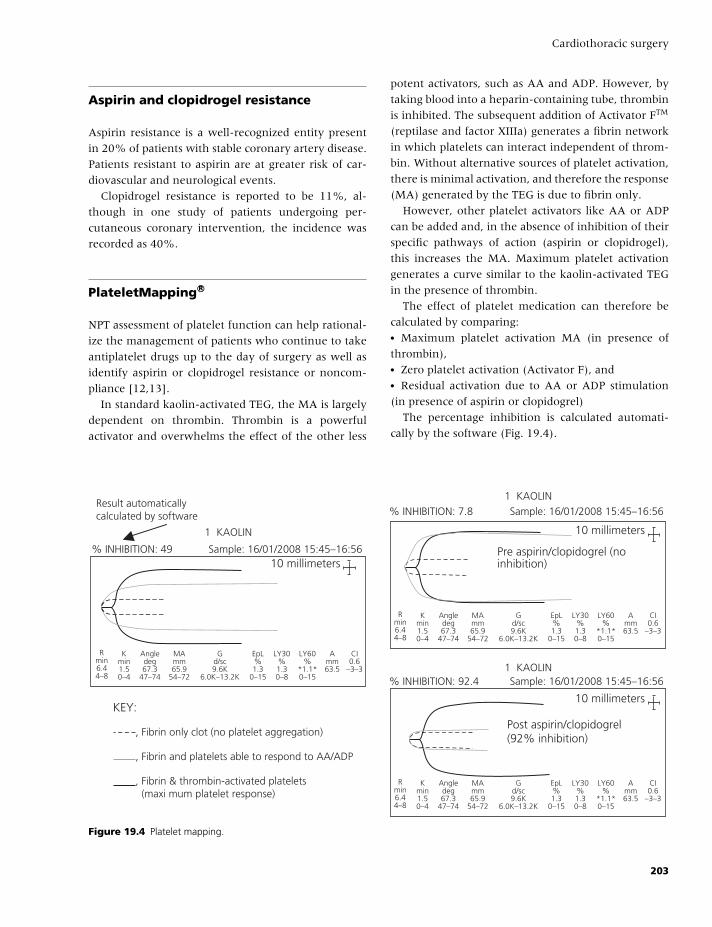
BLBK186-Key April 24, 2009 14:39
Cardiothoracic surgery
Aspirin and clopidrogel resistance
Aspirin resistance is a well-recognized entity present
in 20% of patients with stable coronary artery disease.
Patients resistant to aspirin are at greater risk of car-
diovascular and neurological events.
Clopidrogel resistance is reported to be 11%, al-
though in one study of patients undergoing per-
cutaneous coronary intervention, the incidence was
recorded as 40%.
PlateletMapping R©
NPT assessment of platelet function can help rational-
ize the management of patients who continue to take
antiplatelet drugs up to the day of surgery as well as
identify aspirin or clopidrogel resistance or noncom-
pliance [12,13].
In standard kaolin-activated TEG, the MA is largely
dependent on thrombin. Thrombin is a powerful
activator and overwhelms the effect of the other less
potent activators, such as AA and ADP. However, by
taking blood into a heparin-containing tube, thrombin
is inhibited. The subsequent addition of Activator FTM
(reptilase and factor XIIIa) generates a fibrin network
in which platelets can interact independent of throm-
bin. Without alternative sources of platelet activation,
there is minimal activation, and therefore the response
(MA) generated by the TEG is due to fibrin only.
However, other platelet activators like AA or ADP
can be added and, in the absence of inhibition of their
specific pathways of action (aspirin or clopidrogel),
this increases the MA. Maximum platelet activation
generates a curve similar to the kaolin-activated TEG
in the presence of thrombin.
The effect of platelet medication can therefore be
calculated by comparing:� Maximum platelet activation MA (in presence of
thrombin),� Zero platelet activation (Activator F), and� Residual activation due to AA or ADP stimulation
(in presence of aspirin or clopidogrel)
The percentage inhibition is calculated automati-
cally by the software (Fig. 19.4).
, Fibrin only clot (no platelet aggregation)
, Fibrin and platelets able to respond to AA/ADP
, Fibrin & thrombin-activated platelets (maxi mum platelet response)
Result automaticallycalculated by software
KEY:Post aspirin/clopidogrel(92% inhibition)
Pre aspirin/clopidogrel (noinhibition)
% INHIBITION: 49
% INHIBITION: 7.8
% INHIBITION: 92.4
1 KAOLIN
Sample: 16/01/2008 15:45–16:56
1 KAOLIN
1 KAOLIN
Sample: 16/01/2008 15:45–16:56
Sample: 16/01/2008 15:45–16:56
10 millimeters
Rmin6.44–8
Kmin1.50–4
Angledeg67.3
47–74
MAmm65.9
54–72
Gd/sc9.6K
6.0K–13.2K
EpL%1.3
0–15
LY60%
*1.1*0–15
Amm63.5
CI0.6
–3–3
LY30%1.30–8
Rmin6.44–8
Kmin1.50–4
Angledeg67.3
47–74
MAmm65.9
54–72
Gd/sc9.6K
6.0K–13.2K
EpL%1.3
0–15
LY60%
*1.1*0–15
Amm63.5
CI0.6
–3–3
LY30%1.30–8
10 millimeters
10 millimeters
Rmin6.44–8
Kmin1.50–4
Angledeg67.3
47–74
MAmm65.9
54–72
Gd/sc9.6K
6.0K–13.2K
EpL%1.3
0–15
LY60%
*1.1*0–15
Amm63.5
CI0.6
–3–3
LY30%1.30–8
Figure 19.4 Platelet mapping.
203
BLBK186-Key April 24, 2009 14:39
CHAPTER 19
Blood and hemostatic componentmanagement: future development
It is well recognized that postoperative bleeding
and the subsequent need for reoperation to control
bleeding is associated with an increase in morbidity
and mortality following cardiac surgery. Replacement
therapy using red cells and plasma-based hemostatic
components may themselves be contributors to the
morbidity and mortality.
Clinical indications to reduce exposureThe complex relationship between transfusion, mor-
tality, and morbidity is ill defined. There is emerg-
ing evidence that blood transfusion is an independent
risk factor for death after cardiac surgery. In addition,
platelet transfusion is associated with an increased
risk of organ dysfunction or death from uncertain
causes. Immune modulation may play a role, because
leukodepletion of blood may reduce mortality in the
critically ill adult and neonate. Given the complexity
of these issues, it would seem to be prudent to avoid
transfusion unless necessary and to use simple, safe,
available methods to reduce the chances of patients
needing a transfusion during surgery.
Logistical indications to reduce exposure
The current donor pool is known to be decreasing at
6% per annum, and may well continue to decrease.
This trend is probably multifactorial; however, the
ongoing public debate concerning variant Creutzfeldt-
Jacob disease (vCJD) has to be considered a significant
contributory element. Some estimates put the possi-
ble overall donor reduction at 50% due to the even-
tual inclusion of a screening test for vCJD. It remains
to be seen whether this trend is capable of being re-
versed, even with the advent of increased public rela-
tions awareness and legislative measures introduced
to lower the acceptable donor age limit. This must
be viewed against a projected increase in demand for
blood and hemostatic products of approximately 4.9%
by 2008.
The true role for TEG R© analysis is as a platform for
an integrated approach to hemostasis management.
Information is the key to this whole process, and any
technology that fails to provide relevant information
because of scientific or logistical failures only serves to
further exacerbate an already complex clinical man-
agement task.
Methods to reduce blood loss
� Mechanical strategies [14],� Pharmacological strategies,� Preoperative methods, and� Anesthetic methods.
Pharmacological methodsPharmacotherapy is a component in minimizing blood
loss and transfusion in cardiothoacic surgery. Nothing
beats meticulous surgical technique, but some loss is
inevitable. Both aprotinin and tranexamic acid are an-
tifibrinolytic agents that have been used widely in this
setting to reduce blood loss.
AprotininThis is a nonspecific serine protease inhibitor (inhibits
plasmin at low dose, kallikrien at high dose, and in-
hibits activated protein C and thrombin); in addition
to its antifibrinolytic properties, it may have effects on
preventing platelet activation by blocking the throm-
bin activated protease-activated receptor 1 (PAR1) and
appears to affect novel anti-inflammatory targets pre-
venting transmigration of leukocytes.
Efficacy is dose-dependent over a wide range of
surgery, and high-dose regime reduces blood re-
quirements and perioperative bleeding by two-thirds;
however, adverse events have been reported, and, as a
result of the recent BART study, its routine use is now
precluded.
Tranexamic acidTranexamic acid is a synthetically derived antifibri-
nolytic agent that has its effects by the prevention of
the interaction between plasminogen with fibrin via
interaction with lysine residues. It is has been shown
to reduce blood loss and transfusion but not to the
same extent as aprotinin. There is little evidence about
the optimal or safe dose.
Studies comparing antifibrinolytic agentsAntifibrinolytic therapy has been extensively stud-
ied in cardiac surgical patients, with three major
204
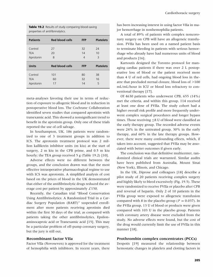
BLBK186-Key April 24, 2009 14:39
Cardiothoracic surgery
Table 19.2 Results of study comparing blood-saving
properties of antifibrinolytics.
Patients Red blood cells FFP Platelets
Control 27 32 24
TEA 20 14 10
Aprotonin 8 4 3
Units Red blood cells FFP Platelets
Control 101 80 38
TEA 60 32 16
Aprotonin 17 14 5
meta-analyses favoring their use in terms of reduc-
tion of exposure to allogenic blood and in reduction in
postoperative blood loss. The Cochrane Collaboration
identified seven studies that compared aprotinin with
tranexamic acid. This showed a nonsignificant trend to
benefit in the aprotinin group. Only one of these trials
reported the use of cell salvage.
In Southampton, UK, 186 patients were random-
ized to one of 3 treatment groups in addition to
ICS. The aprotonin treatment protocol was 2 mil-
lion kallikrein inhibitor units (m kiu) at the start of
surgery, 2 m kiu in the CPB prime, and 0.5 m kiu
hourly; the TEA group received 5 g (Table 19.2) [10].
Adverse effects were no different between the
groups, and the conclusion drawn was that the most
effective intraoperative pharmacological regime to use
with ICS was aprotonin. A simplified analysis of cost
based on the prices of blood in the UK demonstrated
that either of the antifibrinolytic drugs reduced the av-
erage cost per patient by approximately £150.
Recently, the Canadian trial “Blood Conservation
Using Antifibrinolytics: A Randomized Trial in a Car-
diac Surgery Population (BART)” suspended enroll-
ment after more patients receiving aprotinin died
within the first 30 days of the trial, as compared with
patients taking the other antifibrinolytics, Epsilon-
aminocaproic acid or Tranexamic acid [15]. This may
be a particular problem of off-pump coronary surgery,
but the jury is still out.
Recombinant factor VIIaFactor VIIa (Novoseven) is approved for the treatment
of hemophilia with inhibitors. In recent years, there
has been increasing interest in using factor VIIa in ma-
jor hemorrhage in nonhemophilia patients.
A total of 89% of patients with complex noncoro-
nary surgery on CPB will have an allogeneic transfu-
sion. FVIIa has been used on a named patient basis
to terminate bleeding in patients with serious hemor-
rhage who already have had numerous units of blood
and products [16].
Kartoutis designed the Toronto protocol for man-
aging cardiac patients if there was over 2 L postop-
erative loss of blood or the patient received more
than 4 U of red cells, had ongoing blood loss in the-
atre that precluded sternal closure, blood loss of �100
mL/mL/hour in ICU or blood loss refractory to con-
ventional therapy [17].
Of 4630 patients who underwent CPB, 655 (14%)
met the criteria, and within this group, 114 received
at least one dose of FVIIa. The study cohort had a
higher overall risk profile and more frequently under-
went complex surgical procedures and longer bypass
times. Those receiving ≤8 U of blood were classified as
the early therapy group. The recorded adverse events
were 24% in the untreated group, 30% in the early
therapy, and 60% in the late therapy groups. How-
ever, there were many confounding effects, which, if
taken into account, suggested that FVIIa may be asso-
ciated with better outcomes if given early.
The conclusion was that definitive multicenter, ran-
domized clinical trials are warranted. Similar audits
have been published from Australia, Mount Sinai
(New York), Illinois, and Chicago.
In the UK, Diprose and colleagues [18] describe a
pilot study of 20 patients receiving complex surgery
and highly likely to bleed excessively (Fig. 19.5). These
were randomized to receive FVIIa or placebo after CPB
and reversal of heparin. Only 2 of 10 patients in the
FVIIa group were exposed to allogeneic transfusion
compared with 8 in the placebo group (P = 0.037). In
the FVIIa group, 13 U of blood or products were given
compared with 103 U in the placebo group. Patients
with coronary artery disease were excluded from the
study. No adverse effects were found, but the cost of
the drug would currently limit the use of FVIIa in this
manner [18].
Prothrombin complex concentrates (PCCs)Despotis [19] measured the relationship between
hemostatic changes in platelets and clotting factors in
205
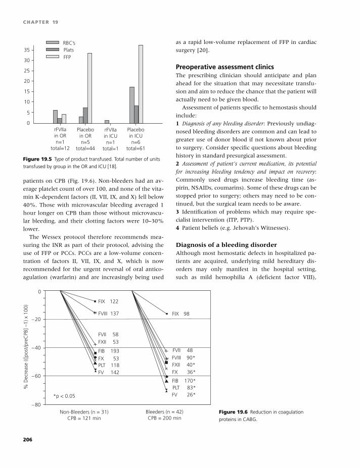
BLBK186-Key April 24, 2009 14:39
CHAPTER 19
rFVIIain ORn=1
total=12
rFVIIain ICUn=1
total=1
Placeboin ORn=5
total=44
Placeboin ICUn=6
total=61
10
5
0
15
20
25
FFPPlatsRBC’s
30
35
Figure 19.5 Type of product transfused. Total number of units
transfused by group in the OR and ICU [18].
patients on CPB (Fig. 19.6). Non-bleeders had an av-
erage platelet count of over 100, and none of the vita-
min K-dependent factors (II, VII, IX, and X) fell below
40%. Those with microvascular bleeding averaged 1
hour longer on CPB than those without microvascu-
lar bleeding, and their clotting factors were 10–30%
lower.
The Wessex protocol therefore recommends mea-
suring the INR as part of their protocol, advising the
use of FFP or PCCs. PCCs are a low-volume concen-
tration of factors II, VII, IX, and X, which is now
recommended for the urgent reversal of oral antico-
agulation (warfarin) and are increasingly being used
as a rapid low-volume replacement of FFP in cardiac
surgery [20].
Preoperative assessment clinicsThe prescribing clinician should anticipate and plan
ahead for the situation that may necessitate transfu-
sion and aim to reduce the chance that the patient will
actually need to be given blood.
Assessment of patients specific to hemostasis should
include:
1 Diagnosis of any bleeding disorder: Previously undiag-
nosed bleeding disorders are common and can lead to
greater use of donor blood if not known about prior
to surgery. Consider specific questions about bleeding
history in standard presurgical assessment.
2 Assessment of patient’s current medication, its potential
for increasing bleeding tendency and impact on recovery:
Commonly used drugs increase bleeding time (as-
pirin, NSAIDs, coumarins). Some of these drugs can be
stopped prior to surgery; others may need to be con-
tinued, but the surgical team needs to be aware.
3 Identification of problems which may require spe-
cialist intervention (ITP, PTP).
4 Patient beliefs (e.g. Jehovah’s Witnesses).
Diagnosis of a bleeding disorderAlthough most hemostatic defects in hospitalized pa-
tients are acquired, underlying mild hereditary dis-
orders may only manifest in the hospital setting,
such as mild hemophilia A (deficient factor VIII),
FIX 122
FVIII 137
FVII 58FXII 53
FIB 193
FX 53PLT 118FV 142
FIX 98
FVIII 90*FVII 48
FXII 40*
FIB 170*
FX 36*
PLT 83*FV 26**p < 0.05
–80
% D
ecre
ase
(([po
st/p
reC
PB] –
1) x
100
)
–60
–40
–20
0
Non-Bleeders (n = 31)CPB = 121 min
Bleeders (n = 42)CPB = 200 min
Figure 19.6 Reduction in coagulation
proteins in CABG.
206

BLBK186-Key April 24, 2009 14:39
Cardiothoracic surgery
mild hemophilia B (deficient factor IX), and mild
hemophilia C (deficient factor XI), all of which prolong
the APTT. If patients are found to have hemophilia, it
is essential that a hematologist advises on best treat-
ment, which can vary from DDAVP preoperatively
followed by an antifibrinolytic postoperatively to the
giving of regular doses of a recombinant replacement
factor that can be monitored with the TEG R©. The lat-
ter may not be available in all hospitals out of hours
without prior notice.
Assessment of current medicationAntiplatelet drugsClopidogrel causes platelet inhibition via a different
mechanism to aspirin, and following coronary stent-
ing, the two drugs are increasingly being prescribed
together. There is growing evidence that the hemor-
rhagic risk is increased when the two drugs are taken
concurrently. An increasing number of patients take
antiplatelet agents. NSAIDs, dipyridamole, aspirin,
and clopidogrel are all implicated in increased surgical
blood loss. Ideally, these drugs should be stopped
prior to surgery, to allow platelet function to return
to normal.
The time required off the drug to ensure normal
platelet function varies. NSAIDs provide reversible
inhibition of cyclooxygenase, and their antiplatelet
effects are half-life-dependent (usually hours). As-
pirin and clopidogrel lead to irreversible inhibition of
platelet aggregration for the lifespan of the platelet
(∼10 days). These drugs need to be stopped for 7 days
to be confident of adequate platelet function. How-
ever, due consideration must be given to the risks as-
sociated with stopping these drugs in surgical patients.
Many patients are presenting for emergency coro-
nary revascularization having had failed coronary
stenting procedures. These patients have usually re-
ceived aspirin and clopidogrel. Hemorrhage during the
subsequent surgery may be a major problem. Use of
the new TEG reagents is very useful here, as 15% of
patients have normal platelet function despite therapy,
and in others the degree of dysfunction is variable.
Clopidogrel is a pro-drug. The active metabolite cir-
culates for approximately 18 hours after the last dose,
and may permanently inhibit any platelets present
during this time (whether endogenous or transfused).
Surgery is best delayed for at least 24 hours after the
last dose of clopidogrel.
Surgery in patients who have received clopidogrel
in the last 7 days should, where possible, be post-
poned. If the surgery is a genuine emergency, platelets
should be made available for transfusion, and consid-
eration given to using aprotinin. Delaying for 24 hours
after the last dose of clopidogrel will improve the re-
sponse to platelet transfusion.
WarfarinWith a patient on oral anticoagulant therapy, it is
sufficient to stop warfarin 3 days before surgery and
restart the usual maintenance dose the evening of the
surgery. If they have a mechanical heart valve or have
had a venous thromboembolism in the past, this pe-
riod should be covered by heparin. Having stopped
warfarin, if the INR pre-op is over 2.5, small amounts
of vitamin K (1–2 mg) may be given.
Anesthetic techniques to reduce blood lossThere are some basic things that the anesthetist and
surgeon can do to reduce blood loss during surgery:� Positioning of the anesthetized patient so as to min-
imize any venous congestion in the operating field.� The use of local vasoconstrictors.� The sequencing of a multistage procedure (e.g. a
coronary artery bypass procedure where the saphe-
nous vein is harvested by one member of the team as
another is opening and preparing the chest. The vein
harvester needs to close his operation site fully before
ascending to assist with the chest).
There are also some specific procedures that may
help in reducing blood loss, such as:� preventing hypertension.� minimizing the period of hypothermia, and� controlled hypotension.
References
1 Bevan DH. A review of cardiac bypass haemosta-
sis , putting blood through the mill. Br J Haematol
1999;104:208–19.
2 Van Dijk D, Nierich AP, Jansen EWL. Early outcome
after off-pump vs on pump CABG, results from a ran-
domised study. Circulation 2001;104:1761–6.
3 Mack MJ, Pfister A, Bachand D, et al. Comparison of
coronary bypass surgery with and without cardiopul-
monary bypass in patients with multivessel disease.
J Thorac Cardiovasc Surg 2004;127:167–73.
207
BLBK186-Key April 24, 2009 14:39
CHAPTER 19
4 Goodenough LT, Johnston MFM, Toy PTCY, and the
Medicine Academic Award Group. The variability of
transfusion practice in coronary artery bypass surgery.
JAMA 1991;265:86–90.
5 Stover FP, Stegel IC, Parks R, et al. Variability in
transfusion practice for coronary artery bypass surgery
persists despite national consensus guidelines: a 24-
institution study. Anesthesiology 1998;88(2):327–33.
6 Avidan MS, Alcock EL, Da Fonseca J, et al. Com-
parison of structured use of laboratory tests or near-
patient assessment with clinical judgement in the man-
agement of bleeding after cardiac surgery. Br J Anaesth
2004;92(2):178–86.
7 Spiess BD. Thrombelastograph analysis and cardiopul-
monary bypass. Semin Thromb Hemost 1995;21:S4.
8 Shore-Lesserson L, Manspeizer HE, Francis S, De-
perio M. Intraoperative Thrombelastograph analysis
(TEG r) reduces transfusion requirements. Anesthesia
Analg 1998;86:S104.
9 Von Kier S, Royston D. Reduced hemostatic factor
transfusion using heparinase-modified Thrombelasto-
graph analysis (TEG) during cardioplumonary bypass.
Anesthesiology 1998;89(3A):A911.
10 Diprose P, Herbertson MJ, O’Shaughnessy D, Deakin
CD, Gill RS. A randomised double-blind placebo-
controlled trial of antifibrinolytic therapies used in
addition to intra-operative cell salvage. Br J Anaesth
2005;94(3):271–8.
11 Hobson AR, Agarwala RA, Swallow RA, Dawkins KD,
Curzen NP. Thrombelastography: current clinical appli-
cations and its potential role in interventional cardiol-
ogy. Platelets 2006;17(8):509–18.
12 Agarwal S, Coakely M, Reddy K, Riddell A, Mallett S. A
comparison of the platelet function analyzer and modi-
fied TEG with light transmission platelet aggregometry.
Anaesthesiology 2004;105(4):676–83.
13 Gwozdziewicz M, Nemec P, Zezula R, Novotny J.
Platelet mapping in postoperative management of
acute aortocoronary bypass thrombosis. Kardiochirurgia
I Torakochirurgia Polska 2006;3(2):214–16.
14 McGill N, O’Shaughnessy D, Pickering R, Herbertson
M, Gill R. Mechanical methods of reducing blood loss in
cardiac surgery. A randomised controlled trial. Br Med J
2002;324:1299.
15 Fergusson MHA, Hebert PC, Mazer D, et al. A com-
parison of aprotinin and lysine analogues in high-
Risk cardiac surgery: the BART Study. N Engl J Med
2008;358(22):2319–31.
16 Despotis G, Avidan M, Lublin DM. Off-label use
of recombinant factor VIIa concentrates after cardiac
surgery. Ann Thorac Surg 2005;80:3–5.
17 Kartoukis K, Yau TM, Riazi S, et al. Determinants
of complications with recombinant factor VIIa for re-
fractory blood loss in cardiac surgery. Can J Anaesth
2006;53(8):802–9.
18 Diprose P, Herbertson MJ, O’Shaughnessy D, Gill RS.
Activated recombinant factor VII after CPB reduces al-
logeneic transfusion in complex non-coronary cardiac
surgery: randomised double-blind placebo-controlled
pilot study. Br J Anaesth 2005;95:596–602.
19 Despotis GJ, Joist JH, Goodenough LT. Monitoring
of haemostasis in cardiac surgical patients: impact of
point-of-care testing on blood loss and transfusion out-
comes. Clin Chem 1997;43:1684–96.
20 Stuklis RG, O’Shaughnessy DF, Ohri SK. Novel ap-
proach to bleeding in patients undergoing cardiac
surgery with liver dysfunction. Eur J Cardiothorac Surg
2001;19(2):219–20.
208
BLBK186-Key April 11, 2009 13:3
20 NeurologyNatalie Aucutt-Walter, Valerie Jewells, and David Y. Huang
Neurological complications of hematological disease
can present in many ways. Examples include seizure
triggered by cerebral ischemia or hemorrhage and
headache in patients with sickle cell disease. The over-
whelming majority of these neurologic complications
are vascular in nature, owing to the fact that most
hematological abnormalities lead to either thrombosis
in the cerebral vasculature or brain hemorrhage. As-
sociated cerebrovascular events include both ischemic
and hemorrhagic strokes, as well as cerebral venous
sinus thromboses.
Ischemic stroke
Ischemic stroke can be divided into two broad cate-
gories: embolic and lacunar. The characteristic clini-
cal profile of acute embolic stroke is sudden onset of a
maximal neurological deficit. Emboli often arise from
the heart or from ulcerated carotid plaques. Atrial fib-
rillation, which predisposes patients to forming car-
diac thrombi, is associated with a six-fold increased
risk for stroke [1]. Cardiac emboli are highly corre-
lated with large vessel ischemia. Warfarin is strongly
recommended for stroke prevention in the presence
of atrial fibrillation unless otherwise contraindicated.
Thrombotic infarction, including lacunar infarction, is
often preceded by transient ischemic attacks (TIAs)
and may progress over hours or days in a stuttering
fashion. TIAs correlate with carotid stenosis and of-
ten present with border zone or “watershed” ischemic
injury distal to the area of critical stenosis. The prog-
nosis of TIAs varies considerably. Up to 33% of pa-
tients who experience a TIA will develop a disabling
stroke within 5 years. The incidence of stroke after TIA
is 10–20% in the first 12 months and 5% each year
thereafter [1]. Watershed territory infarcts may be
seen with clinically significant drops in blood pressure.
Lacunar infarcts are usually deep, small-vessel ische-
mic lesions �10 mm in diameter and account for be-
tween 10% and 25% of all ischemic strokes [1]. They
are often found in patients with a long-standing his-
tory of hypertension, diabetes, hypercholesterolemia
with atherosclerotic disease, and tobacco abuse. The
pathophysiology is thought to be multifactorial and
includes small-vessel lipohyalinosis and fibrinoid de-
generation, decreased perfusion of the penetrating
arteries, and atheromatous occlusion or embolism.
Acute management of suspected ischemic stroke
involves rapid assessment of the patient’s present-
ing symptoms by a neurovascular specialist or at the
nearest emergency department. Patients who present
�3 hours from symptom onset may be candidates for
thrombolytic therapy with intravenous recombinant
tissue plasminogen activator (IV-tPA). Prior to admin-
istering IV-tPA, the patient should have a noncon-
trast head CT to rule out intracerebral bleeding and
laboratory tests, including coagulation studies, com-
plete blood count (CBC), and serum glucose, must
be checked. Contraindications and guidelines for IV-
tPA administration are widely published and should
be reviewed carefully prior to administering the drug
[2]. Treatment is associated with a 6% risk of bleed-
ing complications, including intracranial hemorrhage,
and patients and/or their families should be coun-
seled about the benefits and risks associated with
thrombolytic therapy. Other acute treatments include
intra-arterial thrombolytic therapy and mechanical
endovascular clot retrieval using aspiration or evac-
uation devices, but such interventions are less well-
studied and are limited to centers with experienced
interventionalists.
Beyond the acute interventions, general manage-
ment of ischemic stroke concentrates on rehabilitation
209
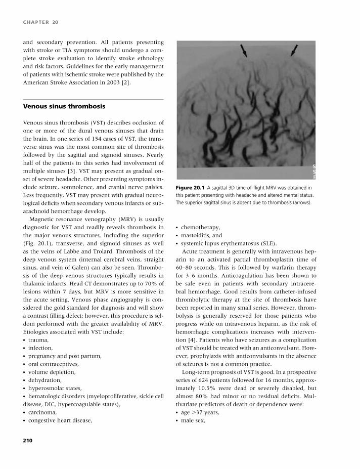
BLBK186-Key April 11, 2009 13:3
CHAPTER 20
and secondary prevention. All patients presenting
with stroke or TIA symptoms should undergo a com-
plete stroke evaluation to identify stroke ethnology
and risk factors. Guidelines for the early management
of patients with ischemic stroke were published by the
American Stroke Association in 2003 [2].
Venous sinus thrombosis
Venous sinus thrombosis (VST) describes occlusion of
one or more of the dural venous sinuses that drain
the brain. In one series of 154 cases of VST, the trans-
verse sinus was the most common site of thrombosis
followed by the sagittal and sigmoid sinuses. Nearly
half of the patients in this series had involvement of
multiple sinuses [3]. VST may present as gradual on-
set of severe headache. Other presenting symptoms in-
clude seizure, somnolence, and cranial nerve palsies.
Less frequently, VST may present with gradual neuro-
logical deficits when secondary venous infarcts or sub-
arachnoid hemorrhage develop.
Magnetic resonance venography (MRV) is usually
diagnostic for VST and readily reveals thrombosis in
the major venous structures, including the superior
(Fig. 20.1), transverse, and sigmoid sinuses as well
as the veins of Labbe and Trolard. Thrombosis of the
deep venous system (internal cerebral veins, straight
sinus, and vein of Galen) can also be seen. Thrombo-
sis of the deep venous structures typically results in
thalamic infarcts. Head CT demonstrates up to 70% of
lesions within 7 days, but MRV is more sensitive in
the acute setting. Venous phase angiography is con-
sidered the gold standard for diagnosis and will show
a contrast filling defect; however, this procedure is sel-
dom performed with the greater availability of MRV.
Etiologies associated with VST include:� trauma,� infection,� pregnancy and post partum,� oral contraceptives,� volume depletion,� dehydration,� hyperosmolar states,� hematologic disorders (myeloproliferative, sickle cell
disease, DIC, hypercoagulable states),� carcinoma,� congestive heart disease,
Figure 20.1 A sagittal 3D time-of-flight MRV was obtained in
this patient presenting with headache and altered mental status.
The superior sagittal sinus is absent due to thrombosis (arrows).
� chemotherapy,� mastoiditis, and� systemic lupus erythematosus (SLE).
Acute treatment is generally with intravenous hep-
arin to an activated partial thromboplastin time of
60–80 seconds. This is followed by warfarin therapy
for 3–6 months. Anticoagulation has been shown to
be safe even in patients with secondary intracere-
bral hemorrhage. Good results from catheter-infused
thrombolytic therapy at the site of thrombosis have
been reported in many small series. However, throm-
bolysis is generally reserved for those patients who
progress while on intravenous heparin, as the risk of
hemorrhagic complications increases with interven-
tion [4]. Patients who have seizures as a complication
of VST should be treated with an anticonvulsant. How-
ever, prophylaxis with anticonvulsants in the absence
of seizures is not a common practice.
Long-term prognosis of VST is good. In a prospective
series of 624 patients followed for 16 months, approx-
imately 10.5% were dead or severely disabled, but
almost 80% had minor or no residual deficits. Mul-
tivariate predictors of death or dependence were:� age �37 years,� male sex,
210
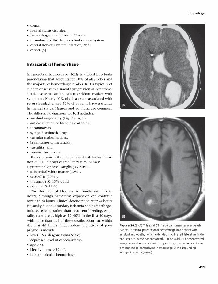
BLBK186-Key April 11, 2009 13:3
Neurology
� coma,� mental status disorder,� hemorrhage on admission CT scan,� thrombosis of the deep cerebral venous system,� central nervous system infection, and� cancer [5].
Intracerebral hemorrhage
Intracerebral hemorrhage (ICH) is a bleed into brain
parenchyma that accounts for 10% of all strokes and
the majority of hemorrhagic strokes. ICH is typically of
sudden onset with a smooth progression of symptoms.
Unlike ischemic stroke, patients seldom awaken with
symptoms. Nearly 40% of all cases are associated with
severe headache, and 50% of patients have a change
in mental status. Nausea and vomiting are common.
The differential diagnosis for ICH includes:� amyloid angiopathy (Fig. 20.2A, B),� anticoagulation or bleeding diatheses,� thrombolysis,� sympathomimetic drugs,� vascular malformations,� brain tumor or metastasis,� vasculitis, and� venous thrombosis.
Hypertension is the predominant risk factor. Loca-
tion of ICH in order of frequency is as follows:� putaminal or basal ganglia (35–50%),� subcortical white matter (30%),� cerebellar (15%),� thalamic (10–15%), and� pontine (5–12%).
The duration of bleeding is usually minutes to
hours, although hematoma expansion can continue
for up to 24 hours. Clinical deterioration after 24 hours
is usually due to secondary ischemia and hemorrhage-
induced edema rather than recurrent bleeding. Mor-
tality rates are as high as 30–40% in the first 30 days,
with more than half of these deaths occurring within
the first 48 hours. Independent predictors of poor
prognosis include:� low GCS (Glasgow Coma Scale),� depressed level of consciousness,� age �75,� bleed volume �30 mL,� intraventricular hemorrhage,
(A)
(B)
Figure 20.2 (A) This axial CT image demonstrates a large left
parietal–occipital parenchymal hemorrhage in a patient with
amyloid angiopathy, which extended into the left lateral ventricle
and resulted in the patient’s death. (B) An axial T1 noncontrasted
image in another patient with amyloid angiopathy demonstrates
a mirror image parenchymal hemorrhage with surrounding
vasogenic edema (arrow).
211

BLBK186-Key April 11, 2009 13:3
CHAPTER 20
� concurrent antiplatelet therapy,� hyperglycemia, and� infratentorial location.
Diagnosis is made by emergent head CT. Angiog-
raphy may be necessary to evaluate for an underly-
ing source of bleed, such as an arterial venous malfor-
mation, aneurysm, angioma, cavernoma (which is not
seen with angiography, and is therefore called “cryp-
tic”), or tumor. If initial angiography is unrevealing,
it should be repeated in 3–4 months when the intra-
parenchymal blood has cleared.
Guidelines for the management of ICH were pub-
lished in Stroke in 2007 [6]. ICH management includes
control of blood pressure, seizure, infection, fever,
glucose, and increased intracranial pressure (ICP). Ag-
gressive blood pressure management remains con-
troversial, and blood pressure guidelines vary. In
general, patients are at increased risk for rebleeding
and hematoma enlargement with systolic blood pres-
sures �160 mm Hg [7]. However, blood pressure re-
duction should be balanced with the risk of concur-
rent ischemia, as blood pressures that are dramatically
lower than the patient’s baseline can lead to decreased
cerebral perfusion pressure (CPP). This is of particular
concern in patients with a large ICH, cerebral edema,
or other factors that increase ICP [8]. When ICP is ele-
vated (�20 mm Hg), blood pressure should be titrated
to maintain a CPP of 60–80 mm Hg (CPP = mean
arterial pressure – ICP). In the acute setting, pres-
sures should be lowered with short-acting agents, such
as intravenous labetalol, nitroprusside, or nicardipine,
which allow for rapid titration.
When monitoring and treating cerebral edema
and increased ICP, intraventricular pressure monitors
should be placed in patients with a GCS �9 or with
clinical deterioration in their neurological exam [8].
Approaches such as head-of-bed elevation and head
positioning are simple and often effective for quickly
lowering ICP. Other interventions should be limited to
situations where herniation is of immediate concern.
Patients with significant bleeding or intraventricular
extension are at risk for obstructive hydrocephalus,
and ventricular drain placement may be necessary.
Hyperventilation to keep the PCO2 between 28 and
30 torr is effective to reduce increased ICP, with peak
effect within 30 minutes. However, the effect is tran-
sient and only lasts until the pH of cerebrospinal fluid
equilibrates with systemic pH, usually within a few
hours [6]. Osmotic agents such as mannitol and hy-
pertonic saline may be used for short periods, but use
for more than a few days can lead to rebound increases
in ICP. Steroids should be avoided as they have not
been shown to be effective.
There are few indications for surgical intervention
in ICH. Indications for surgical intervention are gener-
ally limited to patients with:� cerebellar hemorrhage �3 cm in size and brainstem
compression,� acute hydrocephalus, or� neurological deterioration.
Patients with lobar clots within 1 cm of the cortex
may also be considered for surgery based on a trend
toward a positive effect of surgery over medical man-
agement for such patients in the International Surgical
Trial for Intracerebral Hemorrhage (STICH) [9].
Hemostasis treatment using recombinant activated
factor VII (rFVIIa) for ICH has been investigated in
a phase 3 trial [10]. Compared with placebo, treat-
ment with rFVIIa at 20 and 80 µg reduced hematoma
growth but did not improve functional outcome. In
addition, 80 µg of rFVIIa was associated with a non-
significant but increased frequency of adverse arte-
rial thromboembolic events compared with placebo.
rFVIIa is not currently recommended for treatment of
acute ICH.
Use of warfarin for anticoagulation to INR 2.5–4.5
increases the risk of ICH by up to 10-fold and doubles
the mortality. Patients with ICH who are anticoagu-
lated with warfarin should have their INR corrected as
quickly as possible with prothrombin complex concen-
trate (PCC) or rFVIIa. Intravenous vitamin K should
be administered without delay, because peak effect
is dependent on protein synthesis, approximately
6–8 hours later. Although fresh frozen plasma (FFP) is
commonly used, large volumes need to be given and
only partial correction is observed.
Subarachnoid hemorrhage
Subarachnoid hemorrhage (SAH) is often the result
of a ruptured saccular aneurysm but may also arise
from head trauma, extension of ICH into the sub-
arachnoid space, spinal arteriovenous malformation,
or idiopathic causes. Aneurysmal ruptures account for
80% of all nontraumatic SAHs and are of greatest
212
BLBK186-Key April 11, 2009 13:3
Neurology
concern, given a high mortality rate of approximately
45%. Presenting symptoms include a sudden and se-
vere “thunder clap” headache, with an acute change
in mental status, in some cases leading to lethargy and
coma. Sudden loss of consciousness occurs in up to
20% of patients. Meningeal signs, papilledema, and
seizure are common at presentation. Increased size of
the bleed and the presence of intraventricular exten-
sion are correlated with increased mortality. Head CT
is often diagnostic of SAH, but up to 15% of cases
of aneurysmal SAH will have a normal study. If the
head CT is normal but the suspicion for SAH is high,
an emergent lumbar puncture should be performed
to evaluate for blood or xanthrochromia in the spinal
fluid, which is indicative of a sentinel bleed. Patients
with sentinel bleeds have a �50% risk of rebleeding
in the next 48–72 hours.
Initial management of SAH is focused on reduc-
ing the likelihood of rebleeding. Treating hyperten-
sion and maintaining blood pressure in a normal range
has been shown to decrease the rate of rebleeding. Af-
ter 4 days and for up to 2 weeks, patients are at in-
creased risk for ischemic stroke from vasospasm and
should receive nimodipine, which reduces long-term
injury from vasospasm. The antifibrinolytic agent ep-
silon aminocaproic acid (AMICAR) has been shown to
decrease mortality associated with rebleeding, but its
benefits were offset by the increased risk for ischemic
stroke. Surgical or endovascular interventions to se-
cure ruptured aneurysms should be performed once
patients are stabilized. Patients with extensive bleed-
ing or intraventricular extension may develop obstruc-
tive hydrocephalus, and a ventricular drain may be
necessary to treat elevated ICP. Anticonvulsants are
often administered as prophylaxis against seizure.
Diseases associated with ischemic strokes
Hereditary and acquired hypercoagulablestatesA number of factors have been implicated in the de-
velopment of ischemic stroke.
Table 20.1 lists a variety of hypercoagulable states
and the strength of their correlation with stroke.
Notably, sickle cell disease, antiphospholipid anti-
body syndrome, and hyperhomocystinemia have the
strongest association with arterial stroke.
Table 20.1 Strength of association of coagulopathy with
arterial stroke.
Coagulopathy Arterial stroke risk
Sickle cell disease Strong
Antiphospholipid antibody syndrome Strong
Hyperhomocystinemia Moderate
Activated protein C resistance Mild
Prothrombin gene mutation Mild
Protein S deficiency Mild
Protein C deficiency Rare
Antithrombin III deficiency Rare
Adapted from Moster ML. Coagulopathies and arterial stroke.
J Neuroophthalmol 2003;23:63–71.
Activated protein C resistance/factor V LeidenCongenital activated protein C resistance (APC-R) is
the most common inherited risk factor for venous
thrombosis. A total of 95% of patients with APC-R
have the factor V Leiden mutation. The mutation is
present in 2–7% of the Caucasian population [11].
With respect to neurological complications, APC-R
correlates almost exclusively with venous thrombo-
sis, with only a few reported cases of arterial strokes
in young patients. Symptoms of acute cerebral ve-
nous thrombosis include headache, seizure, somno-
lence, and cranial nerve palsies. Patients with sus-
pected venous thrombosis should have neurological
imaging with MRI/MRA and MRV. SAH can result
from the rupture of congested cerebral veins. If cranial
nerve palsies are present on examination (i.e. defects
of cranial nerves III, IV, and VI associated with pto-
sis and facial pain), cavernous sinus thrombosis should
be suspected. Treatment for stroke patients with cere-
bral venous thrombosis is low-molecular-weight hep-
arin or warfarin.
Antiphospholipid antibody syndromeAntiphospholipid syndrome (aPLs) is an acquired hy-
percoagulable state that is associated with venous as
well as arterial thrombotic events. Arterial events oc-
cur most commonly in the cerebrovasculature. Stroke
or TIA are the initial clinical manifestation in ap-
proximately 20% of patients subsequently diagnosed
with aPLs. Involvement of the cerebral cortex and
213
BLBK186-Key April 11, 2009 13:3
CHAPTER 20
subadjacent white matter by platelet-fibrin mi-
crothrombi is most common [12]. The pathogenesis of
thrombosis in aPLs is uncertain (see chapter 17).
Antiphospholipid antibodies are found in �10% of
patients with acute ischemic stroke, and the vast ma-
jority of patients are young (�50 years) [11]. aPLs
should be considered in the work-up of all young pa-
tients presenting with an ischemic arterial or venous
stroke secondary to thrombosis. aPLs is suspected in
patients with a history of multiple miscarriages, de-
mentia, optic neuropathy, thrombocytopenia, SLE or
SLE-like syndromes, or complicated migraine.
Testing for aPLs includes evaluation for IgG an-
tiphospholipids on two separate occasions at least 12
weeks apart. Stroke risk is greatest with IgG antiphos-
pholipids �40 GPL units and may not be clinically sig-
nificant at lower levels [13]. Treatment is generally
with warfarin to prevent recurrent systemic throm-
bosis. However, in patients with prior stroke and a
single positive test result for antiphospholipid antibod-
ies, aspirin (325 mg/day) appears to be as effective as
moderate-intensity warfarin (PT 1.4–2.8) for prevent-
ing recurrent stroke [12].
HyperhomocystinemiaHyperhomocystinemia has a prevalence of 5–10% in
the general population and is associated with accel-
erated premature atherosclerosis. Increased fasting
levels of homocysteine have been related to the preva-
lence of extracranial common carotid artery stenosis of
�25% in the Framingham cohort. Fasting homocys-
teine levels above 15.4 µmol/L significantly increase
the patient’s risk for stroke, with an odds ratio of
2.5–4.7. Elevated levels increase the odds of carotid
intimal thickening more than three-fold. Proposed
mechanisms of coagulopathy include increased
platelet adhesion, activation of the coagulation cas-
cade, conversion of LDL to a pro-atherogenic form,
and endothelial damage.
Most often, hyperhomocystinemia is acquired due
to a diet deficient in folate, B6, and/or B12. Folate and
B12 levels should be checked in all patients, especially
young patients with unexplained stroke and prema-
ture atherosclerosis [14]. Treatment includes vitamin
supplementation with folic acid, B6, and B12. Elevated
levels of homocysteine can also be seen with renal in-
sufficiency and concurrent anti-epileptic drug use, es-
pecially phenytoin.
Hyperhomocystinemia needs to be distinguished
from autosomal recessive homocystinuria. Patients
who are homozygous for cystathionine beta synthase
deficiency can have homocystine concentrations up
to 400 µmol/L and present with a marfanoid body
habitus, mental retardation, seizure, lenticular dislo-
cations, skeletal abnormalities, and a 20-fold increase
in urinary homocysteine excretion over other amino
acids [11]. These patients are at high risk for myocar-
dial infarction and ischemic stroke as well as prema-
ture death secondary to vascular disease. The inci-
dence of stroke increases with increased homocysteine
levels, and heterozygous patients have a milder course
and clinical picture.
Sickle cell diseaseChildren with sickle cell disease (SCD) present with
a wide variety of chronic neurological syndromes, in-
cluding:� ischemic and hemorrhagic stroke,� dural VST,� spinal cord infarction,� transient ischemic attack,� headache,� seizure,� altered mental status,� cognitive difficulties, and� covert “silent” infarction.
Up to 25% of children with HbSS will have covert
or “silent” infarction by adolescence. Silent ischemia
can be detected with diffusion-weighted MRI, which
reveals ischemic regions, characteristically in the
anterior or posterior watershed /border zones. One
study enrolled and followed the neuroimaging of 213
HbSS children without a history of overt stroke. In
this group, 160 children had normal baseline MRIs,
and 53 children had MRIs showing silent infarcts. The
patients were followed with serial MRIs, and the chil-
dren with silent infarcts at baseline were significantly
more likely to demonstrate new or progressive neu-
rologically silent lesions compared with those whose
baseline MRIs were normal. Only 2.5% children with
normal baseline MRIs developed silent infarcts on
follow-up MRI examination compared with 24.5%
who had a baseline silent infarct [15]. These patients
may have a normal T2-weighted MRI and a normal
neurological examination. Seizure is common in
patients with known cerebrovascular disease as well
214
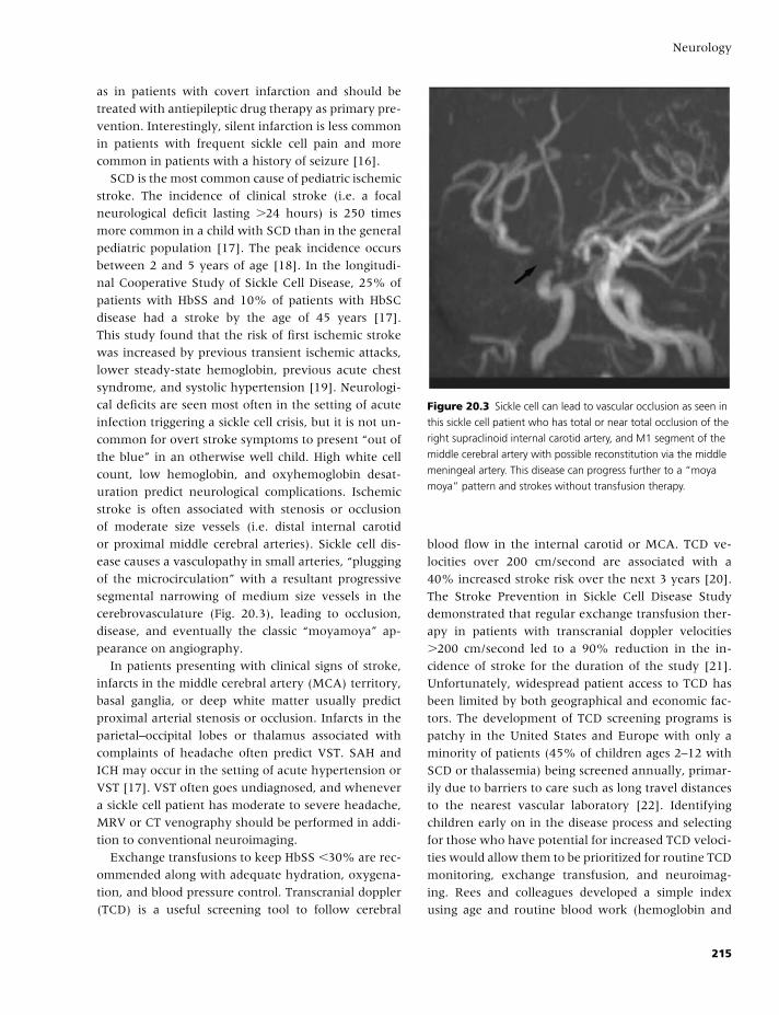
BLBK186-Key April 11, 2009 13:3
Neurology
as in patients with covert infarction and should be
treated with antiepileptic drug therapy as primary pre-
vention. Interestingly, silent infarction is less common
in patients with frequent sickle cell pain and more
common in patients with a history of seizure [16].
SCD is the most common cause of pediatric ischemic
stroke. The incidence of clinical stroke (i.e. a focal
neurological deficit lasting �24 hours) is 250 times
more common in a child with SCD than in the general
pediatric population [17]. The peak incidence occurs
between 2 and 5 years of age [18]. In the longitudi-
nal Cooperative Study of Sickle Cell Disease, 25% of
patients with HbSS and 10% of patients with HbSC
disease had a stroke by the age of 45 years [17].
This study found that the risk of first ischemic stroke
was increased by previous transient ischemic attacks,
lower steady-state hemoglobin, previous acute chest
syndrome, and systolic hypertension [19]. Neurologi-
cal deficits are seen most often in the setting of acute
infection triggering a sickle cell crisis, but it is not un-
common for overt stroke symptoms to present “out of
the blue” in an otherwise well child. High white cell
count, low hemoglobin, and oxyhemoglobin desat-
uration predict neurological complications. Ischemic
stroke is often associated with stenosis or occlusion
of moderate size vessels (i.e. distal internal carotid
or proximal middle cerebral arteries). Sickle cell dis-
ease causes a vasculopathy in small arteries, “plugging
of the microcirculation” with a resultant progressive
segmental narrowing of medium size vessels in the
cerebrovasculature (Fig. 20.3), leading to occlusion,
disease, and eventually the classic “moyamoya” ap-
pearance on angiography.
In patients presenting with clinical signs of stroke,
infarcts in the middle cerebral artery (MCA) territory,
basal ganglia, or deep white matter usually predict
proximal arterial stenosis or occlusion. Infarcts in the
parietal–occipital lobes or thalamus associated with
complaints of headache often predict VST. SAH and
ICH may occur in the setting of acute hypertension or
VST [17]. VST often goes undiagnosed, and whenever
a sickle cell patient has moderate to severe headache,
MRV or CT venography should be performed in addi-
tion to conventional neuroimaging.
Exchange transfusions to keep HbSS �30% are rec-
ommended along with adequate hydration, oxygena-
tion, and blood pressure control. Transcranial doppler
(TCD) is a useful screening tool to follow cerebral
Figure 20.3 Sickle cell can lead to vascular occlusion as seen in
this sickle cell patient who has total or near total occlusion of the
right supraclinoid internal carotid artery, and M1 segment of the
middle cerebral artery with possible reconstitution via the middle
meningeal artery. This disease can progress further to a “moya
moya” pattern and strokes without transfusion therapy.
blood flow in the internal carotid or MCA. TCD ve-
locities over 200 cm/second are associated with a
40% increased stroke risk over the next 3 years [20].
The Stroke Prevention in Sickle Cell Disease Study
demonstrated that regular exchange transfusion ther-
apy in patients with transcranial doppler velocities
�200 cm/second led to a 90% reduction in the in-
cidence of stroke for the duration of the study [21].
Unfortunately, widespread patient access to TCD has
been limited by both geographical and economic fac-
tors. The development of TCD screening programs is
patchy in the United States and Europe with only a
minority of patients (45% of children ages 2–12 with
SCD or thalassemia) being screened annually, primar-
ily due to barriers to care such as long travel distances
to the nearest vascular laboratory [22]. Identifying
children early on in the disease process and selecting
for those who have potential for increased TCD veloci-
ties would allow them to be prioritized for routine TCD
monitoring, exchange transfusion, and neuroimag-
ing. Rees and colleagues developed a simple index
using age and routine blood work (hemoglobin and
215
BLBK186-Key April 11, 2009 13:3
CHAPTER 20
aspartate transaminase) in order to predict which chil-
dren are likely to have TCD readings �170 cm/second,
placing them at higher risk of developing cerebrovas-
cular disease and resultant ischemic infarcts. This in-
dex has been shown to have 100% sensitivity and
between 60% and 70% specificity for predicting in-
creased arterial velocities [19].
In addition to the standard therapies (exchange
transfusions, hydroxyurea, and blood pressure man-
agement), antiplatelet therapy with aspirin has been
shown to reduce ischemic stroke risk, as well as pre-
vent silent ischemia and cognitive impairment. A pilot
trial using aspirin therapy in sickle cell patients is un-
derway. In the meantime, aspirin therapy should be
used with caution in patients with a history of large
territory ischemic stroke, subdural, or SAH because of
the unknown risk of hemorrhage [1].
Diseases associated withhemorrhagic strokes
Hemophilia AThe most devastating and common neurological com-
plication of hemophilia A is ICH. The incidence of ICH
in the general population is around 2%. In contrast,
the incidence of ICH in patients with hemophilia A can
be as high as 12%. ICH can occur spontaneously or as
a result of a minor/trivial trauma. A review of 170 pa-
tients with hemophilia A documented 42 episodes of
ICH or spinal hemorrhage in 32 patients. Of those pa-
tients presenting with ICH or spinal hemorrhage, 36%
were associated with a minor or obvious head trauma,
whereas 64% occurred spontaneously. All of the pa-
tients presenting with an acute bleed where known to
have severe hemophilia A, and 9 of the 32 patients
(17.6%) presented with recurrent ICH [23].
Sudden onset of headache is the most common
presenting symptom of ICH (97.5%) [23]. Other as-
sociated symptoms, including nausea, vomiting, and
progressive neurologic deterioration, are strongly sug-
gestive of intraparenchymal brain hemorrhage and
warrant immediate neurological imaging with a CT
of the head to assess for intra- or extraparenchymal
blood. Patients with brain herniation at presentation
have the worst prognosis, as concurrent herniation is
near 100% fatal.
Acquired hemophilia A is a rare bleeding disor-
der caused by the development of autoantibodies that
inhibit the action of naturally occurring factor VIII.
Patients classically present with prominent extensive
subcutaneous hematomas. Unlike classic hemophilia,
ICH and hemarthroses are rare with hemophilia A.
In addition to standard management of ICH, treat-
ment of bleeding in a patient with hemophilia consists
of administration of coagulation factor concentrates in
order to correct the deficiency. If FVIII concentrate is
not available, one should not wait for concentrate but
should begin treatment with cryoprecipitate, each unit
of which generally contains 80–100 IU of FVIII, or FFP,
which contains all clotting factors.
References
1 Zaidat OO, Lerner AJ. The Little Black Book of Neurology
(4th edition). St. Louis: Mosby, 2002.
2 Adams HP Jr., Adams RJ, Brott T, et al. Guidelines for
the early management of patients with ischemic stroke.
Stroke 2003;34:1056–83.
3 Goske-Bierska I, Wysokinski W, Brown RD Jr., et al.
Cerebral venous sinus thrombosis: Incidence of ve-
nous thrombosis recurrence and survival. Neurology
2006;67:814–19.
4 Mohr JP, Choi D, Grotter J, Weir B, Wolf PA. Stroke:
Pathophysiology, Diagnosis, and Management (4th edition).
New York: Churchill Livingstone, 2004.
5 Ferro JM, Canhao P, Stam J, et al. Prognosis of cerebral
vein and dural sinus thrombosis: results of the Interna-
tional Study on Cerebral Vein and Dural Sinus Throm-
bosis (ISCVT). Stroke 2004;35:664–70.
6 Broderick JP, Connolly S, Feldmann E, et al. Guide-
lines for the management of spontaneous intracere-
bral hemorrhage in adults: 2007 update: a guideline
from the American Heart Association/American Stroke
Association Stroke Council, High Blood Pressure Re-
search Council, and the Quality of Care and Outcomes
in Research Interdisciplinary Working Group. Stroke
2007;38:2001–23.
7 Ohwaki K, Yano E, Nagushima H, et al. Blood pres-
sure management in acute intracerebral hemorrhage:
relationship between elevated blood pressure and
hematoma enlargement. Stroke 2004;35:1364–7.
8 Broderick JP, Adams HP, Barsan W, et al. Guidelines for
the management of spontaneous intracerebral hemor-
rhage: a statement for healthcare professionals from a
216
BLBK186-Key April 11, 2009 13:3
Neurology
special writing group of the Stroke Council, American
Heart Association. Stroke 1999;30:905–15.
9 Mendelow AD, Gregson BA, Fernandes HM, et al.
Early surgery versus initial conservative treatment in
patients with spontaneous supratentorial intracerebral
haematomas in the International Surgical Trial in In-
tracerebral Haemorrhage (STICH): a randomised trial.
Lancet 2005;365:387–97.
10 Mayer SA, Brun NC, Begtrup K, et al. Efficacy and
safety of recombinant activated factor VII for acute in-
tracerebral hemorrhage. N Engl J Med 2008;358:2127–
37.
11 Moster ML. Coagulopathies and arterial stroke. J Neu-
roophthalmol 2003;23:63–71.
12 Lim W, Crowther MA, Eikelboom JW. Management of
antiphospholipid antibody syndrome: a systematic re-
view. J Am Med Assoc 2006;295:1050–7.
13 Rand JH. The antiphospholipid syndrome. Annu Rev
Med 2003;54:409–424.
14 Caplan LR. Caplan’s Stroke: A Clinical Approach (3rd edi-
tion). Boston: Butterworth Heinemann, 2000.
15 Pegelow CH, Macklin EA, Moser FG, et al. Longitudinal
changes in brain magnetic resonance imaging findings
in children with sickle cell disease. Blood 2002;99:3014–
18.
16 Kinney TR, Sleeper LA, Wang WC, et al. Silent cere-
bral infarcts in sickle cell anemia: a risk factor analysis.
Pediatrics 1999;103:640–5.
17 Kirkham F. Therapy Insight: stroke risk and its manage-
ment in patients with sickle cell disease. Nat Clin Pract
Neurol 2007;3:264–78.
18 Ohene-Frempong K, Weiner SJ, Sleeper LA, et al. Co-
operative study of sickle cell disease: cerebrovascular
accidents in sickle cell disease: rates and risk factors.
Blood 1998;91:288–94.
19 Rees DC, Dick MC, Height SE, et al. A simple index us-
ing age, hemoglobin, and aspartate transaminase pre-
dicts increased intracerebral blood velocity as measured
by transcranial doppler scanning in children with sickle
cell anemia. Pediatrics 2008;121:1628–32.
20 Adams RJ, McKie VC, Carl EM, et al. Long-term stroke
risk in children with sickle cell disease screened with
transcranial doppler. Ann Neurol 1997;42:699–704.
21 Adams RJ, McKie VC, Hsu L, et al. Prevention
of a first stroke by transfusions in children with
sickle cell anemia and abnormal results on transcra-
nial doppler ultrasonography. N Engl J Med 1998;339:
5–11.
22 Fullerton HJ, Gardner M, Adams RJ, Lo LC, Johnson
SC. Obstacles to primary stroke prevention in children
with sickle cell disease. Neurology 2006;67:1098–9.
23 Chinthamitr Y, Ruchutrakool T, Suwanawiboon B,
Nakkinkun Y, Ayprasert N, Issaragrisil S. Intracranial
and spinal hemorrhage in adult hemophelia A in siriraj
hospital, Thiland. J Thromb Haemost 2007;5(Suppl 2):P-
S150.
217

BLBK186-Key April 24, 2009 14:58
21 HepatologyRaj K. Patel and Roopen Arya
Introduction
Hepatic diseases are associated with a variety of de-
fects affecting both primary and secondary hemosta-
sis (Table 21.1). It is therefore not surprising that
advanced hepatic disease is associated with bleeding
[1,2]. Chronic liver disease frequently causes por-
tal hypertension with resultant hypersplenism and
thrombocytopenia. This leads to formation of fragile
vascular anomalies (varices) that may bleed profusely
on a background of hemostatic failure. Not all pa-
tients with liver disease have bleeding manifestations,
but these tend to be unpredictable when they occur.
Common clinical manifestations include petechiae, ec-
chymoses, recurrent epistaxes, and gingival bleeding.
Invasive procedures, such as liver biopsy and ascitic
shunts, are particularly high risk in chronic liver dis-
ease as they may precipitate bleeding in previously sta-
ble patients.
Liver disease may be classified into two broad cate-
gories:
1 Acute liver disease (e.g. fulminant hepatic failure sec-
ondary to paracetamol overdose); or
2 Chronic liver disease (e.g. alcohol-induced cirrhosis,
primary biliary cirrhosis).
Most advanced cases of liver disease are associated
with at least one and frequently multiple hemostatic
defects. Orthotopic liver transplantation corrects hep-
atic function and coagulopathy in long term but is as-
sociated with a substantial perioperative increase in
bleeding risk.
A delicate balance exists between the procoagulant
and anticoagulant defects associated with liver dis-
ease. Although bleeding episodes are more common,
thrombotic events may occur despite a coexisting
hemorrhagic tendency. These include symptomatic
lower limb deep vein thrombosis, pulmonary em-
bolism, thrombosis of the abdominal veins, and
thrombosis in central venous catheters or extra-
corporeal circuits. It is also possible that the pro-
thrombotic state contributes to other hepatic disease
processes, including portopulmonary hypertension,
parenchymal extinction, and accelerated hepatic fibro-
sis [3]. Thrombosis may also occur as a result of local
endothelial dysfunction. There is as yet no univer-
sally available laboratory test with which to accu-
rately characterize the prothrombotic state in hepatic
disease.
Pathophysiology of coagulopathy
Impaired coagulation factor synthesisThe liver is the major synthetic site for:� Coagulation factors of both intrinsic and extrinsic
pathways, including factors II, V, VII, VIII, IX, X, XI,
and XII and fibrinogen;� Anticoagulant proteins (antithrombin, protein C,
protein S); and� Fibrinolytic regulators (plasminogen, α1-antiplas-
min).
Coagulation proteinsLoss of hepatocyte function in disease states leads to
a reduction in the levels of most coagulation proteins
(except factor VIII) and therefore predisposes to bleed-
ing. Reduced levels of these proteins broadly reflect
the extent of liver damage but are poor predictors of
bleeding risk in individual patients.� In acute liver injury (e.g. following paracetamol
overdose), prothrombin time (PT) has been shown
to be an accurate predictor of hepatocellular damage,
218
BLBK186-Key April 24, 2009 14:58
Hepatology
Table 21.1 Hemostatic defects in hepatic disease.
Hemostatic abnormality
Reduced biosynthesis of hepatic coagulation factors
Reduced biosynthesis of anticoagulant and fibrinolytic proteins
Reduced clearance of coagulation proteins and inhibitors
Dysfibrinogenemia
Systemic fibrinolysis
Disseminated intravascular coagulation
Thrombocytopenia
Platelet dysfunction
bleeding risk, and likelihood of progression to fulmi-
nant liver failure.� Factor V concentration is a particularly sensitive and
specific indicator of hepatic synthetic function and
plasma levels fall with increasing disease severity.� Malabsorption of fat-soluble vitamins may lead to
low levels of circulating vitamin K-dependent coagu-
lation factors.
Whereas the majority of circulating coagulation fac-
tors decrease in liver disease, the reverse is true of fac-
tor VIII, von Willebrand factor (VWF), and fibrinogen.
Fibrinogen and most of factor VIII are synthesized in
hepatocytes, whereas VWF is synthesized by platelets
and vascular endothelium. Circulating levels of these
proteins increase in the acute phase response associ-
ated with hepatic disease, although low levels of fib-
rinogen in late disease may herald the onset of acute
liver failure.
The formation of abnormal forms of vitamin K-
dependent coagulation factors (e.g. des-�-carboxyl
prothrombin) may be seen in both acute and chronic
liver disease. These proteins, raised in the absence of
vitamin K (PIVKAs), form as a result of an acquired
carboxylation defect but do not reach high enough
concentrations to cause bleeding.
Thrombocytopenia and platelet dysfunctionMild to moderate thrombocytopenia is common in
hepatic disease, affecting up to 30% of all cases of
chronic liver disease and 90% of subjects with end-
stage disease.
Chronic liver disease is associated with:� Portal hypertension and congestive splenomegaly.
The resultant increase in platelet pooling by splenic
sequestration is the principal mechanism by which
thrombocytopenia occurs in these patients.� Increasing portal venous pressures, blood is shunted
into the systemic circulation via portosystemic collat-
erals (varices) from which blood loss may occur, par-
ticularly on a background of thrombocytopenia.� Ineffective production of platelets secondary to a
decrease in liver thrombopoietin synthesis has been
reported.
Alcohol-associated liver disease may cause throm-
bocytopenia by a variety of mechanisms:� Alcohol is directly toxic to megakaryocytes, lead-
ing to inhibition of megakaryopoiesis and decreased
platelet production.� Folate deficiency resulting from poor dietary intake
or ineffective hepatic metabolism may result in inef-
fective megakaryopoiesis.� Alcohol ingestion is itself associated with decreased
platelet survival.
In fulminant viral hepatitis, the marked thrombo-
cytopenia often encountered is caused by both sup-
pression of megakaryopoiesis by virus and increased
platelet destruction.
The increase in bleeding time seen in many subjects
with severe liver disease is often out of proportion to
the associated degree of thrombocytopenia, suggest-
ing the presence of platelet dysfunction. The results
of platelet function testing in these patients are incon-
sistent. Whereas some studies have demonstrated ab-
normalities in primary and secondary aggregation to
adenosine diphosphate (ADP), adrenaline, thrombin
and ristocetin, others have failed to show any func-
tional defect.
The cause of platelet dysfunction in liver disease
is unclear. There is an increase in levels of circu-
lating platelet-inhibitors, including fibrin degradation
products. Ethanol or abnormal high-density lipopro-
teins may contribute to aggregatory abnormalities in
some cases. In others, intrinsic platelet abnormalities
have been demonstrated, including acquired storage
pool deficiency (platelet nucleotide deficiency), re-
duced platelet arachidonic acid, and abnormalities of
platelet membrane composition and signaling.
Disseminated intravascular coagulationIt is generally accepted that many patients with
advanced liver disease have activated coagulation
and chronic low-grade disseminated intravascular
219
BLBK186-Key April 24, 2009 14:58
CHAPTER 21
coagulation (DIC). The diagnosis of DIC in subjects
with chronic liver disease is complicated by the fact
that many of the laboratory abnormalities present are
common to both conditions.
Bleeding or thrombosis is usually present in DIC but
is not a frequent finding in patients with liver disease
coagulopathy alone. Evidence of increased thrombin
generation has been demonstrated in chronic liver dis-
ease. These effects are at least partially reversible by
heparin and include reduced fibrinogen survival and
increased markers of thrombin generation (D-dimer,
thrombin–antithrombin complexes, fibrinopeptide A,
and plasmin–antiplasmin complexes). It may be that
liver disease confers a state of increased intravascular
coagulation, whereas additional factors such as sepsis
or bleeding trigger DIC.
A number of possible causes of chronic DIC in liver
disease have been suggested:� Procoagulant factors released from damaged hepato-
cytes.� Release of intestinal endotoxins into the portal cir-
culation.� Impaired clearance of activated coagulation factors
by the damaged failing liver.� In addition, levels of naturally occurring anticoag-
ulants, including antithrombin, protein C, protein S,
and heparin cofactor II, are reduced in proportion to
the degree of hepatic dysfunction.
Vitamin K deficiencyVitamin K is a fat-soluble vitamin required for the pro-
duction of a variety of coagulation proteins, including
factors II, VII, IX, and X and proteins C and S. Vitamin
K deficiency may occur in liver disease as a result of:� poor dietary intake;� destruction of vitamin K2-producing intestinal bac-
teria by antibiotic therapy;� bile salts are required for the absorption of vitamin K
in the small intestine, so biliary obstruction may there-
fore lead to vitamin K deficiency; and� prolonged cholestasis secondary to calculi or neopla-
sia leads to deficiencies in the vitamin K-dependent
coagulation proteins and prolongation of the PT.
DysfibrinogenemiaOne of the earliest coagulation abnormalities seen in
chronic liver disease is the production of a dysfibrino-
gen. This molecule is rich in sialic acid residues and re-
sults in abnormal fibrin polymerization. The reduced
efficiency in fibrin clot production prolongs both the
thrombin time and reptilase time, but has not been
shown to contribute to clinical bleeding. Dysfibrino-
genemia is most commonly seen in chronic hepatitis
and cirrhosis but has also been reported in hepatocel-
lular carcinoma.
HyperfibrinolysisAccelerated fibrinolysis is well recognized in hepatic
cirrhosis. Forty percent of patients awaiting liver trans-
plant show laboratory evidence of hyperfibrinolysis
with short euglobulin lysis times and elevated serum
fibrin degradation product concentrations. In addition,
low plasminogen levels and elevated fibrinopeptide B,
D-dimerm and plasmin–α2-antiplasmin complex con-
centrations may be demonstrated in subjects with
chronic liver disease. Possible mechanisms behind this
include decreased hepatic clearance of plasminogen
activators (e.g. tissue plasminogen activator, tPA) and
a decrease in circulating the fibrinolytic inhibitors
plasminogen activator inhibitor type 1 (PAI-1), α2-
antiplasmin, and histidine-rich glycoprotein.
Clinical manifestations of liverdisease coagulopathy
HemorrhageBleeding is a common manifestation of chronic liver
disease (Table 21.2) and is associated with substantial
Table 21.2 Clinical manifestations of liver disease
coagulopathy.
Ecchymoses
Purpura
Oozing from venipuncture or intravenous cannula sites
Dental bleeding
Hematuria
Gastrointestinal and variceal hemorrhage
Epistaxis
Postoperative hemorrhage
220
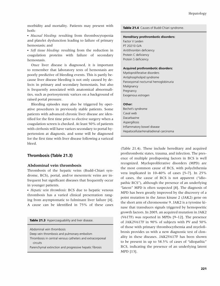
BLBK186-Key April 24, 2009 14:58
Hepatology
morbidity and mortality. Patients may present with
both:� Mucosal bleeding: resulting from thrombocytopenia
and platelet dysfunction leading to failure of primary
hemostasis; and� Soft tissue bleeding: resulting from the reduction in
coagulation proteins with failure of secondary
hemostasis.
Once liver disease is diagnosed, it is important
to remember that laboratory tests of hemostasis are
poorly predictive of bleeding events. This is partly be-
cause liver disease bleeding is not only caused by de-
fects in primary and secondary hemostasis, but also
is frequently associated with anatomical abnormali-
ties, such as portosystemic varices on a background of
raised portal pressure.
Bleeding episodes may also be triggered by oper-
ative procedures in previously stable patients. Some
patients with advanced chronic liver disease are iden-
tified for the first time prior to elective surgery when a
coagulation screen is checked. At least 50% of patients
with cirrhosis will have varices secondary to portal hy-
pertension at diagnosis, and some will be diagnosed
for the first time with liver disease following a variceal
bleed.
Thrombosis (Table 21.3)
Abdominal vein thrombosisThrombosis of the hepatic veins (Budd-Chiari syn-
drome, BCS), portal, and/or mesenteric veins are in-
frequent but significant diseases that frequently occur
in younger patients.� Hepatic vein thrombosis: BCS due to hepatic venous
thrombosis has a varied clinical presentation rang-
ing from asymptomatic to fulminant liver failure [4].
A cause can be identified in 75% of these cases
Table 21.3 Hypercoaguability and liver disease.
Abdominal vein thrombosis
Deep vein thrombosis and pulmonary embolism
Thrombosis in central venous catheters and extracorporeal
circuits
Parenchymal extinction and progressive hepatic fibrosis
Table 21.4 Causes of Budd-Chiari syndrome.
Hereditary prothrombotic disorders:Factor V Leiden
PT 20210 G/A
Antithrombin deficiency
Protein C deficiency
Protein S deficiency
Acquired prothrombotic disorders:Myeloproliferative disorders
Antiphospholipid syndrome
Paroxsysmal nocturnal hemoglobinuria
Malignancy
Pregnancy
Exogenous estrogen
Other:Bechet’s syndrome
Caval web
Dacarbazine
Aspergillosis
Inflammatory bowel disease
Hepatocellular/renal/adrenal carcinoma
(Table 21.4). These include hereditary and acquired
prothrombotic states, trauma, and infection. The pres-
ence of multiple predisposing factors in BCS is well
recognized. Myeloproliferative disorders (MPD) are
the most common cause of BCS, with polychthemia
vera implicated in 10–40% of cases [5–7]. In 25%
of cases, the cause of BCS is not apparent (“idio-
pathic BCS”), although the presence of an underlying
“latent” MPD is often suspected [8]. The diagnosis of
MPD has been greatly improved by the discovery of a
point mutation in the Janus kinase 2 (JAK2) gene on
the short arm of chromosome 9. JAK2 is a tyrosine ki-
nase that transduces signals triggered by hemopoeitic
growth factors. In 2005, an acquired mutation in JAK2
(V617F) was reported in MPDs [9–12]. The presence
of JAK2V617F in 90% of subjects with PV and 50%
of those with primary thrombocythemia and myelofi-
brosis provides us with a new diagnostic test of clon-
ality in these diseases. JAK2V617F has been shown
to be present in up to 58.5% of cases of “idiopathic”
BCS, indicating the presence of an underlying latent
MPD [13].
221
BLBK186-Key April 24, 2009 14:58
CHAPTER 21
� Portal vein thrombosis: Portal vein thrombosis (PVT) is
often silent and may not be discovered until variceal
hemorrhage occurs. Clinical features include abdom-
inal pain, ascites, and rectal bleeding. Thrombosis
extending to the mesenteric vessels may lead to
mesenteric infarction. Common causes of PVT include
hepatic cirrhosis, abdominal sepsis, tumors, and pan-
creatitis. As in BCS, the role of multiple etiological
factors is well recognized, including hereditary and ac-
quired prothrombotic disorders and estrogen therapy.
JAK2V617F has been reported to occur in 17–36% of
patients with PVT [14–17]. Anticoagulation therapy
with vitamin K antagonists may be hazardous in pa-
tients with esophageal varices, and consequently, de-
cisions on treatment are based on extent/age of throm-
bosis, presence of varices, history of bleeding, and the
presence of an underlying prothrombotic disorder. In
acute PVT, anticoagulation is frequently given for a pe-
riod of 6 months; a longer duration of anticoagulation
may be beneficial in chronic PVT or in patients with
underlying prothrombotic disorders [18].
Venous thromboembolism (VTE)Deep vein thrombosis and pulmonary embolism oc-
cur frequently in hospitalized medical patients, and
routine risk assessment and thromboprophylaxis with
heparin is now widely recommended [19]. Despite the
hemorrhagic tendency of chronic liver disease, VTE
occurs not infrequently in these patients. Prothrom-
botic coagulation disturbances in liver disease include
reduced levels of anticoagulant proteins (antithrom-
bin, protein C, protein S), antiphospholipid antibodies,
and hyperfibrinolysis. The incidence of VTE in chronic
liver disease may well be underestimated, as lower
limb edema and dyspnea are nonspecific and com-
monly present in these patients. In one retrospective
case-control study of patients with cirrhosis, new VTE
was present in 0.5% of inpatients with cirrhosis [20].
Progression of fibrosis due toparenchymal extinction
It is clear that, in patients with chronic liver disease
(particularly cirrhosis), the prothrombotic state can
lead to further hepatic injury (“parenchymal extinc-
tion”) and progression of fibrosis. This may be due to
thrombosis in small intrahepatic vessels. There is some
evidence that the prothrombotic state predisposes to
accelerated fibrogenesis, for example, the observed as-
sociation between factor V Leiden mutation and ac-
celerated fibrosis in patients with hepatitis C infection.
There is no good evidence to support the use of stan-
dard anticoagulation to prevent progression of hepatic
fibrosis, but the advent of newer antithrombotics may
kindle new interest in this area.
Extracorporeal circuits
Continuous venovenous hemodialysis (CVVH) and
artificial liver support machines both require the ex-
posure of blood to artificial surfaces, inevitably lead-
ing to coagulation activation and clotting in the extra-
corporeal circuit. A variety of anticoagulant strategies
have been advocated, often depending on local exper-
tise and the perceived bleeding risk in individual cases.
Laboratory investigation of hemostasisin liver disease
Clotting screenThe PT and activated partial thromboplastin time
(APTT) are commonly prolonged in chronic liver dis-
ease, reflecting a reduction in coagulation factor pro-
duction by the failing liver (Table 21.5). Patients with
abnormal laboratory tests only require treatment to
correct coagulopathy when there is evidence of active
bleeding or prior to surgery.
Chronic liver diseaseNo single coagulation test is predictive of hemorrhage
or thrombosis in patients with chronic liver disease:� Factor VII has a short half-life and levels fall early
in subjects with hepatic impairment. An isolated
prolongation of the PT may be the only demonstrable
laboratory abnormality in those with mild disease.� A prolonged PT or international normalized ratio
(INR) is a key indicator of hepatic dysfunction and
commonly used as a trigger for liver transplantation;
however, it is vitamin K-dependent. Although a pro-
longed PT is often used as a marker of hepatic dysfunc-
tion, it is most sensitive to low coagulation FVII levels
222
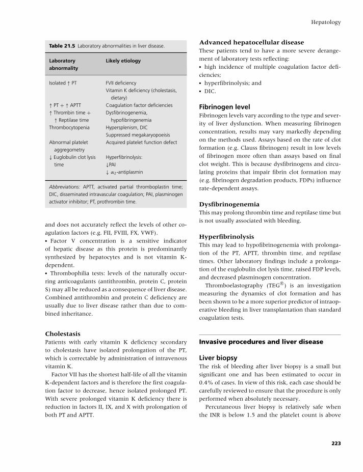
BLBK186-Key April 24, 2009 14:58
Hepatology
Table 21.5 Laboratory abnormalities in liver disease.
Laboratory Likely etiologyabnormality
Isolated ↑ PT FVII deficiency
Vitamin K deficiency (cholestasis,
dietary)
↑ PT + ↑ APTT Coagulation factor deficiencies
↑ Thrombin time +↑ Reptilase time
Dysfibrinogenemia,
hypofibringenemia
Thrombocytopenia Hypersplenism, DIC
Suppressed megakaryopoeisis
Abnormal platelet
aggregometry
Acquired platelet function defect
↓ Euglobulin clot lysis
time
Hyperfibrinolysis:
↓PAI
↓ α2-antiplasmin
Abbreviations: APTT, activated partial thromboplastin time;
DIC, disseminated intravascular coagulation; PAI, plasminogen
activator inhibitor; PT, prothrombin time.
and does not accurately reflect the levels of other co-
agulation factors (e.g. FII, FVIII, FX, VWF).� Factor V concentration is a sensitive indicator
of hepatic disease as this protein is predominantly
synthesized by hepatocytes and is not vitamin K-
dependent.� Thrombophilia tests: levels of the naturally occur-
ring anticoagulants (antithrombin, protein C, protein
S) may all be reduced as a consequence of liver disease.
Combined antithrombin and protein C deficiency are
usually due to liver disease rather than due to com-
bined inheritance.
CholestasisPatients with early vitamin K deficiency secondary
to cholestasis have isolated prolongation of the PT,
which is correctable by administration of intravenous
vitamin K.
Factor VII has the shortest half-life of all the vitamin
K-dependent factors and is therefore the first coagula-
tion factor to decrease, hence isolated prolonged PT.
With severe prolonged vitamin K deficiency there is
reduction in factors II, IX, and X with prolongation of
both PT and APTT.
Advanced hepatocellular diseaseThese patients tend to have a more severe derange-
ment of laboratory tests reflecting:� high incidence of multiple coagulation factor defi-
ciencies;� hyperfibrinolysis; and� DIC.
Fibrinogen levelFibrinogen levels vary according to the type and sever-
ity of liver dysfunction. When measuring fibrinogen
concentration, results may vary markedly depending
on the methods used. Assays based on the rate of clot
formation (e.g. Clauss fibrinogen) result in low levels
of fibrinogen more often than assays based on final
clot weight. This is because dysfibrinogens and circu-
lating proteins that impair fibrin clot formation may
(e.g. fibrinogen degradation products, FDPs) influence
rate-dependent assays.
DysfibrinogenemiaThis may prolong thrombin time and reptilase time but
is not usually associated with bleeding.
HyperfibrinolysisThis may lead to hypofibrinogenemia with prolonga-
tion of the PT, APTT, thrombin time, and reptilase
times. Other laboratory findings include a prolonga-
tion of the euglobulin clot lysis time, raised FDP levels,
and decreased plasminogen concentration.
Thromboelastography (TEG R©) is an investigation
measuring the dynamics of clot formation and has
been shown to be a more superior predictor of intraop-
erative bleeding in liver transplantation than standard
coagulation tests.
Invasive procedures and liver disease
Liver biopsyThe risk of bleeding after liver biopsy is a small but
significant one and has been estimated to occur in
0.4% of cases. In view of this risk, each case should be
carefully reviewed to ensure that the procedure is only
performed when absolutely necessary.
Percutaneous liver biopsy is relatively safe when
the INR is below 1.5 and the platelet count is above
223
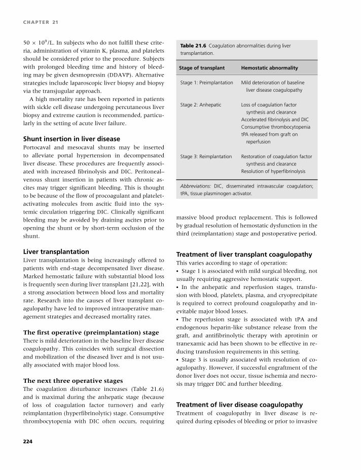
BLBK186-Key April 24, 2009 14:58
CHAPTER 21
50 × 109/L. In subjects who do not fulfill these crite-
ria, administration of vitamin K, plasma, and platelets
should be considered prior to the procedure. Subjects
with prolonged bleeding time and history of bleed-
ing may be given desmopressin (DDAVP). Alternative
strategies include laparoscopic liver biopsy and biopsy
via the transjugular approach.
A high mortality rate has been reported in patients
with sickle cell disease undergoing percutaneous liver
biopsy and extreme caution is recommended, particu-
larly in the setting of acute liver failure.
Shunt insertion in liver diseasePortocaval and mesocaval shunts may be inserted
to alleviate portal hypertension in decompensated
liver disease. These procedures are frequently associ-
ated with increased fibrinolysis and DIC. Peritoneal–
venous shunt insertion in patients with chronic as-
cites may trigger significant bleeding. This is thought
to be because of the flow of procoagulant and platelet-
activating molecules from ascitic fluid into the sys-
temic circulation triggering DIC. Clinically significant
bleeding may be avoided by draining ascites prior to
opening the shunt or by short-term occlusion of the
shunt.
Liver transplantationLiver transplantation is being increasingly offered to
patients with end-stage decompensated liver disease.
Marked hemostatic failure with substantial blood loss
is frequently seen during liver transplant [21,22], with
a strong association between blood loss and mortality
rate. Research into the causes of liver transplant co-
agulopathy have led to improved intraoperative man-
agement strategies and decreased mortality rates.
The first operative (preimplantation) stageThere is mild deterioration in the baseline liver disease
coagulopathy. This coincides with surgical dissection
and mobilization of the diseased liver and is not usu-
ally associated with major blood loss.
The next three operative stagesThe coagulation disturbance increases (Table 21.6)
and is maximal during the anhepatic stage (because
of loss of coagulation factor turnover) and early
reimplantation (hyperfibrinolytic) stage. Consumptive
thrombocytopenia with DIC often occurs, requiring
Table 21.6 Coagulation abnormalities during liver
transplantation.
Stage of transplant Hemostatic abnormality
Stage 1: Preimplantation Mild deterioration of baseline
liver disease coagulopathy
Stage 2: Anhepatic Loss of coagulation factor
synthesis and clearance
Accelerated fibrinolysis and DIC
Consumptive thrombocytopenia
tPA released from graft on
reperfusion
Stage 3: Reimplantation Restoration of coagulation factor
synthesis and clearance
Resolution of hyperfibrinolysis
Abbreviations: DIC, disseminated intravascular coagulation;
tPA, tissue plasminogen activator.
massive blood product replacement. This is followed
by gradual resolution of hemostatic dysfunction in the
third (reimplantation) stage and postoperative period.
Treatment of liver transplant coagulopathyThis varies according to stage of operation:� Stage 1 is associated with mild surgical bleeding, not
usually requiring aggressive hemostatic support.� In the anhepatic and reperfusion stages, transfu-
sion with blood, platelets, plasma, and cryoprecipitate
is required to correct profound coagulopathy and in-
evitable major blood losses.� The reperfusion stage is associated with tPA and
endogenous heparin-like substance release from the
graft, and antifibrinolytic therapy with aprotinin or
tranexamic acid has been shown to be effective in re-
ducing transfusion requirements in this setting.� Stage 3 is usually associated with resolution of co-
agulopathy. However, if successful engraftment of the
donor liver does not occur, tissue ischemia and necro-
sis may trigger DIC and further bleeding.
Treatment of liver disease coagulopathyTreatment of coagulopathy in liver disease is re-
quired during episodes of bleeding or prior to invasive
224
BLBK186-Key April 24, 2009 14:58
Hepatology
procedures. The type of treatment required will de-
pend on the specific hemostatic abnormalities present
and the nature of the bleeding event. It is impor-
tant to remember that most patients with coagulopa-
thy are stable and do not require specific therapy.
When bleeding does occur, the associated triggers (e.g.
esophageal varices secondary to portal hypertension)
need to be addressed in conjunction with strategies to
correct coagulopathy.
Vitamin KDeficiency of vitamin K may occur in liver disease, re-
sulting from poor diet or secondary to malabsorption.
Administration of 10 mg vitamin K1 will correct the
PT, at least partially, in most patients within 48 hours.
The PT will not fully correct if there is a coexisting de-
fect in hepatic synthetic function.
PlasmaFresh frozen plasma (FFP) or solvent detergent plasma
(SDP) contains all the coagulation factors synthesized
by the healthy liver. It may be used to correct multi-
ple coagulation factor deficiencies in bleeding patients
or prior to invasive procedures. A significant problem
with FFP is the large volume of transfusion required to
correct the PT and APTT in severe liver disease, partic-
ularly in volume-overloaded patients with ascites and
peripheral edema. In addition, repeated transfusions
are required to maintain circulating coagulation fac-
tor levels. Prothrombin complex concentrates should
be used with caution in liver disease, as their use has
been associated with thromboembolism and DIC. Cry-
oprecipitate or fibrinogen concentrate should be used
to correct hypofibrinogenemia associated with hyper-
fibrinolysis or DIC.
PlateletsPlatelet transfusions are indicated in bleeding patients
with platelet counts of �10 × 109/L, or in patients
undergoing invasive procedures. Platelet increments
are generally poor in subjects with portal hyperten-
sion because of sequestration of transfused platelets
in the spleen. DDAVP (0.3�g/kg) may be of value in
patients with acquired platelet dysfunction and pro-
longed bleeding time, but its value in bleeding patients
is uncertain.
AntifibrinolyticsAprotinin, tranexamic acid, and ε-aminocaproic acid
have all been shown to reduce operative blood loss
and transfusion requirements in liver transplantation.
The use of these agents to reduce fibrinolysis associ-
ated with chronic liver disease is of uncertain value,
and their use in DIC is not recommended.
Other agents
Heparin and antithrombinTheir use in DIC has not led to significant improve-
ments in blood loss or mortality and is therefore not
recommended.
EstrogensThere are some reports on efficacy in bleeding related
to chronic liver disease, but further data from clini-
cal trials are required before their use can be recom-
mended.
Fibrin glueLocal endoscopic applications have been shown to be
effective in the treatment of bleeding gastric varices.
Recombinant factor VIIaSmall studies have demonstrated reduced clotting
times in chronic liver disease and a reduction in trans-
fusion requirements in liver transplantation. The opti-
mal role for recombinant factor VIIa in the treatment
of liver coagulopathy has yet to be defined.
References
1 Amirano L, Guardascione MA, Brancaccio V, et al.
Coagulation disorders in liver disease. Semin Liver Dis
2002;22:83–96.
2 Ratnoff OD. Hemostatic defects in liver and biliary
tract disease. In: Ratoff OD, Forbes CD, eds. Disorders
of Hemostasis. Philadelphia: WB Saunders, 1996:422.
3 Northup PG, Sundaram V, Fallon MB, et al. Hyperco-
agulation and thrombophilia in liver disease. J Thromb
Haemost 2007;6:2–9.
4 Narayanan KV, Shah V, Kamath PS. The Budd–Chiari
syndrome. N Eng J Med 2004;350:578–85.
5 Denninger MH, Chait Y, Casadevall N, et al. Cause of
portal or hepatic vein thrombosis in adults: the role
225
BLBK186-Key April 24, 2009 14:58
CHAPTER 21
of multiple concurrent factors. Hepatology 2000;31:587–
91.
6 Valla D, Casadevall N, Lacombe C, et al. Primary myelo-
proliferative disorder and hepatic vein thrombosis: a
prospective study of erythroid colony formation in vitro
in 20 patients with Budd-Chiari Syndrome. Ann Intern
Med 1985;103:329–34.
7 Primignani M, Martinelli I, Bucciarelli P. Risk factors for
thrombophilia in extrahepatic portal vein obstruction.
Hepatology 2005;41:603–8.
8 Pagliuca A, Mufti GJ, Janossa-Tahernia M, et al. In
vitro colony culture and chromosomal studies in hep-
atic and portal vein thrombosis: possible evidence of
an occult myeloproliferative state. Q J Med 1990;76:
981–9.
9 Baxter EJ, Scott LM, Campbell PJ, et al. Acquired mu-
tation of the tyrosine kinase JAK2 in human myelopro-
liferative diseases. Lancet 2005;365:1054–61.
10 Levine RL, Wadleigh M, Cools J, et al. Activating mu-
tation in the tyrosine kinase JAK2 in polycythemia
vera, essential thrombocythemia, and myeloid meta-
plasia with myelofibrosis. Cancer Cell 2005;7:387–97.
11 James C, Ugo V, Le Couedic JP, et al. A unique clonal
JAK2 mutation leading to constitutive signaling causes
polycythemia vera. Nature 2005;434:1144–8.
12 Kralovics R, Passamonti F, Buser AS, et al. A gain-of-
function mutation of JAK2 in myeloproliferative disor-
ders. N Engl J Med 2005;352:1779–90.
13 Patel RK, Lea NC, Heneghan MA, et al. Preva-
lence of the activating JAK2 tyrosine kinase mutation
V617F in the Budd-Chiari Syndrome. Gastroenterology
2006;130:2031–8.
14 Primigani M, Barosi G, Bergamaschi G, et al. Role of the
JAK2 mutation in the diagnosis of chronic myeloprolif-
erative disorders in splanchnic vein thrombosis. Hepa-
tology 2006;44(6):1528–34.
15 Kiladjian J, Cervantes F, Leebeek F, et al. Role of JAK2
mutation detection in Budd-Chiari Syndrome (BCS)
and portal vein thrombosis (PVT) associated to MPD.
Blood 2006;108:Abstract 377.
16 Colaizzo D, Amitrano L, Tiscia GL, et al. The JAK2
V617F mutation frequently occurs in patients with
portal and mesenteric venous thrombosis. J Thromb
Haemost 2006;5:55–61.
17 Regina S, Herault O, D’Iteroche L, et al. The JAK2
mutation V617F is specifically associated with idio-
pathic splanchnic vein thrombosis. J Thromb Haemost
2007;5(4):859–61.
18 Condat B, Pessione F, Helene Denninger M, et al. Re-
cent portal or mesenteric venous thrombosis: increased
recognition and frequent recanalization on anticoagu-
lant therapy. Hepatology 2000;32:466–70.
19 Hirsh J, Dalen JE, Master MPM, et al. American College
of Chest Physicians The Sixth (2000) ACCP Guidelines
for Antithrombotic Therapy for Prevention and Treat-
ment of Thrombosis. Chest 2001;119:1S–2S.
20 Northup PG, McMahon MM, Ruhl AP, et al. Coagu-
lopathy does not fully protect hospitalized cirrhosis pa-
tients from peripheral venous thromboembolism. Am J
Gastroenterol 2006;101:1524–8.
21 Porte RJ, Knot EA, Bontempo FA. Hemostasis in liver
transplantation. Gastroenterology 1989;97:488–501.
22 Starzl TE, Demertris A, van Thiel DH. Liver transplan-
tation. N Engl J Med 1989;321:1014–22, 1092–99.
226
BLBK186-Key April 11, 2009 13:4
22 NephrologyStephanie Perry and Thomas L. Ortel
Bleeding in renal disease
Clinical presentationIn 1764, the association between bleeding and re-
nal disease was first entertained in Morgagni’s “Opera
Omnia” [1,2]. Signs of bleeding may appear as easy
bruising, petechia, gingival bleeding, epistaxis, or pro-
longed bleeding or hematomas from venipuncture
or catheter sites [1,3,4]. Life-threatening bleeding
can occur from pericardial tamponade, retroperitoneal
bleeding, intracranial bleeding, and gastrointestinal
bleeding [1,3]. Retroperitoneal bleeding can be spon-
taneous or postprocedure. For example, bleeding rates
postrenal biopsy are reported to range from 11% to
22% [4]. Patients with uncontrolled hypertension and
undergoing hemodialysis treatments are at risk of in-
tracranial bleeds. Gastrointestinal bleeding has been
reported to be the second leading cause of death in
patients with acute renal failure [3].
EtiologyThere are many factors that may contribute to bleed-
ing in renal disease as can occur in other disease states.
Factors such as anemia or use of antiplatelet or anti-
coagulant drugs may increase the risk of bleeding in
patients. The mechanisms behind anemia contributing
to risk of bleeding include the following:� decreased laminar flow effect of red cell facilitating
platelet interaction with the endothelial lining [3,5];� red cells release ADP and thromboxane A2, which
enhances platelet aggregation [3,5]; and� hemoglobin scavenges nitric oxide (NO) [3,5].
Drugs that may reach higher levels in patients with
renal disease, such as penicillin G, carbenicillin, ticar-
cillin, ampicillin, and moxalactam, can increase the
risk of bleeding by binding to platelets and blocking
platelet-membrane agonist receptors.
Even more specific to patients with renal disease
is bleeding due to uremia, which disrupts normal
platelet–platelet and platelet–vessel wall interactions
[2,3]. These disruptions in platelet function due to
uremia may be multifactorial. Mechanisms to explain
uremia-induced platelet dysfunction have included
the following:� altered arachidonic acid metabolism [2,6];� deficient platelet stores of adenosine diphosphate
and serotonin [2,3,5,6]; and� impaired binding of fibrinogen [2,3] and von Wille-
brand factor (vWF) [2,5,6].
One area of great interest in explaining the “ure-
mia effect” on platelets is the role of guanidinosuc-
cinic acid [2,5]. Guanidinosuccinic acid accumulates
during ammonia detoxification when an amidine is
transferred to aspartic acid from L-arginine. L-arginine
has been found to be a major substrate for NO as
well. NO is known to modulate vascular tone and
interferes with platelet adhesion to endothelium and
platelet–platelet interaction. NO has been found to
be higher in uremic patients on hemodialysis when
compared with healthy subjects. Similarly, guanidi-
nosuccinic acid appears to have vasodilation effects on
intact endothelium. In addition to having similar bi-
ological activities of NO, high guanidinosuccinic con-
centrations appear to cause formation of NO by uremic
vessels [2].
Prevention and treatmentThe site, extent, and acuity of bleeding will dictate
the treatment. For external bleeding, mechanical ma-
neuvers such as applying pressure over the area of
bleeding and, if an extremity is involved, elevating the
area above the level of the heart can help control or
227
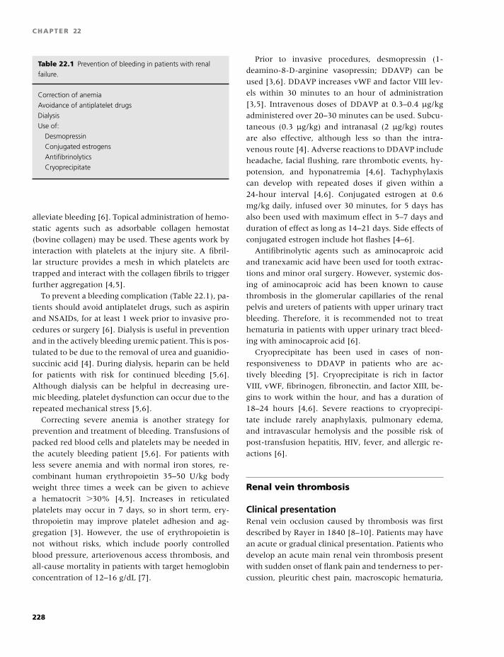
BLBK186-Key April 11, 2009 13:4
CHAPTER 22
Table 22.1 Prevention of bleeding in patients with renal
failure.
Correction of anemia
Avoidance of antiplatelet drugs
Dialysis
Use of:
Desmopressin
Conjugated estrogens
Antifibrinolytics
Cryoprecipitate
alleviate bleeding [6]. Topical administration of hemo-
static agents such as adsorbable collagen hemostat
(bovine collagen) may be used. These agents work by
interaction with platelets at the injury site. A fibril-
lar structure provides a mesh in which platelets are
trapped and interact with the collagen fibrils to trigger
further aggregation [4,5].
To prevent a bleeding complication (Table 22.1), pa-
tients should avoid antiplatelet drugs, such as aspirin
and NSAIDs, for at least 1 week prior to invasive pro-
cedures or surgery [6]. Dialysis is useful in prevention
and in the actively bleeding uremic patient. This is pos-
tulated to be due to the removal of urea and guanidio-
succinic acid [4]. During dialysis, heparin can be held
for patients with risk for continued bleeding [5,6].
Although dialysis can be helpful in decreasing ure-
mic bleeding, platelet dysfunction can occur due to the
repeated mechanical stress [5,6].
Correcting severe anemia is another strategy for
prevention and treatment of bleeding. Transfusions of
packed red blood cells and platelets may be needed in
the acutely bleeding patient [5,6]. For patients with
less severe anemia and with normal iron stores, re-
combinant human erythropoietin 35–50 U/kg body
weight three times a week can be given to achieve
a hematocrit �30% [4,5]. Increases in reticulated
platelets may occur in 7 days, so in short term, ery-
thropoietin may improve platelet adhesion and ag-
gregation [3]. However, the use of erythropoietin is
not without risks, which include poorly controlled
blood pressure, arteriovenous access thrombosis, and
all-cause mortality in patients with target hemoglobin
concentration of 12–16 g/dL [7].
Prior to invasive procedures, desmopressin (1-
deamino-8-D-arginine vasopressin; DDAVP) can be
used [3,6]. DDAVP increases vWF and factor VIII lev-
els within 30 minutes to an hour of administration
[3,5]. Intravenous doses of DDAVP at 0.3–0.4 µg/kg
administered over 20–30 minutes can be used. Subcu-
taneous (0.3 µg/kg) and intranasal (2 µg/kg) routes
are also effective, although less so than the intra-
venous route [4]. Adverse reactions to DDAVP include
headache, facial flushing, rare thrombotic events, hy-
potension, and hyponatremia [4,6]. Tachyphylaxis
can develop with repeated doses if given within a
24-hour interval [4,6]. Conjugated estrogen at 0.6
mg/kg daily, infused over 30 minutes, for 5 days has
also been used with maximum effect in 5–7 days and
duration of effect as long as 14–21 days. Side effects of
conjugated estrogen include hot flashes [4–6].
Antifibrinolytic agents such as aminocaproic acid
and tranexamic acid have been used for tooth extrac-
tions and minor oral surgery. However, systemic dos-
ing of aminocaproic acid has been known to cause
thrombosis in the glomerular capillaries of the renal
pelvis and ureters of patients with upper urinary tract
bleeding. Therefore, it is recommended not to treat
hematuria in patients with upper urinary tract bleed-
ing with aminocaproic acid [6].
Cryoprecipitate has been used in cases of non-
responsiveness to DDAVP in patients who are ac-
tively bleeding [5]. Cryoprecipitate is rich in factor
VIII, vWF, fibrinogen, fibronectin, and factor XIII, be-
gins to work within the hour, and has a duration of
18–24 hours [4,6]. Severe reactions to cryoprecipi-
tate include rarely anaphylaxis, pulmonary edema,
and intravascular hemolysis and the possible risk of
post-transfusion hepatitis, HIV, fever, and allergic re-
actions [6].
Renal vein thrombosis
Clinical presentationRenal vein occlusion caused by thrombosis was first
described by Rayer in 1840 [8–10]. Patients may have
an acute or gradual clinical presentation. Patients who
develop an acute main renal vein thrombosis present
with sudden onset of flank pain and tenderness to per-
cussion, pleuritic chest pain, macroscopic hematuria,
228
BLBK186-Key April 11, 2009 13:4
Nephrology
unilateral radiographic abnormalities by intra-
venous pyelogram, and worsening renal function.
Patients with nephrotic syndrome may present with
no symptoms except peripheral edema [11]. Neonates
and infants more often have an acute presentation
and are found to have abdominal distension, a flank
mass from increase in kidney size, hematuria, and
proteinuria and may also present with bilateral renal
vein thrombosis. Neonates and infants are often
diagnosed in the setting of severe dehydration and
present with dry mouth, decreased urine output,
and decreased skin turgidity. In cases of gradual
onset, patients may have no symptoms or nonspecific
chronic complaints of nausea, apathy, weakness, and
generalized edema and may have symptoms of upper
abdominal or flank pain [11].
EtiologyIn adults, renal vessel occlusion is usually from vein
thrombosis [8]. Renal vein thrombosis is a compli-
cation of nephrotic syndrome and has been found
in patients with primary glomerular diseases, such
as membranous glomerulopathy, minimal change dis-
ease, membranoproliferative glomerulonephritis, focal
glomerulosclerosis, and rapidly progressive glomeru-
lonephritis, and in other diseases with nephrosis, such
as lupus erythematosus, diabetes mellitus, primary
amyloidosis, familial Mediterranean fever with amy-
loidosis, sickle cell disease, sarcoidosis, and vasculiltis.
Various studies have reported the incidence of renal
vein thrombosis in nephrotic syndrome ranging from
5% to 62% with a high incidence among patients with
membranous glomerulopathy with reports of 50–60%
of patients evaluated [8,9,11].
Renal vein thrombosis is more common in primary
glomerular disease but also occurs in other renal dis-
eases, such as acute pyelonephritis, lupus nephritis,
or amyloidosis in the setting of nephrotic syndrome.
Other mechanisms associated with renal vein throm-
bosis include the following:� thrombosis of the inferior vena cava with secondary
renal vein involvement;� direct extension of tumor into the lumen of the renal
veins causing occlusion with thrombosis proximal to
the tumor;� alteration in renal blood flow (i.e. volume loss,
diarrhea, sepsis, adrenal hemorrhage, hypoglycemia,
seizure disorders or hypoxia in cyanotic congenital
heart disease, tricuspid insufficiency, constrictive peri-
carditis);� systemic diseases with hypercoagulable states, such
as sickle cell disease, primary antiphospholipid syn-
drome, advanced malignancy; and� surgically induced renal vein occlusion with throm-
bosis beyond the ligature [8].
Diagnosis, treatment, and prognosisIn cases of acute onset with complete occlusion, kid-
ney size increases within the first week with subse-
quent decrease in size over a couple of weeks and
later renal atrophy. In the early phase, therefore, an
ultrasound will show an enlarged kidney and hy-
perechogenic kidney in about 90% of cases [12].
Color Doppler ultrasound improves the ability to de-
tect flow in the renal artery and the renal vein and
has a high degree of sensitivity in detecting renal
vein thrombosis in post-renal transplant patients. In
chronic renal vein thrombosis, renal venous occlusion
causes the development of varicosities, which shows
a notching appearance in the ureter and collateral
venous drainage around the kidney by intravenous
urography [12].
The imaging method of choice is CT [12]. Screen-
ing with spiral CT has a sensitivity and specificity for
covert renal vein thrombosis of 90–100% compared
with the gold standard of renal venous angiography
[10]. CT also has the advantage of detecting renal
tumors and other renal diseases [12]. Doppler ultra-
sonography has high false-positive and false-negative
rates for renal vein thrombosis (40% and 15%, respec-
tively) [10].
Magnetic resonance angiography (MRA) has the ad-
vantage of avoiding nephrotoxic intravenous contrast
agents. MRA is better at showing anatomic variants,
vessel displacement, collateral circulation, and neo-
plastic vessel infiltration [10].
Treatment consists of correcting the underlying
problem when due to secondary reasons for decreased
renal blood flow. Dialysis may be needed in causes
of renal failure from renal vein thrombosis [8]. The
mortality rate can be high, and often patients with re-
nal vein thrombosis are at risk of death from other
thromboembolic events, such as pulmonary emboli.
For patients with nephrotic syndrome and renal vein
229
BLBK186-Key April 11, 2009 13:4
CHAPTER 22
thrombosis, chronic anticoagulation therapy is war-
ranted to prevent further extension of the throm-
bus and to prevent other thromboembolic events
[9–11].
Thrombolytic agents have been given; however, this
has been associated with high frequency of death due
to bleeding complications [9]. Surgical thrombectomy
has also been tried but is only rarely indicated for pa-
tients not responding to medical therapy [9]. Percuta-
neous mechanical thrombectomy has also been used
with success [9].
Outcomes in a retrospective study from the Mayo
Clinic from 1980 to 2000 found a high incidence
of underlying renal malignancy (66%) and nephritic
syndrome (20%) as the most common causes of re-
nal vein thrombosis. In this cohort, the overall sur-
vival was poor with predictors of mortality including
cancer and infection [13]. In patients with untreated
renal vein thrombosis, the incidence of pulmonary
embolus has been found to range from 20% to
40% [15].
In a retrospective review of neonatal renal vein
thrombosis from 1992 to 2006, 70.6% of neonates, re-
gardless of the treatment [about 40% with unfraction-
ated heparin (UFH)/low-molecular-weight heparin
(LMWH) and about 40% with supportive treatment]
received, had irreversible damage. In this study,
the mortality rate was observed to be 3.3% [14].
It is reported that approximately 20% of neonates
may develop persistent hypertension and about 3%
may need chronic dialysis or kidney transplantation
[14].
Nephrotic syndrome/hypercoagulability
Incidence and prevalence ofthromboembolic eventsAddis in 1948 noted the frequent occurrence of
thromboembolic events in patients with nephrotic
syndrome. The increase in incidence that clinicians
have noted since described by Addis could in part be
due to longer survival of patients with improvements
in care, especially with the introduction of antibiotics
[11].
Certain renal diseases are associated with throm-
bophilia, notably primary and secondary nephrotic
syndrome, systemic lupus erythematosus with lupus
anticoagulant, granulamatous vasculitis (Wegener’s
granulomatosis), and Behcet syndrome. Consistently
associated with thromboembolic events are membra-
nous nephropathy (primary and secondary), membra-
noproliferative glomerulonephritis, minimal change
disease, and possible amyloidosis [10].
Thromboembolic complications are one of the most
serious outcomes for patients with nephrotic syn-
drome. Sites involved include pulmonary arteries, ax-
illary and subclavian veins, femoral veins, coronary
arteries, and mesenteric arteries. The most common
presentation is for deep vein thrombosis (DVT) of the
extremities [11]. Various studies have found that the
prevalence for thromboembolic events other than re-
nal vein thrombosis ranges from 8.5% to 44% [11].
About 15% of patients with nephrotic syndrome are
reported to develop DVT, with or without renal vein
thrombosis. Renal vein thrombosis, unilateral or bi-
lateral, has been reported to develop in about 25–
30% of patients with nephrotic syndrome from pri-
mary renal disease. The highest risks are reported with
membranous glomerulonephritis at 37%, membra-
noproliferative glomeruonephritis at 26%, and mini-
mal change disease at 24% [10]. The combined rates
of DVT and renal vein thrombosis in patients with
membranous nephropathy have been reported to be
as high as 45% [10]. Others have reported that 40%
of patients with membranous nephropathy and serum
albumin �2.5g/dL had venous thromboembolism ver-
sus only 2.7% of patients with albumin �2.5g/dL [10].
The prevalence of thromboembolic events in children
with nephrotic syndrome has been reported to range
from 2% to 25% [16]. In patients older than 60 years
with membranous nephropathy, it was found that the
majority of deaths were caused by thromboembolic
events [17].
EtiologyThe thrombophilia associated in patients with
nephrotic syndrome may be mulifactorial. Environ-
mental risk shared by patients with medical illnesses
include immobilization, obesity, need for surgeries
and procedures, and co-morbidity such as congestive
heart failure. Environmental factors that may be more
specific for patients with nephrotic syndrome include
volume depletion and the use of diuretic and/or
steroid therapy [10].
230
BLBK186-Key April 11, 2009 13:4
Nephrology
The hypercoagulability state observed in patients
with nephrotic syndrome may also be multifactorial
[17,18] and include:� increased levels of clotting factors,� decreased levels of anticoagulant proteins,� increased platelet activity,� increase in vWF, and� abnormal finbrinolysis.
Prothrombotic factors have been reported to in-
clude increased fibrinogen levels, factor VIII levels,
and platelet adhesiveness; whereas, the antithrom-
botic factors that have been found to be decreased in-
clude antithrombin levels and proteins C and S activ-
ity. It has been reported that decreased plasminogen
levels, elevated plasminogen activator inhibitor levels,
or albumin deficiency-related impairment of the in-
teraction of plasminogen–fibrin may account for the
impaired thrombolytic activity [10]. Also reports of
increases in platelet count and aggregation to ADP
and collagen and increased β-thromboglobulin levels,
a marker for platelet aggregation, have been noted
[11]. Hypoalbuminemia may play a role in increas-
ing free arachidonic acid and subsequent increase in
thromboxane [18]. LDL cholesterol, which is usually
elevated in patients with nephrotic syndrome, is toxic
to the endothelium, which leads to impaired NO pro-
duction and may increase platelet–vessel wall interac-
tions [18]. Most likely, an increase in thrombin ac-
tivity accelerates fibrinogen-induced fibrin formation
and contributes to the thrombotic risk. This in part
may be due to increases in clotting factors V and VIII
and decreases in the inhibition of the coagulation cas-
cade due to decreased levels of proteins C and S and
antithrombin [18]. Antithrombin may be one of the
most important coagulation inhibitors and inhibits ac-
tivated factors XII, IX, X, and XI and plasmin. An-
tithrombin increases after steroid therapy [11]. Also,
decreased fibrinolytic activity may be due to several
mechanisms:� increased α2-antiplasmin;� decreased albumin may lead to decreased binding of
plasminogen to fibrin; and� elevated Lp (a) competes for the binding to fibrino-
gen or fibrin [18].
More specific to patients with membranous
nephropathy is the association of anti-enolase au-
toantibodies. These autoantibodies may interfere with
fibrinolysis [10].
TreatmentTreatment for thromboembolic events in patients with
nephrotic syndrome is anticoagulation for the dura-
tion of the nephrotic state [10]. Given the high in-
cidence of thromboembolic events in patients with
nephrotic syndrome and membranous glomerulopa-
thy, Bellomo and Atkins have recommended prophy-
lactic anticoagulation [19].
Graft loss due tothromobosis/thrombophilias
Incidence and clinical presentation ofthromboembolic eventsRenovascular thrombosis was found to be the cause of
graft loss posttransplant in approximately 8% of recip-
ients, with thrombosis accounting for 25% of graft loss
in �1 year posttransplant, as reported by Matas and
colleagues [20]. Bakir and colleagues found throm-
bosis to be the cause of graft loss in 45% of recipi-
ents in �90 days posttransplant and 37% in �1 year
[21]. Thrombosis of the renal vein graft is more com-
mon, causes pain and swelling of the graft, and can
frequently lead to allograft rupture [20,21]. Thrombo-
sis of the renal artery does not cause pain, swelling, or
rupture. Also, thrombosis of both renal vein and artery
can occur at the same time.
EtiologyProposed mechanisms for renovascular thrombosis
have included problems associated with the surgi-
cal procedure such as donor vessel abnormalities,
including difference in diameter of vessels, multi-
ple renal arteries, stenosis of the renal artery of the
donor, atherosclerosis of the donor or recipient ves-
sel, excessive surgical trauma of the vessels due to
repeated re-anastomosis, lymphocele posttransplant,
and prolonged ischemia with resulting reperfusion
damage [21]. However, these technical problems or
concerns for immunosuppressive drugs have not been
able to explain the often unexpected graft thrombosis
spurring the interest in thromphilia, inherited or ac-
quired, as possible risk factors for renovascular throm-
bosis and subsequent graft loss [20,21].
Posttransplantation, the coagulation system is
activated due to tissue trauma causing inflammation
and expression of tissue factor, and fibrinolysis may
231
BLBK186-Key April 11, 2009 13:4
CHAPTER 22
be impaired due to overexpression of plasminogen
activator inhibitor-I in the endothelium [20,21].
Inherited thrombophilia has been associated with
allograft thrombosis. Irish reported a 6% prevalence
of Factor V Leiden in 300 transplant recipients who
had a four-fold increase in allograft thrombosis, which
represented 20% of graft loss in this cohort [22].
One study found that the presence of prothrombin
gene G20210A polymorphism was associated with
a shorter median allograft survival of 65.9 months
versus 149 months. Acquired thrombophilias have
also been associated with allograft thrombosis. An-
other study evaluated 502 patients, of which 11 of 23
identified with antiphospholipid antibody syndrome
underwent transplant. Of the 11 patients, 7 who did
not receive anticoagulation had a graft thrombosis
within 1 week, whereas 3 of the 4 patients who
received anticoagulation maintained long-term graft
function [19,22,23]. Allograft recipients with SLE and
antiphospholipid antibodies were found to have a
40% risk of thrombosis, graft loss, or death caused by
thromboembolism versus 8% of SLE patients without
antiphospholipid antibodies [23].
Diagnosis and preventionThe diagnosis of allograft thrombosis can be made by
performing angiography or by histology [21]. Color
Doppler ultrasonagraphy has become a standard pro-
cedure for evaluating renal allografts and can re-
liably detect complete allograft vein thrombosis if
the pathognomonic reversed diastolic flow exists in
the arteries [24]. Whether or not patients should be
screened prior to transplant has been debated. Some
have advocated thrombophilia screening for high-risk
patients, such as patients with personal or family his-
tory of thrombosis and in children and adolescents
who appear to be at higher risk of allograft thrombosis
[19,21].
Using anticoagulation at prophylactic or treatment
dosing to decrease allograft thrombosis needs to be
weighed against the risk of bleeding. One study used
dalteparin 2500 U daily just during the period of hos-
pitalization for low-risk patients and dalteparin 5000
U daily for at least 1 month for high-risk patients. In
120 allograft recipients, the high-risk group included
patients with hypercoagulable state (15%) or grafts
with multiple vessels (31%) [25]. There were no re-
ports of allograft thrombosis or major hemorrhagic
events; however, there was also no control group for
comparison.
Dose adjustment of anticoagulants inrenal insufficiency
AnticoagulantsAs discussed in the previous sections, patients with re-
nal disease may have problems with bleeding as well
as thrombosis. Additionally, most of the anticoagu-
lants that are used in practice are excreted by the kid-
neys. Therefore treating patients with anticoagulants
offers a greater challenge with dosing and requires
closer monitoring for signs of bleeding.
UFH is principally metabolized by the reticulen-
dothelial system with approximately �10% excreted
in the urine unchanged and, for this reason, is the
anticoagulant of choice for patients with severe renal
impairment. However, Thorevska and coworkers per-
formed a retrospective cohort study which concluded
that full-dose enoxaparin and UFH had similar ma-
jor hemorrhagic events in patients with renal insuffi-
ciency [26]. In their cohort of 620 patients, there were
a total of 149 hemorrhagic events of which 60 were
major hemorrhages. Of interest is the timing of the
hemorrhagic events between enoxaparin and UFH. A
higher percentage of major hemorrhagic events in the
enoxaparin group occurred after 3 days of therapy,
whereas approximately half of the major hemorrhagic
events in the UFH group occurred within the first 3
days of therapy. Also of interest is that patients with
severe renal insufficiency (GFR ≤20 mL/min) had
20% more major hemorrhagic events and 150% more
minor hemorrhagic events in the enoxaparin group.
Although the increase in major hemorrhagic events in
the enoxaparin group was not statistically significant,
the number of patients receiving clopidrogel or glyco-
protein IIB/IIIA drugs was statistically higher in the
group receiving UFH and therefore there may have
been a bias toward using UFH in patients who were
more at risk of bleeding.
Guidelines for mild to moderaterenal insufficiencyFor the LMWHs, including enoxaparin, dalteparin,
and tinzaparin, there are no dosage adjustments given
for mild renal insufficiency (CLcr of 50–80 mL/min)
232
BLBK186-Key April 11, 2009 13:4
Nephrology
and moderate renal insufficiency (CLcr of 30–50
mL/min) [27]. For enoxaparin, it has been reported
that the clearance is reduced by 30% in patients with
moderate renal insufficiency. Because of concern for
drug accumulation, it may be advisable to reduce the
dose and perhaps follow anti-factor Xa levels to help
guide therapy in patients with moderate renal insuf-
ficiency. Data are even more limited for dalteparin
and tinzaparin. Also, for the factor Xa inhibitor fon-
daparinux, there are no dosage adjustments given for
mild and moderate renal insufficiency. Therefore, pa-
tients need to be monitored closely for any signs of
hemorrhage and consideration of following anti-factor
Xa levels, especially if therapy is anticipated to be pro-
longed.
In the direct thrombin inhibitor (DTI) class of
agents, only argatroban can be used without dosage
adjustments for renal insufficiency. For acute coronary
syndrome (ACS) patients undergoing percutaneous
intervention, bivalirudin is not dose-reduced. How-
ever, for use in patients with heparin-induced throm-
bocytopenia (HIT), we would recommend reducing
the dose from 0.15 mg/kg/hour to 0.05 mg/kg/hour.
Patients should be monitored closely with checking ac-
tivated partial thromboplastin time (APTT) 2–3 hours
after initiation of drug and after dosage changes. For
lepirudin, the manufacturer recommends dosage re-
duction for patients with CLcr �60. For CLcr between
30 mL/minute and 60 mL/minute, a reduced bolus
dose of 0.2 mg/kg is recommended. For Clcr 45–60
mL/minute, the infusion rate should be reduced to
0.075 mg/kg/hour, and for Clcr 30–44 mL/minute, the
infusion rate should be reduced to 0.045 mg/kg/hour.
Others have advocated even lower doses of lep-
irudin infusion as follows: (1) normal renal func-
tion, 0.1 mg/kg/hour; (2) CLcr 45–60 mL/minute, 0.05
mg/kg/hour; and (3) CLcr 30–44 mL/minute, 0.03
mg/kg/hour [27]. When treating HIT with thrombo-
sis a bolus dose is usually given; however, for isolated
HIT, a bolus dose is not recommended. Also, some
clinicians will not use a bolus dose in elderly patients
and in patients with renal insufficiency. It is also rec-
ommended to monitor APTT 4 hours after initiating
the infusion and after any dosage changes.
Guidelines for severe renal insufficiencyFor severe renal insufficiency, defined as a Clcr �30
mL/minute, dose reductions are recommended for
LMWHs. For DVT prophylaxis, enoxaparin is reduced
to 30 mg once daily for the following: abdominal
surgery, hip replacement, knee replacement, and in
medical patients. For DVT treatment, enoxaparin is
reduced to 1 mg/kg once daily. For dalteparin, the
manufacturing guidelines only comment that, for can-
cer patients being treated for a venous thromboem-
bolic event, anti-Xa levels should be monitored and
the dose adjusted accordingly. For tinzaparin, there is
a 24% decrease in clearance, and therefore it should
be used with caution. Fondaparinux is contraindicated
for patients with severe renal insufficiency.
As for mild and moderate renal insufficiency, ar-
gatroban is the only DTI that does not require a
dose adjustment. For ACS patients undergoing percu-
taneous intervention, bivalirudin should be reduced
to 1 mg/kg/hour. For dialysis-dependent patients on
nondialysis days, the dose should be reduced to 0.25
mg/kg/hour. For use in patients with HIT, we would
recommend reducing the dose to 0.03 mg/kg/hour.
Patients should be monitored closely with checking
APTT 2–3 hours after initiation of drug and after
dosage changes. For lepirudin, the manufacturer rec-
ommends reducing the bolus dose to 0.2 mg/kg and
to reduce the infusion rate to 0.0225 mg/kg/hour for
Clcr 15–29 mL/minute and to not use lepirudin for Clcr
�15 mL/minute. It is also recommended to monitor
APTT after 4 hours of initiating the infusion and after
any dosage changes.
Acknowledgment
Grant support: NIH K23-HL084233 (SLP); CDC UO1-
DD000014 (TLO); NIH UO1-HL072289 (TLO); NIH
U54-HL077878 (TLO).
References
1 Sohal AS, Gangji AS, Crowther MA, Treleavan D. Ure-
mic bleeding: pathophysiology and clinical risk factors.
Thromb Res 2006;118:417–22.
2 Salmaon S. Uremic bleeding: pathophysiology, di-
agnosis, and management. Hosp Physician 2001;76:
45–50.
3 Hedges SJ, Dehoney SB, Hooper JS, Amanzadeh J,
Busti AJ. Evidence-based treatment recommendations
233
BLBK186-Key April 11, 2009 13:4
CHAPTER 22
for uremic bleeding. Nat Clin Pract Nephrol 2007;3:
138–53.
4 Kaw D, Malhaotra D. Platelet dysfunction and end-
stage renal disease. Semin Dial 2006;19:317–22.
5 Noris M, Remuzzi G. Uremic bleeding: closing the circle
after 30 years of controversies? Blood 1999;94:2569–74.
6 Gangji AS, Sohal AS, Treleaven D, Crowther MA.
Bleeding in patients with renal insufficiency: a
practical guide to clinical management. Thromb Res
2006;118:423–8.
7 Phrommintikul A, Haas SJ, Elsik M, Krum H. Mortal-
ity and target haemoglobin concentrations in anaemic
patients with chronic kidney disease treated with ery-
thropoietin: a meta-analysis. Lancet 2007;369:381–8.
8 Witz M, Kantarovsky A, Morag B, Shifrin EG. Renal
vein occlusion: a review. J Urol 1996;155:1173–9.
9 Jaar BG, Kim HS, Samaniego, Lund GB, Atta MG.
Percutaneous mechanical thrombectomy: a new ap-
proach in the treatment of acute renal-vein thrombosis.
Nephrol Dial Transplant 2002;17:1122–5.
10 Wagoner RD, Stanson AW, Holley KE, Winter CS.
Renal vein thrombosis in idiopathic membranous
glomerulpathy and nephrotic syndrome: incidence and
significance. Kidney Int 1983;23:368–74.
11 Llach F. Hypercoagulability, renal vein thrombosis, and
other thrombotic complications of nephrotic syndrome.
Kidney Int 1985;28:429–39.
12 Asgher M, Ahmed K, Shah SS, Siddique MK, Dasgupta
P, Khan MS. Renal vein thrombosis. Eur J Vasc Endovasc
Surg 2007;34:217–23.
13 Wysokinski WE, Gosk-Bierska I, Greene EL, Grill D,
Wiste H, McBane RD. Clinical characteristics and long-
term follow-up of patients with renal vein thrombosis.
Am J Kidney Dis 2008;51:224–32.
14 Lau KK, Stoffman JM, Williams S, et al. Neona-
tal renal vein thrombosis: review of the English-
language literature between 1992 and 2006. Pediatrics
2007;120:e1278–84.
15 Glassock RJ. Prophylactic anticoagulation in nephrotic
syndrome: a clinical conundrum. J Am Soc Nephrol
2007;18:2221–5.
16 Fabri D, Belangero MS, Annichino-Bizzacchi JM, Ar-
ruda VR. Inherited risk factors for thombophilia in chil-
dren with nephrotic syndrome. Eur J Pediatr 1998;157:
939–42.
17 O’Callaghan CA, Hicks J, Doll H, Sacks SH, Cameron
JS. Characteristics and outcome of membranous
nephropathy in older patients. Int Urol Nephrol 2002;33:
157–65.
18 Rabelink TJ, Zwaninga JJ, Koomans HA, Sixma JJ.
Thrombosis and hemostasis in renal disease. Kidney Int
1994;46:287–96.
19 Bellomo R, Atkins RC. Membranous nephropathy
and thromboembolism: is prophylactic anticoagulation
warranted? Nephron 1993;63:249–54.
20 Matas AJ, Humar A, Gillingham KJ, et al. Five pre-
ventable cause of kidney graft loss in the 1990s: a
single-center analysis. Kidney Int 2002;62:704–14.
21 Bakir N, Sluiter WJ, Ploeg RJ, van Son WJ, Tegzess AM.
Primary renal graft thrombosis. Nephrol Dial Transplant
1996;11:140–7.
22 Irish A. Renal allograft thrombosis: can throm-
bophilia explain the inexplicable? Nephrol Dial Trans-
plant 1999;14:2297–303.
23 Andrassy J, Zeier M, Anrassy K. Do we need screen-
ing for thrombophilia prior to kidney transplantation?
Nephrol Dial Transplant 2004:19;iv64–8.
24 Schwenger V, Hinkel UP, Nahm A, Morath C, Zeier
M. Color doppler ultrasonography in the diagnos-
tic evaluation of renal allografts. Nephron Clin Pract
2006;104:c107–12.
25 Alkunaizi AM, Olyaei AJ, Barry JM, et al. Efficacy and
safety of low molecular weight heparin in renal trans-
plantation. Transplantation 1998;66:533–4.
26 Thorevska N, Amoateng-Adjepong Y, Sabahi R, et al.
Anticoagulation in hospitalized patients with renal in-
sufficiency: a comparison of bleeding rates with unfrac-
tionated heparin vs enoxaparin. Chest 2004;125:856–
63.
27 Lobo BL. Use of newer anticoagulants in patients with
chronic kidney disease. Am J Health-Syst Pharm 2007;64:
2017–6.
234
BLBK186-Key April 11, 2009 13:5
23 OncologyAnna Falanga and Marina Marchetti
Introduction
The association between cancer and thrombosis has
been known for more than a century. The occurrence
of venous thromboembolism is a common complica-
tion of cancer. It can also precede the onset of an oc-
cult neoplasia, as first reported by Armand Trousseau
in 1865. Almost at the same time, the possibility that a
relation between the clotting mechanisms and the de-
velopment of metastasis may occur was postulated by
Billroth in 1878.
In the last three decades, remarkable progress has
been made in this field, both by basic research and
clinical studies. It is now clear that there is a two-way
connection between coagulation and cancer [1]:� malignant disease results in a prothrombotic imbal-
ance of the host hemostatic system; and� prothrombotic mechanisms may promote tumor
growth and dissemination.
Recently, molecular studies have demonstrated that
oncogenes responsible for neoplastic transformation
also drive programs for hemostatic protein expression
and clotting system activation [2–4]. Specifically,� Targeting activated human mesenchymal–epithelial
transition factor (MET) to the mouse liver with lentivi-
ral vector determined progressive hepatocarcinogene-
sis, which is preceded and accompanied by a throm-
bohemorrhagic syndrome (i.e. venous thrombosis in
tail vein and fatal internal hemorrhage) and laboratory
signs of disseminated intravascular coagulation (DIC).
Genome-wide expression profiling of hepatocytes ex-
pressing MET showed up-regulation of PAI-1 and
COX-2 genes with a two- to three-fold increase in
circulating protein levels [2].� In an in vitro model of human glioma cells, the loss
of the tumor suppressor gene PTEN up-regulated the
expression of tissue factor (TF) and increased the levels
of plasma clotting proteins [3].� In a model of human colorectal cancer cells, TF ex-
pression was shown to be under the control of two
major transforming events driving disease progression:
the activation of K-ras oncogene and the inactivation
of the p53 tumor suppressor [4].
Patients with cancer are exposed to a significant risk
of thrombosis [5]. This situation is aggravated by anti-
tumor therapies [6]. Data derived from large, random-
ized, controlled trials have been used to determine the
true incidence of this complication and to define the
major risk factors for thrombosis in cancer [7].
Very commonly, cancer patients present with ab-
normalities of laboratory tests of blood coagulation,
even without clinical manifestations of thromboem-
bolism and/or hemorrhage. These abnormalities re-
veal different degrees of blood clotting activation and
characterize the hypercoagulable state of these sub-
jects [8]. The results of laboratory tests in these
patients demonstrate that a process of fibrin formation
and removal is continuously ongoing during the de-
velopment of malignancy.
The pathogenesis of thrombophilia in cancer is mul-
tifactorial; however, an important role is attributed to
the tumor cell capacity to interact with and activate
the host hemostatic system. Among other factors that
contribute to the increased thrombotic diathesis in pa-
tients with cancer are the antitumor therapies.
Experimental studies show that fibrin and other
coagulation proteins are involved in multiple steps
of tumor growth and dissemination. Therefore, phar-
macological interventions to prevent thrombotic phe-
nomena in malignancy may possibly contribute to the
control of the malignant disease progression.
The aim of this chapter is to summarize the most re-
cent advances in our knowledge on the thrombophilic
235
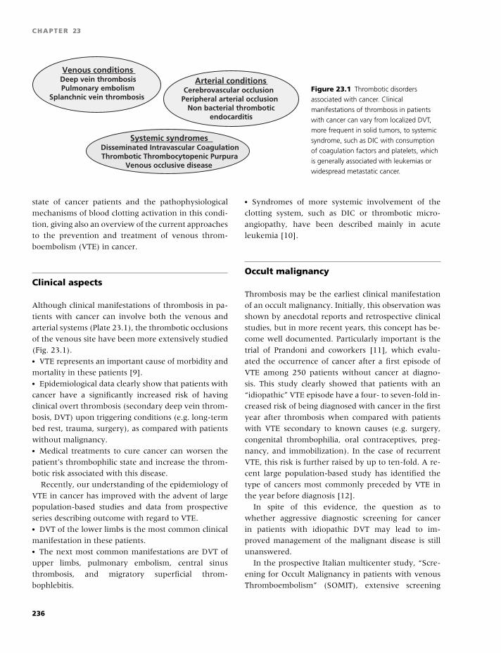
BLBK186-Key April 11, 2009 13:5
CHAPTER 23
Venous conditionsDeep vein thrombosisPulmonary embolism
Splanchnic vein thrombosis
Arterial conditionsCerebrovascular occlusion
Peripheral arterial occlusionNon bacterial thrombotic
endocarditis
Systemic syndromesDisseminated Intravascular CoagulationThrombotic Thrombocytopenic Purpura
Venous occlusive disease
Figure 23.1 Thrombotic disorders
associated with cancer. Clinical
manifestations of thrombosis in patients
with cancer can vary from localized DVT,
more frequent in solid tumors, to systemic
syndrome, such as DIC with consumption
of coagulation factors and platelets, which
is generally associated with leukemias or
widespread metastatic cancer.
state of cancer patients and the pathophysiological
mechanisms of blood clotting activation in this condi-
tion, giving also an overview of the current approaches
to the prevention and treatment of venous throm-
boembolism (VTE) in cancer.
Clinical aspects
Although clinical manifestations of thrombosis in pa-
tients with cancer can involve both the venous and
arterial systems (Plate 23.1), the thrombotic occlusions
of the venous site have been more extensively studied
(Fig. 23.1).� VTE represents an important cause of morbidity and
mortality in these patients [9].� Epidemiological data clearly show that patients with
cancer have a significantly increased risk of having
clinical overt thrombosis (secondary deep vein throm-
bosis, DVT) upon triggering conditions (e.g. long-term
bed rest, trauma, surgery), as compared with patients
without malignancy.� Medical treatments to cure cancer can worsen the
patient’s thrombophilic state and increase the throm-
botic risk associated with this disease.
Recently, our understanding of the epidemiology of
VTE in cancer has improved with the advent of large
population-based studies and data from prospective
series describing outcome with regard to VTE.� DVT of the lower limbs is the most common clinical
manifestation in these patients.� The next most common manifestations are DVT of
upper limbs, pulmonary embolism, central sinus
thrombosis, and migratory superficial throm-
bophlebitis.
� Syndromes of more systemic involvement of the
clotting system, such as DIC or thrombotic micro-
angiopathy, have been described mainly in acute
leukemia [10].
Occult malignancy
Thrombosis may be the earliest clinical manifestation
of an occult malignancy. Initially, this observation was
shown by anecdotal reports and retrospective clinical
studies, but in more recent years, this concept has be-
come well documented. Particularly important is the
trial of Prandoni and coworkers [11], which evalu-
ated the occurrence of cancer after a first episode of
VTE among 250 patients without cancer at diagno-
sis. This study clearly showed that patients with an
“idiopathic” VTE episode have a four- to seven-fold in-
creased risk of being diagnosed with cancer in the first
year after thrombosis when compared with patients
with VTE secondary to known causes (e.g. surgery,
congenital thrombophilia, oral contraceptives, preg-
nancy, and immobilization). In the case of recurrent
VTE, this risk is further raised by up to ten-fold. A re-
cent large population-based study has identified the
type of cancers most commonly preceded by VTE in
the year before diagnosis [12].
In spite of this evidence, the question as to
whether aggressive diagnostic screening for cancer
in patients with idiopathic DVT may lead to im-
proved management of the malignant disease is still
unanswered.
In the prospective Italian multicenter study, “Scre-
ening for Occult Malignancy in patients with venous
Thromboembolism” (SOMIT), extensive screening
236
BLBK186-Key April 11, 2009 13:5
Oncology
was found to be effective in identifying precociously
an occult malignancy [13]. Computerized tomography
(CT) scanning of the abdomen and pelvis was the most
effective diagnostic test, and CT scan and a gastroin-
testinal investigation (such as hemoccult) was the best
diagnostic combination. Based on the data from the
SOMIT study, an analysis of costs of different screen-
ing strategies (in relation to the expected live years
gained with each of them) shows that some of these
strategies may be cost-effective. Finally, a prospective
cohort follow-up study of 864 consecutive patients
with acute VTE [14] suggests that a limited diagnos-
tic workup (i.e. abdominal and pelvic ultrasound and
laboratory markers for malignancy) may have the ca-
pacity to identify approximately one-half of the ma-
lignancies present in patients who were negative on
routine clinical evaluation. In most of the cases, the
malignancies identified by extensive screening are in
an early stage; therefore, larger clinical trials to estab-
lish the impact of this finding on cancer prognosis are
warranted.
The hypercoagulable state of malignancy
Even without thrombosis and before any therapy, pa-
tients with cancer present with multiple laboratory ab-
normalities of hemostasis showing a hypercoagulable
condition [8].
Routine laboratory tests
Coagulation profiles performed in the past have re-
vealed that the most frequent routine abnormalities
reported are:� Elevated levels of plasma coagulation factor (i.e. fib-
rinogen, factors V, VIII, IX, and X);� Increased plasma levels of fibrin(ogen) degradation
products (FDP or D-dimers); and� Thrombocytosis.
In two large prospective clinical trials evaluating
routine coagulation tests in cancer patients:� FDP levels and thrombin times were increased only
in 8% and 14% of cases, respectively;� Fibrinogen and platelet count were found more fre-
quently elevated (48% and 36% of the cases, respec-
tively);
� The increase in the levels of these two markers over
time directly correlated with the disease progression;
and� Activation of the clotting system occurs in the ab-
sence of DIC or manifest thrombosis.
Specialized tests
Recently, the development of novel, more sensitive
laboratory tests for the detection of the hypercoag-
ulable state or subclinical DIC (which are listed in
Table 23.1) has enabled the detection of ongoing ac-
tivation of blood coagulation in vivo. These tests mea-
sure the final products of clotting reactions in plasma
and include:� Peptides released during the proteolytic activation of
pro-enzymes into active clotting enzymes, i.e.:
◦ prothrombin fragment 1 + 2 [F1+2],
◦ protein C activation fragment,
◦ factor IX and X activation fragments, and
◦ fibrinopeptide A.� Enzyme–inhibitor complexes produced during the
activation of the coagulation and fibrinolytic systems,
i.e.:
◦ thrombin–antithrombin complexes [TAT] and
◦ plasmin–antiplasmin complexes [PAP].� Cross-linked fibrin degradation product, i.e.:
◦ D-dimer.� Cell membrane-associated markers to study the ac-
tivation of cellular components of the hemostatic sys-
tem, including platelets and leukocytes, i.e.:
◦ P-selectin (or CD62P), and CD63 on platelet sur-
face, and
◦ Mac1 (or CD11b) and leukocyte alkaline phos-
phatase (LAP) on leukocyte surface.
Predictors of thrombosisStudies on the plasma levels of these markers have
provided a biochemical definition of the hypercoag-
ulable state in humans. However, no studies of sound
methodological design have been performed to indi-
cate whether any of these tests of blood coagulation
can serve as an adequate predictor of thrombosis in
cancer patients. No studies have prospectively com-
pared, in the same subjects, the levels of the plasma
markers with the thrombotic events (confirmed by ob-
jective tests). Large studies are still required to answer
237
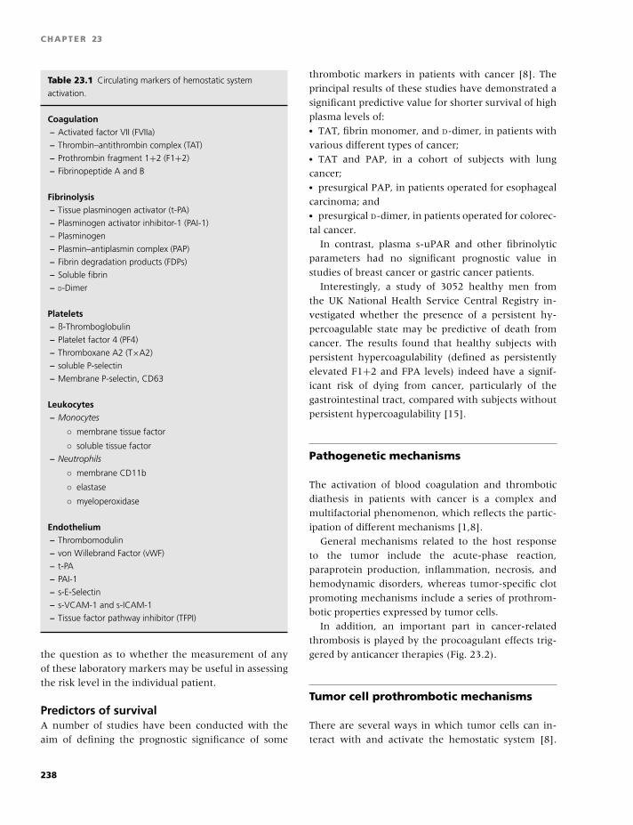
BLBK186-Key April 11, 2009 13:5
CHAPTER 23
Table 23.1 Circulating markers of hemostatic system
activation.
Coagulation– Activated factor VII (FVIIa)
– Thrombin–antithrombin complex (TAT)
– Prothrombin fragment 1+2 (F1+2)
– Fibrinopeptide A and B
Fibrinolysis– Tissue plasminogen activator (t-PA)
– Plasminogen activator inhibitor-1 (PAI-1)
– Plasminogen
– Plasmin–antiplasmin complex (PAP)
– Fibrin degradation products (FDPs)
– Soluble fibrin
– D-Dimer
Platelets– ß-Thromboglobulin
– Platelet factor 4 (PF4)
– Thromboxane A2 (T×A2)
– soluble P-selectin
– Membrane P-selectin, CD63
Leukocytes– Monocytes
◦ membrane tissue factor
◦ soluble tissue factor
– Neutrophils
◦ membrane CD11b
◦ elastase
◦ myeloperoxidase
Endothelium– Thrombomodulin
– von Willebrand Factor (vWF)
– t-PA
– PAI-1
– s-E-Selectin
– s-VCAM-1 and s-ICAM-1
– Tissue factor pathway inhibitor (TFPI)
the question as to whether the measurement of any
of these laboratory markers may be useful in assessing
the risk level in the individual patient.
Predictors of survivalA number of studies have been conducted with the
aim of defining the prognostic significance of some
thrombotic markers in patients with cancer [8]. The
principal results of these studies have demonstrated a
significant predictive value for shorter survival of high
plasma levels of:� TAT, fibrin monomer, and D-dimer, in patients with
various different types of cancer;� TAT and PAP, in a cohort of subjects with lung
cancer;� presurgical PAP, in patients operated for esophageal
carcinoma; and� presurgical D-dimer, in patients operated for colorec-
tal cancer.
In contrast, plasma s-uPAR and other fibrinolytic
parameters had no significant prognostic value in
studies of breast cancer or gastric cancer patients.
Interestingly, a study of 3052 healthy men from
the UK National Health Service Central Registry in-
vestigated whether the presence of a persistent hy-
percoagulable state may be predictive of death from
cancer. The results found that healthy subjects with
persistent hypercoagulability (defined as persistently
elevated F1+2 and FPA levels) indeed have a signif-
icant risk of dying from cancer, particularly of the
gastrointestinal tract, compared with subjects without
persistent hypercoagulability [15].
Pathogenetic mechanisms
The activation of blood coagulation and thrombotic
diathesis in patients with cancer is a complex and
multifactorial phenomenon, which reflects the partic-
ipation of different mechanisms [1,8].
General mechanisms related to the host response
to the tumor include the acute-phase reaction,
paraprotein production, inflammation, necrosis, and
hemodynamic disorders, whereas tumor-specific clot
promoting mechanisms include a series of prothrom-
botic properties expressed by tumor cells.
In addition, an important part in cancer-related
thrombosis is played by the procoagulant effects trig-
gered by anticancer therapies (Fig. 23.2).
Tumor cell prothrombotic mechanisms
There are several ways in which tumor cells can in-
teract with and activate the hemostatic system [8].
238
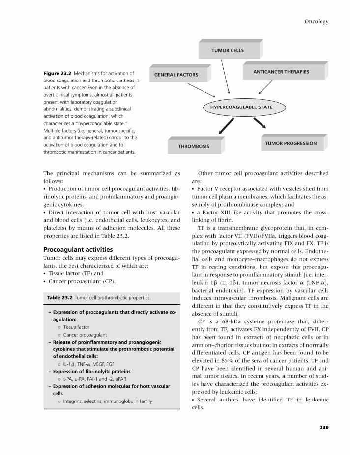
BLBK186-Key April 11, 2009 13:5
Oncology
TUMOR CELLS
ANTICANCER THERAPIESGENERAL FACTORS
HYPERCOAGULABLE STATE
THROMBOSISTUMOR PROGRESSION
Figure 23.2 Mechanisms for activation of
blood coagulation and thrombotic diathesis in
patients with cancer. Even in the absence of
overt clinical symptoms, almost all patients
present with laboratory coagulation
abnormalities, demonstrating a subclinical
activation of blood coagulation, which
characterizes a “hypercoagulable state.”
Multiple factors (i.e. general, tumor-specific,
and antitumor therapy-related) concur to the
activation of blood coagulation and to
thrombotic manifestation in cancer patients.
The principal mechanisms can be summarized as
follows:� Production of tumor cell procoagulant activities, fib-
rinolytic proteins, and proinflammatory and proangio-
genic cytokines.� Direct interaction of tumor cell with host vascular
and blood cells (i.e. endothelial cells, leukocytes, and
platelets) by means of adhesion molecules. All these
properties are listed in Table 23.2.
Procoagulant activitiesTumor cells may express different types of procoagu-
lants, the best characterized of which are:� Tissue factor (TF) and� Cancer procoagulant (CP).
Table 23.2 Tumor cell prothrombotic properties.
– Expression of procoagulants that directly activate co-agulation:
◦ Tissue factor
◦ Cancer procoagulant
– Release of proinflammatory and proangiogeniccytokines that stimulate the prothrombotic potentialof endothelial cells:
◦ IL-1β, TNF-α, VEGF, FGF
– Expression of fibrinolyitc proteins
◦ t-PA, u-PA, PAI-1 and -2, uPAR
– Expression of adhesion molecules for host vascularcells
◦ Integrins, selectins, immunoglobulin family
Other tumor cell procoagulant activities described
are:� Factor V receptor associated with vesicles shed from
tumor cell plasma membranes, which facilitates the as-
sembly of prothrombinase complex; and� a Factor XIII-like activity that promotes the cross-
linking of fibrin.
TF is a transmembrane glycoprotein that, in com-
plex with factor VII (FVII)/FVIIa, triggers blood coag-
ulation by proteolytically activating FIX and FX. TF is
the procoagulant expressed by normal cells. Endothe-
lial cells and monocyte–macrophages do not express
TF in resting conditions, but expose this procoagu-
lant in response to proinflammatory stimuli [i.e. inter-
leukin 1β (IL-1β), tumor necrosis factor α (TNF-α),
bacterial endotoxin]. TF expression by vascular cells
induces intravascular thrombosis. Malignant cells are
different in that they constitutively express TF in the
absence of stimuli.
CP is a 68-kDa cysteine proteinase that, differ-
ently from TF, activates FX independently of FVII. CP
has been found in extracts of neoplastic cells or in
amnion–chorion tissues but not in extracts of normally
differentiated cells. CP antigen has been found to be
elevated in 85% of the sera of cancer patients. TF and
CP have been identified in several human and ani-
mal tumor tissues. In recent years, a number of stud-
ies have characterized the procoagulant activities ex-
pressed by leukemic cells:� Several authors have identified TF in leukemic
cells.
239
BLBK186-Key April 11, 2009 13:5
CHAPTER 23
� CP has been found in blasts of various acute
myelogenous leukemia phenotypes, with the greatest
expression in acute promyelocytic leukemia (APL)
subtype.� The differentiating treatment with all-trans retinoic
acid (ATRA) of APL blasts in vitro reduces the expres-
sion of both TF and CP.� In patients with APL, the remission induction with
ATRA treatment induces the rapid resolution of the se-
vere coagulopathy of this disease and significantly af-
fects the procoagulant activities expressed by the bone
marrow cells in vivo.� Similar observations have been reported for breast
cancer.
Recent studies suggest a new role for TF in the
tumor growth and metastasis, which is not entirely
mediated via clotting activation, but may be depen-
dent on signaling through the cytoplasmic domain,
suggesting a “non-coagulation” role for TF in cancer
disease.
Fibrinolytic activitiesTumor cells can express all the proteins of the fibri-
nolytic system, including the urokinase-type (u-PA)
and the tissue-type (t-PA) plasminogen activators, and
their inhibitors, i.e. plasminogen activator inhibitor 1
and 2 (PAI-1 and PAI-2). Cancer cells also carry on
their membranes the specific plasminogen activator
receptor u-PAR, which favors the assembly of all the
fibrinolytic components, facilitating the activation of
the fibrinolytic cascade. It has been suggested that, in
leukemia patients, the expression of these activities by
blast cells may have a role in the pathogenesis of the
bleeding symptoms. An impaired plasma fibrinolytic
activity has been found in patients with solid tumors,
which represents per se another tumor-associated pro-
thrombotic mechanism.
Fibrinolysis is also a key component in tumor bio-
logy, as it is essential in releasing tumor cells from
their primary site of origin, in neo-angiogenesis, and
in promoting cell mobility and motility. Fibrinolytic
proteins are under evaluation as potentially valuable
predictors of disease-free interval and long-term sur-
vival in malignant disease. In breast cancer, patients
with low levels of u-PA and PAI-1 have a signifi-
cantly better survival than patients with high levels
of either factor, particularly in node-negative breast
cancer [16].
Cytokine activity
Down-regulation of anticoagulant activityTumor cells synthesize and release a variety of pro-
inflammatory cytokines (i.e. TNF-α, IL-1β) and
proangiogenic factors (VEGF, bFGF), which can act
on the different hemostatic cells and affect their an-
tithrombotic status.
These cytokines can induce the expression of TF
procoagulant activity by endothelial cells and mono-
cytes, and in parallel down-regulate the expression
of thrombomodulin (TM), a potent anticoagulant, ex-
pressed by endothelial cells. The up-regulation of TF,
together with the down-regulation of TM, leads to a
prothrombotic condition of the vascular wall.
Increased fibrinolysisThe same cytokines stimulate endothelial cells to
increase the production of the fibrinolysis in-
hibitor PAI-1, resulting in a subsequent inhibi-
tion of fibrinolysis, which further contributes to
the prothrombotic state. Cytokines also contribute
to enhance the adhesion potential of the vascular
wall, by increasing the expression of cell adhesion
molecules of endothelial cells, which become more
capable to attract tumor cells and support their ex-
travasation.
Procoagulant propertiesFurther, tumor cells and/or tumor cell cytokines can
induce the expression of monocyte TF. Monocyte ac-
tivation has been described to occur both in vitro
and in vivo. Indeed, tumor-associated macrophages
harvested from experimental and human tumors ex-
press significantly more TF than control cells. In
addition, circulating monocytes from patient with dif-
ferent types of cancer have been shown to express
increased TF activity. The generation of procoagu-
lant activity by monocyte–macrophages in vivo is
conceivably one mechanism for clotting activation in
malignancy.
Recruitment of white cellsThe cytokines and chemokines produced by malig-
nant cells are also mitogenic and/or chemoattrac-
tants for polymorphonuclear leukocytes. These cells,
upon activation, secrete proteolytic enzymes, which
240
BLBK186-Key April 11, 2009 13:5
Oncology
can damage the endothelial monolayer, and produce
additional cytokines and chemokines, which support
tumor growth, stimulate angiogenesis, and enable
metastatic spread via engagement with either venous
or lymphatic networks. They also synthetize VEGF
which is chemotactic for macrophages and can induce
TF procoagulant activity by monocytes and endothe-
lial cells.
Cell adhesion molecules
During the hematogenous spread, tumor cells directly
interact with endothelial cells, platelets, and leuko-
cytes. These interactions occur through surface cell-
adhesion molecules (i.e. integrins, selectins, and im-
munoglobulin superfamily).� The integrin family of cell-adhesion proteins pro-
motes the attachment and migration of cells to the sur-
rounding extracellular matrix (ECM). Through signals
transduced upon integrin ligation by ECM proteins or
immunoglobulin superfamily molecules, this family of
proteins has key roles in regulating tumor growth and
metastasis as well as tumor angiogenesis.� Selectins are multifunctional cell-adhesion mole-
cules that mediate the initial interactions between cir-
culating leukocytes and activated endothelium as well
as the adhesion of tumor cells during the metastatic
process.
The tumor cell capacity to adhere to the endothe-
lium and the underlying matrix is well described, and
adhesion molecule pathways specific to different tu-
mor cell types have been identified. The relevance of
the tight interaction of tumor cells with endothelial
cells in the pathogenesis of thrombosis in cancer is re-
lated to the localized promotion of clotting activation
and thrombus formation. The tumor cell attached to
endothelium can release its cytokine content into a
protected milieu that favors their prothrombotic and
proangiogenic activities. In addition, the adhesion of
tumor cells to leukocytes or vascular cells represents
the first step for cell migration and extravasation.
Experimental and in vitro studies have shown
that polymorphonuclear leukocytes may function
to promote tumor growth and metastasis. Tumor
cell-derived factors can regulate the expression of
various adhesion molecules (i.e. the β2-integrin
CD11b/CD18) by leukocytes, which in turn attach to
tumor cells and facilitate tumor cell migration through
the endothelium.
Platelets
Similarly to leukocytes, clinical and experimental ev-
idence suggests the importance of platelets in tu-
mor cell dissemination via the bloodstream. Platelets
can facilitate tumor cell adhesion and migration
through the vessel wall by a variety of mecha-
nisms, including bridging between tumor cells and
endothelial cells, and allowing migration of tumor
cells through the endothelial cell matrix by hep-
aranase activity. Tumor cells can activate platelets di-
rectly or through the release of proaggregatory me-
diators, including ADP, thrombin, and a cathepsin-
like cysteine protease. Upon activation, platelets ag-
gregate and release their granule contents, as shown
by the detection of elevated plasma levels of β-
thromboglobulin and PF4 (which are both localized in
alpha-granules of platelets), and of increased expres-
sion of platelet membrane activation markers, such
as P-selectin (or CD62P) and CD63, in patients with
malignancy.
In addition, activated platelets release VEGF and
PDGF, which play an important part in tumor neo-
angiogenesis.
Antitumor therapy prothromboticmechanisms
The pathogenesis of thrombosis during antitumor
therapies is not entirely understood, but a number of
mechanisms have been identified (Table 23.3) [17].
Table 23.3 Antitumor therapy prothrombotic mechanisms.
a. Release of procoagulant activities and cytokines from
damaged cells
b. Direct drug toxicity on vascular endothelium
c. Induction of monocyte tissue factor
d. Decrease of physiological anticoagulants (i.e. protein C,
proteins S, antithrombin)
e. Apoptosis
241
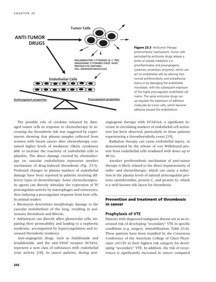
BLBK186-Key April 11, 2009 13:5
CHAPTER 23
ANTI-TUMORDRUGS
INFLAMMATORY CYTOKINES (IL-1, TNF)ANGIOGENIC CYTOKINES (VEGF, bFGF)PROTEOLYTIC ENZYMESCELL ADHESION MOLECULES
Anticoagulant properties Procoagulant properties
Tumor Cells
Endothelial Cells
Figure 23.3 Antitumor therapy
prothrombotic mechanisms. Tumor cells
perturbed by antitumor drugs release a
series of soluble mediators (i.e.
proinflammatoy and proangiogenic
cytokines, proteolytic enzymes), which can
act on endothelial cells by altering their
normal antithrombotic and antiadhesive
status or by damaging the endothelial
monolayer, with the subsequent exposure
of the highly procoagulant endothelial cell
matrix. The same antitumor drugs can
up-regulate the expression of adhesion
molecules by tumor cells, which become
adhesive toward the endothelium.
The possible role of cytokine released by dam-
aged tumor cells in response to chemotherapy in in-
creasing the thrombotic risk was suggested by exper-
iments showing that plasma samples collected from
women with breast cancer after chemotherapy con-
tained higher levels of mediators (likely cytokines)
able to increase the reactivity of endothelial cells to
platelets. The direct damage exerted by chemother-
apy on vascular endothelium represents another
mechanism of drug-induced thrombosis (Fig. 23.3).
Profound changes in plasma markers of endothelial
damage have been reported in patients receiving dif-
ferent types of chemotherapy. Some chemotherapeu-
tic agents can directly stimulate the expression of TF
procoagulant activity by macrophages and monocytes,
thus inducing a procoagulant response from host cells.
In animal studies:� Bleomycin determines morphologic damage to the
vascular endothelium of the lung, resulting in pul-
monary thrombosis and fibrosis.� Adriamycin can directly affect glomerular cells, im-
pairing their permeability and leading to a nephrotic
syndrome, accompanied by hypercoagulation and in-
creased thrombotic tendency.
Anti-angiogenic drugs, such as thalidomide and
lenalidomide, and the anti-VEGF receptor SU5416,
represent a new class of substances with endothelial
toxic activity [18]. In cancer patients, during anti-
angiogenic therapy with SU5416A, a significant in-
crease in circulating markers of endothelial cell activa-
tion has been observed, particularly in those patients
experiencing a thromboembolic event [19].
Radiation therapy can cause endothelial injury, as
demonstrated by the release of von Willebrand pro-
tein from endothelial cells irradiated with doses up to
40 Gy.
Another prothrombotic mechanism of anti-tumor
therapy is likely related to the direct hepatotoxicity of
radio- and chemotherapy, which can cause a reduc-
tion in the plasma levels of natural anticoagulant pro-
teins (antithrombin, protein C, and protein S), which
is a well-known risk factor for thrombosis.
Prevention and treatment of thrombosisin cancer
Prophylaxis of VTEPatients with diagnosed malignant disease are at an in-
creased risk of developing “secondary” VTE in specific
conditions (e.g. surgery, immobilization; Table 23.4).
These patients have been stratified by the Consensus
Conference of the American College of Chest Physi-
cians (ACCP) in their highest risk category for devel-
oping “secondary” VTE. In addition, the risk of recur-
rences is significantly increased in cancer compared
242
BLBK186-Key April 11, 2009 13:5
Oncology
Table 23.4 Risk of VTE in cancer patients undergoing
surgery.∗
Type of surgery Risk (%)
General 29
Gynecologic 41
Orthopedic 50–60
Neurosurgery 28
∗Adapted from Clagett et al. Ann Surg 1988;208:227.
with noncancer patients, even during treatments for
VTE. There is no evidence that there is a benefit from
giving antithrombotic prophylaxis to all cancer pa-
tients; however, there are selected conditions in which
prophylaxis has to be considered, such as surgical in-
terventions, acute medical illness, and administration
of antitumor therapies [20].
Cancer surgeryCancer surgery carries a two- to three-fold increased
thrombotic risk compared with noncancer surgery of
equal intensity. Perioperative prophylaxis with low
doses of unfractionated heparin (UFH) or with fixed
dose low-molecular-weight heparin (LMWH) is effec-
tive in significantly reducing the incidence of post-
operative VTE. LMWH has a good safety profile also
in this condition. Further, a higher dose of LMWH
has been shown to be more effective than a lower
dose in surgical cancer patients, without increasing
the hemorrhagic risk [21]. This is of particular rel-
evance as cancer patients are also at high risk of
bleeding. A prolonged postoperative prophylaxis up to
Table 23.5 Prolonged prophylaxis with LMWH in surgical cancer patients.
Study Cancer patients n % Prophylaxis Major VTEbleeding % incidence %
ENOXACAN II 332 (100%) Enoxaparin vs. placebo for:
Bergqvist, NEJM 2002 19–21 days 0.4 4.8
6–10 days 0 12
FAME 198 (58%) Dalteparin vs. no prophylaxis
for:
Rasmussen, JTH 2006 4 weeks 0 8.8
1 week 0 19.6
1 month after surgery for cancer can add a benefit
to reduce the rate of postoperative VTE. Two large
clinical trials have shown the safety and efficacy of
extended prophylaxis in cancer patients undergoing
abdominal or pelvic surgery. In the ENOXACAN II
study, a trial designed ad hoc for cancer patients, a
60% reduced rate of postoperative VTE was observed
in the arm randomized to receive prolonged prophy-
laxis with LMWH. The FAME study confirmed the
same results in a subgroup of patients with cancer
(Table 23.5) [20].
Medical conditionsThe advantages of thromboprophylaxis in nonsurgi-
cal conditions, such as in cancer patients with cen-
tral venous catheters (CVC) or during chemotherapy,
are still under evaluation. In recent prospective clin-
ical studies, the incidence of CVC-related thrombotic
complications in cancer patients appears to be lower
than that reported by earlier studies, with a rate of
about 4% for symptomatic VTE. These studies show
no significant benefit of thromboprophylaxis with
either LMWH or 1 mg/day fixed dose warfarin in pre-
venting CVC-related thrombosis. Therefore, routine
thromboprophylaxis has not been recommended so
far [20,22].
The role of thromboprophylaxis in medical can-
cer patients receiving chemo- and/or hormone ther-
apy is still undefined. In hospitalized cancer patients
with an acute medical illness, thromboprophylaxis is
recommended as for all other acute medical patients
[20,22,23].
In ambulatory cancer patients receiving chemo-
and/or hormone-therapy, there is no sufficient
243
BLBK186-Key April 11, 2009 13:5
CHAPTER 23
evidence to recommend routine thromboprophylaxis.
A randomized, controlled trial demonstrated that pro-
phylaxis with low-dose warfarin [international nor-
malized ratio (INR) range 1.3–1.9] is effective and
safe in reducing the incidence of thrombosis in
women with stage IV metastatic breast cancer receiv-
ing chemotherapy. Recently, other clinical trials to
test the efficacy of LMWH to prevent VTE in can-
cer patients receiving chemotherapies have been con-
ducted. The preliminary results are presented in ab-
stract form and do not demonstrate the efficacy of
prophylaxis.
In two double-blind, placebo-controlled trials, pa-
tients with metastatic breast cancer (TOPIC I) or with
non-small cell lung carcinoma stage III or IV (TOPIC
II) were randomly assigned to receive or not a LMWH
during chemotherapy.� In the breast cancer trial, no differences in the rate
of VTE were observed.� In contrast, in the lung cancer trial, an effectiveness
of LMWH prophylaxis was found in the subgroup with
stage IV disease.
In the placebo-controlled, double-blind PRODIGE
trial, patients with malignant glioma were assigned to
receive LMWH prophylaxis or placebo in association
with chemo- and radiotherapy.� In this glioma trial, no statistically significant reduc-
tion in VTE rate was observed in the experimental
arm, and there was a significant increase in bleeding
complications.
A multicenter Italian study (acronym PROTECHT)
has been recently conducted to test the efficacy
of LMWH thromboprophylaxis in patients receiv-
ing chemotherapy for five types of solid tumors,
including lung, breast, gastrointestinal, ovary, and
head/neck cancers. The results are currently under
evaluation.
Therefore, thromboprophylaxis in ambulatory can-
cer patients receiving pharmacological antitumor
drugs cannot be recommended until more defi-
nite data will be produced by large randomized
clinical trials. One exception is made for am-
bulatory patients with multiple myeloma receiv-
ing thalidomide and lenalidomide in combination
with chemotherapy or steroids. Due to the un-
acceptably high thrombotic risk associated with
this condition, an antithrombotic prophylaxis is
recommended [20].
Treatment of VTEThe standard treatment for an acute episode of VTE
consists of:� The administration of LMWH at dose adjusted to
body weight or UFH i.v. adjusted to achieve and main-
tain an activated partial thromboplastin time (APTT)
prolongation of 1.5–2.5 times the basal value.� Heparins are administered for 5 days concomitantly
with vitamin K antagonists and suspended when full
anticoagulation with vitamin K antagonists has been
achieved (i.e. INR range 2–3) for at least two consecu-
tive days.� Thereafter, vitamin K antagonists are continued for
at least 3–6 months.
In cancer patients with VTE, a new regimen exists:� Initial treatment with weight-adjusted dose of
LMWH for 1 month,� Long-term treatment with 70–80% of initial dose
LMWH from the 2nd to the 5th month.
This regimen was tested in the international ran-
domized multicenter CLOT trial and demonstrated to
be more effective than the conventional treatment in
preventing recurrent VTE in cancer patients. The data
are confirmed by two other randomized clinical trials
[20,22,23].
However, vitamin K antagonists with a targeting
INR of 2–3 are acceptable when LMWH is not avail-
able [20].
The duration of VTE treatment depends on the
activity of the cancer.� Indefinite anticoagulation is recommended for
patients with active malignancy, i.e. those with
metastatic disease or receiving continued chemother-
apy, as cancer is a strong continuing risk factor for re-
current VTE [20].� The role of the new oral anticoagulant drugs needs
to be tested.
Anticoagulation and cancer survival
An antineoplastic effect of antithrombotic agents in
various experimental models (i.e. tumor cell in cul-
ture, experimental animals, and cancer patients) has
often been suggested. Anticoagulant drugs such as
heparins and vitamin K antagonists have both been
tested in this context. However, heparins have been
more extensively studied.
244
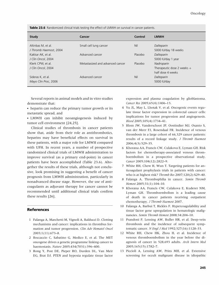
BLBK186-Key April 11, 2009 13:5
Oncology
Table 23.6 Randomized clinical trials testing the effect of LMWH on survival in cancer patients.
Study Cancer Control LMWH
Altinbas M, et al.
J Thromb Haemost, 2004
Small cell lung cancer Nil Dalteparin
5000 IU/day 18 weeks
Kakkar AK, et al.
J Clin Oncol, 2004
Advanced cancer Placebo Dalteparin
5000 IU/day 1 year
Klerk CPW, et al.
J Clin Oncol, 2004
Metastasized and advanced cancer Placebo Nadroparin
Therapeutic dose 2 weeks +half dose 4 weeks
Sideras K, et al.
Mayo Clin Proc, 2006
Advanced cancer Nil Dalteparin
5000 IU/day
Several reports in animal models and in vitro studies
demonstrate that:� heparin can reduce the primary tumor growth or its
metastatic spread, and� LMWH can inhibit neoangiogenesis induced by
tumor cell environment [24,25].
Clinical studies of thrombosis in cancer patients
show that, aside from their role as antithrombotics,
heparins may have beneficial effects on survival in
these patients, with a major role for LMWH compared
with UFH. In recent years, a number of prospective
randomized clinical trials of LMWH administration to
improve survival (as a primary end-point) in cancer
patients have been accomplished (Table 23.6). Alto-
gether the results of these trials, although not conclu-
sive, look promising in suggesting a benefit of cancer
prognosis from LMWH administration, particularly in
nonadvanced disease stage. However, the use of anti-
coagulants as adjuvant therapy for cancer cannot be
recommended until additional clinical trials confirm
these results [26].
References
1 Falanga A, Marchetti M, Vignoli A, Balducci D. Clotting
mechanisms and cancer: implications in thrombus for-
mation and tumor progression. Clin Adv Hematol Oncol
2003;1(11):673–8.
2 Boccaccio C, Sabatino G, Medico E, et al. The MET
oncogene drives a genetic programme linking cancer to
haemostasis. Nature 2005;434(7031):396–400.
3 Rong Y, Post DE, Pieper RO, Durden DL, Van Meir
EG, Brat DJ. PTEN and hypoxia regulate tissue factor
expression and plasma coagulation by glioblastoma.
Cancer Res 2005;65(4):1406–13.
4 Yu JL, May L, Lhotak V, et al. Oncogenic events regu-
late tissue factor expression in colorectal cancer cells:
implications for tumor progression and angiogenesis.
Blood 2005;105(4):1734–41.
5 Blom JW, Vanderschoot JP, Oostindier MJ, Osanto S,
van der Meer FJ, Rosendaal FR. Incidence of venous
thrombosis in a large cohort of 66,329 cancer patients:
results of a record linkage study. J Thromb Haemost
2006;4(3):529–35.
6 Khorana AA, Francis CW, Culakova E, Lyman GH. Risk
factors for chemotherapy-associated venous throm-
boembolism in a prospective observational study.
Cancer 2005;104(12):2822–9.
7 White RH, Chew H, Wun T. Targeting patients for an-
ticoagulant prophylaxis trials in patients with cancer:
who is at highest risk? Thromb Res 2007;120(2):S29–40.
8 Falanga A. Thrombophilia in cancer. Semin Thromb
Hemost 2005;31(1):104–10.
9 Khorana AA, Francis CW, Culakova E, Kuderer NM,
Lyman GH. Thromboembolism is a leading cause
of death in cancer patients receiving outpatient
chemotherapy. J Thromb Haemost 2007.
10 Falanga A, Barbui T, Rickles F. Hypercoagulability and
tissue factor gene upregulation in hematologic malig-
nancies. Semin Thromb Hemost 2008;34:204–10.
11 Prandoni P, Lensing AW, Buller HR, et al. Deep-vein
thrombosis and the incidence of subsequent symp-
tomatic cancer. N Engl J Med 1992;327(16):1128–33.
12 White RH, Chew HK, Zhou H, et al. Incidence of
venous thromboembolism in the year before the di-
agnosis of cancer in 528,693 adults. Arch Intern Med
2005;165(15):1782–7.
13 Piccioli A, Lensing AW, Prins MH, et al. Extensive
screening for occult malignant disease in idiopathic
245
BLBK186-Key April 11, 2009 13:5
CHAPTER 23
venous thromboembolism: a prospective randomized
clinical trial. J Thromb Haemost 2004;2(6):884–9.
14 Monreal M, Lensing AW, Prins MH, et al. Screen-
ing for occult cancer in patients with acute deep vein
thrombosis or pulmonary embolism. J Thromb Haemost
2004;2(6):876–81.
15 Miller GJ, Bauer KA, Howarth DJ, Cooper JA,
Humphries SE, Rosenberg RD. Increased incidence of
neoplasia of the digestive tract in men with persistent
activation of the coagulant pathway. J Thromb Haemost
2004;2(12):2107–14.
16 Annecke K, Schmitt M, Euler U, et al. uPA and PAI-
1 in breast cancer: review of their clinical utility and
current validation in the prospective NNBC-3 trial. Adv
Clin Chem 2008;45:31–45.
17 Lee AY, Levine MN. The thrombophilic state induced
by therapeutic agents in the cancer patient. Semin
Thromb Hemost 1999;25(2):137–45.
18 Palumbo A, Rajkumar SV, Dimopoulos MA, et al. Pre-
vention of thalidomide- and lenalidomide-associated
thrombosis in myeloma. Leukemia 2008;22(2):414–23.
19 Kuenen BC, Levi M, Meijers JC, et al. Analysis of
coagulation cascade and endothelial cell activation
during inhibition of vascular endothelial growth fac-
tor/vascular endothelial growth factor receptor path-
way in cancer patients. Arterioscler Thromb Vasc Biol
2002;22(9):1500–5.
20 Lyman GH, Khorana AA, Falanga A, et al. Amer-
ican Society of Clinical Oncology guideline: recom-
mendations for venous thromboembolism prophylaxis
and treatment in patients with cancer. J Clin Oncol
2007;25(34):5490–505.
21 Bergqvist D, Burmark US, Flordal PA, et al. Low molec-
ular weight heparin started before surgery as prophy-
laxis against deep vein thrombosis: 2500 versus 5000
XaI units in 2070 patients. Br J Surg 1995;82(4):496–
501.
22 Mandala M, Falanga A, Piccioli A, et al. Venous throm-
boembolism and cancer: guidelines of the Italian As-
sociation of Medical Oncology (AIOM). Crit Rev Oncol
Hematol 2006;59(3):194–204.
23 National Comprehensive Cancer Network. NCCN Clin-
ical Practice Guidelines in Oncology. Venous Throm-
boembolic Disease. 2008;vol 1:Available from: http://
www.nccn.org/professionals/physician gls/PDF/vte.pdf.
Accessed June 2, 2008.
24 Marchetti M, Vignoli A, Russo L, et al. Endothe-
lial capillary tube formation and cell proliferation in-
duced by tumor cells are affected by low molecular
weight heparins and unfractionated heparin. Thromb
Res 2008;121(5):637–45.
25 Norrby K. Low-molecular-weight heparins and angio-
genesis. APMIS 2006;114(2):79–102.
26 Kuderer NM, Khorana AA, Lyman GH, Francis CW.
A meta-analysis and systematic review of the efficacy
and safety of anticoagulants as cancer treatment: im-
pact on survival and bleeding complications. Cancer
2007;110(5):1149–61.
246
BLBK186-Key April 11, 2009 13:5
24 Obstetrics, contraception, andestrogen replacementIsobel D. Walker
Introduction
Thrombosis prevention and management has become
the major focus for hematologists with an interest in
women’s health. Normal pregnancy is associated with
increasing hypercoagulability as gestation progresses.
In addition, the pregnant woman experiences increas-
ing lower limb venous stasis due to compression of the
venous flow by the gravid uterus and inevitably suf-
fers endothelial damage due to the vascular trauma as-
sociated with delivery, particularly operative delivery.
Thrombophilias may play a role in the etiology of not
only venous thromboembolism (VTE) but a range of
other vascular complications of pregnancy, and much
debate has centered on the possibility of interven-
tion to reduce the burden of these adverse pregnancy
outcomes.
Advances in artificial reproductive technology and
in the management of women with serious medi-
cal disorders, including valvular heart disease, have
meant that increasingly women who would have
been denied pregnancy in the past now have the op-
portunity to have a child of their own, but these
women inevitably need specialist care, often involving
a hematologist.
The risk of VTE associated with the use of female
hormones for contraception or for estrogen replace-
ment is now widely recognized, but much work re-
mains to identify products that are as safe and effective
as possible.
Hemostasis in normal pregnancy
Normal pregnancy is associated with major changes
in all aspects of hemostasis: increasing concentrations
of most clotting factors, including fibrinogen and fac-
tors VII, VIII, IX, X and XII; decreasing levels of some
of the natural anticoagulants, such as protein S; in-
creased resistance to activated protein C, and reduc-
ing fibrinolytic activity (see Table 24.1). As a result, as
pregnancy progresses, and during the puerperium, the
overall hemostatic balance is shifted toward hyperco-
agulability.
Choice of anticoagulation
WarfarinCoumarins such as warfarin cross the placenta. Ma-
ternal coumarin ingestion between 6 and 12 weeks
gestation may result in developmental abnormalities
of fetal cartilage and bone, including stippling of the
epiphyses and nasal hypoplasia. Different series have
reported widely varying incidences of warfarin embry-
opathy, but a reasonable estimate of the incidence is
around 5%. Warfarin use later in pregnancy is linked
to abnormalities of the fetal central nervous system,
including impaired brain growth due to repeated mi-
crohemorrhage and scarring. It has been suggested
that the risks of fetal warfarin complications may be
dose-dependent with an increased risk when the daily
warfarin dose exceeds 5 mg [1]. It is generally rec-
ommended that coumarins should not be used for the
prevention or treatment of VTE during pregnancy, but
coumarins remain the anticoagulants of choice for the
management of some pregnant women with mechan-
ical heart valve prostheses. In all pregnant women,
because of the hemorrhagic risk to both mother and
fetus, warfarin should be avoided beyond 36 weeks
gestation.
247
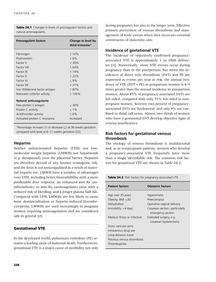
BLBK186-Key April 11, 2009 13:5
CHAPTER 24
Table 24.1 Changes in levels of procoagulant factors and
natural anticoagulants.
Procoagulant factors Change in level bythird trimester∗
Fibrinogen ↑ 10%
Prothrombin ↑ 6%
Factor V ↑ 30%
Factor VIII ↑ 64%
Factor IX ↑ 14%
Factor X ↑ 22%
Factor XI ↓ 9%
Factor XII ↑ 31%
Von Willebrand factor antigen ↑ 87%
Ristocetin cofactor activity ↑ 105%
Natural anticoagulantsFree protein S antigen ↓ 30%
Protein C activity ↓ 1%
Antithrombin activity ↑ 6%
Activated protein C resistance Increased
∗Percentage increase (↑) or decrease (↓) at 36 weeks gestation
compared with level at 6–11 weeks gestation [25].
HeparinsNeither unfractionated heparins (UFH) nor low-
molecular-weight heparins (LMWH) nor heparinoids
(e.g. danaparoid) cross the placental barrier. Heparins
are therefore devoid of any known teratogenic risk,
and the fetus is not anticoagulated as a result of mater-
nal heparin use. LMWH have a number of advantages
over UFH, including better bioavailability with a more
predictable dose response, an enhanced anti-Xa (an-
tithrombotic) to anti-IIa (anticoagulant) ratio with a
reduced risk of bleeding, and a longer plasma half-life.
Compared with UFH, LMWHs are less likely to cause
bone demineralization or heparin-induced thrombo-
cytopenia. LMWHs are used increasingly in pregnant
women requiring anticoagulation and are considered
safe in general [2].
Gestational VTE
In the developed world, pulmonary embolism (PE) re-
mains a leading cause of maternal death. Furthermore,
gestational VTE is a major cause of morbidity not only
during pregnancy but also in the longer term. Effective
primary prevention of venous thrombosis and man-
agement of acute events when they occur are essential
constituents of maternity care.
Incidence of gestational VTEThe incidence of objectively confirmed pregnancy-
associated VTE is approximately 1 in 1000 deliver-
ies [3]. Numerically, more VTE events occur during
pregnancy than in the puerperium, but when the in-
cidences of direct vein thrombosis (DVT) and PE are
expressed as events per year at risk, the annual inci-
dence of VTE (DVT + PE) in postpartum women is 4–5
times greater than the annual incidence in antepartum
women. About 85% of pregnancy-associated DVTs are
left-sided, compared with only 55% left-sided in non-
pregnant women. Seventy-two percent of pregnancy-
associated DVTs are ileofemoral, and only 9% are con-
fined to distal calf veins. Almost two-thirds of women
who have a gestational DVT develop objective signs of
venous insufficiency.
Risk factors for gestational venousthrombosisThe etiology of venous thrombosis is multifactorial
and, as in nonpregnant patients, women who develop
a pregnancy-associated VTE frequently have more
than a single identifiable risk. The common risk fac-
tors for gestational VTE are shown in Table 24.2.
Table 24.2 Risk factors for pregnancy-associated VTE.
Patient factors Obstetric factors
Age over 35 years Hyperemesis
Obesity; BMI ≥30 Preeclampsia
Dehydration Operative vaginal delivery
Immobility >4 days Cesarean section, particularly
emergency section
Medical illness or infection Extended surgery, e.g.
cesarean hysterectomy
Gross varicose veins
Intravenous drug use
Long-distance travel
Previous venous thrombosis
Thrombophilia
248
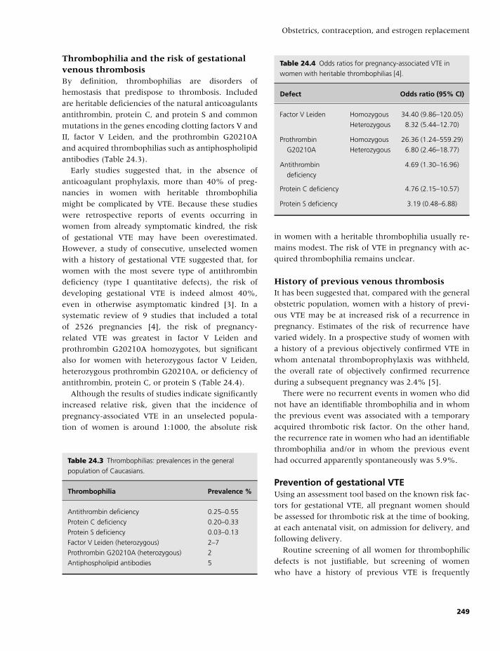
BLBK186-Key April 11, 2009 13:5
Obstetrics, contraception, and estrogen replacement
Thrombophilia and the risk of gestationalvenous thrombosisBy definition, thrombophilias are disorders of
hemostasis that predispose to thrombosis. Included
are heritable deficiencies of the natural anticoagulants
antithrombin, protein C, and protein S and common
mutations in the genes encoding clotting factors V and
II, factor V Leiden, and the prothrombin G20210A
and acquired thrombophilias such as antiphospholipid
antibodies (Table 24.3).
Early studies suggested that, in the absence of
anticoagulant prophylaxis, more than 40% of preg-
nancies in women with heritable thrombophilia
might be complicated by VTE. Because these studies
were retrospective reports of events occurring in
women from already symptomatic kindred, the risk
of gestational VTE may have been overestimated.
However, a study of consecutive, unselected women
with a history of gestational VTE suggested that, for
women with the most severe type of antithrombin
deficiency (type I quantitative defects), the risk of
developing gestational VTE is indeed almost 40%,
even in otherwise asymptomatic kindred [3]. In a
systematic review of 9 studies that included a total
of 2526 pregnancies [4], the risk of pregnancy-
related VTE was greatest in factor V Leiden and
prothrombin G20210A homozygotes, but significant
also for women with heterozygous factor V Leiden,
heterozygous prothrombin G20210A, or deficiency of
antithrombin, protein C, or protein S (Table 24.4).
Although the results of studies indicate significantly
increased relative risk, given that the incidence of
pregnancy-associated VTE in an unselected popula-
tion of women is around 1:1000, the absolute risk
Table 24.3 Thrombophilias: prevalences in the general
population of Caucasians.
Thrombophilia Prevalence %
Antithrombin deficiency 0.25–0.55
Protein C deficiency 0.20–0.33
Protein S deficiency 0.03–0.13
Factor V Leiden (heterozygous) 2–7
Prothrombin G20210A (heterozygous) 2
Antiphospholipid antibodies 5
Table 24.4 Odds ratios for pregnancy-associated VTE in
women with heritable thrombophilias [4].
Defect Odds ratio (95% CI)
Factor V Leiden Homozygous 34.40 (9.86–120.05)
Heterozygous 8.32 (5.44–12.70)
Prothrombin
G20210A
Homozygous 26.36 (1.24–559.29)
Heterozygous 6.80 (2.46–18.77)
Antithrombin
deficiency
4.69 (1.30–16.96)
Protein C deficiency 4.76 (2.15–10.57)
Protein S deficiency 3.19 (0.48–6.88)
in women with a heritable thrombophilia usually re-
mains modest. The risk of VTE in pregnancy with ac-
quired thrombophilia remains unclear.
History of previous venous thrombosisIt has been suggested that, compared with the general
obstetric population, women with a history of previ-
ous VTE may be at increased risk of a recurrence in
pregnancy. Estimates of the risk of recurrence have
varied widely. In a prospective study of women with
a history of a previous objectively confirmed VTE in
whom antenatal thromboprophylaxis was withheld,
the overall rate of objectively confirmed recurrence
during a subsequent pregnancy was 2.4% [5].
There were no recurrent events in women who did
not have an identifiable thrombophilia and in whom
the previous event was associated with a temporary
acquired thrombotic risk factor. On the other hand,
the recurrence rate in women who had an identifiable
thrombophilia and/or in whom the previous event
had occurred apparently spontaneously was 5.9%.
Prevention of gestational VTEUsing an assessment tool based on the known risk fac-
tors for gestational VTE, all pregnant women should
be assessed for thrombotic risk at the time of booking,
at each antenatal visit, on admission for delivery, and
following delivery.
Routine screening of all women for thrombophilic
defects is not justifiable, but screening of women
who have a history of previous VTE is frequently
249
BLBK186-Key April 11, 2009 13:5
CHAPTER 24
recommended, and many clinicians would also offer
thrombophilia screening to women who give a family
history of proven VTE [6].
All women assessed to be at increased risk of gesta-
tional VTE should be encouraged to wear graduated
compression stockings throughout their pregnancy
and puerperium. Given the evidence that the risk
of VTE is greatest following delivery, many obstetri-
cians would also offer women assessed to be at in-
creased risk of pregnancy-associated VTE, who have
no contraindication to anticoagulation or antithrom-
botics, pharmacological thromboprophylaxis following
delivery—usually daily prophylactic doses of LMWH,
self-administered subcutaneously, for 6 weeks follow-
ing delivery [6,7].
Consideration may also be given to offering phar-
macological thromboprophylaxis during pregnancy to
women perceived to be at relatively higher risk of ges-
tational VTE [6,7]. This group includes:� Any woman who has a history of spontaneous
(idiopathic) VTE, and� Women who have had a thrombotic event in rela-
tion to a previous pregnancy or while using a com-
bined oral contraceptive (COC), and any woman who
has been found to have a thrombophilic defect because
she has been investigated following a previous throm-
botic event.
Also in this group are women who have no per-
sonal history of thrombosis, but who have been in-
vestigated because of a family history of VTE and have
been found to have a thrombophilic defect associated
with a relatively high risk of gestational VTE (e.g. type
1 antithrombin deficiency, homozygosity for factor V
Leiden or prothrombin G20210A, or double heterozy-
gosity for factor V Leiden and prothrombin G20210A).
Daily self-administered LMWH in prophylactic
doses throughout pregnancy and for 6 weeks follow-
ing delivery is usually considered adequate for most
of these women at higher risk, but in some cases, the
daily dose of LMWH may be increased to a level inter-
mediate between that which is usually used for pro-
phylaxis and the dose usually used for treatment of
acute VTE [6,7].
The incidental finding of antiphospholipids in preg-
nancy should trigger increased clinical surveillance,
but pharmacological intervention should be reserved
for these women with antiphospholipids who are
symptomatic. Women with antiphospholipids and a
past history of VTE may usually be considered to be at
highly increased risk of recurrent VTE associated with
pregnancy and offered pharmacological thrombopro-
phylaxis, using intermediate doses of LMWH as de-
scribed above during pregnancy and the puerperium.
Diagnosis of gestational VTEThe general poor specificity of the clinical diagnosis of
DVT and PE is compounded in pregnancy by the rela-
tive frequency of nonthrombotic leg swelling, breath-
lessness, and chest discomfort in pregnant women.
Objective diagnosis is essential in all women present-
ing with suspected VTE in pregnancy or the puer-
perium. Failure to identify and treat thrombosis places
mother’s life at risk while unnecessary treatment ex-
poses both her and her unborn child to risk.
In pregnant women presenting with suspected VTE,
anticoagulation with heparin (usually LMWH) in full
therapeutic doses should be commenced while await-
ing confirmation of the diagnosis, except in the few
cases where there is a contraindication to anticoag-
ulation. D-dimer assays are generally unhelpful dur-
ing pregnancy because normal pregnancy is associated
with elevated D-dimer levels. In pregnant women,
compression duplex ultrasound is the primary diag-
nostic tool for the confirmation of DVT.
Women in whom the presence of a DVT is con-
firmed should continue anticoagulation. Patients with
a negative ultrasound and a low level of clinical sus-
picion do not require continuing anticoagulation. Pa-
tients with a negative ultrasound but a high level of
clinical suspicion should continue on anticoagulation
and either have a repeat ultrasound in a week or un-
dergo alternative diagnostic testing. If this repeat or
alternative testing is negative, anticoagulation may be
discontinued. In patients with back pain and swelling
of the entire leg in whom iliac vein thrombosis is sus-
pected, magnetic resonance venography or conven-
tional contrast venography may be considered [8].
Maternal chest x-ray exposes the fetus to a negligi-
ble dose of radiation, and although it is uninforma-
tive in over half of pregnant women who have an
objectively proven PE, it may reveal PE-related ab-
normality or non-PE-related pulmonary disease, such
as pneumonia. If the chest x-ray is normal, bilateral
lower limb compression duplex ultrasound may be
considered. A diagnosis of DVT will indirectly sup-
port a diagnosis of PE and, because the management
250
BLBK186-Key April 11, 2009 13:5
Obstetrics, contraception, and estrogen replacement
is the same for DVT or PE, it may be possible to avoid
further investigation that would expose the fetus to
radiation [8]. Whatever the chest x-ray shows, if there
is clinical suspicion of PE, definitive testing is essen-
tial. The choice of technique—ventilation perfusion
(V/Q) scanning or computed tomographic pulmonary
angiography (CTPA)—will depend on local availabil-
ity and policy. During pregnancy, it is often possible
to omit the ventilation component of the V/Q scan,
thereby reducing the radiation dose to the fetus. Com-
pared with V/Q scanning, maternal CTPA exposes the
fetus to only about 10% of the radiation dose. How-
ever, there is a relatively high radiation dose to ma-
ternal breast tissue with a postulated associated in-
creased lifetime risk of breast cancer. For this reason,
some clinicians recommend lung perfusion scanning
in young pregnant women, particularly if there is a
family history of breast cancer. If iodinated contrast
medium is administered to a mother having CTPA,
thyroid function should be checked in the neonate.
Management of gestational VTEThe recommended treatment dose of LMWH varies ac-
cording to manufacturer. Pregnancy alters the phar-
macokinetics of some LMWHs (enoxaparin and dal-
teparin), and 12-hourly dosing with these products is
recommended for the treatment of pregnant women
with VTE. Because of the physiological hypercoagula-
bility of pregnancy with its associated risk of recurrent
VTE, most experts suggest continuation of full thera-
peutic doses of LMWH for the remainder of the preg-
nancy [8].
A modified regimen with intermediate LMWH doses
after the first month at full doses may be useful in pa-
tients considered to be at increased risk of bleeding.
The total duration of anticoagulation should usually
be no less than 6 months, and anticoagulation should
continue until at least 6–12 weeks after delivery.
Warfarin can be used after delivery, but many
women find it more convenient to remain on a LMWH
for this period. For women with a DVT, pain and
swelling improve more rapidly and the risk of post-
thrombotic syndrome is reduced if the patient is mo-
bile and properly fitting compression hosiery is worn
on the affected leg during the daytime. In women in
whom a proximal DVT is diagnosed close to the time
of expected delivery, there is evidence that (tempo-
rary) insertion of an inferior vena caval filter prior to
labor or delivery reduces the risk of PE [8]. In general,
delivery should be delayed if possible.
Management of delivery in women usinganticoagulants during pregnancy
To avoid unwanted anticoagulation during delivery,
pregnant women should be advised to discontinue
their heparin injections as soon as they think they may
be in labor.
Because the prolongation of the activated partial
thromboplastin time (APTT) may persist longer than
expected, in women using UFH, the APTT should
be checked and protamine sulphate given if neces-
sary. Epidural or spinal anesthesia is generally safe
in women using UFH, providing their coagulation
screen is within normal and their platelet count is
�80 × 109/L.
When the delivery date is planned, LMWH should
be stopped 12–24 hours ahead of induction or ce-
sarean section. In spite of considerable debate, it re-
mains unclear what period of time should elapse
between the last dose of LMWH and insertion or re-
moval of an epidural or spinal catheter or how long
the time interval should be until the next dose. For
guidance, the Royal College of Obstetricians, London
suggest that, in women on full treatment doses of
LMWH, 24 hours should elapse after the last dose
of LMWH before insertion of an epidural or spinal
catheter, the cannula not be removed within 12 hours
of the most recent injection, and no further dose of
LMWH given for at least 4 hours after its removal
[8]. For women on prophylactic doses of LMWH, re-
gional anesthetic techniques should not be used un-
til 12 hours have elapsed since the last injection. As
above, the cannula should not be removed within 12
hours of the most recent injection, and no further dose
of LMWH should be given for at least 4 hours after its
removal. Local policies should be decided after discus-
sion with the anesthetists providing the service.
Thrombophilia and vascularcomplications of pregnancy
Inadequate or abnormal placental vasculature may re-
sult in a number of complications that have potentially
251
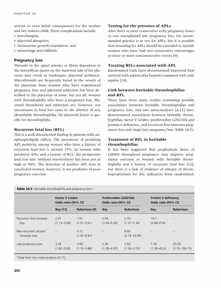
BLBK186-Key April 11, 2009 13:5
CHAPTER 24
serious or even lethal consequences for the mother
and her unborn child. These complications include:� preeclampsia,� placental abruption,� intrauterine growth retardation, and� miscarriage and stillbirth.
Pregnancy lossThrombi in the spiral arteries or fibrin deposition in
the intervillous spaces on the maternal side of the pla-
centa may result in inadequate placental perfusion.
Microthrombi are frequently found in the vessels of
the placentae from women who have experienced
pregnancy loss, and placental infarction has been de-
scribed in the placentae of some, but not all, women
with thrombophilia who have a pregnancy loss. Pla-
cental thrombosis and infarction are, however, not
uncommon in fetal loss cases in the absence of any
identifiable thrombophilia. No placental lesion is spe-
cific for thrombophilia.
Recurrent fetal loss (RFL)This is a well-documented finding in patients with an-
tiphospholipids (APLs). The prevalence of persisting
APL positivity among women who have a history of
recurrent fetal loss is around 15%. In women with
persistent APLs and a history of RCL, the prospective
fetal loss rate (without intervention) has been put as
high as 90%. The detection of positive APL tests in
unselected women, however, is not predictive of poor
pregnancy outcome.
Testing for the presence of APLsAfter three or more consecutive early pregnancy losses
or one unexplained late pregnancy loss, the recom-
mended practice is to test for APLs, but it is possible
that screening for APLs should be extended to include
women who have had two consecutive miscarriages
or three or more nonconsecutive events [9].
Treating RFLs associated with APLRandomized trials have demonstrated improved fetal
survival with aspirin plus heparin compared with only
aspirin [10].
Link between heritable thrombophiliasand RFLThere have been many studies examining possible
associations between heritable thrombophilias and
pregnancy loss, and two meta-analyses [4,11] have
demonstrated associations between heritable throm-
bophilias, factor V Leiden, prothrombin G20210A and
protein S deficiency, and recurrent first trimester preg-
nancy loss and single late-pregnancy loss (Table 24.5).
Treatment of RFL in heritablethrombophiliasIt has been suggested that prophylactic doses of
LMWH throughout pregnancy may improve preg-
nancy outcome in women with heritable throm-
bophilia and a history of recurrent fetal loss [12],
but there is a lack of evidence of efficacy of throm-
boprophylaxis for this indication from randomized,
Table 24.5 Heritable thrombophilia and pregnancy loss.∗
Factor V Leiden Prothrombin G20210A Protein S deficiencyOdds ratio (95% CI) Odds ratio (95% CI) Odds ratio (95% CI)
Rey [11] Robertson [4] Rey Robertson Rey Robertson
Recurrent first trimester
loss
2.01
(1.13–3.58)
1.91
(1.01–3.61)
2.56
(1.04–6.29)
2.70
(1.37–5.34)
14.7
(0.99–218)
–
Non-recurrent second
trimester loss
– 4.12
(1.93–8.81)
– 8.60
(2.18–33.95)
– –
Late-pregnancy loss 3.26
(1.82–5.83)
2.06
(1.10–3.86)
2.30
(1.09–4.87)
2.66
(1.28–5.53)
7.39
(1.28–42.6)
20.09
(3.70–109.15)
∗Data from two meta-analyses [4,11].
252
BLBK186-Key April 11, 2009 13:5
Obstetrics, contraception, and estrogen replacement
controlled trials. This insufficient evidence on which
to recommend antithrombotic intervention in women
with a history of pregnancy loss with no other identi-
fied abnormality apart from a heritable thrombophilia
has been recognized by the British Committee for
Standards in Haematology [6] and by the authors of
a recently published Cochrane Review [13].
However, some will extrapolate from the evidence
in randomized, controlled trials, a benefit from inter-
vention with heparin and low-dose aspirin in women
with APL syndrome and recurrent pregnancy loss and
judge that the prophylactic doses of LMWH in preg-
nancy are relatively safe, so an increasing number
of clinicians are willing to prescribe antithrombotic
agents to women with heritable thrombophilia and a
history of two or more otherwise unexplained mis-
carriages or one unexplained later intrauterine fetal
death.
PreeclampsiaThe weight of evidence currently available would ap-
pear to support the conclusion that the prevalent types
of inherited thrombophilia (factor V Leiden and pro-
thrombin G20210A) are not strong independent risk
factors for preeclampsia. They may, however, charac-
terize a subpopulation of women in whom the risk is
elevated or in whom the clinical presentation may be
more severe. In particular, there is evidence that car-
riage of factor V Leiden may increase the risk of se-
vere preeclampsia in women who are susceptible to
preeclampsia [14,15].
Artificial reproductive technology
In artificial reproductive technology (ART), exoge-
nous gonadotrophins and gonadotrophin-releasing
hormone are given to induce ovulation. Between 1%
and 25% of women undergoing ART develop ovarian
hyperstimulation syndrome (OHSS), a condition asso-
ciated with increased levels of coagulation factors and
reduced levels of some natural anticoagulants. Venous
thrombosis occurs with an incidence of around 1 in
1000 treatment cycles in women undergoing ART and
is usually associated with severe forms of OHSS, but
may occur in patients who do not display evidence of
OHSS.
Many of the case reports describing VTE in asso-
ciation with OHSS report DVT in subclavian and in-
ternal jugular veins. VTE should be suspected in pa-
tients who have had ovarian stimulation who present
with neck pain and/or swelling. The reason for local-
ization of VTE in these sites in OHSS patients is not
clear. In one review, 9 of 22 (41%) women who had
an ART-associated upper extremity DVT had an iden-
tifiable thrombophilic defect [16].
Pregnant women with heart valves
Throughout the world each year, a large number of
prosthetic heart valves are implanted, many of them
in women of childbearing age. Maternal mortality in
women with prosthetic heart valves is estimated to be
between 1% and 4% and is mostly related to throm-
boembolism.
Choice of prosthetic valve: bioprostheticThe choice of prosthesis is in itself difficult. In gen-
eral, unless they have atrial fibrillation or an intrac-
ardiac thrombus, patients with bioprosthetic (tissue)
heart valves do not need long-term anticoagulation,
although some may take aspirin. Women with bio-
prosthetic valves may expect an uncomplicated preg-
nancy providing they have a normally functioning
prosthesis, normal ventricular function, and no sig-
nificant pulmonary hypertension. Tissue valves, how-
ever, undergo structural degeneration, and this ap-
pears to happen particularly with mitral tissue valves
and more quickly in younger patients (under the age
of 40 years). Some, but not all, studies have suggested
that valve structural deterioration is accelerated by
pregnancy.
Choice of prosthetic valve: mechanicalPatients with mechanical heart valve prostheses re-
quire lifelong anticoagulation, and this includes a re-
quirement for continuing anticoagulation throughout
pregnancy with the attendant risks to both mother and
fetus. Women have a high thrombotic risk with:� older type mechanical prostheses (e.g. Starr-
Edwards or Bjork-Shiley),� a prosthesis in the mitral position,� multiple prosthetic valves,� atrial fibrillation,� a history of a previous thrombotic event.
253
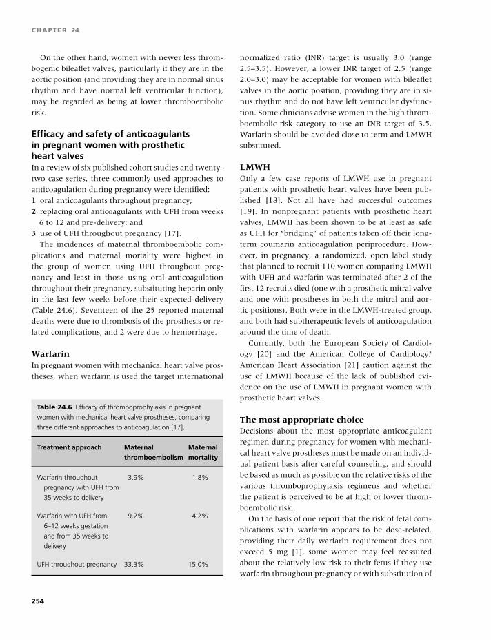
BLBK186-Key April 11, 2009 13:5
CHAPTER 24
On the other hand, women with newer less throm-
bogenic bileaflet valves, particularly if they are in the
aortic position (and providing they are in normal sinus
rhythm and have normal left ventricular function),
may be regarded as being at lower thromboembolic
risk.
Efficacy and safety of anticoagulantsin pregnant women with prostheticheart valvesIn a review of six published cohort studies and twenty-
two case series, three commonly used approaches to
anticoagulation during pregnancy were identified:
1 oral anticoagulants throughout pregnancy;
2 replacing oral anticoagulants with UFH from weeks
6 to 12 and pre-delivery; and
3 use of UFH throughout pregnancy [17].
The incidences of maternal thromboembolic com-
plications and maternal mortality were highest in
the group of women using UFH throughout preg-
nancy and least in those using oral anticoagulation
throughout their pregnancy, substituting heparin only
in the last few weeks before their expected delivery
(Table 24.6). Seventeen of the 25 reported maternal
deaths were due to thrombosis of the prosthesis or re-
lated complications, and 2 were due to hemorrhage.
WarfarinIn pregnant women with mechanical heart valve pros-
theses, when warfarin is used the target international
Table 24.6 Efficacy of thromboprophylaxis in pregnant
women with mechanical heart valve prostheses, comparing
three different approaches to anticoagulation [17].
Treatment approach Maternal Maternalthromboembolism mortality
Warfarin throughout
pregnancy with UFH from
35 weeks to delivery
3.9% 1.8%
Warfarin with UFH from
6–12 weeks gestation
and from 35 weeks to
delivery
9.2% 4.2%
UFH throughout pregnancy 33.3% 15.0%
normalized ratio (INR) target is usually 3.0 (range
2.5–3.5). However, a lower INR target of 2.5 (range
2.0–3.0) may be acceptable for women with bileaflet
valves in the aortic position, providing they are in si-
nus rhythm and do not have left ventricular dysfunc-
tion. Some clinicians advise women in the high throm-
boembolic risk category to use an INR target of 3.5.
Warfarin should be avoided close to term and LMWH
substituted.
LMWHOnly a few case reports of LMWH use in pregnant
patients with prosthetic heart valves have been pub-
lished [18]. Not all have had successful outcomes
[19]. In nonpregnant patients with prosthetic heart
valves, LMWH has been shown to be at least as safe
as UFH for “bridging” of patients taken off their long-
term coumarin anticoagulation periprocedure. How-
ever, in pregnancy, a randomized, open label study
that planned to recruit 110 women comparing LMWH
with UFH and warfarin was terminated after 2 of the
first 12 recruits died (one with a prosthetic mitral valve
and one with prostheses in both the mitral and aor-
tic positions). Both were in the LMWH-treated group,
and both had subtherapeutic levels of anticoagulation
around the time of death.
Currently, both the European Society of Cardiol-
ogy [20] and the American College of Cardiology/
American Heart Association [21] caution against the
use of LMWH because of the lack of published evi-
dence on the use of LMWH in pregnant women with
prosthetic heart valves.
The most appropriate choiceDecisions about the most appropriate anticoagulant
regimen during pregnancy for women with mechani-
cal heart valve prostheses must be made on an individ-
ual patient basis after careful counseling, and should
be based as much as possible on the relative risks of the
various thromboprophylaxis regimens and whether
the patient is perceived to be at high or lower throm-
boembolic risk.
On the basis of one report that the risk of fetal com-
plications with warfarin appears to be dose-related,
providing their daily warfarin requirement does not
exceed 5 mg [1], some women may feel reassured
about the relatively low risk to their fetus if they use
warfarin throughout pregnancy or with substitution of
254
BLBK186-Key April 11, 2009 13:5
Obstetrics, contraception, and estrogen replacement
LMWH from 6–12 weeks gestation. However, women
whose daily warfarin requirement exceeds 5 mg, par-
ticularly if they are classified into the lower throm-
boembolic risk group, may wish to minimize the risk of
fetal complication and may be prepared to rely on ad-
justed doses of LMWH. Women with mechanical heart
valve prostheses who choose to use LMWH for antico-
agulation during pregnancy must be made aware that
both the European Society of Cardiology [20] and the
American College of Cardiology/American Heart Asso-
ciation [21] recommend warfarin as the anticoagulant
of choice for pregnant women with mechanical heart
valve prostheses.
For patients with prosthetic valves on either UFH or
LMWH, regular (at least weekly) monitoring is recom-
mended. With UFH, the APTT should be maintained
between 2.0 and 2.5 times the control APTT, and for
LMWH, the peak anti-Xa level 4 hours postinjection
should be between 1.0 and 1.2 U/mL [7].
Female hormone use and the risk of VTE
Oral contraceptivesSince their introduction in the 1960s, it has been
evident that COCs are associated with an increased
risk of VTE. Although it was originally assumed that
the magnitude of this risk was related to the estro-
gen dose in the COC, more recently the role of the
progestogen content has been examined. Oral con-
traceptives containing third-generation progestogens
(desogestrel or gestodene) are associated with an ap-
proximately two-fold increased risk of VTE compared
with COCs containing second-generation progesto-
gens (levonorgestrel). COCs cause slight increases in
some procoagulant factors, reduce the levels of some
natural anticoagulants, and increase resistance to ac-
tivated protein C. These effects are more marked
with third-generation than with second-generation
COCs.
Although there is a significantly increased risk of
VTE for COC users, because these products are used by
young women in whom VTE is uncommon, the over-
all absolute risk of VTE for COC users remains low at
around 3–4 in 10,000 users per year. The relative risk
of VTE for third-generation COC users is around six- to
nine-fold that in non-COC users, and in a prospective
study, the absolute risk of VTE associated with third-
generation COC use was around 1 in 1000 new users
per year.
Thrombophilia and COCA super-additive risk of VTE has been observed be-
tween the use of COCs and the presence of throm-
bophilia, with the odds of developing VTE substan-
tially amplified in women with thrombophilia who
use a COC. The most significant increased risk has
been observed with factor V Leiden [22,23]. The in-
teraction between the factor V Leiden mutation and
COCs is enhanced for users of COCs containing third-
generation progestogens. Prothrombin G20210A has
also been shown to increase the risk of VTE in COC
users, as has deficiency of antithrombin or protein
C [23]. The increased risk of VTE associated with COC
use in patients with heritable thrombophilias has led
to the suggestion that women should be screened for
these defects prior to prescription of a COC, but this is
widely accepted as not cost effective [24].
Progestogen-only preparationsThese are used to treat menstrual disorders and are as-
sociated with increased risk of VTE, but in the general
population, progestogen-only pills used for contracep-
tion appear not to be associated with significantly in-
creased VTE risk.
Hormone replacement therapyHormone (estrogen) replacement therapy (HRT) can
be administered orally, transdermally, transvaginally,
or subcutaneously.
Nonoral administration avoids the hepatic “first
pass” effect and has minimal effects on blood coagu-
lation. Oral preparations include unopposed estrogen
and combined preparations, usually containing conju-
gate equine estrogens (CEEs) or micronized estradiol
combined with a progestogen. Transdermal HRT may
also contain both estrogen and progestogen or estro-
gen only. The changes in hemostasis associated with
HRT use are similar in type and direction to those asso-
ciated with COC use but lesser in magnitude. Nonoral
HRT preparations provoke lesser changes in hemosta-
sis than oral preparations.
Early studies suggested that HRT did not signifi-
cantly increase the risk of VTE. Later, however, case
control studies linking HRT use and VTE have been
published, and the increased risk of VTE has been
255
BLBK186-Key April 11, 2009 13:5
CHAPTER 24
confirmed in randomized studies. The evidence is con-
sistent in demonstrating a relative risk of VTE of the
order of two to four in women using HRT compared
with nonusers. The risk of VTE is greatest for orally
administered preparations and is minimal for women
using nonoral HRT. In observational studies, the risk
appeared similar irrespective of the type of estrogen
used, and no significant difference in risk was observed
in users of opposed (with progestogen) versus unop-
posed estrogen. Recently, however, a lower risk of VTE
was found in women using unopposed estrogen HRT
compared with women using combined HRT.
As in COC users, the risk of VTE in HRT users seems
to be higher near the start of therapy and in women
with thrombophilia, in particular factor V Leiden. Al-
though the relative risk of VTE is similarly increased in
COC and HRT users, the absolute risk is higher in HRT
than in COC users, due to their older age.
OthersThere is limited information about the risk of VTE in
users of Selective Estrogen Receptor Modulators, but
in a randomized, placebo-controlled trial, the relative
risk of VTE in users of Raloxifene was 3.1 (95% CI
1.5–6.2), suggesting that the risk is similar to that with
estrogen-containing HRT. A similarly increased risk of
venous thrombosis has been reported in women using
tamoxifen for the prevention or treatment of breast
cancer.
References
1 Vitale N, De Feo M, De Santo LS, Pollice A, Tedesco N,
Cotrufo M. Dose-dependent fetal complications of war-
farin in pregnant women with mechanical heart valves.
J Am Coll Cardiol 1999;33(6):1637–41.
2 Greer IA, Nelson-Piercy C. Low-molecular-weight hep-
arins for thromboprophylaxis and treatment of venous
thromboembolism in pregnancy: a systematic review of
safety and efficacy. Blood 2005;106(2):401–7.
3 McColl MD, Ramsay JE, Tait RC, et al. Risk factors
for pregnancy associated venous thromboembolism.
Thromb Haemost 1997;78(4):1183–8.
4 Robertson L, Wu O, Langhorne P, et al. Throm-
bophilia in pregnancy: a systematic review. Br J Haema-
tol 2006;132(2):171–96.
5 Brill-Edwards P, Ginsberg JS, Gent M, et al. Safety of
withholding heparin in pregnant women with a his-
tory of venous thromboembolism. N Engl J Med 2000;
343(20):1439–44.
6 Walker ID, Greaves M, Preston FE. Investigation and
management of heritable thrombophilia. Br J Haematol
2001; 14(3):512–28.
7 Bates SM, Greer IA, Pabinger I, Sofaer S, Hirsh J.
Venous thromboembolism, thrombophilia, antithrom-
botic therapy, and pregnancy: ACCP Evidenced based
Clinical Practice guidelines (the Eighth edition). Chest
2008;133(6 Suppl):844S–86S.
8 Greer IA, Thomson AJ. Thromboembolic disease in
pregnancy and the puerperium: acute management.
Green Top Guideline. No 28. 2007. London, Royal Col-
lege of Obstetricians. Available from: http://www.rcog.
org.uk/resources/Public/pdf/green top 28 thromboem
bolic minorrevision.pdf.
9 Greaves M, Cohen H, Machin SJ, Mackie I. Guide-
lines on the investigation and management of the an-
tiphospholipid syndrome. Br J Haematol 2000;109(4):
704–15.
10 Rai R, Cohen H, Dave M, Regan L. Randomised con-
trolled trial of aspirin and aspirin plus heparin in preg-
nant women with recurrent miscarriage associated with
phospholipid antibodies (or antiphospholipid antibod-
ies). Br Med J 1997;314(7076):253–7.
11 Rey E, Kahn SR, David M, Shrier I. Thrombophilic
disorders and fetal loss: a meta-analysis. Lancet 2003;
361(9361):901–8.
12 Brenner B, Hoffman R, Carp H, Dulitsky M, Younis
J. Efficacy and safety of two doses of enoxaparin in
women with thrombophilia and recurrent pregnancy
loss: the LIVE-ENOX study. J Thromb Haemost 2005;
3(2):227–9.
13 Di Nisio M, Peters L, Middeldorp S, Di Nisio M, Pe-
ters L, Middeldorp S. Anticoagulants for the treat-
ment of recurrent pregnancy loss in women without
antiphospholipid syndrome. Cochrane Database Syst Rev
2005;(2):CD004734.
14 Morrison ER, Miedzybrodzka ZH, Campbell DM, et
al. Prothrombotic genotypes are not associated with
preeclampsia and gestational hypertension: results
from a large population-based study and systematic re-
view. Thromb Haemost 2002;87(5):779–85.
15 Lin J, August P, Lin J, August P. Genetic thrombophil-
ias and preeclampsia: a meta-analysis. Obstet Gynecol
2005;105(1):182–92.
16 Chan WS, Ginsberg JS. A review of upper extrem-
ity deep vein thrombosis in pregnancy: unmasking the
‘ART’ behind the clot. J Thromb Haemost 2006;4(8):
1673–7.
17 Chan WS, Anand S, Ginsberg JS. Anticoagulation
of pregnant women with mechanical heart valves: a
256
BLBK186-Key April 11, 2009 13:5
Obstetrics, contraception, and estrogen replacement
systematic review of the literature. Arch Intern Med
2000;160(2):191–6.
18 Ellison J, Thomson AJ, Walker ID, Greer IA. Use of
enoxaparin in a pregnant woman with a mechanical
heart valve prosthesis. Br J Obstet Gynaecol 2001;108(7):
757–9.
19 Lev-Ran O, Kramer A, Gurevitch J, Shapira I, Mohr
R. Low-molecular-weight heparin for prosthetic heart
valves: treatment failure. Ann Thorac Surg 2000;69(1):
264–5.
20 Vahanian A, Baumgartner H, Bax J, et al. Guidelines
on the management of valvular heart disease: The Task
Force on the Management of Valvular Heart Disease
of the European Society of Cardiology. Eur Heart J
2007;28(2):230–68.
21 Bonow RO, Carabello B, de Leon AC Jr, et al.
Guidelines for the management of patients with
valvular heart disease: executive summary. A re-
port of the American College of Cardiology/American
Heart Association Task Force on Practice Guide-
lines (Committee on Management of Patients with
Valvular Heart Disease). Circulation 1998;98(18):1949–
84.
22 Vandenbroucke JP, Koster T, Briet E, Reitsma PH,
Bertina RM, Rosendaal FR. Increased risk of venous
thrombosis in oral-contraceptive users who are carri-
ers of factor V Leiden mutation. Lancet 1994;344(8935):
1453–7.
23 Wu O, Robertson L, Langhorne P, et al. Oral contracep-
tives, hormone replacement therapy, thrombophilias
and risk of venous thromboembolism: a systematic re-
view. The Thrombosis: Risk and Economic Assessment
of Thrombophilia Screening (TREATS) Study. Thromb
Haemost 2005;94(1):17–25.
24 Wu O, Robertson L, Twaddle S, et al. Screening for
thrombophilia in high-risk situations: a meta-analysis
and cost-effectiveness analysis. Br J Haematol 2005;
131(1):80–90.
25 Clark P, Brennand J, Conkie JA, McCall F, Greer IA,
Walker ID. Activated protein C sensitivity, protein C,
protein S and coagulation in normal pregnancy. Thromb
Haemost 1998;79(6):1166–70.
257
BLBK186-Key May 22, 2009 13:25
25 PediatricsMary E. Bauman and M. Patricia Massicotte
Quaternary care pediatrics: trading oneproblem for another
The age of highly specialized pediatric care has resulted
in many medical and surgical successes. However, new
life-threatening challenges have resulted, which in-
clude thrombosis. Prior to this era, there were little
data concerning the risk or occurrence of both venous
and arterial thrombosis in infants and children. The
studies over the last 15 years, although inferior in de-
sign, have begun to define a number of important ar-
eas in pediatric thrombosis:� cohorts at high risk,� the overall incidence,� clinical presentation,� diagnostic methods,� treatment modalities, and� long-term outcomes.
Many health professionals are now confronted with
these surviving infants and children who require spe-
cial care, which cannot be extrapolated from adult
practice as a result of many differences compared
with adults. These unique differences include nu-
trition sources and ongoing growth, developmental
hemostasis, and drug metabolism.
(Note: For the purposes of this chapter, the term “children”
will be used to describe infants and children, unless otherwise
stated.)
Kids are not little adults: the differences
Normal growth and developmentThe differences in children compared with adults alter
incidence and etiology of thrombosis, but also the type
and dose of anticoagulant agent used.
� Full-term infants double their birth weight by
5 months of age and triple their birth weight by 1 year.� Nutrition sources differ among infants; for exam-
ple, breastfed infants receive almost no vitamin K,
whereas bottle-fed infants receive varying amounts of
vitamin K depending on the amount and type of for-
mula taken.� Young children will binge eat, then not eat for a pe-
riod of time [1].� Renal function (GFR) increases over time until
adulthood.
Developmental hemostasisNormal physiological hemostasis is dependent on
maintaining a fine balance between thrombosis and
hemorrhage. Coagulation and fibrinolysis, the two
pathways responsible for hemostasis, have a number
of protein components that, when activated by a stim-
ulus, interact with red blood cells and platelets and
result in thrombus formation (coagulation) and/or
thrombus degradation (fibrinolysis). Historically, alter-
ations in blood flow, composition, and vessel wall in-
tegrity have been recognized as the most important
elements involved in thrombus formation, known as
Virchow’s Triad.
Normal hemostasis results in a fine balance between
bleeding and clotting (Fig. 25.1).� Procoagulant proteins present in the blood (factors
XII, XI, HMWK, X, IX, VIII, VII, V, II, and fibrinogen).� Procoagulant factors are activated by a stimulus (e.g.
sepsis, trauma, surgery).� Thrombin (FIIa) is then produced.� Thrombin activates fibrinogen into fibrin, the pre-
cursor of a polymerized clot.� The fibrinolytic system is subsequently activated to
break down the clot.
258
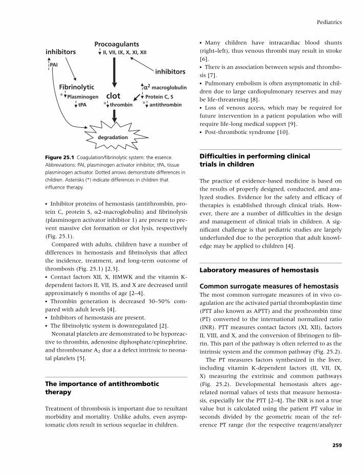
BLBK186-Key May 22, 2009 13:25
Pediatrics
Procoagulantsinhibitors II, VII, IX, X, XI, XII
PAIinhibitors
Fibrinolytic macroglobulinαα2
* Plasminogen clot Protein C, S
tPA * thrombin * antithrombin
degradation
Figure 25.1 Coagulation/fibrinolytic system: the essence.
Abbreviations: PAI, plasminogen activator inhibitor; tPA, tissue
plasminogen activator. Dotted arrows demonstrate differences in
children. Asterisks (*) indicate differences in children that
influence therapy.
� Inhibitor proteins of hemostasis (antithrombin, pro-
tein C, protein S, �2-macroglobulin) and fibrinolysis
(plasminogen activator inhibitor 1) are present to pre-
vent massive clot formation or clot lysis, respectively
(Fig. 25.1).
Compared with adults, children have a number of
differences in hemostasis and fibrinolysis that affect
the incidence, treatment, and long-term outcome of
thrombosis (Fig. 25.1) [2,3].� Contact factors XII, X, HMWK and the vitamin K-
dependent factors II, VII, IS, and X are decreased until
approximately 6 months of age [2–4].� Thrombin generation is decreased 30–50% com-
pared with adult levels [4].� Inhibitors of hemostasis are present.� The fibrinolytic system is downregulated [2].
Neonatal platelets are demonstrated to be hyporeac-
tive to thrombin, adenosine diphosphate/epinephrine,
and thromboxane A2 due a a defect intrinsic to neona-
tal platelets [5].
The importance of antithrombotictherapy
Treatment of thrombosis is important due to resultant
morbidity and mortality. Unlike adults, even asymp-
tomatic clots result in serious sequelae in children.
� Many children have intracardiac blood shunts
(right–left), thus venous thrombi may result in stroke
[6].� There is an association between sepsis and thrombo-
sis [7].� Pulmonary embolism is often asymptomatic in chil-
dren due to large cardiopulmonary reserves and may
be life-threatening [8].� Loss of venous access, which may be required for
future intervention in a patient population who will
require life-long medical support [9].� Post-thrombotic syndrome [10].
Difficulties in performing clinicaltrials in children
The practice of evidence-based medicine is based on
the results of properly designed, conducted, and ana-
lyzed studies. Evidence for the safety and efficacy of
therapies is established through clinical trials. How-
ever, there are a number of difficulties in the design
and management of clinical trials in children. A sig-
nificant challenge is that pediatric studies are largely
underfunded due to the perception that adult knowl-
edge may be applied to children [4].
Laboratory measures of hemostasis
Common surrogate measures of hemostasisThe most common surrogate measures of in vivo co-
agulation are the activated partial thromboplastin time
(PTT also known as APTT) and the prothrombin time
(PT) converted to the international normalized ratio
(INR). PTT measures contact factors (XI, XII), factors
II, VIII, and X, and the conversion of fibrinogen to fib-
rin. This part of the pathway is often referred to as the
intrinsic system and the common pathway (Fig. 25.2).
The PT measures factors synthesized in the liver,
including vitamin K-dependent factors (II, VII, IX,
X) measuring the extrinsic and common pathways
(Fig. 25.2). Developmental hemostasis alters age-
related normal values of tests that measure hemosta-
sis, especially for the PTT [2–4]. The INR is not a true
value but is calculated using the patient PT value in
seconds divided by the geometric mean of the ref-
erence PT range (for the respective reagent/analyzer
259

BLBK186-Key May 22, 2009 13:25
CHAPTER 25
XII XIIaAntithrombin
Tissue FactorHMWK VIIa-VII
XI XIaAntithrombinVIII-VIIIa
IX IXaAntithrombin
Protein C & S
X Xa
V—Va AntithrombinThrombin
Antithrombin
Fibrinogen Fibrin
XIII
Stimulus
Intrinsicpathway
Extrinsicpathway
Generalpathway
Figure 25.2 Simplified coagulation cascade. Dashed lines indicate intrinsic pathway. Dotted lines indicate extrinsic pathway. Solid lines
indicate common pathway.
combination), taken to the power of the international
sensitivity index. The use of the INR value is an at-
tempt to account for different analyzers and thrombo-
plastin reagents used in PT testing.
The INR and PTT measure interaction of coagulation
factors in plasma (as compared to whole blood) in a
test tube. The inability to measure the presence and
interaction of red blood cells and platelets provides an
incomplete measure of hemostasis.
Global measures of hemostasis
Activated clotting time (ACT)The ACT uses activated whole blood and measures
clotting time in seconds as a measure of global
hemostasis, which more closely reflects in vivo co-
agulation. This point-of-care test is used in extra-
corporeal life support (cardiopulmonary bypass, ex-
tracorporeal membranous oxygenation) to monitor
anticoagulation, specifically heparin. There are no
well-designed studies evaluating safety and efficacy of
the use of ACTs to monitor anticoagulation in chil-
dren. Although many health care professionals use the
ACT to measure anticoagulation, the ACT does not
solely reflect the effect of heparin but also reflects re-
cent infusion of blood products.
ThromboelastogramThe thromboelastogram uses whole activated whole
blood to measure hemostasis (formation of a clot)
as well as fibrinolysis (clot degradation). The most
common devices used to measure thromboelastogra-
phy are the ROTEM R© (Pentafarm, Munich, Germany)
and the TEG R© (Haemoscope, Niles IL,). Formal well-
designed studies are required to evaluate the validity,
accuracy, and application of the measure in children.
Therapeutic agents and metabolismEvidence for the safety and efficacy of therapies is es-
tablished through clinical trials. It is challenging to
perform rigorous studies in children [4], and as a re-
sult, the agents that are commonly used for treatment
260
BLBK186-Key May 22, 2009 13:25
Pediatrics
of thrombosis in children include heparin therapy and
vitamin K antagonists. Current data in adults sup-
port the premise that, when patients are maintained
within their defined therapeutic range, they will be
adequately protected from the risk of thrombosis and
minimize the risk of a serious adverse event [11,12].
Newer agents, such as direct thrombin inhibitors, are
available, although there are limited data available to
support their use. Despite this, there are clinical situa-
tions where these agents must be used, for example in
confirmed heparin-induced thrombocytopenia (HIT).
In addition, there are multiple variables that make
the use of antithrombotic drugs in children different
from adults, which include:� Epidemiology of thrombosis in children is different.� Hemostatic system is dynamically evolving.� Distribution, binding, and clearance of antithrom-
botic drugs are age-dependent.� Many children requiring antithrombotic therapy
have underlying conditions that often require concur-
rent medication.� Frequency and type of intercurrent illnesses vary
with age.� Practical ability to deliver the drug is impacted by
difficult venous access, needle phobias, etc.� Pediatric formulations of antithrombotic drugs are
not available, making accurate dose measurement
difficult.� Dietary differences that are inherent to normal
growth and development particularly influence the
use of vitamin K antagonist therapy.
Heparin is a term that encapsulates unfractionated
heparin (UFH) and low-molecular-weight heparins
(LMWH).
Unfractionated heparin (UFH)UFH remains a commonly used anticoagulant agent
used in hospital settings for children at potential in-
creased risk of hemorrhage (i.e. postoperatively) or
when rapid reversal of anticoagulant effect is required.
Heparin is not absorbed orally, therefore must be ad-
ministered intravenously or subcutaneously.
UFH: metabolismUFH acts via antithrombin-mediated catabolism of
thrombin and inhibition of factors IIa, IXa, Xa, XIa,
and XIIa (Fig. 25.2). UFH is poorly bioavailable and
binds with a number of plasma proteins, endothelial
cells, and macrophages, which results in variability in
anticoagulant response.� UFH binds with antithrombin to catabolize throm-
bin. Antithrombin is at decreased levels in infants and
children compared with adults, further increasing the
variable anticoagulant response.� Antithrombin concentrate may be administered
when patient levels are low to strengthen UFH re-
sponse and reduce anticoagulant variability.� UFH binds to von Willebrand factor and inhibits von
Willebrand factor-dependent platelet function [13].
UFH therapy: dosing and monitoringDosing of UFH in children is age-dependent (Ta-
ble 25.1). Dosing guidelines are provided in Table 25.2.
Children will achieve therapeutic UFH (anti-factor Xa)
levels more quickly if a UFH bolus dose of 75–100
U/k/hour is administered; however, it is important to
consider the risk–benefit ratio with regards to hemor-
rhage. Heparin doses are then titrated based on labo-
ratory measure of anti-factor Xa, or PTT if anti-factor
Xa measures are unavailable.
The internationally accepted gold standard measure
of UFH is an anti-factor Xa level, with a target range
of 0.35–0.7 U/mL [12] to reflect a therapeutic heparin
level. Recent data suggest that extrapolating the PTT
from adults to pediatric patients is likely to be invalid,
as normal PTTs in infants and children are increased
secondary to developmental hemostasis [14]. Equally,
there is a different response to heparin compared with
adults; therefore, the use of the PTT to monitor hep-
arin therapy may be invalid. In addition, in vitro and
in vivo data support that the PTT that corresponds to
an anti-factor Xa level of 0.35–0.7 U/mL varies signif-
icantly with age [14].
Some health professionals are using anti-factor Xa
in children despite the absence of studies because of
the lack of correlation between anti-factor Xa levels
and PTT, as investigations have validated UFH dos-
ing nomograms in children. Depending on the reagent
and the machine used to measure the PTT, therapeutic
PTTs can be anywhere from 1.5 to 6.2 times baseline
[13].
UFH: benefits and limitations� Short half-life,therefore clears within 4–6 hours of
cessation.� Fully reversible with protamine sulphate.
261
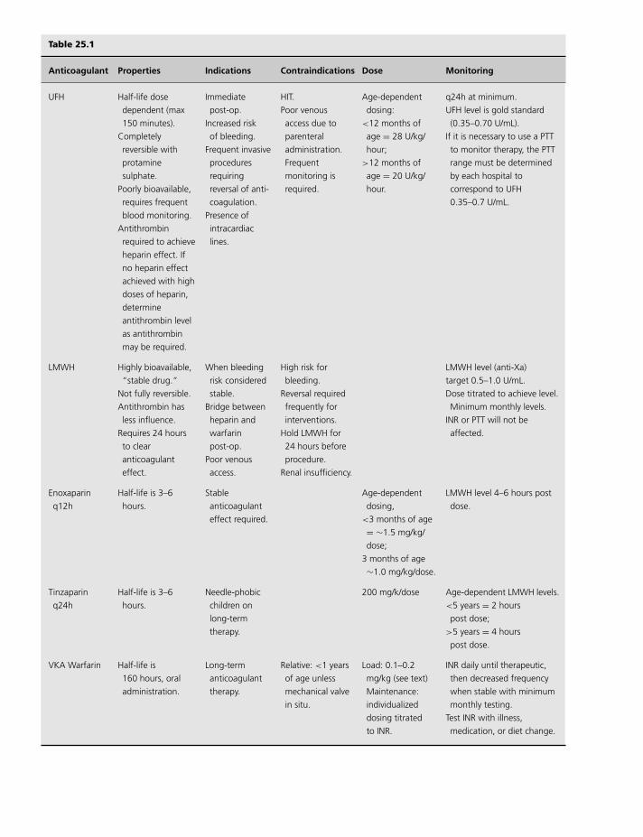
BLBK186-Key May 22, 2009 13:25
Table 25.1
Anticoagulant Properties Indications Contraindications Dose Monitoring
UFH Half-life dose
dependent (max
150 minutes).
Completely
reversible with
protamine
sulphate.
Poorly bioavailable,
requires frequent
blood monitoring.
Antithrombin
required to achieve
heparin effect. If
no heparin effect
achieved with high
doses of heparin,
determine
antithrombin level
as antithrombin
may be required.
Immediate
post-op.
Increased risk
of bleeding.
Frequent invasive
procedures
requiring
reversal of anti-
coagulation.
Presence of
intracardiac
lines.
HIT.
Poor venous
access due to
parenteral
administration.
Frequent
monitoring is
required.
Age-dependent
dosing:
<12 months of
age = 28 U/kg/
hour;
>12 months of
age = 20 U/kg/
hour.
q24h at minimum.
UFH level is gold standard
(0.35–0.70 U/mL).
If it is necessary to use a PTT
to monitor therapy, the PTT
range must be determined
by each hospital to
correspond to UFH
0.35–0.7 U/mL.
LMWH Highly bioavailable,
“stable drug.”
Not fully reversible.
Antithrombin has
less influence.
Requires 24 hours
to clear
anticoagulant
effect.
When bleeding
risk considered
stable.
Bridge between
heparin and
warfarin
post-op.
Poor venous
access.
High risk for
bleeding.
Reversal required
frequently for
interventions.
Hold LMWH for
24 hours before
procedure.
Renal insufficiency.
LMWH level (anti-Xa)
target 0.5–1.0 U/mL.
Dose titrated to achieve level.
Minimum monthly levels.
INR or PTT will not be
affected.
Enoxaparin
q12h
Half-life is 3–6
hours.
Stable
anticoagulant
effect required.
Age-dependent
dosing,
<3 months of age
= ∼1.5 mg/kg/
dose;
3 months of age
∼1.0 mg/kg/dose.
LMWH level 4–6 hours post
dose.
Tinzaparin
q24h
Half-life is 3–6
hours.
Needle-phobic
children on
long-term
therapy.
200 mg/k/dose Age-dependent LMWH levels.
<5 years = 2 hours
post dose;
>5 years = 4 hours
post dose.
VKA Warfarin Half-life is
160 hours, oral
administration.
Long-term
anticoagulant
therapy.
Relative: <1 years
of age unless
mechanical valve
in situ.
Load: 0.1–0.2
mg/kg (see text)
Maintenance:
individualized
dosing titrated
to INR.
INR daily until therapeutic,
then decreased frequency
when stable with minimum
monthly testing.
Test INR with illness,
medication, or diet change.
262
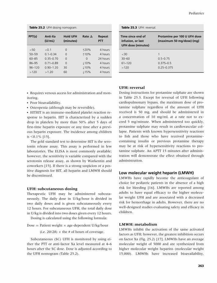
BLBK186-Key May 22, 2009 13:25
Pediatrics
Table 25.2 UFH dosing nomogram.
PPT(s) Anti-Xa Hold UFH Rate ∆ Repeat(U/mL) (minutes) PTT
<50 <0.1 0 ↑20% 4 hours
50–59 0.1–0.34 0 ↑10% 4 hours
60–85 0.35–0.70 0 0 24 hours
86–95 0.71–0.89 0 ↓10% 4 hours
96–120 0.90–1.20 30 ↓10% 4 hours
>120 >1.20 60 ↓15% 4 hours
� Requires venous access for administration and mon-
itoring.� Poor bioavailability.� Osteopenia (although may be reversible).� HITHIT is an immune-mediated platelet reaction re-
sponse to heparin. HIT is characterized by a sudden
drop in platelets by more than 50% after 5 days of
first-time heparin exposure or any time after a previ-
ous heparin exposure. The incidence among children
is �0.1% [15].
The gold standard test to determine HIT is the sero-
tonin release assay. This assay is performed in few
laboratories. The ELISA is most commonly available;
however, the sensitivity is variable compared with the
serotonin release assay, as shown by Warkentin and
coworkers [15]. If there is a strong suspicion or a pos-
itive diagnosis for HIT, all heparin and LMWH should
be discontinued.
UFH: subcutaneous dosingTherapeutic UFH may be administered subcuta-
neously. The daily dose in U/kg/hour is divided in
two daily doses and is given subcutaneously every
12 hours. For subcutaneous UFH, the total daily dose
in U/kg is divided into two doses given every 12 hours.
Dosing is calculated using the following formula:
Dose = Patient weight × age-dependent U/kg/hour
(i.e. 20/28) × the # of hours of coverage.
Subcutaneous (SC) UFH is monitored by using ei-
ther the PTT or anti-factor Xa level measured at 4–6
hours after the SC dose. Dose is adjusted according to
the UFH nomogram (Table 25.2).
Table 25.3 UFH: reversal.
Time since end of Protamine per 100 U UFH doseinfusion, or last (maximum 50 mg/dose) (mg)UFH dose (minutes)
<30 1
30–60 0.5–0.75
61–120 0.375–0.5
>120 0.25–0.375
UFH: reversalDosing instructions for protamine sulphate are shown
in Table 25.3. Except for reversal of UFH following
cardiopulmonary bypass, the maximum dose of pro-
tamine sulphate regardless of the amount of UFH
received is 50 mg, and should be administered in
a concentration of 10 mg/mL at a rate not to ex-
ceed 5 mg/minute. When administered too quickly,
protamine sulphate may result in cardiovascular col-
lapse. Patients with known hypersensitivity reactions
to fish and those who have received protamine-
containing insulin or previous protamine therapy
may be at risk of hypersensitivity reactions to pro-
tamine sulphate. An APTT 15 minutes after adminis-
tration will demonstrate the effect obtained through
administration.
Low molecular weight heparin (LMWH)LMWHs have rapidly become the anticoagulant of
choice for pediatric patients in the absence of a high
risk for bleeding [16]. LMWHs are reported among
adults to have equal efficacy to the higher molecu-
lar weight UFH and are associated with a decreased
risk for hemorrhage in adults. However, there are no
well-designed studies evaluating safety and efficacy in
children.
LMWH: metabolismLMWHs inhibit the activation of the same activated
factors as UFH; however, the greatest inhibition occurs
on factor Xa (Fig. 25.2) [17]. LMWHs have an average
molecular weight of 5000 and are synthesized from
higher molecular weight heparins (molecular weight
15,000). LMWHs have increased bioavailability,
263
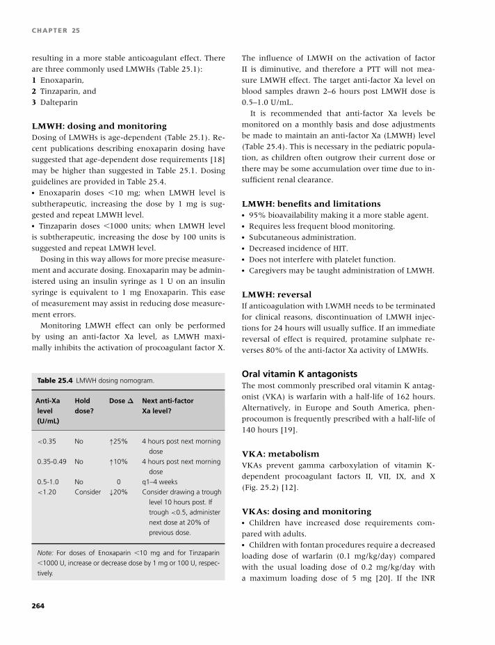
BLBK186-Key May 22, 2009 13:25
CHAPTER 25
resulting in a more stable anticoagulant effect. There
are three commonly used LMWHs (Table 25.1):
1 Enoxaparin,
2 Tinzaparin, and
3 Dalteparin
LMWH: dosing and monitoringDosing of LMWHs is age-dependent (Table 25.1). Re-
cent publications describing enoxaparin dosing have
suggested that age-dependent dose requirements [18]
may be higher than suggested in Table 25.1. Dosing
guidelines are provided in Table 25.4.� Enoxaparin doses �10 mg; when LMWH level is
subtherapeutic, increasing the dose by 1 mg is sug-
gested and repeat LMWH level.� Tinzaparin doses �1000 units; when LMWH level
is subtherapeutic, increasing the dose by 100 units is
suggested and repeat LMWH level.
Dosing in this way allows for more precise measure-
ment and accurate dosing. Enoxaparin may be admin-
istered using an insulin syringe as 1 U on an insulin
syringe is equivalent to 1 mg Enoxaparin. This ease
of measurement may assist in reducing dose measure-
ment errors.
Monitoring LMWH effect can only be performed
by using an anti-factor Xa level, as LMWH maxi-
mally inhibits the activation of procoagulant factor X.
Table 25.4 LMWH dosing nomogram.
Anti-Xa Hold Dose ∆ Next anti-factorlevel dose? Xa level?(U/mL)
<0.35 No ↑25% 4 hours post next morning
dose
0.35-0.49 No ↑10% 4 hours post next morning
dose
0.5-1.0 No 0 q1–4 weeks
<1.20 Consider ↓20% Consider drawing a trough
level 10 hours post. If
trough <0.5, administer
next dose at 20% of
previous dose.
Note: For doses of Enoxaparin �10 mg and for Tinzaparin
�1000 U, increase or decrease dose by 1 mg or 100 U, respec-
tively.
The influence of LMWH on the activation of factor
II is diminutive, and therefore a PTT will not mea-
sure LMWH effect. The target anti-factor Xa level on
blood samples drawn 2–6 hours post LMWH dose is
0.5–1.0 U/mL.
It is recommended that anti-factor Xa levels be
monitored on a monthly basis and dose adjustments
be made to maintain an anti-factor Xa (LMWH) level
(Table 25.4). This is necessary in the pediatric popula-
tion, as children often outgrow their current dose or
there may be some accumulation over time due to in-
sufficient renal clearance.
LMWH: benefits and limitations� 95% bioavailability making it a more stable agent.� Requires less frequent blood monitoring.� Subcutaneous administration.� Decreased incidence of HIT.� Does not interfere with platelet function.� Caregivers may be taught administration of LMWH.
LMWH: reversalIf anticoagulation with LWMH needs to be terminated
for clinical reasons, discontinuation of LMWH injec-
tions for 24 hours will usually suffice. If an immediate
reversal of effect is required, protamine sulphate re-
verses 80% of the anti-factor Xa activity of LMWHs.
Oral vitamin K antagonistsThe most commonly prescribed oral vitamin K antag-
onist (VKA) is warfarin with a half-life of 162 hours.
Alternatively, in Europe and South America, phen-
procoumon is frequently prescribed with a half-life of
140 hours [19].
VKA: metabolismVKAs prevent gamma carboxylation of vitamin K-
dependent procoagulant factors II, VII, IX, and X
(Fig. 25.2) [12].
VKAs: dosing and monitoring� Children have increased dose requirements com-
pared with adults.� Children with fontan procedures require a decreased
loading dose of warfarin (0.1 mg/kg/day) compared
with the usual loading dose of 0.2 mg/kg/day with
a maximum loading dose of 5 mg [20]. If the INR
264
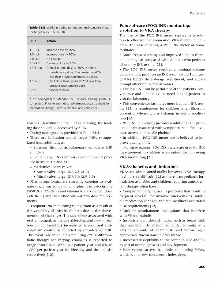
BLBK186-Key May 22, 2009 13:25
Pediatrics
Table 25.5 Warfarin dosing nomogram: maintenance phase
for target INR 2.5 (2.0–3.0).
INR* Action
1.1–1.4 Increase dose by 20%.
1.5–1.9 Increase dose by 10%.
2.0–3.0 No change.
3.1–3.5 Decrease dose by 10%.
>3.5–4.0 Administer one dose at 50% less than
maintenance dose. Then restart at 20%
less than previous maintenance dose.
4.1–5.0 Hold 1 dose then restart at 20% less than
previous maintenance dose.
>5.0 Consider reversal.
*This nomogram is intended for use once loading phase is
completed. Prior to each dose adjustment, assess patient for
medication change, illness (cold, flu), and adherence.
reaches 1.6 within the first 3 days of dosing, the load-
ing dose should be decreased by 50%.� Dosing nomogram is provided in Table 25.5.� There are indication-related target INRs extrapo-
lated from adult ranges.
◦ Systemic thrombosis/pulmonary embolism INR
2.5 (2–3)
◦ Fontan target INRs can vary upon individual prac-
tice between 1.5 and 3.0.
◦ Mechanical heart valves� Aortic valve: target INR 2.5 (2–3)� Mitral valve: target INR 3.0 (2.5–3.5)
� Pharmacogenomics are currently ongoing to eval-
uate single nucleotide polymorphisms in cytochrome
P450 2C9 (CYP2C9) and vitamin K epoxide reductase
(VKORC1) and their effect on warfarin dose require-
ments.
Frequent INR monitoring is important as a result of
the variability of INRs in children due to the above-
mentioned challenges. The side effects associated with
oral anticoagulant therapy (bleeding and new or ex-
tension of thrombus) increase with poor oral anti-
coagulant control as reflected by out-of-range INR.
The event rate in children requiring oral antithrom-
botic therapy for varying etiologies is reported to
range from 0% to 0.5% per patient year and 0% to
1.3% per patient year for bleeding and thrombosis,
respectively [12].
Point-of-care (POC) INR monitoring:a solution to VKA therapyThe use of the POC INR meter represents a solu-
tion to effective management of VKA therapy in chil-
dren. The ease of using a POC INR meter at home
facilitates:� More frequent testing and improved time in thera-
peutic range as compared with children who perform
laboratory INR testing [21].� The POC INR meter requires a minimal volume
blood sample, produces an INR result within 1 minute,
enables timely drug dosage adjustment, and allows
prompt attention to critical values.� The POC INR can be performed at the patients’ con-
venience and eliminates the need for the patient to
visit the laboratory.� This convenience facilitates more frequent INR test-
ing [22], a requirement for children when illness is
present or when there is a change in diet or medica-
tion [12].� POC INR monitoring provides a solution to the prob-
lem of pain associated with venipuncture, difficult ve-
nous access, and needle phobias.� In addition, POC INR meter use is believed to im-
prove quality of life.
For these reasons, POC INR meters are used for INR
measurement in children as an option for improving
VKA monitoring [21].
VKAs: benefits and limitationsVKAs are administered orally; however, VKA therapy
in children is difficult [12] as there is no pediatric for-
mulation available, and children requiring anticoagu-
lant therapy often have:� Complex underlying health problems that result in
frequent reversal for invasive interventions, multi-
ple medication changes, and require illness-associated
dose requirements [12].� Multiple simultaneous medications that interfere
with VKA metabolism.� Inconsistent nutritional intake, such as breast milk
that contains little vitamin K, bottled formula with
varying amounts of vitamin K, and normal age-
appropriate fluctuations in daily intake.� Increased susceptibility to the common cold and flu
as part of normal growth and development.� Poor venous access that limits monitoring VKAs,
which is a narrow therapeutic index drug.
265
BLBK186-Key May 22, 2009 13:25
CHAPTER 25
� Anxiety and needle phobias.� The nonreported use of complementary alternative
medications.
In addition, the care of children requiring long-
term primary thromboprophylaxis, such as children
with congential heart disease (CHD), presents in-
creased challenges, including life-long monitoring (a
child with a mechanical heart valve may initiate an-
ticoagulation at age 5 years, whereas an adult may
begin VKAs much later in life; this results in many
more patient years of anticoagulation). In addition,
there are data to strongly suggest that long-term
VKA therapy in children may be associated with
osteoporosis [23].
VKAs: the challenge of complementaryalternative medicinesComplementary alternative medicines (CAMs) in-
clude nutritional and dietary supplements. The use of
CAMs is highly underreported by children and their
families [24]. When children receiving anticoagulation
use CAMs, this may influence their level of anticoag-
ulation, resulting in thrombosis or hemorrhage. It is
necessary to educate families about CAM use and its
potential influence on their child’s level of anticoagu-
lation increasing their risk of thrombosis and/or hem-
orrhage [25].
VKA: reversalThe antidote for warfarin is dependent on whether ur-
gent or nonurgent reversal is necessary. For nonur-
gent reversal, vitamin K is administered at a dose of
0.5–1 mg orally, depending on the patient’s size. The
administration of vitamin K either intravenously or in-
tramuscularly has been shown to be less efficacious
than orally, as long as gut absorption is not severely
compromised. For urgent reversal (major bleeding or
interventional procedure), factor VIIa 50 U/kg IV or
FFP 20 mL/kg IV is administered.
As described in adults, in children who are consid-
ered to be at high risk for thrombosis (i.e. mechani-
cal valves), bridge anticoagulant therapy using heparin
may be considered [26].
New agentsDirect thrombin inhibitors, such as argatroban, lep-
irudin, and bilvalirudin, are approved in many coun-
tries for use in adults with confirmed HIT. There are no
well-designed studies published describing their use in
children. However, there are a few case reports and
small cohort studies describing their use.
Danaparoid, a factor Xa inhibitor, is available in
many countries with the exception of the United
States. Dosing guidelines for children are published
elsewhere [12].
Thrombolytic therapy
In the presence of thrombosis that threatens the vi-
ability of organ, limb, or life, rapid clot lysis should
be strongly considered in the absence of contraindica-
tions, such as an elevated PTT and INR, decreased fib-
rinogen, platelets �100,000, cerebral bleeding, early
post-op, or massive bleeding.
The most common agent used is tissue plasmino-
gen activator (tPa) (activase, alteplase; Genentech, San
Francisco, CA).� The doses in the literature range from 0.01 to 0.6
mg/kg/hour for varying amounts of time [12].� It is important to ensure that plasminogen levels are
sufficient to allow thrombolysis. For this reason, in
the absence of clinical trials, the use of fresh frozen
plasma (10–20 mL/kg IV every 8-12 hours with tPa in-
fusion) as a plasminogen source is recommended prior
to/during tPa infusion.
In children, the risk of major hemorrhage is as
high as 68% with bleeding requiring transfusion
in 39%.� Serious discussion about the risk/benefit of throm-
bolytic therapy with other health care professionals
and parents/caregivers followed by documentation of
the discussion within the patients’ medical records
should occur prior to use.
Streptokinase, another thrombolytic agent, is not
recommended in children due to the potential for
anaphylactic reaction secondary to antibody develop-
ment.
Factor VIIaFactor VIIa is a recombinant activated blood product
that has been used to manage bleeding. There are lit-
tle data in nonhemophiliac children to support recom-
mendations for its use.
The suggested dosing is 15–30 µg/kg body weight.
266
BLBK186-Key May 22, 2009 13:25
Pediatrics
Antiplatelet therapyAntiplatelet therapy is used for a number of indica-
tions, although there are no dose finding, safety, and
efficacy studies. Common indications include:� Cardiac (extracardiac palliative shunts, intravascu-
lar stents, mechanical aortic valves, kawasaki’s disease,
following heart transplantation, and others).� Post organ transplant (heart, liver).
The most common antiplatelets used are aspirin and
dipyridamole. There are other agents with some data
appearing in the literature. These agents include clopi-
dogrel and abciximab [12].
Antiplatelet therapy: metabolismEach agent inhibits platelet function by interrupting
different metabolic pathways that are important for
optimal platelet shape change, adhesion, and aggre-
gation.
Antiplatelet therapy: dosingand monitoring� Aspirin 1–5 mg/kg/day� Dipyridamole 2–5 mg/kg/day� Clopidogrel 0.2 mg/kg/day
There have been various methods used to monitor
antiplatelet effect (platelet aggregation, PFA100,
accumetrics, TEG R©); however, none has been demon-
strated to be associated with safety and efficacy
outcomes.
Antiplatelet therapy: benefitsand limitations� Monitoring not currently recommended.� Oral administration.� Aspirin is associated with gastrointestinal bleeding.
Antiplatelet therapy: reversal� Discontinuation of therapy is sufficient to clear effect
(may take up to 7 days).� Special consideration should be given to withhold-
ing aspirin with fever or exposure to chicken pox due
to the small risk of developing Reyes syndrome.� Immunizations and injections may be administered;
however, it is imperative to apply 5 minutes of firm
pressure on the injection site to minimize bruising.� The manufacturer of the varicella vaccine recom-
mends withholding aspirin for 1 week before and 6
weeks following varicella immunization.
Cohorts of children at risk for thrombosis
There are a number of cohorts of children that are
identified to be at high risk for venous or arterial
thrombosis:� Children with central lines
◦ Central venous (CVL)
◦ Central arterial lines� Children with congenital heart disease� Children who undergo organ transplantation
Children with central venous or arterial linesSystemic venous thromboembolic events in children
most often occur due to interaction of multiple risk
factors with the presence of a CVL appearing to be one
of the strongest risk factors.
Systemic arterial thromboembolic events in children
most often occur as a result of the placement of an ar-
terial line or following cardiac catheterization. Throm-
boprophylaxis during cardiac catheterization using
UFH of 50–150 U/kg bolus has been demonstrated to
be safe and efficacious in children in a randomized
clinical trial [27].
Diagnosis of venous and arterialor intracardiac thrombosisClinical symptoms of thrombosis vary depending on
the location of the thrombus. For example, a deep
venous thrombosis in a limb may be associated with
pain, swelling, skin discoloration, and altered perfu-
sion, whereas an intracardiac thrombus may range
from asymptomatic to congestive heart failure, pul-
monary embolism, or sequelae secondary to an embo-
lus, including stroke, and organ or limb compromise.
Both venous and arterial thromboses require rapid
diagnosis and treatment to prevent thrombus exten-
sion or embolism, which could result in mortality or
morbidity.
Clinical studies have determined that the most sen-
sitive diagnostic methods for diagnosing upper sys-
tem thrombosis are the ultrasound for jugular venous
thrombosis and venography for intrathoracic vessels.
For symptomatic thrombosis of both the upper and
lower system, ultrasound may be used; however, if
the clinical suspicion for thrombosis is high and ultra-
sound is negative, consideration should be given to
further imaging, such as magnetic resonance imaging
267
BLBK186-Key May 22, 2009 13:25
CHAPTER 25
(MRI), computed tomography (CT), and/or venogra-
phy of the suspicious venous or arterial system. There
are no studies determining the sensitivity and speci-
ficity of these newer imaging techniques in children;
however, they are commonly used. The concern in
children using diagnostic tests with high radiation
doses has resulted in a move toward MRI [28].
Intracardiac thromboses are often incidental find-
ings for children with comprised cardiac function and
may be identified through echocardiogram, cardiac
catheterization, angiogram, or cardiac MRI or CT.
Duration of antithrombotic therapyfor systemic venous thrombosisDuration and intensity of therapy is based on adult
recommendations and may be in excess of what is re-
quired in children. Until studies are completed, it is
reasonable to base therapy on adult recommendations.
Duration of treatment: systemicvenous thrombosisThis depends on several factors:� Risk factor resolved; 3 months duration.� Continued risk factor; long-term therapy.� Idiopathic; minimum 6–12 months of therapy.� Life-threatening pulmonary embolus; consider
thrombectomy or thrombolytic therapy.
There are no data to support the use of routine
thromboprophylaxis of CVLs in children.
Some outcomes of systemicvenous thrombosisPostthrombotic syndrome is reported to be approxi-
mately 20%. Postthrombotic syndrome is character-
ized by pain, swelling, and alterations in perfusion that
may result in skin ulceration. There is no treatment;
however, palliation may include the use of compres-
sion stockings.
Frequently there are challenges associated with
thrombosis-related loss of venous access that is often
required for future procedures or treatment.
Duration of treatment: systemicarterial thrombosis� Catheter related; immediate removal of the catheter
with variable duration of therapy described. Throm-
bolysis and or thrombectomy may be considered.
� Idiopathic; if life threatening, thrombectomy or
thrombolysis would be recommended as initial treat-
ment. Anticoagulation following clot removal has
been used in varying doses and duration.
Outcomes of systemic arterial thrombosis� Loss of life, limb, or organ dependent on thrombus
location.� Alteration of organ function.� Limb length discrepancy.� Intermittent claudication secondary to decreased
perfusion.
Pulmonary embolismPulmonary embolism (PE) is rare in children, and most
commonly occur as a result of deep venous thrombo-
sis [29]. The following radiographic tests may be used
to diagnose PE in children: ventilation perfusion scan,
spiral CT, MRI, MRV, or pulmonary angiogram.
Altered quality of life associatedwith long-term anticoagulationQuality of life (QOL) is an abstract entity that can be
measured by a questionnaire developed specific to the
patient condition. There are a number of character-
istics of long-term anticoagulation that may induce
treatment dissatisfaction and reduce QOL for children.
A validated pediatric QOL inventory for children
requiring long-term anticoagulation would assess
general constructs that are most salient for this patient
population. Identification and systematic evaluation
of these constructs is critical to recognizing influences
on patient adherence to improve patient care. Once
confounders are identified, the “best” management
(best QOL associated with best safety and efficacy) for
children requiring long-term anticoagulation can be
established.
Children with congenital heart disease
CHD is one of the most common inborn defects occur-
ring in 0.8% of newborn infants. Many children with
CHD have extracardiac shunts surgically placed as pal-
liation for their condition, including Blalock Taus-
sig shunts, Norwood Sano, Central Right Ventricle to
Pulmonary Artery shunts, Glenn shunts, and Fontan
shunts. These shunts vary in diameter and flow
268
BLBK186-Key May 22, 2009 13:25
Pediatrics
characteristics and are often considered at increased
risk for thrombosis. Although there are no well-
designed studies evaluating the use of anticoagulant or
antiplatelet agents in this patient population, they are
commonly used as thromboprophylaxis [12]. There
are a number of other cardiac indications where an-
ticoagulants and/or antiplatelets are used as throm-
boprophylaxis; however, there are no well-designed
studies to provide safety and efficacy data for any ther-
apeutic agent.
Children with mechanical heart valves placed are
prescribed long-term VKAs as thromboprophylaxis as
per adult guidelines [12].
Children with CHD: durationof antithrombotic therapyThere are no evidence-based guidelines for duration of
therapy with the exception of mechanical heart valves
(life-long as per adult guidelines). Discussion of dura-
tion of therapy based on the available literature may
be found in Chest 2008 [12].
Children with organ transplantation:liver transplantVascular complications at the site of vessel anastamo-
sis are more common in pediatric patients and are
demonstrated to be a significant cause of graft loss and
patient morbidity. These complications have decreased
in recent years due to the use of microsurgical tech-
niques; however:� Hepatic artery thrombosis is reported as 5–17%
[30]. One-third of the patients who develop hep-
atic artery thrombosis will develop hepatic gan-
grene and liver failure requiring further high-risk
interventions.� Portal vein thrombosis is reported as high as 33%
[30].
There are no properly designed studies investigating
the use of antithrombotics or antiplatelet agents for
thromboprophylaxis post liver transplant.
Future perspectives
Thrombosis in children occurs as sequelae secondary
to quaternary care pediatrics. Currently, there are few
properly designed clinical studies to determine the best
prevention and treatment in children with or at risk
for thrombosis. The complications of thrombosis in
children may be catastrophic, and thus therapy is indi-
cated. The incidence of thrombotic complications con-
tinues to increase as a result of continued advances
in medical and surgical therapy. It is imperative that
internationally collaborative clinical studies be per-
formed to determine the best diagnostic, treatment,
and preventative measures for thrombosis in children.
New agents have significant potential due to ease of
administration and stable metabolism, resulting in the
lack of the need for monitoring. Studies evaluating the
use of new agents in children are in the early phases
and may provide new options for therapy.
References
1 Satter E. How to Get Your Kid to Eat - But Not Too Much.
Boulder, CO: Bull Publishing Company, 1987.
2 Andrew M, Vegh P, Johnston M, Bowker J, Ofosu F,
Mitchell L. Maturation of the hemostatic system during
childhood. Blood 1992;80(8):1998–2005.
3 Kuhle S, Massicotte PM. Maturation of the coagu-
lation system during childhood. Prog Pediatr Cardiol
2005;21(1):3–7.
4 Massicotte MP, Sofronas M, deVeber G. Difficulties in
performing clinical trials of antithrombotic therapy in
neonates and children. Thromb Res 2006;118(1):153–
63.
5 Rajasekhar D, Barnard MR, Bednarek FJ, Michelson
AD. Platelet hyporeactivity in very low birth weight
neonates. Thromb Haemost 1997;77(5):1002–7.
6 Barnes C, Newall F, Furmedge J, Mackay M, Monagle
P. Arterial ischaemic stroke in children. J Paediatr Child
Health 2004;40(7):384–7.
7 Randolph AG, Cook DJ, Gonzalez CA, Andrew M. Ben-
efit of heparin in central venous and pulmonary artery
catheters: a meta-analysis of randomized controlled tri-
als. Chest 1998;113(1):165–71.
8 Biss TT, Brandaao LR, Kahr WH, Chan AK, Williams S.
Clinical features and outcome of pulmonary embolism
in children. Br J Haematol 2008;142(5):808–18.
9 Monreal M, Davant E. Thrombotic complications of
central venous catheters in cancer patients. Acta Haema-
tol 2001;106(1–2):69–72.
10 Goldenberg NA. Long-term outcomes of venous throm-
bosis in children. Curr Opin Hematol 2005;12(5):370–6.
11 Ansell J, Hirsh J, Hylek E, Jacobson A, Crowther M,
Palareti G. Pharmacology and management of the
vitamin K antagonists: American College of Chest
269
BLBK186-Key May 22, 2009 13:25
CHAPTER 25
Physicians Evidence-Based Clinical Practice Guidelines
(8th Edition). Chest 2008;133(6 Suppl):160S–98S.
12 Monagle P, Chan A, Massicotte P, Chalmers E, Michel-
son AD. Antithrombotic therapy in neonates and chil-
dren: the Eighth ACCP Conference on Antithrombotic
and Thrombolytic Therapy. Chest 2008;133(6 Suppl):
645S–87S.
13 Hirsh J, Bauer KA, Donati MB, Gould M, Samama MM,
Weitz JI. Parenteral anticoagulants: American Col-
lege of Chest Physicians Evidence-Based Clinical Prac-
tice Guidelines (8th Edition). Chest 2008;133(6 Suppl);
141S–59S) [Published erratum in Chest 2008;134(2):
473.]
14 Ignjatovic V, Summerhayes R, Gan A, et al. Monitoring
unfractionated heparin (UFH) therapy: which anti fac-
tor Xa assay is appropriate? Thromb Res 2007;120(3):
347–51.
15 Warkentin TE, Sheppard JI, Moore JC, Sigouin CS,
Kelton JG. Quantitative interpretation of optical den-
sity measurements using PF4-dependent enzyme-
immunoassays. J Thromb Haemost 2008;6(8):1304–12.
16 Kuhle S, Massicotte P, Dinyari M, et al. Dose-finding
and pharmacokinetics of therapeutic doses of tinza-
parin in pediatric patients with thromboembolic events.
Thromb Haemost 2005;94(6):1164–71.
17 Hirsch J, Warkenton T, Shaughnessy S. Heparin and
low molecular weight heparin. Mechanism of action,
pharmacokinetics, dosing, monitoring, efficacy, and
safety. Chest 2001;119:64S–94S.
18 Malowany J, Knoppert D, Chan A, Pepelassis D, Lee
D. Enoxaparin use in the neonatal intesnive care unit:
experience over 8 years. Pharmacotherapy 2007;27(9):
1263–71.
19 Hirsh J, Dalen J, Anderson DR, et al. Oral anticoagu-
lants: mechanism of action, clinical effectiveness, and
optimal therapeutic range. Chest 2001;119(1 Suppl):8S–
21S.
20 Crowther MA, Ginsberg JB, Kearon C, et al. A random-
ized trial comparing 5-mg and 10-mg warfarin loading
doses. Arch Intern Med 1999;159(1):46–8.
21 Newall F, Monagle P, Johnston L. Home INR moni-
toring of oral anticoagulant therapy in children using
the CoaguChek trademark S point-of-care monitor and
a robust education program. Thromb Res 2006;118(5):
587–93.
22 Bauman ME, Conroy S, Massicotte MP. Point-of-care
INR measurement in children requiring warfarin: what
has been evaluated and future directions. Pediatr Health
2008;2(5):651–9.
23 Barnes C, Newall F, Ignjatovic V, et al. Reduced bone
density in children on long-term warfarin. Pediatr Res
2005;57(4):578–81.
24 McCann LJ, Newell SJ. Survey of paediatric comple-
mentary and alternative medicine use in health and
chronic illness. Arch Dis Child 2006;91(2):173–4.
25 Smith LFP, Ernst E, Ewings P, Myers P, Smith C. Co-
ingestion of herbal medicines and warfarin. Br J Gen
Pract 2004;54(503):439–41.
26 Douketis JD, Berger PB, Dunn AS, et al. The periop-
erative management of antithrombotic therapy: Amer-
ican College of Chest Physicians evidence-based clini-
cal practice guidelines (8th edition). Chest 2008;133(6
Suppl):299S–339S.
27 Freed MD, Keane JF, Rosenthal A. The use of hep-
arinization to prevent arterial thrombosis after percu-
taneous cardiac catheterization in children. Circulation
1974;50(3):565–9.
28 Frush DP, Goske MJ, Hernanz-Schulman M. Computed
tomography and radiation exposure [5]. N Engl J Med
2008;358(8):851.
29 Babyn PS, Gahunia HK, Massicotte P. Pulmonary
thromboembolism in children. Pediatr Radiol 2005;
35(3):258–74.
30 Aw MM, Phua KB, Ooi BC, et al. Outcome of liver
transplantation for children with liver disease. Singapore
Med J 2006;47(7):595–8.
270
BLBK186-Key May 4, 2009 14:31
26 Intensive and critical careBeverley J. Hunt
Introduction
Thrombotic and bleeding problems are common prob-
lems in the intensive care unit (ICU). The manage-
ment of bleeding, massive blood loss, and dissemi-
nated intravascular coagulation are covered elsewhere
in this book, whereas this chapter covers the preven-
tion and acute management of venous thromboem-
bolism, the thrombotic microangiopathies, heparin-
induced thrombocytopenia, thrombocytopenia and
thrombocytosis, sepsis, and SERS.
Thrombocytosis
Thrombocytosis is defined, as a platelet count of
greater than 450 × 109/L. Reactive thrombocytosis
is common in ICU patients, particularly in associa-
tion with surgery or trauma, hemorrhage, acute and
chronic infection, malignancy, iron deficiency ane-
mia, inflammatory disease, and post splenectomy. The
platelet count does not usually exceed 1000 × 109/L
in reactive thrombocytosis. Differential diagnoses in-
clude myeloproliferative disorders, such as essential
thrombocythemia, chronic idiopathic myelofibrosis,
and polycythemia vera. A blood film and even assess-
ment of JAK-2 status may be helpful in discriminating
an underlying malignancy in difficult cases. If a patient
is not actively bleeding, thromboprophylaxis with as-
pirin 75 mg daily is appropriate as there is an increased
risk of thrombosis with thrombocytosis [1].
Thrombocytopenia
Patients with thrombocytopenia may have petechiae,
purpura, and bruising or frank hemorrhage. A full
blood count and blood film will confirm a low platelet
count and the presence or absence of other diagnostic
features, such as red cell fragmentation, platelet mor-
phological abnormalities, or evidence of dysplasia or
hematinic deficiency.
Thrombocytopenia may arise because of:� decreased platelet production,� increased platelet destruction, and/or� sequestration in the spleen.
It occurs in up to 20% of medical and 35% of sur-
gical admissions to ICU and may be multifactorial.
Table 26.1 lists the differential diagnoses of thrombo-
cytopenia in the ICU setting. There is an inverse rela-
tionship between severity of sepsis and platelet count.
Platelet clumping
Patients with sepsis may develop ethylene diaminete-
traacetic acid (EDTA)-dependent antibodies, which
cause platelet clumping ex vivo, resulting in pseu-
dothrombocytopenia. If platelet clumping is seen on
a blood film, a fresh sample should be taken into an
alternative anticoagulant, such as citrate.
Patients with sepsis
Immune mechanismsNonimmune destruction of platelets occurs in sepsis.
Immune mechanisms may also contribute, with
nonspecific platelet-associated antibodies detected
in up to 30% of ICU patients. It is thought that IgG
binds to bacterial products on the platelet surface or
to an altered platelet surface. A subset of patients with
platelet-associated antibodies also have autoantibodies
directed against glycoprotein IIb/IIIa [i.e. they have
271
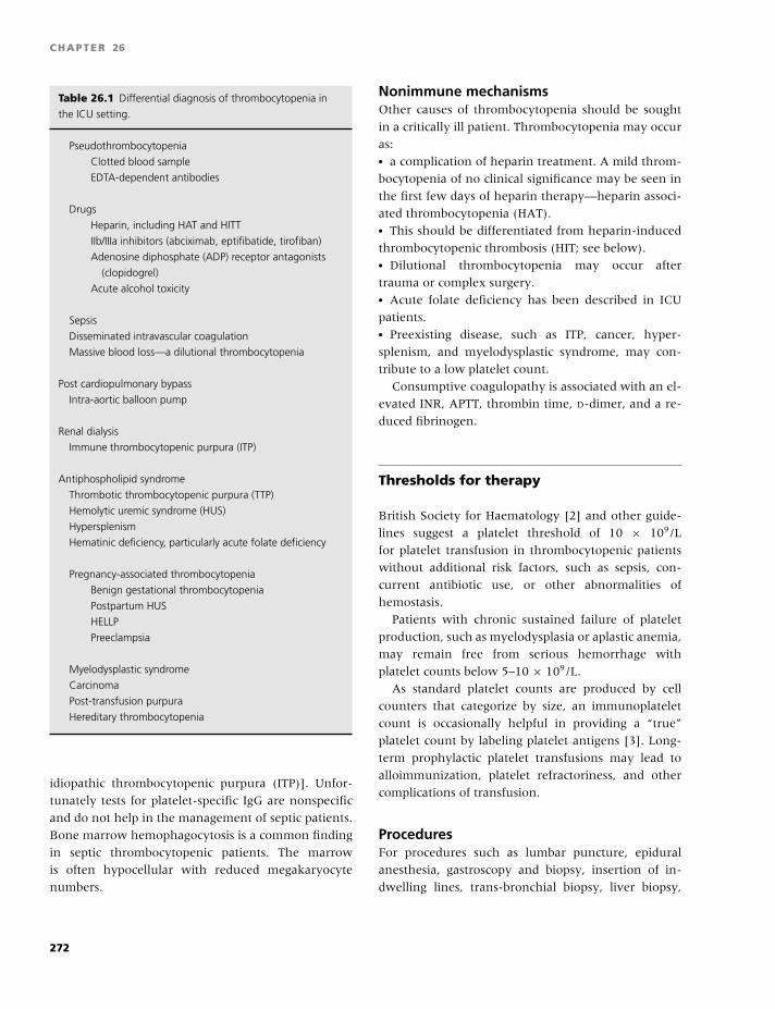
BLBK186-Key May 4, 2009 14:31
CHAPTER 26
Table 26.1 Differential diagnosis of thrombocytopenia in
the ICU setting.
Pseudothrombocytopenia
Clotted blood sample
EDTA-dependent antibodies
Drugs
Heparin, including HAT and HITT
IIb/IIIa inhibitors (abciximab, eptifibatide, tirofiban)
Adenosine diphosphate (ADP) receptor antagonists
(clopidogrel)
Acute alcohol toxicity
Sepsis
Disseminated intravascular coagulation
Massive blood loss—a dilutional thrombocytopenia
Post cardiopulmonary bypass
Intra-aortic balloon pump
Renal dialysis
Immune thrombocytopenic purpura (ITP)
Antiphospholipid syndrome
Thrombotic thrombocytopenic purpura (TTP)
Hemolytic uremic syndrome (HUS)
Hypersplenism
Hematinic deficiency, particularly acute folate deficiency
Pregnancy-associated thrombocytopenia
Benign gestational thrombocytopenia
Postpartum HUS
HELLP
Preeclampsia
Myelodysplastic syndrome
Carcinoma
Post-transfusion purpura
Hereditary thrombocytopenia
idiopathic thrombocytopenic purpura (ITP)]. Unfor-
tunately tests for platelet-specific IgG are nonspecific
and do not help in the management of septic patients.
Bone marrow hemophagocytosis is a common finding
in septic thrombocytopenic patients. The marrow
is often hypocellular with reduced megakaryocyte
numbers.
Nonimmune mechanismsOther causes of thrombocytopenia should be sought
in a critically ill patient. Thrombocytopenia may occur
as:� a complication of heparin treatment. A mild throm-
bocytopenia of no clinical significance may be seen in
the first few days of heparin therapy—heparin associ-
ated thrombocytopenia (HAT).� This should be differentiated from heparin-induced
thrombocytopenic thrombosis (HIT; see below).� Dilutional thrombocytopenia may occur after
trauma or complex surgery.� Acute folate deficiency has been described in ICU
patients.� Preexisting disease, such as ITP, cancer, hyper-
splenism, and myelodysplastic syndrome, may con-
tribute to a low platelet count.
Consumptive coagulopathy is associated with an el-
evated INR, APTT, thrombin time, D-dimer, and a re-
duced fibrinogen.
Thresholds for therapy
British Society for Haematology [2] and other guide-
lines suggest a platelet threshold of 10 × 109/L
for platelet transfusion in thrombocytopenic patients
without additional risk factors, such as sepsis, con-
current antibiotic use, or other abnormalities of
hemostasis.
Patients with chronic sustained failure of platelet
production, such as myelodysplasia or aplastic anemia,
may remain free from serious hemorrhage with
platelet counts below 5–10 × 109/L.
As standard platelet counts are produced by cell
counters that categorize by size, an immunoplatelet
count is occasionally helpful in providing a “true”
platelet count by labeling platelet antigens [3]. Long-
term prophylactic platelet transfusions may lead to
alloimmunization, platelet refractoriness, and other
complications of transfusion.
ProceduresFor procedures such as lumbar puncture, epidural
anesthesia, gastroscopy and biopsy, insertion of in-
dwelling lines, trans-bronchial biopsy, liver biopsy,
272
BLBK186-Key May 4, 2009 14:31
Intensive and critical care
and laparotomy, the platelet count should be raised
to at least 50 × 109/L. For operations on critical sites,
such as the brain or eyes, recommendations are for a
platelet count of 75–100 × 109/L.
Antiplatelet therapyDrugs known to have antiplatelet activity should be
withdrawn. Any underlying disorder associated with
platelet dysfunction, such as uremia, should be treated
if possible. The hematocrit should be corrected to
�0.30 in those with renal failure. The use of DDAVP
can be considered.
Massive transfusionIn massive blood loss, the platelet count is preserved
until relatively late. A platelet count of around 50 ×109/L is expected when red cell concentrates equiva-
lent to two blood volumes have been transfused. The
platelet count should be maintained above 50 × 109/L
in patients with acute bleeding. A higher target of
100 × 109/L is recommended for those with multiple
trauma or central nervous system injury.
Disseminated intravascularcoagulopathy (DIC)Platelet transfusions are indicated in acute DIC when
there is bleeding associated with thrombocytopenia.
Management of the underlying disorder and coag-
ulation factor replacement are also required. Fre-
quent full blood count and coagulation screening tests
should be carried out, and the platelet count main-
tained above 50 × 109/L. Platelet transfusions should
not be given simply to correct a low platelet count in
chronic DIC in the absence of bleeding.
Immune thrombocytopeniaIn patients with ITP, platelet transfusions are reserved
for patients with life-threatening gastrointestinal, gen-
itourinary, or central nervous system bleeding or other
bleeding associated with severe thrombocytopenia. In
ITP, the residual platelets tend to be young and have
good hemostatic effect, so patients tend not to bleed
unless the platelet count is very low. Platelet transfu-
sions may not produce an incremental rise in patients
with ITP due to the effect of the platelet antibodies
on the donor platelets. IV methylprednisolone, IVIg or
anti-D (only to be used in the Rhesus-positive patients
who have a spleen) can be given to produce platelet
increments [4]. The emerging thrombopoeitic agents
may gain a place in the future management of acute
ITP.
Post-transfusion purpuraPost-transfusion purpura is due to the presence of
a platelet specific allo-antibody [usually anti-human
platelet antigen-1a (HPA-1a)] in the recipient, which
reacts with donor platelets, destroying them and also
the recipient’s own platelets. High dose IVIg (2g/kg
given over 2 or 5 days) is used in the treatment of post-
transfusion purpura, with responses in about 85% of
patients. Large doses of platelet transfusions may be
required to control severe bleeding before there is a re-
sponse to IVIg. There is limited evidence that HPA-1a-
negative platelets are more effective than those from
random donors [5].
The thrombotic microangiopathies
Profound thrombocytopenia and microangiopathic
hemolytic anemia characterize thrombotic microan-
giopathy, which includes three major disorders:
thrombotic thrombocytopenic purpura (TTP), hemo-
lytic uremic syndrome (HUS), and HELLP syndrome
(Haemolysis, Elevated Liver function tests and Low
Platelets). The hemolysis is due to the breakdown of
red cells as they pass over areas of thrombosis.
Thrombotic thrombocytopenicpurpura (TTP)
TTP is a clinical diagnosis characterized by:� thrombocytopenia,� microangiopathic hemolytic anemia,� fluctuating neurological signs,� renal impairment, and� fever.
Excessive platelet aggregation results in platelet
microvascular thrombi, which particularly affect the
cerebral circulation. This is mediated by ultra-large
von Willebrand factor (vWF) multimers due to a
deficiency of vWF cleaving protease (vWF-CP), also
known as ADAMTS13. Deficiency of vWF-CP activity
273
BLBK186-Key May 4, 2009 14:31
CHAPTER 26
may be genetic due to absence of the enzyme or
acquired due to the presence of an autoantibody to
vWF-CP. Cirrhosis, acute inflammation, DIC, and
malignancy have all been associated with reduced
VWF-CP activity but do not cause TTP.
TTP is characterized by:� severe thrombocytopenia.� Red cell fragments may be absent from the periph-
eral blood in the first 24–48 hours following clinical
presentation.� Coagulation profiles are usually normal.
Secondary DIC due to prolonged tissue ischemia is
an ominous prognostic indicator.
Many specialized units now measure levels of vWF-
CP and its inhibitor to confirm the diagnosis of TTP,
but results are not available quickly. Thus, if a case
of TTP is suspected, they must be treated immedi-
ately and the diagnosis must be confirmed or refuted
retrospectively. Renal or skin biopsy performed after
recovery of the thrombocytopenia may also aid ret-
rospective diagnosis. There is a prominent arteriolar
and capillary thrombosis with thrombi, largely com-
posed of platelets, which stain strongly for VWF. This
contrasts with HUS, where the primary histological
changes are glomerular, and arteriolar fibrin thrombi
and subendothelial widening of the glomerular capil-
lary wall.
Factors that may precipitate TTPThese include drugs, autoimmune disease, malig-
nancy, and infection and are listed in Table 26.2. In
some series, up to 14% of TTP episodes have been
associated increasingly with HIV infection, with the
greatest risk at CD4 counts of less than 250 × 109/L.
E. coli 0157:H7 is more closely linked with HUS, but
there have been cases with typical TTP features.
A panel of investigations required in a suspected
case of TTP includes:� FBC and film (Plate 26.1),� Reticulocyte count,� Clotting screen including fibrinogen and D-dimers,� Urea and electrolytes,� Liver function tests,� Lactate dehydrogenase,� Urinalysis,� Direct antiglobulin test, and� HIV and hepatitis serology.
Table 26.2 TTP precipitating factors.
Drugs Autoimmune disease
Oral contraceptives Systemic lupus erythematosis
Ticlopidine
Ciclosporin Malignancy
Mitomycin C Pregnancy
Infection Post–bone marrow transplantation
HIV
Immediate treatment (plasma exchange)If a patient presents with signs suggestive of TTP (i.e.
those with neurological signs and a microangiopathic
haemolytic anemia and thrombocytopenia in the ab-
sence of any other identifiable cause), it is increasingly
being recognized that delay in treatment may result in
sudden death due to thrombotic occlusion of the coro-
nary arteries [6].
Single volume daily plasma exchange should be
commenced within 24 hours. Theoretically, plasma
exchanges using cryosupernatant or solvent–detergent
prepared FFP may be more efficacious than using
standard fresh frozen, although there are no clini-
cal data to support this currently. Both cryosuper-
natant and solvent–detergent prepared FFP are defi-
cient in highmolecular-weight VWF multimeric forms
and thus may be less likely to stimulate further throm-
bosis. Daily plasma exchange should continue for a
minimum of 2 days after complete remission. Platelet
transfusions are contraindicted.
Concomitant therapyAdjuvant corticosteroid therapy with pulsed methyl-
prednisolone 1 g IV daily for 3 days can be considered.
Low-dose aspirin (75 mg daily) should be commenced
on platelet recovery (platelet counts �50 × 109/L).
Red cell transfusion should be administered according
to clinical need. Folate supplementation is required in
all patients. Platelet transfusions are contraindicated
in TTP unless there is life-threatening hemorrhage.
Hepatitis vaccination is recommended [7].
Refractory diseaseIn the presence of refractory disease, intensification
of plasma exchange should be considered. The use of
274
BLBK186-Key May 4, 2009 14:31
Intensive and critical care
Ritoximab is emerging as a major advance in the man-
agement of acquired TTP. Having gained a place in sal-
vaging patients not responding to plasma exchange,
there is some evidence to suggest it induces faster re-
missions when it has been used early in the course of
the disease [8].
In refractory TTP, advice should be sought from a
specialist in this field. Vincristine 1 mg repeated every
3–4 days or intensive immunosuppression using either
cyclosporin or cyclophosphamide has been used in se-
vere refractory or recurrent TTP. Protein A column
immunoabsorption may be considered. Urgent self-
referral is advised if a patient develops symptoms sug-
gestive of relapse. Splenectomy may reduce the risk of
relapse.
MortalityPrior to the advent of plasma exchange, mortality rates
were in excess of 90%. With prompt plasma exchange,
the mortality has fallen to 10–30%. Thirty-five percent
do not have neurological involvement at presentation,
but a reduced level of consciousness has been identi-
fied as a poor prognostic indicator with an overall sur-
vival of 54%. The average number of plasma exchange
procedures required for remission was 15.8 (range
3–36) in one series.
HHUS
HUS is characterized by:� microangiopathic hemolytic anemia,� thrombocytopenia, and� renal failure.
There may be associated multiorgan disease, includ-
ing enterocolitis, neurological complications, liver, and
pancreatic and cardiac dysfunction.
The epidemic form (D+) is associated with:� a prodromal illness,� bloody diarrhea, and� enterotoxin enterococcal (VTEC) infection.
Rare sporadic or atypical cases have:� no prodrome, and� may be associated with HIV, cytomegalovirus, or
bacterial infection.
Secondary causes of HUS include:� post–solid-organ or -bone marrow transplantation,
� drug exposure (pentostatin, cyclosporine, mito-
mycin C, heroin, and quinine),� malignancy, and� Systemic lupus erythematosus.
However, approximately 50% of HUS cases are as-
sociated with a mutation in one or more genes coding
for proteins involved in regulation or activation of the
alternative pathway of complement, such as factor H
deficiency [9].
Laboratory investigationsEarly stool culture is essential for the diagnosis of
VTEC-associated HUS (E. coli 0157:H7) [10]. Other in-
vestigations are as for TTP.
Treatment of HUSManagement involves meticulous fluid and elec-
trolyte balance and blood pressure control, with
renal dialysis as required. Antimotility drugs and
antibiotic treatment adversely affect the outcome and
should be avoided. At present, there is no conclusive
evidence that either FFP or plasma exchange im-
proves outcome. Adjuvant treatment with antiplatelet
agents, anticoagulation, antifibrinolytics, or IVIg is not
recommended.
HELLP
HELLP is diagnosed by the presence of:� hemolysis,� elevated liver function tests, and� thrombocytopenia
in the second and third trimesters of pregnant or
postpartum woman.
It occurs in up to 10% of women with severe
preeclampsia. Severe thrombocytopenia and abnormal
liver function tests can occur in the absence of signifi-
cant hypertension or proteinuria. Exacerbations may
occur postpartum, and there is a recurrence risk of
approximately 3% in subsequent pregnancies. HELLP
occasionally presents postpartum, usually within 48
hours, but rarely as late as 6 days after delivery.
Common presenting symptoms include:� nausea,� malaise,� epigastric or right upper quadrant abdominal pain,� and edema.
275
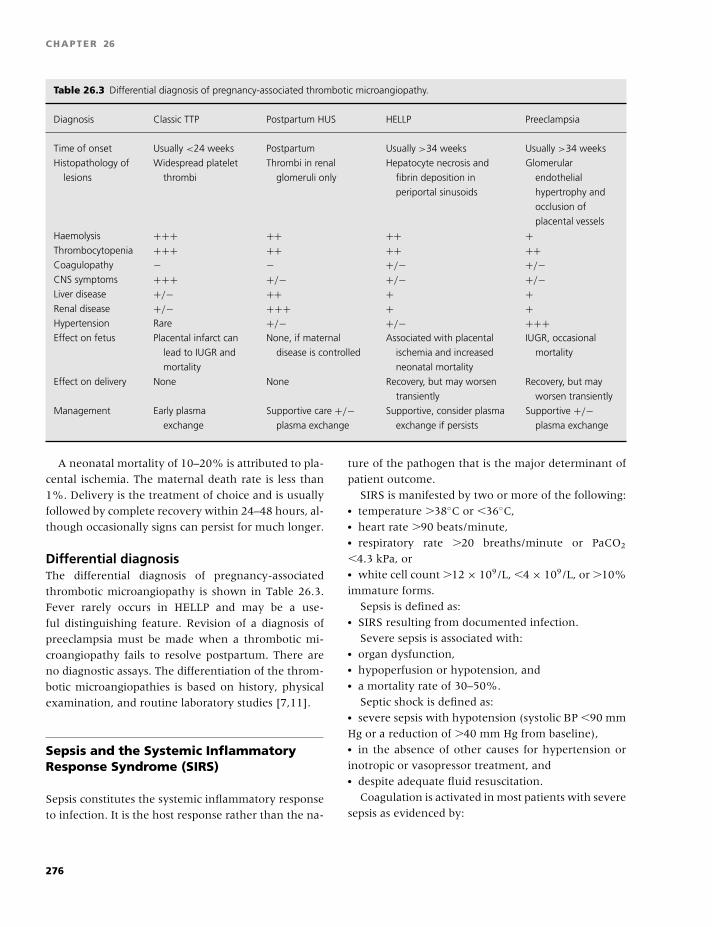
BLBK186-Key May 4, 2009 14:31
CHAPTER 26
Table 26.3 Differential diagnosis of pregnancy-associated thrombotic microangiopathy.
Diagnosis Classic TTP Postpartum HUS HELLP Preeclampsia
Time of onset Usually <24 weeks Postpartum Usually >34 weeks Usually >34 weeks
Histopathology of
lesions
Widespread platelet
thrombi
Thrombi in renal
glomeruli only
Hepatocyte necrosis and
fibrin deposition in
periportal sinusoids
Glomerular
endothelial
hypertrophy and
occlusion of
placental vessels
Haemolysis +++ ++ ++ +Thrombocytopenia +++ ++ ++ ++Coagulopathy − − +/− +/−CNS symptoms +++ +/− +/− +/−Liver disease +/− ++ + +Renal disease +/− +++ + +Hypertension Rare +/− +/− +++Effect on fetus Placental infarct can
lead to IUGR and
mortality
None, if maternal
disease is controlled
Associated with placental
ischemia and increased
neonatal mortality
IUGR, occasional
mortality
Effect on delivery None None Recovery, but may worsen
transiently
Recovery, but may
worsen transiently
Management Early plasma
exchange
Supportive care +/−plasma exchange
Supportive, consider plasma
exchange if persists
Supportive +/−plasma exchange
A neonatal mortality of 10–20% is attributed to pla-
cental ischemia. The maternal death rate is less than
1%. Delivery is the treatment of choice and is usually
followed by complete recovery within 24–48 hours, al-
though occasionally signs can persist for much longer.
Differential diagnosisThe differential diagnosis of pregnancy-associated
thrombotic microangiopathy is shown in Table 26.3.
Fever rarely occurs in HELLP and may be a use-
ful distinguishing feature. Revision of a diagnosis of
preeclampsia must be made when a thrombotic mi-
croangiopathy fails to resolve postpartum. There are
no diagnostic assays. The differentiation of the throm-
botic microangiopathies is based on history, physical
examination, and routine laboratory studies [7,11].
Sepsis and the Systemic InflammatoryResponse Syndrome (SIRS)
Sepsis constitutes the systemic inflammatory response
to infection. It is the host response rather than the na-
ture of the pathogen that is the major determinant of
patient outcome.
SIRS is manifested by two or more of the following:� temperature �38◦C or �36◦C,� heart rate �90 beats/minute,� respiratory rate �20 breaths/minute or PaCO2
�4.3 kPa, or� white cell count �12 × 109/L, �4 × 10⁹/L, or �10%
immature forms.
Sepsis is defined as:� SIRS resulting from documented infection.
Severe sepsis is associated with:� organ dysfunction,� hypoperfusion or hypotension, and� a mortality rate of 30–50%.
Septic shock is defined as:� severe sepsis with hypotension (systolic BP �90 mm
Hg or a reduction of �40 mm Hg from baseline),� in the absence of other causes for hypertension or
inotropic or vasopressor treatment, and� despite adequate fluid resuscitation.
Coagulation is activated in most patients with severe
sepsis as evidenced by:
276
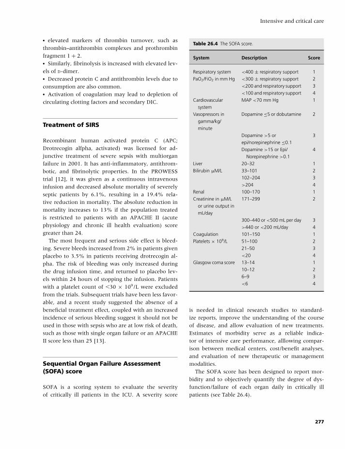
BLBK186-Key May 4, 2009 14:31
Intensive and critical care
� elevated markers of thrombin turnover, such as
thrombin–antithrombin complexes and prothrombin
fragment 1 + 2.� Similarly, fibrinolysis is increased with elevated lev-
els of D-dimer.� Decreased protein C and antithrombin levels due to
consumption are also common.� Activation of coagulation may lead to depletion of
circulating clotting factors and secondary DIC.
Treatment of SIRS
Recombinant human activated protein C (APC;
Drotrecogin alfpha, activated) was licensed for ad-
junctive treatment of severe sepsis with multiorgan
failure in 2001. It has anti-inflammatory, antithrom-
botic, and fibrinolytic properties. In the PROWESS
trial [12], it was given as a continuous intravenous
infusion and decreased absolute mortality of severely
septic patients by 6.1%, resulting in a 19.4% rela-
tive reduction in mortality. The absolute reduction in
mortality increases to 13% if the population treated
is restricted to patients with an APACHE II (acute
physiology and chronic ill health evaluation) score
greater than 24.
The most frequent and serious side effect is bleed-
ing. Severe bleeds increased from 2% in patients given
placebo to 3.5% in patients receiving drotrecogin al-
pha. The risk of bleeding was only increased during
the drug infusion time, and returned to placebo lev-
els within 24 hours of stopping the infusion. Patients
with a platelet count of �30 × 109/L were excluded
from the trials. Subsequent trials have been less favor-
able, and a recent study suggested the absence of a
beneficial treatment effect, coupled with an increased
incidence of serious bleeding suggest it should not be
used in those with sepsis who are at low risk of death,
such as those with single organ failure or an APACHE
II score less than 25 [13].
Sequential Organ Failure Assessment(SOFA) score
SOFA is a scoring system to evaluate the severity
of critically ill patients in the ICU. A severity score
Table 26.4 The SOFA score.
System Description Score
Respiratory system <400 ± respiratory support 1
PaO2/FiO2 in mm Hg <300 ± respiratory support 2
<200 and respiratory support 3
<100 and respiratory support 4
Cardiovascular
system
MAP <70 mm Hg 1
Vasopressors in
gamma/kg/
minute
Dopamine ≤5 or dobutamine 2
Dopamine >5 or 3
epi/norepinephrine ≤0.1
Dopamine >15 or Epi/
Norepinephrine >0.1
4
Liver 20–32 1
Bilirubin µM/L 33–101 2
102–204 3
>204 4
Renal 100–170 1
Creatinine in µM/L
or urine output in
mL/day
171–299 2
300–440 or <500 mL per day 3
>440 or <200 mL/day 4
Coagulation 101–150 1
Platelets × 109/L 51–100 2
21–50 3
<20 4
Glasgow coma score 13–14 1
10–12 2
6–9 3
<6 4
is needed in clinical research studies to standard-
ize reports, improve the understanding of the course
of disease, and allow evaluation of new treatments.
Estimates of morbidity serve as a reliable indica-
tor of intensive care performance, alllowing compar-
ison between medical centers, cost/benefit analyses,
and evaluation of new therapeutic or management
modalities.
The SOFA score has been designed to report mor-
bidity and to objectively quantify the degree of dys-
function/failure of each organ daily in critically ill
patients (see Table 26.4).
277
BLBK186-Key May 4, 2009 14:31
CHAPTER 26
HIT
HIT is a transient drug-induced autoimmune pro-
thrombotic disorder initiated by heparin. Heparin ex-
posure can induce the formation of pathogenic IgG an-
tibodies that cause platelet activation by recognizing
complexes of platelet factor 4 (PF4) and heparin on
platelet surfaces. Platelet activation results in throm-
bocytopenia and thrombin generation, with an in-
creased risk of venous and arterial thrombosis [14].
HIT antibodies are directed against multiple neoepi-
tope sites. Only a minority of PF4/heparin-reactive
HIT sera activate platelets in vitro. Some HIT-IgG rec-
ognize PF4 bound to solid phase even in the absence
of heparin. PF4 antibodies usually decline to unde-
tectable levels within a few weeks or months of an
episode of HIT, and there is no anamnestic response.� The frequency of HIT varies widely depending on
the type of heparin used and the patient group.� Unfractionated heparin is associated with a higher
incidence of HIT than fractionated heparin.� Surgical patients have a higher frequency of HIT
than either medical or obstetric patients with the same
heparin exposure.� Postoperative orthopedic patients receiving unfrac-
tionated heparin have the highest HIT frequency (up
to 5%) and require more intense platelet count mon-
itoring than pregnant women receiving LMWH, who
have an almost negligible risk.
Laboratory diagnosisHIT antibodies are detected using either:� commercially available PF4-dependent antigen
immunoassays, or� functional assays of platelet activation and aggrega-
tion.
Clinically insignificant HIT antibodies are common
in patents that have received heparin 5–100 days ear-
lier. In the ICU setting, HIT is uncommon (0.3–0.5%),
whereas thrombocytopenia from other causes is very
common (30–50%).
For laboratory diagnosis of HIT antibodies, both
antigen assays and functional (platelet activation) as-
says are available. Both tests are very sensitive (high
negative predictive value) but specificity is poor, es-
pecially for the antigen assays, which will also detect
nonpathogenic immunoglobulin M and immunoglob-
ulin A class antibodies. Detection of immunoglobulin
M or immunoglobulin A antibodies could potentially
lead to adverse events, such as bleeding, if a false diag-
nosis of HIT prompts replacement of heparin by an al-
ternative anticoagulant. Dosing regimens of the direct
thrombin inhibitors are too high, especially in ICU pa-
tients. Assays of platelet activation are technically de-
manding, time consuming, and not available in some
centers. Testing should be performed when HIT is clin-
ically suspected.
Clinical diagnosisThe diagnosis of HIT should be based on:� clinical abnormalities (thrombocytopenia with or
without thrombosis), and� a positive test for HIT antibodies, as outlined in
Table 26.5.
Isolated HIT is the occurrence of thrombocytope-
nia without thrombosis. Retrospective cohort studies
indicate that 25–50% of these patients develop clin-
ically overt thrombosis after stopping heparin, usu-
ally within the first week. Subclinical thrombosis was
found in 8 of 16 patients who underwent routine
lower-limb duplex ultrasonography for isolated HIT.
Early heparin cessation alone does not reduce the risk
of thrombosis in patients with isolated HIT, so alterna-
tive anticoagulation is required.
About 25% of HIT patients receiving a heparin
bolus develop signs or symptoms, such as fever, chills,
respiratory distress, or hypertension. Transient global
amnesia and cardiorespiratory arrest have also been
reported. About 5–15% of HIT patients develop de-
compensated DIC.
Thombocytopenia does not usually develop until
day 5–10 of heparin treatment and reaches a median
nadir of 55 × 109/L. The platelet count falls below
150 × 109/L in around 90% of HIT cases. Hemor-
rhage and platelet counts below 10 × 109/L suggest
an alternative cause, such as post-transfusion purpura.
Patients who have received heparin within the last
100 days may have a fall in platelet count within one
day of reexposure to heparin.
Treatment of HIT
Heparin should be stopped immediately, and not re-
peated, in those who develop thrombocytopenia or
278
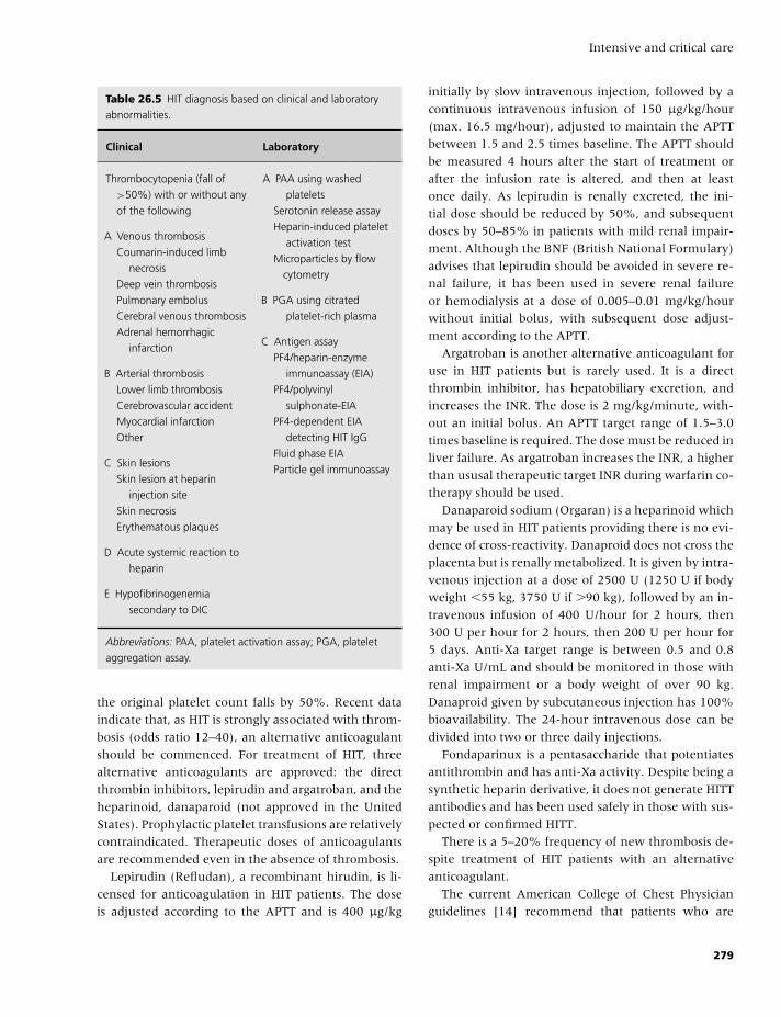
BLBK186-Key May 4, 2009 14:31
Intensive and critical care
Table 26.5 HIT diagnosis based on clinical and laboratory
abnormalities.
Clinical Laboratory
Thrombocytopenia (fall of
>50%) with or without any
of the following
A Venous thrombosis
Coumarin-induced limb
necrosis
Deep vein thrombosis
Pulmonary embolus
Cerebral venous thrombosis
Adrenal hemorrhagic
infarction
B Arterial thrombosis
Lower limb thrombosis
Cerebrovascular accident
Myocardial infarction
Other
C Skin lesions
Skin lesion at heparin
injection site
Skin necrosis
Erythematous plaques
D Acute systemic reaction to
heparin
E Hypofibrinogenemia
secondary to DIC
A PAA using washed
platelets
Serotonin release assay
Heparin-induced platelet
activation test
Microparticles by flow
cytometry
B PGA using citrated
platelet-rich plasma
C Antigen assay
PF4/heparin-enzyme
immunoassay (EIA)
PF4/polyvinyl
sulphonate-EIA
PF4-dependent EIA
detecting HIT IgG
Fluid phase EIA
Particle gel immunoassay
Abbreviations: PAA, platelet activation assay; PGA, platelet
aggregation assay.
the original platelet count falls by 50%. Recent data
indicate that, as HIT is strongly associated with throm-
bosis (odds ratio 12–40), an alternative anticoagulant
should be commenced. For treatment of HIT, three
alternative anticoagulants are approved: the direct
thrombin inhibitors, lepirudin and argatroban, and the
heparinoid, danaparoid (not approved in the United
States). Prophylactic platelet transfusions are relatively
contraindicated. Therapeutic doses of anticoagulants
are recommended even in the absence of thrombosis.
Lepirudin (Refludan), a recombinant hirudin, is li-
censed for anticoagulation in HIT patients. The dose
is adjusted according to the APTT and is 400 µg/kg
initially by slow intravenous injection, followed by a
continuous intravenous infusion of 150 µg/kg/hour
(max. 16.5 mg/hour), adjusted to maintain the APTT
between 1.5 and 2.5 times baseline. The APTT should
be measured 4 hours after the start of treatment or
after the infusion rate is altered, and then at least
once daily. As lepirudin is renally excreted, the ini-
tial dose should be reduced by 50%, and subsequent
doses by 50–85% in patients with mild renal impair-
ment. Although the BNF (British National Formulary)
advises that lepirudin should be avoided in severe re-
nal failure, it has been used in severe renal failure
or hemodialysis at a dose of 0.005–0.01 mg/kg/hour
without initial bolus, with subsequent dose adjust-
ment according to the APTT.
Argatroban is another alternative anticoagulant for
use in HIT patients but is rarely used. It is a direct
thrombin inhibitor, has hepatobiliary excretion, and
increases the INR. The dose is 2 mg/kg/minute, with-
out an initial bolus. An APTT target range of 1.5–3.0
times baseline is required. The dose must be reduced in
liver failure. As argatroban increases the INR, a higher
than ususal therapeutic target INR during warfarin co-
therapy should be used.
Danaparoid sodium (Orgaran) is a heparinoid which
may be used in HIT patients providing there is no evi-
dence of cross-reactivity. Danaproid does not cross the
placenta but is renally metabolized. It is given by intra-
venous injection at a dose of 2500 U (1250 U if body
weight �55 kg, 3750 U if �90 kg), followed by an in-
travenous infusion of 400 U/hour for 2 hours, then
300 U per hour for 2 hours, then 200 U per hour for
5 days. Anti-Xa target range is between 0.5 and 0.8
anti-Xa U/mL and should be monitored in those with
renal impairment or a body weight of over 90 kg.
Danaproid given by subcutaneous injection has 100%
bioavailability. The 24-hour intravenous dose can be
divided into two or three daily injections.
Fondaparinux is a pentasaccharide that potentiates
antithrombin and has anti-Xa activity. Despite being a
synthetic heparin derivative, it does not generate HITT
antibodies and has been used safely in those with sus-
pected or confirmed HITT.
There is a 5–20% frequency of new thrombosis de-
spite treatment of HIT patients with an alternative
anticoagulant.
The current American College of Chest Physician
guidelines [14] recommend that patients who are
279
BLBK186-Key May 4, 2009 14:31
CHAPTER 26
receiving heparin or have received heparin within
the previous 2 weeks, they should be investigated
for a diagnosis of HIT if the platelet count falls by
≥50%, and/or a thrombotic event occurs, between
days 5 and 14 (inclusive) following initiation of hep-
arin, even if the patient is no longer receiving hep-
arin therapy when thrombosis or thrombocytopenia
has occurred (Grade 1C). For patients with strongly
suspected (or confirmed) HIT, whether or not compli-
cated by thrombosis, we recommend use of an alter-
native, nonheparin anticoagulant [danaparoid (Grade
1B), lepirudin (Grade 1C), argatroban (Grade 1C),
fondaparinux (Grade 2C), or bivalirudin (Grade 2C)]
over the further use of unfractionated heparin (UFH)
or low-molecular-weight heparin (LMWH) therapy
or initiation/continuation of vitamin K antagonists
(Grade 1B).
Management of thromboembolism in ICU
Massive pulmonary embolismVenous thromboembolism (VTE) is an important
cause of morbidity and mortality in ICU patients.
Among patients who died in ICU, pulmonary emboli
(PE) were reported in 7–27% of postmortem exami-
nations. The mortality rate for PE is �8% when the
condition is recognized and treated, but approximately
30% when untreated.
Massive PE has a mortality of 18–33% and may
present with shock, dyspnea, and confusion. In pa-
tients with massive PE and hemodynamic instabil-
ity, rapid risk assessment is paramount and bedside
echocardiography has become the most popular tool.
Multislice chest computed tomography (CT) is also
useful for identifying patients who may benefit from
thrombolysis or embolectomy. Cardiac biomarkers, in-
cluding troponin and the natriuretic peptides, are sen-
sitive markers of right ventricular function. Low lev-
els of troponin, B-type natriuretic peptide (BNP), and
NT-terminal proBNP are all highly sensitive assays for
identifying patients with an uneventful clinical course.
Multislice chest CT is not only useful to diagnose or ex-
clude PE; it also is useful for risk assessment. A right-
to-left ventricular dimension ratio �0.9 on the recon-
structed CT four-chamber view identifies patients at
increased risk of early death [15].
Treatment of PELMWH and fondaparinux are equal or superior in ef-
ficacy to UFH for the treatment of DVT and PE [16].
The benefit-to-risk ratio of thrombolysis in deep vein
thrombosis (DVT) is dubious but is recommended for
unstable patients with PE, although these patients rep-
resent �5% of all patients hospitalized for PE.
The streptokinase/urokinase PE thrombolysis tri-
als showed that thrombolytic therapy successfully de-
creases pulmonary artery pressures acutely with im-
provements in the lung scan and arteriogram at 12
and 24 hours. There was no overall decrease in mor-
tality in those receiving thrombolysis compared with
those receiving heparin therapy. The use of throm-
bolytic treatment in patients with submassive PE re-
mains controversial. Contraindications to thromboly-
sis include active internal bleeding, a stroke within
2 months, and an intracranial process such as neo-
plasm or abscess. Relative contraindications include
surgery or organ biopsy within 10 days, uncontrolled
hypertension, and pregnancy.
The dose of alteplase is 10 mg IV injection over
1–2 minutes followed by an IV infusion of 90 mg
over 2 hours (max. 1.5 mg/kg in patients �65 kg).
The dose of streptokinase is 250,000 U by IV infusion
over 30 minutes, then 100,000 U every hour for up to
12–72 hours according to clinical condition, with
monitoring of clotting parameters. A simplified al-
gorithm for alteplase consisting of 0.6 mg/kg over
15 minutes has been used successfully in many cen-
ters, with equivalence to the standard regime demon-
strated in two prospective randomized studies. Hem-
orrhagic complications are higher in patients with a
recent invasive procedure, such as pulmonary an-
giogram or placement of an IVC filter. There is a re-
ported incidence of intracranial hemorrhage of ap-
proximately 2%, with higher rates in the elderly and
those with poorly controlled hypertension. The major
hemorrhage rate ranges from 11% to 20%.
Two indications are widely recognized for inferior
vena cava filters:� The first is a permanent or temporary contraindica-
tion to anticoagulation, in patients with proximal DVT
or PE.� The second is the occurrence of PE or propagation of
the thrombus in patients treated for DVT or recurrence
in patients with PE [17].
280
BLBK186-Key May 4, 2009 14:31
Intensive and critical care
The PREPIC study [18] demonstrated that, at 8
years, vena cava filters reduced the risk of PE but in-
creased that of DVT and had no effect on survival.
The authors concluded that, although their use may
be beneficial in patients at high risk of PE, systematic
use in the general population with VTE is not recom-
mended. A Cochrane review concluded that further
trials, especially with retrievable filters, are needed to
assess vena caval filter safety and effectiveness [19].
Surgical intervention should be considered for pa-
tients whose condition worsens despite intensive med-
ical treatment. A randomized study of embolectomy
versus medical therapy is unavailable. Thrombolytic
treatment fails in 15–20% of patients. The mortality
after surgical embolectomy is around 30–40%, with
a higher mortality in those with a longer duration
of hemodynamic instability, a requirement for car-
diopulmonary resuscitation and intubation, high doses
of catecholamines, metabolic, and respiratory acidosis,
and poor urine output. Early diagnosis and treatment
leads to improved outcomes.
Thromboprophylaxis in the ICU
The critically ill are at substantially increased risk of
VTE, which contributes significantly to their morbidity
and mortality. PE is frequently seen at postmortem in
these patients, the incidence being as high as 27%. The
incidence of image-proven DVT in critically ill patients
ranges from 10% to almost 100%, depending on the
screening methods and diagnostic criteria used.
Most critically ill patients have multiple risk fac-
tors for VTE. Many risk factors predate ICU admission
in particular recent surgery, immobilization, trauma,
sepsis, malignancy, increased age, heart or respiratory
failure, and previous VTE. These initial VTE risk factors
are confounded by others, which are acquired on the
ICU including immobilization, pharmacological paral-
ysis, central venous catheterization, additional surgical
procedures, sepsis, vasopressors, and hemodialysis.
Clinically undetected DVT may be present on admis-
sion to a critical care unit. Five studies using Doppler
ultrasound, in a total of 990 patients reported a rate of
5.5% DVT on admission to ICU with rates up to 29%
in patients not given thromboprophylaxis prior to ICU
admission. Although the majority of DVTs are clini-
cally silent and often confined to the calf veins, asymp-
tomatic DVT can become symptomatic and lead to
embolic complications. There is no way of predicting
which at-risk patients will develop symptomatic VTE;
it is, however, well recognized that massive PE fre-
quently occurs without warning and is often fatal. PE
is found in 15% of in-patients’ deaths at postmortem.
Hospitalized patients recovering from major trauma
have the highest risk of developing VTE. Without ade-
quate thromboprophylaxis, patients with multisystem
failure or major trauma have a DVT risk exceeding
50%, with PE being the third leading cause of mor-
tality after the first day.
Despite extensive trials of thromboprophylaxis for
medical and surgical patients, there are few for crit-
ical care patients. Extrapolating data relating to spe-
cific medical and surgical patients to the critically ill is
not easy, for the risk–benefit ratio may be significantly
different between these groups. There have been two
systematic reviews of thromboprophylaxis [20,21].
With few exceptions, thromboprophylaxis should
be used in all ICU patients. Decisions regarding the ini-
tiation and method of prophylaxis should be based on
the balance of bleeding and thrombotic risk. Patients
with a high risk of bleeding should be given mechan-
ical prophylaxis with either graduated anti-embolic
stockings alone or stockings combined with intermit-
tent pneumatic compression devices until bleeding
risk decreases and prophylaxis with heparin can be
commenced.
Prophylaxis should be reviewed daily and altered
as necessitated by the patient’s clinical status. Pro-
phylaxis should not be interrupted for procedures or
surgery unless there is a particularly high bleeding
risk. Procedures such as insertion or removal of epidu-
ral catheters should be planned to coincide with the
nadir of anticoagulant effect. Table 26.6 outlines rec-
ommendations for prophylaxis in critically ill patients
suggested by Geerts and coworkers [22].
Those that receive either suboptimal or no thrombo-
prophylaxis should have Doppler ultrasound screen-
ing. Thromboprophylaxis should be continued until
hospital discharge in those at high risk, and this period
includes inpatient rehabilitation. The ACCP guidelines
also recommend that thromboprophylaxis should be
continued post discharge in those with continuing im-
mobility.
281
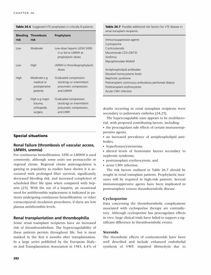
BLBK186-Key May 4, 2009 14:31
CHAPTER 26
Table 26.6 Suggested VTE prophylaxis in critically ill patients.
Bleeding Thrombosis Prophylaxisrisk risk
Low Moderate Low-dose heparin (LDH) 5000
U sc bd or LMWH at
prophylactic doses
Low High LMWH in thromboprophylactic
doses
High Moderate e.g
medical or
postoperative
patients
Graduated compression
stockings or intermittent
pneumatic compression,
and LMWH
High High e.g major
trauma,
orthopedic
surgery
Graduated compression
stockings or intermittent
pneumatic compression,
and LMW
Special situations
Renal failure (thrombosis of vascular access,LMWH, uremia)For continuous hemofiltration, UFH or LMWH is used
commonly, although some units use prostacyclin or
regional citrate. Regional citrate anticoagulation is
gaining in popularity as studies have shown it is as-
sociated with prolonged filter survival, significantly
decreased bleeding risk, and increased completion of
scheduled filter life span when compared with hep-
arin [23]. With the use of a heparin, an occasional
need for antithrombin replacement is indicated in pa-
tients undergoing continuous hemofiltration, or other
extracorporeal circulation procedures, if there are low
plasma antithrombin levels.
Renal transplantation and thrombophiliaSome renal transplant recipients have an increased
risk of thromboembolism. The hypercoagulability of
these patients persists throughout life, but is most
marked in the first 6 months after transplantation.
In a large series published by the European Dialy-
sis and Transplantation Association in 1983, 4.4% of
Table 26.7 Possible additional risk factors for VTE disease in
renal transplant recipients.
Immunosuppressive agents
Cyclosporine
Corticosteroids
Muromonab-CD3 (OKT3)
Sirolimus
Mycophenolate Mofetil
Antiphospholipid antibodies
Elevated homocysteine levels
Nephrotic syndrome
Pretransplant continuous ambulatory peritoneal dialysis
Posttransplant erythrocytosis
Acute CMV infection
deaths occurring in renal transplant recipients were
secondary to pulmonary embolus [24,25].
The hypercoagulable state appears to be multifacto-
rial, with proposed contributing factors, including:� the procoagulant side effects of certain immunosup-
pressive agents,� an increased prevalence of antiphospholipid anti-
bodies,� hyperhomocysteinemia,� altered levels of hemostatic factors secondary to
nephrotic syndrome,� posttransplant erythrocytosis, and� acute CMV infection.
The risk factors outlined in Table 26.7 should be
sought in renal transplant patients. Prophylactic mea-
sures will be required in high-risk patients. Several
immunosuppressive agents have been implicated in
posttransplant venous thromboembolic disease.
CyclosporineData concerning the thromboembolic complications
associated with cyclosporine therapy are contradic-
tory. Although cyclosporine has procoagulant effects
in vivo, large clinical trials have failed to support a sig-
nificant difference in thromboembolic events.
SteroidsThe thrombotic effects of corticosteroids have been
well described and include enhanced endothelial
synthesis of VWF, impaired fibrinolysis due to
282
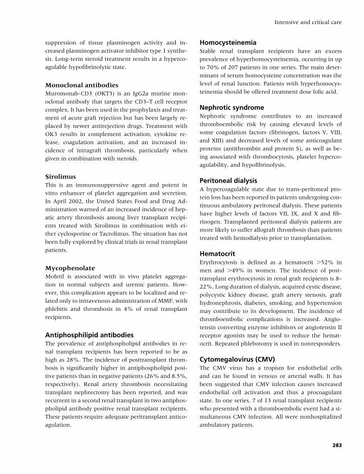
BLBK186-Key May 4, 2009 14:31
Intensive and critical care
suppression of tissue plasminogen activity and in-
creased plasminogen activator inhibitor type 1 synthe-
sis. Long-term steroid treatment results in a hyperco-
agulable hypofibrinolytic state.
Monoclonal antibodiesMuromonab-CD3 (OKT3) is an IgG2a murine mon-
oclonal antibody that targets the CD3–T cell receptor
complex. It has been used in the prophylaxis and treat-
ment of acute graft rejection but has been largely re-
placed by newer antirejection drugs. Treatment with
OK3 results in complement activation, cytokine re-
lease, coagulation activation, and an increased in-
cidence of intragraft thrombosis, particularly when
given in combination with steroids.
SirolimusThis is an immunosuppressive agent and potent in
vitro enhancer of platelet aggregation and secretion.
In April 2002, the United States Food and Drug Ad-
ministration warned of an increased incidence of hep-
atic artery thrombosis among liver transplant recipi-
ents treated with Sirolimus in combination with ei-
ther cyclosporine or Tacrolimus. The situation has not
been fully explored by clinical trials in renal transplant
patients.
MycophenolateMofetil is associated with in vivo platelet aggrega-
tion in normal subjects and uremic patients. How-
ever, this complication appears to be localized and re-
lated only to intravenous administration of MMF, with
phlebitis and thrombosis in 4% of renal transplant
recipients.
Antiphosphilipid antibodiesThe prevalence of antiphospholipid antibodies in re-
nal transplant recipients has been reported to be as
high as 28%. The incidence of posttransplant throm-
bosis is significantly higher in antiphospholipid posi-
tive patients than in negative patients (26% and 8.5%,
respectively). Renal artery thrombosis necessitating
transplant nephrectomy has been reported, and was
recurrent in a second renal transplant in two antiphos-
pholipid antibody positive renal transplant recipients.
These patients require adequate peritransplant antico-
agulation.
HomocysteinemiaStable renal transplant recipients have an excess
prevalence of hyperhomocysteinemia, occurring in up
to 70% of 207 patients in one series. The main deter-
minant of serum homocysteine concentration was the
level of renal function. Patients with hyperhomocys-
teinemia should be offered treatment dose folic acid.
Nephrotic syndromeNephrotic syndrome contributes to an increased
thromboembolic risk by causing elevated levels of
some coagulation factors (fibrinogen, factors V, VIII,
and XIII) and decreased levels of some anticoagulant
proteins (antithrombin and protein S), as well as be-
ing associated with thrombocytosis, platelet hyperco-
agulability, and hypofibrinolysis.
Peritoneal dialysisA hypercoagulable state due to trans-peritoneal pro-
tein loss has been reported in patients undergoing con-
tinuous ambulatory peritoneal dialysis. These patients
have higher levels of factors VII, IX, and X and fib-
rinogen. Transplanted peritoneal dialysis patients are
more likely to suffer allograft thrombosis than patients
treated with hemodialysis prior to transplantation.
HematocritErythrocytosis is defined as a hematocrit �52% in
men and �49% in women. The incidence of post-
transplant erythrocytosis in renal graft recipients is 8–
22%. Long duration of dialysis, acquired cystic disease,
polycystic kidney disease, graft artery stenosis, graft
hydronephrosis, diabetes, smoking, and hypertension
may contribute to its development. The incidence of
thromboembolic complications is increased. Angio-
tensin converting enzyme inhibitors or angiotensin II
receptor agonists may be used to reduce the hemat-
ocrit. Repeated phlebotomy is used in nonresponders.
Cytomegalovirus (CMV)The CMV virus has a tropism for endothelial cells
and can be found in venous or arterial walls. It has
been suggested that CMV infection causes increased
endothelial cell activation and thus a procoagulant
state. In one series, 7 of 13 renal transplant recipients
who presented with a thromboembolic event had a si-
multaneous CMV infection. All were nonhospitalized
ambulatory patients.
283
BLBK186-Key May 4, 2009 14:31
CHAPTER 26
Jehovah’s Witnesses (JW)Jehovah’s witnesses (JW) do not accept transfusion
of blood or its major components, based on the belief
that to be transfused with blood is equivalent to eating
it and therefore prohibited by scripture. Until 2000,
any JW transfused with a prohibited blood product
was expelled from the society and ostracised by other
JWs. Since 2000, any JW who “wilfully and without
regret” accepts blood transfusion is no longer expelled
but instead “revokes his own membership by his
own actions.” Doctors should consider the possibility
that individual JW patients have interpreted this
change as allowing them to accept transfusion under
certain circumstances. This will require clarification
in a one-to-one consultation in absolute medical
confidentiality [26].
The Association of Anaesthetists of Great Britain
and Ireland (AAGBI) advise that, although it is
unlawful to give blood to a patient who has refused it,
“for unconscious patients, the doctor will be expected
to perform to the best of his/her ability, and this
may include giving blood” (AABI 1999). This would
only apply when JW status is unclear and/or rela-
tives/associates cannot produce an Advance Directive
document.
Before dismissing the use of blood products, there
must be a certainty that the patient is a committed JW,
has independently and freely decided to refuse trans-
fusion, and has thought this decision through to the
point of death at the time of making an Advance Di-
rective (living will) or additional consent to surgery.
A copy of the Advance Directive should be placed
in the patient’s notes and the contents respected. If
life-threatening bleeding occurs and time allows, a
doctor of Consultant status should discuss with the
patient, or relative, the implications of withholding
blood, and a clear, signed entry should be written in
the patient’s notes.
The 2000 Watch Tower directive stated that “pri-
mary components” of blood must be refused, but that
“when it comes to fractions of the primary compo-
nents, each Christian must conscientiously decide for
himself.”
Every JW should decide which products are ac-
ceptable to him/her during the consent process. All
available blood products should be discussed, as in-
terpretations of a “fraction of the primary com-
ponent” may hypothetically include products such
as leukocyte-depleted red cells and platelets, intra-
venous immunoglobulin, fibrinogen concentrates, and
solvent–detergent treated FFP.
Most JW patients refuse autologous predonation
because blood is separated from the body in stor-
age. Normovolemic hemodilution and some forms of
intraoperative cell salvage and hemodialysis may be
acceptable because the extracorporeal blood remains
in contact with the circulation. Hematological pa-
rameters should be optimized preoperatively. Metic-
ulous surgical hemostasis, minimal access surgery,
and systemic pre- and perioperative administration
of antifibrinolytic agents (tranexamic acid or apro-
tinin) or desmopressin (DDAVP) should be consid-
ered. The use of topical hemostatic plasma fractiona-
tion products, such as fibrin glue, may be acceptable
to some.
JW patients accept crystalloids and synthetic col-
loids, including dextran, hydroxyethylstarch, and
gelatins (Haemaccel and Gelofusin) for circulatory
support. Most requiring plasma exchange will refuse
human albumin but may accept Hetastarch or protein
A immunoabsorption as alternatives.
Recombinant blood products are acceptable to many
JW. Epoetin beta (NeoRecormin) contains a trace of
albumin, whereas Epoetin alpha does not contain al-
bumin and so is more widely accepted. Epoetin al-
pha (Eprex) is licensed for the treatment of moderate
anemia (hemoglobin concentration 10–13 g/100 mL)
before elective orthopedic surgery in adults with ex-
pected moderate blood loss, to reduce exposure to al-
logeneic transfusion. It is given by subcutaneous in-
jection (max. 1 mL per injection site), 600 U/kg every
week for 3 weeks before surgery and on the day of
surgery or 300 Units/kg daily for 15 days starting 10
days before surgery.
Supplementation with folic acid and oral iron, or in-
travenous folinic acid and iron, should be considered,
particularly if the patient is maintained on erythropoi-
etin. Frequency and amount of blood sampling should
be minimized.
Granulocyte colony stimulating factor (G-CSF) is ac-
ceptable treatment for neutropenia. Recombinant ac-
tivated factor VII (rFVIIa, NovoSeven) is licensed for
the treatment of bleeding episodes in hemophiliacs
with inhibitors, and has been used to treat bleeding
in platelet disorders as well as those without a pre-
existing hemostatic disorder.
284
BLBK186-Key May 4, 2009 14:31
Intensive and critical care
Recombinant factor VIII and XI, particularly second-
generation products containing no albumin, facili-
tate therapy of hemophilia A and B in JW patients.
DDAVP is a synthetic product suitable for use in mild
hemophilia A and type 1 von Willebrand disease and
uremia. Some patients with rare hemorrhagic disor-
ders that currently require plasma-derived therapeutic
products (e.g. type 2 or 3 vWD) will accept a purified
fractionated product.
Some JW will regard their peripheral blood and
bone marrow stem cell as a permissible fraction and
consent to collection by leukapheresis or marrow aspi-
ration. Specific treatment of the JW with other hema-
tological disorders is beyond the scope of this chapter.
There should be an open, full, and confidential dis-
cussion of all available options. JW exercise the right
of any adult with capacity to refuse medical treatment
and often carry advance directive cards indicating their
incontrovertible refusal of blood.
Despite their belief regarding transfusion, JW do not
have a higher mortality rate after traumatic injury or
surgery. Transfusion requirements are often overes-
timated. Increased morbidity and mortality is rarely
observed in patients with a hemoglobin concentra-
tion �7 g/dL, and the acute hemoglobin threshold for
cardiovascular collapse may be as low as 3–5 g/dL.
There are many modalities to treat the JW patient with
acute blood loss. Treatment with recombinant human
erythropoietin, albumin, and recombinant activated
factor VIIa have all been used with success. Auto-
logous autotransfusion and isovolemic hemodilution
can also be used to treat patients who refuse trans-
fusion. Hemoglobin-based oxygen carriers may play a
future role as intravascular volume expanders in lieu
of transfusion of red blood cell concentrates.
In conclusion, there are many treatment modalities
available to assist in the care of JW patients, especially
because their beliefs on the intricacies of the Blood
Ban appear to be in flux.
References
1 Robinson S, Harrison C. Review: challenges in the man-
agement of thrombocytosis in young patients. Clin Adv
Hematol Oncol 2008;6(2):137–8, 14
2 Guidelines for the use of platelet transfusions. Br J
Haematol 2003;122:10–23.
3 Segal HC, Harrison P. Methods for counting platelets in
severe thrombocytopenia. Curr Hematol Rep 2006;5(1):
70–5.
4 Stasi R, Evangelista ML, Stipa E, Buccisano F, Venditti
A, Amadori S. Idiopathic thrombocytopenic purpura:
current concepts in pathophysiology and management.
Thromb Haemost 2008;99(1):4–13.
5 Allen DL, Samol J, Benjamin S, Verjee S, Tusold A,
Murphy MF. Survey of the use and clinical effective-
ness of HPA-1a/5b-negative platelet concentrates in
proven or suspected platelet alloimmunization. Trans-
fus Med 2004;14(6):409–17.
6 Wahla A, Ruiz J, Nourredine N, Upadhya B,
Sane D, Owen J. Myocardial infarction in thrombotic
thrombocytopenic purpura. A single center experi-
ence and literature review. Eur J Haematol 2008;81:
311–16.
7 Allford SL, Hunt BJ, Rose P, Machin SJ; Haemosta-
sis and Thrombosis Task Force, British Committee for
Standards in Haematology. Guidelines on the diagnosis
and management of the thrombotic microangiopathic
haemolytic anaemias. Br J Haematol 2003;120(4):556–
73.
8 Scully M, Yarranton H, Liesner R, et al. Regional UK
TTP Registry: correlation with laboratory ADAMTS 13
analysis and clinical features. Br J Haematol 2008;142:
819–26.
9 Jokiranta TS, Zipfel PF, Fremeaux-Bacchi V, Taylor
CM, Goodship TJ, Noris M. Where next with atypi-
cal hemolytic uremic syndrome? Mol Immunol 2007;
44(16):3889–9.
10 Goldwater PN. Treatment and prevention of en-
terohemorrhagic Escherichia coli infection and
hemolytic uremic syndrome. Expert Rev Anti Infect
Ther 2007;5(4):653–63.
11 Hay JE. Liver disease in pregnancy. Hepatology 2008;
47(3):1067–76.
12 Bernard GR, Vincent JL, Laterre PF, et al. Recombinant
human protein C Worldwide Evaluation in Severe Sep-
sis (PROWESS) study group. Efficacy and safety of re-
combinant human activated protein C for severe sepsis.
N Engl J Med 2001;344(10):699–709.
13 Abraham E, Laterre PF, Garg R, et al. Administration
of Drotrecogin Alfa (Activated) in Early Stage Severe
Sepsis (ADDRESS) Study Group. Drotrecogin alfa (ac-
tivated) for adults with severe sepsis and a low risk of
death. N Engl J Med 2005;353(13):1332–41.
14 Warkentin TE, Greinacher A, Koster A, Lincoff AM.
Treatment and prevention of heparin-induced throm-
bocytopenia: American College of Chest Physicians
Evidence-Based Clinical Practice Guidelines (8th Edi-
tion). Chest 2008;133(6 Suppl):340S–80S.
285
BLBK186-Key May 4, 2009 14:31
CHAPTER 26
15 Kucher N, Goldhaber SZ. Risk stratification of acute
pulmonary embolism. Semin Thromb Hemost 2006;32(8):
838–44.
16 Becattini C, Agnelli G, Emmerich J, Bura A, Weitz JI.
Initial treatment of venous thromboembolism. Thromb
Haemost 2006;96(3):242–50.
17 Emmerich J, Meyer G, Decousus H, Agnelli G. Role
of fibrinolysis and interventional therapy for acute ve-
nous thromboembolism. Thromb Haemost 2006;96(3):
251–7.
18 PREPIC Study Group. Eight-year follow-up of
patients with permanent vena cava filters in the
prevention of pulmonary embolism: the PREPIC (Pre-
vention du Risque d’Embolie Pulmonaire par Interrup-
tion Cave) randomized study. Circulation 2005;112(3):
416–22.
19 Young T, Tang H, Aukes J, Hughes R. Vena caval filters
for the prevention of pulmonary embolism. Cochrane
Database Syst Rev 2007;(4):CD006212.
20 Attia J, Ray JG, Cook DJ. Deep vein thrombosis and
its prevention in critically ill adults. Arch Intern Med
2001;161:1268–79.
21 Geerts W, Cook D, Selby R, et al. Venous thromboem-
bolism and its prevention in critical care. J Crit Care
2002;17:93–104.
22 Geerts WH, Bergqvist D, Pineo GF, et al. Prevention of
venous thromboembolism: American College of Chest
Physicians Evidence-Based Clinical Practice Guidelines
(8th Edition). Chest 2008;133(6 Suppl):381S–453S.
23 Bagshaw SM, Laupland KB, Boiteau PJ, Godinez-Luna
T. Is regional citrate superior to systemic heparin anti-
coagulation for continuous renal replacement therapy?
A prospective observational study in an adult regional
critical care system. J Crit Care 2005;20(2):155–61.
24 Kazory A, Ducloux D. Acquired hypercoagulable state
in renal transplant recipients. Thromb Haemost 2004;91:
646–51.
25 Irish A. Hypercoagulability in renal transplant recip-
ients. Identifying patients at risk of renal allograft
thrombosis and evaluating strategies for prevention.
Am J Cardiovasc Drugs 2004;4(3):139–49.
26 Hughes DB, Ullery BW, Barie PS. The contemporary
approach to the care of Jehovah’s witnesses. J Trauma
2008;65(1):237–47.
286
BLBK186-Key May 4, 2009 9:39
27 TransfusionAdrian Copplestone
Introduction
The most common request to hematologists for help
in the emergency management of patients in the hos-
pital setting, relates to the control of hemorrhage and
the use of blood products. Whereas most treatment in-
volves the use of purified drugs, blood and blood prod-
ucts are derived from human blood donors. They are
rarely pure; they are subject to biological variation and
carry the risk of infection. This chapter discusses some
of these issues and describes their use in specialized
clinical settings.
Blood transfusion as a formof transplantation
Transfusion with red cells and other blood products is
a form of tissue transplantation, which is made easier
because the cells lack some or all of the HLA antigens.
Because cells lack progenitor capacity, the benefit is
temporary but allows time for the body’s homeostatic
processes to recover. However, the transfused cells
contain surface proteins that are foreign to the host
and give rise to an immune reaction. The common red
cell blood grouping systems are listed in Table 27.1.
The ABO group
These most important antigens are as a result of the in-
heritance of enzymes causing alternative glycosylation
of the red cell membrane.� If individuals lack an A or B antigen, they make anti-
A or anti-B, respectively, after exposure to these gly-
copeptides in food.
� Blood group O is due to the lack of A or B anti-
gen and so these people develop anti-A and anti-B
antibodies.� Group AB people have both antigens and lack the
anti-A and anti-B antibodies; see Table 27.2.
Individuals have naturally occurring circulating im-
munoglobulin M (IgM) antibodies to the A and B
groups they lack. These antibodies are good at fixing
complement, have the capacity to cause intravascular
hemolysis, and can lead to disseminated intravascu-
lar coagulation (DIC). A useful scheme for remember-
ing which ABO groups can be transfused to which pa-
tients is shown in Fig. 27.1. In allogeneic blood and
marrow stem cell transplantation, the picture is more
complex because patients take on the blood group of
the donor, and hemolysis may occur during the period
of changeover.
The Rhesus system
The next most important blood group system is the
Rhesus (Rh), of which the D antigen is the most
immunogenic. The use of Rh D-negative blood for
Rh D-negative patients is partly to prevent immu-
nization but also to prevent hemolytic disease of
the newborn due to the transplacental passage of
anti-D to Rh D-positive children of Rh-D negative
mothers.
Red cell cross-matching
Just over 100 years ago, Landsteiner discovered blood
groups. Transfusion from donor to patient became fea-
sible when it was possible to determine blood groups
and store the blood in an anticoagulated form. In
287
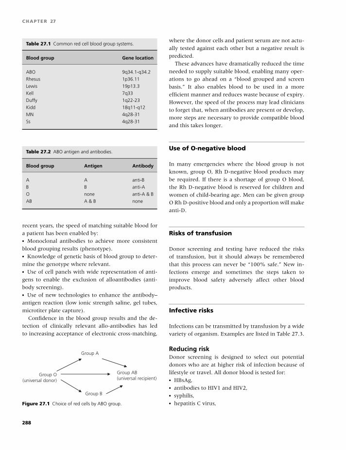
BLBK186-Key May 4, 2009 9:39
CHAPTER 27
Table 27.1 Common red cell blood group systems.
Blood group Gene location
ABO 9q34.1-q34.2
Rhesus 1p36.11
Lewis 19p13.3
Kell 7q33
Duffy 1q22-23
Kidd 18q11-q12
MN 4q28-31
Ss 4q28-31
Table 27.2 ABO antigen and antibodies.
Blood group Antigen Antibody
A A anti-B
B B anti-A
O none anti-A & B
AB A & B none
recent years, the speed of matching suitable blood for
a patient has been enabled by:� Monoclonal antibodies to achieve more consistent
blood grouping results (phenotype).� Knowledge of genetic basis of blood group to deter-
mine the genotype where relevant.� Use of cell panels with wide representation of anti-
gens to enable the exclusion of alloantibodies (anti-
body screening).� Use of new technologies to enhance the antibody–
antigen reaction (low ionic strength saline, gel tubes,
microtiter plate capture).
Confidence in the blood group results and the de-
tection of clinically relevant allo-antibodies has led
to increasing acceptance of electronic cross-matching,
Group A
Group O(universal donor)
Group AB(universal recipient)
Group B
Figure 27.1 Choice of red cells by ABO group.
where the donor cells and patient serum are not actu-
ally tested against each other but a negative result is
predicted.
These advances have dramatically reduced the time
needed to supply suitable blood, enabling many oper-
ations to go ahead on a “blood grouped and screen
basis.” It also enables blood to be used in a more
efficient manner and reduces waste because of expiry.
However, the speed of the process may lead clinicians
to forget that, when antibodies are present or develop,
more steps are necessary to provide compatible blood
and this takes longer.
Use of O-negative blood
In many emergencies where the blood group is not
known, group O, Rh D-negative blood products may
be required. If there is a shortage of group O blood,
the Rh D-negative blood is reserved for children and
women of child-bearing age. Men can be given group
O Rh D-positive blood and only a proportion will make
anti-D.
Risks of transfusion
Donor screening and testing have reduced the risks
of transfusion, but it should always be remembered
that this process can never be “100% safe.” New in-
fections emerge and sometimes the steps taken to
improve blood safety adversely affect other blood
products.
Infective risks
Infections can be transmitted by transfusion by a wide
variety of organism. Examples are listed in Table 27.3.
Reducing riskDonor screening is designed to select out potential
donors who are at higher risk of infection because of
lifestyle or travel. All donor blood is tested for:� HBsAg,� antibodies to HIV1 and HIV2,� syphilis,� hepatitis C virus,
288
BLBK186-Key May 4, 2009 9:39
Transfusion
Table 27.3 Examples of transfusion-transmitted infections.
Viruses Hepatitis A
Hepatitis B
Hepatitis C
HIV
HTLV 1 & 2
CMV
EBV
Parvovirus
West Nile virus
Bacteria Treponema pallidum (syphilis)
Borrelia burgdorferi (Lyme disease)
Staphylococcus spp.
Diphtheroids
Salmonella spp.
Pseudomonas spp.
Yersinia spp.
Protozoa Plasmodium spp. (malaria)
Toxoplasma gondii (toxoplasmosis)
Trypanosoma cruzi (Chaga’s disease)
Abbreviations: CMV, cytomegalovirus; EBV, Epstein–Barr virus;
HTLV, human T cell leukaemia virus.
� human T cell leukaemia virus, and� some donors for cytomegalovirus.
Despite these tests, there exist a small number of
donors who are infected but lack antibody; this will
be reduced further by nucleic acid testing using poly-
merase chain reaction technology to look for viral
genome.
New agents (e.g. West Nile virus and SARS) con-
tinue to emerge as pathogens. Steps taken to reduce
these risks include:� donor lifestyle screening,� antibody testing,� leukodepletion, and� DNA/RNA testing.
For plasma products, it is also possible to:� heat treat,� nanofilter, or� disrupt lipid membranes with solvents, methylene
blue, or psoralens with ultraviolet light.
Widespread leukodepletion was introduced in the
UK in 1998 to reduce the risk of transmission of
variant Creutzfeldt–Jakob Disease (vCJD). In addition,
there was a major shift of procurement of plasma for
plasma products from areas without bovine spongi-
form encephalopathy (BSE), primarily the US. No test
is currently available to detect the abnormal prion.
BSE has been transmitted in sheep by transfusion, and
in the UK, by 2008, there have been four cases of vCJD
transmission by blood transfusion.
Transfusion reactions
Immediate hemolytic reactionsThese are likely to be associated with shock, renal
failure, and DIC. The most common cause is patients
receiving the wrong blood, in 70% because of the la-
beling or checking errors at the bedside or in the labo-
ratory. These errors are preventable by the adherence
to clear transfusion protocols.
Delayed hemolytic reactionsThese are usually caused by extravascular hemolysis
and the boosting of allo-antibody levels.
Febrile transfusion reactionsLess common now that universal leukodepletion is in
place, these are caused by the presence of cytokines
and HLA antibodies. Urticarial and allergic reactions
can still occur.
Transfusion-related acute lung injuryTransfusion-related acute lung injury (TRALI) is
caused by donor leukocyte antibodies which cause
adult respiratory distress syndrome. The patient be-
comes acutely short of breath and often requires arti-
ficial ventilation and circulatory support. TRALI needs
to be distinguished from circulatory fluid overload,
which can occur following the transfusion of large vol-
umes, especially in older patients. In the UK, the num-
ber of cases of TRALI has fallen after the increased use
of male plasma to make fresh frozen plasma (FFP), as
males have less immunization by white cell antigens
than females (related to pregnancy).
ImmunizationAlloimmunization can affect the efficacy of transfu-
sion, especially platelets. It may also affect the sub-
sequent choice of donors for organ transplantation.
Immunomodulation can follow transfusion with an
289
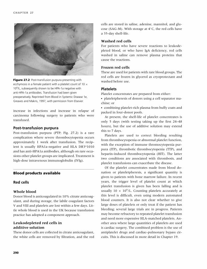
BLBK186-Key May 4, 2009 9:39
CHAPTER 27
Figure 27.2 Post-transfusion purpura presenting with
ecchymosis in a female patient with a platelet count of 10 ×109/L, subsequently shown to be HPA-1a negative with
anti-HPA-1a antibodies. Transfusion had been given
preoperatively. Reprinted from Blood in Systemic Disease 1e,
Greaves and Makris, 1997, with permission from Elsevier.
increase in infections and increase in relapse of
carcinoma following surgery to patients who were
transfused.
Post-transfusion purpuraPost-transfusion purpura (PTP; Fig. 27.2) is a rare
complication where severe thrombocytopenia occurs
approximately 1 week after transfusion. The recip-
ient is usually HPA1a-negative and HLA DR3*1010
and has anti-HPA1a antibodies, although on rare occa-
sions other platelet groups are implicated. Treatment is
high-dose intravenous immunoglobulin (IVIg).
Blood products available
Red cells
Whole bloodDonor blood is anticoagulated in 10% citrate anticoag-
ulant, and during storage, the labile coagulant factors
V and VIII and platelets are lost within a few days. Lit-
tle whole blood is used in the UK because transfusion
practice has adopted a component approach.
Leukodepleted red cells inadditive solutionThese donor cells are collected in citrate anticoagulant,
the white cells are removed by filtration, and the red
cells are stored in saline, adenine, mannitol, and glu-
cose (SAG-M). With storage at 4◦C, the red cells have
a 35-day shelf-life.
Washed red cellsFor patients who have severe reactions to leukode-
pleted blood, or who have IgA deficiency, red cells
washed in saline can remove plasma proteins that
cause the reactions.
Frozen red cellsThese are used for patients with rare blood groups. The
red cells are frozen in glycerol as cryoprotectant and
washed before use.
PlateletsPlatelet concentrates are prepared from either:� plateletpheresis of donors using a cell separator ma-
chine; or� combining platelet-rich plasma from buffy coats and
packed in four-donor pools.
At present, the shelf-life of platelet concentrates is
only 5 days (with testing taking up the first 24–48
hours), but the use of additive solution may extend
this to 7 days.
Platelets are used to correct bleeding resulting
from thrombocytopenia or abnormal platelet function,
with the exception of immune thromocytopenia pur-
pura (ITP), thrombotic thrombocytopenia (TTP), and
heparin-induced thrombocytopenia (HIT). The latter
two conditions are associated with thrombosis, and
platelet transfusions can exacerbate the disease.
Of the platelet concentrates made from blood do-
nation or plateletpheresis, a significant quantity is
given to patients with bone marrow failure. In recent
years, the trigger level of platelet count at which
platelet transfusion is given has been falling and is
usually 10 × 109/L. Counting platelets accurately at
this level is difficult, even using modern automated
blood counters. It is also not clear whether to give
large doses of platelets or only treat if the patient has
bleeding; several large trials are in progress. Patients
may become refractory to repeated platelet transfusion
and need more expensive HLA-matched platelets. An-
other area where large quantities of platelets are used
is cardiac surgery. The combined problem is the use of
antiplatelet drugs and cardiac–pulmonary bypass cir-
cuits. This is discussed in more detail in Chapter 19.
290
BLBK186-Key May 4, 2009 9:39
Transfusion
Fresh frozen plasmaFFP is used to correct coagulation deficiencies, and
there has been considerable debate on the relative
merits of different products.
In the ideal world, FFP would provide high concen-
trations of the relevant factor, be from a low number
of screened regular donors, have a viral inactivation
step in the production that does not adversely af-
fect the coagulation factors, be procured in a country
where BSE is not endemic, come from male donors (to
reduce the risk of TRALI), and have appropriate ABO
group.
The following are available:� Single-donor FFP.� Methylene blue-treated FFP for pediatric use is a
single-donor product, procured in the US. In the
UK, it is used primarily for children born after Jan-
uary 1, 1996 when the risk of vCJD from meat was
minimized, but its use will extend to other age groups
as it becomes more available.� Solvent detergent FFP (Octaplas R©) is a pooled prod-
uct that is solvent treated to reduce the infective risks.
It is used in large quantities in TTP because it is low in
high-molecular-weight multimers of von Willebrand
factor (VWF), but it has been associated with throm-
bosis because of protein S deficiency.
British Committee for Standards in Haematology
(BCSH) guidelines suggest that:� FFP should only be used to replace single inherited
clotting factor deficiencies for which no virus-safe frac-
tionated product is available. Currently, this applies
mainly to factor V.� FFP is indicated when there are demonstrable mul-
tifactor deficiencies associated with severe bleeding
and/or DIC. However, FFP is not indicated in DIC with
no evidence of bleeding.� FFP should not be used to reverse warfarin effect
in the absence of bleeding as it has an incomplete
effect and is not an ideal product as large quantities
are required. Vitamin K and prothrombin complex
concentrate should be used when reversing coumarin
anticoagulants in patients who are bleeding or at high
risk of bleeding.� Large quantities of FFP are used for correction of ab-
normal coagulation tests prior to invasive procedures,
but the evidence base that this reduces bleeding is
weak.
Cryoprecipitate and MB CryoCryoprecipitate forms when FFP is thawed slowly, and
the product, which is refrozen, is rich in fibrinogen
and factors VIII and XIII. It is commonly used in the
treatment of DIC to replace fibrinogen. Methylene
blue-treated Cryo is available for children in the UK.
Cyrosupernatant and MB CryosupernatantThe complementary product cryosupernatant has
been used in conjunction with plasmapheresis in
TTP as it lacks high-molecular-weight multimers of
VWF; however, SDFFP is the recommended product in
the UK.
Human albumin solutionThe final product of the plasma fractionation pro-
cess, human albumin solution (HAS), comes in two
strengths: 4.5 g/dL and 20 g/dL (salt-poor albumin).
It is an important colloid for maintaining the oncotic
pressure in the intravascular compartment, and its
main indication relates to replacing albumin in se-
vere edematous states. Its use as plasma expander
has largely been superseded by crystalloids and gelatin
solutions.
Intravenous immunoglobulinIVIg solutions are pooled normal human donor im-
munoglobulins. In the coagulation disorders, they are
used as an immunomodulator for the treatment of ITP
and PTP. Because supply cannot meet demand, most
countries have adopted national clinical guidelines to-
gether with a demand management plan.
Coagulation factor concentratesConcentrates are prepared from large pools of donor
plasma. They all have steps to reduce viral contamina-
tion and most have steps to remove impure proteins.
Increasing use of recombinant coagulation factors as
these become available is being encouraged:� Factor VIII for hemophilia A. Some of the interme-
diate purity products contain useful amounts of VWF
as well.� Factor IX for hemophilia B.� VWF concentrates are now available for von Wille-
brand disease (VWD).� Prothrombin complex concentrate (combined fac-
tors II, VII, IX, and X concentrate) is primarily used in
291
BLBK186-Key May 4, 2009 9:39
CHAPTER 27
the correction of life-threatening hemorrhage in pa-
tients on oral anticoagulants.� Individual concentrates for factors VII, X, and XIII
and fibrinogen are available for patients with heredi-
tary deficiencies.
Fibrin sealantsMixing thrombin and fibrinogen forms “fibrin glue,”
which is applied to the site of bleeding and is a popular
treatment in neurosurgery.
Autologous bloodIn many situations, it is possible to use the patient’s
own blood and thereby avoid exposure to the risks of
donor blood. However, there are still risks with using
autologous blood, mainly related to bacterial infection
and the blood being transfused to the wrong patient.
A number of approaches are possible.
Predeposit donationBlood is venesected prior to elective surgery and re-
tained for up to 4 weeks. By retransfusing older blood
during the collection process, up to 4 U of blood can be
stored. Surgery must take place on the planned date or
the blood may expire. In the UK, the use of predeposit
donation has fallen as patients can be more anemic
at the time of surgery, and if anemia can be corrected
pre-admission, the patient can often withstand the loss
of volume of blood that would have been transfused.
Cell salvageBlood can be aspirated during an operation and
washed red cells returned to the patient. This is use-
ful in vascular surgery and is also finding a place in
cardiac surgery, trauma, and obstetric patients.
Intraoperative hemodilutionBlood is venesected at the time of anesthesia, and crys-
talloid is used as fluid replacement. If bleeding occurs,
less red cells are lost because of the lower hematocrit.
At the end of the operation, the blood, which also con-
tains coagulation factors and platelets, is retransfused.
Cell salvage from wound drainsBlood is drawn into a sterile container by suction and
transfused. This application has been used extensively
in orthopedic surgery and has reduced the need for
blood in joint-replacement operations.
Drugs that reduce the need fortransfusion
A number of drugs are used to either boost the hemo-
static system or reduce fibrinolysis. Drugs that can in-
crease the red cells mass are also important.
Desmopressin (DDAVP)This analogue of antiduiretic hormone is used in mild
hemophilia, VWD, and some platelet disorders. En-
dothelial stores of VWF are released. Repeated admin-
istration is subject to tachyphylaxis.
Tranexamic acid and otherfibrinolytic inhibitorsThese are useful in major surgery, but their use needs
to be balanced against the risk of venous throm-
boembolism (VTE). They may also be used in patients
with marrow failure who have mucosal bleeding from
chronic thrombocytopenia in patients but are refrac-
tory to platelet transfusions.
Aprotinin (Trasylol R©) is a bovine protease inhibitor
that inactivates plasmin and kallikrein. It has been
used in cardiac surgery in patients on cardiopul-
monary bypass, with a reduction in the need for trans-
fusion, reoperation for bleeding, and length of stay in
ICU and hospital admission. In 2006, concerns of in-
creased frequency of renal failure and multiorgan fail-
ure led to considerable discussion of its role. A suspen-
sion of marketing was agreed in November 2007.
IronThere are many patients who have low iron stores or
frank deficiency as a consequence of chronic hemor-
rhage, either through the disease process or the re-
sult of treatment (e.g. nonsteroidal anti-inflammatory
drugs). Correction with small doses of iron to improve
compliance can avoid the need for transfusion. Where
anemia has developed slowly, patients can tolerate
quite low hemoglobin levels. Treatment with iron and
patience are much safer than “top-up transfusions.”
VitaminsOther vitamins (such as folic acid) may also be re-
quired in anemic patients with poor intake (elderly or
malabsorption) or increased turnover (pregnancy).
292
BLBK186-Key May 4, 2009 9:39
Transfusion
ErythropoietinErythropoietin (rhEPO) can be useful to boost the ery-
thron. Concomitant iron therapy may also be needed
to achieve a rapid response. Its cost has restricted its
use in clinical practice, but many patients with renal
failure no longer require regular transfusion.
Recombinant activated factor VIIThis recombinant protein (rFVIIa) was originally used
in hemophiliacs with inhibitors, but it is now increas-
ingly being used in patients with severe bleeding from
multiple trauma or major bleeding in a critical care
situation.
Use of blood products
How much to give?The decision of when to transfuse and how much to
give can be difficult [1–5]. In general, the rule should
be to try to avoid transfusion if possible, but if it is nec-
essary, to use sufficient quantities of the right product
to achieve the desired effect (usually hemostasis).
Guidelines on the use of red cells have previously
advised transfusion based on the reduction of red cell
mass, but this can be difficult to estimate in clinical
practice. As a result, “Hb triggers” have increasingly
been used in the management of patients, particularly
in the postoperative setting. In a landmark study [6],
Hebert and coworkers showed that, in patients in a
critical care unit, a restrictive transfusion policy (Hb
trigger 7.0 g/dL, aim Hb 7–9 g/dL) had a lower mor-
tality than a more liberal policy (Hb trigger 10 g/dL,
aim Hb 10–12 g/dL), with the possible exception of
patients with acute myocardial infarction and unsta-
ble angina.
Although Hb trigger levels are easy for clinical teams
to use, other factors also affect the Hb level, and the
Hb trigger level may need to be adjusted for individ-
ual patients based on comorbidities. Other measures
may usefully aid the decision as to whether to trans-
fuse, such as the rate of postoperative bleeding. Where
this has been measured for a cohort of patients (e.g.
postcardiac bypass surgery), deviations from the usual
course can be spotted more rapidly and appropriate
action taken. Similarly, if more attention was paid to
improving anemia preoperatively, there would be less
need for transfusion.
Assessment of hemorrhageIn situations where patients are bleeding, the first
question is to determine whether this is surgically
correctable. Simultaneously, blood should be sent for
blood count and coagulation studies. The prothrombin
time (PT) and activated partial thromboplastin time
(APPT), combined with supplementary tests (fibrino-
gen level, thrombin time, equal volume mix with nor-
mal plasma) usually give an indication as to the type
of hemostatic defect. Confirmation with specific factor
levels can follow if necessary.
Blood sampling is important as these patients often
have multiple cannulae, and it is important that the
sample is not taken through a line contaminated with
heparin. The drug chart should be examined especially
for anticoagulants, antifibrinolytics, and antiplatelet
drugs. Caution must be taken with blood count sam-
ples, as patients may be inappropriately transfused if
taken from lines running intravenous fluids.
Near patient testingBecause coagulation tests take at least 20 minutes to
complete (and usually longer, taking sample transport
into account), there has been a move to use near pa-
tient testing (NPT) with a number of different devices.� Whole blood clotting time: ACT; this is used in car-
diac surgery to monitor heparin effect.� PT and APTT devices (e.g. Coaguchek R©): these are
designed mainly for testing patients on oral anticoag-
ulants.� Thromboelastogram: the TEG R© is described in more
detail in Chapter 19, and is used in liver and cardiac
units. It gives information relating to platelet function,
clot strength, and fibrinolysis within approximately
15 minutes.� Platelet function analyses (PFA-100 R©): an in vitro
bleeding time test whose current role is determining
mild VWD and platelet defects.
Although many hematologists dislike NPT equip-
ment as being “uncontrolled” and lacking some of the
strict supervision of laboratory procedures, the imme-
diacy of results will lead to their increased use, and
both laboratory and clinical teams should work to-
gether to define their role in decision making.
293
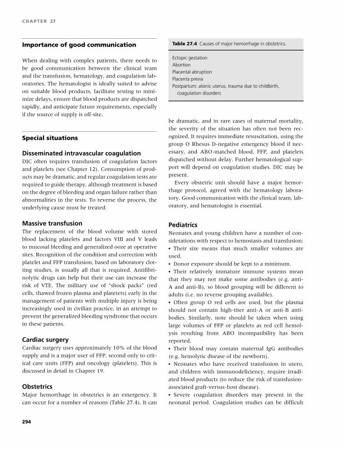
BLBK186-Key May 4, 2009 9:39
CHAPTER 27
Importance of good communication
When dealing with complex patients, there needs to
be good communication between the clinical team
and the transfusion, hematology, and coagulation lab-
oratories. The hematologist is ideally suited to advise
on suitable blood products, facilitate testing to mini-
mize delays, ensure that blood products are dispatched
rapidly, and anticipate future requirements, especially
if the source of supply is off-site.
Special situations
Disseminated intravascular coagulationDIC often requires transfusion of coagulation factors
and platelets (see Chapter 12). Consumption of prod-
ucts may be dramatic, and regular coagulation tests are
required to guide therapy, although treatment is based
on the degree of bleeding and organ failure rather than
abnormalities in the tests. To reverse the process, the
underlying cause must be treated.
Massive transfusionThe replacement of the blood volume with stored
blood lacking platelets and factors VIII and V leads
to mucosal bleeding and generalized ooze at operative
sites. Recognition of the condition and correction with
platelet and FFP transfusion, based on laboratory clot-
ting studies, is usually all that is required. Antifibri-
nolytic drugs can help but their use can increase the
risk of VTE. The military use of “shock packs” (red
cells, thawed frozen plasma and platetets) early in the
management of patients with multiple injury is being
increasingly used in civilian practice, in an attempt to
prevent the generalized bleeding syndrome that occurs
in these patients.
Cardiac surgeryCardiac surgery uses approximately 10% of the blood
supply and is a major user of FFP, second only to crit-
ical care units (FFP) and oncology (platelets). This is
discussed in detail in Chapter 19.
ObstetricsMajor hemorrhage in obstetrics is an emergency. It
can occur for a number of reasons (Table 27.4). It can
Table 27.4 Causes of major hemorrhage in obstetrics.
Ectopic gestation
Abortion
Placental abruption
Placenta previa
Postpartum: atonic uterus, trauma due to childbirth,
coagulation disorders
be dramatic, and in rare cases of maternal mortality,
the severity of the situation has often not been rec-
ognized. It requires immediate resuscitation, using the
group O Rhesus D-negative emergency blood if nec-
essary, and ABO-matched blood, FFP, and platelets
dispatched without delay. Further hematological sup-
port will depend on coagulation studies. DIC may be
present.
Every obstetric unit should have a major hemor-
rhage protocol, agreed with the hematology labora-
tory. Good communication with the clinical team, lab-
oratory, and hematologist is essential.
PediatricsNeonates and young children have a number of con-
siderations with respect to hemostasis and transfusion:� Their size means that much smaller volumes are
used.� Donor exposure should be kept to a minimum.� Their relatively immature immune systems mean
that they may not make some antibodies (e.g. anti-
A and anti-B), so blood grouping will be different to
adults (i.e. no reverse grouping available).� Often group O red cells are used, but the plasma
should not contain high-titer anti-A or anti-B anti-
bodies. Similarly, note should be taken when using
large volumes of FFP or platelets as red cell hemol-
ysis resulting from ABO incompatibility has been
reported.� Their blood may contain maternal IgG antibodies
(e.g. hemolytic disease of the newborn).� Neonates who have received transfusion in utero,
and children with immunodeficiency, require irradi-
ated blood products (to reduce the risk of transfusion-
associated graft-versus-host disease).� Severe coagulation disorders may present in the
neonatal period. Coagulation studies can be difficult
294
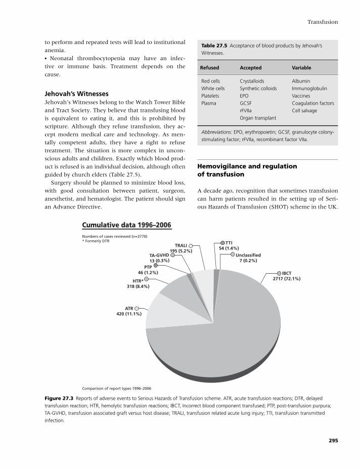
BLBK186-Key May 4, 2009 9:39
Transfusion
to perform and repeated tests will lead to institutional
anemia.� Neonatal thrombocytopenia may have an infec-
tive or immune basis. Treatment depends on the
cause.
Jehovah’s WitnessesJehovah’s Witnesses belong to the Watch Tower Bible
and Tract Society. They believe that transfusing blood
is equivalent to eating it, and this is prohibited by
scripture. Although they refuse transfusion, they ac-
cept modern medical care and technology. As men-
tally competent adults, they have a right to refuse
treatment. The situation is more complex in uncon-
scious adults and children. Exactly which blood prod-
uct is refused is an individual decision, although often
guided by church elders (Table 27.5).
Surgery should be planned to minimize blood loss,
with good consultation between patient, surgeon,
anesthetist, and hematologist. The patient should sign
an Advance Directive.
Table 27.5 Acceptance of blood products by Jehovah’s
Witnesses.
Refused Accepted Variable
Red cells Crystalloids Albumin
White cells Synthetic colloids Immunoglobulin
Platelets EPO Vaccines
Plasma GCSF Coagulation factors
rFVIIa Cell salvage
Organ transplant
Abbreviations: EPO, erythropoietin; GCSF, granulocyte colony-
stimulating factor; rFVIIa, recombinant factor VIIa.
Hemovigilance and regulationof transfusion
A decade ago, recognition that sometimes transfusion
can harm patients resulted in the setting up of Seri-
ous Hazards of Transfusion (SHOT) scheme in the UK.
IBCT2717 (72.1%)
Unclassified7 (0.2%)
TTI54 (1.4%)
TRALI195 (5.2%)
Cumulative data 1996–2006
TA-GVHD13 (0.3%)
PTP46 (1.2%)
HTR*318 (8.4%)
ATR420 (11.1%)
Numbers of cases reviewed (n=3770)* Formerly DTR
Comparison of report types 1996–2006
Figure 27.3 Reports of adverse events to Serious Hazards of Transfusion scheme. ATR, acute transfusion reactions; DTR, delayed
transfusion reaction; HTR, hemolytic transfusion reactions; IBCT, Incorrect blood component transfused; PTP, post-transfusion purpura;
TA-GVHD, transfusion associated graft versus host disease; TRALI, transfusion related acute lung injury; TTI, transfusion transmitted
infection.
295
BLBK186-Key May 4, 2009 9:39
CHAPTER 27
This is a voluntary confidential reporting scheme that
has been copied in many other countries. Analysis of
adverse events has been invaluable in improving the
safety of transfusion. The type of errors is shown in
Fig. 27.3. In the 3770 cases reported, there were 109
deaths and 315 cases of major morbidity. The annual
reports give details and recommendations to improve
transfusion practice.
In Europe, Blood Safety Directives have been in-
corporated into national legislation (Blood Safety and
Quality Regulations in the UK). Reporting of adverse
events is mandatory. There needs to be full traceabil-
ity from donor to patient with records retained for
30 years (in view of vCJD risks). Transfusion labora-
tories have to maintain a quality management system
and are subject to inspection.
In the US, transfusion laboratories are regulated
by the Food and Drug Agency. All deaths relating to
transfusion need to be reported. Hospitals can apply
for acceditation from the Joint Commission for Ac-
creditation for Healthcare Organisations, the College
of American Pathologists, and American Association
of Blood Banks.
Conclusions
Good transfusion practice [7,8] in treating coagulation
disorders is a combination of thinking ahead to re-
duce the need for transfusion and using the appropri-
ate product in the right quantity. Clear documentation
of the reasons for transfusion and good institutional
protocols also help.
References
1 British Committee for Standards in Hematology (BCSH).
Guidelines for the use of platelet transfusions. Br J
Haematol 2003;122:10–23.
2 BCSH. Clinical use of red cell transfusion. Br J Haematol
2001;113:24–31.
3 BCSH. Guidelines for the administration of blood and
blood components and the management of transfused
patients. Transfus Med 1999;9:227–39.
4 BCSH. Guidelines for the use of fresh frozen plasma, cry-
oprecipitate and cryosupernatant. Br J Haematol 2004;
126:11–28. Amendment: http://www.bcshguidelines.
com/pdf/FFPAmendment 2 17 Oct 2007.pdf.
5 BCSH. Guidelines on the management of massive blood
loss. Br J Haematol 2006;135:634–41.
6 Hebert PC, Wells G, Blajchman MA, et al. A multicen-
ter, randomized, controlled clinical trial of transfusion
requirements in critical care. N Engl J Med 1999;340:
409–17.
7 McClelland DBL. Handbook of Transfusion Medicine,
4th edition, 2007. Available from: http://www.transfusi
onguidelines.org.uk/docs/pdfs/htm edition-4 all-pages.
pdf.
8 Murphy MF, Pamphilon DH. Practical Transfusion
Medicine (3rd edition). Oxford: Blackwell Science, 2008.
Web sites of interest
BCSH guidelines: http://www.bcshguidelines.com.
Blood transfusion toolkit: http://www.transfusionguide
lines.org.uk.
Serious Hazards of Transfusion: http://www.shotuk.org.
296
BLBK186-Key April 24, 2009 11:8
Appendix 1 Reference rangesSteven Kitchen and Michael Makris
Background
Interpretation of any laboratory result requires its
comparison with a reference range or reference inter-
val. There are detailed guidelines making recommen-
dations about establishment of reference intervals in
general [1]; and the importance of the reference in-
terval is confirmed by its presence in the US Clinical
and Laboratory Imporvement Ammendments (CLIA)
legislation, which requires that laboratories verify that
any manufacturer’s stated reference intervals are ap-
propriate for the laboratories patient population [2].
This is particularly true for tests of hemostasis, where
it is also the case that relatively subtle local differ-
ences in relation to sample collection, processing, and
testing may have an impact on the results obtained
locally. This means that reference ranges for use in
hemostasis must be established or at the very least val-
idated locally. The reference range is influenced not
just by the biological variability between subjects in
health, but also includes the variability associated with
the analytical process; so even if the population is the
same for two centers, the local validation is still re-
quired to take account of the analytical variability in
that particular center so that it fully reflects the local
conditions.
There are essentially two types of reference inter-
val, the most common of which is health-associated.
This is based on the results obtained for a partic-
ular test when performed in healthy normal indi-
viduals. The second type of reference interval can
be described as decision-based [3] and describes the
specific limits used for making a clinical decision
used to diagnose or manage particular patient groups.
In the latter case, the intervals are defined using
groups other than healthy normal subjects. This chap-
ter will deal mainly with health-associated reference
intervals.
The reference interval derived from healthy nor-
mal subjects is more commonly referred to as the
normal range. The selection of individuals for test-
ing and method of data handling used for construc-
tion of reference ranges is important. Health is not
well defined, and results of some coagulation tests are
influenced by age, sex, hormone replacement ther-
apy, some oral contraceptive pills, blood group, and
other variables, which means that, in some instances,
a reference range established by analysis of a carefully
matched control group might be required.
Selection of subjects
The reference range should be established by analyz-
ing a representative subset of subjects drawn from the
same population as the test samples. This process is not
straightforward because of the many factors that in-
fluence levels of hemostatic factors and therefore the
results of laboratory tests in this area. The most ap-
propriate group of subjects to use for establishment of
a reference range is one which has been matched for
age, sex, diet, lifestyle, etc. to the patient population.
In practice, however, a more pragmatic approach can
be successfully taken provided that the selection cri-
teria are taken into account when making use of the
data. A useful practical approach is to select normal
subjects and adopt inclusion/exclusion criteria before
analysis. A simple questionnaire can be used to iden-
tify subjects taking medications, which may influence
results who can then be excluded. Because there is the
297
BLBK186-Key April 24, 2009 11:8
APPENDIX 1
possibility to identify unexpected abnormalities during
testing, apparently normal subjects may have lifestyle
or health insurance implications as a result of taking
part. The authors recommend the use of written in-
formed consent so that subjects can choose in advance
of recruitment whether they wish to be informed of
any such findings. Once this is in place, subjects can
be recruited from the general population, from blood
donors, or from hospital staff. It is normally unaccept-
able to use hospital patients even if they are carefully
selected because, by definition, they are unlikely to
meet the “normal” criteria.
The demographics of the normal subjects used to
establish a reference interval need to be considered
because, for example, concentrations of factors VII,
VIII:C, and IX and fibrinogen increase with age. In
the case of FVIII:C and von Willebrand factor (VWF),
there are highly significant differences according to
the blood group of the subject [4], with levels ap-
proximately 25% lower in group O individuals com-
pared with non-O blood groups. However, many
centers do not take this latter effect into account
when screening for von Willebrand disease (VWD) be-
cause the clinical management will normally depend
on the actual levels of FVIII and VWF in relation to
the clinical needs of the patient irrespective of blood
group.
For some tests of hemostasis, sex needs to be taken
into account. The lower limits of protein S activity in
women compared with men are probably sufficiently
great (approximately 20% different at age under 45
years) that a sex-specific reference range is warranted,
and where this is not done, the sex of the patient
should be taken into account when interpreting re-
sults obtained by some methods. This is also the case
for homocysteine deteminations (approximately 25%
lower in females).
Recently the ISTH SSC subcomittee on Womens
Health Issues published guidelines on the preanalytical
conditions related to the patients physiological state
and other exogenous factors which need to taken into
account when performing laboratory tests of hemosta-
sis in women [5]. This includes a review of the evi-
dence for the effects of physical stress (up to 10-hour
persistence of a 2.5-fold increase in FVIII/VWF, for
example), mental stress (increase in FVII and VWF af-
ter acute mental stress), hormone effects [6], circa-
dian variations, and the effects of posture and diet.
Some general recommendations were made that were
not restricted to investigation of female patients. These
were as follows:� Abstain from intense physical exercise for 24 hours
prior to venipuncture.� Use an envoirenment where physical and mental
stress are lessened.� Abstain from fatty foods and smoking on the morn-
ing of venipuncture.� Obtain samples early in the morning (7–9 am) after
sitting in a relaxed position for 20–30 minutes.
As discussed elsewhere in this chapter, such con-
ditions should only be used for blood collection from
normal subjects for establishment of reference inter-
vals if the conditions are also used for patient blood
sample collection.
Reference intervals may be required for patient
groups other than healthy normal subjects to take ac-
count of particular physiological or pathological states.
Because of considerable variations in the concentra-
tion of clotting factors during pregnancy and develop-
ment, specific normal ranges for neonatal, pediatric,
and pregnant subjects should be available. This is a
particular problem where, because of ethical and prac-
tical reasons, it is virtually impossible for each labo-
ratory to establish their own neonatal normal ranges,
so many laboratories use the same published ranges
in newborns. Data on the expected results of clot-
ting tests in older children have also been published.
For these studies, it is important to note that ranges
for screening tests are only appropriate for the par-
ticular technique used in the study, whereas the re-
sults of clotting factor assays are normally influenced
much less by the method employed and may there-
fore be a useful guide to centers employing other
techniques.
In some cases, the effects of drugs on coagulation
tests should be taken into account. For example, if
attempting to diagnose protein C (PC) or protein S
(PS) deficiency during oral anticoagulant therapy, a
reference range constructed from subjects receiving
oral anticoagulant prophylaxis is necessary to take ac-
count of the reductions in PC and PS induced by the
therapy.
In general, establishing these types of group-specific
reference ranges may not always be practical, and for
298
BLBK186-Key April 24, 2009 11:8
Reference ranges
many hemostatic parameters, it may be of debatable
clinical value.
Number of subjects required
The number of normal subjects required for analy-
sis and construction of a normal range depends on a
number of issues. From a statistical validity aspect, the
International Federation of Clinical Chemistry and In-
ternational Committee for Standardisation in Haema-
tology have indicated that the number of subjects re-
quired is at least 40 but that this should preferably
be 120 to obtain reliable estimates [7]. However, for
many tests of hemostasis, the effect of increasing num-
bers of subjects from 25–30 up to much larger num-
bers leads to entirely minor and clinically irrelevant
differences in the calculated ranges, and in these cases,
25–30 is probably adequate. A CLSI guideline [8] ad-
dressing the PT and APTT considered that the full
120 normal values should be tested by manufacturers
when they first develop new methods, but for practi-
cal purposes, individual laboratories can obtain a close
approximation by testing a minimum of 20 individu-
als that encompass the age range that patient testing
will include. The same guideline reminds the reader
that the reference intervals are only a guide to be
used in conjunction with the patients clinical picture.
The World Federation of Haemophilia laboratory man-
ual considers that 30 is an adequate number of nor-
mal subjects for construction of reference ranges for
hemostasis tests used in the investigation of bleeding
disorders [9].
Processing of samples
When constructing normal ranges, the samples from
normal subjects should be collected, processed, and
analyzed locally using identical techniques to those
used for the analysis of the patient samples. If the nor-
mal practice is for samples to be stored deep frozen for
batch analysis, then this should also be done for nor-
mal samples. If patient samples are processed after a
delay during which samples are transported to the lab-
oratory over several hours, then a similar delay should
be used between collection of samples and testing for
the samples from normal subjects used to derive ref-
erence intervals. The literature and reagent manufac-
turer’s information should only be used as a guide.
Adopting a manufacturer’s range without local valida-
tion can lead to misdiagnosis; and in one study of 23
genetically confirmed protein S-deficient subjects, all
23 were sucessfully identified as abnormal using a lo-
cally detemined reference range (even though only 20
normal subjetcs were analysed to derive this), whereas
4 deficient subjects would have been misclassifed as
normal based on the manufacturer’s stated reference
range for one particular technique [10].
Change in reagent lot numbers
In the case of some APTT reagents, there is suffi-
cient variation between different production lots or
batches of the reagent to affect the results obtained.
It is particularly important to check that any change in
APTT reagent lot number does not affect results for pa-
tients receiving unfractionated heaprin, because there
are reports that, for some reagents at least, there can
be clinically important differences in the therapeutic
range for different lots of the same type of reagent
[11]. In this case, it is necessary to reassess the ther-
apeutic range before introducing a new lot number. A
method to assess whether a small difference between
different lots is sufficient to require a full establish-
ment of a new theapeutic range has been described
[8]. A change in reagent lot number could also affect
the reference range for other screening tests includ-
ing the PT as well as global test of hemostasis, such
as thrombin generation tests, thrombelastograghy/
thomboelastometry, and tests that screen the protein
C pathway, including activated protein C resistance
tests.
Data analysis
The reference or normal range is usually constructed
from individual results in such a way that it contains
95% of the reference distribution. When the results
are normally distributed, the normal range is con-
ventionally calculated to be the mean ±2 standard
299
BLBK186-Key April 24, 2009 11:8
APPENDIX 1
deviations, which includes 95% of the population. If
the results are not normally distributed, other statisti-
cal tests, such as log transformation, should be used
first to obtain a normally distributed population. In
some cases, non-parametric methods may be used to
identify the central 95% of values.
Results of normal subjects can be inspected graph-
ically to identify skewedness or particularly to iden-
tify outliers amongst the group. Any outliers (i.e. any
result that lies unexpectedly far from the majority of
others) should then be excluded from calculations.
This can be done statistically using a discordancy test,
which identifies extreme outliers amongst the set of
results using the deviation from the sample mean and
taking account of the estimated variance as described
by Barnett and Lewis [12], but visual inspection of
the data in the form of a bar chart showing the num-
ber of observations (vertical axis) against the relevant
test result interval (x-axis) is often sufficient [8]. For
some tests, the exclusion of outliers can have an im-
portant impact on the calculated reference range [13],
but it may be useful to calculate the reference range
with and without the inclusion of potential outliers,
because in many areas of hemostasis testing, this fre-
quently shows that the calculated range is largely un-
affected either way, provided a large enough group of
subjects have been tested. Because of some of these
issues, it is important that those who make inter-
pretations of patient results against reference ranges
keep in mind that the reference range should only
be a guide to use alongside all other available clinical
information.
300
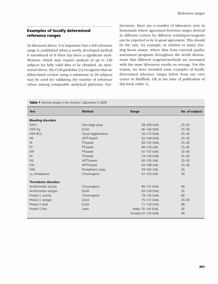
BLBK186-Key April 24, 2009 11:8
Reference ranges
Examples of locally determinedreference ranges
As discussed above, it is important that a full reference
range is established when a newly developed method
is introduced or if there has been a significant mod-
ification, which may require analysis of up to 120
subjects for fully valid data to be obtained. As men-
tioned above, the CLSI guideline [1] recognizes that an
abbreviated version using a minimum of 20 subjects
may be used for validating the transfer of reference
values among comparable analytical platforms. Fur-
thermore, there are a number of laboratory tests in
hemostasis where agreement between ranges derived
in different centers by different techniques/reagents
can be expected to be in good agreement. This should
be the case, for example, in relation to many clot-
ting factor assays, where data from external quality
assessment programs throughout the world demon-
strate that different reagents/methods are associated
with the same laboratory results on average. For this
reason, we have included some examples of locally
determined reference ranges below from our own
center in Sheffield, UK at the time of publication of
this book (table 1).
Table 1 Normal ranges in the Authors’ Laboratory in 2009.
Test Method Range No. of subjects
Bleeding disordersFVIII:C One stage assay 58–209 IU/dL 25–30
VWF:Ag ELISA 46–146 IU/dL 25–30
VWF:RCo Visual Agglutination 50–172 IU/dL 25–30
FIX APTT based 62–144 IU/dL 25–30
FII PT-based 84–132 IU/dL 25–30
FV PT-based 66–126 U/dl 25–30
FVII PT-based 61–157 IU/dL 25–30
FX PT-based 74–149 IU/dL 25–30
FXI APTT-based 60–150 U/dL 25–30
FXII APTT-based 50–180 U/dL 25–30
FXIII Pentapharm assay 59–163 U/dL 20
α2-Antiplasmin Chromogenic 67–103 U/dL 20
Thrombotic disordersAntithrombin activity Chromogenic 85–131 IU/dL 80
Antithrombin antigen ELISA 83–124 IU/dL 30
Protein C activity Chromogenic 79–142 IU/dL 80
Protein C antigen ELISA 75–131 IU/dL 25–30
Protein S total ELISA 71–136 IU/dL 80
Protein S free Latex Males 74–143 IU/dL 40
Females 67–125 IU/dL 40
301
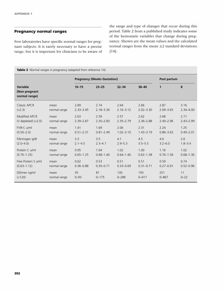
BLBK186-Key April 24, 2009 11:8
APPENDIX 1
Pregnancy normal ranges
Few laboratories have specific normal ranges for preg-
nant subjects. It is rarely necessary to have a precise
range, but it is important for clinicians to be aware of
the range and type of changes that occur during this
period. Table 2 from a published study indicates some
of the hemostatic variables that change during preg-
nancy. Shown are the mean values and the calculated
normal ranges from the mean ±2 standard deviations
[14].
Table 2 Normal ranges in pregnancy (adapted from reference 14).
Pregnancy (Weeks Gestation) Post partum
Variable(Non pregnantnormal range)
10–15 23–25 32–34 38–40 1 8
Classic APCR
(>2.3)
mean 2.89 2.74 2.64 2.66 2.87 3.16
normal range 2.33–3.45 2.18–3.30 2.16–3.12 2.02–3.30 2.09–3.65 2.34–4.00
Modified APCR
(V depleted) (>2.0)
mean 2.63 2.59 2.57 2.62 2.68 2.71
normal range 2.39–2.87 2.35–2.83 2.35–2.79 2.36–2.88 2.40–2.96 2.43–2.99
FVIII:C u/ml
(0.50–2.0)
mean 1.41 1.69 2.06 2.31 2.24 1.25
normal range 0.51–2.31 0.81–2.49 1.02–3.10 1.43–3.19 0.86–3.62 0.49–2.01
Fibrinogen g/dl
(2.0–4.0)
mean 3.3 3.5 4.1 4.5 4.6 2.6
normal range 2.1–4.5 2.3–4.7 2.9–5.3 3.5–5.5 3.2–6.0 1.8–3.4
Protein C u/ml
(0.70–1.25)
mean 0.95 1.04 1.02 1.00 1.16 1.02
normal range 0.65–1.25 0.68–1.40 0.64–1.40 0.62–1.38 0.76–1.56 0.68–1.36
Free Protein S u/ml
(0.63–1.12)
mean 0.62 0.53 0.51 0.51 0.59 0.74
normal range 0.36–0.88 0.35–0.71 0.33–0.69 0.31–0.71 0.27–0.91 0.52–0.96
DDimer ng/ml
(<120)
mean 35 81 130 193 251 11
normal range 0–93 0–175 0–286 0–417 0–867 0–22
302
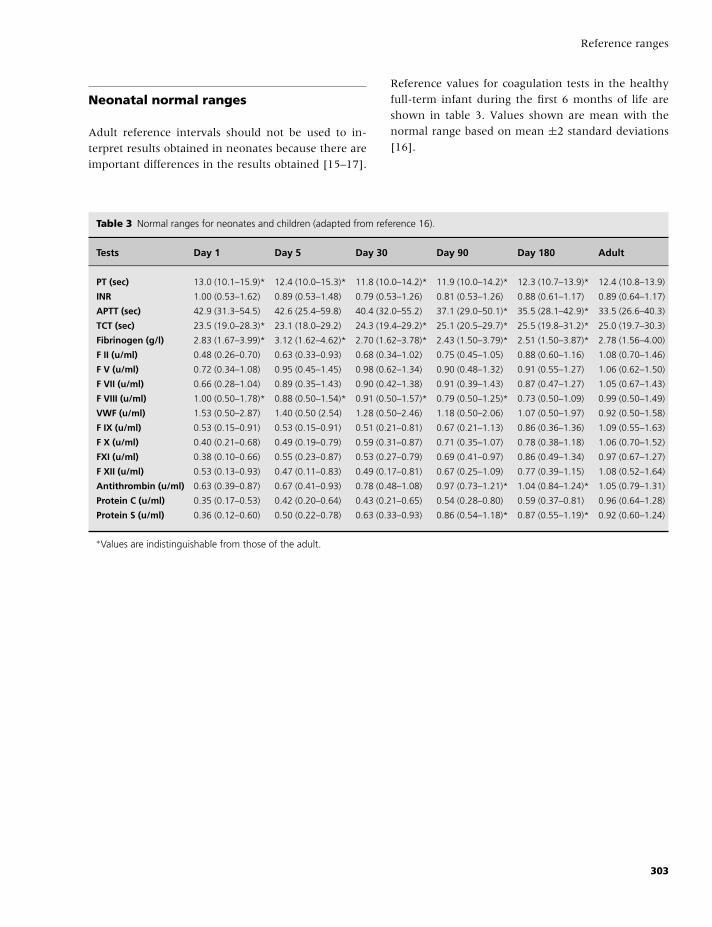
BLBK186-Key April 24, 2009 11:8
Reference ranges
Neonatal normal ranges
Adult reference intervals should not be used to in-
terpret results obtained in neonates because there are
important differences in the results obtained [15–17].
Reference values for coagulation tests in the healthy
full-term infant during the first 6 months of life are
shown in table 3. Values shown are mean with the
normal range based on mean ±2 standard deviations
[16].
Table 3 Normal ranges for neonates and children (adapted from reference 16).
Tests Day 1 Day 5 Day 30 Day 90 Day 180 Adult
PT (sec) 13.0 (10.1–15.9)* 12.4 (10.0–15.3)* 11.8 (10.0–14.2)* 11.9 (10.0–14.2)* 12.3 (10.7–13.9)* 12.4 (10.8–13.9)
INR 1.00 (0.53–1.62) 0.89 (0.53–1.48) 0.79 (0.53–1.26) 0.81 (0.53–1.26) 0.88 (0.61–1.17) 0.89 (0.64–1.17)
APTT (sec) 42.9 (31.3–54.5) 42.6 (25.4–59.8) 40.4 (32.0–55.2) 37.1 (29.0–50.1)* 35.5 (28.1–42.9)* 33.5 (26.6–40.3)
TCT (sec) 23.5 (19.0–28.3)* 23.1 (18.0–29.2) 24.3 (19.4–29.2)* 25.1 (20.5–29.7)* 25.5 (19.8–31.2)* 25.0 (19.7–30.3)
Fibrinogen (g/l) 2.83 (1.67–3.99)* 3.12 (1.62–4.62)* 2.70 (1.62–3.78)* 2.43 (1.50–3.79)* 2.51 (1.50–3.87)* 2.78 (1.56–4.00)
F II (u/ml) 0.48 (0.26–0.70) 0.63 (0.33–0.93) 0.68 (0.34–1.02) 0.75 (0.45–1.05) 0.88 (0.60–1.16) 1.08 (0.70–1.46)
F V (u/ml) 0.72 (0.34–1.08) 0.95 (0.45–1.45) 0.98 (0.62–1.34) 0.90 (0.48–1.32) 0.91 (0.55–1.27) 1.06 (0.62–1.50)
F VII (u/ml) 0.66 (0.28–1.04) 0.89 (0.35–1.43) 0.90 (0.42–1.38) 0.91 (0.39–1.43) 0.87 (0.47–1.27) 1.05 (0.67–1.43)
F VIII (u/ml) 1.00 (0.50–1.78)* 0.88 (0.50–1.54)* 0.91 (0.50–1.57)* 0.79 (0.50–1.25)* 0.73 (0.50–1.09) 0.99 (0.50–1.49)
VWF (u/ml) 1.53 (0.50–2.87) 1.40 (0.50 (2.54) 1.28 (0.50–2.46) 1.18 (0.50–2.06) 1.07 (0.50–1.97) 0.92 (0.50–1.58)
F IX (u/ml) 0.53 (0.15–0.91) 0.53 (0.15–0.91) 0.51 (0.21–0.81) 0.67 (0.21–1.13) 0.86 (0.36–1.36) 1.09 (0.55–1.63)
F X (u/ml) 0.40 (0.21–0.68) 0.49 (0.19–0.79) 0.59 (0.31–0.87) 0.71 (0.35–1.07) 0.78 (0.38–1.18) 1.06 (0.70–1.52)
FXI (u/ml) 0.38 (0.10–0.66) 0.55 (0.23–0.87) 0.53 (0.27–0.79) 0.69 (0.41–0.97) 0.86 (0.49–1.34) 0.97 (0.67–1.27)
F XII (u/ml) 0.53 (0.13–0.93) 0.47 (0.11–0.83) 0.49 (0.17–0.81) 0.67 (0.25–1.09) 0.77 (0.39–1.15) 1.08 (0.52–1.64)
Antithrombin (u/ml) 0.63 (0.39–0.87) 0.67 (0.41–0.93) 0.78 (0.48–1.08) 0.97 (0.73–1.21)* 1.04 (0.84–1.24)* 1.05 (0.79–1.31)
Protein C (u/ml) 0.35 (0.17–0.53) 0.42 (0.20–0.64) 0.43 (0.21–0.65) 0.54 (0.28–0.80) 0.59 (0.37–0.81) 0.96 (0.64–1.28)
Protein S (u/ml) 0.36 (0.12–0.60) 0.50 (0.22–0.78) 0.63 (0.33–0.93) 0.86 (0.54–1.18)* 0.87 (0.55–1.19)* 0.92 (0.60–1.24)
∗Values are indistinguishable from those of the adult.
303
BLBK186-Key April 24, 2009 11:8
APPENDIX 1
Conclusion
In general, the normal range should be used only as a
guide and an aid to clinical interpretation in conjunc-
tion with all other available relevant clinical informa-
tion. The most appropriate normal reference range is
one that has been established locally using the same
system as for patient samples. It is important to use
a technique for which such a local range is in broad
agreement with the published literature.
References
1 CLSI. How to Define and Determine Reference In-
tervals in the Clinical Laboratory: Approved Guide-
line (2nd Edition). Wayne, PA: Clinical and Laboratory
Standards Institute, 2000:Document C28-A2.
2 Clinical Laboratory Improvement Amendments of 1988
(CLIA) 42 CFR section 493.1253, part (b) (1) (ii)
(2003).
3 Freidberg RC, Souers R, Wagar EA, Stankovic AK,
Valenstein PN. The origin of reference intervals: a col-
lege of American Pathologists Q-probes study of “Nor-
mal ranges” used in 163 clinical laboratories. Arch Pathol
Lab Med 2007;131:348–57.
4 Gill JC, Endres-Brooks J, Bauer PJ, Marks WJ, Mont-
gomery RR. The effect of ABO blood group on the
diagnosis of von Willebrand’s disease. Blood 1987;69:
1691–5.
5 Blomback M, Konkle BA, Manco-Johnson MJ,
Bremme K, Hellgren M, Kaaja R, on behalf of the
ISTH SSC on Womens Health Issues. J Thromb Haemost
2007;5:855–8.
6 Lowe GDO, Rumley A, Woodward M, et al. Epidemi-
ology of coagulation factors, inhibitors and activation
markers: the third Glasgow MONICA survey I. Illustra-
tive reference ranges by age, sex and hormone use. Br
J Haematol 1997;97:775–84.
7 Solberg HE on behalf of International Federation of
Clinical Chemistry (IFCC) and International Commit-
tee for Standardization in Hematology (ICSH), IFCC
Expert panel on Reference values. Approved recom-
mendation on the theory of reference values. Part 5
Statistical treatment of Collected Reference values. De-
termination of reference limits. J Clin Chem Clin Biochem
1987;25:645–56.
8 CLSI. One Stage Prothrombin Time (PT) Test and Acti-
vate Partial Thromboplastin Time (APTT) Test: Appro-
ved Guideline (2nd Edition). Wayne, PA: Clinical
and Laboratory Standards Institute, 2008:Document
H47-A2.
9 Kitchen S, McCraw (2000). Diagnosis of haemophilia
and other bleeding disorders: a laboratory manual.
Available from: http://www.wfh.org/publications.
10 Jennings I, Kitchen S, Cooper P, Makris M, Preston FE.
Sensitivity of functional protein S assays to protein S
deficiency: a comparative study of 3 commercial kits.
J Thromb Haemost 2003;1:1112–17.
11 Shojania AM, Tetreault J, Turnbull G. The variations
between heparin sensitivity of different lots of APTT
reagents produced by the same manufacturer. Am J Clin
Pathol 1988;89:19–23.
12 Barnett V, Lewis T. Outliers in Statistical Data. Chicester:
John Wiley, 1978:91–3.
13 Horn PS, Feng L, Yanmei L, Pesce AJ. Effect of outliers
and non-healthy individuals on reference interval esti-
mation. Clin Chem 2001;47:2137–45.
14 Kjellberg U, Ansdersson NE, Rosen S, Tengborn L, Hell-
gren M. APC resistance and other haemostatic variables
during pregnancy and puerperium. Thromb Haemost
1999;81:527–31.
15 Andrew M, Paes B, Milner R, et al. Development of the
human coagulation system in the full-term infant. Blood
1987;70:165–72.
16 Andrew M, Paes B, Johnston M. Development of the
hemostatic system in the neonate and young infant. Am
J Pediatr Hematol Oncol 1990;12:95–104.
17 Andrew M, Vegh P, Johnston, Bowker J, Ofosu F,
Mitchell L. Maturation of the hemostastic system dur-
ing childhood. Blood 1992;80:1998–2005.
304
BLBK186-Key April 28, 2009 15:34
Index
AAbdominal vein thrombosis (AVT), 148,
221–222
ABO group, 287, 288f
Acquired causes, for bleeding
clinical and laboratory data, 56
patients with normal PT/APTT, 58
patient without bleeding history
abnormal PT/APTT, 59
Acquired hemophilia A, 71, 71f, 216
Acquired platelet defects, 120, 120t
Acquired thrombophilia, 17, 18t,
232
Acquired von Willebrand syndrome
(AVWS), 150, 153, 155
Activated clotting time (ACT), 260
Activated factor VII (rVIIa), 91, 92
Activated partial thromboplastin time
(APTT), 4–5, 7, 9–10, 17, 33, 39,
50–51, 91, 128, 141, 181, 187, 197,
210, 233, 244, 255, 259, 293
fibrinogen, 10
lower limit of normal range, 12
mixing studies, 10
thrombin time, 10
variation with reagents, 12
Activated protein C resistance (APC-R), 14,
19, 33, 213
Acute chest syndrome, 215
Acute coronary syndrome (ACS), 157,
161–162, 185, 186, 233
therapies for, 186–192
Acute ischemic syndromes, 157, 159
Acute liver disease, 218
Acute myeloid leukemia (AML), 125, 149,
150
ADAMTS13, 109
ADAMTS-13 deficiency, 131, 133
ADAMTS-13 testing, 131
Advanced hepatocellular disease, 223
All-trans retinoic acid (ATRA), 240
Alpha interferon (�-IFN), 153–154
Anticardiolipin assays, 182
Anticoagulant therapy
duration of, 142
risk of bleeding in, 143
with heparin, 141–142
Anticoagulation
and cancer survival, 244–245
dedicated clinics for, 168
delivery management during pregnancy,
251
guidance during, 167
postoperative management of, 173–174
reversal of, 170
therapies for, 187
warfarin, 247
Antifibrinolytic agent epsilon aminocaproic
acid (AMICAR), 213
Antifibrinolytics, 205, 225
Antifibrinolytic therapy, 204–205
Antiphospholipid antibodies, 179–180,
283
Antiphospholipid syndrome (APS), 160,
162, 177, 178, 179, 213–214
antiphospholipid antibodies in,
179–180
clinical features of, 177–178
definition of, 177
diagnosis of, 180
laboratory assay, 181–182
management of
pregnancy failure, 183–184
thrombosis, 182–183
in pregnancy, 178
testing presence of, 252
transient and nonpathological, 180–181
Antiplatelet drugs, 207
Antiplatelet therapy, 273
children at risk for, 267
dosing/monitoring of, 267
limitations/benefits of, 267
metabolism in, 267
reversal of, 267
Antithrombotic therapy, 259, 268
Aprotinin, 204, 225, 292
Arterial thrombosis
definition of, 157
investigations
routine laboratory, 158–159
specialized, 159–161
traditional risk factors, 157
treatment, 161–162
Artificial reproductive technology (ART),
247, 253
Aspirin
adverse events of, 190
and clopidrogel resistance, 202
benefits of, 190
defined, 189
resistance, 161
Autoimmune lymphoproliferative
syndrome (ALPS), 106
BBernard-Soulier syndrome (BSS), 32, 40t,
43, 45, 53, 112, 115–117
Bethesda assay, 51–52, 56, 69, 71
Bleeding
clinical/laboratory data of
acquired causes, 56
congenital causes, 54–56
disorders
diagnosis of, 206
hemophilia A, 25–28
hemophilia B, 28–29
VWD, 29–32
history of, 48–49
laboratory evaluation of patient
assays for fibrinogen, 52
assessment of fibrinolytic system, 52
Bethesda assay, 51–52
bleeding time, 52–53
coagulation laboratory testing, 50
defect in platelet function, 52
electron microscopy, 53
factor XIII, 52
final integration of clinical and
laboratory data, 54
305
BLBK186-Key April 28, 2009 15:34
Index
Bleeding (Continued )
mixing studies, 51
platelet aggregation testing, 53
platelet function analyzer-100
(PFA-100), 53
PT/APTT, 50–51
specific clotting factor assays, 51
TCT or TT/RT, 51
testing for VWD, 52
normal PT/APTT
patients with bleeding history, 58
patients without bleeding history,
58
physical examination, patient, 49–50
time, 40
Blood loss methods
antifibrinolytic therapy, 204–205
aprotinin, 204
pharmacotherapy, 204
recombinant factor VIIa, 205
tranexamic acid, 204
Blood transfusion
ABO group, 287
hemovigilance and regulation of, 295
immunization in, 289–290
infective risk of, 288
O-negative blood, 288
post-transfusion purpura in, 290
reactions, 289
red cell cross-matching, 287–288
reducing risk of, 288
related acute lung injury, 289
Rhesus (Rh) system, 287
risk of, 288
Blood product, 82–83, 89, 108, 128, 284,
287, 290, 292–293
Bolin-Jamieson syndrome, 116
Bone marrow examination, 98f, 104, 148,
150
Bovine spongiform encephalopathy (BSE),
70, 289, 290
British Committee for Standards in
Haematology (BCSH), 157, 253, 291
B-type natriuretic peptide (BNP), 280
Budd-Chiari syndrome, 148, 221, 221t
CCancer, thrombosis in
clinical aspects, 236
malignancy, hypercoagulable state of,
237
occult malignancy, 236–237
pathogenic mechanism, 238–242
predictors of survival, 238
predictors of thrombosis, 237–238
prevention/treatment, 242–244
routine laboratory test, 237
specialized test, 237
thrombotic disorders, 236t
Cardiac surgery, 294
Cardiopulmonary bypass (CPB), 121,
194–196
hemostasis in, 196–197
inherited qualitative platelet defects,
121
Carpal tunnel syndrome, 50
Catastrophic antiphospholipid syndrome,
178
Cell adhesion molecules, 241
Cell salvage, 292
Central venous catheters (CVC), 7, 159,
218, 243
Central venous thrombosis
children with, 267
diagnosis of, 267–268
Cerebral perfusion pressure (CPP), 212
Chediak-Higashi syndrome, 50, 118
Chronic liver disease, 122, 218
Chronic myelogenous leukemia (CML),
113, 147, 152, 153, 155
Chronic neurological syndromes, 214
Clauss method, 52, 201
Clot lysis, 52, 200, 266
Clotting factor assay design, 13–14
Clotting factor therapy
treatment complications
anaphylaxis as, 71
hepatitis B, 70
hepatitis C, 70
HIV, 70
immune modulation, 70
infections, 70
inhibitor development, 69
parvovirus B19, 70
thrombosis as, 70–71
variant Creutzfeldt-Jakob disease
(vCJD) in, 70
c-mpl gene, 112
Coagulation
activation makers, 161
amplification, 2–3
assays of, 4
conventional tests of, 197
definition of, 1
definitions of parameters using
thrombelastograph, 199–200
disseminated intravascular, 219–220
excessive activation of, 124
factor concentrates of, 291
factor synthesis, 218
factors in, 160–161
inherited factor for
of antithrombin, protein C, and
protein S, 34
deficiencies, 32
thrombotic disease, Molecular
diagnostics for, 32–33
initiation of, 2
localization, 4
propagation, 3–4
proteins, 218–219
screening tests of, 9
vasculature, 1–2
Combination therapy, 160
Combined oral contraceptive (COC), 160,
250
Complementary alternative medicines
(CAM), 266
Complete blood count (CBC), 17, 105, 209
Computed tomographic pulmonary
angiography (CTPA), 139–140, 141,
251
Compression Venous ultrasonography,
137–138
Computed tomographic pulmonary
angiography (CTPA), 139–140
Concomitant therapy, 129, 274
Congenital amegakaryocytic
thrombocytopenia (CAMT), 105, 112
Congenital causes, bleeding
clinical and laboratory data, bleeding,
54–56
patients with normal PT/APTT, 58
patient without bleeding history
abnormal PT/APTT, 59
Congenital heart disease (CHD), children
with, 268–269
Congenital thrombocytopenia, 112, 119,
120
Conjugate equine estrogens (CEE), 255
Continuous venovenous hemodialysis
(CVVH), 222
Coumarin drug, 164, 170, 171. See also
Vitamin K antagonist (VKA) therapy
Coumarin drug administration, 170
COX-2 genes, 235
CPB circuit, 196
CT venography, 138–139
Cyclooxygenase (COX)–1, 189
Cyclosporine, 71, 132, 282, 283
Cytomegalovirus (CMV), 99, 282, 283
DD-dimer blood testing, 138
Deep vein thrombosis (DVT), 22, 83, 147,
166, 178, 183, 218, 230, 236, 280
diagnosis of, 136
diagnosis of recurrent, 139
in pregnancy, 139
versus PE, 143
Defibrination syndrome, 123
306
BLBK186-Key April 28, 2009 15:34
Index
Delayed hemolytic reactions, 289
Dense tubular system (DTS), 38f
Desmopressin (DDAVP), 68, 82, 292
Developmental hemostasis, 258–259
DiGeorge syndrome, 112
Dilute Russel viper venom time (DRVVT),
19
Direct thrombin inhibitor (DTI), 19, 58,
128, 174–175, 188, 233, 266, 278–279
Disseminated intravascular coagulation
(DIC), 52, 57, 100, 108–109, 123,
124t, 219–220, 235, 271, 287, 293
clinical manifestation, 124–126
diagnosis, 126, 127t
non-overt, 127–128
overt, 127
pathogenesis, 123–124
pathological conditions, 123
thrombotic thrombocytopenic purpura,
130
treatment procedures
managing underlying disease, 128
pathophysiology, 130
specific inhibitors of coagulation,
128–129
supportive care/blood products, 128
thrombotic microangiopathies,
129–130
Drug-induced thrombocytopenia, 106–107
Dysfibrinogenemia, 51, 54, 57–58, 90, 220,
223
EEbstein-Barr virus (EBV), 99, 100
E. coli, 131
Ehlers-Danlos (ED) syndrome, 50, 58, 120
Ellis method, 52
Enzyme-linked immunosorbent assay
(ELISA), 19, 21, 79, 82, 179, 182, 263
Epidural anesthesia, 174
Erythrocytosis, 283
Erythropoietin (rhEPO), 292
Estimated percent lysis (EPL), 200
Ethylenediaminetetraacetic acid (EDTA),
19, 21, 104, 271
Estrogen, 23, 83, 225, 228, 255, 256
Estrogen therapy, 142, 222
Euglobulin clot lysis time (ECLT), 52
Evan’s syndrome, 104, 106
External quality assessment (EQA), 11,
15–16, 301
Extracranial hemorrhage, 170
FFactor VIII gene, 61–62
mutation, 62
inheritance, 63
severity/symptoms, 62
Factor V Leiden (FVL), 14–15, 18, 19, 20,
22, 23
Factor Xa, 3
Factor X deficiency, 91
Febrile transfusion reactions, 289
Fibrin glue, 225
Fibrinolytic inhibitors, 292
Fibrinolysis, 124
Fibrinolytic tests, 161
Fibrinolytic therapy, 186–187
Fibrin sealants, 291
Fibrosis progression, 222
Fresh frozen plasma (FFP), 290–291
Frozen red cells, 290
FIX gene (F9), 62
Full-blown syndrome, 131
GGestational venous thrombosis
prevention of, 249–250
risk factors, 248
thrombophilia and risk of, 249
Gestational VTE
diagnosis of, 250–251
management of, 251
prevention of, 249–250
Glanzmann thrombasthenia (GT), 43, 45,
49, 112, 116
Glycoprotein IIb/IIIa inhibitors, 161, 191
Graft loss, thrombosis/thrombophilias
diagnosis/prevention, 232
etiology, 231–232
Gray platelet syndrome (GPS), 101t, 103,
119
HHarefield protocol, 201
HELLP syndrome, 107, 273, 275–276
Hematocrit, 283
Hematopoeitic stem cell transplantation
(HSCT), 20
Hemorrhage, 220–221
assessment of, 293
intracerebral, 211–212
intracranial, 169–170, 186
subarachnoid, 212–213
in surgery, 207
Hemolytic uremic syndrome (HUS), 100,
129–130, 130, 273
Hemophilia A. See also Hemophilia A/B
direct mutation testing for, 26
factor viii inversion mutations, 28
polymorphism linkage analysis in, 26
rationale for direct mutation testing in,
26–27
strategies for direct mutation detection
in, 27, 27f
Hemophilia A/B
bleeding episodes, 65
dental treatment, 67
gastrointestinal bleeding, 66
hematuria, 66
intracranial hemorrhage, 66
joint bleeds, 65–66
muscle bleeds, 66
pseudotumors, 66–67
surgery for, 67
carrier testing, 63
delivery at-risk pregnancy, 64
factor VIII gene, 61–62
factor VIII gene
mutation, 62
severity/symptoms, 62
inheritance, 63
FVIII/IX level, female, 63
making the diagnosis, 64
neonate with, 64
preimplantation Genetic Diagnosis, 64
prenatal diagnosis, 63
treatment
clotting factor replacement, 67
complications of, 69–70
cryoprecipitate and fresh frozen
plasma as agent for, 68
DDAVP, 68–69
plasma-derived concentrates,
67–68
Hemophilia B, 28, 66f. See also Hemophilia
A/B
direct mutation testing for, 28
mutations of particular clinical
significance, 29
polymorphism linkage analysis, 28
Hemostasis
ACT in, 260
anticoagulant and sample filling, 7–8
APTT, 9–10
clinical trail, difficulties in performance
of, 259
clotting factor assay design, 13
defined, 1
mixing with anticoagulant, 7
in normal pregnancy, 247
processing and storage of samples prior to
analysis, 8–9
centrifugation, 8
stability, 8
sample collection of, 7
surrogate measures of, 259–260
tests of fibrinolysis in, 7
therapeutic agents in, 260–261
thromboelastogram, 260
use of coagulation screening tests, 9
venous catheters, 7
307
BLBK186-Key April 28, 2009 15:34
Index
Hemostatic components, blood and
clinical indications to reduce exposure,
203–204
logistical indications to reduce exposure,
204
Heparinase, 200
Heparin associated thrombocytopenia
(HAT), 272
Heparin-induced thrombocytopenia (HIT),
21, 100, 107, 135, 141, 174–175, 187,
233, 248, 261, 263, 266, 278–280, 290
clinical diagnosis of, 278–280
laboratory diagnosis, 278
Hereditary thrombophilia, 18t
Hermansky-Pudlak syndrome (HPS), 41,
118–119
Highly active antiretroviral therapy
(HAART), 70
Homocysteine measurement, 159–160
Homocysteinemia, 22, 34, 159–160, 283
Hormone replacement therapy (HRT), 23,
49, 160, 255–256
Human albumin solution (HAS), 291
Human immunodeficiency virus (HIV)
recombinant clotting factors, 68
viral inactivation and removal
techniques, 68t
Hypercoagulable states, 213, 213t
Hypercoaguability, 200
Hyperfibrinolysis, 220
Hyperhomocystinemia, 214
Hypersplenism, 110
Hyperviscosity syndromes, 158–159
IICU
massive pulmonary embolism, 280–281
thromboprophylaxis, 281
Idiopathic thrombocytopenic purpura (ITP),
48, 96, 100, 103–108, 112, 272, 273,
290
Immediate hemolytic reactions, 289
Immune mechanisms, 271
Immune thrombocytopenia (ITP), 96, 103,
111, 273
Impact R© cone and plate(let) analyzer,
41–42
Increased intracranial pressure (ICP), 212,
213
Inferior vena caval filters, 144
Inherited coagulation disorder
case histories for, 93–94
clinical features, 88–89
deficiencies (fibrinogen/afibrinogenemia/
dysfibrinogenemia), investigation
of, 90
Factor V deficiency, 90
Factor VII deficiency, 90–91
Factor X deficiency, 91
Factor XI deficiency, 91–92
Factor XII deficiency, 92
factors V and VIII deficiency, 90
genetics, 88
pregnancy, 89
prothrombin deficiency, 90
treatment of, 90
vitamin K dependent factors, 92–93
Inherited thrombocytopenia, 111–112
Inherited qualitative platelet defects
abnormalities of membrane
phospholipids, 120
description, 115
function, abnormalities of, 120
acquired platelet defects, 120
Cardiopulmonary bypass, 121
liver disease, 122
medications, 122
myeloproliferative disorders, 121
Platelet transfusions Therapy, 122
uremia, 120–121
platelet granules, abnormalities, 118–119
signal-transduction pathways
Abnormalities, 119
for soluble agonists, 117–118
Internal quality control (IQC), 15
International Society on Thrombosis and
Hemostasis (ISTH), 41, 126, 74, 123,
298
Intracerebral hemorrhage (ICH), 211–212
Intracranial hemorrhage, 169–170
Intraoperative hemodilution, blood, 292
Intravenous immunoglobulin (IVIg), 291
Ischemic stroke, 209–210
Ischemic Syndrome, 157, 159, 187
IV immunoglobulin (IVIG), 105
JJehovah’s witnesses (JW), 206, 284–285,
294–295
KKasabach-Merritt syndrome, 108, 109
LLeft anterior descending (LAD), 194
Left ventricular ejection fraction (LVEF),
195
Light transmission platelet aggregometry,
42–43
Liver biopsy, 223–224
Liver disease
hemorrhage, 220–221
hemostasis, investigation of
cholestasis, 223
chronic liver disease, 222–223
clotting screen, 222
fibrinogen levels, 223
hypofibrinogenemia, 223
antifibrinolytics, 225
invasive procedures, 223–225
plasma, 225
platelet transfusions, 225
shunt insertion for, 224
transplant coagulopathy, 224–225
treatment of coagulopathy, 224
vitamin K deficiency, 225
Liver transplant coagulopathy, 224–225
Liver transplantation, 224
LMAN1 gene, 57
Low-molecular-weight heparin (LMWH),
21–22, 23, 141, 143, 144, 145, 178,
183, 187, 199, 243–245, 248, 250, 251,
254, 263–264, 282
benefits/limitations, 264
dosing and monitoring, 264
metabolism, 2630–264
reversal, 264
Lupus anticoagulant (LAC), 10, 14, 17, 19,
22, 51, 59, 135, 143, 160, 179,
181–182, 199, 230
MMacrothrombocytopenia, Autosomal
dominant, 112
Magnetic resonance angiography (MRA),
213, 229
Magnetic resonance venography (MRV),
210, 250
Marfan syndrome, 120
Massive transfusion, 294
Maximum amplitude/G (MA/G), 200
Mesenchymal-epithelial transition factor
(MET), 235
Microangiopathic hemolytic anemia
(MAHA), 124, 129, 130, 131, 132
Microangiopathies, 108–109
Middle cerebral artery (MCA), 215
Minimally invasive direct coronary artery
bypass (MIDCAB), 194–195
Molecular hemostasis, diagnostics, 35
Montreal platelet syndrome, 120
Mortality, 275
MR venography, 138–139
Munchausen’s syndrome, 49–50
Multiple clotting factor deficiency 2
(MCFD2) gene, 57
Muromonab-CD3 (OKT3), 283
Mycophenolate, 283
Myelodysplastic syndrome, 99, 113,
272
Myelofibrosis (MF), 154–155
308
BLBK186-Key April 28, 2009 15:34
Index
Myeloproliferative disorders (MPD), 56, 58,
113, 121, 221, 271
Myeloproliferative neoplasms (MPN), 147
clinical representation, 147–149
diagnosis, 150
pathogenesis of thrombosis and bleeding
in ET and PV, 149–150
prognosis, 150
treatment
management of thrombosis, 155
myelofibrosis, 154–155
PV and ET, 152–154
NNeed for near patient testing (NPT),
197–198
Neonatal alloimmune thrombocytopenia
(NAIT), 96, 100, 110, 111
Neonatal period, 64
Nephrotic syndrome, 230–231, 282, 283
hypercoagulability state, 230–231
thromboembolic events, 230
treatment, 231
Nonimmune mechanisms, 272
Non-ST elevation myocardial infarction
(NSTEMI), 186, 187, 189
Novel antiplatelet therapies, 191–192
OObstetrics, 294
Off-pump coronary artery bypass (OPCAB),
194, 195
O-negative blood, 288
Open canalicular system (OCS), 38f, 255
Oral anticoagulant drug, 165–166
Oral anticoagulation, 34–35
Oral anticoagulants (OAC), 29, 35, 142,
144, 160, 164, 165–166, 244, 265, 298
Oral contraceptive pills (OCP), 49, 297
Oral vitamin K antagonists, 264
Ovarian hyperstimulation syndrome
(OHSS), 253
Over-anticoagulation, 170–171
Overt DIC, 127
Pp53 tumor suppressor, 235
Paris-Trousseau syndrome, 119
Paroxysmal nocturnal hemoglobinuria
(PNH), 21
Pediatrics, 294
Perioperative bridging therapy, 173
Peritoneal dialysis, 283
Persistent hypercoagulability, 283
PFA-100. See Platelet function analyzer
physiological anticoagulant pathways
down regulation of, 124
plasma exchange, 274
Plasminogen activator inhibitor type 1
(PAI-1), 52, 58, 124, 161, 220, 235,
240, 283
platelet
classification of defects, 38, 40t
decreased, 97–99
diagnostic test
flow cytometry, 44–46
light transmission platelet
aggregometry, 42–43
storage pool or release defects, 44
function of, 37
function testing, 38–40
function tests, 161
global test for, 40–42
bleeding time, 40
ImpactR© cone and plate(let) analyzer,
41–42
PFA-100, 40–41
increased, 99–100
major platelets, 39t
production, 96
structure of, 37
Platelet clumping, 271
Platelet count, 199
Platelet function analyzer, 40–41, 53
Plateletmapping, 202–203
Platelet poor plasma (PPP), 42, 43
Platelet-rich plasma (PRP), 8, 42, 53, 74, 80,
290
Platelet threshold, 272–273
Point-of-care (POC), 265
Portal vein thrombosis (PVT), 110, 148,
222, 269
Postthrombotic syndrome, 136, 144, 251,
259, 268
Post-transfusion purpura (PTP), 103, 108,
273, 278, 290
Predeposit donation, blood, 292
Preeclampsia, 253
Pregnancy-associated thrombocytopenia,
107
Pregnancy loss, 252
Preimplantation stage, 224
Preoperative assessment clinics, 206
Pre-peptide, 62, 73
Primary antiphospholipid syndrome,179,
229
Primary secretion defect (PSD), 41, 118, 120
Produces prostacyclin (PGI2), 1
Progestogen-only preparations, 255
Progressive atherosclerosis, 185–186
Prosthetic heart valve, 253–254
anticoagulant safety in, 254
LMWH in, 254
warfarin, 254
Protease-activated receptors (PAR), 2, 204
Prothrombin complex concentrates (PCC),
57, 69, 70–71, 90, 91, 92, 171, 205,
225
Prothrombotic mechanism
antitumor therapy in, 241–242
cytokine activity, 240
fibrinolytic activities, 240
increased fibrinolysis, 240
procoagulant activities, 239–240
procoagulant properties, 240
tumor cell, 238–239
white cell recruitment, 240–241
Prothrombin time (PT), 9
Pulmonary angiography, 139
Pulmonary embolism (PE), 268
diagnosis of, 139
diagnosis in pregnancy, 140–141
versus DVT, 143
QQuality assurance, 15
in APS, 182
APTT, 15
Quality of life (QOL), 268
RRapidTEGTM
TM, 201
Recombinant factor, 71, 122, 205, 225,
285
Recurrent fetal loss (RFL), 252–253
associated with APL, 252
inheritable treatment, 252–253
link between thrombophilias and, 252
Red cell cross-matching, 287–288
Reduced activated protein C resistance
(APC-R), 14, 15, 19, 20, 23, 213
Reduced intensity conditioning (RIC)
transplant, 154
Refractory disease, 274–275
Renal disease, bleeding in
clinical presentation of, 227
factors affecting (etiology), 227
prevention and treatment, 227–228
Renal failure, 282
Renal insufficiency
anticoagulant, 232
guidelines
for (mild/moderate), 232–233
for severe, 233
Renal vein thrombosis
clinical presentation, 228–229
diagnosis/treatment/prognosis, 229–230
etiology, 229
Reptilase time (RT), 51, 220, 223
Reyes syndrome, 267
Rhesus (Rh) system, 287
309
BLBK186-Key April 28, 2009 15:34
Index
Ristocetin-induced platelet aggregation
(RIPA), 74, 75, 80, 81
Robot-assisted coronary artery bypass
(RACAB), 195
SScott syndrome, 5, 40t, 120
Screening
for congenital thrombophilias, 160
for lupus anticoagulant and
anticardiolipin antibodies, 160
sickle cell, 160
Secondary antiphospholipid syndrome, 179
Sepsis
patient, 271–272
syndromes of, 123, 125, 128
Sequential Organ Failure Assessment
(SOFA) score, 277
Serious Hazards of Transfusion (SHOT),
295, 295f
Sickle cell disease (SCD), 160, 214–216
Signal peptide
of 22 amino acids. See Pre-peptide
Sirolimus, 283
SLE-like syndromes, 214
Solvent detergent plasma (SDP), 225
Spinal anesthesia, 174
Standard laboratory tests, 198–199
ST elevation myocardial infarction (STEMI),
186
Steroids, 282–283
Storage pool disease (SPD), 41, 45, 53, 118
Stormorken syndrome, 116t, 120
Subarachnoid hemorrhage (SAH), 210,
212–213, 215
Surgical Trial for Intracerebral Hemorrhage
(STICH), 212
Systemic Inflammatory Response Syndrome
(SIRS), 276–277
Systemic lupus erythematosus (SLE), 107,
135, 159, 177, 178, 179, 214, 230, 232
TTEG R©, 201
Thienopyridine, 191
Thrombin clotting time (TCT), 51, 54, 56, 57
Thrombin inhibitor, 188
Thrombin receptor-activating peptide
(TRAP), 43, 46
Thrombocytopenia
bone marrow examination, 104
in children, 97, 98f
congenital, 112
decreased platelet production, 97–99
definition of, 96
drugs causing, 102t
drug induced, 106–107
family history, 100
heriditary, 101t
heparin-induced, 100, 107, 233, 290
HCV-associated, 108
HIV-associated, 108
increased platelet destruction, 99–100
inherited, 111–112
ITP, 104–105
laboratory evaluation, 103–104
medical history
infection, 100
medication history, 100
platelet dysfunction and, 219
patient history in, 100
physical examination of, 103
platelet production in, 96
platelet sequestration in, 97
pregnancy-associated, 107, 153–154
sex-linked, 112–113
systemic diseases, 103
transfusion history, 103
Thrombocytopenia-absent radius syndrome
(TAR), 112
Thromboelastogram, 260
Thrombophilia
and vascular complications of pregnancy,
251–252
assessment for presence of
ancillary testing, 21
clinical assessment, 17, 18f
general diagnostic testing, 17
laboratory testing, 17
specialized coagulation testing in,
17–21
definition of, 249
management
acute therapy, 21
antithrombin deficiency, 21
hereditary protein C deficiency, 21
lupus anticoagulant, 22
primary prevention, 21
secondary prophylaxis, 22
secondary prophylaxis based on
clinical predictors, 22
secondary prophylaxis based on
presence of thrombophilia, 22–23
renal transplantation and, 282
testing, 14–15
controversial aspects of, 23
counseling issues related to, 23
estimated prevalence of, 24
timing of, 23
Thrombolytic therapy, 143–144
Thrombosis
APS, 182–183
in cancer, treatment
cancer surgery, 243
medical condition, 243–244
prophylaxis of VTE, 242–243
treatment of VTE, 244
definition of, 1
MPN, 155
pathophysiology, 185–186
Thrombotic disease, molecular diagnostics
for, 32–33
Thrombotic microangiopathy, 129–130
clinical manifestation, 131
differential diagnosis, 131
treatment, 132
immunosuppression, 132
plasma exchange, 132
ADAMTS-13 activity, role of, 132–133
Thrombotic thrombocytopenic
purpura/hemolytic uremic syndrome
(TTP/HUS), 103, 109, 130, 131, 132,
133
factors affecting, 273–274
Tranexamic acid, 69, 204
Transcranial Doppler (TCD), 215
Transfusion-related acute lung injury
(TRALI), 289
Transient ischemic attacks (TIA), 147, 209,
215
UUnfractionated heparin (UFH), 166, 173,
173, 183, 187, 230, 243, 261, 278
dosing monogram, 263t
limitations, 261–263
reversal, 263
subcutaneous dosing, 263
therapy, 261
Uremia, 120–121
Upshaw-Schulman syndrome, 131
VVariant Creutzfeldt-Jakob Disease (vCJD),
70, 204, 289, 295
Venous sinus thrombosis (VST), 210–211,
215
Venous thromboembolism (VTE), 17, 19t,
33, 34, 135, 159, 169, 173, 174, 175,
177, 178, 182, 183, 222, 230, 235–236,
280, 292
acquired predisposition to, 135–136
gestational, 248
hypercoagulability in, 135
incidence of gestational, 248
inherited predisposition to, 135
management of
clinical assessment of, 137
diagnosis of DVT in, 136
diagnosis of VTE in, 136
venography, 136–137
310
BLBK186-Key April 28, 2009 15:34
Index
in orthopedic surgery, 145
potential indicators for increased risk of
recurrent, 143
pregnancy associated, 248, 248t, 249,
249t, 250
prevalence/natural history, 136
prevention of, 144–145
prophylaxis in medical patients, 145
prophylaxis of, 242–243
risk (female hormone use)
oral contraceptive, 255
progestogen-only preparations, 255
thrombophilia and COC, 255
risk factors, 136t
treatment by anticoagulant therapy
duration of, 142
risk of bleeding in, 143
with heparin, 141–142
treatment during pregnancy, 144
treatment of, 244
unprovoked, 142–143
Venous endothelial damage, 135
Venous stasis as risk factor, 135
Ventilation-perfusion lung scanning, 139,
140, 142f
Very-high purity VWF concentrate, 83
Vitamin K antagonist (VKA) therapy, 19,
34, 51, 142, 161–162, 164, 188, 244,
261, 280
complication of anticoagulation with,
168–169
continuation of treatment, 172–173
contradictions for treatment of, 164
dosing and monitoring of, 264–265
indication for treatment, 164
limitation/benefits, 265–266
metabolism, 264
oral anticoagulant drug in, 165–166
patients with highly unstable response,
168
reversal, 266
temporary discontinuation in, 173
warfarin anticoagulation in, 166–168
Vitamin K deficiency, 220
Vitamin K epoxide reductase subunit 1
(VKORC1), 34–35, 165, 265
von Willebrand disease (VWD), 29, 48, 73,
115, 285, 291
bleeding history, 78–79
characterization of subtype, 80–81
classification of, 74
clinical manifestations, 77–78
diagnosis of, 78
genetics/molecular biology, 75–77
library evaluation of, 79–80
management of patients
desmopressin, 82
nontransfusional therapies, 83
secondary long term prophylaxis,
84
transfusional therapies, 83
woman with, 85
physical role of, 73–74
prevalence and frequency of subtypes of,
77
von Willebrand factor (VWF), 2
VTE management
clinical assessment of, 137
diagnosis of DVT in, 136
diagnosis of VTE in, 136
venography, 136–137
VTE prevention
pharmacologic agents in orthopedic
surgery, 145
prophylaxis following surgery, 144–145
prophylaxis in medical patients, 145
WWarfarin anticoagulation
computer guided dosing in, 167–168
dose, advantages/disadvantages in, 167
guidance during anticoagulation, 167
nomograms, use of, 166–167
varying dose because of age, 167
Warfarin dose, 166, 167, 247
Washed red cells, 290, 292
Wessex protocol, 201, 205
Wiskott-Aldrich syndrome (WAS), 101t,
104, 112. See also X-linked
thrombocytopenia (XLT)
XX-linked thrombocytopenia (XLT), 112–113
311

BLBK186-Key April 28, 2009 15:34
312
Related Documents