Dissecting genetic requirements of human breast tumorigenesis in a tissue transgenic model of human breast cancer in mice Min Wu a , Lina Jung a , Adrian B. Cooper a , Christina Fleet a , Lihao Chen a , Lyne Breault a , Kimberly Clark a , Zuhua Cai a , Sylvie Vincent a , Steve Bottega a , Qiong Shen a , Andrea Richardson b , Marcus Bosenburg c , Stephen P. Naber d , Ronald A. DePinho e , Charlotte Kuperwasser f,1 , and Murray O. Robinson a,1 a Aveo Pharmaceuticals Inc., 75 Sidney Street, Cambridge, MA 02139; b Department of Pathology, Brigham & Women’s Hospital and Harvard Medical School, Boston, MA 02115; c Department of Dermatology, Yale University School of Medicine, New Haven, CT 06520; d Department of Pathology, Tufts-New England Medical Center, Boston, MA 02111; e Department of Medical Oncology and Center for Applied Cancer Science, Dana–Farber Cancer Institute, and Departments of Medicine and Genetics, Harvard Medical School, Boston, MA 02115; and f Department of Anatomy and Cellular Biology, Tufts University School of Medicine, Boston, MA 02111 Edited by Tak Wah Mak, University of Toronto, Toronto, ON, Canada, and approved February 24, 2009 (received for review November 19, 2008) Breast cancer development is a complex pathobiological process involving sequential genetic alterations in normal epithelial cells that results in uncontrolled growth in a permissive microenvironment. Accordingly, physiologically relevant models of human breast cancer that recapitulate these events are needed to study cancer biology and evaluate therapeutic agents. Here, we report the generation and utilization of the human breast cancer in mouse (HIM) model, which is composed of genetically engineered primary human breast epithe- lial organoids and activated human breast stromal cells. By using this approach, we have defined key genetic events required to drive the development of human preneoplastic lesions as well as invasive adenocarcinomas that are histologically similar to those in patients. Tumor development in the HIM model proceeds through defined histological stages of hyperplasia, DCIS to invasive carcinoma. More- over, HIM tumors display characteristic responses to targeted thera- pies, such as HER2 inhibitors, further validating the utility of these models in preclinical compound testing. The HIM model is an exper- imentally tractable human in vivo system that holds great potential for advancing our basic understanding of cancer biology and for the discovery and testing of targeted therapies. cancer model human in mouse tissue reconstitution T umor development results from the sequential acquisition of multiple genetic and/or epigenetic lesions (http://cgap.nci.nih- .gov and www.sanger.ac.uk/) (1–3). This complex, pathobiological process is also influenced profoundly by the tumor’s ability to co-opt tissue stromal components, such as endothelium, hemato- poietic cells, fibroblasts, and microphages (4). The ability to model this genetic and biological complexity in an in vivo tissue context would greatly enable the understanding of mechanisms underlying cancer initiation, progression, and invasion, as well as the assess- ment of the impact of specific combinations of genetic alterations on response to conventional and targeted drugs. Human breast cancer cell line-derived xenografts have long served as the in vivo models of choice for studying the mecha- nisms that drive the tumorigenesis process and for evaluating preclinical experimental therapeutics (5). Considerable debate has surrounded the utility and relevance of such culture-adapted cell line models. The limited genetic representation of these lines and the acquisition of genetic aberrations during long-term growth in cell culture (6) are thought to contribute to the inadequate nature of these preclinical xenograft models in predicting the effectiveness of anticancer agents in clinical trials (7). Moreover, such cell culture systems are maintained under nonphysiological conditions, which may have an impact on the genetic alterations necessary for transformation. In addition, transformation in vitro failed to audit the complex evolutionary process of tumor development involving the heterotypical inter- actions between cancer cells and tissue stromal components. To address the challenges of existing human breast cancer models, we established a humanized in vivo model system in which primary human breast epithelial organoids are engineered with a variety of oncogenes and are then introduced along with immor- talized human breast fibroblasts into cleared mammary fat pads to reconstitute genetically altered human breast tissue. By using this model system, we have defined key molecular genetic events required to drive the development of human preneoplastic lesions as well as human breast adenocarcinoma in vivo. Results Overexpression of p53sh/HER2 or p53sh/KRAS Creates Ductal hyper- plasia and Carcinoma in Situ in Reconstituted Human Breast Tissues. Previous work has demonstrated that normal human breast tissue can be reconstituted in mice by implanting human breast fibroblasts along with epithelial organoids isolated directly from human reduction mammoplasty tissue (8, 9). The reconstituted human breast tissue typically filled up 5–20% of the mammary fat pad. By employing this tissue recombinant system and a lentiviral gene transduction system ( Fig. S1), we assessed the in vivo biological consequences of specific genetic alterations in the reconstituted human breast tissue. As a starting point, we tested the effects of combining p53 knockdown (targeted in 30 – 60% of breast cancers) (2) with overexpression of either the NEU/HER2/ ERBB2 oncogene (amplified in 30% of breast cancers and correlated with poor prognosis) (10) or activated RAS family genes (overexpressed in as many as 67% of breast cancers) (11). Accordingly, human breast epithelial organoids from 1 patient were transduced with a bicistronic modified lentivirus encoding a p53 shRNA (12) in addition to either HER2 V659E (p53sh/HER2) or KRAS G12V and GFP (p53sh/KRAS/GFP). Infected organoids were implanted, along with immortalized human breast fibro- blasts (RMF-HGF; fibroblasts with enforced HGF expression) Author contributions: M.W., R.A.D., C.K., and M.O.R. designed research; M.W., L.J., A.B.C., C.F., L.C., K.C., Z.C., S.B., and Q.S. performed research; M.W. and C.K. contributed new reagents/analytic tools; M.W., L.J., A.B.C., C.F., L.C., L.B., S.V., A.R., M.B., S.P.N., C.K., and M.O.R. analyzed data; and M.W., R.A.D., C.K., and M.O.R. wrote the paper. Conflict of interest statement: M.W., L.B., A.B.C., C.F., L.J., L.C., S.V., S.B., Q.S., K.C., Z.C., and M.O.R. are employees of AVEO Pharmaceuticals Inc.; C.K. and M.B. are consultants for AVEO Pharmaceuticals Inc.; R.A.D. is a cofounder of AVEO Pharmaceuticals Inc. M.W., C.K., and M.O.R. have filed a patent application based on the work reported in this manuscript. This article is a PNAS Direct Submission. Freely available online through the PNAS open access option. 1 To whom correspondence may be addressed. E-mail: [email protected] or [email protected]. This article contains supporting information online at www.pnas.org/cgi/content/full/ 0811785106/DCSupplemental. 7022–7027 PNAS April 28, 2009 vol. 106 no. 17 www.pnas.orgcgidoi10.1073pnas.0811785106

Welcome message from author
This document is posted to help you gain knowledge. Please leave a comment to let me know what you think about it! Share it to your friends and learn new things together.
Transcript
Dissecting genetic requirements of human breasttumorigenesis in a tissue transgenic modelof human breast cancer in miceMin Wua, Lina Junga, Adrian B. Coopera, Christina Fleeta, Lihao Chena, Lyne Breaulta, Kimberly Clarka, Zuhua Caia,Sylvie Vincenta, Steve Bottegaa, Qiong Shena, Andrea Richardsonb, Marcus Bosenburgc, Stephen P. Naberd,Ronald A. DePinhoe, Charlotte Kuperwasserf,1, and Murray O. Robinsona,1
aAveo Pharmaceuticals Inc., 75 Sidney Street, Cambridge, MA 02139; bDepartment of Pathology, Brigham & Women’s Hospital and Harvard Medical School,Boston, MA 02115; cDepartment of Dermatology, Yale University School of Medicine, New Haven, CT 06520; dDepartment of Pathology, Tufts-New EnglandMedical Center, Boston, MA 02111; eDepartment of Medical Oncology and Center for Applied Cancer Science, Dana–Farber Cancer Institute, andDepartments of Medicine and Genetics, Harvard Medical School, Boston, MA 02115; and fDepartment of Anatomy and Cellular Biology, Tufts UniversitySchool of Medicine, Boston, MA 02111
Edited by Tak Wah Mak, University of Toronto, Toronto, ON, Canada, and approved February 24, 2009 (received for review November 19, 2008)
Breast cancer development is a complex pathobiological processinvolving sequential genetic alterations in normal epithelial cells thatresults in uncontrolled growth in a permissive microenvironment.Accordingly, physiologically relevant models of human breast cancerthat recapitulate these events are needed to study cancer biology andevaluate therapeutic agents. Here, we report the generation andutilization of the human breast cancer in mouse (HIM) model, whichis composed of genetically engineered primary human breast epithe-lial organoids and activated human breast stromal cells. By using thisapproach, we have defined key genetic events required to drive thedevelopment of human preneoplastic lesions as well as invasiveadenocarcinomas that are histologically similar to those in patients.Tumor development in the HIM model proceeds through definedhistological stages of hyperplasia, DCIS to invasive carcinoma. More-over, HIM tumors display characteristic responses to targeted thera-pies, such as HER2 inhibitors, further validating the utility of thesemodels in preclinical compound testing. The HIM model is an exper-imentally tractable human in vivo system that holds great potentialfor advancing our basic understanding of cancer biology and for thediscovery and testing of targeted therapies.
cancer model � human in mouse � tissue reconstitution
Tumor development results from the sequential acquisition ofmultiple genetic and/or epigenetic lesions (http://cgap.nci.nih-
.gov and www.sanger.ac.uk/) (1–3). This complex, pathobiologicalprocess is also influenced profoundly by the tumor’s ability toco-opt tissue stromal components, such as endothelium, hemato-poietic cells, fibroblasts, and microphages (4). The ability to modelthis genetic and biological complexity in an in vivo tissue contextwould greatly enable the understanding of mechanisms underlyingcancer initiation, progression, and invasion, as well as the assess-ment of the impact of specific combinations of genetic alterationson response to conventional and targeted drugs.
Human breast cancer cell line-derived xenografts have longserved as the in vivo models of choice for studying the mecha-nisms that drive the tumorigenesis process and for evaluatingpreclinical experimental therapeutics (5). Considerable debatehas surrounded the utility and relevance of such culture-adaptedcell line models. The limited genetic representation of these linesand the acquisition of genetic aberrations during long-termgrowth in cell culture (6) are thought to contribute to theinadequate nature of these preclinical xenograft models inpredicting the effectiveness of anticancer agents in clinical trials(7). Moreover, such cell culture systems are maintained undernonphysiological conditions, which may have an impact on thegenetic alterations necessary for transformation. In addition,transformation in vitro failed to audit the complex evolutionary
process of tumor development involving the heterotypical inter-actions between cancer cells and tissue stromal components.
To address the challenges of existing human breast cancermodels, we established a humanized in vivo model system in whichprimary human breast epithelial organoids are engineered with avariety of oncogenes and are then introduced along with immor-talized human breast fibroblasts into cleared mammary fat pads toreconstitute genetically altered human breast tissue. By using thismodel system, we have defined key molecular genetic eventsrequired to drive the development of human preneoplastic lesionsas well as human breast adenocarcinoma in vivo.
ResultsOverexpression of p53sh/HER2 or p53sh/KRAS Creates Ductal hyper-plasia and Carcinoma in Situ in Reconstituted Human Breast Tissues.Previous work has demonstrated that normal human breasttissue can be reconstituted in mice by implanting human breastfibroblasts along with epithelial organoids isolated directly fromhuman reduction mammoplasty tissue (8, 9). The reconstitutedhuman breast tissue typically filled up 5–20% of the mammaryfat pad. By employing this tissue recombinant system and alentiviral gene transduction system ( Fig. S1), we assessed the invivo biological consequences of specific genetic alterations in thereconstituted human breast tissue. As a starting point, we testedthe effects of combining p53 knockdown (targeted in 30–60% ofbreast cancers) (2) with overexpression of either the NEU/HER2/ERBB2 oncogene (amplified in �30% of breast cancers andcorrelated with poor prognosis) (10) or activated RAS familygenes (overexpressed in as many as 67% of breast cancers) (11).Accordingly, human breast epithelial organoids from 1 patientwere transduced with a bicistronic modified lentivirus encodinga p53 shRNA (12) in addition to either HER2V659E (p53sh/HER2)or KRASG12Vand GFP (p53sh/KRAS/GFP). Infected organoidswere implanted, along with immortalized human breast fibro-blasts (RMF-HGF; fibroblasts with enforced HGF expression)
Author contributions: M.W., R.A.D., C.K., and M.O.R. designed research; M.W., L.J., A.B.C.,C.F., L.C., K.C., Z.C., S.B., and Q.S. performed research; M.W. and C.K. contributed newreagents/analytic tools; M.W., L.J., A.B.C., C.F., L.C., L.B., S.V., A.R., M.B., S.P.N., C.K., andM.O.R. analyzed data; and M.W., R.A.D., C.K., and M.O.R. wrote the paper.
Conflict of interest statement: M.W., L.B., A.B.C., C.F., L.J., L.C., S.V., S.B., Q.S., K.C., Z.C., andM.O.R. are employees of AVEO Pharmaceuticals Inc.; C.K. and M.B. are consultants forAVEO Pharmaceuticals Inc.; R.A.D. is a cofounder of AVEO Pharmaceuticals Inc. M.W., C.K.,and M.O.R. have filed a patent application based on the work reported in this manuscript.
This article is a PNAS Direct Submission.
Freely available online through the PNAS open access option.
1To whom correspondence may be addressed. E-mail: [email protected] [email protected].
This article contains supporting information online at www.pnas.org/cgi/content/full/0811785106/DCSupplemental.
7022–7027 � PNAS � April 28, 2009 � vol. 106 � no. 17 www.pnas.org�cgi�doi�10.1073�pnas.0811785106
into cleared and humanized mouse mammary fat pads (n � 40for each genetic combination).
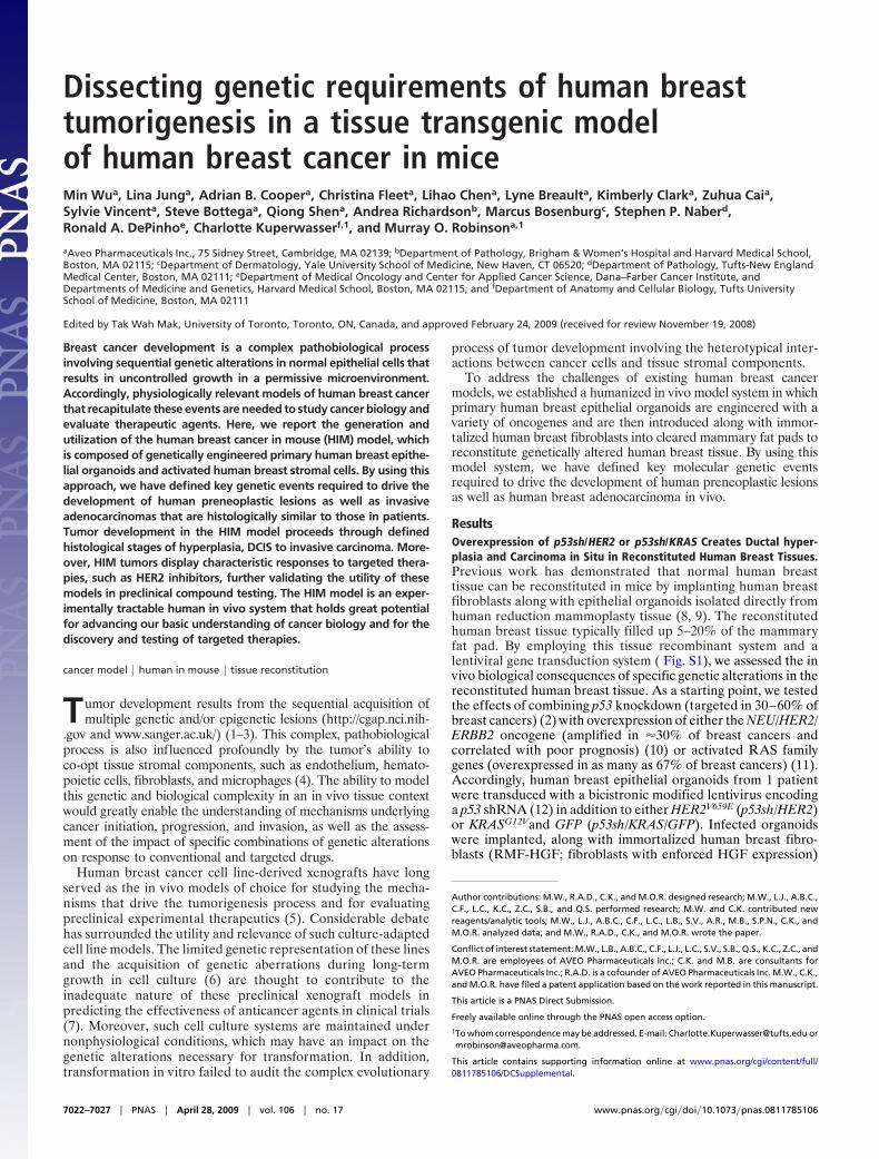
No visible tumors developed over the observation period of upto 12 months after implantation. Tissue recombinants werecollected at various time points (spanning 1–10 months afterimplantation) and subjected to histopathologic analysis (Fig. 1Aand Table S1). Both normal and hyperplastic outgrowths wereobserved in all of the tissue recombinants examined (n � 16 forp53sh/KRAS/GFP tissue recombinants and n � 24 for thep53sh/HER2 tissue recombinants; Fig. 1 A i–vi). Histopatholog-ical analysis confirmed characteristic normal and hyperplastichuman breast ductal architecture in both p53sh/KRAS/GF andp53sh/HER2 tissue recombinants. Lumen formation, basal lo-calized myoepithelial cells, and the presence of multiple layers ofluminal cells in the ducts were all evident in the hyperplasticoutgrowths, mirroring precisely the histopathologic features ofpremalignant changes in humans.
In addition to the normal and hyperplastic outgrowths, car-cinoma in situ (CIS) was observed in 12% (3 of 24) of thep53sh/HER2 tissue recombinants (Fig. 1 A vii–ix). These CISlesions exhibited histological features that are characteristic ofhuman breast ductal carcinoma in situ (DCIS), presenting ascomplete lumen filling of monotonous aggregates of large atyp-ical epithelial cells that stained positive for cytokeratin andHER2/neu. The uniform rim of �SMA-positive myoepithelialcells located in the basal layer confirmed the intraductal natureof these CIS lesions. This result is consistent with the prominentbiological role of HER2 in human breast cancer pathogenesis,and it gains added significance in that HER2 overexpression isdetected in 60–70% of human DCIS specimens (13, 14).
To determine the reproducibility of the results, we generated thep53sh/HER2 and the p53sh/KRAS/GFP tissue recombinants 2 moretimes by using organoids from 2 additional patients [donor 1 (n �17 for each genetic combination) and donor 2 (n � 16 for eachgenetic combination)]. No visible tumors developed from any of thetissue recombinants. Together, these observations demonstratedthat this tissue recombinant system, composed of relevant geneticand cellular components, can readily yield early-stage lesions withclassical features of the human disease.
HER2/SV40er or KRAS/SV40er Leads to Rapid Onset of Basal-likeInvasive Carcinomas in Vivo. Despite the successes in re-creatingearly premalignant breast lesions in vivo, there was a notable lackof tumor development in all of the p53sh/KRAS/GFP or p53sh/HER2 breast tissue recombinants; this is in contrast to transgenicmouse models in which overexpression of an activated HER2oncogene produces a highly penetrant breast cancer phenotype(15). These observations raised the possibility that additionalgenetic events are required to generate advanced disease in thehuman system. To address this notion, we replaced p53 shRNAwith the SV40 early region (SV40er), which encodes for theLarge T (LT) and small t (st) antigens to simultaneously disruptthe p53, RB, and PP2A/PI3K pathways (16).
Epithelial organoids from donor 1 were transduced withHER2V659E and SV40er (HER2/SV40er; n � 10; Table 1, experimentset A), KRASG12V and SV40er (KRAS/SV40er), or SV40er alone(SV40er) and were used as donor epithelium to generate humanbreast tissue recombinants in mice. With the introduction ofadditional genetic alterations provided by SV40er, tumors devel-oped in all of the HER2/SV40er and KRAS/SV40er tissue recom-binants (n � 10 each). Tumors became palpable as early as 5 weeksafter implantation. As a negative control, no tumors were observedin the SV40er tissue recombinants (n � 10) over a 6-monthobservation period. Therefore, the genetic combinations of HER2/SV40er and KRAS/SV40er, but not SV40er alone, were capable ofefficiently transforming primary human breast organoids in vivo.
Histological examination of the HER2/SV40er and KRAS/SV40er tumors revealed poorly differentiated invasive carcino-
GF
P/H
ER
2
P53sh/KRAS P53sh/HER2
Hyperplasia DCIS
H&
EP
an-C
K
i.
TDLU
HYP
vi. ix.
viii.
vii.
v.
iv.
iii. +αSMA
ii.
A
BHER2/SV40er Kras/SV40er
i.
iv.
v.
vi.
ix
x.
ii. vii.
iii. viii.
H&E
αSMA
Pan-CK
SV40 T
HER2/Kras
H&E
αSMA
Pan-CK
SV40 T
HER2/Kras
i. v.
ii. vi.
viii.iv.
iii. vii.
KRAS/p53R175H/CCND1/PIK3CA
C
p53
H&E
Pan-CK
αSMA
p53
H&E
Pan-CK
αSMA
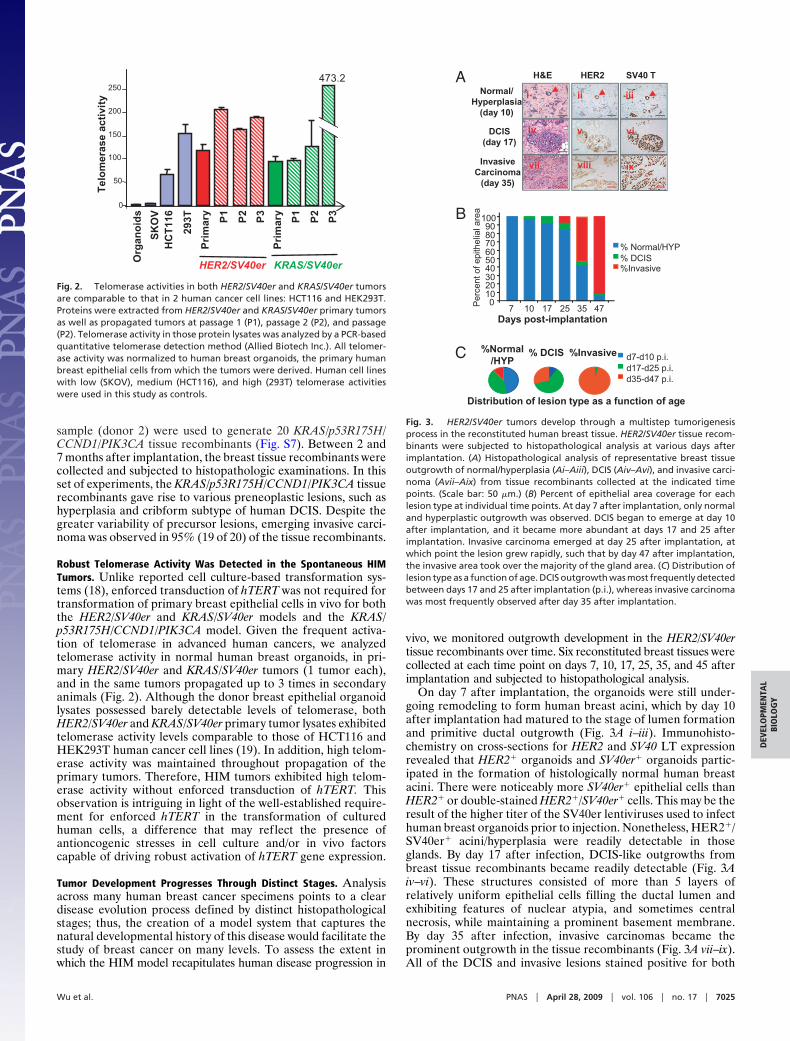
Fig. 1. Human preneoplastic lesions and advanced breast cancers weregenerated in vivo from genetically engineered human breast tissue recombi-nants. (A) Premalignant lesions and CIS developed in vivo from human breasttissue recombinants overexpressing HER2 or KRAS with concomitant knockingdown of p53. (Ai) GFP whole mount of reconstituted p53sh/KRAS/GFP humanbreast tissue reveals KRAS-lentivirus expression in both normal TerminalDuctal Lobular Unit (TDLU) structures (Inset) as well as in hyperplasic nodule(HYP). (Aii) H&E of the p53sh/KRAS/GFP hyperplastic outgrowth in Ai. (Aiii).IHC-stained serial section of Aii demonstrating the filling of the luminal spacewith epithelial cells. (Aiv–Aix) Histological analysis of p53sh/HER2 tissue re-combinants revealed both hyperplastic (Aiv–Avi) and carcinoma in situ out-growth (Avii–Aix) from transduced organoids (Aiv and Avii). (Scale bars: Ai, 0.5mm; Aii–Aix, 50 �m.) (B) Poorly differentiated, invasive human carcinomasdeveloped in vivo from HER2/SV40er and KRAS/SV40er human breast tissuerecombinants. Histological analysis of HER2/SV40er (Bi–Bv) and KRAS/SV40er(Bvi–Bx) tumors. H&E-stained sections (Bi and Bvi) and IHC analysis on serialsections with �SMA (Bii and Bvii), pan-cytokeratin (Pan-CK; Biii and Bviii), SV40LT (Biv and Bix), and HER2 (Bv) demonstrated that tumors were derived fromepithelial cells that overexpressed the transduced oncogenes. RNA in situanalysis (Bx) confirmed the expression of KRAS in KRAS/SV40er tumors. (Scalebar: 100 �m.) (C) Invasive ductal adenocarcinoma developed in vivo fromKRAS/p53R175H/CCND1/PIK3CA tissue recombinants. H&E-stained sections ofgenetically engineered HIM tumor (Ci and Cii) revealed that both sampleswere invasive ductal adenocarcinoma with prominent glandular architectureand low mitotic index. IHC-stained serial sections of HIM tumors with pan-cytokeratin (Pan-CK; Cii and Cvi), p53 (Ciii and Cvii), and �SMA (Civ and Cviii)revealed mutant p53-positive epithelial cancer cells surrounded by myofibro-blastic stroma. (Scale bar: 50 �m.)
Wu et al. PNAS � April 28, 2009 � vol. 106 � no. 17 � 7023
DEV
ELO
PMEN
TAL
BIO
LOG
Y
mas with anaplastic features (Fig. 1B and Fig. S2). The invasivegrowth pattern of nests and islands of tumor cells as well as theconsiderable cellular pleomorphisms are characteristic of highlymalignant disease in breast cancer patients (Fig. S3). Theseanaplastic HER2/SV40er and KRAS/SV40er tumor cells ex-pressed cytokeratins, confirming their epithelial cell origin (Fig.1B). Furthermore, IHC and RNA in situ hybridization analysesconfirmed that these tumors were derived from human breastepithelial cells transduced with HER2 and SV40er (HER2/SV40er) or KRAS and SV40er (KRAS/SV40er). Finally, thetumors contained prominent areas of stromal desmoplasia, afeature present in many human breast carcinomas (Fig. S4).
To further assess the molecular characteristics of the humanbreast tumors generated, IHC analysis was performed using clin-ically relevant markers to determine tumor subtype (Fig. S5). Boththe KRAS/SV40er and the HER2/SV40er tumors were negative forestrogen receptor (ER) and progensterone receptor (PR). Inaddition, the KRAS/SV40er tumors were positive for cytokeratin 5/6and p63 and negative for HER2, exhibiting typical features ofbasal-like breast cancers in human (ER�/PR�/HER2�/CK5/6�/p63�). Although HER2/SV40er tumors also exhibited a similar IHCexpression pattern of basal-type human breast cancer (ER�/PR�/CK5/6�/p63�), they were HER2-positive, resembling basal-HER2subgroup of human breast cancer (ER�/PR�/HER2�/CK5/6�/p63�) (17).
Because lentiviral gene transfer was used in this model system, itis conceivable that HER2/SV40er and KRAS/SV40er tumors devel-oped as a consequence of a random positional effect of lentiviralintegration into the human genome. However, the epithelial or-ganoids used in generating the p53sh/HER2 or p53sh/KRAS/GFPmodels are from the same donor patient as the ones used inHER2/SV40er and KRAS/SV40er models. Therefore, it is unlikelythat positional effect contributed to the tumor development inthose models.
In Kuperwasser et al. (8), spontaneous carcinomas developedfrom 1 of 10 mammoplasty specimens without any genetic modi-fication, a result that authors noted could not be reproduced withmaterials from the same patient. Preexisting genetic/epigeneticchanges in a subpopulation of the donor organoids is likely a factorcontributing to tumor development in the first case. To determinethe effect of different donor material on tumor development, werepeated the HER2/SV40er model by using epithelial organoidsobtained from 4 additional patient samples (donors 2–5; Table 1,experiment sets C, D, E, and F). As a positive control, HER2/SV40ertissue reconstitution was repeated using organoids from donor 1(n � 4; Table 1, experiment set B). Tumors developed from all 4of the patient donor epithelia, with similar kinetics and at similarfrequencies as those from the original patient sample, donor 1.Together, these findings establish that the HER2/SV40er and KRAS/SV40er genetic combinations can drive efficient transformation of
normal human breast epithelial cells into highly aggressive invasivecarcinomas in vivo.
To further verify the tumorigenic potential of the HIMtumors, cancer cells were isolated from 6 primary HER2/SV40ertumors and 6 primary KRAS/SV40er tumors and reinjected intohumanized fat pads of recipient mice. Tumors developed fromboth HER2/SV40er and KRAS/SV40er cancer cells, demonstrat-ing their full malignant transformation (Table S2).
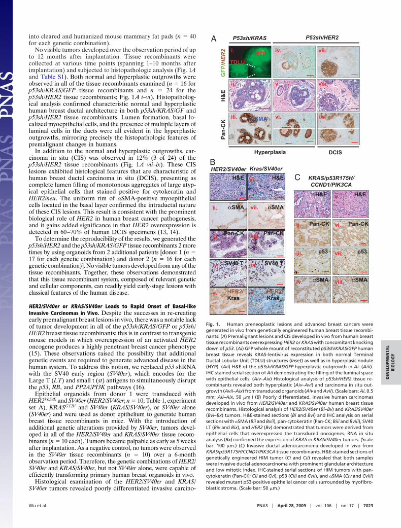
Breast tumors represent highly complex tissue systemswherein poorly defined, yet critical, heterotypic interactions takeplace between tumor cells and their microenvironment (10). Toassess the influence of various stromal fibroblasts on tumordevelopment, we reconstituted breast tissue recombinants withHER2/SV40er-transduced organoids either alone or togetherwith various human breast stromal fibroblasts (Table 1). Con-sistent with previous experiments, tumors were efficiently gen-erated from tissue recombinants when the HER2/SV40er or-ganoids were comixed with immortalized fibroblasts, RMFs withor without enforced expression of HGF (RMF-HGF vs.RMF-EG in Table 1, experiment sets A, B, and G). In contrast,tumor development was rarely observed when the same HER2/SV40er organoids were implanted either alone or comixed withnormal primary fibroblasts derived from 2 different patientsamples (Table 1, experiment sets H, I, and J). Consistent withprevious studies, our results demonstrate that specific stromalproperties can dramatically influence the tumorigenic potentialof oncogenically primed human breast tissue in vivo (4).
Signature Breast Cancer Genes—p53R175H/CCND1/PI3K/KRAS—Drivethe Development of Human Low-Grade Breast Adenocarcinoma. Al-though SV40er has been used extensively to transform humancells, it is not considered to play a pathogenetic role in sponta-neous human breast cancer in the clinic. We therefore selecteda specific set of oncogenes that are implicated in the etiology ofhuman breast cancer and target the pathways that are known tobe affected by SV40er (16). In humans, more than 40% of breastcancers exhibit overexpression of cyclin D1 (CCND1), whichinhibits RB activity (3); 30–60% harbor mutations of p53 (2);and 20–40% of breast cancers carry mutations of PIK3CA (1).We therefore chose to target both RB and p53 pathways throughoverexpression of CCND1 and a point mutant allele of p53(p53R175H). In addition, we targeted the PI3K pathway byoverexpressing a constitutively active form of PIK3CA.
Human breast epithelial organoids from patient no. 1 (donor1) were transduced with KRAS/p53R175H/CCND1/PIK3CA andreconstituted into a total of 20 murine mammary glands. Com-pared with HER2/SV40er and KRAS/SV40er models, tumorlatency in this model was, on average, longer and displayed morevariability. Between 2 and 9 months after implantation, tumorsdeveloped in 90% (18 of 20) of the tissue recombinants (Fig. 1C).These tumors presented as invasive ductal adenocarcinomas thatwere highly reminiscent of spontaneous, invasive, low-gradehuman breast ductal adenocarcinoma in humans. IHC per-formed on tumor sections verified that the cancer cells were ofepithelial cell origin and expressed p53R175H. These tumors alsodisplayed prominent areas of stromal desmoplasia, and theadjacent fibroblasts expressed �SMA. Similar to the KRAS/SV40er tumors, IHC revealed that the KRAS/p53R175H/CCND1/PIK3CA tumors did not express ER/PR or HER2, but werepositive for cytokeratin 5/6 and p63 (ER�/PR�/HER2�/CK5/6�/p63�), indicative of basal-like breast cancers (Fig. S5). Stableintegration of all of the lentiviral genes (p53R175H, CCND1,PIK3CA, and KRAS) and expression of those integrated geneswere confirmed by genomic PCR and RT-PCR analyses (Fig. S6).
To test the reproducibility of the KRAS/p53R175H/CCND1/PIK3CA genetic combination and to evaluate the potentialcontribution of preexisting genetic changes in donor material,human breast epithelial organoids from a different patient
Table 1. Effect of stromal fibroblasts on tumor developmentfrom organoids transduced with Her2/SV40er
Experimentalset Organoids Fibroblasts
Totalsites
Tumor frequency,no. (%)
A Donor 1 RMF-HGF 10 10 of 10 (100)B Donor 1 RMF-HGF 4 4 of 4 (100)C Donor 2 RMF-HGF 6 6 of 6 (100)D Donor 3 RMF-HGF 6 5 of 6 (83)E Donor 4 RMF-HGF 6 5 of 6 (83)F Donor 5 RMF-HGF 6 6 of 6 (100)G Donor 1 RMF 4 3 of 4 (75)H Donor 1 1°RMF-1 6 0I Donor 1 1°RMF-2 6 1 of 6 (17)J Donor 1 None 9 1 of 9 (11)
7024 � www.pnas.org�cgi�doi�10.1073�pnas.0811785106 Wu et al.
sample (donor 2) were used to generate 20 KRAS/p53R175H/CCND1/PIK3CA tissue recombinants (Fig. S7). Between 2 and7 months after implantation, the breast tissue recombinants werecollected and subjected to histopathologic examinations. In thisset of experiments, the KRAS/p53R175H/CCND1/PIK3CA tissuerecombinants gave rise to various preneoplastic lesions, such ashyperplasia and cribform subtype of human DCIS. Despite thegreater variability of precursor lesions, emerging invasive carci-noma was observed in 95% (19 of 20) of the tissue recombinants.
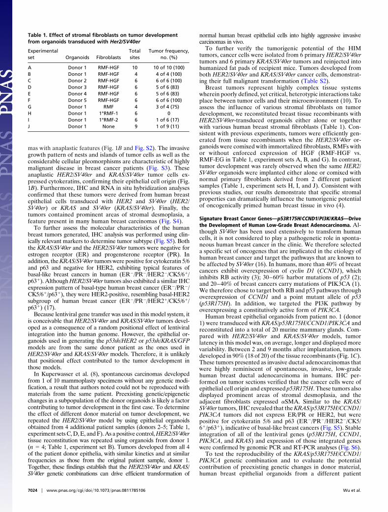
Robust Telomerase Activity Was Detected in the Spontaneous HIMTumors. Unlike reported cell culture-based transformation sys-tems (18), enforced transduction of hTERT was not required fortransformation of primary breast epithelial cells in vivo for boththe HER2/SV40er and KRAS/SV40er models and the KRAS/p53R175H/CCND1/PIK3CA model. Given the frequent activa-tion of telomerase in advanced human cancers, we analyzedtelomerase activity in normal human breast organoids, in pri-mary HER2/SV40er and KRAS/SV40er tumors (1 tumor each),and in the same tumors propagated up to 3 times in secondaryanimals (Fig. 2). Although the donor breast epithelial organoidlysates possessed barely detectable levels of telomerase, bothHER2/SV40er and KRAS/SV40er primary tumor lysates exhibitedtelomerase activity levels comparable to those of HCT116 andHEK293T human cancer cell lines (19). In addition, high telom-erase activity was maintained throughout propagation of theprimary tumors. Therefore, HIM tumors exhibited high telom-erase activity without enforced transduction of hTERT. Thisobservation is intriguing in light of the well-established require-ment for enforced hTERT in the transformation of culturedhuman cells, a difference that may reflect the presence ofantioncogenic stresses in cell culture and/or in vivo factorscapable of driving robust activation of hTERT gene expression.
Tumor Development Progresses Through Distinct Stages. Analysisacross many human breast cancer specimens points to a cleardisease evolution process defined by distinct histopathologicalstages; thus, the creation of a model system that captures thenatural developmental history of this disease would facilitate thestudy of breast cancer on many levels. To assess the extent inwhich the HIM model recapitulates human disease progression in
vivo, we monitored outgrowth development in the HER2/SV40ertissue recombinants over time. Six reconstituted breast tissues werecollected at each time point on days 7, 10, 17, 25, 35, and 45 afterimplantation and subjected to histopathological analysis.
On day 7 after implantation, the organoids were still under-going remodeling to form human breast acini, which by day 10after implantation had matured to the stage of lumen formationand primitive ductal outgrowth (Fig. 3A i–iii). Immunohisto-chemistry on cross-sections for HER2 and SV40 LT expressionrevealed that HER2� organoids and SV40er� organoids partic-ipated in the formation of histologically normal human breastacini. There were noticeably more SV40er� epithelial cells thanHER2� or double-stained HER2�/SV40er� cells. This may be theresult of the higher titer of the SV40er lentiviruses used to infecthuman breast organoids prior to injection. Nonetheless, HER2�/SV40er� acini/hyperplasia were readily detectable in thoseglands. By day 17 after infection, DCIS-like outgrowths frombreast tissue recombinants became readily detectable (Fig. 3Aiv–vi). These structures consisted of more than 5 layers ofrelatively uniform epithelial cells filling the ductal lumen andexhibiting features of nuclear atypia, and sometimes centralnecrosis, while maintaining a prominent basement membrane.By day 35 after infection, invasive carcinomas became theprominent outgrowth in the tissue recombinants (Fig. 3A vii–ix).All of the DCIS and invasive lesions stained positive for both
0
Org
ano
ids
SK
OV
HC
T11
6
293T
Pri
mar
y
Pri
mar
y
473.2
Tel
om
eras
e ac
tivi
ty
50
100
150
200
250
P1
P2
P3
P1
P2
P3
HER2/SV40er KRAS/SV40er
Fig. 2. Telomerase activities in both HER2/SV40er and KRAS/SV40er tumorsare comparable to that in 2 human cancer cell lines: HCT116 and HEK293T.Proteins were extracted from HER2/SV40er and KRAS/SV40er primary tumorsas well as propagated tumors at passage 1 (P1), passage 2 (P2), and passage(P2). Telomerase activity in those protein lysates was analyzed by a PCR-basedquantitative telomerase detection method (Allied Biotech Inc.). All telomer-ase activity was normalized to human breast organoids, the primary humanbreast epithelial cells from which the tumors were derived. Human cell lineswith low (SKOV), medium (HCT116), and high (293T) telomerase activitieswere used in this study as controls.
HER2H&E
Normal/Hyperplasia
(day 10)
DCIS(day 17)
InvasiveCarcinoma
(day 35)
SV40 T
ixvii
viviv
iiiii
viii
i
%Normal/HYP
d35-d47 p.i.d17-d25 p.i.d7-d10 p.i.% DCIS %Invasive
Distribution of lesion type as a function of age
0102030405060708090
100
7 10 17 25 35 47
%Invasive% DCIS% Normal/HYP
Days post-implantation
Per
cent
of e
pith
elia
l are
a
A
B
C
Fig. 3. HER2/SV40er tumors develop through a multistep tumorigenesisprocess in the reconstituted human breast tissue. HER2/SV40er tissue recom-binants were subjected to histopathological analysis at various days afterimplantation. (A) Histopathological analysis of representative breast tissueoutgrowth of normal/hyperplasia (Ai–Aiii), DCIS (Aiv–Avi), and invasive carci-noma (Avii–Aix) from tissue recombinants collected at the indicated timepoints. (Scale bar: 50 �m.) (B) Percent of epithelial area coverage for eachlesion type at individual time points. At day 7 after implantation, only normaland hyperplastic outgrowth was observed. DCIS began to emerge at day 10after implantation, and it became more abundant at days 17 and 25 afterimplantation. Invasive carcinoma emerged at day 25 after implantation, atwhich point the lesion grew rapidly, such that by day 47 after implantation,the invasive area took over the majority of the gland area. (C) Distribution oflesion type as a function of age. DCIS outgrowth was most frequently detectedbetween days 17 and 25 after implantation (p.i.), whereas invasive carcinomawas most frequently observed after day 35 after implantation.
Wu et al. PNAS � April 28, 2009 � vol. 106 � no. 17 � 7025
DEV
ELO
PMEN
TAL
BIO
LOG
Y
HER2 and SV40er expression, indicating they were indeedderived from the infected organoids.
To quantify the frequency of different subtypes of breast tissueoutgrowth over time, 3 to 6 cross-sections for each of the total36 tissue recombinants in this study were stained with H&E andwere subjected to blinded histopathological examination. Tumorprogression was quantified by counting the percent epithelialarea for each lesion type in the tissue recombinants over time(Fig. 3 B and C). Normal/hyperplastic outgrowths predominatedat the early observation times (days 7–10 after infection and days17–25 after infection), and these were gradually replaced bymore advanced outgrowths at later times. DCIS-like structureswere most frequently detected between days 17 and 25 afterinfection. By day 35 after infection, invasive tumor outgrowthswere the predominant outgrowth in the reconstituted humanbreast tissue. Collectively, these data indicate that the HIMmodel system recapitulates the multistep process of humanbreast cancer progression.
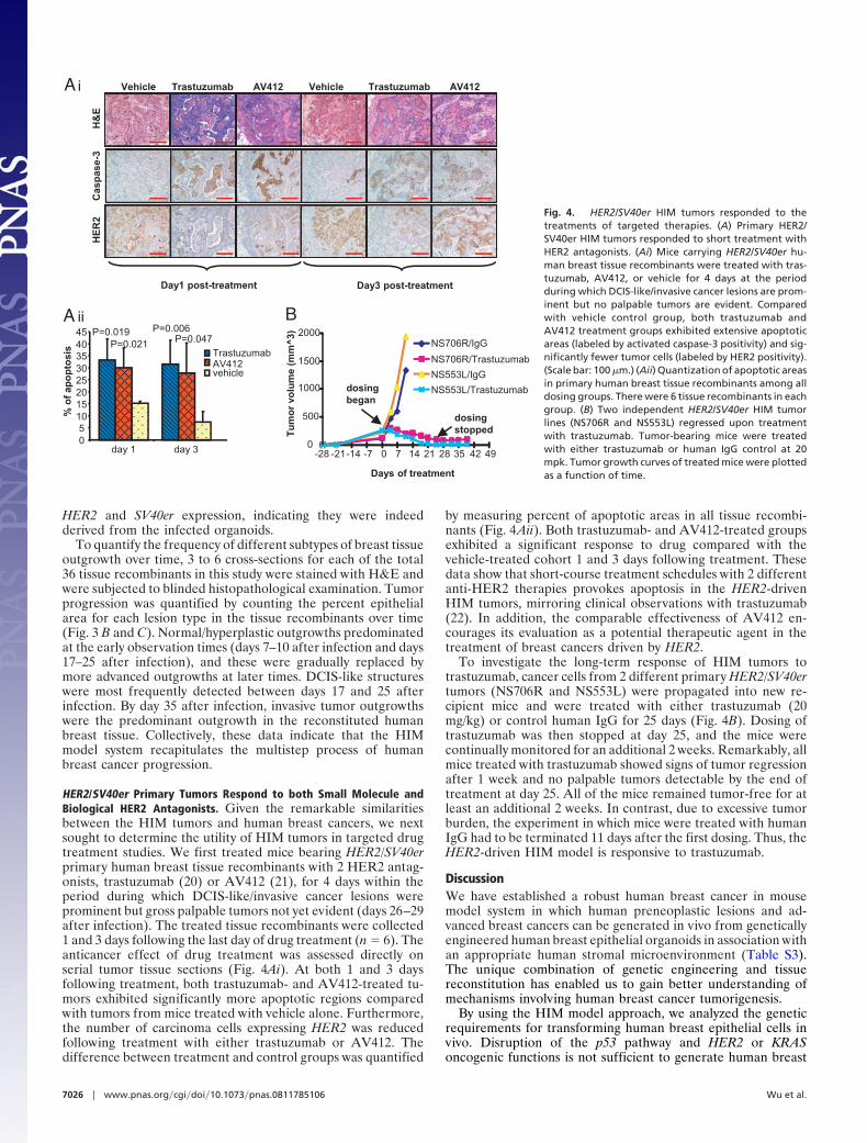
HER2/SV40er Primary Tumors Respond to both Small Molecule andBiological HER2 Antagonists. Given the remarkable similaritiesbetween the HIM tumors and human breast cancers, we nextsought to determine the utility of HIM tumors in targeted drugtreatment studies. We first treated mice bearing HER2/SV40erprimary human breast tissue recombinants with 2 HER2 antag-onists, trastuzumab (20) or AV412 (21), for 4 days within theperiod during which DCIS-like/invasive cancer lesions wereprominent but gross palpable tumors not yet evident (days 26–29after infection). The treated tissue recombinants were collected1 and 3 days following the last day of drug treatment (n � 6). Theanticancer effect of drug treatment was assessed directly onserial tumor tissue sections (Fig. 4Ai). At both 1 and 3 daysfollowing treatment, both trastuzumab- and AV412-treated tu-mors exhibited significantly more apoptotic regions comparedwith tumors from mice treated with vehicle alone. Furthermore,the number of carcinoma cells expressing HER2 was reducedfollowing treatment with either trastuzumab or AV412. Thedifference between treatment and control groups was quantified
by measuring percent of apoptotic areas in all tissue recombi-nants (Fig. 4Aii). Both trastuzumab- and AV412-treated groupsexhibited a significant response to drug compared with thevehicle-treated cohort 1 and 3 days following treatment. Thesedata show that short-course treatment schedules with 2 differentanti-HER2 therapies provokes apoptosis in the HER2-drivenHIM tumors, mirroring clinical observations with trastuzumab(22). In addition, the comparable effectiveness of AV412 en-courages its evaluation as a potential therapeutic agent in thetreatment of breast cancers driven by HER2.
To investigate the long-term response of HIM tumors totrastuzumab, cancer cells from 2 different primary HER2/SV40ertumors (NS706R and NS553L) were propagated into new re-cipient mice and were treated with either trastuzumab (20mg/kg) or control human IgG for 25 days (Fig. 4B). Dosing oftrastuzumab was then stopped at day 25, and the mice werecontinually monitored for an additional 2 weeks. Remarkably, allmice treated with trastuzumab showed signs of tumor regressionafter 1 week and no palpable tumors detectable by the end oftreatment at day 25. All of the mice remained tumor-free for atleast an additional 2 weeks. In contrast, due to excessive tumorburden, the experiment in which mice were treated with humanIgG had to be terminated 11 days after the first dosing. Thus, theHER2-driven HIM model is responsive to trastuzumab.
DiscussionWe have established a robust human breast cancer in mousemodel system in which human preneoplastic lesions and ad-vanced breast cancers can be generated in vivo from geneticallyengineered human breast epithelial organoids in association withan appropriate human stromal microenvironment (Table S3).The unique combination of genetic engineering and tissuereconstitution has enabled us to gain better understanding ofmechanisms involving human breast cancer tumorigenesis.
By using the HIM model approach, we analyzed the geneticrequirements for transforming human breast epithelial cells invivo. Disruption of the p53 pathway and HER2 or KRASoncogenic functions is not sufficient to generate human breast
Cas
pas
e-3
H&
EVehicl rastuzumab AV412 Vehicle Trastuzumab AV412
Day1 post-treatment Day3 post-treatment
HE
R2
Ai
TrastuzumabAV412vehicle
% o
f ap
op
tosi
s
day 1 day 305
1015202530354045
A iiP=0.019
P=0.021
P=0.006P=0.047 NS706R/IgG
NS706R/Trastuzumab
NS553L/IgG
NS553L/Trastuzumab
Days of treatment
0
500
1000
1500
2000
-28 -21 -7 0-14 7 14 21 28 4235 49
Tu
mo
r vo
lum
e (m
m^
3)
dosingstopped
dosingbegan
B
Fig. 4. HER2/SV40er HIM tumors responded to thetreatments of targeted therapies. (A) Primary HER2/SV40er HIM tumors responded to short treatment withHER2 antagonists. (Ai) Mice carrying HER2/SV40er hu-man breast tissue recombinants were treated with tras-tuzumab, AV412, or vehicle for 4 days at the periodduring which DCIS-like/invasive cancer lesions are prom-inent but no palpable tumors are evident. Comparedwith vehicle control group, both trastuzumab andAV412 treatment groups exhibited extensive apoptoticareas (labeled by activated caspase-3 positivity) and sig-nificantly fewer tumor cells (labeled by HER2 positivity).(Scale bar: 100 �m.) (Aii) Quantization of apoptotic areasin primary human breast tissue recombinants among alldosing groups. There were 6 tissue recombinants in eachgroup. (B) Two independent HER2/SV40er HIM tumorlines (NS706R and NS553L) regressed upon treatmentwith trastuzumab. Tumor-bearing mice were treatedwith either trastuzumab or human IgG control at 20mpk. Tumor growth curves of treated mice were plottedas a function of time.
7026 � www.pnas.org�cgi�doi�10.1073�pnas.0811785106 Wu et al.
lesions beyond DCIS (p53sh/HER2 and p53sh/KRAS models).Development of invasive carcinoma requires additional alter-ations targeting the pRB and PI3K pathways, conferred by theenforced expression of either CCND1/PIK3CA or SV40er(KRAS/p53R175H/CCND1/PI3K, KRAS/SV40er and HER2/SV40er models). Therefore, deregulation of the p53, pRB, andPI3K pathways, as well as overexpression of HER2 or KRASoncogenes is sufficient for the development of human breasttumors in this in vivo model system.
Widespread introduction of high-quality mammography screen-ing has caused a dramatic increase in the diagnosis of DCIS. Eventhough most DCISs do not progress to invasive cancer, mostpatients are treated with lumpectomy plus radiotherapy. Overtreat-ment of DCIS patients continues to be an important clinical issue(23). One major contributing factor to this limitation is the lack ofmodel systems that generate both low-risk and high-risk DCISoutgrowths resembling those in humans.
Despite the powerful natural selection that occurs in tumor-igenesis, the DCIS in p53sh/HER2 tissue recombinants did notprogress into invasive tumors, thus representing a model to studythe low-risk DCIS observed in patients. On the other hand, theHER2/SV40er model yielded high-risk DCISs that readilyevolved into invasive carcinoma. Molecular profiling of thesedifferent forms of DCIS outgrowth may help to define biomar-kers that can identify DCIS that will develop into invasivetumors. Moreover, the ability to manipulate the genetic profileof organoids affords an opportunity to discover and validate thegenetic requirements needed for progression from DCIS toinvasive tumors in the p53sh/HER2 model.
Human breast cancer comprises tumors with complex histo-pathology and genetic changes. The HIM system mirrors thiscomplexity in that tumors generated with different geneticcombinations gave rise to invasive tumors of different histolog-ical features, ranging from well/moderately differentiated carci-noma (KRAS/p53R175H/CCND1/PIK3CA) to invasive carci-noma (HER2/SV40er and KRAS/SV40er). In addition, IHCanalysis classified the KRAS/p53R175H/CCND1/PIK3CA andthe KRAS/SV40er tumors as basal-type breast cancer and theHER2/SV40er tumors as the basal-HER2 subtype of humanbreast cancer. How those histological variations reflect somaticchanges, as well as drug treatment outcome, remains to bedetermined.
One goal for the creation of genetically engineered humantumors is to provide a preclinical model to evaluate the efficacy ofcandidate therapies in breast cancer. With the increasing knowledge
of specific genetic alterations in breast cancer, there is now anopportunity to correlate activity of anticancer agents with specificgenetic alterations. As a preliminary proof of principle, we showedthat the approved anti-HER2 antibody trastuzumab is effective atincreasing apoptosis and inhibiting tumor growth of engineeredHER2-expressing tumor, demonstrating that these experimentaltumors exhibit a drug response modeling the clinical response ofHER2� breast cancers to trastuzumab. Furthermore, the experi-mental small molecule HER2 antagonist AV-412 is also effective atincreasing apoptosis of HER2-driven tumors, suggesting that thisexperimental agent may also show promise against HER2-drivenbreast tumors.
The ability of the HIM system to generate human tumors withdefined genetic modifications, that follow the multistep tumorprogression process, and whose development is strongly influ-enced by the specific stromal microenvironment, holds potentialas an improved preclinical model system for more rigorous andaccurate drug testing.
Materials and MethodsTissues. All human breast tissue procurement for these experiments wasobtained in compliance with laws and institutional guidelines as approved bythe institutional review board from Brigham & Women’s Hospital.
Infection and Injection of Human Breast Organoids. Freshly isolated or freshlythawed organoids were subjected to infection with lentiviruses as describedpreviously (SI Methods). Within 4–18 h after the last infection, the infectedorganoids were injected into humanized fat pads without selection.
Constructs and Virus Production. See SI Methods for details.
Whole Mounts and IHC. Whole-mount analysis and IHC were performed asdescribed previously (19). Antibodies used were pan-cytokeratin (catalogueno. A0575; DAKO), �SMA (catalogue no. NCL-SMA; Novo Castra), HER2 (cat-alogue no. 2242; Cell Signaling Technology), SV40T Ag (catalogue no. sc-147;Santa Cruz Biotechnology), p53 (catalogue no. sc-6243; Santa Cruz Biotech-nology), and Cleaved Caspase-3 (catalogue no. CP-229B; Biocare Medical).
Telomerase Assay. Telomerase activity was analyzed by using the PCR-basedQuantitative Telomerase Detection Kit (Allied Biotech Inc.). Each tumor sam-ple was assayed in quadruplicates.
Tumor Treatment. AV412 was resuspended in 0.5% tragacanth and dosed dailyat 100 mg/kg. Trastuzumab was dosed twice a week at 20 mg/kg.
ACKNOWLEDGMENTS. We gratefully acknowledge Geoffrey Boynton andEdyta Tyminski for expert technical assistance, and Steve Clark, William M.Rideout 3rd, Joerg Heyer, and Ronan C. O’Hagan for critical reading of themanuscript.
1. Campbell IG, et al. (2004) Mutation of the PIK3CA gene in ovarian and breast cancer.Cancer Res 64:7678–7681.
2. Coles C, et al. (1992) p53 mutations in breast cancer. Cancer Res 52:5291–5298.3. Umekita Y, Ohi Y, Sagara Y, Yoshida H (2002) Overexpression of cyclinD1 predicts for
poor prognosis in estrogen receptor-negative breast cancer patients. Int J Cancer98:415–418.
4. Hanahan D, Weinberg RA (2000) The hallmarks of cancer. Cell 100:57–70.5. Finn RS, Slamon DJ (2003) Monoclonal antibody therapy for breast cancer: Herceptin.
Cancer Chemother Biol Response Modif 21:223–233.6. Truong K, et al. (1999) Evidence for in vitro selection during cell culturing of breast
cancer: Detection by flow and image cytometry. Cancer Genet Cytogenet 114:154 –155.
7. Sharpless NE, Depinho RA (2006) The mighty mouse: Genetically engineered mousemodels in cancer drug development. Nat Rev Drug Discov 5:741–754.
8. Kuperwasser C, et al. (2004) Reconstruction of functionally normal and malignanthuman breast tissues in mice. Proc Natl Acad Sci USA 101:4966–4971.
9. Parmar H, et al. (2002) A novel method for growing human breast epithelium in vivousing mouse and human mammary fibroblasts. Endocrinology 143:4886–4896.
10. Bacus SS, et al. (1992) HER-2/neu oncogene expression, DNA ploidy and proliferationindex in breast cancer. Anal Quant Cytol Histol 14:433–445.
11. Miyakis S, Sourvinos G, Spandidos DA (1998) Differential expression and mutation of theras family genes in human breast cancer. Biochem Biophys Res Commun 251:609–612.
12. Brummelkamp TR, Bernards R, Agami R (2002) A system for stable expression of shortinterfering RNAs in mammalian cells. Science 296:550–553.
13. Menard S, et al. (2001) HER2 overexpression in various tumor types, focussing on itsrelationship to the development of invasive breast cancer. Ann Oncol 12:S15–S19.
14. Tsuda H, Hirohashi S (1998) Multiple developmental pathways of highly aggressivebreast cancers disclosed by comparison of histological grades and c-erbB-2 expressionpatterns in both the non-invasive and invasive portions. Pathol Int 48:518–525.
15. Guy CT, Cardiff RD, Muller WJ (1996) Activated neu induces rapid tumor progression.J Biol Chem 271:7673–7678.
16. Zhao JJ, et al. (2003) Human mammary epithelial cell transformation through theactivation of phosphatidylinositol 3-kinase. Cancer Cell 3:483–495.
17. Liu H, et al. (2008) Basal-HER2 phenotype shows poorer survival than basal-like phenotypein hormone receptor-negative invasive breast cancers. Hum Pathol 39:167–174.
18. Elenbaas B, et al. (2001) Human breast cancer cells generated by oncogenic transfor-mation of primary mammary epithelial cells. Genes Dev 15:50–65.
19. Zhang X, Mar V, Zhou W, Harrington L, Robinson MO (1999) Telomere shortening andapoptosis in telomerase-inhibited human tumor cells. Genes Dev 13:2388–2399.
20. Slamon DJ, et al. (2001) Use of chemotherapy plus a monoclonal antibody against HER2for metastatic breast cancer that overexpresses HER2. N Engl J Med 344:783–792.
21. Lin J, et al. (2007) AV-412, a potent EGFR/HER2 TK inhibitor causes tumor regression innovel genetically engineered EGFRL858R and EGFRL858R&T790M lung tumor models. InAmerican Association for Cancer Research Annual Meeting: Proceedings (AmericanAssociation for Cancer Research, Philadelphia), abstr 4008.
22. Mohsin SK, et al. (2005) Neoadjuvant trastuzumab induces apoptosis in primary breastcancers. J Clin Oncol 23:2460–2468.
23. Napoli M (2001) Overdiagnosis and overtreatment. Am J Nurs 101:11.
Wu et al. PNAS � April 28, 2009 � vol. 106 � no. 17 � 7027
DEV
ELO
PMEN
TAL
BIO
LOG
Y
Related Documents