Carcinogenesis vol.33 no.4 pp.876–885, 2012 doi:10.1093/carcin/bgs097 Advance Access publication February 3, 2012 Intervention of human breast cell carcinogenesis chronically induced by 2-amino-1- methyl-6-phenylimidazo[4,5-b]pyridine Shambhunath Choudhary 1,2 , Shilpa Sood 1,2 , Robert L.Donnell 2 and Hwa-Chain R.Wang 1,2, 1 Anticancer Molecular Oncology Laboratory and 2 Department of Biomedical and Diagnostic Sciences, College of Veterinary Medicine, The University of Tennessee, Knoxville, TN 37996, USA To whom correspondence should be addressed. Tel: þ1 865 974 3846; Fax: þ1 865 974 5640; Email: [email protected] More than 85% of breast cancers are sporadic and attributable to long-term exposure to environmental carcinogens, such as those in the diet, through a multistep disease process progressing from non-cancerous to premalignant and malignant stages. The chem- ical carcinogen 2-amino-1-methyl-6-phenylimidazo[4,5-b]pyri- dine (PhIP) is one of the most abundant heterocyclic amines found in high-temperature cooked meats and is recognized as a mammary carcinogen. However, the PhIP’s mechanism of action in breast cell carcinogenesis is not clear. Here, we demon- strated, for the first time, that cumulative exposures to PhIP at physiologically achievable, pico to nanomolar concentrations effectively induced progressive carcinogenesis of human breast epithelial MCF10A cells from a non-cancerous stage to premalig- nant and malignant stages in a dose- and exposure-dependent manner. Progressive carcinogenesis was measured by increas- ingly- acquired cancer-associated properties of reduced depen- dence on growth factors, anchorage-independent growth, acinar-conformational disruption, proliferation, migration, inva- sion, tumorigenicity with metastasis and increased stem-like cell populations. These biological changes were accompanied by bio- chemical and molecular changes, including upregulated H-Ras gene expression, extracellular signal-regulated kinase (ERK) pathway activation, Nox-1 expression, reactive oxygen species (ROS) elevation, increased HIF-1a, Sp1, tumor necrosis factor-a, matrix metalloproteinase (MMP)-2, MMP-9, aldehyde dehydro- genase activity and reduced E-cadherin. The Ras-ERK-Nox- ROS pathway played an important role in not only initiation but also maintenance of cellular carcinogenesis induced by PhIP. Using biological, biochemical and molecular changes as targeted endpoints, we identified that the green tea catechin components epicatechin-3-gallate and epigallocatechin-3-gallate, at non-cytotoxic doses, were capable of suppressing PhIP-induced cellular carcinogenesis and tumorigenicity. Introduction Human breast cancer is the most common type of cancer and the second leading cause of cancer deaths among women in northern America and Europe (1). More than 85% of breast cancers are spo- radic and attributable to long-term exposure to environmental factors through a multi-year and multistep disease process progressing from non-cancerous to premalignant and malignant stages (2,3). More than 200 chemical mammary carcinogens have been detected by the exist- ing experimental paradigm that uses high doses of carcinogens (micro to millimolar concentrations) to induce cancerous cells in cultures and tumors in animals as steps in evaluating the potency of carcinogens (3–5). However, since long-term exposure to low doses of carcinogens is responsible for most human cancers, a high-dose approach may not be a proper way to reveal the effect of environmental carcinogens in breast cancer development. Thus, an urgent need exists to take a new approach of validating carcinogens, at physiologically achievable lev- els, effective in chronic induction of human breast cell carcinogenesis and then identifying preventive agents capable of intervening. The dietary carcinogen 2-amino-1-methyl-6-phenylimidazo[4,5-b] pyridine (PhIP) is one of the most mass abundant heterocyclic amines, which are particularly found in high-temperature cooked meats, such as grilled/barbequed meats (6–8). Epidemiological studies indicate that an increased risk of breast cancer is closely associated with increased consumption of PhIP in well-done meats (9,10). Human consumption of PhIP at microgram levels results in systemic PhIP exposure at pico to low nanomolar levels (8). Studies in rats revealed that daily gastric administration of PhIP in milligram ranges induces mammary tumors (8). Studies of genotoxicity to human cell lines and adduct formation reveal genotoxic activity of PhIP at a concentration as low as 450 nmol/l (8,11). However, whether long-term exposure of breast cells to PhIP at physiologically achievable pico to low nano- molar levels may induce carcinogenesis and tumorigenicity remains to be clarified. Daily, oral administration of PhIP at 75 mg/kg into rats for 10 days induces intraductal proliferation, carcinoma in situ and carcinoma with increased frequencies of activation mutations within the H-ras gene (12). Expression of oncogenic H-Ras in immortalized, non-can- cerous human breast epithelial MCF10A cells induces an invasive phenotype, accompanied by expression of matrix metalloproteinase-2 and -9 (MMP-2 and -9) (13) and extracellular signal-regulated kinase (ERK) pathway activation (14). Activation of the ERK pathway leads to reduced nicotinamide adenine dinucleotide phosphate oxidase-1 (Nox-1) expression, and Nox-1 mediates reactive oxygen species (ROS) elevation, which plays an important role in cell proliferation, motility, invasion and angiogenesis (14,15). It has been shown that a single exposure of MCF10A cells to PhIP at nanomolar concentra- tions induces cell proliferation and transient activation of the ERK pathway (16). However, the role of transient ERK pathway activation in PhIP-induced carcinogenesis remains to be clarified. It is well recognized that transition activation of the ERK pathway, which con- sists of Raf, Mek and Erk, contributes to cell proliferation, survival and differentiation, and constant activation of the ERK pathway leads to malignant transformation (17). Whether long-term exposure to PhIP results in constant activation of the Ras-ERK-Nox-ROS pathway for cellular carcinogenesis remains to be determined. We developed a cellular model to mimic chronic induction of human breast cell carcinogenesis associated with accumulated expo- sures to low doses of environmental carcinogens, such as 4-(methyl- nitrosamino)-1-(3-pyridyl)-1-butanone (NNK) and benzo[a]pyrene (18–21). We have used various transient and constitutive endpoints as targets to identify preventive agents, such as green tea catechins, effective in suppression of cellular carcinogenesis (19–22). Green tea catechins include four major components: epicatechin, epicate- chin-3-gallate (ECG), epigallocatechin and epigallocatechin-3-gallate (EGCG) (23). These components, at a non-cytotoxic concentration of 10 lg/ml, suppress cellular carcinogenesis chronically induced by NNK and benzo[a]pyrene (22). However, whether these catechins are effective in suppression of PhIP-induced cellular carcinogenesis remains to be studied. In this communication, we used our model to clarify the carcinogenic activity of PhIP, at physiologically achievable doses, in inducing progressive carcinogenesis of MCF10A cells from a non-cancerous stage to premalignant and malignant stages. We revealed that the Ras-ERK-Nox-ROS pathway played an important role in both Abbreviations: ALDH, aldehyde dehydrogenase; CM, complete medium; DPI, diphenylene iodonium; ECG, epicatechin-3-gallate; EGCG, epigallocatechin-3-gallate; ERK, extracellular signal-regulated kinase; MMP, matrix metalloproteinase; NAC, N-acetyl-L-cysteine; PhIP, 2-amino-1- methyl-6-phenylimidazo[4,5-b]pyridine; ROS, reactive oxygen species; siRNA, small interfering RNA; TNF, tumor necrosis factor; wt, wild-type. Ó The Author 2012. Published by Oxford University Press. All rights reserved. For Permissions, please email: [email protected] 876 Downloaded from https://academic.oup.com/carcin/article/33/4/876/2464091 by guest on 10 July 2022

Welcome message from author
This document is posted to help you gain knowledge. Please leave a comment to let me know what you think about it! Share it to your friends and learn new things together.
Transcript
Carcinogenesis vol.33 no.4 pp.876–885, 2012doi:10.1093/carcin/bgs097Advance Access publication February 3, 2012
Intervention of human breast cell carcinogenesis chronically induced by 2-amino-1-methyl-6-phenylimidazo[4,5-b]pyridine
Shambhunath Choudhary1,2, Shilpa Sood1,2,Robert L.Donnell2 and Hwa-Chain R.Wang1,2,�
1Anticancer Molecular Oncology Laboratory and 2Department of Biomedicaland Diagnostic Sciences, College of Veterinary Medicine, The University ofTennessee, Knoxville, TN 37996, USA
�To whom correspondence should be addressed. Tel: þ1 865 974 3846;Fax: þ1 865 974 5640;Email: [email protected]
More than 85% of breast cancers are sporadic and attributable tolong-term exposure to environmental carcinogens, such as thosein the diet, through a multistep disease process progressing fromnon-cancerous to premalignant and malignant stages. The chem-ical carcinogen 2-amino-1-methyl-6-phenylimidazo[4,5-b]pyri-dine (PhIP) is one of the most abundant heterocyclic aminesfound in high-temperature cooked meats and is recognized asa mammary carcinogen. However, the PhIP’s mechanism ofaction in breast cell carcinogenesis is not clear. Here, we demon-strated, for the first time, that cumulative exposures to PhIP atphysiologically achievable, pico to nanomolar concentrationseffectively induced progressive carcinogenesis of human breastepithelial MCF10A cells from a non-cancerous stage to premalig-nant and malignant stages in a dose- and exposure-dependentmanner. Progressive carcinogenesis was measured by increas-ingly- acquired cancer-associated properties of reduced depen-dence on growth factors, anchorage-independent growth,acinar-conformational disruption, proliferation, migration, inva-sion, tumorigenicity with metastasis and increased stem-like cellpopulations. These biological changes were accompanied by bio-chemical and molecular changes, including upregulated H-Rasgene expression, extracellular signal-regulated kinase (ERK)pathway activation, Nox-1 expression, reactive oxygen species(ROS) elevation, increased HIF-1a, Sp1, tumor necrosis factor-a,matrix metalloproteinase (MMP)-2, MMP-9, aldehyde dehydro-genase activity and reduced E-cadherin. The Ras-ERK-Nox-ROS pathway played an important role in not only initiationbut also maintenance of cellular carcinogenesis induced byPhIP. Using biological, biochemical and molecular changes astargeted endpoints, we identified that the green tea catechincomponents epicatechin-3-gallate and epigallocatechin-3-gallate,at non-cytotoxic doses, were capable of suppressing PhIP-inducedcellular carcinogenesis and tumorigenicity.
Introduction
Human breast cancer is the most common type of cancer and thesecond leading cause of cancer deaths among women in northernAmerica and Europe (1). More than 85% of breast cancers are spo-radic and attributable to long-term exposure to environmental factorsthrough a multi-year and multistep disease process progressing fromnon-cancerous to premalignant and malignant stages (2,3). More than200 chemical mammary carcinogens have been detected by the exist-ing experimental paradigm that uses high doses of carcinogens (microto millimolar concentrations) to induce cancerous cells in cultures andtumors in animals as steps in evaluating the potency of carcinogens
(3–5). However, since long-term exposure to low doses of carcinogensis responsible for most human cancers, a high-dose approach may notbe a proper way to reveal the effect of environmental carcinogens inbreast cancer development. Thus, an urgent need exists to take a newapproach of validating carcinogens, at physiologically achievable lev-els, effective in chronic induction of human breast cell carcinogenesisand then identifying preventive agents capable of intervening.
The dietary carcinogen 2-amino-1-methyl-6-phenylimidazo[4,5-b]pyridine (PhIP) is one of the most mass abundant heterocyclic amines,which are particularly found in high-temperature cooked meats, suchas grilled/barbequed meats (6–8). Epidemiological studies indicatethat an increased risk of breast cancer is closely associated withincreased consumption of PhIP in well-done meats (9,10). Humanconsumption of PhIP at microgram levels results in systemic PhIPexposure at pico to low nanomolar levels (8). Studies in rats revealedthat daily gastric administration of PhIP in milligram ranges inducesmammary tumors (8). Studies of genotoxicity to human cell lines andadduct formation reveal genotoxic activity of PhIP at a concentrationas low as 450 nmol/l (8,11). However, whether long-term exposureof breast cells to PhIP at physiologically achievable pico to low nano-molar levels may induce carcinogenesis and tumorigenicity remainsto be clarified.
Daily, oral administration of PhIP at 75 mg/kg into rats for 10 daysinduces intraductal proliferation, carcinoma in situ and carcinomawith increased frequencies of activation mutations within the H-rasgene (12). Expression of oncogenic H-Ras in immortalized, non-can-cerous human breast epithelial MCF10A cells induces an invasivephenotype, accompanied by expression of matrix metalloproteinase-2and -9 (MMP-2 and -9) (13) and extracellular signal-regulated kinase(ERK) pathway activation (14). Activation of the ERK pathway leadsto reduced nicotinamide adenine dinucleotide phosphate oxidase-1(Nox-1) expression, and Nox-1 mediates reactive oxygen species(ROS) elevation, which plays an important role in cell proliferation,motility, invasion and angiogenesis (14,15). It has been shown thata single exposure of MCF10A cells to PhIP at nanomolar concentra-tions induces cell proliferation and transient activation of the ERKpathway (16). However, the role of transient ERK pathway activationin PhIP-induced carcinogenesis remains to be clarified. It is wellrecognized that transition activation of the ERK pathway, which con-sists of Raf, Mek and Erk, contributes to cell proliferation, survivaland differentiation, and constant activation of the ERK pathway leadsto malignant transformation (17). Whether long-term exposure toPhIP results in constant activation of the Ras-ERK-Nox-ROS pathwayfor cellular carcinogenesis remains to be determined.
We developed a cellular model to mimic chronic induction ofhuman breast cell carcinogenesis associated with accumulated expo-sures to low doses of environmental carcinogens, such as 4-(methyl-nitrosamino)-1-(3-pyridyl)-1-butanone (NNK) and benzo[a]pyrene(18–21). We have used various transient and constitutive endpointsas targets to identify preventive agents, such as green tea catechins,effective in suppression of cellular carcinogenesis (19–22). Greentea catechins include four major components: epicatechin, epicate-chin-3-gallate (ECG), epigallocatechin and epigallocatechin-3-gallate(EGCG) (23). These components, at a non-cytotoxic concentration of10 lg/ml, suppress cellular carcinogenesis chronically induced byNNK and benzo[a]pyrene (22). However, whether these catechinsare effective in suppression of PhIP-induced cellular carcinogenesisremains to be studied.
In this communication, we used our model to clarify the carcinogenicactivity of PhIP, at physiologically achievable doses, in inducingprogressive carcinogenesis of MCF10A cells from a non-cancerousstage to premalignant and malignant stages. We revealed that theRas-ERK-Nox-ROS pathway played an important role in both
Abbreviations: ALDH, aldehyde dehydrogenase; CM, complete medium;DPI, diphenylene iodonium; ECG, epicatechin-3-gallate; EGCG,epigallocatechin-3-gallate; ERK, extracellular signal-regulated kinase;MMP, matrix metalloproteinase; NAC, N-acetyl-L-cysteine; PhIP, 2-amino-1-methyl-6-phenylimidazo[4,5-b]pyridine; ROS, reactive oxygen species; siRNA,small interfering RNA; TNF, tumor necrosis factor; wt, wild-type.
� The Author 2012. Published by Oxford University Press. All rights reserved. For Permissions, please email: [email protected] 876
Dow
nloaded from https://academ
ic.oup.com/carcin/article/33/4/876/2464091 by guest on 10 July 2022
initiation and maintenance of cellular carcinogenesis chronically in-duced by PhIP. We used various transient and constitutive endpointsas targets to identify that ECG and EGCG, at non-cytotoxic levels,were effective in suppression of PhIP-induced cellular carcinogenesisand tumorigenicity.
Materials and methods
Cell cultures and reagents
MCF10A (American Type Culture Collection, Rockville, MD) and derived celllines were maintained in complete (CM) medium (1:1 mixture of Dulbecco’smodified Eagle’s medium and Ham’s F12, supplemented with 100 ng/ml chol-era enterotoxin, 10 lg/ml insulin, 0.5 lg/ml hydrocortisol, 20 ng/ml epidermalgrowth factor and 5% horse serum) (18–21). Human breast cancer MCF7 andurinary bladder cancer J82 cells (American Type Culture Collection) weremaintained in Dulbecco’s modified Eagle’s medium supplemented with 10%heat-inactivated fetal calf serum (21,24). All cultures were maintained inmedium supplemented with 100 U/ml penicillin and 100 lg/ml streptomycinin 5% CO2 at 37�C. Stock aqueous solutions of PhIP (Midwest; NCI ChemicalCarcinogen Reference Standard Repository), chloromethyl dichlorodihydro-fluorescein diacetate (Invitrogen, Carlsbad, CA), U0126 (Cell Signaling,Beverly, MA) and diphenylene iodonium (DPI) (Acro, Morris Plains, NJ) wereprepared in dimethyl sulfoxide and diluted in culture medium. Stock aqueoussolutions of epicatechin, ECG, epigallocatechin, EGCG (Sigma–Aldrich,St Louis, MO) and N-acetyl-L-cysteine (NAC) (Alexis, San Diego, CA) wereprepared in H2O and diluted in culture medium for assays.
Chronic induction of cellular carcinogenesis
To chronically induce cellular carcinogenesis, 24 h after each subculturing,MCF10A cultures were treated with PhIP for 48 h as one cycle of exposure for5–20 cycles; cultures were subcultured every 3 days (21).
Reduced dependence on growth factors
A total of 3 � 103 cells were seeded in 60 mm culture dishes and maintained inlow-mitogen medium, containing reduced total serum and mitogenic additivesto 2% of the concentration formulated in CM medium, for 10 days to developcell colonies (21).
Anchorage-independent growth
A total of 3 � 103 cells were mixed with soft agar consisting of 0.4%SeaPlaque agarose (Sigma–Aldrich) in a mixture (1:1) of CM medium with3 day conditioned medium prepared from MCF10A cultures, plated on top ofthe 2% SeaPlaque agarose base layer in 60 mm culture dishes and maintainedfor 14 days to develop cell clones (21).
Acinar-conformational disruption
A total of 3 � 103 cells were mixed with CM medium containing 4% GrowthFactor-Reduced Matrigel Matrix (BD Biosciences), plated on Matrigel base ineach well of 24-well culture plates and maintained for 14 days to developspheroids (21).
Cell proliferation
Cell proliferation was determined using the 5-bromo-2#-deoxyuridine cell pro-liferation enzyme-linked immunosorbent assay kit (Roche, Indianapolis, IN);quantification of 5-bromo-2#-deoxyuridine-labeled cells was determined withan enzyme-linked immunosorbent assay reader (Bio-Tek), as performed pre-viously (24).
In vitro cell invasion and migration
The cell invasion assay was performed using 24-well transwell insert chamberswith a polycarbonate filter with a pore size of 8.0 lm (Costar, Corning, NY).A total of 2 � 104 cells in serum-free medium were seeded on top of a Matrigel-coated filter (BD Biosciences) in each insert chamber. Then, insert chamberswere placed into wells on top of culture medium containing 10% horse serumas a chemoattractant. The migration assay was performed using 24-well trans-well insert chambers with a polycarbonate filter without Matrigel. The invasiveor migratory ability of cells was determined by the number of cells translocatedto the lower side of filters (25).
Serum-independent non-adherent growth
A total of 1 � 104 cells were seeded on top of 1% agarose-coated, non-adherent100 mm culture plates, incubated in serum-free MCF10A medium supple-mented with 0.4% bovine serum albumin and maintained for 10 days todevelop mammospheres (26).
Aldehyde dehydrogenase
An ALDEFLUOR Kit (StemCell Technologies, Durham, NC) was used todetect aldehyde dehydrogenase (ALDH)-positive cells. Cells were mixed withactivated Aldefluor substrate BODIPY-aminoacetaldehyde and incubated inthe presence and absence of the ALDH inhibitor diethylaminobenzaldehyde,followed by flow cytometric analysis (24). The mean fluorescence intensity ofcells was quantified using Multicycle software (Phoenix Flow System, SanDiego, CA). Cells incubated with BODIPY-aminoacetaldehyde in the presenceof diethylaminobenzaldehyde were used to establish the baseline of fluores-cence for determining the ALDH-positive cell population in which ALDHactivity was not inhibited by diethylaminobenzaldehyde (26).
Intracellular ROS
Cells were incubated with 5 lmol/l chloromethyl dichlorodihydrofluoresceindiacetate in 5% CO2 at 37�C for 1 h to detect ROS level by flow cytometricanalysis; the mean fluorescence intensity of dichlorodihydrofluorescein wasquantified using Multicycle software (Phoenix Flow System), as performedpreviously (14).
DNA damage
DNA damage was measured by a comet assay. Cells were trypsinized andcollected in phosphate-buffered saline at a density of 2 � 104 cells/ml. Cellsuspension was mixed with an equal volume of 1% low-melting agarose(Fisher, Fair Lawn, NJ) and placed on agarose-coated slides. Slides were thenimmersed in lysis solution (1.2 mol/l NaCl, 100 mmol/l Na2-ethylenediami-netetraacetic acid, 1% Triton X-100 and 0.3 nmol/l NaOH, pH 13) at 25�C for1 h and rinsed three times with alkaline buffer (2 mmol/l Na2-ethylenediami-netetraacetic acid and 300 mmol/l NaOH) for 20 min each. After electropho-resis in the same alkaline buffer at 20 V for 30 min (27), slides were stainedwith 2.5 lg/ml of propidium iodide for 20 min and examined with a Zeissfluorescence microscope (Carl Zeiss Inc, Thornwood, NY) equipped with anexcitation filter of 546 nm and barrier filter of 590 nm. Fifty nuclei per slidewere scored for tail moment (% of DNA in the tail � tail length) as a parameterusing CometScore software (Tritek).
Downregulation by small interfering RNAs
Validated human H-Ras-specific (sc-29340) and Nox-1-specific (sc-43939)small interfering RNAs (siRNAs) (Santa Cruz Biotechnology, Santa Cruz,CA) are pools of three to five sequence-specific 19–25 nt siRNAs designedto knock down gene expression. Cells were transfected with 80 pmol/lH-Ras-specific, Nox-1-specific or control siRNAs (sc-37007) in antibiotic-freemedium using siRNA transfection reagent (sc-29528).
Reverse transcription–PCR
Total RNA isolated from cultures using the Absolutely RNA kit (Stratagene,La Jolla, CA) was reverse transcribed to complementary DNA using a VersocDNA Kit (Thermo Scientific, Waltham, MA). The resulting complementaryDNAs were subjected to PCR for H-Ras (forward: 5#-GACGGAATA-TAAGCTGGTGG-3#; reverse: 5#-AGGCACGTCTCCCCATCAAT-3#) andb-actin (forward: 5#-GGACTTCGAGCAAGAGATGG-3#; reverse: 5#-AG-CACTGTGTTGGCGTACAG-3#). PCR products were electrophoresed onagarose gels and visualized after ethidium bromide staining.
Western immunoblotting
Equal amount of cellular proteins were resolved by electrophoresis in either 10or 14% sodium dodecyl sulfate–polyacrylamide gels and transferred to nitro-cellulose filters for western immunoblotting, as described previously (21,24),using specific antibodies to detect H-Ras, Raf-1, phosphorylated Erk1/2(p-Erk1/2), Erk1/2, Nox-1, p53, tumor necrosis factor (TNF)-a, MMP-2,MMP-9, E-cadherin, vascular endothelial growth factor, Sp1, b-actin (SantaCruz Biotechnology), p-Raf-1, p-Mek1/2, Mek1/2, p-H2AX, H2AX and HIF-1a (Cell Signaling). Antigen–antibody complexes on filters were detected bythe Supersignal chemiluminescence kit (Pierce, Rockford, IL).
Tumorigenicity and histopathology
Cells were mixed with Matrigel basement membrane matrix (BD Biosciences),and 1 � 107 cells in 100 ll were injected into the inguinal mammary fat pads of10-week-old female athymic NCr-nu/nu mice (National Cancer Institute,Frederick, MD). Each group of four mice was maintained under pathogen-freeconditions. Animals were monitored every 3 days. Xenograft tumor tissueswere immediately harvested after euthanasia by exposure to CO2. Tumor tis-sues were fixed in neutral-buffered formalin and embedded in paraffin forhistopathological evaluation. Tumor tissue lysates were prepared to detectvarious proteins using western immunoblotting (24). All animal procedureswere approved by The University of Tennessee Animal Care and Use Com-mittee and were in accordance with the National Institutes of Health Guide forthe Care and Use of Laboratory Animals (National Research Council, 1985).
Carcinogenesis of human breast epithelial cells by PhIP
877
Dow
nloaded from https://academ
ic.oup.com/carcin/article/33/4/876/2464091 by guest on 10 July 2022
Immunohistochemistry
Immunohistochemistry procedures were performed on paraffin-embedded tis-sue sections. Briefly, 5 lm tissue sections were adhered to charged glass slides,deparaffinized and pre-soaked in Tris-buffered saline/Tween for 10 min (24).Immunohistochemistry staining was carried out on an Autostainer (Dako,Carpinteria, CA), following a procedure of peroxide blocking for 5 min,serum-free protein blocking for 30 min, exposure to rabbit anti-human TNF-aantibody for 30 min and Dako Envision with horseradish peroxidase anti-rabbittreatment and chromagen labeling with Dako diaminobenzidene þ for 30 min.The sections were then counterstained with hematoxylin for microscopicevaluation.
Statistical analysis
The Student’s t test was used to analyze statistical significance, indicated by�P , 0.05, ��P , 0.01 and ���P , 0.001; a P value of �0.05 was consideredsignificant.
Results
Dose- and exposure-dependent induction of carcinogenesis by PhIP
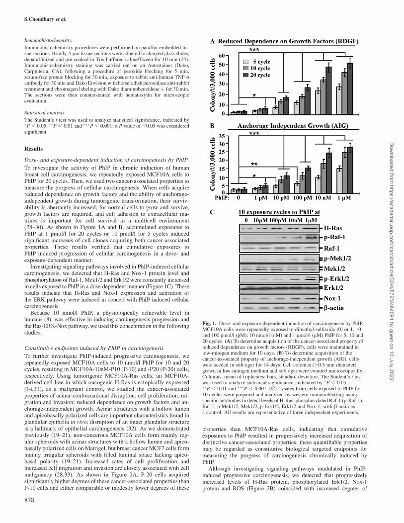
To investigate the activity of PhIP in chronic induction of humanbreast cell carcinogenesis, we repeatedly exposed MCF10A cells toPhIP for 20 cycles. Then, we used two cancer-associated properties tomeasure the progress of cellular carcinogenesis. When cells acquirereduced dependence on growth factors and the ability of anchorage-independent growth during tumorigenic transformation, their surviv-ability is aberrantly increased; for normal cells to grow and survive,growth factors are required, and cell adhesion to extracellular ma-trixes is important for cell survival in a multicell environment(28–30). As shown in Figure 1A and B, accumulated exposures toPhIP at 1 pmol/l for 20 cycles or 10 pmol/l for 5 cycles inducedsignificant increases of cell clones acquiring both cancer-associatedproperties. These results verified that cumulative exposures toPhIP induced progression of cellular carcinogenesis in a dose- andexposure-dependent manner.
Investigating signaling pathways involved in PhIP-induced cellularcarcinogenesis, we detected that H-Ras and Nox-1 protein level andphosphorylation of Raf-1, Mek1/2 and Erk1/2 were constantly increasedin cells exposed to PhIP in a dose-dependent manner (Figure 1C). Theseresults indicate that H-Ras and Nox-1 expression and activation ofthe ERK pathway were induced in concert with PhIP-induced cellularcarcinogenesis.
Because 10 nmol/l PhIP, a physiologically achievable level inhumans (8), was effective in inducing carcinogenesis progression andthe Ras-ERK-Nox pathway, we used this concentration in the followingstudies.
Constitutive endpoints induced by PhIP in carcinogenesis
To further investigate PhIP-induced progressive carcinogenesis, werepeatedly exposed MCF10A cells to 10 nmol/l PhIP for 10 and 20cycles, resulting in MCF10A-10nM-P10 (P-10) and -P20 (P-20) cells,respectively. Using tumorigenic MCF10A-Ras cells, an MCF10A-derived cell line in which oncogenic H-Ras is ectopically expressed(14,31), as a malignant control, we studied the cancer-associatedproperties of acinar-conformational disruption; cell proliferation, mi-gration and invasion; reduced dependence on growth factors and an-chorage-independent growth. Acinar structures with a hollow lumenand apicobasally polarized cells are important characteristics found inglandular epithelia in vivo; disruption of an intact glandular structureis a hallmark of epithelial carcinogenesis (32). As we demonstratedpreviously (19–21), non-cancerous MCF10A cells form mainly reg-ular spheroids with acinar structures with a hollow lumen and apico-basally polarized cells on Matrigel, but breast cancer MCF7 cells formmainly irregular spheroids with filled luminal space lacking apico-basal polarity (19–21). Increased rates of cell proliferation andincreased cell migration and invasion are closely associated with cellmalignancy (28,33). As shown in Figure 2A, P-20 cells acquiredsignificantly higher degrees of these cancer-associated properties thanP-10 cells and either comparable or modestly lower degrees of these
properties than MCF10A-Ras cells, indicating that cumulativeexposures to PhIP resulted in progressively increased acquisition ofdistinctive cancer-associated properties; these quantifiable propertiesmay be regarded as constitutive biological targeted endpoints formeasuring the progress of carcinogenesis chronically induced byPhIP.
Although investigating signaling pathways modulated in PhIP-induced progressive carcinogenesis, we detected that progressivelyincreased levels of H-Ras protein, phosphorylated Erk1/2, Nox-1protein and ROS (Figure 2B) coincided with increased degrees of
Fig. 1. Dose- and exposure-dependent induction of carcinogenesis by PhIP.MCF10A cells were repeatedly exposed to dimethyl sulfoxide (0) or 1, 10and 100 pmol/l (pM); 10 nmol/l (nM) and 1 lmol/l (lM) PhIP for 5, 10 and20 cycles. (A) To determine acquisition of the cancer-associated property ofreduced dependence on growth factors (RDGF), cells were maintained inlow-mitogen medium for 10 days. (B) To determine acquisition of thecancer-associated property of anchorage-independent growth (AIG), cellswere seeded in soft agar for 14 days. Cell colonies (�0.5 mm diameter)grown in low-mitogen medium and soft agar were counted microscopically.Columns, mean of triplicates; bars, standard deviation. The Student’s t testwas used to analyze statistical significance, indicated by �P , 0.05,��P , 0.01 and ���P , 0.001. (C) Lysates from cells exposed to PhIP for10 cycles were prepared and analyzed by western immunoblotting usingspecific antibodies to detect levels of H-Ras, phosphorylated Raf-1 (p-Raf-1),Raf-1, p-Mek1/2, Mek1/2, p-Erk1/2, Erk1/2 and Nox-1, with b-actin asa control. All results are representative of three independent experiments.
S.Choudhary et al.
878
Dow
nloaded from https://academ
ic.oup.com/carcin/article/33/4/876/2464091 by guest on 10 July 2022
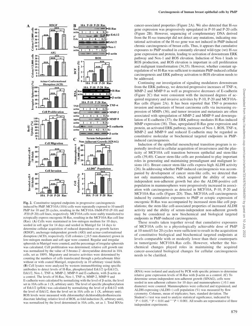
cancer-associated properties (Figure 2A). We also detected that H-rasgene expression was progressively upregulated in P-10 and P-20 cells(Figure 2B). However, sequencing of complementary DNA derivedfrom the H-ras transcript did not detect any mutations, indicating mu-tational activation of the H-ras gene was not induced in PhIP-inducedchronic carcinogenesis of breast cells. Thus, it appears that cumulativeexposures to PhIP resulted in constantly elevated wild-type (wt) H-rasgene expression and protein, leading to activation of downstream ERKpathway and Nox-1 and ROS elevation. Induction of Nox-1 leads toROS production, and ROS elevation is important in cell proliferationand malignant transformation (34,35). However, whether constant up-regulation of wt H-Ras was sufficient to maintain PhIP-induced cellularcarcinogenesis and ERK pathway activation to ROS elevation needs tobe addressed.
Continuing our investigation of signaling modulators downstreamfrom the ERK pathway, we detected progressive increases of TNF-a,MMP-2 and MMP-9 as well as progressive decreases of E-cadherin(Figure 2C) that were consistent with the increased degrees of ac-quired migratory and invasive activities by P-10, P-20 and MCF10A-Ras cells (Figure 2A). It has been reported that TNF-a promotesinvasion and metastasis of breast carcinoma cells via increasing ex-pression of MMPs (36), and tumor invasion and metastasis are oftenassociated with upregulation of MMP-2 and MMP-9 and downregu-lation of E-cadherin (37); the ERK pathway mediates H-Ras-inducedMMP expression (38). Thus, upregulated H-Ras gene expression andprotein, an activated ERK pathway, increases of Nox-1, ROS, TNF-a,MMP-2 and MMP-9 and reduced E-cadherin may be regarded asconstitutive molecular or biochemical targeted endpoints in PhIP-induced carcinogenesis.
Induction of the epithelial mesenchymal transition program is re-portedly involved in cellular acquisition of invasiveness and the plas-ticity of MCF10A cell transition between epithelial and stem-likecells (39,40). Cancer stem-like cells are postulated to play importantroles in generating and maintaining premalignant and malignant le-sions (41). Breast cancer stem-like cells express high ALDH activity(26). In addressing whether PhIP-induced carcinogenesis was accom-panied by development of cancer stem-like cells, we detected thatnot only mammospheres, which acquired the ability of serum-independent non-adherent growth but also the ALDH-positive cellpopulation in mammospheres were progressively increased in associ-ation with carcinogenesis as detected in MCF10A, P-10, P-20 andMCF10A-Ras cells (Figure 2D). Thus, MCF10A cell carcinogenesisinduced by cumulative exposures to PhIP or ectopic expression ofoncogenic H-Ras was accompanied by increased stem-like cell pop-ulations; the stem-like cell-associated properties of increased ALDHactivity and the ability of serum-independent non-adherent growthmay be considered as new biochemical and biological targetedendpoints in PhIP-induced carcinogenesis.
These results taken together reveal that cumulative exposuresof MCF10A cells to a physiologically achievable dose of PhIPat 10 nmol/l for 20 cycles were sufficient to result in the acquisitionof constitutive biological and biochemical targeted endpoints atlevels comparable with or modestly lower than their counterpartsin tumorigenic MCF10A-Ras cells. However, whether the bio-chemical changes played roles in maintaining the acquiredcancer-associated biological changes for cellular carcinogenesisneeds to be clarified.
Fig. 2. Constitutive targeted endpoints in progressive carcinogenesisinduced by PhIP. MCF10A (10A) cells were repeatedly exposed to 10 nmol/lPhIP for 10 and 20 cycles, resulting in the MCF10A-10nM-P10 (P-10) and-P20 (P-20) cell lines, respectively. MCF10A cells were stably transfected toectopically express oncogenic H-Ras, resulting in the MCF10A-Ras cell line(Ras). (A) Cells were maintained in low-mitogen medium for 10 days,seeded in soft agar for 14 days and seeded in Matrigel for 14 days todetermine cellular acquisition of reduced dependence on growth factors(RDGF), anchorage-independent growth (AIG) and acinar-conformationaldisruption (ACD), respectively. Cell colonies (�0.5 mm diameter) grown inlow-mitogen medium and soft agar were counted. Regular and irregularspheroids in Matrigel were counted, and the percentage of irregular spheroidswas calculated. Cell proliferation was determined; relative cell growth ratewas normalized by the value of 5-bromo-2#-deoxyuridine detected in 10Acells, set as 100%. Migratory and invasive activities were determined bycounting the numbers of cells translocated through a polycarbonate filterwithout or with coated Matrigel, respectively in 10 arbitrary visual fields.(B) Cell lysates were analyzed by western immunoblotting using specificantibodies to detect levels of H-Ras, phosphorylated Erk1/2 (p-Erk1/2),Erk1/2, Nox-1, TNF-a, MMP-2, MMP-9 and E-cadherin, with b-actin asa control. The levels of H-Ras, Nox-1, TNF-a, MMP-2, MMP-9 andE-cadherin were calculated by normalizing with the level of b-actin, the levelset in 10A cells as 1 (X, arbitrary unit). The level of specific phosphorylationof Erk1/2 (p/Erk) was calculated by normalizing the level of p-Erk1/2 withthe level of Erk1/2, then the level set in 10A cells as 1 (X, arbitrary unit).ROS levels were measured with chloromethyl dichlorodihydrofluoresceindiacetate labeling; relative level of ROS, as fold induction (X, arbitrary unit),was normalized by the level determined in 10A cells, set as 1. Total RNAs
(RNA) were isolated and analyzed by PCR with specific primers to determinerelative gene expression levels of H-Ras with b-actin as a control. (C) Todetermine serum-independent non-adherent growth (SINAG), cells wereseeded in non-adherent cultures for 10 days and mammospheres (�0.1 mmdiameter) were counted. Mammospheres were collected and trypsinized, andALDH-expressing (ALDHþ) cell population (%) was measured by flowcytometry. Columns, mean of triplicates; bars, standard deviation. TheStudent’s t test was used to analyze statistical significance, indicated by�P , 0.05, ��P , 0.01 and ���P , 0.001. All results are representative of threeindependent experiments.
Carcinogenesis of human breast epithelial cells by PhIP
879
Dow
nloaded from https://academ
ic.oup.com/carcin/article/33/4/876/2464091 by guest on 10 July 2022
H-Ras, ERK, Nox-1 and ROS in maintenance of cancer-associatedproperties
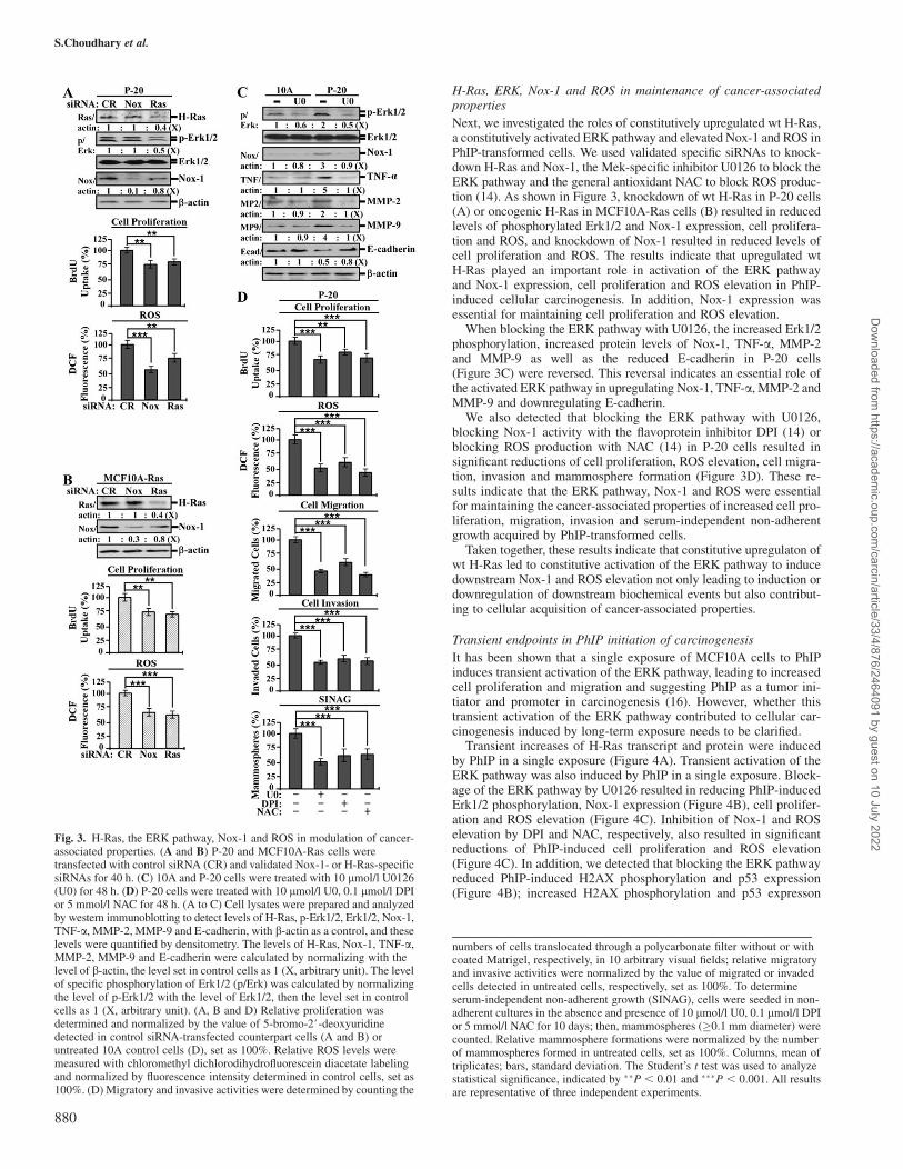
Next, we investigated the roles of constitutively upregulated wt H-Ras,a constitutively activated ERK pathway and elevated Nox-1 and ROS inPhIP-transformed cells. We used validated specific siRNAs to knock-down H-Ras and Nox-1, the Mek-specific inhibitor U0126 to block theERK pathway and the general antioxidant NAC to block ROS produc-tion (14). As shown in Figure 3, knockdown of wt H-Ras in P-20 cells(A) or oncogenic H-Ras in MCF10A-Ras cells (B) resulted in reducedlevels of phosphorylated Erk1/2 and Nox-1 expression, cell prolifera-tion and ROS, and knockdown of Nox-1 resulted in reduced levels ofcell proliferation and ROS. The results indicate that upregulated wtH-Ras played an important role in activation of the ERK pathwayand Nox-1 expression, cell proliferation and ROS elevation in PhIP-induced cellular carcinogenesis. In addition, Nox-1 expression wasessential for maintaining cell proliferation and ROS elevation.
When blocking the ERK pathway with U0126, the increased Erk1/2phosphorylation, increased protein levels of Nox-1, TNF-a, MMP-2and MMP-9 as well as the reduced E-cadherin in P-20 cells(Figure 3C) were reversed. This reversal indicates an essential role ofthe activated ERK pathway in upregulating Nox-1, TNF-a, MMP-2 andMMP-9 and downregulating E-cadherin.
We also detected that blocking the ERK pathway with U0126,blocking Nox-1 activity with the flavoprotein inhibitor DPI (14) orblocking ROS production with NAC (14) in P-20 cells resulted insignificant reductions of cell proliferation, ROS elevation, cell migra-tion, invasion and mammosphere formation (Figure 3D). These re-sults indicate that the ERK pathway, Nox-1 and ROS were essentialfor maintaining the cancer-associated properties of increased cell pro-liferation, migration, invasion and serum-independent non-adherentgrowth acquired by PhIP-transformed cells.
Taken together, these results indicate that constitutive upregulaton ofwt H-Ras led to constitutive activation of the ERK pathway to inducedownstream Nox-1 and ROS elevation not only leading to induction ordownregulation of downstream biochemical events but also contribut-ing to cellular acquisition of cancer-associated properties.
Transient endpoints in PhIP initiation of carcinogenesis
It has been shown that a single exposure of MCF10A cells to PhIPinduces transient activation of the ERK pathway, leading to increasedcell proliferation and migration and suggesting PhIP as a tumor ini-tiator and promoter in carcinogenesis (16). However, whether thistransient activation of the ERK pathway contributed to cellular car-cinogenesis induced by long-term exposure needs to be clarified.
Transient increases of H-Ras transcript and protein were inducedby PhIP in a single exposure (Figure 4A). Transient activation of theERK pathway was also induced by PhIP in a single exposure. Block-age of the ERK pathway by U0126 resulted in reducing PhIP-inducedErk1/2 phosphorylation, Nox-1 expression (Figure 4B), cell prolifer-ation and ROS elevation (Figure 4C). Inhibition of Nox-1 and ROSelevation by DPI and NAC, respectively, also resulted in significantreductions of PhIP-induced cell proliferation and ROS elevation(Figure 4C). In addition, we detected that blocking the ERK pathwayreduced PhIP-induced H2AX phosphorylation and p53 expression(Figure 4B); increased H2AX phosphorylation and p53 expresson
Fig. 3. H-Ras, the ERK pathway, Nox-1 and ROS in modulation of cancer-associated properties. (A and B) P-20 and MCF10A-Ras cells weretransfected with control siRNA (CR) and validated Nox-1- or H-Ras-specificsiRNAs for 40 h. (C) 10A and P-20 cells were treated with 10 lmol/l U0126(U0) for 48 h. (D) P-20 cells were treated with 10 lmol/l U0, 0.1 lmol/l DPIor 5 mmol/l NAC for 48 h. (A to C) Cell lysates were prepared and analyzedby western immunoblotting to detect levels of H-Ras, p-Erk1/2, Erk1/2, Nox-1,TNF-a, MMP-2, MMP-9 and E-cadherin, with b-actin as a control, and theselevels were quantified by densitometry. The levels of H-Ras, Nox-1, TNF-a,MMP-2, MMP-9 and E-cadherin were calculated by normalizing with thelevel of b-actin, the level set in control cells as 1 (X, arbitrary unit). The levelof specific phosphorylation of Erk1/2 (p/Erk) was calculated by normalizingthe level of p-Erk1/2 with the level of Erk1/2, then the level set in controlcells as 1 (X, arbitrary unit). (A, B and D) Relative proliferation wasdetermined and normalized by the value of 5-bromo-2#-deoxyuridinedetected in control siRNA-transfected counterpart cells (A and B) oruntreated 10A control cells (D), set as 100%. Relative ROS levels weremeasured with chloromethyl dichlorodihydrofluorescein diacetate labelingand normalized by fluorescence intensity determined in control cells, set as100%. (D) Migratory and invasive activities were determined by counting the
numbers of cells translocated through a polycarbonate filter without or withcoated Matrigel, respectively, in 10 arbitrary visual fields; relative migratoryand invasive activities were normalized by the value of migrated or invadedcells detected in untreated cells, respectively, set as 100%. To determineserum-independent non-adherent growth (SINAG), cells were seeded in non-adherent cultures in the absence and presence of 10 lmol/l U0, 0.1 lmol/l DPIor 5 mmol/l NAC for 10 days; then, mammospheres (�0.1 mm diameter) werecounted. Relative mammosphere formations were normalized by the numberof mammospheres formed in untreated cells, set as 100%. Columns, mean oftriplicates; bars, standard deviation. The Student’s t test was used to analyzestatistical significance, indicated by ��P , 0.01 and ���P , 0.001. All resultsare representative of three independent experiments.
S.Choudhary et al.
880
Dow
nloaded from https://academ
ic.oup.com/carcin/article/33/4/876/2464091 by guest on 10 July 2022
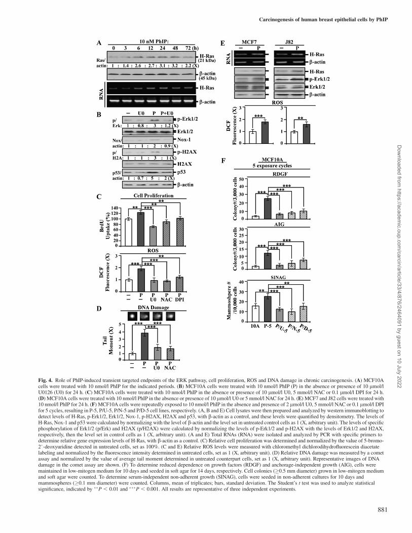
Fig. 4. Role of PhIP-induced transient targeted endpoints of the ERK pathway, cell proliferation, ROS and DNA damage in chronic carcinogenesis. (A) MCF10Acells were treated with 10 nmol/l PhIP for the indicated periods. (B) MCF10A cells were treated with 10 nmol/l PhIP (P) in the absence or presence of 10 lmol/lU0126 (U0) for 24 h. (C) MCF10A cells were treated with 10 nmol/l PhIP in the absence or presence of 10 lmol/l U0, 5 mmol/l NAC or 0.1 lmol/l DPI for 24 h.(D) MCF10A cells were treated with 10 nmol/l PhIP in the absence or presence of 10 lmol/l U0 or 5 mmol/l NAC for 24 h. (E) MCF7 and J82 cells were treated with10 nmol/l PhIP for 24 h. (F) MCF10A cells were repeatedly exposed to 10 nmol/l PhIP in the absence and presence of 2 lmol/l U0, 5 mmol/l NAC or 0.1 lmol/l DPIfor 5 cycles, resulting in P-5, P/U-5, P/N-5 and P/D-5 cell lines, respectively. (A, B and E) Cell lysates were then prepared and analyzed by western immunoblotting todetect levels of H-Ras, p-Erk1/2, Erk1/2, Nox-1, p-H2AX, H2AX and p53, with b-actin as a control, and these levels were quantified by densitometry. The levels ofH-Ras, Nox-1 and p53 were calculated by normalizing with the level of b-actin and the level set in untreated control cells as 1 (X, arbitrary unit). The levels of specificphosphorylation of Erk1/2 (p/Erk) and H2AX (p/H2AX) were calculated by normalizing the levels of p-Erk1/2 and p-H2AX with the levels of Erk1/2 and H2AX,respectively, then the level set in control cells as 1 (X, arbitrary unit). (A and E) Total RNAs (RNA) were isolated and analyzed by PCR with specific primers todetermine relative gene expression levels of H-Ras, with b-actin as a control. (C) Relative cell proliferation was determined and normalized by the value of 5-bromo-2#-deoxyuridine detected in untreated cells, set as 100%. (C and E) Relative ROS levels were measured with chloromethyl dichlorodihydrofluorescein diacetatelabeling and normalized by the fluorescence intensity determined in untreated cells, set as 1 (X, arbitrary unit). (D) Relative DNA damage was measured by a cometassay and normalized by the value of average tail moment determined in untreated counterpart cells, set as 1 (X, arbitrary unit). Representative images of DNAdamage in the comet assay are shown. (F) To determine reduced dependence on growth factors (RDGF) and anchorage-independent growth (AIG), cells weremaintained in low-mitogen medium for 10 days and seeded in soft agar for 14 days, respectively. Cell colonies (�0.5 mm diameter) grown in low-mitogen mediumand soft agar were counted. To determine serum-independent non-adherent growth (SINAG), cells were seeded in non-adherent cultures for 10 days andmammospheres (�0.1 mm diameter) were counted. Columns, mean of triplicates; bars, standard deviation. The Student’s t test was used to analyze statisticalsignificance, indicated by ��P , 0.01 and ���P , 0.001. All results are representative of three independent experiments.
Carcinogenesis of human breast epithelial cells by PhIP
881
Dow
nloaded from https://academ
ic.oup.com/carcin/article/33/4/876/2464091 by guest on 10 July 2022
are indices of chromosomal DNA damage (42,43). Using a cometassay, we verified that blockage of the ERK pathway or ROS eleva-tion by U0126 and NAC, respectively, reduced PhIP-induced DNAdamage (Figure 4D). These results indicate that a single exposure ofnon-cancerous MCF10A cells to PhIP was able to induce transientactivation of the Ras-ERK pathway, leading to Nox-1 expression andROS elevation and resulting in increased cell proliferation and DNAdamage. We detected similar results in breast cancer MCF7 and uri-nary bladder cancer J82 cells, where PhIP was able to induce the Ras-ERK pathway leading to ROS elevation, indicating that its effectswere applicable to other cells (Figure 4E). It has been postulatedthat activation of the ERK pathway leads to ROS elevation resultingin oxidative DNA damage, contributing to genomic mutations in cellularcarcinogenesis (44). However, whether blockage of the PhIP-inducedERK pathway or ROS elevation may suppress cellular carcinogenesisneeds to be determined.
Using U0126 to block the ERK pathway, DPI to block Nox-1activity and NAC to block ROS elevation in each exposure cycle toPhIP, we detected that these blockages resulted in significant inhibi-tion of cellular acquisition of the cancer-associated properties ofreduced dependence on growth factors, anchorage-independent
growth and mammosphere formation after five exposure cycles(Figure 4F). In contrast, blockage of other pathways, such asphosphoinositide-3-kinase, p38/stress-activated protein kinase orc-jun N-terminal kinase/stress-activated protein kinase, did not resultin any reduction of PhIP-induced carcinogenesis (data not shown).Thus, transient induction of the Ras-ERK pathway to ROS elevationwas essential for PhIP-induced initiation of cellular carcinogenesis ineach single exposure; conceivably, accumulation of PhIP-inducedcellular damage contributed to cellular carcinogenesis after cumula-tive exposures. The Ras-ERK-Nox-ROS pathway, cell proliferation,DNA damage and DNA damage-associated phosphorylation of H2AXmay be regarded as transient targeted endpoints for measuring PhIPactivity in initiation of cellular carcinogenesis.
Suppression of PhIP-induced carcinogenesis by ECG and EGCG
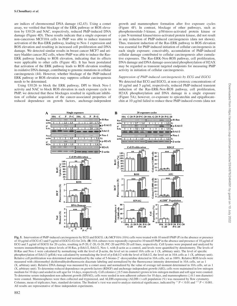
We detected that ECG and EGCG, at non-cytotoxic concentrations of10 lg/ml and 5 lg/ml, respectively, reduced PhIP-induced transientinduction of the Ras-ERK-Nox-ROS pathway, cell proliferation,H2AX phosphorylation and DNA damage in a single exposure(Figure 5A); however, co-exposure to epicatechin and epigallocate-chin at 10 lg/ml failed to reduce these PhIP-induced events (data not
Fig. 5. Intervention of PhIP-induced carcinogenesis by ECG and EGCG. (A) MCF10A (10A) cells were treated with 10 nmol/l PhIP (P) in the absence or presenceof 10 lg/ml of ECG (C) and 5 lg/ml of EGCG (G) for 24 h. (B) 10A cultures were repeatedly exposed to 10 nmol/l PhIP in the absence and presence of 10 lg/ml ofECG and 5 lg/ml of EGCG for 20 cycles, resulting in P-20, C-20, G-20, P/C-20 and P/G-20 cell lines, respectively. Cell lysates were prepared and analyzed bywestern immunoblotting to detect levels of H-Ras, p-Erk1/2, Erk1/2, Nox-1, with b-actin as a control, and levels were quantified by densitometry. The levels ofH-Ras and Nox-1 were calculated by normalizing with the level of b-actin, the level set in control 10A cells as 1 (X, arbitrary unit). The level of specificphosphorylation of Erk1/2 (p/Erk) was calculated by normalizing the level of p-Erk1/2 with the level of Erk1/2, the level set in 10A cells as 1 (X, arbitrary unit).Relative cell proliferation was determined and normalized by the value of 5-bromo-2#-deoxyuridine detected in 10A cells, set as 100%. Relative ROS levels weremeasured with chloromethyl dichlorodihydrofluorescein diacetate labeling and normalized by the fluorescence intensity determined in 10A cells, set as 1(X, arbitrary unit). Relative DNA damage was measured by a comet assay and normalized by the value of average tail moment determined in 10A cells, set as 1(X, arbitrary unit). To determine reduced dependence on growth factors (RDGF) and anchorage-independent growth (AIG), cells were maintained in low-mitogenmedium for 10 days and seeded in soft agar for 14 days, respectively. Cell colonies (�0.5 mm diameter) grown in low-mitogen medium and soft agar were counted.To determine serum-independent non-adherent growth (SINAG), cells were seeded in non-adherent cultures for 10 days, and mammospheres (�0.1 mm diameter)were counted. Mammospheres were then collected and trypsinized, and ALDH-expressing (ALDHþ) cell population (%) was measured by flow cytometry.Columns, mean of triplicates; bars, standard deviation. The Student’s t test was used to analyze statistical significance, indicated by ��P , 0.01 and ���P , 0.001.All results are representative of three independent experiments.
S.Choudhary et al.
882
Dow
nloaded from https://academ
ic.oup.com/carcin/article/33/4/876/2464091 by guest on 10 July 2022
shown), indicating ECG and EGCG were able to block PhIP-inducedtransient events in a single exposure.
Addressing the activity of ECG and EGCG in blocking chronicallyinduced cellular carcinogenesis, MCF10A cells were exposed to PhIPin the presence of ECG and EGCG for 20 cycles, resulting in P/C-20and P/G-20 cell lines, respectively, in addition to PhIP-exposed P20,ECG-exposed C-20 and EGCG-exposed G-20 cell lines. We detectedthat the constantly induced Ras-ERK-Nox-ROS pathway in P-20 cellswas suppressed in P/C-20 and P/G-20 cells (Figure 5B). PhIP-inducedcellular acquisition of the cancer-associated properties of increasedcell proliferation, reduced dependence on growth factors, anchorage-independent growth, mammosphere formation and ALDH activity, asdetected in P-20 cells, was significantly reduced by co-exposure toECG (P/C-20 cells) or EGCG (P/G-20 cells) (Figure 5B). Clearly,non-cytotoxic ECG and EGCG were effective in suppressing PhIP-induced cellular carcinogenesis.
ECG and EGCG suppression of PhIP-induced cellular tumorigenicity
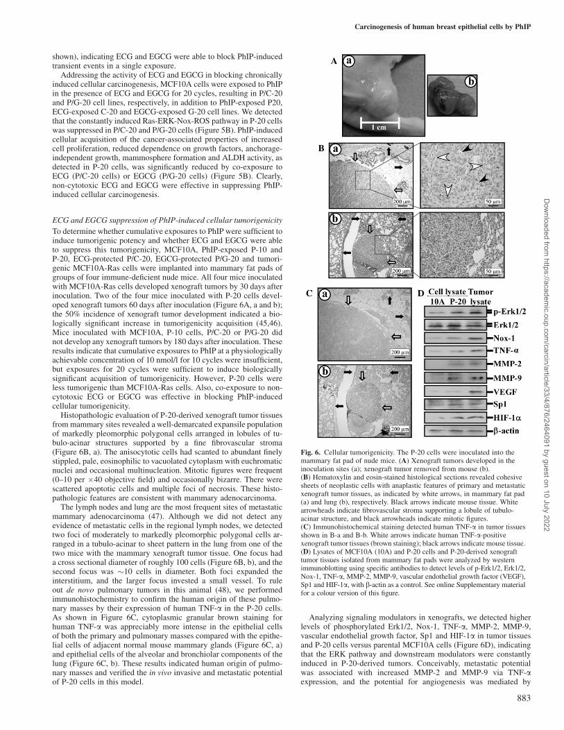
To determine whether cumulative exposures to PhIP were sufficient toinduce tumorigenic potency and whether ECG and EGCG were ableto suppress this tumorigenicity, MCF10A, PhIP-exposed P-10 andP-20, ECG-protected P/C-20, EGCG-protected P/G-20 and tumori-genic MCF10A-Ras cells were implanted into mammary fat pads ofgroups of four immune-deficient nude mice. All four mice inoculatedwith MCF10A-Ras cells developed xenograft tumors by 30 days afterinoculation. Two of the four mice inoculated with P-20 cells devel-oped xenograft tumors 60 days after inoculation (Figure 6A, a and b);the 50% incidence of xenograft tumor development indicated a bio-logically significant increase in tumorigenicity acquisition (45,46).Mice inoculated with MCF10A, P-10 cells, P/C-20 or P/G-20 didnot develop any xenograft tumors by 180 days after inoculation. Theseresults indicate that cumulative exposures to PhIP at a physiologicallyachievable concentration of 10 nmol/l for 10 cycles were insufficient,but exposures for 20 cycles were sufficient to induce biologicallysignificant acquisition of tumorigenicity. However, P-20 cells wereless tumorigenic than MCF10A-Ras cells. Also, co-exposure to non-cytotoxic ECG or EGCG was effective in blocking PhIP-inducedcellular tumorigenicity.
Histopathologic evaluation of P-20-derived xenograft tumor tissuesfrom mammary sites revealed a well-demarcated expansile populationof markedly pleomorphic polygonal cells arranged in lobules of tu-bulo-acinar structures supported by a fine fibrovascular stroma(Figure 6B, a). The anisocytotic cells had scanted to abundant finelystippled, pale, eosinophilic to vacuolated cytoplasm with euchromaticnuclei and occasional multinucleation. Mitotic figures were frequent(0–10 per �40 objective field) and occasionally bizarre. There werescattered apoptotic cells and multiple foci of necrosis. These histo-pathologic features are consistent with mammary adenocarcinoma.
The lymph nodes and lung are the most frequent sites of metastaticmammary adenocarcinoma (47). Although we did not detect anyevidence of metastatic cells in the regional lymph nodes, we detectedtwo foci of moderately to markedly pleomorphic polygonal cells ar-ranged in a tubulo-acinar to sheet pattern in the lung from one of thetwo mice with the mammary xenograft tumor tissue. One focus hada cross sectional diameter of roughly 100 cells (Figure 6B, b), and thesecond focus was �10 cells in diameter. Both foci expanded theinterstitium, and the larger focus invested a small vessel. To ruleout de novo pulmonary tumors in this animal (48), we performedimmunohistochemistry to confirm the human origin of these pulmo-nary masses by their expression of human TNF-a in the P-20 cells.As shown in Figure 6C, cytoplasmic granular brown staining forhuman TNF-a was appreciably more intense in the epithelial cellsof both the primary and pulmonary masses compared with the epithe-lial cells of adjacent normal mouse mammary glands (Figure 6C, a)and epithelial cells of the alveolar and bronchiolar components of thelung (Figure 6C, b). These results indicated human origin of pulmo-nary masses and verified the in vivo invasive and metastatic potentialof P-20 cells in this model.
Analyzing signaling modulators in xenografts, we detected higherlevels of phosphorylated Erk1/2, Nox-1, TNF-a, MMP-2, MMP-9,vascular endothelial growth factor, Sp1 and HIF-1a in tumor tissuesand P-20 cells versus parental MCF10A cells (Figure 6D), indicatingthat the ERK pathway and downstream modulators were constantlyinduced in P-20-derived tumors. Conceivably, metastatic potentialwas associated with increased MMP-2 and MMP-9 via TNF-aexpression, and the potential for angiogenesis was mediated by
Fig. 6. Cellular tumorigenicity. The P-20 cells were inoculated into themammary fat pad of nude mice. (A) Xenograft tumors developed in theinoculation sites (a); xenograft tumor removed from mouse (b).(B) Hematoxylin and eosin-stained histological sections revealed cohesivesheets of neoplastic cells with anaplastic features of primary and metastaticxenograft tumor tissues, as indicated by white arrows, in mammary fat pad(a) and lung (b), respectively. Black arrows indicate mouse tissue. Whitearrowheads indicate fibrovascular stroma supporting a lobule of tubulo-acinar structure, and black arrowheads indicate mitotic figures.(C) Immunohistochemical staining detected human TNF-a in tumor tissuesshown in B-a and B-b. White arrows indicate human TNF-a-positivexenograft tumor tissues (brown staining); black arrows indicate mouse tissue.(D) Lysates of MCF10A (10A) and P-20 cells and P-20-derived xenografttumor tissues isolated from mammary fat pads were analyzed by westernimmunoblotting using specific antibodies to detect levels of p-Erk1/2, Erk1/2,Nox-1, TNF-a, MMP-2, MMP-9, vascular endothelial growth factor (VEGF),Sp1 and HIF-1a, with b-actin as a control. See online Supplementary materialfor a colour version of this figure.
Carcinogenesis of human breast epithelial cells by PhIP
883
Dow
nloaded from https://academ
ic.oup.com/carcin/article/33/4/876/2464091 by guest on 10 July 2022
vascular endothelial growth factor expression possibly via inductionof transcription factors HIF-1a and Sp1.
Discussion
Our model system revealed, for the first time, that cumulative expo-sures of human breast cells to PhIP at physiologically achievable picoto low nanomolar concentrations induced progressive carcinogenesisfrom a non-cancerous stage to premalignant and malignant stages ina dose- and exposure-dependent manner. Long-term exposure to lowdoses of the dietary carcinogen PhIP, through the consumption ofwell-done meats, may contribute to sporadic breast cancer develop-ment. Using various transient and constitutive endpoints as targets, weidentified ECG and EGCG, at non-cytotoxic concentrations, effectivein suppressing PhIP-induced cellular carcinogenesis and tumorigenic-ity. Our model system provides a new platform to address targetedintervention of chronically induced breast cell carcinogenesis andsuppression by co-exposure to non-cytotoxic preventive agents.
Our model system determines the initiation and progress of cellularcarcinogenesis using measurable, transient and constitutive biological,biochemical and molecular changes as targeted endpoints. Transienttargeted endpoints are used for measuring the activity of PhIP in initi-ation of cellular carcinogenesis. Constitutive targeted endpoints areused for measuring the progress of cellular carcinogenesis chronicallyinduced by long-term exposure to PhIP. Using constitutive targetedendpoints, our model revealed that the malignant MCF10A-10nM-P20 cells acquired similar or modestly lower degrees of these endpointscompared with the tumorigenic MCF10A-Ras cells; and the premalig-nant MCF10A-10nM-P10 cells acquired significantly lower degrees ofthese endpoints than MCF10A-Ras cells. Hence, our model system,using in vitro constitutive biological, biochemical and moleculartargeted endpoints, may serve as a platform to predict the in vivotumorigenic potential of carcinogen-exposed cells. For example, PhIP-exposed P-20 cells acquired the targeted endpoints of reduced depen-dence on growth factors and anchorage-independent growth to degreescomparable with those acquired by MCF10A-Ras cells, indicating thatthese targeted endpoints may be used to predict cellular acquisition oftumorigenicity. In addition, P-20 cells acquired lower degrees of thetargeted endpoints of acinar-conformational disruption, increased cellmigration and invasion and increased ability of serum-independent andnon-adherent growth than MCF10A-Ras cells, indicating that thesetargeted endpoints may be used to predict distinguishable degrees oftumorigenic and metastatic potential acquired by carcinogen-exposedcells compared with MCF10A-Ras cells. Thus, by further determiningthe value of individual and grouped, in vitro targeted endpoints, ourmodel will provide an accurate cost-efficient platform to initially predictthe tumorigenic and metastatic potential of carcinogen-exposed cellsand to subsequently verify this potential in in vivo animal studies.
PhIP is able to induce a high frequency of activation–mutation in theH-ras gene in animal mammary tumors (12). The contributing role ofmutationally activated Ras has been widely recognized in human can-cers (17). However, although aberrant upregulation of wt H-ras geneexpression has been detected in .50% of premalignant and malignantbreast lesions isolated from human patients (49,50), the role of upregu-lated wt H-Ras in the development of premalignant and malignantbreast lesions was unclear. Apparently, upregulation of the wt H-rasgene was overlooked in past studies of carcinogenesis. Our modelrevealed, for the first time, an important role of transiently and consti-tutively upregulated wt H-Ras in regulating downstream signaling mod-ulators for PhIP-induced initiation and maintenance of breast cellcarcinogenesis. However, the mechanisms for PhIP-induced transientand constant wt H-Ras upregulation in breast cells remain to be studied.
Using transient targeted endpoints in initial screening of preventiveagents and constitutive targeted endpoints in subsequent validation ofeffective preventive agents, our model system may provide a sensitiveplatform to help accelerate the identification of non-cytotoxicpreventive agents effective in targeted intervention of carcinogenesisas well as tumorigenesis associated with chronic exposure to environ-mental carcinogens. Thereby, our model system may be a tool that
will contribute to future studies for reducing the health risk ofsporadic breast cancer.
Funding
This work was supported by the University of Tennessee, College ofVeterinary Medicine, Center of Excellence in Livestock Diseases andHuman Health (to H.-C.R.W.) and the National Institutes of Health(CA129772 to H.-C.R.W.).
Acknowledgements
We are grateful to Ms. D.J. Trent for technique support in flow cytometricanalysis and Ms. M. Bailey for textual editing of the manuscript.
Conflict of Interest Statement: None declared.
References
1.Gray,J. et al. (2009) State of the evidence: the connection between breastcancer and the environment. Int. J. Occup. Environ. Health, 15, 43–78.
2.DeBruin,L.S. et al. (2002) Perspectives on the chemical etiology of breastcancer. Environ. Health Perspect., 110, 119–128.
3.Kelloff,G.J. et al. (eds.) (2005) Cancer Chemoprevention: Strategies forCancer Chemoprevention. Vol. 2. Human Press, Totowa, NJ.
4.Mehta,R.G. (2000) Experimental basis for the prevention of breast cancer.Eur. J. Cancer, 36, 1275–1282.
5.Rudel,R.A. et al. (2007) Chemicals causing mammary gland tumors inanimals signal new directions for epidemiology, chemicals testing, and riskassessment for breast cancer prevention. Cancer, 109, 2635–2666.
6.Pala,V. et al. (2009) Meat, eggs, dairy products, and risk of breast cancer inthe European Prospective Investigation into Cancer and Nutrition (EPIC)cohort. Am. J. Clin. Nutr., 90, 602–612.
7.Zheng,W. et al. (2009) Well-done meat intake, heterocyclic amine expo-sure, and cancer risk. Nutr. Cancer, 61, 437–446.
8.Sugimura,T. et al. (2004) Heterocyclic amines: mutagens/carcinogensproduced during cooking of meat and fish. Cancer Sci., 95, 290–299.
9.Zheng,W. et al. (1998) Well-done meat intake and the risk of breast cancer.J. Natl Cancer Inst., 90, 1724–1729.
10.Sinha,R. et al. (2000) 2-Amino-1-methyl-6-phenylimidazo[4,5-b]pyridine,a carcinogen in high-temperature-cooked meat, and breast cancer risk.J. Natl Cancer Inst., 92, 1352–1354.
11.Zhu,J. et al. (2003) Detection of 2-amino-1-methyl-6-phenylimidazo[4,5-b]-pyridine-DNA adducts in normal breast tissues and risk of breast cancer.Cancer Epidemiol. Biomarkers Prev., 12, 830–837.
12.Yu,M. et al. (2002) H-ras oncogene mutations during development of2-amino-1-methyl-6-phenylimidazo[4,5-b]pyridine (PhIP)-induced ratmammary gland cancer. Carcinogenesis, 23, 2123–2128.
13.Moon,A. et al. (2000) H-ras, but not N-ras, induces an invasive phenotypein human breast epithelial cells: a role for MMP-2 in the H-ras-inducedinvasive phenotype. Int. J. Cancer, 85, 176–181.
14.Choudhary,S. et al. (2010) FK228 and oncogenic H-Ras synergisticallyinduce Mek1/2 and Nox-1 to generate reactive oxygen species for differ-ential cell death. Anticancer Drugs, 21, 831–840.
15.Bedard,K. et al. (2007) The NOX family of ROS-generating NADPH ox-idases: physiology and pathophysiology. Physiol. Rev., 87, 245–313.
16.Creton,S.K. et al. (2007) The cooked meat carcinogen 2-amino-1-methyl-6-phenylimidazo[4,5-b]pyridine activates the extracellular signal regulatedkinase mitogen-activated protein kinase pathway. Cancer Res., 67, 11455–11462.
17.Campbell,S.L. et al. (1998) Increasing complexity of Ras signaling. Onco-gene, 17, 1395–1413.
18.Mei,J. et al. (2003) Transformation of noncancerous human breast epithe-lial cell MCF10A induced by the tobacco-specific carcinogen NNK. BreastCancer Res. Treat, 79, 95–105.
19.Siriwardhana,N. et al. (2008) Precancerous carcinogenesis of human breastepithelial cells by chronic exposure to benzo[a]pyrene. Mol. Carcinog., 47,338–348.
20.Siriwardhana,N. et al. (2008) Precancerous model of human breast epithe-lial cells induced by the tobacco-specific carcinogen NNK for prevention.Breast Cancer Res. Treat., 109, 427–441.
21.Song,X. et al. (2010) Grape seed proanthocyanidin suppression ofbreast cell carcinogenesis induced by chronic exposure to combined
S.Choudhary et al.
884
Dow
nloaded from https://academ
ic.oup.com/carcin/article/33/4/876/2464091 by guest on 10 July 2022
4-(methylnitrosamino)-1-(3-pyridyl)-1-butanone and benzo[a]pyrene. Mol.Carcinog., 49, 450–463.
22.Rathore,K. et al. (2011) Green tea catechin intervention of reactive oxygenspecies-mediated ERK pathway activation and chronically-induced breastcell carcinogenesis. Carcinogenesis,, 33, 174–183.
23.Yang,C.S. et al. (2009) Cancer prevention by tea: animal studies, molecularmechanisms and human relevance. Nat. Rev. Cancer, 9, 429–439.
24.Choudhary,S. et al. (2007) Proapoptotic ability of oncogenic H-Ras tofacilitate apoptosis induced by histone deacetylase inhibitors in humancancer cells. Mol. Cancer Ther., 6, 1099–1111.
25.Albini,A. et al. (1987) A rapid in vitro assay for quantitating the invasivepotential of tumor cells. Cancer Res., 47, 3239–3245.
26.Ginestier,C. et al. (2007) ALDH1 is a marker of normal and malignanthuman mammary stem cells and a predictor of poor clinical outcome.Cell Stem Cell, 1, 555–567.
27.Olive,P.L. et al. (2006) The comet assay: a method to measure DNA dam-age in individual cells. Nat. Protoc., 1, 23–29.
28.Hanahan,D. et al. (2000) The hallmarks of cancer. Cell, 100, 57–70.29.Larsson,O. et al. (1985) Consequences of parental exposure to serum-free
medium for progeny cell division. J. Cell Sci., 75, 259–268.30.Reddig,P.J. et al. (2005) Clinging to life: cell to matrix adhesion and cell
survival. Cancer Metastasis Rev., 24, 425–439.31.Datta,S. et al. (2007) Bmi-1 cooperates with H-Ras to transform human
mammary epithelial cells via dysregulation of multiple growth-regulatorypathways. Cancer Res., 67, 10286–10295.
32.Debnath,J. et al. (2005) Modelling glandular epithelial cancers in three-dimensional cultures. Nat. Rev. Cancer, 5, 675–688.
33.Preston-Martin,S. et al. (1993) Epidemiologic evidence for the increasedcell proliferation model of carcinogenesis. Prog. Clin. Biol. Res., 369,21–34.
34.Adachi,Y. et al. (2008) Oncogenic Ras upregulates NADPH oxidase 1 geneexpression through MEK–ERK-dependent phosphorylation of GATA-6.Oncogene, 27, 4921–4932.
35.Suh,Y.A. et al. (1999) Cell transformation by the superoxide-generatingoxidase Mox1. Nature, 401, 79–82.
36.Montesano,R. et al. (2005) Tumour necrosis factor alpha confers an in-vasive, transformed phenotype on mammary epithelial cells. J. Cell Sci.,118, 3487–3500.
37.Herbst,R.S. et al. (2000) Differential expression of E-Cadherin and type IVcollagenase genes predicts outcome in patients with stage I non-small celllung carcinoma. Clin. Cancer Res., 6, 790–797.
38.Lecureur,V. et al. (2005) ERK-dependent induction of TNFalpha expres-sion by the environmental contaminant benzo(a)pyrene in primary humanmacrophages. FEBS Lett., 579, 1904–1910.
39.Sarrio,D. et al. (2008) Epithelial-mesenchymal transition in breast cancerrelates to the basal-like phenotype. Cancer Res., 68, 989–997.
40.Gupta,P.B. et al. (2009) Cancer stem cells: mirage or reality? Nat. Med., 15,1010–1012.
41.Charafe-Jauffret,E. et al. (2008) Cancer stem cells in breast: current opin-ion and future challenges. Pathobiology, 75, 75–84.
42.Liu,Y. et al. (2001) p53 protein at the hub of cellular DNA damage responsepathways through sequence-specific and non-sequence-specific DNA bind-ing. Carcinogenesis, 22, 851–860.
43.Rogakou,E.P. et al. (1998) DNA double-stranded breaks induce histoneH2AX phosphorylation on serine. J. Biol. Chem., 139, 5858–5868.
44.Cooke,M.S. et al. (2003) Oxidative DNA damage: mechanisms, mutation,and disease. FASEB J., 17, 1195–1214.
45.Gart,J.J. et al. (1979) Statistical issues in interpretation of chronic bioassaytests for carcinogenicity. J. Natl Cancer Inst., 62, 957–974.
46.Cohrssen,J.J. et al. (1989) Risk Analysis: A Guide to Principles and Meth-ods for Analyzing Health and Environmental Risks. Diane Publishing Co,Darby, PA.
47.Price,J.E. et al. (1990) Tumorigenicity and metastasis of human breastcarcinoma cell lines in nude mice. Cancer Res., 50, 717–721.
48.Sharkey,F.E. et al. (1979) Incidence and pathological features of spontane-ous tumors in athymic nude mice. Cancer Res., 39, 833–839.
49.Hand,P.H. et al. (1987) Quantitation of Harvey ras p21 enhanced ex-pression in human breast and colon carcinomas. J. Natl Cancer Inst., 79,59–65.
50.Pethe,V. et al. (1999) Estrogen inducibility of c-Ha-ras transcription inbreast cancer cells. Identification of functional estrogen-responsive tran-scriptional regulatory elements in exon 1/intron 1 of the c-Ha-ras gene.J. Biol. Chem., 274, 30969–30978.
Received November 2, 2011; revised January 27, 2012;accepted February 1, 2012
Carcinogenesis of human breast epithelial cells by PhIP
885
Dow
nloaded from https://academ
ic.oup.com/carcin/article/33/4/876/2464091 by guest on 10 July 2022
Related Documents