Discovery of Small Molecule Inhibitors of the Interaction of the Thyroid Hormone Receptor with Transcriptional Coregulators * Received for publication, June 20, 2005, and in revised form, October 18, 2005 Published, JBC Papers in Press, October 31, 2005, DOI 10.1074/jbc.M506693200 Leggy A. Arnold ‡ , Eva Este ´ banez-Perpin ˜a ´ §1 , Marie Togashi ¶1 , Natalia Jouravel § , Anang Shelat ‡ , Andrea C. McReynolds ‡ , Ellena Mar § , Phuong Nguyen ¶ , John D. Baxter ¶ , Robert J. Fletterick § , Paul Webb ¶ , and R. Kiplin Guy ‡2 From the ‡ Department of Pharmaceutical Chemistry, Department of Molecular and Cellular Pharmacology, § Department of Biochemistry and Biophysics, ¶ Diabetes Center and Department of Medicine, University of California, San Francisco, California 94143 Thyroid hormone (3,5,3-triiodo-L-thyronine, T3) is an endo- crine hormone that exerts homeostatic regulation of basal meta- bolic rate, heart rate and contractility, fat deposition, and other phe- nomena (1, 2). T3 binds to the thyroid hormone receptors (TRs) and controls their regulation of transcription of target genes. The bind- ing of TRs to thyroid hormone induces a conformational change in TRs that regulates the composition of the transcriptional regulatory complex. Recruitment of the correct coregulators (CoR) is impor- tant for successful gene regulation. In principle, inhibition of the TR-CoR interaction can have a direct influence on gene transcrip- tion in the presence of thyroid hormones. Herein we report a high throughput screen for small molecules capable of inhibiting TR coactivator interactions. One class of inhibitors identified in this screen was aromatic -aminoketones, which exhibited IC 50 values of 2 M. These compounds can undergo a deamination, generat- ing unsaturated ketones capable of reacting with nucleophilic amino acids. Several experiments confirm the hypothesis that these inhibitors are covalently bound to TR. Optimization of these com- pounds produced leads that inhibited the TR-CoR interaction in vitro with potency of 0.6 M and thyroid signaling in cellular sys- tems. These are the first small molecules irreversibly inhibiting the coactivator binding of a nuclear receptor and suppressing its tran- scriptional activity. Thyroid hormone receptors (TRs) 3 regulate development, growth, and metabolism (1, 2). The TRs are nuclear receptors (NR), part of a superfamily whose members function as hormone-activated transcrip- tion factors (3). The majority of thyroid hormone responses are induced by regulation of transcription by the thyroid hormone T3 (4). Two genes, THRA and THRB encode the two protein isoforms TR and TR, which yield four distinct subtypes by alternative splicing (5). Sev- eral functional domains of TRs have been identified: a ligand-indepen- dent transactivation domain (AF-1) on the amino terminus, a central DNA binding domain, a ligand binding domain (LBD), and a carboxyl- terminal ligand dependent activation function (AF-2) (6). TR binds spe- cific sequences of DNA in the 5-flanking regions of T3-responsive genes, known as thyroid response elements, most often as a heterodimer with the retinoid X receptor (7). Both unliganded and liganded TRs can bind thyroid response elements and regulate genes under their control. The unliganded TR complex can recruit a nuclear receptor corepressor (NCoR) or a silencing mediator of retinoic acid to silence basal tran- scription (8). In the presence of T3, TRs undergo a conformational change with the result that the composition of the coregulator complex can change with strong effects on transcriptional regulation. Several coactivator proteins have been identified (9). The best studied group of coactivators is the p160 or steroid receptor coactivator (SRC) proteins (7) including SRC1 (10), SRC2 (11, 12), and SRC3 (13). Another group of ligand-dependent-interacting proteins include the thyroid hormone receptor activating protein (TRAP) (14), peroxisome proliferate-acti- vated receptor- coactivator-1 (PGC-1) (15), and the thyroid hormone receptor binding protein (TRBP) (16). Additionally, quantitative in vitro binding assays (17) have shown strong interactions between TR and the coregulators p300 (18), androgen receptor activator (ARA70) (19), receptor interacting protein 140 (RIP140) (20), dosage-sensitive sex reversal-adrenal hypoplasia congenital critical region of the X chromo- some gene (DAX1) (21), and the small heterodimer partner (SHP) (22). The coregulators mentioned have in common that they have variable numbers of highly conserved LXXLL motifs; termed NR-boxes, in their nuclear receptor interacting domain (NID). The NR boxes are both necessary and sufficient for the interaction between CoR and TR. The coactivator binding site of TR LBD is formed by 16 residues from four helices (H3, H4, H5, and H12) (23). Scanning surface mutagenesis revealed that only six residues (Val 284 , Lys 288 , Ile 302 , Lys 306 , Leu 454 , and Glu 457 ) are crucial for coactivator binding (24). This feature makes the AF-2 domain an ideal target for inhibitor development. Several inhibitors of this interaction have been reported. The first reported inhibitors were macrolactam-constrained SRC2 NR box pep- tides (25). A combinatorial approach discovered novel -helical proteo- mimetics that could selectively inhibit the interaction between coacti- vators and TR or the estrogen receptor (ER), with selectivity between ER isoforms ER and ER (26). A similar approach, using disulfide bridges to constrain peptides, resulted in selective ER coactivator inhibitor with a K d of 25 nM (27, 28). A report identifying a small molecule capable of inhibiting the interaction of a NR and its coactivator was published recently (29). These pyrimidine-based scaffolds showed affinities between 30 and 50 M but did not inhibit NR signaling in cell culture or * This work was supported by the Howard Hughes Medical Institute Research Resources Program Grant 76296-549901, Grants DK58080 and DK61648 from the National Insti- tutes of Health, the Sandler Research Foundation, and a UCSF Prostate Cancer SPORE Research Fellowship. The costs of publication of this article were defrayed in part by the payment of page charges. This article must therefore be hereby marked “adver- tisement” in accordance with 18 U.S.C. Section 1734 solely to indicate this fact. 1 Both authors contributed equally to this work. 2 To whom correspondence should be addressed: Dept. of Chemical Biology in Thera- peutics, St. Jude Children’s Research Hospital, Memphis, TN 38105. Tel.: 901-495- 5714; Fax: 901-495-5715; E-mail: [email protected]. 3 The abbreviations used are: TR, thyroid hormone receptor; NR, nuclear receptors; T3, 3,5,3-triiodo-L-thyronine, thyroid hormone; CoA, coactivator; CoR, coregulator; LBD, ligand binding domain; SRC, steroid receptor coactivator; NID, nuclear receptor inter- acting domain; ER, estrogen receptor; HTS, high throughput screening. THE JOURNAL OF BIOLOGICAL CHEMISTRY VOL. 280, NO. 52, pp. 43048 –43055, December 30, 2005 © 2005 by The American Society for Biochemistry and Molecular Biology, Inc. Printed in the U.S.A. 43048 JOURNAL OF BIOLOGICAL CHEMISTRY VOLUME 280 • NUMBER 52 • DECEMBER 30, 2005 at Biblioteca de la Universitat de Barcelona on February 5, 2008 www.jbc.org Downloaded from

Welcome message from author
This document is posted to help you gain knowledge. Please leave a comment to let me know what you think about it! Share it to your friends and learn new things together.
Transcript
Discovery of Small Molecule Inhibitors of the Interaction ofthe Thyroid Hormone Receptor with TranscriptionalCoregulators*
Received for publication, June 20, 2005, and in revised form, October 18, 2005 Published, JBC Papers in Press, October 31, 2005, DOI 10.1074/jbc.M506693200
Leggy A. Arnold‡, Eva Estebanez-Perpina§1, Marie Togashi¶1, Natalia Jouravel§, Anang Shelat‡,Andrea C. McReynolds‡, Ellena Mar§, Phuong Nguyen¶, John D. Baxter¶, Robert J. Fletterick§,Paul Webb¶, and R. Kiplin Guy‡!2
From the ‡Department of Pharmaceutical Chemistry, !Department of Molecular and Cellular Pharmacology, §Department ofBiochemistry and Biophysics, ¶Diabetes Center and Department of Medicine, University of California,San Francisco, California 94143
Thyroid hormone (3,5,3!-triiodo-L-thyronine, T3) is an endo-crine hormone that exerts homeostatic regulation of basal meta-bolic rate, heart rate and contractility, fat deposition, andother phe-nomena (1, 2). T3 binds to the thyroid hormone receptors (TRs) andcontrols their regulation of transcription of target genes. The bind-ing of TRs to thyroid hormone induces a conformational change inTRs that regulates the composition of the transcriptional regulatorycomplex. Recruitment of the correct coregulators (CoR) is impor-tant for successful gene regulation. In principle, inhibition of theTR-CoR interaction can have a direct influence on gene transcrip-tion in the presence of thyroid hormones. Herein we report a highthroughput screen for small molecules capable of inhibiting TRcoactivator interactions. One class of inhibitors identified in thisscreen was aromatic !-aminoketones, which exhibited IC50 valuesof !2 "M. These compounds can undergo a deamination, generat-ing unsaturated ketones capable of reacting with nucleophilicamino acids. Several experiments confirm the hypothesis that theseinhibitors are covalently bound to TR. Optimization of these com-pounds produced leads that inhibited the TR-CoR interaction invitro with potency of !0.6 "M and thyroid signaling in cellular sys-tems. These are the first small molecules irreversibly inhibiting thecoactivator binding of a nuclear receptor and suppressing its tran-scriptional activity.
Thyroid hormone receptors (TRs)3 regulate development, growth,and metabolism (1, 2). The TRs are nuclear receptors (NR), part of asuperfamily whose members function as hormone-activated transcrip-tion factors (3). Themajority of thyroid hormone responses are inducedby regulation of transcription by the thyroid hormone T3 (4). Twogenes, THRA and THRB encode the two protein isoforms TR! andTR", which yield four distinct subtypes by alternative splicing (5). Sev-eral functional domains of TRs have been identified: a ligand-indepen-
dent transactivation domain (AF-1) on the amino terminus, a centralDNA binding domain, a ligand binding domain (LBD), and a carboxyl-terminal ligand dependent activation function (AF-2) (6). TR binds spe-cific sequences of DNA in the 5"-flanking regions of T3-responsivegenes, known as thyroid response elements,most often as a heterodimerwith the retinoid X receptor (7). Both unliganded and liganded TRs canbind thyroid response elements and regulate genes under their control.The unliganded TR complex can recruit a nuclear receptor corepressor(NCoR) or a silencing mediator of retinoic acid to silence basal tran-scription (8). In the presence of T3, TRs undergo a conformationalchange with the result that the composition of the coregulator complexcan change with strong effects on transcriptional regulation. Severalcoactivator proteins have been identified (9). The best studied group ofcoactivators is the p160 or steroid receptor coactivator (SRC) proteins(7) including SRC1 (10), SRC2 (11, 12), and SRC3 (13). Another group ofligand-dependent-interacting proteins include the thyroid hormonereceptor activating protein (TRAP) (14), peroxisome proliferate-acti-vated receptor-# coactivator-1 (PGC-1) (15), and the thyroid hormonereceptor binding protein (TRBP) (16). Additionally, quantitative in vitrobinding assays (17) have shown strong interactions between TR and thecoregulators p300 (18), androgen receptor activator (ARA70) (19),receptor interacting protein 140 (RIP140) (20), dosage-sensitive sexreversal-adrenal hypoplasia congenital critical region of the X chromo-some gene (DAX1) (21), and the small heterodimer partner (SHP) (22).The coregulators mentioned have in common that they have variable
numbers of highly conserved LXXLLmotifs; termed NR-boxes, in theirnuclear receptor interacting domain (NID). The NR boxes are bothnecessary and sufficient for the interaction between CoR and TR. Thecoactivator binding site of TR LBD is formed by 16 residues from fourhelices (H3, H4, H5, and H12) (23). Scanning surface mutagenesisrevealed that only six residues (Val284, Lys288, Ile302, Lys306, Leu454, andGlu457) are crucial for coactivator binding (24). This feature makes theAF-2 domain an ideal target for inhibitor development.Several inhibitors of this interaction have been reported. The first
reported inhibitors were macrolactam-constrained SRC2 NR box pep-tides (25). A combinatorial approach discovered novel!-helical proteo-mimetics that could selectively inhibit the interaction between coacti-vators andTRor the estrogen receptor (ER), with selectivity between ERisoforms ER! and ER" (26). A similar approach, using disulfide bridgesto constrain peptides, resulted in selective ER! coactivator inhibitorwith aKd of 25 nM (27, 28). A report identifying a smallmolecule capableof inhibiting the interaction of a NR and its coactivator was publishedrecently (29). These pyrimidine-based scaffolds showed affinitiesbetween 30 and 50 $M but did not inhibit NR signaling in cell culture or
* This work was supported by the Howard Hughes Medical Institute Research ResourcesProgram Grant 76296-549901, Grants DK58080 and DK61648 from the National Insti-tutes of Health, the Sandler Research Foundation, and a UCSF Prostate Cancer SPOREResearch Fellowship. The costs of publication of this article were defrayed in part bythe payment of page charges. This article must therefore be hereby marked “adver-tisement” in accordance with 18 U.S.C. Section 1734 solely to indicate this fact.
1 Both authors contributed equally to this work.2 To whom correspondence should be addressed: Dept. of Chemical Biology in Thera-
peutics, St. Jude Children’s Research Hospital, Memphis, TN 38105. Tel.: 901-495-5714; Fax: 901-495-5715; E-mail: [email protected].
3 The abbreviations used are: TR, thyroid hormone receptor; NR, nuclear receptors; T3,3,5,3"-triiodo-L-thyronine, thyroid hormone; CoA, coactivator; CoR, coregulator; LBD,ligand binding domain; SRC, steroid receptor coactivator; NID, nuclear receptor inter-acting domain; ER, estrogen receptor; HTS, high throughput screening.
THE JOURNAL OF BIOLOGICAL CHEMISTRY VOL. 280, NO. 52, pp. 43048 –43055, December 30, 2005© 2005 by The American Society for Biochemistry and Molecular Biology, Inc. Printed in the U.S.A.
43048 JOURNAL OF BIOLOGICAL CHEMISTRY VOLUME 280 • NUMBER 52 • DECEMBER 30, 2005
at Biblioteca de la Universitat de Barcelona on February 5, 2008 www.jbc.org
Downloaded from
in vivomodels. To date, none of these inhibitorsmay be used to regulateNR signaling in cellular systems.All functional TRmodulators known today are analogs of theT3 itself
(30–33). These small molecule derivatives show selectivity toward dif-ferent isoforms of TR resulting in tissue specific activities (34). GC-1, aTR" selective agonist shows interesting properties in vivo and could becrystallized with TR LBD (35–40). The first functional T3 antagonistwas NH-3, which inhibits thyroid hormone function in both cell cultureand whole animal-based assays (41).High throughput screening (HTS) together with computational
screening and fragment discovery are current methods for discoveringlead compounds for manipulation of protein function. Although suchmethods have been applied to discovery of small molecule inhibitors ofprotein-protein interactions (42), only a limited number of successeshave been reported (43, 44). One of the most robust and sensitive HTSmethods for studying protein-protein interactions is the competitivefluorescence polarization assay (45). Herein, we present the first HTSusing an in vitro fluorescence polarization assay tomeasure the ability ofsmall molecules to inhibit the interaction between the TR" LBD and itscoactivator, SRC2. This screen revealed a number of hits for inhibitorsof the TR-CoR interaction. One particular class of compounds has beenexamined carefully, and its mechanism of inhibition has been investi-gated. The resulting lead compounds are potent and selective inhibitorsof both the TR-CoR interaction in vitro and thyroid hormone signalingin cellular systems. They have potential both as drug candidates anduseful biochemical tools for study of the role of the interaction ofTR andits coregulators.
EXPERIMENTAL PROCEDURES
Labeled Peptides—Peptide SRC2-2 (CLKEKHKILHRLLQDSSSPV)labeledwith 5-iodoacetamidofluorescein (Molecular Probes)was kindlyprovided by JamieM. R.Moore (probe) (17);!-helical proteomimetics 3(positive control) and 11 (negative control) were kindly provided byTimothy R. Geistlinger (26).
Vector—hTR" LBD (His6 T209-D461) was cloned into the BamHIand HindIII restriction sites downstream of the hexahistidine tag of theexpression vector pET DUET-1 (Novagen). The replacement of C309for A in the hTR" LBD construct was performed with the QuikChangeXL site-directed mutagenesis kit (Stratagene). The sequence of bothconstructs was verified by DNA sequencing (Elim Biopharmaceuticals,Inc., Hayward, CA).
Protein Expression and Purification—hTR" LBD (His6; residuesT209-D461) was expressed in BL21(DE3) (Invitrogen) (10# 1L culture)at 20 °C, 0.5 mM isopropyl-1-thio-b-D-galactopyranoside added at A600 $0.6 (17). When the A600 reached 4, cells were harvested, resuspended in20 ml of buffer/1 liter of culture (20 mM Tris, 300 mM NaCl, 0.025%Tween 20, 0.10 mM phenylmethylsulfonyl fluoride, 10 mg of lysozyme,pH 7.5), incubated for 30 min on ice, and then sonicated for 3 # 3 minon ice. The lysed cells were centrifuged at 100,000 # g for 1 h, and thesupernatant was loaded onto Talon resin (20ml, Clontech). Protein waseluted with 500 mM imidazole (3 # 5 ml) plus ligand (3,3",5-triiodo-L-thyronine (Sigma)). Protein purity (%90%) was assessed by SDS-PAGEand high pressure size exclusion chromatography, and protein concen-tration was measured by the Bradford protein assay. The protein wasdialyzed overnight against assay buffer (3 # 4 liters, 50 mM sodiumphosphate, 150 mM NaCl, pH 7.2, 1 mM dithiothreitol, 1 mM EDTA,0.01%Nonidet P-40, 10% glycerol). The protein functionality was deter-mined by a direct binding assay of SRC2-2 (see Fig. 3A) giving a Kd forSRC2-2 of 0.44 $M, agreeing with prior results. hTR! LBD (His6; resi-dues Glu148-Val410) was expressed using the same procedure as hTR"
with the exception that 0.5mM isopropyl-1-thio-"-D-galactopyranosidewas added at A600 $ 1.2. Unliganded protein was eluted with 100 mMimidazole. Purity assessment and buffer exchange were carried out asdescribed. The functionality was determined in a direct binding assay(see Fig. 3A) giving a Kd for SRC2-2 of 0.17 $M, agreeing with priorresults. hTR" LBDC309A (His6; residues Thr209-Asp461) was expressedin BL21 cells (Stratagene) at 18–20 °C by using the pET DUET1-hTR"LBD (41). General procedures were as described above. The function-alitywas determined in a direct binding assay (see Fig. 3A) giving aKd forSRC2-2 of 0.17 $M.
Direct Binding Assay—The protein was serially diluted from 70 to0.002 $M in binding buffer (50 mM sodium phosphate, 150 mM NaCl,pH 7.2, 1 mM dithiothreitol, 1 mM EDTA, 0.01% Nonidet P-40, 10%glycerol) containing 1$M ligand T3 in 96-well plates (17). Then 10$l ofdiluted proteinwas added to 10$l of labeled SRC2-2 (20 nM) in 384-wellplates yielding final protein concentrations of 35–0.001 $M and 10 nMfluorescent peptide concentration. The samples were allowed to equil-ibrate for 30 min. Binding was thenmeasured using fluorescence polar-ization (excitation% 485nm, emission% 530nm) on anAnalystADplatereader (Molecular Devices). Two independent experiments, each inquadruplicate, were carried out for each state. Data were analyzed usingSigmaPlot 8.0 (SPSS, Chicago, Il), and the Kd values were obtained byfitting data to the following equation (y $ min & (max ' min)/1 &(x/Kd)Hill slope).
Screening Procedure—The small molecule screen was carried out atthe Bay Area Screening Center (BASC) at the California Institute forQuantitative Biology (QB3). A library comprised of 138,000 compounds(ChemRX, 28,000; ChemDiv, 53,000; ChemBridge, 24,000; SPECS,31,000;Microsource, 2,000) was screened in 384-well format. The com-plete composition of this library is available from the BASC website(ucsf.edu/basc). First, 384-well dilutions plates (costar 3702) were pre-pared by addition of 34 $l of dilution buffer (20 mM Tris-HCl, 100 mMNaCl, pH 7.2, 1 mM dithiothreitol, 1 mM EDTA, 0.01% Nonidet P-40,10% glycerol, 5.9% Me2SO) to each well by using a WellMate (Matrix)followed by addition of 6 $l compound solutions (1 mM compound indimethyl sulfoxide (Me2SO)) using a Multimek (Beckman) equippedwith a 96-channel head and mixing by subsequent aspiration and dis-pensing. Second, 5 $l from the dilution plates were transferred to 384-well assay plates (Costar 3710) using a Multimek followed by the addi-tion of 24$l of proteinmixture (20mMTris-HCl, 100mMNaCl, pH 7.2,1 mM dithiothreitol, 1 mM EDTA, 0.01% Nonidet P-40, 10% glycerol, 1$MTR"LBD, 1$MT3, 0.025$M labeled SRC2-2 using aWellMate. Thefinal concentration of compound was 30 $M with 4% Me2SO content.Each plate was monitored by the addition of a positive control 3 andnegative control 11. After an incubation time of 2 h the binding wasmeasured using fluorescence polarization (excitation % 485 nm, emis-sion % 530 nm) on an Analyst AD plate reader (Molecular Devices).Additionally the fluorescence intensity was measured. All data relevantto the project (plate and compound information, screening data, anno-tation info, etc.) was deposited directly into amySQLdata base (v. 4.1.7).Dataweremanipulated and analyzed using protocols written in PipelinePilot 4.5.1 (Scitegic, Inc). Our protocols automated the process of join-ing experimental data to compound information, flagging suspiciousplates based on low Z-factors, extracting compounds with statisticallysignificant activity, and annotating hits with additional information (i.e.chemical similarity to known bioactive compounds, known genotoxic/cytotoxic molecules, or available compounds, and profiles from ADMEmodels).
Dose-response Experiments—The small molecules were seriallydiluted from 1000 to 4.88 $M in Me2SO into a 96-well plate (Costar
TR-CoR Antagonists
DECEMBER 30, 2005 • VOLUME 280 • NUMBER 52 JOURNAL OF BIOLOGICAL CHEMISTRY 43049
at Biblioteca de la Universitat de Barcelona on February 5, 2008 www.jbc.org
Downloaded from
3365). 10$l of each concentrationwas transferred into 100$l of bindingbuffer (20 mM Tris-HCl, 100 mM NaCl, pH 7.2, 1 mM dithiothreitol, 1mMEDTA, 0.01%Nonidet P-40, 10% glycerol) andmixed by subsequentaspiration and dispensing. Then 10 $l of diluted compound was addedto 10 $l of protein mixture (20 mM Tris-HCl, 100 mM NaCl, pH 7.2, 1mMdithiothreitol, 1mMEDTA, 0.01%Nonidet P-40, 10% glycerol, 2$MTR" LBD, 2 $M T3, 0.02 $M labeled SRC2-2 in 384-well plates yieldingfinal compound concentration of 50–0.024 $M. The samples wereallowed to equilibrate for 3 h. Binding was thenmeasured using fluores-cence polarization (excitation % 485 nm, emission % 530 nm) on anAnalyst AD (Molecular Devices). Two independent experiments, inquadruplicate, were carried out for each compound. Datawere analyzedusing SigmaPlot 8.0, and the Kd values were obtained by fitting data tothe following equation (y $ min & (max ' min)/1 & (x/Kd)Hill slope).
Thyroid Hormone Competition Binding Assay—Full-length hTR"was produced using a TNT T7 quick-coupled transcription translationsystem (Promega). Competition assays for binding of unlabeled T3 andL1 were performed using 1 nM [125I]T3 in gel filtration binding assay asdescribed (46).
Binding Assay with L8 and L9—TR" or TR" C309A (5 $M) and T3(20 $M) were incubated in binding buffer (100 $M) with different con-centrations L8 and L9, respectively. After 3 h at room temperature analiquot of 20$Mwas treatedwith a denaturing buffer (10$M), boiled for2min, and separated using 10% SDS-polyacrylamide gel electrophoresisand visualized by a fluorescence spectrometer.
Pull-down Assays—GST fusions to the thyroid hormone receptor(full-length) were expressed in Escherichia coli BL21. Cultures weregrown to A600 1.2–1.5 at 22 °C and induced with 0.5 mM isopropyl-D-thiogalactoside for 4 h. The cultures were centrifuged (1000 # g), andbacterial pellets were resuspended in 20 mMHepes, pH 7.9, 80 mM KCl,6 mM MgCl2, 1 mM DTT, 1 mM ATP, 0.2 mM phenylmethylsulfonylfluoride, and protease inhibitors and sonicated. Debris was pelleted bycentrifugation (100,000 # g). The supernatant was incubated with glu-tathione-Sepharose 4B beads (Amersham Biosciences) and washed aspreviously described. Protein preparationswere stored at'20 °C in 20%glycerol until use. [35S]methionine-labeled SRC2was produced by usingcoupled in vitro transcription-translation (TNT kit, Promega). Thebinding reactions were carried out on ice in a volume of 150 $l com-posed of 137.5 $l of protein-binding buffer along with 10 $l of GST-bead slurry corresponding to 3 $g of fusion protein, 1 $l of in vitrotranslated protein, and 1.5 $l of ligand or vehicle. The protein-bindingbuffer composed of 20 $l of A-150 (20 mMHepes, 150 mM KCl, 10 mM,MgCl2, 1% glycerol) and 2 $l each of phosphate-buffered saline supple-mented with 1% Triton X-100 and 1% Nonidet P-40. Phenylmethylsul-fonyl fluoride, dithiothreitol, bovine serum albumin, and proteaseinhibitor mixture (Novagen) was freshly prepared. The mix was incu-bated at 4 °C with gentle agitation; the beads were pelleted, washed fourtimes with protein-binding buffer containing no bovine serum albumin,and dried under vacuum for 20 min. The sample was taken up in SDS-PAGE loading buffer and then subjected to SDS-PAGE andautoradiography.
Transient Transfection Assays—Human bone osteosarcoma epithe-lial cells (U2OS) cells (Cell Culture Facility, UCSF) were grown to!80%confluency in Dulbecco’s modified Eagle’s/H-21, 4.5 g/liter glucosemedium containing 10% newborn calf serum (heat-inactivated), 2 mMglutamine, 50 units/ml penicillin, and 50 $g/ml streptomycin. Cells(!1.5 # 106) were collected and resuspended in 0.5 ml of electropora-tion buffer (Dulbecco’s phosphate-buffered saline containing 0.1% glu-cose, 10 mg/ml bioprene). 5 $g of a TR expression vector (full-lengthhTR"-CMV) and 1.5 $g of a reporter plasmid contained a synthetic TR
response element (DR-4) containing two copies of a direct repeat spacedby four nucleotides (AGGTCAcaggAGGTCA) cloned immediatelyupstream of aminimal ('32/&45) thymidine kinase promoter linked toluciferase coding sequence (35). Cells were electroporated using a Bio-Rad gene pulser at 350 V and 960 microfarads, pooled in growthmedium (DME H-21 with 10% charcoal-treated, hormone-stripped,newborn bovine serum), and plated in 96-well dishes. After a 3-h incu-bation compounds were added to the cell culture medium as Me2SOsolutions so as to yield a final Me2SO concentration of 1%. After addi-tional 18 h of incubation, cells were harvested and assayed for luciferaseactivity using the Promega dual luciferase kit (Promega) and an AnalystAD (Molecular Devices). Data were analyzed using SigmaPlot 8.0, andthe IC valueswere obtained by fitting data to the following equation (y$min & (max ' min)/1 & (x/Kd)Hill slope).
RESULTS
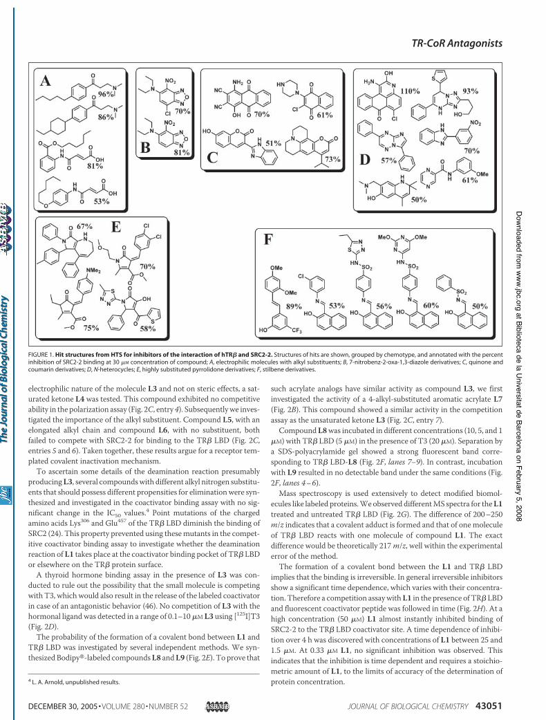
The high-throughput screen was carried out using a 384-well plateformat. A total of 300 compounds as single points together with quad-ruple positive and quadruple negative controls were dispensed in each384-well plate followed by the addition of TR" LBD and the labeledSRC2-2 peptide. The SRC2-2 peptide was utilized because it had thetightest binding (0.44 $M) of all the NR box peptides investigated (17).After incubation for 2 h the fluorescence polarization and fluorescenceintensity was measured. From the 138,000 compounds screened 27 hitcompounds inhibited the interaction between TR" LBD and theSRC2-2 coactivator peptide with at least 50% efficacy at a concentrationof 30$Mand had a fluorescence intensity variation of less then 10%. Thestructures of these hits, along with the percent inhibitions at 30 $M areshown in Fig. 1. Themolecules are divided into six groups depending ontheir chemical properties. Group A represents electrophilic moleculeswith a medium sized alkyl substituent. Based on our results at least twoof them are irreversible inhibitors of the TR-CoA interaction.All hits shown in Fig. 1 were evaluated by performing a dose to the
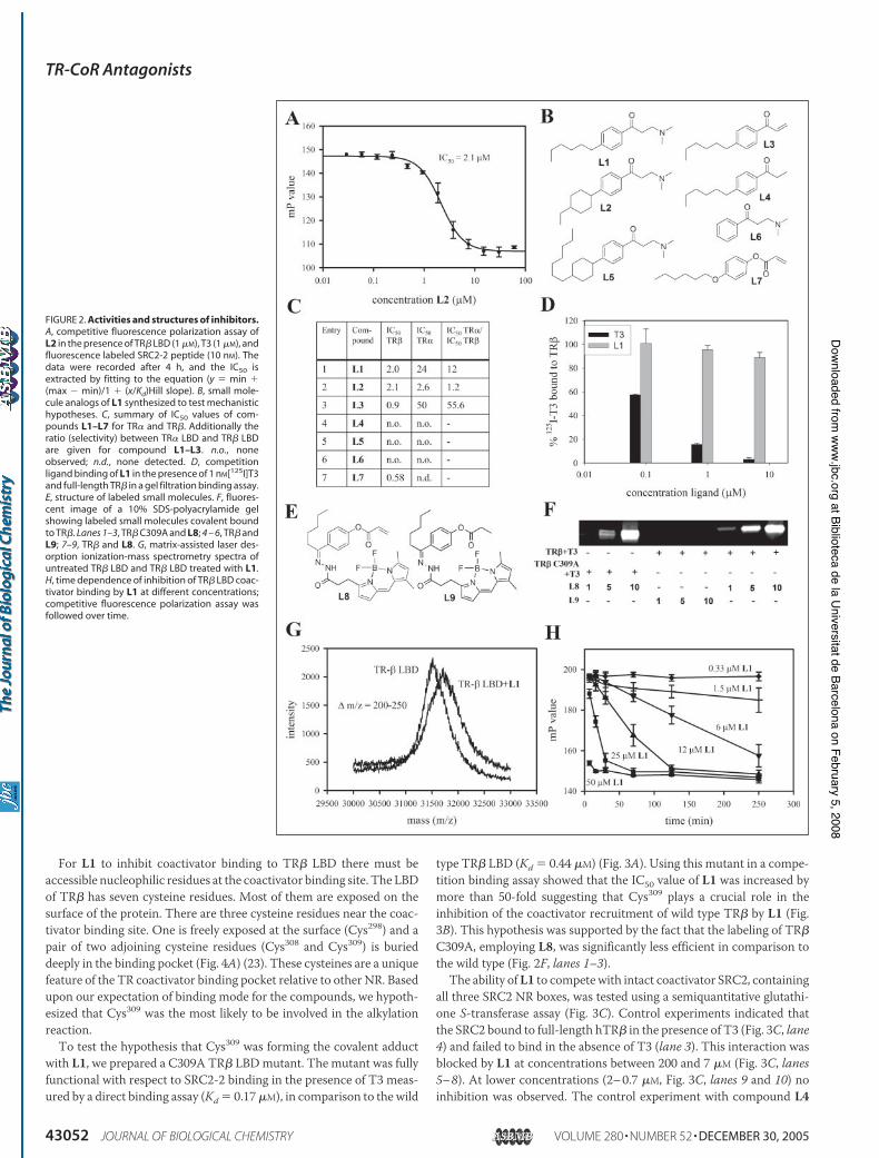
response of inhibition study over a range of compound concentrationsof 0.024–30 $M to allow the calculation of the IC50 values. Only twocompounds (Fig. 2B, L1 and L2) had IC50 values less than 10 $M (C,entries 1 and 2), with a clear saturation at a higher concentration (A).These were designated validated hits. The remaining compounds wereall sufficiently weak in potency to call their validity into question. Thisrepresents an overall hit rate of 0.00145%.Both of the validated hits are "-aminoketones. These compounds are
better known as Mannich bases, first synthesized in the 19th centuryand systematically studied by Carl Mannich in the beginning of lastcentury (47). Several biological activities have been discovered for thiscompound class including anticancer, antimicrobial, and cytotoxicactivities (48). These activities have been attributed to the liberation of!,"-unsaturated ketones by internal elimination of the amino group.Although this reaction proceeds very slowly under physiological pH inwater it has been reported that protein surfaces are able to catalyze thisreaction very efficiently (49). Such soft electrophiles, termed Michaeladdition acceptors, can alkylate protein nucleophiles such as cysteine,tyrosine, and serine. Because of the strong nucleophilicity of organicsulfides, cysteine residues are the most reactive toward this class ofMichael acceptors.To investigate the probability that a similar mechanism underlay
inhibition of coactivator binding to the TR" LBD we tested the unsat-urated ketone L3 (Fig. 2B). Interestingly, it showed a similar inhibitoryability of the coactivator recruitment suggesting that indeed the liber-ated unsaturated ketone L3 is the active species for compounds L1 andL2 (Fig. 2C, entry 3). To determine whether the binding is based on the
TR-CoR Antagonists
43050 JOURNAL OF BIOLOGICAL CHEMISTRY VOLUME 280 • NUMBER 52 • DECEMBER 30, 2005
at Biblioteca de la Universitat de Barcelona on February 5, 2008 www.jbc.org
Downloaded from
electrophilic nature of the molecule L3 and not on steric effects, a sat-urated ketone L4 was tested. This compound exhibited no competitiveability in the polarization assay (Fig. 2C, entry 4). Subsequentlywe inves-tigated the importance of the alkyl substituent. Compound L5, with anelongated alkyl chain and compound L6, with no substituent, bothfailed to compete with SRC2-2 for binding to the TR" LBD (Fig. 2C,entries 5 and 6). Taken together, these results argue for a receptor tem-plated covalent inactivation mechanism.To ascertain some details of the deamination reaction presumably
producingL3, several compoundswith different alkyl nitrogen substitu-ents that should possess different propensities for eliminationwere syn-thesized and investigated in the coactivator binding assay with no sig-nificant change in the IC50 values.4 Point mutations of the chargedamino acids Lys306 and Glu457 of the TR" LBD diminish the binding ofSRC2 (24). This property prevented using thesemutants in the compet-itive coactivator binding assay to investigate whether the deaminationreaction of L1 takes place at the coactivator binding pocket of TR" LBDor elsewhere on the TR" protein surface.A thyroid hormone binding assay in the presence of L3 was con-
ducted to rule out the possibility that the small molecule is competingwith T3, which would also result in the release of the labeled coactivatorin case of an antagonistic behavior (46). No competition of L3 with thehormonal ligand was detected in a range of 0.1–10$M L3 using [125I]T3(Fig. 2D).The probability of the formation of a covalent bond between L1 and
TR" LBD was investigated by several independent methods. We syn-thesized Bodipy"-labeled compoundsL8 andL9 (Fig. 2E). To prove that
such acrylate analogs have similar activity as compound L3, we firstinvestigated the activity of a 4-alkyl-substituted aromatic acrylate L7(Fig. 2B). This compound showed a similar activity in the competitionassay as the unsaturated ketone L3 (Fig. 2C, entry 7).CompoundL8was incubated in different concentrations (10, 5, and 1
$M) with TR" LBD (5 $M) in the presence of T3 (20 $M). Separation bya SDS-polyacrylamide gel showed a strong fluorescent band corre-sponding to TR" LBD-L8 (Fig. 2F, lanes 7–9). In contrast, incubationwith L9 resulted in no detectable band under the same conditions (Fig.2F, lanes 4–6).Mass spectroscopy is used extensively to detect modified biomol-
ecules like labeled proteins.Weobserved differentMS spectra for theL1treated and untreated TR" LBD (Fig. 2G). The difference of 200–250m/z indicates that a covalent adduct is formed and that of one moleculeof TR" LBD reacts with one molecule of compound L1. The exactdifference would be theoretically 217m/z, well within the experimentalerror of the method.The formation of a covalent bond between the L1 and TR" LBD
implies that the binding is irreversible. In general irreversible inhibitorsshow a significant time dependence, which varies with their concentra-tion. Therefore a competition assaywithL1 in the presence of TR" LBDand fluorescent coactivator peptide was followed in time (Fig. 2H). At ahigh concentration (50 $M) L1 almost instantly inhibited binding ofSRC2-2 to the TR" LBD coactivator site. A time dependence of inhibi-tion over 4 h was discovered with concentrations of L1 between 25 and1.5 $M. At 0.33 $M L1, no significant inhibition was observed. Thisindicates that the inhibition is time dependent and requires a stoichio-metric amount of L1, to the limits of accuracy of the determination ofprotein concentration.4 L. A. Arnold, unpublished results.
FIGURE 1. Hit structures from HTS for inhibitors of the interaction of hTR! and SRC2-2. Structures of hits are shown, grouped by chemotype, and annotated with the percentinhibition of SRC2-2 binding at 30 $M concentration of compound; A, electrophilic molecules with alkyl substituents; B, 7-nitrobenz-2-oxa-1,3-diazole derivatives; C, quinone andcoumarin derivatives; D, N-heterocycles; E, highly substituted pyrrolidone derivatives; F, stilbene derivatives.
TR-CoR Antagonists
DECEMBER 30, 2005 • VOLUME 280 • NUMBER 52 JOURNAL OF BIOLOGICAL CHEMISTRY 43051
at Biblioteca de la Universitat de Barcelona on February 5, 2008 www.jbc.org
Downloaded from
For L1 to inhibit coactivator binding to TR" LBD there must beaccessible nucleophilic residues at the coactivator binding site. The LBDof TR" has seven cysteine residues. Most of them are exposed on thesurface of the protein. There are three cysteine residues near the coac-tivator binding site. One is freely exposed at the surface (Cys298) and apair of two adjoining cysteine residues (Cys308 and Cys309) is burieddeeply in the binding pocket (Fig. 4A) (23). These cysteines are a uniquefeature of the TR coactivator binding pocket relative to other NR. Basedupon our expectation of binding mode for the compounds, we hypoth-esized that Cys309 was the most likely to be involved in the alkylationreaction.To test the hypothesis that Cys309 was forming the covalent adduct
with L1, we prepared a C309A TR" LBDmutant. The mutant was fullyfunctional with respect to SRC2-2 binding in the presence of T3 meas-ured by a direct binding assay (Kd $ 0.17$M), in comparison to the wild
type TR" LBD (Kd $ 0.44 $M) (Fig. 3A). Using this mutant in a compe-tition binding assay showed that the IC50 value of L1 was increased bymore than 50-fold suggesting that Cys309 plays a crucial role in theinhibition of the coactivator recruitment of wild type TR" by L1 (Fig.3B). This hypothesis was supported by the fact that the labeling of TR"C309A, employing L8, was significantly less efficient in comparison tothe wild type (Fig. 2F, lanes 1–3).The ability ofL1 to competewith intact coactivator SRC2, containing
all three SRC2 NR boxes, was tested using a semiquantitative glutathi-one S-transferase assay (Fig. 3C). Control experiments indicated thatthe SRC2 bound to full-length hTR" in the presence of T3 (Fig. 3C, lane4) and failed to bind in the absence of T3 (lane 3). This interaction wasblocked by L1 at concentrations between 200 and 7 $M (Fig. 3C, lanes5–8). At lower concentrations (2–0.7 $M, Fig. 3C, lanes 9 and 10) noinhibition was observed. The control experiment with compound L4
FIGURE 2. Activities and structures of inhibitors.A, competitive fluorescence polarization assay ofL2 in the presence of TR" LBD (1 $M), T3 (1 $M), andfluorescence labeled SRC2-2 peptide (10 nM). Thedata were recorded after 4 h, and the IC50 isextracted by fitting to the equation (y $ min &(max ' min)/1 & (x/Kd)Hill slope). B, small mole-cule analogs of L1 synthesized to test mechanistichypotheses. C, summary of IC50 values of com-pounds L1–L7 for TR! and TR". Additionally theratio (selectivity) between TR! LBD and TR" LBDare given for compound L1–L3. n.o., noneobserved; n.d., none detected. D, competitionligand binding of L1 in the presence of 1 nM[125I]T3and full-length TR" in a gel filtration binding assay.E, structure of labeled small molecules. F, fluores-cent image of a 10% SDS-polyacrylamide gelshowing labeled small molecules covalent boundto TR". Lanes 1–3, TR" C309A and L8; 4 – 6, TR" andL9; 7–9, TR" and L8. G, matrix-assisted laser des-orption ionization-mass spectrometry spectra ofuntreated TR" LBD and TR" LBD treated with L1.H, time dependence of inhibition of TR" LBD coac-tivator binding by L1 at different concentrations;competitive fluorescence polarization assay wasfollowed over time.
TR-CoR Antagonists
43052 JOURNAL OF BIOLOGICAL CHEMISTRY VOLUME 280 • NUMBER 52 • DECEMBER 30, 2005
at Biblioteca de la Universitat de Barcelona on February 5, 2008 www.jbc.org
Downloaded from
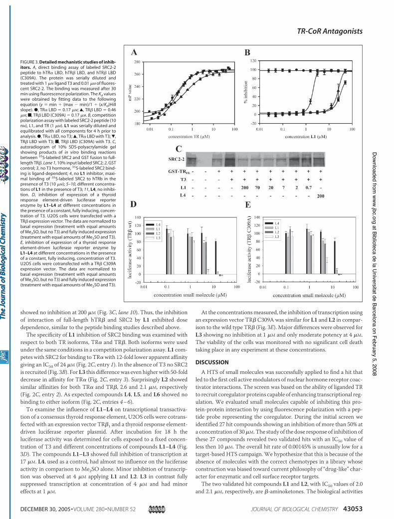
showed no inhibition at 200 $M (Fig. 3C, lane 10). Thus, the inhibitionof interaction of full-length hTR" and SRC2 by L1 exhibited dosedependence, similar to the peptide binding studies described above.The specificity of L1 inhibition of SRC2 binding was examined with
respect to both TR isoforms, TR! and TR". Both isoforms were usedunder the same conditions in a competition polarization assay. L1 com-petes with SRC2 for binding to TR!with 12-fold lower apparent affinitygiving an IC50 of 24 $M (Fig. 2C, entry 1). In the absence of T3 no SRC2is recruited (Fig. 3B). ForL3 this differencewas even higher with 50-folddecrease in affinity for TR! (Fig. 2C, entry 3). Surprisingly L2 showedsimilar affinities for both TR! and TR", 2.6 and 2.1 $M, respectively(Fig. 2C, entry 2). As expected compounds L4, L5, and L6 showed nobinding to either isoform (Fig. 2C, entries 4–6).To examine the influence of L1–L4 on transcriptional transactiva-
tion of a consensus thyroid response element, U2OS cells were cotrans-fected with an expression vector TR"1 and a thyroid response element-driven luciferase reporter plasmid. After incubation for 18 h theluciferase activity was determined for cells exposed to a fixed concen-tration of T3 and different concentrations of compounds L1–L4 (Fig.3D). The compounds L1–L3 showed full inhibition of transcription at17 $M. L4, used as a control, had almost no influence on the luciferaseactivity in comparison to Me2SO alone. Minor inhibition of transcrip-tion was observed at 4 $M applying L1 and L2. L3 in contrast fullysuppressed transcription at concentration of 4 $M and had minoreffects at 1 $M.
At the concentrationsmeasured, the inhibition of transcription usingan expression vector TR" C309A was similar for L1 and L2 in compar-ison to the wild type TR" (Fig. 3E). Major differences were observed forL3 showing no inhibition at 1 $M and only moderate potency at 4 $M.The viability of the cells was monitored with no significant cell deathtaking place in any experiment at these concentrations.
DISCUSSION
A HTS of small molecules was successfully applied to find a hit thatled to the first cell activemodulators of nuclear hormone receptor coac-tivator interactions. The screen was based on the ability of liganded TRto recruit coregulator proteins capable of enhancing transcriptional reg-ulation. We evaluated small molecules capable of inhibiting this pro-tein-protein interaction by using fluorescence polarization with a pep-tide probe representing the coregulator. During the initial screen weidentified 27 hit compounds showing an inhibition of more than 50% ata concentration of 30$M.The study of the dose response of inhibition ofthese 27 compounds revealed two validated hits with an IC50 value ofless then 10 $M. The overall hit rate of 0.00145% is unusually low for atarget-based HTS campaign. We hypothesize that this is because of theabsence of molecules with the correct chemotypes in a library whoseconstruction was biased toward current philosophy of “drug-like” char-acter for enzymatic and cell surface receptor targets.The two validated hit compounds L1 and L2, with IC50 values of 2.0
and 2.1 $M, respectively, are "-aminoketones. The biological activities
FIGURE 3. Detailed mechanistic studies of inhib-itors. A, direct binding assay of labeled SRC2-2peptide to hTR! LBD, hTR" LBD, and hTR" LBD(C309A). The protein was serially diluted andtreated with 1 $M ligand T3 and 0.01 $M of fluores-cent SRC2-2. The binding was measured after 30min using fluorescence polarization. The Kd valueswere obtained by fitting data to the followingequation (y $ min & (max ' min)/1 & (x/Kd)Hillslope). ●, TR! LBD $ 0.17 $M; Œ, TR" LBD $ 0.46$M; f, TR" LBD (C309A) $ 0.17 $M. B, competitionpolarization assay with labeled SRC2-2 peptide (10nM), L1, and TR (1 $M). L1 was serially diluted andequilibrated with all components for 4 h prior toanalysis. ●, TR! LBD, no T3; Œ, TR! LBD with T3; !,TR" LBD with T3; f, TR" LBD (C309A) with T3. C,autoradiogram of 10% SDS-polyacrylamide gelshowing products of in vitro binding reactionsbetween 35S-labeled SRC2 and GST fusion to full-length TR". Lane 1, 10% input labeled SRC2; 2, GSTcontrol; 3, no T3 hormone, 35S-labeled SRC2 bind-ing is ligand-dependent; 4, no L1 inhibitor, maxi-mal binding of 35S-labeled SRC2 to hTRb in thepresence of T3 (10 $M); 5–10, different concentra-tions of L1 in the presence of T3; 11, L4, no inhibi-tion. D, inhibition of expression of a thyroidresponse element-driven luciferase reporterenzyme by L1–L4 at different concentrations inthe presence of a constant, fully inducing, concen-tration of T3. U2OS cells were transfected with aTR" expression vector. The data are normalized tobasal expression (treatment with equal amountsof Me2SO, but no T3) and fully induced expression(treatment with equal amounts of Me2SO and T3).E, inhibition of expression of a thyroid responseelement-driven luciferase reporter enzyme byL1–L4 at different concentrations in the presenceof a constant, fully inducing, concentration of T3.U2OS cells were cotransfected with a TR" C309Aexpression vector. The data are normalized tobasal expression (treatment with equal amountsof Me2SO, but no T3) and fully induced expression(treatment with equal amounts of Me2SO and T3).
TR-CoR Antagonists
DECEMBER 30, 2005 • VOLUME 280 • NUMBER 52 JOURNAL OF BIOLOGICAL CHEMISTRY 43053
at Biblioteca de la Universitat de Barcelona on February 5, 2008 www.jbc.org
Downloaded from
of this class of compounds have been attributed to the fact that they canliberate a corresponding unsaturated ketone capable of alkylating bio-logical nucleophiles. A binding study with the corresponding unsatur-ated ketone L3 showed an IC50 value of 0.9$M. This result suggests thatthe unsaturated ketone is the active species. To exclude the possibilitythat steric properties of L3 are important for inhibition we tested satu-rated compound L4. This compound was not able to inhibit the TR"-CoA binding.We hypothesize that the deamination reaction producing the active
L3 in situ is catalyzed on the protein surface because of the unlikelihoodof an intramolecular mechanism at physiological pH. The small varia-tion of IC50 values based on aminoketones with different alkyl nitrogensubstituents suggests a hydrophobic and fairly rigid catalytic site. How-ever, direct investigation of the most likely catalytic residues of the TR"coactivator binding domain is prevented because these residues are nec-essary for binding of the coactivator.The electrophilic functionality of the active inhibitor species L3 has
been found to be an essential property of the TR antagonists suggestingthat the inhibition is based on the alkylation of nucleophilic residuesforming the TR"-CoA interface. Binding studies with compounds L5and L6 showed no inhibition, concluding that a medium-sized hydro-phobic group at the 4 position of the aromatic "-aminoketones is nec-essary for interaction. Taken together, these studies strongly imply thatthe active species of inhibitors are actually !,"-unsaturated ketonesacting as direct alkylators of nucleophilic residues on the surface of thethyroid receptor. This is supported by the fact that the natural ligand T3is not released by the addition of L1, which implies that the conforma-tion of TR" is not altered in the presence of L1.
Covalent inhibitors have several unique properties. 1) They producean adduct with the target that has increased molecular weight. Thisfeature can be used to permanently label the corresponding bindingpartner; 2) they require stoichiometric, but not largely superstoichio-metric amounts of inhibitor for full activity, and 3) they exhibit strongtime dependence when acting in modest excess relative to the targetconcentration. After the treatment of TR" LBDwithL1we could detecta new species with a 200–250m/z higher mass. We assigned this massto TR"-L1 proving that TR" is selectively alkylated by one equivalent ofL1. In addition we followed the inhibition of TR"-CoA by L1 in time. Asignificant time dependence of inhibition was found between L1 con-centrations of 25 and 1.5 $M when interacted with TR at a concentra-tion of 1 $M. The time dependence altered with the L1 concentrationsuggesting an irreversible inhibition. A covalent complex was formedwhen TR" was treated with fluorescently labeled analog L8, in contrastto the inactive compound L9, lacking the electrophilic properties of L8,which did not. In summary, the detection of the mono-alkylated TR",
its time-dependent formation, and the fact that TR" could be covalentlylabeled with a fluorescent inhibitor supports the postulated mechanismthat L1 forms the unsaturated ketone L3, alkylating irreversibly one ofthe residues of TR" LBD.Based upon the expected chemical reactivity of L1, as predicted by
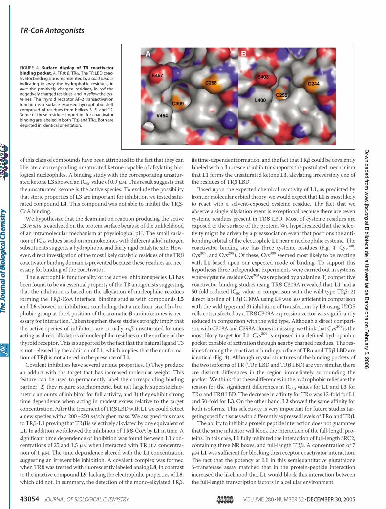
frontier molecular orbital theory, we would expect that L1 is most likelyto react with a solvent-exposed cysteine residue. The fact that weobserve a single alkylation event is exceptional because there are sevencysteine residues present in TR" LBD. Most of cysteine residues areexposed to the surface of the protein. We hypothesized that the selec-tivity might be driven by a preassociation event that positions the anti-bonding orbital of the electrophile L1 near a nucleophilic cysteine. Thecoactivator binding site has three cysteine residues (Fig. 4, Cys308,Cys309, and Cys298). Of these, Cys309 seemed most likely to be reactingwith L1 based upon our expected mode of binding. To support thishypothesis three independent experiments were carried out in systemswhere cysteine residueCys309was replaced by an alanine: 1) competitivecoactivator binding studies using TR" C309A revealed that L1 had a50-fold reduced IC50 value in comparison with the wild type TR"; 2)direct labeling of TR" C309A using L8 was less efficient in comparisonwith the wild type; and 3) inhibition of transfection by L3 using U2OScells cotransfected by a TR" C309A expression vector was significantlyreduced in comparison with the wild type. Although a direct compari-sonwithC308A andC298A clones ismissing, we think that Cys309 is themost likely target for L1. Cys309 is exposed in a defined hydrophobicpocket capable of activation through nearby charged residues. The res-idues forming the coactivator binding surface of TR! and TR" LBD areidentical (Fig. 4). Although crystal structures of the binding pockets ofthe two isoforms of TR (TR! LBD and TR" LBD) are very similar, thereare distinct differences in the region immediately surrounding thepocket.We think that these differences in the hydrophobic relief are thereason for the significant differences in IC50 values for L1 and L3 forTR! and TR" LBD. The decrease in affinity for TR! was 12-fold for L1and 50-fold for L3. On the other hand, L2 showed the same affinity forboth isoforms. This selectivity is very important for future studies tar-geting specific tissues with differently expressed levels of TR! and TR".The ability to inhibit a protein peptide interaction does not guarantee
that the same inhibitor will block the interaction of the full-length pro-teins. In this case, L1 fully inhibited the interaction of full-length SRC2,containing three NR boxes, and full-length TR". A concentration of 7$M L1 was sufficient for blocking this receptor coactivator interaction.The fact that the potency of L1 in this semiquantitative glutathioneS-transferase assay matched that in the protein-peptide interactionincreased the likelihood that L1 would block this interaction betweenthe full-length transcription factors in a cellular environment.
FIGURE 4. Surface display of TR coactivatorbinding pocket. A, TR"; B, TR!. The TR LBD coac-tivator binding site is represented by a solid surfaceindicating in gray the hydrophobic residues, inblue the positively charged residues, in red thenegatively charged residues, and in yellow the cys-teines. The thyroid receptor AF-2 transactivationfunction is a surface exposed hydrophobic cleftcomprised of residues from helices 3, 5, and 12.Some of these residues important for coactivatorbinding are labeled in both TR" and TR!. Both aredepicted in identical orientation.
TR-CoR Antagonists
43054 JOURNAL OF BIOLOGICAL CHEMISTRY VOLUME 280 • NUMBER 52 • DECEMBER 30, 2005
at Biblioteca de la Universitat de Barcelona on February 5, 2008 www.jbc.org
Downloaded from
A reporter gene transfection assay, carried out in cultured U2OScells, showed indeed that compoundsL1,L2, andL3were able to reducetranscriptional activation to basal levels. L3 showed highly increasedpotency in comparison to L1 and L2with almost full inhibition of tran-scription at 4 $M. We concluded that L3 can penetrate the cell mem-brane and is transported to the nucleus. Furthermore it can inhibitcoregulator recruitment and has a direct impact on the transcriptionalactivity of TR".In summary, we report that small molecules are able to inhibit the
interaction between the liganded thyroid hormone receptor and itscoactivator SRC2. To our knowledge this is the first irreversible inhibi-tor of the nuclear receptor coregulator binding that has been reported.Molecules like L1 are a new class of TR antagonist, active in the pres-ence of T3 but silencing its hormone-induced signaling. They open thedoor to understand the coupling ofmultiple thyroid hormone-regulatedsignaling events and the potential for treatment of hyperthyroidismusing approaches that do not affect thyroid hormone levels. Com-pounds L1 and L3 exhibit exceptional TR" selectivities making thempotentially useful for the study of tissue selective thyroid activities. Weare currently investigating the effects of these compounds in cell-basedassays and in vivo studies.
Acknowledgments—The HTS was carried out in the Bay Area Screening Cen-ter (QB3/UCSF) with support from UCSF and the Sandler Research Founda-tion. We thank J. Williams, M. Uehara-Bingen, B. Wolff, and L. Hicks for theirhelp with the HTS and C. Ocasio for assistance in cell culture and the THcompetition assay.
REFERENCES1. Yen, P. M. (2001) Physiol. Rev. 81, 1097–11422. Malm, J. (2004) Curr. Pharm. Des. 10, 3525–35323. Aranda, A., and Pascual, A. (2001) Physiol. Rev. 81, 1269–13044. Harvey, C. B., and Williams, G. R. (2002) Thyroid 12, 441–4465. Williams, G. R. (2000)Mol. Cell. Biol. 20, 8329–83426. Mangelsdorf, D. J., Thummel, C., Beato, M., Herrlich, P., Schutz, G., Umesono, K.,
Blumberg, B., Kastner, P., Mark, M., Chambon, P., and Evans, R. M. (1995) Cell 83,835–839
7. Xu, J. M., and Li, Q. T. (2003)Mol. Endocrinol. 17, 1681–16928. Hu, X., and Lazar, M. A. (2000) Trends Endorinol. Metab. 11, 6–109. Moore, J. M. R., and Guy, R. K. (2005)Mol. Cell. Proteomics 4, 475–48210. Onate, S. A., Tsai, S. Y., Tsai,M. J., andOmalley, B.W. (1995) Science 270, 1354–135711. Voegel, J. J., Heine, M. J. S., Zechel, C., Chambon, P., and Gronemeyer, H. (1996)
EMBO J. 15, 3667–367512. Hong, H., Kohli, K., Garabedian, M. J., and Stallcup, M. R. (1997)Mol. Cell. Biol. 17,
2735–274413. Suen, C. S., Berrodin, T. J., Mastroeni, R., Cheskis, B. J., Lyttle, C. R., and Frail, D. E.
(1998) J. Biol. Chem. 273, 27645–2765314. Ito, M., and Roeder, R. G. (2001) Trends Endorinol. Metab. 12, 127–13415. Puigserver, P., Wu, Z. D., Park, C. W., Graves, R., Wright, M., and Spiegelman, B. M.
(1998) Cell 92, 829–83916. Ko, L., Cardona, G. R., and Chin, W. W. (2000) Proc. Natl. Acad. Sci. U. S. A. 97,
6212–621717. Moore, J. M. R., Galicia, S. J., McReynolds, A. C., Nguyen, N. H., Scanlan, T. S., and
Guy, R. K. (2004) J. Biol. Chem. 279, 27584–2759018. Kalkhoven, E. (2004) Biochem. Pharmacol. 68, 1145–115519. Heinlein, C. A., Ting, H. J., Yeh, S. Y., and Chang, C. S. (1999) J. Biol. Chem. 274,
16147–1615220. Treuter, E., Albrektsen, T., Johansson, L., Leers, J., and Gustafsson, J. A. (1998) Mol.
Endocrinol. 12, 864–88121. Zhang, H., Thomsen, J. S., Johansson, L., Gustafsson, J. A., and Treuter, E. (2000)
J. Biol. Chem. 275, 39855–3985922. Seol, W., Choi, H. S., and Moore, D. D. (1996) Science 272, 1336–133923. Darimont, B. D., Wagner, R. L., Apriletti, J. W., Stallcup, M. R., Kushner, P. J., Baxter,
J. D., Fletterick, R. J., and Yamamoto, K. R. (1998) Genes Dev. 12, 3343–335624. Feng,W. J., Ribeiro, R. C. J.,Wagner, R. L., Nguyen, H., Apriletti, J.W., Fletterick, R. J.,
Baxter, J. D., Kushner, P. J., and West, B. L. (1998) Science 280, 1747–174925. Geistlinger, T. R., and Guy, R. K. (2001) J. Am. Chem. Soc. 123, 1525–152626. Geistlinger, T. R., and Guy, R. K. (2003) J. Am. Chem. Soc. 125, 6852–685327. Leduc, A. M., Trent, J. O., Wittliff, J. L., Bramlett, K. S., Briggs, S. L., Chirgadze, N. Y.,
Wang, Y., Burris, T. P., and Spatola, A. F. (2003) Proc. Natl. Acad. Sci. U. S. A. 100,11273–11278
28. Galande, A. K., Bramlett, K. S., Burris, T. P., Wittliff, J. L., and Spatola, A. F. (2004) J.Peptide Res. 63, 297–302
29. Rodriguez, A. L., Tamrazi, A., Collins, M. L., and Katzenellenbogen, J. A. (2004)J. Med. Chem. 47, 600–611
30. Dietrich, S. W., Bolger, M. B., Kollman, P. A., and Jorgensen, E. C. (1977) J. Med.Chem. 20, 863–880
31. Briel, D., Pohlers, D., Uhlig, M., Vieweg, S., Scholz, G. H., Thormann, M., and Hof-mann, H. J. (1999) J. Med. Chem. 42, 1849–1854
32. Stanton, J. L., Cahill, E., Dotson, R., Tan, J., Tomaselli, H. C., Wasvary, J. M., Stephan,Z. F., and Steele, R. E. (2000) Bioorg. Med. Chem. Lett. 10, 1661–1663
33. Ye, L., Li, Y. L., Mellstrom, K., Mellin, C., Bladh, L. G., Koehler, K., Garg, N., Collazo,A. M. G., Litten, C., Husman, B., Persson, K., Ljunggren, J., Grover, G., Sleph, P. G.,George, R., and Malm, J. (2003) J. Med. Chem. 46, 1580–1588
34. Webb, P., Nguyen, N. H., Chiellini, G., Yoshihara, H. A. I., Lima, S. T. C., Apriletti,J. W., Ribeiro, R. C. J., Marimuthu, A., West, B. L., Goede, P., Mellstrom, K., Nilsson,S., Kushner, P. J., Fletterick, R. J., Scanlan, T. S., and Baxter, J. D. (2002) J. SteroidBiochem. Mol. Biol. 83, 59–73
35. Chiellini, G., Apriletti, J. W., Yoshihara, H. A., Baxter, J. D., Ribeiro, R. C. J., andScanlan, T. S. (1998) Chem. Biol. 5, 299–306
36. Mishra, M. K., Wilson, F. E., Scanlan, T. S., and Chiellini, G. (2004) J. Comp. Physiol.B 174, 471–479
37. Grover, G. J., Egan, D. M., Sleph, P. G., Beehler, B. C., Chiellini, G., Nguyen, N. H.,Baxter, J. D., and Scanlan, T. S. (2004) Endocrinology 145, 1656–1661
38. Freitas, F. R. S.,Moriscot, A. S., Jorgetti, V., Soares, A. G., Passarelli, M., Scanlan, T. S.,Brent, G. A., Bianco, A. C., and Gouveia, C. H. A. (2003) Am. J. Physiol. 285,E1135–E1141
39. Manzano, J., Morte, B., Scanlan, T. S., and Bernal, J. (2003) Endocrinology 144,5480–5487
40. Trost, S. U., Swanson, E., Gloss, B., Wang-Iverson, D. B., Zhang, H. J., Volodarsky, T.,Grover, G. J., Baxter, J. D., Chiellini, G., Scanlan, T. S., and Dillmann, W. H. (2000)Endocrinology 141, 3057–3064
41. Wagner, R. L., Huber, B. R., Shiau, A. K., Kelly, A., Lima, S. T. C., Scanlan, T. S.,Apriletti, J. W., Baxter, J. D., West, B. L., and Fletterick, R. J. (2001) J. Mol. Endocrinol.15, 398–410
42. Arkin, M. R., and Wells, J. A. (2004) Nat. Rev. Drug Discov. 3, 301–31743. Berg, T. (2003) Angew. Chem. Int. Ed. Engl. 42, 2462–248144. Toogood, P. L. (2002) J. Med. Chem. 45, 1543–155845. Roehrl, M. H. A., Wang, J. Y., andWagner, G. (2004) Biochemistry 43, 16056–1606646. Apriletti, J. W., Baxter, J. D., Lau, K. H., andWest, B. L. (1995) Protein Express. Purif.
6, 363–37047. Arend, M., Westermann, B., and Risch, N. (1998) Angew. Chem. Int. Ed. Engl. 37,
1045–107048. Gul, H. I., Gul, M., Vepsalainen, J., Erciyas, E., and Hanninen, O. (2003) Biol. Pharm.
Bull. 26, 631–63749. Davioud-Charvet, E., McLeish, M. J., Veine, D. M., Giegel, D., Arscott, L. D., Andri-
copulo, A. D., Becker, K., Muller, S., Schirmer, R. H., Williams, C. H., and Kenyon,G. L. (2003) Biochemistry 42, 13319–13330
TR-CoR Antagonists
DECEMBER 30, 2005 • VOLUME 280 • NUMBER 52 JOURNAL OF BIOLOGICAL CHEMISTRY 43055
at Biblioteca de la Universitat de Barcelona on February 5, 2008 www.jbc.org
Downloaded from
Related Documents