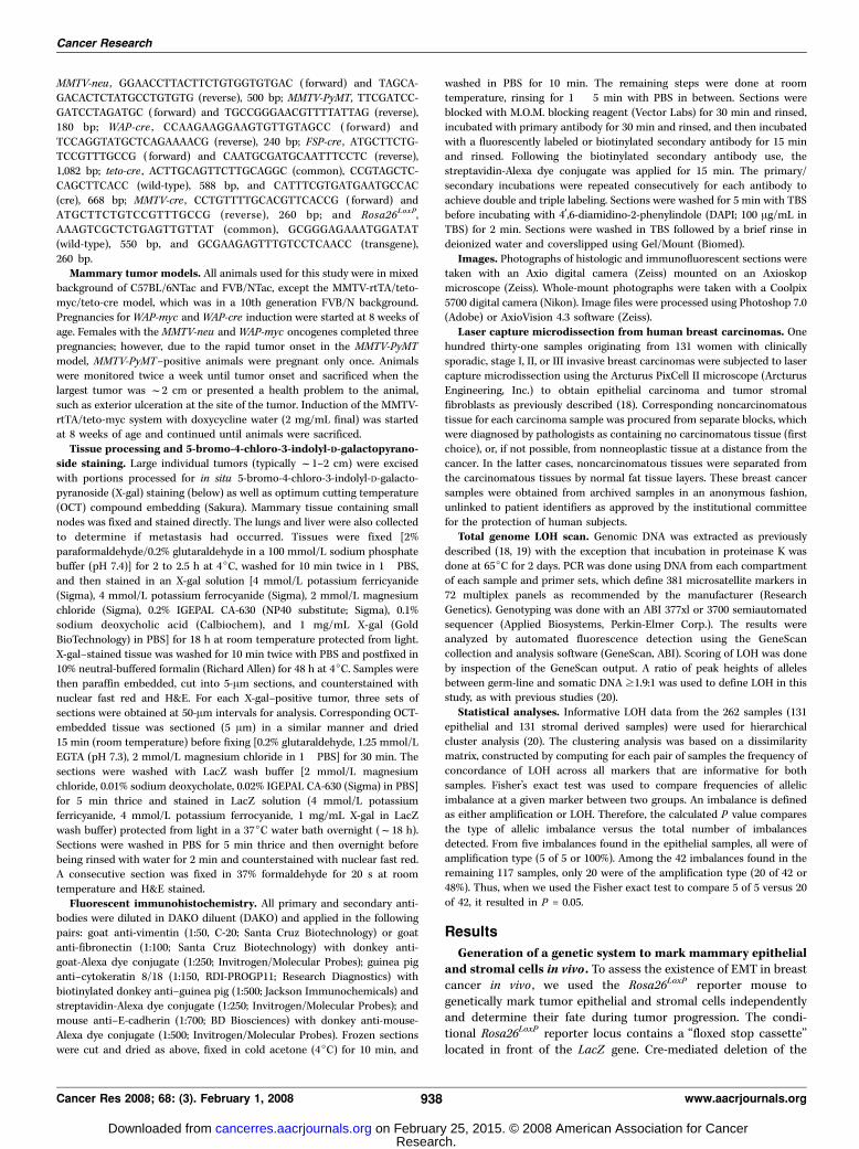
Direct Evidence for Epithelial-Mesenchymal Transitions in Breast Cancer Anthony J. Trimboli, 1,2,3 Koichi Fukino, 2,3 Alain de Bruin, 1,2,3 Guo Wei, 1,2,3 Lei Shen, 4 Stephan M. Tanner, 2,3 Nicholas Creasap, 1,2,3 Thomas J. Rosol, 5 Michael L. Robinson, 6 Charis Eng, 1,2,3,7 Michael C. Ostrowski, 1,2,3 and Gustavo Leone 1,2,3 1 Department of Molecular Genetics; 2 Human Cancer Genetics Program, Comprehensive Cancer Center; 3 Department of Molecular Virology, Immunology, and Medical Genetics, College of Medicine; 4 Division of Epidemiology and Biostatistics, School of Public Health; and 5 Department of Veterinary Biosciences, The Ohio State University; 6 Center for Childhood Cancer, Columbus Children’s Research Institute, Columbus, Ohio; and 7 Genomic Medicine Institute, Cleveland Clinic Lerner Research Institute and Department of Genetics, Case Western Reserve University School of Medicine, Cleveland, Ohio Abstract We developed stromal- and epithelial-specific cre -transgenic mice to directly visualize epithelial-mesenchymal transition (EMT) during cancer progression in vivo. Using three different oncogene-driven mouse mammary tumor models and cell-fate mapping strategies, we show in vivo evidence for the existence of EMT in breast cancer and show that myc can specifically elicit this process. Hierarchical cluster analysis of genome- wide loss of heterozygosity reveals that the incidence of EMT in invasive human breast carcinomas is rare, but when it occurs it is associated with the amplification of MYC . These data provide the first direct evidence for EMT in breast cancer and suggest that its development is favored by myc -initiated events. [Cancer Res 2008;68(3):937–45] Introduction The transdifferentiation of epithelial cells to a more mesenchy- mal state, typically called epithelial-mesenchymal transition (EMT), is an essential process for normal embryonic development that has been implicated in the progression toward an advanced cancer phenotype (1–5). EMT is a multistep process characterized by the loss of cell-to-cell junctions and reorganization of the cytoskeleton, which together result in the loss of apical-basal cell polarity and the acquisition of spindle-shaped morphology (6). This is associated with a decrease in epithelial-specific gene expression, including E-cadherin, and a gain in mesenchymal-specific gene expression. During tumor progression, these changes are thought to ultimately promote tumor cell migration across the basement membrane and invasion into the surrounding microenvironment. EMT in cancer, however, has been inferred predominantly from in vitro studies and the expression of cell type–specific markers (7–9). From these studies, epithelial tumor cells that are presumed to undergo EMT acquire a spindle cell morphology that is histologically indistin- guishable from normal mammary stromal cells. Because tumor cells in vivo have not yet been unambiguously marked and their fate traced during tumor progression, the actual origin of tumor- associated spindle-shaped mesenchymal cells is not known, and hence the occurrence of EMT in cancer remains speculative (10, 11). Tumor-associated fibroblasts are believed to influence tumor behavior and outcome, and thus understanding their biology is of importance to the overall understanding of cancer. A fundamental premise of EMT in the context of cancer implies that a tumor- associated fibroblast that arises from an EMT event contains a similar set of genetic and/or epigenetic insults as the epithelial tumor cell from which it originated. Because of their genetic differences, fibroblasts that originate from tumor cells would be expected to affect the behavior of cancers differently than fibroblasts that originate from normal mesenchymal progenitors. Whereas results from a large body of work done in vitro points to a critical role of EMT in cancer progression, its importance in cancer ultimately rests on whether it actually occurs in vivo . The absence of direct evidence for EMT in cancer has therefore raised substantial controversy. To assess the existence of EMT in breast cancer in vivo , we developed a genetic system to independently mark epithelial and stromal cells and observe their fate following cancer progression via the expression of cre and the Rosa26 LoxP reporter allele (12). We also determined genome-wide loss of heterozygosity (LOH) in both epithelial and stromal cells to assess the occurrence of EMT in invasive human breast carcinomas. Materials and Methods Generation of FSP-cre transgenic mice. The transgene vector for Fsp1- Cre-BGH was created by inserting the BGH polyadenylate signal and the multiple cloning site of pcDNA3 (InvitrogenCA) into a modified pBluescript II plasmid backbone (Stratagene). The 3.1-kb Fsp1 gene promoter (S100a4 ; refs. 13, 14) extending 1.9 kb upstream of exon 1 to the end of intron 1 (1.2 kb) was long-range PCR amplified (Long Template System) from C57BL/6NTac genomic DNA and cloned into the multiple cloning site of the vector. The Cre recombinase open reading frame from pMC-Cre (15) was PCR amplified and inserted between the Fsp1 promoter and the BGH polyadenylate signal. Cloning was completed by standard protocols and all fragments were verified by DNA sequencing. All microinjection constructs were injected into pronuclear stage FVB/N mouse embryos, as previously described (16, 17), after the Fsp1-Cre-BGH fragment was excised by Pac I (New England Biolabs) digestion and gel purified. Genotyping of transgenic mice. DNA was isolated from mouse tail tips and genotyped by PCR analysis using the following primer sets (5¶ to 3¶): WAP-myc , CACCGCCTACATCCTGTCCATTCAAGC ( forward) and TTAGGACAAGGCTGGTGGGCACTG (reverse), 240 bp; MMTV-rtTA , AGTATGCCGCCATTATTACGAC ( forward) and CGATGGTAGACCCG- TAATTGTT (reverse), 170 bp; teto-myc , GGAATGGCAGAAGGCAGG (forward) and GCAGTAGCCTCATCATCACTAGATGG (reverse), 580 bp; Note: Supplementary data for this article are available at Cancer Research Online (http://cancerres.aacrjournals.org/). Requests for reprints: Gustavo Leone, Departments of Molecular Genetics and Molecular Virology, Immunology, and Medical Genetics, Human Cancer Genetics Program, The Ohio State University, 808 Biomedical Research Tower, 460 West 12th Avenue, Columbus, OH 43210. Phone: 614-688-4567; Fax: 614-688-4181; E-mail: [email protected], or Michael C. Ostrowski, Department of Molecular and Cellular Biochemistry, Human Cancer Genetics Program, The Ohio State University, 810 Biomedical Research Tower, 420 West 12th Avenue, Columbus, OH 43210. Phone: 614-688-3824; Fax: 614-688-4181; E-mail: [email protected]. I2008 American Association for Cancer Research. doi:10.1158/0008-5472.CAN-07-2148 www.aacrjournals.org 937 Cancer Res 2008; 68: (3). February 1, 2008 Research Article Research. on February 25, 2015. © 2008 American Association for Cancer cancerres.aacrjournals.org Downloaded from Research. on February 25, 2015. © 2008 American Association for Cancer cancerres.aacrjournals.org Downloaded from Research. on February 25, 2015. © 2008 American Association for Cancer cancerres.aacrjournals.org Downloaded from

Welcome message from author
This document is posted to help you gain knowledge. Please leave a comment to let me know what you think about it! Share it to your friends and learn new things together.
Transcript
Direct Evidence for Epithelial-Mesenchymal Transitions
in Breast Cancer
Anthony J. Trimboli,1,2,3
Koichi Fukino,2,3Alain de Bruin,
1,2,3Guo Wei,
1,2,3Lei Shen,
4
Stephan M. Tanner,2,3Nicholas Creasap,
1,2,3Thomas J. Rosol,
5Michael L. Robinson,
6
Charis Eng,1,2,3,7
Michael C. Ostrowski,1,2,3
and Gustavo Leone1,2,3
1Department of Molecular Genetics; 2Human Cancer Genetics Program, Comprehensive Cancer Center; 3Department of MolecularVirology, Immunology, and Medical Genetics, College of Medicine; 4Division of Epidemiology and Biostatistics, School of PublicHealth; and 5Department of Veterinary Biosciences, The Ohio State University; 6Center for Childhood Cancer, ColumbusChildren’s Research Institute, Columbus, Ohio; and 7Genomic Medicine Institute, Cleveland Clinic Lerner ResearchInstitute and Department of Genetics, Case Western Reserve University School of Medicine, Cleveland, Ohio
Abstract
We developed stromal- and epithelial-specific cre-transgenicmice to directly visualize epithelial-mesenchymal transition(EMT) during cancer progression in vivo. Using three differentoncogene-driven mouse mammary tumor models and cell-fatemapping strategies, we show in vivo evidence for the existenceof EMT in breast cancer and show that myc can specificallyelicit this process. Hierarchical cluster analysis of genome-wide loss of heterozygosity reveals that the incidence of EMTin invasive human breast carcinomas is rare, but when itoccurs it is associated with the amplification of MYC . Thesedata provide the first direct evidence for EMT in breast cancerand suggest that its development is favored by myc-initiatedevents. [Cancer Res 2008;68(3):937–45]
Introduction
The transdifferentiation of epithelial cells to a more mesenchy-mal state, typically called epithelial-mesenchymal transition (EMT),is an essential process for normal embryonic development that hasbeen implicated in the progression toward an advanced cancerphenotype (1–5). EMT is a multistep process characterized by theloss of cell-to-cell junctions and reorganization of the cytoskeleton,which together result in the loss of apical-basal cell polarity and theacquisition of spindle-shaped morphology (6). This is associatedwith a decrease in epithelial-specific gene expression, includingE-cadherin, and a gain in mesenchymal-specific gene expression.During tumor progression, these changes are thought to ultimatelypromote tumor cell migration across the basement membrane andinvasion into the surrounding microenvironment. EMT in cancer,however, has been inferred predominantly from in vitro studies andthe expression of cell type–specific markers (7–9). From thesestudies, epithelial tumor cells that are presumed to undergo EMTacquire a spindle cell morphology that is histologically indistin-guishable from normal mammary stromal cells. Because tumor
cells in vivo have not yet been unambiguously marked and theirfate traced during tumor progression, the actual origin of tumor-associated spindle-shaped mesenchymal cells is not known, andhence the occurrence of EMT in cancer remains speculative (10, 11).Tumor-associated fibroblasts are believed to influence tumor
behavior and outcome, and thus understanding their biology is ofimportance to the overall understanding of cancer. A fundamentalpremise of EMT in the context of cancer implies that a tumor-associated fibroblast that arises from an EMT event contains asimilar set of genetic and/or epigenetic insults as the epithelialtumor cell from which it originated. Because of their geneticdifferences, fibroblasts that originate from tumor cells would beexpected to affect the behavior of cancers differently thanfibroblasts that originate from normal mesenchymal progenitors.Whereas results from a large body of work done in vitro points to acritical role of EMT in cancer progression, its importance in cancerultimately rests on whether it actually occurs in vivo . The absenceof direct evidence for EMT in cancer has therefore raisedsubstantial controversy. To assess the existence of EMT in breastcancer in vivo , we developed a genetic system to independentlymark epithelial and stromal cells and observe their fate followingcancer progression via the expression of cre and the Rosa26LoxP
reporter allele (12). We also determined genome-wide loss ofheterozygosity (LOH) in both epithelial and stromal cells to assessthe occurrence of EMT in invasive human breast carcinomas.
Materials and Methods
Generation of FSP-cre transgenic mice. The transgene vector for Fsp1-Cre-BGH was created by inserting the BGH polyadenylate signal and the
multiple cloning site of pcDNA3 (InvitrogenCA) into a modified pBluescriptII plasmid backbone (Stratagene). The 3.1-kb Fsp1 gene promoter (S100a4 ;
refs. 13, 14) extending 1.9 kb upstream of exon 1 to the end of intron 1
(1.2 kb) was long-range PCR amplified (Long Template System) from
C57BL/6NTac genomic DNA and cloned into the multiple cloning site of thevector. The Cre recombinase open reading frame from pMC-Cre (15) was
PCR amplified and inserted between the Fsp1 promoter and the BGH
polyadenylate signal. Cloning was completed by standard protocols and all
fragments were verified by DNA sequencing. All microinjection constructswere injected into pronuclear stage FVB/N mouse embryos, as previously
described (16, 17), after the Fsp1-Cre-BGH fragment was excised by PacI
(New England Biolabs) digestion and gel purified.
Genotyping of transgenic mice. DNA was isolated from mouse tailtips and genotyped by PCR analysis using the following primer sets (5¶ to3¶): WAP-myc , CACCGCCTACATCCTGTCCATTCAAGC ( forward) and
TTAGGACAAGGCTGGTGGGCACTG (reverse), 240 bp; MMTV-rtTA ,AGTATGCCGCCATTATTACGAC ( forward) and CGATGGTAGACCCG-
TAATTGTT (reverse), 170 bp; teto-myc , GGAATGGCAGAAGGCAGG
( forward) and GCAGTAGCCTCATCATCACTAGATGG (reverse), 580 bp;
Note: Supplementary data for this article are available at Cancer Research Online(http://cancerres.aacrjournals.org/).
Requests for reprints: Gustavo Leone, Departments of Molecular Genetics andMolecular Virology, Immunology, and Medical Genetics, Human Cancer GeneticsProgram, The Ohio State University, 808 Biomedical Research Tower, 460 West 12thAvenue, Columbus, OH 43210. Phone: 614-688-4567; Fax: 614-688-4181; E-mail:[email protected], or Michael C. Ostrowski, Department of Molecular andCellular Biochemistry, Human Cancer Genetics Program, The Ohio State University,810 Biomedical Research Tower, 420 West 12th Avenue, Columbus, OH 43210. Phone:614-688-3824; Fax: 614-688-4181; E-mail: [email protected].
I2008 American Association for Cancer Research.doi:10.1158/0008-5472.CAN-07-2148
www.aacrjournals.org 937 Cancer Res 2008; 68: (3). February 1, 2008
Research Article
Research. on February 25, 2015. © 2008 American Association for Cancercancerres.aacrjournals.org Downloaded from
Research. on February 25, 2015. © 2008 American Association for Cancercancerres.aacrjournals.org Downloaded from
Research. on February 25, 2015. © 2008 American Association for Cancercancerres.aacrjournals.org Downloaded from
MMTV-neu , GGAACCTTACTTCTGTGGTGTGAC ( forward) and TAGCA-GACACTCTATGCCTGTGTG (reverse), 500 bp; MMTV-PyMT, TTCGATCC-
GATCCTAGATGC ( forward) and TGCCGGGAACGTTTTATTAG (reverse),
180 bp; WAP-cre , CCAAGAAGGAAGTGTTGTAGCC ( forward) and
TCCAGGTATGCTCAGAAAACG (reverse), 240 bp; FSP-cre , ATGCTTCTG-TCCGTTTGCCG ( forward) and CAATGCGATGCAATTTCCTC (reverse),
1,082 bp; teto-cre , ACTTGCAGTTCTTGCAGGC (common), CCGTAGCTC-
CAGCTTCACC (wild-type), 588 bp, and CATTTCGTGATGAATGCCAC
(cre), 668 bp; MMTV-cre , CCTGTTTTGCACGTTCACCG ( forward) andATGCTTCTGTCCGTTTGCCG (reverse), 260 bp; and Rosa26LoxP,
AAAGTCGCTCTGAGTTGTTAT (common), GCGGGAGAAATGGATAT
(wild-type), 550 bp, and GCGAAGAGTTTGTCCTCAACC (transgene),
260 bp.Mammary tumor models. All animals used for this study were in mixed
background of C57BL/6NTac and FVB/NTac, except the MMTV-rtTA/teto-
myc/teto-cre model, which was in a 10th generation FVB/N background.Pregnancies forWAP-myc andWAP-cre induction were started at 8 weeks of
age. Females with the MMTV-neu andWAP-myc oncogenes completed three
pregnancies; however, due to the rapid tumor onset in the MMTV-PyMT
model, MMTV-PyMT–positive animals were pregnant only once. Animalswere monitored twice a week until tumor onset and sacrificed when the
largest tumor was f2 cm or presented a health problem to the animal,
such as exterior ulceration at the site of the tumor. Induction of the MMTV-
rtTA/teto-myc system with doxycycline water (2 mg/mL final) was startedat 8 weeks of age and continued until animals were sacrificed.
Tissue processing and 5-bromo-4-chloro-3-indolyl-D-galactopyrano-side staining. Large individual tumors (typically f1–2 cm) were excised
with portions processed for in situ 5-bromo-4-chloro-3-indolyl-D-galacto-
pyranoside (X-gal) staining (below) as well as optimum cutting temperature
(OCT) compound embedding (Sakura). Mammary tissue containing small
nodes was fixed and stained directly. The lungs and liver were also collected
to determine if metastasis had occurred. Tissues were fixed [2%
paraformaldehyde/0.2% glutaraldehyde in a 100 mmol/L sodium phosphate
buffer (pH 7.4)] for 2 to 2.5 h at 4jC, washed for 10 min twice in 1� PBS,
and then stained in an X-gal solution [4 mmol/L potassium ferricyanide
(Sigma), 4 mmol/L potassium ferrocyanide (Sigma), 2 mmol/L magnesium
chloride (Sigma), 0.2% IGEPAL CA-630 (NP40 substitute; Sigma), 0.1%
sodium deoxycholic acid (Calbiochem), and 1 mg/mL X-gal (Gold
BioTechnology) in PBS] for 18 h at room temperature protected from light.
X-gal–stained tissue was washed for 10 min twice with PBS and postfixed in
10% neutral-buffered formalin (Richard Allen) for 48 h at 4jC. Samples were
then paraffin embedded, cut into 5-Am sections, and counterstained with
nuclear fast red and H&E. For each X-gal–positive tumor, three sets of
sections were obtained at 50-Am intervals for analysis. Corresponding OCT-
embedded tissue was sectioned (5 Am) in a similar manner and dried
15 min (room temperature) before fixing [0.2% glutaraldehyde, 1.25 mmol/L
EGTA (pH 7.3), 2 mmol/L magnesium chloride in 1� PBS] for 30 min. The
sections were washed with LacZ wash buffer [2 mmol/L magnesium
chloride, 0.01% sodium deoxycholate, 0.02% IGEPAL CA-630 (Sigma) in PBS]
for 5 min thrice and stained in LacZ solution (4 mmol/L potassium
ferricyanide, 4 mmol/L potassium ferrocyanide, 1 mg/mL X-gal in LacZ
wash buffer) protected from light in a 37jC water bath overnight (f18 h).
Sections were washed in PBS for 5 min thrice and then overnight before
being rinsed with water for 2 min and counterstained with nuclear fast red.
A consecutive section was fixed in 37% formaldehyde for 20 s at room
temperature and H&E stained.Fluorescent immunohistochemistry. All primary and secondary anti-
bodies were diluted in DAKO diluent (DAKO) and applied in the following
pairs: goat anti-vimentin (1:50, C-20; Santa Cruz Biotechnology) or goat
anti-fibronectin (1:100; Santa Cruz Biotechnology) with donkey anti-goat-Alexa dye conjugate (1:250; Invitrogen/Molecular Probes); guinea pig
anti–cytokeratin 8/18 (1:150, RDI-PROGP11; Research Diagnostics) with
biotinylated donkey anti–guinea pig (1:500; Jackson Immunochemicals) andstreptavidin-Alexa dye conjugate (1:250; Invitrogen/Molecular Probes); and
mouse anti–E-cadherin (1:700; BD Biosciences) with donkey anti-mouse-
Alexa dye conjugate (1:500; Invitrogen/Molecular Probes). Frozen sections
were cut and dried as above, fixed in cold acetone (4jC) for 10 min, and
washed in PBS for 10 min. The remaining steps were done at roomtemperature, rinsing for 1 � 5 min with PBS in between. Sections were
blocked with M.O.M. blocking reagent (Vector Labs) for 30 min and rinsed,
incubated with primary antibody for 30 min and rinsed, and then incubated
with a fluorescently labeled or biotinylated secondary antibody for 15 minand rinsed. Following the biotinylated secondary antibody use, the
streptavidin-Alexa dye conjugate was applied for 15 min. The primary/
secondary incubations were repeated consecutively for each antibody to
achieve double and triple labeling. Sections were washed for 5 min with TBSbefore incubating with 4¶,6-diamidino-2-phenylindole (DAPI; 100 Ag/mL in
TBS) for 2 min. Sections were washed in TBS followed by a brief rinse in
deionized water and coverslipped using Gel/Mount (Biomed).
Images. Photographs of histologic and immunofluorescent sections weretaken with an Axio digital camera (Zeiss) mounted on an Axioskop
microscope (Zeiss). Whole-mount photographs were taken with a Coolpix
5700 digital camera (Nikon). Image files were processed using Photoshop 7.0(Adobe) or AxioVision 4.3 software (Zeiss).
Laser capture microdissection from human breast carcinomas. Onehundred thirty-one samples originating from 131 women with clinically
sporadic, stage I, II, or III invasive breast carcinomas were subjected to lasercapture microdissection using the Arcturus PixCell II microscope (Arcturus
Engineering, Inc.) to obtain epithelial carcinoma and tumor stromal
fibroblasts as previously described (18). Corresponding noncarcinomatous
tissue for each carcinoma sample was procured from separate blocks, whichwere diagnosed by pathologists as containing no carcinomatous tissue (first
choice), or, if not possible, from nonneoplastic tissue at a distance from the
cancer. In the latter cases, noncarcinomatous tissues were separated fromthe carcinomatous tissues by normal fat tissue layers. These breast cancer
samples were obtained from archived samples in an anonymous fashion,
unlinked to patient identifiers as approved by the institutional committee
for the protection of human subjects.Total genome LOH scan. Genomic DNA was extracted as previously
described (18, 19) with the exception that incubation in proteinase K was
done at 65jC for 2 days. PCR was done using DNA from each compartment
of each sample and primer sets, which define 381 microsatellite markers in72 multiplex panels as recommended by the manufacturer (Research
Genetics). Genotyping was done with an ABI 377xl or 3700 semiautomated
sequencer (Applied Biosystems, Perkin-Elmer Corp.). The results wereanalyzed by automated fluorescence detection using the GeneScan
collection and analysis software (GeneScan, ABI). Scoring of LOH was done
by inspection of the GeneScan output. A ratio of peak heights of alleles
between germ-line and somatic DNA z1.9:1 was used to define LOH in thisstudy, as with previous studies (20).
Statistical analyses. Informative LOH data from the 262 samples (131
epithelial and 131 stromal derived samples) were used for hierarchical
cluster analysis (20). The clustering analysis was based on a dissimilaritymatrix, constructed by computing for each pair of samples the frequency of
concordance of LOH across all markers that are informative for both
samples. Fisher’s exact test was used to compare frequencies of allelic
imbalance at a given marker between two groups. An imbalance is definedas either amplification or LOH. Therefore, the calculated P value compares
the type of allelic imbalance versus the total number of imbalances
detected. From five imbalances found in the epithelial samples, all were ofamplification type (5 of 5 or 100%). Among the 42 imbalances found in the
remaining 117 samples, only 20 were of the amplification type (20 of 42 or
48%). Thus, when we used the Fisher exact test to compare 5 of 5 versus 20
of 42, it resulted in P = 0.05.
Results
Generation of a genetic system to mark mammary epithelialand stromal cells in vivo . To assess the existence of EMT in breastcancer in vivo , we used the Rosa26LoxP reporter mouse togenetically mark tumor epithelial and stromal cells independentlyand determine their fate during tumor progression. The condi-tional Rosa26LoxP reporter locus contains a ‘‘floxed stop cassette’’located in front of the LacZ gene. Cre-mediated deletion of the
Cancer Research
Cancer Res 2008; 68: (3). February 1, 2008 938 www.aacrjournals.org
Research. on February 25, 2015. © 2008 American Association for Cancercancerres.aacrjournals.org Downloaded from
upstream floxed stop cassette leads to the induction of LacZexpression, which can be easily visualized as blue cells byhistochemical staining with X-gal. Activation of the Rosa26LoxP
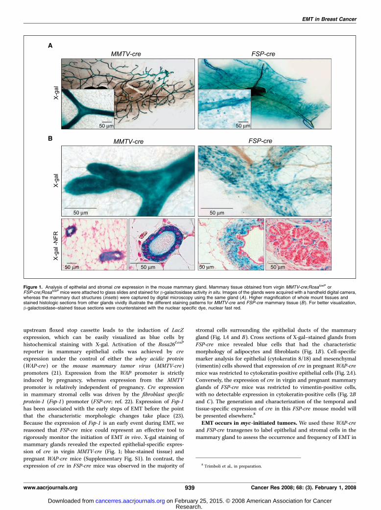
reporter in mammary epithelial cells was achieved by creexpression under the control of either the whey acidic protein(WAP-cre) or the mouse mammary tumor virus (MMTV-cre)promoters (21). Expression from the WAP promoter is strictlyinduced by pregnancy, whereas expression from the MMTVpromoter is relatively independent of pregnancy. Cre expressionin mammary stromal cells was driven by the fibroblast specificprotein-1 (Fsp-1) promoter (FSP-cre ; ref. 22). Expression of Fsp-1has been associated with the early steps of EMT before the pointthat the characteristic morphologic changes take place (23).Because the expression of Fsp-1 is an early event during EMT, wereasoned that FSP-cre mice could represent an effective tool torigorously monitor the initiation of EMT in vivo . X-gal staining ofmammary glands revealed the expected epithelial-specific expres-sion of cre in virgin MMTV-cre (Fig. 1; blue-stained tissue) andpregnant WAP-cre mice (Supplementary Fig. S1). In contrast, theexpression of cre in FSP-cre mice was observed in the majority of
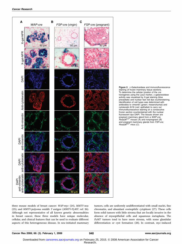
stromal cells surrounding the epithelial ducts of the mammarygland (Fig. 1A and B). Cross sections of X-gal–stained glands fromFSP-cre mice revealed blue cells that had the characteristicmorphology of adipocytes and fibroblasts (Fig. 1B). Cell-specificmarker analysis for epithelial (cytokeratin 8/18) and mesenchymal(vimentin) cells showed that expression of cre in pregnant WAP-cremice was restricted to cytokeratin-positive epithelial cells (Fig. 2A).Conversely, the expression of cre in virgin and pregnant mammaryglands of FSP-cre mice was restricted to vimentin-positive cells,with no detectable expression in cytokeratin-positive cells (Fig. 2Band C). The generation and characterization of the temporal andtissue-specific expression of cre in this FSP-cre mouse model willbe presented elsewhere.8
EMT occurs in myc-initiated tumors. We used these WAP-creand FSP-cre transgenes to label epithelial and stromal cells in themammary gland to assess the occurrence and frequency of EMT in
Figure 1. Analysis of epithelial and stromal cre expression in the mouse mammary gland. Mammary tissue obtained from virgin MMTV-cre;RosaloxP orFSP-cre;RosaloxP mice were attached to glass slides and stained for h-galactosidase activity in situ. Images of the glands were acquired with a handheld digital camera,whereas the mammary duct structures (insets ) were captured by digital microscopy using the same gland (A). Higher magnification of whole mount tissues andstained histologic sections from other glands vividly illustrate the different staining patterns for MMTV-cre and FSP-cre mammary tissue (B). For better visualization,h-galactosidase–stained tissue sections were counterstained with the nuclear specific dye, nuclear fast red.
8 Trimboli et al., in preparation.
EMT in Breast Cancer
www.aacrjournals.org 939 Cancer Res 2008; 68: (3). February 1, 2008
Research. on February 25, 2015. © 2008 American Association for Cancercancerres.aacrjournals.org Downloaded from
three mouse models of breast cancer: WAP-myc (24), MMTV-neu(25), and MMTV-polyoma middle T antigen (MMTV-PyMT ; ref. 26).Although not representative of all known genetic abnormalitiesin breast cancer, these three models have unique molecular,cellular, and clinical features that can be used to evaluate differentaspects of this heterogeneous disease. In neu-initiated mammary
tumors, cells are uniformly undifferentiated with small nuclei, finechromatin, and abundant eosinophilic cytoplasm (27). These cellsform solid tumors with little stroma that are locally invasive in theabsence of myoepithelial cells and squamous metaplasia. ThePyMT tumors tend to have more stroma, with some glandulardifferentiation or cyst formation (28). In contrast, myc-induced
Figure 2. h-Galactosidase and immunofluorescencestaining of frozen mammary tissue sections.To determine the cellular location of the cretransgenes using the LacZ marker, h-galactosidaseactivity was visualized by X-gal staining (blueprecipitate) and nuclear fast red dye counterstaining.Identification of cell types was determined withantibodies to vimentin (green ; mesenchymal) andcytokeratin 8/18 (red ; epithelial) to carry outimmunofluorescence staining on a consecutivefrozen section counterstained with the nuclearfluorescent dye DAPI. The tissues shown arepregnant mammary gland from a WAP-cre;Rosa26loxP mouse (A) and nonpregnant (B)and pregnant mammary glands from FSP-cre;Rosa26loxP mice (C ).
Cancer Research
Cancer Res 2008; 68: (3). February 1, 2008 940 www.aacrjournals.org
Research. on February 25, 2015. © 2008 American Association for Cancercancerres.aacrjournals.org Downloaded from
tumors contain larger cells with pleomorphic nuclei, coarsechromatin, and amphophilic cytoplasm. Importantly, these tumorsare glandular in nature and have abundant stromal cells thatsurround the invasive tumors. Triple transgenic female micepossessing one of the three oncogenes,WAP-cre or FSP-cre , and theRosa26LoxP reporter were generated by appropriate interbreeding.Tumor-bearing females were harvested when the largest tumor was2 cm in diameter. The frequency of EMT in each of the mammarytumor models was initially evaluated by the expression of WAP-crein the tumor stroma and by expression of FSP-cre in the tumorepithelium. Epithelium- and stroma-specific expression wassubsequently confirmed by immunofluorescent staining withepithelial- and mesenchymal-specific antibodies.
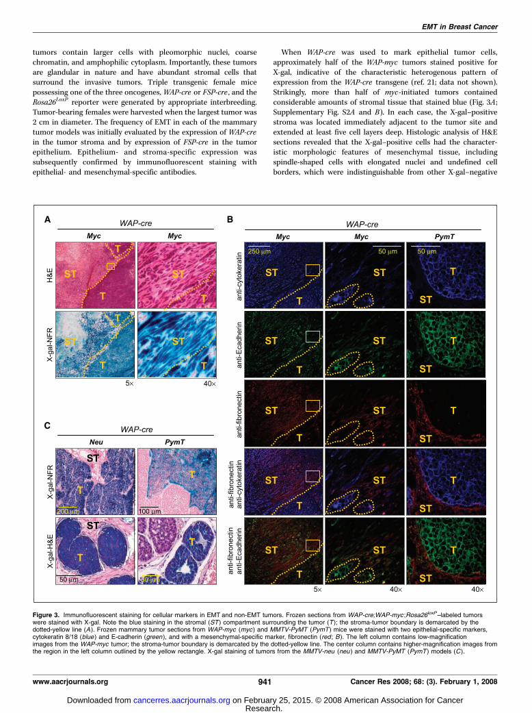
When WAP-cre was used to mark epithelial tumor cells,approximately half of the WAP-myc tumors stained positive forX-gal, indicative of the characteristic heterogenous pattern ofexpression from the WAP-cre transgene (ref. 21; data not shown).Strikingly, more than half of myc-initiated tumors containedconsiderable amounts of stromal tissue that stained blue (Fig. 3A ;Supplementary Fig. S2A and B). In each case, the X-gal–positivestroma was located immediately adjacent to the tumor site andextended at least five cell layers deep. Histologic analysis of H&Esections revealed that the X-gal–positive cells had the character-istic morphologic features of mesenchymal tissue, includingspindle-shaped cells with elongated nuclei and undefined cellborders, which were indistinguishable from other X-gal–negative
Figure 3. Immunofluorescent staining for cellular markers in EMT and non-EMT tumors. Frozen sections from WAP-cre;WAP-myc ;Rosa26loxP–labeled tumorswere stained with X-gal. Note the blue staining in the stromal (ST ) compartment surrounding the tumor (T ); the stroma-tumor boundary is demarcated by thedotted-yellow line (A ). Frozen mammary tumor sections from WAP-myc (myc ) and MMTV-PyMT (PymT ) mice were stained with two epithelial-specific markers,cytokeratin 8/18 (blue ) and E-cadherin (green ), and with a mesenchymal-specific marker, fibronectin (red ; B). The left column contains low-magnificationimages from the WAP-myc tumor; the stroma-tumor boundary is demarcated by the dotted-yellow line. The center column contains higher-magnification images fromthe region in the left column outlined by the yellow rectangle. X-gal staining of tumors from the MMTV-neu (neu ) and MMTV-PyMT (PymT ) models (C ).
EMT in Breast Cancer
www.aacrjournals.org 941 Cancer Res 2008; 68: (3). February 1, 2008
Research. on February 25, 2015. © 2008 American Association for Cancercancerres.aacrjournals.org Downloaded from
mesenchymal tissue in the vicinity. We then used epithelial-(cytokeratin and E-cadherin) and mesenchymal- ( fibronectin)specific cell markers to confirm the identity of the X-gal–positivespindle shaped cells adjacent to the primary tumor. Immunoflu-orescence staining of consecutive sections revealed that some ofthese blue spindle-shaped cells expressed fibronectin only, whereasothers expressed either fibronectin and cytokeratin or all three,fibronectin, cytokeratin, and E-cadherin (Fig. 3B, left and centercolumns), indicating a spectrum of late EMT stages within the sametumor microenvironment. In contrast to WAP-myc–initiatedtumors, pathologic examination of MMTV-neu and MMTV-PyMTtumors revealed no blue cells displaying mesenchymal morphology,suggesting that late-stage EMT had not occurred in these tumormodels (Fig. 3C ; Supplementary Fig. S2A). Indeed, epithelial tumorcells staining positive for E-cadherin or cytokeratin 8/18 weredistinctly separated from stromal cells staining positive forfibronectin (Fig. 3C, right column), confirming the absence ofEMT in neu- and PyMT-initiated tumors.To identify early EMT events in myc-initiated breast cancer,
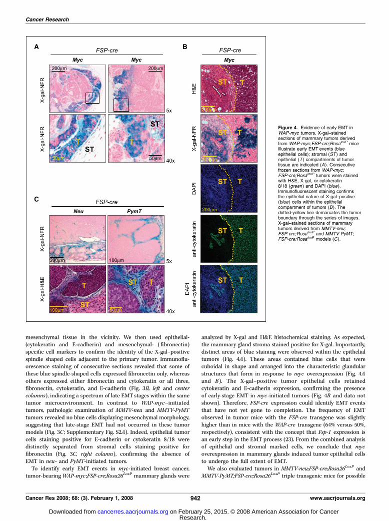
tumor-bearing WAP-myc ;FSP-cre;Rosa26LoxP mammary glands were
analyzed by X-gal and H&E histochemical staining. As expected,the mammary gland stroma stained positive for X-gal. Importantly,distinct areas of blue staining were observed within the epithelialtumors (Fig. 4A). These areas contained blue cells that werecuboidal in shape and arranged into the characteristic glandularstructures that form in response to myc overexpression (Fig. 4Aand B ). The X-gal–positive tumor epithelial cells retainedcytokeratin and E-cadherin expression, confirming the presenceof early-stage EMT in myc-initiated tumors (Fig. 4B and data notshown). Therefore, FSP-cre expression could identify EMT eventsthat have not yet gone to completion. The frequency of EMTobserved in tumor mice with the FSP-cre transgene was slightlyhigher than in mice with the WAP-cre transgene (64% versus 50%,respectively), consistent with the concept that Fsp-1 expression isan early step in the EMT process (23). From the combined analysisof epithelial and stromal marked cells, we conclude that mycoverexpression in mammary glands induced tumor epithelial cellsto undergo the full extent of EMT.We also evaluated tumors in MMTV-neu;FSP-cre;Rosa26LoxP and
MMTV-PyMT;FSP-cre;Rosa26LoxP triple transgenic mice for possible
Figure 4. Evidence of early EMT inWAP-myc tumors. X-gal–stainedsections of mammary tumors derivedfrom WAP-myc ;FSP-cre;RosaloxP miceillustrate early EMT events (blueepithelial cells); stromal (ST ) andepithelial (T ) compartments of tumortissue are indicated (A ). Consecutivefrozen sections from WAP-myc;FSP-cre;RosaloxP tumors were stainedwith H&E, X-gal, or cytokeratin8/18 (green ) and DAPI (blue ).Immunofluorescent staining confirmsthe epithelial nature of X-gal–positive(blue ) cells within the epithelialcompartment of tumors (B ). Thedotted-yellow line demarcates the tumorboundary through the series of images.X-gal–stained sections of mammarytumors derived from MMTV-neu;FSP-cre;RosaloxP and MMTV-PyMT;FSP-cre;RosaloxP models (C ).
Cancer Research
Cancer Res 2008; 68: (3). February 1, 2008 942 www.aacrjournals.org
Research. on February 25, 2015. © 2008 American Association for Cancercancerres.aacrjournals.org Downloaded from
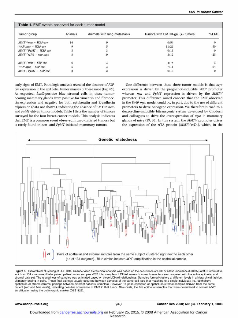
early signs of EMT. Pathologic analysis revealed the absence of FSP-cre expression in the epithelial tumor masses of these mice (Fig. 4C).As expected, LacZ-positive blue stromal cells in these tumor-bearing mammary glands were positive for vimentin and fibronec-tin expression and negative for both cytokeratin and E-cadherinexpression (data not shown), indicating the absence of EMT in neu-and PyMT-driven tumor models. Table 1 lists the number of tumorssurveyed for the four breast cancer models. This analysis indicatesthat EMT is a common event observed in myc-initiated tumors butis rarely found in neu- and PyMT-initiated mammary tumors.
One difference between these three tumor models is that mycexpression is driven by the pregnancy-inducible WAP promoterwhereas neu and PyMT expression is driven by the MMTVpromoter. This difference raised concern that the EMT observedin the WAP-myc model could be, in part, due to the use of differentpromoters to drive oncogene expression. We therefore turned to adoxycycline-inducible bitransgenic system developed by Chodoshand colleagues to drive the overexpression of myc in mammaryglands of mice (29, 30). In this system, the MMTV promoter drivesthe expression of the rtTA protein (MMTV-rtTA), which, in the
Table 1. EMT events observed for each tumor model
Tumor group Animals Animals with lung metastasis Tumors with EMT/X-gal (+) tumors %EMT
MMTV-neu + WAP-cre 14 9 0/54 0
WAP-myc + WAP-cre 9 5 11/22 50MMTV-PyMT + WAP-cre 3 3 0/13 0
MMTV-rtTA + teto-myc 8 0 3/12 25
MMTV-neu + FSP-cre 6 3 4/78 5WAP-myc + FSP-cre 5 3 7/11 64
MMTV-PyMT + FSP-cre 2 2 0/15 0
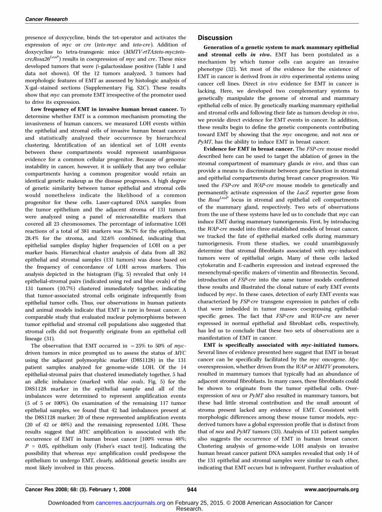
Figure 5. Hierarchical clustering of LOH data. Unsupervised hierarchical analysis was based on the occurrence of LOH or allelic imbalance (LOH/AI) at 381 informativeloci from 131 stromal-epithelial paired patient tumor samples (262 total samples). LOH/AI values from each sample were compared with the entire epithelial andstromal data set. The relatedness of samples was estimated based on close LOH/AI relationships. Samples formed clusters at different levels in a hierarchical fashion,ultimately ending in pairs. These final pairings usually occurred between samples of the same cell type (not matching to a single individual; i.e., epithelium/epithelium or stromal/stromal pairings between different patients’ samples). However, 14 pairs consisted of epithelium/stromal samples derived from the samepatient (red and blue ovals ), indicating possible occurrence of EMT in that tumor. Blue ovals, the five epithelial samples that were determined to contain MYCamplification using the polymorphic marker (D8S1128).
EMT in Breast Cancer
www.aacrjournals.org 943 Cancer Res 2008; 68: (3). February 1, 2008
Research. on February 25, 2015. © 2008 American Association for Cancercancerres.aacrjournals.org Downloaded from
presence of doxycycline, binds the tet-operator and activates theexpression of myc or cre (teto-myc and teto-cre). Addition ofdoxycycline to tetra-transgenic mice (MMTV-rtTA;teto-myc;teto-cre;Rosa26LoxP) results in coexpression of myc and cre . These micedeveloped tumors that were h-galactosidase positive (Table 1 anddata not shown). Of the 12 tumors analyzed, 3 tumors hadmorphologic features of EMT as assessed by histologic analysis ofX-gal–stained sections (Supplementary Fig. S2C). These resultsshow that myc can promote EMT irrespective of the promoter usedto drive its expression.Low frequency of EMT in invasive human breast cancer. To
determine whether EMT is a common mechanism promoting theinvasiveness of human cancers, we measured LOH events withinthe epithelial and stromal cells of invasive human breast cancersand statistically analyzed their occurrence by hierarchicalclustering. Identification of an identical set of LOH eventsbetween these compartments would represent unambiguousevidence for a common cellular progenitor. Because of genomicinstability in cancer, however, it is unlikely that any two cellularcompartments having a common progenitor would retain anidentical genetic makeup as the disease progresses. A high degreeof genetic similarity between tumor epithelial and stromal cellswould nonetheless indicate the likelihood of a commonprogenitor for these cells. Laser-captured DNA samples fromthe tumor epithelium and the adjacent stroma of 131 tumorswere analyzed using a panel of microsatellite markers thatcovered all 23 chromosomes. The percentage of informative LOHreactions of a total of 381 markers was 36.7% for the epithelium,28.4% for the stroma, and 32.6% combined, indicating thatepithelial samples display higher frequencies of LOH on a permarker basis. Hierarchical cluster analysis of data from all 262epithelial and stromal samples (131 tumors) was done based onthe frequency of concordance of LOH across markers. Thisanalysis depicted in the histogram (Fig. 5) revealed that only 14epithelial-stromal pairs (indicated using red and blue ovals) of the131 tumors (10.7%) clustered immediately together, indicatingthat tumor-associated stromal cells originate infrequently fromepithelial tumor cells. Thus, our observations in human patientsand animal models indicate that EMT is rare in breast cancer. Acomparable study that evaluated nuclear polymorphisms betweentumor epithelial and stromal cell populations also suggested thatstromal cells did not frequently originate from an epithelial celllineage (31).The observation that EMT occurred in f25% to 50% of myc-
driven tumors in mice prompted us to assess the status of MYCusing the adjacent polymorphic marker (D8S1128) in the 131patient samples analyzed for genome-wide LOH. Of the 14epithelial-stromal pairs that clustered immediately together, 5 hadan allelic imbalance (marked with blue ovals , Fig. 5) for theD8S1128 marker in the epithelial sample and all of theimbalances were determined to represent amplification events(5 of 5 or 100%). On examination of the remaining 117 tumorepithelial samples, we found that 42 had imbalances present atthe D8S1128 marker; 20 of these represented amplification events(20 of 42 or 48%) and the remaining represented LOH. Theseresults suggest that MYC amplification is associated with theoccurrence of EMT in human breast cancer [100% versus 48%;P = 0.05, epithelium only (Fisher’s exact test)]. Indicating thepossibility that whereas myc amplification could predispose theepithelium to undergo EMT, clearly, additional genetic insults aremost likely involved in this process.
Discussion
Generation of a genetic system to mark mammary epithelialand stromal cells in vivo . EMT has been postulated as amechanism by which tumor cells can acquire an invasivephenotype (32). Yet most of the evidence for the existence ofEMT in cancer is derived from in vitro experimental systems usingcancer cell lines. Direct in vivo evidence for EMT in cancer islacking. Here, we developed two complementary systems togenetically manipulate the genome of stromal and mammaryepithelial cells of mice. By genetically marking mammary epithelialand stromal cells and following their fate as tumors develop in vivo ,we provide direct evidence for EMT events in cancer. In addition,these results begin to define the genetic components contributingtoward EMT by showing that the myc oncogene, and not neu orPyMT, has the ability to induce EMT in breast cancer.Evidence for EMT in breast cancer. The FSP-cre mouse model
described here can be used to target the ablation of genes in thestromal compartment of mammary glands in vivo , and thus canprovide a means to discriminate between gene function in stromaland epithelial compartments during breast cancer progression. Weused the FSP-cre and WAP-cre mouse models to genetically andpermanently activate expression of the LacZ reporter gene fromthe RosaLoxP locus in stromal and epithelial cell compartmentsof the mammary gland, respectively. Two sets of observationsfrom the use of these systems have led us to conclude that myc caninduce EMT during mammary tumorigenesis. First, by introducingthe WAP-cre model into three established models of breast cancer,we tracked the fate of epithelial marked cells during mammarytumorigenesis. From these studies, we could unambiguouslydetermine that stromal fibroblasts associated with myc-inducedtumors were of epithelial origin. Many of these cells lackedcytokeratin and E-cadherin expression and instead expressed themesenchymal-specific makers of vimentin and fibronectin. Second,introduction of FSP-cre into the same tumor models confirmedthese results and illustrated the clonal nature of early EMT eventsinduced by myc . In these cases, detection of early EMT events wascharacterized by FSP-cre transgene expression in patches of cellsthat were imbedded in tumor masses coexpressing epithelial-specific genes. The fact that FSP-cre and WAP-cre are neverexpressed in normal epithelial and fibroblast cells, respectively,has led us to conclude that these two sets of observations are amanifestation of EMT in cancer.EMT is specifically associated with myc-initiated tumors.
Several lines of evidence presented here suggest that EMT in breastcancer can be specifically facilitated by the myc oncogene. Mycoverexpression, whether driven from theWAP orMMTV promoters,resulted in mammary tumors that typically had an abundance ofadjacent stromal fibroblasts. In many cases, these fibroblasts couldbe shown to originate from the tumor epithelial cells. Over-expression of neu or PyMT also resulted in mammary tumors, butthese had little stromal contribution and the small amount ofstroma present lacked any evidence of EMT. Consistent withmorphologic differences among these mouse tumor models, myc-derived tumors have a global expression profile that is distinct fromthat of neu and PyMT tumors (33). Analysis of 131 patient samplesalso suggests the occurrence of EMT in human breast cancer.Clustering analysis of genome-wide LOH analysis on invasivehuman breast cancer patient DNA samples revealed that only 14 ofthe 131 epithelial and stromal samples were similar to each other,indicating that EMT occurs but is infrequent. Further evaluation of
Cancer Research
Cancer Res 2008; 68: (3). February 1, 2008 944 www.aacrjournals.org
Research. on February 25, 2015. © 2008 American Association for Cancercancerres.aacrjournals.org Downloaded from
this data using the polymorphic marker (D8S1128), which residesnear theMYC locus, established that those 14 samples were twice aslikely to have MYC amplification compared with the 117 remainingsamples, supporting an association of MYC with EMT.Our data also suggest that myc overexpression is not sufficient
for EMT to occur because only f50% of the 45 myc-initiatedtumors analyzed had evidence of EMT. In other words, mycoverexpression does not necessarily lead to EMT. Moreover, earlyevents of EMT observed in myc-initiated tumors expressing theFSP-cre;RosalLoxP reporter were visualized as discrete clonalpatches of cells within epithelial tumor masses. As discussedabove, 5 of 14 human breast cancer samples had allelic imbalance(amplification) for the D8S1128 marker residing next to the MYClocus. These mouse and human data would suggest that additionalgenetic or epigenetic events are likely required to collaborate withmyc for the full manifestation of EMT. These could include thealteration of components that function downstream or in parallelto MYC signaling. It will be important in the future to identify theselect cadre of genomic alterations (genetic or epigenetic) thatcollaborate with myc and contribute to EMT in cancer, and toelucidate their mechanism of action in this process.EMT is not a prerequisite for invasiveness and metastasis in
breast cancer. The observation that EMT is specifically detectedin myc-initiated tumors in mice and is associated with amplifica-tion of MYC in human patients is consistent with the highlyinvasive nature of these types of tumors. However, these data alsosuggest that EMT is not required for metastatic progression
because tumors analyzed in mice and humans lacking EMT clearlyhad metastatic potential. Indeed, half of the mice bearing myc-initiated tumors and essentially all mice bearing neu- and PyMT-initiated tumors lacked any evidence of EMT, and yet many of theseanimals had significant amounts of lung metastases (data notshown).Although not a prerequisite for invasive behavior, EMT may
nonetheless represent one mechanism to facilitate progressiontoward a more aggressive metastatic cancer. In addition, theoccurrence of EMT may affect other aspects of cancer biology. Forexample, EMT might provide tumor cells with the ability to adaptto physiologically relevant stresses such as low oxygen or nutrientlevels. EMT might also have a profound consequence in theresponsiveness of tumors to various therapeutic treatments. Thus,our data support the hypothesis that genetic alterations in theepithelium initiate tumorigenesis and that EMTcould contribute tothe microenvironment and modulate interpatient biologicalbehavior (34).
Acknowledgments
Received 6/8/2007; revised 10/8/2007; accepted 11/12/2007.Grant support: NIH grant P01 CA097189 (M.C. Ostrowski), Department of Defense
grant BC030892 (A. de Bruin), and The Pew Charitable Trusts grant 2590SC. C. Eng is arecipient of the Doris Duke Distinguished Clinical Scientist Award, and G. Leone is therecipient of The Pew Charitable Trusts Scholar Award and the Leukemia & LymphomaSociety Scholar Award.
The costs of publication of this article were defrayed in part by the payment of pagecharges. This article must therefore be hereby marked advertisement in accordancewith 18 U.S.C. Section 1734 solely to indicate this fact.
EMT in Breast Cancer
www.aacrjournals.org 945 Cancer Res 2008; 68: (3). February 1, 2008
References1. Bates RC, DeLeo, MJ, Mercurio AM. The epithelial-mesenchymal transition of colon carcinoma involvesexpression of IL-8 and CXCR-1-mediated chemotaxis.Exp Cell Res 2004;299:315–24.2. Bakin AV, Tomlinson AK, Bhowmick NA, Moses HL,Arteaga CL. Phosphatidylinositol 3-kinase function isrequired for transforming growth factor h mediatedepithelial to mesenchymal transition and cell migration.J Biol Chem 2000;275:36803–10.3. Oft M, Heider KH, Beug H. TGFh signaling is necessaryfor carcinoma cell invasiveness and metastasis. CurrBiol 1999;8:1243–52.4. Thiery JP. Epithelial-mesenchymal transitions indevelopment and pathologies. Curr Opin Cell Biol2003;15:740–6.5. Thiery JP. Epithelial-mesenchymal transitions intumour progression. Nat Rev Cancer 2002;2:442–54.6. Huber MA, Kraut N, Beug, H. Molecular requirementsfor epithelial-mesenchymal transition during tumorprogression. Curr Opin Cell Biol 2005;17:548–58.7. Xue C, Plieth D, Venkov C, Xu C, Neilson EG. Thegatekeeper effect of epithelial-mesenchymal transitionregulates the frequency of breast cancer metastasis.Cancer Res 2003;63:3386–94.8. Mikaelian I, Blades N, Churchill GA, et al. Proteotypicclassification of spontaneous and transgenic mammaryneoplasms. Breast Cancer Res 2004;6:R668–79.9. Moody SE, Perez D, Pan TC, et al. The transcriptionalrepressor Snail promotes mammary tumor recurrence.Cancer Cell 2005;8:197–209.10. Tarin D. The fallacy of epithelial mesenchymaltransition in neoplasia. Cancer Res 2005;65:5996–6000.11. Thompson EW, Newgreen DF. Carcinoma invasionand metastasis: a role for epithelial-mesenchymaltransition? Cancer Res 2005;65:5991–5.12. Soriano P. Generalized lacZ expression with theROSA26 Cre reporter strain. Nat Genet 1999;21:70–1.13. Strutz F, Okada H, Lo CW, et al. Identification andcharacterization of a fibroblast marker: FSP1. J Cell Biol1995;130:393–405.
14. Okada H, Danoff TM, Fischer A, Lopez-Guisa JM,Strutz F, Neilson EG. Identification of a novel cis -actingelement for fibroblast-specific transcription of the FSP1gene. Am J Physiol 1998;275:F306–14.15. Gu H, Zou YR, Rajewsky K. Independent control ofimmunoglobulin switch recombination at individualswitch regions evidenced through Cre-loxP-mediatedgene targeting. Cell 1993;73:1155–64.16. Overbeek PA, Aguilar-Cordova E, Hanten G, et al.Coinjection strategy for visual identification of trans-genic mice. Transgenic Res 1991;1:31–7.17. Taketo M, Schroeder AC, Mobraaten LE, et al. FVB/N:an inbred mouse strain preferable for transgenicanalyses. Proc Natl Acad Sci U S A 1991;88:2065–9.18. Kurose K, Hoshaw-Woodard S, Adeyinka A, Leme-show S, Watson PH, Eng C. Genetic model of multi-stepbreast carcinogenesis involving the epithelium andstroma: clues to tumour-microenvironment interac-tions. Hum Mol Genet 2001;10:1907–13.19. Kurose K, Gilley K, Matsumoto S, Watson PH, ZhouXP, Eng C. Frequent somatic mutations in PTEN andTP53 are mutually exclusive in the stroma of breastcarcinomas. Nat Genet 2002;32:355–7.20. Eisen MB, et al. Cluster analysis and display ofgenome-wide expression patterns. Proc Natl Acad SciU S A 1998;95:14863–8.21. Wagner KU, Wall RJ, St-Onge L, et al. Cre-mediatedgene deletion in the mammary gland. Nucleic Acids Res1997;25:4323–30.22. Bhowmick NA, Chytil A, Plieth D, et al. TGF-hsignaling in fibroblasts modulates the oncogenicpotential of adjacent epithelia. Science 2004;303:848–51.23. Okada H, Danoff TM, Kalluri R, Neilson EG. Early roleof Fsp1 in epithelial-mesenchymal transformation. Am JPhysiol 1997;273:F563–74.24. Schoenenberger CA, Andres AC, Groner B, van derValk M, LeMeur M, Gerlinger P. Targeted c-myc geneexpression in mammary glands of transgenic miceinduces mammary tumours with constitutive milkprotein gene transcription. EMBO J 1988;7:169–75.
25. Guy CT, Webster MA, Schaller M, Parsons TJ, CardiffRD, Muller WJ. Expression of the neu protooncogene inthe mammary epithelium of transgenic mice inducesmetastatic disease. Proc Natl Acad Sci U S A 1992;89:10578–82.26. Guy CT, Cardiff RD, Muller WJ. Induction ofmammary tumors by expression of polyomavirusmiddle T oncogene: a transgenic mouse model formetastatic disease. Mol Cell Biol 1992;12:954–61.27. Cardiff RD, Anver MR, Gusterson BA, et al. Themammary pathology of genetically engineered mice:the consensus report and recommendations from theAnnapolis meeting. Oncogene 2000;19:968–88.28. Rosner A, Miyoshi K, Landesman-Bollag E, et al.Pathway pathology: histological differences betweenErbB/Ras and Wnt pathway transgenic mammarytumors. Am J Pathol 2002;161:1087–97.29. D’Cruz CM, Gunther EJ, Boxer RB, et al. c-MYCinduces mammary tumorigenesis by means of apreferred pathway involving spontaneous Kras2 muta-tions. Nat Med 2001;7:235–9.30. Gunther EJ, Belka GK, Wertheim GB, et al. A noveldoxycycline-inducible system for the transgenic anal-ysis of mammary gland biology. FASEB J 2002;16:283–92.31. Allinen M, Beroukhim R, Cai L, et al. Molecularcharacterization of the tumor microenvironment inbreast cancer. Cancer Cell 2004;6:17–32.32. Vincent-Salomon A, Thiery JP. Host microenviron-ment in breast cancer development: epithelial-mesen-chymal transition in breast cancer development. BreastCancer Res 2003;5:101–6.33. Desai KV, Xiao N, Wang W, et al. Initiating oncogenicevent determines gene-expression patterns of humanbreast cancer models. Proc Natl Acad Sci U S A 2002;99:6967–72.34. Fukino K, Shen L, Matsumoto S, Morrison CD, MutterG, Eng C. Combined total genome loss of heterozygosityscan of breast cancer stroma and epithelium revealsmultiplicity of stromal targets. Cancer Res 2004;64:7231–6.
Research. on February 25, 2015. © 2008 American Association for Cancercancerres.aacrjournals.org Downloaded from
0
Announcements
MEETING OF THE RADIATION RESEARCH SOCIETY
The annual meeting of the Radiation Research Society will be held at the State University of Iowa, IowaCity, on June 22—24,1953. The Society will be the guestof the University, and all meetings will be held on thecampus. The program will consist of: (1) Two symposia,one on “TheEffects of Rwliation on Aqueous Solutions,― which includes the following speakers: E. S. G.Barren, Edwin J. Hart, Warren Garrison, J. L. Magee,and A. 0. Allen. The second is “PhysicalMeasurementsfor Radiobiology―and companion talks by Ugo Fano,Burton J. Moyer, G. Failla, L. D. Marinelli, and Payne
The following correction should be made in the article by Beck and Valentine, “TheAerobic CarbohydrateMetabolism of Leukocytes in Health and Leukemia. I.Glycolysis and Respiration,― November, 1952, page 821;substitute for the last paragraph:
The data in Table 3 permit several interesting calculations. If one compares the amount of glucose actually
disappearing with the sum of the amount equivalent tolactic acid produced plus that equivalent to 02 con
sumption, it is seen that the amount of glucose “cleavage products―exceeds the amount of glucose utilized b12 per cent in N and 27 per cent in CML and is exceeded
S. Harris. (2) On Monday night, June 22, a lecture byDr. L. W. Alvarez on meson physics has been tentatively scheduled. On Tuesday night, June 23, Dr. L. H.Gray of the Hammersmith Hospital, London, will speakon a topic to be announced. Dr. Gray's lecture is sponsored by the Iowa Branch of the American Cancer Society. Those desiring to report original research in radiation effects, or interested in attending or desiring additional information, please contact the Secretary of theSociety, Dr. A. Edelmann, Biology Department, Brookhaven National Laboratory, Upton, L.I., New York.
by the glucose utilized by 16 per cent in CLL. If the assumption is made that, in this respect, the myeloid andlymphoid celLsof leukemia are similar to those of norma! blood, it may be that the computed normal figurerepresents a summation of the myeloid (M) andlymphoid (L) cells that make up the normal leukocytepopulation. Thus, if M = +0.27 and L = —0.16 andthe normal differential is 65 per cent M and So per centL, then
0.65 (+0.27) + 0.35 (—0.16) = +0.12
a figure identical to the observed +0.12 for normalleukocytes.
ERRATUM
308
2008;68:937-945. Cancer Res Anthony J. Trimboli, Koichi Fukino, Alain de Bruin, et al. Breast CancerDirect Evidence for Epithelial-Mesenchymal Transitions in
Updated version
http://cancerres.aacrjournals.org/content/68/3/937
Access the most recent version of this article at:
Material
Supplementary
http://cancerres.aacrjournals.org/content/suppl/2008/01/23/68.3.937.DC1.html
Access the most recent supplemental material at:
Cited Articles
http://cancerres.aacrjournals.org/content/68/3/937.full.html#ref-list-1
This article cites by 34 articles, 15 of which you can access for free at:
Citing articles
http://cancerres.aacrjournals.org/content/68/3/937.full.html#related-urls
This article has been cited by 21 HighWire-hosted articles. Access the articles at:
E-mail alerts related to this article or journal.Sign up to receive free email-alerts
Subscriptions
Reprints and
To order reprints of this article or to subscribe to the journal, contact the AACR Publications
Permissions
To request permission to re-use all or part of this article, contact the AACR Publications
Research. on February 25, 2015. © 2008 American Association for Cancercancerres.aacrjournals.org Downloaded from
Related Documents