Behavioral/Systems/Cognitive Diffusion-Weighted Imaging Tractography-Based Parcellation of the Human Parietal Cortex and Comparison with Human and Macaque Resting-State Functional Connectivity Rogier B. Mars, 1,3 Saad Jbabdi, 3 Je ´ro ˆme Sallet, 1 Jill X. O’Reilly, 3,4 Paula L. Croxson, 1,4 Etienne Olivier, 1,5 MaryAnn P. Noonan, 1 Caroline Bergmann, 2 Anna S. Mitchell, 1 Mark G. Baxter, 4 Timothy E. J. Behrens, 3 Heidi Johansen-Berg, 3 Valentina Tomassini, 3 Karla L. Miller, 3 and Matthew F. S. Rushworth 1,3 Departments of 1 Experimental Psychology and 2 Veterinary Services and 3 Centre for Functional Magnetic Resonance Imaging of the Brain, University of Oxford, Oxford OX1 2JD, United Kingdom, 4 Glickenhaus Laboratory of Neuropsychology, Department of Neuroscience, Mount Sinai School of Medicine, New York, New York 10029, and 5 Institute of Neuroscience, Universite ´ Catholique de Louvain, B-1200 Brussels, Belgium Despite the prominence of parietal activity in human neuroimaging investigations of sensorimotor and cognitive processes, there remains uncertainty about basic aspects of parietal cortical anatomical organization. Descriptions of human parietal cortex draw heavily on anatomical schemes developed in other primate species, but the validity of such comparisons has been questioned by claims that there are fundamental differences between the parietal cortex in humans and other primates. A scheme is presented for parcellation of human lateral parietal cortex into component regions on the basis of anatomical connectivity and the functional interactions of the resulting clusters with other brain regions. Anatomical connectivity was estimated using diffusion-weighted magnetic resonance image (MRI)-based tractography, and functional interactions were assessed by correlations in activity mea- sured with functional MRI at rest. Resting-state functional connectivity was also assessed directly in the rhesus macaque lateral parietal cortex in an additional experiment, and the patterns found reflected known neuroanatomical connections. Cross- correlation in the tractography-based connectivity patterns of parietal voxels reliably parcellated human lateral parietal cortex into 10 component clusters. The resting-state functional connectivity of human superior parietal and intraparietal clusters with frontal and extrastriate cortex suggested correspondences with areas in macaque superior and intraparietal sulcus. Functional connectivity patterns with parahippocampal cortex and premotor cortex again suggested fundamental correspondences between inferior parietal cortex in humans and macaques. In contrast, the human parietal cortex differs in the strength of its interactions between the central inferior parietal lobule region and the anterior prefrontal cortex. Introduction Research with both human and nonhuman primates shows that the parietal cortex is involved in numerous cognitive functions, from perception to control of action and higher-order processes, such as numerosity and memory. It is well established that pari- etal cortex consists of a mosaic of subregions, which are distinct both functionally and anatomically (Grefkes and Fink, 2005; Caminiti et al., 2010; Nelson et al., 2010). Our knowledge of the different subregions and their functional properties is primarily based on the macaque model. However, the suitability of this model has been called into question (Passingham, 2009). The parietal cortex has expanded substantially in the human brain compared with other primates and even other hominids, which might have resulted in an increase or a rearrangement of its re- gions (Vanduffel et al., 2002b; Grefkes and Fink, 2005). Particular emphasis has been given to the possibility that human inferior parietal lobule is quite distinct to that of other species (Husain and Nachev, 2007). The function of any brain area is constrained by its extrinsic connections. Indeed, patterns of anatomical connectivity can in- form the segregation of functionally distinct areas (Passingham et al., 2002; Averbeck et al., 2009). Here, we aim to identify and characterize key component regions of human parietal cortex using a combination of non-invasive magnetic resonance imag- ing (MRI) techniques: probabilistic diffusion tractography and resting-state functional connectivity. The primary aim is to pro- vide a parcellation of human parietal cortex that can inform non- invasive neuroimaging studies. An additional aim is to compare Received Sept. 29, 2010; revised Dec. 15, 2010; accepted Jan. 11, 2011. This work was supported by a Marie Curie Intra-European Fellowship within the 6th European Community Framework Programme (R.B.M.), the Wellcome Trust (M.P.N., M.G.B., H.J.-B.), the Medical Research Council (United Kingdom) (R.B.M., S.J., J.X.O., A.S.M., and M.F.S.R.), the Multiple Sclerosis Society (United Kingdom) (V.T.), the Italian Foundation for Multiple Sclerosis (V.T.), the Royal Academy of Engineering (K.L.M.), and the Economic and Social Research Council (United Kingdom) (K.L.M.). We thank Urs Schu ¨ffelgen for help with Caret analyses. Correspondence should be addressed to Rogier B. Mars, Department of Experimental Psychology, University of Oxford, Tinbergen Building, 9 South Parks Road, Oxford OX1 3UD, UK. E-mail: [email protected]. DOI:10.1523/JNEUROSCI.5102-10.2011 Copyright © 2011 the authors 0270-6474/11/314087-14$15.00/0 The Journal of Neuroscience, March 16, 2011 • 31(11):4087– 4100 • 4087

Welcome message from author
This document is posted to help you gain knowledge. Please leave a comment to let me know what you think about it! Share it to your friends and learn new things together.
Transcript

Behavioral/Systems/Cognitive
Diffusion-Weighted Imaging Tractography-BasedParcellation of the Human Parietal Cortex and Comparisonwith Human and Macaque Resting-State FunctionalConnectivity
Rogier B. Mars,1,3 Saad Jbabdi,3 Jerome Sallet,1 Jill X. O’Reilly,3,4 Paula L. Croxson,1,4 Etienne Olivier,1,5
MaryAnn P. Noonan,1 Caroline Bergmann,2 Anna S. Mitchell,1 Mark G. Baxter,4 Timothy E. J. Behrens,3
Heidi Johansen-Berg,3 Valentina Tomassini,3 Karla L. Miller,3 and Matthew F. S. Rushworth1,3
Departments of 1Experimental Psychology and 2Veterinary Services and 3Centre for Functional Magnetic Resonance Imaging of the Brain, University ofOxford, Oxford OX1 2JD, United Kingdom, 4Glickenhaus Laboratory of Neuropsychology, Department of Neuroscience, Mount Sinai School of Medicine,New York, New York 10029, and 5Institute of Neuroscience, Universite Catholique de Louvain, B-1200 Brussels, Belgium
Despite the prominence of parietal activity in human neuroimaging investigations of sensorimotor and cognitive processes, thereremains uncertainty about basic aspects of parietal cortical anatomical organization. Descriptions of human parietal cortex drawheavily on anatomical schemes developed in other primate species, but the validity of such comparisons has been questioned byclaims that there are fundamental differences between the parietal cortex in humans and other primates. A scheme is presented forparcellation of human lateral parietal cortex into component regions on the basis of anatomical connectivity and the functionalinteractions of the resulting clusters with other brain regions. Anatomical connectivity was estimated using diffusion-weightedmagnetic resonance image (MRI)-based tractography, and functional interactions were assessed by correlations in activity mea-sured with functional MRI at rest. Resting-state functional connectivity was also assessed directly in the rhesus macaque lateralparietal cortex in an additional experiment, and the patterns found reflected known neuroanatomical connections. Cross-correlation in the tractography-based connectivity patterns of parietal voxels reliably parcellated human lateral parietal cortexinto 10 component clusters. The resting-state functional connectivity of human superior parietal and intraparietal clusters withfrontal and extrastriate cortex suggested correspondences with areas in macaque superior and intraparietal sulcus. Functionalconnectivity patterns with parahippocampal cortex and premotor cortex again suggested fundamental correspondences betweeninferior parietal cortex in humans and macaques. In contrast, the human parietal cortex differs in the strength of its interactionsbetween the central inferior parietal lobule region and the anterior prefrontal cortex.
IntroductionResearch with both human and nonhuman primates shows thatthe parietal cortex is involved in numerous cognitive functions,from perception to control of action and higher-order processes,such as numerosity and memory. It is well established that pari-etal cortex consists of a mosaic of subregions, which are distinctboth functionally and anatomically (Grefkes and Fink, 2005;Caminiti et al., 2010; Nelson et al., 2010). Our knowledge of thedifferent subregions and their functional properties is primarily
based on the macaque model. However, the suitability of thismodel has been called into question (Passingham, 2009). Theparietal cortex has expanded substantially in the human braincompared with other primates and even other hominids, whichmight have resulted in an increase or a rearrangement of its re-gions (Vanduffel et al., 2002b; Grefkes and Fink, 2005). Particularemphasis has been given to the possibility that human inferiorparietal lobule is quite distinct to that of other species (Husainand Nachev, 2007).
The function of any brain area is constrained by its extrinsicconnections. Indeed, patterns of anatomical connectivity can in-form the segregation of functionally distinct areas (Passingham etal., 2002; Averbeck et al., 2009). Here, we aim to identify andcharacterize key component regions of human parietal cortexusing a combination of non-invasive magnetic resonance imag-ing (MRI) techniques: probabilistic diffusion tractography andresting-state functional connectivity. The primary aim is to pro-vide a parcellation of human parietal cortex that can inform non-invasive neuroimaging studies. An additional aim is to compare
Received Sept. 29, 2010; revised Dec. 15, 2010; accepted Jan. 11, 2011.This work was supported by a Marie Curie Intra-European Fellowship within the 6th European Community
Framework Programme (R.B.M.), the Wellcome Trust (M.P.N., M.G.B., H.J.-B.), the Medical Research Council (UnitedKingdom) (R.B.M., S.J., J.X.O., A.S.M., and M.F.S.R.), the Multiple Sclerosis Society (United Kingdom) (V.T.), theItalian Foundation for Multiple Sclerosis (V.T.), the Royal Academy of Engineering (K.L.M.), and the Economic andSocial Research Council (United Kingdom) (K.L.M.). We thank Urs Schuffelgen for help with Caret analyses.
Correspondence should be addressed to Rogier B. Mars, Department of Experimental Psychology, University ofOxford, Tinbergen Building, 9 South Parks Road, Oxford OX1 3UD, UK. E-mail: [email protected].
DOI:10.1523/JNEUROSCI.5102-10.2011Copyright © 2011 the authors 0270-6474/11/314087-14$15.00/0
The Journal of Neuroscience, March 16, 2011 • 31(11):4087– 4100 • 4087
the parietal regions in the human brainwith macaque parietal regions whose neu-rophysiology and connectional anatomyare well studied.
First, we determine the connectivity pro-files of all human parietal voxels usingdiffusion-weightedmagnetic resonance imag-ing (DW-MRI) and tractography and thenparcellate the parietal cortex into distinctclusters based on the cross-correlations be-tween these connectivity profiles (Johansen-Berg et al., 2004; Anwander et al., 2007;Tomassini et al., 2007; Beckmann et al.,2009; Schubotz et al., 2010). Second, we ex-amine the correlations between spontane-ous activity in voxels in the parietal cortexand in other brain areas, a technique knownas “resting-state functional connectivity”(O’Reilly et al., 2010; Xiang et al., 2010). Theconcept behind this resting-state functionalMRI (fMRI) is that when the brain is “idle,”i.e., not involved in any explicit task, corre-lations in slowly fluctuating spontaneousbrain activity tend to reflect the intrinsicfunctional networks of the brain (Fox andRaichle, 2007). We focused on a limited setof eight areas known, in macaques, to proj-ect to circumscribed regions of parietal cor-tex. By comparing the correlations betweenthe eight target areas with the tractography-based parietal clusters,we can characterize the distinct parietal subregions involved in var-ious large-scale corticocortical interactions. Given the novelty of thisapproach, we first investigate resting-state interactions between pa-rietal cortex and the same target areas in the macaque brain, to assessthe degree to which the resting-state functional connectivity reflectsknown anatomical connections and thus to validate this method.
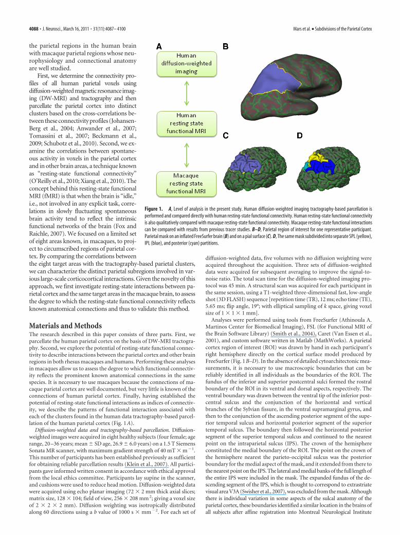
Materials and MethodsThe research described in this paper consists of three parts. First, weparcellate the human parietal cortex on the basis of DW-MRI tractogra-phy. Second, we explore the potential of resting-state functional connec-tivity to describe interactions between the parietal cortex and other brainregions in both rhesus macaques and humans. Performing these analysesin macaques allow us to assess the degree to which functional connectiv-ity reflects the prominent known anatomical connections in the samespecies. It is necessary to use macaques because the connections of ma-caque parietal cortex are well documented, but very little is known of theconnections of human parietal cortex. Finally, having established thepotential of resting-state functional interactions as indices of connectiv-ity, we describe the patterns of functional interaction associated witheach of the clusters found in the human data tractography-based parcel-lation of the human parietal cortex (Fig. 1 A).
Diffusion-weighted data and tractography-based parcellation. Diffusion-weighted images were acquired in eight healthy subjects (four female; agerange, 20 –36 years; mean � SD age, 26.9 � 6.0 years) on a 1.5 T SiemensSonata MR scanner, with maximum gradient strength of 40 mT � m �1.This number of participants has been established previously as sufficientfor obtaining reliable parcellation results (Klein et al., 2007). All partici-pants gave informed written consent in accordance with ethical approvalfrom the local ethics committee. Participants lay supine in the scanner,and cushions were used to reduce head motion. Diffusion-weighted datawere acquired using echo planar imaging (72 � 2 mm thick axial slices;matrix size, 128 � 104; field of view, 256 � 208 mm 2; giving a voxel sizeof 2 � 2 � 2 mm). Diffusion weighting was isotropically distributedalong 60 directions using a b value of 1000 s � mm �2. For each set of
diffusion-weighted data, five volumes with no diffusion weighting wereacquired throughout the acquisition. Three sets of diffusion-weighteddata were acquired for subsequent averaging to improve the signal-to-noise ratio. The total scan time for the diffusion-weighted imaging pro-tocol was 45 min. A structural scan was acquired for each participant inthe same session, using a T1-weighted three-dimensional fast, low-angleshot (3D FLASH) sequence [repetition time (TR), 12 ms; echo time (TE),5.65 ms; flip angle, 19°; with elliptical sampling of k space, giving voxelsize of 1 � 1 � 1 mm].
Analyses were performed using tools from FreeSurfer (Athinoula A.Martinos Center for Biomedical Imaging), FSL (for Functional MRI ofthe Brain Software Library) (Smith et al., 2004), Caret (Van Essen et al.,2001), and custom software written in Matlab (MathWorks). A parietalcortex region of interest (ROI) was drawn by hand in each participant’sright hemisphere directly on the cortical surface model produced byFreeSurfer (Fig. 1 B–D). In the absence of detailed cytoarchitectonic mea-surements, it is necessary to use macroscopic boundaries that can bereliably identified in all individuals as the boundaries of the ROI. Thefundus of the inferior and superior postcentral sulci formed the rostralboundary of the ROI in its ventral and dorsal aspects, respectively. Theventral boundary was drawn between the ventral tip of the inferior post-central sulcus and the conjunction of the horizontal and verticalbranches of the Sylvian fissure, in the ventral supramarginal gyrus, andthen to the conjunction of the ascending posterior segment of the supe-rior temporal sulcus and horizontal posterior segment of the superiortemporal sulcus. The boundary then followed the horizontal posteriorsegment of the superior temporal sulcus and continued to the nearestpoint on the intraparietal sulcus (IPS). The crown of the hemisphereconstituted the medial boundary of the ROI. The point on the crown ofthe hemisphere nearest the parieto-occipital sulcus was the posteriorboundary for the medial aspect of the mask, and it extended from there tothe nearest point on the IPS. The lateral and medial banks of the full length ofthe entire IPS were included in the mask. The expanded fundus of the de-scending segment of the IPS, which is thought to correspond to extrastriatevisual area V3A (Swisher et al., 2007), was excluded from the mask. Althoughthere is individual variation in some aspects of the sulcal anatomy of theparietal cortex, these boundaries identified a similar location in the brains ofall subjects after affine registration into Montreal Neurological Institute
Figure 1. A, Level of analysis in the present study. Human diffusion-weighted imaging tractography-based parcellation isperformed and compared directly with human resting-state functional connectivity. Human resting-state functional connectivityis also qualitatively compared with macaque resting-state functional connectivity. Macaque resting-state functional interactionscan be compared with results from previous tracer studies. B–D, Parietal region of interest for one representative participant.Parietal mask on an inflated FreeSurfer brain (B) and on a pial surface (C). D, The same mask subdivided into separate SPL (yellow),IPL (blue), and posterior (cyan) partitions.
4088 • J. Neurosci., March 16, 2011 • 31(11):4087– 4100 Mars et al. • Subdivisions of the Parietal Cortex
(MNI) space. It should be noted that, although the presence of both func-tional and anatomical differences between the hemispheres is well estab-lished in the human brain, especially in relation to language, the presence ofhemispheric differences in the monkey brain is still a topic of debate (forreview, see Passingham, 2008). Therefore, this study focuses on the righthemisphere, which is not dominant for language in humans.
DW-MRI data were preprocessed using tools from FDT (for Func-tional MRI of the Brain Diffusion Toolbox; part of FSL 4.1). Eddy-current distortions were corrected using affine registration of all volumesto a target volume with no diffusion weighting. Voxelwise estimates ofthe fiber orientation distribution were calculated using Bedpostx, limitedto estimating two fiber orientations at each voxel, because of the b valueand number of gradient orientations in the diffusion data (Behrens et al.,2007). For each participant, probabilistic tractography was run fromvertices at the gray matter/white matter boundary surface within theparietal ROI to assess connectivity with every brain voxel (downsampledto 5 mm isotropic voxels), using a model accounting for multiple fiberorientations in each voxel (Behrens et al., 2007). Crucially, tractographywas seeded from surface vertices (as opposed to voxels). From each seedvertex, the normal to the cortical surface was given by the FreeSurfercortical model, and this information was used to track toward thebrain instead of tracking in both directions as is usually done invoxel-based tractography. This approach dramatically reduces therisks of creating spurious sample tracts that cross gyral walls (fordetails, see www.fmrib.ox.ac.uk/fsl/fdt/fdt_surface.html). It also pro-vides quantitative estimates of connectivity from every surface vertexof the parietal mask to every voxel in the rest of the brain.
A connectivity matrix between parietal vertices and each brain voxelwas derived as described previously (Johansen-Berg et al., 2004) and usedto generate a symmetric cross-correlation matrix of dimensions (numberof seeds � number of seeds) in which the (i,j)th element value is thecorrelation between the connectivity profile of seed i and the connectivityprofile of seed j. The rows or this cross-correlation matrix were thenpermuted using k-means segmentation for automated clustering to de-fine different clusters. The goal of clustering the cross-correlation matrixis to group together regions that share the same connectivity with the restof the brain. To increase the chances of obtaining continuous (non-scattered) clusters, we included a distance constraint (Tomassini et al.,2007). The parietal cortex contains a deep sulcus, the IPS, and the Eu-clidean distance between its banks is small although they are far apartin cortical distance. The flattening of the brain in FreeSurfer space,however, made it possible to implement the distance constraint alonga geodesic that followed the cortical surface. The resulting clusters arethen constrained to consist of vertices that are spatially contiguous,although the border between clusters is still guided by remote con-nectivity information.
The number of clusters in the k-means clustering must be set by theexperimenter. To determine the optimal number of clusters resulting inconsistency across participants, we used an iterative procedure (Beck-mann et al., 2009). First, we looked for clusters to separate the parietalcortex mask into three separate partitions: a superior parietal lobule(SPL) mask, an inferior parietal lobule (IPL) mask, and a posterior IPSmask (Fig. 1 D). Following Beckmann et al. (2009), each of these maskswas then parcellated into the maximum number of areas that led to aconsistent parcellation in all eight participants. Each of the participant’sindividual clusters was then transformed from FreeSurfer space to theMNI template brain. The center of gravity of each cluster in MNI spaceand its overlap with the FSL implementation of the Harvard–Oxfordprobabilistic cortical atlas, an atlas based on manual definition of sulcal/gyral landmarks, was then established.
Resting-state fMRI data acquisition and preprocessing. Human resting-state fMRI data and T1-weighted images were collected for a differentgroup of 12 healthy volunteers (nine females; age range, 31– 61 years;mean � SD age, 43.08 � 9.17 years). This group of participants did notoverlap with the group of participants in the diffusion-weighted imagingexperiment. All participants gave informed written consent in accor-dance with ethical approval from the local ethics committee. Participantslay supine in a 1.5 T Siemens Sonata MR scanner. They were instructed toclose their eyes and lie still. Cushions were used to reduce head motion.
Whole-brain blood oxygen level-dependent (BOLD) fMRI data was col-lected for 11 min from each participant, using the following parameters:45 axial slices; in-plane resolution, 3 � 3 mm; slice thickness, 3 mm; noslice gap; TR, 3400 ms; TE, 41 ms; 200 volumes. A structural scan wasacquired for each participant in the same session, using a T1-weighted 3DFLASH sequence (voxel size of 1 � 1 � 1 mm).
Data were analyzed using tools from FSL (Smith et al., 2004) and SPM(for Statistical Parametric Mapping) (www.fil.ion.ucl.ac.uk/spm). Thefirst six volumes of each functional dataset were discarded, after whichthe preprocessing was performed: motion correction, non-brain re-moval, spatial smoothing [using Gaussian 5 mm full-width at half-maximum (FWHM) kernel], grand-mean intensity normalization of theentire four-dimensional dataset by a single multiplicative factor, andhigh-pass temporal filtering (Gaussian-weighted least-squares straightline fitting, with � � 50.0 s). Registration of functional images to theskull-stripped structural and MNI template was done using FLIRT (Jen-kinson et al., 2002).
Macaque resting-state fMRI and anatomical scans were collected for10 healthy macaques (Macaca mulatta; five females; age range, 3 yearsand 7 months to 4 years and 10 months; mean age, 4 years and 5 months;weight range, 4.34 – 8.53 kg). Protocols for animal care, magnetic reso-nance imaging, and anesthesia were performed under authority of per-sonal and project licenses in accordance with the United KingdomAnimals (Scientific Procedures) Act (1986). Anesthesia was induced us-ing intramuscular injection of ketamine (10 mg/kg), xylazine (0.125–0.25 mg/kg), and midazolam (0.1 mg/kg). Macaques also receivedinjections of atropine (0.05 mg/kg, i.m.), meloxicam (0.2 mg/kg, i.v.),and ranitidine (0.05 mg/kg, i.v.). Local anesthetic (5% lidocaine/prilo-caine cream and 2.5% bupivacaine injected subcutaneously around theears to block peripheral nerve stimulation) was also used at least 15 minbefore placing the macaque in the stereotaxic frame. The anesthetizedanimals were placed in an MRI-compatible stereotactic frame (Crist In-struments) in a sphinx position and placed in a horizontal 3 T MRIscanner with a full-size bore. Scanning commenced �2 h after induction,when the ketamine was unlikely still to be present in the system. Anes-thesia was maintained using the lowest possible concentration of isoflu-rane to ensure that macaques were lightly anesthetized. The depth ofanesthesia was assessed using physiological parameters (heart rate andblood pressure, as well as clinical checks before the scan for muscle re-laxation). During the acquisition of the functional data, the inspiredisoflurane concentration was in the range 1.0 –1.8% (mean, 1.45%), andthe expired isoflurane concentration was in the range 0.9 –1.7% (mean,1.38%). Isoflurane was selected for the scans as resting-state networkshave been demonstrated previously to be present using this agent (Vin-cent et al., 2007). Macaques were maintained with intermittent positivepressure ventilation to ensure a constant respiration rate during the func-tional scan, and respiration rate, inspired and expired CO2, and inspiredand expired isoflurane concentration were monitored and recorded us-ing VitalMonitor software (Vetronic Services Ltd.). In addition to theseparameters, core temperature and SpO2 were monitored throughout thescan. A four-channel phased-array coil was used for data acquisition (H.Kolster, MRI Coil Laboratory, Laboratory for Neuro- and Psychophysi-ology, Katholieke Universiteit Leuven, Leuven, Belgium). Whole-brainBOLD fMRI data was collected for 53 min and 26 s from each animal,using the following parameters: 36 axial slices; in-plane resolution, 2 � 2mm; slice thickness, 2 mm; no slice gap; TR, 2000 ms; TE, 19 ms; 1600volumes. A structural scan (three averages) was acquired for each ma-caque in the same session, using a T1-weighted magnetization-preparedrapid-acquisition gradient echo sequence (either 0.5 � 0.5 � 0.5 or 0.5 �0.5 � 1.0 mm voxel resolution).
The first six volumes of each functional dataset were discarded, and thefollowing preprocessing was performed: non-brain removal, 0.1 Hz low-pass filtering to remove respiratory artifacts, motion correction, spatialsmoothing (using Gaussian 3 mm FWHM kernel), grand-mean intensitynormalization of the entire four-dimensional dataset by a single multi-plicative factor, and high-pass temporal filtering (Gaussian-weightedleast-squares straight line fitting, with � � 50.0 s). Registration of func-tional images to the skull-stripped structural and a macaque templatewas done using FLIRT (Jenkinson et al., 2002).
Mars et al. • Subdivisions of the Parietal Cortex J. Neurosci., March 16, 2011 • 31(11):4087– 4100 • 4089

Resting-state fMRI data analysis. To establishthe functional connectivity of the parietal sub-regions yielded by the tractography-based par-cellation, we created a number of target masksin MNI space from the probable homologs ofbrain areas that are known to be connected tothe parietal cortex in the macaque. Functionalinteractions were determined with target areasin the ventral premotor cortex (PMv) centeredat [56, 6, 22] (Binkofski et al., 1999), dorsolat-eral prefrontal cortex (dlPFC) centered at [40,40, 24] (Rajkowska and Goldman-Rakic,1995), the parahippocampal gyrus (PH) cen-tered at [28, �34, �16] (Suzuki et al., 2005),the anterior prefrontal cortex (aPFC) centeredat [42, 50, �2] (Mars et al., 2008; Boorman etal., 2009), V5/middle temporal area (MT) cen-tered at [44, �67, 0] (Dumoulin et al., 2000),inferior dorsal premotor cortex (PMd) cen-tered at MNI coordinates [36, �2, 50] (Hin-kley et al., 2009), PMd centered at MNIcoordinates [30, �17, 64], (Amiez et al., 2006),and the frontal eye fields (FEF) centered at [24,�6, 44] (see Figs. 4, 9). All masks were of iden-tical size (6 mm 3), so as not to bias the analysis.Equivalent masks were created on the macaquetemplate brain. The equivalent coordinateswere determined by transferring the masks, us-ing affine transformation, to the macaque tem-plate of McLaren et al. (2009), which uses thecoordinates system of the atlas by Saleem andLogothetis (2006). The coordinates of the tar-get areas were PMv [16, 24, 24], dlPFC [17, 37,22], PH [11, 0, 7], aPFC [10, 44, 17], V5 [20,�1, 18], inferior PMd [16, 22, 29], PMd [6, 20, 36], and FEF [15, 27, 25](see Figs. 3A, 8 A).
We used a voxelwise approach to map resting-state functional connec-tivity between the parietal cortex and characteristic time series associatedwith specific target regions. This analysis was performed in both humanand macaques to (1) establish the reliability of the resting-state func-tional connectivity method as an index of known structural connectivityin the macaque brain and (2) allow a preliminary comparison of thefunctional interactions between the macaque and human brain. First, wecalculated the major Eigen time series representing activity in each of thetarget region. The major Eigen time series is the single time series thatbest reflects coherent activity across the mask in that it represents thelargest amount of variance across the set of voxels within the region.Then, we calculated the correlation between each voxel in the parietalcortex and the first Eigen time series of specific target areas for eachparticipant. This was done using the seed-based correlation analysis tool,which is part of FSL (fsl_sbca) and is described by O’Reilly et al. (2010).Correlations between the parietal voxels and each target area were calcu-lated separately. The average time series of the whole brain and the sixmovement parameters were used as confound regressors. The resultingcorrelation masks were then entered into a second-level group generallinear model analysis. The resulting images were thresholded at p � 0.05for the parietal cortex region of interest (cf. O’Reilly et al., 2010).
Having established the reliability of the resting-state fMRI method andcompared the patterns of correlation between the macaque and humanbrains, we then sought to formally compare the pattern of resting-state func-tional connectivity in parietal cortex associated with each of the target masks(PMv, dlPFC, PH, aPFC, V5, inferior PMd, PMd, and FEF) with the locationof each of the parietal clusters established by tractography-based parcella-tion. Functional correlation maps of the target areas with the parietalcortex were overlaid with the thresholded clusters resulting from thetractography-based parcellation to reveal the resting-state connectivityfingerprint (Passingham et al., 2002), illustrating the interactions of eachtarget area with the parietal clusters. For this analysis, the thresholdedresting-state functional correlation map of each target area was binarized
and multiplied by the thresholded (voxels belonging to a particular clus-ter in at least five participants), binarized tractography-based clustermasks. For each of the resulting overlay images resulting from this mul-tiplication, the number of nonzero voxels indicates the strength of over-lap between the area of functional interaction with a given target area andthe tractography-based parietal cluster. The number of voxels in theoverlay images were then first corrected for the size of each cluster andthen normalized with respect to the total number of voxels correlatingwith the target mask across all clusters.
ResultsInferior parietal lobuleTractography-based parcellation of the IPL subdivision (Fig. 1D,blue) identified five clusters, arranged consistently along the an-teroposterior axis in all participants (Fig. 2A,B). The most ante-rior cluster (Fig. 2, red) with a center of gravity at [49, �25, 30]was most likely to be located in the parietal operculum accordingto Harvard–Oxford probabilistic atlas based and the Eickhoff etal. Anatomy Toolbox (Eickhoff et al., 2005). This region wassimilar in shape and location to the parietal opercular region(PFop) defined on cytoarchitectonic grounds by Caspers et al.(2006, 2008). This region, at least in the left hemisphere, is acti-vated when people, but not macaques, observe tools being used,even if the macaques have experience of using the tools (Peeters etal., 2009). The tractography-based parcellation makes clear thatthis region is distinct from a more posterior region that is morecommonly activated during grasping movements.
The two clusters (Fig. 2, blue and green) located directly pos-terior to PFop have centers of gravity at [53, �32, 44] and [50,�44, 43] and were most likely to be located in anterior and pos-terior parts of the supramarginal gyrus. Again the areas corre-spond with ones defined on the basis of cytoarchitecture byCaspers et al. (2008). The more anterior of the two clusters (Fig. 2,
Figure 2. IPL tractography-based parcellation results. A, Five clusters resulting from the tractography-based parcellation over-laid on the MNI brain. B, Centers of gravity for each participant and 95% confidence limits for the five clusters. C, Sagittal sectionshowing overlay of tractography-based clusters (thresholded at 4 or more participants), centers of gravity of cytoarchitectonicregions (Caspers et al., 2008), and peak fMRI activity related to the following: 1, recognition memory (Henson et al., 1999); 2,reorienting of attention (Thiel et al., 2004); 3, reorienting of saccades (Mort et al., 2003); 4, interactions with aPFC reflectingchanges of response strategy (Boorman et al., 2009); 5, exploratory decisions (Daw et al., 2006); 6, object manipulation (Binkofskiet al., 1999); and 7, tool use (Peeters et al., 2009).
4090 • J. Neurosci., March 16, 2011 • 31(11):4087– 4100 Mars et al. • Subdivisions of the Parietal Cortex

blue) resembles the conjunction of the PF and PFt regions de-fined by Caspers and colleagues. It is near the medial border ofthis region that activity is most often seen when human subjectsgrasp objects under visual guidance or manipulate them undervisual guidance (Binkofski et al., 1999; Grefkes et al., 2002; Cul-ham et al., 2003; Frey et al., 2005; Grol et al., 2007). As such, itmight be a homolog of the monkey anterior intraparietal area(AIP). The more posterior of the two clusters (Fig. 2, green)resembles the PFm region defined by Caspers et al. (2008). Activ-ity is recorded in this region under quite distinct circumstances.For example, when people make decisions, activity here increasesin proportion with the evidence that the alternative choice wouldhave been the better one to have taken and is especially promi-nent at the point that subjects switch between choices (Daw et al.,2006; Boorman et al., 2009).
The next most posterior cluster (Fig. 2, magenta), with a center ofgravity of [46, �55, 45], corresponds most to the angular gyrus.Activity in this region is recorded when people redirect visuospatialattention from one location to another (Mort et al., 2003; Thiel et al.,2004). The most posterior cluster (Fig. 2, yellow/orange), with acenter of gravity at [37, �67, 39], covers the most posterior andventral parts of the angular gyrus and the most anterior part of thelateral occipital cortex. Activity in the most posterior IPL region hasbeen noted when people successfully retrieve memories (Hensonet al., 1999). Once again, the clusters correspond to cytoarchitec-tonic regions reported by Caspers et al. (2008). The anterior an-
gular gyrus cluster resembles PGa and the posterior angular gyruscluster resembles PGp.
We performed an explorative resting-state functional connec-tivity analysis looking at the correlation of IPL voxels with fourtarget regions: PMv, PH, dlPFC, and aPFC. In the macaque, PMvis known to be strongly connected to the anterior parts of the IPL,including areas AIP, PF, and PFG (Matelli et al., 1986; Tanne-Gariepy et al., 2002). Resting-state functional connectivity ofPMv with the IPL indeed was confined to the anterior IPL inmacaques, suggesting that it reflects the known anatomical con-nections (Fig. 3). A similar pattern, with predominant interac-tions between anterior IPL and PMv, was seen in the human (Fig.4A). In the macaque, the ventral part of dlPFC, where we placedour target, is also known to be interconnected with anterior IPL(Pandya and Yeterian, 1996), and indeed resting-state functionalconnectivity with dlPFC was most prominent in the same regionof the IPL (Fig. 3). In contrast, PH is most strongly connected toposterior IPL regions in the macaque (Seltzer and Pandya, 1984;Suzuki and Amaral, 1994; Blatt et al., 2003). Once again, thispattern was also evident in the correlations between PH and IPLin both the macaque and the human (Fig. 4C). These resultsillustrate that resting-state functional connectivity reflects someof the most prominent anatomical connections found in ma-caque tracing studies and provides preliminary evidence of sim-ilarities between human and macaque IPL.
Figure 3. Macaque parietal resting-state functional interactions with PMv, dlPFC, PH, and aPFC. A, Target areas displayed on the template of McLaren et al. (2009), which uses the coordinatesystem of the atlas of Saleem and Logothetis (2006). B, Top 80% of activity in the macaque parietal cortex correlating with the time course of activity in the target area, displayed on the Caret F99macaque template (Van Essen, 2002). C, Representative coronal slice of the macaque parietal cortex showing shaded areas that are hypothesized to show interactions with each respective targetarea, adopted from the atlas by Saleem and Logothetis (2006), and population resting-state fMRI results projected on the same slice (thresholded at p � 0.001, except for PH thresholded at p �0.05). Dashed lines indicate intraparietal sulcus.
Mars et al. • Subdivisions of the Parietal Cortex J. Neurosci., March 16, 2011 • 31(11):4087– 4100 • 4091

It has been suggested that lateral parts of the parietal cortex(Denys et al., 2004a) or, perhaps more specifically, the IPL mayhave particularly expanded in humans compared with other pri-mates (Simon et al., 2004; Husain and Nachev, 2007). Becausethere are a number of similarities between posterior IPL andanterior IPL in both humans and macaques, the central part ofthe IPL is the part of the IPL that is particularly likely to haveexpanded in humans compared with macaques (Rushworth etal., 2009). This region is engaged in complex cognitive tasks, suchas numerical processing, task exploration, and task change (Si-mon et al., 2002; Daw et al., 2006). We therefore investigated thefunctional connectivity between the IPL and an area that is par-ticularly extended in the homonids compared with the macaquebrain, namely the aPFC (Semendeferi et al., 2001). In macaque,parietal cortex and aPFC are not strongly connected; there are noconnections between parietal cortex the frontal polar area 10(Petrides and Pandya, 2007), although there are some betweenthe PFG region in the central part of the IPL and anterior parts oflateral prefrontal cortex (Rozzi et al., 2006). In humans, aPFCshowed strong interactions with the central parts of the IPL (Fig.4D). In contrast, there was very little evidence for interactionsbetween IPL and the aPFC regions used in the current investiga-tion in the macaque even when the statistical threshold was re-duced (Figs. 3, 4D).
These results indicate that IPL interactions with lateral PFC inthe macaque are not as prominent as in humans or do not extendas far forward as in humans. Two additional analyses were per-formed to further investigate these results. First, to test whetherthe absence of interactions between aPFC and the central IPL inthe macaque was simply a consequence of a difference in signalstrength in aPFC in the two species, we looked for evidence ofinteractions between aPFC and the superior temporal cortex, aposterior brain area near to, but outside, the parietal cortex; con-nections between these two structures are known to exist in themacaque (Petrides and Pandya, 2007). Evidence for superior
temporal/aPFC interactions was apparent, indicating that the ab-sence of IPL/aPFC interactions in the macaque is not attributableto lack of signal in the aPFC. Second, we further investigated thepattern of IPL/lateral PFC interactions in the macaque by placinga seed in macaque area PFG, in the medial IPL (coordinates [22,5, 29] in the atlas of Saleem and Logothetis, 2006) and establish-ing the location of functional interactions between this seed andthe PFC. As shown in Figure 5A, lateral PFC interactions wereevident near the location of our dlPFC seed but did not extendfarther forward. In addition, prominent interactions were pres-ent between IPL and medial frontal cortex, particularly the cin-gulate motor areas (Fig. 5B).
Figure 4. Human parietal resting-state functional interactions, target areas displayed on the MNI brain, and comparison with macaque resting-state fMRI results. A, Parietal activity correlatingwith PMv activity, showing anterior-IPL/PMv interactions in both humans and macaques. B, Parietal activity correlating with dlPFC activity, showing anterior-IPL/dlPFC interactions in both humansand macaques. C, Parietal activity correlating with PH activity, showing posterior-IPL/PH interactions in both humans and macaques. D, Parietal activity correlating with aPFC activity, showingmid-IPL/aPFC interactions in humans but not in macaques. All results thresholded at p � 0.05.
Figure 5. Macaque resting-state functional connectivity between IPL and prefrontal cortex.Top 80% resting-state functional interactions between the mid-IPL area PFG and the lateral (A)and medial (B) prefrontal cortex displayed on the Caret F99 macaque template (Van Essen,2002).
4092 • J. Neurosci., March 16, 2011 • 31(11):4087– 4100 Mars et al. • Subdivisions of the Parietal Cortex

Finally, the human resting-state functional connectivity wasformally compared with the tractography-based parcellation de-scribed above. Resting-state functional connectivity was deter-mined between the five clusters reported in the parcellation andthe PMv, dlPFC, PH, and aPFC target masks in second-levelrandom-effects analyses, which allow inferences at the popula-tion level. These four target masks correlated with different IPLclusters (Fig. 6). The most anterior IPL clusters (particularly redand blue clusters in Fig. 2A) showed strong interactions withPMv. This result is consistent with earlier diffusion-tractographywork, showing that this region was most likely to connect to theventral frontal cortex via the third branch of the superior longi-tudinal fasciculus (Croxson et al., 2005; Rushworth et al., 2006).It is also consistent with the fact that PMv, like the second mostanterior IPL cluster, is active during reaching and grasping task(Tomassini et al., 2007). The second and third most anterior IPLregions (Fig. 2A, blue and green clusters) showed the strongestinteractions with dlPFC. The most posterior IPL cluster (Fig. 2,yellow), covering the posterior part of the angular gyrus, showedinteractions with PH, again mimicking results obtained from dif-fusion tractography (Rushworth et al., 2006). As such, the resultsfrom the most anterior and most posterior portions of the IPLresemble those found in macaques. Resting-state correlation withthe aPFC, which was much stronger in the human compared withthe macaque, was strongest in the second and third most poste-rior clusters (Fig. 2A, magenta and green). Both this IPL regionand aPFC are active when participants are implementing a deci-sion to change their behavior based on growing evidence in favor
of an alternative course of action (Fig. 2C)(Daw et al., 2006; Boorman et al., 2009).
Superior parietal lobule andintraparietal sulcusThe SPL and adjacent posterior IPS wereconsistently parcellated into four and twosubdivisions, respectively. One clusterwithin the posterior IPS regions was lo-cated around [32, �76, 24] and over-lapped with visual area V7 (Swisher et al.,2007) outside the parietal cortex. The re-maining five regions, however, have con-sistently been designated parietal cortex,and so our analyses focused on them (Fig.7). One area (Fig. 7, red) was located in ananterior position on the medial bank ofthe IPS with a center of gravity at [30,�41, 53]. This area overlaps with the re-gion identified as the ventral intraparietalarea (VIP) on the basis of its activation byboth visual and somatosensory stimula-tion adjacent to and on the face by Serenoand Huang (2006) and area 7PC on thebasis of cytoarchitecture (Scheperjans etal., 2008b). This region is also close to anarea identified by Bremmer et al. (2001) asinvolved in motion processing using vi-sual, tactile, and auditory stimuli and sub-sequently suggested as the equivalent ofmacaque VIP. Adjacent to this area, butlocated more medially, was an area (Fig. 7,blue) with a center of gravity at [12, �50,63] that overlaps with the cytoartchitec-tonically defined area 5L (Scheperjans et
al., 2008b). Activity in this area is present during reaching move-ments and when reaching movements are adjusted when follow-ing a moving target (Diedrichsen et al., 2005).
Behind these two anterior SPL regions, we located a farthercluster in the more posterior part of the medial bank of the IPS(Fig. 7, green), with a center of gravity at [28, �55, 55] thatoverlapped with the hIP3 region identified on the basis of cyto-architecture by Scheperjans et al. (2008b). Activity is also seen inthis area during visually guided reaching (Hinkley et al., 2009)and pointing (Astafiev et al., 2003), other visually guided handmovements (Grefkes et al., 2004) and when visuomotor contin-gencies are changed and updated (Rushworth et al., 2001). Aretinopically organized area, IPS4, responding to visual stimula-tion has also been reported at this location (Swisher et al., 2007).A very similar region has been referred to as DIPSA (Vanduffel etal., 2002b; Denys et al., 2004a; Durand et al., 2009; Georgieva etal., 2009), aIPS (Astafiev et al., 2003), and SPL ant (Koyama et al.,2004), and a region referred to as ant IPs (Corbetta et al., 2000)lies slightly more ventrally but close by. Because activity in thisregion is particularly strong when hand and arm movements aremade or hand and arm intentions are updated (Rushworth et al.,2001; Grefkes et al., 2004; Hinkley et al., 2009), it is reminiscent ofthe medial intraparietal region (MIP) in the macaque (Snyder etal., 1997). An alternative account, based partly on the shape sen-sitivity of the area, has suggested that it may resemble a posteriorsubregion of the AIP area found in the macaque (Orban et al.,2006; Durand et al., 2007, 2009).
Figure 6. Comparison of tractography-based parcellation and human resting-state results for target areas hypothesized tointeract strongly with the IPL. Connectivity fingerprints indicating the relative overlap between z-statistical images and the IPLclusters from the tractography-based parcellation shown in Figure 2 and the SPL parcellation shown in Figure 7, ranging from themost anterolateral SPL cluster (top of spider plot) clockwise to the most posterior SPL cluster and then from the most anterior IPLcluster (bottom of spider plot) to the most posterior IPL cluster. Connectivity fingerprints are from PMv (red), dlPFC (magenta), PH(green), and aPFC (blue). Data are normalized to reflect the relative contribution of each target area.
Mars et al. • Subdivisions of the Parietal Cortex J. Neurosci., March 16, 2011 • 31(11):4087– 4100 • 4093
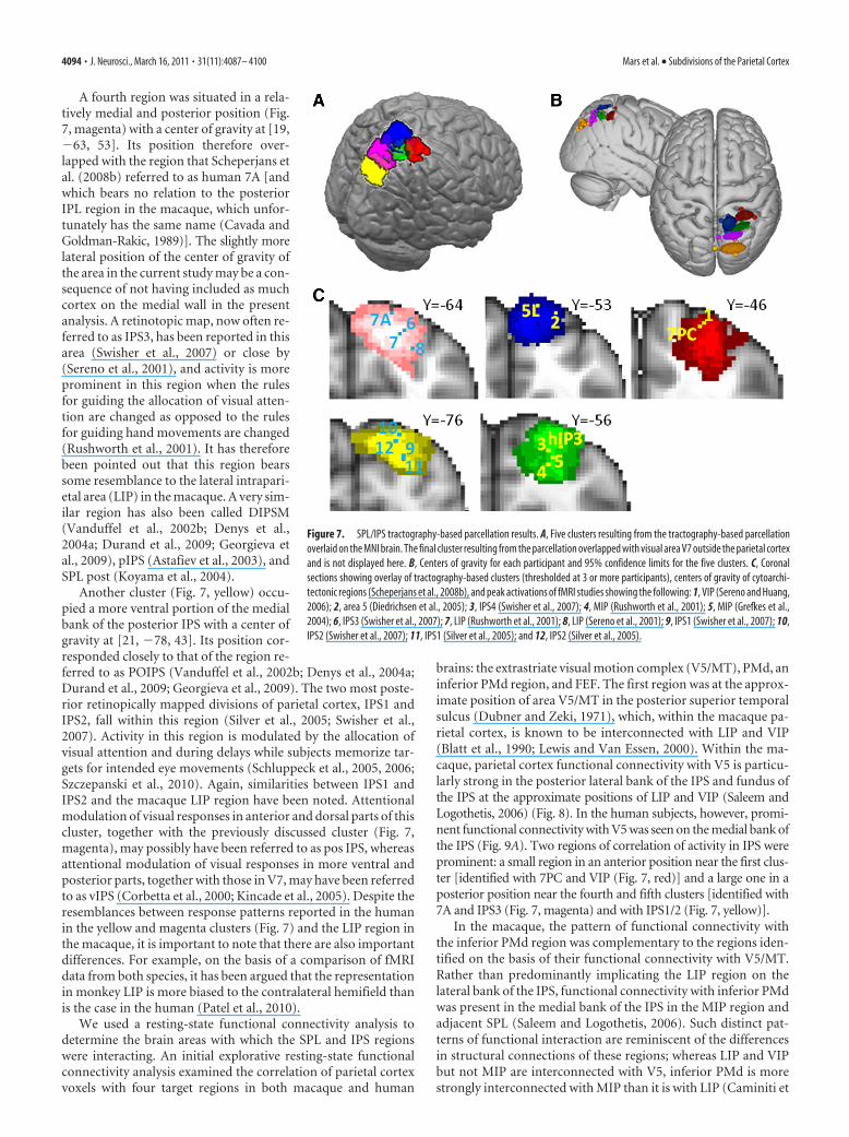
A fourth region was situated in a rela-tively medial and posterior position (Fig.7, magenta) with a center of gravity at [19,�63, 53]. Its position therefore over-lapped with the region that Scheperjans etal. (2008b) referred to as human 7A [andwhich bears no relation to the posteriorIPL region in the macaque, which unfor-tunately has the same name (Cavada andGoldman-Rakic, 1989)]. The slightly morelateral position of the center of gravity ofthe area in the current study may be a con-sequence of not having included as muchcortex on the medial wall in the presentanalysis. A retinotopic map, now often re-ferred to as IPS3, has been reported in thisarea (Swisher et al., 2007) or close by(Sereno et al., 2001), and activity is moreprominent in this region when the rulesfor guiding the allocation of visual atten-tion are changed as opposed to the rulesfor guiding hand movements are changed(Rushworth et al., 2001). It has thereforebeen pointed out that this region bearssome resemblance to the lateral intrapari-etal area (LIP) in the macaque. A very sim-ilar region has also been called DIPSM(Vanduffel et al., 2002b; Denys et al.,2004a; Durand et al., 2009; Georgieva etal., 2009), pIPS (Astafiev et al., 2003), andSPL post (Koyama et al., 2004).
Another cluster (Fig. 7, yellow) occu-pied a more ventral portion of the medialbank of the posterior IPS with a center ofgravity at [21, �78, 43]. Its position cor-responded closely to that of the region re-ferred to as POIPS (Vanduffel et al., 2002b; Denys et al., 2004a;Durand et al., 2009; Georgieva et al., 2009). The two most poste-rior retinopically mapped divisions of parietal cortex, IPS1 andIPS2, fall within this region (Silver et al., 2005; Swisher et al.,2007). Activity in this region is modulated by the allocation ofvisual attention and during delays while subjects memorize tar-gets for intended eye movements (Schluppeck et al., 2005, 2006;Szczepanski et al., 2010). Again, similarities between IPS1 andIPS2 and the macaque LIP region have been noted. Attentionalmodulation of visual responses in anterior and dorsal parts of thiscluster, together with the previously discussed cluster (Fig. 7,magenta), may possibly have been referred to as pos IPS, whereasattentional modulation of visual responses in more ventral andposterior parts, together with those in V7, may have been referredto as vIPS (Corbetta et al., 2000; Kincade et al., 2005). Despite theresemblances between response patterns reported in the humanin the yellow and magenta clusters (Fig. 7) and the LIP region inthe macaque, it is important to note that there are also importantdifferences. For example, on the basis of a comparison of fMRIdata from both species, it has been argued that the representationin monkey LIP is more biased to the contralateral hemifield thanis the case in the human (Patel et al., 2010).
We used a resting-state functional connectivity analysis todetermine the brain areas with which the SPL and IPS regionswere interacting. An initial explorative resting-state functionalconnectivity analysis examined the correlation of parietal cortexvoxels with four target regions in both macaque and human
brains: the extrastriate visual motion complex (V5/MT), PMd, aninferior PMd region, and FEF. The first region was at the approx-imate position of area V5/MT in the posterior superior temporalsulcus (Dubner and Zeki, 1971), which, within the macaque pa-rietal cortex, is known to be interconnected with LIP and VIP(Blatt et al., 1990; Lewis and Van Essen, 2000). Within the ma-caque, parietal cortex functional connectivity with V5 is particu-larly strong in the posterior lateral bank of the IPS and fundus ofthe IPS at the approximate positions of LIP and VIP (Saleem andLogothetis, 2006) (Fig. 8). In the human subjects, however, promi-nent functional connectivity with V5 was seen on the medial bank ofthe IPS (Fig. 9A). Two regions of correlation of activity in IPS wereprominent: a small region in an anterior position near the first clus-ter [identified with 7PC and VIP (Fig. 7, red)] and a large one in aposterior position near the fourth and fifth clusters [identified with7A and IPS3 (Fig. 7, magenta) and with IPS1/2 (Fig. 7, yellow)].
In the macaque, the pattern of functional connectivity withthe inferior PMd region was complementary to the regions iden-tified on the basis of their functional connectivity with V5/MT.Rather than predominantly implicating the LIP region on thelateral bank of the IPS, functional connectivity with inferior PMdwas present in the medial bank of the IPS in the MIP region andadjacent SPL (Saleem and Logothetis, 2006). Such distinct pat-terns of functional interaction are reminiscent of the differencesin structural connections of these regions; whereas LIP and VIPbut not MIP are interconnected with V5, inferior PMd is morestrongly interconnected with MIP than it is with LIP (Caminiti et
Figure 7. SPL/IPS tractography-based parcellation results. A, Five clusters resulting from the tractography-based parcellationoverlaid on the MNI brain. The final cluster resulting from the parcellation overlapped with visual area V7 outside the parietal cortexand is not displayed here. B, Centers of gravity for each participant and 95% confidence limits for the five clusters. C, Coronalsections showing overlay of tractography-based clusters (thresholded at 3 or more participants), centers of gravity of cytoarchi-tectonic regions (Scheperjans et al., 2008b), and peak activations of fMRI studies showing the following: 1, VIP (Sereno and Huang,2006); 2, area 5 (Diedrichsen et al., 2005); 3, IPS4 (Swisher et al., 2007); 4, MIP (Rushworth et al., 2001); 5, MIP (Grefkes et al.,2004); 6, IPS3 (Swisher et al., 2007); 7, LIP (Rushworth et al., 2001); 8, LIP (Sereno et al., 2001); 9, IPS1 (Swisher et al., 2007); 10,IPS2 (Swisher et al., 2007); 11, IPS1 (Silver et al., 2005); and 12, IPS2 (Silver et al., 2005).
4094 • J. Neurosci., March 16, 2011 • 31(11):4087– 4100 Mars et al. • Subdivisions of the Parietal Cortex

Figure 9. Human parietal resting-state functional interactions with V5 (A), inferior PMd (B), PMd (C), and FEF (D), target areas displayed on the MNI brain, and comparison with macaqueresting-state fMRI results. All thresholded at p � 0.05.
Figure 8. Macaque parietal resting-state functional interactions with V5, inferior PMd, PMd, and FEF. A, Target areas displayed on the template of McLaren et al. (2009), which uses the coordinatesystem of the atlas of Saleem and Logothetis (2006). B, Top 80% of activity in the macaque parietal cortex correlating with the time course of activity in the target area, displayed on the Caret F99macaque template (Van Essen, 2002). C, Representative coronal slice of the macaque parietal cortex showing shaded areas that are hypothesized to show interactions with each respective targetarea, adopted from the atlas by Saleem and Logothetis (2006), and population resting-state fMRI results projected on the same slice (thresholded at p � 0.001). Dashed lines indicate intraparietalsulcus.
Mars et al. • Subdivisions of the Parietal Cortex J. Neurosci., March 16, 2011 • 31(11):4087– 4100 • 4095

al., 1996; Johnson et al., 1996; Matelli etal., 1998; Lewis and Van Essen, 2000).Similarly, there were apparent regionaldifferences in the functional connectivityof V5/MT and the inferior PMd region inthe human brain (Fig. 9).
We also compared the functional con-nectivity patterns associated with a moredorsal part of PMd and the FEF. In the ma-caque, there was evidence of functional in-teractions between PMd and a wide swath ofparietal cortex that was centered on area 5and MIP on the medial bank of the IPS butwhich extended into LIP and VIP on the lat-eral bank and fundus of the IPS (Saleem andLogothetis, 2006). In contrast, the func-tional interactions of FEF were most prom-inent in LIP. These results are comparablewith the structural connections of PMd.PMd is known to be connected with parts ofarea 5 and extending into the adjacent pari-etal reach region including MIP (Caminitiet al., 1996; Johnson et al., 1996; Matelli etal., 1998; Lewis and Van Essen, 2000), butthere is also evidence for interconnectionsbetween LIP and PMd (Lewis and Van Es-sen, 2000). In contrast, FEF is intercon-nected with LIP and VIP (Lewis and VanEssen, 2000). Within human parietal cortexfunctional connectivity with PMd wasprominent with the rostral SPL and rostralmedial bank of the IPS (Fig. 9C). Functionalconnectivity with the human FEF was fo-cused on a large region in the anterior me-dial bank of the IPS [the area identified with7PC and VIP (Fig. 7, red)] and a smaller region in the posteriormedial bank of the IPS [the area identified with 7A and IPS3 (Fig. 7,magenta)].
As with the IPL, these results show that resting-state func-tional connectivity in SPL/IPS is found in areas that are known tobe structurally interconnected and illustrate that patterns offunctional connectivity are broadly comparable in the humanand macaque. In the next stage of the analysis, the human resting-state functional connectivity was formally compared with thetractography-based parcellation described above. Resting-statefunctional connectivity was determined between the parietalclusters and V5, inferior PMd, PMd, and FEF (Fig. 10). Evidencefor functional connectivity with V5/MT was found in the mostposterior SPL cluster on the posterior medial bank of the IPS[identified with IPS1/2 (Fig. 7, yellow)], the adjacent fourth IPSregion [identified with 7A and IPS3 (Fig. 7, magenta)], and thefirst IPS region [identified with 7PC and VIP (Fig. 7, red)]. Evi-dence for functional connectivity with PMd was found predom-inantly in three SPL/IPS clusters [identified with 5L (Fig. 7, blue),hIP3/IPS4 (Fig. 7, green), and 7PC/VIP (Fig. 7, red)], with somelimited connectivity also with the IPL (Wise et al., 1997). Evi-dence for functional connectivity within inferior PMd also in-cluded the same three SPL/IPS clusters, but interactions with thehIP3/IPS4 cluster (Fig. 7, green) were more prominent than hadbeen the case with the more superior PMd region. The differencewas particularly apparent at higher thresholds. This pattern re-sembles the pattern in the macaque in which PMd is intercon-nected with a number of SPL regions but a more inferior PMd
region is particularly interconnected with MIP (Matelli et al.,1998).
Functional connectivity with the FEF (Fig. 10) was mostprominent in the anterior lateral SPL/IPS region identified with7PC and VIP (Fig. 7, red). Evidence for functional connectivitybetween the more posterior lateral SPL/IPS region identified with7A and IPS3 (Fig. 7, magenta) and the SPL region identified with5L (Fig. 7, blue) was also present. In addition, it should be notedthat there is some uncertainty regarding the location of FEF in thehuman brain. In the macaque, FEF is located on the rostral bankof the arcuate sulcus. In the human brain, fMRI studies oftenactivate a region more caudal than would be expected on the basisof macaque data, whereas stimulation studies position humanFEF just anterior to the superior precentral sulcus (Amiez andPetrides, 2009). The target area used in the present study is lo-cated at the junction of the superior frontal sulcus and the pre-central sulcus.
DiscussionThe present study characterized the anatomical and functionalorganization of human lateral parietal cortex using non-invasiveMRI techniques and compared it with the organization of themacaque parietal cortex. First, we used diffusion tractography toparcellate the human parietal cortex into 10 subregions. Thenresting-state fMRI was used to examine the functional connectiv-ity patterns of these subregions. Finally, comparing the resting-state fMRI data in humans and macaques revealed fundamentalsimilarities in lateral parietal organization and demonstrated that
Figure 10. Comparison of tractography-based parcellation and human resting-state results for target areas hypothesized tointeract strongly with the SPL. Connectivity fingerprints indicating the relative overlap between z-statistical images and the IPLclusters from the tractography-based parcellation shown in Figure 2 and the SPL parcellation shown in Figure 7, ranging from themost anterolateral SPL cluster (top of spider plot) clockwise to the most posterior SPL cluster and then from the most anterior IPLcluster (bottom of spider plot) to the most posterior IPL cluster. Connectivity fingerprints are from V5 (magenta), inferior PMd(blue), PMd (cyan), and FEF (yellow). Data are normalized to reflect the relative contribution of each target area.
4096 • J. Neurosci., March 16, 2011 • 31(11):4087– 4100 Mars et al. • Subdivisions of the Parietal Cortex

the resting-state functional connectivity of lateral parietal cortexreflected major known anatomical connections. There are funda-mental similarities in lateral parietal organization in humans andmacaques, but some features distinguished human parietal cor-tex. An anterior IPL region active during tool observation wasidentified (Fig. 2, red cluster), and a central IPL region was char-acterized by strong interactions with aPFC.
Diffusion-tractography approaches have identified compo-nent areas within the pre-supplementary motor area (pre-SMA)/SMA region (Johansen-Berg et al., 2004), lateral premotor andventrolateral prefrontal cortex (Anwander et al., 2007; Klein etal., 2007; Tomassini et al., 2007; Schubotz et al., 2010), and cin-gulate cortex (Beckmann et al., 2009), but this is the first timesuch a large expanse of cortex has been parcellated. Previousattempts to establish relationships between human and macaqueparietal cortex have focused on retinotopic mapping, identificationof similarities in functional activation profiles, and postmortem cy-toarchitectonic analysis (Table 1). Because it has different strengthsand weaknesses to these methods, the diffusion-tractography parcel-lation and functional connectivity approach provides independentand convergent evidence about the key component subdivisions ofhuman parietal cortex.
The IPL parcellation yielded five clusters, and in most cases itwas possible to identify their relationships with cytoartchitec-tonically defined areas PFop, PFm, PGa, and PGp (Caspers et al.,2008). One cluster (Fig. 2, blue), however, corresponded to sev-eral cytoarchitectonically defined areas, PF, PFt, and hIP2(Caspers et al., 2006; Choi et al., 2006), which were presumablygrouped together because of similarities in their connections. Thedifferences in anatomical connections that gave rise to parcella-tion should mean that each IPL cluster differs in the access it hasto the information represented in other cortical areas. Activityrelated to grasping, effecting strategy and response changes, at-tentional shifts, and memory retrieval, were preferentially asso-
ciated with different clusters (Fig. 2C). Notably, the location ofthe most anterior IPL cluster was similar to that of activity re-corded during tool use observation that has only been observed inhuman IPL (Peeters et al., 2009).
It has been claimed that human IPL may contain unique re-gions not found in other primates (Husain and Nachev, 2007;Peeters et al., 2009). Resting-state functional connectivity, how-ever, in the posterior and anterior IPL, with PH, dlPFC, and PMvrespectively, was similar in both humans and macaques (Fig. 4)and probably reflects similar anatomical connections in both spe-cies. In contrast, the human central IPL region was notable inthat, unlike macaque central IPL, its activity was correlated withthat of aPFC. It is in this region of the IPL that calculation- andlanguage-related activity is found (Simon et al., 2002, 2004), andactivity changes occur as people weigh up and change responsestrategy (Daw et al., 2006; Boorman et al., 2009). The aPFC regionis close to the frontal pole in which activity has been related to thehighest levels of cognitive control when “branching” betweendifferent subtasks is required (Koechlin et al., 1999). The twoareas are often co-active (Daw et al., 2006; Boorman et al., 2009).There is evidence that the frontal pole is especially large in hu-mans (Semendeferi et al., 2001) and that its increase in size isattributable to its lateral expansion, in hominoids, into the ap-proximate region we found to be connected to mid-IPL. Consis-tent with our results, Petrides and Pandya (2007) have reportedno connections between frontal polar area 10 and parietal cortexin the macaque, although Rozzi et al. (2006) note some betweenmid-IPL and anterior lateral prefrontal cortex. Brain differencesemerge during speciation in a number of ways, including as aresult of new connections invading an area or as specialized divi-sions of an area becoming spatially separate (Krubitzer, 1995,2007), and such changes may underlie the present findings. Thecentral IPL region is an important one to be considered in anyattempt to account for the distinctive expansion of parietal cortexseen in humans even when comparison with other hominids(Bruner, 2010).
Parcellation of SPL and posterior IPS resulted in five clusters.Once again, it was possible to identify correspondences betweenthe clusters and cytoarchitectonically defined regions (Fig. 7C),including 7PC, 5L, hIP3, and 7A (Scheperjans et al., 2008b). Reti-notopic and other mapping procedures have been used to identifysensorimotor regions in IPS and SPL; the diffusion-tractographyapproach provides convergent evidence for the anatomical separa-tion of many of these areas, including VIP (Bremmer et al., 2001;Sereno and Huang, 2006), also referred to as IPS5 (Konen and Kast-ner, 2008), IPS3, and IPS4 (Sereno et al., 2001; Swisher et al., 2007).One ventromedial posterior IPS cluster (Fig. 7, yellow), however,covered a region associated with two retinotopic maps, IPS1 andIPS2 (Silver et al., 2005), which have activity related to visuospatialattention and oculomotor intention (Schluppeck et al., 2005, 2006).IPS1, IPS2, and IPS3 have all been proposed as possible homologs ofmacaque LIP. The assignment of IPS1 and IPS2 to one cluster andthe assignment of IPS3 to another cluster suggests that IPS1 and IPS2share a number of connections and predicts that their functions maybe especially similar. There is some evidence that both IPS1 and IPS2are distinguished by the lateralization of their activity (Szczepanski etal., 2010). A modern, detailed study of the cytoartchitecture of thisregion has yet to be published, but again the diffusion-tractographyresults suggest that it may differ from the adjacent 7A region thatencompasses much of IPS3.
The patterns of functional connectivity associated with thedifferent SPL/IPS parietal clusters clarify the relationship be-tween the parietal cortex in the human and other primates and
Table 1. Correspondences in parietal terminology between the current study andpreviously published nomenclature
DWI-definedcluster
Cytoarchitectonicregion
Retinotopicmap MGH/Leuven
Other functionalnomenclature
IPLRed PFopBlue PF/PFt/hIP2 AIP, hAIPGreen PFmMagenta PGaYellow PGp
SPLRed 7PC VIPBlue 5L IPS5Green hIP3 IPS4 DIPSA Human MIP, posterior AIP,
aIPS, SPL antMagenta 7A IPS3 DIPSM Pos IPS, SPLpostYellow IPS2 POIPS Pos IPS
IPS1 POIPS vIPSNot shown IPS0 VIPS, VIPS/V7 vIPS, IPS/TOS
Diffusion-weighted imaging (DWI)-defined clusters are from the current study, and the colors listed refer to thoseshown in Figures 2 and 7. Cytoarchitectonic regions are based on the descriptions of Choi, Caspars, and Scheperjansand colleagues (Caspers et al., 2006, 2008; Choi et al., 2006; Scheperjans et al., 2008a,b). Retinotopic map designa-tions are based on several studies (Schluppeck et al., 2005; Silver et al., 2005; Levy et al., 2007; Swisher et al., 2007;Konen and Kastner, 2008). The designations used by a group of researchers based in the Massachusetts GeneralHospital (MGH) and Katholieke Universiteit Leuven (Leuven) are listed in the fourth column (Vanduffel et al., 2002a;Denys et al., 2004b; Orban et al., 2006; Durand et al., 2007, 2009; Georgieva et al., 2009). Other designations basedon the behavioral condition in which activations have been found are listed in the final column: a region referred toas AIP or hAIP region reported at the medial boundary of the second most anterior IPL cluster (Fig. 7, blue) (Binkofskiet al., 1999; Grefkes et al., 2002; Culham et al., 2003; Frey et al., 2005; Grol et al., 2007); SPLant, SPLpost, and IPS/TOS(Koyama et al., 2004); MIP (Rushworth et al., 2001; Grefkes et al., 2004); and pos IPS, vIPS, and aIPS from Corbettaand colleagues (Corbetta et al., 2000; Astafiev et al., 2003; Kincade et al., 2005).
Mars et al. • Subdivisions of the Parietal Cortex J. Neurosci., March 16, 2011 • 31(11):4087– 4100 • 4097

are suggestive of function. Macaque VIP is known to be con-nected with V5, PMd, PMv, and FEF (Luppino et al., 1999; Lewisand Van Essen, 2000), and the anterior lateral SPL/IPS putative7PC/VIP/IPS5 region (Fig. 7, red) was found to be functionallyconnected with the same regions in the present study (Fig. 10).The anterior medial, putative 5L, region (Fig. 7, blue) interactedstrongly with PMd. Connections between area 5 and PMd havealso been documented in the macaque (Caminiti et al., 1996;Johnson et al., 1996; Matelli et al., 1998). The mid-lateral SPL/IPSregion near the IPS4 map (Fig. 7, green) was found to be func-tionally connected to PMd, particularly inferior PMd, in a man-ner reminiscent of the connections known to exist between MIPand inferior PMd in macaque (Matelli et al., 1998). As in ma-caque MIP, such a pattern would be expected if the region were tobe involved in the directional control of movement and comple-ments evidence that a similar region is active during visuallyguided hand movements even in the absence of eye movements(Rushworth et al., 2001; Grefkes et al., 2004; Hinkley et al., 2009).The functional connectivity between the most posterior IPS re-gion (Fig. 7, yellow), near IPS1 and IPS2 maps, and V5 suggestedthat it resembles the ventral division of macaque LIP (Lewis andVan Essen, 2000). There was evidence of functional connectivitybetween the fourth, posterior lateral SPL/IPS cluster (magenta,IPS3 location) and FEF, suggesting that it also bore resemblancesto LIP. Human SPL/IPS organization differed from macaque pa-rietal organization in that the areas engaged in functional inter-actions with both FEF and V5, important interactions for anyarea concerned with visuospatial attention, were most prominenton the medial rather than the lateral bank of the IPS (Grefkes andFink, 2005).
In summary, we show the feasibility of using non-invasiveMRI methods to establish the anatomical and functional subre-gions of the human parietal cortex and to compare these with themacaque. Although only tracer injection studies in animal mod-els provide definitive evidence for existence of synaptic connec-tions between brain regions, comparative MRI-based studies areimportant because they facilitate translation to the human brain.Similarly, although studies such as the present one cannot replacedetailed cytoarchitectonic descriptions of brain areas, they indi-cate ways of exploiting such descriptions in in vivo investigationsof human brain anatomy and function in both health and illness.The possibility that functional connectivity is mediated by poly-synaptic pathways cannot be ruled out. Nevertheless, similar,previous functional connectivity studies have recently identifiedcorrespondences between medial parietal and adjacent posteriorcingulate areas in humans and macaques (Margulies et al., 2009).The qualitative similarity in human and macaque IPL is notable,suggesting that the neurophysiology and anatomy of macaqueIPL is likely to be an important guide for understanding humanIPL (Caminiti et al., 2010). Nevertheless, within this framework,human specializations, especially interactions between mid-IPLand aPFC, can be discerned.
ReferencesAmiez C, Petrides M (2009) Anatomical organization of the eye fields in the
human and non-human primate frontal cortex. Prog Neurobiol89:220 –230.
Amiez C, Kostopoulos P, Champod AS, Petrides M (2006) Local morphol-ogy predicts functional organization of the dorsal premotor region in thehuman brain. J Neurosci 26:2724 –2731.
Anwander A, Tittgemeyer M, von Cramon DY, Friederici AD, Knosche TR(2007) Connectivity-based parcellation of Broca’s area. Cereb Cortex17:816 – 825.
Astafiev SV, Shulman GL, Stanley CM, Snyder AZ, Van Essen DC, Corbetta M
(2003) Functional organization of human intraparietal and frontal cor-tex for attending, looking, and pointing. J Neurosci 23:4689 – 4699.
Averbeck BB, Battaglia-Mayer A, Guglielmo C, Caminiti R (2009) StatisticalAnalysis of Parieto-Frontal Cognitive-Motor Networks. J Neurophysiol102:1911–1920.
Beckmann M, Johansen-Berg H, Rushworth MF (2009) Connectivity-basedparcellation of human cingulate cortex and its relation to functional spe-cialization. J Neurosci 29:1175–1190.
Behrens TE, Berg HJ, Jbabdi S, Rushworth MF, Woolrich MW (2007) Prob-abilistic diffusion tractography with multiple fibre orientations: What canwe gain? Neuroimage 34:144 –155.
Binkofski F, Buccino G, Stephan KM, Rizzolatti G, Seitz RJ, Freund HJ(1999) A parieto-premotor network for object manipulation: evidencefrom neuroimaging. Exp Brain Res 128:210 –213.
Blatt GJ, Andersen RA, Stoner GR (1990) Visual receptive field organizationand cortico-cortical connections of the lateral intraparietal area (area LIP)in the macaque. J Comp Neurol 299:421– 445.
Blatt GJ, Pandya DN, Rosene DL (2003) Parcellation of cortical Afferents tothree distinct sectors in the parahippocampal gyrus of the rhesus monkey:An anatomical and neurophysiological study. J Comp Neurol466:161–179.
Boorman ED, Behrens TE, Woolrich MW, Rushworth MF (2009) Howgreen is the grass on the other side? Frontopolar cortex and the evidence infavor of alternative courses of action. Neuron 62:733–743.
Bremmer F, Schlack A, Shah NJ, Zafiris O, Kubischik M, Hoffmann K, ZillesK, Fink GR (2001) Polymodal motion processing in posterior parietaland premotor cortex: A human fMRI study strongly implies equivalenciesbetween humans and monkeys. Neuron 29:287–296.
Bruner E (2010) Morphological differences in the parietal lobes within thehuman genus. A neurofunctional perspective. Curr Anthropol51:S77–S88.
Caminiti R, Ferraina S, Johnson PB (1996) The sources of visual informa-tion to the primate frontal lobe: A novel role for the superior parietallobule. Cereb Cortex 6:319 –328.
Caminiti R, Chafee MV, Battaglia-Mayer A, Averbeck BB, Crowe DA, Geor-gopoulos AP (2010) Understanding the parietal lobe syndrome from aneurophysiological and evolutionary perspective. Eur J Neurosci31:2320 –2340.
Caspers S, Geyer S, Schleicher A, Mohlberg H, Amunts K, Zilles K (2006)The human inferior parietal cortex: Cytoarchitectonic parcellation andinterindividual variability. Neuroimage 33:430 – 448.
Caspers S, Eickhoff SB, Geyer S, Scheperjans F, Mohlberg H, Zilles K, AmuntsK (2008) The human inferior parietal lobule in stereotaxic space. BrainStruct Funct 212:481– 495.
Cavada C, Goldman-Rakic PS (1989) Posterior parietal cortex in rhesusmonkey: II. Evidence for segregated corticocortical networks linking sen-sory and limbic areas with the frontal lobe. J Comp Neurol 287:422– 445.
Choi HJ, Zilles K, Mohlberg H, Schleicher A, Fink GR, Armstrong E, AmuntsK (2006) Cytoarchitectonic identification and probabilistic mapping oftwo distinct areas within the anterior ventral bank of the human intrapa-rietal sulcus. J Comp Neurol 495:53– 69.
Corbetta M, Kincade JM, Ollinger JM, McAvoy MP, Shulman GL (2000)Voluntary orienting is dissociated from target detection in human poste-rior parietal cortex. Nat Neurosci [Erratum (2000) 3:521] 3:292–297.
Croxson PL, Johansen-Berg H, Behrens TE, Robson MD, Pinsk MA, GrossCG, Richter W, Richter MC, Kastner S, Rushworth MF (2005) Quanti-tative investigation of connections of the prefrontal cortex in the humanand macaque using probabilistic diffusion tractography. J Neurosci25:8854 – 8866.
Culham JC, Danckert SL, DeSouza JF, Gati JS, Menon RS, Goodale MA(2003) Visually guided grasping produces fMRI activation in dorsal butnot ventral stream brain areas. Exp Brain Res 153:180 –189.
Daw ND, O’Doherty JP, Dayan P, Seymour B, Dolan RJ (2006) Corticalsubstrates for exploratory decisions in humans. Nature 441:876 – 879.
Denys K, Vanduffel W, Fize D, Nelissen K, Peuskens H, Van Essen D, OrbanGA (2004a) The processing of visual shape in the cerebral cortex of hu-man and nonhuman primates: A functional magnetic resonance imagingstudy. J Neurosci 24:2551–2565.
Denys K, Vanduffel W, Fize D, Nelissen K, Sawamura H, Georgieva S, VogelsR, Van Essen D, Orban GA (2004b) Visual activation in prefrontal cor-tex is stronger in monkeys than in humans. J Cogn Neurosci16:1505–1516.
4098 • J. Neurosci., March 16, 2011 • 31(11):4087– 4100 Mars et al. • Subdivisions of the Parietal Cortex

Diedrichsen J, Hashambhoy Y, Rane T, Shadmehr R (2005) Neural corre-lates of reach errors. J Neurosci 25:9919 –9931.
Dubner R, Zeki SM (1971) Response properties and receptive fields of cellsin an anatomically defined region of superior temporal sulcus in monkey.Brain Res 35:528 –532.
Dumoulin SO, Bittar RG, Kabani NJ, Baker CL Jr, Le Goualher G, Bruce PikeG, Evans AC (2000) A new anatomical landmark for reliable identifica-tion of human area V5/MT: a quantitative analysis of sulcal patterning.Cereb Cortex 10:454 – 463.
Durand JB, Nelissen K, Joly O, Wardak C, Todd JT, Norman JF, Janssen P,Vanduffel W, Orban GA (2007) Anterior regions of monkey parietalcortex process visual 3D shape. Neuron 55:493–505.
Durand JB, Peeters R, Norman JF, Todd JT, Orban GA (2009) Parietal re-gions processing visual 3D shape extracted from disparity. Neuroimage46:1114 –1126.
Eickhoff SB, Stephan KE, Mohlberg H, Grefkes C, Fink GR, Amunts K, ZillesK (2005) A new SPM toolbox for combining probabilistic cytoarchitec-tonic maps and functional imaging data. Neuroimage 25:1325–1335.
Fox MD, Raichle ME (2007) Spontaneous fluctuations in brain activity ob-served with functional magnetic resonance imaging. Nat Rev Neurosci8:700 –711.
Frey SH, Vinton D, Norlund R, Grafton ST (2005) Cortical topography ofhuman anterior intraparietal cortex active during visually guided grasp-ing. Cogn Brain Res 23:397– 405.
Georgieva S, Peeters R, Kolster H, Todd JT, Orban GA (2009) The process-ing of three-dimensional shape from disparity in the human brain. J Neu-rosci 29:727–742.
Grefkes C, Fink GR (2005) The functional organization of the intraparietalsulcus in humans and monkeys. J Anat 207:3–17.
Grefkes C, Weiss PH, Zilles K, Fink GR (2002) Crossmodal processing ofobject features in human anterior intraparietal cortex: an fMRI studyimplies equivalencies between humans and monkeys. Neuron35:173–184.
Grefkes C, Ritzl A, Zilles K, Fink GR (2004) Human medial intraparietalcortex subserves visuomotor coordinate transformation. Neuroimage23:1494 –1506.
Grol MJ, Majdandzic J, Stephan KE, Verhagen L, Dijkerman HC, BekkeringH, Verstraten FA, Toni I (2007) Parieto-frontal connectivity during vi-sually guided grasping. J Neurosci 27:11877–11887.
Henson RN, Rugg MD, Shallice T, Josephs O, Dolan RJ (1999) Recollectionand familiarity in recognition memory: An event-related functional mag-netic resonance imaging study. J Neurosci 19:3962–3972.
Hinkley LB, Krubitzer LA, Padberg J, Disbrow EA (2009) Visual-manualexploration and posterior parietal cortex in humans. J Neurophysiol102:3433–3446.
Husain M, Nachev P (2007) Space and the parietal cortex. Trends Cogn Sci11:30 –36.
Jenkinson M, Bannister P, Brady M, Smith S (2002) Improved optimizationfor the robust and accurate linear registration and motion correction ofbrain images. Neuroimage 17:825– 841.
Johansen-Berg H, Behrens TE, Robson MD, Drobnjak I, Rushworth MF,Brady JM, Smith SM, Higham DJ, Matthews PM (2004) Changes inconnectivity profiles define functionally distinct regions in human medialfrontal cortex. Proc Natl Acad Sci U S A 101:13335–13340.
Johnson PB, Ferraina S, Bianchi L, Caminiti R (1996) Cortical networks forvisual reaching: Physiological and anatomical organization of frontal andparietal lobe arm regions. Cereb Cortex 6:102–119.
Kincade JM, Abrams RA, Astafiev SV, Shulman GL, Corbetta M (2005) Anevent-related functional magnetic resonance imaging study of voluntaryand stimulus-driven orienting of attention. J Neurosci 25:4593– 4604.
Klein JC, Behrens TE, Robson MD, Mackay CE, Higham DJ, Johansen-BergH (2007) Connectivity-based parcellation of human cortex using diffu-sion MRI: Establishing reproducibility, validity and observer indepen-dence in BA 44/45 and SMA/pre-SMA. Neuroimage 34:204 –211.
Koechlin E, Basso G, Pietrini P, Panzer S, Grafman J (1999) The role of theanterior prefrontal cortex in human cognition. Nature 399:148 –151.
Konen CS, Kastner S (2008) Representation of eye movements and stimulusmotion in topographically organized areas of human posterior parietalcortex. J Neurosci 28:8361– 8375.
Koyama M, Hasegawa I, Osada T, Adachi Y, Nakahara K, Miyashita Y (2004)Functional magnetic resonance imaging of macaque monkeys perform-
ing visually guided saccade tasks: comparison of cortical eye fields withhumans. Neuron 41:795– 807.
Krubitzer L (1995) The organization of neocortex in mammals: Are speciesdifferences really so different? Trends Neurosci 18:408 – 417.
Krubitzer L (2007) The magnificent compromise: Cortical field evolution inmammals. Neuron 56:201–208.
Levy I, Schluppeck D, Heeger DJ, Glimcher PW (2007) Specificity of humancortical areas for reaches and saccades. J Neurosci 27:4687– 4696.
Lewis JW, Van Essen DC (2000) Corticocortical connections of visual, sen-sorimotor, and multimodal processing areas in the parietal lobe of themacaque monkey. J Comp Neurol 428:112–137.
Luppino G, Murata A, Govoni P, Matelli M (1999) Largely segregated pari-etofrontal connections linking rostral intraparietal cortex (areas AIP andVIP) and the ventral premotor cortex (areas F5 and F4). Exp Brain Res128:181–187.
Margulies DS, Vincent JL, Kelly C, Lohmann G, Uddin LQ, Biswal BB,Villringer A, Castellanos FX, Milham MP, Petrides M (2009) Precuneusshares intrinsic functional architecture in humans and monkeys. ProcNatl Acad Sci U S A 106:20069 –20074.
Mars RB, Coles MG, Hulstijn W, Toni I (2008) Delay-related cerebral activ-ity and motor preparation. Cortex 44:507–520.
Matelli M, Camarda R, Glickstein M, Rizzolatti G (1986) Afferent and effer-ent projections of the inferior area 6 in the macaque monkey. J CompNeurol 251:281–298.
Matelli M, Govoni P, Galletti C, Kutz DF, Luppino G (1998) Superior area 6afferents from the superior parietal lobule in the macaque monkey.J Comp Neurol 402:327–352.
McLaren DG, Kosmatka KJ, Oakes TR, Kroenke CD, Kohama SG, MatochikJA, Ingram DK, Johnson SC (2009) A population-average MRI-basedatlas collection of the rhesus macaque. Neuroimage 45:52–59.
Mort DJ, Perry RJ, Mannan SK, Hodgson TL, Anderson E, Quest R, McRob-bie D, McBride A, Husain M, Kennard C (2003) Differential corticalactivation during voluntary and reflexive saccades in man. Neuroimage18:231–246.
Nelson SM, Cohen AL, Power JD, Wig GS, Miezin FM, Wheeler ME, Vela-nova K, Donaldson DI, Phillips JS, Schlaggar BL, Petersen SE (2010) Aparcellation scheme for human left lateral parietal cortex. Neuron67:156 –170.
Orban GA, Claeys K, Nelissen K, Smans R, Sunaert S, Todd JT, Wardak C,Durand JB, Vanduffel W (2006) Mapping the parietal cortex of humanand non-human primates. Neuropsychologia 44:2647–2667.
O’Reilly JX, Beckmann CF, Tomassini V, Ramnani N, Johansen-Berg H(2010) Distinct and overlapping functional zones in the cerebellum de-fined by resting state functional connectivity. Cereb Cortex 20:953–965.
Pandya DN, Yeterian EH (1996) Comparison of prefrontal architecture andconnections. Philos Trans R Soc Lond B Biol Sci 351:1423–1432.
Passingham R (2009) How good is the macaque monkey model of the hu-man brain? Curr Opin Neurobiol 19:6 –11.
Passingham RE (2008) What is special about the human brain? Oxford;New York: Oxford University Press.
Passingham RE, Stephan KE, Kotter R (2002) The anatomical basis of func-tional localization in the cortex. Nat Rev Neurosci 3:606 – 616.
Patel GH, Shulman GL, Baker JT, Akbudak E, Snyder AZ, Snyder LH, Cor-betta M (2010) Topographic organization of macaque area LIP. ProcNatl Acad Sci U S A 107:4728 – 4733.
Peeters R, Simone L, Nelissen K, Fabbri-Destro M, Vanduffel W, Rizzolatti G,Orban GA (2009) The representation of tool use in humans and mon-keys: Common and uniquely human features. J Neurosci29:11523–11539.
Petrides M, Pandya DN (2007) Efferent association pathways from the ros-tral prefrontal cortex in the macaque monkey. J Neurosci27:11573–11586.
Rajkowska G, Goldman-Rakic PS (1995) Cytoarchitectonic definition ofprefrontal areas in the normal human cortex: II. Variability in locations ofareas 9 and 46 and relationship to the Talairach Coordinate System. CerebCortex 5:323–337.
Rozzi S, Calzavara R, Belmalih A, Borra E, Gregoriou GG, Matelli M, LuppinoG (2006) Cortical connections of the inferior parietal cortical convexityof the macaque monkey. Cereb Cortex 16:1389 –1417.
Rushworth MF, Paus T, Sipila PK (2001) Attention systems and the organi-zation of the human parietal cortex. J Neurosci 21:5262–5271.
Rushworth MF, Behrens TE, Johansen-Berg H (2006) Connection patterns
Mars et al. • Subdivisions of the Parietal Cortex J. Neurosci., March 16, 2011 • 31(11):4087– 4100 • 4099

distinguish 3 regions of human parietal cortex. Cereb Cortex 16:1418 –1430.
Rushworth MFS, Boorman E, Mars RB (2009) Comparing brain connec-tions in different species using diffusion weighted imaging. In: DiffusionMRI. From quantitative measurement to in vivo neuroanatomy(Johansen-Berg H, Behrens TEJ, eds), pp445– 460. Amsterdam: AcademicPress.
Saleem KS, Logothetis NK (2006) A combinted MRI and histology atlas ofthe rhesus monkey brain. Amsterdam: Academic Press.
Scheperjans F, Hermann K, Eickhoff SB, Amunts K, Schleicher A, Zilles K(2008a) Observer-independent cytoarchitectonic mapping of the humansuperior parietal cortex. Cereb Cortex 18:846 – 867.
Scheperjans F, Eickhoff SB, Homke L, Mohlberg H, Hermann K, Amunts K,Zilles K (2008b) Probabilistic maps, morphometry, and variability ofcytoarchitectonic areas in the human superior parietal cortex. Cereb Cor-tex 18:2141–2157.
Schluppeck D, Glimcher P, Heeger DJ (2005) Topographic organization fordelayed saccades in human posterior parietal cortex. J Neurophysiol94:1372–1384.
Schluppeck D, Curtis CE, Glimcher PW, Heeger DJ (2006) Sustained activ-ity in topographic areas of human posterior parietal cortex duringmemory-guided saccades. J Neurosci 26:5098 –5108.
Schubotz RI, Anwander A, Knosche TR, von Cramon DY, Tittgemeyer M(2010) Anatomical and functional parcellation of the human lateral pre-motor cortex. Neuroimage 50:396 – 408.
Seltzer B, Pandya DN (1984) Further Observations on Parieto-TemporalConnections in the Rhesus-Monkey. Exp Brain Res 55:301–312.
Semendeferi K, Armstrong E, Schleicher A, Zilles K, Van Hoesen GW (2001)Prefrontal cortex in humans and apes: A comparative study of area 10.Am J Phys Anthropol 114:224 –241.
Sereno MI, Huang RS (2006) A human parietal face area contains alignedhead-centered visual and tactile maps. Nat Neurosci 9:1337–1343.
Sereno MI, Pitzalis S, Martinez A (2001) Mapping of contralateral space inretinotopic coordinates by a parietal cortical area in humans. Science294:1350 –1354.
Silver MA, Ress D, Heeger DJ (2005) Topographic maps of visual spatialattention in human parietal cortex. J Neurophysiol 94:1358 –1371.
Simon O, Mangin JF, Cohen L, Le Bihan D, Dehaene S (2002) Topograph-ical layout of hand, eye, calculation, and language-related areas in thehuman parietal lobe. Neuron 33:475– 487.
Simon O, Kherif F, Flandin G, Poline JB, Riviere D, Mangin JF, Le Bihan D,Dehaene S (2004) Automatized clustering and functional geometry ofhuman parietofrontal networks for language, space, and number. Neuro-image 23:1192–1202.
Smith SM, Jenkinson M, Woolrich MW, Beckmann CF, Behrens TE,Johansen-Berg H, Bannister PR, De Luca M, Drobnjak I, Flitney DE,
Niazy RK, Saunders J, Vickers J, Zhang Y, De Stefano N, Brady JM, Mat-thews PM (2004) Advances in functional and structural MR image anal-ysis and implementation as FSL. Neuroimage 23 [Suppl 1]:S208 –S219.
Snyder LH, Batista AP, Andersen RA (1997) Coding of intention in theposterior parietal cortex. Nature 386:167–170.
Suzuki M, Tsukiura T, Matsue Y, Yamadori A, Fujii T (2005) Dissociablebrain activations during the retrieval of different kinds of spatial contextmemory. Neuroimage 25:993–1001.
Suzuki WA, Amaral DG (1994) Topographic organization of the reciprocalconnections between the monkey entorhinal cortex and the perirhinaland parahippocampal cortices. J Neurosci 14:1856 –1877.
Swisher JD, Halko MA, Merabet LB, McMains SA, Somers DC (2007) Vi-sual topography of human intraparietal sulcus. J Neurosci 27:5326 –5337.
Szczepanski SM, Konen CS, Kastner S (2010) Mechanisms of spatial atten-tion control in frontal and parietal cortex. J Neurosci 30:148 –160.
Tanne-Gariepy J, Rouiller EM, Boussaoud D (2002) Parietal inputs to dor-sal versus ventral premotor areas in the macaque monkey: evidence forlargely segregated visuomotor pathways. Exp Brain Res 145:91–103.
Thiel CM, Zilles K, Fink GR (2004) Cerebral correlates of alerting, orientingand reorienting of visuospatial attention: an event-related fMRI study.Neuroimage 21:318 –328.
Tomassini V, Jbabdi S, Klein JC, Behrens TE, Pozzilli C, Matthews PM, Rush-worth MF, Johansen-Berg H (2007) Diffusion-weighted imagingtractography-based parcellation of the human lateral premotor cortexidentifies dorsal and ventral subregions with anatomical and functionalspecializations. J Neurosci 27:10259 –10269.
Van Essen DC (2002) Windows on the brain. The emerging role of atlasesand databases in neuroscience. Curr Opin Neurobiol 12:574 –579.
Van Essen DC, Drury HA, Dickson J, Harwell J, Hanlon D, Anderson CH(2001) An integrated software system for surface-based analyses of cere-bral cortex. J Am Med Inform Assoc 8:443– 459.
Vanduffel W, Fize D, Peuskens H, Denys K, Sunaert S, Todd JT, Orban GA(2002a) Extracting 3D from motion: Differences in human and monkeyintraparietal cortex. Science 298:413– 415.
Vanduffel W, Tootell RB, Schoups AA, Orban GA (2002b) The organizationof orientation selectivity throughout macaque visual cortex. Cereb Cortex12:647– 662.
Vincent JL, Patel GH, Fox MD, Snyder AZ, Baker JT, Van Essen DC, ZempelJM, Snyder LH, Corbetta M, Raichle ME (2007) Intrinsic functional ar-chitecture in the anaesthetized monkey brain. Nature 447:83– 86.
Wise SP, Boussaoud D, Johnson PB, Caminiti R (1997) Premotor and pari-etal cortex: Corticocortical connectivity and combinatorial computa-tions. Annu Rev Neurosci 20:25– 42.
Xiang HD, Fonteijn HM, Norris DG, Hagoort P (2010) Topographicalfunctional connectivity pattern in the perisylvian language networks.Cereb Cortex 20:549 –560.
4100 • J. Neurosci., March 16, 2011 • 31(11):4087– 4100 Mars et al. • Subdivisions of the Parietal Cortex
Related Documents