Differentiation of CD3 - 4- 8 - Human Fetal Thymocytes In Vivo: Characterization of a CD3- 4 + 8- Intermediate By Daniel L. Kraft,* Irving L. Weissman,gll and Edmund K. Waller** From the "Cancer Biology Research Laboratory, and the Departments of*Medicine, Spathology, and IIDevelopmentalBiology, Stanford University School of Medicine, Stanford, California 94305 Summary Human thymocyte differentiation was examined by injecting fetal thymic progenitor populations into human thymic xenografts in SCID-hu mice. Thymic progenitors were fluorescently labeled with the lipophilic dye PKH2. The phenotypes of their progeny could be identified by flow cytometric analysis of cells with a very high fluorescent PKH2 signal. Intrathymic injection of purified triple negative (TN) CD3-4-8- thymocytes resulted in the sequential appearance of CD3-4+8 -, CD3-4+8 +, and CD3+4+8 + cells, with the subsequent appearance of anal1 numbers of phenotypically mature CD3 +4 +8- and CD3 +4-8 + ceils over a 4-d period. Sorted CD3-4+8 - thymocytes injected intrathymically rapidly differentiated to CD4+8 + cells. CD4 +8 + fetal thymocytes in cell eycle differentiated into phenotypically mature CD3 +4+8- and CD3 +4-8 + populations, whereas nondividing CD4+8 + cells failed to differentiate after intrathymic transfer. The number of cell divisions that occurred between the injection of TN thymocytes and their progeny at different time points was estimated based on the decrease in the intensity of the PKH2 label. The average length of the cell cycle for the TN population was calculated to be 24 h. The SCID-hu model thus provides a useful tool for studying the kinetics of cell division and differentiation of human thymocytes in vivo. A major site of T lymphocyte differentiation is the thymus (for a review see reference 1). CD3-4-8-, triple nega- tive (TN) 1 (or in some cases CD3-41~ [2]) thymic pro- genitors from the bone marrow (3), fetal liver, and yolk sac have been shown in mice (4, 5) and humans (6, 7) to migrate to the thymus where they undergo maturation and differenti- ation. In vivo studies of murine thymocytes have demonstrated differentiation of TN thymocytes to mature CD3+4+8 - and CD3 +4- 8 + single positive populations via immature CD3-4-8 + followed by CD4+8 + intermediates (8, 9). The fate of most developing thymocytes is intrathymic death, ei- ther early through a failure of the TCR expressed on the cell surface to be engaged by its self-MHC, or later in matura- tion through a process by which self-reactive thymocytes are eliminated, termed negative selection (10-13). Before or con- comitant to negative selection, CD4+8+3 l~ ceils that are specific for self-MHC undergo positive selection to develop 1Abbreviations used in this paper: APC, allophycocyanin; DP, double positive; NCS, normal calf serum; PI, propidium iodide; SA, Streptavidin; TR, Texas red. into T cells that express high levels of TCR in association with the CD4 or CD8 coreceptor, and which are capable of interacting with antigen bound to self-MHC (10, 14-16). Thymocytes that have successfully passed through positive and negative selection develop into TCR hi T calls with ma- ture phenotypes, emigrate to the periphery, and home to pe- ripheral lymphoid organs (17-19). The kinetics of call divi- sion and differentiation during these processes have been partially defined in murine systems (20-23), and to our knowl- edge, have not been described for human T cell development. Previous descriptions of human thymocyte subpopulations presumably involved in differentiation in vivo have largely been restricted to flow cytometric analysis of antigen expres- sion on human fetal and postnatal thymuses obtained at different points during embryogenesis and postnatal devel- opment (24) or immunohistochemical analysis of fetal and postnatal thymocytes (25), both static models that do not define progenitor-progeny relationships in a lineage. Attempts to study the differentiation of human TN thymocytes in vitro have yielded contradictory results, with some reports showing evidence of partial differentiation of fetal thymocytes, (26-29) whereas in other studies, postnatal thymocytes failed to 265 J. Exp. Med. The Rockefeller University Press 0022-1007/93/07/0265/13 $2.00 Volume 178 July 1993 265-277 Downloaded from http://rupress.org/jem/article-pdf/178/1/265/1104023/265.pdf by guest on 13 February 2022

Welcome message from author
This document is posted to help you gain knowledge. Please leave a comment to let me know what you think about it! Share it to your friends and learn new things together.
Transcript
Different iat ion o f C D 3 - 4 - 8 - H u m a n Fetal T h y m o c y t e s In Vivo: Character izat ion o f a C D 3 - 4 + 8 - Intermediate
By Daniel L. Kraft,* Irving L. Weissman,gll and Edmund K. Waller**
From the "Cancer Biology Research Laboratory, and the Departments of*Medicine, Spathology, and IIDevelopmental Biology, Stanford University School of Medicine, Stanford, California 94305
Summary Human thymocyte differentiation was examined by injecting fetal thymic progenitor populations into human thymic xenografts in SCID-hu mice. Thymic progenitors were fluorescently labeled with the lipophilic dye PKH2. The phenotypes of their progeny could be identified by flow cytometric analysis of cells with a very high fluorescent PKH2 signal. Intrathymic injection of purified triple negative (TN) CD3-4-8- thymocytes resulted in the sequential appearance of CD3-4+8 -, CD3-4+8 +, and CD3+4+8 + cells, with the subsequent appearance of anal1 numbers of phenotypically mature CD3 +4 + 8- and CD3 + 4-8 + ceils over a 4-d period. Sorted CD3-4+8 - thymocytes injected intrathymically rapidly differentiated to CD4+8 + cells. CD4 + 8 + fetal thymocytes in cell eycle differentiated into phenotypically mature CD3 +4+8- and CD3 +4-8 + populations, whereas nondividing CD4+8 + cells failed to differentiate after intrathymic transfer. The number of cell divisions that occurred between the injection of TN thymocytes and their progeny at different time points was estimated based on the decrease in the intensity of the PKH2 label. The average length of the cell cycle for the TN population was calculated to be 24 h. The SCID-hu model thus provides a useful tool for studying the kinetics of cell division and differentiation of human thymocytes in vivo.
A major site of T lymphocyte differentiation is the thymus (for a review see reference 1). CD3-4-8- , triple nega-
tive (TN) 1 (or in some cases CD3-41~ [2]) thymic pro- genitors from the bone marrow (3), fetal liver, and yolk sac have been shown in mice (4, 5) and humans (6, 7) to migrate to the thymus where they undergo maturation and differenti- ation. In vivo studies of murine thymocytes have demonstrated differentiation of TN thymocytes to mature CD3+4+8 - and CD3 +4- 8 + single positive populations via immature CD3-4-8 + followed by CD4+8 + intermediates (8, 9). The fate of most developing thymocytes is intrathymic death, ei- ther early through a failure of the TCR expressed on the cell surface to be engaged by its self-MHC, or later in matura- tion through a process by which self-reactive thymocytes are eliminated, termed negative selection (10-13). Before or con- comitant to negative selection, CD4+8+3 l~ ceils that are specific for self-MHC undergo positive selection to develop
1Abbreviations used in this paper: APC, allophycocyanin; DP, double positive; NCS, normal calf serum; PI, propidium iodide; SA, Streptavidin; TR, Texas red.
into T cells that express high levels of TCR in association with the CD4 or CD8 coreceptor, and which are capable of interacting with antigen bound to self-MHC (10, 14-16). Thymocytes that have successfully passed through positive and negative selection develop into TCR hi T calls with ma- ture phenotypes, emigrate to the periphery, and home to pe- ripheral lymphoid organs (17-19). The kinetics of call divi- sion and differentiation during these processes have been partially defined in murine systems (20-23), and to our knowl- edge, have not been described for human T cell development.
Previous descriptions of human thymocyte subpopulations presumably involved in differentiation in vivo have largely been restricted to flow cytometric analysis of antigen expres- sion on human fetal and postnatal thymuses obtained at different points during embryogenesis and postnatal devel- opment (24) or immunohistochemical analysis of fetal and postnatal thymocytes (25), both static models that do not define progenitor-progeny relationships in a lineage. Attempts to study the differentiation of human TN thymocytes in vitro have yielded contradictory results, with some reports showing evidence of partial differentiation of fetal thymocytes, (26-29) whereas in other studies, postnatal thymocytes failed to
265 J. Exp. Med. �9 The Rockefeller University Press �9 0022-1007/93/07/0265/13 $2.00 Volume 178 July 1993 265-277
Dow
nloaded from http://rupress.org/jem
/article-pdf/178/1/265/1104023/265.pdf by guest on 13 February 2022
differentiate in vitro (30). The discrepancies among these studies may be due to the limitations inherent in in vitro model systems, as thymic stromal microenvironments have been shown to play a crucial role in mediating the differenti- ation of thymocytes (31-36).
Until recently, models for an in vivo stromal microenviron- ment in which to study human hematopoesis and thymocyte differentiation were lacking. The SCID-hu mouse provides such a model for the in vivo study of components of the human immune system (37). Human fetal thymic xenografts, trans- planted under the SCID mouse kidney capsule with frag- ments of fetal liver, develop into thymic organs that have a similar histology to that of human postnatal thymus (37, 38), and provide a system where dynamic events o f T cell differen- tiation can be measured (39).
We herein report studies on the in vivo differentiation of purified human T cell progenitor populations that were in- jected intrathymicaUy into allogeneic human thymic xenografts in SCID-hu mice. To identify injected donor thymic progen- itors, injected cells were labeled with PKH2, a stable, fluores- cent dye that incorporates into the cell membrane. The in- tensity of PKH2 fluorescence was shown to be dependent upon the number of call divisions that foUowed labeling. Anal- ysis of the relative fluorescence of PKH2 +, donor-derived cells in the SCID-hu thymus and their pattern of CD3, CD4, and CD8 expression elucidated the frequency of cell division and at least one pathway of differentiation from C D 3 - 4 - 8 - thymocytes to those with more mature phenotypes. The results show that in this model, human fetal T N thymocytes have the capacity for self-renewal, as well as differentiation via a CD3- 4 + 8- intermediate, to CD3- 4 + 8 +/CD3 + 4 + 8 + pop- ulations and mature CD3+4+8 - and C D 3 + 4 - 8 + subsets.
Materials and Methods Mice. SCID-hu mice were prepared as previously described (38)
by engrafting small fragments of human fetal thymus and liver grafts under the left kidney capsule of 4-6-wk-old C-B17 sold~stir mice. Fetal liver served as a source of hematopoetic progenitor cells. SCID- hu mice were maintained under filter tops in the Stanford Univer- sity research animal care facility. Tissues were obtained from aborted fetuses of 17-22 wk gestational age after consent of the patients and approval of the Institutional Review Board of Stanford Univer- sity Medical Center on the use of Human Subjects in Medical Re- search. Engrafted mice were used 2-4 mo after engraftment, a time at which the human thymic tissue had grown to more than 100 times the original volume (40).
mAbs and Fluorescent Reagents. Murine antibodies to human CD3, CD4, CD5, CD7, CD8, CD10, CD15, CD19, CD20, CD25, CD30, CD34, CD38, CD43, CD45, Leu-8, and HLA-DR were obtained from Becton Dickinson & Co. (San Jose, CA). Strep- tavidin-conjugated Texas red (SA-TK) and allophycocyanin (APC) were purchased from Caltag Laboratories (San Francisco, CA). APC- anti-CD5 was a generous gift of Drs. Neelima Bhat and Aaron Kantor (Stanford University Medical School, Stanford, CA). FITC anti-CD34 and APC-conjugated anti-CD8 were gifts of Dr. Leon Terstappen (Becton Dickinson & Co., San Jose, CA).
Isolation of Fetal Thymocyte Subpopulations. TN thymocytes were enriched by negative selection before flow cytometric cell sorting by magnetic bead depletion of mature thymocytes as described in
detail elsewhere (8, 41, 42). Briefly, thymocytes suspended at 2 x 10S/ml in 5 mmol/liter HBSS, pH 7.4 containing 5% vol/vol heat-inactivated normal calf serum (NCS), and 0.02% wt/vol so- dium azide (Gibco Laboratories, Grand Island, NY) were depleted of marker positive cells by incubation with biotinylated mAbs to CD3, CD4, and CD8, followed by the addition of paramagnetic beads coated with SA (100/al/10 s cells; Advanced Magnetics, Inc., Cambridge, MA) for 30-60 min at 4~ with constant mixing. Bead-coated thymic cells were then removed by magnetic separa- tion, and the remaining cells restained with biotinylated antibodies to CD3, CD4, and CD8, followed by SA-TR. To enrich for CD3-4+8 - cells, magnetic bead depletion with biotinylated anti- CD3 and anti-CD8 mAb was followed by staining with SA-TR and PE anti-CD4. CD3- 4- 8- and CD3- 4 + 8- cells were sorted separately by setting the appropriate electronic gates for CD3-8- cells and sorting either PE-negative (CD3-4-8- cells) or PE- positive populations (CD3-4+8 - cells). FITC-conjugated anti- CD45 or APC-conjugated anti-CD5 mAbs were included in the final staining step in order to identify the T cell progenitors (which are CD45 I~ and CD5 t~ in the CD3-4-8- TN population. Most of the thymic B cells and all of the thymic macrophages in the fetal thymus were excluded from sorts of T cell progenitors, by virtue of their lack of expression of the CD5 marker (in the case of conventional CDS- B cells and macrophages) or their higher level of CD45 expression than CD3-4-8- T cell progenitors (in the case of all thymic B cells and macrophages). In some experi- ments in which APC-conjugated anti-CD5 mAb was used to iden- tify the T cell progenitors, a low level of contamination of the TN population with CD5 § thymic B cells could not be excluded. However, separate FACS | analyses (Becton Dickinson & Co.) in- cluding a variety of T and B cell markers, demonstrated that CD5 l~ CD19 + thymic B cells comprise <11% of the CD5 I~ CD3-4-8- TN population. TN and CD3-4+8- cells were sorted by setting appropriate electronic gates with the highly modified dual-laser FACS | system (Becton Dickinson & Co.) equipped with a four-decade logarithmic amplifier (43). When PKH2 cell labeling preceded cell sorting, electronic gates in the FITC channel were set in such a way that a homogeneously labeled, highly fluorescent population was obtained (see below). After sorting, an aliquot of the sorted cells was reanalyzed, and in all experiments described, the degree of purity was />98%.
PKH2 Labeling of Thymocytes. Just before or after cell sorting, TN-enriched, bead-depleted thymocytes or sorted TN and CD3-4+8 - cells, respectively, were labeled with 4 gM PKH2 (Zynaxis Cell Science Inc., Malveru, PA), a stable, fluorescent dye that incorporates into the cell membrane. PKH2 has an excitation peak of 490 nm and an emission peak of 504 nm. The FITC fluores- cence channel was used to measure PKH2 levels. Thymocytes la- beled with 4 gM PKH2 have a fluorescence in the FITC channel more than 10 times that of cells labeled with saturating concentra- tions of directly conjugated FITC mAb such as FITC-anti-CD4. Thymocytes in conical, polypropylene tubes were washed once at room temperature in HBSS or PBS containing no serum or so- dium azide. Cells were then resuspended in Zynaxis buffer A (1-2 x 10⁶ thymocytes/ml) and an equal volume of 8 gM PKH2 (diluted in buffer A) was added to the cells. Cells were incubated in the resultant 4/~M PKH2 with frequent mixing for 2 min. Staining was terminated by adding an equal volume of NCS, fol- lowed by dilution of labeled cells with HBSS. PKH2-1abeled cells were then washed three times by dilution with HBSS, underlaying with NCS, and centrifugation for 10 min at 4~ 200 g, before FACS | sorting or injection into SCID-hu thymic xenografrs.
Intrathymic Injections. SCID-hu mice were anesthetized with a
266 In Vivo Differentiation of Human Thymocytes
Dow
nloaded from http://rupress.org/jem
/article-pdf/178/1/265/1104023/265.pdf by guest on 13 February 2022
mixture of xylazine (17/~g/g body weight; Mobay Corporation, Shawnee, KA) and Ketaset (ketamine HC1, 83/~g/g body weight; Parke-Davis, Morris Plains, NJ). The SCID-hu thymus graft that had been previously placed beneath the capsule of the left kidney was exposed via a lateral incision and injected with 10s-106 sorted, PKH2-1abeled thymocytes in 10-30 ~1 of HBSS using a gas-tight syringe (Hamilton Co., Reno, NV) and a 30-gauge needle. Sham- injected control grafts were injected with HBSS alone. The wound was then closed with a 4-0 nylon suture and surgical staples.
Flow Cytometric Analysis of Human Thymocytes from SCID-hu Mice. Cell surface staining and flow cytometric analysis of lym- phoid cells was performed with cell suspensions of SCID-hu thymus xenografts that had been injected previously with sorted, PKH2- hbded progenitor populations. Cells were labeled with saturating concentrations ofbiotinyhted anti-CD3 plus SA-TR, PE-conjugated anti-CD4, and APC-conjugated anti-CDH, or unlabeled anti-CD3 followed by goat anti-mouse Ig TR, PE anti-CD4, and biotinylated anti-CD8 plus SA-APC. At each staining step, antibodies were mixed with cells at 4~ for 20 min in HBSS containing 5% NCS in 50-200 /~1 vol. Muhicolor immunofluorescent analysis of cell suspensions in HBSS/NCS (plus 1 #g/m1 propidium iodide (PI), or without PI for fixed cells and those combinations of labeled antibodies using both the PE and APC channels) were performed by FACS | In three-color analysis, dead cells were excluded from the analysis by normal scatter gating methods and by setting electronic gates to exclude PI-positive cells (43). In four-color analysis, dead cells were excluded only by forward and obtuse scatter gating. Computer- generated contour plots from fluorescent measurements of viable cells were used to present the two-color staining patterns. The number of contour lines drawn in a particular area represents the frequency of cells exhibiting a given level of fluorescence, where the area contained between any two lines represents 5% of the total cell population. The contour plots are labeled according to the es- tablished names of the molecules detected by the particular anti- bodies (e.g., CD4, CD8).
Detection of Donor-derived Thymocytes. Because of the small pro- portion of donor-derived cells in SCID-hu grafts, PKH2-positive cells made up <0.5% of cell suspensions from intrathymically in- jected SCID-hu grafts. Analysis of donor-derived PKH2-positive cells was achieved by collecting data on cells with a mean PKH2 fluorescent signal of more than 25 (channel 140), which was 40 times greater than that of the mean fluorescent signal of unlabeled thymocytes (0.6; channd 30 _+ SD 20 channels), using a four-decade scale divided into 250 channels such that a difference of 70 channels represents a 10-fold difference in fluorescent signal intensity, and a difference of 20 channels represents a twofold difference in the mean fluorescent signal intensity. Less than .002% (1/50,000) of lymphoid cells in sham-injected control thymus grafts had fluores- cent signals greater than the threshold of 25 (channel 140). For analysis of PKH2-1abeled cells, cells with PKH2 fluorescence of greater than 25 were counted as being derived from the injected, PKH2-1abeled thymocytes. Cells from injected thymic grafts were analyzed for CD3, CD4, CDH, and the signal intensity of the PKH2 label. PI and forward and obtuse scatter gates were used to exclude dead and nonlymphoid cells.
Hoechst Staining and Sorting for Dividing CD4+8 + Thymocytes. To differentiate and sort between nondividing and dividing CD4 *8 + thymocytes, fetal thymocytes were labeled with the vital DNA stain, HOECHST-33342 (bisBenzimide H-33342; Sigma Chemical Co., St. Louis, MO) (44). Thymocytes were first labeled with FITC-conjugated anti-CD4 and PE-CDH. Cells were con- centrated by centrifugation, then resuspended in HBSS without sodium azide, and warmed to 37~ for 15 min. HOECHST 33342
dye and PI were then added at final concentrations of 5 and I/~g/ml, respectively. After a 30-min incubation at 37~ cells were sorted for CD4§ + HOECHST-negative (cells in G0-1) and -positive (cells in S/G2+M, with greater than 2N DNA content) by setting and appropriate electronic gates. Sorting was performed with a FAC- Star II | flow cytometer (Becton Dickinson & Co.) equipped with two argon lasers, one emitting at 488 nm for detection of FITC, PE, and PI, and the other in the UV region for detection of HOECHST 33342. Dead cells were excluded by gating on for- ward and obtuse scatter and PI.
Results
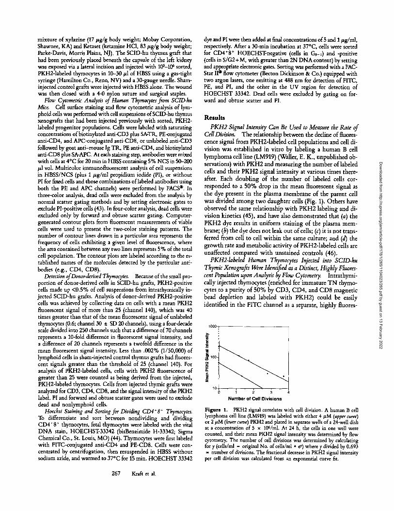
PKH2 Signal Intensity Can Be Used to Measure the Rate of CellDivision. The relationship between the decline of fluores- cence signal from PKH2-1abeled cell populations and cell di- vision was established in vitro by labeling a human B cell lymphoma cell line (LM919) (Waller, E. K., unpublished ob- servations) with PKH2 and measuring the number of labded cells and their PKH2 signal intensity at various times there- after. Each doubling of the number of labeled cells cor- responded to a 50% drop in the mean fluorescent signal as the dye present in the plasma membrane of the parent cell was divided among two daughter cells (Fig. 1). Others have observed the same relationship with PKH2 labeling and di- vision kinetics (45), and have also demonstrated that (a) the PKH2 dye results in uniform staining of the plasma mem- brane; (b) the dye does not leak out of cells; (c) it is not trans- ferred from cell to cell within the same culture; and (d) the growth rate and metabolic activity of PKH2-1abeled cells are unaffected compared with unstained controls (46).
PKH2-1abeled Human Thymocytes Injected into SCID-hu Thymic Xenografis Were Identified as a Distinct, Highly Fluores- cent Population upon Analysis by Flow Cytometry. Intrathymi- cally injected thymocytes (enriched for immature T N thymo- cytes to a purity of 50% by CD3, CD4, and CD8 magnetic bead depletion and labeled with PKH2) could be easily identified in the FITC channel as a separate, highly fluores-
1000 .
I 2 3
N u m b e r o f Ce l l Divisions
Figure 1. PKH2 signal correlates with cell division. A human B cell lymphoma cell line (LM919) was labeled with either 4/~M (upper curve) or 2 #M (lower curve) PKH2 and plated in separate wells of a 24-well dish at a concentration of 5 x 10⁶/mI. At 24 h, the cells in one well were counted, and their mean PKH2 signal intensity was determined by flow cytometry. The number of cell divisions was determined by calculating for y (cells/ml = original No. of cells/ml * ey) where y divided by 0.693 = number of divisions. The fractional decrease in PKH2 signal intensity per cell division was calculated from an exponential curve fit.
267 Kraft et al.
Dow
nloaded from http://rupress.org/jem
/article-pdf/178/1/265/1104023/265.pdf by guest on 13 February 2022
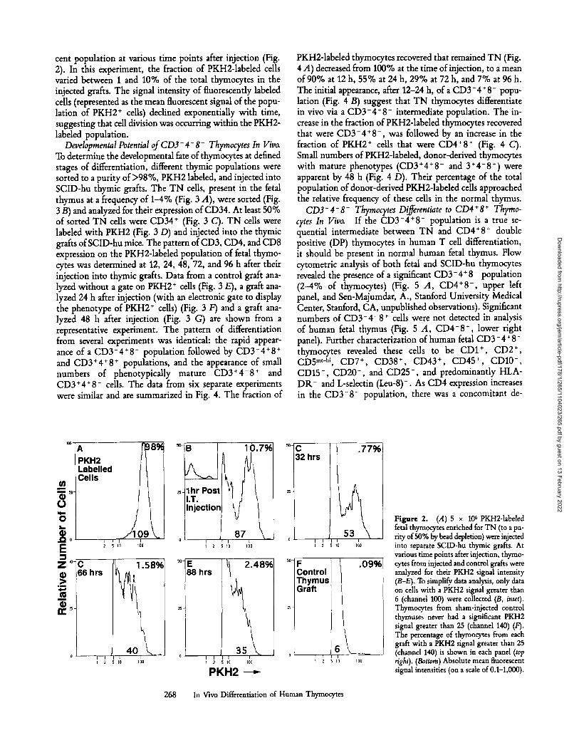
cent population at various time points after injection (Fig. 2). In this experiment, the fraction of PKH2-1abeled cells varied between 1 and 10% of the total thymocytes in the injected grafts. The signal intensity of fluorescently labeled cells (represented as the mean fluorescent signal of the popu- lation of PKH2 + cells) declined exponentially with time, suggesting that cell division was occurring within the PKH2- labeled population.
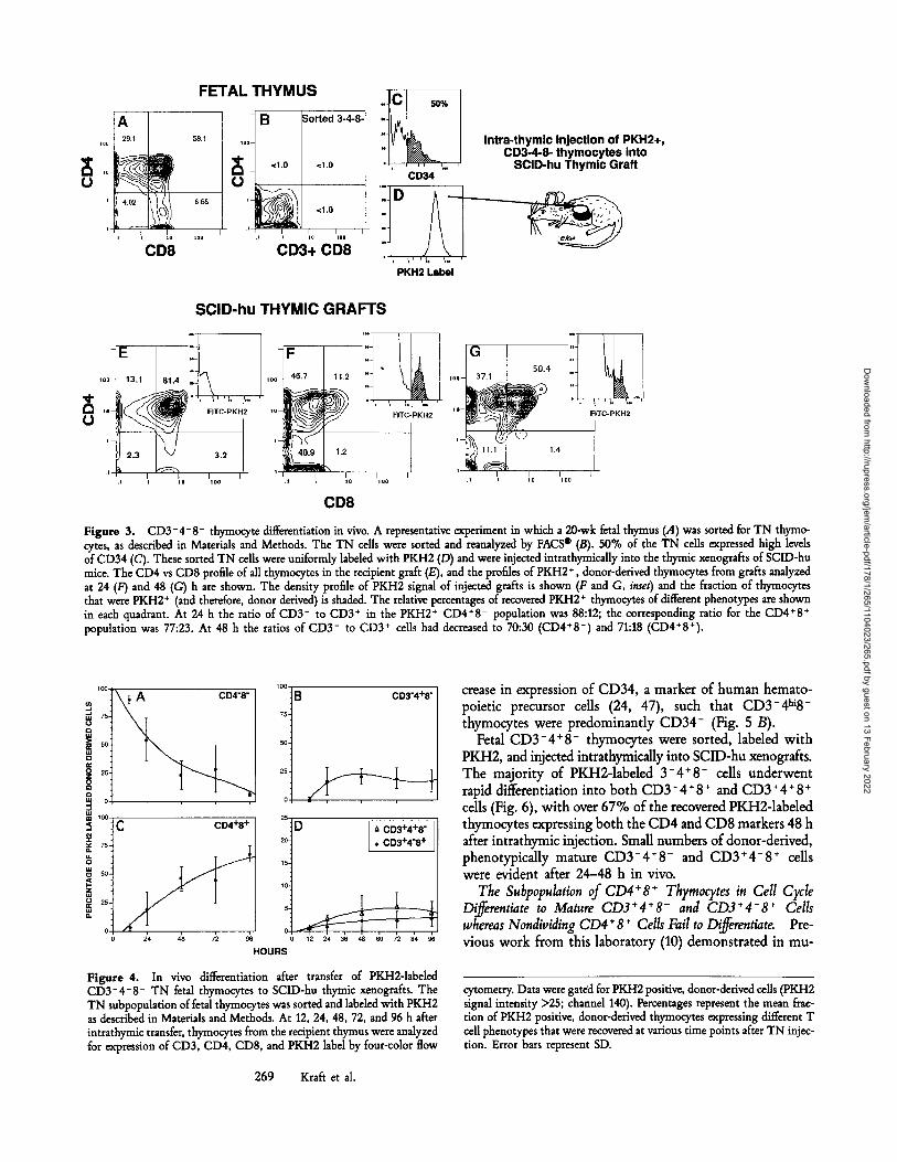
Developmental Potential of CD3-4-8 - Thymocytes In Viva To determine the developmental fate of thymocytes at defined stages of differentiation, different thymic populations were sorted to a purity of >98%, PKH2 labeled, and injected into SCID-hu thymic grafts. The TN cells, present in the fetal thymus at a frequency of 1-4% (Fig. 3 A), were sorted (Fig. 3 B) and analyzed for their expression of CD34. At least 50% of sorted TN cells were CD34 + (Fig. 3 C). TN cells were labeled with PKH2 (Fig. 3 D) and injected into the thymic grafts of SCID-hu mice. The pattern of CD3, CD4, and CD8 expression on the PKH2-1abeled population of fetal thymo- cytes was determined at 12, 24, 48, 72, and 96 h after their injection into thymic grafts. Data from a control graft ana- lyzed without a gate on PKH2 + cells (Fig. 3 E), a graft ana- lyzed 24 h after injection (with an electronic gate to display the phenotype of PKH2 + cells) (Fig. 3 F) and a graft ana- lyzed 48 h after injection (Fig. 3 G) are shown from a representative experiment. The pattern of differentiation from several experiments was identical: the rapid appear- ance of a CD3-4+8 - population followed by CD3-4+8 + and CD3+4+8 + populations, and the appearance of small numbers of phenotypicaUy mature CD3 +4- 8 + and CD3+4+8 - cells. The data from six separate experiments were similar and are summarized in Fig. 4. The fraction of
PKH2-1abded thymocytes recovered that remained TN (Fig. 4 A) decreased from 100% at the time of injection, to a mean of 90% at 12 h, 55% at 24 h, 29% at 72 h, and 7% at 96 h. The initial appearance, after 12-24 h, of a CD3- 4 + 8- popu- lation (Fig. 4 B) suggest that TN thymocytes differentiate in vivo via a CD3-4 + 8- intermediate population. The in- crease in the fraction of PKH2-1abeled thymocytes recovered that were CD3-4§ -, was followed by an increase in the fraction of PKH2 + cells that were CD4+8 + (Fig. 4 C). Small numbers of PKH2-1abded, donor-derived thymocytes with mature phenotypes (CD3*4+8 - and 3+4-8 +) were apparent by 48 h (Fig. 4 D). Their percentage of the total population of donor-derived PKH2-1abded cells approached the relative frequency of these cells in the normal thymus.
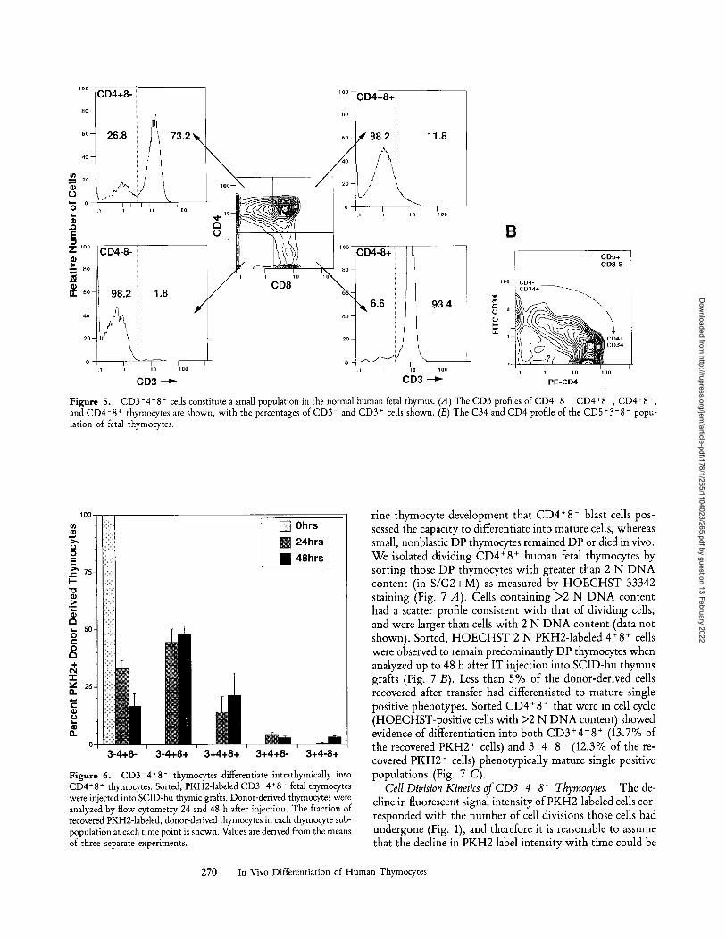
CD3-4+8 - Thymocytes Differentiate to CD4+8 + Thymo- cytes In Viwx If the CD3-4+8 - population is a true se- quential intermediate between TN and CD4+8 + double positive (DP) thymocytes in human T cell differentiation, it should be present in normal human fetal thymus. Flow cytometric analysis of both fetal and SCID-hu thymocytes revealed the presence of a significant CD3-4+8 - population (2-4% of thymocytes) (Fig. 5 A, CD4+8 -, upper left panel, and Sen-Majumdar, A., Stanford University Medical Center, Stanford, CA, unpublished observations). Significant numbers of C D 3 - 4 - 8 + cells were not detected in analysis of human fetal thymus (Fig. 5 A, CD4-8 +, lower right panel). Further characterization of human fetal CD3 - 4 + 8- thymocytes revealed these cells to be CD1 +, CD2 +, CD5 mr-hi, CD7 +, CD38 +, CD43 +, CD45 +, CD10-, CD15-, CD20- , and CD25- , and predominantly HLA- D R - and L-sdectin (Leu-8)-. As CD4 expression increases in the C D 3 - 8 - population, there was a concomitant de-
U) m m 5o- (9 O
o
(9 .Q0 E Z,O-
(9 .>
D
(9
A PKH2 Labelled Cells
C6hrs
I
[
j•109 ~, IJO 100
1.58%
4 0 _
10.7%
I
I 2 10 I00
E % hrs
, , , 3 5 , \ I 2 IQ 100
PKH2 - - - ~
C hrs
Control | Thym us / Graft
25
6 I
10
.77%
100
. 09%
lOG
Figure 2. (.4) 5 x 10⁶ PKH2-labeled fetal thymocytes enriched for TN (to a pu- rity of 50% by bead depletion) were injected into separate SCID-hu thymic grafts. At various time points after injection, thymo- cytes from injected and control grafts were analyzed for their PKH2 signal intensity (B-E). To simplify data analysis, only data on cells with a PKH2 signal greater than 6 (channel 100) were collected (B, inset). Thymocytes from sham-injected control thymuses never had a significant PKH2 signal greater than 25 (channel 140) (F). The percentage of thymocytes from each graft with a PKH2 signal greater than 25 (channel 140) is shown in each panel (top right). (Bottom) Absolute mean fluorescent signal intensities (on a scale of 0.1-1,000).
268 In Vivo Differentiation of Human Thymocytes
Dow
nloaded from http://rupress.org/jem
/article-pdf/178/1/265/1104023/265.pdf by guest on 13 February 2022
FETAL THYMUS
A9 ~ B ~orted 3-4-8- ,oo .1 58.1 ,o~
~ ~ <1.0 <1.0
i t o o " . i Io Iloo
CD3+ CD8
. C
! ~ Intra-thymic Injection of PKH2., CD3-4-8- thymocytes into
, ,, ,,. , SCID-hu Thymic Grail CD34
o
' t ~ ' , . i
PKH2 Label
SClD-hu THYMIC GRAFTS
i'~ ,0 O
' ' t , .',. "C-PKH2
1.2
.i- ,i i i o i o o
i 0 o t6.7
1~ ~ /
1- 40.9
i 11.2
F I T C - P K H 2
1.2
~0 I t 0 0 I
G = 50.4
10o- 37.1
, ( _ ~ ~ ~ ~ 1.4
. i i I 0 JlO0
,, ,, , ,
F I T C - P K H 2
CD8
Figure 3. CD3-4-8- thymocyte differentiation in vivo. A representative experiment in which a 20-wk fetal thymus (,4) was sorted for TN thymo- cytes, as described in Materials and Methods. The TN cells were sorted and reanalyzed by FACS | (B). 50% of the TN cells expressed high levels of CD34 (C). These sorted TN cells were uniformly labeled with PKH2 (/9) and were injected intrathymically into the thymic xenografts of SCID-hu mice. The CD4 vs CD8 profile of all thymocytes in the recipient graft (E), and the profiles of PKH2 + , donor-derived thymocytes from grafts analyzed at 24 (F) and 48 (G) h are shown. The density profile of PKH2 signal of injected grafts is shown (F and C, inset) and the fraction of thymocytes that were PKH2 + (and therefore, donor derived) is shaded. The relative percentages of recovered PKH2 + thymocytes of different phenotypes are shown in each quadrant. At 24 h the ratio of CD3- to CD3 + in the PKH2 + CD4*8 - population was 88:12; the corresponding ratio for the CD4+8 + population was 77:23. At 48 h the ratios of CD3- to CD3 + cells had decreased to 70:30 (CD4+8 -) and 71:18 (CD4+8+).
100
~u 75. =o >= 5o
~ 25.
~ 75.
~ 50.
~ 25,
C CD4+8+
B CD3 "4+8"
20
15
10
CD3+4+8 - �9 CD3+4"8 +
HOURS
Figure 4. In vivo differentiation after transfer of PKH2-1abded CD3-4-8- TN fetal thymocytes to SCID-hu thymic xenografts. The TN subpopulation of fetal thymocytes was sorted and labeled with PKH2 as described in Materials and Methods. At 12, 24, 48, 72, and 96 h after intrathymic transfer, thymocytes from the recipient thymus were analyzed for expression of CD3, CD4, CD8, and PKH2 label by four-color flow
269 Kraft et al.
crease in expression of CD34, a marker of human hemato- poietic precursor cells (24, 47), such that CD3-4hi8- thymocytes were predominantly CD34- (Fig. 5 B).
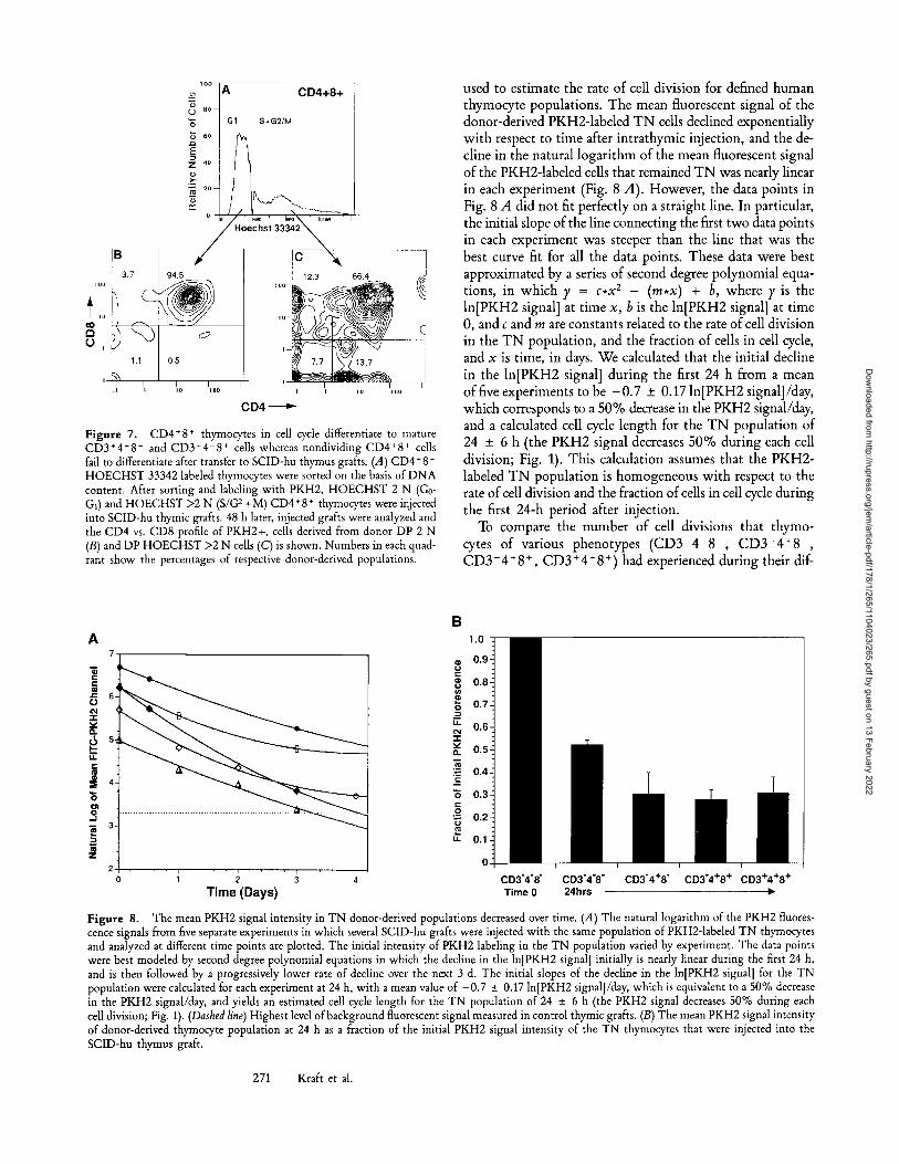
Fetal CD3-4+8 - thymocytes were sorted, labeled with PKH2, and injected intrathymicaUy into SCID-hu xenografts. The majority of PKH2-1abeled 3 -4+8 . cells underwent rapid differentiation into both CD3-4+8 + and CD3+4+8 + cells (Fig. 6), with over 67% of the recovered PKH2-1abeled thymocytes expressing both the CD4 and CD8 markers 48 h after intrathymic injection. Small numbers of donor-derived, phenotypically mature C D 3 - 4 + 8 - and C D 3 + 4 - 8 + cells were evident after 24-48 h in vivo.
The Subpopulation of CD4+8 + Thyraocytes in Cell Cycle Differentiate to Mature CD3*4+8 - and C D 3 + 4 - 8 + Cells whereas Nondividing CD4+8 + Cells Fail to Differentiate Pre- vious work from this laboratory (10) demonstrated in mu-
cytometry. Data Were gated for PKH2 positive, donor-derived cells (PKH2 signal intensity >25; channel 140). Percentages represent the mean frac- tion of PKH2 positive, donor-derived thymocytes expressing different T cell phenotypes that were recovered at various time points after TN injec- tion. Error bars represent SD.
Dow
nloaded from http://rupress.org/jem
/article-pdf/178/1/265/1104023/265.pdf by guest on 13 February 2022
CD4+8-
26.8 88.2 11.8
~ ~ = 20
o-
. (3
E Z IOO
>* , ~ B o -
i
~ 6 0 -
4 0 - -
2 0 - -
CD4-8-
98.2 1.8
.1 i l o l o o
CD3
i o o -
O
1 i io 1
CD8
/
\
93.4
B
i lO loo i i io i o o
CD3 - - ~ PE-CD4
Figure 5. CD3-4+8 - cells constitute a small population in the normal human fetal thymus. (A) The CD3 profiles ofCD4 8 , CD4+8 , CD4+8 +, and CD4-8 + thymocytes are shown, with the percentages of CD3 and CD3 + cells shown. (B) The C34 and CD4 profile of the CD5§ - popu- lation of fetal thymocytes.
1 0 0
= - [ ] 0hrs
[ ] 24hrs 0 o E �9 48hrs
~ " /5
I--- "O
.5
~ 5 0 O r
O
~ 25 a.
0 T - - - ' r - - - ' - -
3-4+8- 3-4+8+ 3+4+8+ 3+4+8- 3+4-8+
Figure 6. CD3 4+8 - thymocytes differentiate intrathymically into CD4+8 + thymocytes. Sorted, PKH2-1abeled CD3 4+8 - fetal thymocytes were injected into SCID-hu thymic grafts. Donor-derived thymocytes were analyzed by flow cytometry 24 and 48 h after injection. The fraction of recovered PKH2-1abeled, donor-derived thymocytes in each thymocyte sub- population at each time point is shown. Values are derived from the means of three separate experiments.
rine thymocyte development that CD4 + 8 § blast cells pos- sessed the capacity to differentiate into mature cells, whereas small, nonblastic DP thymocytes remained DP or died in vivo. We isolated dividing CD4 + 8 + human fetal thymocytes by sorting those DP thymocytes with greater than 2 N D N A content (in S /G2+M) as measured by H O E C H S T 33342 staining (Fig. 7 A). Cells containing >2 N D N A content had a scatter profile consistent with that of dividing cells, and were larger than cells with 2 N D N A content (data not shown). Sorted, H O E C H S T 2 N PKH2-1abeled 4+8 + cells were observed to remain predominantly DP thymocytes when analyzed up to 48 h after IT injection into SCID-hu thymus grafts (Fig. 7 B). Less than 5% of the donor-derived cells recovered after transfer had differentiated to mature single positive phenotypes. Sorted CD4 + 8 + that were in cell cycle (HOECHST-positive cells with >2 N D N A content) showed evidence of differentiation into both CD3 + 4- 8 + (13.7% of the recovered PKH2 + cells) and 3+4+8 - (12.3% of the re- covered PKH2 + cells) phenotypically mature single positive populations (Fig. 7 C).
Cell Division Kinetics of CD3-4-8- Thyrnocytes. The de- cline in fluorescent signal intensity of PKH2-1abeled cells cor- responded with the number of cell divisions those cells had undergone (Fig. 1), and therefore it is reasonable to assume that the decline in PKH2 label intensity with time could be
270 In Vivo Differentiation of Human Thymocytes
Dow
nloaded from http://rupress.org/jem
/article-pdf/178/1/265/1104023/265.pdf by guest on 13 February 2022
l oo
( . j 80 -
~ 60
Z 4 0
~ 20
~. CD4+8+
G1 S+G2 , tM
CD4
Figure 7. CD4+8 + thymocytes in cell cycle differentiate to mature CD3+4+8 - and CD3§ 8 + cells whereas nondividing CD4+8 § cells fail to differentiate after transfer to SCID-hu thymus grafts. (A) CD4+8 § HOECHST 33342 labeled thymocytes were sorted on the basis of DNA content. After sorting and labeling with PKH2, HOECHST 2 N (G0- G1) and HOECHST >2 N (S/G 2 +M) CD4+8 + thymocytes were injected into SCID-hu thymic grafts. 48 h later, injected grafts were analyzed and the CD4 vs. CD8 profile of PKH2+, cells derived from donor DP 2 N (B) and DP HOECHST >2 N cells (C) is shown. Numbers in each quad- rant show the percentages of respective donor-derived populations.
used to estimate the rate of cell division for defined human thymocyte populations. The mean fluorescent signal of the donor-derived PKH2-1abeled T N cells declined exponentially with respect to time after intrathymic injection, and the de- cline in the natural logarithm of the mean fluorescent signal of the PKH2-1abeled cells that remained T N was nearly linear in each experiment (Fig. 8 A). However, the data points in Fig. 8 A did not fit perfectly on a straight line. In particular, the initial slope of the line connecting the first two data points in each experiment was steeper than the line that was the best curve fit for all the data points. These data were best approximated by a series of second degree polynomial equa- tions, in w h i c h y = c , x 2 - ( r e , x ) + b, w h e r e y i s the ln[PKH2 signal] at time x, b is the ln[PKH2 signal] at time 0, and c and m are constants related to the rate of cell division in the T N population, and the fraction of cells in cell cycle, and x is time, in days. We calculated that the initial decline in the ln[PKH2 signal] during the first 24 h from a mean of five experiments to be - 0 . 7 _+ 0.17 ln[PKH2 signal]/day, which corresponds to a 50% decrease in the PKH2 signal/day, and a calculated cell cycle length for the T N population of 24 _+ 6 h (the PKH2 signal decreases 50% during each cell division; Fig. 1). This calculation assumes that the PKH2- labeled T N population is homogeneous with respect to the rate of cell division and the fraction of cells in cell cycle during the first 24-h period after injection.
To compare the number of cell divisions that thymo- cytes of various phenotypes ( C D 3 - 4 - 8 - , C D 3 - 4 + 8 - , C D 3 - 4 § 8 +, CD3 § 4 § 8 § ) had experienced during their dif-
A
. J
7
Time (Days)
B
c
8
o I LI.
Q.
m
g
El. / 1 m "r
CD3"4"8" CD3"4"8" CD3"4+8" CD3"4+8 + CD3+4+8 + Time 0 24hrs �9
Figure 8. The mean PKH2 signal intensity in TN donor-derived populations decreased over time. (A) The natural logarithm of the PKH2 fluores- cence signals from five separate experiments in which several SCID-hu grafts were injected with the same population of PKH2-1abeled TN thymocytes and analyzed at different time points are plotted. The initial intensity of PKH2 labeling in the TN population varied by experiment. The data points were best modeled by second degree polynomial equations in which the decline in the ln[PKH2 signal] initially is nearly linear during the first 24 h, and is then followed by a progressively lower rate of decline over the next 3 d. The initial slopes of the decline in the ln[PKH2 signal] for the TN population were calculated for each experiment at 24 h, with a mean value of -0.7 _+ 0.17 ln[PKH2 signal]/day, which is equivalent to a 50% decrease in the PKH2 signal/day, and yields an estimated cell cycle length for the TN population of 24 _+ 6 h (the PKH2 signal decreases 50% during each cell division; Fig. 1). (Dashed line) Highest level of background fluorescent signal measured in control thymic grafts. (B) The mean PKH2 signal intensity of donor-derived thymocyte population at 24 h as a fraction of the initial PKH2 signal intensity of the TN thymocytes that were injected into the SCID-hu thymus graft.
271 Kraft et al.
Dow
nloaded from http://rupress.org/jem
/article-pdf/178/1/265/1104023/265.pdf by guest on 13 February 2022
Periphery .41--- ~ 1 /~, ~ 4~c~ \ . . . . / " 1.cos,o4+8+)
. . . . . . co.- . . . . . . . . . . . . . . . ' J
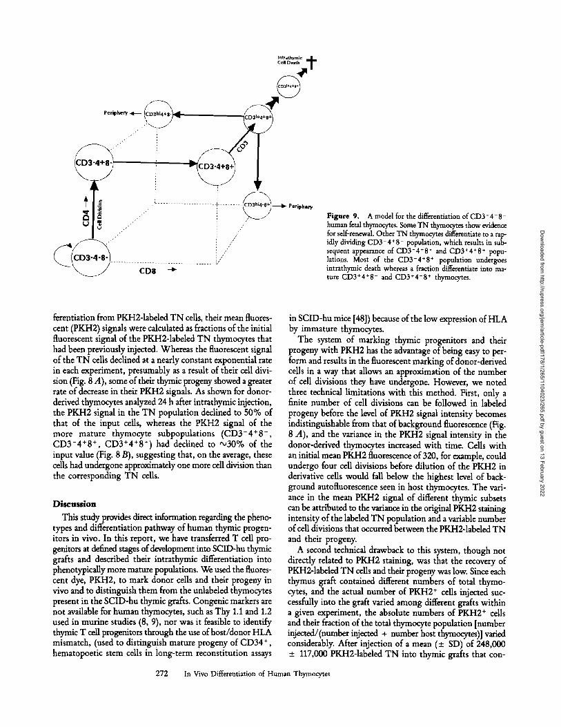
Figure 9. A model for the differentiation of CD3 - 4 - 8 - human fetal thymocytes. Some TN thymocytes show evidence for self-renewal. Other TN thymocytes differentiate to a rap- idly dividing CD3-4+8 - population, which results in sub- sequent appearance of CD3- 4 + 8 + and CD3 + 4 + 8 + popu- lations. Most of the CD3+4+8+ population undergoes intrathymic death whereas a fraction differentiate into ma- ture CD3+4*8 - and CD3+4-8 + thymocytes.
ferentiation from PKH2-1abeled TN cells, their mean fluores- cent (PKH2) signals were calculated as fractions of the initial fluorescent signal of the PKH2-1abeled TN thymocytes that had been previously injected. Whereas the fluorescent signal of the TN cells declined at a nearly constant exponential rate in each experiment, presumably as a result of their cell divi- sion (Fig. 8 A), some of their thymic progeny showed a greater rate of decrease in their PKH2 signals. As shown for donor- derived thymocytes analyzed 24 h after intrathymic injection, the PKH2 signal in the TN population declined to 50% of that of the input cells, whereas the PKH2 signal of the more mature thymocyte subpopulations (CD3-4+8 -, CD3-4+8 +, CD3+4+8+) had declined to '~30% of the input value (Fig. 8 B), suggesting that, on the average, these cells had undergone approximately one more cell division than the corresponding TN cells.
Discussion
This study provides direct information regarding the pheno- types and differentiation pathway of human thymic progen- itors in vivo. In this report, we have transferred T cell pro- genitors at defined stages of development into SCID-hu thymic grafts and described their intrathymic differentiation into phenotypically more mature populations. We used the fluores- cent dye, PKH2, to mark donor cells and their progeny in vivo and to distinguish them from the unlabeled thymocytes present in the SCID-hu thymic grafts. Congenic markers are not available for human thymocytes, such as Thy 1.1 and 1.2 used in routine studies (8, 9), nor was it feasible to identify thymic T cell progenitors through the use of host/donor HLA mismatch, (used to distinguish mature progeny of CD34 +, hematopoetic stem cells in long-term reconstitution assays
in SCID-hu mice [48]) because of the low expression of HLA by immature thymocytes.
The system of marking thymic progenitors and their progeny with PKH2 has the advantage of being easy to per- form and results in the fluorescent marking of donor-derived cells in a way that allows an approximation of the number of cell divisions they have undergone. However, we noted three technical limitations with this method. First, only a finite number of cell divisions can be followed in labeled progeny before the level of PKH2 signal intensity becomes indistinguishable from that of background fluorescence (Fig. 8 A), and the variance in the PKH2 signal intensity in the donor-derived thymocytes increased with time. Cells with an initial mean PKH2 fluorescence of 320, for example, could undergo four cell divisions before dilution of the PKH2 in derivative cells would fall below the highest level of back- ground autofluorescence seen in host thymocytes. The vari- ance in the mean PKH2 signal of different thymic subsets can be attributed to the variance in the original PKH2 staining intensity of the labeled TN population and a variable number of cell divisions that occurred between the PKH2-1abeled TN and their progeny.
A second technical drawback to this system, though not directly related to PKH2 staining, was that the recovery of PKH2-1abeled TN cells and their progeny was low. Since each thymus graft contained different numbers of total thymo- cytes, and the actual number of PKH2 + cells injected suc- cessfully into the graft varied among different grafts within a given experiment, the absolute numbers of PKH2 + cells and their fraction of the total thymocyte population [number injected/(number injected + number host thymocytes)] varied considerably, After injection of a mean (+ SD) of 248,000 + 117,000 PKH2-1abeled TN into thymic grafts that con-
272 In Vivo Differentiation of Human Thymocytes
Dow
nloaded from http://rupress.org/jem
/article-pdf/178/1/265/1104023/265.pdf by guest on 13 February 2022
tained a mean of 36 x 106 + 21 x 106 cells, the absolute number of PKH2-1abeled cells that could be detected at different time points in thymic grafts varied from a mean of 27,340 _+ 3,400 cells at 12 h' (a recovery of 16% of the number of PKH2 + cells injected) to 19,280 _+ 9,500 at 24 h (11.2% recovery); 11,799 _+ 6,200 at 48 h (10.2% recovery); 25,860 _+ 17,100 at 72 h (7.4% recovery); and 21,800 _+ 9,200 at 96 h (6.7% recovery). These data indicate that only about 10-20% of the pKH2-1abeled TN cells that we at- tempted to inject into the graft remained as viable cells at 12 h, and their PKH2-1abeled progeny constituted a slowly declining fraction of the total thymocytes in the graft there- after. The large initial loss of PKH2-1abeled cells immedi- ately after intrathymic injection suggests that a majority of the injected cells fail to remain in the thymus (they leak out of the injection site), or that they die soon after injection, an observation that is true for direct in situ intrathymic in- jection studies (49).
The failure to observe an accumulation of PKH2-1abeled cells 12-96 h after intrathymic injection may be due to the emigration of mature thymocytes into the peripheral circula- tion of the SCID-hu mouse (17, 18, 37); their intrathymic cell death, either at the level of CD31~ + cells (due to the failure of antigen-presenting cells to engage the TCR [16]), or at the level of CD3 + 4 + 8- or 3 + 4- 8 + (due to negative selection) (10); or a competition between the self-renewal of PKH2-1abeled TN and the endogenous production of TN thymocytes within the SCID-hu thymic graft. SCID-hu thymic grafts contain an admixture of thymocytes and pluripo- tent hematopoetic stem cells (7) derived from fragments of fetal liver, and thus have a relatively constant supply of new TN thymic progenitors. Therefore we would predict that the proportion of PKH2-1abeled thymocytes would diminish over time as PKH2+-labeled TN cells mature, differentiate, and/or die, and are replaced by unlabeled TN cells derived from fetal liver hematopoetic stem cells.
The third technical point concerning this system was that we studied the differentiation of fetal thymocytes in heterol- ogous (HLA mismatched) thymus grafts in SCID mice. We used fetal thymus as a source of thymic progenitors because they contained greater numbers of immature thymocytes than were present in established SCID-hu grafts. The differentia- tion of hematopoetic stem cells to mature T cells after their injection into heterologous thymic grafts has been demon- strated (50), indicating the ability of thymocyte progenitors, mismatched with respect to their expression of HLA antigens compared with the recipient thymus (51), to differentiate nor- mally in SCID-hu mice (6, 48). In these experiments, the mature, donor-derived thymocytes had normal phenotypes and were tolerant to the HLA presented by the thymic-host stromal environment (52). Nevertheless, the differentiation ofT cell progenitors in an aUogeneic, HLA mismatched thymic microenvironment might differ from the differentiation of T cell progenitors in an autologous thymic microenviron- ment. Therefore, we sorted TN thymocytes from SCID-hu grafts, labeled them with PKH2, and injected the PKH2- labeled TN cells into SCID-hu mice that had been grafted
with thymic tissue from the same fetal donor. We found im- mature TN thymocytes made up a smaller proportion (<0.2%) of thymocytes in SCID-hu grafts 2 mo after en- graftment as compared with 1-3% TN cells of human fetal thymus. The differentiation of the limited number of TN cells that could be sorted from SCID-hu grafts occurred in an identical pattern as that we have observed with fetal thymo- cytes (data not shown).
CD34 has been shown to be a marker of human hematopoetic stem cells (6, 24, 47, 48) as well as the earliest thymic T cell progenitor (24). We characterized the pheno- type of the TN and CD3-4+8 - progenitors from fetal thymus with respect to their expression of CD34 and other lymphoid markers. We found that TN thymocytes expressed intermediate levels of the CD5, CD45, and HLA-DR an- tigens (CD51~176 Greater than 50% of the sorted TN cells were CD34 + and lacked significant expres- sion of HLA class I antigens. In contrast, more mature T cell phenotypes were CD34- CD5 hi, and CD45 ~. CD34 ex- pression was inversely correlated with CD4 expression (Fig. 5 B). Low levels of CD4 expression have recently been de- scribed on a population of murine pluripotent hematopoi- etic stem cells (53). CD34 + cells present in the CD3-41~ population (which fell between TN and CD3-4+81 cells, the latter being CD34-) could warrant consideration in fu- ture studies of human thymocyte differentiation.
We observed the maturation of C D 3 - 4 - 8 - progenitors to more mature thymocytes in vivo, with the sequential ap- pearance of CD3-4+8 -, 3-4+8 +, 3+4+8+, 3+4+8 -, and 3+4-8 + populations over 12-96 h. Differentiation of mu- fine TN to mature thymocyte subsets in vitro, on short-term thymic stromal cultures, has also recently been shown to occur within 2 d (54, 55). Using the system of PKH2-1abeling thymocyte progenitors, we have observed differentiation of the most immature (TN) thymocyte to cells with the most mature (single positive) phenotype. At 24, 48, and 72 h after injection of PKH2-1abeled TN, there were approximately twice as many PKH2-1abeled CD3+4+8 - cells compared with CD3 + 4-8 + thymocytes, although the total fraction of phenotypically mature thymocytes never rose above 10% of the entire PKH2-1abeled population (Fig. 4 D). The relative ratio of PKH2-1abeled CD4 and CD8 single positive thymo- cytes at the latter time points were similar to those seen in SCID-hu thymic grafts in which the ratio of phenotypically mature CD3 + 4 + 8- and CD3 + 4- 8 + cells was slightly more than 2:1, with CD4 + single positives comprising 12 _+ 7% of thymocytes, and CD8 single positive thymocytes comprising 5 + 6% of the thymocyte population (17).
The exponential decline in PKH2 signal of donor-derived TN thymocytes suggested that TN cells retained the poten- tial for limited self-renewal, since a proportion of the TN progeny cells remain phenotypicatly TN, and demonstrate progressively lower intensities of the PKH2 label, consistent with cell division (Figs. 1 and 8 A). Based on the initial rate of decline of the ln[PKH2 fluorescence signal] of donor-derived PKH2-1abeled TN thymocytes at 24 h after intrathymic in- jection, we estimated the length of the cell cycle for the TN
273 Kraft et al.
Dow
nloaded from http://rupress.org/jem
/article-pdf/178/1/265/1104023/265.pdf by guest on 13 February 2022
population to be 24 _+ 6 h (Fig. 8 A). This calculation as- sumes that the PKH2-1abeled TN cells constitute a uniform population with respect to the fraction of cells in cell cycle and their cell cycle length. After the in vivo growth of PKH2- labeled TN cells, the variance of their fluorescent signal is too large to detect subpopulations of PKH2-1abeled TN cells that could have different cell cycle kinetics. Thus the average cell cycle length of 24 h, that we calculated for the TN popu- lation as a whole, could represent a combination of shorter and longer cell cycle lengths of subpopulations of TN that are dividing more and less rapidly, respectively. The change in the slope of the decline in the ln[PKH2 signal] at later time points (Fig. 8 B) suggests that the PKH2-1abeled TN population is indeed somewhat heterogeneous, and contains a subpopulation(s) of cells that are nondividing, or dividing at a slower rate. Therefore, the decline of the ln[PKH2 signal] is best approximated by a second degree polynomial equa- tion rather than a first degree polynomial (linear) equation. As the PKH2-1abeled TN differentiated, they constituted a diminished fraction of the total PKH2 + population. We have estimated that, on the average, ,o30% of the TN cells differentiate into more mature cells, whereas 70% of the progeny remain phenotypically TN after each cell division.
Compared with the corresponding TN population, the lower intensities of PKH2 label in the thymocyte popula- tions of intermediate differentiation (CD3-4+8 - , 3-4+8 + , and 3+4+8 +) 24 h after intrathymic injection of PKH2- labeled TN thymocytes, suggested that either (a) more cell divisions had occurred in these thymic subpopulations com- pared with those TN cells that remained phenotypically un- changed; or (b) the subpopulations of TN that differentiate to more mature phenotypes undergo more rapid cell division than those PKH2-1abeled cells that remain phenotypically TN (Fig. 8 B). Conclusions from several studies on murine thymo- cyte kinetics (20-23) which demonstrate more rapid cell cy- cling in the CD3-4-8 + population compared with the more immature TN population (23) support the idea that thymocytes of intermediate levels of differentiation (between TN and the phenotypically mature cells) may divide more rapidly than the TN population (56).
After the injection of PKH2-1abeled TN cells, a PKH2- labeled CD3-4+8 - population rapidly appeared in the thymus, largely preceding the appearance of larger numbers of CD3- 4 + 8 + and CD3 + 4 + 8 + cells (Figs. 3 F, 4 B and 4 C). Sorted, PKH2-1abeled CD3-4+8 - cells differentiated rapidly in vivo, to CD3- 4 + 8 + and CD3 + 4 + 8 + populations (Fig. 6), indicating that the CD3-4+8 - population was a sequential intermediate between TN and CD3-4 +8 + / CD3+4+8 + populations. These results are in contrast to the differentiation of mouse thymocytes in C57BL strains, in which the differentiation of TN thymocytes proceeds pri- marily through a CD3-4-8 + intermediate to the CD4 + 8 + population (9), but are similar to the pathway described in CBA mouse strains, in which TN differentiate through a 3-4+8 - intermediate as well (57-59). Murine CD3-4-8 + and CD3-4 + 8- thymocytes have been shown to be large, blast cells, cortisone sensitive, and concentrated in the outer cortex (60).
Spitz et al. (26) have demonstrated that the human post- natal 3-4+8 + population can be subdivided into CD3-4+8o~+Sfl - and CD3-4+8c~+8fl+ populations, and that the expression of CDSc~ + precedes the expression of CDSfl when TN tbymocytes are cultured in vitrcx These authors noted a CD3-4+8c~ - population, that is presum- ably identical to the CD3-4 + 8- population described in the present study, as we have used an antibody to CDS, Leu-2a, that recognizes CDSc~. An additional pathway of human thymic differentiation from CD3-4 -8 - cells to CD4+8 + without going through the CD3-4+8 - immature inter- mediate is possible, and cannot be excluded by our data. How- ever, at least 50% of TN thymocytes appear to mature via a CD3-4+8 - intermediate (Fig. 4). In addition, small num- bers of CD3 + 4- 8- cells appeared at later time points, which contributed to the overall CD4-8- population. Because of the rarity of the CD3 + 4- 8- cells, the developmental path- way that leads to this thymic subset could not be determined.
The maturation of human TN thymocytes in vivo via a CD3-4 + 8- intermediate correlates well with in vitro differ- entiation studies of TN and CD3-4+8 - thymocytes on short-term, heterologous, human-stromal cell cultures (Lie- berman, M., A. Sen-Majumdar, D. L. Kraft, I. L. Weissman, and E. K. Waller, unpublished observations).
The expression of CD4 at an early stage of intrathymic T cell maturation could potentially play a role in HIV infec- tion of the thymus (61). This is testable, as SCID-hu thymic grafts sustain productive HIV infection (62), and in the in- fected thymuses, eventual depletion of CD4 + cells occurs (62a). Furthermore, a rapidly cycling immature CD3-4 + 8- intermediate may play a role in lymphomagenesis. An induc- ible routine T cell lymphoma corresponding to the imma- ture murine CD3-4-8 + thymocyte subset has recently been reported (63). Two CD3-4 + 8- human lymphomas have re- cently been described in immunosuppressed organ transplant recipients, and may represent the neoplastic analog of the CD3-4+8 - thymic intermediate (64).
The technique of FACS | sorting based on relative DNA content (HOECHST staining) combined with PKH2 labeling, allowed an investigation of the relative potential of dividing vs. nondividing populations to differentiate. CD4 + 8 + cells were sorted on the basis of their DNA content, PKH2 la- beled, and injected into thymic grafts (Fig. 7). The subpopu- lation in cell cycle (DNA content >2 N) differentiated into thymocytes of a mature phenotype, whereas CD4 + 8 + cells not engaged in DNA synthesis failed to differentiate to a significant degree in vivo (Fig. 6). It has been reported that the majority of dividing mouse CD4 + 8 + blasts express low levels of TCR/CD3 (65). Taken together, these data indicate that the nondividing subset of small CD4 + 8 + cells are des- tined to die intrathymically, and that only those CD4+8 + cells that have received mitogenic stimuli (presumably as a result of engagement of the low numbers of TCR expressed on their surfaces) are able to differentiate further into thymo- cytes of more mature phenotypes (positive selection) (10).
Fig. 9 presents a model for a differentiation pathway of human TN fetal thymocytes which summarizes our data. The ability to observe human T cell differentiation in vivo pro-
274 In Vivo Differentiation of Human Thymocytes
Dow
nloaded from http://rupress.org/jem
/article-pdf/178/1/265/1104023/265.pdf by guest on 13 February 2022
vides a means to examine the developmental ontogeny of TN thymocyte populations of more defined phenotypes (such as CD34 + pediatric and adult thymocytes). Other develop- mental phenomena can be examined, such as extrathymic T cell differentiation, and the effects of systemic/physiologic vari- ables on thymocyte differentiation (39).
Intrathymic transfer of PKH2-1abeled human thymocyte
populations into the SCID-hu mouse model offers a useful in vivo model for the study of short-term human T cell differentiation. Use of such a system should prove useful in the delineation and ontogeny of various progenitor cell popu- lations in the thymus, and other human tissues that can be engrafted into SCID mice (66).
The authors wish to acknowledge Drs. Anish Sen-Majumdar and Miriam Lieberman for their support and critical reading of the manuscript; Dr. Sen-Majumdar for sharing his unpublished observations on the phenotype of the CD3-4 +8- thymic population; and Dr. Susan Alpert for useful discussions. Spe- cial thanks go to Gun Hansteen, Tim Knaak, the FACS | facility personnel, and Ryan Seay for technical help.
D. L. Kraft was supported by a Howard Hughes Medical Institute Kesearch training fellowship for medical students. I. L. Weissman was supported by National Institutes of Health grant CA-42551. E. K. Waller is a McDonnell Scholar, and this work was supported by the James S. McDonnell Foundation.
Address correspondence to Dr. E. K. Waller, Becton Dickinson Immunocytometry Systems, 2350 Qume Drive, San Jose, CA 95131-1807.
Received for publication 31 December 1992 and in revised form 5 April 1993.
R~ferences
1. ~lkins, B., C. Mueller, C.Y. O~da, K.A. Keichert, I.L. Weissman, and G.J. Spangrude. 1987. Early events in T-cell maturation. Annu. R ~ Immuno[. 5:325.
2. Wu, L., K. Scollar, M. Egerton, M. pearse, G.J. Spangrude, and K. Shortman. 1991. CD4 expressed on earliest T-lineage precursor cells in the adult routine thymus. Nature (Lond.). 349:71.
3. Spangrude, G.J., C.E. Muller-Sieburg, S. Heimfeld, and I.L. Weissman. 1988. Two rare populations of mouse Tby-1 l~ bone marrow cells repopulate the thymus. J. F.xtx Med. 167:1671.
4. Weissman, I., V. Papaioannou, and K. Gardner. 1978. Fetal hematopoietic origins of the adult hematolymphoid system. Cold Spring Harbor Meeting on differentiation of Normal and Neoplastic Hematopoetic Cells. B. Clarkson, P.A. Marsk, and J.E. Till, editors. 33-47.
5. Owen, J.J., and E.J. Jenkinson. 1984. Early events in T lym- phocyte genesis in the fetal thymus. Am. J. Anat. 170:301.
6. Bantu, C.M., I.L. Weissman, A.S. Tsukamoto, A. Buckle, and B. Peault. 1992. Isolation of a candidate human hematopoietic stem-cell population. Pro~ Natl. Acad. Sci. USA. 89:2804.
7. Peault, B., I.L. Weissman, C. Baum, J.M. McCune, and A. Tsukamoto. 1991. Lymphoid reconstitution of the human fetal thymus in SCID mice with CD34 + precursor cells. J. Extx Med. 174.
8. Guidos, C.J., I.L. Weissman, and B. Aatkins. 1989. Develop- mental potential of CD4-8- thymocytes. Peripheral progeny include mature CD4-8- T cells bearing otfl T cell receptor. J. Immunol. 142:3773.
9. Guidos, C.J., I.L. Weissman, and B. Adkins. 1989. Intrathymic maturation of murine T lymphocytes from CD8 + precursors. Proc Natl. Acad. Sci. USA. 86:7542.
10. Guidos, C.J., J.S. Danska, C.G. Fathman, and I.L. Weissman. 1990. T cell receptor-mediated negative selection of autoreac- tire T lymphocyte precursors occurs after commitment to the
275 Kraft et al.
CD4 or CD8 lineages. J. Ex F Med. 172:835. 11. Kappler, J.W., N. Roehm, and P. Marrack. 1987. T cell toler-
ance by clonal elimination in the thymus. Cell. 49:273. 12. Fowlkes, B.J., R.H. Schwartz, D.M. Pardoll, W. Swat, M.
Dessing, A. Baron, P. Kisielow, and H. yon Boehmer. 1992. Phenotypic changes accompanying positive selection of CD4+8 + thymocytes. Eur. J. Iramunol. 22:2367.
13. Fowlkes, B.J., K.H. Schwartz, and D.M. perdoll. 1988. Dele- tion of stir-reactive thymocytes occurs at a CD4+8 + precursor stage. Nature (Lond.). 334:620.
14. yon Boehmer, H. 1992. Thymic selection: a matter of life and death. Imraunol. Today. 13:454.
15. Borgulya, P., H. Kishi, U. Muller, J. Kirberg, and H. yon Boehmer. 1991. Development of the CD4 and CD8 lineage o f t cells: instruction versus sdection. EMBO (Eur. Mol. Biol. Organ.) J. 10:913.
16. Robey, E.A., B.J. Fowlkes, J.W. Gordon, D. Kioussis, H. yon Boehmer, K. Ramsdell, and K. Axe1. 1991. Thymic selection in CD8 transgenic mice supports an instructive model for com- mitment to a CD4 or CD8 lineage. Cell. 64:99.
17. Weissman, I.L. Thymus cell migration. 1967. f Exi~ Ailed. 126:291.
18. Scollay, K., M. Kochen, E. Butcher, and I.L. Weissman. 1978. Lyt markers on thymus cell migrants. Nature (Lond.). 276:79.
19. Scollay, K., E. Butcher, and I.L. Weissman. 1980. Thymus cell migration. Quantitative aspects of cellular traffic from the thymus to the periphery in mice. Eur. j. Immunol. 10:210.
20. Egerton, M., K. Shortman, and K. Scollay. 1990. The kinetics of immature thymocyte development in-viva Int. ImraunoL 2:501.
21. Egerton, M., K. Scollay, and K. Shortman. 1990. Kinetics of mature T-cell development in the thymus. Prog Natl. Acad. Sci. USA. 87:2579.
22. Baron, C., and C. Penit. 1990. Study of the thymocyte cell cycle by bivariate analysis of incorporated bromodeoxyuridine
Dow
nloaded from http://rupress.org/jem
/article-pdf/178/1/265/1104023/265.pdf by guest on 13 February 2022
and DNA content. Eur. J. lmmunol. 20:1231. 23. Spangrude, G., and R. Scollay. 1990. Differentiation ofhema-
topoietic stem cells in irradiated mouse thymic lobes. Kinetics and phenotype of progeny. J. Immunol. 145:3661.
24. Terstappen, L.W., S. Huang, and L.J. Picker. 1992. Flow cyto- metric assessment of human T-cell differentiation in thymus and bone marrow. Blood. 79:666.
25. Swerdlow, S.H., P.A. Angermeier, and A.L. Hartman. 1988. Intrathymic ontogeny of the T cell receptor associated CD3 (T3) antigen. La/1 Invest. 58:421.
26. Hori, T., C. Cupp, N. Wrighton, F. Lee, and H. Spits. 1991. Identification of a novel human thymocyte subset with a phenotype of CD3-4+CDSoe+j8 -. Possible progeny of the CD3-CD4-CDS- subset. J. Immunol. 146:4078.
27. Merkenschlager, M., and A.G. Fisher. 1992. Human postnatal thymocytes generate phenotypically immature CD3dim, CDSdim, CDla bright progeny in organ culture. J. Immunol. 148:1012.
28. Denning, S.M., D.M. Jones, R.E. Ware, K.J. Weinhold, M.B. Brenner, and B.F. Haynes. 1991. Analysis of clones derived from human CD7§ - thymocytes. Int. Immunol. 10:1015.
29. Denning, S.M., J. Kurtzenberg, D.S. Leslie, and B.F. Haynes. 1989. Human postnatal CD4-CDS-CD3- thymic T cell precursors differentiate in vitro into T cell receptor gamma- bearing cells. J. Imraunot. 142:2988.
30. Hori, T., and H. Spits. 1991. Clonal analysis of human CD4-8-3- thymocytes highly purified from postnatal thymus. J. Immunol. 146:2116.
31. Rouse, R.V., W. van Ewijk, P.P. Jones, and I.L. Weissman. 1979. Expression of MHC antigens by mouse thymic dendritic cells. J. lmmunol. 122:2508.
32. van Ewijk, W. 1991. T-cell differentiation is influenced by thymic microenvironments. Annu. Rev. Imraunol. 9:591.
33. Haynes, B.F. 1990. Human thymic epithelium and T cell de- velopment, current issues and future directions. Thymus. 16:143.
34. de la Hera, A., W. Marston, C. Aranda, M.L. Toribio, and C.A. Martinez-A. 1989. Thymic stroma is required for the de- velopment of human T cell lineages in vitro. Int. Immunol. 1:471.
35. Merkenschlager, M., and A.G. Fisher. 1992. Selective manipu- lation of the human T-cell receptor repertoire expressed by thymocytes in organ culture. Proc. Natl. Acad. Sci. USA. 89:4255.
36. Fisher, A.G., L. Larson, L.K. Goff, D.E. Restall, L. Happerfield, and M. Merkenshlager. 1990. Human thymocyte development in mouse organ cultures. Int. Immunol. 2:571.
37. McCune, J.M., R. Namikawa, H. Kaneshima, L.D. Shultz, M. Lieberman, and I.L. Weissman. 1988. The SCID-hu mouse: murine model for the analysis of human hematolymphoid differentiation and function. Science (Wash. DC). 241:1632.
38. McCune, J.M., H. Kaneshima, M. Lieberman, I.L. Weissman, and R. Namikawa. 1989. The SCID-hu mouse: current status and potential applications. Cu~ Tol~ Microbiol. Immunol. 152:183.
39. Waller, E.K., A.S. Majumdar, M.R. Shick, O.W. Kamel, G.A. Hansteen, and I.L. Weissman. 1992. Human T-cell develop- ment in SCID-hu mice: staphylococcal enterotoxins induce specific clonal deletions, proliferation and anergy. B/ood. 80:3144.
40. Waller, E.K., O.S. Kamel, M.L. Cleary, A.$. Majumdar, M.R. Schick, M. Lieberman, and I.L. Weissman. 1991. Growth of primary T-cell non-Hodgkin's lymphomata in SCID-hu mice: requirement for a human lymphoid microenvironment. Blood. 78:2650.
41. Spangrude, G.J., S. Heimfeld, and I.L. Weissman. 1988. Purification and characterization of mouse hematopoietic stem cells. Science (Wash. DC). 241:58.
42. Battye, F.L., and K. Shortman. 1991. Flow cytometry and cell- separation procedures. Cu~.. 0I~ Immunol. 3:238.
43. Parks, D.R., R.R. Hardy, and L.A. Herzenberg. 1983. Dual immunofluorescence-new frontiers in the analysis and sorting. Immunol. Today. 4:145.
44. Hollander, Z., and M.R. Loken. 1988. Simultaneous analysis of DNA content and surface antigens in human bone marrow. Cytometry. 9:485.
45. Melnicoff, M.J., P.S. Morahan, B.D. Jensen, E.W. Breslin, and P.K. Horan. 1988. In vivo labeling of resident peritoneal mac- rophages. J. Leukocyte Biol. 43:387.
46. Horan, P.K., S.E. Slezak, and B.D. Jensen. 1993. Cellular proliferation history by fluorescent analysis: In Flow Cytom- etry. A. Jacquemin-Sablon, editor. NATO ASI Series I-I67. In press.
47. Civin, C.I., L.C. Strauss, C. Brovall, M.J. Fackles, J.F. Schwartz, and J.H. Shapes. 1984. Antigenic analysis of hema- topoiesis. III. A hematopoietic progenitor cell surface antigen defined by a monoclonal antibody raised against KG-la cells. j. Immunol. 133:157.
48. Namikawa, R., K. N. Weilbaecher, H. Kaneshima, E.J. Yee, and J.M. McCune. 1990. Long-term human hematopoiesis in the SCID-hu mouse. J. Exi~ Med. 172:1055.
49. Katsura, Y.T., K. Tatsuo, T. Amagai, T. Tsubata, K. Hirayoshi, Y. Takaoki, T. Sado, and S. Nishirawa. 1986. Limiting dilu- tion analysis of the stem cells for T cell lineage. J. Immunol. 137:2434.
50. Roncarolo, M.G., and B. Vandekerckhove. 1992. SCID-hu mice as a model to study tolerance after fetal stem cell transplanta- tion. Bone Marrow Transplant. 9:(Suppl.)l:83.
51. Arase, H., N. Fukushi, S. Hatakeyama, K. Ogasawara, K. Iwabuchi, C. Iwabuchi, B. Negishi, R.A. Good, and K. Onoe. 1990. Sequential analysis of the thymocyte differentiation in fully allogeneic bone marrow chimera in mice. II. Further char- acterization of the CD4 § or CD8 § single positive thymo- cytes. Immunobiology. 180:167.
52. Vandekerckhove, B.A., R. Namikawa, R. Bacchetta, and M.G. Roncarolo. 1992. Human hematopoietic cells and thymic epi- thelial cells induce tolerance via different mechanisms in the SCID-hu mouse thymus. J. Extx Med. 175:1033.
53. Wineman, J.P., G.L. Gilmore, C. Gritzmacher, B.E. Torbett, and C.E. Muller-Sieburg. 1992. CD4 is expressed on murine pluripotent hematopoietic stem cells. Blood. 80:1717.
54. Sen-Majumdar, A., M. Lieberman, S. Alpert, I.L. Weissman, and M. Small. 1992. Differentiation of CD3-4-8- thymo- cytes in short term thymic stromal cell culture, j . Extx Med. 176:543.
55. Fujiwara, H., M. Ogata, Y. Mizushima, Y. Tatsumi, Y. Takai, and T. Hamaoka. 1990. Proliferation and differentiation of im- mature thymocytes induced by a thymic stromal cell clone. Thymus. 16:159.
56. Bryant, B.J. 1972. Renewal and fate in the mammalian thymus. Mechanisms and inferences of thymocytokinetics. Eur. J. Irn. munol. 2:38.
57. Hugo, P., G.A. Waanders, R. ScoUay, H.T. Petrie, and R.L. Boyd. 1989. Ontogeny of a novel CD4+CDS-CD3 - thymo- cyte subp0pulation: a comparison with CD4-CDS+CD3 - thymocytes. Int. Immunol. 2:209.
58. Matsumoto, K., y.r Yoshikai, G. Matsuzaki, A. Asano, and K. Nomoto. 1989. A novel CD3- Jlld § subset of
276 In Vivo Differentiation of Human Thymocytes
Dow
nloaded from http://rupress.org/jem
/article-pdf/178/1/265/1104023/265.pdf by guest on 13 February 2022
CD4§ - cells repopulating thymus in radiation bone marrow chimeras. Fur. f Immunol. 19:1203.
59. Matsumoto, K., Y. Hoshikai, Y. Moroi, T. Asano, T. Ando, and K. Nomoto. 1991. Two differential pathways from double- negative to double-positive thymocytes. J. Immunol. 72:20.
60. Hugo, P., G.A. Waanders, R. Scollay, H.T. Petrie, and R.L. Boyd. 1991. Characterization of immature CD4+CD8-CD3- thymocytes. Eur. f Immunol. 14:2980.
61. Tanaka, K.E., W.C. Hatch, Y. Kress, K. Soeiro, T. Calvelli, W.K. Rashbaum, A. Kubinstein, and W.D. Lyman. 1992. HIV-1 infection of human fetal thymocytes.f Acquired Immune Deficiency Syndromes. 5:94.
62. Namikawa, K., H. Kaneshima, M. Lieberman, and I.L. Weissman. 1988. Infection of the SCID-hu mouse by HIV-1. Science (Wash. DC). 242:1684.
62a.Bohyhadi, M., L. Rabin, S. Salimi, D. Brown, J. Kosek, M. McCune, and H. Kaneshima. Prophylactic and postexposure effects of DDI on HIV-associated pathology in HIV-1 infected human fetal Thy/Liv implants in SCID-hu mice. 22nd An-
nual Keystone Symposium. J. Cell. Biochem. Suppl. 17E:12. 63. Richie, E.K., 1tB. McEntire, L. Coghlan, and M. Poenie. 1991.
Murine T-lymphomas corresponding to the immature CD4-8 + thymocyte subset. Dev. Immunol. 1:255.
64. Waller, E.K., M. Ziemnanska, C.D. Bangs, M. Cleary, I.L. Weissman, and O. Kamel. 1993. Characterization of post- transplant lymphomas that express T-cell associated markers: immunophenotypes, molecular genetics, cytogenetics and het- erotransplantation in SCID mice. Blood. In press.
65. Shortman, K., D. Vremec, and M. Egerton. 1991. The kinetics of T cell antigen receptor expression by subgroups of CD4+8 § thymocytes: delineation of CD4+8§ ~+ thymocytes as post-selection intermediates leading to mature T ceUs.J. Exp Med. 173:322.
66. London, N.J.M., S.M. Thirdborough, S.M. Swift, P.R..F. Bell, and R.F.L. James. 1991. The diabetic "human reconstituted" severe combined immunodeficient (SCID-hu) mouse: model for isogeneic, allogeneic, and xenogeneic human islet trans- plantation. Transplant. Proc. 23:749.
277 Kraft et al.
Dow
nloaded from http://rupress.org/jem
/article-pdf/178/1/265/1104023/265.pdf by guest on 13 February 2022
Related Documents


![Thymoglobulin (globuline anti-thymocytes [Lapin])](https://static.cupdf.com/doc/110x72/586dfe2c1a28ab114f8babb9/thymoglobulin-globuline-anti-thymocytes-lapin.jpg)