BioMed Central Page 1 of 9 (page number not for citation purposes) BMC Developmental Biology Open Access Research article Dietary regulation of hypodermal polyploidization in C. elegans Luke S Tain* 1,2 , Encarnación Lozano 2,3 , Alberto G Sáez 2,3 and Armand M Leroi* 2 Address: 1 Department of Biomedical Sciences, University of Sheffield, Sheffield, S10 2TN, UK, 2 Department of Biological Sciences, Silwood Park Campus, Imperial College London, Ascot, Berkshire SL5 7PY, U.K and 3 Museo Nacional de Ciencias Naturales (CSIC), Dept. Biodiversidad y Biología Evolutiva, C/José Gutiérrez Abascal 2, 28006 Madrid, Spain Email: Luke S Tain* - [email protected]; Encarnación Lozano - [email protected]; Alberto G Sáez - [email protected]; Armand M Leroi* - [email protected] * Corresponding authors Abstract Background: Dietary restriction (DR) results in increased longevity, reduced fecundity and reduced growth in many organisms. Though many studies have examined the effects of DR on longevity and fecundity, few have investigated the effects on growth. Results: Here we use Caenorhabditis elegans to determine the mechanisms that regulate growth under DR. We show that rather than a reduction in cell number, decreased growth in wild type C. elegans under DR is correlated with lower levels of hypodermal polyploidization. We also show that mutants lacking wild type sensory ciliated neurons are small, exhibit hypo-polyploidization and more importantly, when grown under DR, reduce their levels of endoreduplication to a lesser extent than wild type, suggesting that these neurons are required for the regulation of hypodermal polyploidization in response to DR. Similarly, we also show that the cGMP-dependent protein kinase EGL-4 and the SMA/MAB signalling pathway regulate polyploidization under DR. Conclusion: We show C. elegans is capable of actively responding to food levels to regulate adult ploidy. We suggest this response is dependent on the SMA/MAB signalling pathway. Background Many animals change their life-history, size or shape in response to the environment; a phenomenon known as phenotypic plasticity [1,2]. One environmental factor that exerts great influence over the development and life his- tory of an organism is that of nutrition, or 'dietary restric- tion' [3-8]. Studies in a variety of taxa have shown that restricting the nutrition of juveniles or adults reduces growth and fecundity, while increasing longevity [9-11]. Over the last decade the underlying cellular mechanisms that regulate the effect of DR on growth have been explored more extensively [12]. In metazoans, it appears that much of an organism's ability to respond to DR is determined by insulin-like signalling. For example, over- expression of Insulin-like Growth Factor Binding Protein- 1 (IGFBP-1) is known to cause retardation of bone growth [13] and is found in DR rats at three times the normal level [14]. Drosophila and mice lacking components of the Insulin-like signalling pathway have greatly reduced body [15-19]. This reduction in size is due to a combination of reduced cell number and cell size [18,19]. In contrast, insulin-associated pathways in C. elegans are known to determine fat storage, diapause, and longevity, but their Published: 12 March 2008 BMC Developmental Biology 2008, 8:28 doi:10.1186/1471-213X-8-28 Received: 8 August 2007 Accepted: 12 March 2008 This article is available from: http://www.biomedcentral.com/1471-213X/8/28 © 2008 Tain et al; licensee BioMed Central Ltd. This is an Open Access article distributed under the terms of the Creative Commons Attribution License (http://creativecommons.org/licenses/by/2.0 ), which permits unrestricted use, distribution, and reproduction in any medium, provided the original work is properly cited.

Welcome message from author
This document is posted to help you gain knowledge. Please leave a comment to let me know what you think about it! Share it to your friends and learn new things together.
Transcript

BioMed CentralBMC Developmental Biology
ss
Open AcceResearch articleDietary regulation of hypodermal polyploidization in C. elegansLuke S Tain*1,2, Encarnación Lozano2,3, Alberto G Sáez2,3 and Armand M Leroi*2Address: 1Department of Biomedical Sciences, University of Sheffield, Sheffield, S10 2TN, UK, 2Department of Biological Sciences, Silwood Park Campus, Imperial College London, Ascot, Berkshire SL5 7PY, U.K and 3Museo Nacional de Ciencias Naturales (CSIC), Dept. Biodiversidad y Biología Evolutiva, C/José Gutiérrez Abascal 2, 28006 Madrid, Spain
Email: Luke S Tain* - [email protected]; Encarnación Lozano - [email protected]; Alberto G Sáez - [email protected]; Armand M Leroi* - [email protected]
* Corresponding authors
AbstractBackground: Dietary restriction (DR) results in increased longevity, reduced fecundity andreduced growth in many organisms. Though many studies have examined the effects of DR onlongevity and fecundity, few have investigated the effects on growth.
Results: Here we use Caenorhabditis elegans to determine the mechanisms that regulate growthunder DR. We show that rather than a reduction in cell number, decreased growth in wild type C.elegans under DR is correlated with lower levels of hypodermal polyploidization. We also showthat mutants lacking wild type sensory ciliated neurons are small, exhibit hypo-polyploidization andmore importantly, when grown under DR, reduce their levels of endoreduplication to a lesserextent than wild type, suggesting that these neurons are required for the regulation of hypodermalpolyploidization in response to DR. Similarly, we also show that the cGMP-dependent proteinkinase EGL-4 and the SMA/MAB signalling pathway regulate polyploidization under DR.
Conclusion: We show C. elegans is capable of actively responding to food levels to regulate adultploidy. We suggest this response is dependent on the SMA/MAB signalling pathway.
BackgroundMany animals change their life-history, size or shape inresponse to the environment; a phenomenon known asphenotypic plasticity [1,2]. One environmental factor thatexerts great influence over the development and life his-tory of an organism is that of nutrition, or 'dietary restric-tion' [3-8]. Studies in a variety of taxa have shown thatrestricting the nutrition of juveniles or adults reducesgrowth and fecundity, while increasing longevity [9-11].
Over the last decade the underlying cellular mechanismsthat regulate the effect of DR on growth have been
explored more extensively [12]. In metazoans, it appearsthat much of an organism's ability to respond to DR isdetermined by insulin-like signalling. For example, over-expression of Insulin-like Growth Factor Binding Protein-1 (IGFBP-1) is known to cause retardation of bone growth[13] and is found in DR rats at three times the normallevel [14]. Drosophila and mice lacking components of theInsulin-like signalling pathway have greatly reduced body[15-19]. This reduction in size is due to a combination ofreduced cell number and cell size [18,19]. In contrast,insulin-associated pathways in C. elegans are known todetermine fat storage, diapause, and longevity, but their
Published: 12 March 2008
BMC Developmental Biology 2008, 8:28 doi:10.1186/1471-213X-8-28
Received: 8 August 2007Accepted: 12 March 2008
This article is available from: http://www.biomedcentral.com/1471-213X/8/28
© 2008 Tain et al; licensee BioMed Central Ltd. This is an Open Access article distributed under the terms of the Creative Commons Attribution License (http://creativecommons.org/licenses/by/2.0), which permits unrestricted use, distribution, and reproduction in any medium, provided the original work is properly cited.
Page 1 of 9(page number not for citation purposes)
BMC Developmental Biology 2008, 8:28 http://www.biomedcentral.com/1471-213X/8/28
effect on body size is less evident [20-25]. However,genetic mechanisms of body size determination in C. ele-gans are known to involve DBL-1 signalling (TGF-β ligandhomologous to Drosophila's Dpp and vertebrate's BMP).DBL-1 regulates normal growth in C. elegans through theSMA/MAB pathway [26], along with downstream compo-nents such as LON-1 [27,28]. It seems to us a reasonablehypothesis that the DBL-1 signalling may be involved inthe DR response. Moreover, this relationship may extendto sensory-based regulation of growth. Mutant strainslacking properly formed and functional sensory ciliatedneurons, such as the che mutants (cilia extension defects),together with downstream cGMP-dependent proteinkinase EGL-4, exhibit alterations not only in longevity butalso in body size [29-31].
In this study we investigate whether C. elegans undergoesa programmed regulation of growth in response to DR.First, we characterized life history responses, of wild typeC. elegans, to DR, determining longevity, fecundity andbody size. Second, we determined the role of the sensorysystem in growth regulation in response to DR. Thirdly,we examined the role of TGF-β signalling in DR mediatedgrowth responses and determine how this relates to thesensory system.
ResultsDietary restriction in C. elegans reduces body size, hypodermal ploidy and fecundity but increases longevityWe first set up an experimental system for growing C. ele-gans under DR (also referred as "low food conditions"; seeMaterials and Methods). As we were not interested in thetwo adaptive responses of C. elegans larvae to DR, i.e. L1arrest [32] and dauer formation [33], we exposed L3 ani-mals grown in high food (see Materials and Methods) toDR. They produced adults with substantial differenceswith respect to their longevity (57% longer with DR; Fig-ure 1A), fecundity (67% smaller with DR; Figures 1B and1C) and body size (63% smaller with DR; Figure 1D).
The reduced fecundity and extended longevity are consist-ent with previous studies on DR using C. elegans grown inliquid media [5]. They are also consistent with Drosophila'sexperiments where DR induces adults of smaller size[34,4]. However, unlike in Drosophila, where the reductionin size is due to a combination of reduced cell numberand size, in C. elegans there is no alteration in cell number,at least in the hypodermis (Figure 1E), which secretes thecuticle, scales with body size and regulates it through TGF-β signalling [35]. Our data also show that the reduction inbody size seen on DR is associated with reduced levels ofhypodermal endoreduplication (Figure 1F), which werecently showed drives growth in adult worms [36].
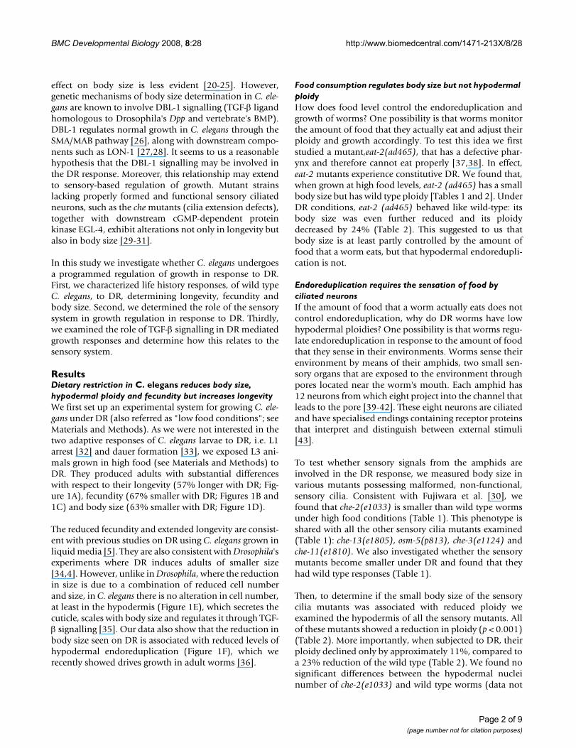
Food consumption regulates body size but not hypodermal ploidyHow does food level control the endoreduplication andgrowth of worms? One possibility is that worms monitorthe amount of food that they actually eat and adjust theirploidy and growth accordingly. To test this idea we firststudied a mutant,eat-2(ad465), that has a defective phar-ynx and therefore cannot eat properly [37,38]. In effect,eat-2 mutants experience constitutive DR. We found that,when grown at high food levels, eat-2 (ad465) has a smallbody size but has wild type ploidy [Tables 1 and 2]. UnderDR conditions, eat-2 (ad465) behaved like wild-type: itsbody size was even further reduced and its ploidydecreased by 24% (Table 2). This suggested to us thatbody size is at least partly controlled by the amount offood that a worm eats, but that hypodermal endoredupli-cation is not.
Endoreduplication requires the sensation of food by ciliated neuronsIf the amount of food that a worm actually eats does notcontrol endoreduplication, why do DR worms have lowhypodermal ploidies? One possibility is that worms regu-late endoreduplication in response to the amount of foodthat they sense in their environments. Worms sense theirenvironment by means of their amphids, two small sen-sory organs that are exposed to the environment throughpores located near the worm's mouth. Each amphid has12 neurons from which eight project into the channel thatleads to the pore [39-42]. These eight neurons are ciliatedand have specialised endings containing receptor proteinsthat interpret and distinguish between external stimuli[43].
To test whether sensory signals from the amphids areinvolved in the DR response, we measured body size invarious mutants possessing malformed, non-functional,sensory cilia. Consistent with Fujiwara et al. [30], wefound that che-2(e1033) is smaller than wild type wormsunder high food conditions (Table 1). This phenotype isshared with all the other sensory cilia mutants examined(Table 1): che-13(e1805), osm-5(p813), che-3(e1124) andche-11(e1810). We also investigated whether the sensorymutants become smaller under DR and found that theyhad wild type responses (Table 1).
Then, to determine if the small body size of the sensorycilia mutants was associated with reduced ploidy weexamined the hypodermis of all the sensory mutants. Allof these mutants showed a reduction in ploidy (p < 0.001)(Table 2). More importantly, when subjected to DR, theirploidy declined only by approximately 11%, compared toa 23% reduction of the wild type (Table 2). We found nosignificant differences between the hypodermal nucleinumber of che-2(e1033) and wild type worms (data not
Page 2 of 9(page number not for citation purposes)

BMC Developmental Biology 2008, 8:28 http://www.biomedcentral.com/1471-213X/8/28
Page 3 of 9(page number not for citation purposes)
The effects of dietary restriction on C. elegans life history traitsFigure 1The effects of dietary restriction on C. elegans life history traits. (A) Kaplan-Maier survival curves showing the longev-ity of C. elegans under excess (closed circles), high (open circles) and low food (closed squares) environments (see Material and Methods). Significance is shown for excess, high, and low food, from Log Rank tests n/censored individuals 175/22, 204/110 and 238/189 respectively. (B) Daily fecundity of C. elegans under excess (closed circles), high (open circles) and low food (closed squares) environments. (C) Total fecundity of C. elegans under excess, high and low food environments; n = 36, 44 and 35 respectively. (D) Growth curves of C. elegans under excess (closed circles), high (open circles) and low food (squares) environ-ments, n = 49, 38, and 21 respectively. Images show representative adults from high food (upper panel) and low food (lower panel) treatments. Scale bar indicates 100 μm. (E) Hypodermal (hyp7) cell number of young adult C. elegans under high and low food environments; n = 10 and 9 respectively. (F) Hypodermal (hyp7) ploidy of C. elegans (120 h) under excess, high and low food environments; n = 19, 254, and 189 respectively. All error bars show 95% confidence intervals, and asterisk show level of significance, *** shows P < 0.0001, by ANOVA.
0
0.001
0.002
0.003
0.004
0.005
0.006
24 48 72 96 120 144
3
0
1 0
2 0
3 0
4 0
5 0
6 0
7 0
Hig h L o w
p = 0.44
0
2 0
4 0
6 0
8 0
1 0 0
1 2 0
1 4 0
1 6 0
0
2
4
6
8
10
12
H ig h L o w
* * *
48 72 96 120 144 168 192 216 240 264 288 312 336 360
B
D
E F
)0
0.2
0.4
0.6
0.8
1
0 1 0 2 0 3 0 4 0
*** ***0.006
p valueA
Excess/High
Excess/Low
High/Low
P > 0.05
0
5 0
1 0 0
1 5 0
2 0 0
2 5 0
3 0 0
Exc e s s H ig h L o w
* * * * * *
* * *
C
Excess
Food level Food level
Food level Time (hours)
Time (hours)Time (days)
Perc
ent a
live
Fecu
nd
ity
Tota
l fec
und
ity
Volu
me
(mm
)3
Cel
l num
ber
Ploi
dy
(C)

BMC Developmental Biology 2008, 8:28 http://www.biomedcentral.com/1471-213X/8/28
shown). These results suggest that signals from theamphids partly control endoreduplication in response toDR.
EGL-4 mediates the response from sensory ciliaPrevious studies have shown that EGL-4, a cGMP-depend-ent protein kinase, functions downstream of sensory cili-
ated neurons in wild type worms [30]. Furthermore,mutations in egl-4 result in increased body length, alteredsensory perception and egg laying behaviour, withoutaffecting cilia structure [44]. To determine whether EGL-4is required for the regulation of body size and endoredu-plication in response to DR, we first characterized thegrowth of a strong loss-of-function mutant, egl-4 (n478),
Table 1: Effect of Dietary Restriction on Body Size. All genotypes show significant (p < 0.0001), wild type-like (genotype by environment interaction term; p > 0.05), reductions in volume under DR.
Body size (mm3)
Genotype High Food Low Food n % reduction
N2 0.0051 (± 1 × 10⁻⁴) 0.0021 (± 1 × 10⁻⁴) 147, 121 63che-2(e1033) 0.0022 (± 1 × 10⁻⁴) 0.0009 (± 2 × 10⁻⁴) 106, 43 62che-3(e1124) 0.0029 (± 2 × 10⁻⁴) 0.0013 (± 8 × 10⁻⁵) 19, 12 55che-11(e1810) 0.0027 (± 1 × 10⁻⁴) 0.0012 (± 1 × 10⁻⁴) 41, 23 56che-13(e1805) 0.0021 (± 3 × 10⁻⁴) 0.0009 (± 5 × 10⁻⁵) 11, 16 57osm-5(p813) 0.0033 (± 2 × 10⁻⁴) 0.0013 (± 2 × 10⁻⁴) 26, 20 61egl-4(n478) 0.0063 (± 2 × 10⁻⁴) 0.0023 (± 1 × 10⁻⁴) 127, 118 60dbl-1(nk3) 0.0025 (± 1 × 10⁻⁴) 0.001 (± 6 × 10⁻⁵) 121, 80 62sma-2(e502) 0.0020 (± 4 × 10⁻⁴) 0.0010 (± 2 × 10⁻⁴) 19, 15 50sma-3(wk20) 0.0025 (± 2 × 10⁻⁴) 0.0010 (± 1 × 10⁻⁴) 32, 24 60sma-4(e729) 0.0010 (± 1 × 10⁻⁴) 0.0006 (± 1 × 10⁻⁴) 33, 22 40sma-6(wk7) 0.0019 (± 3 × 10⁻⁴) 0.0009 (± 2 × 10⁻⁴) 39, 22 53lon-1(e185) 0.0050 (± 6 × 10⁻⁴) 0.0017 (± 3 × 10⁻⁴) 20, 9 66che-2(e1033);dbl-1(nk3) 0.0019 (± 1 × 10⁻⁴) 0.0007 (± 4 × 10⁻⁵) 48, 35 58egl4(n478);dbl-1(nk3) 0.0028 (± 3 × 10⁻⁴) 9eat-2(ad465) 0.0020 (± 1 × 10⁻⁴) 0.0008 (± 4 × 10⁻⁵) 71, 56 59eat-2(ad465);dbl-1(nk3) 0.0011 (± 1 × 10⁻⁴) 0.0005 (± 1 × 10⁻⁴) 85, 45 52
Table 2: Effect of Dietary Restriction on Hypodermal Ploidy. All genotypes, unless stated (NS, p > 0.05), show highly significant (p < 0.0001) alterations from wild type ploidy responses to DR.
Hypodermal ploidy (xC)
Genotype High Food Low Food n % reduction
N2 10.9 (± 0.3) 8.4 (± 0.2) 113, 94 23che-2(e1033) 8.6 (± 0.3) 7.5 (± 0.4) 56, 25 13che-3(e1124) 8.4 (± 0.5) 7.5 (± 0.7) 17, 12 11che-11(e1810) 9.2 (± 0.3) 8.5 (± 0.5) 32, 21 8che-13(e1805) 8.4 (± 0.5) 7.5 (± 0.5) 13, 13 11osm-5(p813) 8.8 (± 0.4) 7.6 (± 0.5) 24, 17 14egl-4(n478) 12.3 (± 0.3) 11.6 (± 0.3) 101, 88 5dbl-1(nk3) 7.5 (± 0.5) 6.9 (± 0.4) 51, 32 8
sma-2(e502) 7.6 (± 0.7) 7.0 (± 0.5) 14, 11 8sma-3(wk20) 8.2 (± 0.4) 7.0 (± 0.4) 39, 23 15sma-4(e729) 7.4 (± 0.3) 6.4 (± 0.5) 35, 22 14sma-6(wk7) 8.3 (± 0.3) 7.1 (± 0.3) 31, 18 14lon-1(e185) 12.2 (± 0.9) 8.5 (± 1.0) 9, 8 30 NS
che-2(e1033);dbl-1(nk3) 9.1 (± 0.5) 7.8 (± 0.5) 29, 18 14egl4(n478);dbl-1(nk3) 8.9 (± 0.8)' 6
eat-2(ad465) 10.1 (± 0.7) 7.7 (± 0.4) 24, 17 24 NSeat-2(ad465);dbl-1(nk3) 8.5 (± 0.4) 7.0 (± 0.4) 43, 23 18
Page 4 of 9(page number not for citation purposes)

BMC Developmental Biology 2008, 8:28 http://www.biomedcentral.com/1471-213X/8/28
under normal levels of food. We found these worms to be21% larger than wild type (Table 1) and possess a 13%higher level of hypodermal endoreduplication (Table 2),while maintaining wild type cell numbers (data notshown).
Surprisingly, under DR, egl-4 exhibits a wild type reduc-tion in volume, but importantly, it fails to show a wildtype reduction in endoreduplication. Hypodermal poly-ploidization, in egl-4 worms, declines only 5% under DRcompared to a 25% decline in N2 (Table 2). Therefore,egl-4 defective worms maintain a hyper-endoreduplicatedstate at their hypodermis even under DR. Their hyper-endoreduplicated state, their failure to show wild typedeclines in endoreduplication, and the placement of EGL-4 downstream of CHE-2 [30] (also see Table 1 &2), alltogether suggest that EGL-4 acts as a negative regulator offood dependent endoreduplication.
DBL-1 signalling regulates the DR endoreduplication responseDBL-1 is known to be a dose-dependent regulator of bodysize and endoreduplication in C. elegans. This protein acti-vates the SMA-6/DAF-4, Ser/Thr kinase receptor, which inturn is thought to activate the cytoplasmic effectors SMA-2, SMA-3 and SMA-4 [26]. Here we confirm that loss-of-function dbl-1(nk3) worms, as well as worms defective fordownstream signalling components such as sma-6, sma-2,sma-3 and sma-4, all show a 60% reduction in body sizewhen grown in normal food levels (Table 1), similarly topreviously reported [45-48]. They also show a ~25%reduction in hypodermal polyploidization (Table 2). Todetermine the role of DBL-1 in the DR response of wildtype worms, all these mutants were subjected to DR andtheir body size and hypodermal endoreduplication char-acterized. All mutants showed a marked decrease in size(40% – 60%), responding to DR in a wild type manner(63%; Table 1). More interestingly, when the effects of DRon endoreduplication were examined, mutants deficientfor dbl-1, sma-6, sma-2, sma-3 and sma-4 all show a distinctnon-wild type response: endoreduplication declines byapproximately 12%, compared to the 23% seen in N2(Table 2). These results suggest that DBL-1 signalling, asdescribed previously for sensory cilia mutants and egl-4, ispartially responsible for the regulation of endoreduplica-tion as a response to DR. We note, however, that loss-of-function lon-1, placed downstream of the dbl-1 pathway[28], behaves as wild type under DR (Table 2). Therefore,we suggest that lon-1, despite its role in determining bodysize and hypodermal endoreduplication, is not part of thepolyploidization response to nutrients availability (Figure2).
CHE-2 and DBL-1 act in the same pathway to regulate body size and hypodermal ploidyIn order to test the hypothesis that sensory signals andDBL-1 signalling act in the same pathway, we generateddouble che-2;dbl-1 mutants and analysed their size andploidy levels under standard and DR conditions. Whengrown in high food conditions, che-2;dbl-1 was similar insize and ploidy (p > 0.05 for all comparisons), to dbl-1,che-2, or related genes (e.g. sma-6, che-13; Tables 1 and 2).The corresponding reduction for both characters underDR was also similar (Tables 1 and 2). This result suggeststhat dbl-1 and the amphid mutants act in the same path-way when controlling body size and hypodermal endore-duplication.
Model of body size regulation by nutrients availability in C. elegans (from L3 onwards)Figure 2Model of body size regulation by nutrients availability in C. elegans (from L3 onwards). Our results suggest that food availability may regulate body size in at least two ways. First, by the "caloric pathway", that is, simply considering that food intake and its absorption by the digestive tract facilitates nutrition, which in turn may inhibit autophagy. Second, by the "sensory pathway", which refers to the sensing food through organs such as the amphids, with their ciliated neurons expressing genes like che-2, would inhibit EGL-4. Down-stream, this cGMP-dependent protein kinase downregulates DBL-1 signalling, which in turn promotes hypodermal endoreduplication, upregulator of body size [36]. LON-1 inhibition by DBL-1 [28] would not influence ploidy upon nutrient activation. This model explains why the nutrient-dependent regulation that the sensory cilia proteins, EGL-4 and DBL-1 are all playing on hypodermal polyploidization has not been observed for body size; their role on body size, but not upon endoreduplication, may be obscured by the domi-nant influence of caloric restriction.
Food
dbl-1
SMA/MAB pathway
eat-2…
(pharyngeal and digestive system genes)
che-2, che-3, che-11,
che-13, osm-5…
(sensor genes, e.g. in amphids)
egl-4
Body size
hypodermalendoreduplication
autophagy
nutrition
SE
NS
OR
Y P
AT
HW
AY
CA
LOR
IC P
AT
HW
AY
lon-1
Page 5 of 9(page number not for citation purposes)

BMC Developmental Biology 2008, 8:28 http://www.biomedcentral.com/1471-213X/8/28
We also asked whether the regulation of body size by foodintake per se was affected by DBL-1 signalling. To test thiswe examined eat-2;dbl-1 double mutants. We found thatat high food levels, these worms are smaller than eithereat-2 or dbl-1 (Table 1). This additive effect suggests thatthese genes regulate body size through different pathways,and is consistent with the finding that eat-2 worms havenormal ploidy.
EGL-4 negatively regulates DBL-1To confirm the effect of EGL-4 on the signalling of DBL-1seen previously [30,31], epistasis analysis was carried outbetween null mutants dbl-1 and egl-4. The nature of thesemutants allowed a relatively simple analysis because egl-4worms are larger than wild type, whereas dbl-1 worms aresmaller [45,46] (Table 1). The same thing can be saidabout hypodermal ploidy (Table 2). Examination of egl-4;dbl-1 worms revealed that, though slightly smaller, thedouble mutant did not significantly differ from dbl-1worms in either adult volume or hypodermal ploidy (P >0.05, for body size and ploidy; Tables 1 and 2, respec-tively), but it did with respect to egl-4 worms (P < 0.0001,for body size and ploidy).
DiscussionGrowth is a fundamental part of biology, yet its regulationis still poorly understood [12]. The notion that growthresponds passively to nutrient availability has beenreplaced with the idea that growth is actively regulated inresponse to constant monitoring of nutrient availability inthe external environment. We observed that when C. ele-gans is exposed to a low food environment there is areduction in adult body size, similar to the reductionsseen in other organisms e.g. Drosophila and Daphnia[4,34,49,50]. However, in contrast to these organisms, thestunting in C. elegans is not due to a lack of cell prolifera-tion, which implies that it is due to a reduction in cell size.
In order to investigate how DR controls adult body size inC. elegans, we studied the growth of wild type and mutantworms subjected to high and low food regimes. We foundthat all of our mutants became smaller by about the sameamount (60%) at low food levels. This absence of interac-tion between food and genotype on growth might meanthat none of the genes examined are involved in the die-tary-dependent regulation of growth, but it could alsosimply mean that severe DR has additional effects.
For this reason we needed a more subtle way of examiningthe effects of DR on worm development. We have previ-ously shown that hypodermal endoreduplication isrequired for growth in adult C. elegans [36]. Strikingly, wealso found that DR inhibits hypodermal endoreduplica-tion and so adult ploidy. This result gave us a sensitiveassay for the effects of DR on the worm's development.
We found that mutations in several genes mimic the DRresponse: even at high food levels, mutations that disruptsensory or DBL-1 signalling show reduced ploidy andbody size. That suggested to us that these genes might beinvolved in the DR endoreuplication response. This infer-ence was confirmed when we examined these mutantsunder DR: in each case, the reduction in ploidy normallyfound at low food levels was largely abrogated. An evenmore striking lack of response to DR was also found in alarge mutant that disrupts egl-4, a cGMP-dependent pro-tein kinase previously associated with food sensing andfood dependent behaviour [30].
These results, and our epistasis experiments, suggest amodel in which the amphids monitor nutrient availabilityand activate a downstream signalling pathway involvingthe growth repressor EGL-4 (Figure 2). This kinase in turnregulates the DBL-1/SMA/MAB pathway, which positivelyregulates hypodermal endoreduplication. As the observedbody size reduction under DR for both the sensory andDBL-1 signalling pathway mutants was similar to that ofwild type (Table 1), we suggest that the main effects of DRon body size do not arise from the lack of endoreduplica-tion, but rather from some other unknown pathway. Alikely candidate could be what we call the "caloric path-way" in Figure 2. That is, the severe food restriction underDR could be masking the "sensory pathway" on body sizewhen this one is impaired (e.g. in dbl-1(nk3); Figure 2).Reduction in food may prevent DBL-1 like mutants,whose endoreduplication levels do not drop as much aswild type under DR, from growing larger. Nevertheless,the reduced ploidy programmed by the sensing of lowerlevels of food (Table 2) must contribute to the stunting,since previous work shows a cause-and-effect relationshipbetween endoreduplication and adult growth [36]. Con-sistent with our model, we showed that eat-2 mutants, oneof the genes active in the feeding mechanism, has smallsize but normal ploidy, and that it reduces both charactersin a wild type manner under DR (Tables 1 and 2). Recentwork suggests that eat mutants have small body sizes dueto increased autophagy [38], which is also included in ourmodel (Figure 2).
ConclusionHow do our results relate to other animal models?Endoreduplication in Drosophila depends on a mitogenfrom the fat body that is regulated in a nutrition-depend-ent manner [51], which may suggest at least an underlyingcommon plan beyond their differences (see Introduc-tion)[52]. However, one of the proteins studied here,EGL-4, is a key regulator of nutrient responses not only inworms but, with the generic name of cGMP-dependentprotein kinase, in organisms such as honeybees and fruit-flies controlling their foraging behaviour [53]. It is some-what surprising that loss-of-function egl-4 has a change in
Page 6 of 9(page number not for citation purposes)

BMC Developmental Biology 2008, 8:28 http://www.biomedcentral.com/1471-213X/8/28
hypodermal endoreduplication in high vs. low foodwhich is half of what it is observed for the sensory ciliatedor for the dbl-1-related mutants (Table 2). We think thatthis difference can be explained because egl-4's role innutrient-dependent growth may be central, not sharedwith other proteins in parallel positions, whereas the var-ious sensor genes investigated may be acting in parallel,either among themselves, or in relation to other genes orpathways (similarly for DBL-1 and the SMA/MAB path-way). In agreement with this, egl-4 is considered a highlypleiotropic gene, a main regulatory hub, not only mediat-ing body size but longevity, locomotion feeding, andother processes [54].
MethodsStrainsApart from the wild type strain N2, the following mutantstrains were used, which were obtained from theCaenorhabditis elegans Genetics Center. Mutations arelisted by linkage group: LGI: che-3(e1124), che-13(e1805);LGII: sma-6(wk7), eat-2(ad465); LGIII: lon-1(e185), sma-2(e502), sma-3(wk20), sma-4(e729); LGIV: egl-4(n478);LGV: che-11(e1810), dbl-1(nk3); LGX: che-2(e1033), osm-5(p813). We also used double mutants that we producedthrough crosses of the previous strains. Double mutantseat-2(ad465);dbl-1(nk3) were confirmed by PCR andsequencing using the following primers: 5'-eat-2: 5' TGAT-CACCCTAGTTGTCTGG; 3'-eat-2: 5' AGTGTAGAGG-TACTGTATGG; 5'-dbl-1: 5' CATGGACAAACATCGGGGA;and 3'-dbl-1: 5' CGTGTACACAAATCTGTTCG. che-2(e1033);dbl-1(nk3) was generated by crossing hetero-zygous dbl-1(nk3) males with che-2(e1033) hermaphro-dites. Then, their F1 progeny was PCR-screened for thenk3 allele, and double mutants in F2 were confirmed byPCR for both nk3 and e1033 alleles, and by DNA sequenc-ing with oligonucleotides 5'- dbl-1, 3'- dbl-1, 5'-che-2: 5'AGATGGATGTTTACTGCC, and 3'-che-2: 5' GAGAAT-GACACAATGTGG.
All strains and experiments were maintained at 20°C.
Dietary restrictionWe developed a novel method of dietary restriction (DR)on solid media. Three different food treatments aredescribed within this study: excess, high, and low foodtreatment plates. Excess food plates: 100 μl of 5.19 × 10⁸/ml E. coli (OP50)-Luria broth was spread around the cen-tre of 5.5 cm NGM plates and left at room temperature for24 hours before being killed by exposure to UV light for 1hour. High food plates were prepared as excess foodplates, but were exposed to UV light for 1 hour immedi-ately after preparation. Low food plates were prepared ashigh food plates, but using a suspension of 3.95 × 10⁷/mlE. coli (OP50)-Luria broth. For each experiment, the sameE. coli culture was used for each food treatment. Treatment
plates were replaced every 24 h during worm growthexperiments to prevent depletion food source.
Body size analysisGrowth curves were determined for each strain, fromworms grown individually on 5 cm Petri dishes. At 24 hintervals from 36 h to 120 h post hatching, images werecaptured using a video camera (JVC KY-F50) attached to adissecting microscope (×50), and analyzed with OBJECT-IMAGE 1.62. Length and area were measured from pic-tures of individual worms and calibrated from a 1 mmgraticule. Volume was calculated assuming cylindricalbody shape using the formula (pi*length*(area/length)2/4) [36,46]. All comparisons of body size use Log-trans-formed data.
Hypodermal ploidy analysisUpon completing growth (120 h), worms were fixed inCarnoy's solution for 24 h, stained in a 0.007 mg/ml solu-tion of 4',6-diamidino-2-phenylindole dihydrochloride(DAPI) [36,48,55,56] and viewed under a Leitz epifluo-rescence microscope. Images of hypodermal and ventralcord nuclei were collected using a CV-M300 video camera,and analyzed using OBJECT-IMAGE 1.62. C values ofhypodermal nuclei were estimated by dividing theirDAPI-based densitometric quantifications by an averageof those values from ventral cord nuclei (divided by two)in the same microscopic preparations [36].
Cell number analysisYoung adult worms were anesthetized with 0.1 M sodiumazide [57], and viewed at ×1000 under differential inter-ference contrast optics with a Nikon Eclipse E600 micro-scope. All nuclei, excluding neuronal and seam cells,between the posterior pharyngeal bulb and anus werecounted. Images were captured with a CV-M300 cameraand reconstructed by using Adobe PHOTOSHOP 4.0.
Longevity analysisWe analysed Kaplan-Meier survival distributions, whichare based on a discrete stepped survival curve, adding timespecific data as each death occurs. Individuals that diedfrom internal hatching of eggs (bagging), or crawled offthe plate were censored. Censoring allows the inclusion ofindividuals that were lost to the study, and thus contributetowards knowledge of survivorship, but nothing to theknowledge of age at death. Log-rank tests were performedto determine if survival curves were significantly differentfrom each other.
Egg-laying assaysIndividual worms were placed OP50-seeded 5.5 cm NGMplates before adult moult occurred and transferred to afresh plate every 24 h. Total fecundity was measured withonly fertilized eggs and larvae being included in the count.
Page 7 of 9(page number not for citation purposes)

BMC Developmental Biology 2008, 8:28 http://www.biomedcentral.com/1471-213X/8/28
Statistical analysesData analysis was undertaken using JMP 3.2 (SAS Insti-tute, Cary NC, USA). Body size and ploidy data were com-pared across food level and genotype using a standardtwo-way ANOVA, including a genotype by environmentinteraction term, to determine responses of each genotypeto DR. A food level by genotype interaction term allowedthe comparison of each mutant genotype's response to DRto that of wild type. Ratios, between high and low foodgroups, were not used in this analysis.
Authors' contributionsLST designed the study, created the dietary restrictionmethodology, performed body size, ploidy, cell number,longevity, egg laying and statistical analysis. EL performedbody size and ploidy analysis. AGS did ploidy analysis.AML conceived the study and participated in its design.The paper was written by LST and AGS, with the help ofAML and EL.
AcknowledgementsWe thank the Caenorhabditis Genetics Center for providing strains, and Almudena de Vivero for helping us to measure some of the mutants and their levels of endoreduplication. Our support came primarily from NERC (UK) and BBSRC. LST was also supported by Parkinson's Diease Society (PDS). EL and AGS were also supported by programs I3P and Ramón y Cajal, respectively, and by Plan Nacional I+D (CGL2005-00307; all from the Ministry of Education and Science, Spain).
References1. Pigliucci M: Phenotypic plasticity. Beyond nature and nurture Baltimore,
The John Hopkins University Press; 2001. 2. West-Eberhard MJ: Developmental plasticity and evolution New York,
Oxford University; 2003. 3. McCay CM, Crowel MF, Maynard LA: The effect of growth upon
the length of the life span and upon the ultimate body size. JNutr 1935, 10:63-79.
4. Robertson FW: Studies in quantitative inheritance. XII. Cellsize and number in relation to genetic and environmentalvariation of body size in Drosophila. Genetics 1959, 44:869-896.
5. Klass MR: Aging in the nematode Caenorhabditis elegans:major biological and environmental factors influencing lifespan. Mech Ageing Dev 1977, 6(6):413-429.
6. Masoro EJ: Caloric restriction. Aging (Milano) 1998,10(2):173-174.
7. Tu MP, Tatar M: Juvenile diet restriction and the aging andreproduction of adult Drosophila melanogaster. Aging Cell2003, 2(6):327-333.
8. Weindruch R, Walford MD: The retardation of aging and disease by die-tary restriction Springfield, IL, Charles C Thomas Publisher; 1988.
9. Chippindale AK, Leroi AM, Kim SB, Rose MR: Phenotypic Plastic-ity and Selection in Drosophila Life-History Evolution.1.Nutrition and the Cost of Reproduction. J Evol Biol 1993,6(2):171-193.
10. Townshend TJ, Wootton RJ: Effects of food supply on the repro-duction of the convict cichlid, Cichlasoma nigrofasciatum. JFish Biol 1985, 24:91-104.
11. Houthoofd K, Braeckman BP, Johnson TE, Vanfleteren JR: Lifeextension via dietary restriction is independent of the Ins/IGF-1 signalling pathway in Caenorhabditis elegans. Exp Ger-ontol 2003:947-54.
12. Conlon I, Raff M: Size control in animal development. Cell 1999,96(2):235-244.
13. Ben Lagha N, Menuelle P, Seurin D, Binoux M, Lebouc Y, Berdal A:Bone formation in the context of growth retardation
induced by hIGFBP-1 overexpression in transgenic mice.Connect Tissue Res 2002, 43(2–3):515-9.
14. Kobayashi S, Nogami H, Ikeda T: Growth hormone and nutritioninteract to regulate expressions of kidney IGF-I and IGFBPmRNAs. Kidney Int 1995, 48(1):65-71.
15. Van Wyk JJ, Smith P: Insulin-like growth factors and skeletalgrowth: possibilities for therapeutic interventions. J Clin Endo-crinol Metab 1999, 84(12):4349-4354.
16. Miron M, Verdu J, Lachance PE, Birnbaum MJ, Lasko PF, Sonenberg N:The translational initiator 4E-BP is an effector if PI(3)K/Aktsignalling and cell growth in Drosophila. Nat Cell Biol 2001,3(6):596-601.
17. Efstratiadis A: Genetics of mouse growth. Int J Dev Biol 1998,42(7):955-976.
18. Oldham S, Bohni R, Stocker H, Brogiolo W, Hafen E: Genetic con-trol of size in Drosophila. Philos Trans R Soc Lond B Biol Sci 2000,355(1399):945-952.
19. Stocker H, Hafen E: Genetic control of cell size. Curr Opin GenetDev 2000, 10(5):529-535.
20. Gregoire FM, Chomiki N, Kachinskas D, Warden CH: Cloning anddevelopmental regulation of a novel member of the insulin-like gene family in Caenorhabditis elegans. Biochem Biophys ResCommun 1998, 249(2):385-390.
21. Hsin H, Kenyon C: Signals from the reproductive system reg-ulate the lifespan of C. elegans. Nature 1999,399(6734):362-366.
22. Kawano T, Ito Y, Ishiguro M, Takuwa K, Nakajima T, Kimura Y:Molecular cloning and characterization of a new insulin/IGF-like peptide of the nematode Caenorhabditis elegans. Bio-chem Biophys Res Commun 2000, 273(2):431-436.
23. Kimura KD, Tissenbaum HA, Liu Y, Ruvkun G: daf-2, an insulinreceptor-like gene that regulates longevity and diapause inCaenorhabditis elegans. Science 1997, 277(5328):942-946.
24. Ogg SS, Paradis S, Gottlieb S, Patterson GI, Lee L, Tissenbaum HA,Ruvkun G: The Fork head transcription factor DAF-16 trans-duces insulin-like metabolic and longevity signals in C. ele-gans. Nature 1997, 369(6654):994-999.
25. McCulloch D, Gems D: Body size, insulin/IGF signaling andaging in the nematode Caenorhabditis elegans. Exp Gerontol2003, 38(1–2):129-136.
26. Patterson GI, Padgett RW: TGF beta-related pathways. Roles inCaenorhabditis elegans development. Trends Genet 2000,16(1):27-33.
27. Maduzia LL, Gumienny TL, Zimmerman CM, Wang H, Shetgiri P,Krishna S, Roberts AF, Padgett RW: lon-1 regulates Caenorhab-ditis elegans body size downstream of the dbl-1 TGF betasignaling pathway. Dev Biol 2002, 246(2):418-428.
28. Morita K, Flemming AJ, Sugihara Y, Mochii M, Suzuki Y, Yoshida S,Wood WB, Kohara Y, Leroi AM, Ueno N: A Caenorhabditis ele-gans TGF-beta, DBL-1, controls the expression of LON-1, aPR-related protein, that regulates polyploidization and bodylength. Embo J 2002, 21(5):1063-1073.
29. Apfeld J, Kenyon C: Regulation of lifespan by sensory percep-tion in Caenorhabditis elegans. Nature 1999, 402:804-809.
30. Fujiwara M, Sengupta P, McIntire SL: Regulation of body size andbehavioral state of C. elegans by sensory perception and theEGL-4 cGMP-dependent protein kinase. Neuron 2002,36(6):1091-1102.
31. Hirose T, Nakano Y, Nagamatsu Y, Misumi T, Ohta H, Ohshima Y:Cyclic GMP-dependent protein kinase EGL-4 controls bodysize and lifespan in C. elegans. Development 2003,130(6):1089-1099.
32. Kao G, Nordenson C, Still M, Ronnlund A, Tuck S, Naredi P: ASNA-1 positively regulates insulin secretion in C. elegans andmammalian cells. Cell 2007, 128(3):577-587.
33. Riddle DL, Albert PS: Genetic and environmental regulation of dauer larvadevelopment Cold Spring Harbor Laboratory; 1997.
34. Alpatov WW: Phenotypical variation in body and cell size ofDrosophila melanogaster. Biol Bull 1930, 58:85-103.
35. Wang J, Tokarz R, Savage-Dunn C: The expression of TGF-β sig-nal transducers in the hypodermis regulates body size in C.elegans. Development 2002, 129:4989-4998.
36. Lozano E, Sáez AG, Flemming A, Cunha A, Leroi AM: Regulation ofgrowth by ploidy in C. elegans. Curr Biol 2006, 16(5):493-498.
Page 8 of 9(page number not for citation purposes)
http://www.ncbi.nlm.nih.gov/entrez/query.fcgi?cmd=Retrieve&db=PubMed&dopt=Abstract&list_uids=9666230
http://www.ncbi.nlm.nih.gov/entrez/query.fcgi?cmd=Retrieve&db=PubMed&dopt=Abstract&list_uids=9988218
http://www.ncbi.nlm.nih.gov/entrez/query.fcgi?cmd=Retrieve&db=PubMed&dopt=Abstract&list_uids=7564093
http://www.ncbi.nlm.nih.gov/entrez/query.fcgi?cmd=Retrieve&db=PubMed&dopt=Abstract&list_uids=7564093
http://www.ncbi.nlm.nih.gov/entrez/query.fcgi?cmd=Retrieve&db=PubMed&dopt=Abstract&list_uids=7564093
http://www.ncbi.nlm.nih.gov/entrez/query.fcgi?cmd=Retrieve&db=PubMed&dopt=Abstract&list_uids=9853827
http://www.ncbi.nlm.nih.gov/entrez/query.fcgi?cmd=Retrieve&db=PubMed&dopt=Abstract&list_uids=9712706
http://www.ncbi.nlm.nih.gov/entrez/query.fcgi?cmd=Retrieve&db=PubMed&dopt=Abstract&list_uids=9712706
http://www.ncbi.nlm.nih.gov/entrez/query.fcgi?cmd=Retrieve&db=PubMed&dopt=Abstract&list_uids=9712706
http://www.ncbi.nlm.nih.gov/entrez/query.fcgi?cmd=Retrieve&db=PubMed&dopt=Abstract&list_uids=9252323
http://www.ncbi.nlm.nih.gov/entrez/query.fcgi?cmd=Retrieve&db=PubMed&dopt=Abstract&list_uids=9252323

BMC Developmental Biology 2008, 8:28 http://www.biomedcentral.com/1471-213X/8/28
Publish with BioMed Central and every scientist can read your work free of charge
"BioMed Central will be the most significant development for disseminating the results of biomedical research in our lifetime."
Sir Paul Nurse, Cancer Research UK
Your research papers will be:
available free of charge to the entire biomedical community
peer reviewed and published immediately upon acceptance
cited in PubMed and archived on PubMed Central
yours — you keep the copyright
Submit your manuscript here:http://www.biomedcentral.com/info/publishing_adv.asp
BioMedcentral
37. Houthoofd K, Braeckman BP, Johnson TE, Vanfleteren JR: No reduc-tion of metabolic rate in food restricted Caenorhabditis ele-gans. Exp Gerontol 2002, 37(12):1359-1369.
38. Mörck C, Pilon M: C. elegans feeding defective mutants haveshorter body lengths and increased autophagy. BMC Dev Biol2006, 6:39.
39. Ward S, Thomson N, White JG, Brenner S: Electron microscopi-cal reconstruction of the anterior sensory anatomy of thenematode Caenorhabditis elegans. J Comp Neurol 1975,160(3):313-337.
40. Ware RW, Crossland K, Russell RL, Clark DV: The nerve ring ofthe nematode C elegans: Sensory input and motor output. JComp Neurol 1975, 162:71-110.
41. Lewis JA, Hodgkin JA: Specific neuroanatomical changes inchemosensory mutants of the nematode Caenorhabditiselegans. J Comp Neurol 1977, 172(3):489-510.
42. Perkins LA, Hedgecock EM, Thomson JN, Culotti JG: Mutant sen-sory cilia in the nematode Caenorhabditis elegans". Dev Biol1986:456-487.
43. Sengupta P, Chou JH, Bargmann CI: odr-10 encodes a seven trans-membrane domain olfactory receptor required forresponses to the odorant diacetyl. Cell 1996, 84:899-909.
44. Daniels SA, Ailion M, Thomas JH, Sengupta P: egl-4 acts through atransforming growth factor-beta/SMAD pathway inCaenorhabditis elegans to regulate multiple neuronal cir-cuits in response to sensory cues. Genetics 2000,156(1):123-141.
45. Suzuki Y, Yandell MD, Roy PJ, Krishna S, Savage-Dunn C, Ross PadgettRW, Wood WB: A BMP homolog acts as a dose-dependentregulator of body size and male tail patterning inCaenorhabditis elegans. Development 1999, 126(2):241-250.
46. Morita K, Chow KL, Ueno N: Regulation of body length andmale tail ray pattern formation of Caenorhabditis elegans bya member of TGF-beta family. Development 1999,126(6):1337-1347.
47. Savage C, Das P, Finelli AL, Townsend SR, Sun CY, Baird SE, PadgettRW: Caenorhabditis elegans genes sma-2, sma-3, and sma-4define a conserved family of transforming growth factor betapathway components. Proc Natl Acad Sci USA 1996,93(2):790-794.
48. Flemming AJ, Shen ZZ, Cunha A, Emmons SW, Leroi AM: Somaticpolyploidization and cellular proliferation drive body sizeevolution in nematodes. Proc Natl Acad Sci USA 2000,97(10):5285-5290.
49. Robertson FW: The ecological genetics of growth in Dro-sophila. 6. The genetic correlation between the duration ofthe larval period and body size in relation to larval diet. GenetRes 1963, 4:74-92.
50. Beaton MJ, Hebert PDN: Shifts in postembryonic somaticploidy levels in Daphnia pulex. Hydrobiologia 1999, 394:29-39.
51. Britton JS, Edgar BA: Environmental control of the cell cycle inDrosophila: nutrition activates mitotic and endoreplicativecells by distinct mechanisms. Development 1998,125(11):2149-2158.
52. Wagner GP: The developmental genetics of homology. Nat RevGen 2007, 8:473-479.
53. Douglas SJ, Dawson-Scully K, Sokolowski MB: The neurogeneticsand evolution of food-related behaviour. Trends Neurosci 2005,28(12):644-52.
54. Raizen DM, Cullison KM, Pack AI, Sundaram MV: A novel gain-of-function mutant of the cyclic GMP-dependent protein kinaseegl-4 affects multiple physiological processes in Caenorhab-ditis elegans. Genetics 2006, 173(1):177-187.
55. Sulston J, Hodgkin J: Methods. In The Nematode Caenorhabditis ele-gans New York: Cold Spring Harbor Laboratory Press; 1988:587-606.
56. Hedgecock EM, White JG: Polyploid tissues in the nematodeCaenorhabditis elegans. Dev Biol 1985, 107(1):128-133.
57. Sulston JE, Horwitz HR: Post-embryonic lineages of the nema-tode Caenorhabditis elegans. Dev Biol 1977, 56:110-156.
Page 9 of 9(page number not for citation purposes)
http://www.ncbi.nlm.nih.gov/entrez/query.fcgi?cmd=Retrieve&db=PubMed&dopt=Abstract&list_uids=1112927
http://www.ncbi.nlm.nih.gov/entrez/query.fcgi?cmd=Retrieve&db=PubMed&dopt=Abstract&list_uids=1112927
http://www.ncbi.nlm.nih.gov/entrez/query.fcgi?cmd=Retrieve&db=PubMed&dopt=Abstract&list_uids=1112927
http://www.ncbi.nlm.nih.gov/entrez/query.fcgi?cmd=Retrieve&db=PubMed&dopt=Abstract&list_uids=2428682
http://www.ncbi.nlm.nih.gov/entrez/query.fcgi?cmd=Retrieve&db=PubMed&dopt=Abstract&list_uids=2428682
http://www.ncbi.nlm.nih.gov/entrez/query.fcgi?cmd=Retrieve&db=PubMed&dopt=Abstract&list_uids=8601313
http://www.ncbi.nlm.nih.gov/entrez/query.fcgi?cmd=Retrieve&db=PubMed&dopt=Abstract&list_uids=8601313
http://www.ncbi.nlm.nih.gov/entrez/query.fcgi?cmd=Retrieve&db=PubMed&dopt=Abstract&list_uids=8601313
http://www.ncbi.nlm.nih.gov/entrez/query.fcgi?cmd=Retrieve&db=PubMed&dopt=Abstract&list_uids=9847238
http://www.ncbi.nlm.nih.gov/entrez/query.fcgi?cmd=Retrieve&db=PubMed&dopt=Abstract&list_uids=9847238
http://www.ncbi.nlm.nih.gov/entrez/query.fcgi?cmd=Retrieve&db=PubMed&dopt=Abstract&list_uids=9847238
http://www.ncbi.nlm.nih.gov/entrez/query.fcgi?cmd=Retrieve&db=PubMed&dopt=Abstract&list_uids=8570636
http://www.ncbi.nlm.nih.gov/entrez/query.fcgi?cmd=Retrieve&db=PubMed&dopt=Abstract&list_uids=8570636
http://www.ncbi.nlm.nih.gov/entrez/query.fcgi?cmd=Retrieve&db=PubMed&dopt=Abstract&list_uids=8570636
http://www.ncbi.nlm.nih.gov/entrez/query.fcgi?cmd=Retrieve&db=PubMed&dopt=Abstract&list_uids=9570778
http://www.ncbi.nlm.nih.gov/entrez/query.fcgi?cmd=Retrieve&db=PubMed&dopt=Abstract&list_uids=9570778
http://www.ncbi.nlm.nih.gov/entrez/query.fcgi?cmd=Retrieve&db=PubMed&dopt=Abstract&list_uids=9570778
http://www.ncbi.nlm.nih.gov/entrez/query.fcgi?cmd=Retrieve&db=PubMed&dopt=Abstract&list_uids=2578115
Related Documents