Diet, microbiota, and microbial metabolites in colon cancer risk in rural Africans and African Americans 1–4 Junhai Ou, Franck Carbonero, Erwin G Zoetendal, James P DeLany, Mei Wang, Keith Newton, H Rex Gaskins, and Stephen JD O’Keefe ABSTRACT Background: Epidemiologic studies have suggested that most cases of sporadic colon cancer can be attributed to diet. The recognition that colonic microbiota have a major influence on colonic health suggests that they might mediate colonic carcinogenesis. Objective: To examine the hypothesis that the influence of diet on colon cancer risk is mediated by the microbiota through their me- tabolites, we measured differences in colonic microbes and their metabolites in African Americans with a high risk and in rural native Africans with a low risk of colon cancer. Design: Fresh fecal samples were collected from 12 healthy Afri- can Americans aged 50–65 y and from 12 age- and sex-matched native Africans. Microbiomes were analyzed with 16S ribosomal RNA gene pyrosequencing together with quantitative polymerase chain reaction of the major fermentative, butyrate-producing, and bile acid–deconjugating bacteria. Fecal short-chain fatty acids were measured by gas chromatography and bile acids by liquid chromatography–mass spectrometry. Results: Microbial composition was fundamentally different, with a predominance of Prevotella in native Africans (enterotype 2) and of Bacteroides in African Americans (enterotype 1). Total bacteria and major butyrate-producing groups were significantly more abundant in fecal samples from native Africans. Microbial genes encoding for secondary bile acid production were more abundant in African Amer- icans, whereas those encoding for methanogenesis and hydrogen sul- fide production were higher in native Africans. Fecal secondary bile acid concentrations were higher in African Americans, whereas short-chain fatty acids were higher in native Africans. Conclusion: Our results support the hypothesis that colon cancer risk is influenced by the balance between microbial production of health-promoting metabolites such as butyrate and potentially car- cinogenic metabolites such as secondary bile acids. Am J Clin Nutr 2013;98:111–20. INTRODUCTION There are wide geographic variations in colorectal incidence around the world, and most of these differences have been at- tributed to diet (1). Within the continental United States, the African American population shoulders the major burden, with an incidence of w65:100,000 and a death rate of 25:100,000 (2). In sharp contrast, rural Africans rarely get the disease (3). Studies of ours have ascribed this difference to higher meat and fat intakes in Americans and to higher resistant starch in- takes in Africans (4). Colonic microbiota are dependent on dietary residues that escape small intestinal digestion and absorption. Consumption of a normal balanced diet predominantly yields carbohydrate res- idues such as fiber, which stimulates saccharolytic fermentation and the production of the health-promoting short-chain fatty acids (SCFAs) 5 acetate, propionate, and butyrate. Butyrate is the preferred energy source for the colonic mucosa, and all 3 SCFAs have antiinflammatory and antiproliferative properties (5). Consumption of an unbalanced diet rich in meat and low in fiber increases the delivery of proteinaceous residues, which promote proteolytic fermentation with the production of am- moniac compounds and branched-chain fatty acids, which are inflammatory and may enhance colon cancer risk (5, 6). The influence of dietary fat on cancer risk may also be de- termined by microbial metabolism, because it increases the hepatic synthesis of bile acids (BAs) and the quantity of BAs that escape the enterohepatic circulation and enter the colon. This provides substrate for microbes with 7a-dehydroxylating en- zymes, which convert primary BA into secondary BAs, which are proinflammatory and have carcinogenic properties (7). Digestion of food is fundamentally different in the small and large intestine. In the small intestine, the enzymic digestion rate is determined by substrate concentrations, according to the Michaelis-Menten equation. In the colon, the fermentation rate is 1 From the Department of Gastroenterology, Hepatology and Nutrition, School of Medicine, University of Pittsburgh, Pittsburgh, PA (JO and SJDO); the Departments of Animal Sciences (FC) and Food Science and Human Nutrition (MW), University of Illinois at Urbana–Champaign, Urbana, IL; the Laboratory of Microbiology, Wageningen University, Wageningen, Neth- erlands (EGZ); the Department of Medicine, Division of Endocrinology and Metabolism, University of Pittsburgh, Pittsburgh, PA (JPD); the Department of Gastroenterology, Nelson R Mandela School of Medicine, University of KwaZulu, Natal, South Africa (KN); and the Department of Animal Sci- ences, Division of Nutritional Sciences, Department of Pathobiology, Insti- tute for Genomic Biology and University of Illinois Cancer Center, University of Illinois at Urbana–Champaign, Urbana, IL (HRG). 2 JO and FC contributed equally to this work. 3 Supported by Public Health Service grant CA-135379 from the NIH. 4 Address reprint requests and correspondence to SJD O’Keefe, Depart- ment of Gastroenterology, University of Pittsburgh, 570 Scaife Hall, 3550 Terrace Street, Pittsburgh, PA 15213. E-mail: [email protected]. 5 Abbreviations used: BA, bile acid; CA, cholic acid; LC, liquid chromatog- raphy; LCA, lithocholic acid; MS, mass spectrometry; qPCR, quantitative poly- merase chain reaction; rRNA, ribosomal RNA; SCFA, short-chain fatty acid. Received December 19, 2012. Accepted for publication April 5, 2013. First published online May 29, 2013; doi: 10.3945/ajcn.112.056689. Am J Clin Nutr 2013;98:111–20. Printed in USA. Ó 2013 American Society for Nutrition 111 Downloaded from https://academic.oup.com/ajcn/article/98/1/111/4578311 by guest on 30 July 2022

Welcome message from author
This document is posted to help you gain knowledge. Please leave a comment to let me know what you think about it! Share it to your friends and learn new things together.
Transcript

Diet, microbiota, and microbial metabolites in colon cancer risk in ruralAfricans and African Americans1–4
Junhai Ou, Franck Carbonero, Erwin G Zoetendal, James P DeLany, Mei Wang, Keith Newton, H Rex Gaskins,and Stephen JD O’Keefe
ABSTRACTBackground: Epidemiologic studies have suggested that most casesof sporadic colon cancer can be attributed to diet. The recognitionthat colonic microbiota have a major influence on colonic healthsuggests that they might mediate colonic carcinogenesis.Objective: To examine the hypothesis that the influence of diet oncolon cancer risk is mediated by the microbiota through their me-tabolites, we measured differences in colonic microbes and theirmetabolites in African Americans with a high risk and in ruralnative Africans with a low risk of colon cancer.Design: Fresh fecal samples were collected from 12 healthy Afri-can Americans aged 50–65 y and from 12 age- and sex-matchednative Africans. Microbiomes were analyzed with 16S ribosomalRNA gene pyrosequencing together with quantitative polymerasechain reaction of the major fermentative, butyrate-producing, andbile acid–deconjugating bacteria. Fecal short-chain fatty acidswere measured by gas chromatography and bile acids by liquidchromatography–mass spectrometry.Results: Microbial composition was fundamentally different, witha predominance of Prevotella in native Africans (enterotype 2) and ofBacteroides in African Americans (enterotype 1). Total bacteria andmajor butyrate-producing groupswere significantlymore abundant infecal samples from native Africans. Microbial genes encoding forsecondary bile acid production weremore abundant in African Amer-icans, whereas those encoding for methanogenesis and hydrogen sul-fide production were higher in native Africans. Fecal secondary bileacid concentrations were higher in African Americans, whereasshort-chain fatty acids were higher in native Africans.Conclusion: Our results support the hypothesis that colon cancerrisk is influenced by the balance between microbial production ofhealth-promoting metabolites such as butyrate and potentially car-cinogenic metabolites such as secondary bile acids. Am JClin Nutr 2013;98:111–20.
INTRODUCTION
There are wide geographic variations in colorectal incidencearound the world, and most of these differences have been at-tributed to diet (1). Within the continental United States, theAfrican American population shoulders the major burden, withan incidence of w65:100,000 and a death rate of 25:100,000(2). In sharp contrast, rural Africans rarely get the disease(3). Studies of ours have ascribed this difference to higher meatand fat intakes in Americans and to higher resistant starch in-takes in Africans (4).
Colonic microbiota are dependent on dietary residues thatescape small intestinal digestion and absorption. Consumption ofa normal balanced diet predominantly yields carbohydrate res-idues such as fiber, which stimulates saccharolytic fermentationand the production of the health-promoting short-chain fattyacids (SCFAs)5 acetate, propionate, and butyrate. Butyrate isthe preferred energy source for the colonic mucosa, and all 3SCFAs have antiinflammatory and antiproliferative properties(5). Consumption of an unbalanced diet rich in meat and low infiber increases the delivery of proteinaceous residues, whichpromote proteolytic fermentation with the production of am-moniac compounds and branched-chain fatty acids, which areinflammatory and may enhance colon cancer risk (5, 6).
The influence of dietary fat on cancer risk may also be de-termined by microbial metabolism, because it increases thehepatic synthesis of bile acids (BAs) and the quantity of BAs thatescape the enterohepatic circulation and enter the colon. Thisprovides substrate for microbes with 7a-dehydroxylating en-zymes, which convert primary BA into secondary BAs, whichare proinflammatory and have carcinogenic properties (7).
Digestion of food is fundamentally different in the small andlarge intestine. In the small intestine, the enzymic digestion rate isdetermined by substrate concentrations, according to theMichaelis-Menten equation. In the colon, the fermentation rate is
1 From the Department of Gastroenterology, Hepatology and Nutrition,
School of Medicine, University of Pittsburgh, Pittsburgh, PA (JO and SJDO);
the Departments of Animal Sciences (FC) and Food Science and Human
Nutrition (MW), University of Illinois at Urbana–Champaign, Urbana, IL;
the Laboratory of Microbiology, Wageningen University, Wageningen, Neth-
erlands (EGZ); the Department of Medicine, Division of Endocrinology and
Metabolism, University of Pittsburgh, Pittsburgh, PA (JPD); the Department
of Gastroenterology, Nelson R Mandela School of Medicine, University of
KwaZulu, Natal, South Africa (KN); and the Department of Animal Sci-
ences, Division of Nutritional Sciences, Department of Pathobiology, Insti-
tute for Genomic Biology and University of Illinois Cancer Center,
University of Illinois at Urbana–Champaign, Urbana, IL (HRG).2 JO and FC contributed equally to this work.3 Supported by Public Health Service grant CA-135379 from the NIH.4Address reprint requests and correspondence to SJD O’Keefe, Depart-
ment of Gastroenterology, University of Pittsburgh, 570 Scaife Hall, 3550
Terrace Street, Pittsburgh, PA 15213. E-mail: [email protected] Abbreviations used: BA, bile acid; CA, cholic acid; LC, liquid chromatog-
raphy; LCA, lithocholic acid; MS, mass spectrometry; qPCR, quantitative poly-
merase chain reaction; rRNA, ribosomal RNA; SCFA, short-chain fatty acid.
ReceivedDecember 19, 2012. Accepted for publication April 5, 2013.
First published online May 29, 2013; doi: 10.3945/ajcn.112.056689.
Am J Clin Nutr 2013;98:111–20. Printed in USA. � 2013 American Society for Nutrition 111
Dow
nloaded from https://academ
ic.oup.com/ajcn/article/98/1/111/4578311 by guest on 30 July 2022
complex, termed autocatalytic, and is determined by using both
the substrate concentration and the microbe concentration. In
autocatalytic reactions, the maximal rate of reaction occurs at an
intermediate, rather than at the highest, reactant concentration
(8). Thus, SCFA and secondary BA production is codetermined
by the microbiota composition.To test our hypothesis that the higher risk of colon cancer in
African Americans than in native Africans is related to the in-
fluence of their diet on the microbiota composition and metabolic
activity, we measured the differences in microbiota composition
and specific bacteria known to influence SCFA and secondary BA
production in fecal samples from these 2 populations.
SUBJECTS AND METHODS
Study design
The relative contents of SCFAs, BAs, and microbes of specific
interest were measured in fresh fecal samples from 2 populations
of varying colon cancer risk, namely high-risk African Ameri-
cans (Americans) and low-risk rural Africans (Africans) (3).
Middle-aged subjects were selected because colon cancer affects
that group most. Microbial analysis was first untargeted, based on
high-throughput 16S ribosomal RNA (rRNA) pyrosequencing,
and secondly targeted based on quantitative polymerase chain
reaction (qPCR) to measure numbers of microbes of specific
interest, which included the major butyrate producers Faecali-
bacterium prausnitzii, Clostridium cluster IV, and XIVa (9); the
major starch fermenters Succinivibrio spp. and Prevotella spp.
(10); the Bacteroides fragilis group, lactic acid bacteria, and
Lactobacillus spp. (11); and Bifdobacterium spp. (12). Finally,
a functional gene analysis was performed to compare the po-
tential for butyrogenesis, methanogenesis, hydrogen sulfide
production, and secondary bile salt conversion.
Study populations
Normal healthy volunteers of either sex aged 50–65 y were
selected on the basis of their medical history and results of
a medical examination. African Americans rather than white
Americans were chosen because their risk of colon cancer
is higher and there is more genetic similarity. Subjects with
a history of gastrointestinal disease or surgery were excluded,
as were those with a history of antibiotic use within the past
6 wk. First-morning fecal samples were collected from 12
healthy Americans in the Pittsburgh area and from 12 age-,
sex-, and BMI-matched Africans from a rural area outside the
town of Empangeni in the KwaZulu-Natal Province of South
Africa (Table 1). Our earlier study showed that the dietary
intake patterns of these 2 populations were widely different,
with Americans consuming twice as much protein and 3 times
as much fat [mean (6SEM) protein: 94 6 9 compared with
58 6 4 g/d; mean (6SEM) fat: 114 6 11 compared with 38 63 g/d] (4). The protocol was reviewed and approved by the
Institutional Review Boards of the University of Pittsburgh
and the University of KwaZulu-Natal (Biomedical Research
Ethics Committee).
Materials
All BA and SCFA analytic chemicals were purchased fromSigma-Aldrich. qPCR primers for different bacteria were syn-thesized by Sigma-Aldrich. qPCR master mix was purchasedfrom Applied Biosystem. An Econo-Cap EC-1000 gas chro-matographic capillary column was purchased from GraceDavison Discovery Science. Millex-GS 0.22-mm syringe filterswere purchased, and microconcentrators were purchased fromMillipore. The Luna C18 column (3 mm, 2.0-mm internal di-ameter 3 150 mm) was purchased from Phenomenex.
Fecal collection, preservation, and transport
Freshly voided fecal samples were collected immediately intoairtight plastic vials and were transported on ice to be stored frozenat 2808C within 2 h. The samples collected in South Africa wereair-couriered frozen on dry ice to Pittsburgh for analysis.
SCFA assay
Fecal samples (0.1 g) were transferred into plastic tubes; 2,2-dimethylbutyric acid was added at 1 mmol/L as internal standard.After undergoing vortex mixing and centrifugation (1900 3 g,10 min), the supernatant fluid was filtered through a Millex-GS0.22-mm syringe filter unit (Millipore). The solution was refilteredthrough a microconcentrator (Ultracel YM-10; Millipore) witha molecular mass cutoff of 10,000 Da, by centrifugation (70003 gat 48C for 1.5 h). The filtrate was then analyzed by using anAgilent Technologies 6890N Network GC System with a flame-ionization detector for SCFA based on the method described byScheppach et al (23). Compounds were separated on a GraceEC-1000 (15 m in length, 1.20-mm film thicknesses, 0.53 mminternal diameter) capillary column (Grace Davison DiscoveryScience). The oven temperature of the gas chromatograph wasprogrammed at 5 min from 808C to 1758C, which was held for 10 minwith a total running time of 25 min. The temperatures of boththe detector and injector were 2008C. The inlet was operated ina splitless mode. High-purity helium was used as carrier gas.
A mixed-SCFA standard solution was prepared by using highpurity (.99%) reagents (Sigma). SCFA concentrations werecomputed by using a peak area ratio of the sample profile rel-ative to the internal standard. A good linear correlation wasfound between the peak area ratio and the corresponding stan-dard SCFA (r2 . 0.99 for all SCFAs). The interday and intradayCVs ranged from 2.4% to 3.9%. This method does not separate2-methylbutyric and isovaleric acids.
BA assay
Fecal BA concentrations were measured by using the methoddescribed by Tagliacozzi et al (24), except that quantification wascarried out by using liquid chromatography (LC)–mass spec-trometry (MS) as opposed to LC-tandem MS. A 125-mL colonicevacuate was mixed with 400 mL acetonitrile, followed by 1 minof vortex mixing. After 15 min of centrifugation at 13,000 3 g,450 mL supernatant fluid was transferred to an autosampler vialand blown to dryness with nitrogen. The residue was dissolvedwith 125 mL methanol and water (1:1). Ten microliters of thissolution was injected into a Shimadzu HPLC-MS (model2010A) for quantification by using electrospray ionization in
112 OU ET AL
Dow
nloaded from https://academ
ic.oup.com/ajcn/article/98/1/111/4578311 by guest on 30 July 2022

negative ion mode by monitoring the (M-H)2 ion. The analyticconditions for LC-MS were as follow: column, Luna 3u, C18,100A (2.0-mm internal diameter 3 150 mm; Phenomenex);mobile phase A: 20% acetonitrile-water containing 10 mmolammonium acetate/L; mobile phase B: 80% acetonitrile-water,gradient program; mobile phase B: 0–6 min 15%, 20 min 30%,30 min 60%, 40 min 80%, 45 min 15%; flow rate: 0.2 mL/min;column temperature: 408C; probe voltage: 4.5 kv; curved des-olvation line temperature: 2308C. BA concentrations were cal-culated based on standard curves run with each sample set.
Microbial identification
DNA isolation
Fecal bacterial DNA was isolated and purified with the QIAampDNA Stool Mini Kit (Qiagen) in combination with a bead-beating
step (30 s at 30 Hz 3 times) by using the FastPrep-24 System(MP Biomedicals), as described by Zoetendal et al (25).
Microbiota composition
Samples for 454 FLX pyrosequencing were amplified withuniversal forward 519F (5#-Fusion A-Barcode -CAGCMGCC-GCGGTAATWC-3#) and reverse 926R (5#-Fusion B-Barcode-CCGTCAATTCMTTTRAGTT-3#) primer pairs (Roche). PCRreaction mixtures were set up with the TopTaq PCR kit (Qiagen)according to the manufacturer’s recommendations with 10pmol/L each of forward and reverse primers in 25 mL reaction.Amplification was carried out with 30 cycles of thermal program(denaturation, 958C for 30 s; annealing, 558C for 45 s; and ex-tension, 728C for 60 s). All amplicons were gel-excised, con-centrated, and purified with the Gel extraction kit (Qiagen), and
TABLE 1
Real-time quantitative polymerase chain reaction primers used to determine the density of specific microbes and
functional microbial genes
Target group and primer Sequence (5#-3#)Annealing
temperature Reference
8CAll bacteria 60 (13)
Uni331F TCCTACGGGAGGCAGCAGT
Uin797R GGACTACCAGGGTATCTATCCTGTT
Butyrate-production gene (BcoA) 60 (14)
BcoA-F GCIGAICATTTCACITGGAAYWSITGGCAYATG
BcoA-R CCTGCCTTTGCAATRTCIACRAANGC
Clostridium cluster IV1 50 (15)
Sg-Clept-F GCACAAGCAGTGGAGT
Sg-Clept-R3 CTTCCTCCGTTTTGTCAA
Costridium cluster XIVa1 50 (15)
g-Ccoc-F AAATGACGGTACCTGACTAA
g-Ccoc-R CTTTGAGTTTCATTCTTGCGAA
Bifidobacterium spp. 60 (16)
Bif164F GGGTGGTAATGCCGGATG
Bif662R CCACCGTTACACCGGGAA
Succinivibrio 60 (17)
SucDex1F CGTCAGCTCGTGTCGTGAGA
SucDex1R CCCGCTGGCAACAAAGG
Prevotella spp. 60 (17)
PreGen4F GGTTCTGAGAGGAAGGTCCCC
PreGen4R TCCTGCACGCTACTTGGCTG
Bacteroides fragilis group 60 (15)
g-Bfra-F ATAGCCTTTCGAAAGRAAGAT
g-Bfra-R CCAGTATCAACTGCAATTTTA
Lactobacillus spp. 60 (18)
LactoF TGGAAACAGRTGCTAATACCG
LactoR GTCCATTGTGGAAGATTCCC
Faecalibacterium prausnitzii 60 (19)
FpF CCCTTCAGTGCCGCAGT
FpR GTCGCAGGATGTCAAGAC
Functional gene for hydrogen sulfide 60
DSR1F+ ACSCACTGGAAGCACGGCGG (20)
DSR-R GTGGMRCCGTGCAKRTTHG (20)
Functional gene for methanogenesis 60
mcrA-F TTCGGTGGATCDCARAGRGC (21)
mcrA-R GBARGTCGWAWCCGTAGAATCC (21)
Functional gene for secondary bile acids 60
BaiCD-F CAGCCCRCAGATGTTCTTTG (unpublished
observation)2BaiCD-R GCATGGAATTCWACTGCYTC
1Number indicates phylogenetic cluster of Clostridium as defined by Collins et al (22).2 J Ou, A Heather, J Riddlon, S Curry, and SJD O’Keefe, April 2012.
MICROBIOTA AND METABOLITES IN COLON CANCER RISK 113
Dow
nloaded from https://academ
ic.oup.com/ajcn/article/98/1/111/4578311 by guest on 30 July 2022

amplicon concentrations were measured with a Qubit fluorom-eter (Invitrogen). A 454 FLX Titanium was used for 454 py-rosequencing (454 Life Sciences; Roche Applied Science). Thepaired-end pyrosequencing services were provided by Roy JCarver Biotechnology Center, University of Illinois. A total of248,594 16S rRNA sequences (also referred to as 16S pyrotags)were obtained from the 454 Titanium pyrosequencing run. The16S pyrotags were sorted based on their respective barcodes andhandled by using the QIIME pipeline (26). RDP Classifier wasused for taxonomic assignments of the aligned 16S pyrotags at the95% confidence level (27).
Real-time quantitative PCR
qPCR was performed with a 7900HT Fast Real-Time PCRSystem (Applied Biosystem). 16S rRNA gene-specific primerswere used to target total and specific bacteria (F. prausnitzii,Clostridium cluster IV and XIVa, Lactobacillus spp., Succini-vibrio spp., Prevotella spp., B. fragilis group, and Bifdobacte-rium spp.) (Table 1). Cloned 16S rRNA genes were used toconstruct standard curves: Clostridium leptum 29065 (represent-ing Clostridium cluster IV), Clostridium coccoides (representingClostridium cluster XIVa), Clostridium sindense, Bifidobacteriumlongum 15707, Lactobacillus delbrueckii 12315, Succinivibriodextrinosolvens 19716, Prevotella ruminicola 19189, B. fragilis25285, and F. prausnitzii 27766 (also a member of Clostridiumcluster IV) (obtained from the American Type Culture Col-lection) were cultured on reinforced clostridial medium brothand incubated at 378C in an anaerobic chamber. Genomic DNAwas extracted from a 2-mL culture by using the QIAamp DNAstool mini kit (Qiagen), and bacterial 16S rRNA genes wereamplified with their respective primers (Table 2). PCR prod-ucts were purified by using the MinElute PCR purification Kit(Qiagen) and cloned into pCR 2.1 TOPO vector with a TOPO-TA cloning kit (Invitrogen). Plasmid DNA was isolated witha QIAprep Spin Miniprep kit (Qiagen), and plasmid DNAconcentrations were measured spectrophotometrically (Nano-Drop 1000; Thermol Scientific). The number of target genecopies was calculated from the mass of DNA with consider-ation of the size of insert and plasmid. The plasmid standardfor sulfate-reducing bacteria was obtained from a previousstudy (28).
Functional microbial genes
In the context of microbial metabolite production, analysis offunctional genes rather than taxonomic groups based on the 16SrRNA gene allows better quantification (29). Thus, we alsoexamined differences in the abundance of genes encoding theenzymes responsible for butyrate production, secondary BAsynthesis, methanogenesis, and hydrogen sulfide production. Thebutyryl-coenzyme-A-CoA transferase (BcoA) gene was used for
quantification of butyrate producers (14). For secondary BAconversion potential, we measured the baiCD gene, which en-codes the enzyme that dehydroxylates the 7a-hydroxy group inprimary BAs to form secondary BAs (30). To compare meth-anogenic potential, we measured the gene that encodes theenzyme methyl coenzyme-M reductase (mcrA), which cata-lyzes the crucial removal of hydrogen produced from fer-mentation into methane (31). Finally, we measured the geneencoding the enzyme dissimilatory (bi)sulfite reductase (dsrA)that catalyzes a step in the reduction of inorganic sulfate tohydrogen sulfide (32).
The primer sequences are listed in Table 1. All PCR experi-ments were done in triplicate with a reaction volume of 10 mL byusing MicroAmp optical 384-well reaction plates sealed withMicroAmp optical adhesive film (Applied Biosystems). Eachreaction contained 5 mL 2Χ Power SYBR Green PCR Mastermix (Applied Biosystems), 1 mL bovine serum albumin (NewEngland Biolabs) at 1 mg/mL (final concentration: 100 mg/mL),0.5 mM of each primer, and 2 mL template DNA. The cyclingconditions were as follows: 508C for 2 min and 958C for 10 minfollowed by 40 cycles of 958C for 15 s, primer-specific an-nealing temperature (Table 1) for 20 s, and 728C for 45 s. Afteramplification, a dissociation step was included to analyze themelting profile of the amplified products. Ten-fold dilution se-ries of the plasmid standard for the respective bacterial group orspecies were run along with the samples. Sample DNA con-centrations were calculated by using standard curves of the di-luted standards containing the respective gene target for each setof primers. Data analysis was processed with SDS v2.3 softwaresupplied by Applied Biosystems.
Statistical analysis
Statistical analysis was conducted by using SPSS 16.0 (SPSSInc). The significance of group differences for normally dis-tributed data was assessed with Student’s t test. The non-parametric data were analyzed with a Mann-Whitney U test. Thesignificance of the association was evaluated with Spearman’srank correlation test. A level of P # 0.05 was accepted asstatistically significant. Data are presented as means 6 SEs.Pyrosequencing data were analyzed by several multivariateordinations (principal component analyses, nonmetric multidi-mensional scaling), Kruskal-Wallis independent tests, and mul-tivariate ANOVA with Bonferroni correction.
RESULTS
Body weight and BMI were similar between the 2 groups(Table 2). Four of the 12 Americans were obese [ie, BMI(in kg/m2) .30], as were 5 of the 12 Africans. The compo-sition of the fecal microbiota, as shown by pyrosequencinganalysis, was fundamentally different between Africans andAmericans, as summarized in Figure 1. Nonmetric multidi-mensional scaling showed an unequivocal distinction (multi-variate ANOVA: P , 0.01) between the 2 groups based on theirfecal microbiota (Figure 1). This difference appeared to overrideinterindividual differences between the groups shown in Figure2. The most distinct feature was a predominance of Prevotellaspecies in most Africans and Bacteroides in most Americans(Figure 2). Thus, Africans correspond to enterotype 1 andAmericans to enterotype 2. Africans also had higher proportions
TABLE 2
Demographic data of the research subjects
Female Male Age Weight BMI
y kg kg/m2
Native Africans 8 4 57 6 1.91 80 6 4.7 32 6 2.4
African Americans 9 3 58 6 2.5 74 6 4.0 28 6 1.8
1Mean 6 SEM (all such values).
114 OU ET AL
Dow
nloaded from https://academ
ic.oup.com/ajcn/article/98/1/111/4578311 by guest on 30 July 2022

of Succinivibrio and Oscillospira—microbes that might be in-volved, such as Prevotella—in starch, hemicellulose, and xylandegradation (Figure 3) (33, 34). The African gut microbiotawere also characterized by a large number of sequences thatcould not be affiliated to reference taxa. Less than one-third ofthe taxa was detected in comparable abundance between thegroups. The American gut microbiota were characterized bya greater abundance of potentially pathogenic proteobacteria(Escherichia, Acinetobacter). Interestingly, the American gutmicrobiota were more diverse (Simpson index: 0.7 comparedwith 0.8; P = 0.04), which presumably reflects consumption ofa more diversified diet.
The targeted qPCR analysis showed that total bacteria,Prevotella spp., Succinivibrio spp. F. prausnitzii, Clostridium
cluster IV, and Clostridium cluster XIVa bacterial counts wereall significantly higher in the fecal samples from Africans, butLactobacillus spp. were more abundant in Americans (Table 3).With regard to functional gene analysis, the gene-encodingsecondary BA production (baiCD) was detected in greaterabundance in African Americans, whereas the functional genestargeting butyrate producers (BcoA), methane producers (mcrA),and hydrogen sulfide producers (dsrA) were more abundant innative Africans.
The chief SCFA products of saccharolytic fermentation,acetate, propionate, and butyrate were significantly higher instool samples from Africans (P , 0.05; Figure 4), whereasthe products of proteolytic fermentation, namely the branchedSCFAs isobutyric and 2-methylbutyric/isovaleric acids, were
FIGURE 1. Illustration of the marked phylogenic differences in microbiota composition between AAs (n = 12) and NAs (n = 12) detected by 16S-rRNA-based taxonomic pyrosequencing. Nonmetric multidimensional scaling shows strong clustering (multivariate ANOVA: P , 0.01) according to ethnic group.AA, African American; NA, native African; rRNA, ribosomal RNA.
FIGURE 2. Composition was dominated by Bacteroides in the 12 AAs, which indicated that they belonged to enterotype 1, and was dominated byPrevotella in the 12 NAs, which categorized them as enterotype 2 (10). AA, African American; NA, native African.
MICROBIOTA AND METABOLITES IN COLON CANCER RISK 115
Dow
nloaded from https://academ
ic.oup.com/ajcn/article/98/1/111/4578311 by guest on 30 July 2022

significantly higher in Americans (isobutyrate: 1.73 6 0.26 com-pared with 1.22 6 0.24 mmol/g feces, P = 0.02; 2-methylbutyric/isovaleric acid: 1.49 6 0.19 compared with 0.33 6 0.19 mmol/gfeces, P = 0.0002; Mann-Whitney U test).
To assess the relation between production of SCFAs and theabundance of specific microbial groups, correlation analyses offecal components were performed. The results showed that thetotal bacterial abundance was significantly correlated (P , 0.05;
FIGURE 3. Illustration of the similarities and differences in fecal microbial taxa between AAs and NAs. The solid circle encloses the taxa that weredetected in NA and the dotted circle those identified in AA. The overlap between the 2 circles contains taxa common to both populations. Much of the sharedtaxa were significantly (independent Kruskal-Wallis tests) more enriched in one group than in the other, indicated by the boxed text in AAs on the left andunderlined text in NAs on the right. AA, African American; NA, native African.
TABLE 3
Bacterial abundance in fecal samples from native Africans and African Americans1
Native African African American P value
gene copies/g feces gene copies/g feces
Total bacteria 7.131011 6 4.531010* 1.831011 6 6.331010 0.046
Butyrate-production gene (BcoA) 5.431010 6 1.131010* 1.431010 6 2.73109 0.022
Faecalibacterium prausnitzii 1.63109 6 2.73108* 8.33108 6 2.73108 0.038
Clostridium cluster IV 5.13109 6 8.73108* 2.93109 6 7.33108 0.032
Clostridium cluster XIVa 9.53109 6 1.73109* 5.13109 6 9.33108 0.049
Succinivibrio spp. 1.13108 6 6.43107* 4.53106 6 1.33106 0.021
Prevotella spp. 8.231010 6 1.431010* 3.531010 6 6.43109 0.011
Bacteroides fragilis group 5.73109 6 1.63106 4.431010 6 1.33107 0.177
Lactobacillus spp. 5.93106 6 3.43106* 7.13107 6 5.73107 0.021
Bifidobacterium spp. 6.83107 6 2.83107 3.03108 6 1.23108 0.291
Hydrogen sulfide producers 1.13107 6 1.53107* 4.13106 6 1.43106 0.048
Methane producers 3.13106 6 2.13106 UDL2
Secondary bile acid producers 2.23107 6 5.33106* 4.73108 6 1.63108 0.037
1Values are means 6 SEMs. *Mann-Whitney U test; the sample number was 12 for each group.2UDL, under detection limit.
116 OU ET AL
Dow
nloaded from https://academ
ic.oup.com/ajcn/article/98/1/111/4578311 by guest on 30 July 2022

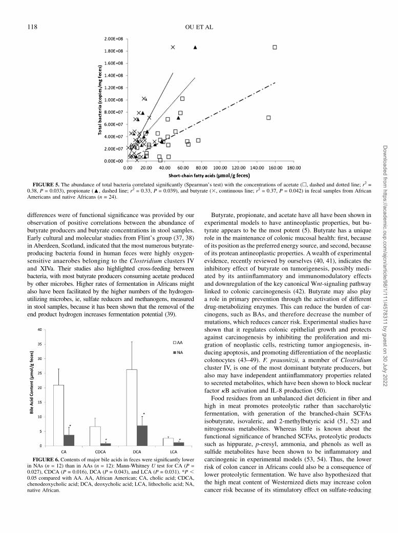
Table 4) with concentrations of stool acetate (r2 = 0.38), pro-pionate (r2 = 0.33), and butyrate (r2 = 0.37) (Figure 5). Second,stool butyrate concentrations were significantly correlated withthe abundance of the butyrate producers Clostridium clusterIV (r2 = 0.29, P = 0.047) and Clostridium cluster XIVa (r2 =0.29, P = 0.047) (Table 4).
Amounts of the 4 major BAs in feces were significantly higher(P , 0.05) in Americans than in Africans (Figure 6). The fecalprimary BAs cholic acid (CA) and chenodeoxycholic acid andthe secondary BAs deoxycholic acid (formed from CA) andlithocholic acid (LCA, formed from CA and chenodeoxycholicacid) were significantly higher (P , 0.05) in Americans than inAfricans (Figure 6). The ratio of butyrate to LCA was signifi-cantly higher in Africans (39.26 19.0 compared with 6.06 3.2;P = 0.047). A significant correlation was also found between 7a-dehydroxylating bacteria and secondary BA concentrations instool (r2 = 0.65, P = 0.01; Spearman’s test) as well (Figure 7),which likely indicated a higher microbial 7a-dehydroxylatingcapacity for colonic BAs in the Americans.
DISCUSSION
The findings of the current study support the hypothesis thatthe balance between health-promoting and inflammatory mi-crobial metabolites may determine colon cancer risk and thattheir production is dependent on both the composition of food andcomposition of the microbiota, as in an autocatalytic system. Thedata provide evidence that saccharolytic fermentation was lower,proteolytic fermentation was higher, and secondary BA pro-duction was higher in African Americans than in Africans. From
our earlier studies in the same populations, it is reasonable topostulate that these differences can be ascribed to differences indiet, with African Americans eating more dietary meat and fatand less complex carbohydrate and fiber (4).
Fecal microbial composition was shown to be very different inAfricans and African Americans, with the pattern in the formercorresponding to enterotype 2 and the latter to enterotype 1.Three enterotypes were recently described by a European con-sortium based on their pooled analysis of fecal samples obtainedfrom healthy adults from 4 European countries, the United States,and Japan (10). From this, they proposed that all human pop-ulations could be categorized into 1 of 3 enterotypes depending ontheir common networks of co- and anticorrelating genera, whichare driven by the genus Bacteroides (type 1), Prevotella (type 2),or Ruminococcus (type 3). Interestingly, the composition in ourAfrican sample was remarkably similar to that shown in CentralAfrican children from Burkina Faso by De Filippo et al (35),dominated by Prevotella (type 2) and showing an overabundanceof microbes that are likely involved in starch and cellulose deg-radation. They related these differences to differences in diet, withAfrican children consuming a diet rich in coarse grains andvegetables that was somewhat similar to what Burkitt described asthe “traditional African diet” that was associated with a low riskof colonic chronic diseases and colon cancer (36). Importantly,butyrate and the other SCFAs were also higher in the Africanchildren—as in our study; the authors proposed that this mighthelp reduce the risk of enteric infections.
Interestingly in the current study, whereas the higher pro-portions of Prevotella, Succinivibrio, and Oscillospira in theAfrican gut microbiome also represented higher numbers ofthese microbes in stool samples, as measured by qPCR, thehigher compositional representation of bacteroides in AfricanAmericans did not appear to reflect higher numbers. It is pos-sible that the total numbers of bacteroides were underestimatedbecause the qPCR assay used targeted the B. fragilis group andnot all human Bacteroides strains. On the other hand, the assaydoes include the 2 major human gut species, namely B. fragilisand Bacteroides thetaiotamicron (15). Presumably, the greaternumbers of total bacteria in the Africans was a consequence ofthe expansion of populations of microbes that degrade starches,hemicelluloses, and xylans—notably Prevotella, Succinivibrio,and Oscillospira—and those that ferment their products.
Our targeted microbe analysis showed that both the butyrateproduction gene and the recognized major butyrate-producingbacteria, including F. prausnitzii, and those contained withinClostridium cluster IV and cluster XIVa were more abundantin stool samples from native Africans. Evidence that these
FIGURE 4. Summary of the differences in mean (6SE) group con-centrations of the major short-chain fatty acids in fecal samples. Con-centrations were significantly greater in NAs (n = 12) than in AAs (n = 12):Mann-Whitney U test for acetate (P = 0.001), propionate (P = 0.003), andbutyrate (P = 0.049). AA, African American; NA, native African.
TABLE 4
Nonparametric Spearman correlations (R) between fecal short-chain fatty acid concentrations and copy numbers of
bacteria1
Total SCFAs Acetic acid Propionic acid Butyric acid
R P R P R P R P
Total bacteria 0.37 0.011 0.38 0.010 0.33 0.026 0.37 0.042
Bifidobacterium spp. 20.23 0.112 20.27 0.066 20.17 0.234 20.19 0.197
Faecalibacterium prausnitzii 20.28 0.215 20.17 0.105 20.27 0.265 20.20 0.246
Clostridium IV 0.26 0.074 0.25 0.083 0.17 0.234 0.29 0.047
Clostridium XIVa 0.26 0.074 0.27 0.066 0.19 0.197 0.29 0.047
1 Sample number = 24. SCFA, short-chain fatty acid.
MICROBIOTA AND METABOLITES IN COLON CANCER RISK 117
Dow
nloaded from https://academ
ic.oup.com/ajcn/article/98/1/111/4578311 by guest on 30 July 2022
differences were of functional significance was provided by ourobservation of positive correlations between the abundance ofbutyrate producers and butyrate concentrations in stool samples.Early cultural and molecular studies from Flint’s group (37, 38)in Aberdeen, Scotland, indicated that the most numerous butyrate-producing bacteria found in human feces were highly oxygen-sensitive anaerobes belonging to the Clostridium clusters IVand XIVa. Their studies also highlighted cross-feeding betweenbacteria, with most butyrate producers consuming acetate producedby other microbes. Higher rates of fermentation in Africans mightalso have been facilitated by the higher numbers of the hydrogen-utilizing microbes, ie, sulfate reducers and methanogens, measuredin stool samples, because it has been shown that the removal of theend product hydrogen increases fermentation potential (39).
Butyrate, propionate, and acetate have all have been shown inexperimental models to have antineoplastic properties, but bu-tyrate appears to be the most potent (5). Butyrate has a uniquerole in the maintenance of colonic mucosal health: first, becauseof its position as the preferred energy source, and second, becauseof its protean antineoplastic properties. Awealth of experimentalevidence, recently reviewed by ourselves (40, 41), indicates theinhibitory effect of butyrate on tumorigenesis, possibly medi-ated by its antiinflammatory and immunomodulatory effectsand downregulation of the key canonical Wnt-signaling pathwaylinked to colonic carcinogenesis (42). Butyrate may also playa role in primary prevention through the activation of differentdrug-metabolizing enzymes. This can reduce the burden of car-cinogens, such as BAs, and therefore decrease the number ofmutations, which reduces cancer risk. Experimental studies haveshown that it regulates colonic epithelial growth and protectsagainst carcinogenesis by inhibiting the proliferation and mi-gration of neoplastic cells, restricting tumor angiogenesis, in-ducing apoptosis, and promoting differentiation of the neoplasticcolonocytes (43–49). F. prausnitzii, a member of Clostridiumcluster IV, is one of the most dominant butyrate producers, butalso may have independent antiinflammatory properties relatedto secreted metabolites, which have been shown to block nuclearfactor kB activation and IL-8 production (50).
Food residues from an unbalanced diet deficient in fiber andhigh in meat promotes proteolytic rather than saccharolyticfermentation, with generation of the branched-chain SCFAsisobutyrate, isovaleric, and 2-methylbutyric acid (51, 52) andnitrogenous metabolites. Whereas little is known about thefunctional significance of branched SCFAs, proteolytic productssuch as hippurate, p-cresyl, ammonia, and phenols as well assulfide metabolites have been shown to be inflammatory andcarcinogenic in experimental models (53, 54). Thus, the lowerrisk of colon cancer in Africans could also be a consequence oflower proteolytic fermentation. We have also hypothesized thatthe high meat content of Westernized diets may increase coloncancer risk because of its stimulatory effect on sulfate-reducing
FIGURE 5. The abundance of total bacteria correlated significantly (Spearman’s test) with the concentrations of acetate (h, dashed and dotted line; r2 =0.38, P = 0.033), propionate (:, dashed line; r2 = 0.33, P = 0.039), and butyrate (3, continuous line; r2 = 0.37, P = 0.042) in fecal samples from AfricanAmericans and native Africans (n = 24).
FIGURE 6. Contents of major bile acids in feces were significantly lowerin NAs (n = 12) than in AAs (n = 12): Mann-Whitney U test for CA (P =0.027), CDCA (P = 0.016), DCA (P = 0.043), and LCA (P = 0.031). *P ,0.05 compared with AA. AA, African American; CA, cholic acid; CDCA,chenodeoxycholic acid; DCA, deoxycholic acid; LCA, lithocholic acid; NA,native African.
118 OU ET AL
Dow
nloaded from https://academ
ic.oup.com/ajcn/article/98/1/111/4578311 by guest on 30 July 2022

bacteria, which use the sulfur residues of meat to release hy-drogen sulfide, which has been shown to be genotoxic in ex-perimental models (54). Surprisingly, the opposite was observed,with higher numbers of sulfate-reducing bacteria in Africans,which suggests that increased saccharolytic fermentation withincreased hydrogen production may be more stimulatory tothese hydrogenotrophs.
Strong epidemiologic evidence links high fat consumption toincreased colon cancer risk (55, 56). Whereas experimentalevidence indicates that fat, particularly fat high in n26 fattyacids, is proinflammatory and that a Westernized diet high in fatand deficient in vitamin D increases colonic tumors in normalmice (57), fat digestion and absorption are very efficient andlittle dietary fat enters the colon, which suggests that othermechanisms may be involved. Perhaps the strongest contender isBAs. Increased fat consumption stimulates the liver to synthe-size more BAs. This in turn increases the quantity of BAs thatescape enterohepatic recirculation and enter the colon. Once inthe colon, primary BAs, notably CA and chenodeoxycholic acid,are converted to secondary BAs, namely deoxycholic acid andLCA, by specific bacteria that contain 7a-dehydroxylating en-zymes. Our studies showed that not only were microbes containingthis enzyme more common in high-risk African Americans, but soalso were fecal secondary BAs. Human studies have associatedincreased fecal secondary BAs with colon polyps (58) and coloncancer (59), and there is substantial experimental evidence thatthey have carcinogenic properties (7). A further potential mecha-nism recently reported in a mouse model was the ability of a high-fat diet to stimulate the delivery of sulfur-rich taurine conjugates ofBAs to the colon, where they produced a blossom of Biophilawadsworthia, which released hydrogen sulfide from taurine thatlead to acute inflammation and colitis (60).
In summary, our study supports the hypothesis that coloncancer risk is determined by the interaction between diet and gutmicrobiota (61, 62) and that the higher risk in African Americanscould be attributed to their chronically lower consumption of fiberand resistant starch and their higher consumption of dietary fat.Further studies are needed to assess the functional significance ofother differences noted in the pyrosequencing analysis (eg,Bacteroides) and the responsiveness of the identified microbialand metabolomic differences to dietary change.
The authors’ responsibilities were as follows—JO, SJDO, KN, and HRG:
participated in the design and conduct of the study; JPD: conducted the BA
and SCFA analyses; FC, EGZ, and MW: assisted with the microbiota anal-
ysis; and JO, FC, and SJDO: performed the data analysis. All authors were
involved in finalizing the manuscript. None of the authors had any conflicts
of interest to declare.
REFERENCES1. Doll R, Peto R. The causes of cancer: quantitative estimates of
avoidable risks of cancer in the United States today. J Natl Cancer Inst1981;66:1191–308.
2. Sharma S, O’Keefe SJD. Environmental influences on the high mor-tality from colorectal cancer in African Americans. Postgrad Med J2007;83:583–9.
3. O’Keefe SJD, Kidd M, Noel G, Owira P. The rarity of colon cancer inAfricans is associated with low animal product consumption, not fiber.Am J Gastroenterol 1999;94:1373–80.
4. O’Keefe SJD, Chung D, Mahmoud N, Sepulveda AR, Manafe M. WhyDo African Americans get more colon cancer than Native Africans?J Nutr 2007;137:175S–82S.
5. Waldecker M, Kautenburger T, Daumann H, Busch C, Schrenk D.Inhibition of histone-deacetylase activity by short-chain fatty acids andsome polyphenol metabolites formed in the colon. J Nutr Biochem2008;19:587–93.
6. Windey K, De Preter V, Verbeke K. Relevance of protein fermentationto gut health. Mol Nutr Food Res 2012;56:184–96.
7. Bernstein C, Holubec H, Bhattacharyya AK, Nguyen H, Payne CM,Zaitlin B, Bernstein H. Carcinogenicity of deoxycholate, a secondarybile acid. Arch Toxicol 2011;85:863–71.
8. Penry DL, Jumars P. Modeling animal guts as chemical reactors. AmNat 1987;129:69–96.
9. Louis P, Flint HJ. Diversity, metabolism and microbial ecology ofbutyrate-producing bacteria from the human large intestine. FEMSMicrobiol Lett 2009;294:1–8.
10. ArumugamM, Raes J, Pelletier E, Le Paslier D, Yamada T, Mende DR,Fernandes GR, Tap J, Bruls T, Batto JM, et al. Enterotypes of thehuman gut microbiome. Nature 2011;473:174–80.
11. Fooks LJ, Gibson GR. Probiotics as modulators of the gut flora. Br JNutr 2002;88:S39–49.
12. Arunachalam KD. Role of Bifidobacteria in nutrition, medicine andtechnology. Nutr Res 1999;19:1559–97.
13. Nadkarni MA, Martin FE, Jacques NA, Hunter N. Determination ofbacterial load by real-time PCR using a broad-range (universal) probeand primers set. Microbiology 2002;148:257–66.
14. Louis P, Flint HJ. Development of a semiquantitative degenerate real-time pcr-based assay for estimation of numbers of butyryl-coenzyme A(CoA) CoA transferase genes in complex bacterial samples. ApplEnviron Microbiol 2007;73:2009–12.
15. Matsuki T, Watanabe K, Fujimoto J, Tanaka R. Development of 16SrRNA-gene-targeted group-specific primers for the detection andidentification of predominant bacteria in human feces. Appl EnvironMicrobiol 2002;68:5445–51.
16. Kok RG, de Waal A, Schut F, Hellingwerf K. Specific detection andanalysis of a probiotic Bifidobacterium strain in infant feces. ApplEnviron Microbiol 1996;62:3668–72.
17. Stevenson DM, Weimer P. Dominance of Prevotella and low abundanceof classical ruminal bacterial species in the bovine rumen revealed byrelative quantification real-time PCR. Appl Microbiol Biotechnol 2007;75:165–74.
18. Byun R, Nadkarni MA, Chhour KL, Martin FE, Jacques NA, Hunter N.Quantitative analysis of diverse Lactobacillus species present in ad-vanced dental caries. J Clin Microbiol 2004;42:3128–36.
19. Rinttila T, Kassinen A, Malinen E, Krogius L, Palva A. Developmentof an extensive set of 16S rDNA-targeted primers for quantification ofpathogenic and indigenous bacteria in faecal samples by real-timePCR. J Appl Microbiol 2004;97:1166–77.
20. Spence C, Whitehead TR, Cotta MA. Development and comparison ofSYBR Green quantitative real-time PCR assays for detection andenumeration of sulfate-reducing bacteria in stored swine manure.J Appl Microbiol 2008;105:2143–52.
21. Denman SE, Tomkins NW, McSweeney CS. Quantitation and diversityanalysis of ruminal methanogenic populations in response to the
FIGURE 7. The abundance of 7a-dehydroxylating bacteria, estimatedfrom detection of their functional genes by polymerase chain reaction,correlated significantly with the concentration of secondary bile acids infecal samples (n = 24). r2 = 0.65, P = 0.001 (Spearman’s test).
MICROBIOTA AND METABOLITES IN COLON CANCER RISK 119
Dow
nloaded from https://academ
ic.oup.com/ajcn/article/98/1/111/4578311 by guest on 30 July 2022

antimethanogenic compound bromochloromethane. FEMS MicrobiolEcol 2007;62:313–22.
22. Collins MD, Lawson PA, Willems A, Cordoba JJ, Fernandez-Garayzabal J, Garcia P, Cai J, Hippe H, Farrow JA. The phylogeny ofthe genus Clostridium: proposal of five new genera and eleven newspecies combinations. Int J Syst Bacteriol 1994;44:812–26.
23. Scheppach W, Fabian C, Kasper H. Fecal short-chain fatty acid (SCFA)analysis by capillary gas-liquid chromatography. Am J Clin Nutr 1987;46:641–6.
24. Tagliacozzi D, Mozzi AF, Casetta B, Bertucci P, Bernardini S, Ilio CD,Urbani A, Federici G. Quantitative analysis of bile acids in humanplasma by liquid chromatography-electrospray tandem mass spec-trometry: a simple and rapid one-step method. Clin Chem Lab Med2003;41:1633–41.
25. Zoetendal EG, Heilig HG, Klaassens ES, Booijink CC, KleerebezemM, Smidt H, de Vos WM. Isolation of DNA from bacterial samples ofthe human gastrointestinal tract. Nat Protoc 2006;1:870–3.
26. Caporaso JG, Kuczynski J, Stombaugh J, Bittinger K, Bushman FD,Costello EK, Fierer N, Pena AG, Goodrich JK, Gordon JI, et al. QIIMEallows analysis of high-throughput community sequencing data. NatMethods 2010;7:335–6.
27. Cole JR, Wang Q, Cardenas E, Fish J, Chai B, Farris RJ, Kulam-Syed-Mohideen AS, McGarrell DM, Marsh T, Garrity GM, et al. The Ri-bosomal Database Project: improved alignments and new tools forrRNA analysis. Nucleic Acids Res 2009;37:D141–5.
28. Nakamura N, Leigh SR, Mackie RI, Gaskins HR. Microbial commu-nity analysis of rectal methanogens and sulfate reducing bacteria in twonon-human primate species. J Med Primatol 2009;38:360–70.
29. Nava GM, Carbonero F, Croix JA, Greenberg E, Gaskins HR. Abun-dance and diversity of mucosa-associated hydrogenotrophic microbesin the healthy human colon. ISME J 2012;6:57–70.
30. Wells JE, Berr F, Thomas LA, Dowling RH, Hylemon PB. Isolationand characterization of cholic acid 7a-dehydroxylating fecal bacteriafrom cholesterol gallstone patients. J Hepatol 2000;32:4–10.
31. Carbonero F, Benefiel AC, Gaskins HR. Contributions of the microbialhydrogen economy to colonic homeostasis. Nat Rev GastroenterolHepatol 2012;9:504–18.
32. Carbonero F, Benefiel AC, Alizadeh-Ghamsari AH, Gaskins HR. Mi-crobial pathways in colonic sulfur metabolism and links with healthand disease. Front Physiol 2012;3:448.
33. Tims S, Derom C, Jonkers DM, Vlietinck R, Saris WH, KleerebezemM, de Vos WM, Zoetendal EG. Microbiota conservation and BMIsignatures in adult monozygotic twins. ISME J 2013;7:707–17.
34. Walker AW, Ince J, Duncan SH, Webster LM, Holtrop G, Ze X, BrownD, Stares MD, Scott P, Bergerat A, et al. Dominant and diet-responsivegroups of bacteria within the human colonic microbiota. ISME J 2011;5:220–30.
35. De Filippo C, Cavalieri D, Di Paola M, Ramazzotti M, Poullet JB,Massart S, Collini S, Pieraccini G, Lionetti P. Impact of diet in shapinggut microbiota revealed by a comparative study in children from Eu-rope and rural Africa. Proc Natl Acad Sci USA 2010;107:14691–6.
36. Burkitt DP. Diseases of the alimentary tract and western diets. PatholMicrobiol (Basel) 1973;39:177–86.
37. Pryde SE, Duncan SH, Hold GL, Stewart CS, Flint HJ. The microbi-ology of butyrate formation in the human colon. FEMS Microbiol Lett2002;217:133–9.
38. Barcenilla A, Pryde SE, Martin JC, Duncan SH, Stewart CS,Henderson C, Flint HJ. Phylogenetic relationships of butyrate-producing bacteria from the human gut. Appl Environ Microbiol2000;66:1654–61.
39. Wolin M. Interactions between H2-producing and methaneproducingspecies. In: Schlegel HG, Gottschalk G, Pfennig N, eds. Microbial for-mation and utilization of gases. Gottingen, Germany: Goltze Press,1976:141–50.
40. Greer JB, O’Keefe SJ. Microbial induction of immunity, inflammationand cancer. Front Physiol 2011;1:168.
41. Vipperla K, O’Keefe SJ. The microbiota and its metabolites in colonicmucosal health and cancer risk. Nutr Clin Pract 2012;27:624–35.
42. Bordonaro M, Lazarova DL, Sartorelli AC. Butyrate and Wnt signal-ing: a possible solution to the puzzle of dietary fiber and colon cancerrisk? Cell Cycle 2008;7:1178–83.
43. Chirakkal H, Leech SH, Brookes KE, Prais AL, Waby JS, Corfe BM.Upregulation of BAK by butyrate in the colon is associated with in-creased Sp3 binding. Oncogene 2006;25:7192–200.
44. Hinnebusch BF, Meng S, Wu JT, Archer SY, Hodin RA. The effects ofshort-chain fatty acids on human colon cancer cell phenotype are as-sociated with histone hyperacetylation. J Nutr 2002;132:1012–7.
45. Comalada M, Bailon E, de Haro O, Lara-Villoslada F, Xaus J, ZarzueloA, Galvez J. The effects of short-chain fatty acids on colon epithelialproliferation and survival depend on the cellular phenotype. J CancerRes Clin Oncol 2006;132:487–97.
46. Andoh A, Shimada M, Araki Y, Fujiyama Y, Bamba T. Sodium buty-rate enhances complement-mediated cell injury via down-regulation ofdecay-accelerating factor expression in colonic cancer cells. CancerImmunol Immunother 2002;50:663–72.
47. Rodrıguez-Salvador J, Armas-PinedaC, Perezpena-DiazcontiM, Chico-Ponce de Leon F, Sosa-Sainz G, Lezama P, Recillas-Targa F, Arenas-Huertero F. Effect of sodium butyrate on pro-matrix metalloproteinase-9and -2 differential secretion in pediatric tumors and cell lines. J Exp ClinCancer Res 2005;24:463–73.
48. Zeng H, Briske-Anderson M. Prolonged butyrate treatment inhibits themigration and invasion potential of HT1080 tumor cells. J Nutr 2005;135:291–5.
49. Zgouras D, Wachtershauser A, Frings D, Stein J. Butyrate impairsintestinal tumor cell-induced angiogenesis by inhibiting HIF-1alphanuclear translocation. Biochem Biophys Res Commun 2003;300:832–8.
50. Sokol H, Pigneur B, Watterlot L, Lakhdari O, Bermudez-Humaran LG,Gratadoux JJ, Blugeon S, Bridonneau C, Furet JP, Corthier G, et al.Faecalibacterium prausnitzii is an anti-inflammatory commensal bac-terium identified by gut microbiota analysis of Crohn disease patients.Proc Natl Acad Sci USA 2008;105:16731–6.
51. Dehority BA, Johnson RR, Bentley OG, Moxon AL. Studies on themetabolism of valine, proline, leucine and isoleucine by rumen mi-croorganisms in vitro. Arch Biochem Biophys 1958;78:15–27.
52. Allison MJ. Production of branched-chain volatile fatty acids by certainanaerobic bacteria. Appl Environ Microbiol 1978;35:872–7.
53. Russell WR, Gratz SW, Duncan SH, Holtrop G, Ince J, Scobbie L,Duncan G, Johnstone AM, Lobley GE, Wallace RJ, et al. High-protein,reduced-carbohydrate weight-loss diets promote metabolite profiles likelyto be detrimental to colonic health. Am J Clin Nutr 2011;93:1062–72.
54. Attene-Ramos MS, Wagner ED, Plewa MJ, Gaskins HR. Evidence thathydrogen sulfide is a genotoxic agent. Mol Cancer Res 2006;4:9–14.
55. NIH. Based on November 2005 SEER data submission, posted to theSEER web site 2006. Available from: http://seer.cancer.gov/csr/1975_2003/(cited 18 December 2012).
56. Stadler J, Stern HS, Yeung KS, McGuire V, Furrer R, Marcon N, BruceWR. Effect of high fat consumption on cell proliferation activity ofcolorectal mucosa and on soluble faecal bile acids. Gut 1988;29:1326–31.
57. Newmark HL, Yang K, Kurihara N, Fan K, Augenlicht LH, Lipkin M.Western-style diet-induced colonic tumors and their modulation bycalcium and vitamin D in C57Bl/6 mice: a preclinical model for humansporadic colon cancer. Carcinogenesis 2009;30:88–92.
58. de Kok TM, van Faassen A, Glinghammar B, Pachen DM, Eng M,Rafter JJ, Baeten CG, Engels LG, Kleinjans JC. Bile acid concentra-tions, cytotoxicity, and pH of fecal water from patients with colorectaladenomas. Dig Dis Sci 1999;44:2218–25.
59. Bernstein H, Bernstein C, Payne CM, Dvorakova K, Garewal H. Bileacids as carcinogens in human gastrointestinal cancers. Mutat Res2005;589:47–65.
60. Devkota S, Wang Y, Musch MW, Leone V, Fehlner-Peach H, Na-dimpalli A, Antonopoulos DA, Jabri B, Chang EB. Dietary-fat-inducedtaurocholic acid promotes pathobiont expansion and colitis in Il102/2mice. Nature 2012;487:104–8.
61. Dethlefsen L, Eckburg PB, Bik EM, Relman DA. Assembly of thehuman intestinal microbiota. Trends Ecol Evol 2006;21:517–23.
62. O’Keefe SJD. Nutrition and colonic health: the critical role of themicrobiota. Curr Opin Gastroenterol 2008;24:51–8.
120 OU ET AL
Dow
nloaded from https://academ
ic.oup.com/ajcn/article/98/1/111/4578311 by guest on 30 July 2022
Related Documents