REVIEW Gut microbiota derived metabolites in cardiovascular health and disease Zeneng Wang & , Yongzhong Zhao Department of Cellular and Molecular Medicine, Lerner Research Institute, Cleveland Clinic, Cleveland, OH 44195, USA & Correspondence: [email protected] (Z. Wang) Received March 16, 2018 Accepted April 16, 2018 ABSTRACT Trillions of microbes inhabit the human gut, not only providing nutrients and energy to the host from the ingested food, but also producing metabolic bioactive signaling molecules to maintain health and elicit dis- ease, such as cardiovascular disease (CVD). CVD is the leading cause of mortality worldwide. In this review, we presented gut microbiota derived metabolites involved in cardiovascular health and disease, including trimethylamine-N-oxide (TMAO), uremic toxins, short chain fatty acids (SCFAs), phytoestrogens, antho- cyanins, bile acids and lipopolysaccharide. These gut microbiota derived metabolites play critical roles in maintaining a healthy cardiovascular function, and if dysregulated, potentially causally linked to CVD. A bet- ter understanding of the function and dynamics of gut microbiota derived metabolites holds great promise toward mechanistic predicative CVD biomarker discov- eries and precise interventions. KEYWORDS gut microbiota, metabolites, cardiovascular health, cardiovascular disease INTRODUCTION There is a big gap in interpreting the molecular physiology by using the human genome coding capacity encompassing 23,000 coding genes (Gonzaga-Jauregui et al., 2012). The human gut is inhabited with 100 trillion microbes, with the majority as bacteria and archaea, fungi and microeukaryotes (Wampach et al., 2017). Almost 10 million coding genes of the microbiota have been uncovered, greatly expanding the coding capacity of our human as a superorganism (Qin et al., 2010; Li et al., 2014). Gut microbiota are essential to human health in many aspects, such as training intestinal epithelial barrier, modulating immuno-function, digesting host indi- gestible nutrients, producing vitamins and hormones and preventing pathogenic bacterium colonization (Schuijt et al., 2016). For a healthy subject, gut microbiota homeostasis is maintained with pathogenic microbe growth under control. Once the balance breaks, i.e., dysbiosis, pathogenic microbes thrive, leading to gut related diseases, such as inflammatory bowel disease (IBD), obesity, allergic disor- ders, diabetes mellitus, autism, colorectal cancer and car- diovascular disease (DeGruttola et al., 2016; Yang et al., 2015; Battson et al., 2017). Fecal microbiota transplantation has shown great efficacy in managing Clostridium difficile infection and Crohn’s disease (Bakken et al., 2013; Paasche 2013; Zhang et al., 2013). In animal model, fecal microbiota transplant to germ free mice recipients has been shown to transmit obesity and atherosclerosis susceptibility, suggest- ing the great potential of fecal microbiota transplantation in treating a panel of complex disease (Gregory et al., 2015; Turnbaugh et al., 2006). In addition, the prebiotic and pro- biotic administrations also show beneficial effects in opti- mizing gut microbiota community structure and preventing dysbiosis (Hamilton et al., 2017; Anhe et al., 2015; Delgado et al., 2014; Kouchaki et al., 2017). The association between gut microbiota and health has become a hot topic, the rapid progress in this field is ascri- bed to next generation sequencing methods as well as the ease of maintaining germ free mice (Mardis, 2008; Bhattarai and Kashyap, 2016). Gut microbes are involved in the biosynthesis of an array of bioactive compounds, contributing to normal human physiological functions or eliciting disease (Fan et al., 2015; Wang et al., 2011). CVD is the leading cause of death worldwide, the association with gut microbiota has been reported in recent few years, which is mediated by gut Electronic supplementary material The online version of this article (https://doi.org/10.1007/s13238-018-0549-0) contains sup- plementary material, which is available to authorized users. © The Author(s) 2018 Protein Cell 2018, 9(5):416–431 https://doi.org/10.1007/s13238-018-0549-0 Protein & Cell Protein & Cell

Welcome message from author
This document is posted to help you gain knowledge. Please leave a comment to let me know what you think about it! Share it to your friends and learn new things together.
Transcript

REVIEW
Gut microbiota derived metabolitesin cardiovascular health and disease
Zeneng Wang&, Yongzhong Zhao
Department of Cellular and Molecular Medicine, Lerner Research Institute, Cleveland Clinic, Cleveland, OH 44195, USA& Correspondence: [email protected] (Z. Wang)
Received March 16, 2018 Accepted April 16, 2018
ABSTRACT
Trillions of microbes inhabit the human gut, not onlyproviding nutrients and energy to the host from theingested food, but also producing metabolic bioactivesignaling molecules to maintain health and elicit dis-ease, such as cardiovascular disease (CVD). CVD is theleading cause of mortality worldwide. In this review, wepresented gut microbiota derived metabolites involvedin cardiovascular health and disease, includingtrimethylamine-N-oxide (TMAO), uremic toxins, shortchain fatty acids (SCFAs), phytoestrogens, antho-cyanins, bile acids and lipopolysaccharide. These gutmicrobiota derived metabolites play critical roles inmaintaining a healthy cardiovascular function, and ifdysregulated, potentially causally linked to CVD. A bet-ter understanding of the function and dynamics of gutmicrobiota derived metabolites holds great promisetoward mechanistic predicative CVD biomarker discov-eries and precise interventions.
KEYWORDS gut microbiota, metabolites, cardiovascularhealth, cardiovascular disease
INTRODUCTION
There is a big gap in interpreting the molecular physiology byusing the human genome coding capacity encompassing23,000 coding genes (Gonzaga-Jauregui et al., 2012). Thehuman gut is inhabited with 100 trillion microbes, with themajority as bacteria and archaea, fungi and microeukaryotes(Wampach et al., 2017). Almost 10 million coding genes ofthe microbiota have been uncovered, greatly expanding the
coding capacity of our human as a superorganism (Qin et al.,2010; Li et al., 2014). Gut microbiota are essential to humanhealth in many aspects, such as training intestinal epithelialbarrier, modulating immuno-function, digesting host indi-gestible nutrients, producing vitamins and hormones andpreventing pathogenic bacterium colonization (Schuijt et al.,2016). For a healthy subject, gut microbiota homeostasis ismaintained with pathogenic microbe growth under control.Once the balance breaks, i.e., dysbiosis, pathogenicmicrobes thrive, leading to gut related diseases, such asinflammatory bowel disease (IBD), obesity, allergic disor-ders, diabetes mellitus, autism, colorectal cancer and car-diovascular disease (DeGruttola et al., 2016; Yang et al.,2015; Battson et al., 2017). Fecal microbiota transplantationhas shown great efficacy in managing Clostridium difficileinfection and Crohn’s disease (Bakken et al., 2013; Paasche2013; Zhang et al., 2013). In animal model, fecal microbiotatransplant to germ free mice recipients has been shown totransmit obesity and atherosclerosis susceptibility, suggest-ing the great potential of fecal microbiota transplantation intreating a panel of complex disease (Gregory et al., 2015;Turnbaugh et al., 2006). In addition, the prebiotic and pro-biotic administrations also show beneficial effects in opti-mizing gut microbiota community structure and preventingdysbiosis (Hamilton et al., 2017; Anhe et al., 2015; Delgadoet al., 2014; Kouchaki et al., 2017).
The association between gut microbiota and health hasbecome a hot topic, the rapid progress in this field is ascri-bed to next generation sequencing methods as well as theease of maintaining germ free mice (Mardis, 2008; Bhattaraiand Kashyap, 2016).
Gut microbes are involved in the biosynthesis of an arrayof bioactive compounds, contributing to normal humanphysiological functions or eliciting disease (Fan et al., 2015;Wang et al., 2011). CVD is the leading cause of deathworldwide, the association with gut microbiota has beenreported in recent few years, which is mediated by gut
Electronic supplementary material The online version of thisarticle (https://doi.org/10.1007/s13238-018-0549-0) contains sup-
plementary material, which is available to authorized users.
© The Author(s) 2018
Protein Cell 2018, 9(5):416–431https://doi.org/10.1007/s13238-018-0549-0 Protein&Cell
Protein
&Cell

microbiota derived metabolites (Wang et al., 2011; Tanget al., 2013; Koeth et al., 2013). In this review, we listed gutmicrobiota derived metabolites and their clinical relevance incardiovascular health and disease pathogenesis.
TRIMETHYLAMINE N OXIDE (TMAO)
Gut microbiota cleave some trimethylamine containingcompounds to produce trimethylamine (TMA), which can befurther oxidized as trimethylamine N oxide (TMAO) in thehost liver by flavin monooxygenase (FMOs) (Wang et al.,2011; Koeth et al., 2013). FMO3 is the most abundantenzyme in the liver, while FMO1 and FMO2 can also cat-alyze the oxidation of TMA (Bennett et al., 2013). In somepatients with loss-of-function mutation of the FMO3 gene,accumulated TMA in vivo spreads all over the body and isreleased in sweat and breath, which is a genetic diseasenamed fish odor syndrome (Dolphin et al., 1997; Ulmanet al., 2014). The precursors for gut microbiota to produceTMA include TMAO, choline, phosphatidylcholine, carnitine,γ-butyrobetaine, betaine, crotonobetaine and glycerophos-phocholine, all of which are abundant in animal diet (Koethet al., 2013; Wang et al., 2015; Rausch et al., 2013).
The diet-gut microbiota-liver to TMAO biosynthesis con-stitutes a metaorganismal pathway (Fig. 1), including fourenzymes involved in production of TMA, choline-TMA lyase(cutC/D) (Craciun et al., 2014), carnitine monooxygenase(cntA/B) (Zhu et al., 2014), betaine reductase (Andreesen,1994), and TMAO reductase (Pascal et al., 1984).
Furthermore, yeaW/X, highly homologous to cntA/B, alsocontributes to production of TMA. Besides carnitine, yeaW/Xcan also use choline, γ-butyrobetaine and betaine as sub-strates to produce TMA (Koeth et al., 2014).
CutC/D has been crystalized and the enzymatic mecha-nism has been demonstrated. CutD, as a radical S-adeno-sylmethionine-activatase, activates CutC, resulting information of a glycyl radical. In CutC, the glycyl radicalabstracts the hydrogen from cysteine to produce a thiylradical and further captures the hydrogen atom from cholineat C1 position, resulting in molecular rearrangement andTMA production. (Craciun et al., 2014; Kalnins et al., 2015;Bodea et al., 2016). CntA/B is a two-component Rieske-typeoxygenase/reductase, carnitine can be first oxidized fol-lowed by cleavage at C-N bond by CntA/B to produce TMAand malic semialdehyde (Zhu et al., 2014). Hundreds ofbacterial strains are predicted to express cutC/D or cntA/B-yeaW/X in the human gut (Fig. 2A, 2B, 2C and Table S1)(Rath et al., 2017; Martinez-del Campo et al., 2015). Proteusmirabilis is a cutC/D expressing bacterium species and sinceit can grow under both aerobic and anaerobic conditions, ithas been used as a model to screen choline trimethylaminelyase inhibitors (Wang et al., 2015). It is most likely the genetree of cutC substantially differs from species tree, e.g.,species of the same genus but with distinct topology forKlebsiella (Fig. 2D). FMO3 expression in mice is regulatedby sex hormone, repressed by androgens and stimulated byestrogens (Bennett et al., 2013).
Choline
Betaine
Carnitine
γ-Butyrobetaine
Crotonobetaine
Glycerophosphocholine
Phosphatidylcholine
Trimethylamine(TMA)
Gut
Liver
CutC/DCntA/BYeaW/XBetaine reductase
Atherosclerosis
Thrombosis
Artery
Diet
Trimethylamine N-oxide
TMAO reductase
TrimethylamineN-oxide(TMAO)
FMOsN N
O
HO N
HO NO
HO
ON
OH
HO
ON
HO
ON
O NPO
O OH
OHHO
O NPO
O OH
OOR1
R2
O
O
NO-
Figure 1. Metaorganismal pathway of trimethylamine N oxide (TMAO) biosynthesis and linking to cardiovascular disease.
FMOs, Flavin monooxygenases. R1, R2, CH3(CH2)n1(CH=CH)n2, n2 = 0, 1, 2…..6, n1+2n2 = 15, 17, 19, 21.
Microbiota metabolites in cardiovascular health and disease REVIEW
© The Author(s) 2018 417
Protein
&Cell

Many lines of evidence show the pro-atherogenic prop-erty of TMAO. Circulating TMAO level is associated withprevalence of cardiovascular disease and can independentlypredict incident risk for major adverse cardiac events,including myocardial infarction, stroke or death after adjust-ment for traditional cardiac risk factors and renal function(Wang et al., 2011; Tang et al., 2013). Circulating choline,betaine and carnitine levels also have been shown associ-ated with prevalence of cardiovascular disease and canpredict incident risk for major adverse cardiac events.However, their prognostic values are dependent on theserum TMAO levels (Koeth et al., 2013; Wang et al., 2014).ApoE-null mice fed a chow diet supplemented with TMAOappear to have an enhanced aortic lesion. Furthermore,choline can also increase aortic lesion and promote
atherosclerosis but indispensable to gut microbiota, indicat-ing the causal of TMAO in atherosclerosis (Wang et al.,2011). In vitro animal models have also confirmed the pro-thrombotic effect of TMAO by enhancing platelet aggregation(Zhu et al., 2016). Consistently, oral choline supplementationincreases fasting TMAO levels and also enhances plateletaggregation (Zhu et al., 2017).
Mechanisms by which how TMAO can promoteatherosclerosis and thrombosis have been studied at themolecular level. TMAO activates vascular smooth musclecell and endothelial cell MAPK, nuclear factor-κB (NF-κB)signaling, leading to inflammatory gene expression andendothelial cell adhesion of leukocytes (Seldin et al., 2016).Meanwhile, TMAO can also activate the NLRP3 inflamma-some (Sun et al., 2016; Boini et al., 2017; Chen et al., 2017).
A
Ortho
691
75
980 2,766
HMPVM
18
34
107
B
DCAeromonas hydrophila AL09-71Aeromonas hydrophila pc104AAeromonas hydrophila ML09-119Aeromonas hydrophila J-lAeromonas hydrophila AH10
Desulfovibrio desulfuricans ATCC 27774Enterabacteriaceae bacterium FGI 57
Ortho
1,009
16
2,858 6,600
MVPMH
3
48
87
YeaW
7,219
57
2,709 1,558
CtuCPMH
10
149
342
Aeromonas hydrophila YL17Klebsiella aerogenes KCTC 2190Klebsiella pneumoniae 342Klebsiella varriicola DSM 15968Escherichta coli O6:K15:H31 536 (UPEC)Raoultella ornithinolytica B6Klebsiella michiganensis KCTC 1686Klebsiella axytoca KONIH 1Proteus mirabilis H14320Proteus mirabilis BB2000Serratia marcescens FG194
Scrraha fonticola DSM 4576Serratia fonticola GS2
Pectobactenum atrosepticum SCR11043Pectobacterium parmeritieri WF P163Pectobactcrium sp. SCC3193
0.050
100100
100100
100100
100
100
51
769871
100
98
56
9991
7747
Figure 2. Predicted bacteria strains encoding the cutC/yeaW/cntA TMA lyases. (A) Predicted bacteria strains encoding cutC
gene. Abbreviation, HMP, the NIH Human Microbiome Project (Data release 1.1, September 26, 2017 e), Ortho, cutC encoding gene
of OrthoDB (http://www.orthodb.org/v9.1/) (Zdobnov et al., 2017), and VM, data from the reference (Rath et al., 2017). (B) Predicted
bacteria stains encoding yeaW/cntA genes. Ortho, yeaW encoding gene of OrthoDB. (C) Predicted bacterial strains encoding both
yeaW and cutC. (D) Phylogenetic gene tree of cutC encoding strains. The Neighbor-Joining tree was built with MEGA7 (Kumar et al.,
2016).
REVIEW Zeneng Wang and Yongzhong Zhao
418 © The Author(s) 2018
Protein
&Cell
TMAO in vivo can increase scavenger receptor, CD36 andSR-A1 expression, leading to more uptake of modified LDLfor macrophage to form foam cell (Wang et al., 2011). On theother hand, TMAO decreases expression of two keyenzymes, CYP7A1 and CYP27A1, essential for bile acidbiosynthesis and multiple bile acid transporters (OATP1,OATP4, MRP2 and NTCP) in the liver, which decreases bileacid pool, resulting in decreased reverse cholesterol efflux(Koeth et al., 2013). Moreover, TMAO increases endoplas-mic recticulum calcium release in platelet cell, consequentlyleading to platelet aggregation and thrombosis (Zhu et al.,2016).
The association between TMAO and cardiovascular dis-ease has been highlighted in different groups by using dif-ferent cohorts worldwide (Troseid et al., 2015; Suzuki et al.,2016, 2017; Schuett et al., 2017). Besides cardiovasculardisease, TMAO also contributes to renal insufficiency andmortality risk in chronic kidney disease, type II diabetes,insulin resistance, non-alcoholic fatty liver disease and col-orectal cancer as well (Tang et al., 2015; Shan et al., 2017;Oellgaard et al., 2017; Kummen et al., 2017). These studiesindicate circulating TMAO levels has the potential to bemanaged for TMAO related diseases intervention. Specially,targeting the metaorganismal pathway for TMAO biosyn-thesis can be achieved by a few key steps, includinginhibiting gut microbiota cleavage of TMA containing com-pounds in nutrient via enzymatic inhibitor, controlling intakeof diet rich in TMA precursors and inhibiting the oxidation ofTMA to TMAO.
As expected, the injection of antisense oligonucleotide toLdlr-null mice decreases the hepatic Fmo3 gene expression,resulting in decreased mouse plasma TMAO therebydecreasing aortic lesion in western diet fed mice (Shih et al.,2015). However, the accumulated TMA in mice will show fishodor syndrome. In addition, Fmo3 knockdown exacerbateshepatic endoplasmic reticulum (ER) stress and inflammation(Warrier et al., 2015). Thus, developing gut microbiotaenzymatic inhibitors to inhibit TMA formation will be morepractical.
A choline analogue, 3,3-dimethylbutanol (DMB), hasbeen uncovered with inhibitory effect to choline TMA lyaseactivity in turn decreasing circulating TMAO, and thereforeattenuating the promoting role of choline in atherosclerosis(Wang et al., 2015). DMB is a natural product, distributed incertain balsamic vinegars, red wines, cold-pressed extravirgin olive oils and grapeseed oils. DMB has not been foundany adverse effect to the liver or renal functions even as highas in mice drinking water up to 1% (Wang et al., 2015). Veryrecently, we have found that several more choline analoguesshow more potent in inhibiting choline TMA lyase activitythan DMB (to be published). But inhibitors to differentenzymatic cleavage of other substrates are still needed.Furthermore, a study shows that resveratrol, a phytoalexin,can decrease plasma TMAO and subsequent atherosclero-sis in ApoE−/− mice via gut microbiota remodeling, charac-terized by increased levels of the genera Lactobacillus and
Bifidobacterium with increased bile salt hydrolase activity toincrease bile acid neosynthesis, suggesting the potential ofresveratrol as prebiotics (Chen et al., 2016).
UREMIC TOXINS
Toxins, such as urea and asymmetric dimethylarginine, canbe accumulated in blood during chronic kidney disease(CKD), associated to CKD complications especially heartfailure which is the leading cause of CKD mortality (Glassock2008). Moreover, protein-bound uremic toxins such asindoxyl sulfate, indoxyl glucuronide, indoleacetic acid, p-cresyl sulfate, p-cresyl glucuronide, phenyl sulfate, phenylglucuronide, phenylacetic acid and hippuric acid have beenreported to be increased in serum in hemodialysis patients(Itoh et al., 2013). These uremic toxins are gut microbiotaderived metabolites of amino acids (Devlin et al., 2016). Thearomatic amino acids in proteins, phenylalanine, tyrosineand tryptophan, can be metabolized by gut microbiota (Nalluet al., 2017; Pereira-Fantini et al., 2017). Both microbiota andhost liver are involved in biosynthesis of these uremic toxins(Fig. 3) (Devlin et al., 2016; Meyer and Hostetter 2012;Webster et al., 1976; Gryp et al., 2005).
The serum indoxyl sulfate level, positively correlated withcoronary atherosclerosis scores, might be a predicativemechanistic biomarker of coronary artery disease severity(Hsu et al., 2013). Further studies have shown that indoxylsulfate aggravates cardiac fibrosis, cardiomyocyte hyper-trophy and atrial fibrillation (Yisireyili et al., 2013; Aoki et al.,2015). Atrial fibrillation, the most common clinical arrhythmia,results in cardiovascular morbidity and mortality attributed tocongestive heart failure and stroke (Hung et al., 2017).Mechanistically, indoxyl sulfate enhances platelet activities,increases response to collagen and thrombin, leading tothrombosis (Yang et al., 2017). Vascular smooth muscle cellcalcification is associated with major adverse cardiovascularevents while indoxyl sulfate has been found to promotevascular smooth muscle cell calcification (Zhang et al.,2018). Indoxyl sulfate activates NF-κB signaling pathway,leading to increased intercellular adhesion molecule-1(ICAM-1) and monocyte chemotactic protein-1 (MCP-1)expression in endothelial cells (Tumur et al., 2010). ICAMsover-expression in endothelial cells is the initiating step foratherosclerotic plaque formation (Moss and Ramji 2016).Indoxyl sulfate inhibits nitric oxide production and inducesreactive oxygen species production, gradually damagingendothelial cell layer (Tumur and Niwa 2009). Taken toge-ther, these studies indicate indoxyl sulfate mechanisticallylinked to CVD at the molecular and cellular levels.
p-Cresyl sulfate is a biomarker in predicting cardiovas-cular event and renal function progression in CKD patientswithout dialysis (Lin et al., 2014; Wu et al., 2012). p-Cresylsulfate can induce NADPH oxidase activity to producereactive oxygen species, resulting in cardiomyocyte apop-tosis and subsequent diastolic dysfunction (Han et al., 2015).Apocynin and N-acetylcysteine, inhibitors to NADPH
Microbiota metabolites in cardiovascular health and disease REVIEW
© The Author(s) 2018 419
Protein
&Cell
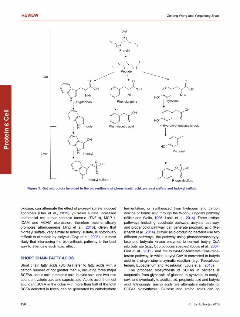
oxidase, can attenuate the effect of p-cresyl sulfate inducedapoptosis (Han et al., 2015). p-Cresyl sulfate increasedendothelial cell tumor necrosis factor-α (TNF-α), MCP-1,ICAM and VCAM expression, therefore mechanisticallypromotes atherogenesis (Jing et al., 2016). Given thatp-cresyl sulfate, very similar to indoxyl sulfate, is notoriouslydifficult to eliminate by dialysis (Gryp et al., 2005), it is mostlikely that intervening the biosynthesis pathway is the bestway to attenuate such toxic effect.
SHORT CHAIN FATTY ACIDS
Short chain fatty acids (SCFAs) refer to fatty acids with acarbon number of not greater than 6, including three majorSCFAs, acetic acid, propionic acid, butyric acid, and two lessabundant valeric acid and caproic acid. Acetic acid, the mostabundant SCFA in the colon with more than half of the totalSCFA detected in feces, can be generated by carbohydrate
fermentation, or synthesized from hydrogen and carbondioxide or formic acid through the Wood-Ljungdahl pathway(Miller and Wolin, 1996; Louis et al., 2014). Three distinctpathways including succinate pathway, acrylate pathway,and propanodiol pathway, can generate propionic acid (Re-ichardt et al., 2014). Butyric acid-producing bacteria use twodifferent pathways, the pathway using phosphotransbutyry-lase and butyrate kinase enzymes to convert butyryl-CoAinto butyrate (e.g., Coprococcus species) (Louis et al., 2004;Flint et al., 2015), and the butyryl-CoA/acetate CoA-trans-ferase pathway, in which butyryl-CoA is converted to butyricacid in a single step enzymatic reaction (e.g., Faecalibac-terium, Eubacterium and Roseburia) (Louis et al., 2010).
The proposed biosynthesis of SCFAs in bacteria issequential from glycolysis of glucose to pyruvate, to acetyl-coA, and eventually to acetic acid, propionic acid and butyricacid. Intriguingly, amino acids are alternative substrate forSCFAs biosynthesis. Glucose and amino acids can be
NH2
OH
O
NH
OH
NH2
O
NH2
OH
O
HO
NH
NH
OH
NH
OS
OH
O
O
OH
O
OH
OHO
HO
OS
OH
O
O
Peptide
Protein
Diet
Tryptophan Phenylalanine Tyrosine
Indole Phenylacetic acid 4-Hydroxyphenylacetic acid
Indoxyl
Indoxyl sulfate
P-cresol
P-cresylsulfate
Gut
Liver
Figure 3. Gut microbiota involved in the biosynthesis of phenylacetic acid, p-cresyl sulfate and indoxyl sulfate.
REVIEW Zeneng Wang and Yongzhong Zhao
420 © The Author(s) 2018
Protein
&Cell

digested from starch and protein in small intestine, respec-tively. Glucose and amino acids can be absorbed into cir-culating system rapidly prior to reaching colon wheremicrobes accumulated, and the main substrate for themicrobes to produce SCFAs is dietary fiber. Both inulin, akind of fructan, found in many plants, and guar gum areprebiotic fiber (den Besten et al., 2015, 2014; Boets et al.,2015). The beneficial effect of inulin include increasing cal-cium absorption in colon and decreasing food intake there-after loss-of-weight (Abrams et al., 2007; Harrold et al., 2013;Liber and Szajewska 2013). Many clinical trials have con-firmed a lot of benefits of inulin on health promoting functionsand reducing the risk of many diseases, leading to inulinextensively used as nutrient supplement (Kaur and Gupta2002). Germ free animals have trace amounts of SCFAs,possibly from diet (Hoverstad et al., 1985; Hoverstad andMidtvedt 1986).
Acetic acid producing bacteria are included in Acetobac-teraceae containing 10 genera which can oxidize sugars orethanol to produce acetic acid during fermentation (Rasporand Goranovic 2008). At least 33 strains can produce pro-pionic acid and 225 strains can produce butyric acid byfermenting dietary fiber in human gut (Reichardt et al., 2014;Vital et al., 2014). More interestingly, dietary fiber canselectively increase SCFAs producing bacterium abundance(Zhao et al., 2018).
Short chain fatty acids play important roles in humanhealth. SCFAs can be used to feed colonocyte, maintain gutbarrier and inhibit pathogenic microbe proliferation due toacidic pH condition (Hashemi et al., 2017; Cherrington et al.,1991; Prohaszka et al., 1990; Duncan et al., 2009; ManriqueVergara and Gonzalez Sanchez, 2017). SCFAs can work asinhibitors to histone deacetylase (HDAC), which decreasesexpression of the miR-106b family and increases p21expression, leading to human colon cancer cell apoptosis(Chen et al., 2003; Hu et al., 2011; Heerdt et al., 1997).SCFAs functions as anticancer therapeutics (Chen et al.,2003). There are three SCFAs receptors expressed in colonepithelial cells including GPR43 (FFAR2), GPR41 (FFAR3)and GPR109A (Karaki et al., 2008; Tazoe et al., 2009;Ahmed et al., 2009). These receptor can trigger secretion ofthe incretin hormone glucagon-like peptide (GLP)-1 to influ-ence metabolic state and increase peripheral glucoseclearance (den Besten et al., 2015; Tolhurst et al., 2012).GPR109A can only be activated by butyric acid, not by aceticacid or propionic acid (Ahmed et al., 2009). Meanwhile, thereis another SCFA receptor, OLFR78, expressed in bloodvessel and activated by acetic acid and propionic acid butnot by butyric acid involved in the modulation of the bloodpressure (Pluznick et al., 2013; Pluznick 2014). In addition,recent studies have found a panel of SCFA receptorsexpressed in distinct cell types, e.g., FFAR2 and FFAR3 inpancreatic β-cells, FFA3 in neurons, FFA2 in leukocytes, aswell as FFA2 and GPR109A in adipocytes, indicating that theubiquitous and cell-type specific functions of SCFAs (Ahmedet al., 2009; Nilsson et al., 2003). Thus, gut microbiota
derived SCFAs actively participate in the host energyhemostasis regulation, play critical regulatory functions inbrain, muscle, airway, white adipose tissue, brown adiposetissue and blood vessel physiology (Kasubuchi et al., 2015).
A double-blind randomized placebo-controlled cross-sectional study, where eleven normotensive subjects with nofamily history of essential hypertension were recruited, hasfound supplementation of miglyol rich in caprylic (8:0) andcapric acids (10:0) results in decreased diastolic bloodpressure (MacIver et al., 1990). Furthermore, rodent modelstudies have shown that SCFAs administration candecrease systolic blood pressure mediated by GPR41expressed in vascular endothelium, while GPR41 knock outmice have isolated systolic hypertension compared withwild-type (WT) mice (Natarajan et al., 2016). Olfr78, amember of the G-protein-coupled receptor family expressedin vascular smooth muscle cells, contributes to blood pres-sure control as Olfr78-deficient mice showed hypertension(Miyamoto et al., 2016). Therefore, such causality studiesincluding randomized controlled trial and instrumental rodentgenetics model, have conclusively shown the pivotal role ofSCFAs in blood pressure regulations.
PHYTOESTROGENS
Phytoestrogens in plant can protect itself from attack bymodulation of the fertility of plant predators, vertebrate her-bivores (Hughes, 1988). Phytoestrogens are similar tohuman estrogens in structure. There are three main groupsof phytoestrogens, isoflavones, ellagitannins and lignans(Gaya et al., 2108). In the gut, phytoestrogens can be furthermetabolized to more active molecules, such as equol, O-desmethylangolensin (O-DMA), dihydrodaidzein, dihydro-genistein, enterolactone and enterodiol (Fig. 4) (Gaya et al.,2108; Axelson and Setchell 1981; Wang et al., 2005). Thebiosynthesis pathway of enterolactone and enterodiol havebeen found from several bacterium strains metabolizing lig-nan (Vanharanta et al., 2003). Both pinoresinol and lari-ciresinol, precursors of enterolactone and enterodiol, are astructural moiety in lignin. Lignin is an abundant plant-derived polymer secondary to cellulose in amount in theearth (Vanharanta et al., 2003). Lignin can be degraded bygut microbiota to release lignans (DeAngelis et al., 2011).Equol and O-DMA can be metabolized from daidzein in thegut by several bacterium strains, such as Adlercreutziaequolifaciens, Eggerthella sp. YY7918, Lactococcus gar-vieae, Slackia equolifaciens, Slackia isoflavoniconvertens,Slackia sp. NATTS (Braune and Blaut, 2018; Guadamuroet al., 2017; Matthies et al., 2012; Frankenfeld et al., 2014).
Phytoestrogens are reported to reduce breast cancer forpostmenopausal women (Goodman et al., 2009). In animalmodel, pretreatment of phytoestrogen-rich, Pueraria mirificatuberous powder resulted in decreasing the virulence of ratbreast tumor development induced by 7,12-dimethylbenz(a)anthracene (Cherdshewasart et al., 2007). Besides breastcancer, phytoestrogens may have protective action against
Microbiota metabolites in cardiovascular health and disease REVIEW
© The Author(s) 2018 421
Protein
&Cell

prostate, bowel and other cancers, cardiovascular disease,brain function disorders and osteoporosis (Zhang et al.,2016; Ward and Kuhnle 2010; Arbabi et al., 2016; Menzeet al., 2015; Trieu and Uckun 1999; Chiechi et al., 1999;Wang et al., 2011; Zhang et al., 2004; Lephart et al., 2001).However, a few investigations implicate that the controver-sial role of phytoestrogens including increasing colorectalcancer and prostate cancer risk and indicate little supportiveevidence of phytoestrogens decreasing cardiovascular dis-ease risk (Ward et al., 2010; van der Schouw et al., 2005;Peterson et al., 2010).
Enterolactone is a biphenol, which can function as anti-oxidant. A study shows that high serum enterolactone levelis associated with reduced CVD mortality (Vanharanta et al.,2003). Furthermore, low serum enterolactone is associatedwith increased in vivo lipid peroxidation, assessed by plasmaF2-isoprostane concentrations (Vanharanta et al., 2002). Inaddition, urinary total and individual phytoestrogens weresignificantly inversely associated with serum C-reactiveprotein (CRP; an inflammation biomarker) (Reger et al.,2017). Phytoestrogens can bind to estrogen receptors(Morito et al., 2001), which either mimics estrogen or worksas antagonist (Fitzpatrick, 1999). Thus, the effects of phy-toestrogens can be biphasic: for example, phytoestrogensboth increases vasodilation and nitric oxide metabolism that
may have a favorable impact on vascular health; on theother hand, phytoestrogen may also have some prothrom-botic or proinflammatory effects that may offset other bene-fits (Herrington, 2000). Both enterolactone and enterodiolcan alleviate the effect of peripheral blood lymphocytesactivated by lipopolysaccharide (Corsini et al., 2010). Suchlymphocytes activation leads to inhibitory-κB (I-κB) degra-dation and nuclear factor-κB (NF-κB) activation therebyresulting in TNF-α production (Corsini et al., 2010). Thus,both enterolactone and enterodiol may have pro-anti-in-flammatory role.
ANTHOCYANINS
Anthocyanins are glycosyl-anthocyanidins, widely dis-tributed in plant vacuole with pH depending color. Antho-cyanidins are flavones with different functional groupscovalently linked to the three cycles. Anthocyanins havebeen found with beneficial effects on obesity and diabetescontrol, cardiovascular disease and cancer prevention, andvisual and brain function improvement (Tsuda, 2012; Han-num, 2004). Mechanistically, the beneficial effect of antho-cyanins on cardiovascular health include working as anantiplatelet agent in atherosclerosis and other CVD preven-tion, inducing nitric oxide formation in vessel thereby
Figure 4. Structural formulas of phytoesterogens and the metabolism pathways.
REVIEW Zeneng Wang and Yongzhong Zhao
422 © The Author(s) 2018
Protein
&Cell

enhancing vasorelaxation, protecting cardiac cells fromoxidative-stress-induced apoptosis, and increasing HDLcholesterol as well (Gaiz et al., 2018; Stoclet et al., 1999;Hassellund et al., 2013; Isaak et al., 2017).
Further investigations have confirmed that the beneficialeffect of some anthocyanins on atherosclerosis is mediatedby gut microbiota metabolites. Ingested dietary anthocyaninsare absorbed with a small part while large amounts are likelyto enter the colon to be degraded by gut microbiota as freeanthocyanidins and protocatechuic acid (PCA) (Fig. 5) (Auraet al., 2005). Anthocyanidin-3-glucoside promotes reversecholesterol transport mediated by its gut microbiotametabolite, PCA. PCA can reduce macrophage miR-10bexpression, therefore increasing ABCA1 and ABCG1expression (Wang et al., 2012). Gallic acid (GA), one of themicrobiota anthocyanin metabolites, has been shownincreasing nitric oxide (NO) levels by increasing phospho-rylation of endothelial nitric oxide synthase (eNOS) (Radtkeet al., 2004). GA inhibited angiotensin-I converting enzyme(ACE), leading to reduced blood pressure in spontaneouslyhypertensive rats (SHR) comparable to captopril (Kanget al., 2015). These results suggest that GA isolated fromSpirogyra sp. exerts multiple therapeutic effects and has agreat potential for CVD intervention.
Anthocyanins can also modulate gut microbiota commu-nity structure. For example, malvidin-3-glucoside canenhance the growth of some beneficial bacterium such asBifidobaterium spp. and Lactobacillus spp. (Hidalgo et al.,2012). On the other hand, gallic acid, one of the microbiotaanthocyanin metabolites, can reduce some potentiallyharmful bacteria such as Clostridium histolyticum, withoutnegative effect on beneficial bacteria (Hidalgo et al., 2012).Study on comparison in gut microbiota fingerprints betweencardiovascular disease patients and healthy controls hasshown that the diversity of beneficial bacteria was reduced inpatients with cardiovascular disease (Vamanu et al., 2016).Thus, anthocyanins play critical role in shaping the micro-biota taxonomic composition especially under CVDconditions.
BILE ACIDS
Bile acids are synthesized from cholesterol in liver. The initialproducts are chenodeoxycholic acid (CDCA) and cholic acid(CA) (Fig. 6), and then conjugated with glycine or taurine,stored and concentrated in gallbladder (Wahlstrom et al.,2016; LaRusso et al., 1974). Bile acids produced in liver arecalled as primary bile acids. Bile acids are released intoduodenum after meal to emulsify dietary fats and oils fordigestion and help absorb lipid soluble vitamins (Danielsson,1963; Hollander et al., 1977; Barnard and Heaton, 1973;Miettinen, 1971). In ileum, conjugated bile acids are thenreabsorbed and carried in the portal blood to liver. Thisprocess is called enterohepatic circulation and preservesmore than 95% of the bile acid pool (Wahlstrom et al., 2016).In distal ileum, conjugated bile acids are hydrolyzed toremove glycine or taurine by bile salt hydrolase in microbesto escape reuptake by apical sodium dependent bile acidtransporter and dehydroxylated by microbes as deoxycholicacid or lithocholic acid, which are called as secondary bileacids (Fig. 6), (Wahlstrom et al., 2016; Chiang, 2009). Thedeconjugated bile acids are hydrophobic and it can beexcreted as feces, which constitutes the last step of reversecholesterol efflux to decrease circulating cholesterol (Daw-son and Karpen, 2015), therefore the risk for atherosclerosiscan be decreased.
Bile acid can modulate gut microbiota composition bykilling bacterium in a species and dosage dependent way(Yokota et al., 2012). Bile acids are associated with meta-bolic disease, obesity, diarrhea, inflammatory bowel disease,colorectal cancer and hepatocellular carcinoma as well(Joyce and Gahan, 2016).
Bile acids can work as hormone to act on farnesoid Xreceptor (FXR) and G protein-coupled membrane receptor 5(TGR5) to decrease triglyceride accumulation, fatty acidoxidation, decrease the expression of pro-inflammatorycytokines and chemokines in aorta through the inactivationof NF-κB (Levi, 2016; Porez et al., 2012).
Gut microbiota can affect cardiovascular health via sec-ondary bile acids, deoxycholic acid and lithocholic acid, both
Figure 5. Colon microbes contribute to protocatechuic acid
biosynthesis from diet anthocyanins. R3′=H, OH or OCH3;
R5′=H, OH or OCH3; R5=OH or OCH3; R6=H or OH; R7=OH or
OCH3. R5, R7 can be glycosylated if it is a hydroxyl group.
Microbiota metabolites in cardiovascular health and disease REVIEW
© The Author(s) 2018 423
Protein
&Cell

of which are the main ligand for TGR5 (Fiorucci et al., 2010;Duboc et al., 2014). Primary bile acids including chen-odeoxycholic acid and cholic acid, with FXR as their thereceptor, have distinct effects on cardiac health when com-pared to secondary bile acids (Fiorucci et al., 2010). Con-sistently, the serum level of primary bile acids were founddecreased while ratios of secondary bile acids to primary bileacids were increased in cardiovascular disease patientscompared to healthy controls (Mayerhofer et al., 2017).
LIPOPOLYSACCHARIDE
Distinguished from the abovementioned gut microbiotaderived metabolites, lipopolysaccharide (LPS, also called asendotoxin) is a component of outer-membrane of Gram-negative bacteria with a very complicated structural formulacomposed of lipid and saccharide. LPS is released from thebacterial membrane after destruction with the capacity ofinducing systemic inflammation and sepsis (Beutler andRietschel, 2003). For healthy subjects, gut-blood barrierprevents LPS entering circulating blood. However, the gut-blood barrier leak due to dysbiosis results in bacteriumentering the bloodstream. For the periodontal patients,bacterium can directly enter circulating blood, leading toincreased levels of circulating LPS (Fukui et al., 1991; Wanget al., 2015; de Punder and Pruimboom, 2015; Lakio et al.,2006).
LPS can induce foam cell formation and cholesteryl esteraccumulation from native low density lipoprotein, indicatingLPS is proatherogenic (Lakio et al., 2006; Funk et al., 1993).LPS induces CD14 and SR-AI expression in macrophagesvia JNK1, leading to oxLDL uptake and foam cell formation(An et al., 2017). LPS binding protein (LBP) is synthesized inliver and released to circulating blood (Schumann et al.,1990). Serum LBP level in patients with angiographicallyconfirmed coronary artery disease (CAD) found significantlyhigher than controls without CAD is an independent predic-tive biomarker for total and cardiovascular mortality (Lepperet al., 2011). Moreover, the high affinity binding complex ofLPS-LBP binds to monocyte and macrophage, triggering thesecretion of tumor necrosis factor (Schumann et al., 1990).Toll-like receptor 4 (TLR4) is the membrane receptor of LPS,when activated, triggering NF-κB signaling and producingproinflammatory cytokines (Lu et al., 2008). Further, inflam-matory caspase-4, -5 and -11 directly recognize bacte-rial LPS, both of which trigger pyroptosis (Shi et al., 2015).Low serum selenium or selenoprotein P (SePP) levels havebeen repetitively observed in severe sepsis, and both puri-fied SePP and synthetic peptides corresponding to the His-rich motifs neutralized LPS (Zhao et al., 2016). Very recently,a study shows itaconate is required for the activation of theanti-inflammatory transcription factor Nrf2 (also known asNFE2L2) by lipopolysaccharide in mouse and human
HO
HO OH
OH OH
O
HO OH
OH
O
HO OH
OH NH
O
SOH
O
O
HO OH
OH NH
O
OHO
HO OH
NH
O
SOH
O
O
HO OH
NH
O
OHO
HO
OH OH
O
HO
OH
O
Cholesterol
dicacilohcyxoedonehCdicacilohC
Taurocholic acid Glycocholic acid Taurochenodeoxycholic acid Glycochenodeoxycholic acid
Deoxycholic acid Lithocholic acid
Liver
Gut
Figure 6. The main bile acids and their metabolic pathways.
REVIEW Zeneng Wang and Yongzhong Zhao
424 © The Author(s) 2018
Protein
&Cell

macrophages via dicarboxylation of KEAP1 (Mills et al.,2018). Taken together, LPS is a mechanistic biomarker forCAD.
PROSPECT
More and more gut microbiota derived metabolites havebeen unveiled as crucial factor contributing to cardiovascularhealth and disease. Thus, a better understanding of the gutmicrobe pathways involved in the biosynthesis of CVDrelated metabolites would greatly facilitate managing cardiachealth especially preventing CVD.
Apparently, for mechanistic biomarker discovery and CVDmanagement, it is of primary importance to pinpoint thecausal role of gut microbiota derived metabolites.Koch’s postulate, which states that a given pathogen leadsto a distinct disease, have been evolving into molecular andecological Koch’s postulate including CVD (Vonaesch et al.,2018). Therefore, many ongoing efforts have been focusingon the causality of gut microbiota derived metabolites inCVD. Key methodologies include randomized controlled tri-als (Tang et al., 2013; Panigrahi et al., 2017), Mendelianrandomization approach (Mendelson et al., 2017) and gno-tobiotic animal models (Hibberd et al., 2017).
Given that diet is the most important factor shaping thedynamics of gut microbiotia (Rothschild et al., 2018), inte-grative studies on diet shaped microbiota-host interactionshave the potential to offer us novel insight on CVD mecha-nisms. From the microbiota side, there is big room to studymolecular genetics mechanisms by which how the physiol-ogy and pathology relevant microbiota taxonomic and func-tional profiles are regulated. Of note, studies on the immunemechanisms of CVD allow us to connect gut microbiotaderived metabolites to key immune components of distinctimmune cell and cytokine profile dynamics. We envisiondiscovering predicative mechanistic CVD microbiomebiomarkers and exploiting the probiotics and prebioticstherapeutics continue to be of primary priority.
ACKNOWLEDGEMENTS
Z. Wang is supported by grants from the National Institutes of Health
and the Office of Dietary Supplements (R01HL130819).
ABBREVIATIONS
ACE, angiotensin-I converting enzyme; CA, cholic acid; CDCA,
chenodeoxycholic acid; CKD, chronic kidney disease; cntA/B,
carnitine monooxygenase; cutC/D, choline-TMA lyase; CVD, car-
diovascular disease; DMB, 3,3-dimethylbutanol; eNOS, endothelial
nitric oxide synthase; ER, endoplasmic reticulum; FMOs, flavin
monooxygenase; GA, gallic acid; IBD, inflammatory bowel disease;
ICAM-1, intercellular adhesion molecule-1; I-κB, inhibitory-κB; LPS,
lipopolysaccharide; MCP-1, monocyte chemotactic protein-1; NF-
κB, nuclear factor-κB; NO, nitric oxide; O-DMA, O-desmethylan-
golensin; PCA, protocatechuic acid; SCFAs, short chain fatty acids;
SHR, spontaneously hypertensive rats; TLR4, Toll-like receptor 4;
TMA, trimethylamine; TMAO, trimethylamine-N-oxide; TNF-α, tumor
necrosis factor-α.
COMPLIANCE WITH ETHICS GUIDELINES
Zeneng Wang is named as co-inventor on pending and issued
patents held by the Cleveland Clinic relating to cardiovascular
diagnostics and therapeutics, and has the right to receive royalty
payment for inventions or discoveries related to cardiovascular
diagnostics or therapeutics from Cleveland Heart Lab or Proctor &
Gamble. Yongzhong Zhao declares that he has no conflict of
interest. This article does not contain any studies with human or
animal subjects performed by the any of the authors.
OPEN ACCESS
This article is distributed under the terms of the Creative Commons
Attribution 4.0 International License (http://creativecommons.org/
licenses/by/4.0/), which permits unrestricted use, distribution, and
reproduction in any medium, provided you give appropriate credit to
the original author(s) and the source, provide a link to the Creative
Commons license, and indicate if changes were made.
REFERENCES
Abrams SA et al (2007) An inulin-type fructan enhances calcium
absorption primarily via an effect on colonic absorption in
humans. J Nutr 137:2208–2212Ahmed K, Tunaru S, Offermanns S (2009) GPR109A, GPR109B
and GPR81, a family of hydroxy-carboxylic acid receptors.
Trends Pharmacol Sci 30:557–562. https://doi.org/10.1016/j.
tips.2009.09.001
An D et al (2017) JNK1 mediates lipopolysaccharide-induced CD14
and SR-AI expression and macrophage foam cell formation.
Front Physiol 8:1075. https://doi.org/10.3389/fphys.2017.01075
Andreesen JR (1994) Glycine metabolism in anaerobes. Antonie
Van Leeuwenhoek 66:223–237Anhe FF et al (2015) Gut microbiota dysbiosis in obesity-linked
metabolic diseases and prebiotic potential of polyphenol-rich
extracts. Curr Obes Rep 4:389–400. https://doi.org/10.1007/
s13679-015-0172-9
Aoki K et al (2015) Role of indoxyl sulfate as a predisposing factor
for atrial fibrillation in renal dysfunction. J Am Heart Assoc 4:
e002023. https://doi.org/10.1161/JAHA.115.002023
Arbabi E, Hamidi G, Talaei SA, Salami M (2016) Estrogen agonist
genistein differentially influences the cognitive and motor disor-
ders in an ovariectomized animal model of Parkinsonism. Iran J
Basic Med Sci 19:1285–1290. https://doi.org/10.22038/ijbms.
2016.7911
Aura AM et al (2005) In vitro metabolism of anthocyanins by human
gut microflora. Eur J Nutr 44:133–142. https://doi.org/10.1007/
s00394-004-0502-2
Axelson M, Setchell KD (1981) The excretion of lignans in rats—
evidence for an intestinal bacterial source for this new group of
compounds. FEBS Lett 123:337–342
Microbiota metabolites in cardiovascular health and disease REVIEW
© The Author(s) 2018 425
Protein
&Cell

Bakken JS, Polgreen PM, Beekmann SE, Riedo FX, Streit JA (2013)
Treatment approaches including fecal microbiota transplantation
for recurrent Clostridium difficile infection (RCDI) among infec-
tious disease physicians. Anaerobe 24:20–24. https://doi.org/10.1016/j.anaerobe.2013.08.007
Barnard DL, Heaton KW (1973) Bile acids and vitamin A absorption
in man: the effects of two bile acid-binding agents, cholestyra-
mine and lignin. Gut 14:316–318Battson ML et al (2017) Suppression of gut dysbiosis reverses
western diet-induced vascular dysfunction. Am J Physiol Endo-
crinol Metab. https://doi.org/10.1152/ajpendo.00187.2017
Bennett BJ et al (2013) Trimethylamine-N-oxide, a metabolite
associated with atherosclerosis, exhibits complex genetic and
dietary regulation. Cell Metab 17:49–60. https://doi.org/10.1016/j.cmet.2012.12.011
Beutler B, Rietschel ET (2003) Innate immune sensing and its roots:
the story of endotoxin. Nat Rev Immunol 3:169–176. https://doi.org/10.1038/nri1004
Bhattarai Y, Kashyap PC (2016) Germ-free mice model for studying
host–microbial interactions. Methods Mol Biol 1438:123–135.https://doi.org/10.1007/978-1-4939-3661-8_8
Bodea S, Funk MA, Balskus EP, Drennan CL (2016) Molecular basis
of C–N bond cleavage by the glycyl radical enzyme choline
trimethylamine-lyase. Cell Chem Biol 23:1206–1216. https://doi.org/10.1016/j.chembiol.2016.07.020
Boets E et al (2015) Quantification of in vivo colonic short chain fatty
acid production from inulin. Nutrients 7:8916–8929. https://doi.org/10.3390/nu7115440
Boini KM, Hussain T, Li PL, Koka S (2017) Trimethylamine-N-oxide
instigates NLRP3 inflammasome activation and endothelial
dysfunction. Cell Physiol Biochem 44:152–162. https://doi.org/
10.1159/000484623
Braune A, Blaut M (2018) Evaluation of inter-individual differences in
gut bacterial isoflavone bioactivation in humans by PCR-based
targeting of genes involved in equol formation. J Appl Microbiol
124:220–231. https://doi.org/10.1111/jam.13616
Chen JS, Faller DV, Spanjaard RA (2003) Short-chain fatty acid
inhibitors of histone deacetylases: promising anticancer thera-
peutics? Curr Cancer Drug Targ 3:219–236Chen ML et al (2016) Resveratrol attenuates trimethylamine-N-oxide
(TMAO)-induced atherosclerosis by regulating TMAO synthesis
and bile acid metabolism via remodeling of the gut microbiota.
MBio 7:e02210–e02215. https://doi.org/10.1128/mBio.02210-15
Chen ML et al (2017) Trimethylamine-N-oxide induces vascular
inflammation by activating the NLRP3 inflammasome through the
SIRT3-SOD2-mtROS signaling pathway. J Am Heart Assoc.
https://doi.org/10.1161/jaha.117.006347
Cherdshewasart W, Panriansaen R, Picha P (2007) Pretreatment
with phytoestrogen-rich plant decreases breast tumor incidence
and exhibits lower profile of mammary ERalpha and ERbeta.
Maturitas 58:174–181. https://doi.org/10.1016/j.maturitas.2007.
08.001
Cherrington CA, Hinton M, Pearson GR, Chopra I (1991) Short-chain
organic acids at ph 5.0 kill Escherichia coli and Salmonella spp.
without causing membrane perturbation. J Appl Bacteriol 70:161–165
Chiang JY (2009) Bile acids: regulation of synthesis. J Lipid Res
50:1955–1966. https://doi.org/10.1194/jlr.R900010-JLR200Chiechi LM, Lobascio A, Grillo A, Valerio T (1999) Phytoestrogen-
containing food and prevention of postmenopausal osteoporosis
and cardiovascular diseases. Minerva Ginecol 51:343–348Corsini E et al (2010) Enterodiol and enterolactone modulate the
immune response by acting on nuclear factor-kappaB (NF-
kappaB) signaling. J Agric Food Chem 58:6678–6684. https://doi.org/10.1021/jf100471n
Craciun S, Marks JA, Balskus EP (2014) Characterization of choline
trimethylamine-lyase expands the chemistry of glycyl radical
enzymes. ACS Chem Biol 9:1408–1413. https://doi.org/10.1021/cb500113p
Danielsson H (1963) Influence of bile acids on digestion and
absorption of lipids. Am J Clin Nutr 12:214–219Dawson PA, Karpen SJ (2015) Intestinal transport and metabolism
of bile acids. J Lipid Res 56:1085–1099. https://doi.org/10.1194/jlr.R054114
de Punder K, Pruimboom L (2015) Stress induces endotoxemia and
low-grade inflammation by increasing barrier permeability. Front
Immunol 6:223. https://doi.org/10.3389/fimmu.2015.00223
DeAngelis KM et al (2011) Characterization of trapped lignin-
degrading microbes in tropical forest soil. PLoS ONE 6:e19306.
https://doi.org/10.1371/journal.pone.0019306
DeGruttola AK, Low D, Mizoguchi A, Mizoguchi E (2016) Current
understanding of dysbiosis in disease in human and animal
models. Inflamm Bowel Dis 22:1137–1150. https://doi.org/10.
1097/MIB.0000000000000750
Delgado S, Leite AM, Ruas-Madiedo P, Mayo B (2014) Probiotic and
technological properties of Lactobacillus spp. strains from the
human stomach in the search for potential candidates against
gastric microbial dysbiosis. Front Microbiol. https://doi.org/10.
3389/fmicb.2014.00766
den Besten G et al (2014) The short-chain fatty acid uptake fluxes by
mice on a guar gum supplemented diet associate with amelio-
ration of major biomarkers of the metabolic syndrome. PLoS ONE
9:e107392. https://doi.org/10.1371/journal.pone.0107392
den Besten G et al (2015) Protection against the metabolic
syndrome by guar gum-derived short-chain fatty acids depends
on peroxisome proliferator-activated receptor gamma and gluca-
gon-like peptide-1. PLoS ONE 10:e0136364. https://doi.org/10.
1371/journal.pone.0136364
Devlin AS et al (2016) Modulation of a circulating uremic solute via
rational genetic manipulation of the gut microbiota. Cell Host
Microbe 20:709–715. https://doi.org/10.1016/j.chom.2016.10.021
Dolphin CT, Riley JH, Smith RL, Shephard EA, Phillips IR (1997)
Structural organization of the human flavin-containing monooxy-
genase 3 gene (FMO3), the favored candidate for fish-odor
syndrome, determined directly from genomic DNA. Genomics
46:260–267. https://doi.org/10.1006/geno.1997.5031Duboc H, Tache Y, Hofmann AF (2014) The bile acid TGR5
membrane receptor: from basic research to clinical application.
Dig Liver Dis 46:302–312. https://doi.org/10.1016/j.dld.2013.10.021
Duncan SH, Louis P, Thomson JM, Flint HJ (2009) The role of pH in
determining the species composition of the human colonic
REVIEW Zeneng Wang and Yongzhong Zhao
426 © The Author(s) 2018
Protein
&Cell

microbiota. Environ Microbiol 11:2112–2122. https://doi.org/10.
1111/j.1462-2920.2009.01931.x
Fan P et al (2015) Metabolites of dietary protein and peptides by
intestinal microbes and their impacts on gut. Curr Protein Pept
Sci 16:646–654Fiorucci S, Cipriani S, Baldelli F, Mencarelli A (2010) Bile acid-
activated receptors in the treatment of dyslipidemia and related
disorders. Prog Lipid Res 49:171–185. https://doi.org/10.1016/j.plipres.2009.11.001
Fitzpatrick LA (1999) Selective estrogen receptor modulators and
phytoestrogens: new therapies for the postmenopausal women.
Mayo Clin Proc 74:601–607Flint HJ, Duncan SH, Scott KP, Louis P (2015) Links between diet,
gut microbiota composition and gut metabolism. Proc Nutr Soc
74:13–22. https://doi.org/10.1017/S0029665114001463Frankenfeld CL, Atkinson C, Wahala K, Lampe JW (2014) Obesity
prevalence in relation to gut microbial environments capable of
producing equol or O-desmethylangolensin from the isoflavone
daidzein. Eur J Clin Nutr 68:526–530. https://doi.org/10.1038/
ejcn.2014.23
Fukui H, Brauner B, Bode JC, Bode C (1991) Plasma endotoxin
concentrations in patients with alcoholic and non-alcoholic liver
disease: reevaluation with an improved chromogenic assay.
J Hepatol 12:162–169Funk JL, Feingold KR, Moser AH, Grunfeld C (1993) Lipopolysac-
charide stimulation of RAW 264.7 macrophages induces lipid
accumulation and foam cell formation. Atherosclerosis 98:67–82Gaiz AA, Mosawy S, Colson N, Singh I (2018) Potential of
anthocyanin to prevent cardiovascular disease in diabetes. Altern
Ther Health Med
Gaya P, Medina M, Sanchez-Jimenez A, Landete JM (2016)
Phytoestrogen metabolism by adult human gut microbiota.
Molecules. https://doi.org/10.3390/molecules21081034
Glassock RJ (2008) Uremic toxins: what are they? An integrated
overview of pathobiology and classification. J Ren Nutr 18:2–6.https://doi.org/10.1053/j.jrn.2007.10.003
Gonzaga-Jauregui C, Lupski JR, Gibbs RA (2012) Human genome
sequencing in health and disease. Annu Rev Med 63:35–61.https://doi.org/10.1146/annurev-med-051010-162644
Goodman MT et al (2009) Urinary phytoestrogen excretion and
postmenopausal breast cancer risk: the multiethnic cohort study.
Cancer Prev Res (Phila) 2:887–894. https://doi.org/10.1158/
1940-6207.CAPR-09-0039
Gregory JC et al (2015) Transmission of atherosclerosis suscepti-
bility with gut microbial transplantation. J Biol Chem 290:5647–5660. https://doi.org/10.1074/jbc.M114.618249
Gryp T, Vanholder R, Vaneechoutte M, Glorieux G (2017) p-Cresyl
sulfate. Toxins (Basel). https://doi.org/10.3390/toxins9020052
Guadamuro L, Dohrmann AB, Tebbe CC, Mayo B, Delgado S (2017)
Bacterial communities and metabolic activity of faecal cultures
from equol producer and non-producer menopausal women
under treatment with soy isoflavones. BMC Microbiol 17:93.
https://doi.org/10.1186/s12866-017-1001-y
Hamilton MK et al (2017) Prebiotic milk oligosaccharides prevent
development of obese phenotype, impairment of gut permeability,
and microbial dysbiosis in high fat-fed mice. Am J Physiol
Gastrointest Liver Physiol 312:G474–G487. https://doi.org/10.
1152/ajpgi.00427.2016
Han H et al (2015) p-Cresyl sulfate aggravates cardiac dysfunction
associated with chronic kidney disease by enhancing apoptosis
of cardiomyocytes. J Am Heart Assoc 4:e001852. https://doi.org/
10.1161/JAHA.115.001852
Hannum SM (2004) Potential impact of strawberries on human
health: a review of the science. Crit Rev Food Sci Nutr 44:1–17.https://doi.org/10.1080/10408690490263756
Harrold JA et al (2013) Acute effects of a herb extract formulation
and inulin fibre on appetite, energy intake and food choice.
Appetite 62:84–90. https://doi.org/10.1016/j.appet.2012.11.018Hashemi Z, Fouhse J, Im HS, Chan CB, Willing BP (2017) Dietary
pea fiber supplementation improves glycemia and induces
changes in the composition of gut microbiota, serum short-chain
fatty acid profile and expression of mucins in glucose intolerant
rats. Nutrient. https://doi.org/10.3390/nu9111236
Hassellund SS et al (2013) Effects of anthocyanins on cardiovas-
cular risk factors and inflammation in pre-hypertensive men: a
double-blind randomized placebo-controlled crossover study.
J Hum Hypertens 27:100–106. https://doi.org/10.1038/jhh.2012.4Heerdt BG, Houston MA, Augenlicht LH (1997) Short-chain fatty
acid-initiated cell cycle arrest and apoptosis of colonic epithelial
cells is linked to mitochondrial function. Cell Growth Differ 8:523–532
Herrington D (2000) Role of estrogens, selective estrogen receptor
modulators and phytoestrogens in cardiovascular protection. Can
J Cardiol 16(Suppl E):5E–9EHibberd MC et al (2017) The effects of micronutrient deficiencies on
bacterial species from the human gut microbiota. Sci Transl Med.
https://doi.org/10.1126/scitranslmed.aal4069
Hidalgo M et al (2012) Metabolism of anthocyanins by human gut
microflora and their influence on gut bacterial growth. J Agric
Food Chem 60:3882–3890. https://doi.org/10.1021/jf3002153Hollander D, Rim E, Ruble PE Jr (1977) Vitamin K2 colonic and ileal
in vivo absorption: bile, fatty acids, and pH effects on transport.
Am J Physiol 233:E124–E129. https://doi.org/10.1152/ajpendo.
1977.233.2.E124
Hoverstad T, Midtvedt T (1986) Short-chain fatty acids in germfree
mice and rats. J Nutr 116:1772–1776Hoverstad T, Midtvedt T, Bohmer T (1985) Short-chain fatty acids in
intestinal content of germfree mice monocontaminated with
Escherichia coli or Clostridium difficile. Scand J Gastroenterol
20:373–380Hsu CC et al (2013) Levels of indoxyl sulfate are associated with
severity of coronary atherosclerosis. Clin Invest Med 36:E42–E49Hu S et al (2011) The microbe-derived short chain fatty acid butyrate
targets miRNA-dependent p21 gene expression in human colon
cancer. PLoS ONE 6:e16221. https://doi.org/10.1371/journal.
pone.0016221
Hughes CL Jr (1988) Phytochemical mimicry of reproductive
hormones and modulation of herbivore fertility by phytoestro-
gens. Environ Health Perspect 78:171–174Hung SC, Kuo KL, Wu CC, Tarng DC (2017) Indoxyl sulfate: a novel
cardiovascular risk factor in chronic kidney disease. J Am Heart
Assoc. https://doi.org/10.1161/jaha.116.005022
Microbiota metabolites in cardiovascular health and disease REVIEW
© The Author(s) 2018 427
Protein
&Cell

Isaak CK, Petkau JC, Blewett H, Karmin O, Siow YL (2017)
Lingonberry anthocyanins protect cardiac cells from oxidative-
stress-induced apoptosis. Can J Physiol Pharmacol 95:904–910.https://doi.org/10.1139/cjpp-2016-0667
Itoh Y, Ezawa A, Kikuchi K, Tsuruta Y, Niwa T (2013) Correlation
between serum levels of protein-bound uremic toxins in
hemodialysis patients measured by LC/MS/MS. Mass Spectrom
(Tokyo) 2:S0017. https://doi.org/10.5702/massspectrometry.
s0017
Jing YJ et al (2016) p-Cresyl sulfate is associated with carotid
arteriosclerosis in hemodialysis patients and promotes atheroge-
nesis in apoE-/-mice. Kidney Int 89:439–449. https://doi.org/10.1038/ki.2015.287
Joyce SA, Gahan CG (2016) Bile acid modifications at the microbe-
host interface: potential for nutraceutical and pharmaceutical
interventions in host health. Annu Rev Food Sci Technol 7:313–333. https://doi.org/10.1146/annurev-food-041715-033159
Kalnins G et al (2015) Structure and function of CutC choline lyase
from human microbiota bacterium Klebsiella pneumoniae. J Biol
Chem 290:21732–21740. https://doi.org/10.1074/jbc.M115.
670471
Kang N et al (2015) Gallic acid isolated from Spirogyra sp. improves
cardiovascular disease through a vasorelaxant and antihyper-
tensive effect. Environ Toxicol Pharmacol 39:764–772. https://
doi.org/10.1016/j.etap.2015.02.006
Karaki S et al (2008) Expression of the short-chain fatty acid
receptor, GPR43, in the human colon. J Mol Histol 39:135–142.https://doi.org/10.1007/s10735-007-9145-y
Kasubuchi M, Hasegawa S, Hiramatsu T, Ichimura A, Kimura I
(2015) Dietary gut microbial metabolites, short-chain fatty acids,
and host metabolic regulation. Nutrients 7:2839–2849. https://doi.org/10.3390/nu7042839
Kaur N, Gupta AK (2002) Applications of inulin and oligofructose in
health and nutrition. J Biosci 27:703–714Koeth RA et al (2013) Intestinal microbiota metabolism of L-carnitine,
a nutrient in red meat, promotes atherosclerosis. Nat Med
19:576–585. https://doi.org/10.1038/nm.3145
Koeth RA et al (2014) Gamma-Butyrobetaine is a proatherogenic
intermediate in gut microbial metabolism of L-carnitine to TMAO.
Cell Metab 20:799–812. https://doi.org/10.1016/j.cmet.2014.10.
006
Kouchaki E et al (2017) Clinical and metabolic response to probiotic
supplementation in patients with multiple sclerosis: a random-
ized, double-blind, placebo-controlled trial. Clin Nutr 36:1245–1249. https://doi.org/10.1016/j.clnu.2016.08.015
Kumar S, Stecher G, Tamura K (2016) MEGA7: molecular evolu-
tionary genetics analysis version 7.0 for bigger datasets. Mol Biol
Evol 33:1870–1874. https://doi.org/10.1093/molbev/msw054
Kummen M et al (2017) Elevated trimethylamine-N-oxide (TMAO) is
associated with poor prognosis in primary sclerosing cholangitis
patients with normal liver function. United Eur Gastroenterol J
5:532–541. https://doi.org/10.1177/2050640616663453Lakio L et al (2006) Pro-atherogenic properties of lipopolysaccharide
from the periodontal pathogen Actinobacillus actinomycetem-
comitans. J Endotoxin Res 12:57–64. https://doi.org/10.1179/
096805106X89099
LaRusso NF, Korman MG, Hoffman NE, Hofmann AF (1974)
Dynamics of the enterohepatic circulation of bile acids. Post-
prandial serum concentrations of conjugates of cholic acid in
health, cholecystectomized patients, and patients with bile acid
malabsorption. N Engl J Med 291:689–692. https://doi.org/10.
1056/NEJM197410032911401
Lephart ED, Adlercreutz H, Lund TD (2001) Dietary soy phytoestro-
gen effects on brain structure and aromatase in Long-Evans rats.
NeuroReport 12:3451–3455Lepper PM et al (2011) Lipopolysaccharide-binding protein (LBP) is
associated with total and cardiovascular mortality in individuals
with or without stable coronary artery disease–results from the
Ludwigshafen Risk and Cardiovascular Health Study (LURIC).
Atherosclerosis 219:291–297. https://doi.org/10.1016/j.
atherosclerosis.2011.06.001
Levi M (2016) Role of bile acid-regulated nuclear receptor FXR and
G protein-coupled receptor TGR5 in regulation of cardiorenal
syndrome (Cardiovascular Disease and Chronic Kidney Dis-
ease). Hypertension 67:1080–1084. https://doi.org/10.1161/
HYPERTENSIONAHA.115.06417
Li J et al (2014) An integrated catalog of reference genes in the
human gut microbiome. Nat Biotechnol 32:834–841. https://doi.org/10.1038/nbt.2942
Liber A, Szajewska H (2013) Effects of inulin-type fructans on
appetite, energy intake, and body weight in children and adults:
systematic review of randomized controlled trials. Ann Nutr
Metab 63:42–54. https://doi.org/10.1159/000350312Lin CJ et al (2014) p-Cresyl sulfate is a valuable predictor of clinical
outcomes in pre-ESRD patients. Biomed Res Int 2014:526932.
https://doi.org/10.1155/2014/526932
Louis P et al (2004) Restricted distribution of the butyrate kinase
pathway among butyrate-producing bacteria from the human
colon. J Bacteriol 186:2099–2106Louis P, Young P, Holtrop G, Flint HJ (2010) Diversity of human
colonic butyrate-producing bacteria revealed by analysis of the
butyryl-CoA: acetate CoA-transferase gene. Environ Microbiol
12:304–314. https://doi.org/10.1111/j.1462-2920.2009.02066.xLouis P, Hold GL, Flint HJ (2014) The gut microbiota, bacterial
metabolites and colorectal cancer. Nat Rev Microbiol 12:661–672. https://doi.org/10.1038/nrmicro3344
Lu YC, Yeh WC, Ohashi PS (2008) LPS/TLR4 signal transduction
pathway. Cytokine 42:145–151. https://doi.org/10.1016/j.cyto.
2008.01.006
MacIver DH, McNally PG, Ollerenshaw JD, Sheldon TA, Heagerty
AM (1990) The effect of short-chain fatty acid supplementation on
membrane electrolyte transport and blood pressure. J Hum
Hypertens 4:485–490Manrique Vergara D, Gonzalez Sanchez ME (2017) Short chain fatty
acids (butyric acid) and intestinal diseases. Nutr Hosp 34:58–61.https://doi.org/10.20960/nh.1573
Mardis ER (2008) Next-generation DNA sequencing methods. Annu
Rev Genomics Hum Genet 9:387–402. https://doi.org/10.1146/annurev.genom.9.081307.164359
Martinez-del Campo A et al (2015) Characterization and detection of
a widely distributed gene cluster that predicts anaerobic choline
utilization by human gut bacteria. MBio. https://doi.org/10.1128/
mbio.00042-15
REVIEW Zeneng Wang and Yongzhong Zhao
428 © The Author(s) 2018
Protein
&Cell

Matthies A, Loh G, Blaut M, Braune A (2012) Daidzein and genistein
are converted to equol and 5-hydroxy-equol by human intestinal
Slackia isoflavoniconvertens in gnotobiotic rats. J Nutr 142:40–46. https://doi.org/10.3945/jn.111.148247
Mayerhofer CCK et al (2017) Increased secondary/primary bile acid
ratio in chronic heart failure. J Card Fail 23:666–671. https://doi.org/10.1016/j.cardfail.2017.06.007
Mendelson MM et al (2017) Association of Body Mass Index with
DNA methylation and gene expression in blood cells and
relations to cardiometabolic disease: a Mendelian randomization
approach. PLoS Med 14:e1002215. https://doi.org/10.1371/
journal.pmed.1002215
Menze ET, Esmat A, Tadros MG, Abdel-Naim AB, Khalifa AE (2015)
Genistein improves 3-NPA-induced memory impairment in
ovariectomized rats: impact of its antioxidant, anti-inflammatory
and acetylcholinesterase modulatory properties. PLoS ONE 10:
e0117223. https://doi.org/10.1371/journal.pone.0117223
Meyer TW, Hostetter TH (2012) Uremic solutes from colon microbes.
Kidney Int 81:949–954. https://doi.org/10.1038/ki.2011.504Miettinen TA (1971) Relationship between faecal bile acids, absorp-
tion of fat and vitamin B 12, and serum lipids in patients with ileal
resections. Eur J Clin Invest 1:452–460Miller TL, Wolin MJ (1996) Pathways of acetate, propionate, and
butyrate formation by the human fecal microbial flora. Appl
Environ Microbiol 62:1589–1592Mills EL et al (2018) Itaconate is an anti-inflammatory metabolite that
activates Nrf2 via alkylation of KEAP1. Nature 556:113–117.https://doi.org/10.1038/nature25986
Miyamoto J et al (2016) The role of short-chain fatty acid on blood
pressure regulation. Curr Opin Nephrol Hypertens 25:379–383.https://doi.org/10.1097/MNH.0000000000000246
Morito K et al (2001) Interaction of phytoestrogens with estrogen
receptors alpha and beta. Biol Pharm Bull 24:351–356Moss JW, Ramji DP (2016) Nutraceutical therapies for atheroscle-
rosis. Nat Rev Cardiol 13:513–532. https://doi.org/10.1038/
nrcardio.2016.103
Nallu A, Sharma S, Ramezani A, Muralidharan J, Raj D (2017) Gut
microbiome in chronic kidney disease: challenges and opportu-
nities. Transl Res 179:24–37. https://doi.org/10.1016/j.trsl.2016.04.007
Natarajan N et al (2016) Microbial short-chain fatty acid metabolites
lower blood pressure via endothelial G protein-coupled receptor
41. Physiol Genomics 48:826–834. https://doi.org/10.1152/
physiolgenomics.00089.2016
Nilsson NE, Kotarsky K, Owman C, Olde B (2003) Identification of a
free fatty acid receptor, FFA2R, expressed on leukocytes and
activated by short-chain fatty acids. Biochem Biophys Res
Commun 303:1047–1052Oellgaard J, Winther SA, Hansen TS, Rossing P, von Scholten BJ
(2017) Trimethylamine N-oxide (TMAO) as a new potential
therapeutic target for insulin resistance and cancer. Curr Pharm
Des 23:3699–3712. https://doi.org/10.2174/
1381612823666170622095324
Paasche S (2013) Fecal microbiota transplantation: an innovative
approach to treating Clostridium difficile disease. JAAPA 26:46–49
Panigrahi P et al (2017) A randomized synbiotic trial to prevent
sepsis among infants in rural India. Nature 548:407–412. https://doi.org/10.1038/nature23480
Pascal MC, Burini JF, Chippaux M (1984) Regulation of the
trimethylamine N-oxide (TMAO) reductase in Escherichia coli:
analysis of tor: Mud1 operon fusion. Mol Gen Genet 195:351–355Pereira-Fantini PM et al (2017) Unravelling the metabolic impact of
SBS-associated microbial dysbiosis: insights from the piglet short
bowel syndrome model. Sci Rep 7:43326. https://doi.org/10.
1038/srep43326
Peterson J et al (2010) Dietary lignans: physiology and potential for
cardiovascular disease risk reduction. Nutr Rev 68:571–603.https://doi.org/10.1111/j.1753-4887.2010.00319.x
Pluznick J (2014) A novel SCFA receptor, the microbiota, and blood
pressure regulation. Gut Microbes 5:202–207. https://doi.org/10.4161/gmic.27492
Pluznick JL et al (2013) Olfactory receptor responding to gut
microbiota-derived signals plays a role in renin secretion and
blood pressure regulation. Proc Natl Acad Sci USA 110:4410–4415. https://doi.org/10.1073/pnas.1215927110
Porez G, Prawitt J, Gross B, Staels B (2012) Bile acid receptors as
targets for the treatment of dyslipidemia and cardiovascular
disease. J Lipid Res 53:1723–1737. https://doi.org/10.1194/jlr.
R024794
Prohaszka L, Jayarao BM, Fabian A, Kovacs S (1990) The role of
intestinal volatile fatty acids in the Salmonella shedding of pigs.
Zentralbl Vet B 37:570–574Qin J et al (2010) A human gut microbial gene catalogue established
by metagenomic sequencing. Nature 464:59–65. https://doi.org/10.1038/nature08821
Radtke OA, Kiderlen AF, Kayser O, Kolodziej H (2004) Gene
expression profiles of inducible nitric oxide synthase and cytoki-
nes in Leishmania major-infected macrophage-like RAW 264.7
cells treated with gallic acid. Planta Med 70:924–928. https://doi.org/10.1055/s-2004-832618
Raspor P, Goranovic D (2008) Biotechnological applications of
acetic acid bacteria. Crit Rev Biotechnol 28:101–124. https://doi.org/10.1080/07388550802046749
Rath S, Heidrich B, Pieper DH, Vital M (2017) Uncovering the
trimethylamine-producing bacteria of the human gut microbiota.
Microbiome 5:54. https://doi.org/10.1186/s40168-017-0271-9
Rausch C, Lerchner A, Schiefner A, Skerra A (2013) Crystal
structure of the omega-aminotransferase from Paracoccus den-
itrificans and its phylogenetic relationship with other class III
aminotransferases that have biotechnological potential. Proteins
81:774–787. https://doi.org/10.1002/prot.24233Reger MK, Zollinger TW, Liu Z, Jones J, Zhang J (2017) Association
between urinary phytoestrogens and C-reactive protein in the
continuous national health and nutrition examination survey.
J Am Coll Nutr 36:434–441. https://doi.org/10.1080/07315724.
2017.1318722
Reichardt N et al (2014) Phylogenetic distribution of three pathways
for propionate production within the human gut microbiota. ISME
J 8:1323–1335. https://doi.org/10.1038/ismej.2014.14
Rothschild D et al (2018) Environment dominates over host genetics
in shaping human gut microbiota. Nature 555:210–215. https://doi.org/10.1038/nature25973
Microbiota metabolites in cardiovascular health and disease REVIEW
© The Author(s) 2018 429
Protein
&Cell

Schuett K et al (2017) Trimethylamine-N-oxide and heart failure with
reduced versus preserved ejection fraction. J Am Coll Cardiol
70:3202–3204. https://doi.org/10.1016/j.jacc.2017.10.064Schuijt TJ et al (2016) The gut microbiota plays a protective role in
the host defence against pneumococcal pneumonia. Gut 65:575–583. https://doi.org/10.1136/gutjnl-2015-309728
Schumann RR et al (1990) Structure and function of lipopolysac-
charide binding protein. Science 249:1429–1431Seldin MM et al (2016) Trimethylamine N-oxide promotes vascular
inflammation through signaling of mitogen-activated protein
kinase and nuclear factor-kappaB. J Am Heart Assoc. https://
doi.org/10.1161/jaha.115.002767
Shan Z et al (2017) Association between microbiota-dependent
metabolite trimethylamine-N-oxide and type 2 diabetes. Am J Clin
Nutr 106:888–894. https://doi.org/10.3945/ajcn.117.157107Shi J et al (2015) Cleavage of GSDMD by inflammatory caspases
determines pyroptotic cell death. Nature 526:660–665. https://doi.org/10.1038/nature15514
Shih DM et al (2015) Flavin containing monooxygenase 3 exerts
broad effects on glucose and lipid metabolism and atheroscle-
rosis. J Lipid Res 56:22–37. https://doi.org/10.1194/jlr.M051680
Stoclet JC, Kleschyov A, Andriambeloson E, Diebolt M, Andriantsi-
tohaina R (1999) Endothelial no release caused by red wine
polyphenols. J Physiol Pharmacol 50:535–540Sun X et al (2016) Trimethylamine N-oxide induces inflammation
and endothelial dysfunction in human umbilical vein endothelial
cells via activating ROS-TXNIP-NLRP3 inflammasome. Biochem
Biophys Res Commun 481:63–70. https://doi.org/10.1016/j.bbrc.2016.11.017
Suzuki T, Heaney LM, Bhandari SS, Jones DJ, Ng LL (2016)
Trimethylamine N-oxide and prognosis in acute heart failure.
Heart 102:841–848. https://doi.org/10.1136/heartjnl-2015-308826Suzuki T, Heaney LM, Jones DJ, Ng LL (2017) Trimethylamine N-
oxide and risk stratification after acute myocardial infarction. Clin
Chem 63:420–428. https://doi.org/10.1373/clinchem.2016.
264853
Tang WH et al (2013) Intestinal microbial metabolism of phos-
phatidylcholine and cardiovascular risk. N Engl J Med 368:1575–1584. https://doi.org/10.1056/NEJMoa1109400
Tang WH et al (2015) Gut microbiota-dependent trimethylamine N-
oxide (TMAO) pathway contributes to both development of renal
insufficiency and mortality risk in chronic kidney disease. Circ
Res 116:448–455. https://doi.org/10.1161/CIRCRESAHA.116.
305360
Tazoe H et al (2009) Expression of short-chain fatty acid receptor
GPR41 in the human colon. Biomed Res 30:149–156Tolhurst G et al (2012) Short-chain fatty acids stimulate glucagon-
like peptide-1 secretion via the G-protein-coupled receptor
FFAR2. Diabetes 61:364–371. https://doi.org/10.2337/db11-1019Trieu VN, Uckun FM (1999) Genistein is neuroprotective in murine
models of familial amyotrophic lateral sclerosis and stroke.
Biochem Biophys Res Commun 258:685–688. https://doi.org/
10.1006/bbrc.1999.0577
Troseid M et al (2015) Microbiota-dependent metabolite trimethy-
lamine-N-oxide is associated with disease severity and survival
of patients with chronic heart failure. J Intern Med 277:717–726.https://doi.org/10.1111/joim.12328
Tsuda T (2012) Dietary anthocyanin-rich plants: biochemical basis
and recent progress in health benefits studies. Mol Nutr Food
Res 56:159–170. https://doi.org/10.1002/mnfr.201100526
Tumur Z, Niwa T (2009) Indoxyl sulfate inhibits nitric oxide
production and cell viability by inducing oxidative stress in
vascular endothelial cells. Am J Nephrol 29:551–557. https://doi.org/10.1159/000191468
Tumur Z, Shimizu H, Enomoto A, Miyazaki H, Niwa T (2010) Indoxyl
sulfate upregulates expression of ICAM-1 and MCP-1 by oxida-
tive stress-induced NF-kappaB activation. Am J Nephrol 31:435–441. https://doi.org/10.1159/000299798
Turnbaugh PJ et al (2006) An obesity-associated gut microbiome
with increased capacity for energy harvest. Nature 444:1027–1031. https://doi.org/10.1038/nature05414
Ulman CA, Trevino JJ, Miller M, Gandhi RK (2014) Fish odor
syndrome: a case report of trimethylaminuria. Dermatol Online J
20:21260
Vamanu E, Pelinescu D, Sarbu I (2016) Comparative fingerprinting
of the human microbiota in diabetes and cardiovascular disease.
J Med Food 19:1188–1195. https://doi.org/10.1089/jmf.2016.
0085
van der Schouw YT et al (2005) Prospective study on usual dietary
phytoestrogen intake and cardiovascular disease risk in Western
women. Circulation 111:465–471. https://doi.org/10.1161/01.CIR.0000153814.87631.B0
Vanharanta M et al (2002) Association between low serum entero-
lactone and increased plasma F2-isoprostanes, a measure of
lipid peroxidation. Atherosclerosis 160:465–469Vanharanta M, Voutilainen S, Rissanen TH, Adlercreutz H, Salonen
JT (2003) Risk of cardiovascular disease-related and all-cause
death according to serum concentrations of enterolactone:
Kuopio Ischaemic Heart Disease Risk Factor Study. Arch Intern
Med 163:1099–1104. https://doi.org/10.1001/archinte.163.9.1099Vital M, Howe AC, Tiedje JM (2014) Revealing the bacterial butyrate
synthesis pathways by analyzing (meta)genomic data. MBio 5:
e00889. https://doi.org/10.1128/mBio.00889-14
Vonaesch P, Anderson M, Sansonetti PJ (2018) Pathogens, micro-
biome and the host: emergence of the ecological Koch’s
postulates. FEMS Microbiol Rev. https://doi.org/10.1093/femsre/
fuy003
Wahlstrom A, Sayin SI, Marschall HU, Backhed F (2016) Intestinal
crosstalk between bile acids and microbiota and its impact on
host metabolism. Cell Metab 24:41–50. https://doi.org/10.1016/j.cmet.2016.05.005
Wampach L et al (2017) Colonization and succession within the
human gut microbiome by archaea, bacteria, and microeukary-
otes during the first year of life. Front Microbiol 8:738. https://doi.
org/10.3389/fmicb.2017.00738
Wang XL, Shin KH, Hur HG, Kim SI (2005) Enhanced biosynthesis
of dihydrodaidzein and dihydrogenistein by a newly isolated
bovine rumen anaerobic bacterium. J Biotechnol 115:261–269.https://doi.org/10.1016/j.jbiotec.2004.08.014
Wang Z et al (2011a) Gut flora metabolism of phosphatidylcholine
promotes cardiovascular disease. Nature 472:57–63. https://doi.org/10.1038/nature09922
Wang Q, Zhang Y, Gao L, Xue Y (2011b) Effects of phytoestrogen,
genistein combined with calcium and vitamin D3 on preventing
REVIEW Zeneng Wang and Yongzhong Zhao
430 © The Author(s) 2018
Protein
&Cell

osteoporosis in ovariectomized mice. Wei Sheng Yan Jiu 40:587–590
Wang D et al (2012) Gut microbiota metabolism of anthocyanin
promotes reverse cholesterol transport in mice via repressing
miRNA-10b. Circ Res 111:967–981. https://doi.org/10.1161/
CIRCRESAHA.112.266502
Wang Z et al (2014) Prognostic value of choline and betaine
depends on intestinal microbiota-generated metabolite trimethy-
lamine-N-oxide. Eur Heart J 35:904–910. https://doi.org/10.1093/eurheartj/ehu002
Wang Z et al (2015a) Non-lethal inhibition of gut microbial trimethy-
lamine production for the treatment of atherosclerosis. Cell
163:1585–1595. https://doi.org/10.1016/j.cell.2015.11.055Wang L et al (2015b) Methods to determine intestinal permeability
and bacterial translocation during liver disease. J Immunol
Methods 421:44–53. https://doi.org/10.1016/j.jim.2014.12.015
Ward HA, Kuhnle GG (2010) Phytoestrogen consumption and
association with breast, prostate and colorectal cancer in EPIC
Norfolk. Arch Biochem Biophys 501:170–175. https://doi.org/10.1016/j.abb.2010.05.018
Ward HA et al (2010) Breast, colorectal, and prostate cancer risk in
the European Prospective Investigation into Cancer and Nutri-
tion–Norfolk in relation to phytoestrogen intake derived from an
improved database. Am J Clin Nutr 91:440–448. https://doi.org/10.3945/ajcn.2009.28282
Warrier M et al (2015) The TMAO-generating enzyme flavin
monooxygenase 3 is a central regulator of cholesterol balance.
Cell Rep. https://doi.org/10.1016/j.celrep.2014.12.036
Webster LT, Siddiqui UA, Lucas SV, Strong JM, Mieyal JJ (1976)
Identification of separate acyl-CoA: glycine and acyl-CoA: L-
glutamine N-acyltransferase activities in mitochondrial fractions
from liver of rhesus monkey and man. J Biol Chem 251:3352–3358
Wu IW et al (2012) Serum free p-cresyl sulfate levels predict
cardiovascular and all-cause mortality in elderly hemodialysis
patients—a prospective cohort study. Nephrol Dial Transplant
27:1169–1175. https://doi.org/10.1093/ndt/gfr453Yang T et al (2015) Gut dysbiosis is linked to hypertension.
Hypertension 65:1331–1340. https://doi.org/10.1161/
HYPERTENSIONAHA.115.05315
Yang K et al (2017) Indoxyl sulfate induces platelet hyperactivity and
contributes to chronic kidney disease-associated thrombosis in
mice. Blood 129:2667–2679. https://doi.org/10.1182/blood-2016-10-744060
Yisireyili M et al (2013) Indoxyl sulfate promotes cardiac fibrosis with
enhanced oxidative stress in hypertensive rats. Life Sci 92:1180–1185. https://doi.org/10.1016/j.lfs.2013.05.008
Yokota A et al (2012) Is bile acid a determinant of the gut microbiota
on a high-fat diet? Gut Microbes 3:455–459. https://doi.org/10.4161/gmic.21216
Zdobnov EM et al (2017) OrthoDB v9.1: cataloging evolutionary and
functional annotations for animal, fungal, plant, archaeal, bacte-
rial and viral orthologs. Nucleic Acids Res 45:D744–D749. https://doi.org/10.1093/nar/gkw1119
Zhang J, Wang YJ, Xu JL (2004) Experimental study on prevention
and treatment of osteoporosis by phytoestrogen. Zhongguo
Zhong Xi Yi Jie He Za Zhi 24:241–243Zhang FM et al (2013) Fecal microbiota transplantation for severe
enterocolonic fistulizing Crohn’s disease. World J Gastroenterol
19:7213–7216. https://doi.org/10.3748/wjg.v19.i41.7213Zhang M, Wang K, Chen L, Yin B, Song Y (2016) Is phytoestrogen
intake associated with decreased risk of prostate cancer? A
systematic review of epidemiological studies based on 17,546
cases. Andrology 4:745–756. https://doi.org/10.1111/andr.12196Zhang H et al (2018) Indoxyl sulfate accelerates vascular smooth
muscle cell calcification via microRNA-29b dependent regulation
of Wnt/beta-catenin signaling. Toxicol Lett 284:29–36. https://doi.org/10.1016/j.toxlet.2017.11.033
Zhao Y et al (2016) Selenoprotein P neutralizes lipopolysaccharide
and participates in hepatic cell endoplasmic reticulum stress
response. FEBS Lett 590:4519–4530. https://doi.org/10.1002/
1873-3468.12494
Zhao L et al (2018) Gut bacteria selectively promoted by dietary
fibers alleviate type 2 diabetes. Science 359:1151–1156Zhu Y et al (2014) Carnitine metabolism to trimethylamine by an
unusual Rieske-type oxygenase from human microbiota. Proc
Natl Acad Sci USA 111:4268–4273. https://doi.org/10.1073/pnas.1316569111
Zhu W et al (2016) Gut microbial metabolite TMAO enhances
platelet hyperreactivity and thrombosis risk. Cell 165:111–124.https://doi.org/10.1016/j.cell.2016.02.011
Zhu W, Wang Z, Tang WHW, Hazen SL (2017) Gut microbe-
generated trimethylamine N-oxide from dietary choline is pro-
thrombotic in subjects. Circulation 135:1671–1673. https://doi.
org/10.1161/CIRCULATIONAHA.116.025338
Microbiota metabolites in cardiovascular health and disease REVIEW
© The Author(s) 2018 431
Protein
&Cell
Related Documents