Diabetic Retinopathy (DR) Ayesha S Abdullah 03.01.2014

Diabetic Retinopathy (DR) Ayesha S Abdullah 03.01.2014.
Dec 27, 2015
Welcome message from author
This document is posted to help you gain knowledge. Please leave a comment to let me know what you think about it! Share it to your friends and learn new things together.
Transcript

Diabetic Retinopathy (DR)
Ayesha S Abdullah03.01.2014
Learning outcomes
By the end of the lecture the students would be able to;
1. Describe the epidemiology of DR2. Correlate the pathogenesis of DR with the
clinical presentation 3. Identify signs of DR in a given fundus
photograph4. Identify the signs of proliferative DR and
high risk Non-proliferative DR on a given fundus photograph
5. Outline the management for DR

Diabetes Mellitus (DM)
Metabolic syndrome characterized by
hyperglycaemia & insulin deficiency
Type 1 , type 2 & Gestational Diabetes
Mellitus
Type 2 is more common than type 1
A micro & macrovasculopathy
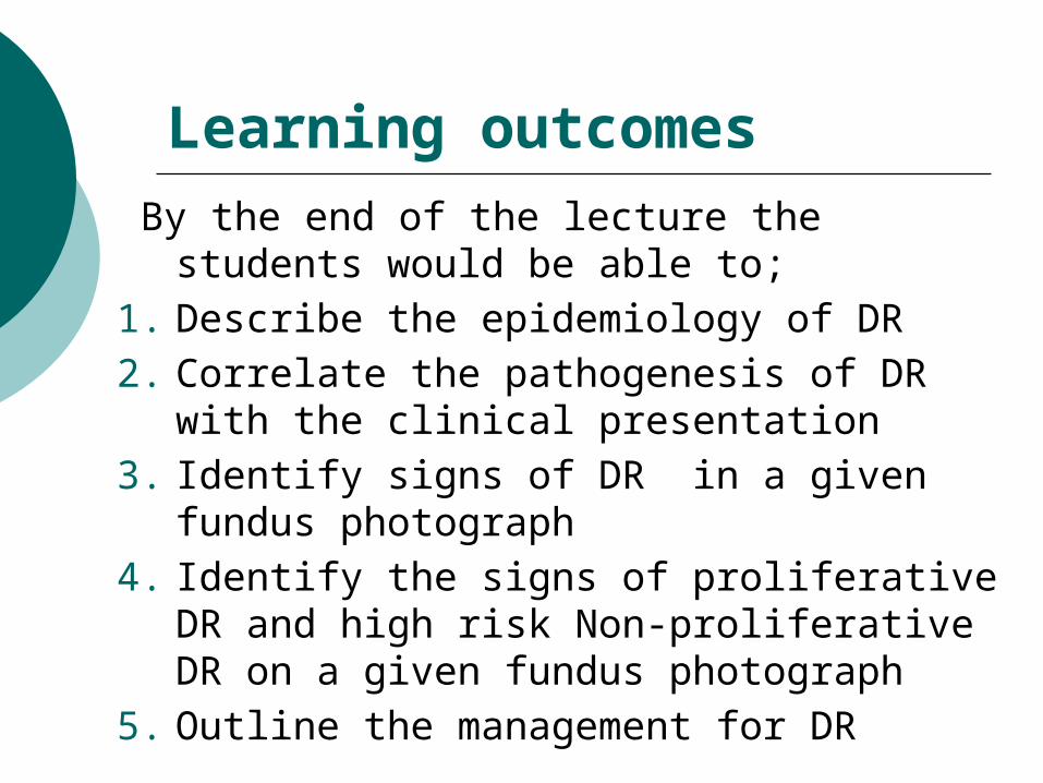
Epidemiology of DM and DR
1. We are having a “global epidemic of DM”.
2. The prevalence of DM is estimated to rise from 2.8% (2000) to 4.4% (2030)
3. Most of this increase will occur as a result of a 150% rise in developing countries.
4. The total number of people with diabetes is projected to rise from 171 million in 2000 to 366 million in 2030.
5. The prevalence is estimated to be 10% in Pakistan
6. With over 5.2 million people with DM , it is the 6th country with the largest population of people with DM.
7. With growing obesity, sedentary life style and increased aging population, the prevalence is estimated to rise further.
Wild S, Roglic G, Green A, Sicree R, King H. Global Prevalence of Diabetes- Estimates for the year 2000 and projections for 2030. Diabetes Care 27:1047–1053, 2004

Diabetic retinopathy
Is a microvascular complication of DM The prevalence is highest among type 1
DM (40%) Patients with DR are 25% more likely to go
blind than non-diabetics In UK 1000 individuals are registered blind
each year due to diabetic eye disease It is the leading cause of blindness in 20-
64 year age group in USA

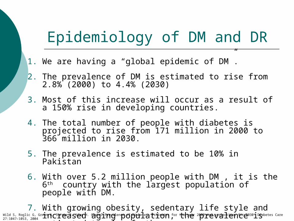
Pathogenesis of Diabetic Retinopathy
DR is a microangiopathy resulting in
Microvascular occlusion Microvascular leakage

Microvascular Occlusion
Factors responsible for occlusion 1. Thickening of capillary basement membrane2. Capillary endothelial cell damage and
proliferation 3. Changes in R.B.Cs 4. Increased stickiness and aggregation of
platelets


Neovascularization
Microvascular occlusion
Retinal capillary non-perfusion
Retinal ischaemia & Hypoxia, ischaemia of the nerve fibres- soft exudates
Arteriovenous shunts - IRMA(intra-retinal microvascualr abnormalities), venous changes, stagnation of blood and more hypoxia
Pathogenesis of Diabetic Retinopathy

Microvascular Leakage
Breakdown of inner blood-retinal barrier Retinal haemorrhages Retinal oedema
Diffuse edema Hard exudates
Microaneurysims
What is inner and outer blood-retinal
barrier?


Classification of diabetic retinopathy
Non-proliferative (NPDR) Proliferative (PDR) Diabetic Maculopathy

Signs of DR
1. Microaneurysms (MA)2. Hard exudates (HE)3. Haemorrrhages (H)4. Retinal oedema- macular oedema(CSME)5. Cotton wool spots (CWS)6. Intra-retinal microvasuclar
abnormalities(IRMA)7. Venous changes8. Fibrovascualr proliferation –
Neovascularization
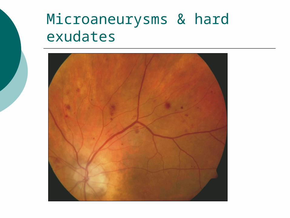
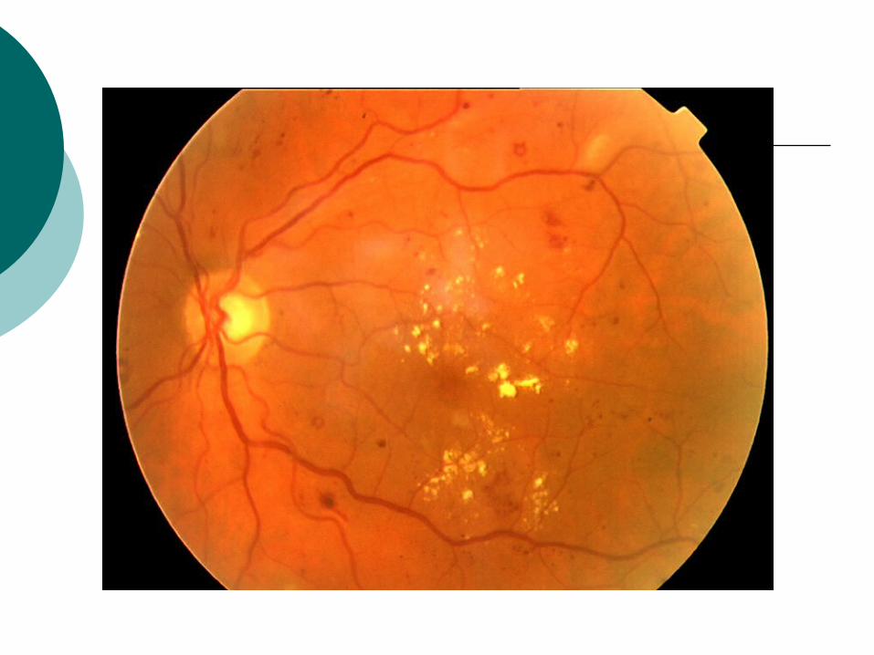
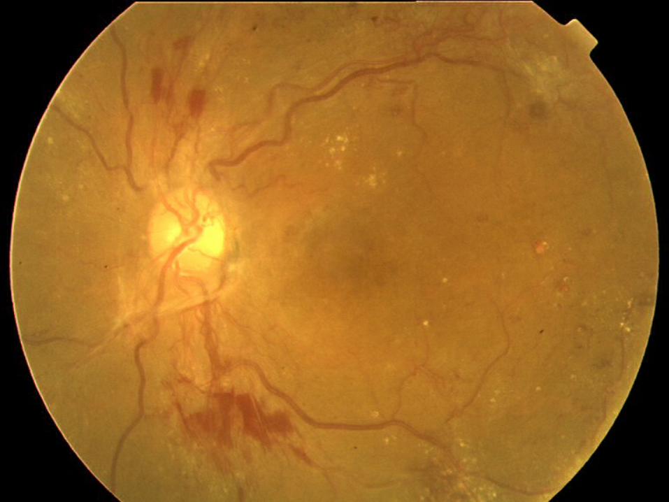
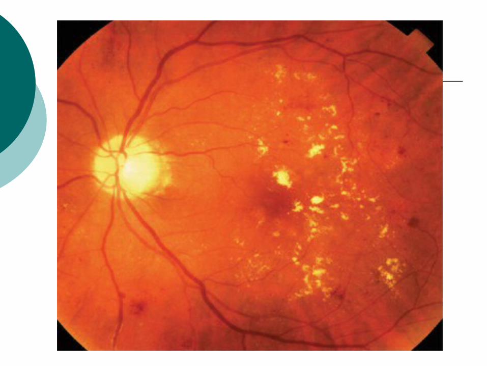
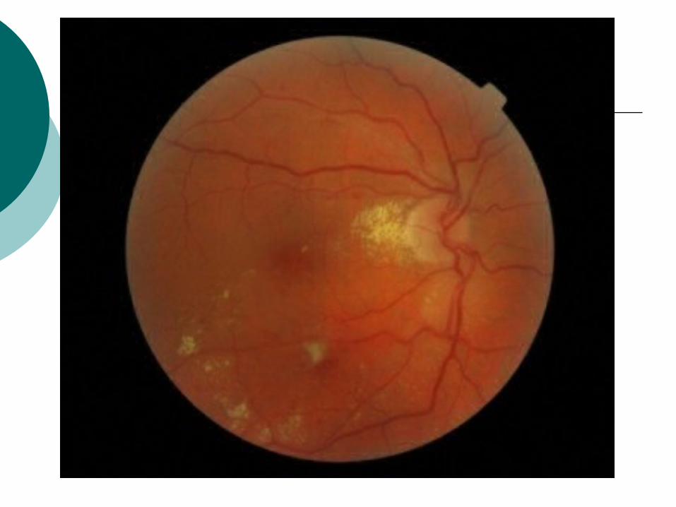
Microaneurysms & hard exudates


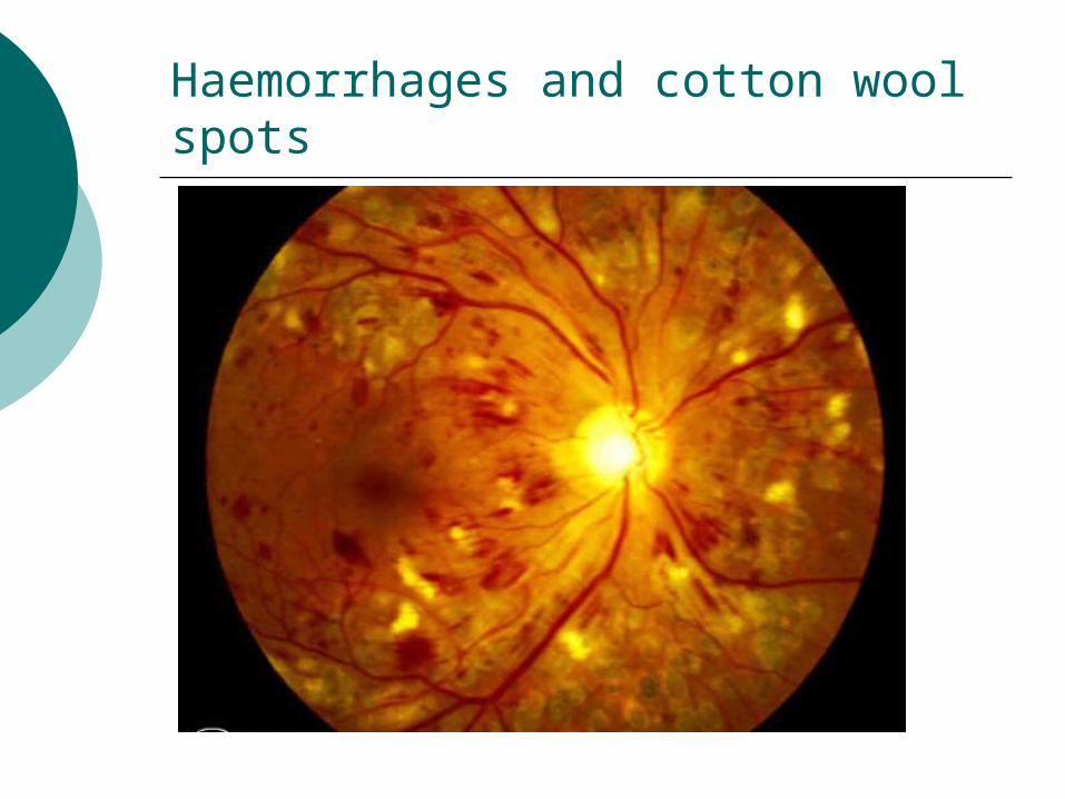
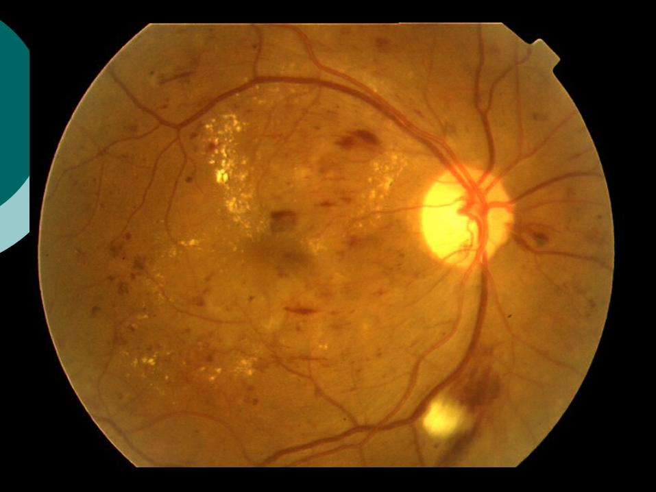
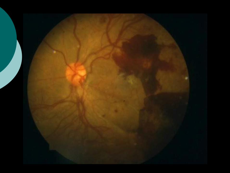
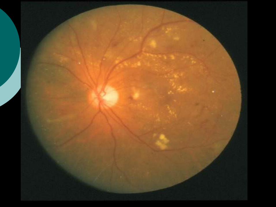
Haemorrhages and cotton wool spots


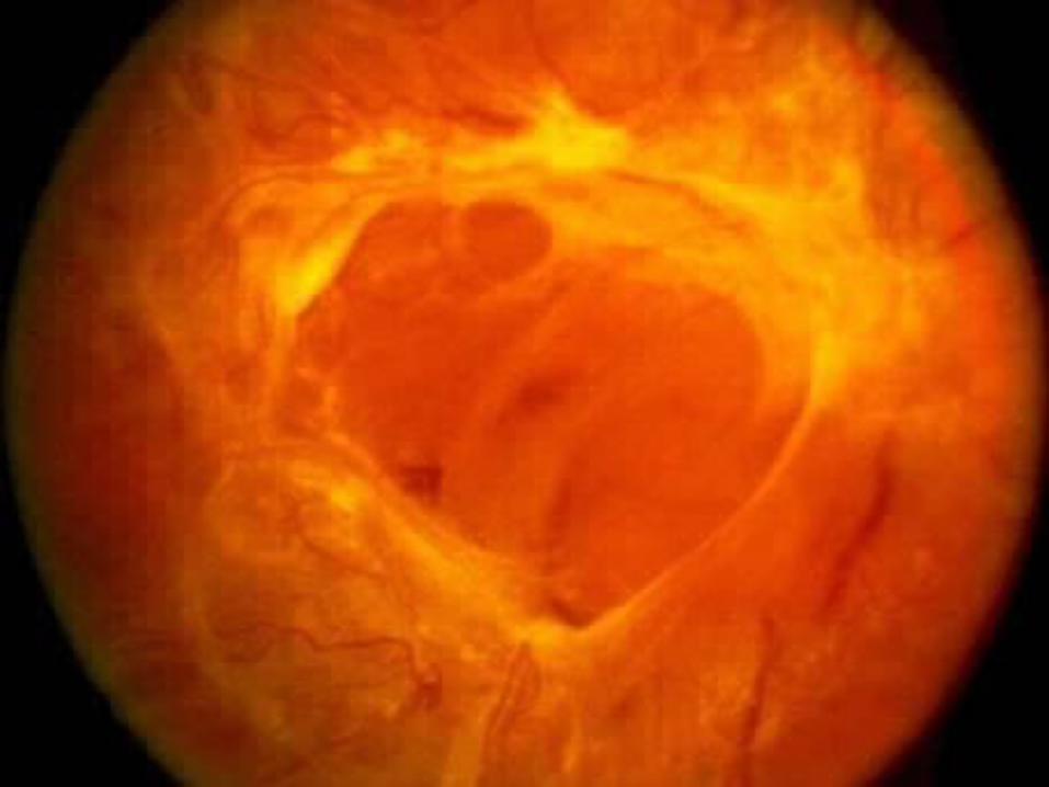
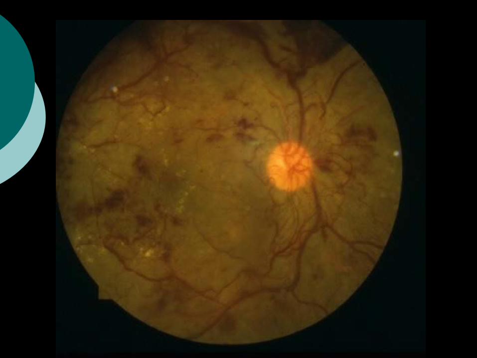
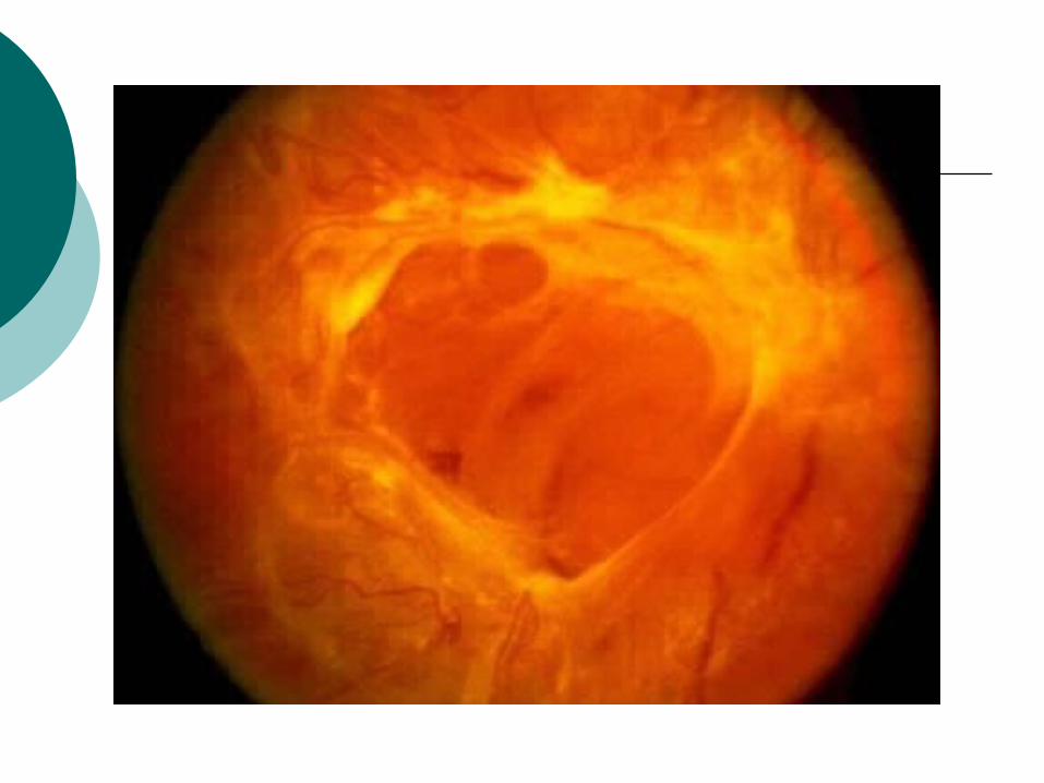
Neovasucalrization and fibrovasucalr proliferation

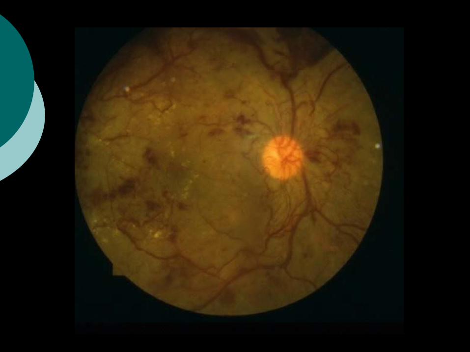
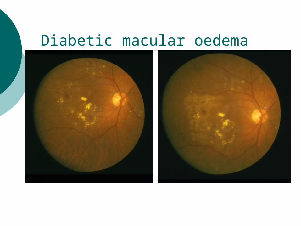
Diabetic macular oedema


Clinical Presentation
o Blurred visiono Reduced visiono Seeing floaterso Reduced night visiono Sudden vision loss


Stages of DR
NPDR PDR
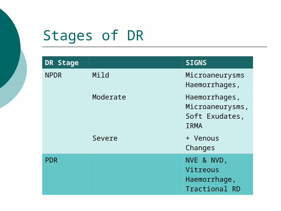
DR Stage SIGNS
NPDR Mild MicroaneurysmsHaemorrhages,
Moderate Haemorrhages, Microaneurysms, Soft Exudates, IRMA
Severe + Venous Changes
PDR NVE & NVD, Vitreous Haemorrhage, Tractional RD
Stages of DR


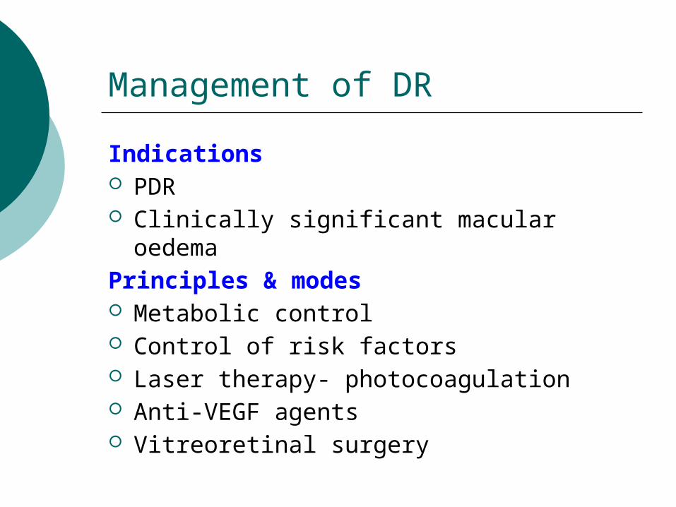
Management of DR
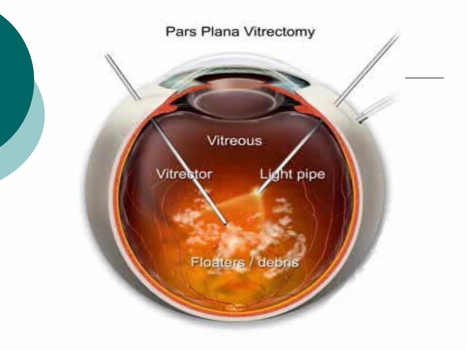
Indications PDR Clinically significant macular oedemaPrinciples & modes Metabolic control Control of risk factors Laser therapy- photocoagulation Anti-VEGF agents Vitreoretinal surgery

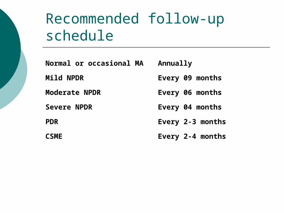
Recommended follow-up schedule
Normal or occasional MA Annually
Mild NPDR Every 09 months
Moderate NPDR Every 06 months
Severe NPDR Every 04 months
PDR Every 2-3 months
CSME Every 2-4 months

Summary
Home workList the risk factors for DRHow does diabetic retinopathy
cause vision [email protected] Last date for submission9th Jan 2014
Related Documents