Diabetic Cardiomyopathy: Evidence, Mechanisms, and Therapeutic Implications ZHI YOU FANG, JOHANNES B. PRINS, AND THOMAS H. MARWICK University of Queensland, Brisbane, Queensland 4102, Australia The presence of a diabetic cardiomyopathy, independent of hypertension and coronary artery disease, is still controver- sial. This systematic review seeks to evaluate the evidence for the existence of this condition, to clarify the possible mech- anisms responsible, and to consider possible therapeutic implications. The existence of a diabetic cardiomyopathy is supported by epidemiological findings showing the association of diabetes with heart failure; clinical studies confirming the association of diabetes with left ventricular dysfunction independent of hypertension, coronary artery disease, and other heart dis- ease; and experimental evidence of myocardial structural and functional changes. The most important mechanisms of dia- betic cardiomyopathy are metabolic disturbances (depletion of glucose transporter 4, increased free fatty acids, carnitine deficiency, changes in calcium homeostasis), myocardial fibrosis (association with increases in angiotensin II, IGF-I, and inflammatory cytokines), small vessel disease (microan- giopathy, impaired coronary flow reserve, and endothelial dysfunction), cardiac autonomic neuropathy (denervation and alterations in myocardial catecholamine levels), and in- sulin resistance (hyperinsulinemia and reduced insulin sensitivity). This review presents evidence that diabetes is associated with a cardiomyopathy, independent of comorbid conditions, and that metabolic disturbances, myocardial fibrosis, small vessel disease, cardiac autonomic neuropathy, and insulin resistance may all contribute to the development of diabetic heart disease. (Endocrine Reviews 25: 543–567, 2004) I. Introduction II. Evidence for a Diabetic Cardiomyopathy A. Diastolic dysfunction in diabetes B. Systolic dysfunction in diabetes C. Structural changes in diabetes III. Mechanisms of Diabetic Cardiomyopathy A. Factors associated with diabetic cardiomyopathy B. Interaction with hypertension and ischemic heart disease C. Stages of diabetic cardiomyopathy IV. Therapeutic Implications of Diabetic Cardiomyopathy V. Summary and Conclusions I. Introduction O VER THE NEXT two decades, the incidence of both type II diabetes and congestive heart failure is antic- ipated to increase to epidemic levels in both the industrial- ized and developing worlds. Patients with diabetes are char- acterized by an increased likelihood of heart failure, largely reflecting the contribution of diabetes to coronary artery disease and its association with hypertension. Over the last three decades, a number of epidemiological, autopsy, ani- mal, and clinical studies have proposed the presence of diabetic heart disease as a distinct clinical entity (1– 4). How- ever, the existence of diabetic heart disease or cardiomyop- athy—referring to myocardial disease in diabetic subjects that cannot be ascribed to hypertension, coronary artery dis- ease, or any other known cardiac disease— has remained controversial. This review seeks to synthesize the existing literature for and against the existence of diabetic cardiomy- opathy, its mechanisms, and its therapeutic implications. This work was performed as a systematic review. We searched MEDLINE (from 1966 to July 2003) to include all animal and human studies of diabetic heart disease not re- lated to hypertension, coronary artery disease, or other known causes. Experimental, pathological, epidemiological, and clinical data were included. All relevant reviews and related references were also examined. Studies were selected on the basis of a combination of the primary terms “diabetic cardiomyopathy” and “diabetic heart disease” with other specific key words related to specific topics. These included left ventricular (LV) dysfunction (e.g., “diastolic dysfunc- tion” and “systolic dysfunction”), structural changes inde- pendently caused by diabetes (e.g., “structural changes,” “pathological,” “histological,” “morphological,” and “back- scatter”), and the relationship between diabetic cardiomy- opathy and diabetic control (“metabolic control,” “HbA1c,” and “glucose levels”). Young diabetic patients or children included in studies were considered to have no or less pos- sibility of coronary artery disease. Studies about diabetic heart disease possibly caused by hypertension, coronary ar- tery disease, and other known causes were excluded, as were those not written in the English language. There were 737 publications related to the terms diabetic cardiomyopathy or diabetic heart disease; 591 papers, including 105 reviews, were written in English and formed the basis of this review. Abbreviations: A, Peak late filling velocity; ACE, angiotensin- converting enzyme; CAN, cardiac autonomic neuropathy; E, peak ve- locity of early mitral flow; FFA, free fatty acid; GLUT, glucose trans- porter; HbA1C, glycosylated hemoglobin; HED, hydroxyephedrine; LV, left ventricular; MIBG, metaiodobenzylguanidine; OLETF, Otsuka Long-Evans Tokushima fatty; PET, positron emission tomography; SERCA, sarcoplasmic reticulum calcium. Endocrine Reviews is published bimonthly by The Endocrine Society (http://www.endo-society.org), the foremost professional society serv- ing the endocrine community. 0163-769X/04/$20.00/0 Endocrine Reviews 25(4):543–567 Printed in U.S.A. Copyright © 2004 by The Endocrine Society doi: 10.1210/er.2003-0012 543 Downloaded from https://academic.oup.com/edrv/article/25/4/543/2355226 by guest on 22 July 2022

Welcome message from author
This document is posted to help you gain knowledge. Please leave a comment to let me know what you think about it! Share it to your friends and learn new things together.
Transcript
Diabetic Cardiomyopathy: Evidence, Mechanisms, andTherapeutic Implications
ZHI YOU FANG, JOHANNES B. PRINS, AND THOMAS H. MARWICK
University of Queensland, Brisbane, Queensland 4102, Australia
The presence of a diabetic cardiomyopathy, independent ofhypertension and coronary artery disease, is still controver-sial. This systematic review seeks to evaluate the evidence forthe existence of this condition, to clarify the possible mech-anisms responsible, and to consider possible therapeuticimplications.
The existence of a diabetic cardiomyopathy is supported byepidemiological findings showing the association of diabeteswith heart failure; clinical studies confirming the associationof diabetes with left ventricular dysfunction independent ofhypertension, coronary artery disease, and other heart dis-ease; and experimental evidence of myocardial structural andfunctional changes. The most important mechanisms of dia-betic cardiomyopathy are metabolic disturbances (depletionof glucose transporter 4, increased free fatty acids, carnitine
deficiency, changes in calcium homeostasis), myocardialfibrosis (association with increases in angiotensin II, IGF-I,and inflammatory cytokines), small vessel disease (microan-giopathy, impaired coronary flow reserve, and endothelialdysfunction), cardiac autonomic neuropathy (denervationand alterations in myocardial catecholamine levels), and in-sulin resistance (hyperinsulinemia and reduced insulinsensitivity).
This review presents evidence that diabetes is associatedwith a cardiomyopathy, independent of comorbid conditions,and that metabolic disturbances, myocardial fibrosis, smallvessel disease, cardiac autonomic neuropathy, and insulinresistance may all contribute to the development of diabeticheart disease. (Endocrine Reviews 25: 543–567, 2004)
I. IntroductionII. Evidence for a Diabetic Cardiomyopathy
A. Diastolic dysfunction in diabetesB. Systolic dysfunction in diabetesC. Structural changes in diabetes
III. Mechanisms of Diabetic CardiomyopathyA. Factors associated with diabetic cardiomyopathyB. Interaction with hypertension and ischemic heart
diseaseC. Stages of diabetic cardiomyopathy
IV. Therapeutic Implications of Diabetic CardiomyopathyV. Summary and Conclusions
I. Introduction
OVER THE NEXT two decades, the incidence of bothtype II diabetes and congestive heart failure is antic-
ipated to increase to epidemic levels in both the industrial-ized and developing worlds. Patients with diabetes are char-acterized by an increased likelihood of heart failure, largelyreflecting the contribution of diabetes to coronary arterydisease and its association with hypertension. Over the lastthree decades, a number of epidemiological, autopsy, ani-mal, and clinical studies have proposed the presence of
diabetic heart disease as a distinct clinical entity (1–4). How-ever, the existence of diabetic heart disease or cardiomyop-athy—referring to myocardial disease in diabetic subjectsthat cannot be ascribed to hypertension, coronary artery dis-ease, or any other known cardiac disease—has remainedcontroversial. This review seeks to synthesize the existingliterature for and against the existence of diabetic cardiomy-opathy, its mechanisms, and its therapeutic implications.
This work was performed as a systematic review. Wesearched MEDLINE (from 1966 to July 2003) to include allanimal and human studies of diabetic heart disease not re-lated to hypertension, coronary artery disease, or otherknown causes. Experimental, pathological, epidemiological,and clinical data were included. All relevant reviews andrelated references were also examined. Studies were selectedon the basis of a combination of the primary terms “diabeticcardiomyopathy” and “diabetic heart disease” with otherspecific key words related to specific topics. These includedleft ventricular (LV) dysfunction (e.g., “diastolic dysfunc-tion” and “systolic dysfunction”), structural changes inde-pendently caused by diabetes (e.g., “structural changes,”“pathological,” “histological,” “morphological,” and “back-scatter”), and the relationship between diabetic cardiomy-opathy and diabetic control (“metabolic control,” “HbA1c,”and “glucose levels”). Young diabetic patients or childrenincluded in studies were considered to have no or less pos-sibility of coronary artery disease. Studies about diabeticheart disease possibly caused by hypertension, coronary ar-tery disease, and other known causes were excluded, as werethose not written in the English language. There were 737publications related to the terms diabetic cardiomyopathy ordiabetic heart disease; 591 papers, including 105 reviews,were written in English and formed the basis of this review.
Abbreviations: A, Peak late filling velocity; ACE, angiotensin-converting enzyme; CAN, cardiac autonomic neuropathy; E, peak ve-locity of early mitral flow; FFA, free fatty acid; GLUT, glucose trans-porter; HbA1C, glycosylated hemoglobin; HED, hydroxyephedrine; LV,left ventricular; MIBG, metaiodobenzylguanidine; OLETF, OtsukaLong-Evans Tokushima fatty; PET, positron emission tomography;SERCA, sarcoplasmic reticulum calcium.Endocrine Reviews is published bimonthly by The Endocrine Society(http://www.endo-society.org), the foremost professional society serv-ing the endocrine community.
0163-769X/04/$20.00/0 Endocrine Reviews 25(4):543–567Printed in U.S.A. Copyright © 2004 by The Endocrine Society
doi: 10.1210/er.2003-0012
543
Dow
nloaded from https://academ
ic.oup.com/edrv/article/25/4/543/2355226 by guest on 22 July 2022
II. Evidence for a Diabetic Cardiomyopathy
Accumulating data from experimental, pathological, epi-demiological, and clinical studies have shown that diabetesmellitus results in cardiac functional and structural changes,independent of hypertension, coronary artery disease, or anyother known cardiac disease, which support the existence ofdiabetic cardiomyopathy.
A. Diastolic dysfunction in diabetes
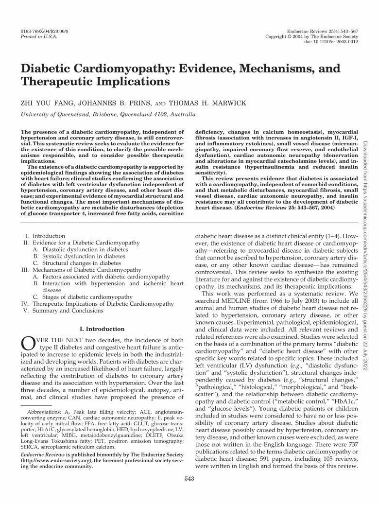
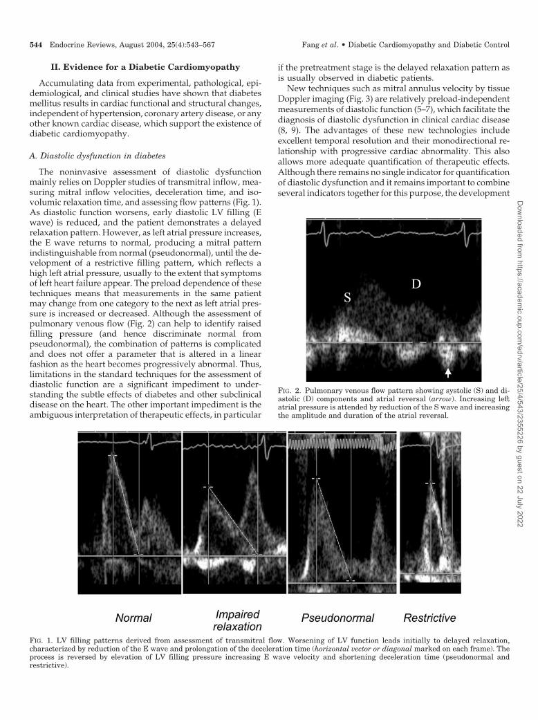
The noninvasive assessment of diastolic dysfunctionmainly relies on Doppler studies of transmitral inflow, mea-suring mitral inflow velocities, deceleration time, and iso-volumic relaxation time, and assessing flow patterns (Fig. 1).As diastolic function worsens, early diastolic LV filling (Ewave) is reduced, and the patient demonstrates a delayedrelaxation pattern. However, as left atrial pressure increases,the E wave returns to normal, producing a mitral patternindistinguishable from normal (pseudonormal), until the de-velopment of a restrictive filling pattern, which reflects ahigh left atrial pressure, usually to the extent that symptomsof left heart failure appear. The preload dependence of thesetechniques means that measurements in the same patientmay change from one category to the next as left atrial pres-sure is increased or decreased. Although the assessment ofpulmonary venous flow (Fig. 2) can help to identify raisedfilling pressure (and hence discriminate normal frompseudonormal), the combination of patterns is complicatedand does not offer a parameter that is altered in a linearfashion as the heart becomes progressively abnormal. Thus,limitations in the standard techniques for the assessment ofdiastolic function are a significant impediment to under-standing the subtle effects of diabetes and other subclinicaldisease on the heart. The other important impediment is theambiguous interpretation of therapeutic effects, in particular
if the pretreatment stage is the delayed relaxation pattern asis usually observed in diabetic patients.
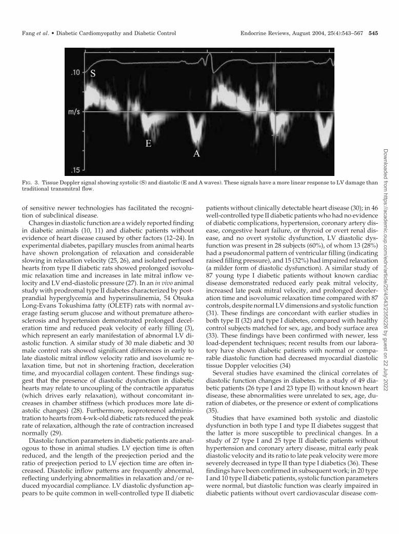
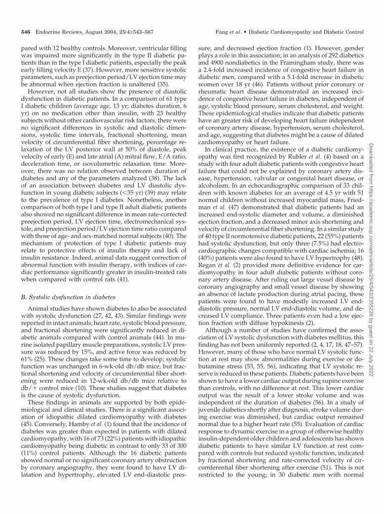
New techniques such as mitral annulus velocity by tissueDoppler imaging (Fig. 3) are relatively preload-independentmeasurements of diastolic function (5–7), which facilitate thediagnosis of diastolic dysfunction in clinical cardiac disease(8, 9). The advantages of these new technologies includeexcellent temporal resolution and their monodirectional re-lationship with progressive cardiac abnormality. This alsoallows more adequate quantification of therapeutic effects.Although there remains no single indicator for quantificationof diastolic dysfunction and it remains important to combineseveral indicators together for this purpose, the development
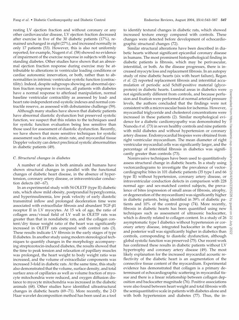
FIG. 2. Pulmonary venous flow pattern showing systolic (S) and di-astolic (D) components and atrial reversal (arrow). Increasing leftatrial pressure is attended by reduction of the S wave and increasingthe amplitude and duration of the atrial reversal.
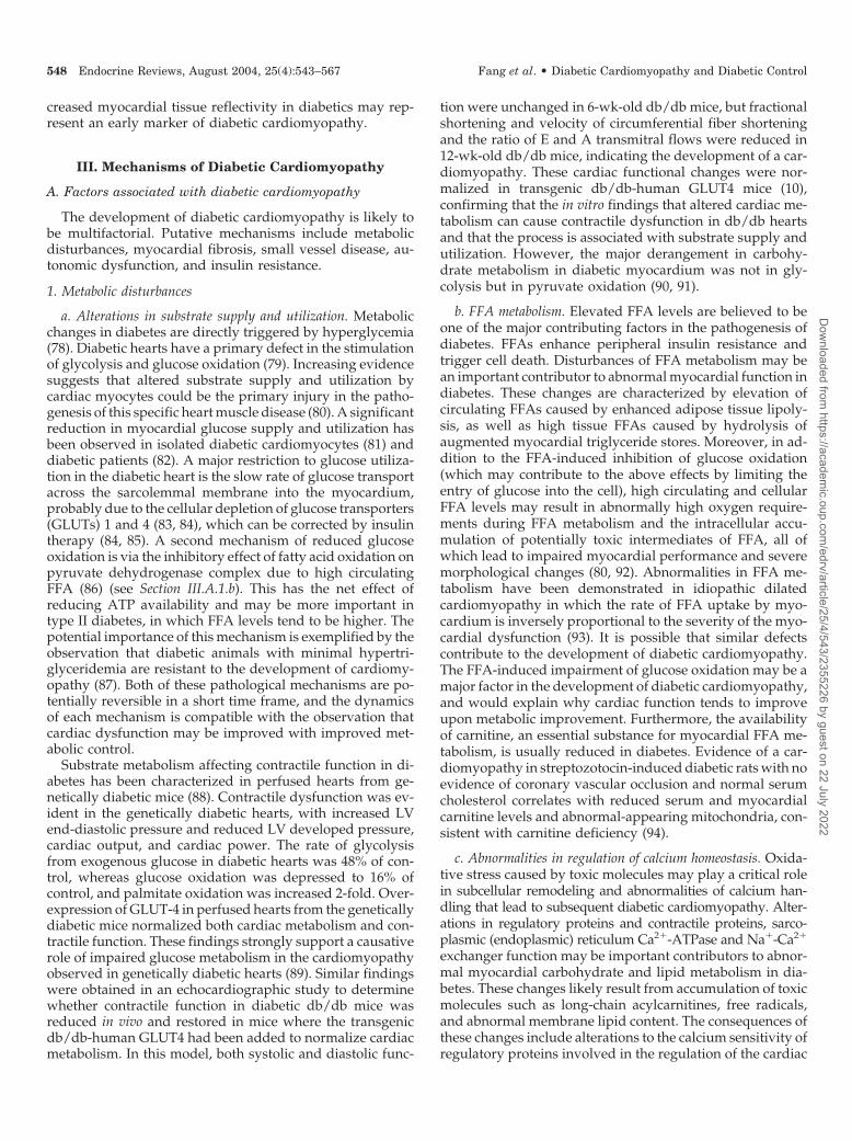
FIG. 1. LV filling patterns derived from assessment of transmitral flow. Worsening of LV function leads initially to delayed relaxation,characterized by reduction of the E wave and prolongation of the deceleration time (horizontal vector or diagonal marked on each frame). Theprocess is reversed by elevation of LV filling pressure increasing E wave velocity and shortening deceleration time (pseudonormal andrestrictive).
544 Endocrine Reviews, August 2004, 25(4):543–567 Fang et al. • Diabetic Cardiomyopathy and Diabetic Control
Dow
nloaded from https://academ
ic.oup.com/edrv/article/25/4/543/2355226 by guest on 22 July 2022
of sensitive newer technologies has facilitated the recogni-tion of subclinical disease.
Changes in diastolic function are a widely reported findingin diabetic animals (10, 11) and diabetic patients withoutevidence of heart disease caused by other factors (12–24). Inexperimental diabetes, papillary muscles from animal heartshave shown prolongation of relaxation and considerableslowing in relaxation velocity (25, 26), and isolated perfusedhearts from type II diabetic rats showed prolonged isovolu-mic relaxation time and increases in late mitral inflow ve-locity and LV end-diastolic pressure (27). In an in vivo animalstudy with prodromal type II diabetes characterized by post-prandial hyperglycemia and hyperinsulinemia, 54 OtsukaLong-Evans Tokushima fatty (OLETF) rats with normal av-erage fasting serum glucose and without premature athero-sclerosis and hypertension demonstrated prolonged decel-eration time and reduced peak velocity of early filling (3),which represent an early manifestation of abnormal LV di-astolic function. A similar study of 30 male diabetic and 30male control rats showed significant differences in early tolate diastolic mitral inflow velocity ratio and isovolumic re-laxation time, but not in shortening fraction, decelerationtime, and myocardial collagen content. These findings sug-gest that the presence of diastolic dysfunction in diabetichearts may relate to uncoupling of the contractile apparatus(which drives early relaxation), without concomitant in-creases in chamber stiffness (which produces more late di-astolic changes) (28). Furthermore, isoproterenol adminis-tration to hearts from 4-wk-old diabetic rats reduced the peakrate of relaxation, although the rate of contraction increasednormally (29).
Diastolic function parameters in diabetic patients are anal-ogous to those in animal studies. LV ejection time is oftenreduced, and the length of the preejection period and theratio of preejection period to LV ejection time are often in-creased. Diastolic inflow patterns are frequently abnormal,reflecting underlying abnormalities in relaxation and/or re-duced myocardial compliance. LV diastolic dysfunction ap-pears to be quite common in well-controlled type II diabetic
patients without clinically detectable heart disease (30); in 46well-controlled type II diabetic patients who had no evidenceof diabetic complications, hypertension, coronary artery dis-ease, congestive heart failure, or thyroid or overt renal dis-ease, and no overt systolic dysfunction, LV diastolic dys-function was present in 28 subjects (60%), of whom 13 (28%)had a pseudonormal pattern of ventricular filling (indicatingraised filling pressure), and 15 (32%) had impaired relaxation(a milder form of diastolic dysfunction). A similar study of87 young type I diabetic patients without known cardiacdisease demonstrated reduced early peak mitral velocity,increased late peak mitral velocity, and prolonged deceler-ation time and isovolumic relaxation time compared with 87controls, despite normal LV dimensions and systolic function(31). These findings are concordant with earlier studies inboth type II (32) and type I diabetes, compared with healthycontrol subjects matched for sex, age, and body surface area(33). These findings have been confirmed with newer, lessload-dependent techniques; recent results from our labora-tory have shown diabetic patients with normal or compa-rable diastolic function had decreased myocardial diastolictissue Doppler velocities (34)
Several studies have examined the clinical correlates ofdiastolic function changes in diabetes. In a study of 49 dia-betic patients (26 type I and 23 type II) without known heartdisease, these abnormalities were unrelated to sex, age, du-ration of diabetes, or the presence or extent of complications(35).
Studies that have examined both systolic and diastolicdysfunction in both type I and type II diabetes suggest thatthe latter is more susceptible to preclinical changes. In astudy of 27 type I and 25 type II diabetic patients withouthypertension and coronary artery disease, mitral early peakdiastolic velocity and its ratio to late peak velocity were moreseverely decreased in type II than type I diabetics (36). Thesefindings have been confirmed in subsequent work; in 20 typeI and 10 type II diabetic patients, systolic function parameterswere normal, but diastolic function was clearly impaired indiabetic patients without overt cardiovascular disease com-
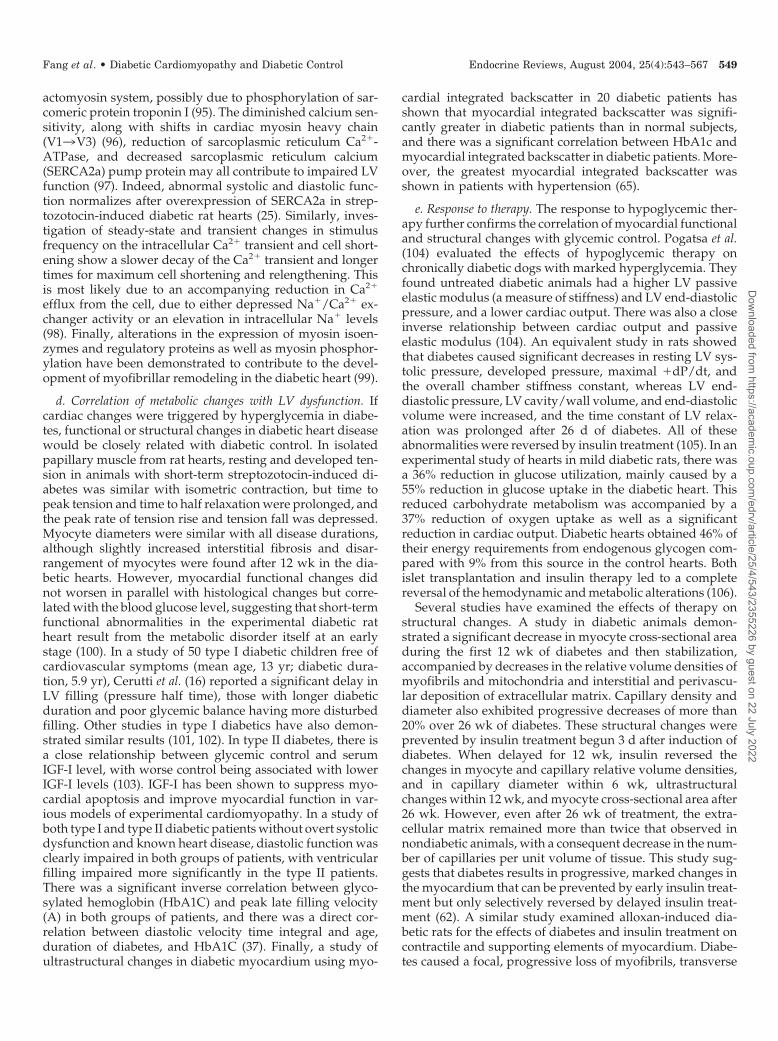
FIG. 3. Tissue Doppler signal showing systolic (S) and diastolic (E and A waves). These signals have a more linear response to LV damage thantraditional transmitral flow.
Fang et al. • Diabetic Cardiomyopathy and Diabetic Control Endocrine Reviews, August 2004, 25(4):543–567 545
Dow
nloaded from https://academ
ic.oup.com/edrv/article/25/4/543/2355226 by guest on 22 July 2022
pared with 12 healthy controls. Moreover, ventricular fillingwas impaired more significantly in the type II diabetic pa-tients than in the type I diabetic patients, especially the peakearly filling velocity E (37). However, more sensitive systolicparameters, such as preejection period/LV ejection time maybe abnormal when ejection fraction is unaltered (35).
However, not all studies show the presence of diastolicdysfunction in diabetic patients. In a comparison of 61 typeI diabetic children (average age, 13 yr; diabetes duration, 6yr) on no medication other than insulin, with 23 healthysubjects without other cardiovascular risk factors, there wereno significant differences in systolic and diastolic dimen-sions, systolic time intervals, fractional shortening, meanvelocity of circumferential fiber shortening, percentage re-laxation of the LV posterior wall at 50% of diastole, peakvelocity of early (E) and late atrial (A) mitral flow, E/A ratio,deceleration time, or isovolumetric relaxation time. More-over, there was no relation observed between duration ofdiabetes and any of the parameters analyzed (38). The lackof an association between diabetes and LV diastolic dys-function in young diabetic subjects (�35 yr) (39) may relateto the prevalence of type I diabetes. Nonetheless, anothercomparison of both type I and type II adult diabetic patientsalso showed no significant difference in mean rate-correctedpreejection period, LV ejection time, electromechanical sys-tole, and preejection period/LV ejection time ratio comparedwith those of age- and sex-matched normal subjects (40). Themechanism of protection of type I diabetic patients mayrelate to protective effects of insulin therapy and lack ofinsulin resistance. Indeed, animal data suggest correction ofabnormal function with insulin therapy, with indices of car-diac performance significantly greater in insulin-treated ratswhen compared with control rats (41).
B. Systolic dysfunction in diabetes
Animal studies have shown diabetes to also be associatedwith systolic dysfunction (27, 42, 43). Similar findings werereported in intact animals; heart rate, systolic blood pressure,and fractional shortening were significantly reduced in di-abetic animals compared with control animals (44). In mu-rine isolated papillary muscle preparations, systolic LV pres-sure was reduced by 15%, and active force was reduced by61% (25). These changes take some time to develop; systolicfunction was unchanged in 6-wk-old db/db mice, but frac-tional shortening and velocity of circumferential fiber short-ening were reduced in 12-wk-old db/db mice relative todb/� control mice (10). These studies suggest that diabetesis the cause of systolic dysfunction.
These findings in animals are supported by both epide-miological and clinical studies. There is a significant associ-ation of idiopathic dilated cardiomyopathy with diabetes(45). Conversely, Hamby et al. (1) found that the incidence ofdiabetes was greater than expected in patients with dilatedcardiomyopathy, with 16 of 73 (22%) patients with idiopathiccardiomyopathy being diabetic in contrast to only 33 of 300(11%) control patients. Although the 16 diabetic patientsshowed normal or no significant coronary artery obstructionby coronary angiography, they were found to have LV di-latation and hypertrophy, elevated LV end-diastolic pres-
sure, and decreased ejection fraction (1). However, genderplays a role in this association; in an analysis of 292 diabeticsand 4900 nondiabetics in the Framingham study, there wasa 2.4-fold increased incidence of congestive heart failure indiabetic men, compared with a 5.1-fold increase in diabeticwomen over 18 yr (46). Patients without prior coronary orrheumatic heart disease demonstrated an increased inci-dence of congestive heart failure in diabetes, independent ofage, systolic blood pressure, serum cholesterol, and weight.These epidemiological studies indicate that diabetic patientshave an greater risk of developing heart failure independentof coronary artery disease, hypertension, serum cholesterol,and age, suggesting that diabetes might be a cause of dilatedcardiomyopathy or heart failure.
In clinical practice, the existence of a diabetic cardiomy-opathy was first recognized by Rubler et al. (4) based on astudy with four adult diabetic patients with congestive heartfailure that could not be explained by coronary artery dis-ease, hypertension, valvular or congenital heart disease, oralcoholism. In an echocardiographic comparison of 33 chil-dren with known diabetes for an average of 4.5 yr with 51normal children without increased myocardial mass, Fried-man et al. (47) demonstrated that diabetic patients had anincreased end-systolic diameter and volume, a diminishedejection fraction, and a decreased minor axis shortening andvelocity of circumferential fiber shortening. In a similar studyof 40 type II normotensive diabetic patients, 22 (55%) patientshad systolic dysfunction, but only three (7.5%) had electro-cardiographic changes compatible with cardiac ischemia; 16(40%) patients were also found to have LV hypertrophy (48).Regan et al. (2) provided more definitive evidence for car-diomyopathy in four adult diabetic patients without coro-nary artery disease. After ruling out large vessel disease bycoronary angiography and small vessel disease by showingan absence of lactate production during atrial pacing, thesepatients were found to have modestly increased LV end-diastolic pressure, normal LV end-diastolic volume, and de-creased LV compliance. Three patients even had a low ejec-tion fraction with diffuse hypokinesis (2).
Although a number of studies have confirmed the asso-ciation of LV systolic dysfunction with diabetes mellitus, thisfinding has not been uniformly reported (2, 4, 17, 18, 47–57).However, many of those who have normal LV systolic func-tion at rest may show abnormalities during exercise or do-butamine stress (53, 55, 56), indicating that LV systolic re-serve is reduced in these patients. Diabetic patients have beenshown to have a lower cardiac output during supine exercisethan controls, with no difference at rest. This lower cardiacoutput was the result of a lower stroke volume and wasindependent of the duration of diabetes (56). In a study ofjuvenile diabetics shortly after diagnosis, stroke volume dur-ing exercise was diminished, but cardiac output remainednormal due to a higher heart rate (55). Evaluation of cardiacresponse to dynamic exercise in a group of otherwise healthyinsulin-dependent older children and adolescents has showndiabetic patients to have similar LV function at rest com-pared with controls but reduced systolic function, indicatedby fractional shortening and rate-corrected velocity of cir-cumferential fiber shortening after exercise (51). This is notrestricted to the young; in 30 diabetic men with normal
546 Endocrine Reviews, August 2004, 25(4):543–567 Fang et al. • Diabetic Cardiomyopathy and Diabetic Control
Dow
nloaded from https://academ
ic.oup.com/edrv/article/25/4/543/2355226 by guest on 22 July 2022
resting LV ejection fraction and without coronary or anyother cardiovascular disease, LV ejection fraction decreasedafter exercise in five of the 30 diabetic patients (17%), re-mained unchanged in eight (27%), and increased normally inonly 17 patients (53). However, this is also not uniformlyreported; for example, Nugent et al. (58) showed no evidenceof impairment of the exercise response in subjects with long-standing diabetes. Other studies have shown that an abnor-mal ejection fraction response during exercise may be at-tributable to alterations in ventricular loading conditions orcardiac autonomic innervation, or both, rather than to ab-normalities in intrinsic ventricular systolic function (contrac-tility). Indeed, despite subgroups showing an abnormal ejec-tion fraction response to exercise, all patients with diabeteshave a normal response to afterload manipulation, normalbaseline ventricular contractility as assessed by load- andheart rate-independent end-systolic indexes and normal con-tractile reserve, as assessed with dobutamine challenge (59).
Although many studies have shown that diabetic patientshave abnormal diastolic dysfunction but preserved systolicfunction, we suspect that this relates to the techniques usedfor systolic function evaluation being less sensitive thanthose used for assessment of diastolic dysfunction. Recently,we have shown that more sensitive techniques for systolicassessment such as strain, strain rate, and myocardial tissueDoppler velocity can detect preclinical systolic abnormalitiesin diabetic patients (49).
C. Structural changes in diabetes
A number of studies in both animals and humans haveshown structural changes in parallel with the functionalchanges of diabetic heart disease, in the absence of hyper-tension, coronary artery disease, or intraventricular conduc-tion defects (60–67).
In an experimental study with 54 OLETF (type II) diabeticrats, which show mild obesity, postprandial hyperglycemia,and hyperinsulinemia, low peak velocity of early diastolictransmitral inflow and prolonged deceleration time wereassociated with extracellular fibrosis and abundant TGF-�1receptor II in LV myocytes. At 15 wk of age, the ratio ofcollagen area/visual field of LV wall in OLETF rats wasgreater than that in nondiabetic rats, and the collagen con-tent/dry tissue weight ratio of the heart was significantlyincreased in OLETF rats compared with control rats (3).These results indicate LV fibrosis in the early stages of typeII diabetes. In another study using modern stereological tech-niques to quantify changes in the morphology accompany-ing streptozotocin-induced diabetes, the results showed thatthe time to peak tension and relaxation of papillary muscleswas prolonged, the heart weight to body weight ratio wasincreased, and the volume of extracellular components wasincreased 3-fold in diabetic rats. At the same time, this studyalso demonstrated that the volume, surface density, and totalsurface area of capillaries as well as volume fraction of myo-cyte mitochondria were reduced, and oxygen diffusion dis-tance to myocyte mitochondria was increased in the diabeticanimals (68). Other studies have identified ultrastructuralchanges in diabetic hearts (69–71). More recently, the 2-DHaar wavelet decomposition method has been used as a tool
to identify textural changes in diabetic rats, which showedincreased texture energy compared with controls. Thesechanges were detected before development of echocardio-graphic structural changes (72).
Similar structural alterations have been described in dia-betic hearts without significant epicardial coronary diseasein humans. The most prominent histopathological finding indiabetic patients is fibrosis, which may be perivascular,interstitial, or both. As the disease progresses, there is in-creased myocyte loss and replacement fibrosis. In an autopsystudy of nine diabetic hearts (six with heart failure), Reganet al. (2) reported replacement fibrosis and interstitial accu-mulation of periodic acid Schiff-positive material (glyco-protein) in diabetic hearts. Luminal areas in diabetics werenot significantly different from controls, and because perfu-sion and fixation were performed at normal arterial pressurelevels, the authors concluded that the findings were notconsistent with a microvascular basis for ischemia. However,myocardial triglyceride and cholesterol concentrations wereincreased in these patients (2). Similar morphological evi-dence for a diabetic cardiomyopathy was demonstrated byNunoda et al. (73) in seven healthy controls and nine patientswith mild diabetes and without hypertension or coronaryartery disease. Endomyocardial biopsies were obtained fromright ventricular myocardium. The mean diameter of rightventricular myocardial cells was significantly larger, and thepercentage of interstitial fibrosis in diabetics was signifi-cantly greater than controls (73).
Noninvasive techniques have been used to quantitativelyassess structural change in diabetic hearts. In a study usingvectorcardiograms to investigate the prevalence of vector-cardiographic bites in 101 diabetic patients (35 type I and 66type II) without hypertension, coronary artery disease, orintraventricular conduction defects in comparison with 228normal age- and sex-matched control subjects, the preva-lence of bites (expression of small areas of fibrosis, atrophy,or degeneration of the myocardium) was significantly higherin diabetic patients, being identified in 39% of diabetic pa-tients and 10% of the control group (74). More recently,fibrosis in diabetic hearts has been quantified using newtechniques such as assessment of ultrasonic backscatter,which is directly related to collagen content. In a study of 26asymptomatic type I diabetics without hypertension or cor-onary artery disease, integrated backscatter in the septumand posterior wall was significantly higher in diabetics thancontrols, corresponding to diastolic dysfunction, althoughglobal systolic function was preserved (75). Our recent workhas confirmed these results in diabetic patients without LVhypertrophy and coronary artery disease (49). The mostlikely explanation for the increased myocardial acoustic re-flectivity of the diabetic heart is an augmentation of theconnective tissue content of the myocardium. Experimentalevidence has demonstrated that collagen is a primary de-terminant of echocardiographic scattering in myocardial tis-sue and there is a linear relationship between collagen dep-osition and backscatter magnitude (76). Positive associationswere also found between heart weight and total fibrosis withthe semiquantitative scale in patients with diabetes alone andwith both hypertension and diabetes (77). Thus, the in-
Fang et al. • Diabetic Cardiomyopathy and Diabetic Control Endocrine Reviews, August 2004, 25(4):543–567 547
Dow
nloaded from https://academ
ic.oup.com/edrv/article/25/4/543/2355226 by guest on 22 July 2022
creased myocardial tissue reflectivity in diabetics may rep-resent an early marker of diabetic cardiomyopathy.
III. Mechanisms of Diabetic Cardiomyopathy
A. Factors associated with diabetic cardiomyopathy
The development of diabetic cardiomyopathy is likely tobe multifactorial. Putative mechanisms include metabolicdisturbances, myocardial fibrosis, small vessel disease, au-tonomic dysfunction, and insulin resistance.
1. Metabolic disturbances
a. Alterations in substrate supply and utilization. Metabolicchanges in diabetes are directly triggered by hyperglycemia(78). Diabetic hearts have a primary defect in the stimulationof glycolysis and glucose oxidation (79). Increasing evidencesuggests that altered substrate supply and utilization bycardiac myocytes could be the primary injury in the patho-genesis of this specific heart muscle disease (80). A significantreduction in myocardial glucose supply and utilization hasbeen observed in isolated diabetic cardiomyocytes (81) anddiabetic patients (82). A major restriction to glucose utiliza-tion in the diabetic heart is the slow rate of glucose transportacross the sarcolemmal membrane into the myocardium,probably due to the cellular depletion of glucose transporters(GLUTs) 1 and 4 (83, 84), which can be corrected by insulintherapy (84, 85). A second mechanism of reduced glucoseoxidation is via the inhibitory effect of fatty acid oxidation onpyruvate dehydrogenase complex due to high circulatingFFA (86) (see Section III.A.1.b). This has the net effect ofreducing ATP availability and may be more important intype II diabetes, in which FFA levels tend to be higher. Thepotential importance of this mechanism is exemplified by theobservation that diabetic animals with minimal hypertri-glyceridemia are resistant to the development of cardiomy-opathy (87). Both of these pathological mechanisms are po-tentially reversible in a short time frame, and the dynamicsof each mechanism is compatible with the observation thatcardiac dysfunction may be improved with improved met-abolic control.
Substrate metabolism affecting contractile function in di-abetes has been characterized in perfused hearts from ge-netically diabetic mice (88). Contractile dysfunction was ev-ident in the genetically diabetic hearts, with increased LVend-diastolic pressure and reduced LV developed pressure,cardiac output, and cardiac power. The rate of glycolysisfrom exogenous glucose in diabetic hearts was 48% of con-trol, whereas glucose oxidation was depressed to 16% ofcontrol, and palmitate oxidation was increased 2-fold. Over-expression of GLUT-4 in perfused hearts from the geneticallydiabetic mice normalized both cardiac metabolism and con-tractile function. These findings strongly support a causativerole of impaired glucose metabolism in the cardiomyopathyobserved in genetically diabetic hearts (89). Similar findingswere obtained in an echocardiographic study to determinewhether contractile function in diabetic db/db mice wasreduced in vivo and restored in mice where the transgenicdb/db-human GLUT4 had been added to normalize cardiacmetabolism. In this model, both systolic and diastolic func-
tion were unchanged in 6-wk-old db/db mice, but fractionalshortening and velocity of circumferential fiber shorteningand the ratio of E and A transmitral flows were reduced in12-wk-old db/db mice, indicating the development of a car-diomyopathy. These cardiac functional changes were nor-malized in transgenic db/db-human GLUT4 mice (10),confirming that the in vitro findings that altered cardiac me-tabolism can cause contractile dysfunction in db/db heartsand that the process is associated with substrate supply andutilization. However, the major derangement in carbohy-drate metabolism in diabetic myocardium was not in gly-colysis but in pyruvate oxidation (90, 91).
b. FFA metabolism. Elevated FFA levels are believed to beone of the major contributing factors in the pathogenesis ofdiabetes. FFAs enhance peripheral insulin resistance andtrigger cell death. Disturbances of FFA metabolism may bean important contributor to abnormal myocardial function indiabetes. These changes are characterized by elevation ofcirculating FFAs caused by enhanced adipose tissue lipoly-sis, as well as high tissue FFAs caused by hydrolysis ofaugmented myocardial triglyceride stores. Moreover, in ad-dition to the FFA-induced inhibition of glucose oxidation(which may contribute to the above effects by limiting theentry of glucose into the cell), high circulating and cellularFFA levels may result in abnormally high oxygen require-ments during FFA metabolism and the intracellular accu-mulation of potentially toxic intermediates of FFA, all ofwhich lead to impaired myocardial performance and severemorphological changes (80, 92). Abnormalities in FFA me-tabolism have been demonstrated in idiopathic dilatedcardiomyopathy in which the rate of FFA uptake by myo-cardium is inversely proportional to the severity of the myo-cardial dysfunction (93). It is possible that similar defectscontribute to the development of diabetic cardiomyopathy.The FFA-induced impairment of glucose oxidation may be amajor factor in the development of diabetic cardiomyopathy,and would explain why cardiac function tends to improveupon metabolic improvement. Furthermore, the availabilityof carnitine, an essential substance for myocardial FFA me-tabolism, is usually reduced in diabetes. Evidence of a car-diomyopathy in streptozotocin-induced diabetic rats with noevidence of coronary vascular occlusion and normal serumcholesterol correlates with reduced serum and myocardialcarnitine levels and abnormal-appearing mitochondria, con-sistent with carnitine deficiency (94).
c. Abnormalities in regulation of calcium homeostasis. Oxida-tive stress caused by toxic molecules may play a critical rolein subcellular remodeling and abnormalities of calcium han-dling that lead to subsequent diabetic cardiomyopathy. Alter-ations in regulatory proteins and contractile proteins, sarco-plasmic (endoplasmic) reticulum Ca2�-ATPase and Na�-Ca2�
exchanger function may be important contributors to abnor-mal myocardial carbohydrate and lipid metabolism in dia-betes. These changes likely result from accumulation of toxicmolecules such as long-chain acylcarnitines, free radicals,and abnormal membrane lipid content. The consequences ofthese changes include alterations to the calcium sensitivity ofregulatory proteins involved in the regulation of the cardiac
548 Endocrine Reviews, August 2004, 25(4):543–567 Fang et al. • Diabetic Cardiomyopathy and Diabetic Control
Dow
nloaded from https://academ
ic.oup.com/edrv/article/25/4/543/2355226 by guest on 22 July 2022
actomyosin system, possibly due to phosphorylation of sar-comeric protein troponin I (95). The diminished calcium sen-sitivity, along with shifts in cardiac myosin heavy chain(V13V3) (96), reduction of sarcoplasmic reticulum Ca2�-ATPase, and decreased sarcoplasmic reticulum calcium(SERCA2a) pump protein may all contribute to impaired LVfunction (97). Indeed, abnormal systolic and diastolic func-tion normalizes after overexpression of SERCA2a in strep-tozotocin-induced diabetic rat hearts (25). Similarly, inves-tigation of steady-state and transient changes in stimulusfrequency on the intracellular Ca2� transient and cell short-ening show a slower decay of the Ca2� transient and longertimes for maximum cell shortening and relengthening. Thisis most likely due to an accompanying reduction in Ca2�
efflux from the cell, due to either depressed Na�/Ca2� ex-changer activity or an elevation in intracellular Na� levels(98). Finally, alterations in the expression of myosin isoen-zymes and regulatory proteins as well as myosin phosphor-ylation have been demonstrated to contribute to the devel-opment of myofibrillar remodeling in the diabetic heart (99).
d. Correlation of metabolic changes with LV dysfunction. Ifcardiac changes were triggered by hyperglycemia in diabe-tes, functional or structural changes in diabetic heart diseasewould be closely related with diabetic control. In isolatedpapillary muscle from rat hearts, resting and developed ten-sion in animals with short-term streptozotocin-induced di-abetes was similar with isometric contraction, but time topeak tension and time to half relaxation were prolonged, andthe peak rate of tension rise and tension fall was depressed.Myocyte diameters were similar with all disease durations,although slightly increased interstitial fibrosis and disar-rangement of myocytes were found after 12 wk in the dia-betic hearts. However, myocardial functional changes didnot worsen in parallel with histological changes but corre-lated with the blood glucose level, suggesting that short-termfunctional abnormalities in the experimental diabetic ratheart result from the metabolic disorder itself at an earlystage (100). In a study of 50 type I diabetic children free ofcardiovascular symptoms (mean age, 13 yr; diabetic dura-tion, 5.9 yr), Cerutti et al. (16) reported a significant delay inLV filling (pressure half time), those with longer diabeticduration and poor glycemic balance having more disturbedfilling. Other studies in type I diabetics have also demon-strated similar results (101, 102). In type II diabetes, there isa close relationship between glycemic control and serumIGF-I level, with worse control being associated with lowerIGF-I levels (103). IGF-I has been shown to suppress myo-cardial apoptosis and improve myocardial function in var-ious models of experimental cardiomyopathy. In a study ofboth type I and type II diabetic patients without overt systolicdysfunction and known heart disease, diastolic function wasclearly impaired in both groups of patients, with ventricularfilling impaired more significantly in the type II patients.There was a significant inverse correlation between glyco-sylated hemoglobin (HbA1C) and peak late filling velocity(A) in both groups of patients, and there was a direct cor-relation between diastolic velocity time integral and age,duration of diabetes, and HbA1C (37). Finally, a study ofultrastructural changes in diabetic myocardium using myo-
cardial integrated backscatter in 20 diabetic patients hasshown that myocardial integrated backscatter was signifi-cantly greater in diabetic patients than in normal subjects,and there was a significant correlation between HbA1c andmyocardial integrated backscatter in diabetic patients. More-over, the greatest myocardial integrated backscatter wasshown in patients with hypertension (65).
e. Response to therapy. The response to hypoglycemic ther-apy further confirms the correlation of myocardial functionaland structural changes with glycemic control. Pogatsa et al.(104) evaluated the effects of hypoglycemic therapy onchronically diabetic dogs with marked hyperglycemia. Theyfound untreated diabetic animals had a higher LV passiveelastic modulus (a measure of stiffness) and LV end-diastolicpressure, and a lower cardiac output. There was also a closeinverse relationship between cardiac output and passiveelastic modulus (104). An equivalent study in rats showedthat diabetes caused significant decreases in resting LV sys-tolic pressure, developed pressure, maximal �dP/dt, andthe overall chamber stiffness constant, whereas LV end-diastolic pressure, LV cavity/wall volume, and end-diastolicvolume were increased, and the time constant of LV relax-ation was prolonged after 26 d of diabetes. All of theseabnormalities were reversed by insulin treatment (105). In anexperimental study of hearts in mild diabetic rats, there wasa 36% reduction in glucose utilization, mainly caused by a55% reduction in glucose uptake in the diabetic heart. Thisreduced carbohydrate metabolism was accompanied by a37% reduction of oxygen uptake as well as a significantreduction in cardiac output. Diabetic hearts obtained 46% oftheir energy requirements from endogenous glycogen com-pared with 9% from this source in the control hearts. Bothislet transplantation and insulin therapy led to a completereversal of the hemodynamic and metabolic alterations (106).
Several studies have examined the effects of therapy onstructural changes. A study in diabetic animals demon-strated a significant decrease in myocyte cross-sectional areaduring the first 12 wk of diabetes and then stabilization,accompanied by decreases in the relative volume densities ofmyofibrils and mitochondria and interstitial and perivascu-lar deposition of extracellular matrix. Capillary density anddiameter also exhibited progressive decreases of more than20% over 26 wk of diabetes. These structural changes wereprevented by insulin treatment begun 3 d after induction ofdiabetes. When delayed for 12 wk, insulin reversed thechanges in myocyte and capillary relative volume densities,and in capillary diameter within 6 wk, ultrastructuralchanges within 12 wk, and myocyte cross-sectional area after26 wk. However, even after 26 wk of treatment, the extra-cellular matrix remained more than twice that observed innondiabetic animals, with a consequent decrease in the num-ber of capillaries per unit volume of tissue. This study sug-gests that diabetes results in progressive, marked changes inthe myocardium that can be prevented by early insulin treat-ment but only selectively reversed by delayed insulin treat-ment (62). A similar study examined alloxan-induced dia-betic rats for the effects of diabetes and insulin treatment oncontractile and supporting elements of myocardium. Diabe-tes caused a focal, progressive loss of myofibrils, transverse
Fang et al. • Diabetic Cardiomyopathy and Diabetic Control Endocrine Reviews, August 2004, 25(4):543–567 549
Dow
nloaded from https://academ
ic.oup.com/edrv/article/25/4/543/2355226 by guest on 22 July 2022
tubules, and sarcoplasmic reticulum and separation of thefasciae adherens at the intercalated disks. These changeswere accompanied by interstitial and perivascular deposi-tion of connective tissue, thickening of the endothelial cy-toplasm with pinocytotic hyperactivity, and characteristicbasal laminar changes. Most, but not all, of these changeswere reversed after 6–12 wk of insulin treatment (64). In-terestingly, experimental studies have also demonstratednormalization of the collagen alteration by endurance train-ing, begun relatively early in the disease process (107). Theimprovement may be related to improved diabetic controldue to increased insulin sensitivity caused by exercisetraining.
In diabetic patients without known cardiac disease, ab-normalities of LV function primarily reflect a diastolic ab-normality. This diastolic abnormality appears related to in-terstitial collagen deposition, although LV hypertrophy mayeventually appear in the absence of hypertension. Revers-ibility of this process can be achieved with chronic insulintherapy. Sykes et al. (108) found that the preejection periodwas shortened in a group of 19 diabetics before treatmentwith either diet or oral hypoglycemic agents and LV ejectiontime was shortened. These abnormalities were reversed after3 months of therapy (108). Shapiro et al. (109) studied 69 typeII diabetics before and after hypoglycemic therapy usingboth systolic time intervals and M-mode echocardiography.The preejection period/LV ejection time ratio was increasedin the untreated group, and this ratio correlated well withblood glucose concentration. Treatment resulted in a fall inpreejection period/LV ejection time ratio in 54 patients witha modestly increased initial ratio but no response in theremaining 15 patients with a markedly elevated initial ratioafter 4 months of therapy. Isovolumetric relaxation was pro-longed in diabetics, but it was not affected by hypoglycemictherapy (109). In another study of 15 type I diabetic subjectswithout known heart disease and diabetic complications,systolic time intervals were evaluated at rest and after dy-namic exercise during poor and good metabolic control, ob-tained by means of insulin therapy. Resting systolic timeintervals were normal during poor and good metaboliccontrol. After exercise, a greater increase in preejectionperiod/LV ejection time ratio as a result of an increasedpreejection period was found during poor control, and asmaller increase in preejection period/LV ejection time ratiooccurred during good metabolic control, suggesting thatgood diabetic control is associated with the improvement inLV function (101).
However, a discordant relationship between diabetic con-trol and functional changes has also been found in somestudies. In a study of type II diabetes without evidence ofhypertension, coronary artery disease, and other known car-diac diseases, the results showed there was no correlationbetween LV diastolic dysfunction and indices of metaboliccontrol in those with normal systolic function and abnormaldiastolic function (30). Friedman et al. (47) demonstrated anincreased LV end-systolic diameter and volume, a dimin-ished ejection fraction, minor axis shortening and velocity ofcircumferential fiber shortening in type I diabetic children.How ever, no relationship between ventricular function andeither the duration or the severity of diabetes was observed
(47). The impact of diabetic treatment is also associated withdiscordant results. Regan et al. (110) demonstrated a lowerstroke volume in animal models of diabetes mellitus in dogsdespite normal LV end-diastolic pressure, normal coronaryarteries, and coronary blood flow. Chamber stiffness wasincreased in diabetic dogs compared with control dogs, pre-sumably related to the deposition of interstitial glycoproteinand collagen. However, these changes could not be reversedwith correction of hyperglycemia or prevented by insulin(110).
2. Myocardial fibrosis. Myocardial fibrosis and myocyte hy-pertrophy are the most frequently proposed mechanisms toexplain cardiac changes in diabetic cardiomyopathy. Studiesin dogs, monkeys, and rabbits have shown that experimen-tally induced diabetes causes defects in cellular calciumtransport (111), defects in myocardial contractile proteins(112), and an increase in collagen formation (110), whichresult in anatomic and physiological changes in themyocardium.
a. Myocyte cell death. Myocyte cell death may be caused byapoptosis or necrosis or both. Apoptosis is an active genet-ically controlled process that removes unwanted or damagedcells, whereas myocyte necrosis refers to myocyte destruc-tion due to biochemical damage. Apoptosis can be evaluatedby the identification of double-strand DNA cleavage withsingle base or longer 3� overhangs. In contrast, myocytenecrosis can be assessed by detection of DNA damage withblunt end fragments (113).
Both apoptosis and necrosis have been identified in dia-betic heart disease. In a study of diabetic and diabetic-hypertensive hearts, myocyte necrosis was 1.4-fold moreprevalent in patients with diabetes and hypertension thanwith diabetes alone, whereas myocyte apoptosis was notaffected by the addition of hypertension (114). These twodistinct forms of cell death also cause different consequences.Apoptosis does not cause scar formation or significant in-terstitial collagen accumulation (115), with nuclear fragmen-tation and cell shrinkage being replaced by the surroundingcells (116, 117). Conversely, myocyte necrosis results in wid-ening of the extracellular compartments among myocytesand increased deposition of collagen in a diffuse or scatteredmanner (118, 119), resulting from both replacement fibrosisdue to myocyte necrosis and connective tissue cell prolifer-ation (120).
b. Process of myocardial fibrosis. Collagen accumulation inthe diabetic myocardium may be due in part to impairedcollagen degradation resulting from glycosylation of the ly-sine residues on collagen. Hyperglycemia also results in theproduction of reactive oxygen and nitrogen species, whichincreases oxidative stress and causes abnormal gene expres-sion, alters signal transduction, and activates the pathwaysleading to programmed myocardial cell death or apoptosis.This process is associated with the glycosylation of p53,which results in an increment in angiotensin II synthesis; thisin turn leads to p53 phosphorylation, increased Bax expres-sion, and also to myocyte apoptosis. These changes parallelthe concentrations of glucose in the medium and the durationof the culture. Inhibition of the p53 glycosylation prevents
550 Endocrine Reviews, August 2004, 25(4):543–567 Fang et al. • Diabetic Cardiomyopathy and Diabetic Control
Dow
nloaded from https://academ
ic.oup.com/edrv/article/25/4/543/2355226 by guest on 22 July 2022
the initial synthesis of angiotensin II and consequent p53activation and apoptosis (121). Evidence in vivo has shownthat hyperglycemia directly induces apoptotic cell death andmyocyte necrosis in the myocardium, triggered by reactiveoxygen species derived from high levels of glucose (122).Interestingly, cardiomyocytes incubated for 3 d with me-dium containing 25 mm glucose showed less hypoxia-induced apoptosis and necrosis than cells exposed to me-dium containing 5 mm glucose, suggesting that glucosetreatment renders the cardiomyocyte resistant to hypoxia-induced apoptosis and necrosis (123).
The increased angiotensin II and angiotensin receptor lev-els have been shown in an in vivo study in streptozotocin-induced diabetic rats, in which changes in angiotensin IIquantity, the fraction of angiotensin II positive cells, and thenumber of angiotensin II receptor sites per myocyte paral-leled the change in myocyte death (124). The change in an-giotensin II and angiotensin II receptors in diabetic heartsappears to be local and independent of the circulating renin-angiotensin system (125, 126). Up-regulation of the localrenin-angiotensin system in diabetes may enhance oxidativedamage, activating cardiac cell apoptosis and necrosis (114).Thus, either increased angiotensin II or increased angiotensinII receptor density enhances the effect of angiotensin II.Whichever the mechanism, angiotensin II has dose-depen-dent effects on collagen secretion and production in rat adultcardiac fibroblasts (128). On the other hand, alterations ofendothelin-1 and its receptors were also associated with in-creased focal fibrous scarring with apoptotic cardiomyocytesin diabetic rats, and the fibrotic process was completely pre-vented by treatment with bosentan, suggesting that hyper-glycemia-induced up-regulation of the endothelin system inthe diabetic heart may also play an important role in myo-cardial fibrosis (129).
Local angiotensin II effects are modulated by the functionof IGF-I, a key factor for cardiac growth and function. An-giotensin II and IGF-I are generated by cardiomyocytes andexert pleiotropic effects in an autocrine/paracrine fashion.Both angiotensin II and apoptosis are reduced by IGF-I. IGF-Iis decreased in diabetes, and exogenous IGF-I treatment hasbeen shown to ameliorate contractile disturbances in cardi-omyocytes from diabetic animals, suggesting that IGF-I alsoplays an important role in myocardial fibrosis and develop-ment of diabetic cardiomyopathy. This was demonstrated instreptozotocin-induced diabetic mice, wherein diabetes pro-gressively depressed ventricular performance but had nohemodynamic effect on those with IGF-I overexpression.Myocyte apoptosis measured at 7 and 30 d after the onset ofdiabetes was 2-fold higher in diabetic mice without than withIGF-I overexpression. Myocyte necrosis was apparent only at30 d and was more severe in diabetic nontransgenic mice,which lost 24% of their ventricular myocytes and showed a28% myocyte hypertrophy, both of which were prevented byIGF-I (130). Therefore, resistance to actions of IGF-I and in-sulin could explain the abnormalities of both diastolic andsystolic function and LV hypertrophy.
The effects of angiotensin II may also be promoted by theproduction and release of TGF-�1 by cardiac fibroblasts (131,132). TGF-�1 plays a critical role in organ morphogenesis,development, growth regulation, cellular differentiation,
gene expression, and tissue remodeling. TGF-�1 induced bymetabolic abnormalities (chronic postprandial hyperglyce-mia, hyperinsulinemia, insulin resistance) has also been im-plicated in the development of diabetic cardiomyopathy. Inthe rat heart, TGF-� increases fibrous tissue formation andup-regulates collagen expression during tissue repair bybinding to the TGF-� type II receptor. TGF-�1 receptor IIexpression has been shown to be significantly increased inthe left ventricle of OLETF (type II diabetes model) rats, andthe ratio of collagen content/dry weight of the left ventriclewas significantly higher in OLETF rats than in control rats at15 wk of age (3). Thus, this cytokine may participate in theonset of cardiac fibrosis by stimulating extracellular matrixsynthesis.
c. Consequences of myocardial fibrosis. Fibrosis is attributedto replacement fibrosis caused by focal myocyte necrosis(133, 134) and increased interstitial fibrosis, in part due to thereaction of connective tissue cells to pathological loads (120).
A biopsy study in patients with diabetes mellitus hasshown that hypertrophy of myocardial cells and interstitialfibrosis of the myocardium are present in mild diabetes mel-litus (73). Indeed, diabetic heart disease may simply reflectincreased interstitial fibrosis in the heart, because collagenaccumulation occurs mainly as a result of an increase in typeIII collagen in the diabetic heart (135). Cell death in thediabetic myocardium is not only necrotic in nature but is alsomediated by apoptosis—thus, interstitial fibrosis may not besevere. In a longitudinal study of cardiac performance instreptozotocin-induced type I diabetic rats for 56 d usingnoninvasive echocardiographic techniques, significant re-ductions in diastolic performance (transmitral flow velocitiesand slopes) and systolic dysfunction (LV fractional shorten-ing, cardiac output) developed in the absence of fibrosis(136), suggesting that abnormal heart function in this modelmay be of metabolic rather than structural origin. This is alsosupported by a similar study to investigate the chronic effectsof streptozotocin-induced diabetes on contraction in rat ven-tricular myocytes, which showed that time to peak contrac-tion was significantly longer at 2 months but appeared tonormalize at 10 months, and the time to half relaxation ofcontraction was not significantly different after 2 months butwas significantly reduced at 10 months. The ultrastructure ofcardiac muscle and sarcomere lengths were not greatly al-tered after streptozotocin treatment, also indicating that mor-phological defects in contractile myofilaments and associ-ated structures do not explain contractile dysfunction seen inthis model (137).
Another question is whether the fibrosis and/or dysfunc-tion in the diabetic heart are a result of small vessel disease.At present, this seems unlikely; several studies have showndecreased LV function without vascular lesions (138). Sim-ilarly, our recent work shows no increment of abnormalfunction (measured by sensitive tissue Doppler indices) afterdobutamine stress (34). The up-regulation of the local renin-angiotensin system suggests that cardiac structural and func-tional changes in diabetes are not a result of change in thecirculating renin-angiotensin system, but are relatively specificto the heart, leading to a specific diabetic cardiomyopathy.
Fang et al. • Diabetic Cardiomyopathy and Diabetic Control Endocrine Reviews, August 2004, 25(4):543–567 551
Dow
nloaded from https://academ
ic.oup.com/edrv/article/25/4/543/2355226 by guest on 22 July 2022
d. Correlation of structural changes to LV dysfunction. Thefunctional abnormality in diabetic myocardium is consid-ered to be associated with myocardial structural changes,and indeed, these structural changes might play a role inprogressive deterioration of cardiac hemodynamics.
Experimental data strongly support the connection be-tween structural changes and heart muscle dysfunction indiabetes. After 2 months of streptozotocin-induced diabetes,in vitro study of myocytes showed a 30% increase in time topeak shortening, which corresponded to a significant reduc-tion in resting sarcomere length and a change in the micro-tubular cytoskeleton (60), suggesting that myocardial struc-tural change may be the basis for cardiac dysfunction.Another study showed that rats with streptozotocin-inducednon-insulin-dependent diabetes had prolonged isovolumicrelaxation time, elevated LV end-diastolic pressure, and in-creased chamber stiffness; these functional changes wereaccompanied with increased LV mass (27). A similar exper-imental study in animals also showed that functionalchanges (e.g., reduced LV compliance) after 1 yr of diabeteswere associated with increased interstitial connective tissue(110). A clear relationship between functional and structuralchanges is indicated by a study showing that diabetic ratsexhibited prolonged deceleration time and low peak velocityof early diastolic transmitral flow, which is associated withextracellular fibrosis in LV myocytes, and higher ratio ofcollagen area/visual field of LV wall and ratio of collagencontent/dry heart weight compared with control rats (3).
In diabetic patients, noninvasive studies revealed abnor-mal systolic and diastolic function present in many diabeticpatients, particularly in the presence of hypertension. Pa-thology studies show that myocardial hypertrophy and fi-brosis are commonly present in these patients. Das et al. (139)have found that there was a correlation between histologicaland clinical features in a study of endomyocardial biopsiesin 16 diabetics, with myocardial changes more pronouncedin the symptomatic group and less so in asymptomatic pa-tients (139), suggesting that myocardial dysfunction in dia-betics might be secondary to accumulation of glycoproteinwithin the interstitium together with mild interstitial fibrosis.Zoneraich (140) also showed increased myocardial fibrosis indiabetics, particularly in those with cardiomegaly, and sug-gested that changes in cardiac interstitial collagen mightincrease myocardial wall stiffness that is usually associatedwith functional changes.
Systolic dysfunction may be more dependent on the de-gree of myocyte loss and myocyte injury. Myocyte cell deathand injury may impair the ability of the myocardium todevelop force, and they account for reduced contractility,decreased pump function, and ejection fraction. The devel-opment of systolic dysfunction during exercise in some pa-tients may reflect loss of contractile reserve related to limitedmyocyte loss, insufficient to influence resting function.
Abnormal LV systolic function in diabetic patients may betransient, reversible, and related to changes in diabetic con-trol within a certain range and need not indicate structuralmyocardial disease (141). This has been well illustrated in astudy of LV ejection fraction by nuclear angiography in ninenewly diagnosed type I diabetic patients at diagnosis andafter a period of stable control, after which five showed a
significant change in LV ejection fraction. In contrast, a con-trol group of 10 type I diabetic patients whose control wasstable showed no significant change in LV ejection fraction.
In contrast, diastolic dysfunction is likely the result of bothaccumulation of collagen and myocyte injury in the heart.This may explain the greater prevalence of diastolic dys-function in type II diabetes, because aging-related incre-ments in cardiac collagen are likely additive, although lesssatisfactory glycemic control may be an important factor aswell. The role of fibrosis is supported by a study showingreversal of cardiac fibrosis by short-term pirfenidone andspironolactone treatment and attenuation of increased dia-stolic stiffness without normalizing cardiac contractility instreptozotocin-induced diabetic rats (142). Nonetheless,myocyte injury does affect diastolic function; diabetes mel-litus can produce a stiff myocardium before the developmentof myocardial fibrosis due to formation of advanced glyco-sylation end products (143). However, the contribution ofmyocyte injury to diastolic dysfunction appears to be smallerthan that due to accumulation of collagen. Alterations inmyocardial structure are usually small at an early stage ofdiabetes, and these may be mainly related to myocyte injury,which may be reversible or partially reversible. As diabetesprogresses, accumulation of collagen becomes obvious andmay play a major role in the development of diastolic dys-function. These chronic alterations are believed to result fromrepeated acute cardiac responses to suddenly increased glu-cose levels at the earlier stage of diabetes.
The relationship between metabolic disturbance, fibrosis,and diastolic dysfunction may be superimposed on the threestages of diastolic dysfunction. Stage 1 represents impairedmyocardial relaxation (both myocardial and mitral inflowE/A � 1 and impaired relaxation mitral inflow pattern).Early relaxation is an active process; thus, this stage is char-acterized by metabolic disturbance more than fibrosis. Stage2 represents moderate diastolic dysfunction (myocardialE/A � 1, mitral inflow E/A � 1, pseudonormal mitral inflowpattern); this stage is characterized by moderate fibrosis andincreased left atrial pressure. Stage 3 represents severe dia-stolic dysfunction (myocardial E/A � 1, mitral inflow E/A �1.5, restricted mitral inflow pattern); this stage features se-vere fibrosis and significantly increased left atrial pressure.
3. Small vessel disease. Structural and functional alterations ofthe small vessels in diabetes have been incriminated in thedevelopment of diabetic cardiomyopathy, although this re-mains controversial. This section will examine structural andfunctional abnormalities in diabetic vessels, and then reviewthe evidence in support of a connection with diabetic myo-cardial disease.
a. Structural abnormalities of vessels. The morphologicalchanges of small vessels seen in diabetic myocardium arecharacterized by a microangiopathy involving arterioles,capillaries, and venules, and by hyaline arteriosclerosis.These changes usually include basement membrane thick-ening, arteriolar thickening, capillary microaneurysms, andreduced capillary density, which may be the results of peri-arterial fibrosis and focal subendothelial proliferation andfibrosis, possibly due to abnormal permeability of diabeticcapillaries. Thus, in a biopsy study, diabetic patients had
552 Endocrine Reviews, August 2004, 25(4):543–567 Fang et al. • Diabetic Cardiomyopathy and Diabetic Control
Dow
nloaded from https://academ
ic.oup.com/edrv/article/25/4/543/2355226 by guest on 22 July 2022
nearly normal or mildly depressed systolic LV function butsignificantly greater thickening of the capillary basementmembrane, accumulation of toluidine blue-positive materi-als (i.e., materials showing metachromasia), interstitial fibro-sis, and smaller myocytes (cell atrophy) compared with thecontrol subjects, and the presence of hypertension was syn-ergistic for these changes (144). This suggests that alterationsin capillaries due to diabetes may lead to myocardial cellinjury and interstitial fibrosis and, ultimately, to diabeticcardiomyopathy. The association of microvascular diseasewith diabetic cardiomyopathy is further supported by astudy in two models of congestive cardiomyopathy includ-ing the hereditary cardiomyopathic Syrian hamster and thehypertensive-diabetic rat. Histopathological study revealedmicrovascular spasm in both the genetic and the acquireddisease models early in the disease associated with smallareas of myocytolytic necrosis that undergo subsequent fi-brosis. The combination of cell loss and slowly decreasingcontractility resulting from the reactive hypertrophy due toa compensatory response to myocellular necrosis culminatesin a cardiomyopathy (145). All of these features have beendescribed in diabetic hearts, suggesting a similar diseaseprocess in the cardiac microcirculation and the presence ofdiffuse myocardial small vessel disease in diabetes.
Examination of the myocardium in diabetic animals showsthat the volume of extracellular components is increased3-fold and the volume of capillaries is reduced. The surfacedensity and total surface area of capillaries was reduced, andoxygen diffusion distance to myocyte mitochondria in-creased (68). An in vivo animal study of diabetic rats alsorevealed numerous areas of microvascular tortuosity, focalconstrictions, and microaneurysm formation, although thesechanges were most prominent in rats with both hypertensionand diabetes (146).
Evidence for the association of small vessel disease withmyocardial disease is supported by an autopsy study of threediabetic patients, in whom both endothelial and subendo-thelial proliferation with fibrosis was observed in the smallcoronary arteries (1). This was further supported by a post-mortem study of intramural coronary arteries in 116 diabeticpatients compared with 105 nondiabetic patients. The resultsshowed that endothelial proliferation with interspersed per-oxidase acid Schiff material was found more commonly invessels of all sizes in diabetics than in those of nondiabetics.Small arteries and arterioles displayed hyaline thickening in50% of diabetics compared with 21% of nondiabetics. Thesechanges were not related to systolic hypertension (147). Fur-thermore, a biopsy study during coronary bypass surgery byFischer et al. (148) found capillary basement membrane thick-ening in diabetics, which was quantitatively greater in pa-tients with overt diabetes compared with those with onlyglucose intolerance.
Despite these findings, it has been proposed that such focalchanges in microvessels are insufficient to account for thediffuse myocardial degeneration with interstitial fibrosis indiabetic cardiomyopathy. Another substantial argumentagainst the contribution of microangiopathy was shown in astudy of patients with diabetes compared with control pa-tients with hypertension, both hypertension and diabetesmellitus, and neither hypertension nor diabetes mellitus. Us-
ing vascular perfusion fixation and sampling tissue blocks inthree different planes, Sunni et al. (66) showed no significantdifferences in the extent of small vessel disease or the densitydistribution of vessels of various size categories between thegroups. No significant differences were found in intramyo-cardial arteries in diabetic cardiomyopathy and arterial le-sions of diabetes compared with controls (66). Althoughmost of these patients with diabetes mellitus also had myo-cardial infarction and the effects of large vessel ischemia mayhave affected any difference between the groups, there is nodirect proof that microvasculopathy is an underlying causeof diabetic cardiomyopathy. In a similar study comparingendomyocardial biopsies from seven symptom-free type Idiabetic patients with biopsies from seven age- and sex-matched nondiabetic subjects, arteriolar hyalinization wasfound in three patients and arteriolar thickening was ob-served in five patients. Morphometry performed on electronmicrographs showed no significant difference in the thick-ness of the capillary basal lamina between diabetics andcontrols. These findings further indicate that the abnormalityof cardiac function described in diabetes is not associatedwith thickening of the myocardial capillary basal lamina(138).
b. Functional abnormalities of vessels. The association of smallvessel disease with diabetic cardiomyopathy is supported bythe observation that similar abnormalities in coronary smallvessel function occur in both diabetes and dilated cardio-myopathy, maximal pharmacological coronary flow reserveis reduced, and endothelium-dependent coronary vasodila-tion is impaired in both dilated cardiomyopathy (149, 150)and diabetes mellitus (32, 151).
Recent studies have directed more attention to the role offunctional alterations in small vessels such as impaired cor-onary vascular reserve and abnormal endothelium-depen-dent vasodilation in diabetic heart disease. Metabolic sub-strates or products such as adenosine play an important rolein regulating microvascular tone to maintain constant cor-onary blood flow for a given level of metabolic demand.Increase of coronary blood flow induced either by pacing orinotropic agents (to increase myocardial oxygen demand)was reduced in spontaneously diabetic rats compared withnondiabetic rats (152).
Reduced coronary flow reserve may lower the thresholdfor myocardial ischemia, particularly when coronary steno-ses are present. It has been proposed that diabetic car-diomyopathy is a consequence of repeated episodes ofmyocardial ischemia resulting from both structural and func-tional abnormalities in small vessels during increased myo-cardial demand or from microvascular spasm due to changesin calcium distribution. Such a process would lead to focalcell loss due to microvascular spasm and reperfusion injury,with the subsequent development of focal fibrosis and re-active hypertrophy in response to the myocardial necrosis.
These findings are, however, outweighed by a larger bodyof work that shows no association between vascular andmyocardial disease in diabetes. A study using dipyridamolein diabetic patients with normal global systolic function andimpaired diastolic function has shown maximal coronaryflow to be significantly reduced and minimal coronary re-
Fang et al. • Diabetic Cardiomyopathy and Diabetic Control Endocrine Reviews, August 2004, 25(4):543–567 553
Dow
nloaded from https://academ
ic.oup.com/edrv/article/25/4/543/2355226 by guest on 22 July 2022
sistance to be increased, although there was no difference inmyocardial oxygen consumption compared with controls(32). Similarly, a 29% reduction of myocardial blood flow andsignificant increase in total coronary resistance during hy-peremia and consequent impairment of coronary flow re-serve have been reported in type I young adult diabeticpatients with no or minimal microvascular complicationsand without any evidence of coronary heart disease (153).Another study in normotensive type II diabetes demon-strated that myocardial blood flow was not only significantlyreduced in diabetic patients but also correlated significantlywith average fasting glucose concentration and averageHbA1c (154). Although a further study confirmed reductionof flow reserve, this was ascribed to a significantly higherresting myocardial blood flow (155).
Similarly, diabetic patients did not exhibit lactate produc-tion during atrial pacing (2, 156), and our studies of thedobutamine response show no further decrement in tissuevelocity with increasing stress (as might be expected withischemia) (34). A number of other studies contest the asso-ciation of a diabetic cardiomyopathy with stenosis of smallcoronary arteries (138, 156). Finally, myocyte alterations havebeen shown to develop before the detection of vascular le-sions in genetically diabetic mice (112).
c. Endothelial dysfunction. Endothelial dysfunction associ-ated with diabetes mellitus has recently been reviewed inEndocrine Reviews (157) and may in part explain the reducedcoronary flow reserve observed in diabetic patients. Endo-thelium-dependent responses of both small and large vesselsare impaired in diabetic rats (158, 159). Diabetic patients withan otherwise low likelihood of atherosclerosis also have im-paired endothelium-dependent dilatation in the epicardialcoronary arteries (151) and in forearm arteries (160). Severalmechanisms have been implicated in the abnormal endothe-lium-dependent vasodilation in diabetes. The half-life of ni-tric oxide is reduced due to increased oxidative stress (27,161–163), and nitric oxide activity is attenuated by accumu-lated glycosylation end products (164). On the other hand,synthesis of vasoconstrictor prostanoids by the endotheliumwas increased, so that vasoconstriction is enhanced in dia-betic subjects (165). In addition, protein kinase C activity isincreased in hyperglycemia and may also play a role indevelopment of endothelial dysfunction in diabetes (166).Protein kinase C activation is associated with abnormal ret-inal and renal hemodynamics in diabetic animals, and over-expression of the �-isoform in myocardium is associatedwith cardiac hypertrophy and failure (167), implying thatthis may play a role in the development of diabetic cardio-myopathy by affecting vascular cells.
d. Summary: abnormal microvascular structure and functionand diabetic cardiomyopathy. In the acute diabetic heart, met-abolic derangements in both fuel supply and utilization byheart tissue could serve as the biochemical lesion initiatingdisease. Over a chronic period, a number of subsequentvascular changes develop and involve an abnormal vascularsensitivity and reactivity to various ligands, depressed au-tonomic function, increased stiffness of the vascular wall,and abnormalities of various proteins that control ion move-
ments, particularly intracellular calcium. Although it is stillunclear how coronary microvascular abnormalities in dia-betes lead to diabetic cardiomyopathy, the association ofmicrovascular disease with diabetic cardiomyopathy is sup-ported by the observation that similar abnormalities in cor-onary microvascular function occur in both diabetes anddilated cardiomyopathy, maximal pharmacological coro-nary flow reserve is reduced, and endothelium-dependentcoronary vasodilation is impaired in both dilated cardiomy-opathy (149, 150) and diabetes mellitus (32, 151). Thus, di-abetic cardiomyopathy can be caused by focal cell loss dueto microvascular spasm and reperfusion injury, with thesubsequent development of focal fibrosis and reactive hy-pertrophy in response to myocardial necrosis.
4. Cardiac autonomic neuropathy (CAN). CAN may also play arole in the development of diabetic cardiomyopathy.
a. Evaluation of CAN. CAN can be assessed by conventionalmethods, including the heart rate response to the Valsalvamaneuver, standing up, or deep breathing; the blood pres-sure response to standing up or exercise; and corrected QTmeasurements (168–170). Heart rate variability determinedin either the time or frequency domain reflects parasympa-thetic, mixed sympathetic, and parasympathetic and circa-dian rhythms (171). Heart rate variability is a good indicatorof CAN in diabetic patients without cardiac disease in re-sponse to exercise stress (172) or dipyridamole stress (173),although the development of autonomic symptoms inasymptomatic patients with abnormal heart rate variabilityis uncommon over a long period (174).
Sympathetic denervation is an important feature of CANin diabetes. Recently, direct assessment of cardiac sympa-thetic integrity has become possible. Scintigraphic studieshave provided unique insights into the effects of diabetes oncardiac sympathetic integrity and the pathophysiologicalconsequences of LV sympathetic denervation. Quantitativescintigraphic assessment of cardiac sympathetic innervationis possible with either 123I-metaiodobenzylguanidine (123I-MIBG) imaged by single photon emission computed tomog-raphy (169, 175–177) or 11C-hydroxyephedrine (HED) withpositron emission tomography (PET) (178–180).
In diabetic patients with CAN, global myocardial uptakeof 123I-MIBG is decreased, indicating the presence of cardiacsympathetic dysfunction (181). Cardiac sympathetic dener-vation is common in long-term type I diabetes without isch-emic heart disease even in patients without other evidence ofCAN. The posterior myocardium is predominantly affected,indicating the presence of regional heterogeneity of cardiacsympathetic denervation (182). Defects of both global andregional cardiac 123I-MIBG uptake have been shown in newlydiagnosed metabolically stabilized type I diabetes patientswithout myocardial perfusion abnormalities. The uptake of123I-MIBG in diabetic patients was reduced more in the pos-terior myocardial region compared with the lateral and api-cal region, and the septal myocardial region exhibited asmaller uptake than the lateral myocardial region. Theseresults suggest that cardiac sympathetic denervation withregional differences was present in newly diagnosed meta-bolically stabilized type I diabetes patients without myocar-dial perfusion defects (170). Furthermore, reduced myocar-
554 Endocrine Reviews, August 2004, 25(4):543–567 Fang et al. • Diabetic Cardiomyopathy and Diabetic Control
Dow
nloaded from https://academ
ic.oup.com/edrv/article/25/4/543/2355226 by guest on 22 July 2022
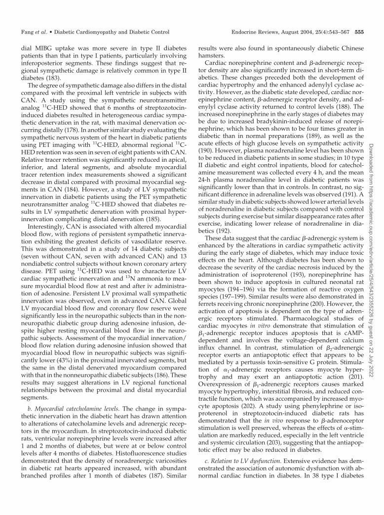
dial MIBG uptake was more severe in type II diabetespatients than that in type I patients, particularly involvinginferoposterior segments. These findings suggest that re-gional sympathetic damage is relatively common in type IIdiabetes (183).
The degree of sympathetic damage also differs in the distalcompared with the proximal left ventricle in subjects withCAN. A study using the sympathetic neurotransmitteranalog 11C-HED showed that 6 months of streptozotocin-induced diabetes resulted in heterogeneous cardiac sympa-thetic denervation in the rat, with maximal denervation oc-curring distally (178). In another similar study evaluating thesympathetic nervous system of the heart in diabetic patientsusing PET imaging with 11C-HED, abnormal regional 11C-HED retention was seen in seven of eight patients with CAN.Relative tracer retention was significantly reduced in apical,inferior, and lateral segments, and absolute myocardialtracer retention index measurements showed a significantdecrease in distal compared with proximal myocardial seg-ments in CAN (184). However, a study of LV sympatheticinnervation in diabetic patients using the PET sympatheticneurotransmitter analog 11C-HED showed that diabetes re-sults in LV sympathetic denervation with proximal hyper-innervation complicating distal denervation (185).
Interestingly, CAN is associated with altered myocardialblood flow, with regions of persistent sympathetic innerva-tion exhibiting the greatest deficits of vasodilator reserve.This was demonstrated in a study of 14 diabetic subjects(seven without CAN, seven with advanced CAN) and 13nondiabetic control subjects without known coronary arterydisease. PET using 11C-HED was used to characterize LVcardiac sympathetic innervation and 13N ammonia to mea-sure myocardial blood flow at rest and after iv administra-tion of adenosine. Persistent LV proximal wall sympatheticinnervation was observed, even in advanced CAN. GlobalLV myocardial blood flow and coronary flow reserve weresignificantly less in the neuropathic subjects than in the non-neuropathic diabetic group during adenosine infusion, de-spite higher resting myocardial blood flow in the neuro-pathic subjects. Assessment of the myocardial innervation/blood flow relation during adenosine infusion showed thatmyocardial blood flow in neuropathic subjects was signifi-cantly lower (43%) in the proximal innervated segments, butthe same in the distal denervated myocardium comparedwith that in the nonneuropathic diabetic subjects (186). Theseresults may suggest alterations in LV regional functionalrelationships between the proximal and distal myocardialsegments.
b. Myocardial catecholamine levels. The change in sympa-thetic innervation in the diabetic heart has drawn attentionto alterations of catecholamine levels and adrenergic recep-tors in the myocardium. In streptozotocin-induced diabeticrats, ventricular norepinephrine levels were increased after1 and 2 months of diabetes, but were at or below controllevels after 4 months of diabetes. Histofluorescence studiesdemonstrated that the density of noradrenergic varicositiesin diabetic rat hearts appeared increased, with abundantbranched profiles after 1 month of diabetes (187). Similar
results were also found in spontaneously diabetic Chinesehamsters.
Cardiac norepinephrine content and �-adrenergic recep-tor density are also significantly increased in short-term di-abetics. These changes preceded both the development ofcardiac hypertrophy and the enhanced adenylyl cyclase ac-tivity. However, as the diabetic state developed, cardiac nor-epinephrine content, �-adrenergic receptor density, and ad-enylyl cyclase activity returned to control levels (188). Theincreased norepinephrine in the early stages of diabetes maybe due to increased bradykinin-induced release of norepi-nephrine, which has been shown to be four times greater indiabetic than in normal preparations (189), as well as theacute effects of high glucose levels on sympathetic activity(190). However, plasma noradrenaline level has been shownto be reduced in diabetic patients in some studies; in 10 typeII diabetic and eight control inpatients, blood for catechol-amine measurement was collected every 4 h, and the mean24-h plasma noradrenaline level in diabetic patients wassignificantly lower than that in controls. In contrast, no sig-nificant difference in adrenaline levels was observed (191). Asimilar study in diabetic subjects showed lower arterial levelsof noradrenaline in diabetic subjects compared with controlsubjects during exercise but similar disappearance rates afterexercise, indicating lower release of noradrenaline in dia-betics (192).
These data suggest that the cardiac �-adrenergic system isenhanced by the alterations in cardiac sympathetic activityduring the early stage of diabetes, which may induce toxiceffects on the heart. Although diabetes has been shown todecrease the severity of the cardiac necrosis induced by theadministration of isoproterenol (193), norepinephrine hasbeen shown to induce apoptosis in cultured neonatal ratmyocytes (194–196) via the formation of reactive oxygenspecies (197–199). Similar results were also demonstrated inferrets receiving chronic norepinephrine (200). However, theactivation of apoptosis is dependent on the type of adren-ergic receptors stimulated. Pharmacological studies ofcardiac myocytes in vitro demonstrate that stimulation of�1-adrenergic receptor induces apoptosis that is cAMP-dependent and involves the voltage-dependent calciuminflux channel. In contrast, stimulation of �2-adrenergicreceptor exerts an antiapoptotic effect that appears to bemediated by a pertussis toxin-sensitive G protein. Stimula-tion of �1-adrenergic receptors causes myocyte hyper-trophy and may exert an antiapoptotic action (201).Overexpression of �1-adrenergic receptors causes markedmyocyte hypertrophy, interstitial fibrosis, and reduced con-tractile function, which was accompanied by increased myo-cyte apoptosis (202). A study using phenylephrine or iso-proterenol in streptozotocin-induced diabetic rats hasdemonstrated that the in vivo response to �-adrenoceptorstimulation is well preserved, whereas the effects of �-stim-ulation are markedly reduced, especially in the left ventricleand systemic circulation (203), suggesting that the antiapop-totic effect may be also reduced in diabetes.
c. Relation to LV dysfunction. Extensive evidence has dem-onstrated the association of autonomic dysfunction with ab-normal cardiac function in diabetes. In 38 type I diabetes
Fang et al. • Diabetic Cardiomyopathy and Diabetic Control Endocrine Reviews, August 2004, 25(4):543–567 555
Dow
nloaded from https://academ
ic.oup.com/edrv/article/25/4/543/2355226 by guest on 22 July 2022
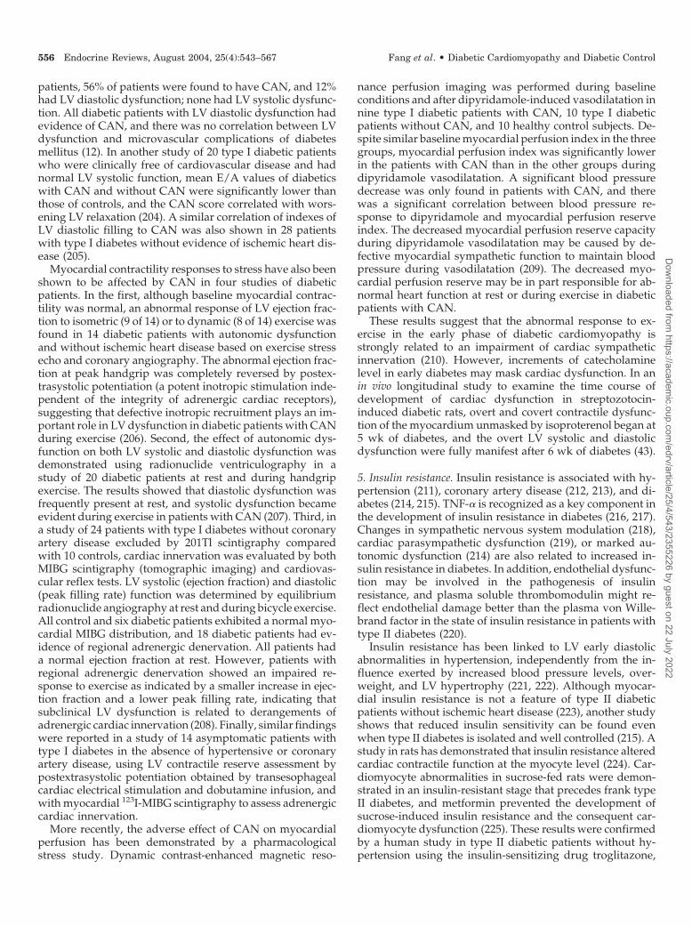
patients, 56% of patients were found to have CAN, and 12%had LV diastolic dysfunction; none had LV systolic dysfunc-tion. All diabetic patients with LV diastolic dysfunction hadevidence of CAN, and there was no correlation between LVdysfunction and microvascular complications of diabetesmellitus (12). In another study of 20 type I diabetic patientswho were clinically free of cardiovascular disease and hadnormal LV systolic function, mean E/A values of diabeticswith CAN and without CAN were significantly lower thanthose of controls, and the CAN score correlated with wors-ening LV relaxation (204). A similar correlation of indexes ofLV diastolic filling to CAN was also shown in 28 patientswith type I diabetes without evidence of ischemic heart dis-ease (205).
Myocardial contractility responses to stress have also beenshown to be affected by CAN in four studies of diabeticpatients. In the first, although baseline myocardial contrac-tility was normal, an abnormal response of LV ejection frac-tion to isometric (9 of 14) or to dynamic (8 of 14) exercise wasfound in 14 diabetic patients with autonomic dysfunctionand without ischemic heart disease based on exercise stressecho and coronary angiography. The abnormal ejection frac-tion at peak handgrip was completely reversed by postex-trasystolic potentiation (a potent inotropic stimulation inde-pendent of the integrity of adrenergic cardiac receptors),suggesting that defective inotropic recruitment plays an im-portant role in LV dysfunction in diabetic patients with CANduring exercise (206). Second, the effect of autonomic dys-function on both LV systolic and diastolic dysfunction wasdemonstrated using radionuclide ventriculography in astudy of 20 diabetic patients at rest and during handgripexercise. The results showed that diastolic dysfunction wasfrequently present at rest, and systolic dysfunction becameevident during exercise in patients with CAN (207). Third, ina study of 24 patients with type I diabetes without coronaryartery disease excluded by 201Tl scintigraphy comparedwith 10 controls, cardiac innervation was evaluated by bothMIBG scintigraphy (tomographic imaging) and cardiovas-cular reflex tests. LV systolic (ejection fraction) and diastolic(peak filling rate) function was determined by equilibriumradionuclide angiography at rest and during bicycle exercise.All control and six diabetic patients exhibited a normal myo-cardial MIBG distribution, and 18 diabetic patients had ev-idence of regional adrenergic denervation. All patients hada normal ejection fraction at rest. However, patients withregional adrenergic denervation showed an impaired re-sponse to exercise as indicated by a smaller increase in ejec-tion fraction and a lower peak filling rate, indicating thatsubclinical LV dysfunction is related to derangements ofadrenergic cardiac innervation (208). Finally, similar findingswere reported in a study of 14 asymptomatic patients withtype I diabetes in the absence of hypertensive or coronaryartery disease, using LV contractile reserve assessment bypostextrasystolic potentiation obtained by transesophagealcardiac electrical stimulation and dobutamine infusion, andwith myocardial 123I-MIBG scintigraphy to assess adrenergiccardiac innervation.
More recently, the adverse effect of CAN on myocardialperfusion has been demonstrated by a pharmacologicalstress study. Dynamic contrast-enhanced magnetic reso-
nance perfusion imaging was performed during baselineconditions and after dipyridamole-induced vasodilatation innine type I diabetic patients with CAN, 10 type I diabeticpatients without CAN, and 10 healthy control subjects. De-spite similar baseline myocardial perfusion index in the threegroups, myocardial perfusion index was significantly lowerin the patients with CAN than in the other groups duringdipyridamole vasodilatation. A significant blood pressuredecrease was only found in patients with CAN, and therewas a significant correlation between blood pressure re-sponse to dipyridamole and myocardial perfusion reserveindex. The decreased myocardial perfusion reserve capacityduring dipyridamole vasodilatation may be caused by de-fective myocardial sympathetic function to maintain bloodpressure during vasodilatation (209). The decreased myo-cardial perfusion reserve may be in part responsible for ab-normal heart function at rest or during exercise in diabeticpatients with CAN.
These results suggest that the abnormal response to ex-ercise in the early phase of diabetic cardiomyopathy isstrongly related to an impairment of cardiac sympatheticinnervation (210). However, increments of catecholaminelevel in early diabetes may mask cardiac dysfunction. In anin vivo longitudinal study to examine the time course ofdevelopment of cardiac dysfunction in streptozotocin-induced diabetic rats, overt and covert contractile dysfunc-tion of the myocardium unmasked by isoproterenol began at5 wk of diabetes, and the overt LV systolic and diastolicdysfunction were fully manifest after 6 wk of diabetes (43).
5. Insulin resistance. Insulin resistance is associated with hy-pertension (211), coronary artery disease (212, 213), and di-abetes (214, 215). TNF-� is recognized as a key component inthe development of insulin resistance in diabetes (216, 217).Changes in sympathetic nervous system modulation (218),cardiac parasympathetic dysfunction (219), or marked au-tonomic dysfunction (214) are also related to increased in-sulin resistance in diabetes. In addition, endothelial dysfunc-tion may be involved in the pathogenesis of insulinresistance, and plasma soluble thrombomodulin might re-flect endothelial damage better than the plasma von Wille-brand factor in the state of insulin resistance in patients withtype II diabetes (220).
Insulin resistance has been linked to LV early diastolicabnormalities in hypertension, independently from the in-fluence exerted by increased blood pressure levels, over-weight, and LV hypertrophy (221, 222). Although myocar-dial insulin resistance is not a feature of type II diabeticpatients without ischemic heart disease (223), another studyshows that reduced insulin sensitivity can be found evenwhen type II diabetes is isolated and well controlled (215). Astudy in rats has demonstrated that insulin resistance alteredcardiac contractile function at the myocyte level (224). Car-diomyocyte abnormalities in sucrose-fed rats were demon-strated in an insulin-resistant stage that precedes frank typeII diabetes, and metformin prevented the development ofsucrose-induced insulin resistance and the consequent car-diomyocyte dysfunction (225). These results were confirmedby a human study in type II diabetic patients without hy-pertension using the insulin-sensitizing drug troglitazone,
556 Endocrine Reviews, August 2004, 25(4):543–567 Fang et al. • Diabetic Cardiomyopathy and Diabetic Control
Dow
nloaded from https://academ
ic.oup.com/edrv/article/25/4/543/2355226 by guest on 22 July 2022
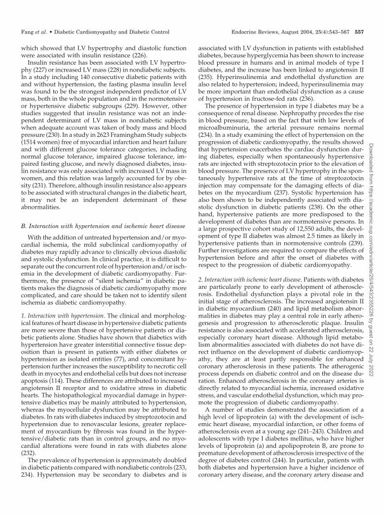
which showed that LV hypertrophy and diastolic functionwere associated with insulin resistance (226).
Insulin resistance has been associated with LV hypertro-phy (227) or increased LV mass (228) in nondiabetic subjects.In a study including 140 consecutive diabetic patients withand without hypertension, the fasting plasma insulin levelwas found to be the strongest independent predictor of LVmass, both in the whole population and in the normotensiveor hypertensive diabetic subgroups (229). However, otherstudies suggested that insulin resistance was not an inde-pendent determinant of LV mass in nondiabetic subjectswhen adequate account was taken of body mass and bloodpressure (230). In a study in 2623 Framingham Study subjects(1514 women) free of myocardial infarction and heart failureand with different glucose tolerance categories, includingnormal glucose tolerance, impaired glucose tolerance, im-paired fasting glucose, and newly diagnosed diabetes, insu-lin resistance was only associated with increased LV mass inwomen, and this relation was largely accounted for by obe-sity (231). Therefore, although insulin resistance also appearsto be associated with structural changes in the diabetic heart,it may not be an independent determinant of theseabnormalities.
B. Interaction with hypertension and ischemic heart disease
With the addition of untreated hypertension and/or myo-cardial ischemia, the mild subclinical cardiomyopathy ofdiabetes may rapidly advance to clinically obvious diastolicand systolic dysfunction. In clinical practice, it is difficult toseparate out the concurrent role of hypertension and/or isch-emia in the development of diabetic cardiomyopathy. Fur-thermore, the presence of “silent ischemia” in diabetic pa-tients makes the diagnosis of diabetic cardiomyopathy morecomplicated, and care should be taken not to identify silentischemia as diabetic cardiomyopathy.
1. Interaction with hypertension. The clinical and morpholog-ical features of heart disease in hypertensive diabetic patientsare more severe than those of hypertensive patients or dia-betic patients alone. Studies have shown that diabetics withhypertension have greater interstitial connective tissue dep-osition than is present in patients with either diabetes orhypertension as isolated entities (77), and concomitant hy-pertension further increases the susceptibility to necrotic celldeath in myocytes and endothelial cells but does not increaseapoptosis (114). These differences are attributed to increasedangiotensin II receptor and to oxidative stress in diabetichearts. The histopathological myocardial damage in hyper-tensive diabetics may be mainly attributed to hypertension,whereas the myocellular dysfunction may be attributed todiabetes. In rats with diabetes induced by streptozotocin andhypertension due to renovascular lesions, greater replace-ment of myocardium by fibrosis was found in the hyper-tensive/diabetic rats than in control groups, and no myo-cardial alterations were found in rats with diabetes alone(232).
The prevalence of hypertension is approximately doubledin diabetic patients compared with nondiabetic controls (233,234). Hypertension may be secondary to diabetes and is
associated with LV dysfunction in patients with establisheddiabetes, because hyperglycemia has been shown to increaseblood pressure in humans and in animal models of type Idiabetes, and the increase has been linked to angiotensin II(235). Hyperinsulinemia and endothelial dysfunction arealso related to hypertension; indeed, hyperinsulinemia maybe more important than endothelial dysfunction as a causeof hypertension in fructose-fed rats (236).
The presence of hypertension in type I diabetes may be aconsequence of renal disease. Nephropathy precedes the risein blood pressure, based on the fact that with low levels ofmicroalbuminuria, the arterial pressure remains normal(234). In a study examining the effect of hypertension on theprogression of diabetic cardiomyopathy, the results showedthat hypertension exacerbates the cardiac dysfunction dur-ing diabetes, especially when spontaneously hypertensiverats are injected with streptozotocin prior to the elevation ofblood pressure. The presence of LV hypertrophy in the spon-taneously hypertensive rats at the time of streptozotocininjection may compensate for the damaging effects of dia-betes on the myocardium (237). Systolic hypertension hasalso been shown to be independently associated with dia-stolic dysfunction in diabetic patients (238). On the otherhand, hypertensive patients are more predisposed to thedevelopment of diabetes than are normotensive persons. Ina large prospective cohort study of 12,550 adults, the devel-opment of type II diabetes was almost 2.5 times as likely inhypertensive patients than in normotensive controls (239).Further investigations are required to compare the effects ofhypertension before and after the onset of diabetes withrespect to the progression of diabetic cardiomyopathy.
2. Interaction with ischemic heart disease. Patients with diabetesare particularly prone to early development of atheroscle-rosis. Endothelial dysfunction plays a pivotal role in theinitial stage of atherosclerosis. The increased angiotensin IIin diabetic myocardium (240) and lipid metabolism abnor-malities in diabetes may play a central role in early athero-genesis and progression to atherosclerotic plaque. Insulinresistance is also associated with accelerated atherosclerosis,especially coronary heart disease. Although lipid metabo-lism abnormalities associated with diabetes do not have di-rect influence on the development of diabetic cardiomyop-athy, they are at least partly responsible for enhancedcoronary atherosclerosis in these patients. The atherogenicprocess depends on diabetic control and on the disease du-ration. Enhanced atherosclerosis in the coronary arteries isdirectly related to myocardial ischemia, increased oxidativestress, and vascular endothelial dysfunction, which may pro-mote the progression of diabetic cardiomyopathy.
A number of studies demonstrated the association of ahigh level of lipoprotein (a) with the development of isch-emic heart disease, myocardial infarction, or other forms ofatherosclerosis even at a young age (241–243). Children andadolescents with type I diabetes mellitus, who have higherlevels of lipoprotein (a) and apolipoprotein B, are prone topremature development of atherosclerosis irrespective of thedegree of diabetes control (244). In particular, patients withboth diabetes and hypertension have a higher incidence ofcoronary artery disease, and the coronary artery disease and
Fang et al. • Diabetic Cardiomyopathy and Diabetic Control Endocrine Reviews, August 2004, 25(4):543–567 557
Dow
nloaded from https://academ
ic.oup.com/edrv/article/25/4/543/2355226 by guest on 22 July 2022
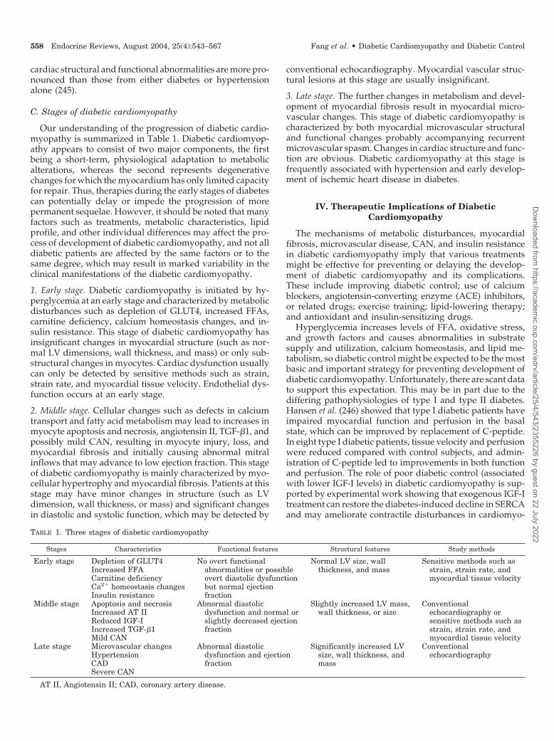
cardiac structural and functional abnormalities are more pro-nounced than those from either diabetes or hypertensionalone (245).
C. Stages of diabetic cardiomyopathy
Our understanding of the progression of diabetic cardio-myopathy is summarized in Table 1. Diabetic cardiomyop-athy appears to consist of two major components, the firstbeing a short-term, physiological adaptation to metabolicalterations, whereas the second represents degenerativechanges for which the myocardium has only limited capacityfor repair. Thus, therapies during the early stages of diabetescan potentially delay or impede the progression of morepermanent sequelae. However, it should be noted that manyfactors such as treatments, metabolic characteristics, lipidprofile, and other individual differences may affect the pro-cess of development of diabetic cardiomyopathy, and not alldiabetic patients are affected by the same factors or to thesame degree, which may result in marked variability in theclinical manifestations of the diabetic cardiomyopathy.
1. Early stage. Diabetic cardiomyopathy is initiated by hy-perglycemia at an early stage and characterized by metabolicdisturbances such as depletion of GLUT4, increased FFAs,carnitine deficiency, calcium homeostasis changes, and in-sulin resistance. This stage of diabetic cardiomyopathy hasinsignificant changes in myocardial structure (such as nor-mal LV dimensions, wall thickness, and mass) or only sub-structural changes in myocytes. Cardiac dysfunction usuallycan only be detected by sensitive methods such as strain,strain rate, and myocardial tissue velocity. Endothelial dys-function occurs at an early stage.
2. Middle stage. Cellular changes such as defects in calciumtransport and fatty acid metabolism may lead to increases inmyocyte apoptosis and necrosis, angiotensin II, TGF-�1, andpossibly mild CAN, resulting in myocyte injury, loss, andmyocardial fibrosis and initially causing abnormal mitralinflows that may advance to low ejection fraction. This stageof diabetic cardiomyopathy is mainly characterized by myo-cellular hypertrophy and myocardial fibrosis. Patients at thisstage may have minor changes in structure (such as LVdimension, wall thickness, or mass) and significant changesin diastolic and systolic function, which may be detected by
conventional echocardiography. Myocardial vascular struc-tural lesions at this stage are usually insignificant.
3. Late stage. The further changes in metabolism and devel-opment of myocardial fibrosis result in myocardial micro-vascular changes. This stage of diabetic cardiomyopathy ischaracterized by both myocardial microvascular structuraland functional changes probably accompanying recurrentmicrovascular spasm. Changes in cardiac structure and func-tion are obvious. Diabetic cardiomyopathy at this stage isfrequently associated with hypertension and early develop-ment of ischemic heart disease in diabetes.
IV. Therapeutic Implications of DiabeticCardiomyopathy
The mechanisms of metabolic disturbances, myocardialfibrosis, microvascular disease, CAN, and insulin resistancein diabetic cardiomyopathy imply that various treatmentsmight be effective for preventing or delaying the develop-ment of diabetic cardiomyopathy and its complications.These include improving diabetic control; use of calciumblockers, angiotensin-converting enzyme (ACE) inhibitors,or related drugs; exercise training; lipid-lowering therapy;and antioxidant and insulin-sensitizing drugs.
Hyperglycemia increases levels of FFA, oxidative stress,and growth factors and causes abnormalities in substratesupply and utilization, calcium homeostasis, and lipid me-tabolism, so diabetic control might be expected to be the mostbasic and important strategy for preventing development ofdiabetic cardiomyopathy. Unfortunately, there are scant datato support this expectation. This may be in part due to thediffering pathophysiologies of type I and type II diabetes.Hansen et al. (246) showed that type I diabetic patients haveimpaired myocardial function and perfusion in the basalstate, which can be improved by replacement of C-peptide.In eight type I diabetic patients, tissue velocity and perfusionwere reduced compared with control subjects, and admin-istration of C-peptide led to improvements in both functionand perfusion. The role of poor diabetic control (associatedwith lower IGF-I levels) in diabetic cardiomyopathy is sup-ported by experimental work showing that exogenous IGF-Itreatment can restore the diabetes-induced decline in SERCAand may ameliorate contractile disturbances in cardiomyo-
TABLE 1. Three stages of diabetic cardiomyopathy
Stages Characteristics Functional features Structural features Study methods
Early stage Depletion of GLUT4Increased FFACarnitine deficiencyCa2� homeostasis changesInsulin resistance
No overt functionalabnormalities or possibleovert diastolic dysfunctionbut normal ejectionfraction
Normal LV size, wallthickness, and mass
Sensitive methods such asstrain, strain rate, andmyocardial tissue velocity
Middle stage Apoptosis and necrosisIncreased AT IIReduced IGF-IIncreased TGF-�1Mild CAN
Abnormal diastolicdysfunction and normal orslightly decreased ejectionfraction
Slightly increased LV mass,wall thickness, or size
Conventionalechocardiography orsensitive methods such asstrain, strain rate, andmyocardial tissue velocity
Late stage Microvascular changesHypertensionCADSevere CAN
Abnormal diastolicdysfunction and ejectionfraction
Significantly increased LVsize, wall thickness, andmass
Conventionalechocardiography
AT II, Angiotensin II; CAD, coronary artery disease.
558 Endocrine Reviews, August 2004, 25(4):543–567 Fang et al. • Diabetic Cardiomyopathy and Diabetic Control
Dow
nloaded from https://academ
ic.oup.com/edrv/article/25/4/543/2355226 by guest on 22 July 2022
cytes from diabetic animals (247). The limited data in supportof prevention of the development of diabetic cardiomyopa-thy by tight glycemic control has also been due to the lack ofsensitive techniques allowing repetitive quantification ofmyocardial perfusion and diastolic and systolic function inthe clinical arena.
Intracellular retention of calcium in diabetes is associatedwith depletion of high-energy phosphate stores and a de-rangement of ultrastructure and cardiac dysfunction. Cal-cium channel blockers are capable of reversing the intracel-lular calcium defects and preventing diabetes-inducedmyocardial changes. Verapamil has been shown to signifi-cantly improve the depressed rate of contraction and rate ofrelaxation, lower peak LV systolic pressure, and elevate LVdiastolic pressure (248), as well as to improve the alteredmyofibrillar ATPase activity, myosin ATPase, myosin isoen-zyme distribution, and sarcoplasmic reticular Ca2�-pumpactivities in streptozocin-induced diabetic rats (249). Dilti-azem has been shown to suppress interstitial fibrotic changesin type II diabetic mice (63), and nifedipine increases insulinsensitivity and prevents the increase in cholesterol and tri-glyceride levels in streptozotocin-induced diabetic rats (250).
ACE inhibitors facilitate blood flow through the micro-circulation in fat and skeletal muscles. Facilitation of bloodflow to insulin-sensitive tissues, such as skeletal muscle,would lead to an increase in glucose delivery to these tissues.Improvement of coronary blood flow may also be beneficialfor microvascular disease-related diabetic cardiomyopathy.Captopril has been demonstrated to increase the number ofperfused capillaries and epicardial perfusion rate, and toprevent the increase of coronary perfusion pressure and end-diastolic pressure in diabetic rats (251). ACE inhibitors canalso improve insulin action at the cellular level (252, 253).ACE inhibition independently increases the basal and insu-lin-stimulated rate of glucose uptake in skeletal muscle ininsulin-resistant obese Zucker rats by improving postrecep-tor insulin signaling and enhancing GLUT-4 translocation tothe cell membrane (254). The action of ACE inhibitors onangiotensin II may improve fibrosis in myocardium andfunctional and structural changes of small vessels in diabe-tes. ACE inhibitors have been demonstrated to reduce car-diovascular disease in diabetic patients, particularly diabeticpatients with hypertension (255). Angiotensin II receptorblockers and aldosterone inhibitors may also have similareffects on myocardial fibrosis in diabetic patients. Because oflocal effects of angiotensin II in diabetic myocardium, controlof myocardial fibrosis should be started at an early stagerather than only in patients who have hypertension or vas-cular complications.
Steps to reduce atherogenesis consist of the basic treatmentfor vascular disease and should be started at the early stageof diabetic cardiomyopathy because abnormalities in lipidmetabolism are already present. Exercise may improve glu-cose homeostasis by reducing the glucose/insulin ratio andincreasing insulin sensitivity. Studies have shown that ex-ercise training increases whole body insulin sensitivity andglucose oxidation by skeletal and cardiac muscle. The im-provement may be associated with both attenuation ofreduction in myocardial GLUT-4 transporters (256) andincrease in myocardial sarcolemmal GLUT-4 protein in di-
abetic hearts (257). Changes in myocardial metabolism in-volving a shift from glucose to fat metabolism in diabetesmellitus increase plasma levels of triglycerides and choles-terol, and these may be lowered by exercise training, result-ing in improved myocardial sarcoplasmic reticulum functionand vascular function. In addition, exercise training im-proves cardiac output (258) and reverses the changes in con-tractile properties of the heart in streptozotocin-diabetic rats(259). Improvements in cardiac function are also mediated bydecreasing the severity of the diabetic state (260). Improvingvascular endothelial dysfunction by exercise training mayalso play an important role. However, whereas low-intensityexercise training seems to improve cardiovascular function,some types of endurance training may further decrease thereduced myocardial Ca2�-activated ATPase and �-adrener-gic receptor number in diabetes (261).
Sympathetic cardiac hyperinnervation can occur concur-rently with denervation in diabetic neuropathy and couldpotentially cause arrhythmia and sudden death. Direct eval-uation of myocardial sympathetic innervation has shown thecorrelation of autonomic dysfunction with diabetic control.In a prospective study over a mean of 4 yr, myocardialsympathetic innervation was investigated in 12 type I dia-betic patients using myocardial 123I-MIBG scintigraphy inconjunction with cardiovascular autonomic function testsusing QTc interval and QT dispersion. Global MIBG uptakeincreased from baseline to follow-up in patients with goodglycemic control and decreased in the poor control group,suggesting that long-term poor glycemic control is associatedwith the progression of LV adrenergic denervation. The stan-dard autonomic function tests were no different in eachgroup (262). However, even in the early stage of diabetes,cardiac sympathetic denervation is only partially reversedwith improved metabolic control (263, 264). The role of ox-idative stress in the development of CAN suggests that an-tioxidants may have beneficial effects on the cardiac auto-nomic nervous system through a decline in oxidative stress.This was demonstrated in a double-blind randomized con-trolled trial, with 50 type II diabetic patients with CAN as-signed to treatment with vitamin E or placebo for 4 months.The results show that chronic vitamin E administration im-proves the ratio of cardiac sympathetic to parasympathetictone in patients with type II diabetes, which might be me-diated by a decline in oxidative stress (265). This is supportedby other antioxidant studies in diabetic animals using vita-min E (266) and acetyl-l-carnitine treatment (267) as well asin diabetic patients using lipoic acid (268–271). In addition,ACE inhibitors such as quinapril have also been shown tosignificantly improve CAN in diabetic patients (272). Simi-larly, aldose reductase inhibitors have demonstrated clinicalimprovement not only in CAN but also in cardiac perfor-mance. The effect on cardiovascular performance of sorbinilwas studied in patients with diabetic autonomic neuropathywho were free of atherosclerotic coronary artery disease.After 1 yr of treatment, significant improvement was dem-onstrated in both the resting cardiac output and the maximalcardiac output, suggesting that the use of an aldose reductaseinhibitor may be useful in treating suboptimal cardiovascu-lar performance in patients with diabetic CAN (273). Fur-thermore, decreased MIBG uptake and increased norepi-
Fang et al. • Diabetic Cardiomyopathy and Diabetic Control Endocrine Reviews, August 2004, 25(4):543–567 559
Dow
nloaded from https://academ
ic.oup.com/edrv/article/25/4/543/2355226 by guest on 22 July 2022
nephrine content in diabetic myocardium were completelyprevented by insulin therapy started immediately after strep-tozotocin injection and partially, but significantly, by aldosereductase inhibitor administered immediately after strepto-zotocin injection. Heterogeneous MIBG distribution also dis-appeared with the aldose reductase inhibitor therapy. Incontrast, diabetic rats treated with insulin or aldose reductaseinhibitor therapy that was started 4 wk after streptozotocininjection showed no improvement in MIBG uptake, suggest-ing the importance of early intervention (274).
Insulin resistance, hyperinsulinemia, atherogenic dyslip-idemia, hypertension, abdominal obesity, and impaired he-mostasis are risk factors for cardiovascular disease. Insulinresistance results from a combination of genetic and envi-ronmental factors and contributes to type II diabetes melli-tus. Obesity is associated with insulin resistance, and weightloss has been shown to correct insulin resistance in diabetes(275, 276), implying that weight reduction through lifestylemodifications (low-calorie diet and exercise) and antiobesitydrugs (orlistat, sibutramine, etc.) is the first step of treatmentof insulin resistance. Metformin and the thiazolidinedionesare used to treat insulin resistance and work through dif-ferent mechanisms. Metformin reduces FFA efflux from fatcells, thereby suppressing hepatic glucose production, andindirectly improving peripheral insulin sensitivity and en-dothelial function. In contrast, thiazolidinediones improveperipheral insulin sensitivity by reducing circulating FFAsbut also by increasing production of adiponectin, which im-proves insulin sensitivity. Thiazolidinediones also improveendothelial function and may prevent or delay the onset ofdiabetes (277). Combination oral hypoglycemic therapy maybe ideal for maintaining adequate glycemic control and im-proving insulin resistance in patients with type II diabetes.In obese type II diabetic patients inadequately controlled onmetformin alone, addition of the insulin-sensitizing agentrosiglitazone improves glycemic control, insulin sensitivity,and �-cell function to a clinically important extent (278).Combination of pioglitazone and metformin has also shownto significantly improve insulin sensitivity as compared withmetformin monotherapy in patients recently diagnosed withtype II diabetes (279). Troglitazone has been shown to reduceplasma insulin levels and restore coronary circulation byimproving insulin resistance in type II diabetic patients (280).In addition, weekly iv IGF-I bolus therapy has been dem-onstrated to be effective in inducing sustained insulin sen-sitivity in a patient with type I diabetes mellitus and massiveinsulin resistance (281). Finally, both ACE inhibitors andexercise may be beneficial for improving insulin resistance.In skeletal muscle, exercise has been demonstrated to recruita separate pool of GLUT4 to that activated by insulin (282).This leads to an additive effect of insulin and exercise onglucose uptake. Furthermore, exercise increases skeletalmuscle GLUT4 gene and protein expression (283). Shouldsimilar changes occur in cardiac muscle, it is conceivable thatexercise may have a direct beneficial effect on myocardialfunction also. The improvement of insulin resistance mayresult in improvement of cardiac function. Glipizide has beenshown to reduce the degree of insulin resistance in the myo-cardium and improves cardiac function in diabetes. In aneonatal streptozotocin-induced rat model of type II dia-
betes, animals treated with glipizide for 1 yr exhibitedimproved myocardial contractile function relative to thevehicle-fed or ad lib-fed diabetic animals. Heart rate wassignificantly elevated, and there was a tendency for boththe rate of relaxation and contractility to be elevated in theglipizide-treated group (284). Finally, improvement in insu-lin sensitivity of cardiac muscle may have benefits other thanimproved energy utilization. Insulin and IGF-I share multi-ple intracellular signaling pathways, and both receptors me-diate antiapoptotic effects. Improvements in the signaling ofthese molecules may have an effect to preserve cardiomyo-cyte number.
V. Summary and Conclusions
In this review we present evidence that strongly supportsthe existence of diabetic cardiomyopathy as a distinct clinicalentity. The pathophysiology of the condition remains to befully elucidated, but includes interstitial fibrosis, cardiomy-ocyte loss, impaired energy utilization, small vessel disease,and neuropathy. Prominent functional consequences includediastolic and systolic dysfunction, which may manifest asdyspnea and exercise intolerance. Traditional cardiac riskssuch as hypertension, atherosclerosis, and dyslipidemia arecommon in diabetic patients and further compromise cardiacstatus. Currently, no specific therapeutic strategies can berecommended for diabetic cardiomyopathy, but manage-ment of traditional risk factors and lifestyle modificationprograms already established in the management of cardiacdisease should be instituted. Further research is urgentlyrequired into the molecular basis of diabetic cardiomyopathysuch that more appropriate therapies may be formulated andtested.
Acknowledgments
Address all correspondence and requests for reprints to: Prof. T.Marwick, University of Queensland Department of Medicine, PrincessAlexandra Hospital, Ipswich Road, Brisbane, Queensland 4012, Aus-tralia. E-mail: [email protected]
This work was supported in part by a Clinical Centre of ResearchExcellence award from the National Health and Medical Research Coun-cil of Australia.
References
1. Hamby RI, Zoneraich S, Sherman L 1974 Diabetic cardiomyopa-thy. JAMA 229:1749–1754
2. Regan TJ, Lyons MM, Ahmed SS, Levinson GE, Oldewurtel HA,Ahmad MR, Haider B 1977 Evidence for cardiomyopathy in fa-milial diabetes mellitus. J Clin Invest 60:884–899
3. Mizushige K, Yao L, Noma T, Kiyomoto H, Yu Y, Hosomi N,Ohmori K, Matsuo H 2000 Alteration in left ventricular diastolicfilling and accumulation of myocardial collagen at insulin-resistantprediabetic stage of a type II diabetic rat model. Circulation 101:899–907
4. Rubler S, Dlugash J, Yuceoglu YZ, Kumral T, Branwood AW,Grishman A 1972 New type of cardiomyopathy associated withdiabetic glomerulosclerosis. Am J Cardiol 30:595–602
5. Sohn DW, Chai IH, Lee DJ, Kim HC, Kim HS, Oh BH, Lee MM,Park YB, Choi YS, Seo JD, Lee YW 1997 Assessment of mitralannulus velocity by Doppler tissue imaging in the evaluation of leftventricular diastolic function. J Am Coll Cardiol 30:474–480
560 Endocrine Reviews, August 2004, 25(4):543–567 Fang et al. • Diabetic Cardiomyopathy and Diabetic Control
Dow
nloaded from https://academ
ic.oup.com/edrv/article/25/4/543/2355226 by guest on 22 July 2022
6. Matsumura Y, Elliott PM, Virdee MS, Sorajja P, Doi Y, McKennaWJ 2002 Left ventricular diastolic function assessed using Dopplertissue imaging in patients with hypertrophic cardiomyopathy: re-lation to symptoms and exercise capacity. Heart 87:247–251
7. Schober KE, Fuentes VL, Bonagura JD 2003 Comparison betweeninvasive hemodynamic measurements and noninvasive assess-ment of left ventricular diastolic function by use of Doppler echo-cardiography in healthy anesthetized cats. Am J Vet Res 64:93–103
8. Garcia MJ, Thomas JD, Klein AL 1998 New Doppler echocardio-graphic applications for the study of diastolic function. J Am CollCardiol 32:865–875
9. von Bibra H, Tuchnitz A, Klein A, Schneider-Eicke J, Schomig A,Schwaiger M 2000 Regional diastolic function by pulsed Dopplermyocardial mapping for the detection of left ventricular ischemiaduring pharmacologic stress testing: a comparison with stress echo-cardiography and perfusion scintigraphy. J Am Coll Cardiol 36:444–452
10. Semeniuk LM, Kryski AJ, Severson DL 2002 Echocardiographicassessment of cardiac function in diabetic db/db and transgenicdb/db-hGLUT4 mice. Am J Physiol Heart Circ Physiol 283:H976–H982
11. Ganguly PK, Thliveris JA, Mehta A 1990 Evidence against theinvolvement of nonenzymatic glycosylation in diabetic cardiomy-opathy. Metabolism 39:769–773
12. Rajan SK, Gokhale SM 2002 Cardiovascular function in patientswith insulin-dependent diabetes mellitus: a study using noninva-sive methods. Ann NY Acad Sci 958:425–430
13. Giampietro O, Di Bello V, Matteucci E, Talarico L, Ruberti F,Boldrini E, Giorgi D, Giusti C 1997 Erythrocyte Na�/H� ex-change and preclinical abnormalities of the left ventricular diastolicfunction in normotensive type 1 (insulin-dependent) diabetic pa-tients. Acta Diabetol 34:223–229
14. Lind L, Berne C, Andren B, Lithell H 1996 Relationship betweendiastolic hypertension and myocardial morphology and function inelderly males with diabetes mellitus. Diabetologia 39:1603–1606
15. Matteucci E, Di Bello V, Giampietro O 1995 Integrated analysis oferythrocyte Na�/H� antiport activity and left ventricular myo-cardial function in type I insulin-dependent diabetes mellitus. JDiabetes Complications 9:208–211
16. Cerutti F, Vigo A, Sacchetti C, Bessone A, Barattia G, Morello M,Casalucci D, Gastaldi L 1994 Evaluation of left ventricular diastolicfunction in insulin dependent diabetic children by M-mode andDoppler echocardiography. Panminerva Med 36:109–114
17. Raev DC 1994 Which left ventricular function is impaired earlierin the evolution of diabetic cardiomyopathy? An echocardio-graphic study of young type I diabetic patients. Diabetes Care17:633–639
18. Raev DC 1994 Left ventricular function and specific diabetic com-plications in other target organs in young insulin-dependent dia-betics: an echocardiographic study. Heart Vessels 9:121–128
19. Shimizu M, Sugihara N, Kita Y, Shimizu K, Shibayama S, TakedaR 1991 Increase in left ventricular chamber stiffness in patients withnon-insulin dependent diabetes mellitus. Jpn Circ J 55:657–664
20. Bouchard A, Sanz N, Botvinick EH, Phillips N, Heilbron D, ByrdIII BF, Karam JH, Schiller NB 1989 Noninvasive assessment ofcardiomyopathy in normotensive diabetic patients between 20 and50 years old. Am J Med 87:160–166
21. Park JW, Ziegler AG, Janka HU, Doering W, Mehnert H 1988 Leftventricular relaxation and filling pattern in diabetic heart muscledisease: an echocardiographic study. Klin Wochenschr 66:773–778
22. Zarich SW, Arbuckle BE, Cohen LR, Roberts M, Nesto RW 1988Diastolic abnormalities in young asymptomatic diabetic patientsassessed by pulsed Doppler echocardiography. J Am Coll Cardiol12:114–120
23. Ruddy TD, Shumak SL, Liu PP, Barnie A, Seawright SJ,McLaughlin PR, Zinman B 1988 The relationship of cardiac dia-stolic dysfunction to concurrent hormonal and metabolic status intype I diabetes mellitus. J Clin Endocrinol Metab 66:113–118
24. Airaksinen J, Ikaheimo M, Kaila J, Linnaluoto M, Takkunen J1984 Impaired left ventricular filling in young female diabetics. Anechocardiographic study. Acta Med Scand 216:509–516
25. Trost SU, Belke DD, Bluhm WF, Meyer M, Swanson E, DillmannWH 2002 Overexpression of the sarcoplasmic reticulum Ca(2�)-
ATPase improves myocardial contractility in diabetic cardiomy-opathy. Diabetes 51:1166–1171
26. Brown RA, Filipovich P, Walsh MF, Sowers JR 1996 Influence ofsex, diabetes and ethanol on intrinsic contractile performance ofisolated rat myocardium. Basic Res Cardiol 91:353–360
27. Joffe II, Travers KE, Perreault-Micale CL, Hampton T, Katz SE,Morgan JP, Douglas PS 1999 Abnormal cardiac function in thestreptozotocin-induced non-insulin-dependent diabetic rat: non-invasive assessment with Doppler echocardiography and contri-bution of the nitric oxide pathway. J Am Coll Cardiol 34:2111–2119
28. Dent CL, Bowman AW, Scott MJ, Allen JS, Lisauskas JB, Janif M,Wickline SA, Kovacs SJ 2001 Echocardiographic characterizationof fundamental mechanisms of abnormal diastolic filling in diabeticrats with a parameterized diastolic filling formalism. J Am SocEchocardiogr 14:1166–1172
29. Flarsheim CE, Grupp IL, Matlib MA 1996 Mitochondrial dys-function accompanies diastolic dysfunction in diabetic rat heart.Am J Physiol 271:H192–H202
30. Poirier P, Bogaty P, Garneau C, Marois L, Dumesnil JG 2001Diastolic dysfunction in normotensive men with well-controlledtype 2 diabetes: importance of maneuvers in echocardiographicscreening for preclinical diabetic cardiomyopathy. Diabetes Care24:5–10
31. Schannwell CM, Schneppenheim M, Perings S, Plehn G, StrauerBE 2002 Left ventricular diastolic dysfunction as an early mani-festation of diabetic cardiomyopathy. Cardiology 98:33–39
32. Strauer BE, Motz W, Vogt M, Schwartzkopff B 1997 Impairedcoronary flow reserve in NIDDM: a possible role for diabetic car-diopathy in humans. Diabetes 46(Suppl 2):S119–S124
33. Paillole C, Dahan M, Paycha F, Solal AC, Passa P, Gourgon R 1989Prevalence and significance of left ventricular filling abnormalitiesdetermined by Doppler echocardiography in young type I (insulin-dependent) diabetic patients. Am J Cardiol 64:1010–1016
34. Fang Z, Najos-Valencia O, Leano R, Marwick T 2003 Patients withearly diabetic heart disease demonstrate a normal myocardial re-sponse to dobutamine. J Am Coll Cardiol 42:446–453
35. Attali JR, Sachs RN, Valensi P, Palsky D, Tellier P, Vulpillat M,Lanfranchi J, Sebaoun J 1988 Asymptomatic diabetic cardiomy-opathy: a noninvasive study. Diabetes Res Clin Pract 4:183–190
36. Robillon JF, Sadoul JL, Jullien D, Morand P, Freychet P 1994Abnormalities suggestive of cardiomyopathy in patients with type2 diabetes of relatively short duration. Diabete Metab 20:473–480
37. Astorri E, Fiorina P, Contini GA, Albertini D, Magnati G, AstorriA, Lanfredini M 1997 Isolated and preclinical impairment of leftventricular filling in insulin-dependent and non-insulin-dependentdiabetic patients. Clin Cardiol 20:536–540
38. Salazar J, Rivas A, Rodriguez M, Felipe J, Garcia MD, Bone J 1994Left ventricular function determined by Doppler echocardiogra-phy in adolescents with type I (insulin-dependent) diabetes mel-litus. Acta Cardiol 49:435–439
39. Mathew P, John L, Jose J, Krishnaswami S 1992 Assessment of leftventricular diastolic function in young diabetics–a two dimen-sional echo Doppler study. Indian Heart J 44:29–32
40. Posner J, Ilya R, Wanderman K, Weitzman S 1983 Systolic timeintervals in diabetes. Diabetologia 24:249–252
41. Schaible TF, Malhotra A, Bauman WA, Scheuer J 1983 Left ven-tricular function after chronic insulin treatment in diabetic andnormal rats. J Mol Cell Cardiol 15:445–458
42. Wold LE, Relling DP, Colligan PB, Scott GI, Hintz KK, Ren BH,Epstein PN, Ren J 2001 Characterization of contractile function indiabetic hypertensive cardiomyopathy in adult rat ventricularmyocytes. J Mol Cell Cardiol 33:1719–1726
43. Hoit BD, Castro C, Bultron G, Knight S, Matlib MA 1999 Non-invasive evaluation of cardiac dysfunction by echocardiography instreptozotocin-induced diabetic rats. J Card Fail 5:324–333
44. Hayashi K, Okumura K, Matsui H, Murase K, Kamiya H, SaburiY, Numaguchi Y, Toki Y, Hayakawa T 2001 Involvement of 1,2-diacylglycerol in improvement of heart function by etomoxir indiabetic rats. Life Sci 68:1515–1526
45. Coughlin SS, Pearle DL, Baughman KL, Wasserman A, Tefft MC1994 Diabetes mellitus and risk of idiopathic dilated cardiomyop-athy. The Washington, DC Dilated Cardiomyopathy Study. AnnEpidemiol 4:67–74
Fang et al. • Diabetic Cardiomyopathy and Diabetic Control Endocrine Reviews, August 2004, 25(4):543–567 561
Dow
nloaded from https://academ
ic.oup.com/edrv/article/25/4/543/2355226 by guest on 22 July 2022
46. Kannel WB, Hjortland M, Castelli WP 1974 Role of diabetes incongestive heart failure: the Framingham study. Am J Cardiol34:29–34
47. Friedman NE, Levitsky LL, Edidin DV, Vitullo DA, Lacina SJ,Chiemmongkoltip P 1982 Echocardiographic evidence for im-paired myocardial performance in children with type I diabetesmellitus. Am J Med 73:846–850
48. Mbanya JC, Sobngwi E, Mbanya DS, Ngu KB 2001 Left ventric-ular mass and systolic function in African diabetic patients: asso-ciation with microalbuminuria. Diabetes Metab 27:378–382
49. Fang ZY, Yuda S, Anderson V, Short L, Case C, Marwick TH 2003Echocardiographic detection of early diabetic myocardial disease.J Am Coll Cardiol 41:611–617
50. Scognamiglio R, Casara D, Avogaro A 2000 Myocardial dysfunc-tion and adrenergic innervation in patients with type 1 diabetesmellitus. Diabetes Nutr Metab 13:346–349
51. Baum VC, Levitsky LL, Englander RM 1987 Abnormal cardiacfunction after exercise in insulin-dependent diabetic children andadolescents. Diabetes Care 10:319–323
52. Zola B, Kahn JK, Juni JE, Vinik AI 1986 Abnormal cardiac functionin diabetic patients with autonomic neuropathy in the absence ofischemic heart disease. J Clin Endocrinol Metab 63:208–214
53. Vered A, Battler A, Segal P, Liberman D, Yerushalmi Y, BerezinM, Neufeld HN 1984 Exercise-induced left ventricular dysfunctionin young men with asymptomatic diabetes mellitus (diabetic car-diomyopathy). Am J Cardiol 54:633–637
54. Jermendy G, Kammerer L, Koltai ZM, Cserhalmi L, Szelenyi J,Tichy M, Pogatsa G 1983 Preclinical abnormality of left ventricularperformance in patients with insulin-dependent diabetes mellitus.Acta Diabetol Lat 20:311–320
55. Carlstrom S, Karlefors T 1970 Haemodynamic studies on newlydiagnosed diabetics before and after adequate insulin treatment. BrHeart J 32:355–358
56. Karlefors T 1966 Haemodynamic studies in male diabetics. ActaMed Scand Suppl 449:45–80
57. Hsu KL, Chiang FT, Lo HM, Tsai CH, Tseng CD, Tseng YZ 1997Cardiac contractility in noninsulin dependent diabetes mellitusevaluated using the relation between endsystolic wall stress andvelocity of circumferential fiber shortening. Jpn Heart J 38:463–471
58. Nugent AM, Steele IC, al Modaris F, Vallely S, Moore A, Camp-bell NP, Bell PM, Buchanan KD, Trimble ER, Nicholls DP 1997Exercise responses in patients with IDDM. Diabetes Care 20:1814–1821
59. Borow KM, Jaspan JB, Williams KA, Neumann A, Wolinski-Walley P, Lang RM 1990 Myocardial mechanics in young adultpatients with diabetes mellitus: effects of altered load, inotropicstate and dynamic exercise. J Am Coll Cardiol 15:1508–1517
60. Howarth FC, Qureshi MA, White E, Calaghan SC 2002 Cardiacmicrotubules are more resistant to chemical depolymerisation instreptozotocin-induced diabetes in the rat. Pflugers Arch 444:432–437
61. Hileeto D, Cukiernik M, Mukherjee S, Evans T, Barbin Y,Downey D, Karmazyn M, Chakrabarti S 2002 Contributions ofendothelin-1 and sodium hydrogen exchanger-1 in the diabeticmyocardium. Diabetes Metab Res Rev 18:386–394
62. Thompson EW 1994 Quantitative analysis of myocardial structurein insulin-dependent diabetes mellitus: effects of immediate anddelayed insulin replacement. Proc Soc Exp Biol Med 205:294–305
63. Shimada T 1993 Correlation between metabolic and histopatho-logical changes in the myocardium of the KK mouse. Effect ofdiltiazem on the diabetic heart. Jpn Heart J 34:617–626
64. Thompson EW 1988 Structural manifestations of diabetic cardio-myopathy in the rat and its reversal by insulin treatment. Am J Anat182:270–282
65. Naito J, Koretsune Y, Sakamoto N, Shutta R, Yoshida J, YasuokaY, Yoshida S, Chin W, Kusuoka H, Inoue M 2001 Transmuralheterogeneity of myocardial integrated backscatter in diabetic pa-tients without overt cardiac disease. Diabetes Res Clin Pract 52:11–20
66. Sunni S, Bishop SP, Kent SP, Geer JC 1986 Diabetic cardiomy-opathy. A morphological study of intramyocardial arteries. ArchPathol Lab Med 110:375–381
67. Lababidi ZA, Goldstein DE 1983 High prevalence of echocardio-graphic abnormalities in diabetic youths. Diabetes Care 6:18–22
68. Warley A, Powell JM, Skepper JN 1995 Capillary surface area isreduced and tissue thickness from capillaries to myocytes is in-creased in the left ventricle of streptozotocin-diabetic rats. Diabe-tologia 38:413–421
69. Eto M, Watanabe K, Sekiguchi M, Iwashima Y, Morikawa A,Oshima E, Ishii K 1987 Metabolic and morphological changes ofthe heart in Chinese hamsters (CHAD strain) with spontaneouslong-term diabetes. Diabetes Res Clin Pract 3:297–305
70. Bhimji S, Godin DV, McNeill JH 1986 Myocardial ultrastructuralchanges in alloxan-induced diabetes in rabbits. Acta Anat (Basel)125:195–200
71. Seager MJ, Singal PK, Orchard R, Pierce GN, Dhalla NS 1984Cardiac cell damage: a primary myocardial disease in streptozo-tocin-induced chronic diabetes. Br J Exp Pathol 65:613–623
72. Kerut EK, Given MB, McIlwain E, Allen G, Espinoza C, Giles TD2000 Echocardiographic texture analysis using the wavelet trans-form: differentiation of early heart muscle disease. Ultrasound MedBiol 26:1445–1453
73. Nunoda S, Genda A, Sugihara N, Nakayama A, Mizuno S,Takeda R 1985 Quantitative approach to the histopathology of thebiopsied right ventricular myocardium in patients with diabetesmellitus. Heart Vessels 1:43–47
74. Vitolo E, Madoi S, Sponzilli C, Palvarini M, Silvestri D, CastiniD, Morabito A 1988 Vectorcardiographic evaluation of diabeticcardiomyopathy and of its contributing factors. Acta Diabetol Lat25:227–234
75. Di Bello V, Talarico L, Picano E, Di Muro C, Landini L, PaterniM, Matteucci E, Giusti C, Giampietro O 1995 Increased echoden-sity of myocardial wall in the diabetic heart: an ultrasound tissuecharacterization study. J Am Coll Cardiol 25:1408–1415
76. Picano E, Pelosi G, Marzilli M, Lattanzi F, Benassi A, Landini L,L’Abbate A 1990 In vivo quantitative ultrasonic evaluation of myo-cardial fibrosis in humans. Circulation 81:58–64
77. van Hoeven KH, Factor SM 1990 A comparison of the pathologicalspectrum of hypertensive, diabetic, and hypertensive-diabeticheart disease. Circulation 82:848–855
78. Chatham JC, Seymour AM 2002 Cardiac carbohydrate metabolismin Zucker diabetic fatty rats. Cardiovasc Res 55:104–112
79. Mokuda O, Sakamoto Y, Ikeda T, Mashiba H 1990 Effects ofanoxia and low free fatty acid on myocardial energy metabolism instreptozotocin-diabetic rats. Ann Nutr Metab 34:259–265
80. Rodrigues B, Cam MC, McNeill JH 1998 Metabolic disturbancesin diabetic cardiomyopathy. Mol Cell Biochem 180:53–57
81. Chen V, Ianuzzo CD, Fong BC, Spitzer JJ 1984 The effects of acuteand chronic diabetes on myocardial metabolism in rats. Diabetes33:1078–1084
82. Ohtake T, Yokoyama I, Watanabe T, Momose T, Serezawa T,Nishikawa J, Sasaki Y 1995 Myocardial glucose metabolism innoninsulin-dependent diabetes mellitus patients evaluated byFDG-PET. J Nucl Med 36:456–463
83. Eckel J, Reinauer H 1990 Insulin action on glucose transport inisolated cardiac myocytes: signalling pathways and diabetes-induced alterations. Biochem Soc Trans 18:1125–1127
84. Garvey WT, Hardin D, Juhaszova M, Dominguez JH 1993 Effectsof diabetes on myocardial glucose transport system in rats: impli-cations for diabetic cardiomyopathy. Am J Physiol 264:H837–H844
85. Russell III RR, Yin R, Caplan MJ, Hu X, Ren J, Shulman GI,Sinusas AJ, Young LH 1998 Additive effects of hyperinsulinemiaand ischemia on myocardial GLUT1 and GLUT4 translocation invivo. Circulation 98:2180–2186
86. Liedtke AJ, DeMaison L, Eggleston AM, Cohen LM, Nellis SH1988 Changes in substrate metabolism and effects of excess fattyacids in reperfused myocardium. Circ Res 62:535–542
87. Rodriques B, Cam MC, Kong J, Goyal RK, McNeill JH 1997 Straindifferences in susceptibility to streptozotocin-induced diabetes: Ef-fects on hypertriglyceridemia and cardiomyopathy. CardiovascRes 34:199–205
88. Lopaschuk GD, Russell JC 1991 Myocardial function and energysubstrate metabolism in the insulin-resistant JCR:LA corpulent rat.J Appl Physiol 71:1302–1308
89. Belke DD, Larsen TS, Gibbs EM, Severson DL 2000 Altered
562 Endocrine Reviews, August 2004, 25(4):543–567 Fang et al. • Diabetic Cardiomyopathy and Diabetic Control
Dow
nloaded from https://academ
ic.oup.com/edrv/article/25/4/543/2355226 by guest on 22 July 2022
metabolism causes cardiac dysfunction in perfused hearts fromdiabetic (db/db) mice. Am J Physiol Endocrinol Metab 279:E1104–E1113
90. Hall JL, Stanley WC, Lopaschuk GD, Wisneski JA, Pizzurro RD,Hamilton CD, McCormack JG 1996 Impaired pyruvate oxidationbut normal glucose uptake in diabetic pig heart during dobu-tamine-induced work. Am J Physiol 271:H2320–H2329
91. Chatham JC, Gao ZP, Bonen A, Forder JR 1999 Preferential inhi-bition of lactate oxidation relative to glucose oxidation in the ratheart following diabetes. Cardiovasc Res 43:96–106
92. Nakayama H, Morozumi T, Nanto S, Shimonagata T, Ohara T,Takano Y, Kotani J, Watanabe T, Fujita M, Nishio M, KusuokaH, Hori M, Nagata S 2001 Abnormal myocardial free fatty acidutilization deteriorates with morphological changes in the hyper-tensive heart. Jpn Circ J 65:783–787
93. Yazaki Y, Isobe M, Takahashi W, Kitabayashi H, Nishiyama O,Sekiguchi M, Takemura T 1999 Assessment of myocardial fattyacid abnormalities in patients with idiopathic dilated cardiomy-opathy using I123 BMIPP SPECT: correlation with clinicopatho-logical findings and clinical course. Heart 81:153–159
94. Malone JI, Schocken DD, Morrison AD, Gilbert-Barness E 1999Diabetic cardiomyopathy and carnitine deficiency. J Diabetes Com-plications 13:86–90
95. Malhotra A, Sanghi V 1997 Regulation of contractile proteins indiabetic heart. Cardiovasc Res 34:34–40
96. Takeda N, Nakamura I, Hatanaka T, Ohkubo T, Nagano M 1988Myocardial mechanical and myosin isoenzyme alterations in strep-tozotocin-diabetic rats. Jpn Heart J 29:455–463
97. Abe T, Ohga Y, Tabayashi N, Kobayashi S, Sakata S, Misawa H,Tsuji T, Kohzuki H, Suga H, Taniguchi S, Takaki M 2002 Leftventricular diastolic dysfunction in type 2 diabetes mellitus modelrats. Am J Physiol Heart Circ Physiol 282:H138–H148
98. Kotsanas G, Delbridge LM, Wendt IR 2000 Stimulus interval-dependent differences in Ca2� transients and contractile responsesof diabetic rat cardiomyocytes. Cardiovasc Res 46:450–462
99. Dhalla NS, Liu X, Panagia V, Takeda N 1998 Subcellular remod-eling and heart dysfunction in chronic diabetes. Cardiovasc Res40:239–247
100. Kita Y, Shimizu M, Sugihara N, Shimizu K, Yoshio H, ShibayamaS, Takeda R 1991 Correlation between histopathological changesand mechanical dysfunction in diabetic rat hearts. Diabetes ResClin Pract 11:177–188
101. Cerasola G, Donatelli M, Cottone S, D’Ignoto G, Grasso L, MoriciML, Terrizzi C, Verga S, Bompiani GD 1987 Effects of dynamicexercise and metabolic control on left ventricular performance ininsulin-dependent diabetes mellitus. Acta Diabetol Lat 24:263–270
102. Jermendy G, Koltai MZ, Kammerer L, Cserhalmi L, Istvanffy M,Szelenyi J, Pogatsa G 1984 Myocardial systolic alterations of in-sulin-dependent diabetics in rest. Acta Cardiol 39:185–190
103. Garay-Sevilla ME, Nava LE, Malacara JM, Wrobel K, Wrobel K,Perez U 2000 Advanced glycosylation end products (AGEs),insulin-like growth factor-1 (IGF-1) and IGF-binding protein-3(IGFBP-3) in patients with type 2 diabetes mellitus. Diabetes MetabRes Rev 16:106–113
104. Pogatsa G, Bihari-Varga M, Szinay G 1979 Effect of diabetes ther-apy on the myocardium in experimental diabetes. Acta Diabetol Lat16:129–138
105. Litwin SE, Raya TE, Anderson PG, Daugherty S, Goldman S 1990Abnormal cardiac function in the streptozotocin-diabetic rat.Changes in active and passive properties of the left ventricle. J ClinInvest 86:481–488
106. Stroedter D, Schmidt T, Bretzel RG, Federlin K 1995 Glucosemetabolism and left ventricular dysfunction are normalized byinsulin and islet transplantation in mild diabetes in the rat. ActaDiabetol 32:235–243
107. Shehadeh A, Regan TJ 1995 Cardiac consequences of diabetesmellitus. Clin Cardiol 18:301–305
108. Sykes CA, Wright AD, Malins JM, Pentecost BL 1977 Changes insystolic time intervals during treatment of diabetes mellitus. BrHeart J 39:255–259
109. Shapiro LM, Leatherdale BA, Coyne ME, Fletcher RF, MackinnonJ 1980 Prospective study of heart disease in untreated maturityonset diabetics. Br Heart J 44:342–348
110. Regan TJ, Wu CF, Yeh CK, Oldewurtel HA, Haider B 1981 Myo-cardial composition and function in diabetes. The effects of chronicinsulin use. Circ Res 49:1268–1277
111. Ganguly PK, Pierce GN, Dhalla KS, Dhalla NS 1983 Defectivesarcoplasmic reticular calcium transport in diabetic cardiomyop-athy. Am J Physiol 244:E528–E535
112. Giacomelli F, Wiener J 1979 Primary myocardial disease in thediabetic mouse. An ultrastructural study. Lab Invest 40:460–473
113. Guerra S, Leri A, Wang X, Finato N, Di Loreto C, Beltrami CA,Kajstura J, Anversa P 1999 Myocyte death in the failing humanheart is gender dependent. Circ Res 85:856–866
114. Frustaci A, Kajstura J, Chimenti C, Jakoniuk I, Leri A, Maseri A,Nadal-Ginard B, Anversa P 2000 Myocardial cell death in humandiabetes. Circ Res 87:1123–1132
115. Haunstetter A, Izumo S 1998 Apoptosis: basic mechanisms andimplications for cardiovascular disease. Circ Res 82:1111–1129
116. Gerschenson LE, Rotello RJ 1992 Apoptosis: a different type of celldeath. FASEB J 6:2450–2455
117. Anversa P, Kajstura J 1998 Myocyte cell death in the diseased heart.Circ Res 82:1231–1233
118. Li B, Setoguchi M, Wang X, Andreoli AM, Leri A, Malhotra A,Kajstura J, Anversa P 1999 Insulin-like growth factor-1 attenuatesthe detrimental impact of nonocclusive coronary artery constrictionon the heart. Circ Res 84:1007–1019
119. Anversa P, Leri A, Beltrami CA, Guerra S, Kajstura J 1998 Myocytedeath and growth in the failing heart. Lab Invest 78:767–786
120. Weber KT, Brilla CG 1991 Pathological hypertrophy and cardiacinterstitium. Fibrosis and renin-angiotensin-aldosterone system.Circulation 83:1849–1865
121. Fiordaliso F, Leri A, Cesselli D, Limana F, Safai B, Nadal-GinardB, Anversa P, Kajstura J 2001 Hyperglycemia activates p53 andp53-regulated genes leading to myocyte cell death. Diabetes 50:2363–2375
122. Cai L, Li W, Wang G, Guo L, Jiang Y, Kang YJ 2002 Hypergly-cemia-induced apoptosis in mouse myocardium: mitochondrialcytochrome C-mediated caspase-3 activation pathway. Diabetes51:1938–1948
123. Schaffer SW, Croft CB, Solodushko V 2000 Cardioprotective effectof chronic hyperglycemia: effect on hypoxia-induced apoptosis andnecrosis. Am J Physiol Heart Circ Physiol 278:H1948–H1954
124. Fiordaliso F, Li B, Latini R, Sonnenblick EH, Anversa P, Leri A,Kajstura J 2000 Myocyte death in streptozotocin-induced diabetesin rats in angiotensin II-dependent. Lab Invest 80:513–527
125. Sechi LA, Griffin CA, Schambelan M 1994 The cardiac renin-angiotensin system in STZ-induced diabetes. Diabetes 43:1180–1184
126. Bojestig M, Nystrom FH, Arnqvist HJ, Ludvigsson J, Karlberg BE2000 The renin-angiotensin-aldosterone system is suppressed inadults with type 1 diabetes. J Renin Angiotensin Aldosterone Syst1:353–356
127. Deleted in proof128. Lijnen PJ, Petrov VV, Fagard RH 2000 Induction of cardiac fibrosis
by angiotensin II. Methods Find Exp Clin Pharmacol 22:709–723129. Chen S, Evans T, Mukherjee K, Karmazyn M, Chakrabarti S 2000
Diabetes-induced myocardial structural changes: role of endothe-lin-1 and its receptors. J Mol Cell Cardiol 32:1621–1629
130. Kajstura J, Fiordaliso F, Andreoli AM, Li B, Chimenti S, MedowMS, Limana F, Nadal-Ginard B, Leri A, Anversa P 2001 IGF-1overexpression inhibits the development of diabetic cardiomyop-athy and angiotensin II-mediated oxidative stress. Diabetes 50:1414–1424
131. Lee AA, Dillmann WH, McCulloch AD, Villarreal FJ 1995 An-giotensin II stimulates the autocrine production of transforminggrowth factor-� 1 in adult rat cardiac fibroblasts. J Mol Cell Cardiol27:2347–2357
132. Campbell SE, Katwa LC 1997 Angiotensin II stimulated expressionof transforming growth factor-�1 in cardiac fibroblasts and myo-fibroblasts. J Mol Cell Cardiol 29:1947–1958
133. Benjamin IJ, Jalil JE, Tan LB, Cho K, Weber KT, Clark WA 1989Isoproterenol-induced myocardial fibrosis in relation to myocytenecrosis. Circ Res 65:657–670
134. Buja LM, Willerson JT 1987 The role of coronary artery lesions inischemic heart disease: insights from recent clinicopathologic, cor-
Fang et al. • Diabetic Cardiomyopathy and Diabetic Control Endocrine Reviews, August 2004, 25(4):543–567 563
Dow
nloaded from https://academ
ic.oup.com/edrv/article/25/4/543/2355226 by guest on 22 July 2022
onary arteriographic, and experimental studies. Hum Pathol 18:451–461
135. Shimizu M, Umeda K, Sugihara N, Yoshio H, Ino H, Takeda R,Okada Y, Nakanishi I 1993 Collagen remodelling in myocardia ofpatients with diabetes. J Clin Pathol 46:32–36
136. Mihm MJ, Seifert JL, Coyle CM, Bauer JA 2001 Diabetes relatedcardiomyopathy time dependent echocardiographic evaluation inan experimental rat model. Life Sci 69:527–542
137. Howarth FC, Qureshi MA, Lawrence P, Adeghate E 2000 Chroniceffects of streptozotocin-induced diabetes on the ultrastructure ofrat ventricular and papillary muscle. Acta Diabetol 37:119–124
138. Sutherland CG, Fisher BM, Frier BM, Dargie HJ, More IA, LindopGB 1989 Endomyocardial biopsy pathology in insulin-dependentdiabetic patients with abnormal ventricular function. Histopathol-ogy 14:593–602
139. Das AK, Das JP, Chandrasekar S 1987 Specific heart muscle dis-ease in diabetes mellitus—a functional structural correlation. IntJ Cardiol 17:299–302
140. Zoneraich S 1988 Small-vessel disease, coronary artery vasodilatorreserve, and diabetic cardiomyopathy. Chest 94:5–7
141. Harrower AD, Small DR, Railton R 1986 Variability of leftventricular function at diagnosis and after treatment in insulin-dependent diabetes. Diabetes Res 3:149–152
142. Miric G, Dallemagne C, Endre Z, Margolin S, Taylor SM, BrownL 2001 Reversal of cardiac and renal fibrosis by pirfenidone andspironolactone in streptozotocin-diabetic rats. Br J Pharmacol 133:687–694
143. Norton GR, Candy G, Woodiwiss AJ 1996 Aminoguanidine pre-vents the decreased myocardial compliance produced by strepto-zotocin-induced diabetes mellitus in rats. Circulation 93:1905–1912
144. Kawaguchi M, Techigawara M, Ishihata T, Asakura T, Saito F,Maehara K, Maruyama Y 1997 A comparison of ultrastructuralchanges on endomyocardial biopsy specimens obtained from pa-tients with diabetes mellitus with and without hypertension. HeartVessels 12:267–274
145. Sonnenblick EH, Fein F, Capasso JM, Factor SM 1985 Microvas-cular spasm as a cause of cardiomyopathies and the calcium-block-ing agent verapamil as potential primary therapy. Am J Cardiol55:179B–184B
146. Factor SM, Minase T, Cho S, Fein F, Capasso JM, Sonnenblick EH1984 Coronary microvascular abnormalities in the hypertensive-diabetic rat. A primary cause of cardiomyopathy? Am J Pathol116:9–20
147. Blumenthal HT, Alex M, Goldenberg S 1960 A study of lesions ofthe intramural coronary branches in diabetes mellitus. Arch Pathol70:27–42
148. Fischer VW, Barner HB, Leskiw ML 1979 Capillary basal laminarthickness in diabetic human myocardium. Diabetes 28:713–719
149. Nitenberg A, Foult JM, Blanchet F, Zouioueche S 1985 Multifac-torial determinants of reduced coronary flow reserve after dipy-ridamole in dilated cardiomyopathy. Am J Cardiol 55:748–754
150. Treasure CB, Vita JA, Cox DA, Fish RD, Gordon JB, Mudge GH,Colucci WS, Sutton MG, Selwyn AP, Alexander RW 1990 Endo-thelium-dependent dilation of the coronary microvasculature isimpaired in dilated cardiomyopathy. Circulation 81:772–779
151. Nitenberg A, Valensi P, Sachs R, Dali M, Aptecar E, Attali JR 1993Impairment of coronary vascular reserve and ACh-induced coro-nary vasodilation in diabetic patients with angiographically nor-mal coronary arteries and normal left ventricular systolic function.Diabetes 42:1017–1025
152. Durante W, Sunahara FA, Sen AK 1989 Effect of diabetes onmetabolic coronary dilatation in the rat. Cardiovasc Res 23:40–45
153. Pitkanen OP, Nuutila P, Raitakari OT, Ronnemaa T, Koskinen PJ,Iida H, Lehtimaki TJ, Laine HK, Takala T, Viikari JS, Knuuti J1998 Coronary flow reserve is reduced in young men with IDDM.Diabetes 47:248–254
154. Yokoyama I, Ohtake T, Momomura S, Yonekura K, Woo-Soo S,Nishikawa J, Sasaki Y, Omata M 1998 Hyperglycemia rather thaninsulin resistance is related to reduced coronary flow reserve inNIDDM. Diabetes 47:119–124
155. Meyer C, Schwaiger M 1997 Myocardial blood flow and glucosemetabolism in diabetes mellitus. Am J Cardiol 80:94A–101A
156. Sakamoto K, Yamasaki Y, Nanto S, Shimonagata T, Morozumi T,
Ohara T, Takano Y, Nakayama H, Kamado K, Nagata S, KusuokaH, Nishimura T, Hori M 1998 Mechanism of impaired left ven-tricular wall motion in the diabetic heart without coronary arterydisease. Diabetes Care 21:2123–2128
157. Calles-Escandon J, Cipolla M 2001 Diabetes and endothelial dys-function: a clinical perspective. Endocr Rev 22:36–52
158. Mayhan WG, Simmons LK, Sharpe GM 1991 Mechanism of im-paired responses of cerebral arterioles during diabetes mellitus.Am J Physiol 260:H319–H326
159. Taylor PD, Graves JE, Poston L 1995 Selective impairment ofacetylcholine-mediated endothelium-dependent relaxation in iso-lated resistance arteries of the streptozotocin-induced diabetic rat.Clin Sci (Lond) 88:519–524
160. Johnstone MT, Creager SJ, Scales KM, Cusco JA, Lee BK, CreagerMA 1993 Impaired endothelium-dependent vasodilation in pa-tients with insulin-dependent diabetes mellitus. Circulation 88:2510–2516
161. Pieper GM, Langenstroer P, Gross GJ 1993 Hydroxyl radicalsmediate injury to endothelium-dependent relaxation in diabeticrat. Mol Cell Biochem 122:139–145
162. Hattori Y, Kawasaki H, Abe K, Kanno M 1991 Superoxide dis-mutase recovers altered endothelium-dependent relaxation in di-abetic rat aorta. Am J Physiol 261:H1086–H1094
163. Rosen P, Ballhausen T, Bloch W, Addicks K 1995 Endothelialrelaxation is disturbed by oxidative stress in the diabetic rat heart:influence of tocopherol as antioxidant. Diabetologia 38:1157–1168
164. Bucala R, Tracey KJ, Cerami A 1991 Advanced glycosylation prod-ucts quench nitric oxide and mediate defective endothelium-dependent vasodilatation in experimental diabetes. J Clin Invest87:432–438
165. Tesfamariam B, Jakubowski JA, Cohen RA 1989 Contraction ofdiabetic rabbit aorta caused by endothelium-derived PGH2-TxA2.Am J Physiol 257:H1327–H1333
166. Tesfamariam B, Brown ML, Cohen RA 1991 Elevated glucoseimpairs endothelium-dependent relaxation by activating proteinkinase C. J Clin Invest 87:1643–1648
167. Koya D, King GL 1998 Protein kinase C activation and the devel-opment of diabetic complications. Diabetes 47:859–866
168. Takahashi N, Nakagawa M, Saikawa T, Ooie T, Yufu K, Shige-matsu S, Hara M, Sakino H, Katsuragi I, Okeda T, Yoshimatsu H,Sakata T 2001 Effect of essential hypertension on cardiac auto-nomic function in type 2 diabetic patients. J Am Coll Cardiol 38:232–237
169. Mantysaari M, Kuikka J, Mustonen J, Tahvanainen K, VanninenE, Lansimies E, Uusitupa M 1992 Noninvasive detection of cardiacsympathetic nervous dysfunction in diabetic patients using[123I]metaiodobenzylguanidine. Diabetes 41:1069–1075
170. Schnell O, Muhr D, Weiss M, Dresel S, Haslbeck M, Standl E 1996Reduced myocardial 123I-metaiodobenzylguanidine uptake innewly diagnosed IDDM patients. Diabetes 45:801–805
171. Stein PK, Kleiger RE 1999 Insights from the study of heart ratevariability. Annu Rev Med 50:249–261
172. Kahn JK, Zola B, Juni JE, Vinik AI 1986 Decreased exercise heartrate and blood pressure response in diabetic subjects with cardiacautonomic neuropathy. Diabetes Care 9:389–394
173. Lee KH, Yoon JK, Lee MG, Lee SH, Lee WR, Kim BT 2001 Di-pyridamole myocardial SPECT with low heart rate response indi-cates cardiac autonomic dysfunction in patients with diabetes.J Nucl Cardiol 8:129–135
174. Sampson MJ, Wilson S, Karagiannis P, Edmonds M, Watkins PJ1990 Progression of diabetic autonomic neuropathy over a decadein insulin-dependent diabetics. Q J Med 75:635–646
175. Nagamachi S, Jinnouchi S, Nakahara H, Flores LG, Ohnishi T,Hoshi H, Futami S, Watanabe K, Nakatsuru K, Toshimori T,Matsukura S 1996 123I-MIBG myocardial scintigraphy in diabeticpatients: relationship to autonomic neuropathy. Nucl Med Com-mun 17:621–632
176. Murata K, Sumida Y, Murashima S, Matsumura K, Takeda H,Nakagawa T, Shima T 1996 A novel method for the assessment ofautonomic neuropathy in type 2 diabetic patients: a comparativeevaluation of 123I-MIBG myocardial scintigraphy and power spec-tral analysis of heart rate variability. Diabet Med 13:266–272
177. Mantysaari M, Kuikka J, Mustonen J, Tahvanainen K, Vanninen
564 Endocrine Reviews, August 2004, 25(4):543–567 Fang et al. • Diabetic Cardiomyopathy and Diabetic Control
Dow
nloaded from https://academ
ic.oup.com/edrv/article/25/4/543/2355226 by guest on 22 July 2022
E, Lansimies E, Uusitupa M 1996 Measurement of myocardialaccumulation of 123I-metaiodobenzylguanidine for studying car-diac autonomic neuropathy in diabetes mellitus. Clin Auton Res6:163–169
178. Schmid H, Forman LA, Cao X, Sherman PS, Stevens MJ 1999Heterogeneous cardiac sympathetic denervation and decreasedmyocardial nerve growth factor in streptozotocin-induced diabeticrats: implications for cardiac sympathetic dysinnervation compli-cating diabetes. Diabetes 48:603–608
179. Hutchins GD, Chen T, Carlson KA, Fain RL, Winkle W, VavrekT, Mock BH, Zipes DP 1999 PET imaging of oxidative metabolismabnormalities in sympathetically denervated canine myocardium.J Nucl Med 40:846–853
180. Hartmann F, Ziegler S, Nekolla S, Hadamitzky M, Seyfarth M,Richardt G, Schwaiger M 1999 Regional patterns of myocardialsympathetic denervation in dilated cardiomyopathy: an analysisusing carbon-11 hydroxyephedrine and positron emission tomog-raphy. Heart 81:262–270
181. Miyanaga H, Yoneyama S, Kamitani T, Kawasaki S, TakahashiT, Kunishige H 1995 Clinical usefulness of 123I-metaiodobenzyl-guanidine myocardial scintigraphy in diabetic patients with car-diac sympathetic nerve dysfunction. Jpn Circ J 59:599–607
182. Schnell O, Kirsch CM, Stemplinger J, Haslbeck M, Standl E 1995Scintigraphic evidence for cardiac sympathetic dysinnervation inlong-term IDDM patients with and without ECG-based autonomicneuropathy. Diabetologia 38:1345–1352
183. Turpeinen AK, Vanninen E, Kuikka JT, Uusitupa MI 1996 Dem-onstration of regional sympathetic denervation of the heart in di-abetes. Comparison between patients with NIDDM and IDDM.Diabetes Care 19:1083–1090
184. Allman KC, Stevens MJ, Wieland DM, Hutchins GD, Wolfe JrER, Greene DA, Schwaiger M 1993 Noninvasive assessment ofcardiac diabetic neuropathy by carbon-11 hydroxyephedrine andpositron emission tomography. J Am Coll Cardiol 22:1425–1432
185. Stevens MJ, Raffel DM, Allman KC, Dayanikli F, Ficaro E, Sand-ford T, Wieland DM, Pfeifer MA, Schwaiger M 1998 Cardiacsympathetic dysinnervation in diabetes: implications for enhancedcardiovascular risk. Circulation 98:961–968
186. Stevens MJ, Dayanikli F, Raffel DM, Allman KC, Sandford T,Feldman EL, Wieland DM, Corbett J, Schwaiger M 1998 Scinti-graphic assessment of regionalized defects in myocardial sympa-thetic innervation and blood flow regulation in diabetic patientswith autonomic neuropathy. J Am Coll Cardiol 31:1575–1584
187. Felten SY, Peterson RG, Shea PA, Besch Jr HR, Felten DL 1982Effects of streptozotocin diabetes on the noradrenergic innervationof the rat heart: a longitudinal histofluorescence and neurochemicalstudy. Brain Res Bull 8:593–607
188. Uekita K, Tobise K, Onodera S 1997 Enhancement of the cardiac�-adrenergic system at an early diabetic state in spontaneouslydiabetic Chinese hamsters. Jpn Circ J 61:64–73
189. Pietrzyk Z, Vogel S, Dietze GJ, Rabito SF 2000 Augmented sym-pathetic response to bradykinin in the diabetic heart before auto-nomic denervation. Hypertension 36:208–214
190. Manzella D, Barbieri M, Rizzo MR, Ragno E, Passariello N, Gam-bardella A, Marfella R, Giugliano D, Paolisso G 2001 Role of freefatty acids on cardiac autonomic nervous system in noninsulin-dependent diabetic patients: effects of metabolic control. J ClinEndocrinol Metab 86:2769–2774
191. Kondo K, Matsubara T, Nakamura J, Hotta N 2002 Characteristicpatterns of circadian variation in plasma catecholamine levels,blood pressure and heart rate variability in type 2 diabetic patients.Diabet Med 19:359–365
192. Ahlborg G, Lundberg JM 1996 Exercise-induced changes in neu-ropeptide Y, noradrenaline and endothelin-1 levels in young peo-ple with type I diabetes. Clin Physiol 16:645–655
193. el Hage AN, Herman EH, Jordan AW, Ferrans VJ 1985 Influenceof the diabetic state on isoproterenol-induced cardiac necrosis. JMol Cell Cardiol 17:361–369
194. Communal C, Singh K, Pimentel DR, Colucci WS 1998 Norepi-nephrine stimulates apoptosis in adult rat ventricular myocytes byactivation of the �-adrenergic pathway. Circulation 98:1329–1334
195. Iwai-Kanai E, Hasegawa K, Araki M, Kakita T, Morimoto T,Sasayama S 1999 �- And �-adrenergic pathways differentially
regulate cell type-specific apoptosis in rat cardiac myocytes. Cir-culation 100:305–311
196. Shizukuda Y, Buttrick PM, Geenen DL, Borczuk AC, Kitsis RN,Sonnenblick EH 1998 �-Adrenergic stimulation causes cardiocyteapoptosis: influence of tachycardia and hypertrophy. Am J Physiol275:H961–H968
197. von Harsdorf R, Li PF, Dietz R 1999 Signaling pathways in reactiveoxygen species-induced cardiomyocyte apoptosis. Circulation 99:2934–2941
198. Khorchid A, Fragoso G, Shore G, Almazan G 2002 Catecholamine-induced oligodendrocyte cell death in culture is developmentallyregulated and involves free radical generation and differential ac-tivation of caspase-3. Glia 40:283–299
199. Tappia PS, Hata T, Hozaima L, Sandhu MS, Panagia V, Dhalla NS2001 Role of oxidative stress in catecholamine-induced changes incardiac sarcolemmal Ca2� transport. Arch Biochem Biophys 387:85–92
200. Qin F, Rounds NK, Mao W, Kawai K, Liang CS 2001 Antioxidantvitamins prevent cardiomyocyte apoptosis produced by norepi-nephrine infusion in ferrets. Cardiovasc Res 51:736–748
201. Singh K, Xiao L, Remondino A, Sawyer DB, Colucci WS 2001Adrenergic regulation of cardiac myocyte apoptosis. J Cell Physiol189:257–265
202. Bisognano JD, Weinberger HD, Bohlmeyer TJ, Pende A,Raynolds MV, Sastravaha A, Roden R, Asano K, Blaxall BC, WuSC, Communal C, Singh K, Colucci W, Bristow MR, Port DJ 2000Myocardial-directed overexpression of the human �(1)-adrenergicreceptor in transgenic mice. J Mol Cell Cardiol 32:817–830
203. Irlbeck M, Zimmer HG 1996 Functional responses of the left andright heart of diabetic rats to �- and �-adrenergic receptor stimu-lation. Diabetes Res Clin Pract 31 Suppl:S79–S86
204. Erbas T, Erbas B, Kabakci G, Aksoyek S, Koray Z, Gedik O 2000Plasma big-endothelin levels, cardiac autonomic neuropathy, andcardiac functions in patients with insulin-dependent diabetes mel-litus. Clin Cardiol 23:259–263
205. Kahn JK, Zola B, Juni JE, Vinik AI 1986 Radionuclide assessmentof left ventricular diastolic filling in diabetes mellitus with andwithout cardiac autonomic neuropathy. J Am Coll Cardiol 7:1303–1309
206. Scognamiglio R, Fasoli G, Ferri M, Nistri S, Miorelli M, Egloff C,Buja G, Fedele D, Dalla-Volta S 1995 Myocardial dysfunction andabnormal left ventricular exercise response in autonomic diabeticpatients. Clin Cardiol 18:276–282
207. Erbas T, Erbas B, Gedik O, Biberoglu S, Bekdik CF 1992 Scinti-graphic evaluation of left ventricular function and correlation withautonomic cardiac neuropathy in diabetic patients. Cardiology 81:14–24
208. Kreiner G, Wolzt M, Fasching P, Leitha T, Edlmayer A, Korn A,Waldhausl W, Dudczak R 1995 Myocardial m-[123I]iodobenzyl-guanidine scintigraphy for the assessment of adrenergic cardiacinnervation in patients with IDDM. Comparison with cardiovas-cular reflex tests and relationship to left ventricular function. Di-abetes 44:543–549
209. Taskiran M, Fritz-Hansen T, Rasmussen V, Larsson HB, HilstedJ 2002 Decreased myocardial perfusion reserve in diabetic auto-nomic neuropathy. Diabetes 51:3306–3310
210. Scognamiglio R, Avogaro A, Casara D, Crepaldi C, Marin M,Palisi M, Mingardi R, Erle G, Fasoli G, Dalla VS 1998 Myocardialdysfunction and adrenergic cardiac innervation in patients withinsulin-dependent diabetes mellitus. J Am Coll Cardiol 31:404–412
211. Eiro M, Katoh T, Sakuma Y, Sakurai K, Suzuki H, Asahi K,Watanabe K, Watanabe T 2003 Insulin resistance highly associateswith hypertension in IgA nephropathy. Clin Nephrol 59:71–78
212. Hong T, Zhao G, Gao W, Huo Y, Zhu G 2002 Insulin sensitivityand the diffuseness of coronary artery disease in humans. ChinMed J (Engl) 115:1886–1888
213. Snehalatha C, Ramachandran A, Saltyamurthy I, Satyavani K,Sivasankari S, Misra J, Viswanathan V 2001 Association of pro-insulin and insulin resistance with coronary artery disease in non-diabetic south Indian men. Diabet Med 18:706–708
214. Nakano S, Kitazawa M, Ito T, Hatakeyama H, Nishizawa M,Nakagawa A, Kigoshi T, Uchida K 2003 Insulin resistant state in
Fang et al. • Diabetic Cardiomyopathy and Diabetic Control Endocrine Reviews, August 2004, 25(4):543–567 565
Dow
nloaded from https://academ
ic.oup.com/edrv/article/25/4/543/2355226 by guest on 22 July 2022
type 2 diabetes is related to advanced autonomic neuropathy. ClinExp Hypertens 25:155–167
215. Bonora E, Targher G, Alberiche M, Formentini G, Calcaterra F,Lombardi S, Marini F, Poli M, Zenari L, Raffaelli A, Perbellini S,Zenere MB, Saggiani F, Bonadonna RC, Muggeo M 2002 Predic-tors of insulin sensitivity in type 2 diabetes mellitus. Diabet Med19:535–542
216. Fernandez-Real JM, Lainez B, Vendrell J, Rigla M, Castro A,Penarroja G, Broch M, Perez A, Richart C, Engel P, Ricart W 2002Shedding of TNF-� receptors, blood pressure, and insulin sensi-tivity in type 2 diabetes mellitus. Am J Physiol Endocrinol Metab282:E952–E959
217. Fernandez-Real JM, Ricart W 1999 Insulin resistance and inflam-mation in an evolutionary perspective: the contribution of cytokinegenotype/phenotype to thriftiness. Diabetologia 42:1367–1374
218. Quilliot D, Fluckiger L, Zannad F, Drouin P, Ziegler O 2001Impaired autonomic control of heart rate and blood pressure inobesity: role of age and of insulin-resistance. Clin Auton Res 11:79–86
219. Valensi P, Nguyen TN, Idriss S, Cazes P, Karam G, Paries J,Miossec P, Attali JR 1998 Influence of parasympathetic dysfunc-tion and hyperinsulinemia on the hemodynamic response to anisometric exercise in non-insulin-dependent diabetic patients. Me-tabolism 47:934–939
220. Aso Y, Fujiwara Y, Tayama K, Takanashi K, Inukai T, TakemuraY 2001 Relationship between plasma soluble thrombomodulin lev-els and insulin resistance syndrome in type 2 diabetes: a compar-ison with von Willebrand factor. Exp Clin Endocrinol Diabetes109:210–216
221. Galderisi M, Paolisso G, Tagliamonte MR, Alfieri A, Petrocelli A,de Divitiis M, Varricchio M, de Divitiis O 1997 Is insulin actiona determinant of left ventricular relaxation in uncomplicated es-sential hypertension? J Hypertens 15:745–750
222. Guida L, Celentano A, Iannuzzi R, Ferrara LA 2001 Insulin re-sistance, ventricular mass and function in normoglycaemic hyper-tensives. Nutr Metab Cardiovasc Dis 11:306–311
223. Utriainen T, Takala T, Luotolahti M, Ronnemaa T, Laine H,Ruotsalainen U, Haaparanta M, Nuutila P, Yki-Jarvinen H 1998Insulin resistance characterizes glucose uptake in skeletal musclebut not in the heart in NIDDM. Diabetologia 41:555–559
224. Hintz KK, Ren J 2002 Prediabetic insulin resistance is not permis-sive to the development of cardiac resistance to insulin-like growthfactor I in ventricular myocytes. Diabetes Res Clin Pract 55:89–98
225. Dutta K, Podolin DA, Davidson MB, Davidoff AJ 2001 Cardio-myocyte dysfunction in sucrose-fed rats is associated with insulinresistance. Diabetes 50:1186–1192
226. Hirayama H, Sugano M, Abe N, Yonemoch H, Makino N 2001Troglitazone, an antidiabetic drug, improves left ventricular massand diastolic function in normotensive diabetic patients. Int J Car-diol 77:75–79
227. Paternostro G, Pagano D, Gnecchi-Ruscone T, Bonser RS, CamiciPG 1999 Insulin resistance in patients with cardiac hypertrophy.Cardiovasc Res 42:246–253
228. Davis CL, Kapuku G, Snieder H, Kumar M, Treiber FA 2002Insulin resistance syndrome and left ventricular mass in healthyyoung people. Am J Med Sci 324:72–75
229. de Kreutzenberg SV, Avogaro A, Tiengo A, Del Prato S 2000 Leftventricular mass in type 2 diabetes mellitus. A study employing asimple ECG index: the Cornell voltage. J Endocrinol Invest 23:139–144
230. Galvan AQ, Galetta F, Natali A, Muscelli E, Sironi AM, Cini G,Camastra S, Ferrannini E 2000 Insulin resistance and hyperinsu-linemia: no independent relation to left ventricular mass in hu-mans. Circulation 102:2233–2238
231. Rutter MK, Parise H, Benjamin EJ, Levy D, Larson MG, Meigs JB,Nesto RW, Wilson PW, Vasan RS 2003 Impact of glucose intol-erance and insulin resistance on cardiac structure and function:sex-related differences in the Framingham Heart Study. Circulation107:448–454
232. Factor SM, Bhan R, Minase T, Wolinsky H, Sonnenblick EH 1981Hypertensive-diabetic cardiomyopathy in the rat: an experimentalmodel of human disease. Am J Pathol 102:219–228
233. 1994 National High Blood Pressure Education Program Working
Group report on hypertension in diabetes. Hypertension 23:145–158
234. Sowers JR, Epstein M 1995 Diabetes mellitus and associated hy-pertension, vascular disease, and nephropathy. An update. Hy-pertension 26:869–879
235. Brands MW, Fitzgerald SM 2002 Blood pressure control early indiabetes: a balance between angiotensin II and nitric oxide. ClinExp Pharmacol Physiol 29:127–131
236. Kamata K, Yamashita K 1999 Insulin resistance and impairedendothelium-dependent renal vasodilatation in fructose-fed hy-pertensive rats. Res Commun Mol Pathol Pharmacol 103:195–210
237. Mathis DR, Liu SS, Rodrigues BB, McNeill JH 2000 Effect ofhypertension on the development of diabetic cardiomyopathy. CanJ Physiol Pharmacol 78:791–798
238. Danielsen R 1988 Factors contributing to left ventricular diastolicdysfunction in long-term type I diabetic subjects. Acta Med Scand224:249–256
239. Gress TW, Nieto FJ, Shahar E, Wofford MR, Brancati FL 2000Hypertension and antihypertensive therapy as risk factors for type2 diabetes mellitus. Atherosclerosis Risk in Communities Study.N Engl J Med 342:905–912
240. Strawn WB, Ferrario CM 2002 Mechanisms linking angiotensin IIand atherogenesis. Curr Opin Lipidol 13:505–512
241. Dahlen GH 1994 Lp(a) lipoprotein in cardiovascular disease. Ath-erosclerosis 108:111–126
242. Rhoads GG, Dahlen G, Berg K, Morton NE, Dannenberg AL 1986Lp(a) lipoprotein as a risk factor for myocardial infarction. JAMA256:2540–2544
243. Wild SH, Fortmann SP, Marcovina SM 1997 A prospective case-control study of lipoprotein(a) levels and apo(a) size and risk ofcoronary heart disease in Stanford Five-City Project participants.Arterioscler Thromb Vasc Biol 17:239–245
244. Florys B, Urban M, Glowinska B 2000 Association of lipid me-tabolism with subclinical diabetic cardiomyopathy in children andadolescents with type 1 diabetes. Med Sci Monit 6:342–347
245. Grossman E, Messerli FH 1996 Diabetic and hypertensive heartdisease. Ann Intern Med 125:304–310
246. Hansen A, Johansson BL, Wahren J, von Bibra H 2002 C-peptideexerts beneficial effects on myocardial blood flow and function inpatients with type 1 diabetes. Diabetes 51:3077–3082
247. Norby FL, Wold LE, Duan J, Hintz KK, Ren J 2002 IGF-I attenuatesdiabetes-induced cardiac contractile dysfunction in ventricularmyocytes. Am J Physiol Endocrinol Metab 283:E658–E666
248. Afzal N, Ganguly PK, Dhalla KS, Pierce GN, Singal PK, DhallaNS 1988 Beneficial effects of verapamil in diabetic cardiomyopathy.Diabetes 37:936–942
249. Afzal N, Pierce GN, Elimban V, Beamish RE, Dhalla NS 1989Influence of verapamil on some subcellular defects in diabeticcardiomyopathy. Am J Physiol 256:E453–E458
250. Shah TS, Satia MC, Gandhi TP, Bangaru RA, Goyal RK 1995Effects of chronic nifedipine treatment on streptozotocin-induceddiabetic rats. J Cardiovasc Pharmacol 26:6–12
251. Rosen R, Rump AF, Rosen P 1995 The ACE-inhibitor captoprilimproves myocardial perfusion in spontaneously diabetic (BB) rats.Diabetologia 38:509–517
252. Sowers JR, Epstein M, Frohlich ED 2001 Diabetes, hypertension,and cardiovascular disease: an update. Hypertension 37:1053–1059
253. Vuorinen-Markkola H, Yki-Jarvinen H 1995 Antihypertensivetherapy with enalapril improves glucose storage and insulin sen-sitivity in hypertensive patients with non-insulin-dependent dia-betes mellitus. Metabolism 44:85–89
254. Henriksen EJ, Jacob S, Kinnick TR, Youngblood EB, Schmit MB,Dietze GJ 1999 ACE inhibition and glucose transport in insulinresistant muscle: roles of bradykinin and nitric oxide. Am J Physiol277:R332–R336
255. Yusuf S, Sleight P, Pogue J, Bosch J, Davies R, Dagenais G 2000Effects of an angiotensin-converting-enzyme inhibitor, ramipril, oncardiovascular events in high-risk patients. The Heart OutcomesPrevention Evaluation Study Investigators. N Engl J Med 342:145–153
256. Hall JL, Sexton WL, Stanley WC 1995 Exercise training attenuatesthe reduction in myocardial GLUT-4 in diabetic rats. J Appl Physiol78:76–81
566 Endocrine Reviews, August 2004, 25(4):543–567 Fang et al. • Diabetic Cardiomyopathy and Diabetic Control
Dow
nloaded from https://academ
ic.oup.com/edrv/article/25/4/543/2355226 by guest on 22 July 2022
257. Osborn BA, Daar JT, Laddaga RA, Romano FD, Paulson DJ 1997Exercise training increases sarcolemmal GLUT-4 protein andmRNA content in diabetic heart. J Appl Physiol 82:828–834
258. DeBlieux PM, Barbee RW, McDonough KH, Shepherd RE 1993Exercise training improves cardiac performance in diabetic rats.Proc Soc Exp Biol Med 203:209–213
259. De Angelis KL, Oliveira AR, Dall’Ago P, Peixoto LR, GadonskiG, Lacchini S, Fernandes TG, Irigoyen MC 2000 Effects of exercisetraining on autonomic and myocardial dysfunction in streptozo-tocin-diabetic rats. Braz J Med Biol Res 33:635–641
260. Paulson DJ, Mathews R, Bowman J, Zhao J 1992 Metabolic effectsof treadmill exercise training on the diabetic heart. J Appl Physiol73:265–271
261. Belcastro AN, Maybank P, Rossiter M, Secord D 1985 Effect ofendurance swimming on rat cardiac myofibrillar ATPase with ex-perimental diabetes. Can J Physiol Pharmacol 63:1202–1205
262. Ziegler D, Weise F, Langen KJ, Piolot R, Boy C, Hubinger A,Muller-Gartner HW, Gries FA 1998 Effect of glycaemic control onmyocardial sympathetic innervation assessed by [123I]metaiodo-benzylguanidine scintigraphy: a 4-year prospective study in IDDMpatients. Diabetologia 41:443–451
263. Schnell O, Muhr D, Dresel S, Weiss M, Haslbeck M, Standl E 1997Partial restoration of scintigraphically assessed cardiac sympa-thetic denervation in newly diagnosed patients with insulin-dependent (type 1) diabetes mellitus at one-year follow-up. DiabetMed 14:57–62
264. Burger AJ, Weinrauch LA, D’Elia JA, Aronson D 1999 Effect ofglycemic control on heart rate variability in type I diabetic patientswith cardiac autonomic neuropathy. Am J Cardiol 84:687–691
265. Manzella D, Barbieri M, Ragno E, Paolisso G 2001 Chronic ad-ministration of pharmacologic doses of vitamin E improves thecardiac autonomic nervous system in patients with type 2 diabetes.Am J Clin Nutr 73:1052–1057
266. Ustinova EE, Barrett CJ, Sun SY, Schultz HD 2000 Oxidative stressimpairs cardiac chemoreflexes in diabetic rats. Am J Physiol HeartCirc Physiol 279:H2176–H2187
267. Lo GP, Careddu A, Magni G, Quagliata T, Pacifici L, CarminatiP 2002 Autonomic neuropathy in streptozotocin diabetic rats: effectof acetyl-L-carnitine. Diabetes Res Clin Pract 56:173–180
268. Androne L, Gavan NA, Veresiu IA, Orasan R 2000 In vivo effectof lipoic acid on lipid peroxidation in patients with diabetic neu-ropathy. In Vivo 14:327–330
269. Ziegler D, Schatz H, Conrad F, Gries FA, Ulrich H, Reichel G 1997Effects of treatment with the antioxidant �-lipoic acid on cardiacautonomic neuropathy in NIDDM patients. A 4-month random-ized controlled multicenter trial (DEKAN Study). Deutsche Kar-diale Autonome Neuropathie. Diabetes Care 20:369–373
270. Ziegler D, Gries FA 1997 �-Lipoic acid in the treatment of diabeticperipheral and cardiac autonomic neuropathy. Diabetes 46(Suppl2):S62–S66
271. Haak ES, Usadel KH, Kohleisen M, Yilmaz A, Kusterer K, HaakT 1999 The effect of �-lipoic acid on the neurovascular reflex arc in
patients with diabetic neuropathy assessed by capillary micros-copy. Microvasc Res 58:28–34
272. Kontopoulos AG, Athyros VG, Didangelos TP, PapageorgiouAA, Avramidis MJ, Mayroudi MC, Karamitsos DT 1997 Effect ofchronic quinapril administration on heart rate variability in pa-tients with diabetic autonomic neuropathy. Diabetes Care 20:355–361
273. Roy TM, Broadstone VL, Peterson HR, Snider HL, Cyrus J, FellR, Rothchild AH, Samols E, Pfeifer MA 1990 The effect of analdose reductase inhibitor on cardiovascular performance in pa-tients with diabetes mellitus. Diabetes Res Clin Pract 10:91–97
274. Kurata C, Okayama K, Wakabayashi Y, Shouda S, Mikami T,Tawarahara K, Sugiyama T 1997 Cardiac sympathetic neuropathyand effects of aldose reductase inhibitor in streptozotocin-induceddiabetic rats. J Nucl Med 38:1677–1680
275. Williams KV, Bertoldo A, Kinahan P, Cobelli C, Kelley DE 2003Weight loss-induced plasticity of glucose transport and phosphor-ylation in the insulin resistance of obesity and type 2 diabetes.Diabetes 52:1619–1626
276. Dixon JB, Dixon AF, O’Brien PE 2003 Improvements in insulinsensitivity and �-cell function (HOMA) with weight loss in theseverely obese. Homeostatic model assessment. Diabet Med 20:127–134
277. Stolar MW 2002 Insulin resistance, diabetes, and the adipocyte.Am J Health Syst Pharm 59 Suppl 9:S3–S8
278. Jones TA, Sautter M, Van Gaal LF, Jones NP 2003 Addition ofrosiglitazone to metformin is most effective in obese, insulin-resistant patients with type 2 diabetes. Diabetes Obes Metab 5:163–170
279. Pavo I, Jermendy G, Varkonyi TT, Kerenyi Z, Gyimesi A, Shous-tov S, Shestakova M, Herz M, Johns D, Schluchter BJ, Festa A,Tan MH 2003 Effect of pioglitazone compared with metformin onglycemic control and indicators of insulin sensitivity in recentlydiagnosed patients with type 2 diabetes. J Clin Endocrinol Metab88:1637–1645
280. Sekiya M, Suzuki J, Watanabe K, Funada J, Otani T, Akutsu H2001 Beneficial effect of troglitazone, an insulin-sensitizing antidi-abetic agent, on coronary circulation in patients with non-insulin-dependent diabetes mellitus. Jpn Circ J 65:487–490
281. Usala AL, Madigan T, Burguera B, Cefalu W, Sinha MK, PowellJG, Usala SJ 1994 High dose intravenous, but not low dose sub-cutaneous, insulin-like growth factor-I therapy induces sustainedinsulin sensitivity in severely resistant type I diabetes mellitus.J Clin Endocrinol Metab 79:435–440
282. Ploug T, van Deurs B, Ai H, Cushman SW, Ralston E 1998 Anal-ysis of GLUT4 distribution in whole skeletal muscle fibers: iden-tification of distinct storage compartments that are recruited byinsulin and muscle contractions. J Cell Biol 142:1429–1446
283. Goodyear LJ, Kahn BB 1998 Exercise, glucose transport, and in-sulin sensitivity. Annu Rev Med 49:235–261
284. Schaffer SW, Warner BA, Wilson GL 1993 Effects of chronic glipi-zide treatment on the NIDD heart. Horm Metab Res 25:348–352
Endocrine Reviews is published bimonthly by The Endocrine Society (http://www.endo-society.org), the foremost professional societyserving the endocrine community.
Fang et al. • Diabetic Cardiomyopathy and Diabetic Control Endocrine Reviews, August 2004, 25(4):543–567 567
Dow
nloaded from https://academ
ic.oup.com/edrv/article/25/4/543/2355226 by guest on 22 July 2022
Related Documents