Developmental Regulation of NO-Mediated VEGF-Induced Effects in the Lung Vineet Bhandari 1 , Rayman Choo-Wing 1 , Chun G. Lee 2 , Kamran Yusuf 1 , Jonathan H. Nedrelow 1 , Namasivayam Ambalavanan 3 , Herbert Malkus 4 , Robert J. Homer 5 , and Jack A. Elias 2 1 Division of Perinatal Medicine, Department of Pediatrics, 2 Section of Pulmonary and Critical Care Medicine, Department of Internal Medicine, 4 Department of Laboratory Medicine, and 5 Department of Pathology, Yale University School of Medicine, New Haven, Connecticut; and 3 Division of Neonatology, Department of Pediatrics, University of Alabama School of Medicine, Birmingham, Alabama Vascular endothelial growth factor (VEGF) is known to have a pivotal role in lung development and in a variety of pathologic conditions in the adult lung. Our earlier studies have shown that NO is a critical mediator of VEGF-induced vascular and extravascular effects in the adult murine lung. As significant differences have been reported in the cytokine responses in the adult versus the neonatal lung, we hypothesized that there may be significant differences in VEGF- induced alterations in the developing as opposed to the mature lung. Furthermore, nitric oxide (NO) mediation of these VEGF-induced effects may be developmentally regulated. Using a novel externally regulatable lung-targeted transgenic murine model, we found that VEGF-induced pulmonary hemorrhage was mediated by NO-de- pendent mechanisms in adults and newborns. VEGF enhanced surfactant production in adults as well as increased surfactant and lung development in newborns, via an NO-independent mechanism. While the enhanced survival in hyperoxia in the adult was partly NO- dependent, there was enhanced hyperoxia-induced lung injury in the newborn. In addition, human amniotic fluid VEGF levels corre- lated positively with surfactant phospholipids. Tracheal aspirate VEGF levels had an initial spike, followed by a decline, and then a subsequent rise, in human neonates with an outcome of broncho- pulmonary dysplasia or death. Our data show that VEGF can have injurious as well as potentially beneficial developmental effects, of which some are NO dependent, others NO independent. This opens up the possibility of selective manipulation of any VEGF-based intervention using NO inhibitors for maximal potential clinical benefit. Keywords: vascular endothelial growth factor; nitric oxide; lung; surfactant Vascular endothelial growth factor (VEGF) is an important cytokine that regulates angiogenesis in many physiologic and pathologic conditions. For the former, VEGF plays a critical role during embryogenesis, skeletal development, and repro- duction (1–3). Pathologic states that involve VEGF-induced neovascularization include cancer, cardiovascular disease, obe- sity, retinopathies, and neurological disorders (1, 4–7). In addition to these vascular effects, recent studies have demon- strated that VEGF has other prominent effects on tissues (8–11). Nitric oxide (NO) is a diffusible gas that is produced from L- arginine in a large number of tissues by the NO synthase (NOS) family of enzymes. There are three isoforms of NOS: nNOS (type I), iNOS (type II), and eNOS (type III). There is mounting evidence demonstrating an interaction between NO and VEGF. These include studies showing that VEGF stimulates the pro- liferation of endothelial cells via an NO-dependent mechanism (12), NO is a downstream imperative of VEGF-induced angio- geneis (13), and VEGF is produced by lung epithelium and up- regulates eNOS (14). In accord with the above, we have recently reported that NO mediates VEGF-induced pulmonary angio- genesis, edema, mucus metaplasia, airway hyperresponsiveness, T- and dendritic cell numbers, but not dendritic cell activation in the adult murine lung (15). VEGF plays a pivotal role in lung development and has been implicated in a variety of disorders in the adult lung (8). This is nicely illustrated in the adult lung, where studies from our laboratory demonstrated that VEGF induces an asthma-like phenotype (9) as well as impressive cytoprotective effects in oxidant-induced lung injury (10, 11). Significant differences have been noted in the cytokine responses of the developing versus the adult lung, for example, in ventilator-induced injury (16) or on exposure to hyperoxia (17, 18). This led us to hypothesize that there may be significant differences in VEGF-induced alterations in the developing as opposed to the mature lung. Furthermore, NO mediation of these VEGF-induced effects may be develop- mentally regulated. In the present article, we show that VEGF induced NO- dependent pulmonary hemosiderosis but NO-independent sur- factant production in both the newborn (NB) and adult lung, as well as alveolar development in the NB lung. We also noted positive correlations with human amniotic fluid (AF) VEGF levels and surfactant phospholipids. While the enhanced sur- vival in hyperoxia in the adult was partially NO dependent, there was enhanced hyperoxia-induced acute lung injury (HALI) in the NB. Furthermore, tracheal aspirate (TA) levels of VEGF were significantly increased early on (first 12 h of life) in premature neonates with respiratory distress syndrome (RDS) who had an adverse outcome (bronchopulmonary dysplasia [BPD]/death). In addition, the TA VEGF levels followed a pattern in which there was an initial spike, followed by a decline, and then a subsequent rise, by Days 21 to 28, in those neonates with an adverse outcome. Our data show, for the first time, that there are significant developmental differences in CLINICAL RELEVANCE This article provides improved understanding of the regu- lation of vascular endothelial growth factor–induced lung maturation and injury in the murine lung with supportive human data. (Received in original form January 26, 2007 and in final form February 26, 2008) This work was supported in part by grants 0755843T (V.B.) from the American Heart Association; HL-74195 (V.B.), HL-64642, HL-61904, and HL-56389 ( J.A.E.) from the NHLBI; and HD-07049 ( J.H.N.) from the NICHD of the National Institutes of Health. Correspondence and requests for reprints should be addressed to Vineet Bhandari, M.D., D.M., Division of Perinatal Medicine, Yale University School of Medicine, Department of Pediatrics, 333 Cedar St., LCI 401B, New Haven, CT 06520-8057. E-mail: [email protected] Am J Respir Cell Mol Biol Vol 39. pp 420–430, 2008 Originally Published in Press as DOI: 10.1165/rcmb.2007-0024OC on April 25, 2008 Internet address: www.atsjournals.org

Welcome message from author
This document is posted to help you gain knowledge. Please leave a comment to let me know what you think about it! Share it to your friends and learn new things together.
Transcript
Developmental Regulation of NO-MediatedVEGF-Induced Effects in the Lung
Vineet Bhandari1, Rayman Choo-Wing1, Chun G. Lee2, Kamran Yusuf1, Jonathan H. Nedrelow1,Namasivayam Ambalavanan3, Herbert Malkus4, Robert J. Homer5, and Jack A. Elias2
1Division of Perinatal Medicine, Department of Pediatrics, 2Section of Pulmonary and Critical Care Medicine, Department of Internal Medicine,4Department of Laboratory Medicine, and 5Department of Pathology, Yale University School of Medicine, New Haven, Connecticut; and 3Divisionof Neonatology, Department of Pediatrics, University of Alabama School of Medicine, Birmingham, Alabama
Vascular endothelial growth factor (VEGF) is known to have a pivotalrole in lung development and in a variety of pathologic conditions inthe adult lung. Our earlier studies have shown that NO is a criticalmediator of VEGF-induced vascular and extravascular effects in theadult murine lung. As significant differences have been reported inthe cytokine responses in the adult versus the neonatal lung, wehypothesized that there may be significant differences in VEGF-inducedalterations in thedevelopingasopposedtothemature lung.Furthermore, nitric oxide (NO) mediation of these VEGF-inducedeffects may be developmentally regulated. Using a novel externallyregulatable lung-targeted transgenic murine model, we found thatVEGF-induced pulmonary hemorrhage was mediated by NO-de-pendent mechanisms in adults and newborns. VEGF enhancedsurfactant production in adults as well as increased surfactant andlung development innewborns, viaanNO-independentmechanism.While the enhanced survival in hyperoxia in the adult was partly NO-dependent, there was enhanced hyperoxia-induced lung injury inthe newborn. In addition, human amniotic fluid VEGF levels corre-lated positively with surfactant phospholipids. Tracheal aspirateVEGF levels had an initial spike, followed by a decline, and thena subsequent rise, in human neonates with an outcome of broncho-pulmonary dysplasia or death. Our data show that VEGF can haveinjurious as well as potentially beneficial developmental effects, ofwhich some are NO dependent, others NO independent. This opensup the possibility of selective manipulation of any VEGF-basedintervention using NO inhibitors for maximal potential clinicalbenefit.
Keywords: vascular endothelial growth factor; nitric oxide; lung;surfactant
Vascular endothelial growth factor (VEGF) is an importantcytokine that regulates angiogenesis in many physiologic andpathologic conditions. For the former, VEGF plays a criticalrole during embryogenesis, skeletal development, and repro-duction (1–3). Pathologic states that involve VEGF-inducedneovascularization include cancer, cardiovascular disease, obe-sity, retinopathies, and neurological disorders (1, 4–7). Inaddition to these vascular effects, recent studies have demon-strated that VEGF has other prominent effects on tissues(8–11).
Nitric oxide (NO) is a diffusible gas that is produced from L-arginine in a large number of tissues by the NO synthase (NOS)family of enzymes. There are three isoforms of NOS: nNOS (typeI), iNOS (type II), and eNOS (type III). There is mountingevidence demonstrating an interaction between NO and VEGF.These include studies showing that VEGF stimulates the pro-liferation of endothelial cells via an NO-dependent mechanism(12), NO is a downstream imperative of VEGF-induced angio-geneis (13), and VEGF is produced by lung epithelium and up-regulates eNOS (14). In accord with the above, we have recentlyreported that NO mediates VEGF-induced pulmonary angio-genesis, edema, mucus metaplasia, airway hyperresponsiveness,T- and dendritic cell numbers, but not dendritic cell activation inthe adult murine lung (15).
VEGF plays a pivotal role in lung development and has beenimplicated in a variety of disorders in the adult lung (8). This isnicely illustrated in the adult lung, where studies from ourlaboratory demonstrated that VEGF induces an asthma-likephenotype (9) as well as impressive cytoprotective effects inoxidant-induced lung injury (10, 11). Significant differences havebeen noted in the cytokine responses of the developing versus theadult lung, for example, in ventilator-induced injury (16) or onexposure to hyperoxia (17, 18). This led us to hypothesize thatthere may be significant differences in VEGF-induced alterationsin the developing as opposed to the mature lung. Furthermore,NO mediation of these VEGF-induced effects may be develop-mentally regulated.
In the present article, we show that VEGF induced NO-dependent pulmonary hemosiderosis but NO-independent sur-factant production in both the newborn (NB) and adult lung, aswell as alveolar development in the NB lung. We also notedpositive correlations with human amniotic fluid (AF) VEGFlevels and surfactant phospholipids. While the enhanced sur-vival in hyperoxia in the adult was partially NO dependent,there was enhanced hyperoxia-induced acute lung injury(HALI) in the NB. Furthermore, tracheal aspirate (TA) levelsof VEGF were significantly increased early on (first 12 h of life)in premature neonates with respiratory distress syndrome(RDS) who had an adverse outcome (bronchopulmonarydysplasia [BPD]/death). In addition, the TA VEGF levelsfollowed a pattern in which there was an initial spike, followedby a decline, and then a subsequent rise, by Days 21 to 28, inthose neonates with an adverse outcome. Our data show, for thefirst time, that there are significant developmental differences in
CLINICAL RELEVANCE
This article provides improved understanding of the regu-lation of vascular endothelial growth factor–induced lungmaturation and injury in the murine lung with supportivehuman data.
(Received in original form January 26, 2007 and in final form February 26, 2008)
This work was supported in part by grants 0755843T (V.B.) from the American
Heart Association; HL-74195 (V.B.), HL-64642, HL-61904, and HL-56389 ( J.A.E.)
from the NHLBI; and HD-07049 ( J.H.N.) from the NICHD of the National
Institutes of Health.
Correspondence and requests for reprints should be addressed to Vineet
Bhandari, M.D., D.M., Division of Perinatal Medicine, Yale University School of
Medicine, Department of Pediatrics, 333 Cedar St., LCI 401B, New Haven, CT
06520-8057. E-mail: [email protected]
Am J Respir Cell Mol Biol Vol 39. pp 420–430, 2008
Originally Published in Press as DOI: 10.1165/rcmb.2007-0024OC on April 25, 2008
Internet address: www.atsjournals.org
the NO-mediated effects of VEGF, some of which are NOdependent, others NO independent. In addition, we also showthe human relevance of the NB murine lung data.
MATERIALS AND METHODS
Animals
Transgenic (TG) mice (VEGF165) were generated and used in thesestudies, as described previously (9). They were generated usingCBA3C57BL/6 zygotes and bred onto a C57BL/6 genetic background.Unless otherwise indicated, wild-type (WT) littermates were used asnegative controls. Four- to six-week-old VEGF TG and WT littermatecontrols were randomized to receive normal water or water containingdoxycycline (dox) (0.5 mg/ml) and evaluated at intervals thereafter. L-NAME was given as daily intraperitoneal injections (10 mg/kg) or putin the drinking water (0.5 mg/ml) for 2 weeks. For the newborns,VEGF induction was accomplished by transplacental (in utero) ortransmammary (ex utero) administration of dox. NOS-inhibition wasaccomplished by transmammary administration of L-NAME for 7 days.
All animal work was approved by the Institutional Animal Care andUse Committee at the Yale University School of Medicine.
Bronchoalveolar Lavage
Mice were killed, the trachea was isolated by blunt dissection, anda small-caliber tube was inserted into the airway and secured. Twovolumes of 1 ml of PBS with 0.1% bovine serum albumin were instilledand gently aspirated and pooled (bronchoalveolar lavage [BAL] fluid).Samples were then centrifuged at 1,250 3 g for 5 minutes to recovercells, and the supernatants were collected and stored at 2708C forfurther analysis. Cell pellets were resuspended in PBS and total cellcounts determined using a hemocytometer. Aliquots were cytospunonto microscope slides and stained for cellular differentials. HumanVEGF levels were measured as per manufacturer’s instructions usingthe ELISA kit from R&D Systems (Minneapolis, MN).
Analysis of mRNA
Mice were anesthetized, and the lungs were rapidly removed andfrozen on liquid nitrogen. RNA was isolated from frozen lungs usingTRIzol Reagent (Life Technologies Inc., Grand Island, NY) accordingto the manufacturer’s instructions. RNA samples were then DNasetreated and subjected to semiquantitative RT-PCR. Primers used:surfactant protein (SP)-A, 59-TCTTGACTGTTGTTGCTGGC-39, 59-AGAAGCCCCATCCAGGTAGT-39; SP-B, 59-GACCTGTGCCAAGAGTGTGA-39, 59-GGCATAGCCTGTTCACTGGT-39; SP-C, 59-GCAAAGAGGTCCTGATGGAG-39, 59-GCCCGTAGGAGAGACACCTT-39; SP-D, 59-CTCTCGCAGAGATCAGTACC-39, 59-GGAAAGCAGCCTTGTTGTGG-39; A1, 59-CAGGGAAGATGGCTGAGTCT-39, 59-TTCTGCCGTATCCATTCTCC-39; iNOS, 59-GGTATGCTGTGTTTGGCCTT-39, 59-GGCTGGACTTTTCACTCTGC-39; eNOS,59-GCAAGACCTCCTGAGGACAG-39, 59-TGCAAAGAAAAGCTCTGGGT-39; nNOS, 5-CCTTAGAGAGTAAGGAAGGGGGCGGG-39, 59-GGGCCGATCATTGACGGCGAGAATGATG-39; b-actin,59-GTGGGCCGCTCTAGGCACCA-39, 59-TGGCCTTAGGGTTCAGGGGG-39.
Data were confirmed by real-time RT-PCR, as described previously(19). Primers used for real-time RT-PCR are as follows: iNOS, 59-GGTATGCTGTGTTTGGCCTT-39, 59-GGCTGGACTTTTCACTCTGC-39; eNOS, 59-GCAAGACCTCCTGAGGACAG-39, 59-TGCAAAGAAAAGCTCTGGGT-39; nNOS, 59-AGTCTCCCAGGCTAATGGTGT-39, 59-AGGTCTCTGTCCACCTGGATT-39; SP-B, 59-CTACTTCCAGAGCCAGATTAAC-39, 59-TGTCCAGCAGAGGGTTTG-39; SP-C, 59-ACTGAGATGGTCCTTGAGATG-39, 59-CGCTGGTAGTCATACACAAC-39.
Histology
Tissues were fixed overnight in 10% buffered formalin. After washingin fresh PBS, fixed tissues were dehydrated, cleared, and embedded inparaffin by routine methods. Sections (5 mm) were collected onSuperfrost Plus positively charged microscope slides (Fisher ScientificCo., Houston, TX), deparaffinized, and stained with hemotoxylin andeosin.
Wet/Dry Lung Weight Ratio
Lactating dams were kept on regular or dox water for 1 week, whichallowed for the transmammary activation of VEGF (in the latterinstance) in the NB VEGF TG mice. L-NAME was given as dailyintraperitoneal injections (10 mg/kg) to the lactating dam for 7 days,which resulted in NOS inhibition by the transmammary route. The NBmice were killed, their lungs removed, and placed in pre-weighed petridishes and the ‘‘wet’’ weight ascertained. After being placed in an ovenkept at a constant temperature of 558C for 72 hours, the petri disheswere removed and the ‘‘dry’’ weight of the lungs measured. The wet/dry lung weight ratio was calculated.
Oxygen Exposure
Four- to six-week-old mice were placed in cages in an airtight Plexiglaschamber (55 3 40 3 50 cm3). Throughout the experiment, they weregiven free access to food and water. Oxygen levels were constantlymonitored by an oxygen sensor that was connected to a relay switchincorporated into the oxygen supply circuit. The inside of the chamberwas kept at atmospheric pressure, and mice were exposed to a 12-hourlight-dark cycle. For the newborn experiments, two lactating damswere used. They were alternated in hyperoxia and room air every24 hours. The litter size was kept limited to 12 pups to control for theeffects of litter size on nutrition and growth.
Western Blotting
We detected SP-B (45 kD) and C (21 kD) protein from lung lysatesusing Western analysis undertaken with antibodies that reactedselectively with SP-B and SP-C (Abcam Inc., Cambridge, MA) andwith b-tubulin (Santa Cruz Biotechnology, Santa Cruz, CA) as control,as previously described (20, 21).
Immunohistochemistry
Staining was performed on a DAKO autostainer (DAKO Corp.,Carpinteria, CA). Paraffin-embedded tissues were cut, exposed tothree changes of xylene, rehydrated in a series of graded alcohols,rinsed, blocked with avidin and biotin (Biotin Blocking Kit; DAKOCorp.), and endogenous peroxidase activity was blocked by incubationin 0.3% hydrogen peroxide for 5 minutes. The slides were incubated for1 hour at room temperature with either a 1:200 dilution of a murineIgG2 mAb that recognized SP-B/SP-C or a 1:200 dilution of a non-specific anti-mouse IgG2 control antibody (R&D Systems). To preventnonspecific binding to mouse tissue, the antibodies were previouslybiotinylated and blocked with nonspecific mouse serum using a com-mercially available kit (Animal Research Kit; DAKO Corp.). Afterincubation with antibody, the slides were incubated with a streptavi-din–peroxidase enzyme conjugate (DAKO Corp.) for 15 minutesfollowed by 3, 39-diaminobenzidine-tetrahydrochloride (DAB; DAKOCorp.) for 7 minutes. The slides were counterstained with hematoxylin,dehydrated in a series of graded alcohols, and cleared with xylene.
Lung Morphometry
Alveolar size was estimated from the mean cord length (Lm) of theairspace by two different methods. This measurement is a standardmeasure of airspace size, but has the advantage that it is independent ofalveolar septal thickness. The first one was done as described pre-viously by our laboratory (22). In addition, Lm was also determined bya second technique of counting intersections with an array of lines, andrecording the numbers of alveoli in rectangles of defined areas, asdescribed (23). Six random 3100 fields were evaluated per section, withtwo independent investigators masked to the study group, each usinga separate technique.
Human AF
Measurement of lecithin/sphingomyelin (L/S) ratios and the presence/absence of phosphatidylglycerol (PG) were done using the HelenaFetal-Tek 200 kit (Helena Laboratories, Beaumont, TX), as per themanufacturer’s instructions.
Human TA
The TA samples were collected from neonates admitted to the Yale-New Haven Children’s Hospital NewBorn Special Care Unit. TA
Bhandari, Choo-Wing, Lee, et al.: Developmental NO-Mediated VEGF-Induced Effects 421
samples were only collected if the infant had a clinically indicatedendotracheal tube in place. There were no statistically significant dif-ferences between the no BPD (n 5 7) versus BPD/death (n 5 9; 4deaths) groups in gestational age (mean 6 SEM, 25.6 6 0.7 versus 26.1 6
0.5 wk) or birth weight (781 6 44 versus 783 6 48 g). All infants hadRDS and were intubated, administered at least one dose of naturalsurfactant, and ventilated for treatment, as per standard nursery guide-lines. TA samples were collected in the first 12 hours of life and sub-sequently on Days 3 to 5, 7 to 14, and 21 to 28. BPD was defined as theneed for supplemental oxygen at 36 weeks after menstrual age, alongwith characteristic radiographic features (24).
Human VEGF levels were measured as per manufacturer’s instruc-tions using the ELISA kit from R&D Systems.
All human work was approved by the Human InvestigationalCommittee at the Yale University School of Medicine.
Statistical Analyses
Values are expressed as means 6 SEM. As appropriate, groups werecompared with the Student’s two-tailed unpaired t test, Mann-Whitneytest, or the logrank test, using GraphPad Prism 3.0 (GraphPad Software,Inc., San Diego, CA). A P < 0.05 was considered statistically significant.
RESULTS
Effect of VEGF on NOS Isoforms in the NB Lung
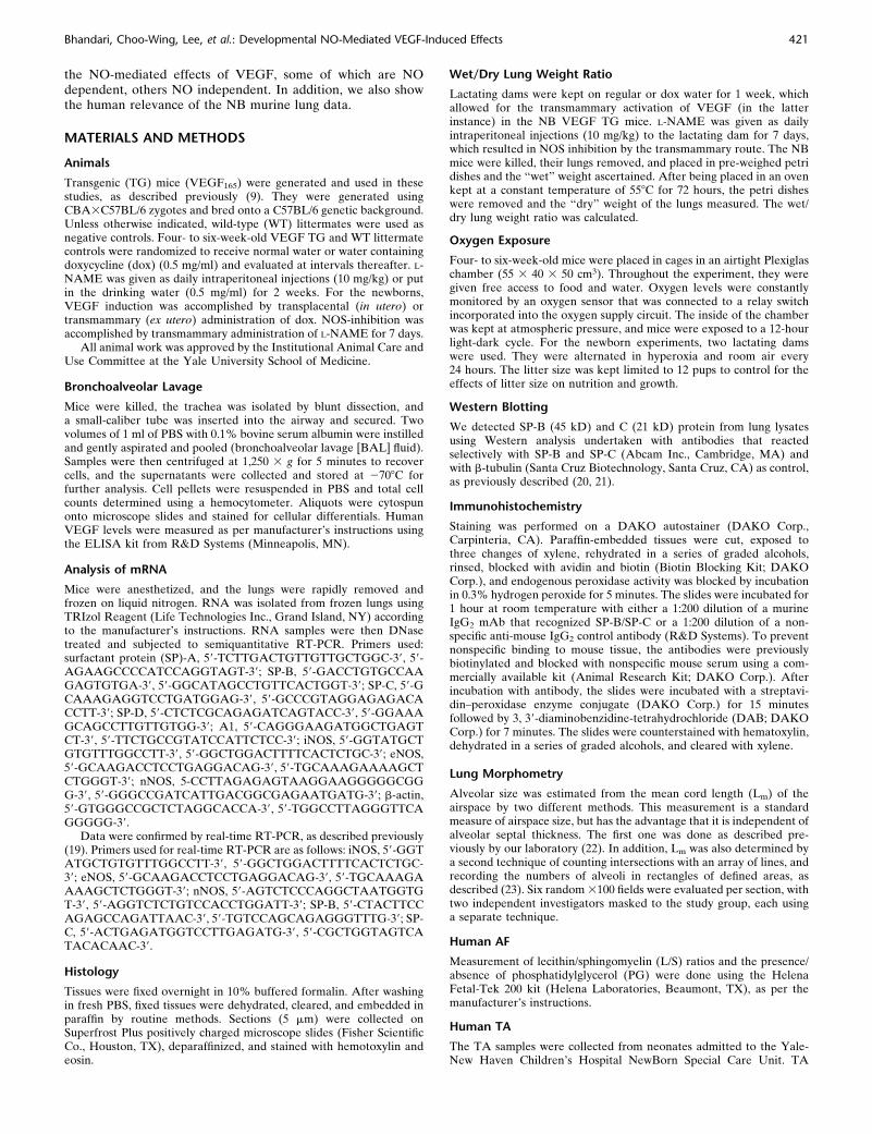
In our earlier report, we have noted that VEGF induces iNOS andeNOS in the adult murine lung, but not nNOS (15). To address theroles of NO and NOS in the pathogenesis of VEGF-inducedpulmonary alterations, we evaluated the levels of mRNA encod-ing the three NOS isoforms in WT and VEGF TG NB mice. Incontrast to the observation in the adult, we note that by postnatal(PN) Day 2 of VEGF induction, there was an impressive increasein the mRNA of all three NOS isoforms in the developing murinelung (Figure 1A). These data were confirmed by real-time RT-PCR (Figures 1B–1D). The levels of mRNA encoding all threeNOS isoforms in lungs from WT or TG mice on normal waterwere near or below the limits of detection our assay (data notshown). Thus, VEGF is a potent and selective stimulator of iNOS,eNOS, and nNOS in the developing murine lung.
Role of NO in VEGF-Induced Pulmonary Hemosiderosis in the
Adult and NB Lungs
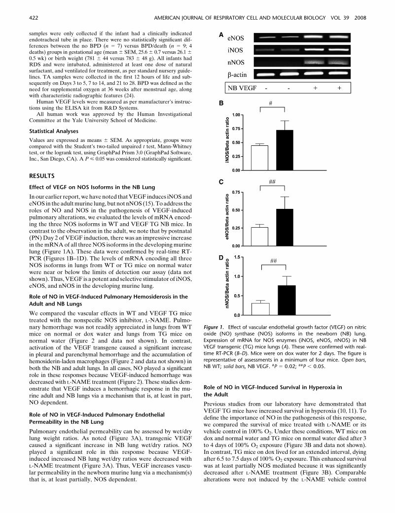
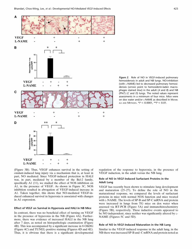
We compared the vascular effects in WT and VEGF TG micetreated with the nonspecific NOS inhibitor, L-NAME. Pulmo-nary hemorrhage was not readily appreciated in lungs from WTmice on normal or dox water and lungs from TG mice onnormal water (Figure 2 and data not shown). In contrast,activation of the VEGF transgene caused a significant increasein pleural and parenchymal hemorrhage and the accumulation ofhemosiderin-laden macrophages (Figure 2 and data not shown) inboth the NB and adult lungs. In all cases, NO played a significantrole in these responses because VEGF-induced hemorrhage wasdecreased with L-NAME treatment (Figure 2). These studies dem-onstrate that VEGF induces a hemorrhagic response in the mu-rine adult and NB lungs via a mechanism that is, at least in part,NO dependent.
Role of NO in VEGF-Induced Pulmonary Endothelial
Permeability in the NB Lung
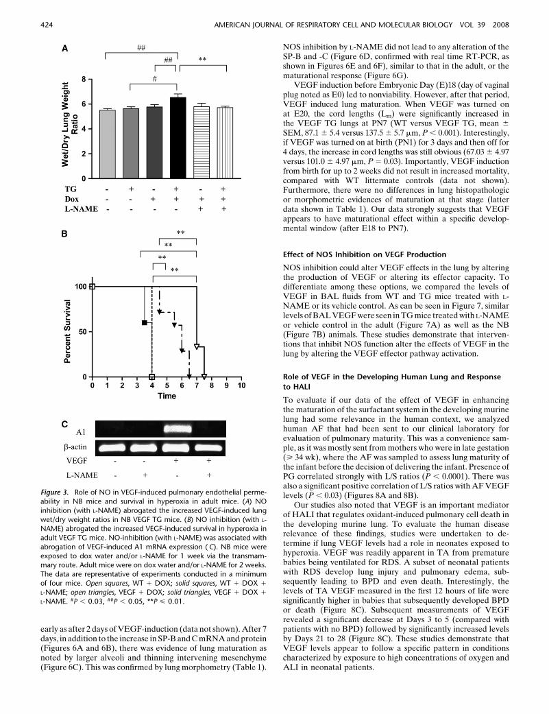
Pulmonary endothelial permeability can be assessed by wet/drylung weight ratios. As noted (Figure 3A), transgenic VEGFcaused a significant increase in NB lung wet/dry ratios. NOplayed a significant role in this response because VEGF-induced increased NB lung wet/dry ratios were decreased withL-NAME treatment (Figure 3A). Thus, VEGF increases vascu-lar permeability in the newborn murine lung via a mechanism(s)that is, at least partially, NOS dependent.
Role of NO in VEGF-Induced Survival in Hyperoxia in
the Adult
Previous studies from our laboratory have demonstrated thatVEGF TG mice have increased survival in hyperoxia (10, 11). Todefine the importance of NO in the pathogenesis of this response,we compared the survival of mice treated with L-NAME or itsvehicle control in 100% O2. Under these conditions, WT mice ondox and normal water and TG mice on normal water died after 3to 4 days of 100% O2 exposure (Figure 3B and data not shown).In contrast, TG mice on dox lived for an extended interval, dyingafter 6.5 to 7.5 days of 100% O2 exposure. This enhanced survivalwas at least partially NOS mediated because it was significantlydecreased after L-NAME treatment (Figure 3B). Comparablealterations were not induced by the L-NAME vehicle control
Figure 1. Effect of vascular endothelial growth factor (VEGF) on nitricoxide (NO) synthase (NOS) isoforms in the newborn (NB) lung.
Expression of mRNA for NOS enzymes (iNOS, eNOS, nNOS) in NB
VEGF transgenic (TG) mice lungs (A). These were confirmed with real-
time RT-PCR (B–D). Mice were on dox water for 2 days. The figure isrepresentative of assessments in a minimum of four mice. Open bars,
NB WT; solid bars, NB VEGF. #P 5 0.02; ##P , 0.05.
422 AMERICAN JOURNAL OF RESPIRATORY CELL AND MOLECULAR BIOLOGY VOL 39 2008
(Figure 3B). Thus, VEGF enhances survival in the setting ofoxidant-induced lung injury via a mechanism that is, at least inpart, NO mediated. Since VEGF-induced protection in HALIwas, in part, mediated by a member of the Bcl-2 family,specifically A1 (11), we studied the effect of NOS inhibition onA1, in the presence of VEGF. As shown in Figure 3C, NOSinhibition resulted in abrogation of VEGF-induced increase inA1. Taken together, this shows that NO-mediated VEGF-in-duced enhanced survival in hyperoxia is associated with changesin A1 expression.
Effect of VEGF on Survival in Hyperoxia and HALI in NB Mice
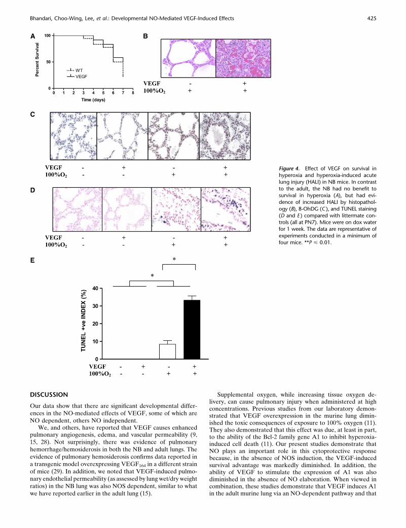
In contrast, there was no beneficial effect of turning on VEGFin the presence of hyperoxia in the NB (Figure 4A). Further-more, there was evidence of increased HALI in the NB lungafter 7 days, as noted on histopathologic examination (Figure4B). This was accompanied by a significant increase in 8-OhDG(Figure 4C) and TUNEL-positive staining (Figures 4D and 4E).Thus, it is obvious that there is a significant developmental
regulation of the response to hyperoxia, in the presence ofVEGF induction, in the adult versus the NB lung.
Role of NO in VEGF-Induced Surfactant Proteins in the
Adult Lung
VEGF has recently been shown to stimulate lung developmentand maturation (25–27). To define the role of NO in thematurational response, we compared the levels of surfactantproteins in mice with normal NOS function and mice treatedwith L-NAME. The levels of SP-B and SP-C mRNA and proteinwere increased in lungs from TG mice on dox water whenassessed via RT-PCR (Figure 5A) and immunohistochemistry(Figure 5B), respectively. These inductive events appeared tobe NO independent, since neither was significantly altered by L-NAME (Figures 5C and 5D).
Role of NO in VEGF-Induced Maturation in the NB Lung
Similar to the VEGF-induced response in the adult lung, in theNB there was increased SP-B and -C mRNA and protein noted as
Figure 2. Role of NO in VEGF-induced pulmonaryhemosiderosis in adult and NB lungs. NO-inhibition
(with L-NAME) led to decreased pulmonary hemosi-
derosis (arrows point to hemosiderin-laden macro-
phages stained blue) in the adult (A and B) and NB(PN7) (C and D) lungs. The noted values represent
assessments in a minimum of four mice. Mice were
on dox water and/or L-NAME as described in MATERI-
ALS AND METHODS. *P < 0.0001, **P < 0.01.
Bhandari, Choo-Wing, Lee, et al.: Developmental NO-Mediated VEGF-Induced Effects 423
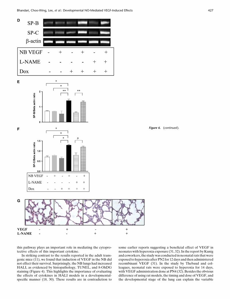
early as after 2 days of VEGF-induction (data not shown). After 7days, in addition to the increase in SP-B and C mRNA and protein(Figures 6A and 6B), there was evidence of lung maturation asnoted by larger alveoli and thinning intervening mesenchyme(Figure 6C). This was confirmed by lung morphometry (Table 1).
NOS inhibition by L-NAME did not lead to any alteration of theSP-B and -C (Figure 6D, confirmed with real time RT-PCR, asshown in Figures 6E and 6F), similar to that in the adult, or thematurational response (Figure 6G).
VEGF induction before Embryonic Day (E)18 (day of vaginalplug noted as E0) led to nonviability. However, after that period,VEGF induced lung maturation. When VEGF was turned onat E20, the cord lengths (Lm) were significantly increased inthe VEGF TG lungs at PN7 (WT versus VEGF TG, mean 6
SEM, 87.1 6 5.4 versus 137.5 6 5.7 mm, P , 0.001). Interestingly,if VEGF was turned on at birth (PN1) for 3 days and then off for4 days, the increase in cord lengths was still obvious (67.03 6 4.97versus 101.0 6 4.97 mm, P 5 0.03). Importantly, VEGF inductionfrom birth for up to 2 weeks did not result in increased mortality,compared with WT littermate controls (data not shown).Furthermore, there were no differences in lung histopathologicor morphometric evidences of maturation at that stage (latterdata shown in Table 1). Our data strongly suggests that VEGFappears to have maturational effect within a specific develop-mental window (after E18 to PN7).
Effect of NOS Inhibition on VEGF Production
NOS inhibition could alter VEGF effects in the lung by alteringthe production of VEGF or altering its effector capacity. Todifferentiate among these options, we compared the levels ofVEGF in BAL fluids from WT and TG mice treated with L-NAME or its vehicle control. As can be seen in Figure 7, similarlevels of BAL VEGF were seen in TG mice treated with L-NAMEor vehicle control in the adult (Figure 7A) as well as the NB(Figure 7B) animals. These studies demonstrate that interven-tions that inhibit NOS function alter the effects of VEGF in thelung by altering the VEGF effector pathway activation.
Role of VEGF in the Developing Human Lung and Response
to HALI
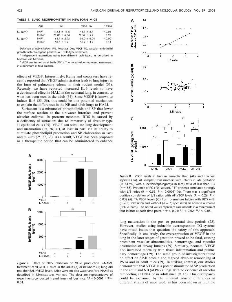
To evaluate if our data of the effect of VEGF in enhancingthe maturation of the surfactant system in the developing murinelung had some relevance in the human context, we analyzedhuman AF that had been sent to our clinical laboratory forevaluation of pulmonary maturity. This was a convenience sam-ple, as it was mostly sent from mothers who were in late gestation(> 34 wk), where the AF was sampled to assess lung maturity ofthe infant before the decision of delivering the infant. Presence ofPG correlated strongly with L/S ratios (P , 0.0001). There wasalso a significant positive correlation of L/S ratios with AF VEGFlevels (P , 0.03) (Figures 8A and 8B).
Our studies also noted that VEGF is an important mediatorof HALI that regulates oxidant-induced pulmonary cell death inthe developing murine lung. To evaluate the human diseaserelevance of these findings, studies were undertaken to de-termine if lung VEGF levels had a role in neonates exposed tohyperoxia. VEGF was readily apparent in TA from prematurebabies being ventilated for RDS. A subset of neonatal patientswith RDS develop lung injury and pulmonary edema, sub-sequently leading to BPD and even death. Interestingly, thelevels of TA VEGF measured in the first 12 hours of life weresignificantly higher in babies that subsequently developed BPDor death (Figure 8C). Subsequent measurements of VEGFrevealed a significant decrease at Days 3 to 5 (compared withpatients with no BPD) followed by significantly increased levelsby Days 21 to 28 (Figure 8C). These studies demonstrate thatVEGF levels appear to follow a specific pattern in conditionscharacterized by exposure to high concentrations of oxygen andALI in neonatal patients.
Figure 3. Role of NO in VEGF-induced pulmonary endothelial perme-
ability in NB mice and survival in hyperoxia in adult mice. (A) NO
inhibition (with L-NAME) abrogated the increased VEGF-induced lung
wet/dry weight ratios in NB VEGF TG mice. (B) NO inhibition (with L-NAME) abrogated the increased VEGF-induced survival in hyperoxia in
adult VEGF TG mice. NO-inhibition (with L-NAME) was associated with
abrogation of VEGF-induced A1 mRNA expression ( C). NB mice were
exposed to dox water and/or L-NAME for 1 week via the transmam-mary route. Adult mice were on dox water and/or L-NAME for 2 weeks.
The data are representative of experiments conducted in a minimum
of four mice. Open squares, WT 1 DOX; solid squares, WT 1 DOX 1
L-NAME; open triangles, VEGF 1 DOX; solid triangles, VEGF 1 DOX 1
L-NAME. #P , 0.03, ##P , 0.05, **P < 0.01.
424 AMERICAN JOURNAL OF RESPIRATORY CELL AND MOLECULAR BIOLOGY VOL 39 2008
DISCUSSION
Our data show that there are significant developmental differ-ences in the NO-mediated effects of VEGF, some of which areNO dependent, others NO independent.
We, and others, have reported that VEGF causes enhancedpulmonary angiogenesis, edema, and vascular permeability (9,15, 28). Not surprisingly, there was evidence of pulmonaryhemorrhage/hemosiderosis in both the NB and adult lungs. Theevidence of pulmonary hemosiderosis confirms data reported ina transgenic model overexpressing VEGF164 in a different strainof mice (29). In addition, we noted that VEGF-induced pulmo-nary endothelial permeability (as assessed by lung wet/dry weightratios) in the NB lung was also NOS dependent, similar to whatwe have reported earlier in the adult lung (15).
Supplemental oxygen, while increasing tissue oxygen de-livery, can cause pulmonary injury when administered at highconcentrations. Previous studies from our laboratory demon-strated that VEGF overexpression in the murine lung dimin-ished the toxic consequences of exposure to 100% oxygen (11).They also demonstrated that this effect was due, at least in part,to the ability of the Bcl-2 family gene A1 to inhibit hyperoxia-induced cell death (11). Our present studies demonstrate thatNO plays an important role in this cytoprotective responsebecause, in the absence of NOS induction, the VEGF-inducedsurvival advantage was markedly diminished. In addition, theability of VEGF to stimulate the expression of A1 was alsodiminished in the absence of NO elaboration. When viewed incombination, these studies demonstrate that VEGF induces A1in the adult murine lung via an NO-dependent pathway and that
Figure 4. Effect of VEGF on survival inhyperoxia and hyperoxia-induced acute
lung injury (HALI) in NB mice. In contrast
to the adult, the NB had no benefit tosurvival in hyperoxia (A), but had evi-
dence of increased HALI by histopathol-
ogy (B), 8-OhDG (C ), and TUNEL staining
(D and E ) compared with littermate con-trols (all at PN7). Mice were on dox water
for 1 week. The data are representative of
experiments conducted in a minimum of
four mice. **P < 0.01.
Bhandari, Choo-Wing, Lee, et al.: Developmental NO-Mediated VEGF-Induced Effects 425
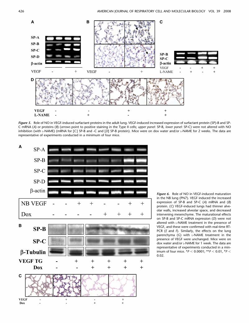
Figure 5. Role of NO in VEGF-induced surfactant proteins in the adult lung. VEGF-induced increased expression of surfactant protein (SP)-B and SP-C mRNA (A) or proteins (B) (arrows point to positive staining in the Type II cells; upper panel: SP-B, lower panel: SP-C) were not altered with NO
inhibition (with L-NAME) (mRNA for [C ] SP-B and -C and [D] SP-B protein). Mice were on dox water and/or L-NAME for 2 weeks. The data are
representative of experiments conducted in a minimum of four mice.
Figure 6. Role of NO in VEGF-induced maturationin the NB lung (PN7). VEGF induced the increased
expression of SP-B and SP-C (A) mRNA and (B)
protein. (C) VEGF-induced lungs had thinner alve-olar walls, increased alveolar space, and decreased
intervening mesenchyme. The maturational effects
on SP-B and SP-C mRNA expression (D) were not
altered with L-NAME treatment in the presence ofVEGF, and these were confirmed with real-time RT-
PCR (E and F). Similarly, the effects on the lung
parenchyma (G) with L-NAME treatment in the
presence of VEGF were unchanged. Mice were ondox water and/or L-NAME for 1 week. The data are
representative of experiments conducted in a min-
imum of four mice. *P , 0.0001; **P , 0.01, #P ,
0.02.
426 AMERICAN JOURNAL OF RESPIRATORY CELL AND MOLECULAR BIOLOGY VOL 39 2008
this pathway plays an important role in mediating the cytopro-tective effects of this important cytokine.
In striking contrast to the results reported in the adult trans-genic mice (11), we found that induction of VEGF in the NB didnot effect their survival. Surprisingly, the NB lungs had increasedHALI, as evidenced by histopathology, TUNEL, and 8-OhDGstaining (Figure 4). This highlights the importance of evaluatingthe effects of cytokines in HALI models in a developmental-specific manner (18, 30). These results are in contradiction to
some earlier reports suggesting a beneficial effect of VEGF inneonates with hyperoxia exposure (31, 32). In the report by Kunigand coworkers, the study was conducted in neonatal rats that wereexposed to hyperoxia after PN2 for 12 days and then administeredrecombinant VEGF (31). In the study by Thebaud and col-leagues, neonatal rats were exposed to hyperoxia for 14 days,with VEGF administration done at PN4 (32). Besides the obviousdifference of using rat models, the timing and dose of VEGF, andthe developmental stage of the lung can explain the variable
Figure 6. (continued ).
Bhandari, Choo-Wing, Lee, et al.: Developmental NO-Mediated VEGF-Induced Effects 427
effects of VEGF. Interestingly, Kunig and coworkers have re-cently reported that VEGF administration leads to lung injury inthe form of pulmonary edema in their rodent model (33).Recently, we have reported increased IL-6 levels to havea detrimental effect in HALI in the neonatal lung, in contrast towhat has been seen in the adult (34). Since VEGF is known toinduce IL-6 (35, 36), this could be one potential mechanismto explain the differences in the NB and adult lungs to HALI.
Surfactant is a mixture of phospholipids and SP that lowerthe surface tension at the air–water interface and preventalveolar collapse. In preterm neonates, RDS is caused bya deficiency of surfactant due to immaturity of alveolar typeII epithelial cells (25). VEGF can stimulate lung developmentand maturation (25, 26, 37), at least in part, via its ability tostimulate phospholipid production and SP elaboration in vivoand in vitro (25, 27, 38). As a result, VEGF has been proposedas a therapeutic option that can be administered to enhance
lung maturation in the pre- or postnatal time periods (25).However, studies using inducible overexpression TG systemshave raised issues that question the safety of this approach.Specifically, in one study, the overexpression of VEGF in thelung in the later stages of gestation proved to be fatal, causingprominent vascular abnormalities, hemorrhage, and vascularobstruction of airway lumens (39). Similarly, neonatal VEGFalso increased mortality with tissue inflammation and pulmo-nary hemorrhage (29). The same group of investigators foundno effect on SP-B protein and marked alveolar remodeling atPN14 and in adult mice (29). In striking contrast, our studiesdemonstrate that VEGF is a potent stimulator of SP productionin the adult and NB (at PN7) lungs, with no evidence of alveolarremodeling at PN14 or in adult mice (9, 15). This discrepancycould be explained by the inherent genetic differences indifferent strains of mice used, as has been shown in multiple
Figure 7. Effect of NOS inhibition on VEGF production. L-NAME
treatment of VEGFTG1 mice in the adult (A) or newborn (B) lung did
not alter BAL hVEGF levels. Mice were on dox water and/or L-NAME as
described in MATERIALS AND METHODS. The data are representative ofexperiments conducted in a minimum of four mice. *P < 0.0001; **P <
0.01.
Figure 8. VEGF levels in human amniotic fluid (AF) and trachealaspirate (TA). AF samples from mothers with infants in late gestation
(> 34 wk) with a lecithin/sphingomyelin (L/S) ratio of less than 3.5
(n 5 58). Presence of PG (‘‘0’’ absent, ‘‘1’’ present) correlated strongly
with L/S ratios (R 5 0.55, P , 0.0001) (A). There was a significantpositive correlation of L/S ratios with AF VEGF levels (R 5 0.26, P ,
0.03) (B). TA VEGF levels (C ) from premature babies with RDS with
(n 5 9; solid bars) and without (n 5 7; open bars) an adverse outcome(BPD /Death). The noted values represent assessments in a minimum of
four infants at each time point. **P < 0.01; #P , 0.02; ##P < 0.05.
TABLE 1. LUNG MORPHOMETRY IN NEWBORN MICE
Age WT VEGF TG P Value
Lm (mm)* PN7† 112.1 6 13.6 143.1 6 8.7 ,0.05
PN14† 71.86 6 6.84 71.52 6 5.2 0.97
Lm (mm)* PN7† 63.7 6 2.95 104.0 6 6.04 ,0.001
PN14† 50.6 6 1.9 56.2 6 3.2 0.14
Definition of abbreviations: PN, Postnatal Day; VEGF TG, vascular endothelial
growth factor transgene positive; WT, wild-type littermate.
* Independent evaluations using two different techniques, as described in
MATERIALS AND METHODS.† VEGF was turned on at birth (PN1). The noted values represent assessments
in a minimum of four animals.
428 AMERICAN JOURNAL OF RESPIRATORY CELL AND MOLECULAR BIOLOGY VOL 39 2008
other model systems (40–43), as well as the timing. Importantly,our studies also demonstrate that the effects on surfactant werenot mediated by NO, as they were not altered by the NOS-based intervention that was employed.
Our data on human AF VEGF levels that correlate well withthe surfactant phospholipids support our contention that VEGFdoes have a role to play in lung maturation in the humancontext. Our data are in accord with another report that VEGFlevels may reflect pulmonary maturity as evidenced by thesignificant positive correlation (P 5 0.03) of TA VEGF levelsfrom premature babies with their AF L/S ratios (44). In a recentstudy, preterm infants with more severe RDS had lower VEGFdetected by immunhistochemistry (45), while another studyreported that cord blood VEGF elevation was significantlycorrelated with absence of RDS (46).
We found increased TA VEGF levels in the first 12 hours oflife in babies with RDS who had an adverse outcome. Others(47) have not found such an association. This could be,however, due to the variable timing or measurement techniquethat was used or the different characteristics of the patientpopulation studied.
Interestingly, we found that the pattern seen in the babies withno BPD shows an initial low VEGF level, followed by a sub-stantial increase, with a subsequent decrease or plateau. In thebabies with an adverse outcome (BPD/death), there was an initialspike, followed by a decline, and then a subsequent rise. As can benoted in Figure 8, Day 7 to 14 and Day 21 to 28 VEGF levels weresignificantly higher than Day 3 to 5 VEGF levels, and Day 21 to 28VEGF levels were significantly higher than Day 7 to 14 VEGFlevels, for the babies who had an adverse outcome (BPD/death).These patterns/relationships held true even when analyzed byone-way ANOVA analyses, with P , 0.04 for the ‘‘no BPD’’group and P 5 0.02 for the ‘‘BPD/died’’ group. This is in accordincreased VEGF staining in the baboon model of BPD (48) aswell as human infants with BPD (45, 49). The pattern of TAVEGF levels in babies with an adverse outcome that we havefound has been shown to occur during acute myocardial in-farction in human adults (50), ALI in adult animals (51, 52), andHALI in NB animals (53), but to our knowledge, our data provideevidence, for the first time, in the human NB lung. We specu-late that the initial increase in VEGF levels could potentiallycontribute to lung injury/pulmonary edema leading to cellulardamage/death, which in turn, results in decreased VEGF levels.Subsequently, there is a surge in VEGF levels associated withlung repair.
In summary, these studies demonstrate that, in contrast tothe adult lung, VEGF is a potent stimulator of nNOS (inaddition to iNOS and eNOS) in the murine NB lung. We reportthat VEGF-induced pulmonary hemosiderosis is NO depen-dent. In contrast, the VEGF-induced surfactant production inthe adult and NB lung is NO independent. Similarly, theVEGF-induced lung maturational effect noted in the NB lungis NO independent. While the enhanced survival in hyperoxia inthe adult was partly NO dependent, there was enhanced HALIin the NB. Our data show that there are significant develop-mental differences in the NO-mediated effects of VEGF, someof which are NO dependent, others NO independent.
Exaggerated VEGF production has been proposed to playan important role in the pathogenesis of a wide variety ofdiseases, including tumor neovascularization, asthma, pulmo-nary edema, atherosclerosis, and the retinopathies of the NBand the diabetic (5, 9, 51, 54–57). The present studies suggestthat the pathologic effects of VEGF in these disorders may becontrolled by interventions that control NO production. Incontrast, a relative deficiency of VEGF has been proposed tocontribute to the pathogenesis of diseases like RDS in the
newborn (25, 46). Our human data is in accord with the same.The present studies suggest that the treatment of this disordercan be optimally accomplished when VEGF is combined withNOS inhibitors. Our study also suggests that timing of deliveryof VEGF could be critical for its effects. This establishes theVEGF-NO pathway as a worthwhile focus for future inves-tigations in VEGF-mediated disorders that can be exploited tomaximize the utility and safety of VEGF as a therapeutic agent.
Conflict of Interest Statement: None of the authors has a financial relationshipwith a commercial entity that has an interest in the subject of this manuscript.
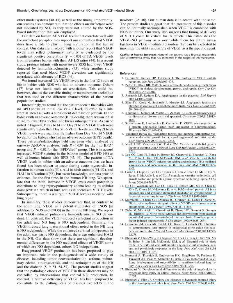
References
1. Ferrara N, Gerber HP, LeCouter J. The biology of VEGF and itsreceptors. Nat Med 2003;9:669–676.
2. Zelzer E, Olsen BR. Multiple roles of vascular endothelial growth factor(VEGF) in skeletal development, growth, and repair. Curr Top DevBiol 2005;65:169–187.
3. Reynolds LP, Redmer DA. Angiogenesis in the placenta. Biol Reprod2001;64:1033–1040.
4. Silha JV, Krsek M, Sucharda P, Murphy LJ. Angiogenic factors areelevated in overweight and obese individuals. Int J Obes Disord 2005;29:1308–1314.
5. Khurana R, Simons M, Martin JF, Zachary IC. Role of angiogenesis incardiovascular disease: a critical appraisal. Circulation 2005;112:1813–1824.
6. Storkebaum E, Lambrechts D, Carmeliet P. VEGF: once regarded asa specific angiogenic factor, now implicated in neuroprotection.Bioessays 2004;26:943–954.
7. Wilkinson-Berka JL. Vasoactive factors and diabetic retinopathy: vas-cular endothelial growth factor, cycoloxygenase-2 and nitric oxide.Curr Pharm Des 2004;10:3331–3348.
8. Voelkel NF, Vandivier RW, Tuder RM. Vascular endothelial growthfactor in the lung. Am J Physiol Lung Cell Mol Physiol 2006;290:L209–L221.
9. Lee CG, Link H, Baluk P, Homer RJ, Chapoval S, Bhandari V, KangMJ, Cohn L, Kim YK, McDonald DM, et al. Vascular endothelialgrowth factor (VEGF) induces remodeling and enhances TH2-mediatedsensitization and inflammation in the lung. Nat Med 2004;10:1095–1103.
10. Corne J, Chupp G, Lee CG, Homer RJ, Zhu Z, Chen Q, Ma B, Du Y,Roux F, McArdle J, et al. IL-13 stimulates vascular endothelial cellgrowth factor and protects against hyperoxic acute lung injury. J ClinInvest 2000;106:783–791.
11. He CH, Waxman AB, Lee CG, Link H, Rabach ME, Ma B, Chen Q,Zhu Z, Zhong M, Nakayama K, et al. Bcl-2-related protein A1 is anendogenous and cytokine-stimulated mediator of cytoprotection inhyperoxic acute lung injury. J Clin Invest 2005;115:1039–1048.
12. Morbidelli L, Chang CH, Douglas JG, Granger HJ, Ledda F, Ziche M.Nitric oxide mediates mitogenic effect of VEGF on coronary venularendothelium. Am J Physiol 1996;270:H411–H415.
13. Ziche M, Morbidelli L, Choudhuri R, Zhang HT, Donnini S, GrangerHJ, Bicknell R. Nitric oxide synthase lies downstream from vascularendothelial growth factor-induced but not basic fibroblast growthfactor-induced angiogenesis. J Clin Invest 1997;99:2625–2634.
14. Leuwerke SM, Kaza AK, Tribble CG, Kron IL, Laubach VE. Inhibitionof compensatory lung growth in endothelial nitric oxide synthase-deficient mice. Am J Physiol Lung Cell Mol Physiol 2002;282:L1272–L1278.
15. Bhandari V, Choo-Wing R, Chapoval SP, Lee CG, Tang C, Kim YK, MaB, Baluk P, Lin MI, McDonald DM, et al. Essential role of nitricoxide in VEGF-induced, asthma-like angiogenic, inflammatory, mu-cus, and physiologic responses in the lung. Proc Natl Acad Sci USA2006;103:11021–11026.
16. Kornecki A, Tsuchida S, Ondiveeran HK, Engelberts D, Frndova H,Tanswell AK, Post M, McKerlie C, Belik J, Fox-Robichaud A, et al.Lung development and susceptibility to ventilator-induced lung in-jury. Am J Respir Crit Care Med 2005;171:743–752.
17. Bhandari V. Developmental differences in the role of interleukins inhyperoxic lung injury in animal models. Front Biosci 2002;7:d1624–d1633.
18. Bhandari V, Elias JA. Cytokines in tolerance to hyperoxia-induced injuryin the developing and adult lung. Free Radic Biol Med 2006;41:4–18.
Bhandari, Choo-Wing, Lee, et al.: Developmental NO-Mediated VEGF-Induced Effects 429
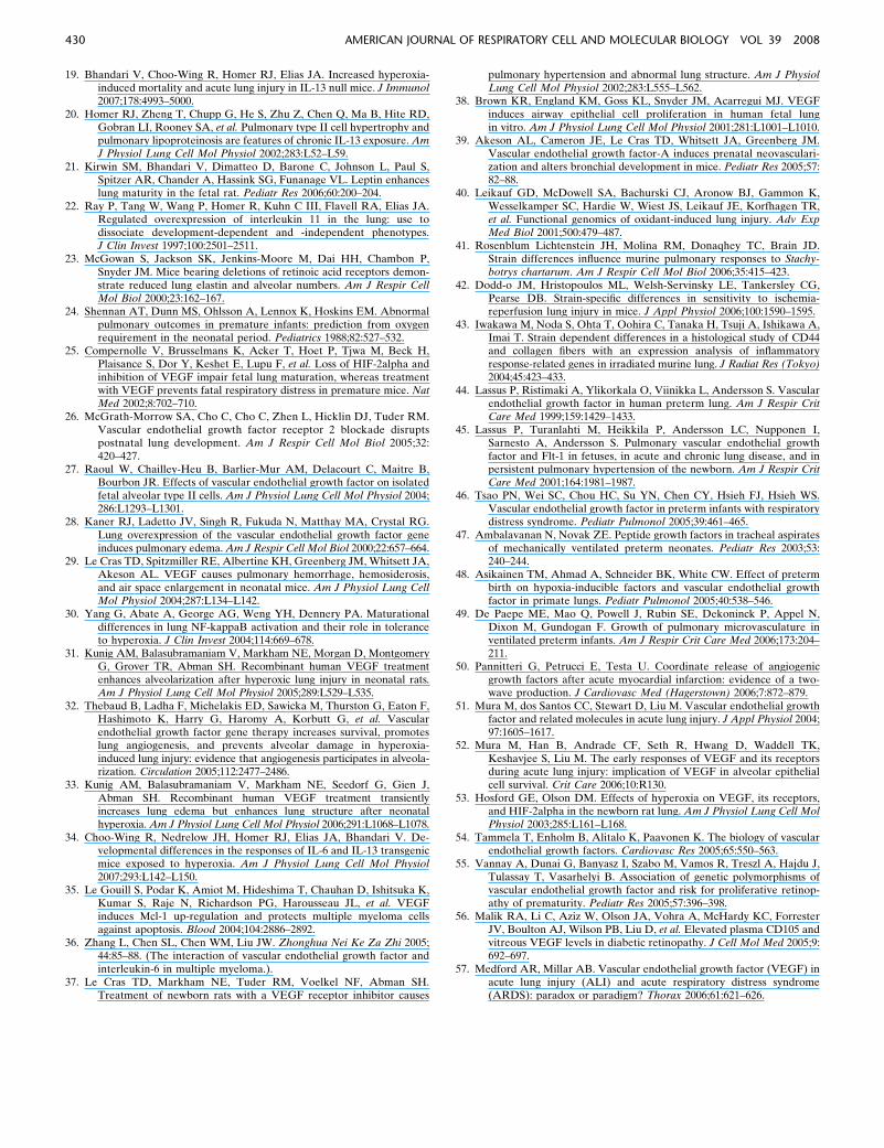
19. Bhandari V, Choo-Wing R, Homer RJ, Elias JA. Increased hyperoxia-induced mortality and acute lung injury in IL-13 null mice. J Immunol2007;178:4993–5000.
20. Homer RJ, Zheng T, Chupp G, He S, Zhu Z, Chen Q, Ma B, Hite RD,Gobran LI, Rooney SA, et al. Pulmonary type II cell hypertrophy andpulmonary lipoproteinosis are features of chronic IL-13 exposure. AmJ Physiol Lung Cell Mol Physiol 2002;283:L52–L59.
21. Kirwin SM, Bhandari V, Dimatteo D, Barone C, Johnson L, Paul S,Spitzer AR, Chander A, Hassink SG, Funanage VL. Leptin enhanceslung maturity in the fetal rat. Pediatr Res 2006;60:200–204.
22. Ray P, Tang W, Wang P, Homer R, Kuhn C III, Flavell RA, Elias JA.Regulated overexpression of interleukin 11 in the lung: use todissociate development-dependent and -independent phenotypes.J Clin Invest 1997;100:2501–2511.
23. McGowan S, Jackson SK, Jenkins-Moore M, Dai HH, Chambon P,Snyder JM. Mice bearing deletions of retinoic acid receptors demon-strate reduced lung elastin and alveolar numbers. Am J Respir CellMol Biol 2000;23:162–167.
24. Shennan AT, Dunn MS, Ohlsson A, Lennox K, Hoskins EM. Abnormalpulmonary outcomes in premature infants: prediction from oxygenrequirement in the neonatal period. Pediatrics 1988;82:527–532.
25. Compernolle V, Brusselmans K, Acker T, Hoet P, Tjwa M, Beck H,Plaisance S, Dor Y, Keshet E, Lupu F, et al. Loss of HIF-2alpha andinhibition of VEGF impair fetal lung maturation, whereas treatmentwith VEGF prevents fatal respiratory distress in premature mice. NatMed 2002;8:702–710.
26. McGrath-Morrow SA, Cho C, Cho C, Zhen L, Hicklin DJ, Tuder RM.Vascular endothelial growth factor receptor 2 blockade disruptspostnatal lung development. Am J Respir Cell Mol Biol 2005;32:420–427.
27. Raoul W, Chailley-Heu B, Barlier-Mur AM, Delacourt C, Maitre B,Bourbon JR. Effects of vascular endothelial growth factor on isolatedfetal alveolar type II cells. Am J Physiol Lung Cell Mol Physiol 2004;286:L1293–L1301.
28. Kaner RJ, Ladetto JV, Singh R, Fukuda N, Matthay MA, Crystal RG.Lung overexpression of the vascular endothelial growth factor geneinduces pulmonary edema. Am J Respir Cell Mol Biol 2000;22:657–664.
29. Le Cras TD, Spitzmiller RE, Albertine KH, Greenberg JM, Whitsett JA,Akeson AL. VEGF causes pulmonary hemorrhage, hemosiderosis,and air space enlargement in neonatal mice. Am J Physiol Lung CellMol Physiol 2004;287:L134–L142.
30. Yang G, Abate A, George AG, Weng YH, Dennery PA. Maturationaldifferences in lung NF-kappaB activation and their role in toleranceto hyperoxia. J Clin Invest 2004;114:669–678.
31. Kunig AM, Balasubramaniam V, Markham NE, Morgan D, MontgomeryG, Grover TR, Abman SH. Recombinant human VEGF treatmentenhances alveolarization after hyperoxic lung injury in neonatal rats.Am J Physiol Lung Cell Mol Physiol 2005;289:L529–L535.
32. Thebaud B, Ladha F, Michelakis ED, Sawicka M, Thurston G, Eaton F,Hashimoto K, Harry G, Haromy A, Korbutt G, et al. Vascularendothelial growth factor gene therapy increases survival, promoteslung angiogenesis, and prevents alveolar damage in hyperoxia-induced lung injury: evidence that angiogenesis participates in alveola-rization. Circulation 2005;112:2477–2486.
33. Kunig AM, Balasubramaniam V, Markham NE, Seedorf G, Gien J,Abman SH. Recombinant human VEGF treatment transientlyincreases lung edema but enhances lung structure after neonatalhyperoxia. Am J Physiol Lung Cell Mol Physiol 2006;291:L1068–L1078.
34. Choo-Wing R, Nedrelow JH, Homer RJ, Elias JA, Bhandari V. De-velopmental differences in the responses of IL-6 and IL-13 transgenicmice exposed to hyperoxia. Am J Physiol Lung Cell Mol Physiol2007;293:L142–L150.
35. Le Gouill S, Podar K, Amiot M, Hideshima T, Chauhan D, Ishitsuka K,Kumar S, Raje N, Richardson PG, Harousseau JL, et al. VEGFinduces Mcl-1 up-regulation and protects multiple myeloma cellsagainst apoptosis. Blood 2004;104:2886–2892.
36. Zhang L, Chen SL, Chen WM, Liu JW. Zhonghua Nei Ke Za Zhi 2005;44:85–88. (The interaction of vascular endothelial growth factor andinterleukin-6 in multiple myeloma.).
37. Le Cras TD, Markham NE, Tuder RM, Voelkel NF, Abman SH.Treatment of newborn rats with a VEGF receptor inhibitor causes
pulmonary hypertension and abnormal lung structure. Am J PhysiolLung Cell Mol Physiol 2002;283:L555–L562.
38. Brown KR, England KM, Goss KL, Snyder JM, Acarregui MJ. VEGFinduces airway epithelial cell proliferation in human fetal lungin vitro. Am J Physiol Lung Cell Mol Physiol 2001;281:L1001–L1010.
39. Akeson AL, Cameron JE, Le Cras TD, Whitsett JA, Greenberg JM.Vascular endothelial growth factor-A induces prenatal neovasculari-zation and alters bronchial development in mice. Pediatr Res 2005;57:82–88.
40. Leikauf GD, McDowell SA, Bachurski CJ, Aronow BJ, Gammon K,Wesselkamper SC, Hardie W, Wiest JS, Leikauf JE, Korfhagen TR,et al. Functional genomics of oxidant-induced lung injury. Adv ExpMed Biol 2001;500:479–487.
41. Rosenblum Lichtenstein JH, Molina RM, Donaqhey TC, Brain JD.Strain differences influence murine pulmonary responses to Stachy-botrys chartarum. Am J Respir Cell Mol Biol 2006;35:415–423.
42. Dodd-o JM, Hristopoulos ML, Welsh-Servinsky LE, Tankersley CG,Pearse DB. Strain-specific differences in sensitivity to ischemia-reperfusion lung injury in mice. J Appl Physiol 2006;100:1590–1595.
43. Iwakawa M, Noda S, Ohta T, Oohira C, Tanaka H, Tsuji A, Ishikawa A,Imai T. Strain dependent differences in a histological study of CD44and collagen fibers with an expression analysis of inflammatoryresponse-related genes in irradiated murine lung. J Radiat Res (Tokyo)2004;45:423–433.
44. Lassus P, Ristimaki A, Ylikorkala O, Viinikka L, Andersson S. Vascularendothelial growth factor in human preterm lung. Am J Respir CritCare Med 1999;159:1429–1433.
45. Lassus P, Turanlahti M, Heikkila P, Andersson LC, Nupponen I,Sarnesto A, Andersson S. Pulmonary vascular endothelial growthfactor and Flt-1 in fetuses, in acute and chronic lung disease, and inpersistent pulmonary hypertension of the newborn. Am J Respir CritCare Med 2001;164:1981–1987.
46. Tsao PN, Wei SC, Chou HC, Su YN, Chen CY, Hsieh FJ, Hsieh WS.Vascular endothelial growth factor in preterm infants with respiratorydistress syndrome. Pediatr Pulmonol 2005;39:461–465.
47. Ambalavanan N, Novak ZE. Peptide growth factors in tracheal aspiratesof mechanically ventilated preterm neonates. Pediatr Res 2003;53:240–244.
48. Asikainen TM, Ahmad A, Schneider BK, White CW. Effect of pretermbirth on hypoxia-inducible factors and vascular endothelial growthfactor in primate lungs. Pediatr Pulmonol 2005;40:538–546.
49. De Paepe ME, Mao Q, Powell J, Rubin SE, Dekoninck P, Appel N,Dixon M, Gundogan F. Growth of pulmonary microvasculature inventilated preterm infants. Am J Respir Crit Care Med 2006;173:204–211.
50. Pannitteri G, Petrucci E, Testa U. Coordinate release of angiogenicgrowth factors after acute myocardial infarction: evidence of a two-wave production. J Cardiovasc Med (Hagerstown) 2006;7:872–879.
51. Mura M, dos Santos CC, Stewart D, Liu M. Vascular endothelial growthfactor and related molecules in acute lung injury. J Appl Physiol 2004;97:1605–1617.
52. Mura M, Han B, Andrade CF, Seth R, Hwang D, Waddell TK,Keshavjee S, Liu M. The early responses of VEGF and its receptorsduring acute lung injury: implication of VEGF in alveolar epithelialcell survival. Crit Care 2006;10:R130.
53. Hosford GE, Olson DM. Effects of hyperoxia on VEGF, its receptors,and HIF-2alpha in the newborn rat lung. Am J Physiol Lung Cell MolPhysiol 2003;285:L161–L168.
54. Tammela T, Enholm B, Alitalo K, Paavonen K. The biology of vascularendothelial growth factors. Cardiovasc Res 2005;65:550–563.
55. Vannay A, Dunai G, Banyasz I, Szabo M, Vamos R, Treszl A, Hajdu J,Tulassay T, Vasarhelyi B. Association of genetic polymorphisms ofvascular endothelial growth factor and risk for proliferative retinop-athy of prematurity. Pediatr Res 2005;57:396–398.
56. Malik RA, Li C, Aziz W, Olson JA, Vohra A, McHardy KC, ForresterJV, Boulton AJ, Wilson PB, Liu D, et al. Elevated plasma CD105 andvitreous VEGF levels in diabetic retinopathy. J Cell Mol Med 2005;9:692–697.
57. Medford AR, Millar AB. Vascular endothelial growth factor (VEGF) inacute lung injury (ALI) and acute respiratory distress syndrome(ARDS): paradox or paradigm? Thorax 2006;61:621–626.
430 AMERICAN JOURNAL OF RESPIRATORY CELL AND MOLECULAR BIOLOGY VOL 39 2008
Related Documents