1 Decreased circulating and neutrophil mediated VEGF release in stable long-term cardiac transplant recipients Damien Vitiello, 1,2,3 Diana Chaar, 1,3 Paul-Eduard Neagoe, 1,2 Anique Ducharme, 3 Michel Carrier, 3 Guy B. Pelletier, 3 Normand Racine, 3 Mark Liszkowski, 3 Martin G. Sirois, 1,2* and Michel White, 1,3* 1 Research Center, Montreal Heart Institute and Université de Montréal, Montréal, Qc, Canada. 2 Pharmacology, Montreal Heart Institute and Université de Montréal, Montréal, Qc, Canada. 3 Medicine, Faculty of Medicine, Montreal Heart Institute and Université de Montréal. Montreal, Qc, Canada. Running Title: VEGF in cardiac transplantation Funding Sources: This study was funded by the Carolyn & Richard Renaud Chair in Heart Failure of the Montreal Heart Institute. Address correspondence to co-senior authors * : Dr. Michel White, Professor of Medicine, Université de Montréal and Montreal Heart Institute, Research Center, 5000 Belanger Street, Montreal, Canada H1T 1C8. Tel.: 514-376-3330 (# 3962). E-mail: [email protected], or Dr. Martin G. Sirois, Montreal Heart Institute and Université de Montréal, Research Center, 5000 Belanger Street, Montreal, Canada H1T 1C8. Tel.: 514-376-3330 (# 3583). E-mail: [email protected]

Welcome message from author
This document is posted to help you gain knowledge. Please leave a comment to let me know what you think about it! Share it to your friends and learn new things together.
Transcript

1
Decreased circulating and neutrophil mediated VEGF release in
stable long-term cardiac transplant recipients
Damien Vitiello,1,2,3 Diana Chaar,1,3 Paul-Eduard Neagoe,1,2 Anique Ducharme,3
Michel Carrier,3 Guy B. Pelletier,3 Normand Racine,3 Mark Liszkowski,3 Martin G. Sirois,1,2*
and Michel White,1,3*
1Research Center, Montreal Heart Institute and Université de Montréal, Montréal, Qc,
Canada. 2Pharmacology, Montreal Heart Institute and Université de Montréal, Montréal, Qc, Canada. 3Medicine, Faculty of Medicine, Montreal Heart Institute and Université de Montréal.
Montreal, Qc, Canada.
Running Title: VEGF in cardiac transplantation
Funding Sources: This study was funded by the Carolyn & Richard Renaud Chair in Heart
Failure of the Montreal Heart Institute.
Address correspondence to co-senior authors*: Dr. Michel White, Professor of Medicine,
Université de Montréal and Montreal Heart Institute, Research Center, 5000 Belanger Street,
Montreal, Canada H1T 1C8. Tel.: 514-376-3330 (# 3962). E-mail: [email protected],
or Dr. Martin G. Sirois, Montreal Heart Institute and Université de Montréal, Research
Center, 5000 Belanger Street, Montreal, Canada H1T 1C8. Tel.: 514-376-3330 (# 3583).
E-mail: [email protected]

2
Abstract
Background: Vascular endothelial growth factor (VEGF) may play a role on the allograft
remodelling following cardiac transplantation (CTx). We measured the circulating levels of
VEGF concomitantly with the proinflammatory (Interleukin-8; IL-8), anti-inflammatory
(IL-1 receptor antagonist; IL-1RA) and their release from neutrophils of CTx recipients.
Methods: Eighteen CTx recipients aged 49.6±3.1 years, being transplanted for 145 ± 20
months were age-matched to 35 healthy control (HC) subjects. Concomitantly to plasma
assessment, circulating neutrophils were isolated, purified and stimulated by vehicle (PBS),
N-formyl-Met-Leu-Phe (fMLP,10-7M), bacterial lipopolysaccharide (LPS,1 µg/mL), or
tumour necrosis factor alpha (TNF-α,10 ng/mL).
Results: Compared with HC, CTx recipients exhibited a decrease (-80%) in plasmatic
circulating levels of VEGF (225 ± 42 (HC) vs 44 ± 10 pg/mL (CTx); (p < 0.001). There were
no differences in the levels of IL-8 and IL-1RA. Under basal or stimulated conditions,
neutrophils from CTx patients exhibited a marked decrease ranging from -30 to -88% on their
capacity to release VEGF, IL-8 and IL-1RA upon stimulation.
Conclusions: Long-term CTx recipients exhibit a marked reduction in the circulating levels
of VEGF, as well as neutrophil-mediated release of VEGF, IL-1RA and IL-8. The
mechanisms and physiological impacts of these findings deserve additional investigations.
Keywords: Allograft ▪ cardiac transplantation ▪ inflammation ▪ interleukin-1 recipient
antagonist ▪ neutrophils ▪ VEGF

3
Background
De novo [1] and long-term cardiac [2] transplantation (CTx) are characterised by an increase
in the circulating markers of subclinical inflammation and oxidative stress. Selected
biomarkers related to these processes remain elevated more than 8 years following cardiac
transplantation [2]. A chronic state of inflammation may contribute to the long-term vascular
complications and coronary allograft vasculopathy (CAV) following solid organ and cardiac
transplantation [3,4].
Vascular endothelial growth factor (VEGF) promotes angiogenesis [5]. However
VEGF also possesses proinflammatory potency [6]. Chronic cardiac hypoxia may contribute
to molecular remodelling in the transplanted human hearts [7-9]. In fact, cardiac VEGF
increases concomitantly with the presence of endomyocardial fibrosis following CTx [9].
Whether or not the increase in cardiac VEGF has protective vs proinflammatory and
profibrotic effects remain unknown.
The neutrophil plays a significant role on vascular proinflammatory responses [10,11].
In addition, neutrophils can promote the release of various interleukins such as IL-1, IL-6 and
IL-8, which may play a significant role on the vascular inflammatory microenvironment and
consequently on the long-term complications following organ transplantation [12-14].
Although VEGF has been measured in cardiac tissue following CTx [9], circulating levels of
VEGF, but also circulating levels of pro- and anti-inflammatory cytokines concomitantly with
the assessment of neutrophil mediated inflammatory response have not been investigated in
this population.
The primary objective of this investigation was to assess the circulating levels of VEGF,
IL-8 and IL-1RA concomitantly with the study of basal and stimulated neutrophil’s
proinflammatory response in stable long-term CTx recipients. The secondary objective of

4
this study was to explore the impact of coronary allograft vasculopathy (CAV) on these
responses.
Methods
Study population
Eighteen CTx recipients and 35 age-matched healthy control (HC) subjects were
recruited. Patients were eligible if they were clinically stable and received stable doses of
immunosuppressive drugs for at least 4 weeks prior to enrolment in the study. The most
significant exclusion criteria included recent cardiac rejection, active infection and any
clinically significant inflammatory condition such as arthritis or recent surgery. In addition,
patients with significant anemia (hemoglobin ≤90 g/L) poorly controlled diabetes mellitus
(glycated hemoglobin ≥ 10%) or systemic hypertension, active cancer (other than skin
cancer), and severe renal failure (creatinine clearance less than 15 ml/min/m2) were excluded.
Patients were recruited regardless of the presence or absence of cardiac allograft vasculopathy
(CAV). The HC group had to be free from any medical condition or medication for at least
10 days prior to the experiments. The study was conducted in accordance with the
Declaration of Helsinki and approved by the Montreal Heart Institute’s ethical committee
(Montreal, QC, Canada; ethics No. ICM #01-406 and No. ICM #12-1374). All HC and CTx
provided written informed consent to the experimental protocol before participating in the
study.
Plasma biomarkers
Venous blood samples were collected from both HC and CTx patients in the morning.
Two milliliters of plasma were centrifuged (11000g, 2 min, 4°C) to obtain platelet-free

5
plasma [19] and the samples were immediately frozen at -80°C. Plasma levels of VEGF,
IL-1RA and IL-8 were further analyzed by ELISA using the R&D Systems Quantikine kits
(Minneapolis, MN, USA).
Neutrophil isolation and purification
One hundred mL of venous blood was drawn in accordance with the guidelines of the
Montreal Heart Institute’s ethical committee. Neutrophils were isolated by using
Ficoll-Hypaque gradient and resuspended in RPMI medium (Lonza, Basel, Switzerland)
supplemented with 25 mM Hepes (N-2-hydroxyethylpiperazine-N’-2-ethanesulfonic acid)
and 1% penicillin / streptomycin as previously described [20,21]. Contamination of isolated
neutrophil suspension with peripheral blood mononuclear cells was less than 0.1% as
determined by morphological analysis and flow cytometry, and viability was found to be
>98%, as assessed by Trypan blue dye exclusion assay.
Neutrophil stimulation and treatments
Purified neutrophils (5x10⁶/mL, 500 mL) were incubated in RPMI-1640 solution
(Gibco, Carlsbad, CA) supplemented with 5% fetal bovine serum (PAA Laboratories,
Etobicoke, ON), 1% penicillin/streptomycin/GlutaMAX (P/S) (Gibco) and 25 mM HEPES
(Sigma, Oakville, ON), and termed RPMI (for complete RPMI-1640 solution). Neutrophils
were then stimulated for 2 and 24 hours with control vehicle (PBS), N-Formyl-Met-Leu-Phe
(fMLP; 10-7 M) (Sigma, Oakville, ON), bacterial lipopolysaccharide (LPS; Escherichia coli
0111:B4; 1 µg/mL) (Sigma, Oakville, ON) or tumor necrosis factor-α (TNF-α; 10 ng/mL)
(Peprotech, Rocky Hill, NJ) at 37°C, 5% CO2. Upon neutrophil stimulation, cells were
centrifuged at 900g for 7 minutes and supernatants stored at -80°C for future quantifications
by ELISA of VEGF, IL-1RA and IL-8 (R&D Systems). The selected aforementioned
6
agonists (i.e. fMLP, LPS or TNF-α were used because of their capacity to promote VEGF,
IL-1RA and IL-8 release by the neutrophils [22-25].
Statistical analyses
All results were expressed as mean ± SEM statistical comparisons were made by a
one-way analysis of variance (ANOVA), followed by a Bonferroni t-test. Software used was
StatView 5.0 and GraphPad Prism5. Differences were considered significant at
p values ≤ 0.05.
Results
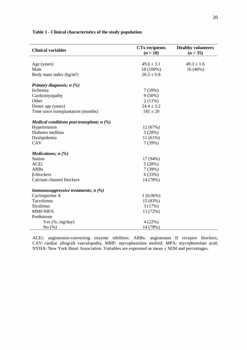
The clinical characteristics of the study population are presented in Table 1. A total of
53 subjects were enrolled including 18 cardiac transplant recipients (CTx) and 35
age-matched healthy volunteers (HV). All CTx were male and the CTx patients were studied
145 ± 20 months following transplantation. More than 60% of our transplanted studied
patients exhibited treated hypertension (n = 12) and dyslipidemia (n = 11) and 39% of them
(n = 7) exhibited CAV. Mean creatinine clearance measured by the MDRD formula was
67.4 ± 19.7 ml/min/m2 (median 66.8; 25.5 – 107). Angiography was used in all patients to
assess for CAV. All patients with CAV exhibited CAV1. By design, all the patients received
stable immunosuppressive drug doses for at least 4 weeks before their enrolment in the study.
The majority of patients received the combination of tacrolimus (TAC) and mycophenolate
mofetil/mycophenolic acid (MMF/MPA).

7
Biomarkers
Plasma levels of VEGF, IL-1RA and IL-8 are presented in Figure 1. Compared with the
healthy control subjects, CTx recipients exhibited an 80% decrease in circulating levels of
VEGF (225 ± 42 (HC) vs 44 ± 10 pg/mL (CTx); p < 0.001). In contrast there were no
significant differences in the circulating levels of the anti-inflammatory cytokine
IL-1RA (205 ± 16 (HC) vs 243 ± 45 pg/mL (CTx)) and the proinflammatory cytokine IL-8
(7.90 ± 1.05 (HC) vs 4.62 ± 0.80 pg/mL (CTx)) between both groups. There were no
significant differences between patients with or without CAV (Table 2).
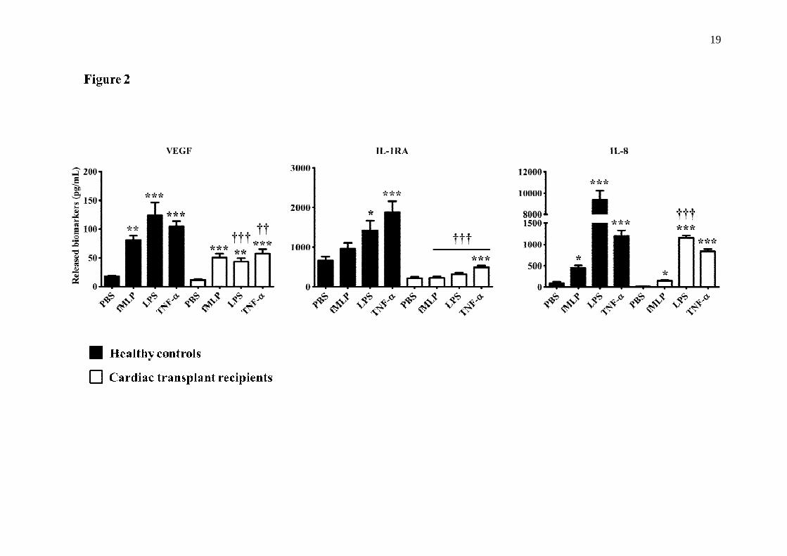
Neutrophil responses
There was a marked reduction in the capacity of neutrophils from CTx patients to
promote the release of all 3 cytokines either under PBS basal condition or upon stimulation
with proinflammatory mediators (fMLP; 10-7 M, LPS; 1 µg/mL and TNF-α; 10 ng/mL)
(Figure 2). The quantification of VEGF and IL-1RA proteins was performed after 2 hours
post-stimulation with the aforementioned proinflammatory agonists, whereas, the
quantification of IL-8 protein was assessed after 24 hours of treatment. All 3 agonists (fMLP,
LPS and TNF-α) were nearly equivalent to promote VEGF release compared with
PBS-treated neutrophils, ranging from 4.6 to 7.0-fold increase in HC. In neutrophils from
CTx patients, we observed a similar pattern in the capacity of the agonists to increase VEGF
release (increase ranging from 3.8 to 5.0-fold) as compared to PBS-treated neutrophils.
Nevertheless, the basal level of VEGF released by the neutrophils from CTx patients was
reduced by 36% as compared to HC neutrophils. In HC, IL-1RA increased by 2.2- to 2.8-fold
in response to LPS and TNF-α respectively as compared to PBS. In contrast CTx patients
yielded a significantly lesser increase in IL-1RA (1.5- and 2.3-fold by LPS and TNF-α
respectively).
8
The release of IL-8 was increased in response to 3 agonists in HC. In contrast CTx
patients exhibited a marked decrease in IL-8 release in response to LPS and TNF-α
stimulation. Interestingly, neutrophils isolated from CTx patients yielded an 88% decrease in
their capacity to release IL-8 under LPS stimulation (powerful proinflammatory mediator),
whereas this reduction was less significant in response to weaker proinflammatory mediators
(fMLP and TNF-α (Figure 2). There were no significant differences in any of the study
parameters in patients with CAV (Table 2).
Discussion
In this clinical investigation, we report a decrease in circulating levels of VEGF but no
significant changes in plasma levels of the proinflammatory cytokine IL-8 and the
anti-inflammatory cytokine IL-1RA in stable long-term CTx recipients. Isolated neutrophils
from CTx patients yielded a marked attenuation in the release of VEGF, IL-1RA and IL-8 in
response to most agonists. Although basal and stimulated levels of VEGF were consistently
lower in patients with CAV, this difference did not reach statistical significance.
VEGF plays a pivotal role on angiogenesis and inflammation [5,6]. VEGF is an
important mediator of angiogenesis, helping to maintain healthy adult vascular function and
homeostasis [5]. However VEGF also possesses significant proinflammatory properties [6],
by its capacity to increase vascular permeability and to promote the adhesion and
transvascular migration of leukocytes [26]. There has been little data on the characterisation
and on the role of VEGF following CTx. Recently, Gramley and coworkers reported a
significant increase in cardiac fibrosis over time in specimens of endomyocardial biopsies [9].
Using immunohistochemistry, this group reported a parallel increase in cardiac VEGF in
these biopsy specimens. This investigation suggested that myocardial hypoxia occurs in
9
long-term CTx recipients and that VEGF may be an adaptive mechanism to reduce hypoxic
stress following CTx.
Herein we report a decrease in circulating VEGF and a marked attenuation in the
capacity of neutrophils to release VEGF. The reasons for an apparent discrepancy between
the decrease in plasma and the increase in cardiac tissue as reported by Gramley remain
unknown. However, one might speculate that an increase in cardiac (cardiomyocytes) VEGF
may be associated with a decrease in circulating level because of the avidity of the injured
myocardium to VEGF. The decreased release of VEGF by stimulated neutrophils may also
be related to some “exhaustion” of the neutrophils related to an increased demand (and/or the
inhibition of corresponding synthesis mediated by the immunosuppressors [27-29].
Interestingly, circulating levels of IL-8 were not significantly decreased in patients suggesting
that chronic immunosuppression may not solely explain these observations. The mechanisms
for these observations and the physiological impacts of a decrease in VEGF in these high-risk
patients deserve further investigations. Our data would be in agreement with Spisani et al.
reporting no significant impacts of cyclosporine A on either basal or agonist-stimulated
neutrophils intracellular calcium concentrations [27].
In this study we observed no significant decrease in plasma levels of IL-1RA and IL-8
in CTx patients compared with the HC subjects. In contrast, the capacity of stimulated
neutrophils to increase the release of these cytokines was significantly attenuated following
CTx. Various cytokines including IL-1, IL-6 and IL-8 play a significant role on vascular
injury and inflammation [12,30]. IL-1 and TNF-α are known to induce the release of
IL-8 [12], the latter promoting the migration of neutrophils to the inflammatory site. IL-8
played a significant role on ocular inflammation and angiogenesis in conjunctiva [30] and on
atherogenesis [12,32] and its inhibition using a specific [12] antibody reduced ischemia
reperfusion injuries in the heart [31]. IL-1RA belongs to the IL-1 family and binds to IL-1

10
receptors, thereby antagonising the inflammatory effects of IL-1α and –α [18] Immune cells
such as neutrophils, can secrete IL-1RA [33,34] and the latter may prevent the
proinflammatory effects of IL-1 [18]. The balance between IL-1 and IL-1RA systemically or
locally plays an important role in many diseases such as arthritis, renal failure, and
cancer [15-18]. In early post renal transplant patients, reduced IL-1RA is associated with
delayed graft function [35] and IL-1RA gene transfer inhibits graft rejection in an
experimental model of corneal transplantation [36]. In addition, low post-transplantation
IL-1RA levels correlate with engraftment syndrome after autologous stem cells
transplantation in plasma cell neoplasms [37]. Basal and stimulated IL-1RA levels have not
been investigated following CTx.
Recently, published work from our group reported a plasmatic increase of various
cytokines including IL-1 and IL-6 within the first 12 weeks following de novo cardiac
transplantation [1]. Plasma levels for these specific cytokines decreased significantly but did
not reach levels observed in healthy control subjects at 12 months. We also reported some
elevation of plasminogen activator inhibitor-1 (PAI-1), fibrinogen, and high sensitivity
C-reactive protein (hsCRP) in long-term CTx recipients [2]. Levels for these specific markers
were only mildly elevated and cytokines were not measured in these patients. Unfortunately
VEGF was not measured in these studies. In the present investigation, we expanded these
latter observations in CTx recipients by reporting minimal changes in basal levels of two
potentially physiologically relevant cytokines. In contrast, we observed a profound decrease
in the release of VEGF along with these two cytokines by stimulated neutrophils. The
mechanisms inducing the decrease in both IL-8 and IL-1RA remain unknown. However, we
may speculate that a chronic state of inflammation may contribute to decrease the potential
release of inflammatory markers by the neutrophils. Similar behaviour has been reported with
other cytokines such as TNF-α in heart failure [38]. The attenuations in IL-1RA release may

11
also suggest that these patients may fail to compensate for an elevation in many
proinflammatory cytokines. To what extent these findings are related to chronic
immunosuppression or other abnormalities in cytokine regulation is a matter for future
investigations?
Study limitations
This clinical investigation reported novel and significant findings on VEGF and on
selected cytokines in CTx recipients. In contrast to healthy controls, cardiac transplant
patients exhibited a high prevalence of hypertension, dyslipidemia and some degree of renal
failure. In addition all transplant recipients were on various immunosuppressive regimen and
most of them were on antihypertensive drugs and on statins. It is likely that these medical
conditions and drug used may have played a role on neutrophil responses. Nevertheless this
potential bias could not be avoided in this clinical investigation. In this study, patients with
CAV exhibited lower but non-significant values for all markers in response to simulation.
However, from all 18 patients, only a small group of our study population exhibited some
mild degree of CAV. As such we cannot conclude about the impact of CAV on these
findings.
In conclusion, CTx recipients exhibit a marked reduction in circulating VEGF as well as
in neutrophil mediated release in VEGF. The mechanisms and physiologic impacts of these
findings and their relationship with various severity of CAV deserve additional investigations.

12
Competing interests
The authors declare that they have no competing interests. Doctor Michel White holds the
Carolyn & Richard Renaud Chair in Heart Failure of the Montreal Heart Institute.
Author’s contributions
Participated in research design (MGS, MW)
Participated in the writing of the paper (DV, DC, P-EN, MGS, and MW)
Participated in the performance of the research (DV, DC, P-E N, AD, MC, GBP, NR, ML,
MGS, and MW)
Contributed new reagents or analytic tools (DV, P-EN, and MGS)
Participated in data analysis (P-EN, MGS, and MW)
Acknowledgements
We are thankful to all our volunteers for so kindly providing us with blood samples. We are
grateful to the dedicated work of the secretarial team of the Research center of the Montreal
Heart Institute.
13
References
1. White M, Cantin B, Haddad H, Kobashigawa JA, Ross H, Carrier M, Pflugfelder PW,
Isaac D, Cecere R, Whittom L, Ali IS, Wang SH, He Y, Groulx A, Touyz RM. Cardiac
signalling molecules and plasma biomarkers after cardiac transplantation: impact of
tacrolimus vs cyclosporine. J Heart Lung Transplant 2013, 32:1222.
2. White M, Ross H, Haddad H, LeBlanc MH, Racine N, Pflugfelder P, Giannetti N,
Davies R, Azevedo E, Isaac D, Burton J, Ferguson R, Genest J. Subclinical
inflammation and prothrombotic state in heart transplant recipients: impact of
cyclosporin microemulsion vs tacrolimus. Transplantation 2006, 82:763.
3. Schiopu A, Nadig SN, Cotoi OS, Hester J, van Rooijen N, Wood KJ. Inflammatory
Ly-6Chi monocytes play an important role in the development of severe transplant
arteriosclerosis in hyperlipidemic recipients. Atherosclerosis 2012, 223:291.
4. Fishbein GA, Fishbein MC. Morphologic and immunohistochemical findings in
antibody-mediated rejection of the cardiac allograft. Hum Immunol 2012, 73:1213.
5 Ylä-Herttuala S, Rissanen TT, Vajanto I, Hartikainen J. Vascular endothelial growth
factors: biology and current status of clinical applications in cardiovascular medicine.
J Am Coll Cardiol 2007, 49:1015.
6 Lee RJ, Springer ML, Blanco-Bose WE, Shaw R, Ursell PC, Blau HM. VEGF gene
delivery to myocardium: deleterious effects of unregulated expression. Circulation
2000, 102:898.
7 Gramley F, Lorenzen J, Jedamzik B, Gatter K, Koellensperger E, Munzel T, Pezzella F.
Atrial fibrillation is associated with cardiac hypoxia. Cardiovasc Pathol 2010, 19:102.
8 De Boer RA, Pinto YM, Van Veldhuisen DJ. The imbalance between oxygen demand
and supply as a potential mechanism in the pathophysiology of heart failure: the role of
microvascular growth and abnormalities. Microcirculation 2003, 10:113.

14
9 Gramley F, Lorenzen J, Pezzella F, Kettering K, Himmrich E, Plumhans C,
Koellensperger E, Munzel T. Hypoxia and myocardial remodelling in human cardiac
allografts: a time-course study. J Heart Lung Transplant 2009, 28:1119.
10 Frangogiannis NG. Regulation of the inflammatory response in cardiac repair. Circ
Res 2012, 110:159.
11 Neagoe PE, Brkovic A, Hajjar F, Sirois MG. Expression and release of angiopoietin-1
from human neutrophils: intracellular mechanisms. Growth Factors 2009, 27:335.
12 Apostolakis S, Vogiatzi K, Amanatidou V, Spandidos DA. Interleukin 8 and
cardiovascular disease. Cardiovasc Res 2009, 84:353.
13 Booth AJ, Grabauskiene S, Wood SC, Lu G, Burrell BE, Bishop DK. IL-6 promotes
cardiac graft rejection mediated by CD4+ cells. J Immunol 2011, 187:5764.
14 George JF, Kirklin JK, Naftel DC, Bourge RC, White-Williams C, McGiffin DC,
Savunen T, Everson MP. Serial measurements of interleukin-6, interleukin-8, tumour
necrosis factor-alpha, and soluble vascular cell adhesion molecule-1 in the peripheral
blood plasma of human cardiac allograft recipients. J Heart Lung Transplant 1997,
16:1046.
15 Dinarello CA. The role of the interleukin-1-receptor antagonist in blocking
inflammation mediated by interleukin-1. N Engl J Med 2000, 343:732.
16 Dinarello CA, Simon A, van der Meer JW. Treating inflammation by blocking
interleukin-1 in a broad spectrum of diseases. Nat Rev Drug Discov 2012, 11:633.
17 Freeman BD, Buchman TG. Interleukin-1 receptor antagonist as therapy for
inflammatory disorders. Expert Opin Biol Ther 2001, 1:301.
18 Arend WP. The balance between IL-1 and IL-1Ra in disease. Cytokine Growth Factor
Rev 2002, 13:323.
15
19 Boulanger CM, Scoazec A, Ebrahimian T, Henry P, Mathieu E, Tedgui A, Mallat Z.
Circulating microparticles from patients with myocardial infarction cause endothelial
dysfunction. Circulation 2001, 104:2649.
20 Dumas E, Martel C, Neagoe PE, Bonnefoy A, Sirois MG. Angiopoietin-1 but not
angiopoietin-2 promotes neutrophil viability: Role of interleukin-8 and
platelet-activating factor. Biochim Biophys Acta 2012, 1823:358.
21 Neagoe PE, Brkovic A, Hajjar F, Sirois MG. Expression and release of angiopoietin-1
from human neutrophils: intracellular mechanisms. Growth Factors 2009, 27:335.
22 Cassatella MA, Bazzoni F, Ceska M, Ferro I, Baggiolini M, Berton G. IL-8 production
by human polymorphonuclear leukocytes. The chemo-attractant formyl-
methionyl-leucyl-phenylalanine induces the gene expression and release of IL-8 through
a pertussis toxin-sensitive pathway. J Immunol 1992, 148:3216.
23 Fujishima S, Hoffman AR, Vu T, Kim KJ, Zheng H, Daniel D, Kim Y, Wallace EF,
Larrick JW, Raffin TA. Regulation of neutrophil interleukin 8 gene expression and
protein secretion by LPS, TNF-alpha, and IL-1 beta. J Cell Physiol 1993, 154:478.
24 Malyak M, Smith MF Jr, Abel AA, Arend WP. Peripheral blood neutrophil production
of interleukin-1 receptor antagonist and interleukin-1 beta. J Clin Immunol 1994, 14:20.
25 Neagoe PE, Brkovic A, Hajjar F, Sirois MG. Expression and release of angiopoietin-1
from human neutrophils: intracellular mechanisms. Growth Factors 2009, 27:335.
26 Bates DO. Vascular endothelial growth factors and vascular permeability. Cardiovasc
Res 2010, 87:262.
27 Spisani S, Fabbri E, Muccinelli M, Cariani A, Barbin L, Trotta F, Dovigo L. Inhibition
of neutrophil responses by cyclosporin A. An insight into molecular mechanisms.
Rheumatology (Oxford) 2001, 40:794.

16
28 McInturff AM, Cody MJ, Elliott EA, Glenn JW, Rowley JW, Rondina MT, Yost CC.
Mammalian target of rapamycin regulates neutrophil extracellular trap formation via
induction of hypoxia-inducible factor 1 α. Blood 2012, 120:3118.
29 He Y, Li D, Cook SL, Yoon MS, Kapoor A, Rao CV, Kenis PJ, Chen J, Wang F.
Mammalian target of rapamycin and Rictor control neutrophil chemotaxis by regulating
Rac/Cdc42 activity and the actin cytoskeleton. Mol Biol Cell 2013, 24:3369.
30 Ghasemi H, Ghazanfari T, Yaraee R, Faghihzadeh S, Hassan ZM. Roles of IL-8 in
ocular inflammations: a review. Ocul Immunol Inflamm 2011, 19:401.
31 Boyle EM Jr, Kovacich JC, Hèbert CA, Canty TG Jr, Chi E, Morgan EN, Pohlman TH,
Verrier ED. Inhibition of interleukin-8 blocks myocardial ischemia-reperfusion injury.
J Thorac Cardiovasc Surg 1998, 116:114.
32 Moreau M, Brocheriou I, Petit L, Ninio E, Chapman MJ, Rouis M. Interleukin-8
mediates downregulation of tissue inhibitor of metalloproteinase-1 expression in
cholesterol-loaded human macrophages: relevance to stability of atherosclerotic plaque.
Circulation 1999, 99:420.
33 Perrier S, Darakhshan F, Hajduch E. IL-1 receptor antagonist in metabolic diseases: Dr
Jekyll or Mr Hyde? FEBS Lett 2006, 580:6289.
34 McColl SR, Paquin R, Ménard C, Beaulieu AD. Human neutrophils produce high
levels of the interleukin 1 receptor antagonist in response to granulocyte/macrophage
colony-stimulating factor and tumor necrosis factor alpha. J Exp Med 1992, 176:593.
35 Sadeghi M, Daniel V, Naujokat C, Schmidt J, Mehrabi A, Zeier M, Opelz G.
Decreasing plasma soluble IL-1 receptor antagonist and increasing monocyte activation
early post-transplant may be involved in pathogenesis of delayed graft function in renal
transplant recipients. Clin Transplant 2010, 24:415.
17
36 Yuan J, Liu Y, Huang W, Zhou S, Ling S, Chen J. The experimental treatment of
corneal graft rejection with the interleukin-1 receptor antagonist (IL-1ra) gene.
PLoS One 2013, 8:e60714
37 Keyzner A, D'Souza A, Lacy M, Gertz M, Hayman S, Buadi F, Kumar S, Dingli D,
Engebretson A, Tong C, Dispenzieri A. Low levels of interleukin-1 receptor antagonist
(IL-1RA) predict engraftment syndrome after autologous stem cell transplantation in
POEMS syndrome and other plasma cell neoplasms. Biol Blood Marrow Transplant
2013, 19:1395.
38 Torre-Amione G, Kapadia S, Lee J, Durand JB, Bies RD, Young JB, Mann DL. Tumor
necrosis factor-alpha and tumor necrosis factor receptors in the failing human heart.
Circulation 1996, 93:704.

18
19
20
Table 1 - Clinical characteristics of the study population
ACEi: angiotensin-converting enzyme inhibitor; ARBs: angiotensin II receptor blockers; CAV: cardiac allograft vasculopathy, MMF: mycophenolate mofetil; MPA: mycophenolate acid; NYHA: New York Heart Association. Variables are expressed as mean ± SEM and percentages.
Clinical variables CTx recipients (n = 18)
Healthy volunteers (n = 35)
Age (years) 49.6 ± 3.1 49.3 ± 1.6 Male 18 (100%) 16 (46%) Body mass index (kg/m²) 26.5 ± 0.8 Primary diagnosis; n (%) Ischemia 7 (39%) Cardiomyopathy 9 (50%) Other 2 (11%) Donor age (years) 24.4 ± 3.2 Time since transplantation (months) 145 ± 20 Medical conditions post-transplant; n (%) Hypertension 12 (67%) Diabetes mellitus 5 (28%) Dyslipidemia 11 (61%) CAV 7 (39%) Medications; n (%) Statins 17 (94%) ACEi 5 (28%) ARBs 7 (39%) β-blockers 6 (33%) Calcium channel blockers 14 (78%) Immunosuppressive treatments; n (%) Cyclosporine A 1 (0.06%) Tacrolimus 15 (83%) Sirolimus 3 (17%) MMF/MPA 13 (72%) Prednisone
Yes (%; mg/day) 4 (22%) No (%) 14 (78%)

21
Table 2 -Circulating biomarkers and neutrophils stimulation profiles for patients with and
without coronary allograph vasculopathy (CAV).
CAV: cardiac allograft vasculopathy; VEGF: vascular endothelial growth factor; IL: interleukin; RA: receptor antagonist; PBS: vehicule; fMLP: N-formyl-Met-Leu-Phe; LPS: bacterial lipopolysaccharides; TNF: tumor necrosis factor. Variables are expressed as mean ± SEM and percentages.
Parameters CAV
(n = 7) No CAV (n = 11)
Mean ± SEM Mean ± SEM Biomarkers
VEGF 63.3 ± 27.8 48.4 ± 15.1 IL-8 6.20 ± 1.59 3.61 ± 0.74 IL-RA 331 ± 99 187 ± 30 Neutrophils Factor
VEGF release
PBS 10.5 ± 2.8 11.7 ± 1.9 fMLP 47.1 ± 9.5 52.7 ± 9.3 LPS 33.4 ± 5.5 46.7 ± 8.9 TNF-α
51.7 ± 11.4 60.7 ± 10.6
IL-8 release
PBS 23.7 ± 5.3 14.7 ± 2.8 fMLP 115 ± 19 161 ± 20 LPS 1100 ± 115 1180 ± 67 TNF-α
728 ± 81 898 ± 76
IL-1RA release
PBS 187 ± 43 229 ± 44 fMLP 178 ± 43 248 ± 42 LPS 253 ± 52 357 ± 54 TNF-α 457 ± 66 515 ± 55
22
Figure legends
Figure 1. Plasma level of vascular endothelial growth factor (VEGF), Interleukin-1 receptor
antagonist (IL-1RA), and Interleukin-8 (IL-8). Data are presented as mean ± SEM.
***p < 0.001 as compared to plasma level between healthy controls (HC) and cardiac
transplant recipient (CTx).
Figure 2. Neutrophil mediated release of VEGF, IL-1RA, and IL-8 in response to various
agonists.
healthy controls; cardiac transplant recipients (CTx). fMLP (10-7 M); LPS (1 g/mL);
TNF-α (10 ng/mL). Data are presented as mean ± SEM. *p < 0.05; ***p < 0.001 compared
to PBS-treated neutrophils. †††p < 0.001 vs healthy controls.
Related Documents