HAL Id: tel-00481944 https://tel.archives-ouvertes.fr/tel-00481944 Submitted on 7 May 2010 HAL is a multi-disciplinary open access archive for the deposit and dissemination of sci- entific research documents, whether they are pub- lished or not. The documents may come from teaching and research institutions in France or abroad, or from public or private research centers. L’archive ouverte pluridisciplinaire HAL, est destinée au dépôt et à la diffusion de documents scientifiques de niveau recherche, publiés ou non, émanant des établissements d’enseignement et de recherche français ou étrangers, des laboratoires publics ou privés. Development of virtual reality tools for arthroscopic surgery training Fadi Yaacoub To cite this version: Fadi Yaacoub. Development of virtual reality tools for arthroscopic surgery training. Other [cs.OH]. Université Paris-Est, 2008. English. NNT : 2008PEST0263. tel-00481944

Welcome message from author
This document is posted to help you gain knowledge. Please leave a comment to let me know what you think about it! Share it to your friends and learn new things together.
Transcript

HAL Id: tel-00481944https://tel.archives-ouvertes.fr/tel-00481944
Submitted on 7 May 2010
HAL is a multi-disciplinary open accessarchive for the deposit and dissemination of sci-entific research documents, whether they are pub-lished or not. The documents may come fromteaching and research institutions in France orabroad, or from public or private research centers.
L’archive ouverte pluridisciplinaire HAL, estdestinée au dépôt et à la diffusion de documentsscientifiques de niveau recherche, publiés ou non,émanant des établissements d’enseignement et derecherche français ou étrangers, des laboratoirespublics ou privés.
Development of virtual reality tools for arthroscopicsurgery training
Fadi Yaacoub
To cite this version:Fadi Yaacoub. Development of virtual reality tools for arthroscopic surgery training. Other [cs.OH].Université Paris-Est, 2008. English. �NNT : 2008PEST0263�. �tel-00481944�
Universite PARIS-EST
Ecole Doctorale ICMS
THESE
Pour obtenir le grade de
Docteur de l’Universite PARIS-EST
Specialite: Informatique
Presentee et soutenue publiquement par
Fadi YAACOUB
Developpement d’Outils de Realite Virtuelle pour
L’enseignement de la Chirurgie Arthroscopique
Development of Virtual Reality Tools for
Arthroscopic Surgery Training
Directeur de these : Yskandar HAMAM , Professeur Emerite
Date de soutenance : 12 Novembre 2008
Composition du Jury :
President du jury: Etienne COLLE Professeur, Universite d’Evry, IBISC CNRS
Rapporteur: Fathi BEN-OUEZDOU Professeur, Universite de Versailles, LISV
Rapporteur: Karim DJOUANI Professeur, F’SATIE-TUT, Afrique du Sud
Examinateur: Gilles BERTRAND Professeur, Universite Paris-Est, ESIEE, LIGM
Examinateur: Alain GILBERT Professeur, M.D., Institut de la Main
Examinateur: Yskandar HAMAM Professeur, Universite Paris-Est, ESIEE, LIGM
Examinateur: Antoine ABCHE Professeur Associe, Universite de Balamand
c© UMLV, 2008.

To my family
ii

Acknowledgements
A few lines are too short to express my deep appreciation for a number of people who
contributed in different ways to my thesis. It is a pleasure to convey my gratitude to
them all in my humble acknowledgment.
In the first place, I would like to record my gratitude to Professor Yskandar Hamam
for his supervision, and guidance from the very early stage of this research as well as
giving me experiences throughout the work. Above all and the most needed, he provided
me with encouragement and support in various ways.
I would like to express my profound appreciation to all the reporters and committee
members who honored me by reading my thesis and assisting at my defense.
I gratefully acknowledge Dr. Antoine Abche for his advices and contributions, which
made him a backbone of this research and so to this thesis. Many thanks go to Dr. Eric
Tallier, orthopedist surgeon at “Centre Hospitalier de Falaise”, for his valuable remarks.
I also acknowledge the Lebanese CNRS for the financial support during my last year of
research.
I would also like to thank the members of the A2SI laboratory: Gilles Bertrand,
Francois Rocaries, Michel Couprie, Denis Bureau, Hugues Talbot, Mohamad Akil, Lau-
rent Najman, Thierry Grandpierre for their support. It was a great pleasure working
with them. Many thanks go in particular to Tarik Al Ani for his helpful advices and
his encouragement. I am not forgetting Eric Lorens and Christophe Dietrich the A2SI
engineers as well as Martine Elichabe and Elisabeth Bastien. They were always available
for my needs.
Special thanks to Lina Bouhaya for everything she made for me. Also, thank you
Benoit Kaufmann, William Kobersy, Salah Helmy, Sami Sassine and John Altrip for all
your support. Without you my friends, life would not be the same.
Finally, I would not be sitting in front of my PC typing these acknowledgement
lines without my family. I owe my parents, Michel and Maha Yaacoub much of what I
have become. I thank my sister Olga and my brother Chadi for their prayers and their
encouragement throughout my graduate work in France.
Fadi YAACOUB
November 2008
iii

Abstract
The minimally invasive approach of arthroscopy means less pain and faster recovery
time for patients compared to open surgery. However, it implies a high difficulty of
performance. Therefore, surgeon should remain at a high level of technical and profes-
sional expertise to perform such operations. Surgeon’s skills are being developed over
years of surgical training on animals, cadavers and patients. Nowadays, cadavers and
animal specimens present an ethical problem also the practice on real humans is usually
risky. For surgeons to reach a high level, new and alternative ways of performing surgical
training are required.
Virtual reality technology has opened new realms in the practice of medicine. Today,
virtual reality simulators have become one of the most important training methods in
the medical field. These simulators allow medical students to examine and study organs
or any structure of the human body in ways that were not possible few years earlier.
Similarly, the surgeon as well as the medical student can gain a valuable experience by
performing a particular surgery with an anatomical accuracy and realism as it is actually
performed in the real world. Thus, they can practice on virtual operation before they
proceed and operate on real patients.
In this thesis, a virtual reality training simulator for wrist arthroscopy is introduced.
Two main issues are addressed: the 3-D reconstruction process and the 3-D interaction.
Based on a sequence of CT images a realistic representation of the wrist joint is obtained
suitable for the computer simulation. Two main components of the computer-based
system interface are illustrated: the 3-D interaction to guide the surgical instruments
and the user interface for haptic feedback. In this context, algorithms that model objects
using the convex hull approaches and simulate real time exact collision detection between
virtual objects are presented. A force feedback device, coupled with a haptic algorithm,
is used as a haptic interface with the computer simulation system. This leads in the
development of a low cost system with the same benefits as professional devices. In this
regard, the wrist arthroscopy can be simulated and medical students can learn the basic
skills required with safety, flexibility and less cost.
Keywords : Virtual Reality, Arthroscopic Surgery, 3-D modeling and Visualization,
Convex Hull, Collision Detection, Haptic Feedback, Healthcare Technology.
iv

Resume
La chirurgie arthroscopique presente actuellement un essor tres important pour le benefice
du plus grand nombre des patients. Cependant, cette technique possede un certain nom-
bre d’inconvenients et il est donc necessaire pour le medecin de s’entrainer et repeter
ses gestes afin de pouvoir executer ce type d’operation d’une facon efficace et certaine.
En effet, les methodes traditionnelles d’enseignement de la chirurgie sont basees sur
l’autopsie des cadavres et l’entrainement sur des animaux. Avec l’evolution de notre
societe, ces deux pratiques deviennent de plus en plus critiquees et font l’objet de
reglementations tres restrictives. Afin d’atteindre un niveau plus eleve, de nouveaux
moyens d’apprentissage sont necessaires pour les chirurgiens.
Recemment, la realite virtuelle commence d’etre de plus en plus utilisee dans la
medecine et surtout la chirurgie. Les simulateurs chirurgicaux sont devenus une des
matieres les plus recentes dans la recherche de la realite virtuelle. Ils sont egalement
devenus une methode de formation et un outil d’entrainement valable pour les chirurgiens
aussi bien que les etudiants en medecine.
Dans ce travail, un simulateur de realite virtuelle pour l’enseignement de la chirurgie
arthroscopique, surtout la chirurgie du poignet, a ete presente. Deux questions princi-
pales sont abordees : la reconstruction et l’interaction 3-D. Une sequence d’images CT a
ete traitee afin de generer un modele 3-D du poignet. Les deux principales composantes
de l’interface du systeme sont illustrees : l’interaction 3-D pour guider les instruments
chirurgicaux et l’interface de l’utilisateur pour le retour d’effort. Dans ce contexte, les
algorithmes qui modelisent les objets en utilisant les approches de “Convex Hull” et qui
simulent la detection de collision entre les objets virtuels en temps reel, sont presentes.
En outre, un dispositif de retour d’effort est utilise comme une interface haptique avec
le systeme. Cela conduit au developpement d’un systeme a faible cout, avec les memes
avantages que les appareils professionnels. A cet egard, l’arthroscopie du poignet peut
etre simulee et les etudiants en medecine peuvent facilement utiliser le systeme et peu-
vent apprendre les competences de base requises en securite, flexibilite et moindre cout.
Mots-cles : Realite Virtuelle, Chirurgie Arthroscopique, Modelisation et Visualisa-
tion 3-D, Enveloppes Convexes, Detection de Collision, Retour d’effort, Technologie
Medicale.
v

Table of contents
List of Figures xi
List of Algorithms xiii
List of Tables xiv
1 Introduction 1
1.1 Objective and Considerations . . . . . . . . . . . . . . . . . . . . . . . . 3
1.2 Design Criteria . . . . . . . . . . . . . . . . . . . . . . . . . . . . . . . . 4
1.3 The VR Surgical Simulation System . . . . . . . . . . . . . . . . . . . . . 5
1.4 Motivations and Contributions . . . . . . . . . . . . . . . . . . . . . . . . 6
1.5 Thesis’s Structure . . . . . . . . . . . . . . . . . . . . . . . . . . . . . . . 7
2 VR Simulators for Minimally Invasive Surgery 9
2.1 Introduction . . . . . . . . . . . . . . . . . . . . . . . . . . . . . . . . . . 9
2.2 Laparoscopy Simulators . . . . . . . . . . . . . . . . . . . . . . . . . . . 11
2.2.1 LapSim . . . . . . . . . . . . . . . . . . . . . . . . . . . . . . . . 11
2.2.2 LapMentor . . . . . . . . . . . . . . . . . . . . . . . . . . . . . . 11
2.2.3 MIST . . . . . . . . . . . . . . . . . . . . . . . . . . . . . . . . . 12
2.2.4 VIST . . . . . . . . . . . . . . . . . . . . . . . . . . . . . . . . . . 12
2.2.5 LASSO . . . . . . . . . . . . . . . . . . . . . . . . . . . . . . . . 13
2.2.6 VEST . . . . . . . . . . . . . . . . . . . . . . . . . . . . . . . . . 13
2.2.7 Karlsruhe Endoscopic Surgery Trainer . . . . . . . . . . . . . . . 13
2.2.8 Liver Biopsy . . . . . . . . . . . . . . . . . . . . . . . . . . . . . . 14
2.2.9 ProMIS . . . . . . . . . . . . . . . . . . . . . . . . . . . . . . . . 14
vi

TABLE OF CONTENTS vii
2.2.10 SEP . . . . . . . . . . . . . . . . . . . . . . . . . . . . . . . . . . 15
2.3 Cystoscopy Simulators . . . . . . . . . . . . . . . . . . . . . . . . . . . . 15
2.3.1 UroMentor . . . . . . . . . . . . . . . . . . . . . . . . . . . . . . . 15
2.4 Colonoscopy Simulators . . . . . . . . . . . . . . . . . . . . . . . . . . . 16
2.4.1 Simbionix GI Mentor II . . . . . . . . . . . . . . . . . . . . . . . 16
2.4.2 VES . . . . . . . . . . . . . . . . . . . . . . . . . . . . . . . . . . 17
2.5 Bronchoscopy Simulators . . . . . . . . . . . . . . . . . . . . . . . . . . . 17
2.5.1 AccuTouch . . . . . . . . . . . . . . . . . . . . . . . . . . . . . . 17
2.5.2 PREOP . . . . . . . . . . . . . . . . . . . . . . . . . . . . . . . . 18
2.6 Hysteroscopy Simulators . . . . . . . . . . . . . . . . . . . . . . . . . . . 18
2.6.1 LAHYSTOTRAIN . . . . . . . . . . . . . . . . . . . . . . . . . . 19
2.6.2 VirtaMed Hysteroscopy Simulator . . . . . . . . . . . . . . . . . . 19
2.7 Cholangio-pancreatography Simulators . . . . . . . . . . . . . . . . . . . 20
2.7.1 GIT/MCG ERCP . . . . . . . . . . . . . . . . . . . . . . . . . . . 20
2.8 Sinoscopy Simulators . . . . . . . . . . . . . . . . . . . . . . . . . . . . . 21
2.8.1 ESS . . . . . . . . . . . . . . . . . . . . . . . . . . . . . . . . . . 21
2.9 Interventional Radiology Simulators . . . . . . . . . . . . . . . . . . . . . 22
2.10 Arthroscopy Simulators . . . . . . . . . . . . . . . . . . . . . . . . . . . . 23
2.10.1 Knee Simulators . . . . . . . . . . . . . . . . . . . . . . . . . . . 24
2.10.2 Shoulder Simulators . . . . . . . . . . . . . . . . . . . . . . . . . 25
2.11 Advantages/Disadvantages . . . . . . . . . . . . . . . . . . . . . . . . . . 26
2.12 Conclusion . . . . . . . . . . . . . . . . . . . . . . . . . . . . . . . . . . . 27
3 Segmentation and 3-D Object Generation 28
3.1 Introduction . . . . . . . . . . . . . . . . . . . . . . . . . . . . . . . . . . 28
3.2 Marker-Based Watershed Segmentation . . . . . . . . . . . . . . . . . . . 29
3.2.1 Segmentation of CT Images . . . . . . . . . . . . . . . . . . . . . 29
3.3 The Marching Cube Algorithm . . . . . . . . . . . . . . . . . . . . . . . 31
3.4 Material and Lighting . . . . . . . . . . . . . . . . . . . . . . . . . . . . 33
3.5 Texture Mapping . . . . . . . . . . . . . . . . . . . . . . . . . . . . . . . 35
3.6 Pseudoarthrosis of the Scaphoid . . . . . . . . . . . . . . . . . . . . . . . 36
3.6.1 Anatomy of the wrist . . . . . . . . . . . . . . . . . . . . . . . . . 36
TABLE OF CONTENTS viii
3.6.2 Pseudoarthrosis . . . . . . . . . . . . . . . . . . . . . . . . . . . . 37
3.7 Conclusion . . . . . . . . . . . . . . . . . . . . . . . . . . . . . . . . . . . 38
4 Convex Hull: A New Hybrid Approach 39
4.1 Introduction . . . . . . . . . . . . . . . . . . . . . . . . . . . . . . . . . . 39
4.2 Convex Hull Definition . . . . . . . . . . . . . . . . . . . . . . . . . . . . 40
4.3 Related Work . . . . . . . . . . . . . . . . . . . . . . . . . . . . . . . . . 40
4.4 3-D Convex Hull Algorithms . . . . . . . . . . . . . . . . . . . . . . . . . 41
4.4.1 Brute Force Algorithm . . . . . . . . . . . . . . . . . . . . . . . . 42
4.4.2 Gift Wrapping Algorithm . . . . . . . . . . . . . . . . . . . . . . 42
4.4.3 QuickHull Algorithm . . . . . . . . . . . . . . . . . . . . . . . . . 43
4.4.4 Chan Algorithm . . . . . . . . . . . . . . . . . . . . . . . . . . . . 44
4.5 The Hybrid Algorithm . . . . . . . . . . . . . . . . . . . . . . . . . . . . 46
4.6 3-D Models and Convex Hulls . . . . . . . . . . . . . . . . . . . . . . . . 48
4.7 Simulations and Results . . . . . . . . . . . . . . . . . . . . . . . . . . . 50
4.7.1 Result Interpretation . . . . . . . . . . . . . . . . . . . . . . . . . 52
4.8 Conclusion . . . . . . . . . . . . . . . . . . . . . . . . . . . . . . . . . . . 55
5 Collision Detection: A Linear Programming Technique 56
5.1 Introduction . . . . . . . . . . . . . . . . . . . . . . . . . . . . . . . . . . 56
5.2 Previous CD Algorithms . . . . . . . . . . . . . . . . . . . . . . . . . . . 57
5.2.1 AABB/OBB . . . . . . . . . . . . . . . . . . . . . . . . . . . . . . 58
5.2.2 Lin-Canny (LC) . . . . . . . . . . . . . . . . . . . . . . . . . . . . 58
5.2.3 Gilbert-Johnson-Keerthi (GJK) . . . . . . . . . . . . . . . . . . . 58
5.2.4 Voronoi-Clip (V-Clip) . . . . . . . . . . . . . . . . . . . . . . . . 59
5.2.5 I-Collide . . . . . . . . . . . . . . . . . . . . . . . . . . . . . . . . 59
5.2.6 Q-Collide . . . . . . . . . . . . . . . . . . . . . . . . . . . . . . . 59
5.2.7 Quick-CD . . . . . . . . . . . . . . . . . . . . . . . . . . . . . . . 59
5.2.8 SWIFT . . . . . . . . . . . . . . . . . . . . . . . . . . . . . . . . 60
5.3 The Proposed CD Approach . . . . . . . . . . . . . . . . . . . . . . . . . 61
5.3.1 Linear Programming Solution . . . . . . . . . . . . . . . . . . . . 61
5.4 The IVRI-CD Technique . . . . . . . . . . . . . . . . . . . . . . . . . . . 63
5.4.1 IVRI-CD . . . . . . . . . . . . . . . . . . . . . . . . . . . . . . . . 63

TABLE OF CONTENTS ix
5.5 Simulations and Results . . . . . . . . . . . . . . . . . . . . . . . . . . . 64
5.5.1 Results . . . . . . . . . . . . . . . . . . . . . . . . . . . . . . . . . 65
5.6 Proximity Queries (PQ) and Penetration Depth (PD) Computation . . . 67
5.7 Conclusion . . . . . . . . . . . . . . . . . . . . . . . . . . . . . . . . . . . 68
6 Design and Implementation of a 3-DOF Haptic Feedback Device 70
6.1 Introduction . . . . . . . . . . . . . . . . . . . . . . . . . . . . . . . . . . 70
6.2 Haptic Devices for VR Medical Simulators . . . . . . . . . . . . . . . . . 71
6.2.1 FEELit Mouse . . . . . . . . . . . . . . . . . . . . . . . . . . . . 72
6.2.2 Microsoft Sidewinder Force Feedback (MSFF) . . . . . . . . . . . 72
6.2.3 PHANToM Haptic Devices . . . . . . . . . . . . . . . . . . . . . . 73
6.3 The Proposed Force Feedback Device . . . . . . . . . . . . . . . . . . . . 74
6.3.1 Design of the Device . . . . . . . . . . . . . . . . . . . . . . . . . 74
6.3.2 Implementation of the Device . . . . . . . . . . . . . . . . . . . . 75
6.4 Data Collection and Acquisition . . . . . . . . . . . . . . . . . . . . . . . 77
6.5 Haptic Feedback Algorithm . . . . . . . . . . . . . . . . . . . . . . . . . 80
6.6 Virtual Simulation of Scaphoid Fixation . . . . . . . . . . . . . . . . . . 81
6.6.1 Surgical Technique For Scaphoid Fracture . . . . . . . . . . . . . 81
6.6.2 Surgical Simulation . . . . . . . . . . . . . . . . . . . . . . . . . . 82
6.7 Advantages . . . . . . . . . . . . . . . . . . . . . . . . . . . . . . . . . . 85
6.8 Conclusion . . . . . . . . . . . . . . . . . . . . . . . . . . . . . . . . . . . 86
7 Conclusions and Perspectives 87
7.1 Summary of the Contributions . . . . . . . . . . . . . . . . . . . . . . . . 88
7.2 Future Work . . . . . . . . . . . . . . . . . . . . . . . . . . . . . . . . . . 89
Publications 90
Resume Detaille 92
Bibliography 107

TABLE OF CONTENTS x
Appendix 117
B Electronic Components 118
B.1 AD512 Entry-Level Data Acquisition Card HUMUSOFT . . . . . . . . . 118
B.1.1 General . . . . . . . . . . . . . . . . . . . . . . . . . . . . . . . . 118
B.1.2 Features . . . . . . . . . . . . . . . . . . . . . . . . . . . . . . . . 119
B.1.3 Specifications . . . . . . . . . . . . . . . . . . . . . . . . . . . . . 119
B.2 Vishay Spectrol Potentiometer Model 357 . . . . . . . . . . . . . . . . . 120
B.3 Servo Motors . . . . . . . . . . . . . . . . . . . . . . . . . . . . . . . . . 122
B.3.1 Servo Wiring . . . . . . . . . . . . . . . . . . . . . . . . . . . . . 122
B.3.2 PWM Signals . . . . . . . . . . . . . . . . . . . . . . . . . . . . . 122

List of Figures
1.1 A Flowchart of the VR Simulation System . . . . . . . . . . . . . . . . . 5
1.2 The Proposed VR Simulation System . . . . . . . . . . . . . . . . . . . . 7
3.1 2-D Slice of CT Image . . . . . . . . . . . . . . . . . . . . . . . . . . . . 30
3.2 Gradient of CT Image with Markers . . . . . . . . . . . . . . . . . . . . . 30
3.3 Watershed Result . . . . . . . . . . . . . . . . . . . . . . . . . . . . . . . 30
3.4 Image of the Wrist . . . . . . . . . . . . . . . . . . . . . . . . . . . . . . 31
3.5 The 15 Cubes Combinations of the Marching Cube Algorithm . . . . . . 32
3.6 The Facets of The Wrist Model . . . . . . . . . . . . . . . . . . . . . . . 33
3.7 3-D Virtual Model of the Wrist Bones . . . . . . . . . . . . . . . . . . . . 34
3.8 Texture Mapping: Source to Destination . . . . . . . . . . . . . . . . . . 35
3.9 3-D Virtual Model of the Wrist Bones After Texture Mapping . . . . . . 36
3.10 Volar Wrist Representation . . . . . . . . . . . . . . . . . . . . . . . . . . 37
3.11 Pseudoarthrosis of the Scaphoid . . . . . . . . . . . . . . . . . . . . . . . 38
4.1 A Gift Wrapping Example . . . . . . . . . . . . . . . . . . . . . . . . . . 43
4.2 A QuickHull Example . . . . . . . . . . . . . . . . . . . . . . . . . . . . 44
4.3 Wrapping a set of [n/m] convex polygons of size m . . . . . . . . . . . . 45
4.4 Capitate and Ulna with their Convex Hulls . . . . . . . . . . . . . . . . . 48
4.5 3rdMetacarpal and Scaphoid with their Convex Hulls . . . . . . . . . . . 49
4.6 Bones of the 3-D wrist enclosed by their corresponding Convex Hulls . . 50
4.7 Execution Time of the 3-D CH Algorithms for Wrist Bones . . . . . . . . 53
4.8 Execution Time of the 3-D CH Algorithms for Knee Bones . . . . . . . . 54
5.1 Different Types Of Bounding Volumes . . . . . . . . . . . . . . . . . . . 57
xi

LIST OF FIGURES xii
5.2 Facets of the Capitate and its Convex Hull . . . . . . . . . . . . . . . . . 65
5.3 Execution time of the two CD algorithms for bones of the 3-D wrist model 66
6.1 The Design of the Force Feedback System . . . . . . . . . . . . . . . . . 75
6.2 The VISHAY Potentiometer Characteristics . . . . . . . . . . . . . . . . 76
6.3 The Servo Motor Futaba FP-S148 . . . . . . . . . . . . . . . . . . . . . . 76
6.4 The Proposed Force Feedback System . . . . . . . . . . . . . . . . . . . . 77
6.5 A Variable Voltage Divider . . . . . . . . . . . . . . . . . . . . . . . . . . 78
6.6 Generation of the PWM Signals . . . . . . . . . . . . . . . . . . . . . . . 79
6.7 The Circuit Driving the Motors . . . . . . . . . . . . . . . . . . . . . . . 80
6.8 The Flowchart of the Haptic Feedback Algorithm . . . . . . . . . . . . . 81
6.9 The Placement of The Guide Wire (Real Operation) . . . . . . . . . . . 83
6.10 The Placement of The Guide Wire (Virtual Operation) . . . . . . . . . . 83
6.11 The Insertion of The Screw in The Scaphoid (Real Operation) . . . . . . 84
6.12 The Insertion of The Screw in The Scaphoid (Virtual Operation) . . . . . 84
6.13 A Virtual View of The Central Placement of The Screw in The Scaphoid 85
A.1 Schema du Systeme . . . . . . . . . . . . . . . . . . . . . . . . . . . . . . 95
A.2 Modele 3-D du Poignet . . . . . . . . . . . . . . . . . . . . . . . . . . . . 96
A.3 Os du Poignet Recouvert par son Enveloppe Convexe Correspondant . . 100
A.4 Le Systeme de Retour D’effort . . . . . . . . . . . . . . . . . . . . . . . . 103
A.5 Le Simulateur Chirurgical Propose . . . . . . . . . . . . . . . . . . . . . 104
A.6 Comparaison Entre une Operation Reelle et une Operation Virtuelle . . 105
B.1 AD512 Data acquisition card . . . . . . . . . . . . . . . . . . . . . . . . . 118
B.2 AD512 Data Sheet . . . . . . . . . . . . . . . . . . . . . . . . . . . . . . 120
B.3 Vishay Spectrol Potentiometer Model 357 . . . . . . . . . . . . . . . . . 121
B.4 Servo Motor: FUTABA Wiring . . . . . . . . . . . . . . . . . . . . . . . 122
B.5 PWM Signals for Servo Control . . . . . . . . . . . . . . . . . . . . . . . 123

List of Algorithms
1 The Hybrid Approach To Construct the CH . . . . . . . . . . . . . . . . 47
xiii

List of Tables
4.1 Comparison of Execution time for Computing the 3-D Convex Hull of the
Wrist Bones . . . . . . . . . . . . . . . . . . . . . . . . . . . . . . . . . . 51
4.2 Comparison of Execution time for Computing the 3-D Convex Hull of the
Knee Bones . . . . . . . . . . . . . . . . . . . . . . . . . . . . . . . . . . 51
5.1 Comparison of Execution Time for Collision Detection Algorithms . . . . 66
xiv

Chapter 1
Introduction
Virtual Environment (VE) provides a new dimension of graphical simulation [1]. It is
described as an application that allows users to navigate and interact with a computer-
generated three dimensional space in real time. In this context, Virtual Reality (VR)
is not only a hardware system. It is also an emerging technology that changes the way
individuals interact with computers. VR has revolutionized several scientific disciplines
by developing novel methods to visualize complex data structures and by providing
means to manipulate the data in real-time and in a natural way. Most promising fields in
which VR systems are applied include: engineering, education, entertainment, military
simulations and medicine.
Recently, medicine has entered a period of intense technological transition, driven by
the need to provide improved care at a lower cost. Since, the outcomes of surgical pro-
cedures are closely related to the skills of the surgeon, the latter should remain at a high
level of technical and professional expertise. These skills are being developed over years
of surgical training on animals, cadavers and patients. Consequently, new and alterna-
tive ways of performing surgical training are required. In addition, the low availability
and high cost of cadaver and animal specimens for traditional medical training and the
public concern with the inhuman treatment of animals have become another impetus
for surgeons and medical students to use new technology in their education and their
training to gain valuable information and experience. VR technology has opened new
realms in the practice of medicine. The graphics capabilities of VR tools, particularly in
1

CHAPTER 1. Introduction 2
modeling and displaying medical data can be of great assistance in teaching, learning,
training and experimenting surgeries [2].
Today, researchers on surgical education depend heavily on VR simulators that have
become one of the most important training methods in the medical field. Simulators
allow medical students to examine and study organs or any structure of the human
body in ways that were not possible few years earlier. They provide an important tool
to acquire valuable information during their education. Similarly, the surgeon as well as
the medical student can gain a valuable experience by performing a particular surgery
with an anatomical accuracy and realism as it is actually performed in the real world.
Thus, the surgeon can practice his operation before he proceeds and operates on real
patients. Therefore, the risks to surgical patients are reduced and the ethical issues
associated with animal experimentation are avoided.
One of the most advanced and important type of surgeries is the Minimally Invasive
Surgery (MIS). The minimally invasive approach means less pain and faster recovery
time for patients in comparison with an open surgery. On the other hand, it implies a
high difficulty of performance. Arthroscopy is a form of endoscopy or minimally invasive
surgery that is concerned specifically with the joints. It offers several advantages over
the traditional open surgery for both: the patient and the healthcare provider. The cor-
responding procedures are generally less invasive, resulting in smaller wounds, increased
rates of recovery, reductions in hospitalization episodes and consequently reductions
in patient intervention costs. While these advantages are attractive to the healthcare
provider, the arthroscopy is associated with some disadvantages. Arthroscopic equip-
ments are expensive and surgeons require additional training to acquire the competence
to operate efficiently and safely. In addition, surgeons agree that the current initial
training protocols are insufficiently challenging and consequently they are entering the
operating room with inadequate skills to use arthroscopic technique to its best advan-
tage. Thus, patients could be at risk in such an environment. Therefore, computer-based
surgical simulation systems, one of the most developed technologies in VR, are used to
train surgeons as well as medical students to practice a particular surgery before they

CHAPTER 1. Introduction 3
enter the operating room. These simulators have become one main component that
has radically changed the traditional medical training and the surgical certification sce-
narios. They allow the process of iterative learning through assessment, evaluation,
decision making and error correction and consequently create a much stronger learning
environment.
1.1 Objective and Considerations
VR surgical simulators have been developed for a wide range of medical applications.
Their names reflect the performed procedures i.e. laparoscopy, endoscopy, cystoscopy,
ureteroscopy colonoscopy, bronchoscopy and flexible sigmoidoscopy simulators. Most
simulators that are mentioned above, are expensive to acquire and need maintenance.
With respect to arthroscopy simulators, developments have been mostly for the knee and
the shoulder and very little work has been done for wrist arthroscopy. Even though the
wrist is a very important joint of the human body that handles many activities, the work
on developing corresponding VR surgical simulators is limited. Thus, the problem of
building an inexpensive and a practical simulator to train medical students and treat the
issue of the wrist arthroscopy remained. In this context, our project has been proposed
by a team of medical professors and surgeons at the “Institut de la Main”, “Clinique
JOUVENET”, Paris XVI. It consists of developing a VR simulator to help teaching,
learning and training on wrist arthroscopic surgical procedures.
This research project, directed by Prof. Yskandar Hamam, started with Charbel
Fares who graduated in June 2006. Our work is a continuation of this project in order
to enhance many algorithms, propose new ones and to develop all the VR tools that are
necessary to complete the project in order to have the entire prototype system.
The design of the proposed computer-based arthroscopy simulator is based on a
trade-off between medical professor’s needs and VR limitations. During the design of
the proposed training system, two major aims are addressed:
1. Apply VR and physical simulation techniques to generate 3-D models and to sim-
ulate operations with fidelity and realism.
CHAPTER 1. Introduction 4
2. Try to cover different requirements for the apprentice learning process and pro-
vide the user with tools to facilitate teaching, learning and training on several
procedures.
In addition, wrist arthroscopy is selected due to several considerations:
• Wrist arthroscopy is a frequent pathology (study of essential nature of disease)
that has been studied and practiced less than the knee and shoulder arthroscopies.
• Various types of involvements and specific surgeries can be covered by wrist arthros-
copy simulation such as: dorsal percutaneous scaphoid fixation, volar percutaneous
scaphoid fixation, capitolunate arthrodesis ...
• There are potentially large and new pathologies that will be facing the medical
practitioners when it comes to wrist arthroscopy. Therefore, there is an increasing
demand on training and learning new techniques.
• Wrist arthroscopy has proven to be extremely valuable in both diagnosis and ther-
apy. It is an important skill for all hand surgeons, in exactly the same way as
shoulder and knee arthroscopies.
1.2 Design Criteria
Our work is focused on developing a VR training system to simulate arthroscopic pro-
cedures, especially wrist arthroscopy, in a virtual environment. The system is developed
for both: educational and pre-operative purposes.
Two main issues are addressed: the three dimensional (3-D) reconstruction process
and the 3-D interaction. The proposed system provides a virtual environment with re-
alistic representation of the region of interest. Based on a sequence of CT images, a
realistic representation of the wrist joint is obtained and is suitable for the computer
simulation. Two main components of the computer-based system interface are illus-
trated: the 3-D interaction to guide the surgical instruments and the user interface for
haptic feedback. In this context, algorithms that model objects using the convex hull

CHAPTER 1. Introduction 5
approaches and simulate real time exact collision detection between virtual objects dur-
ing the training on surgical operations are needed. Also, a force feedback device must
be used as a haptic interface with the computer simulation system. This will lead in
the development of a low cost system that is used by medical students with the same
benefits as professional devices. Then, the procedure can be performed on real patients
with much less risk and injury.
1.3 The VR Surgical Simulation System
A functional prototype of a computer-based training system for simulating wrist arthroscopy
is presented. Figure 1.1 outlines the main components of the proposed VR simulation
system.
Figure 1.1: A Flowchart of the VR Simulation System
Medical images are processed to generate volumetric object models. A sequence of
CT images is segmented and a 3-D virtual model of the wrist is generated. This 3-D

CHAPTER 1. Introduction 6
model is presented both visually via rendering on the computer monitor and haptically
with a force feedback device. Visual parameters such as viewpoint, zooming, color and
lighting effects, can be interactively controlled and object models can be manipulated
with force feedback to change relative probe and object positions, and to simulate many
surgical procedures. Also, simulations include an algorithm that model objects using
the convex hull approach and a method that detects collisions between virtual objects
during the operation. The interaction between the haptic device and the computer closes
the feedback loop between the user and the simulator, offering a better understanding
of the anatomical structures and the functions in the patient’s model.
1.4 Motivations and Contributions
Our research is motivated by the need to develop an inexpensive and practical simulator
to train medical practitioners (students, surgeons ...) and master the wrist arthroscopy
techniques. In this context, several contributions are presented:
• Developing a virtual environment to visualize medical models and medical tools
with high fidelity and precision.
• Developing and presenting a new hybrid approach to generate the convex hull of
the 3-D models. The proposed algorithm converts each 3-D concave model to a
convex representation and allows collision detection algorithms to converge quickly
and report a collision, if it exists.
• Proposing and developing a new technique of collision detection for solid objects.
The collision detection problem is formulated and a linear programming solution
is obtained to determine whether a collision exists or not. The proposed algorithm
is efficient, fast, robust and leads to a decrease of the running time that is required
to detect a collision.
• Designing and implementing a 3-DOF force feedback device. This low cost system
is coupled with a haptic feedback algorithm. The proposed device is used by
medical practitioners with the same benefits as professional devices.

CHAPTER 1. Introduction 7
These main contributions lead to the development of the computer based medical
system that is shown in figure 1.2
Figure 1.2: The Proposed VR Simulation System
1.5 Thesis’s Structure
Chapter 1 introduces the thesis and outlines the contributions.
Chapter 2 presents different VR surgical simulators for Minimally Invasive Surgery
(MIS). These simulators have been developed for a wide range of procedures. The
presented VR simulators are classified based on the application and the relation with
the organs or areas for which the system is developed for. A description of each type of
the minimally invasive surgical simulator is presented.
Chapter 3 describes the segmentation and the generation of the medical model of the
imaged object. First, the segmentation of the CT images using the watershed algorithm
and the reconstruction of the 3-D wrist model are introduced. Then, the representation

CHAPTER 1. Introduction 8
of these models in the virtual environment by associating all the necessary elements
(lights, materials and texture mapping) are illustrated.
In Chapter 4, a hybrid approach to generate the convex hull is developed and pre-
sented. The new algorithm is validated by performing a comparison with conventional
algorithms namely, the Brute Force, the Gift Wrapping, the QuickHull and the Chan
algorithms. The evaluation is achieved by generating the convex envelope of 3-D wrist
bones using the five different approaches. The results show the improvement associated
with the proposed approach.
Having generated the convex hulls, Chapter 5 addresses the issue of the precise
Collision Detection (CD) between virtual objects and a new technique is proposed. The
CD problem is formulated and a linear programming solution is obtained to determine
whether a collision exists or not. The proposed CD approach is evaluated and compared
with a conventional algorithm namely the Industrial Virtual Reality Institute Collision
Detection (IVRI-CD) technique. It is validated using bones of the 3-D wrist model. The
results show that the proposed algorithm is efficient, fast, robust and leads to a decrease
of the running time required to detect a collision.
Chapter 6 proposes a force feedback device which is used as a haptic interface with
the computer simulation system. The design and the implementation of this device are
shown. This leads to the development of a low cost system that is used by medical
students with the same benefits as professional devices. In addition, a haptic feedback
algorithm is implemented and tested for the proposed force feedback device. A virtual
simulation of dorsal percutaneous scaphoid fixation is shown. Also, a comparison be-
tween the real and the virtual processes of the surgery is demonstrated. Consequently,
the wrist arthroscopic surgery can be simulated and students can easily acquire the
system to learn the essential basic skills.
Finally, a short summary of the thesis and an outline of the contributions are pre-
sented in chapter 7.
Chapter 2
VR Simulators for MinimallyInvasive Surgery
This chapter presents different VR surgical simulators for MIS. These simulators have
been developed for a wide range of procedures. The VR simulators presented are clas-
sified based on their applications and their relation to the organs or areas they treat.
Moreover, a description of each type of the minimally invasive surgical simulators, asso-
ciated with specific involvement, is presented.
2.1 Introduction
Minimally invasive surgical procedures provide patients with many advantages such as
making the surgery much easier, faster and more comfortable. Minimally invasive tech-
niques use long slight tools that are inserted into the body through small incisions in
the skin and under the membranes. An optical endoscope equipped with a video cam-
era allows the visualization of the procedure through one of the portals, while surgical
probes and other instruments are inserted through additional portals. This operation
decreases soft tissue disruption which leads to less pain and less chance for infection.
Also, it eliminates potential complications and it is just as effective as conventional open
surgery.
There are many types of endoscope and they are named in relation to the organs or
areas they explore. Endoscopes used to look directly at the ovaries, appendix, or other
abdominal organs, are called laparoscopes (laparoscopy). Other endoscopes are inserted
9
CHAPTER 2. VR Simulators for Minimally Invasive Surgery 10
through incisions to look at joints (arthroscopy). Moreover, others endoscopes are used
to view the inside of the bladder (cystoscopy) or the lungs (bronchoscopy). While
laparoscopy is usually performed under general anesthesia, most other endoscopies can
be achieved while the patient is sedated. An endoscopy may be performed for a variety
of signs and symptoms such as: bleeding, pain, difficulty swallowing and a change in
bowel habits. Exams of the colon (colonoscopy) may also be performed to screen for
colon polyps and colon cancer [3].
On the other hand, MIS is not a friendly procedure to surgeons because the hand-
on tactile feedback is reduced and the visual field is limited. Thus, several minimally
invasive procedures need to be learned by repetition. These procedures are complex for
surgeons and require specialized training in order to reach a high level of proficiency. The
VR simulators provide a new method for apprenticeship and can reduce the difficulty of
the surgery by repeating the procedure as many times as needed, without the required
supervision and without placing the patient at risk. In addition, new and unusual
surgical procedures can be practiced, the same procedure can be carried out on different
case studies which differ in terms of the pathology or anatomical structure and some
complications can be simulated in a safe manner.
Several VR surgical simulators for MIS training have been developed for a wide
range of procedures. However, they are associated with specific involvements. Many
simulators are associated with laparoscopy, others are associated with cystoscopy and
ureteroscopy procedures. Moreover, some of them are involved with colonoscopy, bron-
choscopy and flexible sigmoidoscopy. Regarding arthroscopy simulators, most devel-
opments have been for knee training, the second case of arthroscopy that was treated
is the shoulder arthroscopy simulations and very little work has been done for wrist
arthroscopy even though the wrist is a very important joint in the human body and it
handles many activities.
CHAPTER 2. VR Simulators for Minimally Invasive Surgery 11
2.2 Laparoscopy Simulators
Laparoscopic surgeries refer to the operations within the abdomen or pelvic cavity.
They allow the surgeons to look directly at the contents of a patient’s abdomen or
pelvis, including the fallopian tubes, ovaries, small bowel, large bowel, appendix, liver,
and gallbladder. The purpose of this examination is to directly assess the presence of
a problem that has not been confirmed through noninvasive tests. This approach is
intended to minimize the operative blood loss, the postoperative pain, and to speed up
the recovery time after the procedure.
2.2.1 LapSim
The LapSim simulator focuses on developing and implanting basic skills that would be
needed by the trainee to perform bigger procedures [4]. This system is the first of a series
of digital training aids. This type of training replaces the vulnerable patient by a set of
digitally images (consisting of pixels or voxels) that recreates virtually the procedures
and the environment of the abdominal keyhole surgery. The LapSim program utilizes
an advanced 3-D technology, including an interactive live video to provide the medical
practitioner with a realistic virtual working environment. Nevertheless, the interface
is kept as simple as possible. Practice sessions can vary in graphic complexity as well
as in the level of difficulty. Also, LapSim provides an effective learning experience and
training skills. The basic training skills of LapSim can be summarized as follows: camera
and instrument navigation, coordination, lifting and grasping, cutting, clip applying and
suturing.
2.2.2 LapMentor
The LapMentor is a force feedback laparoscopic simulator with a realistic visualization
of the intra-abdominal cavity. It allows hands-on practice for a single trainee or for
a complete team [5]. It offers training opportunities to medical students as well as
experienced surgeons in order to perfect basic laparoscopic skills and to perform complete
laparoscopic surgical procedures. It has several important features such as: a high

CHAPTER 2. VR Simulators for Minimally Invasive Surgery 12
performance force feedback devices, an endoscope with four degrees of freedom, a freeze
picture switch and a foot switch for activation of electrosurgical coagulation. It also
offers a customizable training program with realistic scenarios of patient situations.
2.2.3 MIST
MIST (Minimally Invasive Surgical Trainer) simulator is a computer-based system where
the trainees are guided through a series of exercises to develop their essential skills for
a good clinical practice [6]. The system allows the trainees to work through a series of
essential surgical tasks with progressive complexity. Each task is based on a key surgical
technique that is performed in laparoscopy. Tasks begin by using simple geometrical
shapes to develop key psychomotor skills. The device is designed to teach and assess
basic minimally invasive surgical skills as well as to acquire more advanced skills such as
suturing. MIST has several features such as a frame that holds two standard laparoscopic
instruments electronically linked to the computer and a screen that displays the VR
movement of the surgical instruments in real-time 3-D graphics.
2.2.4 VIST
VIST (Vascular Intervention Simulation Trainer) is a force-feedback simulator to perform
catheter based procedures. It allows a relevant and a realistic hands-on training for
angiography and different levels of interventional procedures by using real devices which
can be manipulated at any time during the operation [6]. The VIST provides a real
technique to reproduce the physics and the physiology of the human cardiovascular
system for various training procedures. The simulator consists of a simulation software,
a haptic interface and two monitors: a monitor for the synthetic X-ray and another
for the instructional system. Real patient’s data are used to generate the simulated
patient’s case. Modules which replicate the hemodynamics, blood flow and contrast
medium mixing are also provided. In addition, an active tactile feedback that makes
the training experience more realistic is conferred. The Procedicus VIST system enables
the trainee to practice several operations such as: carotid, coronary, pacemaker lead

CHAPTER 2. VR Simulators for Minimally Invasive Surgery 13
placement, transseptal puncture and vena cava procedures.
2.2.5 LASSO
The LASSO project is an integrated development effort to construct a laparoscopic
simulation platform [7]. The modeling process is divided into two stages: anatomical
modeling and organ appearance modeling. The abdominal cavity was modeled using
data from the Visible Human project [8]. Organ surface features were generated using
a combination of texture analysis/synthesis, procedural texturing and L-systems based
methods for growing vascular networks. The real-time deformation, the haptic and the
rendering performance were achieved using a purpose-built 64-node parallel processor.
2.2.6 VEST
The VEST (Virtual Endoscopic Surgery Training) system was developed within the
framework of the joint TT-project (Technology Transfer) and in collaboration with the
Forschungszentrum Karlsruhe Institute [20]. The VEST system is a VR simulator for
minimal invasive surgery. The simulator allows users to practice surgical procedures
using three haptic devices as mock-up endoscopic instruments. It is used for laparoscopic
cholecystectomy and gynaecology scenario.
2.2.7 Karlsruhe Endoscopic Surgery Trainer
The Karlsruhe endoscopic surgery trainer is a VR based training system for MIS [17][18].
The system is developed based on the KISMET environment for virtual surgery [19].
Trainees can interactively manipulate the modelled objects and execute surgical tasks.
Several complications as well as anatomic pathologies can be implemented in a train-
ing session. Structured training steps are repeatable and are reproducible by using an
expert system feedback. The simulator imitates realistically soft tissue and its physical
behaviour and consequently, this leads to simulate deformable objects. Several typical
surgical tasks are performed such as grasping, cutting, coagulating and setting of clips.

CHAPTER 2. VR Simulators for Minimally Invasive Surgery 14
The calculation and the representation of realistic tissue deformations and their manip-
ulation are done in real-time. A full stereo view with shutter glasses is offered to gain a
3-D impression. Furthermore, a special instrument guidance system is developed which
provides a tactile feedback. The developed device can simulate a typical prototyping
scenario of a cholecystectomy.
2.2.8 Liver Biopsy
A laparoscopic liver biopsy is done to obtain a biopsy specimen. For this purpose, small
incisions are made in the abdomen and instruments are introduced through trocars. The
web-based liver biopsy surgical simulator is a tool that simulates the Tru-Cut needle
technique to perform the liver biopsy procedures [21]. A virtual representation of the
liver is displayed so that the clinicians will be well prepared and well rehearsed. The
simulator contains a “marker” tool that allows the doctor to draw on the torso and
mark the edge of the liver. Force feedback can be applied by using The Wingman Force
Feedback Mouse from Logitech [22].
2.2.9 ProMIS
ProMIS is another simulator to acquire the skills and the techniques of MIS [23]. It
enables users to interact with virtual and physical models in the same unit and provides
accurate and comprehensive feedback on performance. ProMIS can combine virtual and
real worlds in the same system. Users can learn, practice and measure their proficiency
either with real instruments on real models with haptics or with virtual models in the
same context. The ProMIS allows the skill’s development based on validated approaches
and is designed to be easily integrated into existing curricula. However, the modules
may vary between virtual and physical models. The simulator offers a series of tasks
that replicate the critical elements of specific procedures, including: LapNissen, Ectopic
pregnancy, Anastomosis, LapCholecyst-ectomy and Prostatectomy. Also, ProMIS ba-
sic skills include: laparoscope orientation, instrument handling, dissection, diathermy,
suturing and intracorporeal knot-tying.

CHAPTER 2. VR Simulators for Minimally Invasive Surgery 15
2.2.10 SEP
SEP (SurgicalSim Education Platform) is a tool for training laparoscopic procedures
[24]. An important application is the laparoscopic cholecystectomy process. The SEP
cholecystectomy simulator system focuses on training the removal of the gallbladder
with a minimal risk of injury to the bile ducts and the surrounding structures. The basic
system includes a flexible surgical interface, an administrative framework, an application
framework for structured training and a basic (task) training program. The basic system
can be extended with procedure modules, a robotic simulation and a 3-D stereoscopic
vision. This device can perform many procedures such as: positioning the patient,
surgeons and equipment; also positioning the trocars, the exploration and the exposure.
Furthermore, the surgeon can practice the dissection of the calot’s triangle, the clipping
and the division of the cystic duct and the cystic artery, and the dissection of the
gallbladder from the liver bed and the abdomen.
2.3 Cystoscopy Simulators
Cystoscopy is the procedure that enables doctors to view inside a patient’s urinary
bladder and urethra in great detail. Diagnostic cystoscopy is usually carried out with
local anaesthesia. General anaesthesia is sometimes used for operative cystoscopic pro-
cedures. There are two types of cystoscopes: the standard rigid cystoscope and the
flexible cystoscope. The method to insert the cystoscope varies. However, the test is
the same. A doctor may recommend cystoscopy for several conditions. They include:
frequent urinary tract infections, blood in the urine (hematuria), loss of bladder control
(incontinence) or overactive bladder, urinary blockage and unusual growth, tumor or
cancer.
2.3.1 UroMentor
VR simulations of cystoscopy procedures can be achieved using the UroMentor [9]. The
UroMentor is a force feedback interactive computerized simulator which enables the
training of basic cystoscopy and ureteroscopy skills such as the eye-hand-coordination

CHAPTER 2. VR Simulators for Minimally Invasive Surgery 16
and the depth perception. This device has a large number of practice modules and pa-
tient’s profiles in order to perform safe surgical procedures. Also, it has several features
that can be summarized as follows: real-time fluoroscopy with the simulation of C-arm
control, the identification of the patient’s anatomy, diagnostic and therapeutic proce-
dures, and a correct tool insertion by changing the C-arm positioning. Also, it enables to
view the fluoroscopy image with the injection of a contrast agent. The offered training
skills are organized into three parts. The first part includes the practice skills which
consist of training the basic grasping and the cystoscope handling. The second part
involves the tasks that have a specific requirement which helps the trainee to perform
a full procedure. Finally, the third part consists of free training exercises such as the
stone manipulation.
2.4 Colonoscopy Simulators
A colonoscopy is a procedure to view the interior’s lining of the large intestine (colon)
using a colonoscope (a flexible tube containing an imaging device). Colonoscopy is
similar to the sigmoidoscopy. The difference between the colonoscopy and the latter is
related to the part of the colon to be examined. While sigmoidoscopy allows doctors to
view only the final part of the colon, colonoscopy allows a complete examination of the
colon.
2.4.1 Simbionix GI Mentor II
The GI Mentor simulator is an interactive computerized force-feedback system that pro-
vides hands-on training in colonoscopic procedures with true-to-life sensations during
the performance [10]. The device includes several specifications such as a specially de-
signed mannequin that switches easily between the upper and the lower GI positions.
Also, it provides a computer simulation program for both the upper and the lower endo-
scopic diagnostic and therapeutic procedures. In addition, the system has an authentic
endoscope which is customized by Simbionix as well as other endoscopic accessories with
authentic tool handles.

CHAPTER 2. VR Simulators for Minimally Invasive Surgery 17
2.4.2 VES
Virtual Endoscope System (VES) is a simulator with a force feedback and sensation
[26]. The system is developed to train medical students or practitioners on colonoscopic
procedures in a virtual digestive tube. It can convey the sensation of a reactive force from
a digestive tube during the insertion. The dynamical models of both, the endoscope and
the digestive tube were implemented in real-time to compute the inter-actional force
between them. The VES simulator consists of three main parts: a force simulation
mechanism, a high-speed micro computer that calculates the reactive force between the
endoscope and the digestive tube in real-time and controls the VES mechanism, and
a monitor on which CT images of the colon are converted into a 3-D model and are
displayed. The VES system improves the skills through the use of the simulated force
and can be used as a platform to train the medical individual on the same procedure,
repeatedly. It can be adapted to simulate all types of data collected from various patients
of different organs or structures. As a conclusion, the system can be used to train medical
students and to simulate operations that require special technical skills.
2.5 Bronchoscopy Simulators
Bronchoscopy is the visualization of the lower airways using a flexible or rigid tube
equipped with a tiny camera at the end. The procedure provides a view of the airways
of the lung and allows the doctors to perform several diagnosis (diagnosis of tumor,
bleeding, infection, or trauma). It is also useful in the treatment of the airway’s ob-
struction by tumors or by foreign bodies. There are two types of bronchoscope: flexible
(fiberoptic) and rigid. Flexible bronchoscopy is often performed under a local anesthesia
with the patient awake. Rigid bronchoscopes may be employed to remove foreign bodies
or to place stents. Such procedures are achieved under a general anesthesia.
2.5.1 AccuTouch
The AccuTouch surgical simulator is a computer-based system that is developed to
teach and assess motor skills and the cognitive knowledge. It enables medical students
CHAPTER 2. VR Simulators for Minimally Invasive Surgery 18
and experienced surgeons to practice in a safe environment [27]. It consists of a PC,
an interface device, a proxy bronchoscope and software modules that provide a wide
range of training scenarios. AccuTouch uses real-time computer graphics that includes
anatomic models, developed from actual patient data, and a robotic interface device. A
force is transmitted through the flexible scope to provide tactile sensations mimicking the
actual feel of procedures. In addition to bronchoscopy, the system offers the simulation
of the upper and the lower gastrointestinal flexible endoscopy. It has several features
such as: a mannequin that provides realistic a force feedback which allows users to
experience the feel of the real procedure, a didactic content and a simulation that allows
novices to learn in an integrated environment. Moreover, it offers realistic images and
an audio feedback that is combined with touch to involve all the key senses. Also, the
device presents digital VR patients that respond in a physiologically accurate manner
and includes an extensive didactic material that can be reviewed before each practice
session.
2.5.2 PREOP
Bro-Nielsen et al. described a PC-based bronchoscopy simulator: the PREOP [11]. The
system integrates multimedia, 3-D graphics simulation and a force feedback technology
on PC. The simulator offers realistic visual effects and a realistic force feedback during
the scope’s insertion. Thus, the flexible bronchoscopy can be correctly performed. Also,
the system has been expanded to perform a colonoscopy and a flexible sigmoidoscopy
(examination of the large intestine from the rectum through the last part of the colon).
2.6 Hysteroscopy Simulators
Hysteroscopy is the inspection of the uterine cavity by using a hysteroscope, which is a
thin telescope that is inserted through the cervix into the uterus. Hysteroscopy allows the
diagnosis of the intrauterine pathology and serves as a method for surgical intervention.
It is useful in a number of uterine conditions such as: leiomyomata, asherman syndrome,
gynecologic bleeding and uterine malformations.
CHAPTER 2. VR Simulators for Minimally Invasive Surgery 19
2.6.1 LAHYSTOTRAIN
The LAHYSTOTRAIN training system is an advanced simulation environment to per-
form hysteroscopic procedures. It combines virtual reality, multimedia and intelligent tu-
toring techniques (ITS) [12][13]. This simulator provides a realistic training environment
to rehears the various intervention procedures and gives a more intuitive 3-D interaction.
It contains the various virtual anatomical structures and simulates the endoscope, the
surgical instruments, and the object behaviors (collision detection, deformation and cut-
ting). In addition, a force feedback device is integrated into the training system. Thus,
the trainee is able to feel the resistance of the anatomical structures via the instruments.
The whole educational process can be covered using the LAHYSTOTRAIN simulator
i.e. starting with the diagnostic procedures and ending with the complex therapeutical
interventions.
2.6.2 VirtaMed Hysteroscopy Simulator
As already defined, hysteroscopy is the minimally invasive inspection and treatment of
the uterus through the vagina. VirtaMed has introduced a hysterscopy simulator in the
market [14]. The VR-based hysteroscopy training simulator was realized in the Swiss
Research Framework CO-ME (Computer-Aided and Image-Guided Medical Interven-
tions) [15]. While the VR environment is developed at the Computer-Vision Laboratory
at ETHZ, the haptic interface is provided by the “Laboratoire de Systemes Robotiques”.
The purpose of the simulator is to go beyond the rehearsal of the basic manipulative
skills. It permits the training of procedural skills such as decision making and problem
solving.
Since the uterus is different from other human organs, it can have large variations
between individuals. These variations are taken into account by developing a 4-DOF
haptic device with a comparatively large workspace [16]. 2-DOF friction drive, associated
with the rotation and translation of the tool, supports the insertion and the complete
removal of the surgery’s tool during a training session. This compact device can be
completely hidden from the view of the surgeon’s eye within the mannequin torso for

CHAPTER 2. VR Simulators for Minimally Invasive Surgery 20
realistic environment.
2.7 Cholangio-pancreatography Simulators
Endoscopic Retrograde Cholangio-Pancreatography (ERCP) is a technique to analyse
and treat the problems of the biliary or pancreatic ductal. This technique involves the
insertion of an endoscope with a flexible tip through the oral cavity, the esophagus, the
stomach and into the first portion of the small intestine, the duodenum. Subsequently,
dyes are injected into the ducts of the biliary tree and the pancreas so that the corre-
sponding organs can be seen on the collected X-rays images. ERCP is used primarily
to diagnose and treat conditions of the bile ducts, including gallstones, inflammatory
strictures, leaks and cancer.
2.7.1 GIT/MCG ERCP
The GIT/MCG ERCP simulator is developed by the Georgia Institute of Technology
and the Medical College of Georgia (GIT/MCG). It consists of a simulation interface
into which an endoscope is inserted, a computer which controls and updates the virtual
environment, a dial and button box to select the simulation parameters and a video
monitor to display the computer generated images [25]. A simulation session begins when
a real endoscope is inserted through the “mouth” of the simulated patient. A position
tracking system reports the endoscope movements to a high performance computer which
controls the interactions and updates the computer generated images on the monitor.
Besides, the display of the visual feedback on the monitor, a computer, which controls
an arrangement of servo motors, provides a force feedback to the endoscope and to the
catheter held by the trainee. The GIT/MCG prototype includes a force feedback in
order to provide a realistic training session and consequently realistic skills. Interactive
simulation allows the user to manipulate 3-D computer models and observe the response
in real-time. An immediate and appropriate model deformation can be achieved by
pushing or pulling the models that are displayed on screen.

CHAPTER 2. VR Simulators for Minimally Invasive Surgery 21
2.8 Sinoscopy Simulators
Endoscopic sinus surgery or sinoscopy is a procedure that is used to remove blockages
in the sinuses (the spaces that are filled with air in some of the bones of the skull). A
thin, lighted instrument is inserted into the nose. The endoscope transmits light beams
into the different parts of the nose and sinuses. Subsequently, the doctor can have an
inside look through an eye-piece to see what is causing the blockages. This procedure
can relieve nasal blockages, facial pain and improve the breathing of the subject under
examination.
2.8.1 ESS
The (ESS) Endoscopic Sinus Surgery simulator consists of four main components: the
forceps simulator, the endoscope tracking unit, the control computer and the interface
card, and the host computer [28]. The forceps simulator is the heart of the ESS sim-
ulator. It includes the mounting platform, the head assembly, the calibration fixture,
and a modified Impulse Engine 3GM with a 3 axis gimbal assembly and integrated for-
ceps. The Impulse Engine 3GM is a three degree of freedom haptic interface that can
track positions and apply the corresponding forces. On the other hand, the endoscope
tracking unit is based on the MicroScribe 3DX. This system is equipped with a special
stylus roll sensor to track the endoscope’s rotation. The kinematics and the mounting
configuration of the MicroScribe enable the system to accurately track the endoscope’s
motion throughout the entire head without interfering with the forceps simulator. In
addition, the control computer communicates with a Silicon Graphics workstation. This
dual processor system visualizes the 3-D models and provides a real-time control of the
haptic system. The position and orientation information from the forceps simulator and
the endoscope tracking unit are sent to the workstation. Then, the corresponding model
is rebuilt to reflect these interactions and retransmits a revised haptic model back to
the forceps control computer. This computer calculates the required forces, in real-time,
from the haptic model, computes the current position and the velocity measures and
transmits them to the Impulse Engine.

CHAPTER 2. VR Simulators for Minimally Invasive Surgery 22
2.9 Interventional Radiology Simulators
Interventional Radiology (IR) is a minimally invasive therapy for endovascular treatment
of vascular deseases and tumors. During an IR procedure, the interventional radiologist,
under fluoroscopy guidance, inserts a catheter into a blood vessel to gain an internal ac-
cess to the diseased site. Then, the catheter is used as a conduit to introduce therapeutic
devices for treatment purposes.
A VR simulator that realizes IR procedures remotely is presented in [36]. This
simulator contains two subsystems: the first subsystem is at the local site and the
other is located at the remote site. At the local site, the interventional radiologist
interacts with a 3-D vascular model extracted from the patient’s data. He inserts IR
devices through the motion tracking box, which converts physical motion (translation
and rotation) of IR devices into a digital signal. Then, the signal is transmitted to
the actuator box at the remote site that controls the IR devices in the patient. The
status of the IR devices is subsequently fed back to the local site and is displayed on the
vascular model. Furthermore, the simulator employs a physical angiography phantom
that mimics the patient and the corresponding 3-D digital model. A magnetic tracking
system provides information about the positioning of the IR devices in the phantom. In
addition, the VR simulator can be potentially useful for remote education and planning
purposes. The trainee is capable of manipulating the therapeutic devices with the 3-D
reconstructed vascular model in real-time in order to acquire the necessary skills and to
improve the hand-eye coordination capabilities.
VIRGIL is another VR system for chest tube insertion training [37]. It was developed
by a team of researchers and scientists from the simulation group at CIMIT (Center for
Integration of Medicine and Innovative Technology). The VIRGIL simulator combines
the use of a realistic mannequin with a PC-based graphical interface that tracks the
internal position of chest darts and chest tubes during the training exercises. In this
context, the simulator provides 3-D anatomic models generated from the CT scans of
actual human anatomy with a mannequin built that utilizes the same measurements as
the computer models. Also, it provides a realistic force feedback during the skin incision,

CHAPTER 2. VR Simulators for Minimally Invasive Surgery 23
the dissection through the intercostal muscle and the pleura, and the placement of the
chest tube. Also, the VIRGIL chest trauma training system can be used in a classroom
environment. The participants have responded enthusiastically to VIRGIL. They have
cited a better visualization and an increase in the understanding of the procedure [38].
The system is used in a trainee/instructor configuration, with a 10 minutes per session,
tracks the trainee’s progress and detects the patterns of error.
2.10 Arthroscopy Simulators
Arthroscopy is a method of viewing or performing a surgery of a joint with the aid
of an arthroscope, which consists of a tube, a lens, and a light source utilizing fiber
optics. In an arthroscopic examination, an orthopaedic surgeon makes a small incision
in the patient’s skin and inserts the pencil-sized instruments that consist of a small lens
and a lighting system to magnify and illuminate the structures inside the joint. The
light is transmitted through fiber optics to the end of the arthroscope. By attaching
the arthroscope to a miniature camera, the surgeon is able to see the interior of the
joint through this very small incision. The image catured by the camera is displayed
on a screen and consequently, the surgeon is capable of looking at the examined joint.
Therefore, the surgeon can determine the amount or the type of the injury and repair,
if it is necessary, the problem. Typically, this procedure is performed on the knee joint
and is similar to the procedure performed on the shoulder, the wrist and the elbow.
Arthroscopic surgery is most commonly performed on the knee and shoulder joints.
The arthroscopic surgery of the wrist, the elbow, the ankle and the hip are less common.
The reason is that the knee and the shoulder are large enough to manipulate the instru-
ments around, and they are amenable to arthroscopic surgery treatments. Due to the
small incisions and reduced tissue’s disruption, arthroscopy is increasingly being used in
the treatment of the hand. Wrist arthroscopy, in particular, has proven to be extremely
valuable in both diagnosis and therapy similar to the shoulder arthroscopy and the knee
arthroscopy. It requires skills to be acquired by all hand surgeons.

CHAPTER 2. VR Simulators for Minimally Invasive Surgery 24
2.10.1 Knee Simulators
The majority of virtual arthroscopy training systems are developed for the knee. Two
important simulators are presented in [39] and [40].
In general, VR knee simulators consist of a computer platform, a video display, and
usually two force-feedback interfaces that monitor the positions of the instruments in the
user’s hands. VR-AKS [29] is an arthroscopic simulator for knee surgeries and is devel-
oped by the American Academy of Orthopaedic Surgeons (AAOS). Besides the previous
components, it contains a software that provides the mathematical representation of the
physical world and replicates the visual, mechanical, and behavioral aspects of the knee.
This includes the haptic interface and the execution of collision detection algorithm that
prevents the instruments from moving through solid surfaces. The modeling software
interacts with the algorithm to send the appropriate images to the video display. This
simulator is used in an educational program. The program can be divided into two
stages. The first step is to perform a proper arthroscopic examination of the knee. The
second step involves the development and learning modules. In this context, the first
step of the simulator is to train the users to complete a detailed and comprehensive ex-
amination of the knee joint. The simulator is programmed to provide a feedback to users
during and after the training session. The measured and reported variables may include
the time required to complete the examination, the user’s ability to see the entire joint’s
space and whether the user has properly recognized all of the presented pathologies
in the simulation. The second stage involves the creation of various learning modules.
Furthermore, the simulator is opened for future enhancements and developments. For
example, programmes can be written to reconstruct a torn anterior cruciate ligament.
Besides, a VR system that simulates arthroscopic knee surgery using volumetric
object representations, real-time volume rendering and haptic feedback is presented in
[30]. 3-D MRI or CT images of a specific patient are processed to generate volumetric
object models. Then, they are displayed on the computer’s monitor and manipulated by
a force feedback device. The haptic device is used to control the relative object positions
and simulate surgical procedures such as cutting, tearing, and suturing.
CHAPTER 2. VR Simulators for Minimally Invasive Surgery 25
Another VR training system for knee arthroscopic surgery is presented in [31]. The
system offers a cost-effective and an efficient alternative to the traditional knee training
methods. Virtual knee models are reconstructed from the Visual Human project dataset
[8]. Also, the device simulates soft tissue’s deformation with a topological change in real-
time using finite-element analysis. Then, a tailor-made force feedback hardware is built
to offer a realistic tactile feedback.
2.10.2 Shoulder Simulators
Procedicus VA (Virtual Arthroscopy) is a VR simulator for arthroscopic surgery with
interactive graphics and haptic feedback. It provides a safe and convenient way for
education and training on arthroscopic procedures [34][35]. The first released module
focuses on minimally invasive shoulder surgery [6]. The simulator has various modes
including anatomy manipulation, pathology and subacromial decompression. Procedicus
VA is virtually identical to the work with actual equipments and real patients. The
primary difference is that the image observed by the surgeon is a computer generated
image instead of a transmitted image from a fiber optic camera inside the shoulder.
The arthroscopy trainer is geared towards surgeons who wish to adopt arthroscopic
approaches to shoulder surgery as well as developers who wish to educate their customers
on the same operations. Furthermore, surgeons, residents, students and physicians can
practice shoulder arthroscopy and improve their skills in a fully realistic environment.
Another VR simulator for shoulder arthroscopic training is presented in [32]. The
system allows the trainee to visualize the shoulder joint with a high degree of fidelity and
to handle the instrumentation tools that are similar to the tools used in the procedures
performed in the operating room. In addition, the simulator offers the possibility of
having a panoramic view to orient the apprentice in the first learning phases of the
operation. Also, the device integrates a force feedback system that enables users to feel
the real touch of the anatomy during the practice and the training sessions.
The insightArthroVR arthroscopy surgical simulator is another arthroscopy training
system for shoulders as well as for knees [33]. The system, manufactured by GMV,
CHAPTER 2. VR Simulators for Minimally Invasive Surgery 26
includes: realistic anatomical models for healthy joints and a variety of pathologies,
and a skill indicators subsystem that allows the evaluation of practitioner’s skills. The
simulator provides a training program that allows the practitioner to advance, learn and
improve his skills through a series of exercises or training modules of increasing difficulty.
Exercises cover a range of arthroscopic skill levels, from basic instrument handling and
diagnostics to complex surgical procedures. Besides, this system contains a multipur-
pose tool that can be adapted to different joints and various arthroscopic techniques.
Moreover, the insightArthroVR simulator comprises a base structure with interchange-
able plastic models which represent the knee’s anatomy and the shoulder’s anatomy.
The shoulder can be set up in the lateral decubitus or beachchair positions. Sensors
detect the working positions of the models and recalibrate the system accordingly. A
simulated arthroscopic camera and a light source can be independently rotated similar
to real arthroscopic tools. As the camera is navigated through the simulated anatomy, a
virtual image is displayed on the computer’s monitor. Shoulder arthroscopy performed
by the insightArthroVR simulator allows students to learn the correct exploration of the
glenohumeral joint and the subacromial space, recognize the various pathologies, and
treat them accordingly.
2.11 Advantages/Disadvantages
VR simulators allow the process of iterative learning through assessment, evaluation,
decision making and error correction. Consequently, a much stronger learning environ-
ment is created. They offer a safe and a relaxed learning environment where failure does
not result in an injury to a patient. Therefore, the real pressure exerted on students
during training and practice sessions are avoided. Thus, medical students are trained
using VR simulators and can perform different or specific surgical exercises. This allows
them to develop proficiency with basic MIS skills before entering the operating room.
Unlike real surgery, they are capable of performing different techniques to accomplish
the same surgery and are able to view the same anatomy from different perspectives.
Furthermore, VR simulators offer trainees the ability to perform exercises in real-time
CHAPTER 2. VR Simulators for Minimally Invasive Surgery 27
with force feedback sensation which enables them to feel the resistance of the touched
structure or object. Moreover, they are the solution to low availability and high cost
of cadaver, and they avoid the ethical issues associated with animal experimentations.
On the other hand, VR simulators are quite expensive to acquire and require certain
maintenance. Also, it is necessary to purchase upgrade modules and/or new software as
they are created.
2.12 Conclusion
Nowadays, the researchers on medical education depend heavily on VR simulators which
have become one of the main components of changing radically the traditional medical
training and the surgical certification scenarios. This chapter presented a review of most
commonly MIS surgical simulators. The presented VR simulators, are classified based on
their applications and their relation to the organs or areas they treat. The laparoscopic
simulators are the most advanced VR systems. The commercially available laparoscopic
systems can teach trainees the required skills. In addition, they are clinically validated
in order to improve their performance in the operating room. Other types of simulators
perform very well and they are used in several laboratories. Despite all the satisfactory
results, VR surgical simulators are still research subjects.

Chapter 3
Segmentation and 3-D ObjectGeneration
In this chapter the segmentation and the generation of the medical models of the imaged
structures are described. First, the segmentation of the acquired CT images is illustrated.
Subsequently, the reconstruction of the 3-D wrist model is performed. Second, the
representation of the generated models in a virtual environment is presented.
3.1 Introduction
The segmentation of medical images in 2-D (slice by slice) or directly in 3-D using
the volume dataset, has several useful applications in the medical field such as the
visualization of the objects of interest and the estimation of the corresponding volume.
The active contour method [41][42] has been widely and successfully implemented
for segmentation purposes, particularly, for medical image segmentation. This technique
has been adapted and extended to achieve 3-D segmentation. However, it still suffers
from initialization problems, i.e. bringing the active contour or deformable model suffi-
ciently close to the boundary of interest to ensure convergence. On the other hand, the
watershed segmentation method, a mathematical morphology based technique, has been
commonly used in geological and histological images. The watershed concept was for-
malized by Buecher and Lantuejoul [43] and was later turned into an “immersion-based”
algorithm by Vincent and Soille [44]. The strength of the watershed segmentation ap-
proach is that it produces a unique solution for a particular image. Nevertheless, the
28

CHAPTER 3. Segmentation and 3-D Object Generation 29
noise in the image results in an over-segmentation of the Region of Interest (ROI). There-
fore, the over-segmentation problem can be solved by placing markers in the regions of
interest.
3.2 Marker-Based Watershed Segmentation
In general, the watershed segmentation technique considers a grayscale image as a topo-
graphical relief i.e. the gray level of a pixel represents the altitude of a point. In this
context, the basins and the valleys of the relief represent the dark areas, whereas the
mountains and the crest lines correspond to the bright areas.
The principle of the watershed algorithm can be illustrated by imagining the magni-
tude of the gradient of the original image as a relief immersed in water with holes pierced
at local minima. The water fills the basins starting at these local minima (bottom of the
relief at grey-level 0) and dams are built at the points where waters coming from dif-
ferent basins would meet. Every time the water reaches a minimum, which corresponds
to a region in the original image, a catchment basin is grown. When two neighboring
catchment basins eventually meet, a dam is created to avoid the water spilling from one
basin into the other. When the water reaches the maximum grey-value, the edges of
the union of all dams form the watershed segmentation. This approach usually leads
to an over-segmentation. Therefore, the watershed algorithm is implemented based on
a set of markers placed in the region of interest to avoid the over-segmentation. Thus,
only catchment basins in regions with a marker are grown and consequently, a binary
segmentation of the image is obtained.
3.2.1 Segmentation of CT Images
In this thesis, the objective is to detect the contour of the bones in the CT images and to
generate distinct segment for each bone. The segmentation is achieved using the marker-
based watershed approach [45] because it is a powerful and a fast technique for contour
detection and region based segmentation [46]. Having a gradient image, the watershed
approach find a thin separation between the components of the given set of points i.e.

CHAPTER 3. Segmentation and 3-D Object Generation 30
the markers. Figures 3.1 and 3.2 shows the original image and the gradient image with
markers, respectively. Consequently, the watershed algorithm is implemented by region
growing from the set of markers. At the end of the process, all minima are completely
separated by dams, called watershed lines. The result is shown in figure 3.3.
Figure 3.1: 2-D Slice of CT Image
Figure 3.2: Gradient of CT Image with Markers
Figure 3.3: Watershed Result
The final result of segmenting the set of CT images is a volumetric image that
represents the labeled bones. Figure 3.4 shows the final 3-D image of the wrist.

CHAPTER 3. Segmentation and 3-D Object Generation 31
Figure 3.4: Image of the Wrist
3.3 The Marching Cube Algorithm
Having segmentated the set of the acquired CT images, the Marching Cube (computer
graphics algorithm presented in [47]) is used to construct the boundaries of the objects.
Each bone is considered to be one object. The algorithm finds the appropriate surface
patch from a look-up table and builds the corresponding patch, interpolated according to
the values of the eight corners of the unit cube. The union of all these patches constitutes
the approximated iso-surface without any singularity and self-intersection. Then, a list
of facets is generated. The algorithm can be summarized in three main steps:
1. Locate the surface in a cube of eight pixels
2. Calculate the normals
3. March to the next cube

CHAPTER 3. Segmentation and 3-D Object Generation 32
The basic principle of the marching cube algorithm is to subdivide the space into a
series of small cubes. Then, the algorithm instructs the user to “march” through each
of the cubes by testing the corner points and by replacing the cube with the appropriate
set of polygons. The sum of all generated polygons will be the surface that approximates
the ones that the data set describes.
A voxel is defined by the pixel values at the eight corners of the cube. If one or
more pixels of the cube have values less than the user-specified iso-value and one or
more pixels have values greater than the specified value, the voxel must contribute some
component of the iso-surface. Then, triangular patches are created by determining which
edges of the cube are intersected by the iso-surface. The patches divide the cube into
regions within the iso-surface and regions outside. The connection of the patches from
all cubes on the iso-surface boundary results into a surface representation [48]. Each
corner is specified as either being below or being above the iso-value and consequently
there will be 256 (2 power 8) possible configurations of corner classifications. The facet
configuration is looked up in a table that contains the various possible edge intersection
configurations. Using the symmetric properties, the 256 configurations can be reduced
to 15 basic configurations, which are shown in figure 3.5.
Figure 3.5: The 15 Cubes Combinations of the Marching Cube Algorithm

CHAPTER 3. Segmentation and 3-D Object Generation 33
The output of the marching cube algorithm is a list of facet constituting the 3-D
model. Figure 3.6 shows the facets of the wrist model.
Figure 3.6: The Facets of The Wrist Model
3.4 Material and Lighting
The implementation of the Marching Cube algorithm involves the determination of the
gradient of the scalar field. The latter is obtained at each grid point and is also the
normal vector of a hypothetical iso-surface at that point. Therefore, the interpolation
of these normals along the edges of each cube leads to the determination of the normals
of the generated vertices. These normals are essential to perform a shading procedure
on the resulting mesh with some illumination (lighting).
When light is added to a 3-D scene, all the objects to be lighted must have a material.
The material specifies the color, the light reflection and the transparency. Usually, the
material node has six fields: diffuse color, emissive color, ambient intensity, shininess,

CHAPTER 3. Segmentation and 3-D Object Generation 34
specular color and transparency. The diffuse color field defines the color of the geometry.
While, the emissive color defines the glowing objects, the specular color field reflects the
color of the shiny spots of the geometry. The ambient intensity field specifies the amount
of light reflected by the geometry. The shininess controls the intensity of the glow for
the shiny spots i.e. small values represent soft glows and high values represent smaller
and sharper highlights. Finally, the transparency field controls the transparency of the
associated geometry i.e. if the value is 0, the related geometry is completely opaque and
if the value is 1 the geometry is completely transparent.
Having identified the normals and defined the material, the next step is to add
the light to the scene. The addition of light can make a big difference to the final
appearance of the 3-D model. The reflected light from the figure is affected by the color
of the material. Therefore, having selected the material, the color, the direction and
the position of the light source, the virtual representation of the 3-D wrist model is
generated. Figure 3.7 shows a 3-D virtual model of the wrist.
Figure 3.7: 3-D Virtual Model of the Wrist Bones

CHAPTER 3. Segmentation and 3-D Object Generation 35
3.5 Texture Mapping
Having generated the 3-D wrist model and created the corresponding effects using the
color and materials, an interesting feature called texture mapping, is introduced. This
feature allows the user to attach graphic images to the created object. Therefore, the
texture mapping is a method to add details and surface texture to a computer-generated
graphic or 3-D model.
When a texture is mapped to a primitive shape, the same image is placed on all faces
of the shape. The texture image can be stretched to fit the face if it is necessary. Figure
3.8 shows an affine texture mapper that maps a rectangular bitmap texture (120x130
pixels with 256 colors) to a triangular polygon with a full texture coordinate support.
Figure 3.8: Texture Mapping: Source to Destination
The texture mapping is a multi-step process. The steps of the texture mapping
process can be summarized as follows:
1. Select the texture image to be mapped
2. Use 2-D texture coordinates to describe the perimeter of the texture to be mapped
3. Bind the texture to a face on the shape

CHAPTER 3. Segmentation and 3-D Object Generation 36
4. Bound each corner of the texture peice to a 3-D coordinate on the face
This process is applied to the generated 3-D virtual model of the wrist. Figure 3.9
shows the 3-D model after the implementation of the texture mapping process.
Figure 3.9: 3-D Virtual Model of the Wrist Bones After Texture Mapping
3.6 Pseudoarthrosis of the Scaphoid
3.6.1 Anatomy of the wrist
The anatomy of the wrist’s joint is extremely complex. The wrist is actually a collection
of many bones and joints [91]. There are fifteen bones that constitute connections from
the end of the forearm to the hand. The wrist itself encloses eight small bones, called
carpal bones. These bones are grouped in two rows: the proximal row and the distal
row. The proximal row of carpal bones is made up of three bones: the Scaphoid, the
Lunate and the Triquetrum. The second row of carpal bones, called the distal row, is
made up of five bones: the Trapezium, the Trapezoid, the Capitate, the Hamate and the

CHAPTER 3. Segmentation and 3-D Object Generation 37
Pisiform. The proximal row of carpal bones connects the two bones of the forearm, the
Radius and the Ulna, to the bones of the hand. The bones of the hand are called the
metacarpal bones. Five metacarpal bones exist (1st, 2nd, 3rd, 4th and 5th Metacarpal).
They are the long bones that lie within the palm of the hand. The metacarpals are
attached to the phalanges, which are the bones of the fingers and the thumb.
Figure 3.9 showed a 3-D virtual representation of the dorsal wrist. While, figure 3.10
illustrates a 3-D virtual representation of the volar wrist of the same patient.
Figure 3.10: Volar Wrist Representation
3.6.2 Pseudoarthrosis
Pseudoarthrosis is the formation of a false joint caused by the failure of the bones to fuse
[49]. This occurs when the bones do not heal properly after a fracture. Pseudoarthrosis
usually causes pain and additional surgery is required to align the bones and fuse them
together.
The 3-D wrist model generated form the scanned images of the patient has a pseudoar-
throsis of the scaphoid. The arrow in figure 3.11 shows this pseudoarthrosis. Therefore,
the goal of the proposed VR simulator is to operate the scaphoid in order to heal the two
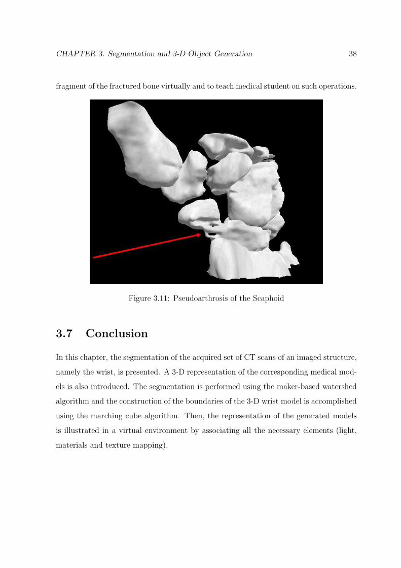
CHAPTER 3. Segmentation and 3-D Object Generation 38
fragment of the fractured bone virtually and to teach medical student on such operations.
Figure 3.11: Pseudoarthrosis of the Scaphoid
3.7 Conclusion
In this chapter, the segmentation of the acquired set of CT scans of an imaged structure,
namely the wrist, is presented. A 3-D representation of the corresponding medical mod-
els is also introduced. The segmentation is performed using the maker-based watershed
algorithm and the construction of the boundaries of the 3-D wrist model is accomplished
using the marching cube algorithm. Then, the representation of the generated models
is illustrated in a virtual environment by associating all the necessary elements (light,
materials and texture mapping).

Chapter 4
Convex Hull: A New HybridApproach
In this chapter, a new hybrid approach to generate the Convex Hull (CH) is developed
and presented. The new algorithm is validated by performing a comparison with con-
ventional algorithms namely the Brute Force, the Gift Wrapping, the QuickHull and
the Chan algorithms. The evaluation is achieved by generating the convex envelope of
3-D wrist bones using the five different approaches. The results show the improvement
associated with the proposed approach.
4.1 Introduction
In general, a real object does not have a deterministic shape. Therefore, it is impossible
to define a geometric equation to model such an object. Thus, alternative approaches
are the CH algorithms to form the convex envelopes of any object and to mimic realistic
environment with exact collision detection between objects in the virtual world. Also,
CHs play an important role in many applications that are based on cluster analysis,
image processing and pattern recognition [50]. Other problems can be reduced to CH
such as half-space intersections [51], Delaunay triangulation and Voronoi diagrams [52].
Since computers are used to simulate a physical environment, the simulation of the
model is essentially based on geometry. Many of the computational problems associated
with designing and building a VR system are geometric in nature. An important problem
that must be addressed to make VR more realistic is the problem of real-time interactive
39

CHAPTER 4. Convex Hull: A New Hybrid Approach 40
collision detection. Most exact collision detection systems work almost exclusively with
convex objects because they have certain properties that make them highly suitable
for testing the intersection. Actually, convex hulls or convex envelopes have received
considerable attention to tackle geometric problems. These kinds of problems are usually
not easy to deal with and they are known to be NP-hard. Also, fast algorithms that
compute CH are still a challenging issue in many fields and research areas for real-time
applications.
4.2 Convex Hull Definition
The convex hull or convex envelope of a finite set S of n points in the Euclidean space
ℜd of dimension d, denoted as CH(S), is defined by the smallest convex set containing
all the points or simply the intersection of all half-spaces containing the set S. The CH
in ℜd is the set of solutions to a finite system of linear inequalities in d-variables:
CH(S) = {x ∈ ℜd : Ax ≤ b} (4.1)
Where A ∈ ℜn∗d and b ∈ ℜn.
A solution of the above system can be written as:
CH(S) =n∑
i=1
λipi,n∑
i=1
λi = 1, λi ≥ 0 (4.2)
4.3 Related Work
One of the central problems in computational geometry is the computation of CHs. It
is an intensively studied subject by researchers. Early studies dealt primarily with the
planer 2-D case [53]. Currently, this interest has been extended to calculate CH in 3-D
space [54].
Brute Force is a simple algorithm that works in both 2-D and 3-D to construct
the convex envelope of the object under analysis. It requires a running time in the

CHAPTER 4. Convex Hull: A New Hybrid Approach 41
order of O(n3) and O(n4) for a 2-D and a 3-D application, respectively. A lower bound
algorithm is proposed by Yao [55] to compute the CH vertices using a quadratic decision
tree model and has a complexity of O(nlogn). This lower bound was later generalized
to the algebraic decision tree and to the algebraic computation tree models developed
by Ben-Or [62].
Another 2-D approach, known as Graham’s scan [56], achieves a running time in the
order of O(nlogn). Jarvis March algorithm constructs the convex envelope in O(nh)
time, where h denotes the number of vertices of the convex hull. It can be imple-
mented for 2-D/3-D CH and it is output sensitive because it depends on h in its running
time. Furthermore, a 2-D divide-and-conquer algorithm [57] is proposed following the
implementation of sorting algorithms, such as MergeSort and QuickSort, and needs
O(nlogn) running time. Based on this algorithm, Preparata and Hong presented their
first O(nlogn) time algorithm in 3-D. QuickHull [58] is another fast technique that works
in 2-D and can be generalized to 3-D case. The running time to compute the CH is also
in the order of O(nlogn). Moreover, Gift Wrapping is a 3-D algorithm that constructs
the convex envelope in O(nh) time [59]. This output sensitive method proposed by
Chand and Kapur was a generalization of Jarvis’s march and can be implemented with
arbitrary dimensions.
A 3-D approach proposed by Chazelle and Matousek [60] has succeeded to accom-
plish running time in the order of O(nlogh). Edelsbrunner and Shi [61] made up a
deterministic technique that requires O(nlog2h) running time. The last two algorithms
are not very practical and tend to be complicated. Thus, the problem of finding optimal
and practical algorithms that construct convex envelope in 3-D remained.
4.4 3-D Convex Hull Algorithms
Four algorithms to construct the convex envelope of an imaged object in 3-D are de-
scribed and discussed. They are: the Brute Force, the Chan algorithm, the Gift Wrap-
ping and the QuickHull. Then, a hybrid technique based on the last two approaches is
proposed and a comparison is performed between the individual algorithms.

CHAPTER 4. Convex Hull: A New Hybrid Approach 42
4.4.1 Brute Force Algorithm
Brute Force is the simplest algorithm. However, it is the slowest. It begins by selecting
a random point pi and three other different points (pj, pk, pl) as a facet on CH. Then,
it checks if the point pi is in the counterclockwise (ccw) direction with respect to the
selected facet. The procedure is repeated by checking all the facets formed by all points
other than pi. If it is found that the selected point is in the counter clockwise direction
with respect to all facets, then the point pi is on the CH. In this manner, vertices on
CH can be easily recovered.
It is clear that it is a time consuming algorithm. It needs O(n4) operations to
construct the convex hull in 3D. This method becomes very slow if the set of input points
is increased. Consequently, it is not an appropriate approach for real time applications.
4.4.2 Gift Wrapping Algorithm
The Gift Wrapping algorithm, known also as the Jarvis March approach, is developed
to work with arbitrary dimensions. It consists of an initialization phase followed by a
series of wrapping steps. The initialization phase begins by finding a starting edge (a, b).
This is achieved by performing a 2-D algorithm on the projection of the points into the
XY plane. Then, an initial plane P is pivoted around the edge (a, b) of the hull. It
finds the smallest angle between the plane P containing the starting edge (a, b) and a
plane T formed by point pi and the edge (a, b). The point pi is replaced by c and a
triangular face containing (a, b, c) is formed. The plane (a, b, c) is a facet on the CH. All
points now lie to the left of this plane. A set F of frontier edges is initially defined and
contains the three edges (a, b), (a, c) and (b, c). Each frontier edge in F is associated
with a triangle or facet on the CH of S. The wrapping steps are repeated recursively for
the edges (a, c) and (b, c) to find other triangles adjacent to those edges. All steps are
repeated for every explored edge until all facets have been examined. Figure 4.1 shows
the steps for constructing the CH using the Gift Wrapping algorithm.
The Gift Wrapping algorithm needs O(nh) time operations to construct the CH. It
is clear that for every hull edge point, the algorithm requires O(h) time where h is the
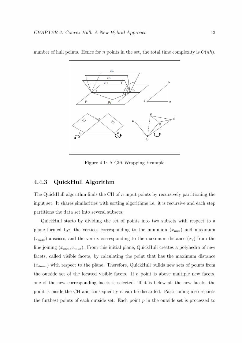
CHAPTER 4. Convex Hull: A New Hybrid Approach 43
number of hull points. Hence for n points in the set, the total time complexity is O(nh).
Figure 4.1: A Gift Wrapping Example
4.4.3 QuickHull Algorithm
The QuickHull algorithm finds the CH of n input points by recursively partitioning the
input set. It shares similarities with sorting algorithms i.e. it is recursive and each step
partitions the data set into several subsets.
QuickHull starts by dividing the set of points into two subsets with respect to a
plane formed by: the vertices corresponding to the minimum (xmin) and maximum
(xmax) abscises, and the vertex corresponding to the maximum distance (xd) from the
line joining (xmin, xmax). From this initial plane, QuickHull creates a polyhedra of new
facets, called visible facets, by calculating the point that has the maximum distance
(xdmax) with respect to the plane. Therefore, QuickHull builds new sets of points from
the outside set of the located visible facets. If a point is above multiple new facets,
one of the new corresponding facets is selected. If it is below all the new facets, the
point is inside the CH and consequently it can be discarded. Partitioning also records
the furthest points of each outside set. Each point p in the outside set is processed to
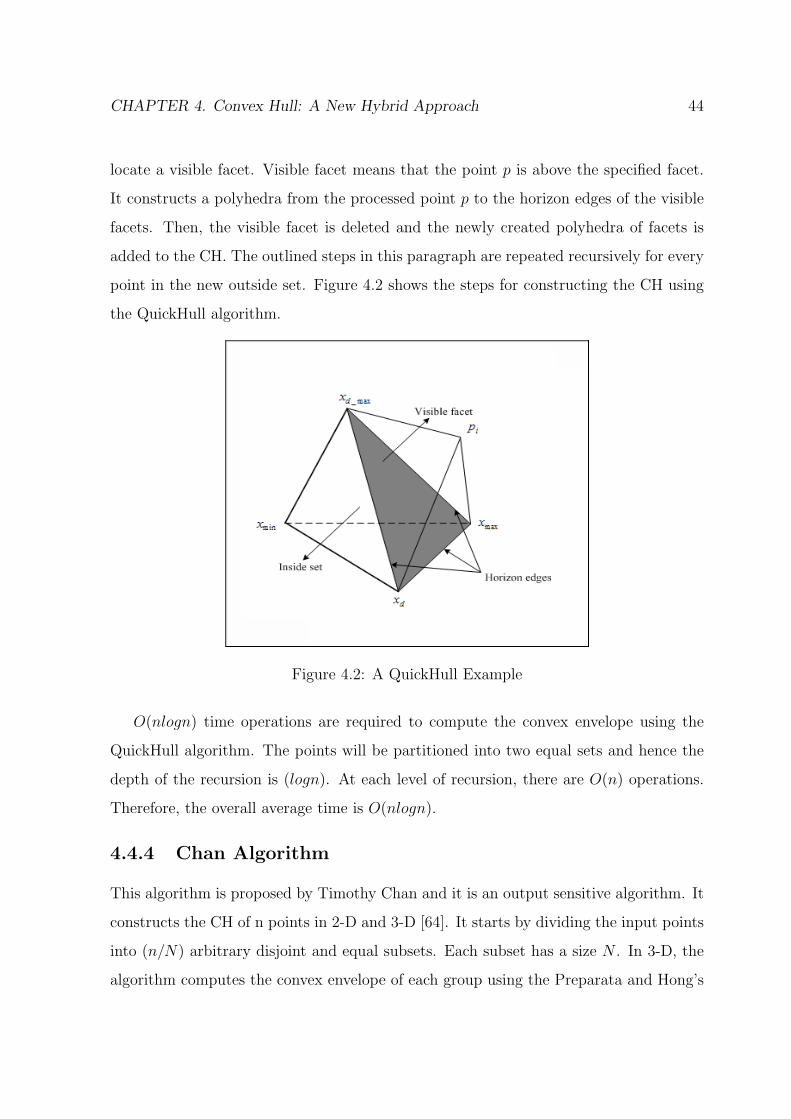
CHAPTER 4. Convex Hull: A New Hybrid Approach 44
locate a visible facet. Visible facet means that the point p is above the specified facet.
It constructs a polyhedra from the processed point p to the horizon edges of the visible
facets. Then, the visible facet is deleted and the newly created polyhedra of facets is
added to the CH. The outlined steps in this paragraph are repeated recursively for every
point in the new outside set. Figure 4.2 shows the steps for constructing the CH using
the QuickHull algorithm.
Figure 4.2: A QuickHull Example
O(nlogn) time operations are required to compute the convex envelope using the
QuickHull algorithm. The points will be partitioned into two equal sets and hence the
depth of the recursion is (logn). At each level of recursion, there are O(n) operations.
Therefore, the overall average time is O(nlogn).
4.4.4 Chan Algorithm
This algorithm is proposed by Timothy Chan and it is an output sensitive algorithm. It
constructs the CH of n points in 2-D and 3-D [64]. It starts by dividing the input points
into (n/N) arbitrary disjoint and equal subsets. Each subset has a size N . In 3-D, the
algorithm computes the convex envelope of each group using the Preparata and Hong’s
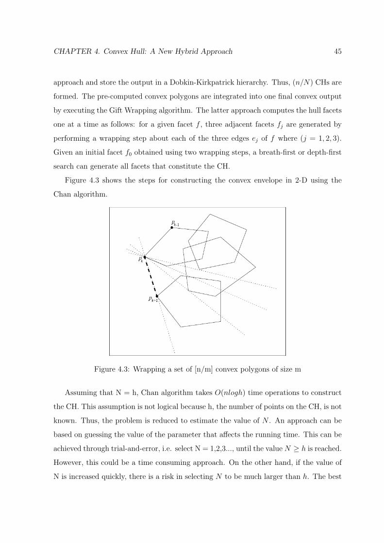
CHAPTER 4. Convex Hull: A New Hybrid Approach 45
approach and store the output in a Dobkin-Kirkpatrick hierarchy. Thus, (n/N) CHs are
formed. The pre-computed convex polygons are integrated into one final convex output
by executing the Gift Wrapping algorithm. The latter approach computes the hull facets
one at a time as follows: for a given facet f , three adjacent facets fj are generated by
performing a wrapping step about each of the three edges ej of f where (j = 1, 2, 3).
Given an initial facet f0 obtained using two wrapping steps, a breath-first or depth-first
search can generate all facets that constitute the CH.
Figure 4.3 shows the steps for constructing the convex envelope in 2-D using the
Chan algorithm.
Figure 4.3: Wrapping a set of [n/m] convex polygons of size m
Assuming that N = h, Chan algorithm takes O(nlogh) time operations to construct
the CH. This assumption is not logical because h, the number of points on the CH, is not
known. Thus, the problem is reduced to estimate the value of N . An approach can be
based on guessing the value of the parameter that affects the running time. This can be
achieved through trial-and-error, i.e. select N = 1,2,3..., until the value N ≥ h is reached.
However, this could be a time consuming approach. On the other hand, if the value of
N is increased quickly, there is a risk in selecting N to be much larger than h. The best
CHAPTER 4. Convex Hull: A New Hybrid Approach 46
way is to choose (at the beginning) small values of N and increase it successively by
squaring each time the value until the algorithm returns successful results. This process
is called the doubling search. In other words, at iteration t (starting at t = 0), a value
of N = min(22t
, n) is selected and the steps of the Chan algorithm are repeated until
N ≥ h. Consequently, the convex envelope is formed.
4.5 The Hybrid Algorithm
Several methods are developed to speed up CH algorithms by preprocessing the input
set of points. Some techniques start by dividing the input points into two arbitrary
sets, right and left, followed by the computation of the final CH. Divide-and-Conquer
is such an algorithm that starts recursively by computing the convex envelopes of the
right and the left sets followed by merging the two hulls into a final convex output [63].
Other techniques divide the input points into many subsets such as the presented Chan
algorithm. It starts by dividing the input points into (n/N) arbitrary disjoint subsets.
Each subset has a size N . Then, the convex envelope of each group is computed. Thus,
N partial hulls are formed and they are integrated into one final convex output. The idea
underlying these techniques is always dividing a large set into several subsets in order to
speed up the running time of the algorithm and reduce its complexity. In this context,
a hybrid technique is proposed to construct the CH with a faster time by preprocessing
the input set of points. Two important points are taken into consideration:
• The running time of the convex hull algorithms depends on the number of points
n that constitutes the object.
• The wrapping step embedded in the Gift Wrapping algorithm can be achieved
faster if the set of input points is preprocessed.
Therefore, a hybrid approach to generate the CH is developed. It is based on Quick-
Hull and Gift Wrapping algorithms. It is decomposed into two stages. The first stage

CHAPTER 4. Convex Hull: A New Hybrid Approach 47
reduces the number of the input points. The second stage underlines the reconstruc-
tion of the corresponding convex envelope. A pseudo-code of the hybrid algorithm is
illustrated below (Algorithm 1).
Algorithm 1 The Hybrid Approach To Construct the CH
1: find an initial plane from the min and max abssice and the max distance withrespect to (xmin, xmax)
2: construct a polyhedron from the initial plane and the max distance to this plane3: for each facet F of the polyhedra do4: for each unassigned point p do5: if p is above F then6: assign p to F ′s outside set7: end if8: end for9: end for
10: Discard all points inside the polyhedron forming a new imput set (nnew)11: find a starting edge (a, b) using the 2D Gift Wrapping algorithm on the XY projec-
tion12: for i = 1 ... nnew do13: find point pi corresponding to min angle bewteen plane P in XY containing (a, b)
and plane T = (a, b, pi)14: replace c← pi
15: save (a,b,c) into Q16: wrap the edge (a, c)17: if facet has been explored then18: wrap the edge (b, c)19: if facet has been explored then20: return21: end if22: end if23: end for
The hybrid method is initiated by applying the QuickHull algorithm to divide the
input points into two subsets (upper and lower) with an initial plane. Then, a polyhedron
of new facets is created by calculating the point having the maximum distance with
respect to this plane. Consequently, the points that are inside the polyhedron are inside
the convex envelope and they are discarded. The same procedure is repeated for the
lower set. This leads to the reduction of the number of input points and the formation
of a new data set. The new set is fed as an input to the Gift Wrapping algorithm.

CHAPTER 4. Convex Hull: A New Hybrid Approach 48
Consequently, wrapping steps are performed by scanning the new data to obtain the
final convex envelope. That is, the hybrid method apply the initialization phase followed
by the series of the wrapping steps. It computes the facets of the hull one at a time,
in the ccw direction using the sequence of the wrapping steps. The wrapping steps are
repeated recursively for every explored edge until all facets have been examined.
4.6 3-D Models and Convex Hulls
The Hybrid approach to construct the CH is implemented. Different bones (3-D struc-
tures) having various dimensions (different number of vertices) and concavities are cho-
sen. Figure 4.4 shows the Capitate bone before (upper left) and after (upper right) the
implementation of proposed algorithm to construct the CH of the corresponding bone.
Also, it illustrates the Ulna bone of the wrist before (lower left) and after (lower right)
the CH is formed using the hybrid approach.
Figure 4.4: Capitate and Ulna with their Convex Hulls

CHAPTER 4. Convex Hull: A New Hybrid Approach 49
Similarly, Figure 4.5 presents another two wrist bones, namely, the 3rdMetacarpal
(upper half) and the Scaphoid (lower half). In each half, the 3-D objects (left) and the
corresponding convex hulls (right) are shown.
Figure 4.5: 3rdMetacarpal and Scaphoid with their Convex Hulls
The conceptual basis for the collision detection is to construct a CH using the vertices
of each object. Consequently, the collision is observed as the intersection of the corre-
sponding CHs. In this regard, Figure 4.6 illustrates the collision detection problem in a
realistic way. It shows different bones constituting the 3-D wrist model: 1st Metacarpal
(a), 2nd Metacarpal (b), 4th Metacarpal (c), Scaphoid (d), Capitate (e), Hamate (f),
Radius (g) and Ulna (h). Each bone is covered with its corresponding convex envelope.
In addition to the proposed approach (Hybrid algorithm), the conventional tech-
niques (Brute Force, Gift Wrapping, QuickHull and Chan algorithms) are also tested
and simulated using the 3-D data of a patient’s wrist. A comparison is performed
between these algorithms and the corresponding results are presented, analyzed and
discussed.
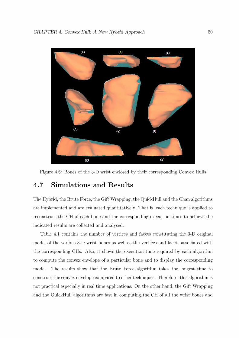
CHAPTER 4. Convex Hull: A New Hybrid Approach 50
Figure 4.6: Bones of the 3-D wrist enclosed by their corresponding Convex Hulls
4.7 Simulations and Results
The Hybrid, the Brute Force, the Gift Wrapping, the QuickHull and the Chan algorithms
are implemented and are evaluated quantitatively. That is, each technique is applied to
reconstruct the CH of each bone and the corresponding execution times to achieve the
indicated results are collected and analysed.
Table 4.1 contains the number of vertices and facets constituting the 3-D original
model of the various 3-D wrist bones as well as the vertices and facets associated with
the corresponding CHs. Also, it shows the execution time required by each algorithm
to compute the convex envelope of a particular bone and to display the corresponding
model. The results show that the Brute Force algorithm takes the longest time to
construct the convex envelope compared to other techniques. Therefore, this algorithm is
not practical especially in real time applications. On the other hand, the Gift Wrapping
and the QuickHull algorithms are fast in computing the CH of all the wrist bones and

CHAPTER 4. Convex Hull: A New Hybrid Approach 51
have approximately similar behaviour. The hybrid method and the Chan algorithm are
faster than the latter approaches. The hybrid method outperforms the Gift Wrapping
and the QuickHull algorithms in the reconstruction of all CHs of the various wrist bones.
On the other hand, the hybrid technique surpasses the Chan algorithm in most cases
especially, when the number of points that are reduced is high enough.
Table 4.1: Comparison of Execution time for Computing the 3-D Convex Hull of theWrist Bones
3-D Model Original Model Convex Hull Brute Force Gift Wrap QuickHull Chan Hybrid# vertices # facets # vertices # facets time (s) time (s) time (s) time (s) time (s)
1st Meta. 2179 4320 379 790 19014.22.1 0.66 0.61 0.39 0.432nd Meta. 1168 2258 300 596 12015.41 0.47 0.38 0.33 0.253rd Meta. 675 1272 150 296 2330.77 0.26 0.22 0.15 0.124rd Meta. 532 1002 147 290 2302.72 0.21 0.19 0.13 0.09Hamate 2812 5620 394 784 19231.20 0.89 0.82 0.67 0.71Ulna 977 1864 312 620 12153.03 0.41 0.37 0.32 0.22Scaphoid 2890 5784 530 1056 22125.1 1.31 1.22 1.03 0.88Capitate 3026 6048 635 1266 25300.35 1.46 1.38 1.31 1.25Radius 2454 4754 288 572 11260.12 0.78 0.72 0.55 0.62
Similarly, table 4.2 shows the number of vertices and facets constituting the 3-D
original model of the knee bones as well as the vertices and facets associated with the
corresponding CHs. The execution time required for each algorithm to compute the
convex envelope and to display the corresponding model is also presented. The table
shows the results associated with the Gift Wrapping, the QuickHull, the Chan and
the Hybrid algorithms. It is evident that the proposed approach surpasses the other
conventional algorithms.
Table 4.2: Comparison of Execution time for Computing the 3-D Convex Hull of theKnee Bones
3D Model Original Model Convex Hull Gift Wrap QuickHull Chan Hybrid# vertices # facets # vertices # facets time (s) time (s) time (s) time (s)
Fibula 3732 7460 914 1924 3.32 3.01 2.56 2.28Patella 7058 14112 2228 4452 10.04 9.53 9.15 9.04Tibia 27342 54680 2653 5301 46.16 44.02 33.19 30.12Femur 34716 69428 3664 7324 83.27 81.03 69.89 63.32

CHAPTER 4. Convex Hull: A New Hybrid Approach 52
4.7.1 Result Interpretation
In this subsection, the results are analyzed and a discussion is provided for both cases:
the wrist and the knee.
Wrist
It is evident that the hybrid method reduces the number of vertices of the 3rd Metacarpal
from 675 to 559 as a first stage. Then, the wrapping process constructs the corresponding
convex envelope, i.e. translated into a reduction of the running time to 0.12 s. On the
other hand, the Gift Wrapping algorithm, the QuickHull and the Chan algorithm require
0.26, 0.22 and 0.15 s, respectively. Thus, the proposed technique decreases the execution
time by 53.87% in comparison with the Gift Wrapping, by 42.85% with respect to the
QuickHull and by 13.33% with respect to Chan algorithm.
In the same way, the number of vertices of the Scaphoid is decreased form 2890 to
2539 using the proposed approach. Consequently, a decrease in the running time from
1.31 s (for the Gift Wrapping), 1.22 s (for the QuickHull) and 1.03 s (for Chan algorithm)
to 0.88 s is achieved. That is, the running time of the proposed algorithm is decreased
by 32.82, 27.86 and 14.56% in comparison with the Gift Wrapping, the QuickHull and
the Chan algorithm, respectively. On the other hand, if the number of points is not
reduced much, the Chan algorithm can outperform the hybrid approach in some cases
and consequently, it constructs the convex envelope faster. The 1st Metacarpal and the
Radius are illustrative examples of this case.
The achieved improvement using the hybrid approach is further illustrated in figure
4.7. It shows a graph representing the execution time of the 3-D CH algorithms for the
various wrist bones. In this graph, each plot is associated with a different algorithm: the
Hybrid, the Gift Wrapping, the QuickHull and the Chan algorithms. The execution time
is plotted as a function of the number of vertices constituting each bone. The number of
vertices represents nine of the 15 bones forming the 3-D wrist model presented in Table
4.1. It is evident that the hybrid method surpasses the Gift Wrapping and the QuickHull
approaches for all the wrist bones and it performs better than the Chan algorithm in

CHAPTER 4. Convex Hull: A New Hybrid Approach 53
constructing the CH of most of the bones. Subsequently, the hybrid method is very
efficient and is practical in modelling 3-D medical data (wrist) and simulating them in
the virtual environment.
Figure 4.7: Execution Time of the 3-D CH Algorithms for Wrist Bones
Knee
The proposed method reduces the number of vertices of the Fibula from 3732 to 3662 as
a first stage. Then, the wrapping process constructs the corresponding convex envelope
leading to a reduction of the execution time to 2.28 s. On the other hand, the Gift Wrap-
ping, the QuickHull and the Chan algorithm require 3.32, 3.01 and 2.56 s, respectively.
Thus, the proposed technique decreases the execution time by 31.33% in comparison
with the Gift Wrapping, by 24.26 % with respect to the QuickHull and by 10.93% with
respect to the Chan algorithm. In a similar fashion, the number of vertices of the Femur
is decreased from 34716 to 32490 using the hybrid approach. Consequently, a decrease
in the running time from 83.27 s (for the Gift Wrapping), 81.03 s (for the QuickHull)

CHAPTER 4. Convex Hull: A New Hybrid Approach 54
and 69.89 (for Chan algorithm) to 63.32 s is achieved. That is, the running time of the
proposed algorithm is decreased by 23.96, 21.92 and 9.4 % in comparison with the Gift
Wrapping, the QuickHull and the Chan algorithm, respectively. Therefore, the proposed
approach reconstructs the CH of the knee faster than the other conventional algorithms.
The quantative evaluation of the hybrid approach is also illustrated in figure 4.8. It
shows a graph representing the execution time of the 3-D CH algorithms for the knee
bones. The execution time is plotted as a function of the number of vertices constituting
each bone. The number of vertices represents the four bones forming the 3-D knee model
presented in Table 4.2.
Figure 4.8: Execution Time of the 3-D CH Algorithms for Knee Bones
Similarly, it is evident that the hybrid method outperforms the three conventional
approaches in the reconstruction of the CH of the bones constituting the knee model.
That is translated into a reduction in the overall execution time (smaller value) and is
clearly observed when the number of vertices is high.
CHAPTER 4. Convex Hull: A New Hybrid Approach 55
4.8 Conclusion
Since most 3-D objects are not characterised by a shape that could be modelled using
precise mathematical equations, CH algorithms are considered as a solution to model
such objects. The need to CH algorithms arises with the intention of simulating realistic
scenes with a real-time interactive collision detection between objects in the virtual
world. Since fast collision detection systems work almost exclusively with convex objects,
quick CH algorithms are implemented to fulfil real-time requirements. In this chapter,
a hybrid approach to construct the CH of 3-D objects (bones of the wrist and the knee)
is proposed and is compared with four published algorithms: the Gift Wrapping, the
QuickHull, Chan and the Brute Force algorithms. The results show that the proposed
approach performs faster than the other conventional techniques. That is, the execution
time to construct the convex envelope is achieved in less time than the other approaches
(i.e. a faster execution time). The proposed hybrid method is an output sensitive
algorithm that works in 2-D as well as in 3-D. It is very efficient, practical and useful in
modelling and simulating 3-D medical data in a virtual environment.

Chapter 5
Collision Detection: A LinearProgramming Technique
In this chapter, the issue of the precise Collision Detection (CD) between virtual objects
is addressed and a new technique is proposed. The CD problem is formulated and a linear
programming solution is obtained to determine whether a collision exists or not. The
proposed approach is compared with a conventional algorithm, namely, the Industrial
Virtual Reality Institute Collision Detection (IVRI-CD) technique and is validated using
bones from the 3-D wrist model. The results show that the proposed algorithm is
efficient, fast, robust and leads to a decrease of the running time that is required to
detect a collision.
5.1 Introduction
Collision detection or contact determination is the approach that reports a geometric
contact when a collision is about to happen or has actually happened. It is of interest
in several research areas such as robotics and computational geometry. CD is provided
in every prototype system of computer-based surgical simulators. Most CD algorithms
integrated in surgical simulators are based on solving systems of equations with dimen-
sions proportional to the number of contact points between the objects. For example, in
3-D object models, the number of contact volume elements is proportional to the area
of the contact surfaces resulting in thousands of contact elements for relatively small
surfaces. In this regard, the issue of a precise CD between virtual objects is addressesed
56
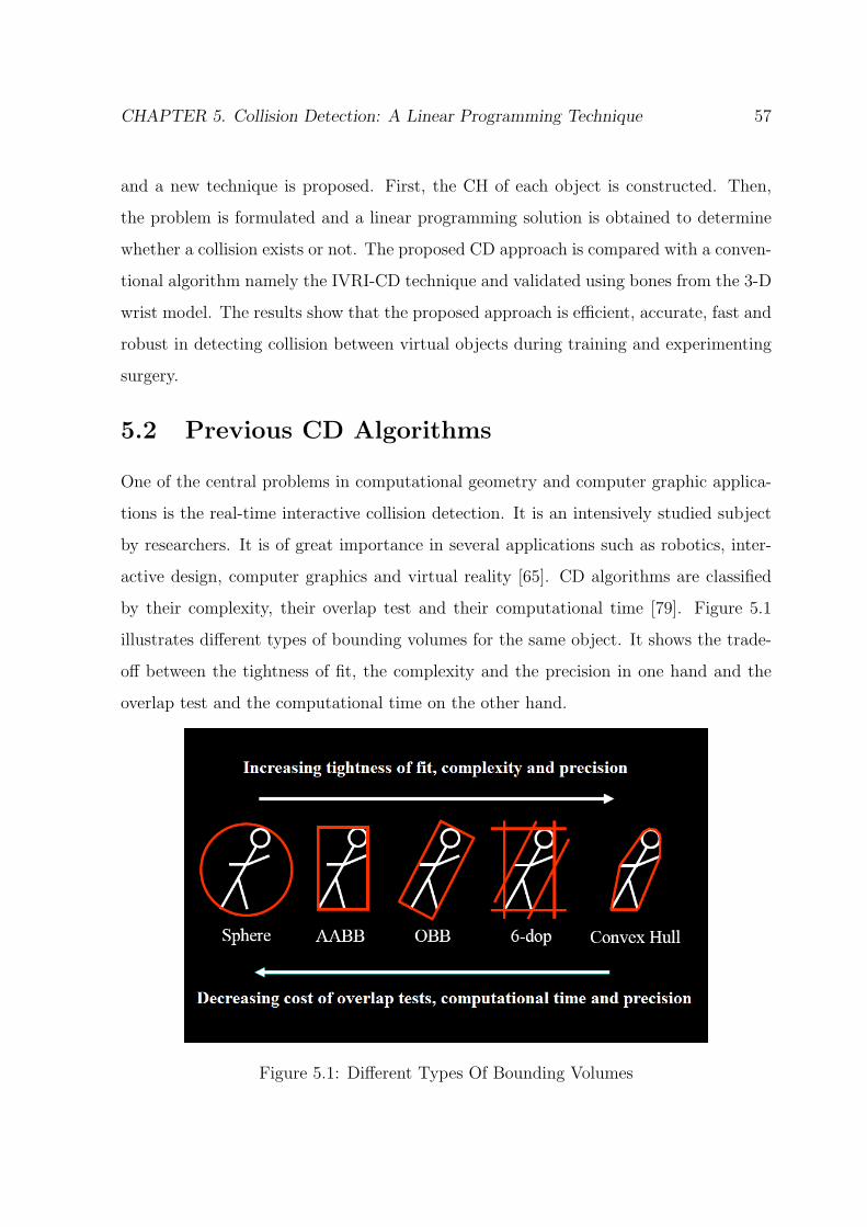
CHAPTER 5. Collision Detection: A Linear Programming Technique 57
and a new technique is proposed. First, the CH of each object is constructed. Then,
the problem is formulated and a linear programming solution is obtained to determine
whether a collision exists or not. The proposed CD approach is compared with a conven-
tional algorithm namely the IVRI-CD technique and validated using bones from the 3-D
wrist model. The results show that the proposed approach is efficient, accurate, fast and
robust in detecting collision between virtual objects during training and experimenting
surgery.
5.2 Previous CD Algorithms
One of the central problems in computational geometry and computer graphic applica-
tions is the real-time interactive collision detection. It is an intensively studied subject
by researchers. It is of great importance in several applications such as robotics, inter-
active design, computer graphics and virtual reality [65]. CD algorithms are classified
by their complexity, their overlap test and their computational time [79]. Figure 5.1
illustrates different types of bounding volumes for the same object. It shows the trade-
off between the tightness of fit, the complexity and the precision in one hand and the
overlap test and the computational time on the other hand.
Figure 5.1: Different Types Of Bounding Volumes
CHAPTER 5. Collision Detection: A Linear Programming Technique 58
5.2.1 AABB/OBB
Most collision detection algorithms are based on the bounding volumes approach. The
absence of bounding volume collisions guarantees the absence of a collision between the
corresponding objects. The bounding boxes techniques are used to identify the objects
that may be in contact (in pairs) and to reduce the number of facets. Different kinds of
static and dynamic sizes of bounding boxes are implemented. The bounding boxes are
a preliminary test of collisions. The two effective methods are: Axis-Aligned Bounding
Boxes (AABB) [66] and Oriented Bounding Boxes (OBB) [67].
5.2.2 Lin-Canny (LC)
An important CD approach is the Lin-Canny (LC) closest feature algorithm [68]. Lin
and Canny have proposed the first algorithm that exploits the coherence [61]-[62]. This
technique retains the pair of closest features i.e. vertices, edges or facets between two
convex polyhedra by exploiting the fact that the current closest features are probably
near the previous one. Having identified those features, the distance between the two
polyhedra is found and a collision is declared. The algorithm starts from the closest
pair that is computed in the last time step, and “walks” on the surface of the polyhedra
until it reaches the new closest pair. The convexity guarantees that one can determine
locally whether a pair of features is the closest pair, and if it is not, a neighboring pair is
closer. Once those features are identified, the distance , which is below some ǫ, between
the two polyhedra is found and a collision is declared. Finally, the Lin-Canny algorithm
does not handle penetrating polytopes.
5.2.3 Gilbert-Johnson-Keerthi (GJK)
Another significant technique is the Gilbert-Johnson-Keerthi (GJK) algorithm [69]. The
GJK approach involves an iterative procedure to determine the Minkowski difference
between the convex objects among which the collision is being detected. The GJK
algorithm is a Simplex-based algorithm. This approach uses the CH of a polytope and
performs on the simplex defined by the subsets of these points [96]. The main strength

CHAPTER 5. Collision Detection: A Linear Programming Technique 59
of this algorithm is that, in addition to detect whether two objects have collided or not,
it can also return a measure of interpenetration. GJK achieves almost the same time
complexity as Lin-Canny. Researchers have developed an enhanced version of the GJK
algorithm, known as SOLID (Software Library for Interference Detection) [70]. It is a
fast and a robust way to check for collision.
Besides, the use of bounding boxes is essential to speed up the computational time
for collision checks when objects are far away. On the other hand, algorithms such as the
LC and the GJK are required when the objects to be tested for collision are sufficiently
close.
5.2.4 Voronoi-Clip (V-Clip)
Voronoi-Clip (V-Clip) [71] is another approach that tracks the closest pairs of features of
convex objects in a similar fashion to the LC algorithm. However, it handles penetrating
polyhedra and can be extended to the non convex polyhedra by representing them as
groups of convex polyhedra.
5.2.5 I-Collide
The I-Collide [72] is an interactive and an exact CD library for large environments
composed of convex polyhedra. It exploits the coherence and the properties of convexity
to achieve a very fast and an exact collision.
5.2.6 Q-Collide
The Q-Collide [73] algorithm is developed to deal with the convex polyhedra. It works as
follows: if two objects are not colliding, the algorithm finds a separating plane between
them. Otherwise, a collision is reported. If a collision does not occur, the separating
plane found for one time frame is cached as a witness for the next time frame.
5.2.7 Quick-CD
The Quick-CD algorithm is based on constructing hierarchies of bounding volumes (BV-
trees) to approximate the input models [74]. The choice of bounding volumes is made

CHAPTER 5. Collision Detection: A Linear Programming Technique 60
using Discrete Orientation Polytopes (DOP), which are convex polytopes whose facets
have normals composed from a given discrete set of k vectors. In addition, Ehmann and
Lin have introduced a new algorithm called SWIFT [75]. They have shown that this
approach is faster and more robust than earlier techniques.
5.2.8 SWIFT
SWIFT is a C++ collision detection package. It can compute an exact and an approx-
imate distance, and it allows the contact’s determination between the 3-D polyhedral
objects undergoing a rigid motion. It has been successfully applied to many applications
such as virtual prototyping, dynamic simulation and path planning.
SWIFT works with geometric models that are closed, convex or composed of convex
pieces and with any objects composed of a set of polygons that describe the boundary of a
solid in 3-D. The SWIFT algorithm is based on the Lin-Canny approach to minimize the
distance between two convex models. The intersection is tested directly without the need
to construct any bounding volume hierarchy. However, the bounding volume hierarchies
are implemented at a higher level. The SWIFT algorithm has many advantages over the
conventional techniques. It is faster and is robust than the other packages such as the
I-Collide and the V-Clip algorithms. It automatically identifies the hierarchy’s bounding
properties, computes the deviation and builds its own simplified hierarchy in two different
ways: the modified Dobkin-Kirkpatrick and the QSlim methods. Moreover, SWIFT
shares the sweep and prune algorithm with I-Collide. In other words, the bounding boxes
which are chosen automatically, are placed around each object to prune the unnecessary
computation using the sweep and prune sorting algorithm. Besides, SWIFT supports
object geometry sharing. The objects may be added to a scene using arrays or files of
different formats. Also, the objects that are declared as static (fixed or not moving) are
automatically optimized and any pair may be activated or deactivated.

CHAPTER 5. Collision Detection: A Linear Programming Technique 61
5.3 The Proposed CD Approach
The possibility of using mathematical programming techniques has helped researchers to
correlate the Linear Programming (LP) and the CD problem. Thus, a compact formu-
lation of the collision detection coupled with a methodology that effectively implements
the LP problem is proposed. The theory of this algorithm is proposed in [76], [77]. This
will be a promissing step toward the computer-based applications in terms of precision,
speed and robustness.
The goal of a medical simulator is to support medical students during the training
and the practice on surgeries and to perform the operations with a high precision. In
this regard, medical objects are modeled with a tightness fit i.e. each object is modeled
by its corresponding CH as shown in the previous chapter. CHs or convex envelopes
have less contact points than real objects and can be quickly computed. This leads to a
decrease in the size of the system of equations needed to test for a collision. Hence, the
real time performance of CD algorithms is increased and the algorithms become faster.
In addition, most exact CD systems work almost exclusively with convex objects. This is
due to the fact that convex objects allow CD algorithms to converge quickly and detect
the collision if it exists. This will give the simulator a high degree of precision but at
the same time an increase in the cost of the complexity and the computational time to
check for a collision. Therefore, by taking advantages of the speed and the robustness
of LP techniques, the problem of collision detection is formulated and solved. In this
regard, the CH of each object is reconstructed and the CD problem is formulated as an
optimization problem based on convex objects and is solved using LP (simplex method).
5.3.1 Linear Programming Solution
To formulate the problem, each facet i of the convex envelope is represented by the plane
inequality in the form of:
aix + biy + ciz ≤ di (5.1)
Any point lying on the object must satisfy the inequalities of the planes constituting

CHAPTER 5. Collision Detection: A Linear Programming Technique 62
the object. These equations form the constraints of the collision problem and represent
the facets that separate two regions in space. Therefore, if a point satisfies two sets
of inequalities simultaneously, it belongs to the corresponding convex objects. Thus, a
collision is detected at that point between these two objects.
The problem is reduced to maximize an objective function in the form of (x+ y + z).
It is formulated as follows:
max cT X (5.2)
subject to
AX ≤ b (5.3)
where X = [x y z]T ,
A =
a1 b1 c1
a2 b2 c2
a3 b3 c3
. . .
. . .
(5.4)
b = [d1 d2 d3 . . .]T , c = [1 1 1]T (5.5)
The coefficients of the matrices A and b are calculated using the facets obtained
from the convex hulls reconstructed by the proposed approach presented in the previous
chapter (i.e. using the hybrid approach). Using the duality property, the problem
becomes:
min bT π (5.6)
subject to
AT π ≥ c (5.7)
Having formulated the problem, the dual system is solved using a linear programming
algorithm (simplex method). If the system is bounded, a feasible solution exists and
consequently, a collision is detected. Otherwise there is no collision.

CHAPTER 5. Collision Detection: A Linear Programming Technique 63
Axis-Aligned Bounding Boxes (AABB) approach can be used as a preliminary test
for collisions. For AABB construction, the minimum and maximum points that are
orthogonally projected on the x, y and z axes are determined. Then, the projections
are used to determine the intervals on each axis for each object. If two objects are in
collision, the corresponding AABBs are also in collision.
5.4 The IVRI-CD Technique
In this section, an exact CD algorithm, namely the IVRI-CD, is described and dis-
cussed. Then, a comparison is performed between the proposed CD approach and this
conventional technique.
5.4.1 IVRI-CD
The IVRI-CD approach [80] checks if a collision occurs between a pair of objects based on
their CHs. Given an object P1 with n1 vertices and a second object P2 with n2 vertices,
the problem of collision is formulated using the definition of CH and by minimizing the
following function:
uφ = −
(
n1∑
i=1
viαi −n2∑
i=1
wiβi
)
(5.8)
wheren1∑
i=1
αi = 1 (5.9)
n2∑
i=1
βi = 1 (5.10)
vi and wi are the vertices constituting the first and the second object, respectively.
The variable u represents the coordinates of the distance between a point that is guar-
anteed to belong to P1 and another point that is guaranteed to belong to P2. Assuming
that µ = max {abs (u)}, the problem is reduced to minimize the following objective
function:

CHAPTER 5. Collision Detection: A Linear Programming Technique 64
minµφ (5.11)
subject to
Aγ = b (5.12)
γ ≥ 0 (5.13)
where
A =
vx1
... vxn1−wx
1... −wx
n2ux
vy1
... vyn1−wy
1... −wy
n2uy
vz1
... vzn1−wz
1... −wz
n2uz
1 ... 1 0 ... 0 0
0 ... 0 1 ... 1 0
(5.14)
bT = [0 0 0 1 1], γT = [α1...αn1β1...βn2
φ] (5.15)
If the objective function µφ ≤ ǫ (where ǫ is a very small quantity), a collision is
detected. It is to be noted that the matrix “A” in this method contains all the elements
constituting the objects while the matrix “A” in the proposed approach is composed of
the elements constituting the convex hull of the objects to be tested. Since the convex
hulls have less contact points than the real objects, the real time performance of the
new algorithm will increase and the cost of updating the matrices during each iteration
of the simulation process decreases.
5.5 Simulations and Results
The wrist’s bones of a patient are used to evaluate quantitatively the proposed approach
by performing a comparison with the IVRI-CD technique. That is, each method is
applied to test the collision between the different bones and the corresponding execution
time to achieve the indicated results are collected and analyzed.
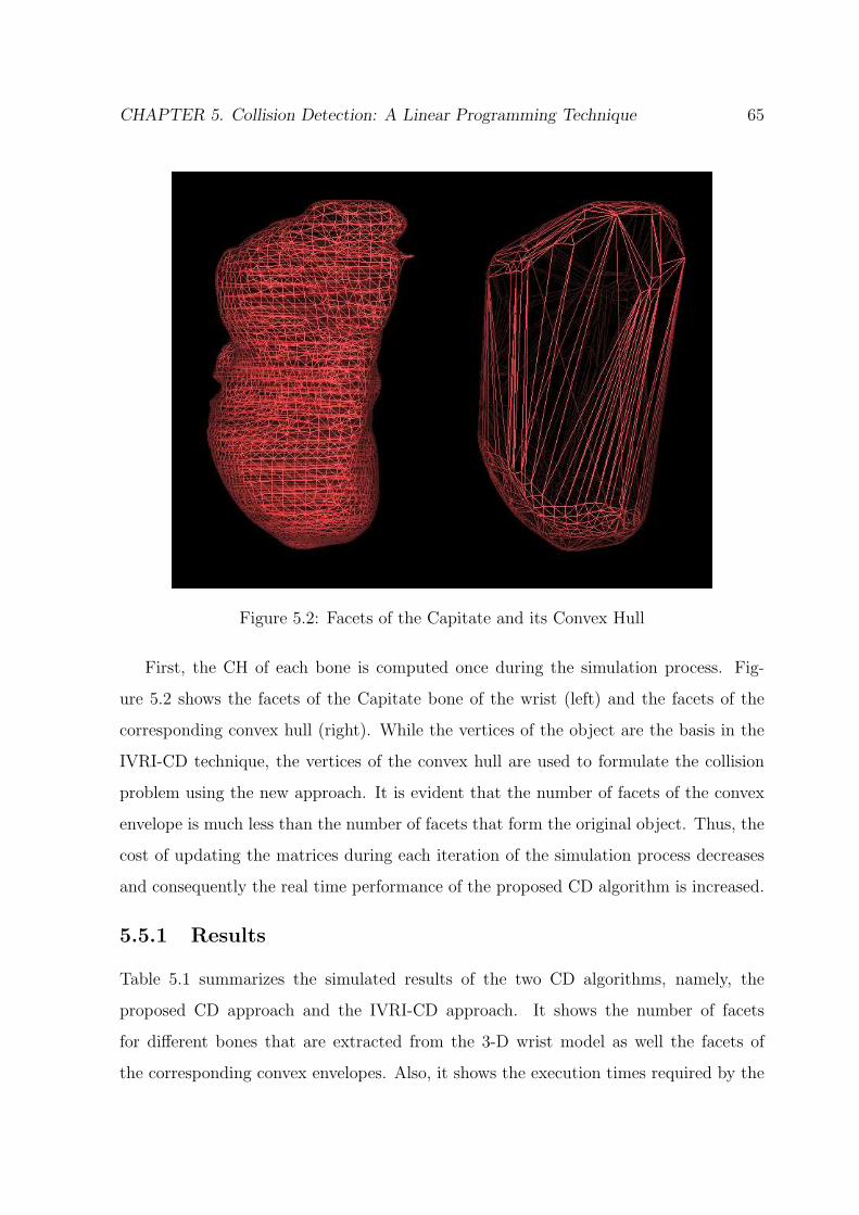
CHAPTER 5. Collision Detection: A Linear Programming Technique 65
Figure 5.2: Facets of the Capitate and its Convex Hull
First, the CH of each bone is computed once during the simulation process. Fig-
ure 5.2 shows the facets of the Capitate bone of the wrist (left) and the facets of the
corresponding convex hull (right). While the vertices of the object are the basis in the
IVRI-CD technique, the vertices of the convex hull are used to formulate the collision
problem using the new approach. It is evident that the number of facets of the convex
envelope is much less than the number of facets that form the original object. Thus, the
cost of updating the matrices during each iteration of the simulation process decreases
and consequently the real time performance of the proposed CD algorithm is increased.
5.5.1 Results
Table 5.1 summarizes the simulated results of the two CD algorithms, namely, the
proposed CD approach and the IVRI-CD approach. It shows the number of facets
for different bones that are extracted from the 3-D wrist model as well the facets of
the corresponding convex envelopes. Also, it shows the execution times required by the

CHAPTER 5. Collision Detection: A Linear Programming Technique 66
Table 5.1: Comparison of Execution Time for Collision Detection Algorithms3D Model Original Model Convex Hull Proposed Approach IVRI-CD Approach
# vertices # facets # vertices # facets time (s) time (s)1st Meta. 2179 4320 379 790 0.0369 0.03972nd Meta. 1168 2258 300 596 0.0391 0.04413rd Meta. 675 1272 150 296 0.0411 0.05214rd Meta. 532 1002 147 290 0.0431 0.0661Hamate 2812 5620 394 784 0.0523 0.0703Ulna 977 1864 312 620 0.0563 0.0753Scaphoid 2890 5784 530 1056 0.0583 0.0769Capitate 3026 6048 635 1266 0.0592 0.0782Radius 2454 4754 288 572 0.0623 0.0812
proposed algorithm as well as the IVRI-CD technique to detect a collision. It is clear
that the running time of the new approach is always less than the time of the IVRI-
CD method. Thus, it can be concluded that the proposed method is fast in detecting
collisions and consequently is more suited for real time applications. The improvement
that is achieved by the new approach is further illustrated in figure 5.3.
Figure 5.3: Execution time of the two CD algorithms for bones of the 3-D wrist model
It shows two plots, each plot is associated with a different algorithm: the IVRI-CD

CHAPTER 5. Collision Detection: A Linear Programming Technique 67
and the proposed approach. The execution time (in second) is plotted as a function of
the number of vertices that form each object (i.e. the bones of the wrist). The vertices
represent nine of the fifteen bones of the 3-D wrist model (table 5.1). It is evident that
the proposed method outperforms the IVRI-CD approach for every number of vertices.
Furthermore, the new algorithm is more efficient as the number of vertices is increased.
5.6 Proximity Queries (PQ) and Penetration Depth
(PD) Computation
The CD method can be generalized to calculate the proximity vector [78]. The proximit
query problem is also formulated as an optimisation problem and is solved using LP.
Each pair of convex objects in the scene is treated separately.
Similarly, each object is represented by a set of triangular facets i in the form of
planes inequality:
aix + biy + ciz ≤ di (5.16)
Since the time step is small enough, all the movements in the scene can be approx-
imated by a local translation without any level of detail’s degradation. The movement
of the objects is assumed to be at a constant velocity. Thus, the problem may be refor-
mulated by adding a variable t which represents the time to the equation of each facet
i as follows:
aix + biy + ciz + eit ≤ di (5.17)
Based on the above formula, if a point satisfies two sets of inequalities simultaneously,
then this point belongs to the corresponding objects and a collision is detected at that
point. This collision takes place at time t. Thus the obtained solution will be a vector
of four elements: the direction components (x, y, z) and the time t. Since the velocity is
considered constant, the distance D is proportionel to t and consequently, D is computed.
Therefore, the proximity vector of two objects is obtained by minimizing t subject to
the constraints simultaneously.

CHAPTER 5. Collision Detection: A Linear Programming Technique 68
The PQ problem is written as follow:
min cT X (5.18)
subject to
AX ≥ b (5.19)
where X = [x y z t]T ,
A =
a1 b1 c1 e1
a2 b2 c2 e2
a3 b3 c3 e3
. . . .
. . . .
(5.20)
b = [d1 d2 d3 . . .]T , c = [0 0 0 1]T (5.21)
the above max problem is converted to min using the duality property and the
problem has the following form:
max bT λ (5.22)
subject to
AT λ ≤ c (5.23)
Depending on the sign of t, the results will be the PQ vector if t is positive and the
PD vector if it is negative.
5.7 Conclusion
The detection of collision is an essentiel problem that occurs in the computer-based
surgical simulators. In this chapter, a new approach to detect collision with a fast
execution time is presented. A compact formulation of the CD problem coupled with
a methodology that effectively implements the LP problem is introduced. The new
approach is compared with a conventional algorithm, namely, the IVRI-CD method.

CHAPTER 5. Collision Detection: A Linear Programming Technique 69
The two CD techniques are simulated and are tested using the bones from the 3-D wrist
model. The CD problem is extended to treat the PQ and PD computation. The results
show that the proposed algorithm is efficient, fast, robust and leads to a decrease of the
running time that is required to detect a collision with the bones of the 3-D wrist model.
Chapter 6
Design and Implementation of a3-DOF Haptic Feedback Device
Most of the professional 3-DOF haptic feedback devices that are incorporated in VR
surgical simulators are relatively expensive. In this chapter, a low cost force feedback
device is proposed and is used as a haptic interface with the computer simulation system.
The design and the implementation of this device are shown. The proposed device has a
3-DOF along the three different axis (X, Y, Z) and provides force feedback to them all.
In addition, a haptic feedback algorithm is implemented and is tested for the proposed
force feedback device. Also, a virtual simulation of dorsal percutaneous scaphoid fixation
is presented and a comparison between the real and the virtual process is shown. This
have lead to the development of a low cost system that can be easily used by medical
students with the same benefits as professional devices. The trainees can easily acquire
the system and can learn the basic skills required with a great safety and a high flexibility.
6.1 Introduction
The word “haptic” is derived from the Greek word “haptesthai” which means “to touch”.
Therefore, the haptic sensing is defined as the use of motor behaviors in combination
with the touch to identify the objects [81]. In other words, haptics deal with the control
of the human movements and the reception of a feedback through the sense of touch.
A haptic interface transmits the forces to a person’s hand in a way that mimics the
sensation of touching the real objects. Thus, it becomes possible for the user to touch
70

CHAPTER 6. Design and Implementation of a 3-DOF Haptic Feedback Device 71
virtual objects, corresponding to the way a sighted person can see objects or pictures
on a computer screen. Haptics are considered as an important key component of the
VR systems by adding the sense of touch to the visual-only models. Researchers in
the VR field are concerned with the development of force feedback devices and software
that permit users to feel and manipulate virtual objects. Hence, force feedback plays
an important role in collaborative VR environments, mainly to the programmers of the
haptic visualization tools. Whereas, a lots of work has been done for graphical displays,
few work has been made for the force feedback input interface side. One of the problems
that has slowed down the development in this area is the difficulty of integrating the
visualization of a scene, the interaction of the user with the scene, the feeling of the user
to be immersed inside the scene, and finally, the input devices [82].
Various haptic interfaces for medical simulation have proven to be very useful for
training on minimally invasive procedures. During the training sessions, the users are
expected to perform some surgical tasks under the supervision of a trainer while they
are navigating and interacting realistically with the virtual medical model. Therefore,
besides the appearance and the simulation of the virtual model, realism implies also
the visual embodiment of the users and the means of interaction with the world. Con-
sequently, the trainees that uses the force feedback devices benefit from being able to
manipulate the medical models, feel the form and the contact of a collision and sense a
realistic touch with these models.
6.2 Haptic Devices for VR Medical Simulators
Haptic interfaces are devices that stimulate the sense of a touch such as the sensory
capabilities within our hands. The surge of the computer capability and the desire for
better ways to connect to the computer-generated worlds have driven the creation and
the development of practical devices for haptic interaction. The addition of the haptic
systems to a VR medical training simulator will greatly increase its effectiveness in
simulating the real-world situations and consequently, such system provides doctors with
the “feel” of virtual patients. Various force feedback systems currently exist and provide

CHAPTER 6. Design and Implementation of a 3-DOF Haptic Feedback Device 72
information to the human senses of touch. The force feedback devices are classified
according to the number of DOF. For example, joysticks have two DOF and provied
force feedback to both. The DOF enables the joystick to restrict its movements and
to exert forces in order to simulate different conditions. The professional systems often
have three DOF, sometimes six, and force feedback in at least three of them. These
devices can simulate 3-D objects as well as objects in the plane (2-D). An example of
professional systems is the PHANToM from SensAble Technologies. This device is used
in most VR medical simulators. However, other devices such as the FEELit Mouse from
Immersion and force feedback joysticks from Logitech and Microsoft are also used.
6.2.1 FEELit Mouse
The FEELit Mouse which is produced by Immersion Corporation [83] is a desk-top 2-
DOF interface that enables the user to feel the simulated objects, such as hard surfaces,
rough textures and smooth contours. Its workspace is 2.5 × 1.9 cm, and its maximum
output force is equivalent to 1 N in the X and Y directions. Therefore, the FEELit
Desktop software renders the windows screen haptically in two dimensions. Moreover,
this device works with the web and allows the user to “snap to” hyperlinks or to feel the
texture of a textile using a FeeltheWeb ActiveX control. The drawback of this device is
the limited work envelope and the point/arrow interaction modality.
6.2.2 Microsoft Sidewinder Force Feedback (MSFF)
The Microsoft Sidewinder Force Feedback II joystick [84] is one of several force feedback
joysticks currently in the market. It is a low cost device and has a USB port and
an on-board 16-bit processor running at 25 MHz. The processor handles all the force
effects. There are three force effects that can be represented by the MSFF device. First,
the time-based effects such as jolts and vibrations are represented. They are not really
related to the orientation of the joystick handle’s. Instead, they depend on the temporal
profile of the force. Second, the space-based effects such as springs, dampers and walls
are incorporated. In addition, the SideWinder joystick supports a number of effects that

CHAPTER 6. Design and Implementation of a 3-DOF Haptic Feedback Device 73
may be combined to generate new ones. These effects vary from simple raw forces in an
arbitrary direction, to complex force-waves in spatially located walls. The co-processor
of the device is the main controller: it decides if the joystick is inside or outside the
object and applies the corresponding forces. The basic movements of the device are:
translate sideways, raise and lower vertically, push and pull forwards and backwards,
tilt forwards and backwards, rotate about vertical axis, and tilt sideways. Moreover, the
strength (the magnitude) of the joystick force it varies according to a percentage value.
It is measured using a scale that runs from zero (indicating no force) to 10,000. The
latter value indicates the maximum force that can be applied by the device.
6.2.3 PHANToM Haptic Devices
The PHANToM is a haptic interface device developed by SensAble Technologies [85]. It
is primarily planned for adding 3-D touch to 3-D graphics programs. The PHANToM
family of arms are the most popular haptic feedback interfaces at present. This device
adds a new dimension to the human computer interaction.
The PHANToM is a small robot with a very low back drive friction. The standard
model of the PHANToM has three full degrees of freedom, i.e., three motors and three
encoders. The tip of the robot is attached to a stylus via a passive gimbal that allows
rotational movements. However, the normal use of the PHANToM is the opposite of a
robot: the user holds on to the stylus, the end of the robot arm, and moves it. Thus, the
robot provides feedback to the user by applying forces via the stylus. More advanced
devices of PHANToM have six degrees of freedom and three electrical actuators, and each
model has different dimensions. Depending on the model, its work envelope progresses
from the wrist motion to the shoulder motion. The maximum force level is 22 N, and
sustained forces of only 3 N.
With the standard PHANToM models, the user places a finger in a thimble connected
to a metal arm. The user can feel virtual 3-D objects that are programmed into a
computer by moving his finger around. Moreover, he can control the computer as if
the PHANToM was a mouse or a joystick. When it is activated, the PHANToM works

CHAPTER 6. Design and Implementation of a 3-DOF Haptic Feedback Device 74
with the computer to interpret the position of the user’s hand in 3-D space and to apply
the appropriate and variable resisting force. The system is equipped with sensors to
track the position of the user’s hand and the identified location is transmitted to the
computer. The haptic device works as follows: every millisecond, the computer that
controls the PHANToM reads the position of the stylus. Then, it compares the position
with the boundaries of the objects in the virtual environment. If the user is not near
any of the virtual objects, no current is sent to the motors therefore the calculated force
is zero and the user is free to move the stylus around. However, if the system detects a
collision between the stylus and one of the virtual objects, it drives the motors to exert
a force on the user’s hand (via the stylus) to push the user’s hand back to the surface of
the virtual object. In practice, the user is prevented from penetrating the virtual object
just as if the stylus has collided with a real object. The high frequency and the high
resolution of the encoders makes it possible to feel realistically almost any shape. The
characteristics of the PHANToM make it well suited for interaction mediated by a single
virtual finger, a stylus or a pencil.
6.3 The Proposed Force Feedback Device
The PHANToMS are the haptic feedback systems that are mostly used in VR surgical
simulators. However, they are relatively expensive. Hence, an inexpensive force feed-
back device which is used as a haptic interface with the computer simulation system is
proposed. This will lead to the development of a low cost system that is used by medical
students with the same benefits as professional devices. The design and the construction
of the force feedback device are presented in the following subsections.
6.3.1 Design of the Device
The proposed force feedback device was built with the cooperation of Mr. Jean-Louis
Miramand and the LISV Laboratory (Laboratoire d’Ingenierie des Systemes de Ver-
sailles). A mechanical design of the device with a 3-DOF is proposed. The system was
developed based on a well-defined plan. First, the base of the device was built and
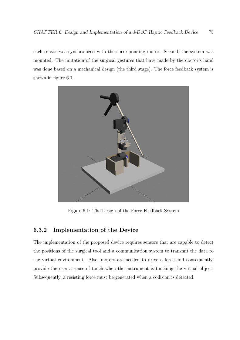
CHAPTER 6. Design and Implementation of a 3-DOF Haptic Feedback Device 75
each sensor was synchronized with the corresponding motor. Second, the system was
mounted. The imitation of the surgical gestures that have made by the doctor’s hand
was done based on a mechanical design (the third stage). The force feedback system is
shown in figure 6.1.
Figure 6.1: The Design of the Force Feedback System
6.3.2 Implementation of the Device
The implementation of the proposed device requires sensors that are capable to detect
the positions of the surgical tool and a communication system to transmit the data to
the virtual environment. Also, motors are needed to drive a force and consequently,
provide the user a sense of touch when the instrument is touching the virtual object.
Subsequently, a resisting force must be generated when a collision is detected.
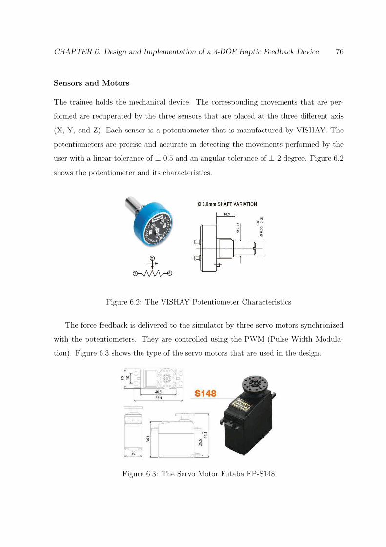
CHAPTER 6. Design and Implementation of a 3-DOF Haptic Feedback Device 76
Sensors and Motors
The trainee holds the mechanical device. The corresponding movements that are per-
formed are recuperated by the three sensors that are placed at the three different axis
(X, Y, and Z). Each sensor is a potentiometer that is manufactured by VISHAY. The
potentiometers are precise and accurate in detecting the movements performed by the
user with a linear tolerance of ± 0.5 and an angular tolerance of ± 2 degree. Figure 6.2
shows the potentiometer and its characteristics.
Figure 6.2: The VISHAY Potentiometer Characteristics
The force feedback is delivered to the simulator by three servo motors synchronized
with the potentiometers. They are controlled using the PWM (Pulse Width Modula-
tion). Figure 6.3 shows the type of the servo motors that are used in the design.
Figure 6.3: The Servo Motor Futaba FP-S148
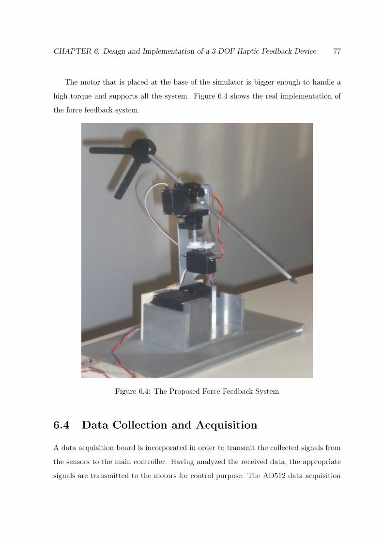
CHAPTER 6. Design and Implementation of a 3-DOF Haptic Feedback Device 77
The motor that is placed at the base of the simulator is bigger enough to handle a
high torque and supports all the system. Figure 6.4 shows the real implementation of
the force feedback system.
Figure 6.4: The Proposed Force Feedback System
6.4 Data Collection and Acquisition
A data acquisition board is incorporated in order to transmit the collected signals from
the sensors to the main controller. Having analyzed the received data, the appropriate
signals are transmitted to the motors for control purpose. The AD512 data acquisition

CHAPTER 6. Design and Implementation of a 3-DOF Haptic Feedback Device 78
card form HUMUSOFT is used [86] in this work. It contains several features. The most
important features for this application are:
• Eight single-ended 12-bit analog input channels and two 12-bit analog output
channels
• Sampling rate up to 100 kHz
• 8 digital input channels and 8 digital output channels
• Programmable A/D ranges
• Low power consumption
The potentiometer, which is a three-terminal resistor, is connected to a voltage source
to form an adjustable voltage divider. If all three terminals are connected, it can act
as a variable voltage divider. Therefore, a simple linear circuit that produces an output
voltage (Vout) which is a fraction of its input voltage (Vin), is created. Figure 6.5 shows
the resistive strip of the potentiometer (left) that is translated into a variable voltage
divider.
Figure 6.5: A Variable Voltage Divider
Each time the user moves the force feedback device, the voltages across the three ter-
minals of the three potentiometers, that are placed along the different axes, are adjusted

CHAPTER 6. Design and Implementation of a 3-DOF Haptic Feedback Device 79
accordingly. Then, the corresponding electric signals are generated and transmitted to
the DAQ board for processing. The change of voltage is due to the rotation of the
resistive strip. Subsequently, the potentiometers that indicate a change in the voltage
(electric signals) also indicate a variation in the position of the virtual medical probe in
the virtual environment. If there is no collision detection between the virtual medical
probe and the virtual medical model, the motors are off i.e. there is no signal transmit-
ted to control the motors. On the other hand, if a collision is detected, PWM signals
are generated and transmitted via the output port of the DAQ board to the motors.
Therefore, the user can feel the force feedback that is generated by these motors. Figure
6.6 shows the circuit that generates the PWM signals.
Figure 6.6: Generation of the PWM Signals
The Humusoft board is designed to deal with logic signals. It can not cope with large
voltages and large currents. In addition, the output current of the board is approximately
38 mA. Therefore, this current is not sufficient to run the motors. In this regard, the
IRF630 power Mosfets are used to drive the motors. That is, the generated PWM input
signal is transmitted via the gate (g) of the Mosfet and the motor is attached to the
output (out) of the drain (d). The drain is also connected to a source voltage (V) via
a small resistance and the source node (s) of the Mosfet is grounded. Figure 6.7 shows
the circuit that drives each motor.

CHAPTER 6. Design and Implementation of a 3-DOF Haptic Feedback Device 80
Figure 6.7: The Circuit Driving the Motors
6.5 Haptic Feedback Algorithm
Having performed the design and the implementation of the device, a haptic feedback
algorithm that supervises all the corresponding activities is described. The algorithm
will enhance the surgical performance by guiding the user (surgeon, student ...) and give
him a sense of touch and resistance when the collision is virtually detected between the
instrument and the object.
When the user moves the haptic device, sensors that are placed on different axis
of rotation generate the signals. The latter are transmitted by the acquisition card, to
change relatively the position of the virtual medical instrument. This change allows
the dynamic interactions within the virtual environment. That is, the position of the
medical tool is updated at every step and the collision detection is checked between
the tool and the bones of the wrist by applying the proposed algorithm (presented in
chapter 5) i.e. update the matrices that formulate the collision problem and solve the
system of linear equations for every step change. If collision is detected, PWM signals
are generated to control the servo motors. Consequently, these motors generate a force
which is applied against the motion of the user of the haptic device. Therefore, the
user can feel the resistance of the applied force against his hand’s motion, i.e. against

CHAPTER 6. Design and Implementation of a 3-DOF Haptic Feedback Device 81
the force applied by the trainee to move the haptic device. This force-reflecting device
enables medical students during the training session to experience the real feeling of
touch. Touching virtual objects rather than seeing them enhance the capability of the
computer-based system and gives the user the feeling of so called “Immersion”. Figure
6.8 shows the flowchart of the haptic feedback algorithm.
Figure 6.8: The Flowchart of the Haptic Feedback Algorithm
6.6 Virtual Simulation of Scaphoid Fixation
A virtual simulation of dorsal percutaneous scaphoid fixation is presented and a com-
parison between the real and the virtual process is shown.
6.6.1 Surgical Technique For Scaphoid Fracture
Techniques to perform the wrist arthroscopy have been developed to evaluate, diagnose
and treat the various wrist disorders, such as scaphoid fractures. For example, the dorsal
percutaneous approach is a very efficient way in the treatment of the displaced proximal
pole scaphoid fractures in many clinical and operating rooms. This technique allows a
faster rehabilitation without any restriction once CT scan confirms a solid union.

CHAPTER 6. Design and Implementation of a 3-DOF Haptic Feedback Device 82
Scaphoid Fracture
The scaphoid is the wrist bone that is the most likely to break. It is located on the
thumb side of the wrist, in the region where the wrist bends. Fractures of the scaphoid
occur in people of all ages, including children, and are most common in young men
due to their athletic activities. Scaphoid fractures usually cause pain in the base of the
thumb, with a swelling in the same area. The pain may be severe when the thumb or
wrist is moved or when the hand grips anything. When the scaphoid is broken at the
waist or proximal pole, a surgery may be recommended.
Displaced fractures may need an open reduction and an internal fixation [92]. In fact,
this method is very efficient and it has been associated with good results [93]. However,
the drawbacks of the open reduction and the internal fixation include the need to cut
the important palmar radiocarpal wrist ligaments [94] and may cause a possible injury
to the limited blood supply to the scaphoid [95]. Therefore, undisplaced fractures of the
scaphoid can be treated with a percutaneous screw fixation. A screw or wire may be
used to stabilize the scaphoid while the bone heals.
6.6.2 Surgical Simulation
Percutaneous arthroscopically assisted internal fixation by a dorsal approach may be
considered in all acute scaphoid fractures that are selected for surgical fixation [88]. The
dorsal guidewire permits the dorsal and the volar implantation of a cannulated screw
along the central axis of the scaphoid [89]. The surgical technique described in [87]
uses the Standard Acutrak screw. This screw is a headless, cannulated, tapered screw
with a graduated thread pitch to provide an interfragmentary compression without a
hardware protrusion. This technique permits the percutaneous reduction and a rigid
internal fixation of proximal pole fractures. First, the wrist is flexed and pronated so
that the scaphoid appear’s as a cylinder. The center of the cylinder is the location to
guide the wire placement. Then, the guide wire is driven dorsal to volar through the
center of the scaphoid. The wire exits at the base of the thumb. Figure 6.9 shows the
real placement of the guide wire during the surgery. Figure 6.9 is taken from a real
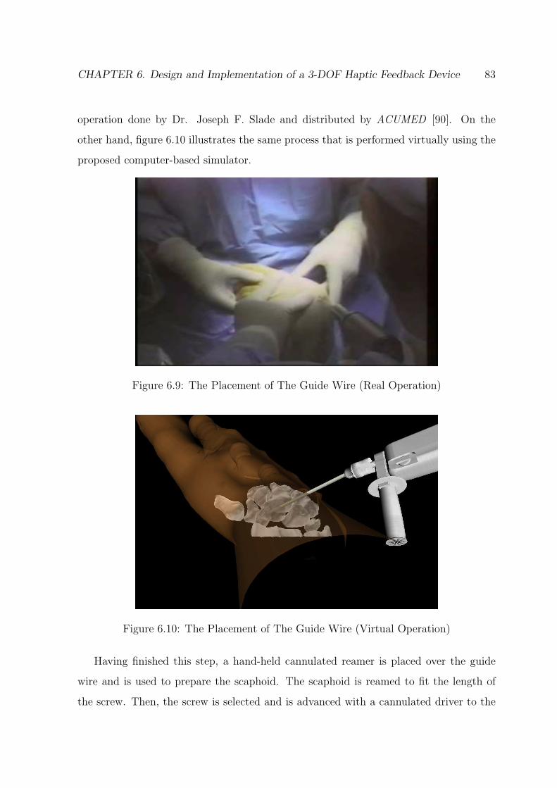
CHAPTER 6. Design and Implementation of a 3-DOF Haptic Feedback Device 83
operation done by Dr. Joseph F. Slade and distributed by ACUMED [90]. On the
other hand, figure 6.10 illustrates the same process that is performed virtually using the
proposed computer-based simulator.
Figure 6.9: The Placement of The Guide Wire (Real Operation)
Figure 6.10: The Placement of The Guide Wire (Virtual Operation)
Having finished this step, a hand-held cannulated reamer is placed over the guide
wire and is used to prepare the scaphoid. The scaphoid is reamed to fit the length of
the screw. Then, the screw is selected and is advanced with a cannulated driver to the

CHAPTER 6. Design and Implementation of a 3-DOF Haptic Feedback Device 84
level of the reamed scaphoid. Figure 6.11 shows the real insertion of the screw in the
scaphoid and is also taken from the real operation done by Dr. Joseph F. Slade. On the
other hand, figure 6.12 shows the virtual operation of the same procedure.
Figure 6.11: The Insertion of The Screw in The Scaphoid (Real Operation)
Figure 6.12: The Insertion of The Screw in The Scaphoid (Virtual Operation)
Having implanted the screw in its corresponding location, the position of the scaphoid
is confirmed with a mini-fluoroscopy and the guide wire is removed. The screw is placed

CHAPTER 6. Design and Implementation of a 3-DOF Haptic Feedback Device 85
at the center of the scaphoid. Figure 6.13 shows the final phase of the virtual simulation
of the operation.
Figure 6.13: A Virtual View of The Central Placement of The Screw in The Scaphoid
6.7 Advantages
The dorsal percutaneous approach to scaphoid fixation has many advantages [87] and
they can be summrized in three. This three main advantages are:
1. The proximal-to-distal placement of the guide pin and the screw allow for more
precise placement along the central axis of the scaphoid, which decreases healing
time and reduces risk of screw thread exposure.
2. The dorsal approach avoids injuring the vulnerable volar ligament anatomy.
3. The insertion of the screw from the proximal to distal direction allows the more
rigid fixation of proximal scaphoid fractures.

CHAPTER 6. Design and Implementation of a 3-DOF Haptic Feedback Device 86
6.8 Conclusion
In this chapter, the design and the implementation of a functional haptic feedback device
are presented. The proposed device has a 3-DOF, each one along a particular axis (X,
Y and Z). The system is capable to provide force feedback to them all. The developed
prototype can be used by medical students and trainees and have the same benefits as
the professional devices. The proposed force feedback device is coupled with a haptic
simulation algorithm and it is incorporated in the whole computer-based system. Also,
a virtual simulation of dorsal percutaneous scaphoid fixation is presented. The VR
simulation is compared with the real surgery of the same procedure. The 3-D wrist
model, presented in chapter 3, the model of the hand and the virtual medical tools
are integrated in the simulation process. The entire system is tested and the wrist
arthroscopic surgery can be simulated. The users (student, doctor, surgeon ...) can
learn the required basic skills and then perform the training procedure on real patients.
This low cost system is safe, flexible and can provide medical students with a precise
and detailed information for training and educational purposes.

Chapter 7
Conclusions and Perspectives
The work presented in this thesis has addressed an important issue in VR and its ap-
plication in the medical field: the development of a VR simulator for wrist arthroscopic
surgery. In this work, two main issues are addressed: the 3-D reconstruction process
and the 3-D interaction. The proposed simulator provides a virtual environment with a
realistic representation of the region of interest. Based on a sequence of CT images, a
realistic representation of the wrist joint suitable for the computer simulation is recon-
structed. Two main components of the computer-based system interface are illustrated:
the 3-D interaction to guide the surgical instruments and the user interface for haptic
feedback. In this context, algorithms that model 3-D objects using the convex hull ap-
proaches and simulate real time exact collision detection between virtual objects during
the training on the surgical operation are presented. Also, a force feedback device, cou-
pled with a haptic feedback algorithm, is designed and developed as a haptic interface
with the computer simulation system. A distinctive feature of this work is that the
proposed computer-based system satisfies the following specifications:
1. The system treats the wrist arthroscopy problem. The wrist arthroscopy requires
skills to be acquired by all hand surgeons. These skills are important as the skills
needed by surgeons to perform the shoulder and the knee arthroscopy.
2. The development of an inexpensive and a practical simulator for medical students
as well as for surgeons is of great value, specially if the medical practioners can
harness the same benefits as the professional devices. Then, the surgical procedure
87
CHAPTER 7. Conclusions and Perspectives 88
can be performed on real patients with much less risk and injury.
7.1 Summary of the Contributions
The main contributions of this thesis are summarized as follow:
• Survey on MIS Surgical Simulators: In this survey, an overview of the ex-
isting MIS simulators is presented. The VR simulators are classified based on the
application and the organs or structures for which the system is developed. The
study may be used by the researchers to gain a general idea about the commer-
cialized and the scientific simulators that were developed to perform different MIS
approaches.
• Virtual Environment: A virtual environment to visualize the medical models
and the medicals tools with a high fidelity and a high precision is developed.
• Algorithms: A new hybrid approach to generate the convex hull of the 3-D models
is developed and presented. The proposed algorithm converts each 3-D concave
model to a convex representation that allows the collision detection algorithms to
converge quickly and report a collision, if it exists. The proposed hybrid method
is an output sensitive algorithm that works in 2-D as well as in 3-D. It is very
efficient, practical and useful in modelling and simulating 3-D medical data in a
virtual environment. In addition, a new technique of collision detection for solid
objects is proposed and introduced. The collision detection problem is formulated
and a linear programming solution is obtained to determine whether a collision
occurs between virtual objects or not. The proposed algorithm is efficient, fast,
robust and leads to a decrease of the running time that is required to detect a
collision.
• 3-DOF Force Feedback System: A 3-DOF force feedback device is designed,
developed and implemented. The low cost system is coupled with a haptic feed-
back algorithm. The proposed device can be used by medical students as well as
surgeons to harness the same benefits as the professional devices.

CHAPTER 7. Conclusions and Perspectives 89
• Virtual Simulation of a Surgical Operation: A virtual simulation of the dor-
sal percutaneous scaphoid fixation is presented. The virtual procedure is compared
with the real surgery that is performed by doctors inside the operating room. The
virtual surgery ensures that the proposed computer-based system is appropriate to
train medical students as well as surgeons in order to acquire the necessary skills
to perform the specific surgeries.
7.2 Future Work
The proposed computer based surgical simulator can be extended to handle other oper-
ations and medical activities.
In order to complete the system, the corresponding ligaments should be modeled and
added to the 3-D wrist. In fact, the ligaments displayed in the CT images are blurred and
the appearance is almost non existing in MRI images. Therefore the contour detection
using the marker-based watershed algorithm can not lead to a good extraction of these
ligaments from the CT and the MRI images. Thus, real images from the Visible Human
dataset maybe be more suited for ligament extraction. This is achieved by addressing
two main issues: the need of special information about the ligaments from an expert
doctor and the implementation of preprocessing techniques for segmentation purposes
such as features extraction algorithms.
Once the ligaments are modeled and extracted, new collision detection algorithms
should be investigated and incorporated in the VR simulator to accomodate any varia-
tions. This is due to the fact that the ligaments have properties that are different from
solid objects such as the bones.
At a later stage, the researcher may upgrade the proposed prototype so that it can
handle all types of wrist surgery operations. Consequently, the surgeon will be guided
through the whole process and has always the full control over the device.

Publications
• International Journals
1. F. Yaacoub, Y. Hamam and A. Abche, “A 3D Convex Hull Algorithm for Model-
ing Medical Data in a Virtual Environment”, International Journal of Intelligent
Systems Technologies and Applications (IJISTA) , Special Issue on Advances in
Intelligent Systems for Applied Simulation and Prototyping, Vol. 5, Nos 1/2, PP.
3-19, ISSN: 1740-8865, Inderscience Enterprise, 2008.
2. F. Yaacoub, Y. Hamam and A. Abche, “Development of Virtual Reality Tools For
Simulating Wrist Arthroscopic Surgery”, Submitted to: The International Journal
of Simulation Modelling Practice and Theory , SIMPAT, Elsevier, 2009.
• International Conferences
1. F. Yaacoub, Y. Hamam and A. Abche, “Computer-Based Training System for Sim-
ulating Wrist Arthroscopy”, The 21th IEEE International Symposium on Computer-
Based Medical Systems , CBMS, pp. 421-423, ISBN: 978-0-7695-3165-6, Jyvaskyla,
Finland, June 2008.
2. F. Yaacoub, Y. Hamam and A. Abche, “Collision Detection for Virtual Arthro-
scopic Surgical Simulation”, The 2007 International Conference on Computer
Graphics and Virtual Reality , CGVR’07, WORLDCOMP’07, CSREA Press, pp.
87-93, ISBN: 1-60132-028-0, Las Vegas, USA, June 2007.
90

2. Publications 91
3. F. Yaacoub, Y. Hamam and A. Abche, “Collision Detection in Computer Simula-
tions for Wrist Arthroscopic Surgery Training”, The IEEE International Confer-
ence on Computer as a Tool , EUROCON’07, pp. 2088-2095, ISBN: 978-1-4244-
0813-9, Warsaw, Poland, September 2007.
4. F. Yaacoub, Y. Hamam and A. Abche, “Computer-Based Surgical Simulation
System for Students Training”, The 6th EUROSIM Congress on Modelling and
Simulation , ISBN: 978-3-901608-32-2, Ljubljana, Slovenia, September 2007.
5. F. Yaacoub, Y. Hamam, A. Abche and C. Fares, “Convex Hull in Medical Sim-
ulations: A New Hybrid Approach”, The 32nd Annual International Conference
of IEEE Industrial Electronics Society , IECON’06, pp. 3308-3313, ISBN: 1-4244-
0391, Paris, France, November 2006.
6. F. Yaacoub, Y. Hamam and A. Abche, “Convex Envelope in Arthroscopic Knee
Surgery Simulation”, UK Society for Modelling and Simulation, European Mod-
elling Symposium, EMS’06, pp. 46-50, ISBN: 0-9516509-3-9, London, UK, Sep-
tember 2006.
7. F. Yaacoub, Y. Hamam and A. Abche, “A Virtual Reality Simulator For Training
Wrist Arthroscopic Surgery”, The International Joint Conference on Biomedical
Engineering Systems and Technologies , BIOSTEC, Porto, Portugal, January 2009.

Resume Detaille
Introduction
L’environnement virtuel est decrit comme une application qui permet aux utilisateurs
de naviguer et d’interagir dans un espace quasi-realiste, tri-dimensionnel et en temps reel.
Dans ce contexte, la realite virtuelle repose sur l’integration en temps reel de situations
generees par ordinateur, d’information sur la position de l’individu dans l’espace et de
stimuli visuels qui recreent un environnement interactif d’apparence realiste. La realite
virtuelle permet donc d’immerger l’usager dans un environnement standardise afin de
lui permettre de s’entrainer et de simuler plusieurs experiences, ce qui lui donne certains
avantages vis-a-vis des methodes d’exposition traditionnelles.
La realite virtuelle a revolutionne des nombreuses disciplines scientifiques en four-
nissant de nouvelles methodes de visualisation des donnees complexes et en manipulant
ces donnees en temps reel. Elle a ete utilisee dans differentes applications : l’ingenierie,
l’education, les simulations militaires et aerospatiales, et la medecine.
La medecine et surtout la chirurgie arthroscopique presente actuellement un essor
tres important pour le benefice du plus grand nombre des patients. Cependant, cette
technique possede un certain nombre d’inconvenients et il est donc necessaire pour le
medecin de s’entrainer et repeter ses gestes afin de pouvoir executer ce type d’operation
d’une facon efficace et certaine. En effet, les methodes traditionnelles d’enseignement de
la chirurgie sont basees sur l’autopsie des cadavres et l’entrainement sur des animaux.
Avec l’evolution de notre societe, ces deux pratiques deviennent de plus en plus critiquees
et font l’objet de reglementations tres restrictives.
Afin d’atteindre un niveau plus eleve, de nouveaux moyens d’apprentissage sont
92
1. Resume Detaille 93
necessaires pour les chirurgiens. A cet egard, les simulateurs chirurgicaux sont devenus
une des matieres les plus recentes dans la recherche de la realite virtuelle. Ils sont
egalement devenus une methode de formation et un outil d’entraınement valable pour
les chirurgiens aussi bien que les etudiants en medecine.
Objectif et Considerations
Les simulateurs chirurgicaux ont ete elabores pour un large eventail de procedures
medicales. Cependant, ils sont souvent associes a des engagements specifiques. De nom-
breux simulateurs sont associes avec la laparoscopie, d’autres sont lies a l’endoscopie. En
outre, de nombreux simulateurs sont associes a la cystoscopie et ureteroscopie. Certains
d’entre eux participent a la colonoscopie, bronchoscopie et sigmoıdoscopie. Toutefois,
la plupart des simulateurs mentionnes ci-dessus sont couteux a acquerir. Concernant
les simulateurs d’arthroscopie, la plupart d’entre eux ont ete developpes pour la forma-
tion du genou et de l’epaule alors que tres peu de travail a ete fait pour l’arthroscopie
du poignet malgre l’importance de celui-ci. En consequence, le probleme de la con-
struction d’un simulateur de realite virtuelle pour l’enseignement de la chirurgie arthro-
scopique du poignet reste a traiter. Dans ce contexte, notre projet a ete propose par une
equipe de professeurs et chirurgiens a l’Institut de la Main, “Clinique Jouvenet” Paris
XVI. Il consiste a developper des outils de realite virtuelle pour aider a l’enseignement,
l’apprentissage et la formation sur la chirurgie arthroscopique du poignet. Ce projet
de recherche, dirige par le Prof. Yskandar Hamam, a commence avec Charbel Fares.
Mon travail est une continuation de ce projet afin de developper tous les outils de realite
virtuelle qui sont necessaires a la realisation complete du simulateur. Au cours de la
conception du projet, deux objectifs principaux sont vises :
1. Developper des outils de realite virtuelle et des techniques de simulation pour
generer des modeles 3-D et pour simuler des operations virtuelles avec fidelite et
realisme.
2. Essayer de couvrir differentes exigences pour le processus d’apprentissage et de

1. Resume Detaille 94
fournir a l’utilisateur des outils pour faciliter l’enseignement et la formation sur
plusieurs experiences chirurgicales.
En outre, l’arthroscopie du poignet a ete choisie en raison de plusieurs considerations:
1. L’arthroscopie du poignet est une pathologie (etude de la nature essentielle de la
maladie) qui a ete moins etudiee et pratiquee que celle du genou et de l’epaule.
2. Des differents types de participation et d’intervention chirurgicales peuvent etre
couverts par la simulation arthroscopique du poignet.
3. Les medecins confrontent de nouvelles et importantes pathologies en ce qui con-
cerne l’arthroscopie du poignet. Par consequent, il existe une demande croissante
sur la formation et sur l’apprentissage des nouvelles techniques.
Par consequent, un simulateur de realite virtuelle pour l’enseignement de la chirurgie
arthroscopique, surtout la chirurgie du poignet, a ete presente. Deux questions princi-
pales sont abordees: la reconstruction et l’interaction 3-D. Une sequence d’images CT a
ete traitee afin de generer un modele 3-D du poignet. Les deux principales composantes
de l’interface du systeme sont illustrees: l’interaction 3-D pour guider les instruments
chirurgicaux et l’interface de l’utilisateur pour le retour d’effort. Dans ce contexte, les
algorithmes qui modelisent les objets en utilisant les approches de “Convex Hull” et qui
simulent la detection de collision entre les objets virtuels en temps reel sont presentes.
En outre, un dispositif de retour d’effort est utilise comme une interface haptique avec
le systeme. Cela conduit au developpement d’un systeme a faible cout, avec les memes
avantages que les appareils professionnels. A cet egard, l’arthroscopie du poignet peut
etre simulee et les etudiants en medecine peuvent facilement utiliser le systeme et peu-
vent apprendre les competences de base requises en securite, flexibilite et moindre cout.
Description du Systeme
Les images medicales sont traitees afin de generer des modeles volumetriques. Une
sequence d’images CT est segmentee et un modele virtuel 3-D du poignet est genere.
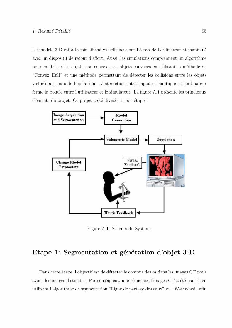
1. Resume Detaille 95
Ce modele 3-D est a la fois affiche visuellement sur l’ecran de l’ordinateur et manipule
avec un dispositif de retour d’effort. Aussi, les simulations comprennent un algorithme
pour modeliser les objets non-convexes en objets convexes en utilisant la methode de
“Convex Hull” et une methode permettant de detecter les collisions entre les objets
virtuels au cours de l’operation. L’interaction entre l’appareil haptique et l’ordinateur
ferme la boucle entre l’utilisateur et le simulateur. La figure A.1 presente les principaux
elements du projet. Ce projet a ete divise en trois etapes:
Figure A.1: Schema du Systeme
Etape 1: Segmentation et generation d’objet 3-D
Dans cette etape, l’objectif est de detecter le contour des os dans les images CT pour
avoir des images distinctes. Par consequent, une sequence d’images CT a ete traitee en
utilisant l’algorithme de segmentation “Ligne de partage des eaux” ou “Watershed” afin

1. Resume Detaille 96
de generer un model 3-D du poignet. Apres la segmentation de l’ensemble des images,
l’algorithme “Marching Cube” est utilise pour construire les frontieres des objets dans
la scene. Chaque os est considere comme etant un objet et le resultat final de la segmen-
tation el la reconstruction de l’ensemble des images est une image volumetrique consti-
tuant le poignet d’un patient. Ensuite, la representation 3-D de ces modeles est illustree
dans un environnement virtuel en associant tous les elements necessaires (l’eclairage, les
materiaux et les textures). La figure A.2 montre la representation 3-D des os constituant
le poignet d’un patient.
Figure A.2: Modele 3-D du Poignet
Etape 2: Modelisation et Simulation
L’objectif d’un simulateur medical est de soutenir les etudiants en medecine au cours
de leur formation sur des chirurgies avec une grande precision. A cet egard, les objets
1. Resume Detaille 97
medicaux sont modelises suivant une methode de “Convex Hull” proposee. Ceci donnera
le simulateur plus de precision, mais en meme temps une augmentation de complexite
et du temps de calcul pour verifier la collision. Par consequent, en se basant sur les
avantages des techniques de programmation lineaire (vitesse et robustesse), le probleme
de detection de collision est formule et resolu. En outre, les objets convexes permettent
aux algorithmes de programmation lineaire de converger rapidement et de detecter la
collision, si elle existe. Ainsi, l’enveloppe convexe de chaque objet est reconstruite.
Ensuite, le probleme de collision est formule comme un probleme d’optimisation base sur
ces objets convexes et resolu en utilisant la programmation lineaire (methode simplex).
Convex Hull
Etant donne que la plupart des objets 3-D ne sont pas caracterises par une forme
exacte qui pourrait etre modelisee en utilisant des equations mathematiques precises,
“Convex Hull” algorithmes sont consideres comme une solution pour modeliser ces ob-
jets. La necessite des ces algorithmes se pose avec l’intention de simuler des scenes
en temps reel et de detecter la collision entre les objets dans un environnent virtuel.
De plus, la plupart des algorithmes de detection de collision sont bases sur les objets
convexes, car ces derniers permettent aux algorithmes de collision de converger rapide-
ment. En outre, les enveloppes convexes ont moins de points de contact que les objets
reels. Cela conduit a une diminution de la taille du systeme d’equations necessaires
pour calculer la collision. Dans ce contexte, une approche hybride pour la construction
des enveloppes convexe des objets 3-D (os du poignet) avec un temps d’execution plus
rapide a ete proposee. Cette methode a ete comparee avec quatre algorithmes: Brute
Force, GiftWrapping, QuickHull et Chan algorithme.
Methode Hybride
Plusieurs methodes sont developpees afin d’accelerer les algorithmes de “Convex
Hull”. Certaines techniques commencent par diviser les points constituant l’objet 3-D
1. Resume Detaille 98
en deux ensembles arbitraires et fixes, droite et gauche. “Divide-and-conquer” est un
algorithme qui commence par calculer recursivement l’enveloppe convexe de la partie
droite puis la partie gauche suivie par la fusion des deux enveloppes dans une enveloppe
convexe finale. Autres techniques divisent l’ensemble des points d’entree en plusieurs
sous-ensembles comme le Chan algorithme. Cet algorithme divise les points (n) qui
constituent l’objet 3-D en differents sous-ensembles (n / N) arbitraire, disjoints et de
taille N. Ensuite, l’enveloppe convexe de chaque sous-groupe est calculee. Ainsi, N
enveloppes sont formees et elles sont integrees dans une enveloppe convexe finale.
L’idee derriere ces techniques est toujours de diviser l’ensemble des points en plusieurs
sous-ensembles afin d’accelerer le temps d’execution de l’algorithme qui construit l’enveloppe
convexe et de reduire sa complexite. Dans ce contexte, une methode hybride est proposee
et deux points importants sont pris en consideration:
• Le temps d’execution des algorithmes qui construit l’enveloppe convexe depend du
nombre de points (n) qui constitue l’objet.
• Les etapes d’emballage dans l’algorithme “Gift Wrapping” peuvent etre acquises
plus vite si l’ensemble des points d’entree a ete traite.
Par consequent, une approche hybride pour generer les enveloppes convexes est
developpee. Elle est basee sur le QuickHull et le GiftWrapping algorithmes. Elle est
decomposee en deux etapes: la premiere etape permet de reduire le nombre de points
d’entree et la deuxieme etape souligne la reconstruction de l’enveloppe convexe corre-
spondant. La methode hybride est initiee par l’application de l’algorithme QuickHull
pour diviser les points d’entree en deux sous-ensembles (superieur et inferieur) par un
premier plan. Ensuite, un polyedre de nouvelles facettes est cree en calculant le point
ayant la distance maximale par rapport a ce plan. Par consequent, les points qui sont
dans le polyedre sont a l’interieur de l’enveloppe convexe et ils sont elimines. La meme
procedure est repetee pour l’ensemble des points inferieurs. Cela conduit a la reduction
du nombre de points d’entree et a la formation d’un nouvel ensemble de points. Ce
dernier constitue les donnees d’entrees pour l’algorithme GiftWrapping et les etapes

1. Resume Detaille 99
d’emballage sont effectuees sur ces nouvelles donnees pour obtenir l’enveloppe convexe
finale. En d’autres termes, la methode hybride applique une phase d’initialisation suivie
par une serie d’etapes d’emballage. Elle calcule les facettes de l’enveloppe convexe dans
le sens oppose de l’aiguille d’une montre. Le conditionnement des etapes repetees est
recursive pour explorer tous les bords jusqu’a ce que tous les aspects aient ete examines.
Un pseudo-code de l’algorithme propose est montre ci-dessous:
1: find an initial plane from the min and max abssice and the max distance withrespect to (xmin, xmax)
2: construct a polyhedron from the initial plane and the max distance to this plane3: for each facet F of the polyhedra do4: for each unassigned point p do5: if p is above F then6: assign p to F ′s outside set7: end if8: end for9: end for
10: Discard all points inside the polyhedron forming a new imput set (nnew)11: find a starting edge (a, b) using the 2D Gift Wrapping algorithm on the XY projec-
tion12: for i = 1 ... nnew do13: find point pi corresponding to min angle bewteen plane P in XY containing (a, b)
and plane T = (a, b, pi)14: replace c← pi
15: save (a,b,c) into Q16: wrap the edge (a, c)17: if facet has been explored then18: wrap the edge (b, c)19: if facet has been explored then20: return21: end if22: end if23: end for
L’algorithme hybride propose est applique a differents os du poignet. La figure
A.3 montre le resultat sur les differents os testes : 1st Metacarpal (a), 2nd Metacarpal
(b), 4th Metacarpal (c), Scaphoid (d), Capitate (e), Hamate (f), Radius (g) and Ulna
(h). Chaque os est recouvert par son enveloppe convexe correspondant. Les resultats

1. Resume Detaille 100
montrent que l’approche proposee s’effectue plus rapidement que les autres techniques
classiques.
Figure A.3: Os du Poignet Recouvert par son Enveloppe Convexe Correspondant
La methode hybride proposee est une methode qui fonctionne en 2-D ainsi qu’en
3-D. Elle est tres efficace, pratique et utile dans la modelisation et la simulation 3-D des
donnees medicales dans un environnement virtuel.
Detection de Collision
Etant donne que les enveloppes convexes reconstruites ont moins de points de contact
que les objets reels et peuvent etre rapidement calculees, cela conduit a une diminution de
la taille du systeme d’equations necessaires pour detecter une collision. Par consequent,
les performances en temps reel des algorithmes de detection de collision sont augmentees
et les algorithmes deviennent plus rapides. En outre, la plupart des systemes exacts de
CD sont bases exclusivement sur les objets convexes. Cela est du au fait que les objets

1. Resume Detaille 101
convexes permettent aux algorithmes de CD de converger rapidement et de detecter la
collision, si elle existe. Cela donnera le simulateur un degre eleve de precision, mais
en meme temps une augmentation du cout de la complexite et du temps de calcul
pour verifier la collision. Par consequent, en prenant les avantages des techniques de
programmation lineaire (la vitesse et la robustesse), le probleme de detection de collision
est formule et resolu. Ainsi, le CH de chaque objet est reconstruit ensuite le CD probleme
est formule comme un probleme d’optimisation base sur les objets convexes et resolu en
utilisant la programmation lineaire (la methode simplex).
Pour formuler le probleme de collision, chaque facette i de l’enveloppe convexe est
representee par l’equation du plan dans l’espace sous la forme:
aix + biy + ciz ≤ di (A.1)
Tout point situe sur l’objet convexe doit satisfaire les inegalites des plans constituant
cet objet. Ces equations forment les contraintes du probleme de collision et representent
les facettes qui separent les deux regions dans l’espace. Par consequent, si un point
appartient a deux series d’inegalite en meme temps, une collision est detectee a ce
moment entre ces deux objets. Le probleme est reduit a maximiser une fonction sous la
forme de (x + y + z). Il peut etre formule par l’equation suivante:
max cT X (A.2)
subject to
AX ≤ b (A.3)
where X = [x y z]T ,
A =
a1 b1 c1
a2 b2 c2
a3 b3 c3
. . .
. . .
(A.4)
b = [d1 d2 d3 . . .]T , c = [1 1 1]T (A.5)
1. Resume Detaille 102
Les coefficients des matrices A et b sont calcules en se basant sur les facettes de
l’enveloppe convexe reconstruit par l’approche proposee. Le systeme lineaire presente est
resolu en utilisant un algorithme de programmation lineaire (Simplex). Cette methode
a ete comparee avec un algorithme classique nomme IVRI-CD.
Etape 3: Interface Homme-Machine
Un dispositif mecanique de retour d’effort avec trois degres de liberte est propose et
concu. Le systeme a ete developpe suivant un plan bien defini. Tout d’abord, la base de
l’appareil a ete construite et chaque capteur a ete synchronise avec le moteur correspon-
dant. Deuxiemement, le systeme a ete monte. L’imitation des gestes chirurgicaux faites
par le medecin au niveau de la main a ete realisee suite a une conception mecanique
dans la troisieme etape.
La mise en oeuvre du dispositif necessite des capteurs qui sont capables de detecter
les positions de l’outil chirurgical et un systeme de communication pour transmettre les
donnees a l’environnement virtuel. Aussi, les moteurs sont egalement necessaires pour
le retour d’effort. Par consequent, une resistance doit etre generee lors d’une detection
de collision entre les objets virtuels. Le systeme est illustre dans la figure A.4.
Collection et Acquisition des Donnees
Les mouvements qui sont effectues par l’utilisateur sont recuperes par les trois cap-
teurs (potentiometres) places sur les trois differents axes (X, Y et Z). Chaque fois que
l’utilisateur deplace le dispositif de retour de force, les tensions entre les trois terminaux
des trois potentiometres sont ajustees en consequence. Ensuite, les signaux electriques
sont generes et transmis a la carte d’acquisition pour le traitement. Le changement
de tension est du a la rotation de la bande resistive. Les potentiometres qui indiquent
un changement de la tension (signaux electriques) indiquent aussi une variation de la
position de l’outil medicale dans l’environnement virtuel. S’il n’y a pas de detection de
collision entre cet outil et le modele 3-D du poignet, les moteurs sont eteints c’est-a-dire

1. Resume Detaille 103
Figure A.4: Le Systeme de Retour D’effort
il n’y a pas de signal transmis pour controler les moteurs. D’autre part, si une collision
est detectee, les signaux PWM sont produits et transmis via le port de sortie de la carte
d’acquisition vers les moteurs. Par consequent, l’utilisateur peut sentir le retour de force
qui est genere par ces moteurs.
Resultat
La figure A.5 montre le systeme chirurgical propose et construit au cours de la these.
Afin de tester notre simulateur propose, une comparaison entre une chirurgie reelle et
une operation virtuelle est faite.
La figure A.6 montre deux etapes d’une operation chirurgicale de la main. La partie
reelle est faite dans un hopital alors que la partie virtuelle est faite par notre simulateur.

1. Resume Detaille 104
Figure A.5: Le Simulateur Chirurgical Propose
Conclusion
La force du simulateur propose repose sur l’integration entre le monde des ingenieurs
et le monde medical. Le monde des ingenieurs qui est plutot un monde “non-vivant”
tandis que le monde medical est un “monde vivant”. Cette integration simplifie la
comprehension mutuelle des deux domaines qui sont parmi les plus importants dans la
vie humaine. Ce qui est aussi important dans ce projet, c’est qu’il rassemble plusieurs
domaines de recherche. Il contient de l’imagerie, de la realite virtuelle, de la detection
de collision, de la robotique, du retour d’effort et de la methodologie pedagogique. Les
principaux resultats obtenus dans cette these sont:
• Etat de l’Art: Dans cette these, un etat de l’art sur les simulateurs chirurgicaux
existants est presente. Les simulateurs sont classes selon l’application, les organes
ou les structures pour lesquels ils sont developpes. Cette etude peut etre utilisee

1. Resume Detaille 105
Figure A.6: Comparaison Entre une Operation Reelle et une Operation Virtuelle
par les chercheurs pour avoir une idee generale sur les simulateurs commerciaux
et scientifiques qui ont ete developpes pour effectuer differentes approches de la
chirurgie minimalement invasive.
• Environnement Virtuel: Un environnement virtuel pour visualiser les modeles
medicaux et les instruments chirurgicaux avec une haute fidelite et une grande
precision est developpe.
• Algorithmes: Une nouvelle approche hybride pour generer les enveloppes con-
vexes des modeles 3-D est elaboree et presentee. L’algorithme propose convertit
chaque modele 3-D concave en un modele convexe, ceci permet aux algorithmes de
detection de collision de converger rapidement et de signaler une collision, si elle

1. Resume Detaille 106
existe. Cette methode est tres efficace, pratique et utile dans la modelisation et la
simulation des donnees medicales dans un environnement virtuel. En outre, une
nouvelle technique de detection de collision pour les objets solides est proposee et
mise en place. Le probleme de detection de collision est formule et une solution de
programmation lineaire est obtenue pour determiner si une collision existe entre
des objets virtuels ou non. L’algorithme propose est efficace, rapide, robuste et
conduit a une diminution de temps de calcul necessaire pour detecter une collision.
• Un dispositif de Retour d’Effort: Un dispositif de retour d’effort avec trois
degres de liberte est concu, developpe et mis en oeuvre. Ce systeme est couple avec
un algorithme de retour haptique. Ce dispositif peut etre utilise par les etudiants
en medecine ainsi que les chirurgiens avec les memes avantages que les appareils
professionnels.
• Simulation Virtuelle d’une Operation Chirurgicale: Une simulation virtuelle
de “dorsal percutaneous scaphoid fixation” est presentee. Une comparaison entre
une chirurgie reelle effectuee par des medecins a l’interieur de la salle d’operation
et une chirurgie virtuelle realisee par notre simulateur est montree. La chirurgie
virtuelle assure que le systeme propose aide a la formation des etudiants en medecine
ainsi que les chirurgiens dans le but d’acquerir les competences necessaires pour
effectuer leurs operations.

Bibliography
[1] M. Goebel, (Ed.): “Virtual Reality, Computers and Graphics”, Special Issue.
vol.17, 6, November 1993.
[2] R. Ziegler, W. Mueller, G. Fischer, M. Goebel, “A Virtual Reality Medical Training
System”, Computer Vision, Virtual Reality and Robotics in Medicine, vol. 905, pp.
282-286, April 2006.
[3] D. Watts, J. Higgins, F. Seddiqui, A. Niel, R. Desai, “Catheter With Multiple
Visual Elements”, AVANTIS MEDICAL SYSTEMS, INC., Santa Ana Ct., Sun-
nyvale, CA, USA, June 2006.
[4] LapSim, Surgical Science Products, [online]. http://www.surgical-science.com
[5] LapMentor, Simbionix Products, [online]. http://www.simbionix.com/LAPMentor
[6] Procedicus VIST, MIST, VA, Mentice Products, [online]. http://www.mentice.com
[7] G. Szekely, C. Brechbuhler, J. Dual, R. Enzler, J. Hug, R. Hutter, N. Ironmonger,
M. Kauer, V. Meier, P. Niederer, A. Rhomberg, P. Schmid, G. Schweitzer, M.
Thaler, V. Vuskovic, G. Troster, U. Haller, and M. Bajka, “Virtual reality-based
simulation of endoscopic surgery”, Teleoperators and Virtual Environments, 9(3),
pp. 310-333, 2000.
[8] The Visible Human Project, [online]. http://www.nlm.nih.gov/research/visible/
[9] UroMentor, Simbionix Products, [online]. http://www.simbionix.com/UROMentor
[10] GI Mentor II, Simbionix Products [online]. http://www.simbionix.com/GIMentor
107

BIBLIOGRAPHY 108
[11] M. Bro-Nielsen, J. Tasto, R. Cunningham, and G. Merril, “PreOp endoscopic
simulator: A PC-based immersive training system for bronchoscopy”, Medicine
meets virtual reality, MMVR, vol. 7, pp. 76-82, 1999.
[12] G. Voss, U. Bockholt, J. Los Arcos, W. Muller, P. Oppelt, J. Stahler, “Lahystotrain
- Intelligent Training System for Laparoscopy and Hysteroscopy”, Proceedings Of
Medicine Meets Virtual Reality , pp. 359-364, 2000.
[13] W.M. Wittig, M. Becker, U. Bockholt, T. Elias, G. Voss, “Virtual Reality Hys-
teroscopy Training Simulator”, Proceedings of the SimTecT Conference , 2002.
[14] VirtaMed, [online]. Available: http://www.virtamed.com/cms/
[15] M. Harders, M. Bajka, U. Spaelter, S. Tuchschmid, H. Bleuler and G. Szekely,
“Highly-Realistic, Immersive Training Environment for Hysteroscopy”, Medicine
Meets Virtual Reality , vol. 14, pp. 176-181, January 2006.
[16] M. Harders, U. Spaelter, P. Leskovsky, G. Szekely, and H. Bleuler, “Haptic Inter-
face Module for Hysteroscopy Simulator System”, Medicine Meets Virtual Reality ,
2007.
[17] Ch. Kuhn, U. Kuhnapfel, H.G. Krumm, B. Neisius, “Karlushe Endoscopic Surgery
Trainer A Virtual Reality based Training System for Minimally Invasive Surgery”,
SMIT 96 , 1996.
[18] U. Kuhnapfel, Ch. Kuhn, M. Hubner, H.G. Krumm, H. Maa, B. Neisius, “The
Karlsruhe Endoscopic Surgery Trainer as an example for Virtual Reality in Medical
Education”, Minimally Invasive Therapy and Allied Technologies (MITAT) pp.
122-125, 1997.
[19] U. Kuhnapfel, Ch. Kuhn, M. Hubner, H. Krumm, “VR Technology Based Min-
imally Invasive Surgery Training using the KISMET Software”, Proceedings of
IMAG-INA 96 , Monte Carlo, February 1996.

BIBLIOGRAPHY 109
[20] Virtual Endoscopic Surgery Training (VEST), [online]. http://www-
kismet.iai.fzk.de/KISMET/VestSystem
[21] Liver Biopsy, [online]. http://www.hpv.cs.bangor.ac.uk/Sim
[22] Wingman Force Feedback Mouse, [online]. http://www.hpv.cs.bangor.ac.uk/Sim/force
[23] ProMIS, Haptica, [online]. http://www.haptica.com/promis
[24] SEP (SurgicalSim Education Platform), SimSurgery Products, [online].
http://www.simsurgery.com/products.htm
[25] J. Peifer, W. Curtis, and M. Sinclair, “Applied virtual reality for simulation of en-
doscopic retrograde cholangio-pancreatography (ERCP)”, Medicine Meets Virtual
Reality , No. 4, pp. 36-42, Amsterdam. 1996.
[26] K. Ikuta, M. Takeichi, T. Namiki, “Virtual endoscope system with force sensa-
tion”,Proceedings of the IEEE International Conference on Robotics and Automa-
tion, vol. 3, pp.1715 - 1721, Detroit, Michigan, May 1999.
[27] AccuTouch, Immersion , [online]. http://www.immersion.com/medical/products
[28] G. Wiet, R. Yagel, D. Stredney, P. Schmalbrock, D. Sessanna, Y. Kurzion, L.
Rosenberg, M. Levin, K. Martin, “A Volumetric Approach to Virtual Simulation of
Functional Endoscopic Sinus Surgery”, Medicine Meets Virtual Reality San Diego,
CA, January, 1997.
[29] J. D. Mabrey, S. D. Gilogly, J. R. Kasser, H. J. Sweeney, B. Zarins, H. Mevis, W. E.
Garrett, R. Poss, and W. D. Cannon, “Virtual Reality Simulation of Arthroscopy
of the Knee”, The Journal of Arthroscopic and Related Surgery , vol. 18, July-
August 2002.
[30] S. Gibson, J. Samosky, A. Mor, C. Fyock, E. Grimson, T. Kanade, R. Kikinis,
H. Lauer, N. McKenzie, S. Nakajima, H. Ohkami, R. Osborne and A. Sawada,
“Simulating Arthroscopic Knee Surgery using Volumetric Object Representations,

BIBLIOGRAPHY 110
Real-Time Volume Rendering and Haptic Feedback”, Proceedings of First Joint
Conference CVRMed-MRCAS’97 , pp. 369-378, 1997.
[31] P. Heng, Ch. Cheng, T. Wong, Y. Xu, Y. Chui, K. Chan and S. Tso, “A Virtual
Reality Training System for Knee Arthroscopic Surgery”, IEEE Transactions on
Information Technology in Biomedicine, vol. 8, no. 2, pp. 217-227, June 2004.
[32] S. Bayonat, M. Garcia, C. Mendoza and J.M. Fernandez, “Shoulder Arthroscopy
Training System with Force Feedback”, IEEE conference on Medical Information
Visualization, MedVis , pp. 71-76, July 2006.
[33] Arthroscopy Surgical Simulation: insightArthroVR System. Immersion , [online].
Available: http://www.immersion.com/medical/products/arthroscopy/
[34] S. Smith, A. Wan, N. Taffinder, S. Read, R. Emery, and A. Darzi, “Early ex-
perience and validation work with Procedicus VA - the Prosolvia virtual reality
shoulder arthroscopy trainer”, Medicine Meets Virtual Reality , pp. 337-343, Am-
sterdam, 1999.
[35] Prosolvia AB, [online]. Available: http://www.prosolvia.com
[36] M. Xin, Z. Lei, I. Volkau, Z. Weili, A. Aziz, M. Ang, and W. Nowinski, “A Virtual
Reality Simulator for Remote Interventional Radiology: Concept and Prototype
Design”, IEEE Transactions on Biomedical Engineering , vol. 53, no. 8, pp. 1696-
1700, August 2006.
[37] S. Dawson, “Simulators for training: Assessment, validation and acceptance strate-
gies, Executive Summary”, Medicine Meets Virtual Reality MMVR, January 2003.
[38] VIRGIL , [online]. Available: http://www.medicalsim.org/virgil.htm
[39] A. McCarthy, P. Harley, R. and Smallwood, “Virtual arthroscopy training: do
the “virtual skills” developed match the real skills required ? ”, Medicine Meets
Virtual Reality , pp. 221-227, Amsterdam 1999.

BIBLIOGRAPHY 111
[40] W. Muller, and U. Bockholt, “The Virtual Reality Arthroscopy Training Simula-
tor”, Medicine Meets Virtual Reality pp. 13-19, Amsterdam, 1998.
[41] M. Kass, A. Witkin and D. Terzopoulos, “Snakes: active contour models”, Inter-
national Journal of Computer Vision vol 1, no. 4 pp. 321-331, January, 1988.
[42] C. Xu, D. Pham and J. Prince, “Chapter 3: Image Segmentation Using De-
formable Models”, Handbook of Medical Imaging. Vol.2 Medical Image Processing
and Analysis , pp. 175-272. SPIE, London, June, 2000.
[43] S. Buecher and C. Lantuejoul, “Use of Watershed in Contour Detection”, Proceed-
ings of International Workshop of Image Processing, Real-Time Edge and Motion
Detection/Estimation, pp. 17-21, Rennes, France, September 1979.
[44] L. Vincent and P. Soille, “Watersheds in Digital Spaces: An Efficient Algorithm
Based on Immersion simulations”, IEEE Transactions on Pattern Analysis and
Machine Intelligence , vol. 13, no. 6, pp. 583-598, June 1991.
[45] M. Couprie, L. Najman, G. Bertrand, “Quasi-linear Algorithms for the Topological
Watershed”, Journal of Mathematical Imaging and Vision, vol. 22, pp. 231-249,
2005.
[46] L. Najman, M. Couprie, G. Bertrand, “Watersheds, Mosaics and the Emergence
Paradigm”, Discrete Applied Mathematics , pp. 301-324, 2005.
[47] W. E. Lorensen and H. E. Cline, “Marching cubes: A high resolution 3D surface
construction algorithm”, Proceedings ACM SIGGRAPH Computer Graphics, vol.
21, Issue 4, pp. 163-169, 1987.
[48] X. Daragon, M. Couprie, G. Bertrand, “Discrete Frontiers”, Discrete Geometry
for Computer Imagery , LNCS, Springer Verlag, vol. 2886, pp. 236-245, 2003.
[49] http://www.medicineword.com/pseudoarthrosis.shtml
BIBLIOGRAPHY 112
[50] S. Akl, “Efficient Convex Hull Algorithms for Pattern Recognition Applications”,
4th International Conference on Pattern Recognition, Japan, 1978.
[51] M. Breg, O. Schwarzkopf, M. Kreveld, M. Overmars, Computational Geometry:
Algorithms and Applications 2nd edition, published by Springer-Verlag, 2000.
[52] S. Fortune, “Voronoi diagrams and Delaunay triangulations”, In Euclidean Geom-
etry and Computers, World Scientific Publishing Co., 1992.
[53] V. Bayer, “Survey of Algorithms for the Convex Hull Problem”, Department of
Computer Science, Oregon State University, 1999.
[54] A. Day, “The implementation of an algorithm to find the convex hull of a set of
three-dimensional points”, ACM Transaction on Graphics, 1990.
[55] A. C. Yao, “A lower bound to finding convex hulls”, Journal of the ACM (JACM),
1981.
[56] R. Graham, “An efficient algorithm for determining the convex hull of a finite
planar set”, Information Processing Letters , 1972.
[57] F. Preparata and S. Hong, “Convex hulls of finite sets of points in two and three
dimensions”, ACM Transactions on Mathematical Softwares, 1977.
[58] C. Barber, D. Dobkin, H. Huhdanpaa, “The QuickHull Algorithm for Convex
Hulls”, ACM Transactions on Mathematical Softwares, 1996.
[59] J. O’Rourke, Computational Geometry in C , Cambridge University Press, New
York, 1994.
[60] B. Chazelle and J. Matousek, “Derandomizing an output-sensitive convex hull al-
gorithm in three dimensions”, Computational Geometry Theory and Applications,
1995.
[61] H. Edelsbrunner and W. Shi, “An O(nlog2h) time algorithm for the three-
dimensional convex hull problem”, SIAM Journal on Computing , 1991.
BIBLIOGRAPHY 113
[62] M. Ben-Or, “Lower Bounds For Algebraic Computation Trees”, in Procceding of
the 15th Annual ACM Symposiom Theory Comput pp. 80-86, 1983.
[63] J. Bentley and M. Shamos, “Divide and conquer for linear expected time”, Infor-
mation Processing Letters , 1978.
[64] T. M. Chan, “Optimal output-sensitive convex hull algorithms in two and three
dimensions”, Discrete and Computational Geometry , 1996.
[65] C. M. Lin, D. Manocha, J. Cohen and S. Gottschalk. “Collision Detection: Al-
gorithms and Applications”, Algorithms for Robot Motion and Manipulation, pp.
129-142, 1996.
[66] S. Quinlan. “Efficient Distance Computation Between Non-Convex Objects”, Pro-
ceedings in IEEE Conference on Robotics and Automation, pp. 3324-3329, 1994.
[67] S. Gottschalk, C. Lin, and D. Manocha. “OBB-Tree: A Hierarchical Structure for
Rapid Interference Detection”, in Proceedings of ACM SIGGRAPH , 1996.
[68] C. M. Lin, and J. F. Canny. “A Fast Algorithm for Incremental Distance Calcu-
lation”, Proceedings in IEEE Conference on Robotics and Automation, Vol. 2, pp.
1008-1014, 1991.
[69] E. G. Gilbert, D. W. Johnson and S. S. Keerthi. “A Fast Procedure for Comput-
ing the Distance Between Complex Objects in Three-Dimensional Space”, IEEE
Transaction on Robtics and Automation, pp. 193-203, 1988.
[70] G. Van Den Bergen. “A Fast and Robust GJK Implementation for Collision De-
tection of Convex Objects”, Journal of Graphics Tools , pp. 7-25. 1999.
[71] B. Mirtich. “V-clip: Fast and robust polyhedra collision detection”, ACM Trans-
action Graphics 17 , po 177-208, 1998.
[72] J. D. Cohen, M. C. Lin, D. Manocha, and M. Ponamgi. “I-COLLIDE: An Interac-
tive and Exact Collision Detection System for Large Scale Environments”, ACM
Int. 3D Graphics Conference pp. 189-196, 1995.

BIBLIOGRAPHY 114
[73] K. Chung and W. Wang. “Discrete Moving Frames for Sweep Surface Modeling”,
Pacific Graphics’96, Hsinchu, Taiwan, 1996.
[74] J. Klosowski, M. Held, J. Mitchell, H. Sowizral, and K. Zikan. “Efficient Collision
Detection Using Bounding Volume Hierarchies of K-Dops”, IEEE Transactions on
Visualization and Computer Graphics, 1998.
[75] S. A. Ehmann and M.C Lin. “Accelerated Proximity Queries Between Convex
Polyhedra By Multi-Level Voronoi Marching”, Proc. International Conf. on Intel-
ligent Robots and Systems , 2000.
[76] C. Fares, Y. Hamam, “Collision Detection Between Virtual Objects Using Opti-
mization Techniques”, IFIP2003 , Sophia Antipolis, France, 2003.
[77] C. Fares, Y. Hamam, “A Hybrid Algorithm for Collision Detection”, EUROSIM
2004 , ESIEE-Paris, France, 2004.
[78] C. Fares, Y. Hamam, “Proximity Queries Computation Using Optimisation”,
ECCO XVII, American University of Beyrouth, Lebanon, 2004.
[79] C. Fares, Y. Hamam, “Collision Detection for Rigid Bodies: A State of the Art
Review”, STAR Report in GraphiCon, Russia, 2005.
[80] A. Akgunduz, P. Banerjee, S. Mehrotra. “A Linear Programming Solution for
Exact Collision Detection”, Journal of Computing and Information Science in
Engineering , Vol. 5, Issue 1, pp. 48-55, March 2005.
[81] S. Appelle. “Haptic perception of form: Activity and stimulus attributes”, In The
Psychology of Touch, M. Heller and W. Schiff, Eds. Lawrence Erlbaum Associates
Inc., Hillsdale, NJ, pp. 169-188, 1991.
[82] M. Rodrigues, R. Chaves, W. Silva. “Collaborative Virtual Training Using Force
Feedback Devices”, : Proceedings in Brazilian Symposium on Computer Graphics
and Image Processing , pp. 332-3392, October 2004.

BIBLIOGRAPHY 115
[83] Immersion Corporation, “FEELit Mouse”, Technical Document, San Jose, CA, 12
pp., October 1997. Electronic version: http://www.immerse.com.
[84] The Microsoft SideWinder Force Feedback II joystick. [online]. Available:
http://www.microsoft.com/ hardware/sidewinder/Joysticks.asp.
[85] PHANToM Haptic Devices, SensAble Technologies. [online]. Available:
http://www.sensable.com/
[86] Humusoft DAQ Boards, [online]. Available: http://www.humusoft.cz/datacq/ad512.htm
[87] J. Slade, D. Jaskwhich, “Percutaneous Fixation of Scaphoid Fractures”, Hand
clinics , vol. 17(4), pp. 553-574, November 2001.
[88] M. Rettig, K. Raskin, “Retrograde Compression Screw Fixation of Acute Proximal
Pole Scaphoid Fractures”, Journal of Hand Surgery , vol. 24 pp. 1206-1210, 1999.
[89] G. Wozasek, K. Moser, “Percutaneous Screw Fixation of Fractures of The
Scaphoid”, Journal of Bone Joint Surgery , vol. 73, pp. 138-142, 1991.
[90] ACUMED, Innovative Orthopedic Implants and Accessories, [online]. Available:
http://www.acumed.net/
[91] The Orthogate Website (Internet Society of Orthopaedic Surgery and Trauma).
[Online]. Available: http://www.orthogate.org/
[92] M. Bhat, M. McCarthy, TR. Davis, JA. Oni and S. Dawson, “MRI and Plain
Radiography in the Assessment of Displaced Fractures of the Waist of the Carpal
Scaphoid”, Journal of Bone and Joint Surgery , 86B: pp. 705-713, 2004.
[93] ME. Rettig, SH. Kozin, WP. Cooney, “Open Reduction and Internal Fixation of
Acute Displaced Scaphoid Waist Fractures”, Journal of Hand Surgery , 26A: pp.
271-276, 2001.

BIBLIOGRAPHY 116
[94] M. Garcia-Elias, A. Vall, JM. Salo, AL. Lluch, “Carpal Alignment after Different
Surgical Approaches to the Scaphoid: a Comparative Study”, Journal of Hand
Surgery , 13A: pp. 604-612, 1988.
[95] MJ. Botte, WW. Mortensen, RH. Gelberman, CE. Rhoades, H. Gellman, “Internal
Vascularity of the Scaphoid in Cadavers After Insertion of the Herbert Screw”,
Journal of Hand Surgery , 13A: pp. 216-220, 1988.

Appendix
117

Appendix B
Electronic Components
B.1 AD512 Entry-Level Data Acquisition Card HU-
MUSOFT
The AD512 data acquisition card by HUMUSOFT, shown in figure B.1, has many fea-
tures and specifications some of them are:
Figure B.1: AD512 Data acquisition card
B.1.1 General
• Power consumption: 100 mA @ +5 V, 50 mA @ +12 V, 50 mA @ -12 V
• Operating temperature: 0 to 50 ◦C
118

APPENDIX 2. Electronic Components 119
• Connector: DB-37
• I/O ports: 16 consecutive addresses
B.1.2 Features
• Eight single-ended 12-bit analog input channels
• Two 12-bit analog output channels
• Sampling rate up to 100 kHz
• 8 digital input channels, 8 digital output channels
• Programmable A/D ranges
• Jumper selectable D/A ranges
• Low power consumption
• Driver for Real Time Toolbox for MATLAB included
B.1.3 Specifications
Analog I/O
• Channels: 8 single-ended
• Channels: 2 double-buffered 12-bit output channels
• A/D converter: 12-bit, 10 microsec. conversion time
• Input ranges: ±10 V, ±5 V, 0-10 V, 0-5 V
• Trigger mode: software
• Overvoltage: ±16 V max
• Output Ranges: ±10 V, ±5 V, 0-10 V, 0-5 V
• Output current: 10 mA max.
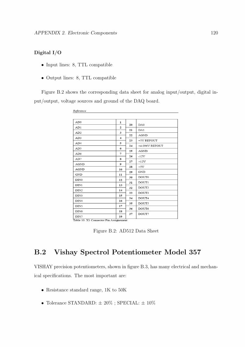
APPENDIX 2. Electronic Components 120
Digital I/O
• Input lines: 8, TTL compatible
• Output lines: 8, TTL compatible
Figure B.2 shows the corresponding data sheet for analog input/output, digital in-
put/output, voltage sources and ground of the DAQ board.
Figure B.2: AD512 Data Sheet
B.2 Vishay Spectrol Potentiometer Model 357
VISHAY precision potentiometers, shown in figure B.3, has many electrical and mechan-
ical specifications. The most important are:
• Resistance standard range, 1K to 50K
• Tolerance STANDARD: ± 20% ; SPECIAL: ± 10%

APPENDIX 2. Electronic Components 121
• Linearity (Independent) STANDARD ± 2.0%
• Power Rating 1.0 watt at 70 degree Derate to 0 watt at 125 degree
• Electrical Travel 340 ± 3
• End Voltage 0.5% maximum
• Dielectric Withstanding Voltage 1,000VRMS, 60Hz
• Insulation Resistance 1000M, 500VDC
• Rotation 360 continuous (optional mechanical stops 340 degree ± 3)
• Operating Torque Maximum Starting and running 3.68 mNm (0.5 oz - in)
• Weight 17.5gms (0.62 oz)
Figure B.3: Vishay Spectrol Potentiometer Model 357

APPENDIX 2. Electronic Components 122
B.3 Servo Motors
A servo motor is a small device that has an output shaft. This shaft can be positioned
to a specific angular positions by sending to the servo a specific signal. As long as the
signal exists on the input line, the servo will maintain the angular position of the shaft.
B.3.1 Servo Wiring
All servo motors have three wires, the ones chosen in our applications are the Futaba
servos. Therefore, the colors of the wirings are the following: Black for ground, Red
for power and white for the input signal. Servos can operate under a range of voltages.
Typical operation is from 4.8V to 6V. Figure B.4 shows the wiring of the FUTABA servo
motors.
Figure B.4: Servo Motor: FUTABA Wiring
B.3.2 PWM Signals
While the black and red wires provide power to the motor, the signal wire is used to
command the servo. The general concept is to simply send a logic square wave signal to
the servo motor at a specific wave length. This signal is called PWM signal.
The servo expects to see a pulse every 20 milliseconds (0.02 seconds). The length of
the pulse will determine how far the motor turns. A 1.5 millisecond pulse, for example,
will make the motor turn to the 90 degree position (often called the neutral position).
If the pulse is shorter than 1.5 ms, then the motor will turn the shaft to closer to 0

APPENDIX 2. Electronic Components 123
degress. If the pulse is longer than 1.5 ms, the shaft turns closer to 180 degress. Figure
B.5 shows the PWM signals for servo motor control.
Figure B.5: PWM Signals for Servo Control
Related Documents