Thesis for the degree Master of Pharmacy DEVELOPMENT OF LIPOSOMAL CURCUMIN FOR VAGINAL DRUG DELIVERY Haider Hussain Tromsø 2010 Supervisors Professor Natasa Skalko-Basnet Assoc. Professor Ingunn Tho Drug Transport and Delivery Research Group Department of Pharmacy University of Tromsø

Welcome message from author
This document is posted to help you gain knowledge. Please leave a comment to let me know what you think about it! Share it to your friends and learn new things together.
Transcript

Thesis for the degree Master of Pharmacy
DEVELOPMENT OF LIPOSOMAL CURCUMIN
FOR VAGINAL DRUG DELIVERY
Haider Hussain
Tromsø 2010
Supervisors
Professor Natasa Skalko-Basnet
Assoc. Professor Ingunn Tho
Drug Transport and Delivery Research Group
Department of Pharmacy
University of Tromsø

1
TABLE OF CONTENTS
TABLE OF CONTENT ...................................................................................................... 1
LIST OF FIGURES............................................................................................................. 3
LIST OF TABLES............................................................................................................... 4
ACKNOWLEDGMENTS .................................................................................................. 5
ABSTRACT ........................................................................................................................ 6
ABBREVIATIONS ............................................................................................................. 7
1. INTRODUCTION .......................................................................................................... 8
1.1. Vagina as Site for Drug Therapy............................................................................. 9
1.1.1. Anatomy and Physiology of Vagina .................................................................... 9
1.1.2. Vaginal Environment ........................................................................................ 11
1.1.3. Changes in Inflammation .................................................................................. 12
1.1.4. Vaginal Dosage Forms ...................................................................................... 15
1.2. Curcumin ................................................................................................................ 16
1.2.1. Origin ................................................................................................................ 16
1.2.2. Chemical Properties of Curcumin ..................................................................... 17
1.2.3. Pharmacological Effects of Curcumin Related to Inflammation ...................... 19
1.2.4. Limitations in Formulating Dosage Forms and Delivery Systems ................... 20
1.2.5. Delivery Systems for Curcumin ........................................................................ 21
1.3. Liposomes as Drug Delivery System .................................................................... 23
1.3.1. Classification of Liposomes .............................................................................. 25
1.3.2. Methods of Preparation ..................................................................................... 26
1.3.3. Topical Administration ..................................................................................... 27
1.3.4. Vaginal Application .......................................................................................... 28
2. AIM OF THE STUDY ................................................................................................. 30
3. METERIALS AND METHODS ................................................................................. 31
3.1. Materials ................................................................................................................. 32
3.1.1. Chemicals .......................................................................................................... 32
3.1.2. Solutions ............................................................................................................ 33
3.2. Methods ................................................................................................................... 34
3.2.1. Preparation of Liposomes ................................................................................. 34
3.2.2. Size Reduction of Liposomes ........................................................................... 35

2
3.2.3. Separation of Unentrapped Active Ingredients ................................................. 36
3.2.4. Particle Size and Size Distribution Analysis ..................................................... 37
3.2.5. Determination of Entrapment Efficiency .......................................................... 38
3.2.6. HPLC Analysis of Standard Curcumin and Curcuma Extracts ........................ 39
3.2.7. Phosphatidylcholine Quantification .................................................................. 40
3.2.8. Stability Experiment ......................................................................................... 42
3.2.9. Determination of Antioxidant Activity of Curcumin and Curcuma Extract by
DPPH assay ........................................................................................................ 43
4. RESULTS AND DISCUTIONS .................................................................................. 44
4.1. Optimization of Liposomal Preparation Method ................................................ 45
4.1.1. Sonication Procedure ........................................................................................ 45
4.1.2. Extrusion ............................................................................................................ 49
4.2. Entrapment Efficiency ........................................................................................... 51
4.2.1. Curcumin ........................................................................................................... 51
4.2.2. Curcuma Extract and Curcumin I ..................................................................... 53
4.3. Evaluation of Separation Method ......................................................................... 55
4.3.1. Centrifugation ................................................................................................... 55
4.3.2. Size Exclusion Chromatography ....................................................................... 56
4.4. Stability of Formulations ....................................................................................... 58
4.5. Antioxidant Activity of Curcumoids ..................................................................... 60
5. CONCLUSION ............................................................................................................. 62
6. FUTURE PERSPECTIVE ........................................................................................... 63
7. REFERENCES ............................................................................................................. 64
8. APPENDIX ................................................................................................................... 71
3
LIST OF FIGURES
Figure 1: Schematic illustration of the vaginal wall.
Figure 2: Association between chemical irritation and inflammatory pathways.
Figure 3: The extraction of curcumin.
Figure 4: Chemical structures of curcuminoids.
Figure 5: Keto-enol tautomerism of curcumin.
Figure 6: Schematic illustration of small unilamellar liposome-drug carrier.
Figure 7: Schematic illustration of reactions in colorimetric determination of lipid content.
Figure 8: The effect of the presence of curcumin in liposomal membrane.
Figure 9: The size of extruded formulations.
Figure 10: Entrapment efficiency for curcumin in sonicated liposomes.
Figure 11: Phosphatidylcholine (PC) recovery from sonicated formulations.
Figure 12: Size measurement of fractions collected during size exclusion chromatography.
Figure 13: Determination of curcumin content in the fractions separated by size exclusion
chromatography.
Figure 14: Changes in particle size during storage at 40 °C for 4 weeks.
Figure 15: Loss of entrapped curcumin during the accelerated stability testing.
Figure 16: Radical scavenging activity of curcumoids.
4
LIST OF TABLES
Table 1: Chemicals.
Table 2: Composition of lipid solutions.
Table 3: Preliminarily experiments to investigate sonication variables.
Table 4: PCS parameters.
Table 5: Selection of sonication conditions.
Table 6: The effect of the amount of curcumin taken into the liposome preparation on
liposomal size.
Table 7: The effect of extrusion on liposomal size.
Table 8: Entrapment of Curcuma extract and pure curcumin in liposomes.

5
ACKNOWLEDGMENTS
This study was conducted at the Drug Transport and Delivery Research Group, Institute of
Pharmacy, University of Tromsø.
First I would like to express my deep gratitude to my supervisor Professor Dr. Natasa
Skalko-Basnet for the excellent scientific guidance and support. The support of Assoc.
Professor Ingunn Tho is highly appreciated. It has been an honor and a pleasure to work
with you.
My heartfelt thanks go to Professor Dr. Purusotam Basnet for the scientific support and
providing the DPPH data. Without your contribution, this work would not been realized.
Further, I wish to thank Merete Skar for continuous support in the laboratory and the
members of our Research Group for the pleasant time and social meetings.
Special thanks to my fellow students for creating such a wonderful time here in Tromsø.
Cheering me up and laughing together shortened these years and made even studying for
exams enjoyable.
I would like to thank my family for the support throughout these years. You have always
been in my heart and my mind.
May, 2010
Haider Hussain

6
ABSTRACT
Curcumin (I), demethoxy curcumin (II) and bisdemethoxy curcumin (III) are commonly
called curcuminoids, and derived products from the spice, turmeric. It has reported
numerous of therapeutic activities including, anti-inflammatory, and anticancer properties.
The aim of the current study was to develop a formulation which can overcome the
limitation of curcumin being so poorly soluble in aqueous medium. Our approach has been
directed toward investigating the potential of using liposomal formulations as carrier
system for curcumin destined for treatment of vaginal inflammation. Curcumin containing
liposomes were prepared using soya phosphatidylcholine by the modified film method.
Moreover, we added cholesterol in various molar ratios to affect the vesicle membrane
rigidity. Curcumin entrapped in the liposomes was quantified and the entrapment
efficiency was found to be reaching up to 100%. The size and size distribution of
liposomes were determined on photon correlation spectroscopy. The results showed an
increase in size of liposomes containing curcumin in comparison with empty liposomes.
The accelerated stability testing was used to predict the stability of the formulations. The
test revealed changes in the characteristics of the liposomes. The free radical scavenging
activity (DPPH) assay of curcumin and Curcuma extract, as well as isolated pure curcumin
I, revealed that curcuminoids mixtures have stronger activity.
7
ABBREVIATIONS
Chol Cholesterol
DPPH 1, 1- diphenyl-2-picryl hydrazyl
EC50 Effective concentration required to reduce DPPH radical by 50%
E.E.% Entrapment efficiency
HPLC High performance liquid chromatography
Lipoid S-100 Soya phosphatidylcholine containing 100% PC
LUVs Large unilamellar vesicles
MLVs Multilamellar vesicles
PC Phosphatidylcholine
PC/Chol (2:1) Mixture of phosphatidylcholine and cholesterol with molar ratio 2:1
PC/Chol (4:1) Mixture of phosphatidylcholine and cholesterol with molar ratio 4:1
PCS Photon correlation spectroscopy
Rec. Recovery
P.I. Polydispersity index
SD Standard deviation
SEC Size exclusion chromatography
SUVs Small unilamellar vesicles
vol/vol Volume ratio
w/w Weight ratio

8
1. INTRODUCTION

9
1.1. Vagina as Site for Drug Therapy
The effectiveness of the vagina as a site of drug administration for local effects has been
well established (Jain et al., 1997; Pavelic et al., 2001). It is an important route for local
treatment of several gynecological conditions, such as infections and in hormonal therapy.
This route provides advantages such as reducing or eliminating the incidence and severity
of side effects, being a non-invasive route of administration and accessibility. These
benefits could contribute to a better compliance, thus achieving improved therapeutic
outcome (Knuth et al., 1993; Pavelic et al., 2004a). Furthermore, the vagina possesses
properties which include: large surface area of the vaginal wall, permeability, a rich blood
supply and importantly, the ability to bypass first-pass liver metabolism (Vermani and
Garg 2000; Pavelic et al., 2001). These properties are considered to be advantageous in
relation to drug absorption.
Currently, there is a variety of pharmaceutical products available on the market designed
for intravaginal therapy (tablets, creams, suppositories, pessaries, foams, solutions,
ointments and gels). However, their efficacy is often limited by a poor retention at the site
of action due to the self-cleansing action of the vaginal tract (Pavelic et al., 2001).
Furthermore, the vagina has unique features in terms of microflora, pH and cyclic changes,
and these factors influence the performance of the formulations and must be considered
during the development and evaluation of vaginal delivery systems (Valenta, 2005).
Therefore, a successful delivery of drugs through the vagina represents a pharmaceutical
challenge.
1.1.1. Anatomy and Physiology of Vagina
The human vagina is a tubular, fibromuscular organ that extends from the cervix of the
uterus to the vaginal vestibule measured in a length of approximately 9 cm (das Neves and
Bahia, 2006). The vaginal blood supply comes from the internal iliac arteries branching
into a complex network of arteries and veins surrounding the vaginal wall. Blood leaving
the vagina enters the peripheral circulation via the internal iliac veins, thus bypassing the
liver (Richardson and Illum, 1992; Knuth et al., 1993).

10
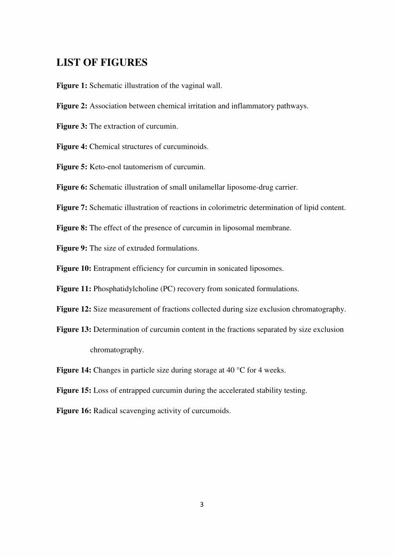
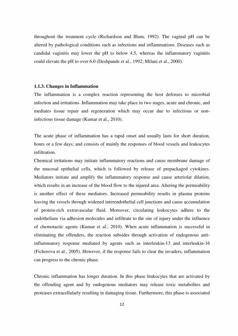
Histologically vagina consists of three distinct layers: an epithelial layer, a middle
muscular layer and an outer fibrous layer. The epithelial layer is classified as a non-
cornified, stratified squamous epithelium. This layer is composed of lamina propria and an
epithelial cell layer which contain particulate glycogen (Figure 1). The normal thickness of
the vaginal epithelium is approximately 200 µm (Richardson and Illum, 1992). It is usually
considered to be a mucosal surface, although it has no goblet cells and lacks the direct
release of mucins (Robinson and Bologna, 1994; Hussain and Ahsan, 2005; Valenta,
2005). The surface of vagina has a number of folds or also called rugae, which increase the
surface area of the vaginal wall (Hussain and Ahsan, 2005).
Figure 1: Schematic illustration of the vaginal wall (Washington et al., 2001).
Despite of the absence of any glands, the vagina produces a large amount of fluid (Hussain
and Ahsan, 2005). This fluid is a mixture of several components and includes leukocytes,
inorganic and organic salts, mucins, proteins, carbohydrates, urea and fatty acids (Vermani
and Garg, 2000). Like the thickness of the epithelial layer, the amount and composition of
the vaginal fluid change upon the hormonal activity (Robinson and Bologna, 1994;
Hussain and Ahsan, 2005).
11
The lactobacilli bacteria are an important component of the vaginal microflora. This
bacteria converts glycogen from exfoliated epithelial cells into lactic acid, and as a result,
maintains the pH around 4-5, with lowest being around the cervix. Body fluids such as
menstrual blood, cervical and uterine secretions will all act as alkalizing agents and
increase the vaginal pH (Richardson and Illum, 1992).
1.1.2. Vaginal Environment
The volume, viscosity and pH of vaginal fluid as well as the thickness and porosity of
epithelial layer may have either negative or positive impact on vaginal drug absorption
(Hussain and Ahsan, 2005). Therefore it is crucial to understand the conditions that might
influence these parameters. It is noteworthy that the histology and physiology of the vagina
may vary with age and with the menstrual cycle. Post-menopausal women experience
important changes in the vaginal physiology. These changes manifest as decline in
estrogen production during the pre-menopause and ongoing menopause. This leads to a
permanent decrease in the vaginal glycogen content, and consequently thinning of the
vaginal epithelium (Valenta, 2005). Furthermore, elevation of vaginal pH to 6.0–7.5, and a
decrease in the quantity of vaginal secretions have been reported (das Neves and Bahia,
2006). It was estimated that the vaginal secretion produced by postmenopausal women is
reduced by 50% compared to that produced by women of reproductive age (Washington et
al., 2001; Hussain and Ahsan, 2005).
Menstruation is another physiological factor associated with hormonal events. The
epithelial layer changes in thickness by approximately 200-300 µm as estrogen levels
change throughout the menstrual cycle (Hussain and Ahsan, 2005). Changes in the vaginal
pH and viscosity during the menstrual cycle are results of these changes in vaginal
histology and physiology. The vaginal pH tends to be lowest at ovulation when estrogen
levels reach a peak and both glycogen accumulation and epithelial desquamation at its
maximum (Deshpande et al., 1992; Richardson and Illum, 1992).
These physiological cyclic variations will be affected by the use of oral contraceptives.
Progestin-containing formulations are associated with the production of viscous mucus
12
throughout the treatment cycle (Richardson and Illum, 1992). The vaginal pH can be
altered by pathological conditions such as infections and inflammations. Diseases such as
candidal vaginitis may lower the pH to below 4.5, whereas the inflammatory vaginitis
could elevate the pH to over 6.0 (Deshpande et al., 1992; Milani et al., 2000).
1.1.3. Changes in Inflammation
The inflammation is a complex reaction representing the host defenses to microbial
infection and irritations. Inflammation may take place in two stages, acute and chronic, and
mediates tissue repair and regeneration which may occur due to infectious or non-
infectious tissue damage (Kumar et al., 2010).
The acute phase of inflammation has a rapid onset and usually lasts for short duration,
hours or a few days; and consists of mainly the responses of blood vessels and leukocytes
infiltration.
Chemical irritations may initiate inflammatory reactions and cause membrane damage of
the mucosal epithelial cells, which is followed by release of prepackaged cytokines.
Mediators initiate and amplify the inflammatory response and cause arteriolar dilation,
which results in an increase of the blood flow to the injured area. Altering the permeability
is another effect of these mediators. Increased permeability results in plasma proteins
leaving the vessels through widened interendothelial cell junctions and cause accumulation
of protein-rich extravascular fluid. Moreover, circulating leukocytes adhere to the
endothelium via adhesion molecules and infiltrate to the site of injury under the influence
of chemotactic agents (Kumar et al., 2010). When acute inflammation is successful in
eliminating the offenders, the reaction subsides through activation of endogenous anti-
inflammatory response mediated by agents such as interleukin-13 and interleukin-16
(Fichorova et al., 2005). However, if the response fails to clear the invaders, inflammation
can progress to the chronic phase.
Chronic inflammation has longer duration. In this phase leukocytes that are activated by
the offending agent and by endogenous mediators may release toxic metabolites and
proteases extracellularly resulting in damaging tissue. Furthermore, this phase is associated

13
with the presence of lymphocytes and macrophages, the proliferation of blood vessels,
fibrosis, causing destruction and remodeling of the tissue (Kumar et al., 2010).
The inflammatory reaction within the female reproductive tract may be essential for
immune responses in clearance of infections and offending agents. However, if this
reaction enters the chronic phase and persists, there will be higher probability for
complications to arise.
Numbers of studies have linked chronic inflammation with increased risk of viral
infections such as human immunodeficiency virus (HIV). Proinflammatory cytokines and
the transcription factors controlling the cytokine expression play a major role in HIV-1
pathogenesis (Fichorova et al., 2005). It was reported that continuous stimulation of
vaginal epithelial cells by proinflammatory mediators could lead to an increase in the
availability of potential host cells for infection and viral replication to occur. Furthermore,
it also leads to higher probability of viral transmission during sexual intercourse
(Fichorova et al., 2005).
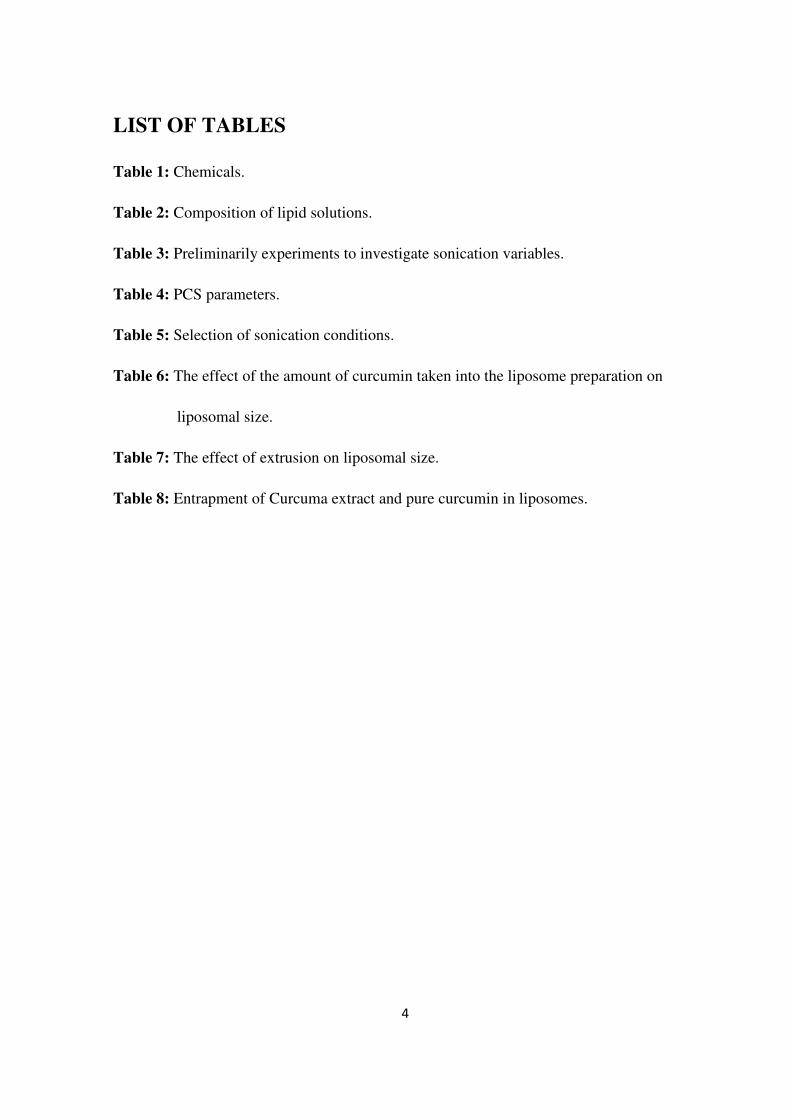
Several inflammatory agents are also linked to cancer promotion and development. An
example of these mediators is the tumor necrosis factor (TNF). TNF induces death of
diseased cells at the site of inflammation, and stimulates fibroblast growth. However, if
this agent is produced chronically, it may act as an endogenous tumour promoter,
contributing to the tissue remodelling and tumour growth and spread (Balkwill and
Mantovani, 2001).

14
Figure 2: Association between chemical irritation and inflammatory pathways.
Abbreviations: TNFα, tumor necrosis factor alpha; STNF-RI, soluble tumor necrosis factor
α receptor I; NF-κB, nuclear factor-kappa B; IL, Interleukin. (Fichorova et al., 2005)

15
1.1.4. Vaginal Dosage Forms
The majority of commercially available vaginal delivery systems are usually targeting
topical administration. Pessaries (tablets or suppositories) are among the most widely used
systems. The principle of their action is that they provide sustained release of the drug as
they gradually dissolve or melt. However, this mechanism has a drawback and can result in
low bioavailability if the formulation melted faster than intended, thereby giving shorter
residence time in the vagina (Brannon-Peppas, 1993). Vaginal tablets may contain binders,
disintegrants and other excipients that are used to prepare conventional oral tablets
(Brannon-Peppas, 1993; Hussain and Ahsan, 2005). Formulating very hydrophobic drugs
as vaginal tablets may not be an ideal approach. However, it was suggested that by adding
penetration enhancing agents such as surfactants can significantly enhance the drug
absorption (Hussain and Ahsan, 2005). Moreover, attempts have been carried out to use
mucoadhesive polymers in vaginal tablet formulations in order to increase the residence
time. Polyacrylic acid (PAA) is among the bioadhesive polymers that have been utilized
for vaginal formulations due to its high bioadhesive strength which allows a longer contact
time with vaginal surface (Ahuja et al., 1997).
Ideally, vaginal drug delivery system that is designed for local effect should distribute
uniformly throughout the site of action. However, the distribution and coverage of
formulation within the vaginal cavity varies with the properties of the delivery system. It
was reported that disintegrating tablet show low coverage whereas solution, suspension
and emulsions display greater distribution profile (Knuth et al., 1993; Washington et al.,
2001).
Creams and gels are another type of delivery systems frequently used. Creams are
normally emulsions whereas gels are usually hydrophilic polymers that utilize covalent
bonds to create cross-linked three-dimensional structures (Hussain and Ahsan, 2005).
Some formulations such as antifungal emulsion-based formulations seem to have greater
advantage over many suppository formulations (Washington et al., 2001). An example of
gel product is the progesterone gel formulation that is based on a loosely cross-linked poly
acrylic acid (Noveon AA1®). This formulation was found to remain on vaginal tissue for
3-4 days, thus allowing dosing intervals of twice a week (Knuth et al., 1993). However, a
disadvantage that can be associated with the use of creams and gels is that they may not

16
provide an exact dose, thus compromising the efficacy of the drug therapy (Hussain and
Ahsan, 2005).
1.2. Curcumin
The growing public interest in traditional medicine, particularly plants-based medicine, has
led to extensive research on the potentials of natural origin substances. Hundreds of studies
were conducted to investigate the effects of natural origin compounds on human health and
prevention and treatment of chronic diseases (Schmidt et al., 2007). Among studied
compounds, polyphenols appear as one of the most promising groups. In plants,
polyphenols are important for growth and protection against pathogens. Polyphenols have
recently received much attention in disease prevention and treatment due to their proven
antioxidant capabilities (Zern and Fernandez, 2005). Polyphenols are derived from many
components of the human food including peanuts, dark chocolate, green and black tea and
turmeric. Among polyphenols, curcumin is currently one of the most studied substances. It
is a hydrophobic, low molecular weight polyphenol widely used in form of the spice,
turmeric (Anand et al., 2007; Suresh and Srinivasan, 2007).
1.2.1. Origin
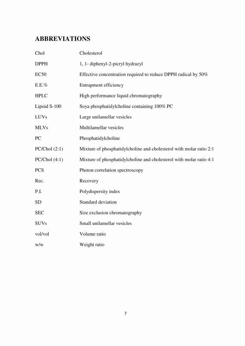
Turmeric has been used in Asia for thousands of years in food, preservation of food, and as
traditional medicine (Aggarwal et al., 2007). It is the yellow spice derived from the roots,
rhizome, of the plant Curcuma longa. The powdered extracts of dried roots, often called
turmeric, ukon (in Japanese), or haldi (in Hindi), may contain volatile and nonvolatile oils,
proteins, fat, minerals, carbohydrates, moisture and curcuminoids. The curcuminoids
which constitute approximately 5% of most turmeric preparations are a mixture of three
principal compounds: curcumin (sometimes referred to as curcumin I),
demethoxycurcumin (curcumin II), and bisdemethoxycurcumin (curcumin III) (Strimpakos
and Sharma, 2008). The majority of commercially available curcumin contains the
following composition: curcumin I (77%), curcumin II (17%) and curcumin III (3%) (Goel
et al., 2008).

17
Figure 3: The extraction of curcumin (Goel et al., 2008).
1.2.2. Chemical Properties of Curcumin
As indicated earlier, turmeric contains three different analogues of curcumin. The chemical
names and properties are shown below (Litwinienko and Ingold, 2004; Scotter, 2009):
Curcumin I: 1,7-bis-(4-hydroxy-3-methoxyphenyl)-hepta-1,6- diene-3,5-dione.
Chemical formula: C21H20O6; Molecular weight: 368 g/mol. pKa= 8.54
Curcumin II: 1-(4-hydroxy-3-methoxyphenyl)-7-(4-hydroxyphenyl)-hepta-1,6-diene-3,5,-
dione.
Chemical formula: C20H18O5; Molecular weight: 338 g/mol. pKa= 9.30
Curcumin III: 1,7-bis-(4-hydroxyphenyl)-hepta-1,6-diene-3,5-dione.
Chemical formula: C19H16O4; Molecular weight: 308 g/mol. pKa= 10.69
These compounds are practically insoluble in water at acidic and neutral pH, and soluble in
methanol, ethanol, dimethylsulfoxide, and acetone. The maximum absorption (λmax) of
curcumin in methanol occurs at 430 nm (Goel et al., 2008).

18
Curcumin I : R1 = R2 = OCH3
Curcumin II: R1 = OCH3, R2 = H
Curcumin III : R1 = R2 = H
Figure 4: Chemical structures of curcuminoids (Aggarwal et al., 2007).
Molecular configuration of curcumin can exist in tautomeric forms, bis-keto and enolate.
In acidic, neutral conditions and in solid phase, the keto form predominates, and curcumin
acts as a potent donor of H-atoms. However, under alkaline conditions the enolic form
predominates, as shown in Figure 5 (Strimpakos and Shrama, 2008).
Several researchers have proven the sensitivity of curcumin to light, and as a result they
suggested that biologic samples containing curcumin should be protected from light
(Strimpakos and Sharma, 2008). Another stability issue is the stability of curcumin in
phosphate buffer. It was reported that most of curcumin (>90%) is rapidly degraded within
30 min of placement in phosphate buffer systems of pH 7.2 (Goel et al., 2008).
Figure 5: Keto-enol tautomerism of curcumin (Strimpakos and Sharma, 2008).

19
1.2.3. Pharmacological Effects of Curcumin Related to Inflammation
The accumulated evidences over the years have shown that many of anti-inflammatory
drugs, such as steroids and NSAIDs are associated with numerous of side effects. Probably
the best example is the cardiovascular complications caused by the use of most coxibs
(Moodley, 2008). Consequently, there is an increasing demand for safer and more efficient
anti-inflammatory agents. Curcumin has been reported as one of the most promising
candidates of natural origin anti-inflammatory agents, with almost no reported side effects
(Aggarwal and Sung, 2009).
Curcumin has been traditionally used in prevention and treatment of several conditions and
diseases. Several of these effects have been already well documented scientifically. Studies
indicated that curcumin exerts hepato- and nephro-protective, thrombosis suppressing,
myocardial infarction-protective properties. Additionally, its strong antioxidant,
antimicrobial, anticarcinogenic and anti-inflammatory activities were also reported
(Aggarwal and Harikumar, 2009).
Until recently, many of the anti-inflammatory molecular targets of curcumin were
unknown. However, the establishment of modern biology in the recent decades led to the
discovery of more than 90 targets (Aggarwal et al., 2007). The mechanisms implicated in
the anti-inflammatory potential of curcumin may include (Brouet and Ohshima, 1995;
Kawamori et al., 1999; Aggarwal et al., 2007; Menon and Sudheer, 2007; Jurenka, 2009):
1) Suppression of the activation of the transcription factor NF–κB, which regulates the
expression of pro-inflammatory gene products.
2) Down-regulation of the expression of cyclooxygenase-2 (COX-2), an enzyme
linked with most types of inflammations.
3) Decreasing the activity and protein levels of inducible nitric oxide synthase (iNOS)
enzymes through reducing the expression of iNOS genes.
4) Inhibition of arachidonic acid metabolism via lipoxygenase and scavenging the free
radicals generated in this pathway.

20
5) Down-regulation of the expression of various cell surface adhesion molecules that
have been linked with inflammation.
6) Decreasing the expression of various inflammatory cytokines, including TNF, IL-1,
IL-6, IL-8, and chemokines.
7) Curcumin is a potent antioxidant, which contributes to its anti-inflammatory action.
All these effects are thought to lead to lowering the formation of inflammatory compounds
and suppressing the inflammatory response. This outcome is considered to be beneficial in
many abnormal conditions such as autoimmune diseases (Jagetia and Aggarwal, 2007).
Furthermore, there are growing evidences linking many of the targets mentioned above
with tumor promotion (Suresh and Srinivasan, 2007; Jurenka, 2009). Studies have shown
that enzymes such as COX-2 and iNOS overexpression have been implicated in the
carcinogenesis of many tumors (Brouet and Ohshima, 1995; Surh et al., 2001; Menon and
Sudheer, 2007). Although it has not a direct effect on the human cells, it should be noted
that antimicrobial activity of curcumin is potentially chemopreventive because an
increasing evidence that number of pathogens are directly linked with human cancers
(Strimpakos and Sharma, 2008).
1.2.4. Limitations in Formulating Dosage Forms and Delivery Systems
Despite the demonstrated efficacy of curcumin, it appears that its poor systemic
bioavailability after oral dosing compromises the potential for therapeutic uses. The major
reasons contributing to the low bioavailability of curcumin include poor absorption and
rapid systemic elimination (Strimpakos and Sharma, 2008).
Oral drug administration is usually considered as a practical and easy way to administrate
drugs. However, in order for a drug from solid dosage form to be absorbed, in this case
through the epithelial layer of the intestine, these substances must become dissolved.
Curcumin is a hydrophobic compound with very low solubility in water. The partition
coefficient and solubility in water was measured to be 3.2 and 0.6 µg/ml, respectively
(Kurien et al., 2007; Patel et al., 2009). When water-solubility is less than 1 µg/ml, which
is the case for curcumin, the bioavailability from oral formulations such as conventional
21
tablets may be unacceptable (Pouton, 2006). This was demonstrated in clinical trial study
to evaluate the pharmacokinetics and effective dose of curcumin in humans. In this study a
number of patients were given 8000 mg of free curcumin orally per day in order to achieve
detectable systemic levels. However, beyond 8 grams, the bulky volume of the drug was
unacceptable to the patients (Cheng et al., 2001; Bisht et al., 2007).
Furthermore, studies performed on humans and animals shown that orally administrated
curcumin undergoes rapid metabolism in the liver particularly via glucuronidation, while
curcumin given intraperitoneally or systemically undergoes reduction (Aggarwal and Sung,
2009). Metabolites produced from these pathways show low or no pharmacological
activity (Aggarwal et al., 2007; Aggarwal and Harikumar, 2009).
1.2.5. Delivery Systems for Curcumin
It is necessary to improve the bioavailability of curcumin in order to fully utilize the
potential of this agent, and therefore a growing number of research groups are working on
this aim. There are studies designed to investigate new approaches that could overcome
these limitations seen with free curcumin. Number of studies has evaluated the liposomal
formulation in vivo and their effectiveness. The study conducted by Li et al. (2005)
investigated the effect of liposomal curcumin on pancreatic carcinoma cells and
suppression of KF-kB activity. The incorporated curcumin in liposomes showed a dose-
related increase in apoptosis of carcinoma cells and suppression of NF- B activity.
Moreover, the liposomal curcumin was found to be as effective as or better than free
curcumin. Another experiment studied the effect unilamellar liposomal curcumin after
tumor implantation on mice. It concluded that liposomal curcumin could increase the life
span of the animals by up to 74% in comparison with untreated (Rubya et al., 1995). The
study conducted by Kunwar et al. (2006) compared the cellular uptake of liposomal and
albumin-loaded-curcumin by the spleenic lymphocytes and EL4 lymphoma cells. They
reported that liposomes were able to deliver more curcumin into the cells than human
serum albumin.
22
The absorption of a micellular formulation was evaluated using everted rat intestinal sacs.
This micellular formulation was composed of phosphatidylcholine and sodium
deoxycholate. The authors reported that after the incubation for 3 hours, the percentage of
free curcumin absorbed was 49%, whereas the percentage for micellular formulation was
56%. (Suresh and Srinivasan, 2007).
In another approach phospholipid complex of soya phospholipid and curcumin was tested
in vivo on rats. The study showed higher plasma concentrations, and longer half-life of
phospholipid complex in comparison with free curcumin. Furthermore the bioavailability
was also seen to be improved significantly after oral administration. The relative
bioavailability of curcumin was estimated to be around 330% for the phospholipid
complex as compared to free curcumin (Liu et al., 2006).
Another strategy of delivering curcumin is self-microemulsifying drug delivery system
(SMEDDS). This system is basically composed of isotropic mixtures of oil, surfactant, co-
surfactant and drug which has the ability to form o/w microemulsion when it comes in
contact with aqueous medium in gastro intestinal tract after oral intake (Borhade et al.,
2008). The curcumin-SMEDDS formulation was composed of 57.5% surfactant, 30% co-
surfactant and 12.5% oil. The in situ evaluation of this formulation showed that the
absorption percentage of curcumin-loaded SMEDDS was 3.86 times higher than that of
curcumin suspension (Cui et al., 2009).
“Nanocurcumin” is another formulation recently developed for curcumin. The principle of
this formulation is that curcumin is encapsulated in cross-linked polymeric particle with a
hydrophobic core and a hydrophilic shell. The size of these particles lies in nanometer
range and typically less than 100 nm. The group tested the product on pancreatic cancer
cells and NFkB and reported to be effective in inhibition of these cells and has similar
activity as free curcumin on inflammatory cytokines (Bisht et al., 2007).
Loaded solid lipid nanoparticles (SLN) is another type of nano-particle based delivery
formulations. The system is usually consisting of biodegradable solid lipids. At room
temperature the particles are in the solid state. Therefore, the mobility of incorporated
molecules is reduced, thus it may offer possibility of modified release (Mühlen et al.,
1998). The study on SLN loaded curcumin was preformed by Tiyaboonchai et al. (2007)
23
and aimed at using this formulation in topical application. The stability and release was
tested and found that properties of cream containing curcumin incorporated into SLNs was
improved in comparison to free curcumin in the cream formulation.
Using pharmacological agents such as piperine (a component of black pepper) as
suppressor of glucuronidation process of curcumin was also investigated. It was reported
the inhibition of this process which occur primarily in the liver and in the intestine could
enhance the bioavailability of curcumin (Aggarwal et al., 2007).
As presented, there are numerous studies suggesting different approaches of delivery
systems in order to improve the absorption of curcumin. All of these studies have
concluded that it is possible to develop formulations and methods which can improve the
bioavailability and give higher plasma concentrations.
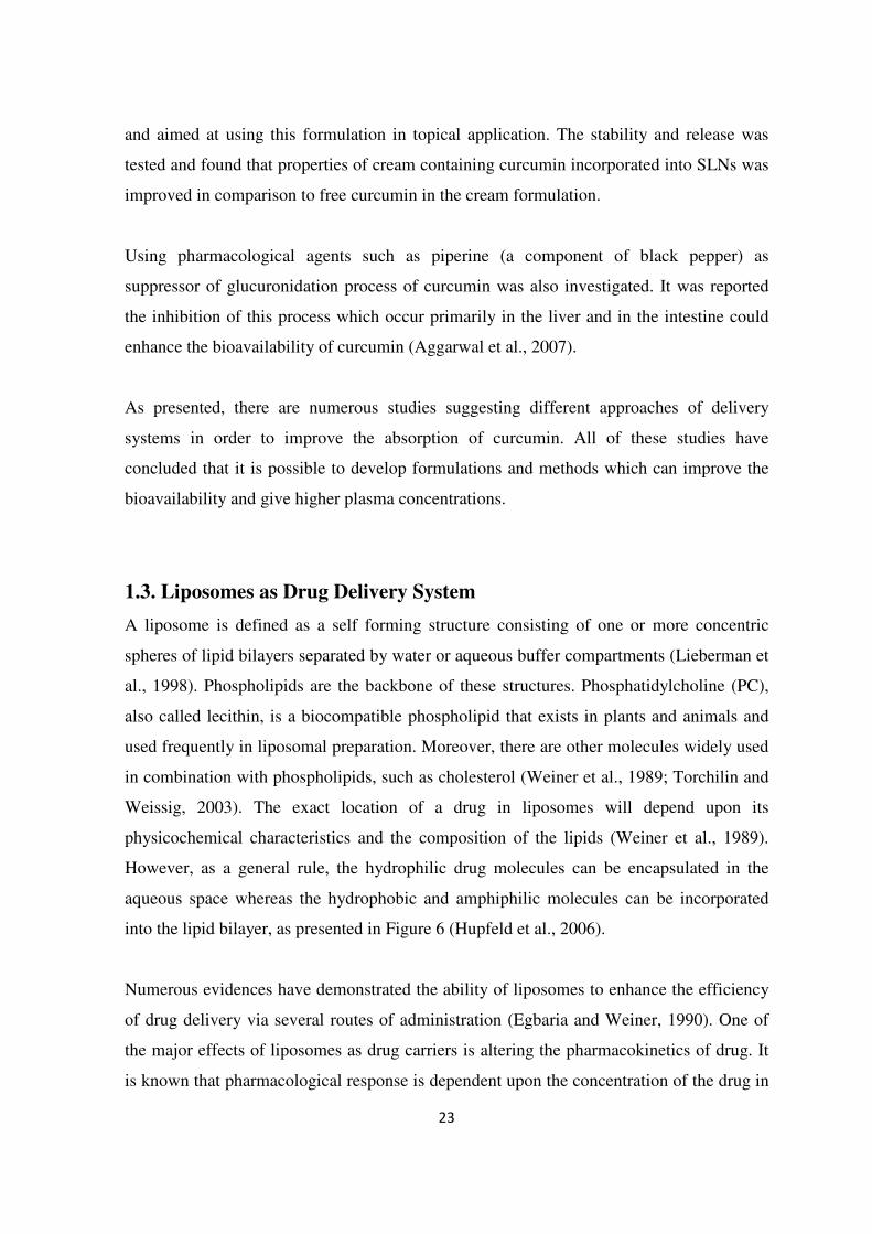
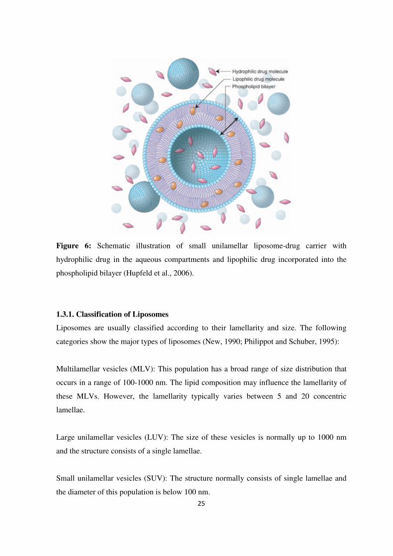
1.3. Liposomes as Drug Delivery System
A liposome is defined as a self forming structure consisting of one or more concentric
spheres of lipid bilayers separated by water or aqueous buffer compartments (Lieberman et
al., 1998). Phospholipids are the backbone of these structures. Phosphatidylcholine (PC),
also called lecithin, is a biocompatible phospholipid that exists in plants and animals and
used frequently in liposomal preparation. Moreover, there are other molecules widely used
in combination with phospholipids, such as cholesterol (Weiner et al., 1989; Torchilin and
Weissig, 2003). The exact location of a drug in liposomes will depend upon its
physicochemical characteristics and the composition of the lipids (Weiner et al., 1989).
However, as a general rule, the hydrophilic drug molecules can be encapsulated in the
aqueous space whereas the hydrophobic and amphiphilic molecules can be incorporated
into the lipid bilayer, as presented in Figure 6 (Hupfeld et al., 2006).
Numerous evidences have demonstrated the ability of liposomes to enhance the efficiency
of drug delivery via several routes of administration (Egbaria and Weiner, 1990). One of
the major effects of liposomes as drug carriers is altering the pharmacokinetics of drug. It
is known that pharmacological response is dependent upon the concentration of the drug in

24
the target cell. The drug concentration in the target site is governed by absorption,
distribution and elimination. These processes may influence the pharmacokinetics of the
drug and lead to inefficient utilization of the therapeutic agent. Thus, higher doses need to
be administrated. Furthermore, higher drug doses often lead to resistance and undesirable
immunological and toxicological effects (Fendler and Romero, 1977).
Liposomes are thought to shield all or most of the drug molecules resulting in decreasing
the direct contact of drug with biological environment, thus the pharmacokinetic profile of
the drug will be determined by the physiochemical properties of liposomes, rather than the
drug itself (Sætern, 2004). Incorporating the drug into a vehicle capable of delivering it
intact would overcome many of the disadvantages of the free drug administration.
Improving the pharmacokinetics of the drug by this method could lead to beneficial effects
such as reduced dosages, increased cellular permeability and delayed drug elimination
(Fendler and Romero, 1977). It is worth mentioning that liposomes are also non-toxic,
biodegradable and can be manufactured on large scales (Washington et al., 2001).
The potentials of liposomes to serve as delivery systems have been proven by the number
of liposomal formulations already approved by FDA for clinical use such as AmBisome®
(amphothericin B) and DaunoXome® (Daunorubicin). These formulations were clinically
compared with conventional drug formulations and proved superiority of liposomal
delivery (Allison, 2007).
25
Figure 6: Schematic illustration of small unilamellar liposome-drug carrier with
hydrophilic drug in the aqueous compartments and lipophilic drug incorporated into the
phospholipid bilayer (Hupfeld et al., 2006).
1.3.1. Classification of Liposomes
Liposomes are usually classified according to their lamellarity and size. The following
categories show the major types of liposomes (New, 1990; Philippot and Schuber, 1995):
Multilamellar vesicles (MLV): This population has a broad range of size distribution that
occurs in a range of 100-1000 nm. The lipid composition may influence the lamellarity of
these MLVs. However, the lamellarity typically varies between 5 and 20 concentric
lamellae.
Large unilamellar vesicles (LUV): The size of these vesicles is normally up to 1000 nm
and the structure consists of a single lamellae.
Small unilamellar vesicles (SUV): The structure normally consists of single lamellae and
the diameter of this population is below 100 nm.
26
The choice of preparation method can influence the size and lamellarity of liposomes,
resulting in different characteristics and fate of entrapped drug in vivo (Philippot and
Schuber, 1995).
1.3.2. Methods of Preparation
There are various methods to produce different types of liposomes. However, all
preparation methods can be simplified as to involve three basic steps: 1) Preparation of the
dispersion of the lipids in aqueous media 2) purification of prepared liposomes, and 3)
analysis of final product (New, 1990). The following is a brief description of methods that
are considered among the most widely used in liposome preparation:
Hand-shaking, (MLV)
The principle of this method is to dissolve the lipid in efficient solvent. Lipophilic drugs
can be dissolved in compatible solvent which can be added later to the lipid solution. This
mixture will be dried down under pressure till it forms lipid films. Hydrophilic drugs can
be dissolved in aqueous solution which will be added to the lipid films under shaking. This
will produce swelling and peeling of the lipid films and gives a milky suspension of
MLVs. Using this technique, one can entrap as high as 100% of the lipid soluble
molecules, whereas hydrophilic compounds are often encapsulated in amount of 5-10%
(New, 1990).
Sonication, (SUV)
Sonication is the most widely used method for producing small vesicles. It is usually used
to convert MLVs to SUVs through the employment of energy at high levels by exposure of
MLVs to ultrasonic irradiation. Probe-sonication and bath sonication are frequently used
techniques to reduce the size of liposomes. The probe has the most efficient transfer of
energy to the liposomal dispersion. However, it is associated with metal particle shedding
from its probe and therefore one must be aware of the potential contamination. Bath
sonication is much milder than probe sonication, but it is time-consuming and may result
in low yield of smaller liposomes (New, 1990).
27
Extrusion, (LUV)
The principle in extrusion technique is based on employment of moderate pressure to force
MLVs through polycarbonate filters with defined pore size. At low pressures (100 psi),
MLVs display a reduced-size while maintaining their multi-lamillarity, whereas at higher
pressure the liposomes are broken down as they pass though the membrane filter resulting
in reorganizing of the phospholipid bilayer giving rise to unilamellar vesicles (Philippot
and Schuber, 1995). It is simple method and easy to use and there are several products
available on the market. Among the equipments are the Hamilton® syringes. These are two
syringes connected by a filter holder allowing the samples to pass back-and-forth through
the polycarbonate filter. However, this method can be limited by production of small
volumes of LUVs and the back pressure that can be tolerated by the syringe and filter
holder is limited (Gregoriadis, 2007).
Unprocessed MLVs have limited uses in in vivo studies because of their large diameter and
heterogeneity of size (Gregoriadis, 2007). However, the techniques used to change these
parameters may influence physical properties of liposomes. The conversion to SUVs from
MLVs may result in vesicles with very low trapped volumes. Furthermore, SUVs can be
unstable and prone to fusion process due to the high curvature of the lipid bilayer (New,
1990). Extrusion technique used to produce LUVs may result in rupturing and resealing
which leads further to leakage of the entrapped drug and the final vesicles may have lower
amount of entrapped material, depending on the lipophilicity of the drug (Gregoriadis,
2007).
1.3.3. Topical Administration
Liposomes have been widely used to enhance the efficiency of drug delivery though
various routes of administration and have been shown to be significantly superior to
conventional dosage forms especially for intravenous and topical administration (Egbaria
and Weiner, 1990). However, therapeutic applications of systemically administered
liposomes have been limited by their rapid clearance from the bloodstream and their
uptake by reticuloendothelial system (RES) in liver and spleen. Furthermore, the use of
liposomal formulations in oral administration has been limited due to physiological factors.
28
The three major factors are pH, bile salt, and pancreatic enzymes in the gastrointestinal
(GI) tract can destabilize the structure of the vesicles and limit their potential (Lian and Ho,
2001).
Topical liposomal administration might offer an opportunity for developing a novel
delivery system that could overcome these limitations experienced with the systemic and
oral liposomal formulation as well as conventional products. The major advantages of
topical liposomal drug include (Egbaria and Weiner, 1990):
1) Reduction of side effects and incompatibilities that may arise from undesirably high
systemic absorption of drug.
2) Markedly increasing the liposomal drug accumulation in the desired tissues.
3) Capability for incorporation of a wide variety of hydrophilic and hydrophobic drugs.
Additionally, their ability to provide a sustained/controlled release and an enhancement of
the cellular penetration of the incorporated material could improve their potential for being
applied vaginally (Pavelic et al., 1999; Pavelic et al., 2004b).
1.3.4. Vaginal Application
Liposomes due to their ability to encapsulate both lipophilic and hydrophilic active
ingredient, represent a promising delivery system in regard to vaginal therapy. Some of
studies have investigated the release profiles of number of pharmaceutical agents
incorporated in liposomal formulations in simulated vaginal conditions. The study
conducted by Pavelic et al. (1999) had an aim to investigate the stability of liposomal
formulations for metronidazole, clotrimazole and chloramphenicol, drugs which are
frequently applied in the treatment of vaginal infections. The group concluded that the
liposomes retained approximately 28-40% of the entrapped drug even after 6 hours of
incubation in an environment that mimics the vaginal cavity of pre- and post
postmenopausal women.
The cervical mucus present in the vagina is believed to assist in the bioadhesion process
(Brannon-Peppas, 1993). However, being in a liquid form as liposomal suspensions, there
might be higher probability for the formulation to be expelled from the vagina and reduce

29
the retention time at the site of action (Pavelic et al., 2001). Therefore research groups are
considering incorporation of liposomes in a bioadhesive base, such as hydrogels.
Among various types of hydrogels, Carbopol hydrogels have demonstrated a good
compatibility with liposomal formulations (Skalko et al., 1998). The polymer adhesion to
tissues permits intimacy of contact and also improves the drug absorption. Furthermore, it
also prolongs the residence time at the site of administration (Knuth et al., 1993).
One study investigated the comparison of the release profiles between liposomal gels
containing hydrophilic marker substance (FITC-dextrans) with the same gel with the
marker present in non-liposomal form. The conditions in this study were set to be close to
the physiological environment. The retention of encapsulated hydrophilic model compound
FITC-dextrans was much higher than the control gel without incorporated liposomes. It
was found that about 20% of dye was released from liposomes incorporated in the gel after
72 hours, whereas the control gel released approximately 89% during the same period
(Pavelic et al., 2004b). Another study was investigating the effect of Carbopol gel on the
stability of liposomal chloramphenicol. It was calculated that more than 40% of the
entrapped drug was in the liposomal gel even after 24 hours of incubation in simulated
vaginal conditions. The authors concluded that the gel formulation provided a stable
vehicle suitable for vaginal application in which liposomes are distributed uniformly and
their original size distribution of liposomes is preserved (Pavelic et al., 2004a).
The same group compared the in vitro stability of liposomal suspensions and liposomal
Carbopol gel formulations of acyclovir, an antiviral agent with low bioavailability
especially in topical dosage forms. Acyclovir was encapsulated in three types of liposomes,
namely neutral, negatively- and positively- charged liposomes. However, even with
incorporation of different liposomes in hydrogels the results showed the slower release
profile of drug from liposomal gel in comparison with liposomal suspension, as well as
protective effect of a hydrogel matrix on liposomes which yielded in improvement of
stability (Pavelic et al., 2005).

30
2. AIM OF THE STUDY
The aim of this project has been to develop liposomal formulation for curcumin in order to
improve its solubility. Liposomes would serve as carrier system enabling the highly
lipophilic substance to be prepared in aqueous formulation. For that purpose liposomal
preparations were optimized in order to:
• Study the size and methods of size reduction for liposomes containing curcumin.
• Measure the entrapment efficiency in prepared formulation and optimize the
preparation method.
• Investigate the stability of liposomal formulations using accelerated stability
testing.
• Evaluate the curcumin potential as an anti-inflammatory agent by using DPPH
assay.

31
3. METERIALS AND METHODS

32
3.1. Materials
3.1.1. Chemicals
Table 1: Chemicals
Chemical Manufacturer / provider
Calcium chloride hexahydrate AnalaR, UK
Cholesterol (lanolin) Sigma Aldrich, USA
Curcumin Sigma Aldrich, India
Chloroform Merck, Germany
Ethanol 96% Acrus, Norway
Ethyl acetate
Merck, Germany
Lipoid S100 Lipoid, Germany
Methanol (LiChrosolv) Merck, Germany
Sepharose CL-4B Pharmacia Biotech, Sweden
Tris-HCl
(Tris(hydroxymethyl)aminomethane-
hydrochloride)
Merck, Germany
Triton X-100 Merck, Germany

33
3.1.2. Solutions
The following solutions are given in examples of 1L volume:
Triton 2.5% Buffer pH 8.0
- Used to prepare standard solutions for measurements of lipid content in supernatant
and pallets in the evolution of the separation method for formulations which dose not
contain cholesterol.
1. Calcium chloride hexahydrate 0.075 g
2. TRIS-HCl 7.88 g
3. Triton X-100 26.74 g
4. Adjust to pH 8.0 by adding 1 M NaOH q.s.
5. Distilled water ad 1000 mL
Triton 10% Buffer pH 8.0
- Used to prepare standard solutions for measurements of lipid content for the evaluation
of the separation method for cholesterol containing formulations.
1. Calcium chloride hexahydrate 0.075 g
2. TRIS-HCl 7.88 g
3. Triton X-100 107 g
4. Adjust to pH 8.0 by adding 1 M NaOH q.s.
5. Distilled water ad 1000 mL

34
3.2. Methods
3.2.1. Preparation of Liposomes
Preparation of lipid solutions:
PC solution: A solution of soya phosphatidylcholine (PC; Lipoid S100) was prepared by
dissolving 200 mg of PC in 5 mL methanol.
PC/Chol 4:1 solution: An amount of 25.4 mg of cholesterol was dissolved in 5 mL
methanol/chloroform with (4:1; vol/vol). Then 200 mg of PC was added to the solution to
obtain the molar ratio of 4:1.
PC/Chol 2:1 solution: The same procedure as PC/Chol 4:1 solution was used to prepare
this solution except the amount of cholesterol used was 50.9 mg.
Table 2: Composition of lipid solutions.
Amount of Lipids Formulations Molar Ratio
Phosphatidyl
choline (mg)
Cholesterol
(mg)
Solvent
PC 1 200 - Methanol
PC/Chol
4:1 200 25.4 Methanol/Chloroform
(4:1; vol/vol)
PC/Chol
2:1 200 50.9 Methanol/Chloroform
(4:1; vol/vol)
Preparation of Curcuma extract
Curcuma powder (100 g) was extracted with 1) water (2000 mL), 2) 96% Ethanol (2000
mL) and 3) Ethyl acetate (200 mL), respectively. For each extraction the mixture was bath
sonicated for 10 minutes and left overnight with occasional shaking prior to filtration. The
residues were separated after filtration and solvents in the filtrate evaporated with rotary

35
evaporator at low pressure. Percentage yield was expressed as w/w of dry powder from the
market.
Preparation of lipid film by rotary evaporation and hydration
The dissolved lipids (5 mL) were transferred into three separate round bottom flasks of 500
mL. In the case of curcumin containing liposomes, the drug was dissolved in methanol
yielding a concentration of 5 mg/mL (extract or curcumin mixture) or 1 mg/mL (curcumin
I) and mixed with lipid solutions. The solvent was removed under vacuum of 55 hPa and
rotation of 60 rpm at 25 °C in 90 minutes by a Büshi R-124 rotary evaporator with vacuum
pump 500-system (Büshi, Switzerland). The deposited lipid film was removed from the
rotary evaporator and left at room temperature for an additional period of 60 minutes to
remove traces of solvent. Subsequently the lipid film was hydrated using 10 mL of freshly
distilled water. The resultant liposomal suspension was manually shaken for 15 minutes till
homogeneous suspension was obtained. The suspension was left at room temperature over
night prior to further treatments.
3.2.2. Size Reduction of Liposomes
Sonication
Liposomal suspension of MLVs (1.8 mL) transferred to a 2 mL round bottom vial
(Eppendorf, Germany) and placed in ice bath. The position of a needle probe tip 407
probe-sonicator Labsonic U (B. Braun Biotech, Germany) was fixed in vial. Vertically, it
was fully immersed into the vial and horizontally, it was positioned in the middle of the
volume. The liposomal suspension was exposed to ultrasonic irradiation with an output of
30, 40 and 50 Watt and duration of continuous 30 and 150 seconds, as described in Table
3. The sample was left to cool down and placed in the fridge at 4 °C for 1 day prior to
further test e.g. size analysis and centrifugation. This experiment was executed in order to
determine suitable sonication procedure. After comparing the results a final method for
sonication was chosen to be 40 W for 150 sec.

36
Table 3: Preliminarily experiments to investigate sonication variables in order to
determine their influence on size and size distribution.
Output (Watt) Duration (Seconds)
30 150
30 Empty PC,
PC/Chol (2:1)
Empty PC,
PC/Chol (2:1)
40 Empty PC,
PC/Chol (2:1)
Empty PC,
PC/Chol (2:1)
50 Empty PC,
PC/Chol (2:1)
Empty PC,
PC/Chol (2:1)
For each sonication conditions the experiment was performed in duplicate.
Extrusion
The liposomal suspension was filter-extruded through a polycarbonate membrane Track-
Etch Nuclepore membrane (Whatman, UK). Up to 1 mL of MLVs were passed five times,
back-and-forth, through the 0.4 µm polycarbonate membrane filters at room temperature.
The extrusion was done by hand with a syringe extruder Liposofast™ (Avestin Inc.,
Canada). The resultant products were stored in the fridge at 4 °C over night prior to size
analyses and centrifugation.
3.2.3. Separation of Unentrapped Active Ingredients
Centrifugation
For separation of liposomes from unentraped active ingredient, a portion of the liposomal
dispersion was transferred to 3-mL thick wall polycarbonate centrifuge tubes. The samples
were then centrifuged using SW60Ti rotor and Beckman Optima L8-M centrifugator
(Beckman Inc., USA). The centrifugation was done at a temperature of 10 °C and speed of
37
35000 rpm (150000 g) for 150 minutes. The content of active ingredient in both
supernatant and pellet was determined.
Size Exclusion Chromatography
Suspension of liposomes was fractionated by size exclusion chromatography as well. A
MLVs sample of PC/Chol (molar ratio 2:1) formulation containing 5 mg curcumin was
exposed for mild ultra-sonic irradiation in bath-sonicator for 5 minutes and vortexed for 1
minute prior to the separation. The sample was then added to a glass tube packed with
Sepharose CL-4B (Pharmacia Biotech, Sweden) to approximately 25 mL (25 cm). Distilled
water was added repeatedly to insure continues flow and prevent the column from drying.
The collection of the portions was started immediately and continued until 6 samples after
yellow color disappeared from the column, in total 30 mL including the void volume. The
flow rate was estimated to be 0.5 mL·min-1
and took about 60 minutes for a sample of 1
mL of PC/Chol (2:1) liposomal formulation to pass through.
3.2.4. Particle Size and Size Distribution Analysis
Photon correlation spectroscopy (PCS):
Also known as dynamic light scattering is a simple and rapid method to determine the
particle size and size distribution of liposomes. The principle is based upon the Brownian
motion of particles in medium. As the particles diffuse in the fluid the collisions with
medium molecules causes a random movement of the particles. When the PCS machine
focuses laser light on the sample, it registers the signals from the moving particles as
fluctuations in the scattered light. The analysis is based on the time dependence of these
fluctuations. Small particles diffuse more rapidly than larger ones, thus the fluctuations
vary accordingly. The software then calculates the radius of the particles by using Stokes-
Einstein equation (NICOMP, 1997; Torchilin and Weissig, 2003).
38
Experiment
PCS measurements were performed on NICOMP Submicron particle sizer, Model 370
(NICOMP Particle Sizing Systems, USA). Sample preparations were preformed in a
laminar airflow bench. The cuvettes (borosilicate glass) were cleaned and filled with
distilled water and sonicated in bath-sonicator for 30 minutes to reduce the possibility of
contaminations. Before measurements were preformed, the instrument parameters were
adjusted according to the values listed in Table 4. The samples were diluted with freshly
filtrated distilled water using an Acrodisc 0.2 µm syringe filter (Pall Corp., USA) to obtain
an intensity count rate between 250 and 350 kHz.
Table 4: PCS parameters.
Parameters Values
Temperature 23 °C
Viscosity 0.933 cp
Liquid index of refraction 1.333
Intensity set point 300 ± 50
Channel width Auto
Number of cycles 1 cycle
Run time 5 minutes
3.2.5. Determination of Entrapment Efficiency
The entrapment efficiency measurements were preformed on UV-spectrophotometer
Aligent 8453 equipped with deuterium and tungsten lamp (Agilent Technologies,
Germany). In order to quantify the content of curcumin in supernatant and pellets in
samples, series of standard solutions were prepared. The known amounts of curcumin were
dissolved in ethanol and diluted to obtain a stock solution of 1000 ng/mL. The standard
solutions were then prepared using the stock solution the respective concentrations (100,
200, 400, 600, 800 and 1000 ng/mL). The absorbance was measured at 425 nm based on

39
the spectral analysis. A calibration curve of curcumin was developed by plotting
absorbance versus concentration of standard solutions.
The supernatant and pellets were each dissolved in methanol. The measurements were
done in triplicate. The entrapment efficiency was calculated using the following equation:
Equation 1:
Where A is amount of curcumin in pellet and B is amount of curcumin in supernatant.
3.2.6. HPLC Analysis of Standard Curcumin and Curcuma Extracts
High performance liquid chromatography (HPLC) (Waters, USA) system consists of a
HPLC Water 2690 Separation Module, a Water 996 Photodiode Array detector. Column
was YMC pro C18 (250 x 4.60 mm) joined with precolumn. Mobile phase: CH3CN-2.5%
acetic acid (54:46); Injection volume: 10 µL. The temperature of column was maintained
35 °C during the chromatoghaphic separation. The flow rate was 1.0 mL/min and run for
12 minutes. The eluting compounds were monitored at UV 425 nm.
Analysis of standard curcumin
Standard curcumin was evaluated for its purity as well as it was expected that standard
curcumin contains the mixture of curcumin (curcumin I), desmethoxy curcumin (curcumin
II) and bisdesmethoxycurcumin (curcumin III).
Analysis of Curcuma extracts
Water extract, ethanol extract and ethyl acetate extract were analyzed by the HPLC method
as described above.

40
3.2.7. Phosphatidylcholine Quantification
Enzymatic phospholipid assay:
The principle of quantitative enzymatic phospholipid assay is based on a sequence of
reactions preformed by three enzymes (Phospholipase D, Choline oxidase, Peroxidase)
allowing the colorimetrical quantification of the lipid content. The chain starts with
phospholipids (lecithin, lysolecithin and sphingomyelin) being hydrolyzed by
phospholipase D and which results in choline to be released (Figure 7). The liberated
choline acts as a substrate in a reaction preformed by choline oxidase which forms an
amount of hydrogen peroxide. The latter takes part in a peroxidase-catalyzed coupling
reaction resulting in red dye. The amount of choline is proportional to the amount of
resulting quinoneimine, thus the amount of phospholipids contained in the sample can be
determined by measuring the absorbance of the red color (Grohganz et al., 2003;
BioMérieux sa, 2009).
Figure 7: Schematic illustration of reactions in colorimetric determination of lipid content
(BioMérieux sa, 2009)
Experiment
The amount of phospholipids was determined in terms of quantification of the
phosphatidylchline content according to the protocol developed by Grohganz et al. (2003)
and using an enzymatic kit, phospholipids enzymatique PAP 150 (BioMérieux sa, France)
as a coloring reagent. The method can be divided into three stages namely, activation of

41
coloring reagent, preparation of standard solutions and finally measurement of the
absorbance.
Activation of the coloring reagent
The preparation was done according to the user manual. The coloring reagent was
activated by adding 25 mL of buffer solution (consisting of Tris pH 7.8, surfactant and
phenol) to the dry enzyme reagent (Choline oxidase, Phospholipase D, Peroxidase and 4-
aminoantipyrine).
Preparations of standard solutions
PC formulations: An amount of 100 mg of PC were dispersed in 10 mL of Triton 2.5%
buffer in order to obtain a concentration of 10 mg/mL. The phospholipids in this mixture
were not homogenously dissolved, and therefore it was necessary to use Retsch MM200
mixer mill (F. Kurt Retsch, Germany). To the mixture, 5 glass beads (diameter ~1 mm)
were added and placed in mixer mill for 25 minutes at frequency of 30 Hertz. The mixture
was warmed in a Memmert incubator (Memmert, Germany) for 16 hours to assure that all
phospholipids are solubilized. After cooling down, 2 mL of 10 mg/mL stock solution was
mixed with 18 mL Triton 2.5% buffer in order to get a concentration of 1000 µg/mL. This
concentration was then used as a starting point for further dilutions. Six concentrations
namely, 50, 100, 200, 400, 600 and 800 µg/mL were used in experiments.
PC/Chol (4:1) and PC/Chol (2:1) formulations: The same procedure was used as for PC
formulations except that the amount of cholesterol and composition of Triton buffer were
different. The amount of cholesterol used for PC/Chol (molar ratio 4:1) formulation was
12.7 mg, whereas the amount for PC/Chol (2:1) was 25.4 mg. The buffer used in these two
measurements was Triton 10%.
42
Measurements of the absorbance
A volume of 50 µL for the standards and samples were mixed with 250 µL activated
phospholipids reagent solution in a microplate Costar 96 plate (Corning Inc., USA). The
plates were shaken for 5 minutes and incubated at 37 °C for 45 minutes. Some of the
samples were diluted in order to get an absorbance within the standard curve range. The
absorbance was measured with excitation filter A-492 nm on microplate reader PolarStar
Galaxy (BMG Labtechnology, Germany). The measurements were executed in triplicates.
3.2.8. Stability Experiment
This experiment was preformed in order to predict the stability of the formulation using
accelerated stability test (Florence and Attwood, 2006). The stability of empty and
liposomes containing active ingredient were measured according to the method given
below:
• Empty PC liposomes and liposomes containing curcumin, curcumin extract or
curcumin, as well as empty PC/Chol (2:1) liposomes were sonicated at 40 W for
150 sec and left to cool down.
• Sonicated and untreated samples (MLVs) were centrifuged at the same conditions
as decribed earlier.
• The quantity of curcumin was determined both in supernatant and pellet. The size
of liposomes was measured in the PCS.
• All liposomal formulations were incubated at 40 °C in an incubator (Memmert,
Germany) for 1 month period.
• Loss of entrapped curcumin (where applicable) and change in vesicle size were
determined.

43
3.2.9. Determination of Antioxidant Activity of Curcumin and Curcuma
Extract by DPPH assay
The radical scavenging (antioxidant) activity and the superoxide anion radical scavenging
activity were determined as described by Basnet et al. (1997). The 1, 1- diphenyl-2-picryl
hydrazyl (DPPH) (Sigma Aldrich, Germany) is a relatively stable free radical.
In brief, 1 mL of methanolic solution of each sample at various concentrations (10, 50 and
100 µg/mL) was mixed with 1 mL of methanolic solution of DPPH (approx. 60 µM). The
reaction mixture was shaken vigorously and left for 30 min at room temperature.
The radical scavenging (antioxidative) activity of samples corresponding to the scavenging
of DPPH radical was measured at 520 nm by absorbance of UV-spectrophotometer Aligent
8453 (Agilent Technologies, Germany) by following formula:
Equation 2:
Where A is the absorbance of the control and B is the absorbance of the sample. Control
represents the test solution without sample. Throughout all the determinations, ascorbic
acid was used as the positive control.
Calibration curve was plotted and effective concentration (EC50) value was calculated.
The antioxidative activity was expressed by EC50. The EC50 value is defined as the
concentration (µg/mL) of the sample required for 50% reduction of the DPPH radical
absorbance.
Each value represented the mean of three readings. Statistical comparisons were made by
Student’s t-test.

44
4. RESULTS AND DISCUTIONS

45
4.1. Optimization of Liposomal Preparation Method
4.1.1. Sonication Procedure
It is well known that the sonication process may influence the size and size distribution of
liposomes (Woodbury et al., 2006). In order determine the optimal conditions for
sonication, it was necessary to preform number of trials and evaluate the impact of
duration of sonication and energy output on the liposomal characteristics. Sonication
parameters were evaluated in regard to vesicle size and size distribution by using PC and
PC/Chol (2:1) liposomal compositions (Table 5).
Table 5: Selection of sonication conditions.
Duration (Seconds)
30 150
Output
(Watt)
Liposomal
composition
Mean size
(nm ± SD)
P.I.
(mean ± SD)
Mean size
(nm± SD)
P.I.
(mean ± SD)
30 PC
805 ± 145 0.63 ± 0.03 145 ± 28 0.34 ± 0.01
30 PC/Chol
(2:1)
912 ± 0 0.58 ± 0.09 665 ± 270 0.37 ± 0.03
40 PC
447 ± 76 0.43 ± 0.02 97 ± 5 0.34 ± 0.01
40 PC/Chol
(2:1)
909 ± 4 0.39 ± 0.05 237 ± 122 0.37 ± 0.02
50 PC
236 ± 30 0.42 ± 0.02 162 ± 12 0.39 ± 0.04
50 PC/Chol
(2:1)
489 ± 324 0.50 ± 0.02 199 ± 45 0.32 ± 0.01
The amount of phosphatidylcholine used was 200 mg. The results represent the mean of
two separate experiments.
P.I. represents the polydispersity index used as indication of size distribution of vesicles.
Lower values of P.I. indicate more homogeneous liposomal sample.
It is important to note that the PCS machine used for measurements was equipped with
mono-modal (Gaussian) distribution and multimodal (NICOMP) distribution options. Both
systems can give useful information about the size and size distribution in submicron

46
range. When the size distribution shows bimodal or tri modal vesicle population
distributions (NICOMP), it is rather difficult to determine the actual mean diameter. The
polydispersity index over 0.3 indicates that the vesicle populations are very polydispersed
as can be seen by the SD as well (Table 5).
The size of the liposomes before the sonication was measured for all samples and found to
have very high P.I. and Chi square (χ2), which is used to describe the quality of the fit. If
the χ2 range between 0 and 2; the Gaussian can be used to determine the size. However, if
the value is higher than 3 it seemed reasonable to choose the NICOMP system.
The measured size of the liposomes was found to be very large and beyond the submicron
range. The upper size limit that can be displayed on the NICOMP distribution is usually
given as 912 nm, although this is not necessary to the actual size of the liposomes but it
could indicate that the samples contain very large particles (> 1µm) as well.
Two samples from different batches of PC formulations were tested. By keeping one
parameter constant while changing the other, all the parameters presented in Table 5 were
tested on both PC and PC/Chol (2:1) formulations. The test started with low energy output,
in this case 30 W, and lasted for a short time e.g. 30 sec. The result obtained from this
condition suggested that by operating with short durations, the particle size will be large
and P.I. is high indicating that the efficiency of size reduction is low and the samples are
still containing highly polydispersed population of liposomes. However, as the energy
output increases, the size appears to decrease in both formulations. In the case of PC/Chol
(2:1), the results show larger particle size with larger standard deviations in comparison
with PC only formulation. The duration of the sonication process was increased from 30 to
150 seconds. The result showed an obvious decrease in size and P.I. suggesting that it is
rather more efficient to prolong the sonication time to 150 seconds instead of 30 seconds.
Furthermore, the difference in liposome size between PC and PC/Chol (2:1) formulations
appears to decrease as the energy output increases.
The primary goal was to obtain a sufficient size reduction and more monodispersed
liposomal size for both liposomal compositions. The targeted size of liposomes was set to
be around 300 nm (Skalko et al., 1998). After comparing the results, it seemed that there
are two conditions e.g. (40 W or 50 W for 150 sec) that could give desired size of
liposomes. Although in the case of sonication with 50 W the difference in size between the

47
PC and PC/Chol (2:1) formulations appears to be relatively small, the batches sonicated
with 50 W (150 sec) showed tendency to agglomerate upon standing. Moreover, having in
mind that as the size of liposomes is reduced, the amount of drug in liposomes may be
reduced as well (Gregoriadis, 2007), the conditions of sonication under 40 W for 150 sec
were selected. These conditions were again tested in preparation PC/Chol (4:1) liposomes.
The mean diameter was found to be 126 ± 20 nm (P.I. of 0.38 ± 0.02). This size of
PC/Chol (4:1) was between PC and PC/Chol 2:1 (see Table 5) and confirmed that the
conditions of 40 W for 150 seconds were indeed suitable for sonication. It is expected that
the inclusion of cholesterol in liposomal membrane makes liposomes more rigid and more
resistant to size reduction (New, 1990).
Table 6: The effect of the amount of curcumin taken into the liposome preparation on
liposomal size.
Mean particle size
(nm ± SD)
P.I.
(mean ± SD)
Liposomal
composition
Curcumin
(mg)
Nonsonicated Sonicated
Nonsonicated Sonicated
PC 0 912*
98 ± 4 1.03 ± 0.70 0.35 ± 0.01
PC/Chol (4:1) 0 912* 112 ± 28 0.44 ± 0.07 0.37 ± 0.03
PC/Chol (2:1) 0 912* 174 ± 61 0.45 ± 0.21 0.36 ± 0.06
PC 5 912* 196 ± 88 0.48 ± 0.09 0.34 ± 0.02
PC/Chol (4:1) 5 912* 138 ± 36 0.48 ± 0.16 0.38 ± 0.03
PC/Chol (2:1) 5 912* 175 ± 43 0.48 ± 0.07 0.33 ± 0.01
PC 10 912* 209 ± 20 0.42 ± 0.03 0.37 ± 0.02
PC/Chol (4:1) 10 912* 203 ± 82 0.50 ± 0.10 0.38 ± 0.02
PC/Chol (2:1) 10 912* 177 ± 28 0.49 ± 0.10 0.35 ± 0.02
* The size was too large to be accurately determined on PCS. The sonication conditions
applied were 150 sec at power of 40 W. The amount of phosphatidylcholine used was 200
mg. The values denote the mean of three separate sets of experiments ± SD.
For easier comparison, the same results are presented in the Figure 8, and it appears that
the size of sonicated PC formulations increases as the amount of drug used in liposomal
preparation increased in comparison with empty control liposomes. These results

48
correspond to previous finding by Takahashi et al. (2008). The group measured the size of
liposomes from soybean lecithins with different amounts of curcumin and discovered that
as the amount of the drug increases the size of vesicles increases as well.
Figure 8: The effect of the presence of curcumin in liposomal membrane.
The sonication conditions applied were 150 sec at power of 40 W. The amount of
phosphatidylcholine used was 200 mg. The values denote the mean of three separate sets
of experiments ± SD.
The size of both PC and PC/Chol (4:1) liposomes containing curcumin appears to be larger
than the size of empty liposomes. However, in the case of PC/Chol (2:1) liposomes, the
size appears not to be affected by the presents of curcumin.
Regardless of the sonication conditions and type of liposomes, the standard deviation of
the mean diameter and P.I. suggest that probe sonicator is not reducing the size in
reproducible manner (Table 6).

49
4.1.2. Extrusion
Due to the problems with rather large SD measured for sonicated liposomes, we applied
the extrusion process to reduce the particle size in more controllable manner.
Table 7: The effect of extrusion on liposomal size.
Mean particle size
(nm ± SD)
P.I.
(mean ± SD)
Liposomal
composition
Curcumin
(mg)
Nonextruded Extruded Nonextruded Extruded
PC 0 912* 347 ± 21 1.03 ± 0.70 0.22 ± 0.02
PC/Chol (4:1) 0 912* 359 ± 39 0.44 ± 0.07 0.22 ± 0.01
PC/Chol (2:1) 0 912* 382 ± 8 0.45 ± 0.21 0.21 ± 0.02
PC 5 912* 310 ± 47 0.48 ± 0.08 0.22 ± 0.02
PC/Chol (4:1) 5 912* 362 ± 50 0.47 ± 0.16 0.15 ± 0.01
PC/Chol (2:1) 5 912* 408 ± 87 0.48 ± 0.07 0.21 ± 0.02
* The size was too large to be accurately determined on PCS.
The empty and liposomes containing 5 mg curcumin samples were extruded through a 0.4
µm polycarbonate membrane five times back-and-forth using Liposofast™ syringe at room
temperature. The values denote the mean of three separte sets of experiments ± SD.
The size of extruded samples appears to correlate to the content of cholesterol. As the
amount of cholesterol increases in the liposomes, the size of extruded liposomes increases
as well. It is important to clarify that cholesterol dos not form liposomes by itself but it is
known to cause an increase in the thickness of the lipid layer (Papahadjopoulos et al.,
1972). This could probably be the reason for the cholesterol containing liposomes to show
more residence to the reduction process, thus giving larger particles.

50
Figure 9: The size of extruded formulations.
The empty and liposomes containing 5 mg curcumin samples were extruded through a 0.4
µm polycarbonate membrane five times back-and-forth using Liposofast™ syringes at
room temperature. The values denote the mean of three separte sets of experiments ± SD.
It appears that there is no size difference between the empty and liposomes containing 5
mg curcumin. This might be expected since the extruded samples typically have narrow
particle size distribution, which is determined by the size of the pores they pass through
(Thassu et al., 2007). However, during the extrusion process, an increase in resistance to
the passage through the Liposofast™ syringes was noticeable when extruding the curcumin
liposomes in comparison to the empty ones. Moreover, the samples with 10 mg of
curcumin taken into the preparation were very difficult to extrude through the
polycarbonate membrane, therefore it was necessary to preform the extrusion procedure
only with empty and formulations containing 5 mg of curcumin. This might indicate that
the size of the starting MLVs with 10 mg curcumin were larger or more tightly packed than
empty or formulation with less curcumin, thus required more energy to perform the size
reduction process.

51
Theoretically, it is possible to extrude samples first through larger pores and gradually
decreases the size till the desired size is obtained. However, this process is not risk-free, in
fact by doing so, there will be higher probability of the drug leaking out after each
reduction step (Gregoriadis, 2007). Moreover this process can be very time consuming.
4.2. Entrapment Efficiency
4.2.1. Curcumin
The entrapment efficiency of curcumin in liposomes was calculated after centrifugation. In
the case of untreated and extruded liposomes the amount of curcumin in the pellet medium
was similar in all samples and ranged from 98-100%. Began et al. (1999) have reported
that curcumin showed high binding affinity towards lecithin, apparently due to its
hydrophobic nature.
In comparison with literature, Patel et al. (2009) and Takahashi et al. (2007) as well
reported an entrapment efficiency of curcumin in liposomes of various compositions to be
as high as 90% and 85%, respectively. However, a direct comparison with these values is
difficult to make due to differences in experimental approaches.
In the case of sonicated samples, it appears that the entrapment efficiency increased as the
amount of the added curcumin increased. Moreover, it was also observed that the PC/Chol
(2:1) liposomes had the highest amount of the active ingredient whereas PC only
liposomes had the lowest. The relationship between the amount of lipid used for
preparation and the drug incorporated in the liposomes was investigated by Takahashi et al.
(2009) and reported that as the content of lipid increases, the amount of curcumin
incorporated in the liposomes increased as well. Similar tendency was also observed in our
study. It appears that the entrapment of the drug is dependent on the composition of the
liposomes and partly on the starting amount curcumin used in preparation of liposomes.

52
Figure 10: Entrapment efficiency for curcumin in sonicated liposomes.
The applied sonication conditions were 150 sec at output of 40 W. The samples were
separated using centrifugation at 150000g for 150 minutes. The values are the mean of
three separte sets of experiments ± SD.
It is important to note that the centrifugation conditions were extensive and expected to be
sufficient to separate the pellets from supernatant in the sonicated formulations. The
recovery of the entrapped drug varied between the formulations. In the case of PC only
formulations, generally higher (over 90 %) curcumin recovery was observed whereas the
PC/Chol (2:1) liposomes had the lowest recovery (80-90%). One explanation might be that
the volume of methanol used to dissolve the liposomes was optimized for PC only
formulations. This applies for liposomes prepared with lower amount of curcumin.
However, it was difficult to obtain good recovery from the liposomal samples where 10 mg
curcumin was used in preparation.
We tried to prepare liposomal preparations with curcumin by starting with 12.5 and 15 mg
of curcumin, respectively. The visual examination of untreated 15 mg formulations
revealed crystals sedimentation after centrifugation indicating unentrapped curcumin was
precipitating out of dispersions, whereas the appearance of nonsonicated liposomes with

53
12.5 mg curcumin sample did not show any sediment formation in the first week after
preparation. However, one month later at a temperature of 4 °C the same precipitate was
observed and these preparations were not further tested.
The liposomal suspensions did not show any signs of precipitation before centrifugation
and even the PCS measurements did not detect any unusual size distribution. However, the
centrifugation probably induced loss of curcumin from the liposomes through the
mechanical stress from this process which may have an impact on the drug present in the
liposomes (Torchilin and Weissig, 2003). It would be advisable to use other separation
methods, such as column chromatography. However, the process duration would than be
much longer.
4.2.2. Curcuma Extract and Curcumin I
Curcuma Extract
The prepared Curcuma extracts yielded the following content expressed as the amount of
curcumin present in the extract:
Water extract 11.3%
EtOH extract 18.3% and
Ethyl acetate extract 8.7%.
Percentage yield was expressed w/w of dry Curcuma powder from the market.
Curcumin I
Curcumin I was isolated from the standard curcumin as the UV spectroscopy, HPLC,
NMR and Mass spectroscopy analyses data confirmed that the commercially available
standard curcumin contains the mixture of curcumin (curcumin I), desmethoxy curcumin
(curcumin II) and bisdesmethoxycurcumin (curcumin III).

54
The Curcuma extract and the pure curcumin I were incorporated into liposomes and the
vesicle characterized as for preparations where standard curcumin was used, in
corresponding amount of curcumin.
Table 8: Entrapment of Curcuma extract and pure curcumin in liposomes.
Material
to be entrapped
Treatment E.E.
(%)
Size
(nm)
P.I.
Extract Nonsonicated 100** 911* 0.406
Curcumin I Nonsonicated 100** 912* 0.666
Extract Sonicated 81 103 0.353
Curcumin I Sonicated 82 158 0.376
* The size was too large to be accurately determined on PCS; ** It was not possible to
separate extract resides from the MLVs containing curcumin during the centrifugation.
The applied sonication conditions were 150 sec at output of 40 W. The samples were
separated using centrifugation at 150 000g for 150 minutes. The values are the mean of
three separte sets of experiments ± SD.
During the entrapment efficiency determination, the same volume of methanol was used to
dissolve curcumin I as for the rest of the experiment. However, the solubility of curcumin I
in methanol was low, and as a consequence the volume of methanol need to be increased
from 1 to 5 mL.
The recovery of Curcuma extract and curcumin I appear to be very low (below 50%).
However, as indicated earlier, the curcumin extract usually contains variety of substances
which may interfere with the readings. In the case of curcumin I, the reason for the low
recovery is unclear, but we suspect that it could be due to very limited solubility of
curcumin I as compared to standard curcumin. Furthermore, we observed faster
sedimentation of the curcumin I formulations both in the sonicated samples and MLVs.
The reason for this phenomenon is unknown but this may influence the stability of the
formulation and therefore further investigation is recommended.

55
As we observed that centrifugation has serious drawbacks in regard to separation of highly
lipophilic natural origin substances from liposomes, we tried to evaluate the suitability of
the separation method by determining the amount of phosphatidylcholine in both
supernatants and pellets obtained after the centrifugation.
4.3. Evaluation of the Separation Method
4.3.1. Centrifugation
The relative content of phosphatidylcholine (PC) in the pellets as compared with whole
dispersion revealed that nonsonicated formulations with 5 mg and 10 mg curcumin as well
as extruded liposomal samples with 5 mg samples were separated sufficiently. There was
no difference between the untreated and extruded formulations as all samples showed high
recovery of phospholipids (>97%).
Figure 11: Phosphatidylcholine (PC) recovery from sonicated formulations.
The amount of phosphatidylcholine used for liposome preparation was 200 mg.
Centrifugation was used as separation method (conditions 150000g for 150 min). The
phosphatidylcholine was quantified by phospholipid assay and the relative amount of
phosphatidylcholine calculated.
56
The relative percentage of recovered PC is similar to that of recovered curcumin in the
supernatant as well as in the pellets. This similarity confirms that there is high entrapment
efficiency of curcumin in the sonicated formulations.
The separation of liposomes from unentraped drug by using centrifugation is an easy and
fast method. However, it appears that the chosen conditions in our experiment were not
optimal. It was difficult to distinguish between supernatant and pellets for the sonicated
formulations, especially when using pure Curcumin I and Curcuma extracts.
Cholesterol containing liposomes appear to be larger and denser than PC only liposomes
and therefore the percentage of lipid recovered was higher (Figure 11). It thus appears that
the addition of cholesterol to phospholipids modifies their molecular packing, which
results in membranes which are more condensed than pure phospholipids
(Papahadjopoulos et al., 1972).
We tried to evaluate another method of separation, column chromatography as well.
4.3.2. Size Exclusion Chromatography
This evaluation was preformed on PC/Chol (2:1) liposomes with 5 mg curcumin. The
liposomes suspension of large MLVs was applied to gel column to separate any residual of
free curcumin associated with the liposomes. The particle size of all fractions were
measured in PCS. However only fractions 5-8 contained particles (Figure 12). The
recovery from the fractions was calculated and found to be 70%. This low recovery is
assumed to be due to sample loss during the fractionation. An explanation for this loss is
probably that some agglomerated liposomes were too large to pass through the gel and
remained on the top of the column. Nevertheless, the loss of curcumin in the column was
calculated and amount of curcumin was normalized for this loss.
By this method the percentage of the free curcumin was calculated using Equation 3 and
found to be 1.97% of the theoretical amount used in the test. In order to compare the
results, a sample from the same batch was centrifuged and the amount of the free curcumin
was calculated after normalizing for recovery and found to be 0.2%.

57
Figure 12: Size measurement of fractions collected during size exclusion chromatography
(SEC) (Sepharose 4B-CL).
Equation 3:
Where A is amount of curcumin in fraction #. And B is total theoretical amount.
Both methods of separation confirmed that there is high content of curcumin in the
liposomes. Moreover this finding confirms that results obtained from the centrifugation are
reliable, and therefore it was no need to use another separation method, especially one as
time consuming as column chromatography. Moreover, the chromatography dilutes the
liposomal samples as well.
Moreover, the results also implicate that the majority of curcumin is in the larger
liposomes, and that the majority of formed liposomes are relatively large.

58
Figure 13: Determination of curcumin content in the fractions separated by size exclusion
chromatography.
The amount of curcumin in samples was determined using UV-spectrophotometer at 425
nm.
4.4. Stability of Formulations
The accelerated stability test was preformed on empty liposomal formulation (control) and
liposomal samples containing 10 mg of Curcuma extract, curcumin and curcumin I. The
chosen formulations were prepared according to the section 3.2.1. and exposed to the same
conditions e.g. sonication, centrifugation and temperature to insure comparative results as
described in the section 3.2.8.
We could not observe the increase in mean particle size during the accelerated stability
testing (Figure 14). On the contrary, in the case of some formulations, the actual size
appeared to be smaller, which might be explained by the effect of temperature on the
separation of agglomerated particles.

59
Figure 14: Changes in particle size during storage at 40 °C for 4 weeks.
Regarding the loss of entrapped curcumin during the accelerated stability testing, the
findings in the stability experiment showed that the loss of drug was seen among the
sonicated formulations. This finding is as we expected as unilamellar vesicles have
tendency to lose the entrapped or exchange the entrapped material more easily than the
multilamellar vesicles (New, 1990). The results presented in Figure 15 show significant
loss of drug content for sonicated PC formulation with 10 mg curcumin. Apparently the
incorporation of cholesterol increased the retention of curcumin in the liposomes and
limited the changes in size seen with PC formulations, thus improved the stability. MLVs
were clearly more stable than SUVs with respect to leakage.

60
Figure 15: Loss of entrapped curcumin during the accelerated stability testing.
Visual examination of the samples showed that liposomes formed sedimentation, probably
due to fusion and agglomeration of the liposomes. These agglomerations appeared to affect
the recovery from the tested samples.
4.5. Antioxidant Activity of Curcumoids
The radical scavenging (antioxidant) activity was determined using the test described in
section 3.2.9. The results presented in Figure 16 shows that liposomal formulations appear
to have an antioxidant activity. Moreover, the mixtrure of curcumoids in curcmin appears
to be more potent than the pure compound (curcumin I). This finding is suggesting that it is
rather more cost-effective to use the mixture of curcumoids directly in the formulations
than purifying the mixture to get pure compounds. However, this concept of using

61
mixtures could be difficult to apply in pharmaceutical industry, therefore it is necessary to
standardize the composition of the mixture in order to obtain reproducible results.
Figure 16: Radical scavenging activity of curcumoids.
A sample of 1 mL with 5 µg/mL of each liposomal formulation was mixed with 1 mL of
methanolic solution of DPPH. The scavenging of DPPH radical was measured at 520 nm.
Each value represents the mean of three readings.

62
5. CONCLUSION
We were able to entrap curcumin and curcumin from Curcuma extract successfully in
liposomes. Moreover, the entrapment efficiency was found to be dependent on the ratio
between phospholipids and curcumin used. The stability experiment showed that the
characteristics of liposomes were dependent on liposomal compositions. The inclusion of
cholesterol in liposomal membrane affected membrane rigidity as well as stability of
vesicles. During the process, we discovered that the standard curcumin, commercially
available, was not really pure compound, rather a mixture of three curcuminoids. However,
DPPH radical scavenging activity study revealed that pure curcumin has less potency as
compared to curcuminoids mixture and Curcuma extract. In regard to the development of
industrial product, the liposomal formulations with curcumin or Curcuma extract would be
more cost-effective than liposomal formulations with pure curcumin I.

63
6. FUTURE PERSPECTIVE
It would be of interest to optimize the stability of the formulations. Incorporation of
liposomes into a vehicle with suitable viscosity and bioadhesiveness would modify the
properties of liposomes and make the formulation more suitable for the topical application.
In this case, adding liposomes into hydrogels such as Carpobol would be interesting since
it could theoretically provide the desired properties. Moreover, it would be interesting to
investigate the release profile of the drug from liposomes.

64
7. REFERENCES
1. Aggarwal, B. B., Harikumar, K. B. (2009). Potential therapeutic effects of
curcumin, the anti-inflammatory agent, against neurodegenerative, cardiovascular,
pulmonary, metabolic, autoimmune and neoplastic diseases. The International
Journal of Biochemistry & Cell Biology 41, 40-59.
2. Aggarwal, B. B., Sung, B. (2009). Pharmacological basis for the role of curcumin
in chronic diseases: an age-old spice with modern targets. Trends in
Pharmacological Sciences 30, 85-94.
3. Aggarwal, B. B., Sundaram, C., Malani, N., Ichikawa, H. (2007). Curcumin: the
Indian solid gold. Advances in Experimental Medicine and Biology 595, 1-75.
4. Ahuja, A., Khar, R. K., Ali, J. (1997). Mucoadhesive drug delivery systems. Drug
Development and Industrial Pharmacy 23, 489 – 515.
5. Allison, S. D. (2007). Liposomal drug delivery. Journal of Infusion Nursing 30, 89-
95.
6. Anand, P., Kunnumakkara A. B., Newman, R. A., Aggarwal, B. B. (2007).
Bioavailability of curcumin: problems and promises. Molecular Pharmaceutics 4,
807–818.
7. Balkwill, F., Mantovani, A. (2001). Inflammation and cancer: back to Virchow?
The Lancet 357, 539-545.
8. Basnet, P., Matsuno, T., Neidlein, R. (1997). Potent free radical scavenging
activity of propol isolated from propolis. Zeitschrift fur Naturforschung 52, 828-
833.
9. Began, G., Sundharshan, E., Sankar, K. U., Appu Rao, A. G. (1999). Interaction
of curcumin with phosphatidylcholine: a spectrofluorometric study. Journal of
Agricultural and Food Chemistry 47, 4992–4997.
10. BioMérieux sa (2009). Phospholipids Enzymatique PAP 150 test kit user manual.
BioMérieux sa, France.
11. Bisht, S., Feldmann, G., Soni, S., Ravi, R., Karikar, C., Maitra, A., Maitra A.
(2007). Polymeric nanoparticle-encapsulated curcumin ("nanocurcumin"): a novel
strategy for human cancer therapy. Journal of Nanobiotechnology 5, 1-18.
12. Borhade, V., Nair, H., Hegde, D. (2008). Design and evaluation of self-
microemulsifying drug delivery system (SMEDDS) of tacrolimus. AAPS
PharmSciTech 9, 13-21.

65
13. Brannon-Peppas, L. (1993). Novel vaginal drug release applications. Advanced
Drug Delivery Reviews 11, 169-177.
14. Brouet, I., Ohshima, H. (1995). Curcumin, an anti-tumor promoter and anti-
inflammatory agent, inhibits induction of nitric oxide synthase in activated
macrophages. Biochemical and Biophysical Research Communications 206, 533-
540.
15. Cheng, A. L., Hsu, C. H., Lin, J. K., Hsu, M. M., Ho, Y. F., Shen, T. S., Ko, J.
Y., Lin, J. T., Lin, B. R., Ming-Shiang, W., Yu, H. S., Jee, S. H., Chen, G. S.,
Chen, T. M., Chen, C. A., Lai, M. K., Pu, Y. S., Pan, M. H., Wang, Y. J., Tsai,
C. C., Hsieh, C. Y. (2001). Phase I clinical trial of curcumin, a chemopreventive
agent, in patients with high-risk or premalignant Lesions. Anticancer Research 21,
2895-2900.
16. Cui, J., Yu, B., Zhao, Y., Zhu, W., Li, H., Lou, H., Zhai, G. (2009).
Enhancement of oral absorption of curcumin by self-microemulsifying drug
delivery systems. International Journal of Pharmaceutics 371, 148-155.
17. das Neves, J., Bahia, M. F. (2006). Gels as vaginal drug delivery systems.
International Journal of Pharmaceutics 318, 1-14.
18. Deshpande, A. A., Rhodes, C. T., Danish M. (1992). Intravaginal drug delivery.
Drug Development and Industrial Pharmacy 18, 1225-1279.
19. Egbaria, K., Weiner, N. (1990). Liposomes as a topical drug delivery system.
Advanced Drug Delivery Reviews 5l, 287-300.
20. Fendler, J. H., Romero, A. (1977). Liposomes as drug carriers. Life Sciences 20,
1109-1120.
21. Fichorova, R. N., Zhou, F., Ratnam, V., Atanassova V., Jiang, S., Strick, N.,
Neurath, A. R. (2005). Anti-human immunodeficiency virus type 1 microbicide
cellulose acetate 1,2-benzenedicarboxylate in a human in vitro model of vaginal
inflammation. Antimicrobial Agents and Chemotherapy 49, 323-335.
22. Florence, A. T., Attwood, D. (2006). Physicochemical principles of pharmacy 4th
Edition. Pharmaceutical Press, London. ISBN: 085369608X.
23. Goel, A., Kunnumakkara, A. B., Aggarwal, B. B. (2008). Curcumin as
"Curecumin": From kitchen to clinic. Biochemical Pharmacology 75, 787-809.
24. Gregoriadis, G. (2007). Liposome technology: liposome preparation and related
techniques 3rd
Edition. Informa Healthcare, New York. ISBN: 084938821X.

66
25. Grohganz, H., Ziroli, V., Massing, U., Brandl, M. (2003). Quantification of
various phosphatidylcholines in liposomes by enzymatic assay. AAPS
PharmSciTech 4, 500-505.
26. Hupfeld, S., Holsæter, A. M., Skar, M., Frantzen, C. B., Brandl, M. (2006).
Liposome size analysis by dynamic/static light scattering upon size exclusion-
/field flow-fractionation. Journal of Nanoscience and Nanotechnology 6, 3025-
3031.
27. Hussain, A., Ahsan, F. (2005). The vagina as a route for systemic drug delivery.
Journal of Controlled Release 103, 301-313.
28. Jagetia, G. C., Aggarwal, B. B. (2007). "Spicing up" of the immune system by
curcumin. Journal of Clinical Immunology 27, 19-35.
29. Jain, S. K., Singh, R., Sahu, B. (1997). Development of a liposome based
contraceptive system for intravaginal administration of progesterone. Drug
Development and Industrial Pharmacy 23, 827 - 830.
30. Jurenka, J. S. (2009). Anti-inflammatory properties of curcumin, a major
constituent of Curcuma longa: a review of preclinical and clinical research.
Alternative Medicine Review: A Journal of Clinical Therapeutic 14, 141-153.
31. Kawamori, T., Lubet, R., Steele, V. E., Kelloff, G. J., Kaskey, R. B., Rao, C.
V., Reddy, B.S. (1999). Chemopreventive effect of curcumin, a naturally
occurring anti-inflammatory agent, during the promotion/progression stages of
colon cancer. Cancer Research 59, 597-601.
32. Knuth, K., Amiji, M., Robinson, J. R. (1993). Hydrogel delivery systems for
vaginal and oral applications: Formulation and biological considerations.
Advanced Drug Delivery Reviews 11, 137-167.
33. Kumar, V., Abbas, A. K., Fausto, N., Aster, J. (2010). Robbins and Cotran
pathologic basis of disease 8th
Edition. Elsevier Saunders, Philadelphia. ISBN:
1416049304.
34. Kunwar, A., Barik, A., Pandey, R., Priyadarsini, K. I. (2006). Transport of
liposomal and albumin loaded curcumin to living cells: An absorption and
fluorescence spectroscopic study. Biochimica et Biophysica Acta 1760, 1513-
1520.
35. Kurien, B. T., Singh, A., Matsumoto, H., Scofield, R. H. (2007). Improving the
solubility and pharmacological efficacy of curcumin by heat treatment. Assay and
Drug Development Technologies 5, 567-576.
36. Lasic, D. D. (1993). Liposomes: From Physics to Applications. Elsevier
Publishing Company, ISBN: 0444895485.
67
37. Li, L., Braiteh, F., Kurzrock, R. (2005). Liposome-encapsulated curcumin in
vitro and in vivo effects on proliferation, apoptosis, signaling, and angiogenesis.
Cancer 104, 1322-31.
38. Lian, T., Ho, R. J. (2001). Trends and developments in liposome drug delivery
systems. Journal of Pharmaceutical Science 90, 667-680.
39. Lieberman, H. A., Rieger, M. M., Banker, G. S. (1998). Pharmaceutical dosage
forms: disperse systems 2nd
Edition. Marcel Dekker, New York. ISBN:
0824798422.
40. Litwinienko, G., Ingold, K. U. (2004). Abnormal solvent effects on hydrogen
atom abstraction. 2. Resolution of the curcumin antioxidant controversy. The role
of sequential proton loss electron transfer. Journal of Organic Chemistry 69, 5888-
5896.
41. Liu, A., Lou, H., Zhao, L., Fan, P. (2006). Validated LC/MS/MS assay for
curcumin and tetrahydrocurcumin in rat plasma and application to
pharmacokinetic study of phospholipid complex of curcumin. Journal of
Pharmaceutical and Biomedical Analysis 40, 720-727.
42. Menon, V. P., Sudheer, A. R. (2007). Antioxidant and anti-inflammatory
properties of curcumin. Advances in Experimental Medicine and Biology 595,
105-125.
43. Milani, M., Molteni, B., Silvani, I. (2000). Effect on vaginal pH of polycarbophil
vaginal gel compared with an acidic douche in women with suspected bacterial
vaginosis: a randomized, controlled study. Current Therapeutic Research 61, 781-
788.
44. Moodley, I. (2008). Review of the cardiovascular safety of COXIBs compared to
NSAIDS. Cardiovascular Journal of Africa 19, 102-107.
45. Mühlen, A., Schwarz, C., Mehnert, W. (1998). Solid lipid nanoparticles (SLN)
for controlled drug delivery – Drug release and release mechanism. European
Journal of Pharmaceutics and Biopharmaceutics 45, 149-155.
46. New, R. R. C. (1990). Liposomes: a practical approach, IRL Press, Oxford. ISBN:
0199630763.
47. NICOMP (1997). NICOMP 370 dynamic light scattering windows based software
user manual, NICOMP Particle Sizing Systems, USA.

68
48. Papahadjopoulos, D., Nik, S., Ohki, S. (1972). Permeability properties of
phospholipid membranes: effect of cholesterol and temperature. Biochimica et
Biophysica Acta 266, 561–583.
49. Patel, R., Singh, S. K., Singh, S., Sheth, N.R., Gendle, R. (2009). Development
and characterization of curcumin loaded transfersome for transdermal delivery.
Journal of Pharmaceutical Sciences and Research 1, 71-80.
50. Pavelic, Z., Skalko-Basnet, N., Jalsenjak, I. (1999). Liposomes containing drugs
for treatment of vaginal infections. European Journal of Pharmaceutical Sciences
8, 345-351.
51. Pavelic, Z., Skalko-Basnet, N., Jalsenjak, I. (2004a). Liposomal gel with
chloramphenicol characterization and in vitro release. Acta Pharmaceutica 54,
319-330.
52. Pavelic, Z., Skalko-Basnet, N., Schubert, R. (2001). Liposomal gels for vaginal
drug delivery. International Journal of Pharmaceutics 219, 139-149.
53. Pavelic, Z., Skalko-Basnet, N., Schubert, R., Jalsenjak, I. (2004b). Liposomal
gels for vaginal drug delivery. Methods in Enzymology 387, 287-299.
54. Pavelic, Z., Skalko-Basnet, N., Filipovic-Grcic, J., Martinac, A., Jalsenjak, I.
(2005). Development and in vitro evaluation of a liposomal vaginal delivery
system for acyclovir. Journal of Controlled Release 106, 34-43.
55. Philippot, J. R., Schuber, F. (1995). Liposomes as tools on basic research and
industry. CRC Press, Boca Raton. ISBN: 0849345693.
56. Pouton, C. W. (2006). Formulation of poorly water-soluble drugs for oral
administration: Physicochemical and physiological issues and the lipid formulation
classification system. European Journal of Pharmaceutical Sciences 29, 278-228.
57. Richardson, J. L., Illum, L. (1992). (D) Routes of delivery: Case studies: (8) The
vaginal route of peptide and protein drug delivery. Advanced Drug Delivery
Reviews 8, 341-366.
58. Robinson, J. R., Bologna, W. J. (1994). Vaginal and reproductive system
treatments using a bioadhesive polymer. Journal of Controlled Release 28, 87-94.
59. Rubya, A., Kuttan, G., Babub, K. D., Rajasekharanb, K. N., Kutta, R. (1995).
Anti-tumour and antioxidant activity of natural curcuminoids. Cancer Letters 94,
79-83.
60. Schmidt, B. M., Ribnicky, D. M., Lipsky, P. E., Raskin, I. (2007). Revisiting the
ancient concept of botanical therapeutics. Nature Chemical Biology 3, 360 – 366.

69
61. Scotter, M. (2009). Synthesis and chemical characterisation of curcuminoid
colouring principles for their potential use as HPLC standards for the
determination of curcumin colour in foods. LWT- Food Science and Technology
42, 1345-1351.
62. Skalko, N., Cajkovac, M., Jalsenjak, I. (1998). Liposomes with metronidazole
for topical use: the choice of preparation method and vehicle. Journal of Liposome
Research 8, 283-293.
63. Strimpakos, A. S., Sharma, R. A. (2008). Comprehensive invited review
curcumin: preventive and therapeutic properties in laboratory studies and clinical
trials. Antioxidants & Redox Signaling 10, 511-546.
64. Suresh, D., Srinivasan, K. (2007). Studies on the in vitro absorption of spice
principles - curcumin, capsaicin and piperine in rat intestines. Food and Chemical
Toxicology 45, 1437-1442.
65. Surh, Y., Chun, K., Cha, H., Han S. S., Keum, Y., Park, K., Lee, S. S. (2001).
Molecular mechanisms underlying chemopreventive activities of anti-
inflammatory phytochemicals: down-regulation of COX-2 and iNOS through
suppression of NF-[kappa]β activation. Mutation Research/Fundamental and
Molecular Mechanisms of Mutagenesis 480-481, 243-268.
66. Sætern, A. M. (2004). Parenteral liposome- and cyclodextrin formulations of
camptothecin. Ph.D. Thesis, University of Tromsø ISBN: 8249702344.
67. Takahashi, M., Kitamoto, D., Imura, T., Oku, H. Takara, K., Wada, K.
(2008). Characterization and bioavailability of liposomes containing a ukon
extract. Bioscience, Biotechnology, and Biochemistry 72, 1199–1205.
68. Takahashi, M., Uechi, S., Asikin, Y., Takara, K., Wada, K. (2009). Evaluation
of an oral carrier system in rats: bioavailability and antioxidant properties of
liposome-encapsulated curcumin. Journal of Agricultural and Food Chemistry 57,
9141–9146.
69. Takahashi, M., Wada, K., Inafuku, K., Miyagi, T., Oku, H. Kitmoto, D.,
Imura, T. (2007). Efficient preparation of liposomes encapsulating food materials
using lecithins by a mechanochemical method. Journal of Oleo Science. 56, 35-42.
70. Thassu, D., Deleers, M., Pathak, Y. (2007). Nanoparticulate drug delivery
systems. Informa Healthcare ISBN: 0849390737.
71. Tiyaboonchai, W., Tungpradit, W., Plianbangchang, P. (2007). Formulation
and characterization of curcuminoids loaded solid lipid nanoparticles. International
Journal of Pharmaceutics 337, 299-306.

70
72. Torchilin, V. P., Weissig, V. (2003). Liposomes: Practical approach 2nd
Edition.
Oxford University Press, New York ISBN: 0199636540.
73. Valenta, C. (2005). The use of mucoadhesive polymers in vaginal delivery.
Advanced Drug Delivery Reviews 57, 1692-1712.
74. Vermani, K., Garg, S. (2000). The scope and potential of vaginal drug delivery.
Pharmaceutical Science & Technology Today 3, 359-364.
75. Washington, N., Washington, C., Wilson, C. G. (2001). Physiological
pharmaceutics: barriers to drug absorption 2nd
Edition. Taylor & Francis, London.
ISBN: 0748406107.
76. Weiner, N., Martin, F., Riaz, M. (1989). Liposomes as a drug delivery system.
Drug Development and Industrial Pharmacy 15, 1523-1554.
77. Woodbury, D. J., Richardson, E. S., Grigg, A. W., Welling R. D., Knudson, B.
H. (2006). Reducing liposome size with ultrasound: bimodal size distributions
Journal of Liposome Research, 16, 57–80.
78. Zern, T. L., Fernandez, M. L. (2005). Cardioprotective effects of dietary
polyphenols. The Journal Nutrition 135, 2291-2294.

71
8. APPENDIX
ABSTRACT SUBMITTED
LIPOSOMAL CURCUMIN: THE WAY TO IMPROVED ANTIINFLAMMATORY
ACTIVITY
Purusotam Basnet, Haider Hussain, Ingunn Tho, Natasa Skalko-Basnet
Drug Transport and Delivery Research Group, Department of Pharmacy,
Faculty of Health Sciences, University of Tromsø, Tromsø, Norway
Email: [email protected]
Purpose: Curcumin (I), demethoxy curcumin (II) and bisdemethoxy curcumin (III) are
commonly called curcuminoids and are major constituents of the widely used spice,
turmeric powder (Curcuma longa rhizome). Curcumin has shown diverse pharmacological
effects in treatment of various diseases due to its strong antioxidant and anti-inflammatory
activities. Its therapeutic potential is acknowledged in over 10,000 research papers and
over 40 clinical trials. Its broader use is limited by its poor solubility. We propose that drug
delivery system such as liposomes can provide a mean of improved therapeutic action.
Methods: We standardized water-, alcoholic- and ethyl acetate-extracts of turmeric
powder and characterized compounds I, II and III by HPLC, NMR and MS. The extracts
with major curcuminoid constituents and/or individual compounds I, II and III were
incorporated in liposomes. Formulations were optimized in regard to vesicle size, lipid
composition entrapment efficiency and stability. The in vitro antioxidant and anti-
inflammatory activities of free and liposomally entrapped curcuminoids were determined.
Results: Ethanol extract of turmeric powder contains almost exclusively three
curcuminods. Liposomal preparation significantly enhanced in vitro anti-inflammatory
activity, however antioxidant activity based on DPPH assay was not significantly changed.
Ethanolic extract expresses stronger biological response than individual curcuminoids.
Conclusions: Use of turmeric powder extract in form of liposomal preparation is found to
be superior and cost-effective. Anti-inflammatory activity of curcumin/curcuminoids
enhanced by liposomal carrier makes the proposed system especially interesting for topical
treatment of vaginal inflammation.

72
Related Documents