Citation: Chang, H.-I.; Shao, C.-W.; Huang, E.; Huang, K.-Y. Development of Astaxanthin-Loaded Nanosized Liposomal Formulation to Improve Bone Health. Pharmaceuticals 2022, 15, 490. https://doi.org/10.3390/ph15040490 Academic Editor: Serge Mordon Received: 2 April 2022 Accepted: 16 April 2022 Published: 18 April 2022 Publisher’s Note: MDPI stays neutral with regard to jurisdictional claims in published maps and institutional affil- iations. Copyright: © 2022 by the authors. Licensee MDPI, Basel, Switzerland. This article is an open access article distributed under the terms and conditions of the Creative Commons Attribution (CC BY) license (https:// creativecommons.org/licenses/by/ 4.0/). pharmaceuticals Article Development of Astaxanthin-Loaded Nanosized Liposomal Formulation to Improve Bone Health Hsin-I. Chang 1 , Chu-Wen Shao 1 , Evelyn Huang 2 and Kuo-Yuan Huang 3, * 1 Department of Biochemical Science and Technology, National Chiayi University, No. 300, Syuefu Rd, Chiayi City 60004, Taiwan; [email protected] (H.-I.C.); [email protected] (C.-W.S.) 2 Taipei American School, 800 Zhongshan North Road, Section 6, Taipei 11152, Taiwan; [email protected] 3 Department of Orthopedics, National Cheng Kung University Hospital, College of Medicine, National Cheng Kung University, Tainan 70403, Taiwan * Correspondence: [email protected] Abstract: Astaxanthin is a xanthophyll carotenoid commonly found in marine organisms. Due to its super antioxidative ability, astaxanthin has been widely applied as a human nutraceutical supplement for health benefits. In order to enhance the bioavailability of astaxanthin, we used soybean phosphatidylcholine to encapsulate astaxanthin for liposomal formation. The physical properties of astaxanthin (asta)-loaded liposomes were determined by particle size, encapsulation efficiency and polydispersity index. The results revealed that the particle sizes of asta-loaded liposomes with various concentrations exhibited mean diameters in the range of 109 to 134 nm and had a narrow PDI value. As expected, the entrapment efficiency of liposomes loaded with a low concentration of astaxanthin (0.05 μg/mL) was 89%, and that was reduced to 29% for 1.02 μg/mL asta loading. Alizarin red staining and calcium content measurement showed that there was a significant reduction in calcium deposition for 7F2 osteoblasts treated with asta-loaded liposomes (0.25–1.02 μg/mL) in comparison with the cells treated with drug-free liposomes and mineralization medium (MM). Although liposomal formulation can reduce the cytotoxicity of astaxanthin and possess antioxidant, anti-inflammatory and anti-osteoclastogenic activities in RAW264.7 macrophages, asta-loaded liposomes with high concentrations may suppress ALP activity and mineralization level in 7F2 osteoblasts. Therefore, astaxanthin extract may be able to protect bones against oxidative stress and inflammation through liposomal formulation. Keywords: astaxanthin; marine natural product; liposomes; anti-inflammation; osteoblast mineralization 1. Introduction Recently, astaxanthin (3,3 0 -dihydroxy-β,β-carotene-4,4 0 dione, Figure 1) has been widely applied as a human nutraceutical supplement for health care or an essential ingredient in the food and cosmetic industry. Astaxanthin is a lipid-soluble reddish pigment belonging to the xanthophyll group of carotenoids, produced by microalgae under pressure such as strong light, high salinity and low nutrient utilization for protection [1]. Astaxanthin could accumulate in the tissues of marine organisms feeding on microalgae. Therefore, astaxanthin is commonly found in green microalgae Haematococcus pluvialis; red yeast Xanthophyllomyces dendrorhous (Phaffia rhodozyma); and marine animals such as shrimp, krill, salmon, crab and lobster. According to numerous studies, astaxanthin showed better biological potential than other antioxidants such as lutein, lycopene, β-carotene and vita- min C [2,3]. Unlike other types of carotenoids, astaxanthin has two keto groups located at the 4,4 0 position of the β-ionone ring that activate adjacent hydroxyl groups for captur- ing per-oxidants and stabilizing the trapped radicals [4]. Through its antioxidant ability, astaxanthin can suppress cancer cell proliferation, migration or invasion; prevent cardio- vascular diseases and diabetes; and promote immune system and ocular health [2,5–9]. In addition, astaxanthin has been reported to have several other health benefits, including Pharmaceuticals 2022, 15, 490. https://doi.org/10.3390/ph15040490 https://www.mdpi.com/journal/pharmaceuticals

Welcome message from author
This document is posted to help you gain knowledge. Please leave a comment to let me know what you think about it! Share it to your friends and learn new things together.
Transcript
�����������������
Citation: Chang, H.-I.; Shao, C.-W.;
Huang, E.; Huang, K.-Y.
Development of Astaxanthin-Loaded
Nanosized Liposomal Formulation to
Improve Bone Health.
Pharmaceuticals 2022, 15, 490.
https://doi.org/10.3390/ph15040490
Academic Editor: Serge Mordon
Received: 2 April 2022
Accepted: 16 April 2022
Published: 18 April 2022
Publisher’s Note: MDPI stays neutral
with regard to jurisdictional claims in
published maps and institutional affil-
iations.
Copyright: © 2022 by the authors.
Licensee MDPI, Basel, Switzerland.
This article is an open access article
distributed under the terms and
conditions of the Creative Commons
Attribution (CC BY) license (https://
creativecommons.org/licenses/by/
4.0/).
pharmaceuticals
Article
Development of Astaxanthin-Loaded Nanosized LiposomalFormulation to Improve Bone HealthHsin-I. Chang 1, Chu-Wen Shao 1, Evelyn Huang 2 and Kuo-Yuan Huang 3,*
1 Department of Biochemical Science and Technology, National Chiayi University, No. 300, Syuefu Rd,Chiayi City 60004, Taiwan; [email protected] (H.-I.C.); [email protected] (C.-W.S.)
2 Taipei American School, 800 Zhongshan North Road, Section 6, Taipei 11152, Taiwan; [email protected] Department of Orthopedics, National Cheng Kung University Hospital, College of Medicine,
National Cheng Kung University, Tainan 70403, Taiwan* Correspondence: [email protected]
Abstract: Astaxanthin is a xanthophyll carotenoid commonly found in marine organisms. Dueto its super antioxidative ability, astaxanthin has been widely applied as a human nutraceuticalsupplement for health benefits. In order to enhance the bioavailability of astaxanthin, we usedsoybean phosphatidylcholine to encapsulate astaxanthin for liposomal formation. The physicalproperties of astaxanthin (asta)-loaded liposomes were determined by particle size, encapsulationefficiency and polydispersity index. The results revealed that the particle sizes of asta-loadedliposomes with various concentrations exhibited mean diameters in the range of 109 to 134 nm andhad a narrow PDI value. As expected, the entrapment efficiency of liposomes loaded with a lowconcentration of astaxanthin (0.05 µg/mL) was 89%, and that was reduced to 29% for 1.02 µg/mLasta loading. Alizarin red staining and calcium content measurement showed that there was asignificant reduction in calcium deposition for 7F2 osteoblasts treated with asta-loaded liposomes(0.25–1.02 µg/mL) in comparison with the cells treated with drug-free liposomes and mineralizationmedium (MM). Although liposomal formulation can reduce the cytotoxicity of astaxanthin andpossess antioxidant, anti-inflammatory and anti-osteoclastogenic activities in RAW264.7 macrophages,asta-loaded liposomes with high concentrations may suppress ALP activity and mineralization levelin 7F2 osteoblasts. Therefore, astaxanthin extract may be able to protect bones against oxidative stressand inflammation through liposomal formulation.
Keywords: astaxanthin; marine natural product; liposomes; anti-inflammation; osteoblast mineralization
1. Introduction
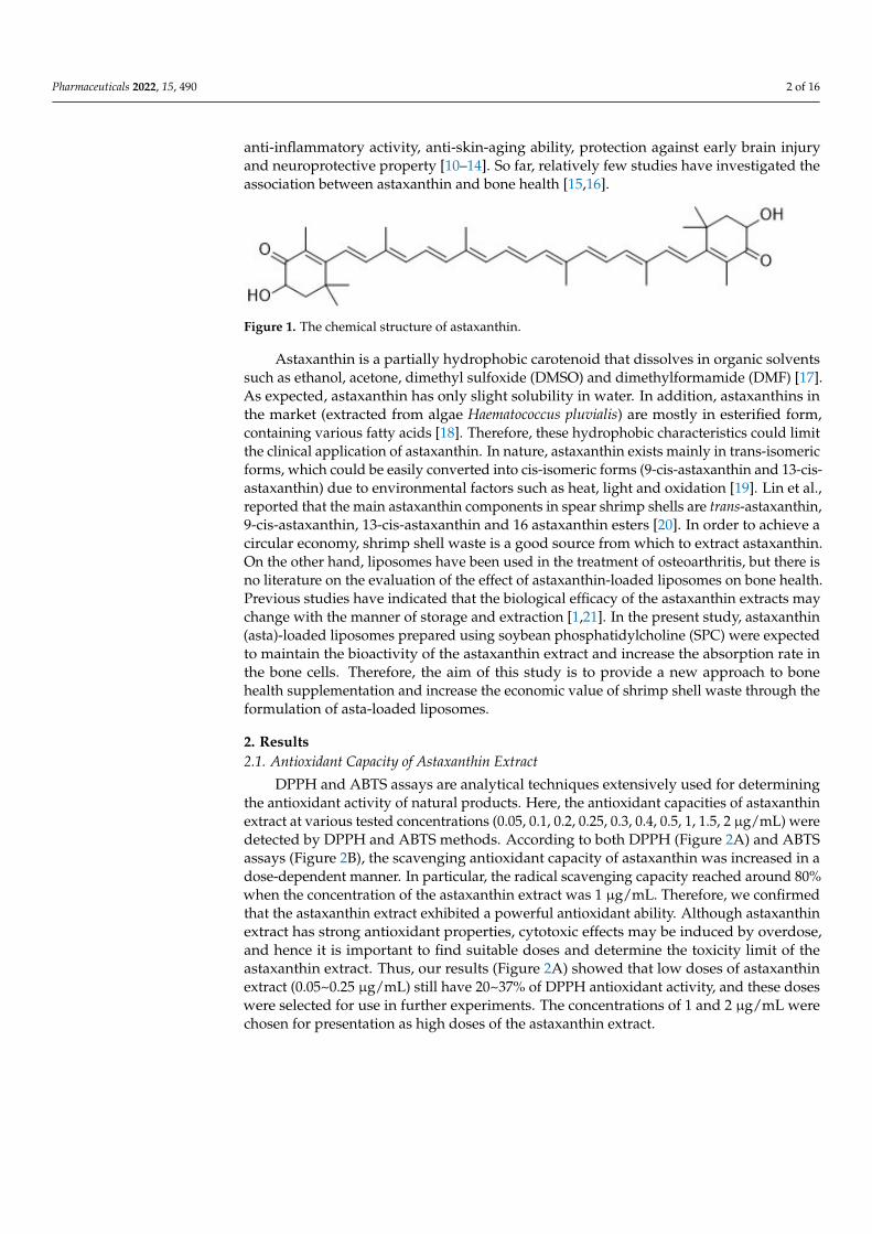
Recently, astaxanthin (3,3′-dihydroxy-β,β-carotene-4,4′dione, Figure 1) has been widelyapplied as a human nutraceutical supplement for health care or an essential ingredient inthe food and cosmetic industry. Astaxanthin is a lipid-soluble reddish pigment belongingto the xanthophyll group of carotenoids, produced by microalgae under pressure suchas strong light, high salinity and low nutrient utilization for protection [1]. Astaxanthincould accumulate in the tissues of marine organisms feeding on microalgae. Therefore,astaxanthin is commonly found in green microalgae Haematococcus pluvialis; red yeastXanthophyllomyces dendrorhous (Phaffia rhodozyma); and marine animals such as shrimp,krill, salmon, crab and lobster. According to numerous studies, astaxanthin showed betterbiological potential than other antioxidants such as lutein, lycopene, β-carotene and vita-min C [2,3]. Unlike other types of carotenoids, astaxanthin has two keto groups locatedat the 4,4′ position of the β-ionone ring that activate adjacent hydroxyl groups for captur-ing per-oxidants and stabilizing the trapped radicals [4]. Through its antioxidant ability,astaxanthin can suppress cancer cell proliferation, migration or invasion; prevent cardio-vascular diseases and diabetes; and promote immune system and ocular health [2,5–9]. Inaddition, astaxanthin has been reported to have several other health benefits, including
Pharmaceuticals 2022, 15, 490. https://doi.org/10.3390/ph15040490 https://www.mdpi.com/journal/pharmaceuticals
Pharmaceuticals 2022, 15, 490 2 of 16
anti-inflammatory activity, anti-skin-aging ability, protection against early brain injuryand neuroprotective property [10–14]. So far, relatively few studies have investigated theassociation between astaxanthin and bone health [15,16].
Pharmaceuticals 2022, 15, x FOR PEER REVIEW 2 of 16
antioxidant ability, astaxanthin can suppress cancer cell proliferation, migration or inva-
sion; prevent cardiovascular diseases and diabetes; and promote immune system and oc-
ular health [2,5–9]. In addition, astaxanthin has been reported to have several other health
benefits, including anti-inflammatory activity, anti-skin-aging ability, protection against
early brain injury and neuroprotective property [10–14]. So far, relatively few studies have
investigated the association between astaxanthin and bone health [15,16].
Figure 1. The chemical structure of astaxanthin.
Astaxanthin is a partially hydrophobic carotenoid that dissolves in organic solvents
such as ethanol, acetone, dimethyl sulfoxide (DMSO) and dimethylformamide (DMF)
[17]. As expected, astaxanthin has only slight solubility in water. In addition, astaxanthins
in the market (extracted from algae Haematococcus pluvialis) are mostly in esterified form,
containing various fatty acids [18]. Therefore, these hydrophobic characteristics could
limit the clinical application of astaxanthin. In nature, astaxanthin exists mainly in trans-
isomeric forms, which could be easily converted into cis-isomeric forms (9-cis-astaxanthin
and 13-cis-astaxanthin) due to environmental factors such as heat, light and oxidation [19].
Lin et al., reported that the main astaxanthin components in spear shrimp shells are trans-
astaxanthin, 9-cis-astaxanthin, 13-cis-astaxanthin and 16 astaxanthin esters [20]. In order
to achieve a circular economy, shrimp shell waste is a good source from which to extract
astaxanthin. On the other hand, liposomes have been used in the treatment of osteoarthri-
tis, but there is no literature on the evaluation of the effect of astaxanthin-loaded lipo-
somes on bone health. Previous studies have indicated that the biological efficacy of the
astaxanthin extracts may change with the manner of storage and extraction [1,21]. In the
present study, astaxanthin (asta)-loaded liposomes prepared using soybean phosphati-
dylcholine (SPC) were expected to maintain the bioactivity of the astaxanthin extract and
increase the absorption rate in the bone cells. Therefore, the aim of this study is to provide
a new approach to bone health supplementation and increase the economic value of
shrimp shell waste through the formulation of asta-loaded liposomes.
2. Results
2.1. Antioxidant Capacity of Astaxanthin Extract
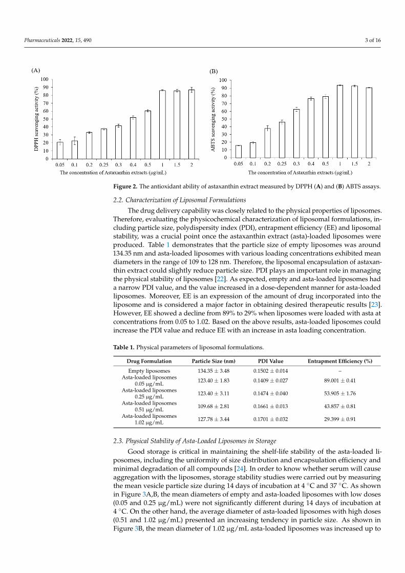
DPPH and ABTS assays are analytical techniques extensively used for determining
the antioxidant activity of natural products. Here, the antioxidant capacities of astaxan-
thin extract at various tested concentrations (0.05, 0.1, 0.2, 0.25, 0.3, 0.4, 0.5, 1, 1.5, 2 μg/mL)
were detected by DPPH and ABTS methods. According to both DPPH (Figure 2A) and
ABTS assays (Figure 2B), the scavenging antioxidant capacity of astaxanthin was in-
creased in a dose-dependent manner. In particular, the radical scavenging capacity
reached around 80% when the concentration of the astaxanthin extract was 1 μg/mL.
Therefore, we confirmed that the astaxanthin extract exhibited a powerful antioxidant
ability. Although astaxanthin extract has strong antioxidant properties, cytotoxic effects
may be induced by overdose, and hence it is important to find suitable doses and deter-
mine the toxicity limit of the astaxanthin extract. Thus, our results (Figure 2A) showed
that low doses of astaxanthin extract (0.05~0.25 μg/mL) still have 20~37% of DPPH anti-
oxidant activity, and these doses were selected for use in further experiments. The con-
centrations of 1 and 2 μg/mL were chosen for presentation as high doses of the astaxanthin
extract.
Figure 1. The chemical structure of astaxanthin.
Astaxanthin is a partially hydrophobic carotenoid that dissolves in organic solventssuch as ethanol, acetone, dimethyl sulfoxide (DMSO) and dimethylformamide (DMF) [17].As expected, astaxanthin has only slight solubility in water. In addition, astaxanthins inthe market (extracted from algae Haematococcus pluvialis) are mostly in esterified form,containing various fatty acids [18]. Therefore, these hydrophobic characteristics could limitthe clinical application of astaxanthin. In nature, astaxanthin exists mainly in trans-isomericforms, which could be easily converted into cis-isomeric forms (9-cis-astaxanthin and 13-cis-astaxanthin) due to environmental factors such as heat, light and oxidation [19]. Lin et al.,reported that the main astaxanthin components in spear shrimp shells are trans-astaxanthin,9-cis-astaxanthin, 13-cis-astaxanthin and 16 astaxanthin esters [20]. In order to achieve acircular economy, shrimp shell waste is a good source from which to extract astaxanthin.On the other hand, liposomes have been used in the treatment of osteoarthritis, but there isno literature on the evaluation of the effect of astaxanthin-loaded liposomes on bone health.Previous studies have indicated that the biological efficacy of the astaxanthin extracts maychange with the manner of storage and extraction [1,21]. In the present study, astaxanthin(asta)-loaded liposomes prepared using soybean phosphatidylcholine (SPC) were expectedto maintain the bioactivity of the astaxanthin extract and increase the absorption rate inthe bone cells. Therefore, the aim of this study is to provide a new approach to bonehealth supplementation and increase the economic value of shrimp shell waste through theformulation of asta-loaded liposomes.
2. Results2.1. Antioxidant Capacity of Astaxanthin Extract
DPPH and ABTS assays are analytical techniques extensively used for determiningthe antioxidant activity of natural products. Here, the antioxidant capacities of astaxanthinextract at various tested concentrations (0.05, 0.1, 0.2, 0.25, 0.3, 0.4, 0.5, 1, 1.5, 2 µg/mL) weredetected by DPPH and ABTS methods. According to both DPPH (Figure 2A) and ABTSassays (Figure 2B), the scavenging antioxidant capacity of astaxanthin was increased in adose-dependent manner. In particular, the radical scavenging capacity reached around 80%when the concentration of the astaxanthin extract was 1 µg/mL. Therefore, we confirmedthat the astaxanthin extract exhibited a powerful antioxidant ability. Although astaxanthinextract has strong antioxidant properties, cytotoxic effects may be induced by overdose,and hence it is important to find suitable doses and determine the toxicity limit of theastaxanthin extract. Thus, our results (Figure 2A) showed that low doses of astaxanthinextract (0.05~0.25 µg/mL) still have 20~37% of DPPH antioxidant activity, and these doseswere selected for use in further experiments. The concentrations of 1 and 2 µg/mL werechosen for presentation as high doses of the astaxanthin extract.
Pharmaceuticals 2022, 15, 490 3 of 16Pharmaceuticals 2022, 15, x FOR PEER REVIEW 3 of 16
Figure 2. The antioxidant ability of astaxanthin extract measured by DPPH (A) and (B) ABTS assays.
2.2. Characterization of Liposomal Formulations
The drug delivery capability was closely related to the physical properties of lipo-
somes. Therefore, evaluating the physicochemical characterization of liposomal formula-
tions, including particle size, polydispersity index (PDI), entrapment efficiency (EE) and
liposomal stability, was a crucial point once the astaxanthin extract (asta)-loaded lipo-
somes were produced. Table 1 demonstrates that the particle size of empty liposomes was
around 134.35 nm and asta-loaded liposomes with various loading concentrations exhib-
ited mean diameters in the range of 109 to 128 nm. Therefore, the liposomal encapsulation
of astaxanthin extract could slightly reduce particle size. PDI plays an important role in
managing the physical stability of liposomes [22]. As expected, empty and asta-loaded
liposomes had a narrow PDI value, and the value increased in a dose-dependent manner
for asta-loaded liposomes. Moreover, EE is an expression of the amount of drug incorpo-
rated into the liposome and is considered a major factor in obtaining desired therapeutic
results [23]. However, EE showed a decline from 89% to 29% when liposomes were loaded
with asta at concentrations from 0.05 to 1.02. Based on the above results, asta-loaded lip-
osomes could increase the PDI value and reduce EE with an increase in asta loading con-
centration.
Table 1. Physical parameters of liposomal formulations.
Drug Formulation Particle Size (nm) PDI Value Entrapment
Efficiency (%)
Empty liposomes 134.35 ± 3.48 0.1502 ± 0.014 --
Asta-loaded liposomes
0.05 μg/mL 123.40 ± 1.83 0.1409 ± 0.027 89.001 ± 0.41
Asta-loaded liposomes
0.25 μg/mL 123.40 ± 3.11 0.1474 ± 0.040 53.905 ± 1.76
Asta-loaded liposomes
0.51 μg/mL 109.68 ± 2.81 0.1661 ± 0.013 43.857 ± 0.81
Asta-loaded liposomes
1.02 μg/mL 127.78 ± 3.44 0.1701 ± 0.032 29.399 ± 0.91
2.3. Physical Stability of Asta-Loaded Liposomes in Storage
Good storage is critical in maintaining the shelf-life stability of the asta-loaded lipo-
somes, including the uniformity of size distribution and encapsulation efficiency and min-
imal degradation of all compounds [24]. In order to know whether serum will cause ag-
gregation with the liposomes, storage stability studies were carried out by measuring the
mean vesicle particle size during 14 days of incubation at 4 °C and 37 °C. As shown in
Figure 2. The antioxidant ability of astaxanthin extract measured by DPPH (A) and (B) ABTS assays.
2.2. Characterization of Liposomal Formulations
The drug delivery capability was closely related to the physical properties of liposomes.Therefore, evaluating the physicochemical characterization of liposomal formulations, in-cluding particle size, polydispersity index (PDI), entrapment efficiency (EE) and liposomalstability, was a crucial point once the astaxanthin extract (asta)-loaded liposomes wereproduced. Table 1 demonstrates that the particle size of empty liposomes was around134.35 nm and asta-loaded liposomes with various loading concentrations exhibited meandiameters in the range of 109 to 128 nm. Therefore, the liposomal encapsulation of astaxan-thin extract could slightly reduce particle size. PDI plays an important role in managingthe physical stability of liposomes [22]. As expected, empty and asta-loaded liposomes hada narrow PDI value, and the value increased in a dose-dependent manner for asta-loadedliposomes. Moreover, EE is an expression of the amount of drug incorporated into theliposome and is considered a major factor in obtaining desired therapeutic results [23].However, EE showed a decline from 89% to 29% when liposomes were loaded with asta atconcentrations from 0.05 to 1.02. Based on the above results, asta-loaded liposomes couldincrease the PDI value and reduce EE with an increase in asta loading concentration.
Table 1. Physical parameters of liposomal formulations.
Drug Formulation Particle Size (nm) PDI Value Entrapment Efficiency (%)
Empty liposomes 134.35 ± 3.48 0.1502 ± 0.014 –Asta-loaded liposomes
0.05 µg/mL 123.40 ± 1.83 0.1409 ± 0.027 89.001 ± 0.41
Asta-loaded liposomes0.25 µg/mL 123.40 ± 3.11 0.1474 ± 0.040 53.905 ± 1.76
Asta-loaded liposomes0.51 µg/mL 109.68 ± 2.81 0.1661 ± 0.013 43.857 ± 0.81
Asta-loaded liposomes1.02 µg/mL 127.78 ± 3.44 0.1701 ± 0.032 29.399 ± 0.91
2.3. Physical Stability of Asta-Loaded Liposomes in Storage
Good storage is critical in maintaining the shelf-life stability of the asta-loaded li-posomes, including the uniformity of size distribution and encapsulation efficiency andminimal degradation of all compounds [24]. In order to know whether serum will causeaggregation with the liposomes, storage stability studies were carried out by measuringthe mean vesicle particle size during 14 days of incubation at 4 ◦C and 37 ◦C. As shownin Figure 3A,B, the mean diameters of empty and asta-loaded liposomes with low doses(0.05 and 0.25 µg/mL) were not significantly different during 14 days of incubation at4 ◦C. On the other hand, the average diameter of asta-loaded liposomes with high doses(0.51 and 1.02 µg/mL) presented an increasing tendency in particle size. As shown inFigure 3B, the mean diameter of 1.02 µg/mL asta-loaded liposomes was increased up to
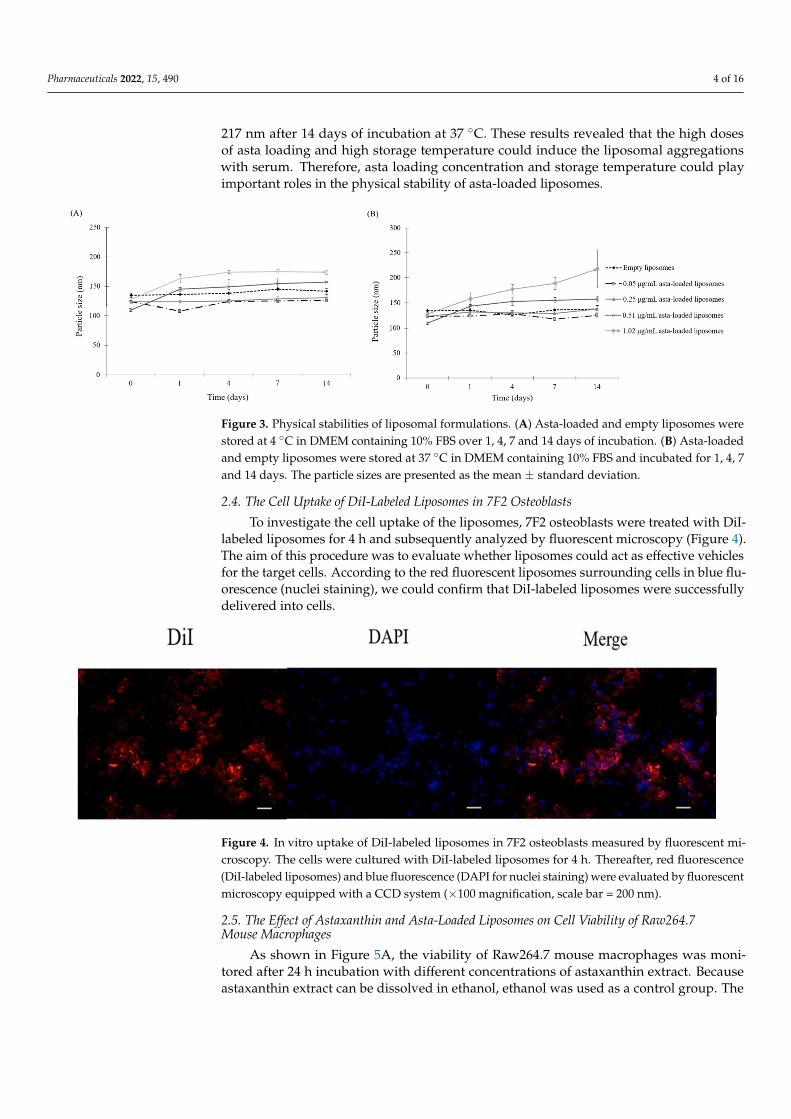
Pharmaceuticals 2022, 15, 490 4 of 16
217 nm after 14 days of incubation at 37 ◦C. These results revealed that the high dosesof asta loading and high storage temperature could induce the liposomal aggregationswith serum. Therefore, asta loading concentration and storage temperature could playimportant roles in the physical stability of asta-loaded liposomes.
Pharmaceuticals 2022, 15, x FOR PEER REVIEW 4 of 16
Figure 3A,B, the mean diameters of empty and asta-loaded liposomes with low doses (0.05
and 0.25 μg/mL) were not significantly different during 14 days of incubation at 4 °C. On
the other hand, the average diameter of asta-loaded liposomes with high doses (0.51 and
1.02 μg/mL) presented an increasing tendency in particle size. As shown in Figure 3B, the
mean diameter of 1.02 μg/mL asta-loaded liposomes was increased up to 217 nm after 14
days of incubation at 37 °C. These results revealed that the high doses of asta loading and
high storage temperature could induce the liposomal aggregations with serum. Therefore,
asta loading concentration and storage temperature could play important roles in the
physical stability of asta-loaded liposomes.
Figure 3. Physical stabilities of liposomal formulations. (A) Asta-loaded and empty liposomes were
stored at 4 °C in DMEM containing 10% FBS over 1, 4, 7 and 14 days of incubation. (B) Asta-loaded
and empty liposomes were stored at 37 °C in DMEM containing 10% FBS and incubated for 1, 4, 7
and 14 days. The particle sizes are presented as the mean ± standard deviation.
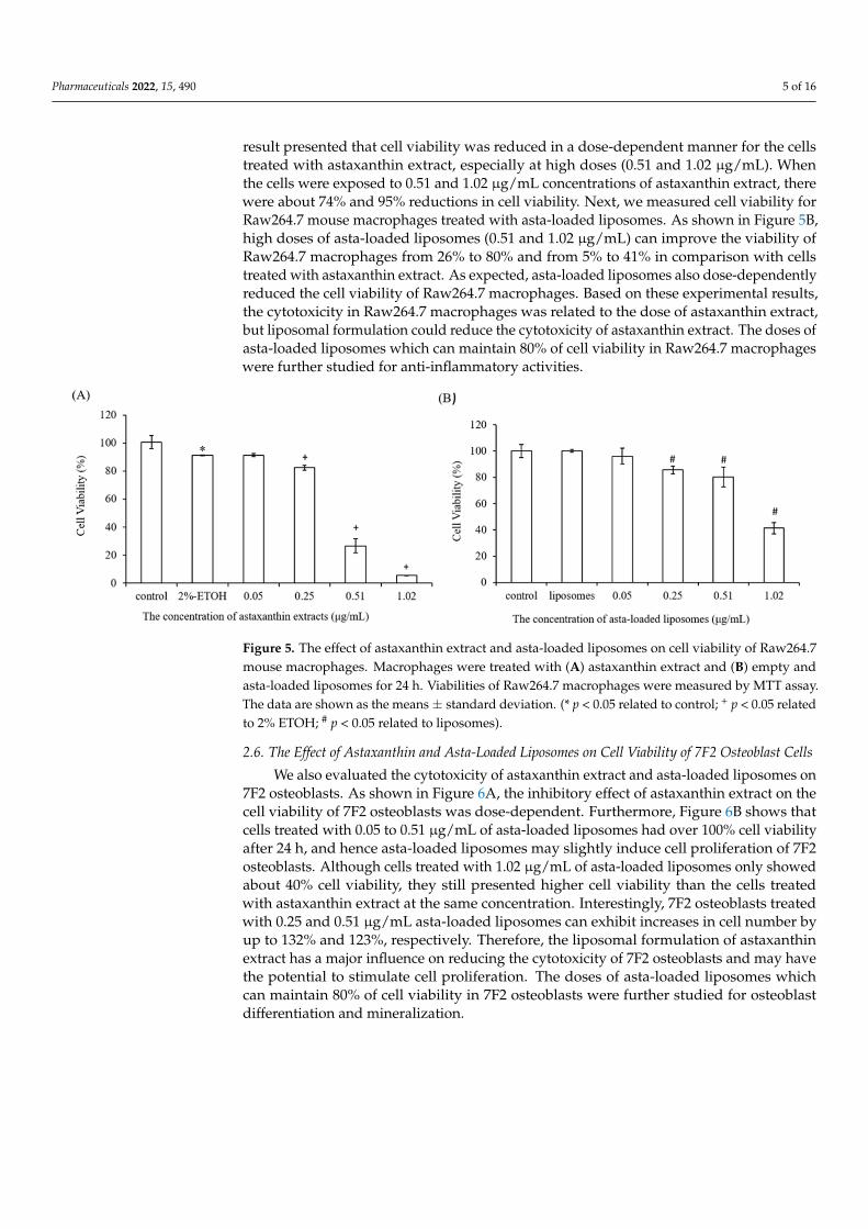
2.4. The Cell Uptake of DiI-Labeled Liposomes in 7F2 Osteoblasts
To investigate the cell uptake of the liposomes, 7F2 osteoblasts were treated with DiI-
labeled liposomes for 4 h and subsequently analyzed by fluorescent microscopy (Figure
4). The aim of this procedure was to evaluate whether liposomes could act as effective
vehicles for the target cells. According to the red fluorescent liposomes surrounding cells
in blue fluorescence (nuclei staining), we could confirm that DiI-labeled liposomes were
successfully delivered into cells.
Figure 4. In vitro uptake of DiI-labeled liposomes in 7F2 osteoblasts measured by fluorescent mi-
croscopy. The cells were cultured with DiI-labeled liposomes for 4 h. Thereafter, red fluorescence
(DiI-labeled liposomes) and blue fluorescence (DAPI for nuclei staining) were evaluated by fluores-
cent microscopy equipped with a CCD system (×100 magnification, scale bar = 200 nm).
Figure 3. Physical stabilities of liposomal formulations. (A) Asta-loaded and empty liposomes werestored at 4 ◦C in DMEM containing 10% FBS over 1, 4, 7 and 14 days of incubation. (B) Asta-loadedand empty liposomes were stored at 37 ◦C in DMEM containing 10% FBS and incubated for 1, 4, 7and 14 days. The particle sizes are presented as the mean ± standard deviation.
2.4. The Cell Uptake of DiI-Labeled Liposomes in 7F2 Osteoblasts
To investigate the cell uptake of the liposomes, 7F2 osteoblasts were treated with DiI-labeled liposomes for 4 h and subsequently analyzed by fluorescent microscopy (Figure 4).The aim of this procedure was to evaluate whether liposomes could act as effective vehiclesfor the target cells. According to the red fluorescent liposomes surrounding cells in blue flu-orescence (nuclei staining), we could confirm that DiI-labeled liposomes were successfullydelivered into cells.
Pharmaceuticals 2022, 15, x FOR PEER REVIEW 4 of 16
Figure 3A,B, the mean diameters of empty and asta-loaded liposomes with low doses (0.05
and 0.25 μg/mL) were not significantly different during 14 days of incubation at 4 °C. On
the other hand, the average diameter of asta-loaded liposomes with high doses (0.51 and
1.02 μg/mL) presented an increasing tendency in particle size. As shown in Figure 3B, the
mean diameter of 1.02 μg/mL asta-loaded liposomes was increased up to 217 nm after 14
days of incubation at 37 °C. These results revealed that the high doses of asta loading and
high storage temperature could induce the liposomal aggregations with serum. Therefore,
asta loading concentration and storage temperature could play important roles in the
physical stability of asta-loaded liposomes.
Figure 3. Physical stabilities of liposomal formulations. (A) Asta-loaded and empty liposomes were
stored at 4 °C in DMEM containing 10% FBS over 1, 4, 7 and 14 days of incubation. (B) Asta-loaded
and empty liposomes were stored at 37 °C in DMEM containing 10% FBS and incubated for 1, 4, 7
and 14 days. The particle sizes are presented as the mean ± standard deviation.
2.4. The Cell Uptake of DiI-Labeled Liposomes in 7F2 Osteoblasts
To investigate the cell uptake of the liposomes, 7F2 osteoblasts were treated with DiI-
labeled liposomes for 4 h and subsequently analyzed by fluorescent microscopy (Figure
4). The aim of this procedure was to evaluate whether liposomes could act as effective
vehicles for the target cells. According to the red fluorescent liposomes surrounding cells
in blue fluorescence (nuclei staining), we could confirm that DiI-labeled liposomes were
successfully delivered into cells.
Figure 4. In vitro uptake of DiI-labeled liposomes in 7F2 osteoblasts measured by fluorescent mi-
croscopy. The cells were cultured with DiI-labeled liposomes for 4 h. Thereafter, red fluorescence
(DiI-labeled liposomes) and blue fluorescence (DAPI for nuclei staining) were evaluated by fluores-
cent microscopy equipped with a CCD system (×100 magnification, scale bar = 200 nm).
Figure 4. In vitro uptake of DiI-labeled liposomes in 7F2 osteoblasts measured by fluorescent mi-croscopy. The cells were cultured with DiI-labeled liposomes for 4 h. Thereafter, red fluorescence(DiI-labeled liposomes) and blue fluorescence (DAPI for nuclei staining) were evaluated by fluorescentmicroscopy equipped with a CCD system (×100 magnification, scale bar = 200 nm).
2.5. The Effect of Astaxanthin and Asta-Loaded Liposomes on Cell Viability of Raw264.7Mouse Macrophages
As shown in Figure 5A, the viability of Raw264.7 mouse macrophages was moni-tored after 24 h incubation with different concentrations of astaxanthin extract. Becauseastaxanthin extract can be dissolved in ethanol, ethanol was used as a control group. The
Pharmaceuticals 2022, 15, 490 5 of 16
result presented that cell viability was reduced in a dose-dependent manner for the cellstreated with astaxanthin extract, especially at high doses (0.51 and 1.02 µg/mL). Whenthe cells were exposed to 0.51 and 1.02 µg/mL concentrations of astaxanthin extract, therewere about 74% and 95% reductions in cell viability. Next, we measured cell viability forRaw264.7 mouse macrophages treated with asta-loaded liposomes. As shown in Figure 5B,high doses of asta-loaded liposomes (0.51 and 1.02 µg/mL) can improve the viability ofRaw264.7 macrophages from 26% to 80% and from 5% to 41% in comparison with cellstreated with astaxanthin extract. As expected, asta-loaded liposomes also dose-dependentlyreduced the cell viability of Raw264.7 macrophages. Based on these experimental results,the cytotoxicity in Raw264.7 macrophages was related to the dose of astaxanthin extract,but liposomal formulation could reduce the cytotoxicity of astaxanthin extract. The doses ofasta-loaded liposomes which can maintain 80% of cell viability in Raw264.7 macrophageswere further studied for anti-inflammatory activities.
Pharmaceuticals 2022, 15, x FOR PEER REVIEW 5 of 16
2.5. The Effect of Astaxanthin and Asta-Loaded Liposomes on Cell Viability of Raw264.7
Mouse Macrophages
As shown in Figure 5A, the viability of Raw264.7 mouse macrophages was monitored
after 24 h incubation with different concentrations of astaxanthin extract. Because astaxan-
thin extract can be dissolved in ethanol, ethanol was used as a control group. The result
presented that cell viability was reduced in a dose-dependent manner for the cells treated
with astaxanthin extract, especially at high doses (0.51 and 1.02 μg/mL). When the cells
were exposed to 0.51 and 1.02 μg/mL concentrations of astaxanthin extract, there were
about 74% and 95% reductions in cell viability. Next, we measured cell viability for
Raw264.7 mouse macrophages treated with asta-loaded liposomes. As shown in Figure
5B, high doses of asta-loaded liposomes (0.51 and 1.02 μg/mL) can improve the viability
of Raw264.7 macrophages from 26% to 80% and from 5% to 41% in comparison with cells
treated with astaxanthin extract. As expected, asta-loaded liposomes also dose-de-
pendently reduced the cell viability of Raw264.7 macrophages. Based on these experi-
mental results, the cytotoxicity in Raw264.7 macrophages was related to the dose of
astaxanthin extract, but liposomal formulation could reduce the cytotoxicity of astaxan-
thin extract. The doses of asta-loaded liposomes which can maintain 80% of cell viability
in Raw264.7 macrophages were further studied for anti-inflammatory activities.
Figure 5. The effect of astaxanthin extract and asta-loaded liposomes on cell viability of Raw264.7
mouse macrophages. Macrophages were treated with (A) astaxanthin extract and (B) empty and
asta-loaded liposomes for 24 h. Viabilities of Raw264.7 macrophages were measured by MTT assay.
The data are shown as the means ± standard deviation. (* p < 0.05 related to control; + p < 0.05 related
to 2% ETOH; # p < 0.05 related to liposomes).
2.6. The Effect of Astaxanthin and Asta-Loaded Liposomes on Cell Viability of 7F2 Osteoblast
Cells
We also evaluated the cytotoxicity of astaxanthin extract and asta-loaded liposomes
on 7F2 osteoblasts. As shown in Figure 6A, the inhibitory effect of astaxanthin extract on
the cell viability of 7F2 osteoblasts was dose-dependent. Furthermore, Figure 6B shows
that cells treated with 0.05 to 0.51 μg/mL of asta-loaded liposomes had over 100% cell
viability after 24 h, and hence asta-loaded liposomes may slightly induce cell proliferation
of 7F2 osteoblasts. Although cells treated with 1.02 μg/mL of asta-loaded liposomes only
showed about 40% cell viability, they still presented higher cell viability than the cells
treated with astaxanthin extract at the same concentration. Interestingly, 7F2 osteoblasts
treated with 0.25 and 0.51 μg/mL asta-loaded liposomes can exhibit increases in cell num-
ber by up to 132% and 123%, respectively. Therefore, the liposomal formulation of
astaxanthin extract has a major influence on reducing the cytotoxicity of 7F2 osteoblasts
Figure 5. The effect of astaxanthin extract and asta-loaded liposomes on cell viability of Raw264.7mouse macrophages. Macrophages were treated with (A) astaxanthin extract and (B) empty andasta-loaded liposomes for 24 h. Viabilities of Raw264.7 macrophages were measured by MTT assay.The data are shown as the means ± standard deviation. (* p < 0.05 related to control; + p < 0.05 relatedto 2% ETOH; # p < 0.05 related to liposomes).
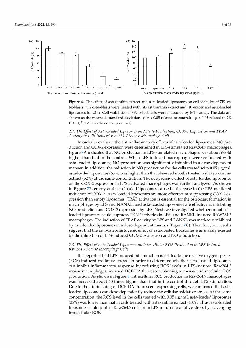
2.6. The Effect of Astaxanthin and Asta-Loaded Liposomes on Cell Viability of 7F2 Osteoblast Cells
We also evaluated the cytotoxicity of astaxanthin extract and asta-loaded liposomes on7F2 osteoblasts. As shown in Figure 6A, the inhibitory effect of astaxanthin extract on thecell viability of 7F2 osteoblasts was dose-dependent. Furthermore, Figure 6B shows thatcells treated with 0.05 to 0.51 µg/mL of asta-loaded liposomes had over 100% cell viabilityafter 24 h, and hence asta-loaded liposomes may slightly induce cell proliferation of 7F2osteoblasts. Although cells treated with 1.02 µg/mL of asta-loaded liposomes only showedabout 40% cell viability, they still presented higher cell viability than the cells treatedwith astaxanthin extract at the same concentration. Interestingly, 7F2 osteoblasts treatedwith 0.25 and 0.51 µg/mL asta-loaded liposomes can exhibit increases in cell number byup to 132% and 123%, respectively. Therefore, the liposomal formulation of astaxanthinextract has a major influence on reducing the cytotoxicity of 7F2 osteoblasts and may havethe potential to stimulate cell proliferation. The doses of asta-loaded liposomes whichcan maintain 80% of cell viability in 7F2 osteoblasts were further studied for osteoblastdifferentiation and mineralization.
Pharmaceuticals 2022, 15, 490 6 of 16
Pharmaceuticals 2022, 15, x FOR PEER REVIEW 6 of 16
and may have the potential to stimulate cell proliferation. The doses of asta-loaded lipo-
somes which can maintain 80% of cell viability in 7F2 osteoblasts were further studied for
osteoblast differentiation and mineralization.
Figure 6. The effect of astaxanthin extract and asta-loaded liposomes on cell viability of 7F2 osteo-
blasts. 7F2 osteoblasts were treated with (A) astaxanthin extract and (B) empty and asta-loaded lip-
osomes for 24 h. Cell viabilities of 7F2 osteoblasts were measured by MTT assay. The data are shown
as the means ± standard deviation. (* p < 0.05 related to control; + p < 0.05 related to 2% ETOH; # p <
0.05 related to liposomes).
2.7. The Effect of Asta-Loaded Liposomes on Nitrite Production, COX-2 Expression and TRAP
Activity in LPS-Induced Raw264.7 Mouse Macrophage Cells
In order to evaluate the anti-inflammatory effects of asta-loaded liposomes, NO pro-
duction and COX-2 expression were determined in LPS-stimulated Raw264.7 macro-
phages. Figure 7A indicated that NO production in LPS-stimulated macrophages was
about 9-fold higher than that in the control. When LPS-induced macrophages were co-
treated with asta-loaded liposomes, NO production was significantly inhibited in a dose-
dependent manner. In addition, the reduction in NO production for the cells treated with
0.05 μg/mL asta-loaded liposomes (63%) was higher than that observed in cells treated
with astaxanthin extract (52%) at the same concentration. The suppressive effect of asta-
loaded liposomes on the COX-2 expression in LPS-activated macrophages was further an-
alyzed. As shown in Figure 7B, empty and asta-loaded liposomes caused a decrease in the
LPS-mediated induction of COX-2. Asta-loaded liposomes are more effective at suppress-
ing COX-2 expression than empty liposomes. TRAP activation is essential for the osteo-
clast formation in macrophages by LPS and NANKL, and asta-loaded liposomes are ef-
fective at inhibiting NO production and COX-2 expression by LPS. Next, we investigated
whether or not asta-loaded liposomes could suppress TRAP activities in LPS- and
RANKL-induced RAW264.7 macrophages. The induction of TRAP activity by LPS and
RANKL was markedly inhibited by asta-loaded liposomes in a dose-dependent manner
(Figure 7C). Therefore, our results suggest that the anti-osteoclastogenic effect of asta-
loaded liposomes was mainly exerted by the inhibition of LPS-induced COX-2 expression
and NO production.
Figure 6. The effect of astaxanthin extract and asta-loaded liposomes on cell viability of 7F2 os-teoblasts. 7F2 osteoblasts were treated with (A) astaxanthin extract and (B) empty and asta-loadedliposomes for 24 h. Cell viabilities of 7F2 osteoblasts were measured by MTT assay. The data areshown as the means ± standard deviation. (* p < 0.05 related to control; + p < 0.05 related to 2%ETOH; # p < 0.05 related to liposomes).
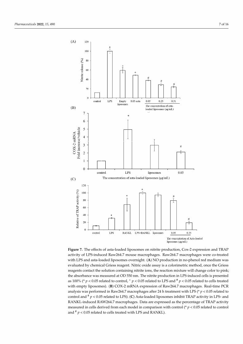
2.7. The Effect of Asta-Loaded Liposomes on Nitrite Production, COX-2 Expression and TRAPActivity in LPS-Induced Raw264.7 Mouse Macrophage Cells
In order to evaluate the anti-inflammatory effects of asta-loaded liposomes, NO pro-duction and COX-2 expression were determined in LPS-stimulated Raw264.7 macrophages.Figure 7A indicated that NO production in LPS-stimulated macrophages was about 9-foldhigher than that in the control. When LPS-induced macrophages were co-treated withasta-loaded liposomes, NO production was significantly inhibited in a dose-dependentmanner. In addition, the reduction in NO production for the cells treated with 0.05 µg/mLasta-loaded liposomes (63%) was higher than that observed in cells treated with astaxanthinextract (52%) at the same concentration. The suppressive effect of asta-loaded liposomeson the COX-2 expression in LPS-activated macrophages was further analyzed. As shownin Figure 7B, empty and asta-loaded liposomes caused a decrease in the LPS-mediatedinduction of COX-2. Asta-loaded liposomes are more effective at suppressing COX-2 ex-pression than empty liposomes. TRAP activation is essential for the osteoclast formation inmacrophages by LPS and NANKL, and asta-loaded liposomes are effective at inhibitingNO production and COX-2 expression by LPS. Next, we investigated whether or not asta-loaded liposomes could suppress TRAP activities in LPS- and RANKL-induced RAW264.7macrophages. The induction of TRAP activity by LPS and RANKL was markedly inhibitedby asta-loaded liposomes in a dose-dependent manner (Figure 7C). Therefore, our resultssuggest that the anti-osteoclastogenic effect of asta-loaded liposomes was mainly exertedby the inhibition of LPS-induced COX-2 expression and NO production.
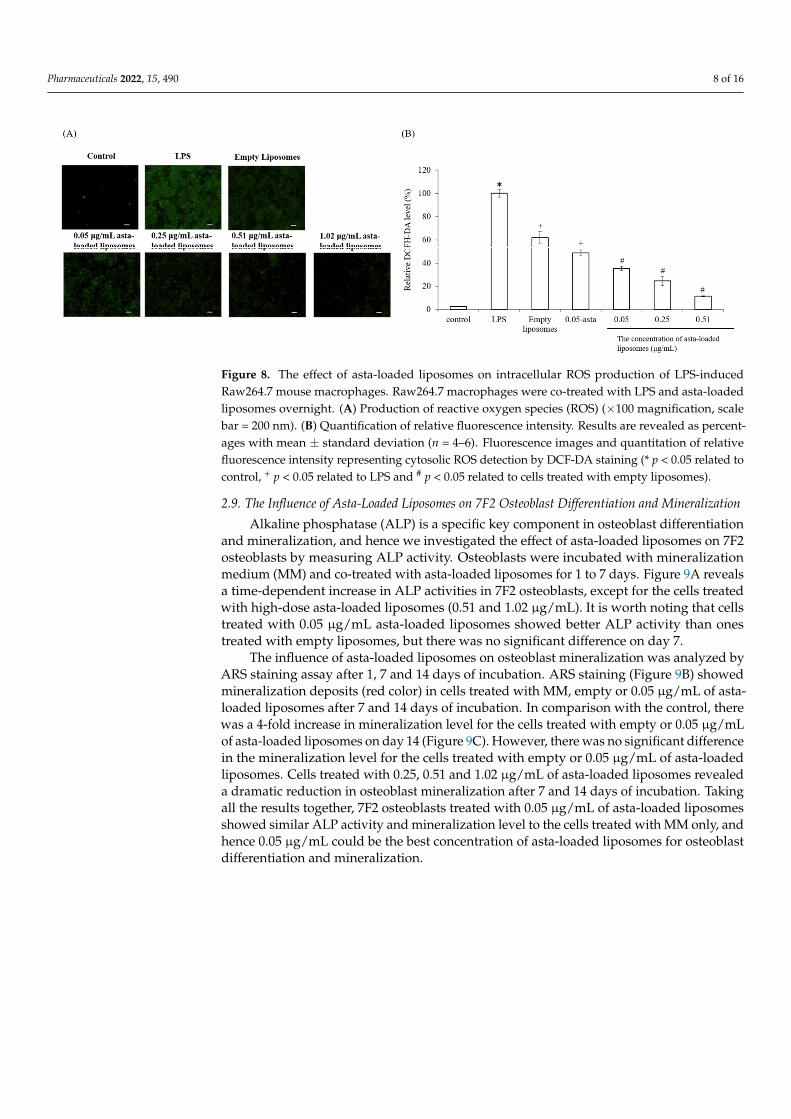
2.8. The Effect of Asta-Loaded Liposomes on Intracellular ROS Production in LPS-InducedRaw264.7 Mouse Macrophage Cells
It is reported that LPS-induced inflammation is related to the reactive oxygen species(ROS)-induced oxidative stress. In order to determine whether asta-loaded liposomescan inhibit inflammatory response by reducing ROS levels in LPS-induced Raw264.7mouse macrophages, we used DCF-DA fluorescent staining to measure intracellular ROSproduction. As shown in Figure 8, intracellular ROS production in Raw264.7 macrophageswas increased about 50 times higher than that in the control through LPS stimulation.Due to the diminishing of DCF-DA fluorescent expressing cells, we confirmed that asta-loaded liposomes can dose-dependently reduce the cellular oxidative stress. At the sameconcentration, the ROS level in the cells treated with 0.05 µg/mL asta-loaded liposomes(35%) was lower than that in cells treated with astaxanthin extract (48%). Thus, asta-loadedliposomes could protect Raw264.7 cells from LPS-induced oxidative stress by scavengingintracellular ROS.
Pharmaceuticals 2022, 15, 490 7 of 16Pharmaceuticals 2022, 15, x FOR PEER REVIEW 7 of 16
Figure 7. The effects of asta-loaded liposomes on nitrite production, Cox-2 expression and TRAP
activity of LPS-induced Raw264.7 mouse macrophages. Raw264.7 macrophages were co-treated
with LPS and asta-loaded liposomes overnight. (A) NO production in no-phenol red medium was
evaluated by chemical Griess reagent. Nitric oxide assay is a colorimetric method, once the Griess
reagents contact the solution containing nitrite ions, the reaction mixture will change color to pink;
the absorbance was measured at OD 550 nm. The nitrite production in LPS-induced cells is pre-
sented as 100% (* p < 0.05 related to control, + p < 0.05 related to LPS and # p < 0.05 related to cells
treated with empty liposomes). (B) COX-2 mRNA expression of Raw264.7 macrophages. Real-time
PCR analysis was performed in Raw264.7 macrophages after 24 h treatment with LPS (* p<0.05 re-
lated to control and # p < 0.05 related to LPS). (C) Asta-loaded liposomes inhibit TRAP activity in
Figure 7. The effects of asta-loaded liposomes on nitrite production, Cox-2 expression and TRAPactivity of LPS-induced Raw264.7 mouse macrophages. Raw264.7 macrophages were co-treatedwith LPS and asta-loaded liposomes overnight. (A) NO production in no-phenol red medium wasevaluated by chemical Griess reagent. Nitric oxide assay is a colorimetric method, once the Griessreagents contact the solution containing nitrite ions, the reaction mixture will change color to pink;the absorbance was measured at OD 550 nm. The nitrite production in LPS-induced cells is presentedas 100% (* p < 0.05 related to control, + p < 0.05 related to LPS and # p < 0.05 related to cells treatedwith empty liposomes). (B) COX-2 mRNA expression of Raw264.7 macrophages. Real-time PCRanalysis was performed in Raw264.7 macrophages after 24 h treatment with LPS (* p < 0.05 related tocontrol and # p < 0.05 related to LPS). (C) Asta-loaded liposomes inhibit TRAP activity in LPS- andRANKL-induced RAW264.7 macrophages. Data are expressed as the percentage of TRAP activitymeasured in cells derived from each model in comparison with control (* p < 0.05 related to controland # p < 0.05 related to cells treated with LPS and RANKL).
Pharmaceuticals 2022, 15, 490 8 of 16
Pharmaceuticals 2022, 15, x FOR PEER REVIEW 8 of 16
LPS- and RANKL-induced RAW264.7 macrophages. Data are expressed as the percentage of TRAP
activity measured in cells derived from each model in comparison with control (* p < 0.05 related to
control and # p < 0.05 related to cells treated with LPS and RANKL).
2.8. The Effect of Asta-Loaded Liposomes on Intracellular ROS Production in LPS-Induced
Raw264.7 Mouse Macrophage Cells
It is reported that LPS-induced inflammation is related to the reactive oxygen species
(ROS)-induced oxidative stress. In order to determine whether asta-loaded liposomes can
inhibit inflammatory response by reducing ROS levels in LPS-induced Raw264.7 mouse
macrophages, we used DCF-DA fluorescent staining to measure intracellular ROS pro-
duction. As shown in Figure 8, intracellular ROS production in Raw264.7 macrophages
was increased about 50 times higher than that in the control through LPS stimulation. Due
to the diminishing of DCF-DA fluorescent expressing cells, we confirmed that asta-loaded
liposomes can dose-dependently reduce the cellular oxidative stress. At the same concen-
tration, the ROS level in the cells treated with 0.05 μg/mL asta-loaded liposomes (35%)
was lower than that in cells treated with astaxanthin extract (48%). Thus, asta-loaded lip-
osomes could protect Raw264.7 cells from LPS-induced oxidative stress by scavenging
intracellular ROS.
Figure 8. The effect of asta-loaded liposomes on intracellular ROS production of LPS-induced
Raw264.7 mouse macrophages. Raw264.7 macrophages were co-treated with LPS and asta-loaded
liposomes overnight. (A) Production of reactive oxygen species (ROS) (×100 magnification, scale
bar = 200 nm). (B) Quantification of relative fluorescence intensity. Results are revealed as percent-
ages with mean ± standard deviation (n = 4–6). Fluorescence images and quantitation of relative
fluorescence intensity representing cytosolic ROS detection by DCF-DA staining (* p < 0.05 related
to control, + p < 0.05 related to LPS and # p < 0.05 related to cells treated with empty liposomes).
2.9. The Influence of Asta-Loaded Liposomes on 7F2 Osteoblast Differentiation
and Mineralization
Alkaline phosphatase (ALP) is a specific key component in osteoblast differentiation
and mineralization, and hence we investigated the effect of asta-loaded liposomes on 7F2
osteoblasts by measuring ALP activity. Osteoblasts were incubated with mineralization
medium (MM) and co-treated with asta-loaded liposomes for 1 to 7 days. Figure 9A re-
veals a time-dependent increase in ALP activities in 7F2 osteoblasts, except for the cells
treated with high-dose asta-loaded liposomes (0.51 and 1.02 μg/mL). It is worth noting
that cells treated with 0.05 μg/mL asta-loaded liposomes showed better ALP activity than
ones treated with empty liposomes, but there was no significant difference on day 7.
The influence of asta-loaded liposomes on osteoblast mineralization was analyzed by
ARS staining assay after 1, 7 and 14 days of incubation. ARS staining (Figure 9B) showed
Figure 8. The effect of asta-loaded liposomes on intracellular ROS production of LPS-inducedRaw264.7 mouse macrophages. Raw264.7 macrophages were co-treated with LPS and asta-loadedliposomes overnight. (A) Production of reactive oxygen species (ROS) (×100 magnification, scalebar = 200 nm). (B) Quantification of relative fluorescence intensity. Results are revealed as percent-ages with mean ± standard deviation (n = 4–6). Fluorescence images and quantitation of relativefluorescence intensity representing cytosolic ROS detection by DCF-DA staining (* p < 0.05 related tocontrol, + p < 0.05 related to LPS and # p < 0.05 related to cells treated with empty liposomes).
2.9. The Influence of Asta-Loaded Liposomes on 7F2 Osteoblast Differentiation and Mineralization
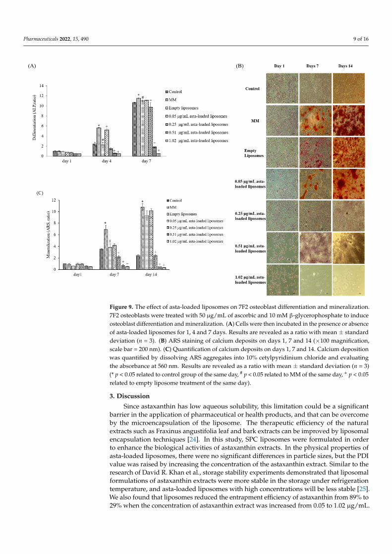
Alkaline phosphatase (ALP) is a specific key component in osteoblast differentiationand mineralization, and hence we investigated the effect of asta-loaded liposomes on 7F2osteoblasts by measuring ALP activity. Osteoblasts were incubated with mineralizationmedium (MM) and co-treated with asta-loaded liposomes for 1 to 7 days. Figure 9A revealsa time-dependent increase in ALP activities in 7F2 osteoblasts, except for the cells treatedwith high-dose asta-loaded liposomes (0.51 and 1.02 µg/mL). It is worth noting that cellstreated with 0.05 µg/mL asta-loaded liposomes showed better ALP activity than onestreated with empty liposomes, but there was no significant difference on day 7.
The influence of asta-loaded liposomes on osteoblast mineralization was analyzed byARS staining assay after 1, 7 and 14 days of incubation. ARS staining (Figure 9B) showedmineralization deposits (red color) in cells treated with MM, empty or 0.05 µg/mL of asta-loaded liposomes after 7 and 14 days of incubation. In comparison with the control, therewas a 4-fold increase in mineralization level for the cells treated with empty or 0.05 µg/mLof asta-loaded liposomes on day 14 (Figure 9C). However, there was no significant differencein the mineralization level for the cells treated with empty or 0.05 µg/mL of asta-loadedliposomes. Cells treated with 0.25, 0.51 and 1.02 µg/mL of asta-loaded liposomes revealeda dramatic reduction in osteoblast mineralization after 7 and 14 days of incubation. Takingall the results together, 7F2 osteoblasts treated with 0.05 µg/mL of asta-loaded liposomesshowed similar ALP activity and mineralization level to the cells treated with MM only, andhence 0.05 µg/mL could be the best concentration of asta-loaded liposomes for osteoblastdifferentiation and mineralization.
Pharmaceuticals 2022, 15, 490 9 of 16
Pharmaceuticals 2022, 15, x FOR PEER REVIEW 9 of 16
mineralization deposits (red color) in cells treated with MM, empty or 0.05 μg/mL of asta-
loaded liposomes after 7 and 14 days of incubation. In comparison with the control, there
was a 4-fold increase in mineralization level for the cells treated with empty or 0.05 μg/mL
of asta-loaded liposomes on day 14 (Figure 9C). However, there was no significant differ-
ence in the mineralization level for the cells treated with empty or 0.05 μg/mL of asta-
loaded liposomes. Cells treated with 0.25, 0.51 and 1.02 μg/mL of asta-loaded liposomes
revealed a dramatic reduction in osteoblast mineralization after 7 and 14 days of incuba-
tion. Taking all the results together, 7F2 osteoblasts treated with 0.05 μg/mL of asta-loaded
liposomes showed similar ALP activity and mineralization level to the cells treated with
MM only, and hence 0.05 μg/mL could be the best concentration of asta-loaded liposomes
for osteoblast differentiation and mineralization.
Figure 9. The effect of asta-loaded liposomes on 7F2 osteoblast differentiation and mineralization.
7F2 osteoblasts were treated with 50 μg/mL of ascorbic and 10 mM β-glycerophosphate to induce
osteoblast differentiation and mineralization. (A) Cells were then incubated in the presence or ab-
sence of asta-loaded liposomes for 1, 4 and 7 days. Results are revealed as a ratio with mean ± stand-
ard deviation (n = 3). (B) ARS staining of calcium deposits on days 1, 7 and 14 (×100 magnification,
scale bar = 200 nm). (C) Quantification of calcium deposits on days 1, 7 and 14. Calcium deposition
was quantified by dissolving ARS aggregates into 10% cetylpyridinium chloride and evaluating the
absorbance at 560 nm. Results are revealed as a ratio with mean ± standard deviation (n = 3) (* p <
0.05 related to control group of the same day, # p < 0.05 related to MM of the same day, + p < 0.05
related to empty liposome treatment of the same day).
Figure 9. The effect of asta-loaded liposomes on 7F2 osteoblast differentiation and mineralization.7F2 osteoblasts were treated with 50 µg/mL of ascorbic and 10 mM β-glycerophosphate to induceosteoblast differentiation and mineralization. (A) Cells were then incubated in the presence or absenceof asta-loaded liposomes for 1, 4 and 7 days. Results are revealed as a ratio with mean ± standarddeviation (n = 3). (B) ARS staining of calcium deposits on days 1, 7 and 14 (×100 magnification,scale bar = 200 nm). (C) Quantification of calcium deposits on days 1, 7 and 14. Calcium depositionwas quantified by dissolving ARS aggregates into 10% cetylpyridinium chloride and evaluatingthe absorbance at 560 nm. Results are revealed as a ratio with mean ± standard deviation (n = 3)(* p < 0.05 related to control group of the same day, # p < 0.05 related to MM of the same day, + p < 0.05related to empty liposome treatment of the same day).
3. Discussion
Since astaxanthin has low aqueous solubility, this limitation could be a significantbarrier in the application of pharmaceutical or health products, and that can be overcomeby the microencapsulation of the liposome. The therapeutic efficiency of the naturalextracts such as Fraxinus angustifolia leaf and bark extracts can be improved by liposomalencapsulation techniques [24]. In this study, SPC liposomes were formulated in orderto enhance the biological activities of astaxanthin extracts. In the physical properties ofasta-loaded liposomes, there were no significant differences in particle sizes, but the PDIvalue was raised by increasing the concentration of the astaxanthin extract. Similar to theresearch of David R. Khan et al., storage stability experiments demonstrated that liposomalformulations of astaxanthin extracts were more stable in the storage under refrigerationtemperature, and asta-loaded liposomes with high concentrations will be less stable [25].We also found that liposomes reduced the entrapment efficiency of astaxanthin from 89% to29% when the concentration of astaxanthin extract was increased from 0.05 to 1.02 µg/mL.
Pharmaceuticals 2022, 15, 490 10 of 16
The asta-loaded liposomes were less toxic in vitro than the free drug, and that may promoteosteoblast proliferation.
To confirm the powerful antioxidant, anti-inflammatory and anti-osteoclastogenicpotential of astaxanthin, we performed a scavenging intracellular ROS assay, nitrite pro-duction assay, TRAP activity assay and RT-PCR analysis. Lee et al., have mentioned thatastaxanthin can inhibit the expression of iNOS, COX-2, TNF-α and IL-1β as well as the pro-duction of NO and PGE2 in LPS-induced macrophages [26]. Our results also demonstratedthat asta-loaded liposomes can exert anti-inflammatory effects via the suppression of COX-2and NO. Oxidative stress and inflammation are closely related pathological processes, andhence inflammatory mediators and cytokines can accelerate intracellular ROS accumulation.In our study, asta-loaded liposomes were found to protect RAW264.7 macrophages againstLPS-stimulated oxidative stress by reducing intracellular ROS accumulation. Similar to ourresults, Liu et al., indicated that astaxanthin could significantly suppress ROS productioninduced by 6-hydroxydopamine (6-OHDA), which may cause Parkinson’s disease [27].Therefore, we might confirm that asta-loaded liposomes can reduce oxidative damagecaused by excessive ROS or inflammation.
In previous studies reported by our lab, mineralization medium (MM, 5 mM β-glycerophosphate and 50 µg/mL ascorbic acid) was able to induce osteoblast differentiationand mineralization in 7F2 osteoblast-like cells [28]. In comparison with cells treated withMM only, 0.05 µg/mL asta-loaded liposomes showed a similar pattern in osteoblast differ-entiation and mineralization. Similar to our finding, Zhang and Peng (2019) demonstratedthat 20 ng/mL astaxanthin-encapsulated polymeric micelles could enhance osteogenic dif-ferentiation of human mesenchymal stem cells [29]. Moreover, Hwang et al., indicated thatastaxanthin may inhibit osteoclast formation through the expression of the nuclear factorof activated T cells (NFAT-c1), dendritic cell-specific transmembrane protein (DC-STAMP),TRAP and cathepsin K [16]. Consistent with previous results, asta-loaded liposomes couldsuppress TRAP activities in LPS- and RANKL-induced RAW264.7 macrophages throughanti-inflammatory and antioxidant pathways. In addition, El-Baz et al., demonstratedthat Heamatococcus pluvialis microalgae, containing astaxanthin, could ameliorate boneloss through the downregulation of serum OPG and upregulation of serum RANKL [15].Therefore, asta-loaded liposomes with low doses could inhibit osteoclastogenesis throughthe inhibition of TRAP activities in concurrence with no inhibitory effect on osteoblastproliferation and differentiation.
Astaxanthin is a valuable functional ingredient that has the ability to resist ROSaccumulation and suppress oxidative stress associated with inflammation. Liposomalformulation of astaxanthin extract can reduce the cytotoxicity of free drugs, and asta-loaded liposomes with a low dose (0.05 µg/mL) can retain osteoblastic activity. Thus,we suggested that asta-loaded liposomes could be applied as an antioxidant and anti-inflammatory ingredient as well as a keeper of osteoblastic mineralization in nutritionalhealth products.
4. Materials and Methods4.1. Materials
Astaxanthin extract from dried shrimp heads was purchased from Acorty Biotechnol-ogy Co., Ltd., Chia Yi City, Taiwan. 2,2-Diphenyl-1-picrylhydrazyl (DPPH), alizarin red S(ARS) and thiazolyl blue tetrazolium bromide (MTT) were purchased from Sigma-Aldrich,Saint Louis, MO, USA. Alkaline Phosphatase Assay kit (colorimetric) was acquired fromAbcam (Cambridge, UK). ABTS was purchased from Calbiochem (San Diego, CA, USA).All of the cell culture materials were acquired from Gibco (Grand Island, NT, USA), and allsolvents used were of analytical grade (J.T. Baker, Phillipsburg, NJ, USA).
4.2. Cell Culture
Mouse osteoblast-like cells (7F2) and mouse macrophage cells (Raw264.7) were ob-tained from Bioresource Collection and Research Center (BCRC), Taiwan. 7F2 and Raw264.7
Pharmaceuticals 2022, 15, 490 11 of 16
cells were cultured in Dulbecco’s modified Eagle’s medium (DMEM, Gibco ThermoFisher Scientific, Inc., Waltham, MA, USA) containing 10% fetal bovine serum (FBS),1% 100 units/mL penicillin and 100 µg/mL of streptomycin and 1% 200 mM L-glutamine.Cells were maintained at 37 ◦C with 5% CO2 in a humidified incubator and subcultured atan initial density of 5 × 10⁵/mL every 2–3 days.
4.3. DPPH Scavenging Antioxidant Activity
Astaxanthin extract was diluted with ethanol to make various concentrations (0.05,0.1, 0.2, 0.25, 0.3, 0.4, 0.5, 1, 1.5, 2 µg/mL). Briefly, 0.25 mM 2,2-diphenyl-1-picrylhydrazyl(DPPH) was prepared with methanol before the measurement, the blank solution wassample solvent (ethanol, 100 µL), and 100 µL DPPH reagent was mixed with 100 µL ethanolto serve as a control. Next, 100 µL astaxanthin samples and 100 µL DPPH reagent wereadded to 96-well plates, shaken gently and incubated for 20 min in the dark at roomtemperature. Finally, the absorbance of mixture solutions was determined at 517 nm byusing an ELISA reader (Tecan, Infinite M200). The measurements were performed intriplicate. The radical-scavenging activity was expressed as percentage of inhibition (AS %)and calculated by the following equation:
AS % =
[1− ABS sample− ABS blank
ABS control
]× 100%
4.4. ABTS Radical Scavenging Activity
The 2,2-azinobis (3-ethylbenzothiazoline-6-sulfonic acid) (ABTS) assay determinesthe scavenging ability of antioxidant activity by reaction with a strong antioxidant agent(potassium permanganate or potassium persulfate) in the presence of ABTS salt [30]. TheABTS stock solution was prepared by mixing 7 mM ABTS aqueous solution with 2.45 mMaqueous solution of potassium peroxodisulfate in equal quantities and allowed to react atroom temperature in the dark for 12–16 h. To make sure the working solution was able to beused in the further experiment, the stock solution was diluted with distilled water, and theabsorbance was measured at 734 nm by using an ELISA reader (Tecan, Infinite M200). Theabsorbance value must be in the range of 0.7 ± 0.02. Astaxanthin extract was diluted withethanol to prepare various concentrations (0.05, 0.1, 0.2, 0.25, 0.3, 0.4, 0.5, 1, 1.5, 2 µg/mL).Then, 40 µL astaxanthin samples and 160 µL ABTS working solution were added to 96-wellplates carefully, and the mixtures were shaken tenderly and incubated in the dark at 37 ◦Cfor 10 min. Finally, the absorbance of the mixture solutions was determined at 734 nm byusing an ELISA reader (Tecan, Infinite M200).
4.5. Liposomal Formulation
The preparation of astaxanthin-loaded liposomes was performed based on the modi-fied thin-film hydration method [31]. Ong et al., demonstrated that liposomes preparedusing chloroform possessed better encapsulation efficiency [23]. Therefore, chloroform waschosen as the solvent for the preparation of astaxanthin-loaded liposomes. First, 100 mgof phospholipids was dissolved in 8 mL of chloroform. Next, different amounts of as-taxanthin were dissolved in 2 mL of ethanol. Later, both phospholipid and astaxanthinsolutions were mixed together in a round-bottom flask. The organic solvent of the astaxan-thin/phospholipid mixtures was evaporated by a rotary evaporator (Eyela, N-1000, Tokyo,Japan) at 45 ◦C and then vacuum-dried to form a lipid film. Next, 2 mL phosphate-bufferedsaline was used to rehydrate the dry lipid film to form liposomes. Finally, the liposomeswere downsized by sequence passing through polycarbonate membranes with pore sizesof 400 nm and 200 nm using an extruder (Avanti Mini-Extruder, Alabaster, AL, USA) foruniform and reduced particle size. Empty liposomes were prepared by the same procedurebut only with drug-free ethanol.
Pharmaceuticals 2022, 15, 490 12 of 16
4.6. Particle Characterization
The particle size of liposomes was measured using a dynamic light scattering in-strument (LB-550, Horiba Ltd., Kyoto, Japan). Liposomal dispersions were diluted withdouble-distilled water at the ratio of 5:1 in cuvettes to guarantee the light scattering in-tensity in the instrument’s sensitivity range. The particle size and polydispersity index(PDI) of liposomes were measured immediately after liposomal extrusion. The liposomeswere incubated with culture medium at the ratio of 1:10 at different temperatures (4 ◦C and37 ◦C) for 1, 4, 7 and 14 days. All measurements were taken in triplicate. The polydispersityindex (PDI) was calculated by the following equation according to the average value of theparticle size:
PDI = (S.D÷mean size)2
4.7. Entrapment Efficiency
Once the liposomes were made, high-speed centrifugation was used to analyze theamount of astaxanthin loaded in liposomes. Astaxanthin-loaded liposomes were spun at80,000 rpm for about 30 min using a Beckman ultra-high centrifuge. Then, the supernatantswhich contained unentrapped astaxanthin were carefully withdrawn. Next, the pelletswere dissolved with the same volume of ethanol, and absorbance was measured at 480 nm,which is one of the major absorbance peaks of astaxanthin, by an ELISA reader (Tecan,Infinite M200). The entrapment efficiency of astaxanthin in liposomes was calculated by thestandard curve. The entrapment efficiency (EE) was estimated using the following equation:
EE% =The amount o f astaxanthin in liposomes
Initial amount o f asatxanthin f or drug loading× 100%
4.8. Determination of Cell Uptake of DiI-Labeled Liposomes in 7F2 Osteoblasts byFluorescence Staining
After liposomal formulation, a lipophilic solution of DiI (1,1′-dioctadecyl-3,3,3′3′-tetramethylindocarbocyanine perchlorate, St Louis, MO, USA) was used for the fluorescentstaining in order to investigate cell uptake of astaxanthin-loaded nanoparticle liposomes.First, 1 µL of DiI stock solution (10 mg of DiI powder dissolved in 1 mL of ethanol) wasadded to the phospholipid solution to form DiI-loaded liposomes. Briefly, 7F2 osteoblast-like cells were seeded in a 3.5 cm dish at a density of 5 × 10⁴ cells/dish. After 24 h, the cellculture medium was replaced with a basal medium containing DiI-loaded liposomes, andcells were incubated for 4 h. Later, 4% formaldehyde was used to fix the cells for 30 min,and the cells were then rinsed with PBS and stained with DAPI dye (2-(4-amidinophenyl)-1H-indole-6-carboxamidine, 10 µg/mL) for 10 min. After washing with PBS twice, the cellswere soaked with 1 mL PBS and photographed using a microscope (Nikon TI-E) and aCCD camera system (SPOT RT3). The photographs were quantified by ImageJ software.
4.9. Cell Viability and Proliferation Assay
A stock thiazolyl blue tetrazolium bromide (MTT, Sigma-Aldrich, USA) solution(5.0 mg/mL in phosphate-buffered saline (PBS) was prepared immediately prior to useand filtered through a 0.22 m Millipore filter (Burlington, MA, USA). The MTT solutionwas stored in 1.5 mL centrifuge tubes in the dark at −20 ◦C. Briefly, 7F2 osteoblast-likecells and Raw264.7 mouse macrophage cells were seeded in 96-well plates at a density of1 × 104 cells/well and 5 × 10⁴ cells/well, respectively. After seeding, cells were treatedwith DMEM medium containing astaxanthin and asta-loaded liposomes at various concen-trations for 24 h at 37 ◦C with 5% (v/v) CO2. Next, the cell supernatants were withdrawnand 100 µL of MTT (3-(4,5-dimethylthiazol-2-yl)-2,5-diphenyltetrazolium bromide) solutionwas added to each well for 4 h of incubation. Once the MTT reagent was removed, theformazan product was dissolved in 200 µL dimethyl sulfoxide (DMSO), and the absorbancewas measured at 570 nm with an ELISA reader (Tecan, Infinite M200). The measurements
Pharmaceuticals 2022, 15, 490 13 of 16
were performed in quadruplicate, and cell viability was expressed as a percentage offormazan absorbance compared to the control.
4.10. Determination of Anti-Inflammatory Activity by Nitrite Assay
The nitrite production in Raw264.7 macrophage cells was measured by Griess reagent.Briefly, mouse macrophage Raw264.7 cells were seeded in 24-well plates at a density of4 × 105 cells/well and cultured at 37 ◦C with 5% CO2 (v/v) overnight. Next, media werewithdrawn and cells were treated with 1 mL of culture medium containing various concen-trations of astaxanthin extract in the presence of lipopolysaccharide (LPS, 0.5 µg/mL) for24 h of incubation. Cells treated with culture medium and 0.5 µg/mL of LPS were indicatedas positive control. After removing supernatants, 400 µL of no-phenol red medium wasadded for 6 h of incubation. NO release from LPS-induced macrophages was analyzed bydetermining nitrite concentration. Later, 100 µL aliquots of nitrite-containing supernatantswere mixed with the same volume of Griess reagent in 96-well plates and gently shakenin the dark at room temperature for 15 min. To quantify the nitrite concentration, theabsorbance of the mixture solutions was evaluated using an ELISA reader (Tecan, Infi-nite M200) at a wavelength of 550 nm. The data were shown as the mean percentage ofabsorbance in comparison with the LPS-treated group.
4.11. Osteoclast Differentiation Assay
TRAP activity was determined by para-nitrophenylphosphate (pNPP) according tothe microplate assay method of Park et al., with the following modifications [28]: Cellswere fixed with 10% w/v formaldehyde for 1 min and then treated with an equal mixtureof acetone and formalin for another minute. After desiccation, cells were incubated with100 µL phosphate substrate solution (3.7 mM pNPP and 10 mM sodium tartrate in 50 mMcitrate butter, pH 4.6) at 37 ◦C for 10 min. Then, the enzyme reaction was stopped by theaddition of 100 µL 0.1 N sodium hydroxide solution, and the absorbance of the resultingyellow color product was measured by an ELISA reader at a wavelength of 405 nm.
4.12. Detection of Intracellular Reactive Oxygen Species (ROS) by DCF-DA Staining
Cellular ROS levels can be evaluated in live cells by a technique that converts 2′,7′-dichlorofluorescin diacetate (DCF-DA) to a high fluorescence dye (green), 2′,7′-dichlorofluorescein(DCF), upon oxidation. 2′,7′-Dichlorofluorescin diacetate (DCF-DA, Cayman Chemical, AnnArbor, MI, USA) stock solution was dissolved in ethanol at a concentration of 5 mg/mLand further diluted with PBS saline to 25 µM prior to use. Briefly, Raw264.7 mousemacrophages were seeded in 24-well plates at a density of 1.5 × 10⁵ cells/well. After 24 h,cells were treated with LPS and various concentrations of asta-loaded liposomes (0.05, 0.25,0.51 µg/mL). Cells were then washed twice with PBS, and fixed with 4% paraformaldehydefor 30 min in dark. Thereafter, cells were rinsed again with PBS, and 25 µM of DCF-DAsolution was added to each well for 1 h of staining. Finally, the cells were soaked with PBSand photographed using a microscope (Nikon TI-E) and a CCD camera system (SPOT RT3).The related fluorescence DCF-DA intensity was quantified by ImageJ. Data were shown asthe mean percentage in comparison to LPS-induced cells.
4.13. Cellular Alkaline Phosphatase (ALP) Assay
The ALP activity was assessed using a colorimetric alkaline phosphatase assay kit.Firstly, the ALP detection reagent was prepared by mixing Alkaline Phosphatase BlueMicrowell Substrate Component A&B (SIGMA-ALDRICH) at the ratio of 1:1 at roomtemperature. Briefly, 7F2 mouse osteoblast-like cells were seeded in 24-well plates at adensity of 1 × 10⁴ cells/well. In this experiment, 7F2 osteoblasts were cultivated withmineralization medium (DMEM medium containing 10% FBS, 1% penicillin–streptomycin,1% L-glutamine, 5 mM β-glycerophosphate and 50 µg/mL ascorbic acid). Then, the cellswere treated with the mineralization medium containing various concentrations of asta-loaded liposomes and incubated for 1, 4 and 7 days at 37 ◦C with 5% CO2 (v/v) atmosphere.
Pharmaceuticals 2022, 15, 490 14 of 16
Subsequently, the media were withdrawn and cells were rinsed with PBS carefully. Next,300 µL of 1% Triton-X100 lysis buffer was added and incubated at 37 ◦C with 5% CO2 (v/v)atmosphere for 10 min to lyse the cells. The cell culture supernatants were moved intothe 1.5 mL microtubes and centrifuged at 10,000 rpm for 5 min. Later, 100 µL aliquots ofthe supernatants were transferred to the 96-well plates, and the same volume of the ALPdetection reagent was added to each well to react for 15 min in the dark. The absorbancewas measured by an ELISA reader (Tecan, Infinite M200) at a wavelength of 560 nm. Themeasurements were taken in triplicate.
4.14. Mineralization of the Extracellular Matrix
Calcium content measurement and alizarin red S (ARS) staining were performedto assess the mineralization process of 7F2 osteoblasts on days 1, 7 and 14 after cellswere treated with asta-loaded liposomes. Briefly, 7F2 osteoblast cells were seeded in a24-well culture plate at a density of 104 cells/well. In this experiment, 7F2 osteoblasts werecultivated with mineralization medium (DMEM medium containing 10% FBS, 1% penicillin–streptomycin, 1% L-glutamine, 5 mM β-glycerophosphate and 50 µg/mL ascorbic acid)containing various concentrations of asta-loaded liposomes. The cells were washed twicewith PBS and fixed with 75% ethanol for 20 min at 37 ◦C. Then, the cells were stained with200 µL of 1% alizarin red S solution for an hour and rinsed with PBS until the supernatantsbecame colorless. The calcified depositions, which appeared in maroon red, were imagedusing a microscope (Nicon TI-E) and CCD camera system (SPOT RT3). To quantify thecalcium production, 400 µL of 10% w/v cetylpyridinium chloride (CPC) was added to eachwell and gently shaken for 10 min to dissolve the calcified depositions. Eventually, theabsorbance was measured by an ELISA reader (Tecan, Infinite M200) at a wavelength of560 nm. The experiments were performed in triplicate.
4.15. Quantitative Real-Time PCR
Cells were seeded at a density of 2 × 10⁵ cells in 6 cm dishes and incubated for 24 h.Inflammatory responses in RAW264.7 cells were stimulated with LPS (0.5 µg/mL), and thecells were treated with 0.05 µg/mL asta-loaded liposomes for 24 h.
After treatment, total RNA was extracted using Trizol reagent (RiboZol, AMRESCO,Solon, OH, USA) following the protocol of the manufacturer’s instructions. For reversetranscription, 1 µg of the total RNA was converted to first-strand cDNA using a reversetranscription kit (Promega, Madison, WI, USA). The resulting cDNA (equivalent to 20 ng)was used in a StepOnePlus Real-Time PCR System using FastStart DNA Master-PLUSSYBR Green I (Applied Biosystems, Foster City, CA, USA). The designed primers are shownin Table 1, and all primers used nucleotide sequences present in the PrimerBank database.Each sample was corrected using the mean cycle threshold (CT) value for GAPDH. Relativegene expression was analyzed using the ∆CT method and expressed as fold change (2−∆CC
T)T relative to the expression values in nonstimulated cells.
4.16. Statistical Analysis
Each experiment was performed in triplicate and repeated at least three times withsimilar results. The values are shown as means ± standard deviations. Data from allexperiments were analyzed by Dunn’s post-test using SPSS Version 12 (IBN, New York,NY, USA) and Sigma Plot (San Jose, CA, USA). A p value less than 0.05 was consideredstatistically significant.
Pharmaceuticals 2022, 15, 490 15 of 16
Author Contributions: Conceptualization, K.-Y.H. and H.-I.C.; methodology, K.-Y.H. and H.-I.C.;software, C.-W.S. and H.-I.C.; validation, K.-Y.H., H.-I.C. and C.-W.S.; formal analysis, C.-W.S.and H.-I.C.; investigation, C.-W.S. and E.H.; resources, E.H.; data curation, C.-W.S. and H.-I.C.;writing—original draft preparation, E.H., C.-W.S. and H.-I.C.; writing—review and editing, K.-Y.H.;supervision, H.-I.C.; project administration, K.-Y.H. and H.-I.C.; funding acquisition, K.-Y.H. Allauthors have read and agreed to the published version of the manuscript.
Funding: This research was funded by the Ministry of Science and Technology (Taiwan), grantnumbers MOST 110-2314-B-006-020 and MOST 108-2314-B-006-048-MY2.
Institutional Review Board Statement: Not applicable.
Informed Consent Statement: Not applicable.
Data Availability Statement: The data is contained within the article.
Conflicts of Interest: The authors declare no conflict of interest.
References1. Ambati, R.R.; Phang, S.M.; Ravi, S.; Aswathanarayana, R.G. Astaxanthin: Sources, extraction, stability, biological activities and its
commercial applications—A review. Mar. Drugs 2014, 12, 128–152. [CrossRef] [PubMed]2. Yamashita, E. Astaxanthin as a Medical Food. J. Funct. Foods Health Dis. 2013, 3, 254–258. [CrossRef]3. Miki, W. Biological Functions and Activities of Animal Carotenoids. Pure Appl. Chem. 1991, 63, 141–146. [CrossRef]4. Terao, J. Antioxidant activity of β-carotene-related carotenoids in solution. Lipids 1989, 24, 659–661. [CrossRef] [PubMed]5. Jyonouchi, H.; Sun, S.; Iijima, K.; Gross, M.D. Antitumor activity of astaxanthin and its mode of action. Nutr. Cancer 2000, 36,
59–65. [CrossRef] [PubMed]6. Chen, Y.; Kao, C.; Huang, H.; Huang, S.; Chen, C.; Lin, Y.; Wen, Z.; Wang, H.D. Astaxanthin reduces MMP expressions, suppresses
cancer cell migrations, and triggers apoptotic caspases of in vitro and in vivo models in melanoma. J. Funct. Foods 2017, 31, 20–31.[CrossRef]
7. Iwamoto, T.; Hosoda, K.; Hirano, R.; Kurata, H.; Matsumoto, A.; Miki, W.; Kamiyama, M.; Itakura, H.; Yamamoto, S.; Kondo, K.Inhibition of low-density lipoprotein oxidation by astaxanthin. J. Atheroscler. Thromb. 2000, 7, 216–222. [CrossRef]
8. Sila, A.; Ghlissi, Z.; Kamoun, Z.; Makni, M.; Nasri, M.; Bougatef, A.; Sahnoun, Z. Astaxanthin from shrimp by-productsameliorates nephropathy in diabetic rats. Eur. J. Nutr. 2015, 54, 301–307. [CrossRef]
9. Park, J.S.; Chyun, J.H.; Kim, Y.K.; Line, L.L.; Chew, B.P. Astaxanthin decreased oxidative stress and inflammation and enhancedimmune response in humans. Nutr. Metab. 2010, 7, 18. [CrossRef]
10. Miyachi, M.; Matsuno, T.; Asano, K.; Mataga, I. Anti-inflammatory effects of astaxanthin in the human gingival keratinocyte lineNDUSD-1. J. Clin. Biochem. Nutr. 2015, 56, 171–178. [CrossRef]
11. Tominaga, K.; Hongo, N.; Karato, M.; Yamashita, E. Cosmetic benefits of astaxanthin on humans subjects. Acta Biochim. Pol. 2012,59, 43–47. [CrossRef] [PubMed]
12. Zhang, X.S.; Zhang, X.; Wu, Q.; Li, W.; Zhang, Q.R.; Wang, C.X.; Zhou, X.M.; Li, H.; Shi, J.X.; Zhou, M.L. Astaxanthin alleviatesearly brain injury following subarachnoid hemorrhage in rats: Possible involvement of Akt/bad signaling. Mar. Drugs 2014, 12,4291–4310. [CrossRef] [PubMed]
13. Grimmig, B.; Daly, L.; Subbarayan, M.; Hudson, C.; Williamson, R.; Nash, K.; Bickford, P.C. Astaxanthin attenuates neurotoxicityin a mouse model of Parkinson’s disease. J. Funct. Foods Health Dis. 2017, 7, 562–576. [CrossRef]
14. Wu, H.; Niu, H.; Shao, A.; Wu, C.; Dixon, B.J.; Zhang, J.; Yang, S.; Wang, Y. Astaxanthin as a Potential Neuroprotective Agent forNeurological Diseases. Mar. Drugs 2015, 13, 5750–5766. [CrossRef] [PubMed]
15. El-Baz, F.K.; Saleh, D.O.; Abdel Jaleel, G.A.; Hussein, R.A.; Hassan, A. Heamatococcus pluvialis ameliorates bone loss inexperimentally-induced osteoporosis in rats via the regulation of OPG/RANKL pathway. Biomed. Pharmacother. 2019,116, 1090172019. [CrossRef] [PubMed]
16. Hwang, Y.-H.; Kim, K.-J.; Kim, S.-J.; Mun, S.-K.; Hong, S.-G.; Son, Y.-J.; Yee, S.-T. Suppression Effect of Astaxanthin on OsteoclastFormation In Vitro and Bone Loss In Vivo. Int. J. Mol. Sci. 2018, 19, 912. [CrossRef]
17. Stachowiak, B.; Szulc, P. Astaxanthin for the Food Industry. Molecules 2021, 26, 2666. [CrossRef]18. Galarza, J.I.; Arredondo Vega, B.O.; Villón, J.; Henríquez, V. Deesterification of astaxanthin and intermediate esters from
Haematococcus pluvialis subjected to stress. Biotechnol. Rep. 2019, 23, e00351. [CrossRef]19. Brotosudarmo, T.H.P.; Limantara, L.; Setiyono, E.; Heriyanto. Structures of Astaxanthin and Their Consequences for Therapeutic
Application. Int. J. Food Sci. 2020, 2020, 2156582. [CrossRef]20. Lin, W.C.; Chien, J.T.; Chen, B.H. Determination of carotenoids in spear shrimp shells (Parapenaeopsis hardwickii) by liquid
chromatography. J. Agric. Food Chem. 2005, 53, 5144–5149. [CrossRef]21. Flores-Miranda, G.A.; Yáñez-Fernández, J.; San Martin Martinez, E. Effect of processing parameters on astaxanthin nanoemulsions
with stearic acid using ultrasonic emulsification. Rev. Mex. Ing. Química 2020, 19, 1301–1313. [CrossRef]22. Lakshmi, P.; Kumar, G.A. Nanosuspension technology: A review. Int. J. Pharm. Pharm. Sci. 2010, 2, 35–40.
Pharmaceuticals 2022, 15, 490 16 of 16
23. Ong, S.G.; Ming, L.C.; Lee, K.S.; Yuen, K.H. Influence of the Encapsulation Efficiency and Size of Liposome on the OralBioavailability of Griseofulvin-Loaded Liposomes. Pharmaceutics 2016, 8, 25. [CrossRef] [PubMed]
24. Yadav, A.V.; Murthy, M.S.; Shete, A.S.; Sakhare, S. Stability aspects of liposome. Indian J. Pharm. Educ. 2011, 45, 402–413.25. Khan, D.R.; Rezler, E.M.; Lauer-Fields, J.; Fields, G.B. Effects of drug hydrophobicity on liposomal stability. Chem. Biol. Drug Des.
2008, 71, 3–7. [CrossRef]26. Lee, S.J.; Bai, S.K.; Lee, K.S.; Namkoong, S.; Na, H.J.; Ha, K.S.; Han, J.A.; Yim, S.V.; Chang, K.; Kwon, Y.G.; et al. Astaxanthin
inhibits nitric oxide production and inflammatory gene expression by suppressing I(kappa)B kinase-dependent NF-kappaBactivation. Mol. Cells 2003, 16, 97–105.
27. Liu, X.; Osawa, T. Cis astaxanthin and especially 9-cis astaxanthin exhibits a higher antioxidant activity in vitro compared to theall-trans isomer. Biochem. Biophys. Res. Commun. 2007, 357, 187–193. [CrossRef]
28. Yeh, C.C.; Su, Y.H.; Lin, Y.J.; Chen, P.J.; Shi, C.S.; Chen, C.N.; Chang, H.I. Evaluation of the protective effects of curcuminoid(curcumin and bisdemethoxycurcumin)-loaded liposomes against bone turnover in a cell-based model of osteoarthritis. Drug Des.Dev. Ther. 2015, 9, 2285–2300.
29. Zhang, J.; Peng, C.A. Enhanced proliferation and differentiation of mesenchymal stem cells by astaxanthin-encapsulatedpolymeric micelles. PLoS ONE 2019, 14, e0216755. [CrossRef]
30. Shalaby, A. Antioxidant compounds, assays of determination and mode of action. Afr. J. Pharm. Pharmacol. 2013, 7, 528–539.[CrossRef]
31. Fang, C.L.; Wang, Y.; Tsai, K.H.; Chang, H.I. Liposome-Encapsulated Baicalein Suppressed Lipogenesis and Extracellular MatrixFormation in Hs68 Human Dermal Fibroblasts. Front. Pharmacol. 2018, 9, 155. [CrossRef] [PubMed]
Related Documents