RESEARCH Open Access Determination of SGK1 mRNA in non-small cell lung cancer samples underlines high expression in squamous cell carcinomas Claudia Abbruzzese 1† , Stefano Mattarocci 1† , Laura Pizzuti 2 , Anna M Mileo 1 , Paolo Visca 3 , Barbara Antoniani 3 , Gabriele Alessandrini 4 , Francesco Facciolo 4 , Rosario Amato 5 , Lucia D’Antona 5 , Massimo Rinaldi 2 , Armando Felsani 6 , Nicola Perrotti 5 and Marco G Paggi 1* Abstract Background: Lung cancer represents the most frequent cause of death for cancer. In non-small cell lung cancer (NSCLC), which accounts for the vast majority of this disease, only early detection and treatment, when possible, may significantly affect patient’s prognosis. An important role in NSCLC malignancy is attributed to the signal transduction pathways involving PI3Kinase, with consequent activation of the AKT family factors. The serum and glucocorticoid kinase (SGK) factors, which share high structural and functional homologies with the AKT factors, are a family of ubiquitously expressed serine/threonine kinases under the control of cellular stress and hormones. SGK1 is the most represented SGK member. Methods: By means of immunohistochemistry and quantitative real-time PCR, we determined SGK1 protein and mRNA expression in a cohort of 66 formalin-fixed, paraffin-embedded NSCLC surgical samples. All samples belonged to patients with a well-documented clinical history. Results: mRNA expression was significantly higher in squamous cell carcinomas, and correlated with several clinical prognostic indicators, being elevated in high-grade tumors and in tumors with bigger size and worse clinical stage. No correlation was found between SGK1 protein expression and these clinical parameters. Conclusions: This explorative analysis of SGK1 expression in NSCLC samples highlights the potential role of this factor in NSCLC patients’ prognosis. Moreover, the higher expression in the squamous cell carcinoma subtype opens new therapeutic possibilities in this NSCLC subtype by designing specific kinase inhibitors. Keywords: SGK1, NSCLC, mRNA, quantitative PCR, archival samples, retrospective analysis Background Lung cancer represents the foremost cause of cancer death, at least in Western countries [1-3]. From a clini- cal point of view, lung cancer is classified as “small cell lung cancer” (SCLC) and “non-small cell lung cancer” (NSCLC), the form by far most frequent (about 85% of the total cases). NSCLCs are histopathologically subdi- vided into adenocarcinoma, squamous cell carcinoma and large cell carcinoma [1]. Recently, this NSCLC subclassification has been shown to reflect also specific epidemiological as well as biological behaviors, which can be epitomized in a higher incidence in never-smo- kers and in women of the adenocarcinomatous subtype [4-7] and in its higher sensitivity to EGFR tyrosine kinase inhibitors [8]. In NSCLC, a major role is attributed to the mem- brane-bound tyrosine kinase receptors, mainly EGFR, which in their active, phosphorylated form generate a cascade of biological effects which strongly favor several biological processes, as cell proliferation, neo-angiogen- esis and invasive capability [9]. Interestingly, also insulin and insulin receptor have been recently involved in lung epithelial cells transformation [10,11]. A pivotal step of * Correspondence: [email protected] † Contributed equally 1 Department of Development of Therapeutic Programs, National Cancer Institute “Regina Elena”, IRCCS, 00144 Rome, Italy Full list of author information is available at the end of the article Abbruzzese et al. Journal of Experimental & Clinical Cancer Research 2012, 31:4 http://www.jeccr.com/content/31/1/4 © 2012 Abbruzzese et al; licensee BioMed Central Ltd. This is an Open Access article distributed under the terms of the Creative Commons Attribution License (http://creativecommons.org/licenses/by/2.0), which permits unrestricted use, distribution, and reproduction in any medium, provided the original work is properly cited.

Welcome message from author
This document is posted to help you gain knowledge. Please leave a comment to let me know what you think about it! Share it to your friends and learn new things together.
Transcript
RESEARCH Open Access
Determination of SGK1 mRNA in non-small celllung cancer samples underlines high expressionin squamous cell carcinomasClaudia Abbruzzese1†, Stefano Mattarocci1†, Laura Pizzuti2, Anna M Mileo1, Paolo Visca3, Barbara Antoniani3,Gabriele Alessandrini4, Francesco Facciolo4, Rosario Amato5, Lucia D’Antona5, Massimo Rinaldi2, Armando Felsani6,Nicola Perrotti5 and Marco G Paggi1*
Abstract
Background: Lung cancer represents the most frequent cause of death for cancer. In non-small cell lung cancer(NSCLC), which accounts for the vast majority of this disease, only early detection and treatment, when possible,may significantly affect patient’s prognosis. An important role in NSCLC malignancy is attributed to the signaltransduction pathways involving PI3Kinase, with consequent activation of the AKT family factors. The serum andglucocorticoid kinase (SGK) factors, which share high structural and functional homologies with the AKT factors, area family of ubiquitously expressed serine/threonine kinases under the control of cellular stress and hormones. SGK1is the most represented SGK member.
Methods: By means of immunohistochemistry and quantitative real-time PCR, we determined SGK1 protein andmRNA expression in a cohort of 66 formalin-fixed, paraffin-embedded NSCLC surgical samples. All samplesbelonged to patients with a well-documented clinical history.
Results: mRNA expression was significantly higher in squamous cell carcinomas, and correlated with several clinicalprognostic indicators, being elevated in high-grade tumors and in tumors with bigger size and worse clinical stage.No correlation was found between SGK1 protein expression and these clinical parameters.
Conclusions: This explorative analysis of SGK1 expression in NSCLC samples highlights the potential role of thisfactor in NSCLC patients’ prognosis. Moreover, the higher expression in the squamous cell carcinoma subtypeopens new therapeutic possibilities in this NSCLC subtype by designing specific kinase inhibitors.
Keywords: SGK1, NSCLC, mRNA, quantitative PCR, archival samples, retrospective analysis
BackgroundLung cancer represents the foremost cause of cancerdeath, at least in Western countries [1-3]. From a clini-cal point of view, lung cancer is classified as “small celllung cancer” (SCLC) and “non-small cell lung cancer”(NSCLC), the form by far most frequent (about 85% ofthe total cases). NSCLCs are histopathologically subdi-vided into adenocarcinoma, squamous cell carcinomaand large cell carcinoma [1]. Recently, this NSCLC
subclassification has been shown to reflect also specificepidemiological as well as biological behaviors, whichcan be epitomized in a higher incidence in never-smo-kers and in women of the adenocarcinomatous subtype[4-7] and in its higher sensitivity to EGFR tyrosinekinase inhibitors [8].In NSCLC, a major role is attributed to the mem-
brane-bound tyrosine kinase receptors, mainly EGFR,which in their active, phosphorylated form generate acascade of biological effects which strongly favor severalbiological processes, as cell proliferation, neo-angiogen-esis and invasive capability [9]. Interestingly, also insulinand insulin receptor have been recently involved in lungepithelial cells transformation [10,11]. A pivotal step of
* Correspondence: [email protected]† Contributed equally1Department of Development of Therapeutic Programs, National CancerInstitute “Regina Elena”, IRCCS, 00144 Rome, ItalyFull list of author information is available at the end of the article
Abbruzzese et al. Journal of Experimental & Clinical Cancer Research 2012, 31:4http://www.jeccr.com/content/31/1/4
© 2012 Abbruzzese et al; licensee BioMed Central Ltd. This is an Open Access article distributed under the terms of the CreativeCommons Attribution License (http://creativecommons.org/licenses/by/2.0), which permits unrestricted use, distribution, andreproduction in any medium, provided the original work is properly cited.
the cascade triggered by tyrosine kinase receptors is theactivation of the phosphoinositide-3-kinase (PI3Kinase)pathway, which allows the convergence of several signalsin activating the AKT family of serine/threonine kinases,thus stimulating cell growth, mitosis, survival and energymetabolism [12-14]. The serum and glucocorticoidkinase (SGK) family of serine/threonine kinases sharemany structural and functional similarities with theAKT family, since the metabolic pathways over whichboth families exert their activity are extremely similar[15]. SGK family is composed of three members, SGK1,SGK2 and SGK3, coded by three different genes, whichare in turn subdivided into different splicing variants[16]. SGK1, the most represented member of the SGKfamily, is ubiquitously expressed and is under the con-trol of cellular stress (including cell shrinkage) and hor-mones (including gluco-and mineral-corticoids). Allisoforms are activated by insulin and other growth fac-tors [15].SGKs are involved in numerous pathophysiological
functions, and, among these, also neoplastic growth,where SGK factors show often enhanced activity, influ-encing several control mechanisms as cell growth andproliferation [15], cell survival [17,18], cell migrationand invasion [19,20].Recently, our group described the role of insulin and
insulin receptor in the early carcinogenic steps of someNSCLCs [11]. Here we used quantitative real-time PCR(qPCR) and immunohistochemistry (IHC) to determinerespectively mRNA and protein expression of SGK1(total and phosphorylated/activated), the most repre-sented family member, in archival NSCLC samples frompatients with a well-documented clinical history. This isa retrospective study aiming at characterizing the role ofSGK1 in NSCLC onset and progression, and in settingthe ground for the possible use of SGK1 as a prognosticfactor or therapeutic target.
MethodsPatientsTissues from 66 NSCLC surgical specimens (35 adeno-carcinomas, 25 squamous cell carcinomas, plus 6 speci-mens classified as “other”, which are 1 adenosquamous
carcinoma, 4 undifferentiated carcinomas and 1 largecell carcinoma) were evaluated. All the patients werediagnosed and treated at the Regina Elena Cancer Insti-tute, Rome, Italy. Patients underwent international stan-dard radio- and/or chemotherapeutic protocols. Clinicaldata (patient history, diagnosis, staging and survival)were obtained from the National Cancer Institute“Regina Elena” databases. Survival data were integratedby periodic interviews with patients and/or their rela-tives. Samples were collected according to institutionalethical guidelines. Written informed consent wasobtained from the patients for publication of this casereport and accompanying images. A copy of the writtenconsent is available for review by the Editor-in-Chief ofthis journal.
RNA extraction and Quantitative gene expression analysisin NSCLC archival samplesTotal RNA extraction from formalin-fixed, paraffin-embedded (FFPE) NSCLC specimens was done essen-tially according to the method described in previouspapers [21,22], using modifications concerning slicethickness (7.5 μm instead of 10 μm) and optimizing thetime for proteinase digestion (5 h). Total RNA extractedwas examined and quantified using the 2100 bioanalizer(Agilent, Santa Clara, CA). For qPCR reaction, the 7900HT thermal cycler (Applied Biosystems, Branchburg,NJ) apparatus was employed, as described previously[11], using the sequence specific primer pairs describedin Table 1 [specific for SGK1 (all four isoforms), foreach of the four isoforms and for glyceraldehyde-3-phosphate dehydrogenase (GAPDH), a qualitative andquantitative transcripts control].
Histological examination and IHCThe histological diagnosis was re-evaluated in 2 μmFFPE sections after routine laboratory haematoxylin/eosin staining.IHC analysis was done as described [11], omitting the
antigen retrieval step, and using a primary monoclonalantibody for SGK1 (sc-28338, Santa Cruz Biotechnology,Inc. Santa Cruz, CA), applied overnight (O.N.) at 4°C ata dilution of 1:300. Phospho-SGK1 (pSGK1 Ser422) was
Table 1 Sequences of the primers used for qPCR of transcripts coding for SGK1 (all four isoforms), for each of the fourisoforms and for glyceraldehyde-3-phosphate dehydrogenase (GAPDH).
Gene Symbol Accession Number Sense Primer Antisense Primer
SGK1 (all 4 isoforms) N/A AGGGCAGTTTTGGAAAGGTT CTGTAAAACTTTGACTGCATAGAACA
SGK1 (isoform 1) NM_005627.3 GGCACCCTCACTTACTCCAG GGCAATCTTCTGAATAAAGTCGTT
SGK1 (isoform 2) NM_001143676.1 CGGTGGAAAATGGTAAACAAA CTTGATCCACCTTCGTACCC
SGK1 (isoform 3) NM_001143677.1 GAAGCTATAAAACCCCCTTTGAA GGCAATCTTCTGAATAAAGTCGTT
SGK1 (isoform 4) NM_001143678.1 CTTCCTGCTGAGCGGACT GGCAATCTTCTGAATAAAGTCGTT
GAPDH NM_002046 AGCCACATCGCTCAGACA GCCCAATACGACCAAATCC
Abbruzzese et al. Journal of Experimental & Clinical Cancer Research 2012, 31:4http://www.jeccr.com/content/31/1/4
Page 2 of 7
detected by means of a rabbit polyclonal antibody (sc-16745, Santa Cruz Biotechnology) applied for 2 h at 4°Cat a dilution of 1:100). For both antibodies, optimalworking dilution was defined on the basis of titrationexperiments. The secondary antibody solution andstreptavidin-biotin, both contained in the QP900-9L kit(BioGenex, San Ramon, CA.), were applied according tothe manufacturer’s instructions. Finally, 3-amino-9-ethylcarbazide (AEC substrate kit, ScyTek, Logan, UT)was used as chromogen. Mayer’s haematoxylin was usedfor the nuclear counterstaining. Negative controls foreach tissue section were prepared by omitting the pri-mary antibody.
Scoring and quantification of mRNA expression andimmunoreactivitymRNA expressionProgression of the qPCR reaction, performed using theprimer pairs specified in Table 1, was monitored. Allthe experiments were performed in quadruplicate.ImmunoreactivityTwo examiners (P.V. and M.G.P.) evaluated indepen-dently the staining pattern of SGK1 and phospho-SGK1,with subsequent discussion for the cases in which diver-gent diagnoses were given. According to the amount ofstaining, cases were classified in tertiles as follows: a)negative/low; b) medium; c) high.
Statistical analysisFor quantitative variables, average values were deter-mined, and the non-parametric Mann-Whitney U-testwas applied to evaluate statistical significance. All cate-gorical variables were tested for statistical significanceby using Pearson’s c2 test or Fisher’s exact test. Overallsurvival (OS) and disease-free survival (DFS) curveswere done using the Kaplan-Meier method; the log-rank(Mantel-Cox) test was used to compare survival timesbetween patient groups.For all statistical tests, a two-tailed P-value < 0.05 was
considered as statistically significant.
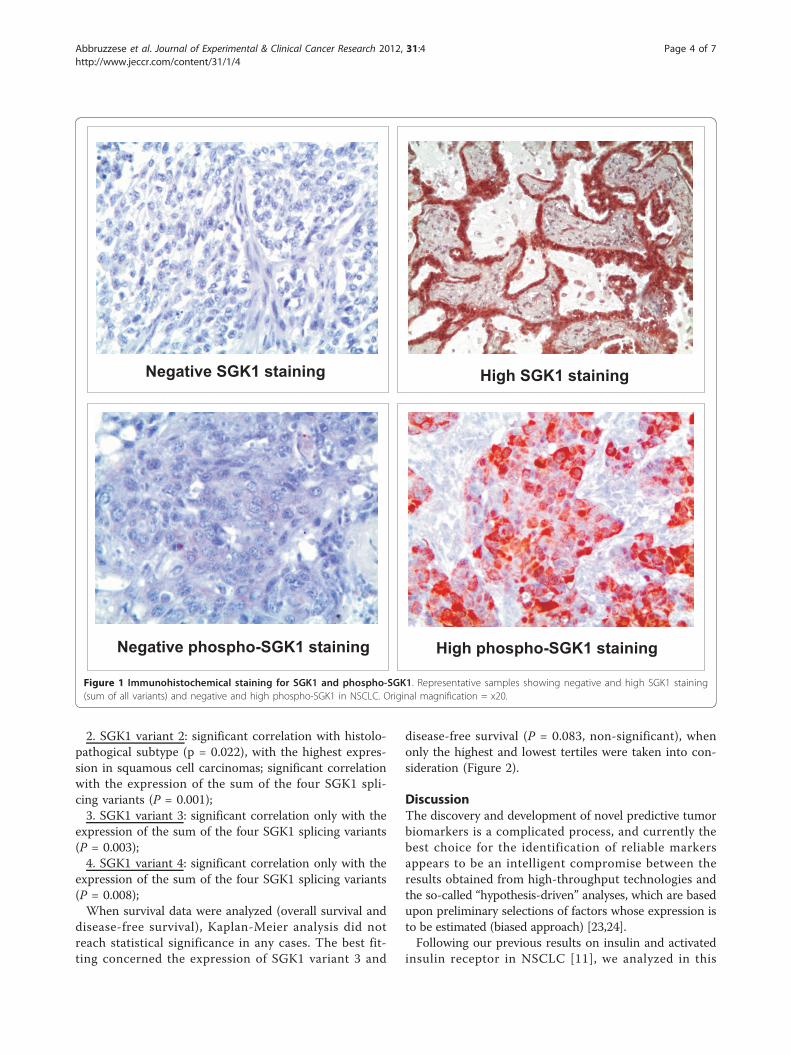
ResultsSGK1 and phospho-SGK1 protein detection in NSCLCsamplesSGK1 and phospho-SGK1 protein detection was doneby IHC on tissue sections from 66 NSCLC specimensfrom patients with a well-documented clinical history.The antibodies employed did not allow discriminatingamong the SGK1 forms deriving from the four splicingvariants. Samples stained for SGK1 displayed a granularcytoplasmic staining, considered specific due to itsabsence in the negative controls. Staining appeared non-homogeneous, with an intensity which was variable indifferent areas of the sample. Samples stained for
phospho-SGK1 displayed a granular cytoplasmic stainingas well, with a range of intensity comparable to that ofSGK1. Figure 1 shows examples of negative and highSGK1 and phospho-SGK1 staining in NSCLC samples.According to staining intensity, samples were subdividedinto tertiles, consistent with the scoring given by twopathologists, with null/low, medium and high SGK1expression. Statistical evaluation found no correlationbetween SGK1 or phospho-SGK1 staining and the fol-lowing clinical parameters: a) age at diagnosis; b) gen-der; c) smoking habit; d) histolopathogical subtype; e)histopathological grade; f) tumor size; g) lymph nodestage; h) clinical tumor stage.
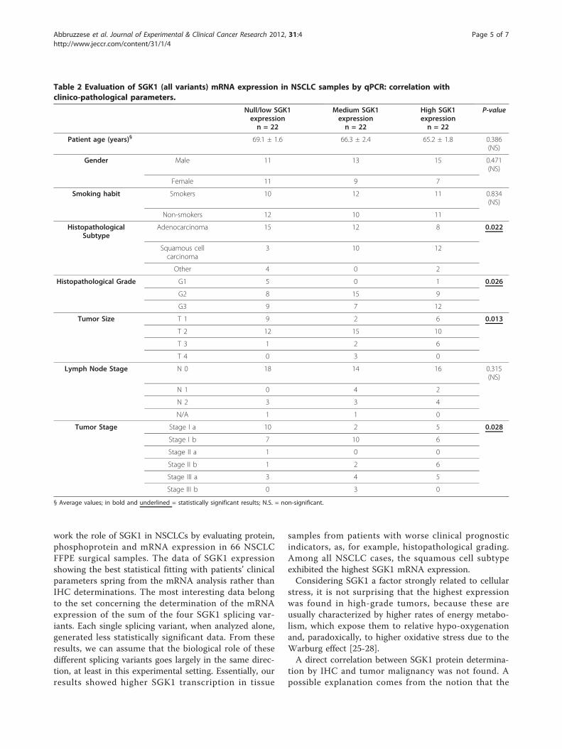
SGK1 mRNA detection in NSCLC samplesBy means of the specific primers illustrated in Table 1,we determined the mRNA amount of SGK1 either asthe sum of the four different splicing variants or as thevalue specific for each single variant. In all cases,GAPDH mRNA expression was used for an internalcheck of the quality of the FFPE-extracted RNA and fornormalization. Total SGK1 mRNA expression data, andthe values for each splicing variant, were subdivided intertiles of 22 patients each. Data were challenged againstthe clinical parameters described above. As far as it con-cerns the evaluation of the expression of the sum of thefour SGK1 mRNA, statistically significant correlationwas found with:a) histolopathogical subtype (P = 0.022), with the
highest expression in squamous cell carcinomas;b) histopathological grade (P = 0.026), with the low-
est expression in low-grade tumors (G1) and the highestexpression in high-grade tumors (G3);c) tumor size (P = 0.013), with lower expression in T1
and higher in T3-T4 tumors.d) tumor stage (P = 0.028), where the highest expres-
sion was found in patients with worse clinical stage.In this experimental set, no statistical significance was
found between SGK1 total mRNA expression andpatient’s gender, age, smoking history and lymph nodestage. All these data are summarized in Table 2. In addi-tion, no correlation between SGK1 mRNA quantificationby qPCR and SGK1 protein (or phosphoprotein) expres-sion by IHC was found.When mRNA expression of each single SGK1 splicing
variant was considered, lower levels of statistical signifi-cance were achieved, as reported below:1. SGK1 variant 1: significant correlation with histolo-
pathogical subtype (P = 0.017), with the highest expres-sion in squamous cell carcinomas; significant correlationwith the expression of the sum of the four SGK1 spli-cing variants (P = 4.7 × 10⁻⁶). Such a high significancewas due to the fact that this SGK1 form was by far themost abundant splicing variant;
Abbruzzese et al. Journal of Experimental & Clinical Cancer Research 2012, 31:4http://www.jeccr.com/content/31/1/4
Page 3 of 7
2. SGK1 variant 2: significant correlation with histolo-pathogical subtype (p = 0.022), with the highest expres-sion in squamous cell carcinomas; significant correlationwith the expression of the sum of the four SGK1 spli-cing variants (P = 0.001);3. SGK1 variant 3: significant correlation only with the
expression of the sum of the four SGK1 splicing variants(P = 0.003);4. SGK1 variant 4: significant correlation only with the
expression of the sum of the four SGK1 splicing variants(P = 0.008);When survival data were analyzed (overall survival and
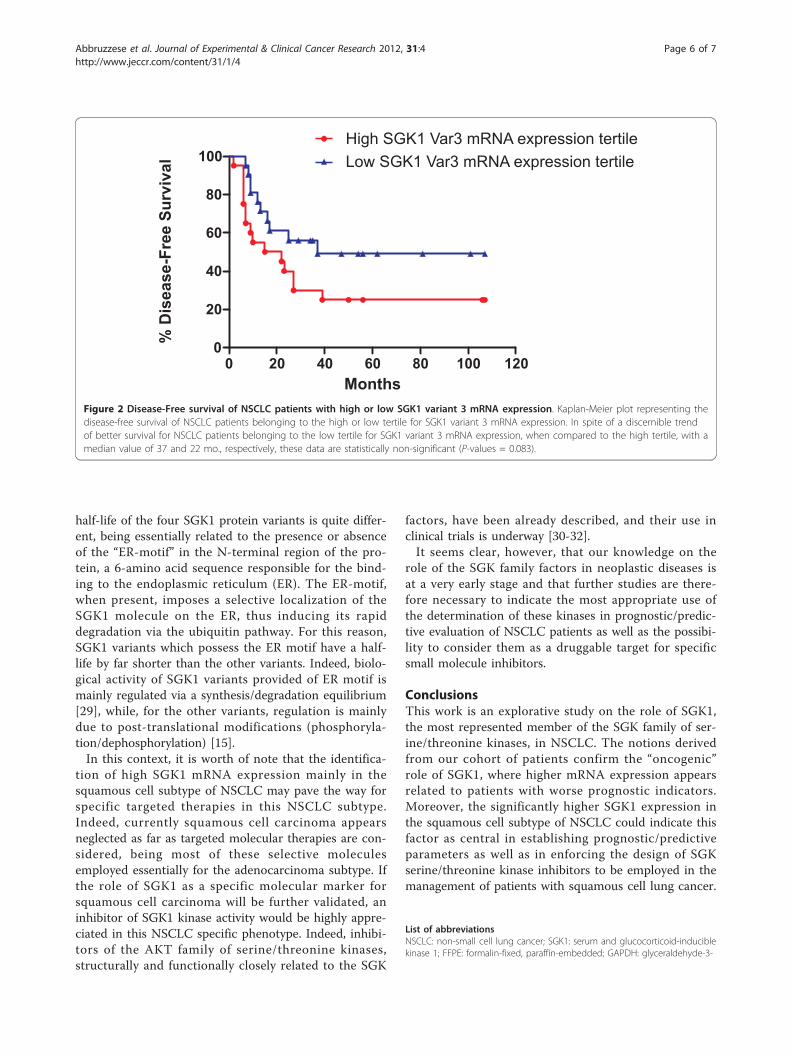
disease-free survival), Kaplan-Meier analysis did notreach statistical significance in any cases. The best fit-ting concerned the expression of SGK1 variant 3 and
disease-free survival (P = 0.083, non-significant), whenonly the highest and lowest tertiles were taken into con-sideration (Figure 2).
DiscussionThe discovery and development of novel predictive tumorbiomarkers is a complicated process, and currently thebest choice for the identification of reliable markersappears to be an intelligent compromise between theresults obtained from high-throughput technologies andthe so-called “hypothesis-driven” analyses, which are basedupon preliminary selections of factors whose expression isto be estimated (biased approach) [23,24].Following our previous results on insulin and activated
insulin receptor in NSCLC [11], we analyzed in this
High SGK1 staining
Negative phospho-SGK1 staining High phospho-SGK1 staining
Negative SGK1 staining
Figure 1 Immunohistochemical staining for SGK1 and phospho-SGK1. Representative samples showing negative and high SGK1 staining(sum of all variants) and negative and high phospho-SGK1 in NSCLC. Original magnification = x20.
Abbruzzese et al. Journal of Experimental & Clinical Cancer Research 2012, 31:4http://www.jeccr.com/content/31/1/4
Page 4 of 7
work the role of SGK1 in NSCLCs by evaluating protein,phosphoprotein and mRNA expression in 66 NSCLCFFPE surgical samples. The data of SGK1 expressionshowing the best statistical fitting with patients’ clinicalparameters spring from the mRNA analysis rather thanIHC determinations. The most interesting data belongto the set concerning the determination of the mRNAexpression of the sum of the four SGK1 splicing var-iants. Each single splicing variant, when analyzed alone,generated less statistically significant data. From theseresults, we can assume that the biological role of thesedifferent splicing variants goes largely in the same direc-tion, at least in this experimental setting. Essentially, ourresults showed higher SGK1 transcription in tissue
samples from patients with worse clinical prognosticindicators, as, for example, histopathological grading.Among all NSCLC cases, the squamous cell subtypeexhibited the highest SGK1 mRNA expression.Considering SGK1 a factor strongly related to cellular
stress, it is not surprising that the highest expressionwas found in high-grade tumors, because these areusually characterized by higher rates of energy metabo-lism, which expose them to relative hypo-oxygenationand, paradoxically, to higher oxidative stress due to theWarburg effect [25-28].A direct correlation between SGK1 protein determina-
tion by IHC and tumor malignancy was not found. Apossible explanation comes from the notion that the
Table 2 Evaluation of SGK1 (all variants) mRNA expression in NSCLC samples by qPCR: correlation withclinico-pathological parameters.
Null/low SGK1expressionn = 22
Medium SGK1expressionn = 22
High SGK1expressionn = 22
P-value
Patient age (years)§ 69.1 ± 1.6 66.3 ± 2.4 65.2 ± 1.8 0.386(NS)
Gender Male 11 13 15 0.471(NS)
Female 11 9 7
Smoking habit Smokers 10 12 11 0.834(NS)
Non-smokers 12 10 11
HistopathologicalSubtype
Adenocarcinoma 15 12 8 0.022
Squamous cellcarcinoma
3 10 12
Other 4 0 2
Histopathological Grade G1 5 0 1 0.026
G2 8 15 9
G3 9 7 12
Tumor Size T 1 9 2 6 0.013
T 2 12 15 10
T 3 1 2 6
T 4 0 3 0
Lymph Node Stage N 0 18 14 16 0.315(NS)
N 1 0 4 2
N 2 3 3 4
N/A 1 1 0
Tumor Stage Stage I a 10 2 5 0.028
Stage I b 7 10 6
Stage II a 1 0 0
Stage II b 1 2 6
Stage III a 3 4 5
Stage III b 0 3 0
§ Average values; in bold and underlined = statistically significant results; N.S. = non-significant.
Abbruzzese et al. Journal of Experimental & Clinical Cancer Research 2012, 31:4http://www.jeccr.com/content/31/1/4
Page 5 of 7
half-life of the four SGK1 protein variants is quite differ-ent, being essentially related to the presence or absenceof the “ER-motif” in the N-terminal region of the pro-tein, a 6-amino acid sequence responsible for the bind-ing to the endoplasmic reticulum (ER). The ER-motif,when present, imposes a selective localization of theSGK1 molecule on the ER, thus inducing its rapiddegradation via the ubiquitin pathway. For this reason,SGK1 variants which possess the ER motif have a half-life by far shorter than the other variants. Indeed, biolo-gical activity of SGK1 variants provided of ER motif ismainly regulated via a synthesis/degradation equilibrium[29], while, for the other variants, regulation is mainlydue to post-translational modifications (phosphoryla-tion/dephosphorylation) [15].In this context, it is worth of note that the identifica-
tion of high SGK1 mRNA expression mainly in thesquamous cell subtype of NSCLC may pave the way forspecific targeted therapies in this NSCLC subtype.Indeed, currently squamous cell carcinoma appearsneglected as far as targeted molecular therapies are con-sidered, being most of these selective moleculesemployed essentially for the adenocarcinoma subtype. Ifthe role of SGK1 as a specific molecular marker forsquamous cell carcinoma will be further validated, aninhibitor of SGK1 kinase activity would be highly appre-ciated in this NSCLC specific phenotype. Indeed, inhibi-tors of the AKT family of serine/threonine kinases,structurally and functionally closely related to the SGK
factors, have been already described, and their use inclinical trials is underway [30-32].It seems clear, however, that our knowledge on the
role of the SGK family factors in neoplastic diseases isat a very early stage and that further studies are there-fore necessary to indicate the most appropriate use ofthe determination of these kinases in prognostic/predic-tive evaluation of NSCLC patients as well as the possibi-lity to consider them as a druggable target for specificsmall molecule inhibitors.
ConclusionsThis work is an explorative study on the role of SGK1,the most represented member of the SGK family of ser-ine/threonine kinases, in NSCLC. The notions derivedfrom our cohort of patients confirm the “oncogenic”role of SGK1, where higher mRNA expression appearsrelated to patients with worse prognostic indicators.Moreover, the significantly higher SGK1 expression inthe squamous cell subtype of NSCLC could indicate thisfactor as central in establishing prognostic/predictiveparameters as well as in enforcing the design of SGKserine/threonine kinase inhibitors to be employed in themanagement of patients with squamous cell lung cancer.
List of abbreviationsNSCLC: non-small cell lung cancer; SGK1: serum and glucocorticoid-induciblekinase 1; FFPE: formalin-fixed, paraffin-embedded; GAPDH: glyceraldehyde-3-
0 20 40 60 80 100 1200
20
40
60
80
100
High SGK1 Var3 mRNA expression tertile
Low SGK1 Var3 mRNA expression tertile
Months
% D
ise
as
e-F
ree
Su
rviv
al
Figure 2 Disease-Free survival of NSCLC patients with high or low SGK1 variant 3 mRNA expression. Kaplan-Meier plot representing thedisease-free survival of NSCLC patients belonging to the high or low tertile for SGK1 variant 3 mRNA expression. In spite of a discernible trendof better survival for NSCLC patients belonging to the low tertile for SGK1 variant 3 mRNA expression, when compared to the high tertile, with amedian value of 37 and 22 mo., respectively, these data are statistically non-significant (P-values = 0.083).
Abbruzzese et al. Journal of Experimental & Clinical Cancer Research 2012, 31:4http://www.jeccr.com/content/31/1/4
Page 6 of 7
phosphate dehydrogenase; qPCR: quantitative real-time PCR; IHC:immunohistochemistry; ER: endoplasmic reticulum.
AcknowledgementsThe authors thank Dr. Irene Terrenato for her help in statistical analysis.This work was supported by grants from Associazione Italiana Ricerca sulCancro (AIRC), Ministero della Salute and Human Health Foundation (HHF)to M.G.P.
Author details1Department of Development of Therapeutic Programs, National CancerInstitute “Regina Elena”, IRCCS, 00144 Rome, Italy. 2Medical OncologyDivision B, National Cancer Institute “Regina Elena”, Via Elio Chianesi, 53,00144 Rome, Italy. 3Department of Pathology, Regina Elena Cancer Institute,Via E. Chianesi, 53, 00144 Rome, Italy. 4Department of Oncologic ThoracicSurgery, Regina Elena Cancer Institute, Via E. Chianesi, 53, 00144 Rome, Italy.5Department of Experimental and clinical Medicine “G. Salvatore”, Faculty ofMedicine, University Magna Graecia, 88100 Catanzaro, Italy. 6CNR, Istituto diNeurobiologia e Medicina Molecolare, Via Fosso di Fiorano, 64, 00143 Rome,Italy.
Authors’ contributionsCA: Research planning, IHC and qPCR determinations, statistical analysis. SM:Research planning, IHC and qPCR determinations, statistical analysis. LP:Research planning, collection of patients’ information, manuscript drafting.AMM: Research planning and qPCR determinations. PV: Patients’ diagnosis,IHC scoring. BA: Tissue slices preparation, haematoxylin/eosin staining. GA:Collection of patients’ information, patients’ database maintenance. FF:Surgery and patients’ database maintenance. RA: qPCR determinations. LD’A:qPCR determinations. MR: Research planning, collection of patients’information, manuscript drafting. AF: Research planning, qPCRdeterminations, statistical analysis. NP: Research planning, qPCRdeterminations, statistical analysis, manuscript drafting. MGP: Researchplanning, coordination of the whole project, IHC scoring, manuscriptdrafting. All authors read and approved the final manuscript.
Competing interestsThe authors declare that they have no competing interests.
Received: 7 November 2011 Accepted: 12 January 2012Published: 12 January 2012
References1. Herbst RS, Heymach JV, Lippman SM: Lung cancer. N Engl J Med 2008,
359:1367-1380.2. Jemal A, Siegel R, Ward E, Hao Y, Xu J, Thun MJ: Cancer statistics, 2009. CA
Cancer J Clin 2009, 59:225-249.3. Boffetta P: Epidemiology of environmental and occupational cancer.
Oncogene 2004, 23:6392-6403.4. Patel JD: Lung cancer in women. J Clin Oncol 2005, 23:3212-3218.5. Subramanian J, Govindan R: Lung cancer in never smokers: a review. J
Clin Oncol 2007, 25:561-570.6. Samet JM, Avila-Tang E, Boffetta P, Hannan LM, Olivo-Marston S, Thun MJ,
et al: Lung cancer in never smokers: clinical epidemiology andenvironmental risk factors. Clin Cancer Res 2009, 15:5626-5645.
7. Paggi MG, Vona R, Abbruzzese C, Malorni W: Gender-related disparities innon-small cell lung cancer. Cancer Lett 2010, 298:1-8.
8. Miller VA, Kris MG, Shah N, Patel J, Azzoli C, Gomez J, et al:Bronchioloalveolar pathologic subtype and smoking history predictsensitivity to gefitinib in advanced non-small-cell lung cancer. J ClinOncol 2004, 22:1103-1109.
9. Schlessinger J: Ligand-induced, receptor-mediated dimerization andactivation of EGF receptor. Cell 2002, 110:669-672.
10. Pollak M: Insulin and insulin-like growth factor signalling in neoplasia.Nat Rev Cancer 2008, 8:915-928.
11. Mattarocci S, Abbruzzese C, Mileo AM, Visca P, Antoniani B, Alessandrini G,et al: Intracellular presence of insulin and its phosphorylated receptor innon-small cell lung cancer. J Cell Physiol 2009, 221:766-770.
12. Bellacosa A, Kumar CC, Di Cristofano A, Testa JR: Activation of AKT kinasesin cancer: implications for therapeutic targeting. Adv Cancer Res 2005,94:29-86.
13. Ruggero D, Sonenberg N: The Akt of translational control. Oncogene 2005,24:7426-7434.
14. Testa JR, Tsichlis PN: AKT signaling in normal and malignant cells.Oncogene 2005, 24:7391-7393.
15. Bruhn MA, Pearson RB, Hannan RD, Sheppard KE: Second AKT: the rise ofSGK in cancer signalling. Growth Factors 2010, 28:394-408.
16. Lang F, Bohmer C, Palmada M, Seebohm G, Strutz-Seebohm N, Vallon V:(Patho)physiological significance of the serum- and glucocorticoid-inducible kinase isoforms. Physiol Rev 2006, 86:1151-1178.
17. Liu D, Yang X, Songyang Z: Identification of CISK, a new member of theSGK kinase family that promotes IL-3-dependent survival. Curr Biol 2000,10:1233-1236.
18. Mikosz CA, Brickley DR, Sharkey MS, Moran TW, Conzen SD: Glucocorticoidreceptor-mediated protection from apoptosis is associated withinduction of the serine/threonine survival kinase gene, sgk-1. J Biol Chem2001, 276:16649-16654.
19. Tangir J, Bonafe N, Gilmore-Hebert M, Henegariu O, Chambers SK: SGK1, apotential regulator of c-fms related breast cancer aggressiveness. ClinExp Metastasis 2004, 21:477-483.
20. Failor KL, Desyatnikov Y, Finger LA, Firestone GL: Glucocorticoid-induceddegradation of glycogen synthase kinase-3 protein is triggered byserum- and glucocorticoid-induced protein kinase and Akt signaling andcontrols beta-catenin dynamics and tight junction formation inmammary epithelial tumor cells. Mol Endocrinol 2007, 21:2403-2415.
21. Cronin M, Pho M, Dutta D, Stephans JC, Shak S, Kiefer MC, et al:Measurement of gene expression in archival paraffin-embedded tissues:development and performance of a 92-gene reverse transcriptase-polymerase chain reaction assay. Am J Pathol 2004, 164:35-42.
22. Antonov J, Goldstein DR, Oberli A, Baltzer A, Pirotta M, Fleischmann A, et al:Reliable gene expression measurements from degraded RNA byquantitative real-time PCR depend on short amplicons and a propernormalization. Lab Invest 2005, 85:1040-1050.
23. Bianchi F, Nicassio F, Di Fiore PP: Unbiased vs. biased approaches to theidentification of cancer signatures: the case of lung cancer. Cell Cycle2008, 7:729-734.
24. Guan P, Huang D, He M, Zhou B: Lung cancer gene expression databaseanalysis incorporating prior knowledge with support vector machine-based classification method. J Exp Clin Cancer Res 2009, 28:103.
25. Nakashima RA, Paggi MG, Pedersen PL: Contributions of glycolysis andoxydative phosphorylation to adenosine-5’-triphosphate production inAS-30D hepatoma cells. Cancer Res 1984, 44:5702-5706.
26. Nakashima RA, Paggi MG, Arora KK, Pedersen PL: Integration ofmitochondrial function with high aerobic glycolysis in tumors: role ofhexokinase binding to the outer mitochondrial membrane. In Integrationof Mitochondrial Function. Edited by: Lemasters JJ, Hackenbrock CR,Thurman RG, Westhoff HV. New York, N.Y.: Plenum Publishing Company;1990:405-411.
27. Wallace DC: Mitochondria and cancer: Warburg addressed. Cold SpringHarb Symp Quant Biol 2005, 70:363-374.
28. Pedersen PL: Warburg, me and Hexokinase 2: Multiple discoveries of keymolecular events underlying one of cancers’ most common phenotypes,the “Warburg Effect”, i.e., elevated glycolysis in the presence of oxygen.J Bioenerg Biomembr 2007, 39:211-222.
29. Brickley DR, Mikosz CA, Hagan CR, Conzen SD: Ubiquitin modification ofserum and glucocorticoid-induced protein kinase-1 (SGK-1). J Biol Chem2002, 277:43064-43070.
30. Mattmann ME, Stoops SL, Lindsley CW: Inhibition of Akt with smallmolecules and biologics: historical perspective and current status of thepatent landscape. Expert Opin Ther Pat 2011, 21:1309-1338.
31. Morrow JK, Du-Cuny L, Chen L, Meuillet EJ, Mash EA, Powis G, et al: Recentdevelopment of anticancer therapeutics targeting Akt. Recent PatAnticancer Drug Discov 2011, 6:146-159.
32. Hixon ML, Paccagnella L, Millham R, Perez-Olle R, Gualberto A:Development of inhibitors of the IGF-IR/PI3K/Akt/mTOR pathway. RevRecent Clin Trials 2010, 5:189-208.
doi:10.1186/1756-9966-31-4Cite this article as: Abbruzzese et al.: Determination of SGK1 mRNA innon-small cell lung cancer samples underlines high expression insquamous cell carcinomas. Journal of Experimental & Clinical CancerResearch 2012 31:4.
Abbruzzese et al. Journal of Experimental & Clinical Cancer Research 2012, 31:4http://www.jeccr.com/content/31/1/4
Page 7 of 7
Related Documents


![Phosphorylation of Nicastrin by SGK1 Leads to Its ......C99 is a direct substrate of gamma-secretase [33,44]. Because APP is cleaved by gamma-secretase activity, we first attempted](https://static.cupdf.com/doc/110x72/611bb7cea20dfc58ec16fbce/phosphorylation-of-nicastrin-by-sgk1-leads-to-its-c99-is-a-direct-substrate.jpg)