Delayed treatment with monoclonal antibody IN-1 1 week after stroke results in recovery of function and corticorubral plasticity in adult rats Andrew B Seymour 1,2 , Ellen M Andrews 1,2,8 , Shih-Yen Tsai 1,8 , Tiffanie M Markus 1,3 , Melanie R Bollnow 1,4 , Miranda M Brenneman 5 , Timothy E O’Brien 6 , Anthony J Castro 2 , Martin E Schwab 7 and Gwendolyn L Kartje 1,2,3,4 1 Neurology and Research Service, Hines VA Hospital, Hines, Illinois, USA; 2 Department of Cell Biology, Neurobiology and Anatomy, Loyola University Chicago, Maywood, Illinois, USA; 3 Neuroscience and Aging Institute, Loyola University Chicago, Maywood, Illinois, USA; 4 Department of Neurology, Loyola University Medical Center, Maywood, Illinois, USA; 5 Department of Psychology, Northern Illinois University, DeKalb, Illinois, USA; 6 Statistical Consulting Center, Department of Mathematics and Statistics, Loyola University Chicago, Chicago, Illinois, USA; 7 Brain Research Institute University of Zurich and Department of Biology, Swiss Federal Institute of Technology, Zurich, Switzerland Neuronal death due to ischemic stroke results in permanent deficits in sensory, language, and motor functions. The growth-restrictive environment of the adult central nervous system (CNS) is an obstacle to functional recovery after stroke and other CNS injuries. In this regard, Nogo-A is a potent neurite growth-inhibitory protein known to restrict neuronal plasticity in adults. Previously, we have found that treatment with monoclonal antibody (mAb) IN-1 to neutralize Nogo-A immediately after stroke enhanced motor cortico-efferent plasticity and recovery of skilled forelimb function in rats. However, immediate treatment for stroke is often not clinically feasible. Thus, the present study was undertaken to determine whether cortico-efferent plasticity and functional recovery would occur if treatment with mAb IN-1 was delayed 1 week after stroke. Adult rats were trained on a forelimb- reaching task, and the middle cerebral artery was occluded to induce focal cerebral ischemia to the forelimb sensorimotor cortex. After 1 week, animals received mAb IN-1 treatment, control antibody, or no treatment, and were tested for 9 more weeks. To assess cortico-efferent plasticity, the sensorimotor cortex opposite the stroke lesion was injected with an anterograde neuroanatomical tracer. Behavioral analysis demonstrated a recovery of skilled forelimb function, and anatomical studies revealed neuroplasticity at the level of the red nucleus in animals treated with mAb IN-1, thus demonstrating the efficacy of this treatment even if administered 1 week after stroke. Journal of Cerebral Blood Flow & Metabolism (2005) 25, 1366–1375. doi:10.1038/sj.jcbfm.9600134; published online 11 May 2005 Keywords: neuroplasticity; Nogo-A; recovery; stroke Introduction Stroke is a prominent cause of serious, long-term disability (Bonita, 1992; Bonita et al, 1994) and the third leading cause of death in the United States (Minino et al, 2002). Total health costs for disability due to stroke in the United States are estimated at $53.6 billion annually (Morbidity and Mortality Chart Book on Cardiovascular, Lung, and Blood Diseases, 2004). Ischemic strokes comprise over 88% of all strokes, making them the most common type of cerebrovascular injury (Heart Disease and Stroke Statistics-2004 Update, 2004). Ischemic con- ditions in the brain cause neuronal death, leading to permanent sensorimotor and cognitive deficits. While thrombolytic therapy with a strong neuropro- tective effect is sometimes administered up to 3 h after stroke, currently, there is no effective treatment for stroke after the acute stage (del Zoppo, 2000). Given that immediate treatment for stroke is often impossible in the clinical setting, the need for new treatment strategies for stroke is urgent. Received 5 November 2004; revised 2 February 2005; accepted 11 March 2005; published online 11 May 2005 Correspondence: Mr AB Seymour, BS, Neurology and Research Service, Research 151, Building 1, D400L, Hines VA Hospital, Roosevelt Road & Fifth Avenue, Hines, IL 60141, USA. E-mail: [email protected] This work was supported by the Department of Veterans Affairs, NIH Grant NS 40960, and the Swiss NSF. 8 These authors contributed equally. Journal of Cerebral Blood Flow & Metabolism (2005) 25, 1366–1375 & 2005 ISCBFM All rights reserved 0271-678X/05 $30.00 www.jcbfm.com

Welcome message from author
This document is posted to help you gain knowledge. Please leave a comment to let me know what you think about it! Share it to your friends and learn new things together.
Transcript
Delayed treatment with monoclonal antibody IN-11 week after stroke results in recovery of functionand corticorubral plasticity in adult rats
Andrew B Seymour1,2, Ellen M Andrews1,2,8, Shih-Yen Tsai1,8, Tiffanie M Markus1,3,Melanie R Bollnow1,4, Miranda M Brenneman5, Timothy E O’Brien6, Anthony J Castro2,Martin E Schwab7 and Gwendolyn L Kartje1,2,3,4
1Neurology and Research Service, Hines VA Hospital, Hines, Illinois, USA; 2Department of Cell Biology,Neurobiology and Anatomy, Loyola University Chicago, Maywood, Illinois, USA; 3Neuroscience and AgingInstitute, Loyola University Chicago, Maywood, Illinois, USA; 4Department of Neurology, Loyola UniversityMedical Center, Maywood, Illinois, USA; 5Department of Psychology, Northern Illinois University, DeKalb,Illinois, USA; 6Statistical Consulting Center, Department of Mathematics and Statistics, Loyola UniversityChicago, Chicago, Illinois, USA; 7Brain Research Institute University of Zurich and Department of Biology,Swiss Federal Institute of Technology, Zurich, Switzerland
Neuronal death due to ischemic stroke results in permanent deficits in sensory, language, andmotor functions. The growth-restrictive environment of the adult central nervous system (CNS) is anobstacle to functional recovery after stroke and other CNS injuries. In this regard, Nogo-A is a potentneurite growth-inhibitory protein known to restrict neuronal plasticity in adults. Previously, we havefound that treatment with monoclonal antibody (mAb) IN-1 to neutralize Nogo-A immediately afterstroke enhanced motor cortico-efferent plasticity and recovery of skilled forelimb function in rats.However, immediate treatment for stroke is often not clinically feasible. Thus, the present study wasundertaken to determine whether cortico-efferent plasticity and functional recovery would occur iftreatment with mAb IN-1 was delayed 1 week after stroke. Adult rats were trained on a forelimb-reaching task, and the middle cerebral artery was occluded to induce focal cerebral ischemia to theforelimb sensorimotor cortex. After 1 week, animals received mAb IN-1 treatment, control antibody,or no treatment, and were tested for 9 more weeks. To assess cortico-efferent plasticity, thesensorimotor cortex opposite the stroke lesion was injected with an anterograde neuroanatomicaltracer. Behavioral analysis demonstrated a recovery of skilled forelimb function, and anatomicalstudies revealed neuroplasticity at the level of the red nucleus in animals treated with mAb IN-1, thusdemonstrating the efficacy of this treatment even if administered 1 week after stroke.Journal of Cerebral Blood Flow & Metabolism (2005) 25, 1366–1375. doi:10.1038/sj.jcbfm.9600134; published online11 May 2005
Keywords: neuroplasticity; Nogo-A; recovery; stroke
Introduction
Stroke is a prominent cause of serious, long-termdisability (Bonita, 1992; Bonita et al, 1994) and thethird leading cause of death in the United States(Minino et al, 2002). Total health costs for disability
due to stroke in the United States are estimated at$53.6 billion annually (Morbidity and MortalityChart Book on Cardiovascular, Lung, and BloodDiseases, 2004). Ischemic strokes comprise over88% of all strokes, making them the most commontype of cerebrovascular injury (Heart Disease andStroke Statistics-2004 Update, 2004). Ischemic con-ditions in the brain cause neuronal death, leading topermanent sensorimotor and cognitive deficits.While thrombolytic therapy with a strong neuropro-tective effect is sometimes administered up to 3 hafter stroke, currently, there is no effective treatmentfor stroke after the acute stage (del Zoppo, 2000).Given that immediate treatment for stroke is oftenimpossible in the clinical setting, the need for newtreatment strategies for stroke is urgent.
Received 5 November 2004; revised 2 February 2005; accepted 11March 2005; published online 11 May 2005
Correspondence: Mr AB Seymour, BS, Neurology and ResearchService, Research 151, Building 1, D400L, Hines VA Hospital,Roosevelt Road & Fifth Avenue, Hines, IL 60141, USA.E-mail: [email protected]
This work was supported by the Department of Veterans Affairs,
NIH Grant NS 40960, and the Swiss NSF.
8These authors contributed equally.
Journal of Cerebral Blood Flow & Metabolism (2005) 25, 1366–1375& 2005 ISCBFM All rights reserved 0271-678X/05 $30.00
www.jcbfm.com

A major obstacle to functional recovery afterstroke is the growth-restrictive environment of theadult central nervous system (CNS), mainly due toneurite growth-inhibitory proteins, especially inmyelin (see Schwab (2004) for review). Nogo-A isone of the known neurite inhibitory proteins presentin myelin (Chen et al, 2000; Filbin, 2003; GrandPreet al, 2000; Prinjha et al, 2000; Schwab, 2004). Nogo-A, a protein product of the nogo gene, is a potentneurite growth inhibitor that is predominantlyexpressed in the CNS. In CNS myelin, confocaland immunoelectron microscopy have shown thatNogo-A is expressed in oligodendrocyte cell bodiesand processes and is localized in the innermostadaxonal and outermost myelin layers (Huber et al,2002). Nogo-A may exert its activity throughstimulation of GTPase RhoA, which activates Rhokinase, or ROCK, and through elevation of intracel-lular calcium (Bandtlow, 2003; He and Koprivica,2004; Schweigreiter et al, 2004). These proteins areknown to regulate cytoskeletal changes throughmodulation of actin polymerization (Olson et al,1995). Neutralization of Nogo-A promotes growth ofaxons in vitro (Oertle et al, 2003) and in vivo, leadingto enhanced neural plasticity (Kartje et al, 1999;Thallmair et al, 1998), regeneration, and functionalrecovery (Brosamle et al, 2000; GrandPre et al,2002). These findings illustrate its importance as atarget for treatment of CNS injury.
Several studies have shown that recovery offunction after injury is much better in the neonatalbrain, presumably due to the formation of newneuronal connections, that is, structural plasticity.For example, functional reorganization of the un-ablated hemisphere occurs after unilateral motorcortical lesions in neonatal rats, due to the lessrestrictive environment of the neonatal CNS (Kartje-Tillotson et al, 1985, 1987). In neonatal rats receiv-ing sensorimotor cortical lesion, the uninjuredcontralateral cortex forms bilateral projections tode-afferented subcortical targets from the opposite,unablated cortex (Kartje-Tillotson et al, 1986; Kolbet al, 1992; Leong and Lund, 1973; Naus et al, 1985;Rouiller et al, 1991; Yu et al, 1995). Importantly,remodeling of uninjured neuroanatomical pathwaysis accompanied by functional recovery after CNSinjuries in young rats. However, re-routing of neuralpathways in response to CNS lesions is lesspronounced in adult rats after lesions, but can befacilitated by neutralizing antibodies against theneurite inhibitory protein Nogo-A (Wenk et al, 1999;Kartje et al, 1999).
Our laboratory has investigated antibody-mediated Nogo-A neutralization at the time ofstroke. The monoclonal antibody (mAb) IN-1 isspecific to the amino component of Nogo-A (Fiedleret al, 2002), that is, amino-Nogo/NiG, a potentinhibitory domain of Nogo-A (Niederost et al,2002; Schweigreiter et al, 2004). Previously, wefound that immediate treatment with the mAb IN-1resulted in improvement of the impaired forelimb
associated with the ischemic stroke (Papadopouloset al, 2002). Furthermore, this improvement wasassociated with neuroanatomical plasticity from theuninjured cortex at the level of the red nucleus. Thecortico-rubral pathway, part of the cortico-rubro-spinal system, has been implicated in precise limbmovements, which are directly affected by lesions tothe sensorimotor cortex (Jarratt and Hyland, 1999;Whishaw and Gorny, 1996). There is some evidencethat delayed nogo-A neutralization after strokeresults in functional recovery and neuroanatomicalplasticity. For example, the administration of anti-Nogo-A antibodies specific to NiG 24 h after strokehas been shown to promote recovery of skilledforelimb movements as well as motor corticospinaltract (CST) remodelling (Wiessner et al, 2003).However, to date, it is unknown how long thetime-to-treatment window with anti-Nogo-A anti-bodies can be extended. This information is ofcrucial importance from the clinical viewpointwhen treating stroke patients in subacute or evenchronic settings.
The purpose of the present study was to deter-mine whether animals treated with anti-Nogo-Aantibody at a later time point after stroke, that is,1 week, would exhibit functional recovery andneuroplasticity. We show here that neutralizationof Nogo-A 1 week after stroke in adult rats resultedin marked improvement of skilled forelimb functionas well as corticorubral plasticity.
Materials and methods
Animals
Adult male Long Evans black-hooded rats (250 to 350 g,ages 10 to 12 weeks) (Harlan, Indianapolis, IN, USA) weredivided into three experimental groups: (1) stroke plusmAb IN-1 (n¼ 9); (2) stroke plus control antibody (n¼ 4);(3) stroke only (n¼ 7). Animals were number-coded toensure investigators were masked to treatment groups.Experiments were approved by the Joint InstitutionalAnimal Care and Use Committee of Loyola University andHines Veterans Affairs Hospital.
Skilled Forelimb-Reaching Task
Using a restricted diet of 12 g of rat chow per day, animalswere reduced to 95% of their ideal body weight beforetesting; this diet was maintained throughout the testingperiod. The skilled forelimb-reaching task was usedaccording to previous work to assess skilled motorfunctional recovery after stroke (Papadopoulos et al,2002; Wiessner et al, 2003) and other injuries to motorpathways governing forelimb function (Z’Graggen et al,1998). The testing apparatus consisted of a transparentPlexiglass chamber with an elliptical opening in one walland a shelf attached outside the opening. Small sucrosepellets (45 mg; Research Diets, Inc., New Brunswick, NJ,USA) were placed one at a time onto the pellet shelf at a
Delayed treatment with monoclonal antibody IN-1AB Seymour et al
1367
Journal of Cerebral Blood Flow & Metabolism (2005) 25, 1366–1375

distance of 1.5 cm from the opening of the chamber.During a 2- to 4-week training period, limb preference wasdetermined. To meet the criteria for inclusion in the study,rats had to reach, grasp, and bring to their mouth aminimum of 16 pellets on the first attempt (maximum of20) in a 5-min period using the preferred limb. The resultsof the last three testing sessions were averaged together toestablish a baseline value for each animal. After baselinewas established, each animal received a middle cerebralartery occlusion (MCAO) and then was tested daily for 1week to determine whether a deficit was present. Onlyanimals that had a deficit signified by successfullyreaching six or less pellets (r37.5% of baseline) duringa 5-min testing session were included in the study.Animals that remained in the study were tested dailyand taped once weekly throughout the remaining 8 weeksuntil sacrifice.
Middle Cerebral Artery Occlusion
Strokes were induced via occlusion of the middle cerebralartery (MCA) as described previously (Chen et al, 1986),with some modifications. After training and baselinetesting to determine limb preference, rats underwent anMCAO in the hemisphere contralateral to, and thuscorresponding to, their preferred limb. Rats were anesthe-tized with sodium pentobarbital (50 mg/kg, intraperitone-ally) and placed in a stereotaxic instrument. A craniotomywas made in either the right or left parietal bone with theuse of an operating microscope. The resultant burr holewas extended to follow the distal branches of the MCA towhere it transversed the rhinal sulcus. The MCA waspermanently ligated with a 10-0 nylon suture andtransected with microscissors. Then, the common carotidartery (CCA) ipsilateral to the occluded MCA waspermanently ligated with a 4-0 silk suture, and thecontralateral CCA was temporarily occluded with ananeurysm clip for 60 mins.
Antibody Application
Antibodies were applied in the same method as performedin previous work (Emerick et al, 2003; Papadopoulos et al,2002; Z’Graggen et al, 1998) (see Figure 1). The mAb IN-1was raised against the Nogo-A band from SDS-polyacry-lamide gel electrophoresis separations of rat spinal cordmyelin (Caroni and Schwab, 1988b). At 6 days afterMCAO, animals with a stable deficit were reanesthetizedwith sodium pentobarbital (50 mg/kg, intraperitoneally),and a cell suspension (6 mL) containing a total of 1� 105
mouse hybridoma cells secreting either mAb IN-1 or thecontrol antibody, anti-horseradish peroxidase (anti-HRP)was stereotaxically injected with a Hamilton microsyringeinto the hippocampus ipsilateral to the MCAO (coordi-nates: 4 mm caudal, 5 mm lateral and 5 mm ventral tobregma). This site was chosen to avoid damaging areas ofmotor cortex and to allow the antibody to reach theventricular system and circulate in the CSF (Papadopouloset al, 2002).
These mouse hybridoma cells produce an IgM antibodyagainst the rat neurite growth-inhibitory protein Nogo-A.As a control, an mAb raised against HRP (anti-HRP, an IgGantibody without inhibitory effect on HRP enzymaticactivity) was generated from the same parent myelomaline. As a hybridoma line producing an IgM against HRP isnot available, the IgG-producing hybridoma was used.Hybridoma cells were grown in Iscove’s modified Dulbec-co’s medium using standard mammalian tissue culturetechniques. Before implantation into the brain, hybridomacells were tested for inhibitory neutralizing activity in abioassay using neurons on a myelin-protein substrate, andwere tested for antibody production by FITC-coupled anti-mouse antibodies. The stroke-only group of rats receivedvehicle only, that is, media, at the same injection site. Allanimals received cyclosporin A (10 mg/kg, intraperitone-ally) daily starting 1 day before, and continuing for 7 daysafter cell implantation, to prevent rejection of thehybridoma xenograft; stroke-only animals also receivedcyclosporin A according to the same schedule. Based onthis paradigm, hybridoma cells survive for approximately2 weeks and continue to secrete antibody during suchtime. According to our past experience and publishedreports, antibody-secreting hybridoma cells given astreatment will continue to secrete antibody for at least2 weeks after implantation in the immune-suppressedanimal, and this is equal between the mAb IN-1 and anti-HRP antibody-secreting hybridomas (Emerick and Kartje,2004; Schnell and Schwab, 1990; Thallmair et al, 1998;von Meyenburg et al, 1998; Wenk et al, 1999; Z’Graggen etal, 1998).
Biotinylated Dextran Amine Tracing
After the period of behavioral observation and datacollection (9 weeks), all rats were re-anesthetized withsodium pentobarbital (50 mg/kg, intraperitoneally) and
Figure 1 A schematic diagram illustrating the experimentalprocedures. Corticofugal fibers from the right forelimb motorcortex to the termination areas in the red nucleus arerepresented. In the left hemisphere is shown the stroke lesion(shaded) and the antibody-secreting hybridoma cell implanta-tion site. The BDA tracer injection site is contralateral to thestroke lesion (adapted from Wenk et al, 1999).
Delayed treatment with monoclonal antibody IN-1AB Seymour et al
1368
Journal of Cerebral Blood Flow & Metabolism (2005) 25, 1366–1375

placed in a stereotaxic instrument. A craniotomy wasmade to expose the sensorimotor cortex of the hemispherecontralateral to the stroke lesion (Figure 1). Using aHamilton microsyringe, 1mL of a 10% solution of theanterograde tracer biotinylated dextran amine (BDA,Molecular Probes, Eugene, OR, USA) was injected at twosites (1 to 3 mm lateral to bregma, at a depth of 1.5 mmfrom the cortical surface) in the uninjured caudal forelimbmotor cortex as described by Neafsey et al (1986). At2 weeks after BDA injection, rats were overdosed withsodium pentobarbital and transcardially perfused withsaline followed by 4% paraformaldehyde.
Neuroanatomical Analysis
Anatomical structures were identified using the atlas ofPaxinos and Watson (1998). Brains were removed, post-fixed overnight in 4% paraformaldehyde, cryoprotected in30% sucrose, and kept at �801C before processing. Tissuewas cut on a Leica cryostat in serial 50 mm coronal sectionsand reacted for BDA-positive fibers by the semi-free-floating method (Herzog and Brosamle, 1997); alternatesections were processed for Nissl stain and analyzed forlesion size and location. All slides were coded to ensureinvestigators were blind to treatment groups. Coronalsections were analyzed by computer-aided image analysisusing NIH Image version 1.51 (National Institutes ofHealth, Bethesda, MD, USA). A Macintosh computer(Centris 650) interfaced with a digitizing board (DataTranslation, Marlboro, MA, USA) to a digital camera(model MOS VK-C150; Hitachi; Brisbane, CA, USA) wasused with a Leitz DMR microscope.
Quantification of Cortico-Rubral Fiber Number
Biotinylated dextran amine-positive fibers at the level ofthe parvocellular red nucleus were analyzed by calculat-ing ratios of BDA-labeled fibers between red nuclei in eachhemisphere using an adaptation of a previously describedmethod (Emerick and Kartje, 2004). Using five consecutivesections for each animal, a 2500 mm2 grid was placed infour standardized locations within the red nucleus in eachhemisphere and fibers present within the grid werecounted and totalled, yielding an estimate of fiber densitywithin the red nucleus in each hemisphere. To provide aninternal control for fiber tracing, the total fiber count in thered nucleus within the lesioned hemisphere was dividedby the total fiber count in the red nucleus within theunlesioned hemisphere to yield the cortico-rubral fiberratio. Histological data from three stroke/control Ab andtwo stroke-only animals were combined after a Student’st-test showed no significant difference between thesecontrols.
Stroke Size Analysis
The volume of the stroke lesion for each animal wasquantitatively analyzed on Nissl-stained sections, accord-ing to the atlas of Paxinos and Watson (1998) (þ 4.7 to
�5.2 mm from bregma). Using the method described byKawamata et al (1997), the area of the damaged hemi-sphere was subtracted from the area of the intact hemi-sphere; this value was divided by the area of the intacthemisphere, then multiplied by 100 to give stroke lesionvolume as a percent of the intact hemisphere. Stroke sizedata were omitted for three animals: two in the stroke/mAb IN-1 group and one in the stroke-only group.
Statistics
Analysis of all data was performed with SPSS version12.0. (SPSS, Inc.). For behavioral data, a repeated-measures analysis of variance (ANOVA) with a TukeyHSD post hoc test was used to test for overall significance,differences in initial deficits, and to determine the timepoints at which the predicted recovery differed among thegroups. For anatomical data, a Student’s t-test was used tocompare the mean fiber ratios at the level of the rednucleus between stroke/mAb IN-1-treated rats and controlrats. For all analyses, Po0.05 was considered significant.All data are presented as mean values7standard error ofthe mean (s.e.m.).
Results
Functional Recovery in Animals Treated withMonoclonal Antibody IN-1 One Week after Stroke
To determine whether blockade of the neuriteinhibitory protein Nogo-A 1 week after strokeresulted in functional recovery, rats were tested ona skilled forelimb-reaching task (Figure 2). At base-line testing before stroke, all animals achieved thesame success score in grasping pellets with nosignificant difference between groups (P¼ 0.21). At1 week after stroke, all the three groups showed asevere deficit in pellet grasping (mean of two pellets,or 11% of baseline), with no significant differencesbetween groups (P¼ 0.63). By 5 weeks after stroke,rats treated with mAb IN-1 had significantly greatermean pellet-reaching success scores compared withthe stroke/control antibody group (Po0.05), but notcompared with the stroke-only group. At week 7 andcontinuing through week 9 after stroke, animalstreated with mAb IN-1 had significantly greatermean pellet-reaching success than both controlgroups (weeks 7 and 8 after stroke, Po0.05; week9 after stroke, Po0.001). There were no significantdifferences between the stroke/control Ab and thestroke-only groups at any time point. In addition,at nine weeks after stroke, the last time pointtested, animals in the stroke/mAb IN-1 group wereable to successfully grasp a mean of 12.7 pellets, or75% of their baseline success level. However, ratsreceiving control antibody or stroke only were onlyable to grasp a mean of 5 (26% of baseline) and6 (29% of baseline) pellets by 9 weeks after-stroke,respectively.
Delayed treatment with monoclonal antibody IN-1AB Seymour et al
1369
Journal of Cerebral Blood Flow & Metabolism (2005) 25, 1366–1375

Quantification of Stroke Lesions in the SensorimotorCortex
All stroke lesions were localized in the sensorimotorcortex ipsilateral to the MCAO (Figure 3). Analysisof stroke size showed no stroke volume differencebetween groups (stroke/mAb IN-1¼ 1073%; allcontrols¼ 1474%; P¼ 0.25).
Cortico-Rubral Plasticity in Animals Treated withMonoclonal Antibody IN-1 1 Week after Stroke
To relate behavioral changes to the development ofnew cortico-efferent pathways, the cortico-rubralprojection from the spared hemisphere wasexamined with the anterograde neuroanatomicaltracer, BDA. In the intact CNS, the corticorubralpathway is primarily an ipsilateral projection with aminor contralateral component. In all groups,the parvocellular region of the red nucleus on the
BDA-injection side showed more dense BDA-posi-tive fibers than on the contralateral side (Figures 4Aand 4B), as expected. However, mAb IN-1-treatedrats appeared to have a higher density of BDA-positive fibers in the contralateral de-afferented rednucleus when compared with control groups.Quantitative analysis confirmed these results and
Figure 2 The skilled forelimb-reaching task. (A) The skilledforelimb-reaching task requires the animal to reach through anopening, extending its digits toward the pellet (left frame), andgrasp the pellet and supinate the forelimb during retraction/elbow flexion, bringing the pellet to the mouth (right frame).Normal animals can successfully grasp 16 to 20 pellets in lessthan 2 mins. (B) Rats treated with mAb IN-1 1 week after strokedemonstrated significant functional recovery in a skilledforelimb-reaching task, as compared with stroke/control Aband stroke-only rats (*Po0.05; **Po0.001).
Figure 3 Representative photomicrographs of ischemic strokelesions in the sensorimotor cortex. (A) Stroke-only controlbrain; (B) stroke/control antibody brain; (C) stroke/mAb IN-1brain. Location of the stroke lesion is indicated by arrows, Nisslstain. No significant differences were found between groups(P40.05).
Delayed treatment with monoclonal antibody IN-1AB Seymour et al
1370
Journal of Cerebral Blood Flow & Metabolism (2005) 25, 1366–1375
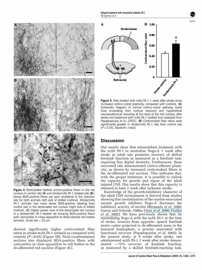
showed significantly higher corticorubral fiberratios in stroke/mAb IN-1 animals as compared withcontrols (Po0.05) (Figure 5B). Nissl-counterstainedsections also displayed BDA-positive fibers withvaricosities in close apposition to cell bodies in thede-afferented red nucleus (Figure 4C).
Discussion
Our results show that intracerebral treatment withthe mAb IN-1 to neutralize Nogo-A 1 week afterstroke in adult rats promotes recovery of skilledforelimb function as measured in a forelimb task,requiring fine digital dexterity. Furthermore, theserecovered rats demonstrated cortico-efferent plasti-city, as shown by increased corticorubral fibers inthe de-afferented red nucleus. This indicates that,with the proper treatment, it is possible to unlockthe capacity for growth and repair of the adultinjured CNS. Our results show that this capacity isretained at least 1 week after ischemic stroke.
Knowledge of the growth-inhibitory character ofthe adult CNS environment is derived from studiesshowing that neutralization of the myelin-associatedneurite growth inhibitor Nogo-A decreases theinhibitory activity of myelin (Brosamle et al, 2000;Caroni and Schwab, 1988a; Chen et al, 2000; Fiedleret al, 2002). We have previously shown that, byneutralizing Nogo-A with the mAb IN-1 at the timeof stroke, neurons from opposite, spared forelimbmotor cortex projected to de-afferented areas in thelesioned hemisphere, a process associated withfunctional recovery (Papadopoulos et al, 2002). Inthe present study, at 9 weeks after stroke, ratsadministered mAb IN-1 1 week after stroke demon-strated B75% recovery of forelimb function,as measured by a skilled forelimb-reaching task.
Figure 4 Biotinylated dextran amine-positive fibers in the rednucleus in control rats (A) and stroke/mAb IN-1-treated rats (B).Dense BDA-positive fibers are seen ipsilateral to the injectionsite for both animals (left side of dotted midline). Stroke/mAbIN-1 animals had more dense BDA-positive labeling thancontrol rats in the denervated red nucleus (right side of dottedmidline). (C) Higher power view of the denervated red nucleusin a stroke/mAb IN-1-treated rat showing BDA-positive fiberswith varicosities in close apposition to Nissl-stained cell bodies(arrows). Scale bar¼20mm.
Figure 5 Rats treated with mAb IN-1 1 week after stroke showincreased cortico-rubral plasticity, compared with controls. (A)Schematic diagram of normal cortico-rubral pathway (solidlines emanating from cortical neurons) and hypotheticalneuroanatomical rerouting at the level of the red nucleus afterstroke and treatment with mAb IN-1 (dotted line) (adapted fromPapadopoulos et al, 2002). (B) Corticorubral fiber ratios weresignificantly greater in stroke/mAb IN-1 rats than control rats(Po0.05, Student’s t-test).
Delayed treatment with monoclonal antibody IN-1AB Seymour et al
1371
Journal of Cerebral Blood Flow & Metabolism (2005) 25, 1366–1375

Similarly, Papadopoulos et al (2002) found that at8 weeks after stroke, rats that had been administeredmAb IN-1 immediately after stroke demonstrated80% recovery of forelimb function on the exact sametask. Interestingly, in the present study, animalsadministered mAb IN-1 1 week after stroke showedsignificant recovery at 5 weeks after treatment,whereas animals administered mAb IN-1 at the timeof stroke (Papadopoulos et al, 2002), showedsignificant recovery a week later, that is, 6 weeksafter treatment. Therefore, delaying treatment withNogo-A blockade 1 week after stroke may actuallyshorten the time to recovery, while the level ofrecovery appears to be unchanged with such a delay.Studies comparing various treatment timepoints areunderway to determine the optimal time-to-treat-ment window. In a related study using a specificanti-Nogo-A antibody 24 h after stroke in adult rats,functional recovery in skilled forelimb use on theMontoya Staircase task was seen by 6 weeks afterstroke (Wiessner et al, 2003). In that study, increasednumbers of CST fibers originating in the uninjuredmotor cortex were found to sprout across themidline in the cervical spinal cord in animalstreated with anti-Nogo-A antibody. Further, similarto the work by Papadopoulos et al (2002), increasedaxonal sprouting was correlated to improvedsuccesses in the reaching task. A similar studyin rats targeted the Nogo-66 receptor (NgR), acommon receptor for the Nogo-66 inhibitory domainof Nogo-A, oligodendrocyte myelin glycoprotein/Omgp, and myelin-associated glycoprotein/MAG (Filbin, 2003; Lee et al, 2004). This studyreported functional recovery and cortico-efferentplasticity when an antagonist to NgR was adminis-tered continuously for over 2 months beginning7 days after stroke. Interestingly, NiG, a potentinhibitory domain of Nogo-A to which the mAbIN-1 binds, has recently been shown to mediategrowth inhibition independent of NgR, and thebinding site for NiG is physically distinct from thatof Nogo-66 on neural tissue (Schweigreiter et al,2004). Therefore, neutralization of Nogo-A withspecific antibodies targeting NiG remains a powerfulpotential treatment option to promote cortico-efferent plasticity and functional recovery afterischemic stroke.
In addition to behavioral and neuroanatomicalevidence of the capacity of the adult CNS to recoverlost function after cortical injury and anti-Nogo-Atreatment, our laboratory has shown that ratsadministered mAb IN-1 at the time of motor cortexinjury displayed functional reorganization of theopposite unablated cortex (Emerick et al, 2003). Inthis work, intracortical microstimulation of thenon-injured motor cortex resulted in a significantincrease in movements of the lesion-impaired fore-limb in animals treated with mAb IN-1. Thesestudies indicate that anti-Nogo-A treatment afterunilateral cortical injury provides a permissiveenvironment in which new growth from the
uninjured cortex results in functional connectionsthat restore motor movements.
Other processes may play important roles infunctional reorganization after focal ischemic stroke.For instance, changes in neuronal excitability due toreductions in binding of gamma amino butyric acid(GABA)-A receptors (Qu et al, 1998), as well asincreases in dendritic spine density and growth, andsynaptogenesis (Jones et al, 1996) have been foundafter unilateral ischemic lesions. Stroke lesions havebeen shown to induce the expression of growth-promoting factors, for example, brain-derivedneurotrophic factor (BDNF), basic fibroblast growthfactor (bFGF), and growth-associated molecules, forexample, growth-associated protein-43 (GAP-43)(Nudo, 1999). Blockade of one of these factors,bFGF, after cortical lesions in the rat resulted indecreased spontaneous recovery in the affectedforelimb as measured by the skilled forelimb-reach-ing task (Rowntree and Kolb, 1997). The adult CNSthus retains a repertoire of growth- and repair-related responses needed for new axonal growth andsynapse formation. These studies indicate that thesegrowth-related factors might be peaking in the earlyweeks after injury. Therefore, treatment with mAbIN-1 1 week after stroke might be an opportune timeto take advantage of the growth-promoting micro-environment.
Related work has shown that at 1 week after a highunilateral lesion of the CST in the rat withouttreatment, an increase in gene expression of growthfactors, extracellular matrix proteins, and axonalguidance molecules, in addition to compensatorysprouting from the uninjured CST, was found in thespinal cord (Bareyre et al, 2002). Rats administeredmAb IN-1 at the time of CST lesion had increasedtarget-specific, stable compensatory sproutingwithin the spinal cord. Anti-Nogo-A treatmentwas associated with increased gene expression ofgrowth-associated proteins, including GAP-43,BDNF, and insulin-like growth factor (IGF), but alsowith increased expression of cytoskeletal proteins.Taken together, these results suggest that at leasttwo mechanisms must be present for a meaningfulcompensatory reorganization of the injured CNS.The first is the release from inhibition resulting fromeffects of Nogo-A neutralization, and the second isthe denervation-induced modulation of guidancemolecules and growth-related factors that allowfibers to reinnervate denervated targets in anorganized manner.
Functional recovery after CNS lesions in theperinatal period is associated with a high degree ofneuroanatomical plasticity. For example, unilateralcortical lesions in the neonatal rat results in thedevelopment of bilateral connections with thestriatum (Kolb et al, 1992), thalamus (Yu et al,1995), red nucleus (Naus et al, 1985), tectum, basilarpontine gray (Kartje-Tillotson et al, 1986), andspinal cord (Rouiller et al, 1991). These new,lesion-induced pathways are thought to be the
Delayed treatment with monoclonal antibody IN-1AB Seymour et al
1372
Journal of Cerebral Blood Flow & Metabolism (2005) 25, 1366–1375
underlying substrate for functional recovery afterperinatal injury. Interestingly, we have found similarstructural plasticity after unilateral cortical lesion inadult rats followed by immediate anti-Nogo-Atherapy (Emerick and Kartje, 2004; Kartje et al,1999; Papadopoulos et al, 2002; Wenk et al, 1999). Inthe present study, using delayed anti-Nogo-A block-ade, we again found structural plasticity in the rednucleus, a well-established subcortical target of theforelimb motor cortex, and an important nucleus formotor control.
In this regard, special attention has been paid tothe cortico-rubral tract and the cortico-spinal tract,pathways which undergo neuroanatomical remodel-ing after motor cortex lesions (Iwaniuk andWhishaw, 2000; Papadopoulos et al, 2002) or CSTlesions (Raineteau et al, 2001, 2002) after anti-Nogo-A treatment. The cortico-rubral pathway in theadult rat is mainly an ipsilateral projection with avery minor contralateral contribution (Naus et al,1985; Z’Graggen et al, 1998). Although the CSTdirectly controls fine finger and hand movementsin humans, monkeys, and rats, the cortico-rubro-spinal system is also implicated in precise limbmovements, a function that parallels that of the CST(Naus et al, 1985). While selective lesions of therubro-spinal tract induce only slight deficits inreaching, when the motor cortex is removed inaddition to portions of the rubrospinal tract, greaterdeficits are seen (Whishaw et al, 1990). Electro-physiology studies have shown that, with regard tothe distribution of onsets of neuronal spiking,neuronal activity in the rubro-spinal tract during askilled forelimb-reaching task closely resembles thatof the CST (Hyland, 1998; Jarratt and Hyland, 1999).Neuroanatomical data from the present study showthat, compared with controls, animals administeredmAb IN-1 1 week after stroke had an increase inBDA-positive fiber density in the de-afferented rednucleus. The extent of neuroanatomical remodelingobserved in the present study does not appear tocorrespond with the remarkable behavioral recoveryon the skilled forelimb-reaching task after stroke anddelayed mAb IN-1 treatment. It is not known whatquantity of fiber growth within or towards de-afferented regions is necessary to facilitate func-tional recovery. Our studies would suggest that acomplete pathway is not needed to show somereturn of function after a CNS lesion (Papadopouloset al, 2002). As our animals did not completelyrecover, perhaps more fiber sprouting would haveincreased recovery.
Anti-Nogo-A treatment to promote axonal regen-eration and functional recovery was first demon-strated in vivo after spinal cord injury in a set ofseminal studies (Bregman et al, 1995; Schnell andSchwab, 1990). When treatment with the mAb IN-1and neurotrophin-3 (NT-3) was delayed for 2 weeksafter spinal cord injury, rats exhibited improvementin functional recovery, and regenerating CST fiberswere found (von Meyenburg et al, 1998). This was
one of the first studies to show that transected adultcorticospinal axons retain the ability to regenerate atleast a few weeks after injury. In another study,delayed blockade of NgR 1 week after spinal cordinjury was shown to promote functional recoveryand axonal outgrowth in the spinal cord (Li andStrittmatter, 2003). Thus, delayed anti-Nogo-A ther-apy is a promising treatment to enhance recoveryafter CNS injury. In conclusion, if the degree ofdebilitating functional impairment caused by strokecan be minimized through anti-Nogo-A therapyadministered in a more practical time window afterischemia, then a new era of stroke treatment optionsmay evolve.
Acknowledgements
Special thanks goes to Josh Rosales for his help inpreparing figures.
References
Bandtlow CE (2003) Regeneration in the central nervoussystem. Exp Gerontol 38:79–86
Bareyre FM, Haudenschild B, Schwab ME (2002) Long-lasting sprouting and gene expression changes inducedby the monoclonal antibody IN-1 in the adult spinalcord. J Neurosci 22:7097–110
Bonita R (1992) Epidemiology of stroke. Lancet 339:342–4Bonita R, Beaglehole R, Asplund K (1994) The worldwide
problem of stroke. Curr Opin Neurol 7:5–10Bregman BS, Kunkel-Bagden E, Schnell L, Dai HN, Gao D,
Schwab ME (1995) Recovery from spinal cord injurymediated by antibodies to neurite growth inhibitors.Nature 378:498–501
Brosamle C, Huber AB, Fiedler M, Skerra A, Schwab ME(2000) Regeneration of lesioned corticospinal tractfibers in the adult rat induced by a recombinant,humanized IN-1 antibody fragment. J Neurosci 20:8061–8
Caroni P, Schwab ME (1988a) Antibody against myelin-associated inhibitor of neurite growth neutralizesnonpermissive substrate properties of CNS whitematter. Neuron 1:85–96
Caroni P, Schwab ME (1988b) Two membrane proteinfractions from rat central myelin with inhibitoryproperties for neurite growth and fibroblast spreading.J Cell Biol 106:1281–8
Chen MS, Huber AB, van der Haar ME, Frank M, SchnellL, Spillmann AA, Christ F, Schwab ME (2000) Nogo-Ais a myelin-associated neurite outgrowth inhibitor andan antigen for monoclonal antibody IN-1. Nature403:434–9
Chen ST, Hsu CY, Hogan EL, Maricq H, Balentine JD(1986) A model of focal ischemic stroke in the rat:reproducible extensive cortical infarction. Stroke17:738–43
del Zoppo GJ (2000) Antithrombotic treatments in acuteischemic stroke. Curr Opin Hematol 7:309–15
Emerick AJ, Kartje GL (2004) Behavioral recovery andanatomical plasticity in adult rats after cortical lesion
Delayed treatment with monoclonal antibody IN-1AB Seymour et al
1373
Journal of Cerebral Blood Flow & Metabolism (2005) 25, 1366–1375

and treatment with monoclonal antibody IN-1. BehavBrain Res 152:315–25
Emerick AJ, Neafsey EJ, Schwab ME, Kartje GL (2003)Functional reorganization of the motor cortex in adultrats after cortical lesion and treatment with monoclonalantibody IN-1. J Neurosci 23:4826–30
Fiedler M, Horn C, Bandtlow C, Schwab ME, Skerra A(2002) An engineered IN-1 F(ab) fragment with im-proved affinity for the Nogo-A axonal growth inhibitorpermits immunochemical detection and showsenhanced neutralizing activity. Protein Eng 15:931–41
Filbin MT (2003) Myelin-associated inhibitors of axonalregeneration in the adult mammalian CNS. Nat RevNeurosci 4:703–13
GrandPre T, Li S, Strittmatter SM (2002) Nogo-66 receptorantagonist peptide promotes axonal regeneration.Nature 417:547–51
GrandPre T, Nakamura F, Vartanian T, Strittmatter SM(2000) Identification of the Nogo inhibitor of axonregeneration as a Reticulon protein. Nature 403:439–44
He Z, Koprivica V (2004) The Nogo signaling pathway forregeneration block. Annu Rev Neurosci 27:341–68
American Heart Association (2004) Heart Disease andStroke Statistics—2004 Update. Dallas, TX: AmericanHeart Association
Herzog A, Brosamle C (1997) ‘Semifree-floating’ treatment:a simple and fast method to process consecutivesections for immunohistochemistry and neuronaltracing. J Neurosci Methods 72:57–63
Huber AB, Weinmann O, Brosamle C, Oertle T, SchwabME (2002) Patterns of Nogo mRNA and proteinexpression in the developing and adult rat and afterCNS lesions. J Neurosci 22:3553–67
Hyland B (1998) Neural activity related to reaching andgrasping in rostral and caudal regions of rat motorcortex. Behav Brain Res 94:255–69
Iwaniuk AN, Whishaw IQ (2000) On the origin of skilledforelimb movements. Trends Neurosci 23:372–6
Jarratt H, Hyland B (1999) Neuronal activity in rat rednucleus during forelimb reach-to-grasp movements.Neuroscience 88:629–42
Jones TA, Kleim JA, Greenough WT (1996) Synaptogenesisand dendritic growth in the cortex opposite unilateralsensorimotor cortex damage in adult rats: a quantitativeelectron microscopic examination. Brain Res 733:142–8
Kartje GL, Schulz MK, Lopez-Yunez A, Schnell L, SchwabME (1999) Corticostriatal plasticity is restricted bymyelin-associated neurite growth inhibitors in theadult rat. Ann Neurol 45:778–86
Kartje-Tillotson G, Neafsey EJ, Castro AJ (1985) Electro-physiological analysis of motor cortical plasticityafter cortical lesions in newborn rats. Brain Res 332:103–11
Kartje-Tillotson G, Neafsey EJ, Castro AJ (1986) Topogra-phy of corticopontine remodelling after cortical lesionsin newborn rats. J Comp Neurol 250:206–14
Kartje-Tillotson G, O’Donoghue DL, Dauzvardis MF,Castro AJ (1987) Pyramidotomy abolishes the abnormalmovements evoked by intracortical microstimulation inadult rats thatsustained neonatal cortical lesions. BrainRes 415:172–7
Kawamata T, Speliotes EK, Finklestein SP (1997) The roleof polypeptide growth factors in recovery from stroke.Adv Neurol 73:377–82
Kolb B, Gibb R, van der Kooy D (1992) Cortical and striatalstructure and connectivity are altered by neonatalhemidecortication in rats. J Comp Neurol 322:311–24
Lee JK, Kim JE, Sivula M, Strittmatter SM (2004)Nogo receptor antagonism promotes stroke recoveryby enhancing axonal plasticity. J Neurosci 24:6209–6217
Leong SK, Lund RD (1973) Anomalous bilateral cortico-fugal pathways in albino rats after neonatal lesions.Brain Res 62:218–21
Li S, Strittmatter SM (2003) Delayed systemic Nogo-66receptor antagonist promotes recovery from spinal cordinjury. J Neurosci 23:4219–27
Minino AM, Arias E, Kochanek KD, Murphy SL, Smith BL(2002) Deaths: final data for 2000. Natl Vital Stat Rep50:1–119
Morbidity and Mortality Chart Book on Cardiovascular,Lung, and Blood Diseases (2004). National Heart Lungand Blood Institute
Naus C, Flumerfelt BA, Hrycyshyn AW (1985) Ananterograde HRP-WGA study of aberrant corticorubralprojections following neonatal lesions of the ratsensorimotor cortex. Exp Brain Res 59:365–71
Neafsey EJ, Bold EL, Haas G, Hurley-Gius KM, Quirk G,Sievert CF, Terreberry RR (1986) The organization of therat motor cortex: a microstimulation mapping study.Brain Res 396:77–96
Niederost B, Oertle T, Fritsche J, McKinney RA, BandtlowCE (2002) Nogo-A and myelin-associated glycoproteinmediate neurite growth inhibition by antagonisticregulation of RhoA and Rac1. J Neurosci 22:10368–76
Nudo RJ (1999) Recovery after damage to motor corticalareas. Curr Opin Neurobiol 9:740–7
Oertle T, van der Haar ME, Bandtlow CE, Robeva A,Burfeind P, Buss A, Huber AB, Simonen M, Schnell L,Brosamle C, Kaupmann K, Vallon R, Schwab ME(2003) Nogo-A inhibits neurite outgrowth and cellspreading with three discrete regions. J Neurosci 23:5393–406
Olson MF, Ashworth A, Hall A (1995) An essential role forRho, Rac, and Cdc42 GTPases in cell cycle progressionthrough G1. Science 269:1270–2
Papadopoulos CM, Tsai SY, Alsbiei T, O’Brien TE, SchwabME, Kartje GL (2002) Functional recovery and neuro-anatomical plasticity following middle cerebral arteryocclusion and IN-1 antibody treatment in the adult rat.Ann Neurol 51:433–41
Paxinos G, Watson C (1998) The Rat Brain in StereotaxicCoordinates. New York: Academic Press
Prinjha R, Moore SE, Vinson M, Blake S, Morrow R,Christie G, Michalovich D, Simmons DL, Walsh FS(2000) Inhibitor of neurite outgrowth in humans.Nature 403:383–4
Qu M, Buchkremer-Ratzmann I, Schiene K, Schroeter M,Witte OW, Zilles K (1998) Bihemispheric reduction ofGABAA receptor binding following focal corticalphotothrombotic lesions in the rat brain. Brain Res813:374–80
Raineteau O, Fouad K, Bareyre FM, Schwab ME (2002)Reorganization of descending motor tracts in the ratspinal cord. Eur J Neurosci 16:1761–71
Raineteau O, Fouad K, Noth P, Thallmair M, Schwab ME(2001) Functional switch between motor tracts in thepresence of the mAb IN-1 in the adult rat. Proc NatlAcad Sci USA 98:6929–34
Rouiller EM, Liang FY, Moret V, Wiesendanger M (1991)Trajectory of redirected corticospinal axons after uni-lateral lesion of the sensorimotor cortex in neonatal rat;a phaseolus vulgaris-leucoagglutinin (PHA-L) tracingstudy. Exp Neurol 114:53–65
Delayed treatment with monoclonal antibody IN-1AB Seymour et al
1374
Journal of Cerebral Blood Flow & Metabolism (2005) 25, 1366–1375

Rowntree S, Kolb B (1997) Blockade of basic fibroblastgrowth factor retards recovery from motor cortex injuryin rats. Eur J Neurosci 9:2432–41
Schnell L, Schwab ME (1990) Axonal regeneration inthe rat spinal cord produced by an antibody againstmyelin-associated neurite growth inhibitors. Nature343:269–72
Schwab ME (2004) Nogo and axon regeneration. Curr OpinNeurobiol 14:118–24
Schweigreiter R, Walmsley AR, Niederost B, ZimmermannDR, Oertle T, Casademunt E, Frentzel S, Dechant G,Mir A, Bandtlow CE (2004) Versican V2 and the centralinhibitory domain of Nogo-A inhibit neurite growth viap75(NTR)/NgR-independent pathways that converge atRhoA. Mol Cell Neurosci 27:163–74
Thallmair M, Metz GA, Z’Graggen WJ, Raineteau O, KartjeGL, Schwab ME (1998) Neurite growth inhibitorsrestrict plasticity and functional recovery followingcorticospinal tract lesions. Nat Neurosci 1:124–31
von Meyenburg J, Brosamle C, Metz GA, Schwab ME(1998) Regeneration and sprouting of chronicallyinjured corticospinal tract fibers in adult rats promotedby NT-3 and the mAb IN-1, which neutralizes myelin-associated neurite growth inhibitors. Exp Neurol154:583–94
Wenk CA, Thallmair M, Kartje GL, Schwab ME (1999)Increased corticofugal plasticity after unilateral cortical
lesions combined with neutralization of the IN-1antigen in adult rats. J Comp Neurol 410:143–57
Whishaw IQ, Gorny B (1996) Does the red nucleus providetonic support against which fractionated movementsoccur? A study on forepaw movements used in skilledreaching by the rat. Behav Brain Res 74:79–90
Whishaw IQ, Tomie JA, Ladowsky RL (1990) Red nucleuslesions do not affect limb preference or use, butexacerbate the effects of motor cortex lesions ongrasping in the rat. Behav Brain Res 40:131–44
Wiessner C, Bareyre FM, Allegrini PR, Mir AK, Frentzel S,Zurini M, Schnell L, Oertle T, Schwab ME (2003)Anti-Nogo-A antibody infusion 24 hours after experi-mental stroke improved behavioral outcome andcorticospinal plasticity in normotensive and sponta-neously hypertensive rats. J Cereb Blood Flow Metab23:154–65
Yu XH, Moret V, Rouiller EM (1995) Re-examination of theplasticity of the corticothalamic projection after uni-lateral neonatal lesion of the sensorimotor cortex in therat: a phaseolus vulgaris-leucoagglutinin tracing study.J Hirnforsch 36:123–33
Z’Graggen WJ, Metz GA, Kartje GL, Thallmair M, SchwabME (1998) Functional recovery and enhanced cortico-fugal plasticity after unilateral pyramidal tract lesionand blockade of myelin-associated neurite growthinhibitors in adult rats. J Neurosci 18:4744–57
Delayed treatment with monoclonal antibody IN-1AB Seymour et al
1375
Journal of Cerebral Blood Flow & Metabolism (2005) 25, 1366–1375
Related Documents