Deferasirox and deferiprone remove cardiac iron in the iron- overloaded gerbil JOHN C. WOOD, MAYA OTTO-DUESSEL, IGNACIO GONZALEZ, MICHELLE I. AGUILAR, HIRO SHIMADA, HANSPETER NICK, MARVIN NELSON, and REX MOATS Division of Cardiology, the Department of Pediatric Radiology, and the Department of Pathology, Childrens Hospital of Los Angeles, Los Angeles, Calif; and Novartis Pharma, AG, Basel, Switzerland Abstract Introduction—Deferasirox effectively controls liver iron concentration; however, little is known regarding its ability to remove stored cardiac iron. Deferiprone seems to have increased cardiac efficacy compared with traditional deferoxamine therapy. Therefore, the relative efficacy of deferasirox and deferiprone were compared in removing cardiac iron from iron-loaded gerbils. Methods—Twenty-nine 8- to 10-week-old female gerbils underwent 10 weekly iron dextran injections of 200 mg/kg/week. Prechelation iron levels were assessed in 5 animals, and the remainder received deferasirox 100 mg/kg/D po QD (n = 8), deferiprone 375 mg/kg/D po divided TID (n = 8), or sham chelation (n = 8), 5 days/week for 12 weeks. Results—Deferasirox reduced cardiac iron content 20.5%. No changes occurred in cardiac weight, myocyte hypertrophy, fibrosis, or weight-to-dry weight ratio. Deferasirox treatment reduced liver iron content 51%. Deferiprone produced comparable reductions in cardiac iron content (18.6% reduction). Deferiprone-treated hearts had greater mass (16.5% increase) and increased myocyte hypertrophy. Deferiprone decreased liver iron content 24.9% but was associated with an increase in liver weight and water content. Conclusion—Deferasirox and deferiprone were equally effective in removing stored cardiac iron in a gerbil animal model, but deferasirox removed more hepatic iron for a given cardiac iron burden. Transfusional iron overload is a major cause of morbidity and mortality in thalassemia, sickle- cell disease, and other chronic anemias. Regular transfusions deliver between 0.3 and 0.5 mg of iron per kg per day or nearly 10 g per year in a 70-kg man. 1 Although iron is toxic to many organ systems, cardiac deposition remains the leading cause of death. 2 Subcutaneous deferoxamine chelation prevents cardiac dysfunction, but the regimen is onerous, requiring subcutaneous infusions 8–12 h per day, 5–7 days per week. 3 Unfortunately, the discomfort and inconvenience of long, subcutaneous infusions discourages many patients from optimal therapy. Noncompliance is lethal; patients who take less than 225 doses/year have a 50% mortality by 30 years of age. 4 The oral chelator deferasirox offers inherent advantages with respect to chelation compliance. 5 Deferasirox can be administered as a single morning dose because of its long elimination half-life (11–19 h). 1,6 When administered at 20 mg/kg/day, deferasirox produces comparable iron balance to deferoxamine therapy administered at 40 mg/kg/day, 5 days weekly. 7,8 Reprint requests: John C. Wood, MD, PhD, Division of Cardiology, Mailstop 34, Childrens Hospital of Los Angeles, 4650 Sunset Blvd, Los Angeles, CA 90027-0034. [email protected]. NIH Public Access Author Manuscript Transl Res. Author manuscript; available in PMC 2010 July 2. Published in final edited form as: Transl Res. 2006 November ; 148(5): 272–280. doi:10.1016/j.trsl.2006.05.005. NIH-PA Author Manuscript NIH-PA Author Manuscript NIH-PA Author Manuscript

Welcome message from author
This document is posted to help you gain knowledge. Please leave a comment to let me know what you think about it! Share it to your friends and learn new things together.
Transcript
Deferasirox and deferiprone remove cardiac iron in the iron-overloaded gerbil
JOHN C. WOOD, MAYA OTTO-DUESSEL, IGNACIO GONZALEZ, MICHELLE I. AGUILAR, HIROSHIMADA, HANSPETER NICK, MARVIN NELSON, and REX MOATSDivision of Cardiology, the Department of Pediatric Radiology, and the Department of Pathology,Childrens Hospital of Los Angeles, Los Angeles, Calif; and Novartis Pharma, AG, Basel, Switzerland
AbstractIntroduction—Deferasirox effectively controls liver iron concentration; however, little is knownregarding its ability to remove stored cardiac iron. Deferiprone seems to have increased cardiacefficacy compared with traditional deferoxamine therapy. Therefore, the relative efficacy ofdeferasirox and deferiprone were compared in removing cardiac iron from iron-loaded gerbils.
Methods—Twenty-nine 8- to 10-week-old female gerbils underwent 10 weekly iron dextraninjections of 200 mg/kg/week. Prechelation iron levels were assessed in 5 animals, and the remainderreceived deferasirox 100 mg/kg/D po QD (n = 8), deferiprone 375 mg/kg/D po divided TID (n = 8),or sham chelation (n = 8), 5 days/week for 12 weeks.
Results—Deferasirox reduced cardiac iron content 20.5%. No changes occurred in cardiac weight,myocyte hypertrophy, fibrosis, or weight-to-dry weight ratio. Deferasirox treatment reduced liveriron content 51%. Deferiprone produced comparable reductions in cardiac iron content (18.6%reduction). Deferiprone-treated hearts had greater mass (16.5% increase) and increased myocytehypertrophy. Deferiprone decreased liver iron content 24.9% but was associated with an increase inliver weight and water content.
Conclusion—Deferasirox and deferiprone were equally effective in removing stored cardiac ironin a gerbil animal model, but deferasirox removed more hepatic iron for a given cardiac iron burden.
Transfusional iron overload is a major cause of morbidity and mortality in thalassemia, sickle-cell disease, and other chronic anemias. Regular transfusions deliver between 0.3 and 0.5 mgof iron per kg per day or nearly 10 g per year in a 70-kg man.1 Although iron is toxic to manyorgan systems, cardiac deposition remains the leading cause of death.2 Subcutaneousdeferoxamine chelation prevents cardiac dysfunction, but the regimen is onerous, requiringsubcutaneous infusions 8–12 h per day, 5–7 days per week.3 Unfortunately, the discomfort andinconvenience of long, subcutaneous infusions discourages many patients from optimaltherapy. Noncompliance is lethal; patients who take less than 225 doses/year have a 50%mortality by 30 years of age.4
The oral chelator deferasirox offers inherent advantages with respect to chelation compliance.5 Deferasirox can be administered as a single morning dose because of its long eliminationhalf-life (11–19 h).1,6 When administered at 20 mg/kg/day, deferasirox produces comparableiron balance to deferoxamine therapy administered at 40 mg/kg/day, 5 days weekly.7,8
Reprint requests: John C. Wood, MD, PhD, Division of Cardiology, Mailstop 34, Childrens Hospital of Los Angeles, 4650 Sunset Blvd,Los Angeles, CA 90027-0034. [email protected].
NIH Public AccessAuthor ManuscriptTransl Res. Author manuscript; available in PMC 2010 July 2.
Published in final edited form as:Transl Res. 2006 November ; 148(5): 272–280. doi:10.1016/j.trsl.2006.05.005.
NIH
-PA Author Manuscript
NIH
-PA Author Manuscript
NIH
-PA Author Manuscript
Although deferasirox seems to control total iron burden, little data exist regarding cardiacchelation efficacy. Deferasirox’s long half-life should suppress labile iron species, or NTBI,over an entire day.9 As the heart selectively takes up labile iron species, deferasirox may offergreater protection against cardiac iron uptake than intermittent deferoxamine therapy.10 Inmyocyte cultures, deferasirox readily enters myocytes, binds iron, and prevents redox-cycling;however, the ability for deferasirox to mobilize and remove stored cardiac iron has not beenwell characterized in either humans or animals.11,12
Therefore, the purpose of this study was to determine the efficacy of deferasirox to extractcardiac iron in a gerbil model. As deferiprone removes cardiac iron effectively in humans, thecardiac chelation efficacy of deferasirox was compared with comparably dosed deferiprone.The gerbil emulates many of the functional abnormalities observed in human ironcardiomyopathy.13–20 This model has also been used to study chelator efficacy.14,15,19–22 Thisstudy differs in that iron loading and chelation were performed sequentially, rather thanconcurrently, to assess stored iron mobilization rather than prophylaxis of iron accumulation.
METHODSAnimals and iron loading
All animal studies were conducted with approval of the IACUC of Children’s Hospital LosAngeles. Overall, twenty-nine 8- to 10-week-old female Mongolian gerbils (Merionesunguiculatus) were obtained from Charles River Laboratories and housed in the CHLA-accredited animal care facility. All animals received 10 weekly subcutaneous injections of irondextran (Sigma Chemical Co., St. Louis, Mo) at a dose of 200 mg/kg. After the last injection,a 13-day iron equilibration period was allowed before starting chelation therapy.
ChelationOverall, 5 animals were sacrificed before initiation of chelation therapy to characterize initialiron levels. The remaining 24 iron-loaded gerbils were divided into the 3 groups of 8 animalseach: sham-chelated gerbils, deferasirox, and deferiprone-treated animals. All animals receivedchelation for 12 weeks.
To avoid the stress of chronic, repeated gavage feeding, deferiprone and deferasirox werehomogeneously mixed in plain peanut butter for oral feeding via a 1-mL syringe; all chelatorswere provided by Novartis Pharma, AG (Basel, Switzerland).
Deferasirox was given at a single daily dose of 100 mg/kg and deferiprone at a dose of 375mg/kg/day divided into 3 equal doses. Chelator doses were based on a previous dose-findingstudy21; these doses represent 67% of human values when normalized for body surface area.Oral chelator administration gave the animal approximately 0.15 mL of peanut butter per day,providing <1/1000 of the binding capacity of the administered chelator in administrated iron.
MRIPilot data suggested strong hepatic efficacy using deferasirox,21 so liver R2 was measured in4 animals from the deferasirox group at 8 weeks to monitor for overchelation. The MRItechniques have previously been described.23
Electrocardiography and exercise testsElectrocardiography and exercise tests were performed at baseline, immediately beforechelation, and at the end of the study. Limb lead electrocardiography was performed using astandard electrocardiograph (Mac Vu; Marquette/GE Medical Systems, Milwaukee, Wis).Animals were sedated with a single intraperitoneal injection of ketamine (100 mg/kg) plus
WOOD et al. Page 2
Transl Res. Author manuscript; available in PMC 2010 July 2.
NIH
-PA Author Manuscript
NIH
-PA Author Manuscript
NIH
-PA Author Manuscript
xylosine (10 mg/kg) and positioned supine for electrode placement. PR, QRS, QTc, and RRintervals were averaged over 5 consecutive heartbeats.
Maximum running time was assessed on a rodent treadmill (Exer 6M; Columbus Instruments,Columbus, Ohio) equipped with an electrified grid. Gerbils were acclimated for 10 min attreadmill speeds of 10 m/min several hours before the exercise tests. Animals were run atsystematically increasing treadmill speeds, beginning at 10 m/min and increasing at a rate of2.5 m per min every 3 min. Gerbils were run to exhaustion, with exhaustion determined asspending more than 10 consecutive seconds on the stimulator grid, or staying on it for morethan half of the time.20 To ensure maximum effort, examinations were repeated 2 days apart,with the longer exercise time used for analysis.
Histology and organ iron assessmentEuthanization was performed with 5% CO2 according to institutional guidelines. Aftersacrifice, the hearts and livers were removed, weighed, and sent for quantitative irondetermination (Mayo Medical Laboratories, Rochester, Minn). Tissue dry weight and dryweight iron concentrations were recorded as well. Liver and heart were immersion-fixed in10% formalin, paraffin-embedded, and stained with Prussian blue, Masson’s trichrome, andH&E.
All histologic sections were reviewed in a blinded fashion by an experienced pathologist.Cardiac iron deposition, muscular hypertrophy, and fibrosis were scored with respect tolocation and intensity using a relative scale from 0 t o 4. Hepatic iron staining was assessedseparately in the sinusoidal cells and hepatocytes. The number, size, and staining intensity oflobular aggregates of reticuloendothelial cells were also scored on a 0 to 4 scale. The pathologistalso assigned a total iron score reflecting visually weighted contributions of each of these ironpools.
Portions of each heart were processed for electron microscopy using standard techniques.Imaging was performed on a Philips CM 12 transmission electron microscope in the ChildrensHospital Los Angeles Pathology Department.
StatisticsIron concentration, iron content, organ weight, and wet-to-dry weight ratio were analyzed usingone-way ANOVA over the 3 treatment arms (sham-chelated, deferasirox, and deferiprone-treated animals). The mean of each treatment group was compared with the mean value fromthe sham-chelated animals using Dunnett’s test, which corrects for multiple comparisons. Aone-sided test was used for iron concentrations and iron content, based on pilot datademonstrating chelator efficacy21; a two-sided analysis was used for organ weight and wet-to-dry ratio. Comparison of these variables between 10-week controls and sham-chelated animalswas performed by an unpaired t-test. Electrocardiographic intervals and running times wereprocessed in an identical manner. Linear regression was also used to assess the relationshipbetween electrocardiographic intervals and organ iron concentration. Histology scores wereassessed using Wilcoxon signed-rank analysis because of the limited number of grades usedin the scoring. Bonferroni correction was applied for multiple comparisons.
RESULTSAll animals tolerated the iron loading and chelation without any apparent ill effects. After 8weeks of chelation, the estimated iron concentration by MRI was 4.4-mg/g wet weight, sochelation was continued.23 One animal from the deferasirox group died from an anestheticcomplication. It was healthy before sedation.
WOOD et al. Page 3
Transl Res. Author manuscript; available in PMC 2010 July 2.
NIH
-PA Author Manuscript
NIH
-PA Author Manuscript
NIH
-PA Author Manuscript
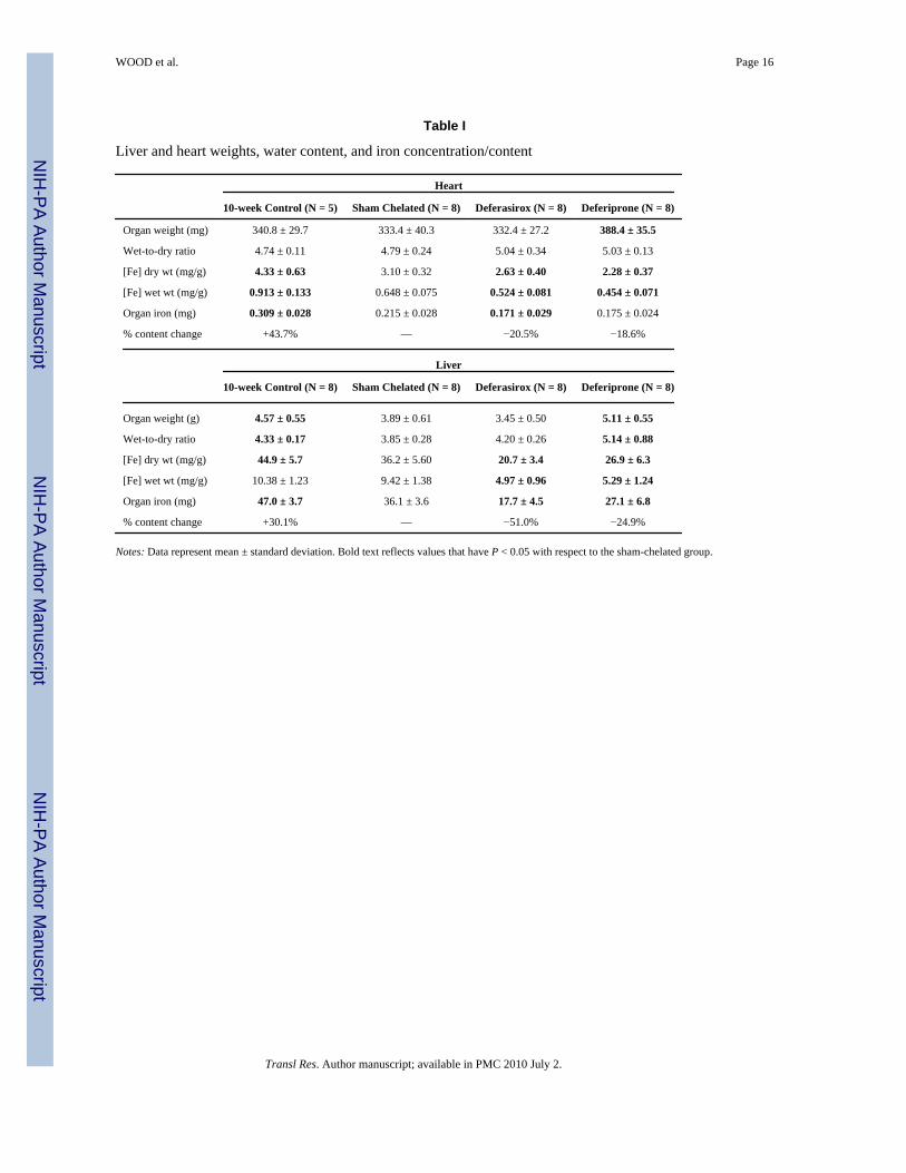
Chelation efficacy is summarized in Table I. Cardiac and liver iron concentrations and contentsafter sham chelation were significantly lower than observed in the 10-week control animals,representing spontaneous (not chelator-mediated) iron redistribution and elimination (P <0.001). All subsequent chelator comparisons are reported with respect to the sham-chelatedanimals, not the 10-week control animals.
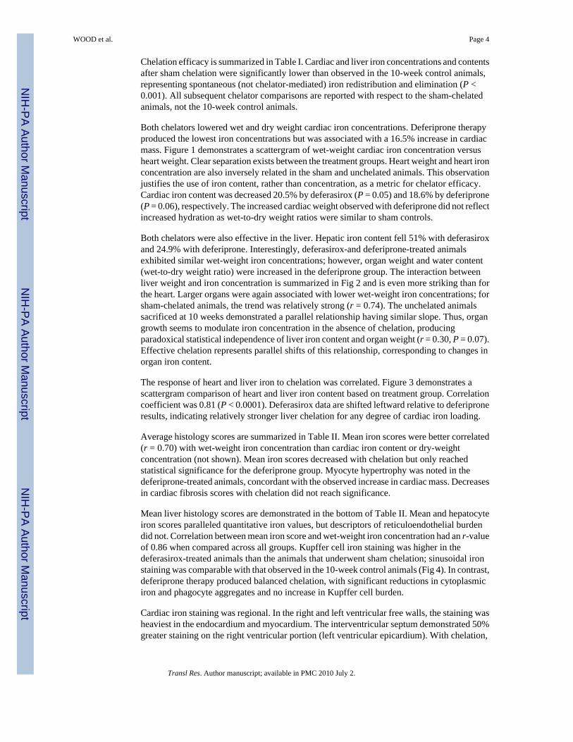
Both chelators lowered wet and dry weight cardiac iron concentrations. Deferiprone therapyproduced the lowest iron concentrations but was associated with a 16.5% increase in cardiacmass. Figure 1 demonstrates a scattergram of wet-weight cardiac iron concentration versusheart weight. Clear separation exists between the treatment groups. Heart weight and heart ironconcentration are also inversely related in the sham and unchelated animals. This observationjustifies the use of iron content, rather than concentration, as a metric for chelator efficacy.Cardiac iron content was decreased 20.5% by deferasirox (P = 0.05) and 18.6% by deferiprone(P = 0.06), respectively. The increased cardiac weight observed with deferiprone did not reflectincreased hydration as wet-to-dry weight ratios were similar to sham controls.
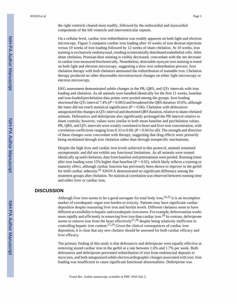
Both chelators were also effective in the liver. Hepatic iron content fell 51% with deferasiroxand 24.9% with deferiprone. Interestingly, deferasirox-and deferiprone-treated animalsexhibited similar wet-weight iron concentrations; however, organ weight and water content(wet-to-dry weight ratio) were increased in the deferiprone group. The interaction betweenliver weight and iron concentration is summarized in Fig 2 and is even more striking than forthe heart. Larger organs were again associated with lower wet-weight iron concentrations; forsham-chelated animals, the trend was relatively strong (r = 0.74). The unchelated animalssacrificed at 10 weeks demonstrated a parallel relationship having similar slope. Thus, organgrowth seems to modulate iron concentration in the absence of chelation, producingparadoxical statistical independence of liver iron content and organ weight (r = 0.30, P = 0.07).Effective chelation represents parallel shifts of this relationship, corresponding to changes inorgan iron content.
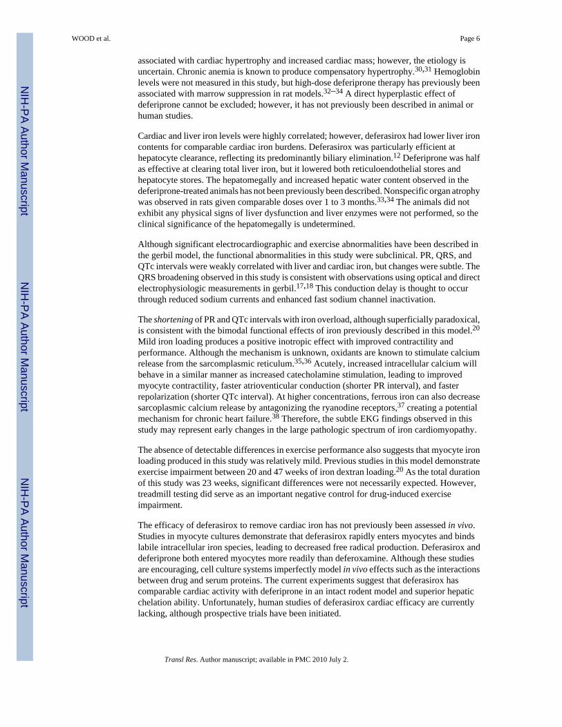
The response of heart and liver iron to chelation was correlated. Figure 3 demonstrates ascattergram comparison of heart and liver iron content based on treatment group. Correlationcoefficient was 0.81 (P < 0.0001). Deferasirox data are shifted leftward relative to deferiproneresults, indicating relatively stronger liver chelation for any degree of cardiac iron loading.
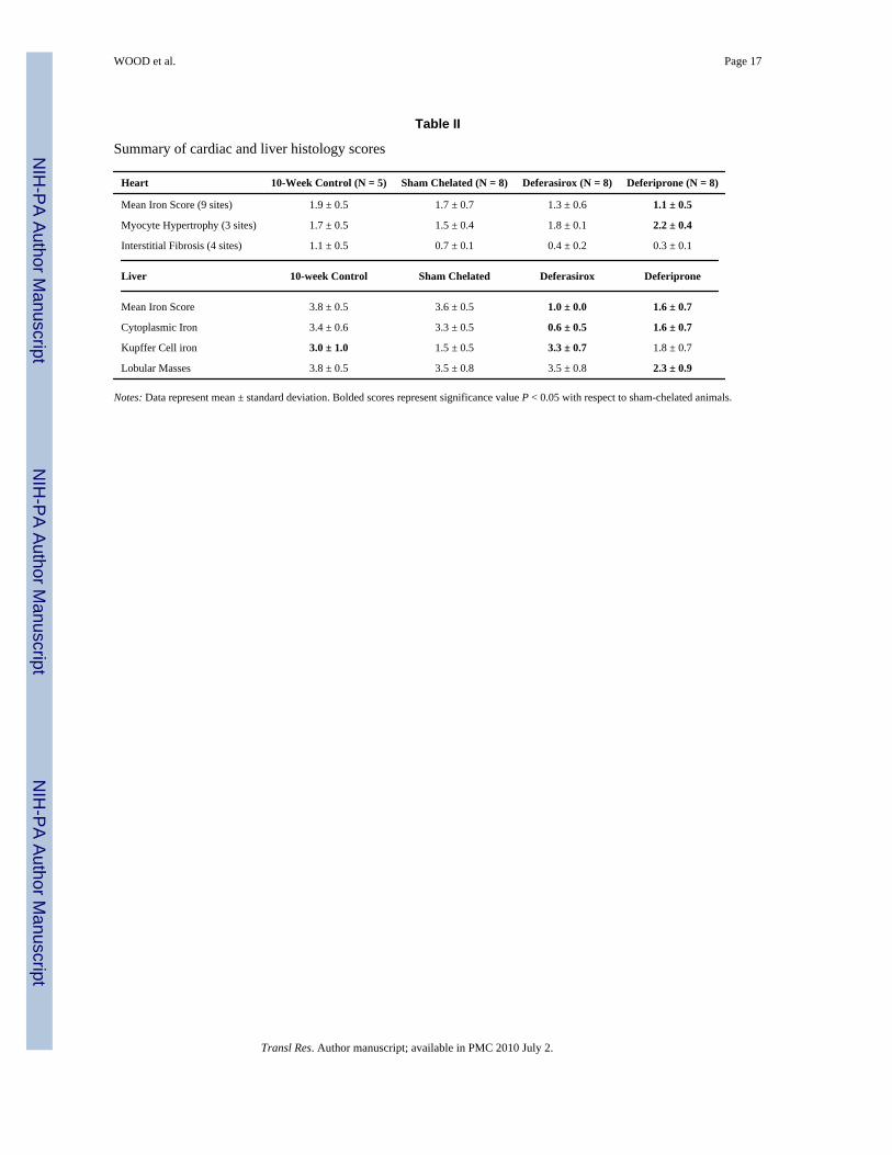
Average histology scores are summarized in Table II. Mean iron scores were better correlated(r = 0.70) with wet-weight iron concentration than cardiac iron content or dry-weightconcentration (not shown). Mean iron scores decreased with chelation but only reachedstatistical significance for the deferiprone group. Myocyte hypertrophy was noted in thedeferiprone-treated animals, concordant with the observed increase in cardiac mass. Decreasesin cardiac fibrosis scores with chelation did not reach significance.
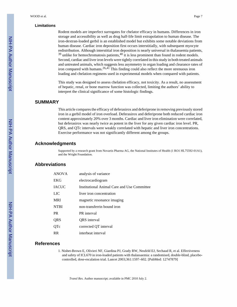
Mean liver histology scores are demonstrated in the bottom of Table II. Mean and hepatocyteiron scores paralleled quantitative iron values, but descriptors of reticuloendothelial burdendid not. Correlation between mean iron score and wet-weight iron concentration had an r-valueof 0.86 when compared across all groups. Kupffer cell iron staining was higher in thedeferasirox-treated animals than the animals that underwent sham chelation; sinusoidal ironstaining was comparable with that observed in the 10-week control animals (Fig 4). In contrast,deferiprone therapy produced balanced chelation, with significant reductions in cytoplasmiciron and phagocyte aggregates and no increase in Kupffer cell burden.
Cardiac iron staining was regional. In the right and left ventricular free walls, the staining washeaviest in the endocardium and myocardium. The interventricular septum demonstrated 50%greater staining on the right ventricular portion (left ventricular epicardium). With chelation,
WOOD et al. Page 4
Transl Res. Author manuscript; available in PMC 2010 July 2.
NIH
-PA Author Manuscript
NIH
-PA Author Manuscript
NIH
-PA Author Manuscript
the right ventricle cleared most readily, followed by the endocardial and myocardialcomponents of the left ventricle and interventricular septum.
On a cellular level, cardiac iron redistribution was readily apparent on both light and electronmicroscopy. Figure 5 compares cardiac iron loading after 10 weeks of iron dextran injectionsversus 10 weeks of iron loading followed by 12 weeks of sham chelation. At 10 weeks, ironstaining is exclusively endomyosial, residing in interstitially distributed endothelial cells. Aftersham chelation, Prussian-blue staining is visibly decreased, concordant with the net decreasein cardiac iron measured biochemically. Nonetheless, detectable myocyte iron staining is notedon both light and electron microscopy, suggesting a slow iron redistribution process. Ironchelation therapy with both chelators attenuated the redistribution of stainable iron. Chelationtherapy produced no other discernable microstructural changes on either light microscopy orelectron microscopy.
EKG assessment demonstrated subtle changes in the PR, QRS, and QTc intervals with ironloading and chelation. As all animals were handled identically for the first 11 weeks, baselineand iron-loaded/prechelation data points were pooled among the groups. Iron loadingshortened the QTc interval 7.4% (P = 0.003) and broadened the QRS duration 10.6%, althoughthe latter did not reach statistical significance (P = 0.06). Chelation with deferasiroxantagonized the changes in QTc interval and shortened QRS duration, relative to sham-chelatedanimals. Deferasirox and deferiprone also significantly prolonged the PR interval relative tosham controls; however, values were similar to both mean baseline and prechelation values.PR, QRS, and QTc intervals were weakly correlated to heart and liver iron concentration, withcorrelation coefficients ranging from 0.33 to 0.60, (P < 0.04 for all). The strength and directionof these changes were concordant with therapy, suggesting that drug effects were primarilybeing modulated through iron chelation rather than through nonspecific mechanisms.
Despite the high liver and cardiac iron levels achieved in this protocol, animals remainedasymptomatic and did not exhibit any functional limitations. As all animals were treatedidentically up until chelation, data from baseline and pretreatment were pooled. Running timesafter iron loading were 15% higher than baseline (P = 0.02), which likely reflects a training ormaturity effect, although cardiac function has previously been shown to improve in the gerbilfor mild cardiac siderosis.20 ANOVA demonstrated no significant difference among thetreatment groups after chelation. No statistical correlation was observed between running timeand either liver or cardiac iron.
DISCUSSIONAlthough liver iron seems to be a good surrogate for total body iron,24,25 it is an incompletemarker of extrahepatic organ iron burden or toxicity. Patients may have significant cardiacdeposition despite reassuring liver iron and ferritin levels. Different chelators seem to havedifferent accessibility to hepatic and extrahepatic iron stores. For example, deferoxamine worksmore rapidly and efficiently in removing liver iron than cardiac iron.26 In contrast, deferiproneseems to remove iron from the heart effectively27,28 despite being relatively inefficient incontrolling hepatic iron content.27,29 Given the clinical consequences of cardiac irondeposition, it is clear that any new chelator should be assessed for both cardiac efficacy andliver efficacy.
The primary finding of this study is that deferasirox and deferiprone were equally effective atremoving stored cardiac iron in the gerbil at a rate between 1.6% and 1.7% per week. Bothdeferasirox and deferiprone prevented redistribution of iron from endomysial deposits tomyocytes, and both antagonized subtle electrocardiographic changes associated with iron. Ironloading was insufficient to cause significant functional abnormalities. Deferiprone was
WOOD et al. Page 5
Transl Res. Author manuscript; available in PMC 2010 July 2.
NIH
-PA Author Manuscript
NIH
-PA Author Manuscript
NIH
-PA Author Manuscript
associated with cardiac hypertrophy and increased cardiac mass; however, the etiology isuncertain. Chronic anemia is known to produce compensatory hypertrophy.30,31 Hemoglobinlevels were not measured in this study, but high-dose deferiprone therapy has previously beenassociated with marrow suppression in rat models.32–34 A direct hyperplastic effect ofdeferiprone cannot be excluded; however, it has not previously been described in animal orhuman studies.
Cardiac and liver iron levels were highly correlated; however, deferasirox had lower liver ironcontents for comparable cardiac iron burdens. Deferasirox was particularly efficient athepatocyte clearance, reflecting its predominantly biliary elimination.12 Deferiprone was halfas effective at clearing total liver iron, but it lowered both reticuloendothelial stores andhepatocyte stores. The hepatomegally and increased hepatic water content observed in thedeferiprone-treated animals has not been previously been described. Nonspecific organ atrophywas observed in rats given comparable doses over 1 to 3 months.33,34 The animals did notexhibit any physical signs of liver dysfunction and liver enzymes were not performed, so theclinical significance of the hepatomegally is undetermined.
Although significant electrocardiographic and exercise abnormalities have been described inthe gerbil model, the functional abnormalities in this study were subclinical. PR, QRS, andQTc intervals were weakly correlated with liver and cardiac iron, but changes were subtle. TheQRS broadening observed in this study is consistent with observations using optical and directelectrophysiologic measurements in gerbil.17,18 This conduction delay is thought to occurthrough reduced sodium currents and enhanced fast sodium channel inactivation.
The shortening of PR and QTc intervals with iron overload, although superficially paradoxical,is consistent with the bimodal functional effects of iron previously described in this model.20
Mild iron loading produces a positive inotropic effect with improved contractility andperformance. Although the mechanism is unknown, oxidants are known to stimulate calciumrelease from the sarcomplasmic reticulum.35,36 Acutely, increased intracellular calcium willbehave in a similar manner as increased catecholamine stimulation, leading to improvedmyocyte contractility, faster atrioventicular conduction (shorter PR interval), and fasterrepolarization (shorter QTc interval). At higher concentrations, ferrous iron can also decreasesarcoplasmic calcium release by antagonizing the ryanodine receptors,37 creating a potentialmechanism for chronic heart failure.38 Therefore, the subtle EKG findings observed in thisstudy may represent early changes in the large pathologic spectrum of iron cardiomyopathy.
The absence of detectable differences in exercise performance also suggests that myocyte ironloading produced in this study was relatively mild. Previous studies in this model demonstrateexercise impairment between 20 and 47 weeks of iron dextran loading.20 As the total durationof this study was 23 weeks, significant differences were not necessarily expected. However,treadmill testing did serve as an important negative control for drug-induced exerciseimpairment.
The efficacy of deferasirox to remove cardiac iron has not previously been assessed in vivo.Studies in myocyte cultures demonstrate that deferasirox rapidly enters myocytes and bindslabile intracellular iron species, leading to decreased free radical production. Deferasirox anddeferiprone both entered myocytes more readily than deferoxamine. Although these studiesare encouraging, cell culture systems imperfectly model in vivo effects such as the interactionsbetween drug and serum proteins. The current experiments suggest that deferasirox hascomparable cardiac activity with deferiprone in an intact rodent model and superior hepaticchelation ability. Unfortunately, human studies of deferasirox cardiac efficacy are currentlylacking, although prospective trials have been initiated.
WOOD et al. Page 6
Transl Res. Author manuscript; available in PMC 2010 July 2.
NIH
-PA Author Manuscript
NIH
-PA Author Manuscript
NIH
-PA Author Manuscript
LimitationsRodent models are imperfect surrogates for chelator efficacy in humans. Differences in ironstorage and accessibility as well as drug half-life limit extrapolation to human disease. Theiron-dextran-loaded gerbil is an established model but exhibits some notable deviations fromhuman disease. Cardiac iron deposition first occurs interstitially, with subsequent myocyteredistribution. Although interstitial iron deposition is nearly universal in thalassemia patients,39 unlike for hemochromatosis patients,40 it is less prominent than found in rodent models.Second, cardiac and liver iron levels were tightly correlated in this study in both treated animalsand untreated animals, which suggests less asymmetry in organ loading and clearance rates ofiron compared with humans.26,41 This finding could also reflect the more strenuous ironloading and chelation regimens used in experimental models when compared with patients.
This study was designed to assess chelation efficacy, not toxicity. As a result, no assessmentof hepatic, renal, or bone marrow function was collected, limiting the authors’ ability tointerpret the clinical significance of some histologic findings.
SUMMARYThis article compares the efficacy of deferasirox and deferiprone in removing previously storediron in a gerbil model of iron overload. Deferasirox and deferiprone both reduced cardiac ironcontent approximately 20% over 3 months. Cardiac and liver iron elimination were correlated,but deferasirox was nearly twice as potent in the liver for any given cardiac iron level. PR,QRS, and QTc intervals were weakly correlated with hepatic and liver iron concentrations.Exercise performance was not significantly different among the groups.
AcknowledgmentsSupported by a research grant from Novartis Pharma AG, the National Institutes of Health (1 RO1 HL75592-01A1),and the Wright Foundation.
Abbreviations
ANOVA analysis of variance
EKG electrocardiogram
IACUC Institutional Animal Care and Use Committee
LIC liver iron concentration
MRI magnetic resonance imaging
NTBI non-transferrin bound iron
PR PR interval
QRS QRS interval
QTc corrected QT interval
RR interbeat interval
References1. Nisbet-Brown E, Olivieri NF, Giardina PJ, Grady RW, Neufeld EJ, Sechaud R, et al. Effectiveness
and safety of ICL670 in iron-loaded patients with thalassaemia: a randomised, double-blind, placebo-controlled, dose-escalation trial. Lancet 2003;361:1597–602. [PubMed: 12747879]
WOOD et al. Page 7
Transl Res. Author manuscript; available in PMC 2010 July 2.
NIH
-PA Author Manuscript
NIH
-PA Author Manuscript
NIH
-PA Author Manuscript
2. Borgna-Pignatti C, Rugolotto S, De Stefano P, Piga A, Di Gregorio F, Gamberini MR, et al. Survivaland complications in patients with thalassemia major treated with transfusion and deferoxamine.Haematologica 2004;89:1187–93. [PubMed: 15477202]
3. Olivieri NF, Brittenham GM. Iron-chelating therapy and the treatment of thalassemia. Blood1997;89:739–61. [PubMed: 9028304]
4. Gabutti V, Piga A. Results of long-term iron-chelating therapy. Acta Haematol 1996;95:26–36.[PubMed: 8604584]
5. Hershko C, Link G, Konijn AM, Cabantchik ZI. Iron chelation therapy. Curr Hematol Rep 2005;4:110–6. [PubMed: 15720959]
6. Galanello R, Piga A, Alberti D, Rouan MC, Bigler H, Sechaud R. Safety, tolerability, andpharmacokinetics of ICL670, a new orally active iron-chelating agent in patients with transfusion-dependent iron overload due to beta-thalassemia. J Clin Pharmacol 2003;43:565–72. [PubMed:12817519]
7. Piga A, Galanello R, Foschini ML, Zappu A, Bordone E, Longo F, et al. Once-daily treatment withthe oral chelator ICL670(Exjade): results of a phase II study in pediatric patients with beta-thalassemiamajor. Blood 2004;11:983a.
8. Porter J, Vichinsky E, Rose C, Piga A, Olivieri N, Gattermann N, et al. A phase II study with ICL670,a once-daily oral iron chelator, in patients with various transfusional dependent anemias and ironoverload. Blood 200;11:872a.
9. Cabantchik ZI, Breuer W, Zanninelli G, Cianciulli P. LPI-labile plasma iron in iron overload. BestPract Res Clin Haematol 2005;18:277–87. [PubMed: 15737890]
10. Parkes JG, Olivieri NF, Templeton DM. Characterization of Fe2+ and Fe3+ transport by iron-loadedcardiac myocytes. Toxicology 1997;117:141–51. [PubMed: 9057893]
11. Hasinoff BB, Patel D, Wu X. The oral iron chelator ICL670A (deferasirox) does not protect myocytesagainst doxorubicin. Free Radic Biol Med 2003;35:1469–79. [PubMed: 14642395]
12. Hershko C, Konijn AM, Nick HP, Breuer W, Cabantchik ZI, Link G. ICL670A: a new synthetic oralchelator: evaluation in hypertransfused rats with selective radioiron probes of hepatocellular andreticuloendothelial iron stores and in iron-loaded rat heart cells in culture. Blood 2001;97:1115–22.[PubMed: 11159545]
13. Carthew P, Dorman BM, Edwards RE, Francis JE, Smith AG. A unique rodent model for both thecardiotoxic and hepatotoxic effects of prolonged iron overload. Lab Invest 1993;69:217–22.[PubMed: 7688836]
14. Carthew P, Smith AG, Hider RC, Dorman B, Edwards RE, Francis JE. Potentiation of ironaccumulation in cardiac myocytes during the treatment of iron overload in gerbils with thehydroxypyridinone iron chelator CP94. Biometals 1994;7:267–71. [PubMed: 7812111]
15. Hershko C, Link G, Konijn AM, Huerta M, Rosenmann E, Reinus C. The iron-loaded gerbil modelrevisited: effects of deferoxamine and deferiprone treatment. J Lab Clin Med 2002;139:50–8.[PubMed: 11873245]
16. Kaiser L, Davis JM, Schwartz KA. Does the gerbil model mimic human iron overload? J Lab ClinMed 2003;141:419–20. author reply 420–21. [PubMed: 12819640]
17. Kuryshev YA, Brittenham GM, Fujioka H, Kannan P, Shieh C-C, Cohen SA, et al. Decreased sodiumand increased transient outward potassium currents in iron-loaded cardiac myocytes. Implicationsfor the arrhythmogenesis of human siderotic heart disease. Circulation 1999;100:675–83. [PubMed:10441107]
18. Laurita KR, Chuck ET, Yang T, Dong WQ, Kuryshev YA, Brittenham GM, et al. Optical mappingreveals conduction slowing and impulse block in iron-overload cardiomyopathy. J Lab Clin Med2003;142:83–9. [PubMed: 12960954]
19. Obejero-Paz CA, Yang T, Dong WQ, Levy MN, Brittenjam GM, Kuryshev YA, et al. Deferoxaminepromotes survival and prevents electrocardiographic abnormalities in the gerbil model of iron-overload cardiomyopathy. J Lab Clin Med 2003;141:121–30. [PubMed: 12577048]
20. Yang T, Dong WQ, Kuryshev YA, Obejero-Paz CA, Levy MN, Brittenham GM, et al. Bimodalcardiac dysfunction in an animal model of iron overload. J Lab Clin Med 2002;140:263–71. [PubMed:12389025]
WOOD et al. Page 8
Transl Res. Author manuscript; available in PMC 2010 July 2.
NIH
-PA Author Manuscript
NIH
-PA Author Manuscript
NIH
-PA Author Manuscript
21. Wood JC, Otto-Duessel M, Aguilar M, Nick H, Coates TD, Moats R. Dose response of deferoxamine,deferiprone, and ICL670 chelation therapy in a gerbil model of iron overload. Blood 2004;104:985a.
22. Yang T, Brittenham GM, Dong WQ, Levy MN, Obejero-Paz CA, Kuryshev YA, et al. Deferoxamineprevents cardiac hypertrophy and failure in the gerbil model of iron-induced cardiomyopathy. J LabClin Med 2003;142:332–40. [PubMed: 14647037]
23. Wood JC, Otto-Duessel M, Aguilar M, Nick H, Nelson MD, Coates TD, et al. Cardiac iron determinescardiac T2*, T2, and T1 in the gerbil model of iron cardiomyopathy. Circulation 2005;112:535–43.[PubMed: 16027257]
24. Brittenham GM, Griffith PM, Ninhuis AW, McLaren CE, Yound NS, Tucker EE, et al. Efficacy ofdeferoxamine in preventing complications of iron overload in patients with thalassemia major. NEngl J Med 1994;331:567–73. [PubMed: 8047080]
25. Angelucci E, Brittenham GM, McLaren CE, Ripalti M, Baronciani D, Giardini C, et al. Hepatic ironconcentration and total body iron stores in thalassemia major. N Engl J Med 2000;343:327–31.[PubMed: 10922422]
26. Anderson LJ, Westwood MA, Holden S, Davis B, Prescott E, Wonke B, et al. Myocardial ironclearance during reversal of siderotic cardiomyopathy with intravenous desferrioxamine: aprospective study using T2* cardiovascular magnetic resonance. Br J Haematol 2004;127:348–55.[PubMed: 15491298]
27. Anderson LJ, Wonke B, Prescott E, Holden S, Walker JM, Pennell DJ. Comparison of effects of oraldeferiprone and subcutaneous desferrioxamine on myocardial iron concentrations and ventricularfunction in beta-thalassaemia. Lancet 2002;360:516–20. [PubMed: 12241655]
28. Piga A, Gaglioti C, Fogliacco E, Tricta F. Comparative effects of deferiprone and deferoxamine onsurvival and cardiac disease in patients with thalassemia major: a retrospective analysis.Haematologica 2003;88:489–96. [PubMed: 12745268]
29. Addis A, Loebstein R, Koren G, Einarson TR. Meta-analytic review of the clinical effectiveness oforal deferiprone (L1). Eur J Clin Pharmacol 1999;55:1–6. [PubMed: 10206077]
30. Aessopos A, Farmakis D, Hatziliami A, Fragodimitri C, Karabatsos F, Joussef J, et al. Cardiac statusin well-treated patients with thalassemia major. Eur J Haematol 2004;73:359–66. [PubMed:15458515]
31. Covitz W, Espeland M, Gallagher D, Hellenbrand W, Leff S, Talner N. The heart in sickle cell anemia.The Cooperative Study of Sickle Cell Disease (CSSCD). Chest 1995;108:1214–9. [PubMed:7587419]
32. Kontoghiorghes GJ, Nasseri-Sina P, Goddard JG, Barr JM, Nortey P, Sheppard LN. Safety of oraliron chelator L1. Lancet 1989;2:457–8.
33. Grady RW, Srinivasan R, Dunn JB, Coursey MP, Lemert RF, Calvano SE. Evidence of toxicity dueto 1,2-dimethyl-3-hydroxypyrid-4-one (L1) in normal rats. Drugs Today 1992;28(Suppl A):S73–9.
34. Berdoukas V, Bentley P, Frost H, Schnebli HP. Toxicity of oral iron chelator L1. Lancet1993;341:1088. [PubMed: 8096974]
35. Cheong E, Tumbev V, Abramson J, Salama G, Stoyanovsky DA. Nitroxyl triggers Ca2+ release fromskeletal and cardiac sarcoplasmic reticulum by oxidizing ryanodine receptors. Cell Calcium2005;37:87–96. [PubMed: 15541467]
36. Hidalgo C, Aracena P, Sanchez G, Donoso P. Redox regulation of calcium release in skeletal andcardiac muscle. Biol Res 2002;35:183–93. [PubMed: 12415735]
37. Kim E, Giri SN, Pessah IN. Iron(II) is a modulator of ryanodine-sensitive calcium channels of cardiacmuscle sarcoplasmic reticulum. Toxicol Appl Pharmacol 1995;130:57–66. [PubMed: 7530865]
38. Scoote M, Williams AJ. The cardiac ryanodine receptor (calcium release channel): emerging role inheart failure and arrhythmia pathogenesis. Cardiovasc Res 2002;56:359–72. [PubMed: 12445877]
39. Buja LM, Roberts WC. Iron in the heart. Etiology and clinical significance. Am J Med 1971;51:209–21. [PubMed: 5095527]
40. Olson LJ, Edwards WD, McCall JT, Ilstrup DM, Gersh BJ. Cardiac iron deposition in idiopathichemochromatosis: histologic and analytic assessment of 14 hearts from autopsy. J Am Coll Cardiol1987;10:1239–43. [PubMed: 3680791]
WOOD et al. Page 9
Transl Res. Author manuscript; available in PMC 2010 July 2.
NIH
-PA Author Manuscript
NIH
-PA Author Manuscript
NIH
-PA Author Manuscript
41. Wood JC, Tyszka JM, Ghugre N, Carson S, Nelson MD, Coates TD. Myocardial iron loading intransfusion-dependent thalassemia and sickle-cell disease. Blood 2004;103:1934–6. [PubMed:14630822]
WOOD et al. Page 10
Transl Res. Author manuscript; available in PMC 2010 July 2.
NIH
-PA Author Manuscript
NIH
-PA Author Manuscript
NIH
-PA Author Manuscript
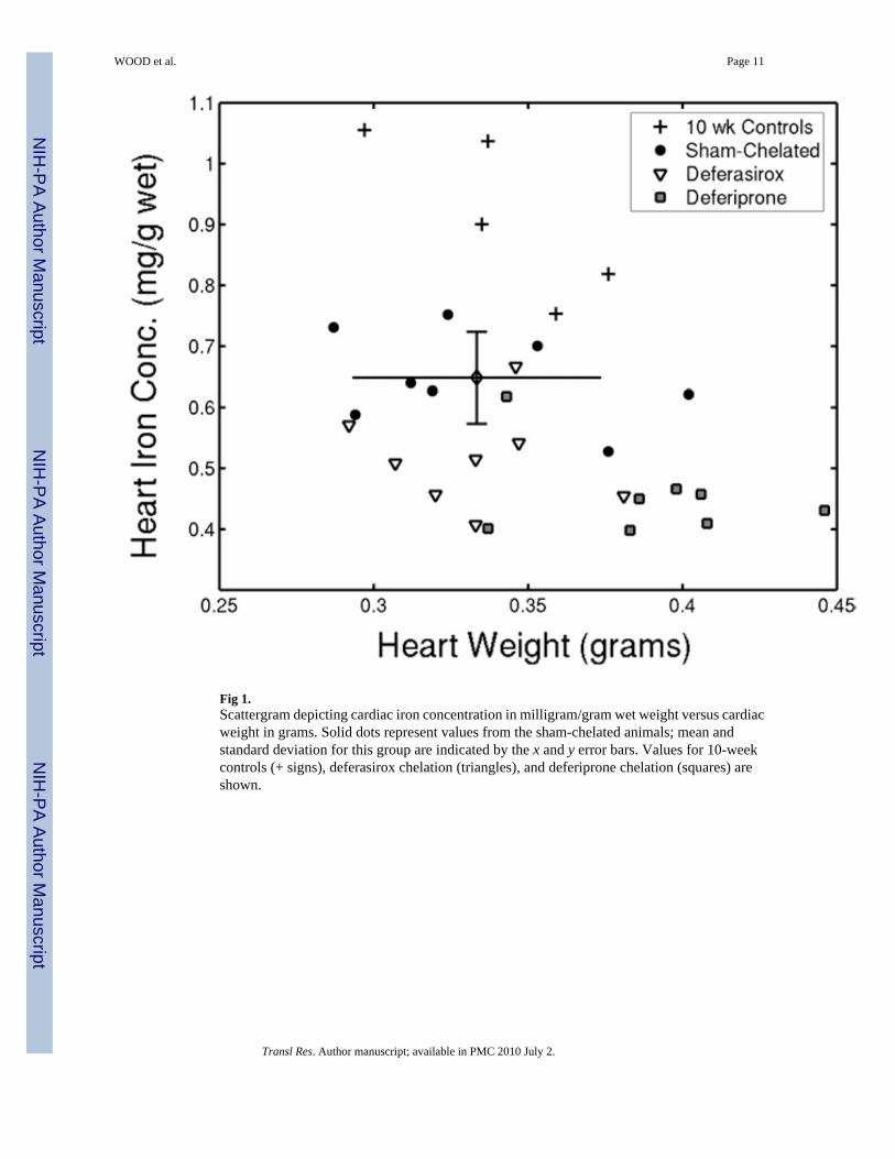
Fig 1.Scattergram depicting cardiac iron concentration in milligram/gram wet weight versus cardiacweight in grams. Solid dots represent values from the sham-chelated animals; mean andstandard deviation for this group are indicated by the x and y error bars. Values for 10-weekcontrols (+ signs), deferasirox chelation (triangles), and deferiprone chelation (squares) areshown.
WOOD et al. Page 11
Transl Res. Author manuscript; available in PMC 2010 July 2.
NIH
-PA Author Manuscript
NIH
-PA Author Manuscript
NIH
-PA Author Manuscript
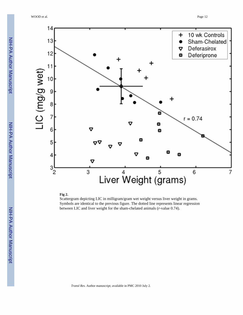
Fig 2.Scattergram depicting LIC in milligram/gram wet weight versus liver weight in grams.Symbols are identical to the previous figure. The dotted line represents linear regressionbetween LIC and liver weight for the sham-chelated animals (r-value 0.74).
WOOD et al. Page 12
Transl Res. Author manuscript; available in PMC 2010 July 2.
NIH
-PA Author Manuscript
NIH
-PA Author Manuscript
NIH
-PA Author Manuscript
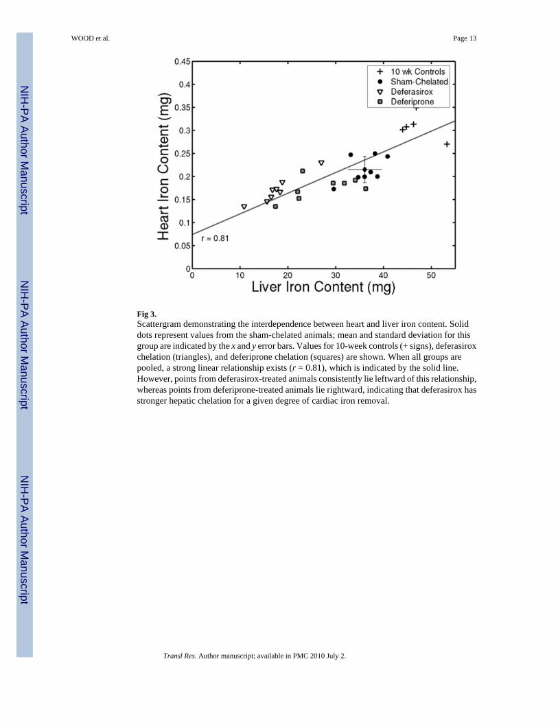
Fig 3.Scattergram demonstrating the interdependence between heart and liver iron content. Soliddots represent values from the sham-chelated animals; mean and standard deviation for thisgroup are indicated by the x and y error bars. Values for 10-week controls (+ signs), deferasiroxchelation (triangles), and deferiprone chelation (squares) are shown. When all groups arepooled, a strong linear relationship exists (r = 0.81), which is indicated by the solid line.However, points from deferasirox-treated animals consistently lie leftward of this relationship,whereas points from deferiprone-treated animals lie rightward, indicating that deferasirox hasstronger hepatic chelation for a given degree of cardiac iron removal.
WOOD et al. Page 13
Transl Res. Author manuscript; available in PMC 2010 July 2.
NIH
-PA Author Manuscript
NIH
-PA Author Manuscript
NIH
-PA Author Manuscript
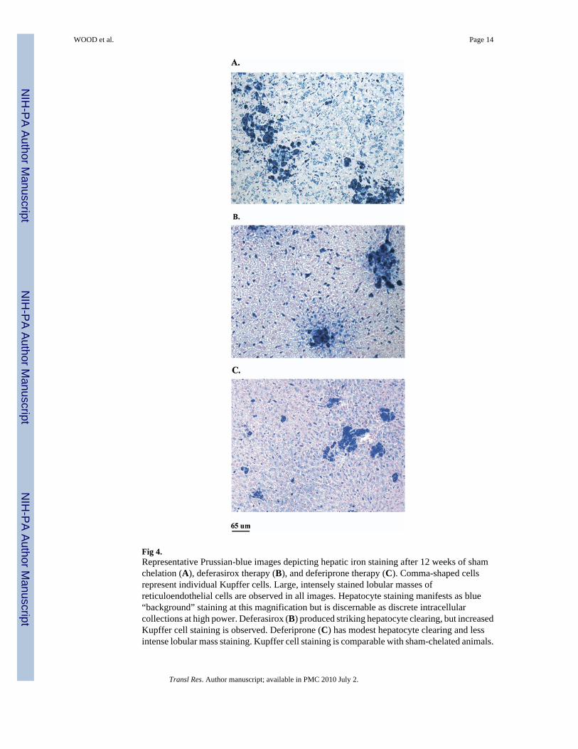
Fig 4.Representative Prussian-blue images depicting hepatic iron staining after 12 weeks of shamchelation (A), deferasirox therapy (B), and deferiprone therapy (C). Comma-shaped cellsrepresent individual Kupffer cells. Large, intensely stained lobular masses ofreticuloendothelial cells are observed in all images. Hepatocyte staining manifests as blue“background” staining at this magnification but is discernable as discrete intracellularcollections at high power. Deferasirox (B) produced striking hepatocyte clearing, but increasedKupffer cell staining is observed. Deferiprone (C) has modest hepatocyte clearing and lessintense lobular mass staining. Kupffer cell staining is comparable with sham-chelated animals.
WOOD et al. Page 14
Transl Res. Author manuscript; available in PMC 2010 July 2.
NIH
-PA Author Manuscript
NIH
-PA Author Manuscript
NIH
-PA Author Manuscript
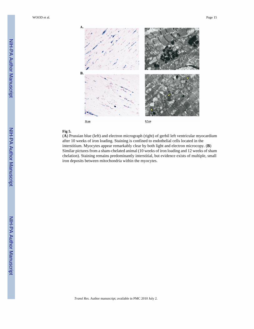
Fig 5.(A) Prussian blue (left) and electron micrograph (right) of gerbil left ventricular myocardiumafter 10 weeks of iron loading. Staining is confined to endothelial cells located in theinterstitium. Myocytes appear remarkably clear by both light and electron microcopy. (B)Similar pictures from a sham-chelated animal (10 weeks of iron loading and 12 weeks of shamchelation). Staining remains predominantly interstitial, but evidence exists of multiple, smalliron deposits between mitochondria within the myocytes.
WOOD et al. Page 15
Transl Res. Author manuscript; available in PMC 2010 July 2.
NIH
-PA Author Manuscript
NIH
-PA Author Manuscript
NIH
-PA Author Manuscript
NIH
-PA Author Manuscript
NIH
-PA Author Manuscript
NIH
-PA Author Manuscript
WOOD et al. Page 16
Table I
Liver and heart weights, water content, and iron concentration/content
Heart
10-week Control (N = 5) Sham Chelated (N = 8) Deferasirox (N = 8) Deferiprone (N = 8)
Organ weight (mg) 340.8 ± 29.7 333.4 ± 40.3 332.4 ± 27.2 388.4 ± 35.5
Wet-to-dry ratio 4.74 ± 0.11 4.79 ± 0.24 5.04 ± 0.34 5.03 ± 0.13
[Fe] dry wt (mg/g) 4.33 ± 0.63 3.10 ± 0.32 2.63 ± 0.40 2.28 ± 0.37
[Fe] wet wt (mg/g) 0.913 ± 0.133 0.648 ± 0.075 0.524 ± 0.081 0.454 ± 0.071
Organ iron (mg) 0.309 ± 0.028 0.215 ± 0.028 0.171 ± 0.029 0.175 ± 0.024
% content change +43.7% — −20.5% −18.6%
Liver
10-week Control (N = 8) Sham Chelated (N = 8) Deferasirox (N = 8) Deferiprone (N = 8)
Organ weight (g) 4.57 ± 0.55 3.89 ± 0.61 3.45 ± 0.50 5.11 ± 0.55
Wet-to-dry ratio 4.33 ± 0.17 3.85 ± 0.28 4.20 ± 0.26 5.14 ± 0.88
[Fe] dry wt (mg/g) 44.9 ± 5.7 36.2 ± 5.60 20.7 ± 3.4 26.9 ± 6.3
[Fe] wet wt (mg/g) 10.38 ± 1.23 9.42 ± 1.38 4.97 ± 0.96 5.29 ± 1.24
Organ iron (mg) 47.0 ± 3.7 36.1 ± 3.6 17.7 ± 4.5 27.1 ± 6.8
% content change +30.1% — −51.0% −24.9%
Notes: Data represent mean ± standard deviation. Bold text reflects values that have P < 0.05 with respect to the sham-chelated group.
Transl Res. Author manuscript; available in PMC 2010 July 2.
NIH
-PA Author Manuscript
NIH
-PA Author Manuscript
NIH
-PA Author Manuscript
WOOD et al. Page 17
Table II
Summary of cardiac and liver histology scores
Heart 10-Week Control (N = 5) Sham Chelated (N = 8) Deferasirox (N = 8) Deferiprone (N = 8)
Mean Iron Score (9 sites) 1.9 ± 0.5 1.7 ± 0.7 1.3 ± 0.6 1.1 ± 0.5
Myocyte Hypertrophy (3 sites) 1.7 ± 0.5 1.5 ± 0.4 1.8 ± 0.1 2.2 ± 0.4
Interstitial Fibrosis (4 sites) 1.1 ± 0.5 0.7 ± 0.1 0.4 ± 0.2 0.3 ± 0.1
Liver 10-week Control Sham Chelated Deferasirox Deferiprone
Mean Iron Score 3.8 ± 0.5 3.6 ± 0.5 1.0 ± 0.0 1.6 ± 0.7
Cytoplasmic Iron 3.4 ± 0.6 3.3 ± 0.5 0.6 ± 0.5 1.6 ± 0.7
Kupffer Cell iron 3.0 ± 1.0 1.5 ± 0.5 3.3 ± 0.7 1.8 ± 0.7
Lobular Masses 3.8 ± 0.5 3.5 ± 0.8 3.5 ± 0.8 2.3 ± 0.9
Notes: Data represent mean ± standard deviation. Bolded scores represent significance value P < 0.05 with respect to sham-chelated animals.
Transl Res. Author manuscript; available in PMC 2010 July 2.
Related Documents