Hashemite University DEAD-Box helicase proteins disrupt RNA tertiary structure through helix capture Course : Biochemistry _ Advanced Instructor : Dr. Mohammed Wedyan Presenter : Belal Abu Haneya

DEAD-box helicase proteins disrupt RNA tertiary structure through helix capture
Aug 07, 2015
Welcome message from author
This document is posted to help you gain knowledge. Please leave a comment to let me know what you think about it! Share it to your friends and learn new things together.
Transcript

Hashemite University
DEAD-Box helicase proteins disrupt RNA tertiary structure through helix capture
Course : Biochemistry _ Advanced
Instructor : Dr. Mohammed Wedyan
Presenter : Belal Abu Haneya

DEAD-Box helicase proteins disrupt RNA tertiary structure through helix capture

Introduction

RNA functions
• Play a wide variety of roles in every cell, structural - informational - catalytic
• Depends on its ability to adopt a three dimensional conformation
• Often in tight association with protein or other poly nucleotides This confirmation is dynamic

• RNA could be as both..
1. Genetic material
2. Biological catalyst like ( proteins and enzymes )

Representative structures of group I and group II intron RNAs

Ribozymes
• Catalytic RNA molecules or a combined of RNA + Protein
• Cleave and ligate RNA, DNA and Peptide bond formation
• As a part of large ribosomal subunit.. link amino acids during protein synthesis
• RNA processing .. RNA splicing – Viral replication – tRNA biosynthesis

• Ribozymes are capable of catalyzing specific biochemical reactions

DEAD-Box and Chaperones
• Like proteins, RNAs are prone to misfolding
• To cope with this problem, cells in all branches of life have evolved a set of RNA chaperone proteins
• whose job it is to remodel the structure of their target RNAs, correcting mistakes and shepherding them into their correct shapes

DEAD-Box proteins
• Among these chaperones are the so called DEAD-box helicases
• Bind to short double-helical sections of RNA
powered by ATP
unwind them

• DEAD-Box helicase undoing the tertiary contacts made between the
target helix and the other parts of the RNA molecule

DEAD-Box Helicase Mechanism
Wait and Capture strategy!!
• Helicase simply Waits for tertiary structure contacts to detach
• Helicase then Capture the helix
• Preventing rebinding and preparing it for unwinding

CYT-19

Materials and methods
• DEAD-Box Protein : CYT-19 from Bread mold Neurospora Crassa
• Target : Ribozyme P1 helix from Protozoan Tetrahymena thermophila

Materials and methods
• Dynamics of the ribozyme’s interactions with CYT-19 interrogated using
single-molecule Forster or fluorescence resonance energy transfer smFRET.
In this technique, two different dyes are attached to each member of aninteracting pair of molecules

smFRET Technique


smFRET Technique• In this case the ribozyme and its oligonucleotide substrate, which binds to the
ribozyme to form the P1 helix
If the two dye molecules are close together
Laser excitation of one of the dyes (the donor)
Causes light to be emitted from the other dye (the acceptor)
Due to resonance between the two dye molecules

smFRET TechniqueThe degree of separation of the two dyes
Determined by monitoring the emitted light
The specific wavelength of the emission
Indicates whether it comes from the donor or acceptor dyes
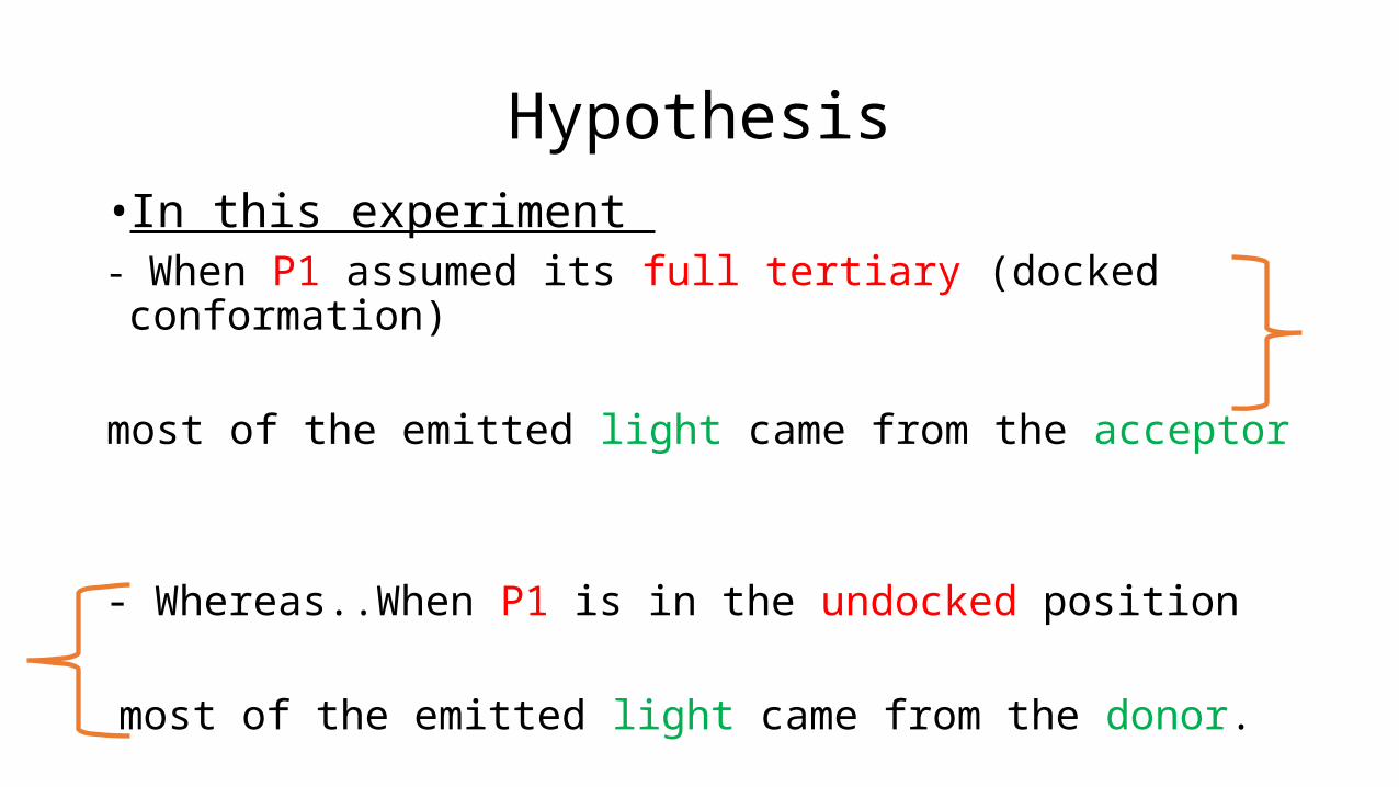
Hypothesis• In this experiment - When P1 assumed its full tertiary (docked conformation)
most of the emitted light came from the acceptor
- Whereas..When P1 is in the undocked position
most of the emitted light came from the donor.

Hypothesis
The unwinding of the P1 helix secondary structure led to the loss of all fluorescence
As the dye-labeled oligonucleotide was released into solution.

Results

CYT-19 destabilize tertiary docking of the P1 helix into the Tetrahymena ribozyme core

Conclusion
• The authors found that
• Without CYT-19 P1 helix spontaneous conversion between the docked and undocked conformations with the secondary structure of the helical segment remaining intact
• When they added CYT-19 and ATP helix unwinding commenced but was almost entirely from the undocked state

Sensitivity of CYT-19 to RNA 3°ry structure

Conclusion
• Adding more CYT-19 did not increase the rate of docked-to undocked conversion
• Indicating that this transition was not facilitated by the helicase
• Instead, the helicase in effect ‘‘waited’’ for the ribozyme to undock and then capture it in its undocked state
Conclusion
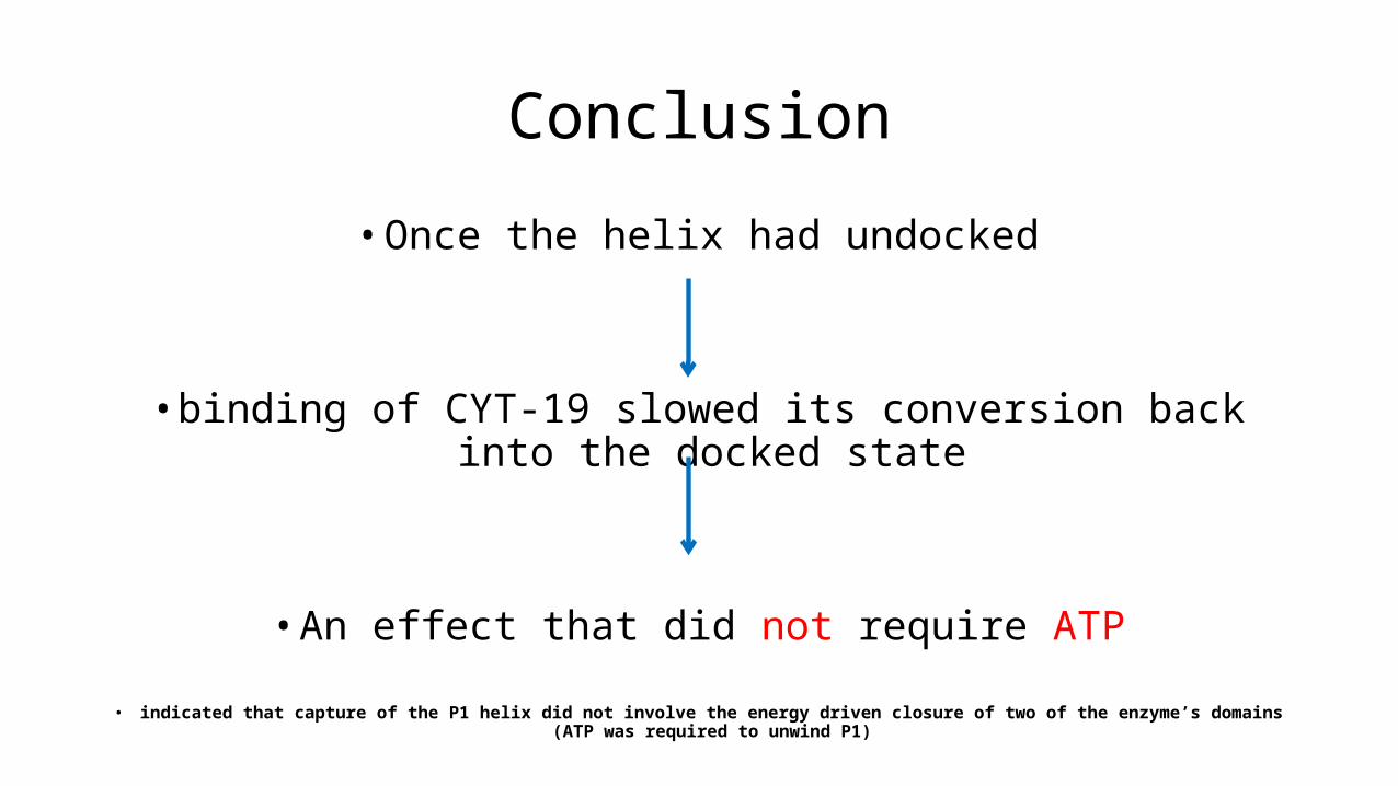
• Once the helix had undocked
• binding of CYT-19 slowed its conversion back into the docked state
• An effect that did not require ATP
• indicated that capture of the P1 helix did not involve the energy driven closure of two of the enzyme’s domains (ATP was required to unwind P1)

model for RNA tertiary structure disruption by helix capture

Conclusion
• Further experiments showed that CYT-19 remained bound to the ribozyme
even after its helicase core disengaged from P1
most likely by employing a strongly basic and unstructured tail

Conclusion
• The same essential behavior was seen in the yeast DEAD-box protein Ded1
• which, like CYT-19, did not weaken tertiary contacts between P1 and other portions of the ribozyme but did slow their re-formation.
• Unlike CYT-19, Ded1 needed to have ATP in place for helix capture

Conclusion
• The fact that two different DEAD-box proteins employ a similar capture strategy for their interaction with RNA helices suggests this
• may be a mechanism used elsewhere, not only for RNA chaperone functions but perhaps in synthesis of ribosomes and spliceosomes
• both of which rely on DEAD-box proteins for conformational transitions during their assembly

Conclusion
• Further investigations are likely to reveal the variations different helicases in different organisms have evolved to make use of this basic tool for RNA remodeling.

References
" DEAD-Box Helicase Proteins Disrupt RNA Tertiary Structure Through Helix Capture.(2014) PLoS - Biol 12(10): e1001981.
doi:10.1371/journal.pbio.1001981
Done by : Belal Abu Haneya Hashemite University – Jordan
Supervisor : Dr. Mohammed Wedyan
Related Documents