Citation: Mahaling, B.; Low, S.W.Y.; Beck, M.; Kumar, D.; Ahmed, S.; Connor, T.B.; Ahmad, B.; Chaurasia, S.S. Damage-Associated Molecular Patterns (DAMPs) in Retinal Disorders. Int. J. Mol. Sci. 2022, 23, 2591. https://doi.org/10.3390/ ijms23052591 Academic Editors: De-Kuang Hwang and Shih-Jen Chen Received: 9 February 2022 Accepted: 25 February 2022 Published: 26 February 2022 Publisher’s Note: MDPI stays neutral with regard to jurisdictional claims in published maps and institutional affil- iations. Copyright: © 2022 by the authors. Licensee MDPI, Basel, Switzerland. This article is an open access article distributed under the terms and conditions of the Creative Commons Attribution (CC BY) license (https:// creativecommons.org/licenses/by/ 4.0/). International Journal of Molecular Sciences Review Damage-Associated Molecular Patterns (DAMPs) in Retinal Disorders Binapani Mahaling 1 , Shermaine W. Y. Low 1 , Molly Beck 1 , Devesh Kumar 1 , Simrah Ahmed 1 , Thomas B. Connor 1,2 , Baseer Ahmad 1,2 and Shyam S. Chaurasia 1,3, * 1 Ocular Immunology and Angiogenesis Lab, Department of Ophthalmology and Visual Sciences, Froedtert and MCW Eye Institute, Medical College of Wisconsin, Milwaukee, WI 53226, USA; [email protected] (B.M.); [email protected] (S.W.Y.L.); [email protected] (M.B.); [email protected] (D.K.); [email protected] (S.A.); [email protected] (T.B.C.); [email protected] (B.A.) 2 Vitreoretinal Surgery, Froedtert and MCW Eye Institute, Medical College of Wisconsin, Milwaukee, WI 53226, USA 3 Department of Cell Biology, Neurobiology and Anatomy, Medical College of Wisconsin, Milwaukee, WI 53226, USA * Correspondence: [email protected]; Tel.: +1-414-955-2050 Abstract: Damage-associated molecular patterns (DAMPs) are endogenous danger molecules re- leased from the extracellular and intracellular space of damaged tissue or dead cells. Recent evidence indicates that DAMPs are associated with the sterile inflammation caused by aging, increased ocular pressure, high glucose, oxidative stress, ischemia, mechanical trauma, stress, or environmental con- ditions, in retinal diseases. DAMPs activate the innate immune system, suggesting their role to be protective, but may promote pathological inflammation and angiogenesis in response to the chronic insult or injury. DAMPs are recognized by specialized innate immune receptors, such as receptors for advanced glycation end products (RAGE), toll-like receptors (TLRs) and the NOD-like receptor family (NLRs), and purine receptor 7 (P2X7), in systemic diseases. However, studies describing the role of DAMPs in retinal disorders are meager. Here, we extensively reviewed the role of DAMPs in retinal disorders, including endophthalmitis, uveitis, glaucoma, ocular cancer, ischemic retinopathies, diabetic retinopathy, age-related macular degeneration, rhegmatogenous retinal detachment, prolifer- ative vitreoretinopathy, and inherited retinal disorders. Finally, we discussed DAMPs as biomarkers, therapeutic targets, and therapeutic agents for retinal disorders. Keywords: DAMPs; endophthalmitis; uveitis; glaucoma; ocular cancer; ischemic retinopathies; diabetic retinopathy; age-related macular degeneration; proliferative vitreoretinopathy; inherited retinal disorders 1. Introduction Damage-associated molecular patterns (DAMPs) are endogenous danger molecules released from the extracellular and intracellular space of the damaged tissue or dead cells [1]. DAMPs are (i) rapidly released following necrosis; (ii) produced by the activated immune cells via specialized secretion systems or by the endoplasmic reticulum (ER)— Golgi apparatus secretion pathway; (iii) known to activate the innate immune system by interacting with pattern-recognition receptors (PRRs), and thereby directly or indirectly promote adaptive immunity responses; (iv) inclined to contribute to the host’s defense and pathological inflammatory responses in non-infectious diseases; and (v) responsible for restoring homeostasis by promoting the reconstruction of the tissue [1,2]. Accumulating ev- idence indicates that DAMPs are associated with the sterile inflammation caused by aging, increased ocular pressure, hyperglycemia, oxidative stress, ischemia, mechanical trauma, stress, environmental condition, and genetic defects during retinal development [3–6]. Re- cent studies suggested that DAMPs that include extracellular matrix pro (ECM)-proteins Int. J. Mol. Sci. 2022, 23, 2591. https://doi.org/10.3390/ijms23052591 https://www.mdpi.com/journal/ijms

Welcome message from author
This document is posted to help you gain knowledge. Please leave a comment to let me know what you think about it! Share it to your friends and learn new things together.
Transcript
�����������������
Citation: Mahaling, B.; Low, S.W.Y.;
Beck, M.; Kumar, D.; Ahmed, S.;
Connor, T.B.; Ahmad, B.; Chaurasia,
S.S. Damage-Associated Molecular
Patterns (DAMPs) in Retinal
Disorders. Int. J. Mol. Sci. 2022, 23,
2591. https://doi.org/10.3390/
ijms23052591
Academic Editors: De-Kuang Hwang
and Shih-Jen Chen
Received: 9 February 2022
Accepted: 25 February 2022
Published: 26 February 2022
Publisher’s Note: MDPI stays neutral
with regard to jurisdictional claims in
published maps and institutional affil-
iations.
Copyright: © 2022 by the authors.
Licensee MDPI, Basel, Switzerland.
This article is an open access article
distributed under the terms and
conditions of the Creative Commons
Attribution (CC BY) license (https://
creativecommons.org/licenses/by/
4.0/).
International Journal of
Molecular Sciences
Review
Damage-Associated Molecular Patterns (DAMPs) inRetinal DisordersBinapani Mahaling 1, Shermaine W. Y. Low 1, Molly Beck 1, Devesh Kumar 1 , Simrah Ahmed 1, Thomas B. Connor 1,2,Baseer Ahmad 1,2 and Shyam S. Chaurasia 1,3,*
1 Ocular Immunology and Angiogenesis Lab, Department of Ophthalmology and Visual Sciences,Froedtert and MCW Eye Institute, Medical College of Wisconsin, Milwaukee, WI 53226, USA;[email protected] (B.M.); [email protected] (S.W.Y.L.); [email protected] (M.B.); [email protected] (D.K.);[email protected] (S.A.); [email protected] (T.B.C.); [email protected] (B.A.)
2 Vitreoretinal Surgery, Froedtert and MCW Eye Institute, Medical College of Wisconsin,Milwaukee, WI 53226, USA
3 Department of Cell Biology, Neurobiology and Anatomy, Medical College of Wisconsin,Milwaukee, WI 53226, USA
* Correspondence: [email protected]; Tel.: +1-414-955-2050
Abstract: Damage-associated molecular patterns (DAMPs) are endogenous danger molecules re-leased from the extracellular and intracellular space of damaged tissue or dead cells. Recent evidenceindicates that DAMPs are associated with the sterile inflammation caused by aging, increased ocularpressure, high glucose, oxidative stress, ischemia, mechanical trauma, stress, or environmental con-ditions, in retinal diseases. DAMPs activate the innate immune system, suggesting their role to beprotective, but may promote pathological inflammation and angiogenesis in response to the chronicinsult or injury. DAMPs are recognized by specialized innate immune receptors, such as receptorsfor advanced glycation end products (RAGE), toll-like receptors (TLRs) and the NOD-like receptorfamily (NLRs), and purine receptor 7 (P2X7), in systemic diseases. However, studies describing therole of DAMPs in retinal disorders are meager. Here, we extensively reviewed the role of DAMPs inretinal disorders, including endophthalmitis, uveitis, glaucoma, ocular cancer, ischemic retinopathies,diabetic retinopathy, age-related macular degeneration, rhegmatogenous retinal detachment, prolifer-ative vitreoretinopathy, and inherited retinal disorders. Finally, we discussed DAMPs as biomarkers,therapeutic targets, and therapeutic agents for retinal disorders.
Keywords: DAMPs; endophthalmitis; uveitis; glaucoma; ocular cancer; ischemic retinopathies;diabetic retinopathy; age-related macular degeneration; proliferative vitreoretinopathy; inheritedretinal disorders
1. Introduction
Damage-associated molecular patterns (DAMPs) are endogenous danger moleculesreleased from the extracellular and intracellular space of the damaged tissue or deadcells [1]. DAMPs are (i) rapidly released following necrosis; (ii) produced by the activatedimmune cells via specialized secretion systems or by the endoplasmic reticulum (ER)—Golgi apparatus secretion pathway; (iii) known to activate the innate immune system byinteracting with pattern-recognition receptors (PRRs), and thereby directly or indirectlypromote adaptive immunity responses; (iv) inclined to contribute to the host’s defense andpathological inflammatory responses in non-infectious diseases; and (v) responsible forrestoring homeostasis by promoting the reconstruction of the tissue [1,2]. Accumulating ev-idence indicates that DAMPs are associated with the sterile inflammation caused by aging,increased ocular pressure, hyperglycemia, oxidative stress, ischemia, mechanical trauma,stress, environmental condition, and genetic defects during retinal development [3–6]. Re-cent studies suggested that DAMPs that include extracellular matrix pro (ECM)-proteins
Int. J. Mol. Sci. 2022, 23, 2591. https://doi.org/10.3390/ijms23052591 https://www.mdpi.com/journal/ijms
Int. J. Mol. Sci. 2022, 23, 2591 2 of 38
such as decorin, biglycan, versican, aggrecan, phosphacan, low-molecular-weight (LMW)hyaluronan, heparan sulfate (HS), fibronectin, laminin, tenascin-C, and tenascin-R; cytoso-lic proteins such as leukemia inhibitory factor (LIF), S100 proteins, uric acid, heat-shockproteins (HSP), adenosine triphosphate (ATP), cyclophilin A, F-actin; those of nuclearorigins such as histones, high-mobility group box 1 (HMGB1), high-mobility group nu-cleosome binding domain 1 (HMGN1), interleukin (IL)-1α, IL-33, surface-interacting 3A(Sin3A)-associated protein 130 (Sap130), deoxyribonucleic acid (DNA), and ribonucleicacid (RNA); those of mitochondrial origins such as mtDNA, transcription factor A mito-chondrial (TFAM), formylated peptides, mitochondrial reactive-oxygen species (mtROS);those of endoplasmic reticulum (ER) origins such as calreticulin, defensins, cathelicidins(LL37), endothelin-1 (ET-1) and granulysin; those of plasma membrane origins such assyndecans, glypicans, perlecan; and plasma proteins such as fibrinogen, Gc-globulin, andserum amyloid A (SAA), are increased; this suggests a protective or pathogenic role indifferent retinal disorders [1,7–9]. DAMPs function through multiple specialized innateimmune receptors, such as receptors for advanced glycation end products (RAGE), toll-likereceptors (TLRs) and the NOD-like receptor (NLRs) family, purine receptor 7 (P2X7), NLRpyrin domain 3 (NLRP3), in retinal disorders [1,10–12].
The eye is an immune privilege tissue and limits its local immune and inflamma-tory responses to preserve vision. Though the mechanism of immune privilege is notentirely understood, the tear-fluid barrier, epithelial barrier, blood–ocular barrier, and theinner and outer blood–retinal barriers play essential roles in the immune responses of theeye [13–15]. The retinal cells that play a regulatory role in the posterior segment of theeye are retinal pigment epithelial (RPE) cells which express Fas ligand and programmeddeath-ligand 1 (PDL1), and microglia/macrophages expressing regulatory elements such asCD200/C200R, PDL1, and Treg cells. The anterior and posterior segment of the eye containsimmunosuppressive fluid containing neuropeptides such as transforming growth factor-β(TGF-β), vasoactive intestinal peptide (VIP), somatostatin, calcitonin, gene-related peptide,alpha-melanocyte-stimulating hormone, neuropeptide Y, and pigment epithelial-derivedfactor (PEDF) [13,14]. Any perturbations in the retinal microenvironment are recognizedby astrocytes and microglia present at the forefront of the defense system. Perturbationscan arise from two major sources: (i) microbial pathogens and (ii) age- or disease-relatedinjury. Astrocytes and microglial cells possess signaling mechanisms for host defensethat are activated by recognizing structural characteristics found in pathogens, known aspathogen-associated molecular patterns (PAMPs) and DAMPs [4].
The innate immune system provides the first line of defense against the DAMPs.In the early stages of retinal disorders, microglia and the complement system activateat low levels. This low level of inflammation is essential to maintain homeostasis andrestore functionality in retinal homeostasis. However, prolonged insult and stimulation byDAMPs in chronic retinal disorders such as glaucoma, age-related macular degeneration(AMD), diabetic retinopathy (DR), ischemic retinopathies, and uveitis lead to maladaptationof the innate immune system and dysregulated inflammation. As a result, increasedpro-inflammatory cytokines such as tumor necrosis factor (TNF)-α, IL-1β, IL-6, and IL-8contribute to further progression of the disease. Finally, immune privilege is compromisedin retinal disorders, resulting in a vicious cycle of inflammation, leukocyte infiltration, andretinal neurodegeneration.
2. DAMPs in Retinal Disorders2.1. DAMPs in Endophthalmitis
Endophthalmitis is a devastating and potentially blinding disorder caused by an in-fection from exogenous or endogenous microorganisms, typically in the vitreous cavity ofthe eye [16]. The inflammatory component in endophthalmitis is strongly associated withthe recognition of microorganism PAMPs and damaged or dying cell DAMPs by TLRs lo-cated on the cell membrane and within endosomes [17,18]. Staphylococcus aureus (S. aureus)infection significantly enhances the expression of DAMPs such as S100A7/S100A9 in the
Int. J. Mol. Sci. 2022, 23, 2591 3 of 38
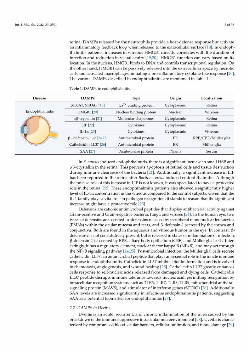
retina. DAMPs released by the neutrophils provide a host-defense response but activatean inflammatory feedback loop when released to the extracellular surface [18]. In endoph-thalmitis patients, increases in vitreous HMGB1 directly correlates with the duration ofinfection and reduction in visual acuity [19,20]. HMGB1 function can vary based on itslocation. In the nucleus, HMGB1 binds to DNA and controls transcriptional regulation. Onthe other hand, HMGB1 can be passively released into the extracellular space by necroticcells and activated macrophages, initiating a pro-inflammatory cytokine-like response [20].The various DAMPs described in endophthalmitis are mentioned in Table 1.
Table 1. DAMPs in endophthalmitis.
Disease DAMPs Type Origin Localization
S100A7, S100A9 [18] Ca2+ binding protein Cytoplasmic Retina
HMGB1 [20] Nuclear binding protein Nuclear Vitreous
αβ-crystallin [21] Molecular chaperones Cytoplasmic Retina
LIF [22] Cytokines Cytoplasmic Retina
IL-1α [23] Cytokines Cytoplasmic Vitreous
β−defensin-1, -2 [24,25] Antimicrobial protein ER RPE/CBE/Müller glia
Cathelicidin LL37 [26] Antimicrobial protein ER Müller glia
Endophthalmitis
Int. J. Mol. Sci. 2022, 23, x FOR PEER REVIEW 3 of 38
TLRs located on the cell membrane and within endosomes [17,18]. Staphylococcus aureus (S. aureus) infection significantly enhances the expression of DAMPs such as S100A7/S100A9 in the retina. DAMPs released by the neutrophils provide a host-defense response but activate an inflammatory feedback loop when released to the extracellular surface [18]. In endophthalmitis patients, increases in vitreous HMGB1 directly correlates with the duration of infection and reduction in visual acuity [19,20]. HMGB1 function can vary based on its location. In the nucleus, HMGB1 binds to DNA and controls transcrip-tional regulation. On the other hand, HMGB1 can be passively released into the extracel-lular space by necrotic cells and activated macrophages, initiating a pro-inflammatory cy-tokine-like response [20]. The various DAMPs described in endophthalmitis are men-tioned in Table 1.
Table 1. DAMPs in endophthalmitis.
Disease DAMPs Type Origin Localization
Endophthalmitis
S100A7, S100A9 [18] Ca2+ binding protein Cytoplasmic Retina HMGB1 [20] Nuclear binding protein Nuclear Vitreous
αβ-crystallin [21] Molecular chaperones Cytoplasmic Retina LIF [22] Cytokines Cytoplasmic Retina
IL-1α [23] Cytokines Cytoplasmic Vitreous β−defensin-1, -2
[24,25] Antimicrobial protein ER RPE/CBE/Müller glia
Cathelicidin LL37 [26]
Antimicrobial protein ER Müller glia
SAA [27] Acute-phase protein Plasma Serum
In S. aureus-induced endophthalmitis, there is a significant increase in small HSP and αβ-crystallin in the retina. This prevents apoptosis of retinal cells and tissue destruction during immune clearance of the bacteria [21]. Additionally, a significant increase in LIF has been reported in the retina after Bacillus cereus-induced endophthalmitis. Although the precise role of this increase in LIF is not known, it was speculated to have a protective role in the retina [22]. These endophthalmitis patients also showed a significantly higher level of IL-1α concentration in the vitreous compared to the control subjects. Given that the IL-1 family plays a vital role in pathogen recognition, it stands to reason that the sig-nificant increase might have a protective role [23].
Defensins are cationic antimicrobial peptides that display antibacterial activity against Gram-positive and Gram-negative bacteria, fungi, and viruses [24]. In the human eye, two types of defensins are secreted: α-defensins released by peripheral mononuclear leukocytes (PMNs) within the ocular mucosa and tears, and β-defensin-1 secreted by the cornea and conjunctiva. Both are found in the aqueous and vitreous humor in the eye. In contrast, β-defensin-2 is not constitutively present, but is released in states of inflamma-tion or infection. β-defensin-2 is secreted by RPE, ciliary body epithelium (CBE), and Mül-ler glial cells. Interestingly, it has a regulatory element, nuclear factor kappa B (NFκB), and may act through the NFκB signaling pathway [24,25]. Post-microbial infection, the Müller glial cells secrete cathelicidin LL37, an antimicrobial peptide that plays an essential role in the innate immune response to endophthalmitis. Cathelicidin LL37 inhibits biofilm formation and is involved in chemotaxis, angiogenesis, and wound healing [25]. Catheli-cidin LL37 greatly enhances cells response to self-nucleic acids released from damaged and dying cells. Cathelicidin LL37 peptide disrupts immune tolerance towards nucleic acid, permitting recognition by intracellular recognition systems such as TLR3, TLR7, TLR8, TLR9, mitochondrial antiviral-signaling protein (MAVS), and stimulator of inter-feron genes (STING) [26]. Additionally, SAA levels are increased significantly in
SAA [27] Acute-phase protein Plasma Serum
In S. aureus-induced endophthalmitis, there is a significant increase in small HSP andαβ-crystallin in the retina. This prevents apoptosis of retinal cells and tissue destructionduring immune clearance of the bacteria [21]. Additionally, a significant increase in LIFhas been reported in the retina after Bacillus cereus-induced endophthalmitis. Althoughthe precise role of this increase in LIF is not known, it was speculated to have a protectiverole in the retina [22]. These endophthalmitis patients also showed a significantly higherlevel of IL-1α concentration in the vitreous compared to the control subjects. Given that theIL-1 family plays a vital role in pathogen recognition, it stands to reason that the significantincrease might have a protective role [23].
Defensins are cationic antimicrobial peptides that display antibacterial activity againstGram-positive and Gram-negative bacteria, fungi, and viruses [24]. In the human eye, twotypes of defensins are secreted: α-defensins released by peripheral mononuclear leukocytes(PMNs) within the ocular mucosa and tears, and β-defensin-1 secreted by the cornea andconjunctiva. Both are found in the aqueous and vitreous humor in the eye. In contrast, β-defensin-2 is not constitutively present, but is released in states of inflammation or infection.β-defensin-2 is secreted by RPE, ciliary body epithelium (CBE), and Müller glial cells. Inter-estingly, it has a regulatory element, nuclear factor kappa B (NFκB), and may act throughthe NFκB signaling pathway [24,25]. Post-microbial infection, the Müller glial cells secretecathelicidin LL37, an antimicrobial peptide that plays an essential role in the innate immuneresponse to endophthalmitis. Cathelicidin LL37 inhibits biofilm formation and is involvedin chemotaxis, angiogenesis, and wound healing [25]. Cathelicidin LL37 greatly enhancescells response to self-nucleic acids released from damaged and dying cells. CathelicidinLL37 peptide disrupts immune tolerance towards nucleic acid, permitting recognition byintracellular recognition systems such as TLR3, TLR7, TLR8, TLR9, mitochondrial antiviral-signaling protein (MAVS), and stimulator of interferon genes (STING) [26]. Additionally,SAA levels are increased significantly in infectious endophthalmitis patients, suggestingSAA as a potential biomarker for endophthalmitis [27].
2.2. DAMPS in Uveitis
Uveitis is an acute, recurrent, and chronic inflammation of the uvea caused by thebreakdown of the immunosuppressive intraocular microenvironment [28]. Uveitis is charac-terized by compromised blood–ocular barriers, cellular infiltration, and tissue damage [29].

Int. J. Mol. Sci. 2022, 23, 2591 4 of 38
As a result, inappropriate intraocular inflammation can be detrimental to the eye andits visual function. DAMPs play a significant role in non-infectious uveitis by activatingPRRs and TLRs, thus initiating an acute inflammatory response [28]. The different DAMPmolecules increased in uveitis are S100 proteins, HMGB1, HSP70, SAA, fibronectin, andfibrinogen, as mentioned in Table 2.
Table 2. DAMPs in uveitis.
Disease DAMPs Type Origin Localization
S100A8, S100A9,S100A12 [30] Ca2+ binding protein Cytoplasmic Serum/aqueous/tears
HMGB1 [31] Nuclear binding protein Nuclear Retina
HSP70 [32] Molecular chaperones Cytoplasmic Serum
SAA [33] Acute-phase protein Plasma Aqueous
Fibronectin [34] Glycoprotein ECM Iris
Uveitis
Int. J. Mol. Sci. 2022, 23, x FOR PEER REVIEW 4 of 38
infectious endophthalmitis patients, suggesting SAA as a potential biomarker for endoph-thalmitis [27].
2.2. DAMPS in Uveitis Uveitis is an acute, recurrent, and chronic inflammation of the uvea caused by the
breakdown of the immunosuppressive intraocular microenvironment [28]. Uveitis is char-acterized by compromised blood–ocular barriers, cellular infiltration, and tissue damage [29]. As a result, inappropriate intraocular inflammation can be detrimental to the eye and its visual function. DAMPs play a significant role in non-infectious uveitis by activating PRRs and TLRs, thus initiating an acute inflammatory response [28]. The different DAMP molecules increased in uveitis are S100 proteins, HMGB1, HSP70, SAA, fibronectin, and fibrinogen, as mentioned in Table 2.
Table 2. DAMPs in uveitis.
Disease DAMPs Type Origin Localization Uveitis
S100A8, S100A9, S100A12 [30]
Ca2+ binding protein Cytoplasmic Serum/aque-
ous/tears HMGB1 [31] Nuclear binding protein Nuclear Retina HSP70 [32] Molecular chaperones Cytoplasmic Serum SAA [33] Acute-phase protein Plasma Aqueous
Fibronectin [34] Glycoprotein ECM Iris Fibrinogen [35] Glycoprotein ECM Iris
S100 proteins play an essential role in uveitis inflammation. Increased levels of S100A8/A9 and S100A12 were reported in the serum and aqueous humor of patients with autoimmune uveitis. S100A12 was found to be increased in the tear fluid of uveitis pa-tients [30] and is actively secreted by the phagocytic cells upon cell activation. Once se-creted, S100A8/A9 and S100A12 act as pro-inflammatory ligands and bind to TLR4 or RAGE, triggering inflammatory pathways [36]. Retinal cells also release HMGB1 in uveitis [31]. Usually, HMGB1 is secreted by macrophages during cellular stress or necrosis and mediates its actions as a DAMP through RAGE, TLR2, and TLR4 receptor signaling. HMGB1 recruits inflammatory cells and amplifies the local inflammatory response by in-ducing pro-inflammatory cytokines such as TNF-α, IL-1, and IL6 [37].
Serum uric acid levels are increased in many inflammatory conditions in the eye, including uveitis. Uric acid triggers endothelial dysfunction, oxidative stress, inflamma-tion, and microvascular disease. However, the study did not find any significant increase in serum uric acid in uveitis patients [6]. The serum concentration of HSP70 has been re-ported to be enhanced in patients with concurrent Behcet’s disease and uveitis relative to those without uveitis [32]. When released to the extracellular space from the necrotic cells or cells under stress, HSPs act as DAMPs on multiple receptors such as TLR2 and TLR4. Additionally, they activate the NFκB signaling pathway in macrophages and dendritic cells to stimulate the production of cytokines and chemokines, thereby mediating the up-take and presentation of peptides via the major histocompatibility complex (MHC) to fa-cilitate cell migration [28,37,38]. Furthermore, HSP-derived peptides 336 – 351 induce clin-ical and histological characteristics of uveitis in 80% of rats [39]. HSP90 inhibitors showed promising results in ameliorating experimental uveitis through the inhibition of NFκB, hypoxia induced factor (HIF)-1α, p38, and phosphatidylinositol 3-Kinase (PI3K) activity, and a reduction in vascular endothelial growth factor (VEGF), TNF-α, and IL-1β levels [38–40].
SAA is an acute-phase protein found in increased levels in the systemic circulation during chronic inflammatory disorders. Patients with uveitis or juvenile idiopathic arthri-tis with chronic anterior uveitis had higher SAA levels than their respective controls in the aqueous humor [33,41]. SAA acts on TLRs, NFκB, and P2X7-dependent NLRP3
Fibrinogen [35] Glycoprotein ECM Iris
S100 proteins play an essential role in uveitis inflammation. Increased levels ofS100A8/A9 and S100A12 were reported in the serum and aqueous humor of patientswith autoimmune uveitis. S100A12 was found to be increased in the tear fluid of uveitispatients [30] and is actively secreted by the phagocytic cells upon cell activation. Oncesecreted, S100A8/A9 and S100A12 act as pro-inflammatory ligands and bind to TLR4or RAGE, triggering inflammatory pathways [36]. Retinal cells also release HMGB1 inuveitis [31]. Usually, HMGB1 is secreted by macrophages during cellular stress or necrosisand mediates its actions as a DAMP through RAGE, TLR2, and TLR4 receptor signaling.HMGB1 recruits inflammatory cells and amplifies the local inflammatory response byinducing pro-inflammatory cytokines such as TNF-α, IL-1, and IL6 [37].
Serum uric acid levels are increased in many inflammatory conditions in the eye,including uveitis. Uric acid triggers endothelial dysfunction, oxidative stress, inflammation,and microvascular disease. However, the study did not find any significant increase inserum uric acid in uveitis patients [6]. The serum concentration of HSP70 has been reportedto be enhanced in patients with concurrent Behcet’s disease and uveitis relative to thosewithout uveitis [32]. When released to the extracellular space from the necrotic cells orcells under stress, HSPs act as DAMPs on multiple receptors such as TLR2 and TLR4.Additionally, they activate the NFκB signaling pathway in macrophages and dendriticcells to stimulate the production of cytokines and chemokines, thereby mediating theuptake and presentation of peptides via the major histocompatibility complex (MHC) tofacilitate cell migration [28,37,38]. Furthermore, HSP-derived peptides 336 – 351 induceclinical and histological characteristics of uveitis in 80% of rats [39]. HSP90 inhibitorsshowed promising results in ameliorating experimental uveitis through the inhibition ofNFκB, hypoxia induced factor (HIF)-1α, p38, and phosphatidylinositol 3-Kinase (PI3K)activity, and a reduction in vascular endothelial growth factor (VEGF), TNF-α, and IL-1βlevels [38–40].
SAA is an acute-phase protein found in increased levels in the systemic circulationduring chronic inflammatory disorders. Patients with uveitis or juvenile idiopathic arthritiswith chronic anterior uveitis had higher SAA levels than their respective controls in theaqueous humor [33,41]. SAA acts on TLRs, NFκB, and P2X7-dependent NLRP3 inflam-masome in macrophage and antigen-presenting cells (APCs), thus playing an essentialrole in inflammatory cytokine production, neutrophil transmigration, monocyte migra-tion, and peripheral blood mononuclear cell (PBMC) adhesion and differentiation [42–44].Though IL-33 acts as a DAMP, it has an anti-inflammatory effect for its role in activatingM2 macrophage polarization and attenuating the development of experimental autoim-mune uveitis [45]. Additionally, the intraocular cellular fibronectin levels were significantly
Int. J. Mol. Sci. 2022, 23, 2591 5 of 38
higher in patients with active uveitis [34]. In another study, the concentrations of fibronectin,fibrinogen, and immunoglobulins were significantly higher in the iris of the uveitis sub-jects compared to the controls. The irises of patients with uveitis also showed higherT-lymphocytic infiltration. These findings suggest that the presence of fibronectin, fib-rinogen, and immunoglobulins significantly contribute to T-lymphocyte infiltration andinflammation in uveitis [35].
2.3. DAMPs in Glaucoma
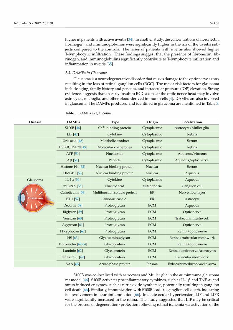
Glaucoma is a neurodegenerative disorder that causes damage to the optic nerve axons,resulting in the loss of retinal ganglion cells (RGC). The major risk factors for glaucomainclude aging, family history and genetics, and intraocular pressure (IOP) elevation. Strongevidence suggests that an early insult to RGC axons at the optic nerve head may involveastrocytes, microglia, and other blood-derived immune cells [4]. DAMPs are also involvedin glaucoma. The DAMPs produced and identified in glaucoma are mentioned in Table 3.
Table 3. DAMPs in glaucoma.
Disease DAMPs Type Origin Localization
S100B [46] Ca2+ binding protein Cytoplasmic Astrocyte/Müller glia
LIF [47] Cytokine Cytoplasmic Retina
Uric acid [48] Metabolic product Cytoplasmic Serum
HSP60, HSP70 [49] Molecular chaperones Cytoplasmic Retina
ATP [50] Nucleotide Cytoplasmic Aqueous/vitreous
Aβ [51] Peptide Cytoplasmic Aqueous/optic nerve
Histone-H4 [52] Nuclear binding protein Nuclear Serum
HMGB1 [53] Nuclear binding protein Nuclear Aqueous
IL-1α [54] Cytokine Cytoplasmic Aqueous
mtDNA [55] Nucleic acid Mitchondria Ganglion cell
Calreticulin [56] Multifunction soluble protein ER Nerve fiber layer
ET-1 [57] Ribonuclease A ER Astrocyte
Decorin [58] Proteoglycan ECM Aqueous
Biglycan [59] Proteoglycan ECM Optic nerve
Versican [60] Proteoglycan ECM Trabecular meshwork
Aggrecan [61] Proteoglycan ECM Optic nerve
Phosphocan [62] Proteoglycan ECM Retina/optic nerve
HS [63] Glycosaminoglycan ECM Retina/trabecular meshwork
Fibronectin [62,64] Glycoprotein ECM Retina/optic nerve
Laminin [62] Glycoprotein ECM Retina/optic nerve/astrocytes
Tenascin-C [62] Glycoprotein ECM Trabecular meshwork
Glaucoma
Int. J. Mol. Sci. 2022, 23, x FOR PEER REVIEW 5 of 38
inflammasome in macrophage and antigen-presenting cells (APCs), thus playing an es-sential role in inflammatory cytokine production, neutrophil transmigration, monocyte migration, and peripheral blood mononuclear cell (PBMC) adhesion and differentiation [42–44]. Though IL-33 acts as a DAMP, it has an anti-inflammatory effect for its role in activating M2 macrophage polarization and attenuating the development of experimental autoimmune uveitis [45]. Additionally, the intraocular cellular fibronectin levels were sig-nificantly higher in patients with active uveitis [34]. In another study, the concentrations of fibronectin, fibrinogen, and immunoglobulins were significantly higher in the iris of the uveitis subjects compared to the controls. The irises of patients with uveitis also showed higher T-lymphocytic infiltration. These findings suggest that the presence of fi-bronectin, fibrinogen, and immunoglobulins significantly contribute to T-lymphocyte in-filtration and inflammation in uveitis [35].
2.3. DAMPs in Glaucoma Glaucoma is a neurodegenerative disorder that causes damage to the optic nerve ax-
ons, resulting in the loss of retinal ganglion cells (RGC). The major risk factors for glau-coma include aging, family history and genetics, and intraocular pressure (IOP) elevation. Strong evidence suggests that an early insult to RGC axons at the optic nerve head may involve astrocytes, microglia, and other blood-derived immune cells [4]. DAMPs are also involved in glaucoma. The DAMPs produced and identified in glaucoma are mentioned in Table 3.
Table 3. DAMPs in glaucoma.
Disease DAMPs Type Origin Localization
Glaucoma
S100B [46] Ca2+ binding protein Cytoplasmic Astrocyte/Müller glia LIF [47] Cytokine Cytoplasmic Retina
Uric acid [48] Metabolic product Cytoplasmic Serum HSP60, HSP70 [49] Molecular chaperones Cytoplasmic Retina
ATP [50] Nucleotide Cytoplasmic Aqueous/vitreous Aβ [51] Peptide Cytoplasmic Aqueous/optic nerve
Histone-H4 [52] Nuclear binding protein Nuclear Serum HMGB1 [53] Nuclear binding protein Nuclear Aqueous
IL-1α [54] Cytokine Cytoplasmic Aqueous mtDNA [55] Nucleic acid Mitchondria Ganglion cell
Calreticulin [56] Multifunction soluble pro-
tein ER Nerve fiber layer
ET-1 [57] Ribonuclease A ER Astrocyte Decorin [58] Proteoglycan ECM Aqueous Biglycan [59] Proteoglycan ECM Optic nerve Versican [60] Proteoglycan ECM Trabecular meshwork Aggrecan [61] Proteoglycan ECM Optic nerve
Phosphocan [62] Proteoglycan ECM Retina/optic nerve
HS [63] Glycosaminoglycan ECM Retina/trabecular mesh-
work Fibronectin [62,64] Glycoprotein ECM Retina/optic nerve
Laminin [62] Glycoprotein ECM Retina/optic nerve/astro-
cytes Tenascin-C [62] Glycoprotein ECM Trabecular meshwork
SAA [65] Acute-phase protein Plasma Trabecular meshwork
and plasma SAA [65] Acute-phase protein Plasma Trabecular meshwork and plasma
S100B was co-localized with astrocytes and Müller glia in the autoimmune glaucomarat model [46]. S100B activates pro-inflammatory cytokines, such as IL-1β and TNF-α, andstress-induced enzymes, such as nitric oxide synthetase, potentially resulting in ganglioncell death [66]. Similarly, immunization with S100B leads to ganglion cell death, indicatingits involvement in neuroinflammation [66]. In acute ocular hypertension, LIF and LIFRwere significantly increased in the retina. The study suggested that LIF may be criticalfor the process of degeneration/protection following retinal ischemia via activation of the

Int. J. Mol. Sci. 2022, 23, 2591 6 of 38
Janus kinase (JAK)/STAT and Akt signaling pathways [47]. In fact, a neuroprotective roleis postulated based on observations following intravitreal injection of LIF [67]. Serum uricacid levels were also increased in primary open-angle glaucoma patients compared to thecontrol group [48]. The increase in uric acid concentration was also reported in aqueoushumor of subjects with glaucoma [68]. On the contrary, lower serum uric acid concentrationwas observed in primary angle-closure glaucoma in another study. Further, its negativeassociation with disease severity suggests uric acid as an important candidate in responseto glaucoma-associated oxidative stress [69].
In previous studies, HSPs were increased in response to elevated IOP, as also seen inhuman glaucomatous retinas [4,49]. Immunization with HSP27 and HSP60 led to pressure-independent RGC degeneration and axon loss, mimicking glaucoma-like damage [70].These findings indicate that HSPs in glaucoma may be directly involved in disease onsetand glaucoma progression. Notably, there was a significant increase in ATP in the aqueousand vitreous humor of patients with primary open-angle glaucoma. The activation ofP2X7 by ATP elevates intracellular calcium, resulting in rat RGC death [50]. In addition,significantly high levels of amyloid beta (Aβ) have been reported in the optic nerve headand aqueous humor of glaucoma patients [51]. Aβ co-localizes with apoptotic RGC in theexperimental glaucoma rat model and induces significant RGC apoptosis in vivo in a dose-and time-dependent manner [71]. Additionally, intraocular injection of Aβ1–40 appearedto have a time- and dose-dependent effect on neurodegeneration with increased axonalswelling and RGC cell death, leading to ganglion cell layer (GCL) thinning and optic nerveinjury [72]. The activation of Aβ may lead to activation of neuroinflammatory pathways,and hence, glaucoma progression with or without IOP-elevation-related triggers [73].There was also a significant increase in autoantibodies against Histone H4 in the serum ofglaucoma patients [52]. However, the precise role of Histone H4 in glaucoma is not known.
HMGB1 concentrations were significantly higher in the aqueous humor of primaryopen-angle glaucoma patients, whereas in rodents, HMGB1 was linked to glaucoma in-duced by elevated IOP. HMGB1 significantly upregulates canonical NLRP3 inflammasomevia caspase-1 and non-canonical caspase-8-driven inflammasome, which results in IL-1β re-lease, thereby causing ganglion cell death [53,74]. Additionally, IL-1α concentrations werenoted to be significantly increased in the aqueous humor of primary open-angle glaucomawith and without diabetes [54]. Furthermore, there was a significant increase in nuclearand mitochondrial DNA damage during ganglion cell death [55,75]. However, the exactrole of extracellular DNA released from dead cells in glaucoma has not been described.
The progressive retinal atrophy (PRA)1 family protein 3, calnexin, calreticulin, clus-terin, 78 kDa glucose-regulated protein, heterogeneous nuclear ribonucleoprotein R, malectin,peptidyl-prolyl cis–trans isomerase B, protein disulfide isomerase, reticulocalbin 3, andheterogeneous nuclear ribonucleoprotein Q, were reported to be significantly high in anon-human primate model of early experimental glaucoma [56]. However, the role of ERstress in glaucoma has not been studied yet. Optic nerve astrocytes proliferate after treat-ment with ET-1 (also known as EDN1), and reactive astrocytes increase endothelin receptorB (ETB) expression in both human and experimental neuronal injury models. Increasedexpression of ET-1 causes vasoconstriction, which prevents the optic nerve vasculaturefrom responding to the need for increased blood flow. Hence, ET-1 could be central toautoregulatory disturbances in glaucoma [57]. Additionally, ET-1 causes neuronal celldeath in glaucoma by activating pro-apoptotic transcription factor JUN (the canonicaltarget of JNK signaling) [76].
The small, leucine-rich proteoglycan (SLRP) family of DAMP proteins has been sug-gested to play a critical role in glaucoma. Decorin concentrations decreased significantly inthe aqueous humor of primary open-angle glaucoma patients [58]. Intracameral injectionof recombinant human (rh) decorin decreased TGF-β -induced fibrosis, lowered IOP, andprevented ganglion cell loss [77]. Another SLRP, biglycan, was significantly increased in theoptic nerve head of non-human primates in early experimental glaucoma, indicating its rolein disease progression [59]. Versican, a large proteoglycan, may organize glycosaminogly-
Int. J. Mol. Sci. 2022, 23, 2591 7 of 38
cans (GAGs) and other ECM components to facilitate and control open flow channels in thetrabecular meshwork, which appear to be a central component of the outflow resistance [60].Interestingly, a significant decrease in aggrecan was found in the optic nerve head of glau-comatous eyes compared to control eyes of non-human primates [61]. However, suchfindings were absent in rodent models [62]. A significant increase in phosphacan levelswas also observed in an autoimmune glaucoma rat model, with studies indicating its rolein disease progression [62,78]. There was a significant increase in chondroitin sulfate andHS in serum and optic nerve heads of glaucoma patients [63]. In a mouse glaucoma model,increased fibronectin, laminin, and tenascin-C levels were also found in the glaucomatousheterozygous retina and optic nerve compared to the wild-type group [62]. Fibronectin wasexplicitly found at higher levels in the trabecular meshwork of glaucomatous compared tonon-glaucomatous eyes. This is significant, as elevated IOP results from increased ECMrigidity regulated by collagen IV and fibrillin deposition [64]. Tenascin-C is up-regulatedin glaucomatous eyes, especially in astrocytes. As an endogenous activator of the TLR4,tenascin-C’s inflammatory role is being studied in glaucoma research [4,62]. SAA is alsoassociated with glaucoma-related increased IOP and inflammation [65].
2.4. DAMPs in Ocular Cancer
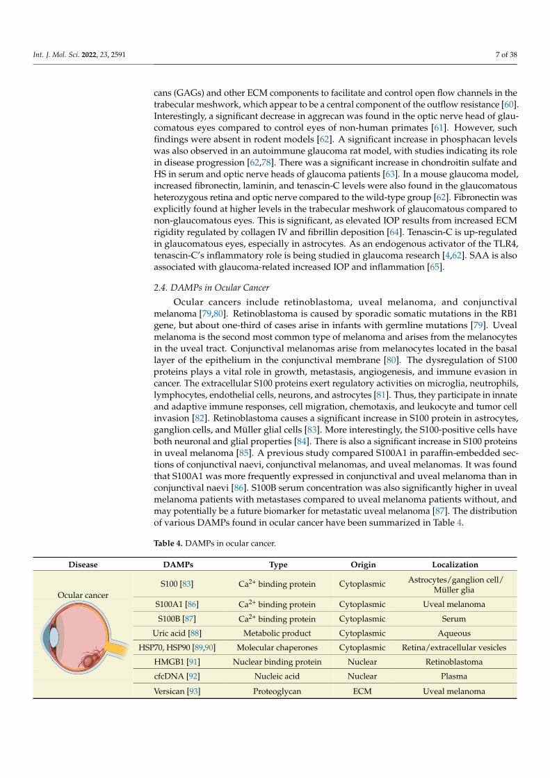
Ocular cancers include retinoblastoma, uveal melanoma, and conjunctivalmelanoma [79,80]. Retinoblastoma is caused by sporadic somatic mutations in the RB1gene, but about one-third of cases arise in infants with germline mutations [79]. Uvealmelanoma is the second most common type of melanoma and arises from the melanocytesin the uveal tract. Conjunctival melanomas arise from melanocytes located in the basallayer of the epithelium in the conjunctival membrane [80]. The dysregulation of S100proteins plays a vital role in growth, metastasis, angiogenesis, and immune evasion incancer. The extracellular S100 proteins exert regulatory activities on microglia, neutrophils,lymphocytes, endothelial cells, neurons, and astrocytes [81]. Thus, they participate in innateand adaptive immune responses, cell migration, chemotaxis, and leukocyte and tumor cellinvasion [82]. Retinoblastoma causes a significant increase in S100 protein in astrocytes,ganglion cells, and Müller glial cells [83]. More interestingly, the S100-positive cells haveboth neuronal and glial properties [84]. There is also a significant increase in S100 proteinsin uveal melanoma [85]. A previous study compared S100A1 in paraffin-embedded sec-tions of conjunctival naevi, conjunctival melanomas, and uveal melanomas. It was foundthat S100A1 was more frequently expressed in conjunctival and uveal melanoma than inconjunctival naevi [86]. S100B serum concentration was also significantly higher in uvealmelanoma patients with metastases compared to uveal melanoma patients without, andmay potentially be a future biomarker for metastatic uveal melanoma [87]. The distributionof various DAMPs found in ocular cancer have been summarized in Table 4.
Table 4. DAMPs in ocular cancer.
Disease DAMPs Type Origin Localization
S100 [83] Ca2+ binding protein Cytoplasmic Astrocytes/ganglion cell/Müller glia
S100A1 [86] Ca2+ binding protein Cytoplasmic Uveal melanoma
S100B [87] Ca2+ binding protein Cytoplasmic Serum
Uric acid [88] Metabolic product Cytoplasmic Aqueous
HSP70, HSP90 [89,90] Molecular chaperones Cytoplasmic Retina/extracellular vesicles
HMGB1 [91] Nuclear binding protein Nuclear Retinoblastoma
cfcDNA [92] Nucleic acid Nuclear Plasma
Ocular cancer
Int. J. Mol. Sci. 2022, 23, x FOR PEER REVIEW 8 of 38
Table 4. DAMPs in ocular cancer.
Disease DAMPs Type Origin Localization
Ocular cancer
S100 [83] Ca2+ binding protein Cytoplasmic Astrocytes/ganglion
cell/Müller glia S100A1 [86] Ca2+ binding protein Cytoplasmic Uveal melanoma S100B [87] Ca2+ binding protein Cytoplasmic Serum
Uric acid [88] Metabolic product Cytoplasmic Aqueous HSP70, HSP90
[89,90] Molecular chaperones Cytoplasmic Retina/extracellular vesicles
HMGB1 [91] Nuclear binding protein Nuclear Retinoblastoma cfcDNA [92] Nucleic acid Nuclear Plasma Versican [93] Proteoglycan ECM Uveal melanoma
Uric acid was elevated in the aqueous humor of eyes with melanoma, and in both the aqueous humor and tears of eyes with retinoblastoma [88]. The overexpression of HSPs provides a selective advantage to malignant cells by inhibiting apoptosis, promoting tu-mor metastasis, and regulating immune responses [94]. In control subjects, the human adult retina did not show HSP70/HSP90 immunoreactivity, whereas higher-to-moderate expressions of these proteins were observed in subjects with retinoblastoma tumors [89]. There was no significant difference in HSP27, HSP70, and HSP90 in uveal melanoma [95]. However, another study showed a higher degree of HSP90-positive staining in uveal mel-anoma cases, with 68% of cases staining positive and an average of 50% of tumor cells stained. The expression level was directly correlated with tumor diameter [96]. Addition-ally, extracellular vesicles derived from uveal metastatic melanoma have higher HSP70 and HSP90 than normal choroidal melanocytes, and more interestingly, these extracellu-lar vesicles play an essential role in progression and metastasis [90].
Intracellular and extracellular HMGB1 has been implicated in tumor formation, pro-gression, and metastasis. There is a significant increase in HMGB1 expression in reti-noblastoma (RB) cells. HMGB1 levels have also been found to be significantly higher in human patient samples and associated with tumor differentiation and optic nerve inva-sion [97,98]. In the uvea, there is upregulation of HMGB1 with a binding affinity for the retinoblastoma tumor suppressor protein [91,99]. In patients with cancer, the circulating cell-free (cfc) DNA has the same genetic and epigenetic alterations compared to the related primary tumor. The majority of cfcDNA is derived from tissue tumor cells rather than from circulating tumor cells [92]. The aqueous humor of retinoblastoma patients contains tumor-derived cfcDNA, which can be used to diagnose the disorder [100]. The plasma cfcDNA can also detect somatic RB1 mutations in patients with unilateral retinoblastoma [101]. The blood plasma and aqueous humor of uveal melanoma patients also contain tu-mor-derived cfDNA which can be used for diagnosis [92,102]. The versican has been im-plicated in tumor progression, with abnormal mRNA expression observed in uveal mela-noma. However, versican protein levels have not been reported [93].
2.5. DAMPs in Ischemic Retinopathies Retinal ischemia occurs due to inadequate blood supply to the retina, required for
oxygen diffusion and high metabolic activity. Circulatory failure can result from choroidal or retinal vessel obstruction. This lack of blood supply alters metabolic functions in the highly demanding retina and can ultimately result in irreversible neuronal cell death, vi-sion loss, and blindness. Ischemic retinopathy causes include central retinal artery occlu-sion (CRAO), branch retinal artery occlusion (BRAO), central retinal vein occlusion (CRVO), branch retinal vein occlusion (BRVO), and DR. The location and level of obstruc-tion to the blood supply determines the severity of ischemia, the area of retina affected, and its deleterious effects on the retina. DAMPs involved in ischemic retinopathies
Versican [93] Proteoglycan ECM Uveal melanoma
Int. J. Mol. Sci. 2022, 23, 2591 8 of 38
Uric acid was elevated in the aqueous humor of eyes with melanoma, and in boththe aqueous humor and tears of eyes with retinoblastoma [88]. The overexpression ofHSPs provides a selective advantage to malignant cells by inhibiting apoptosis, promotingtumor metastasis, and regulating immune responses [94]. In control subjects, the humanadult retina did not show HSP70/HSP90 immunoreactivity, whereas higher-to-moderateexpressions of these proteins were observed in subjects with retinoblastoma tumors [89].There was no significant difference in HSP27, HSP70, and HSP90 in uveal melanoma [95].However, another study showed a higher degree of HSP90-positive staining in uvealmelanoma cases, with 68% of cases staining positive and an average of 50% of tumor cellsstained. The expression level was directly correlated with tumor diameter [96]. Addition-ally, extracellular vesicles derived from uveal metastatic melanoma have higher HSP70and HSP90 than normal choroidal melanocytes, and more interestingly, these extracellularvesicles play an essential role in progression and metastasis [90].
Intracellular and extracellular HMGB1 has been implicated in tumor formation, pro-gression, and metastasis. There is a significant increase in HMGB1 expression in retinoblas-toma (RB) cells. HMGB1 levels have also been found to be significantly higher in humanpatient samples and associated with tumor differentiation and optic nerve invasion [97,98].In the uvea, there is upregulation of HMGB1 with a binding affinity for the retinoblastomatumor suppressor protein [91,99]. In patients with cancer, the circulating cell-free (cfc) DNAhas the same genetic and epigenetic alterations compared to the related primary tumor. Themajority of cfcDNA is derived from tissue tumor cells rather than from circulating tumorcells [92]. The aqueous humor of retinoblastoma patients contains tumor-derived cfcDNA,which can be used to diagnose the disorder [100]. The plasma cfcDNA can also detectsomatic RB1 mutations in patients with unilateral retinoblastoma [101]. The blood plasmaand aqueous humor of uveal melanoma patients also contain tumor-derived cfDNA whichcan be used for diagnosis [92,102]. The versican has been implicated in tumor progression,with abnormal mRNA expression observed in uveal melanoma. However, versican proteinlevels have not been reported [93].
2.5. DAMPs in Ischemic Retinopathies
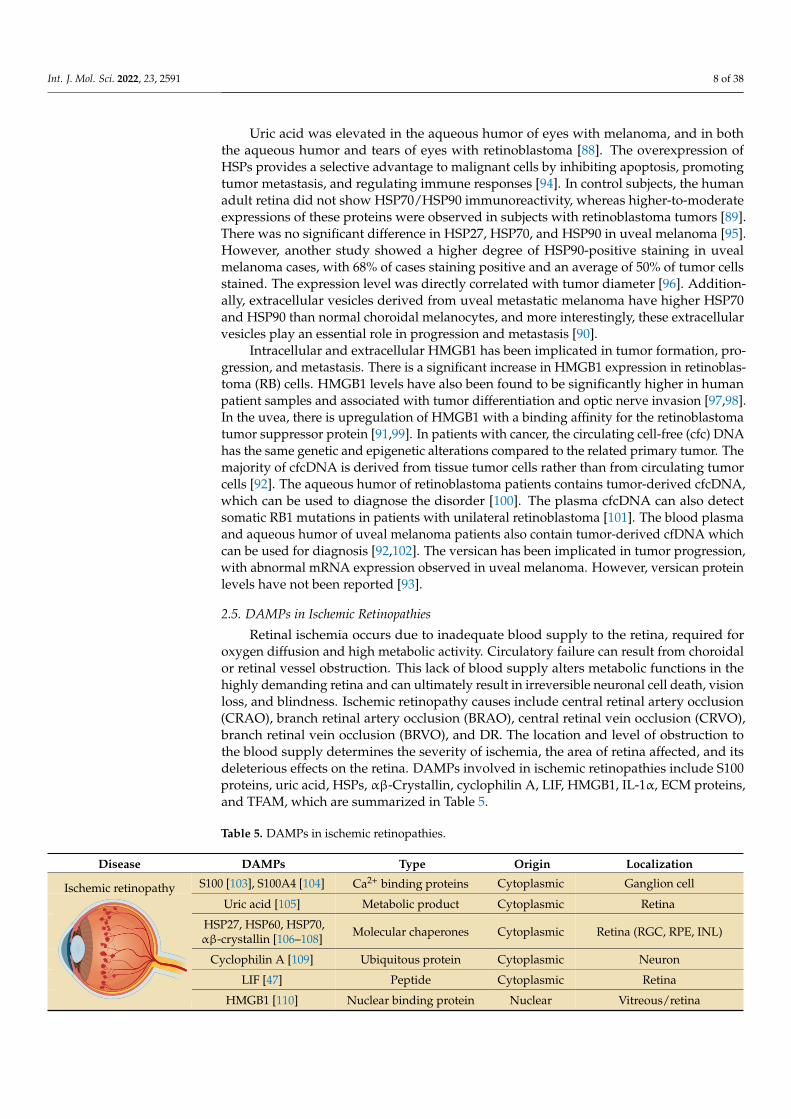
Retinal ischemia occurs due to inadequate blood supply to the retina, required foroxygen diffusion and high metabolic activity. Circulatory failure can result from choroidalor retinal vessel obstruction. This lack of blood supply alters metabolic functions in thehighly demanding retina and can ultimately result in irreversible neuronal cell death, visionloss, and blindness. Ischemic retinopathy causes include central retinal artery occlusion(CRAO), branch retinal artery occlusion (BRAO), central retinal vein occlusion (CRVO),branch retinal vein occlusion (BRVO), and DR. The location and level of obstruction tothe blood supply determines the severity of ischemia, the area of retina affected, and itsdeleterious effects on the retina. DAMPs involved in ischemic retinopathies include S100proteins, uric acid, HSPs, αβ-Crystallin, cyclophilin A, LIF, HMGB1, IL-1α, ECM proteins,and TFAM, which are summarized in Table 5.
Table 5. DAMPs in ischemic retinopathies.
Disease DAMPs Type Origin Localization
S100 [103], S100A4 [104] Ca2+ binding proteins Cytoplasmic Ganglion cell
Uric acid [105] Metabolic product Cytoplasmic Retina
HSP27, HSP60, HSP70,αβ-crystallin [106–108] Molecular chaperones Cytoplasmic Retina (RGC, RPE, INL)
Cyclophilin A [109] Ubiquitous protein Cytoplasmic Neuron
LIF [47] Peptide Cytoplasmic Retina
Ischemic retinopathy
Int. J. Mol. Sci. 2022, 23, x FOR PEER REVIEW 9 of 38
include S100 proteins, uric acid, HSPs, αβ-Crystallin, cyclophilin A, LIF, HMGB1, IL-1α, ECM proteins, and TFAM, which are summarized in Table 5.
Table 5. DAMPs in ischemic retinopathies.
Disease DAMPs Type Origin Localization
Ischemic retinopathy
S100 [103], S100A4 [104] Ca2+ binding proteins Cytoplasmic Ganglion cell Uric acid [105] Metabolic product Cytoplasmic Retina
HSP27, HSP60, HSP70, αβ-crystallin [106–108]
Molecular chaperones Cytoplasmic Retina (RGC, RPE,
INL) Cyclophilin A [109] Ubiquitous protein Cytoplasmic Neuron
LIF [47] Peptide Cytoplasmic Retina HMGB1 [110] Nuclear binding protein Nuclear Vitreous/retina
IL-1α [111,112] Cytokine Cytoplasmic Retina/plasma
TFAM [113,114] Transcription factor Mitchondria Retina (OPL, INL,
IPL, GCL) Decorin [115] Proteoglycan ECM Retina (INL)
Fibronectin [115] Glycoprotein ECM Retina Laminin [115] Glycoprotein ECM Optic nerve
Tenascin-C [115] Glycoprotein ECM Optic nerve HS [116] Glycosaminoglycan ECM Optic nerve
Chondritin sulfate [115] Glycosaminoglycan ECM Optic nerve Aggrecan [115] Proteoglycan ECM Optic nerve
A significant increase in the S100 protein in ganglion cells was reported in border zones damaged by retinal vein occlusion (RVO). However, this immunoreactivity was ab-sent inside areas of completely non-perfused capillaries, indicating inflammatory recruit-ment of S100 proteins in RVO [103]. In addition, the expression of S100A4 was also found to be positively correlated with the progression of retinal neovascularization observed in oxygen-induced retinopathy (OIR) models [117]. Silencing S100A4 reduces brain-derived neurotrophic factor (BDNF) activation and VEGF expression, suggesting its role in regu-lating retinal neovascularization [117]. In addition, suppression of S100A4 can also reduce the expression of cAMP response element-binding protein (CREB) and B-cell lymphoma-2 (Bcl-2), and increase the expression of caspase-3, to promote apoptosis and prevent ab-normal neovascularization [118]. Interestingly, overexpression of S100A4 provides neu-roprotection in ischemic mice by activating the Akt pathway, thus suppressing apoptosis in RGCs [104]. Damage signals from S100A4 may influence diverse signaling pathways in different retinal cell types to elicit unique responses for protection against ischemia. The animal models that have been subjected to ischemia exhibit increased uric acid concentra-tions in the retina. Uric acid expression is transiently decreased following reperfusion, and subsequently increased in the later stages after 60 min [105,119]. Additionally, the oxida-tion of hypoxanthine and xanthine results in the production of uric acid during is-chemic/reperfusion (I/R) injury [120].
Several HSPs, including HSP27, HSP70, and HSP72 play a role as DAMPs in the is-chemic retina. HSP27 is a neuroprotective component that can be induced after acute pres-sure-induced ischemia [121]. During ischemic injury, its expression is upregulated in the neuronal and non-neuronal inner retinal layers [106,122]. Rats subjected to bilateral com-mon carotid artery occlusion (BCCAO) displayed a significant increase in HSP27 and HSP70 immunoreactivity in the GCL after ischemic injury [107]. It was suggested that HSP27 might play a protective role in the retina. The delivery of HSP27 to RGCs via elec-troporation increased RGC survival rate after I/R injury [123]. In ARPE-19 cells induced with myeloperoxidase-mediated oxidative injury, HSP27 expression was increased, sug-gesting its role in the RPE injury response [108]. Similarly, HSP70 was also increased in
HMGB1 [110] Nuclear binding protein Nuclear Vitreous/retina
Int. J. Mol. Sci. 2022, 23, 2591 9 of 38
Table 5. Cont.
Disease DAMPs Type Origin Localization
IL-1α [111,112] Cytokine Cytoplasmic Retina/plasma
TFAM [113,114] Transcription factor Mitchondria Retina (OPL, INL, IPL, GCL)
Decorin [115] Proteoglycan ECM Retina (INL)
Fibronectin [115] Glycoprotein ECM Retina
Laminin [115] Glycoprotein ECM Optic nerve
Tenascin-C [115] Glycoprotein ECM Optic nerve
HS [116] Glycosaminoglycan ECM Optic nerve
Chondritin sulfate [115] Glycosaminoglycan ECM Optic nerve
Aggrecan [115] Proteoglycan ECM Optic nerve
A significant increase in the S100 protein in ganglion cells was reported in borderzones damaged by retinal vein occlusion (RVO). However, this immunoreactivity wasabsent inside areas of completely non-perfused capillaries, indicating inflammatory re-cruitment of S100 proteins in RVO [103]. In addition, the expression of S100A4 was alsofound to be positively correlated with the progression of retinal neovascularization ob-served in oxygen-induced retinopathy (OIR) models [117]. Silencing S100A4 reducesbrain-derived neurotrophic factor (BDNF) activation and VEGF expression, suggestingits role in regulating retinal neovascularization [117]. In addition, suppression of S100A4can also reduce the expression of cAMP response element-binding protein (CREB) andB-cell lymphoma-2 (Bcl-2), and increase the expression of caspase-3, to promote apoptosisand prevent abnormal neovascularization [118]. Interestingly, overexpression of S100A4provides neuroprotection in ischemic mice by activating the Akt pathway, thus suppressingapoptosis in RGCs [104]. Damage signals from S100A4 may influence diverse signalingpathways in different retinal cell types to elicit unique responses for protection againstischemia. The animal models that have been subjected to ischemia exhibit increased uricacid concentrations in the retina. Uric acid expression is transiently decreased followingreperfusion, and subsequently increased in the later stages after 60 min [105,119]. Addi-tionally, the oxidation of hypoxanthine and xanthine results in the production of uric acidduring ischemic/reperfusion (I/R) injury [120].
Several HSPs, including HSP27, HSP70, and HSP72 play a role as DAMPs in theischemic retina. HSP27 is a neuroprotective component that can be induced after acutepressure-induced ischemia [121]. During ischemic injury, its expression is upregulated inthe neuronal and non-neuronal inner retinal layers [106,122]. Rats subjected to bilateralcommon carotid artery occlusion (BCCAO) displayed a significant increase in HSP27and HSP70 immunoreactivity in the GCL after ischemic injury [107]. It was suggestedthat HSP27 might play a protective role in the retina. The delivery of HSP27 to RGCsvia electroporation increased RGC survival rate after I/R injury [123]. In ARPE-19 cellsinduced with myeloperoxidase-mediated oxidative injury, HSP27 expression was increased,suggesting its role in the RPE injury response [108]. Similarly, HSP70 was also increased inrat retinas following I/R injury [124]. HSP-70 prevents apoptosis by upregulating Bcl-2 andinterfering with apoptotic peptidase activating factor-1 (Apaf-1) to prevent apoptosomeformation [125]. HSP72 expression has also been studied in ischemic retinopathy. The loss ofretinal neurons in ischemic retinopathy is associated with glutamate-induced excitotoxicity.Intravitreal injection of a glutamate receptor agonist, N-methyl-D-aspartate (NMDA), caninduce inner retina cell death. Post-NMDA injection, HSP72 expression was elevated in theretinal GCL [126]. The number of HSP72 stained RGCs was also significantly higher afteracute pressure-induced retinal ischemia [121]. This study suggests that HSP72 can exhibitDAMP properties involved in the ischemic stress response.

Int. J. Mol. Sci. 2022, 23, 2591 10 of 38
Cytoplasmic cyclophilin A plays a fundamental role in cell metabolism, and its ex-pression levels can be altered in the presence of retinal lesions [109]. Rats exposed to moreextended periods of ischemia exhibited a loss of circulating anti-cyclophilin A antibodies.These antibodies have been speculated to bind to damaged retinal tissues in response toischemic injury. However, more analysis is required to determine the roles of cyclophilinA in ischemic retinopathy [127]. Aβ is another DAMP associated with neurodegenerativeretinal disorders [128]. Production of Aβ is associated with neuronal apoptosis and cellloss. In primary retinal neuron cells treated with CoCl2 to induce hypoxia, Aβ expressionwas significantly increased, suggesting that Aβ may be altered during ischemic retinaldamage [129]. LIF expression may also be altered in neuronal injuries and retinal disorders.LIF regulates gliosis and is a neuroprotective factor. Following retinal ischemia and retinalcell apoptosis induced by acute ocular hypertension, LIF and LIF receptor (LIF-R) expres-sion were found to be increased, along with elevated levels of phosphorylated Akt [47].LIF may modulate retinal injury and repair via the PI3K-Akt pathway. LIF can also in-hibit retinal vascular development independent of VEGF, suggesting its role in vascularremodeling [130].
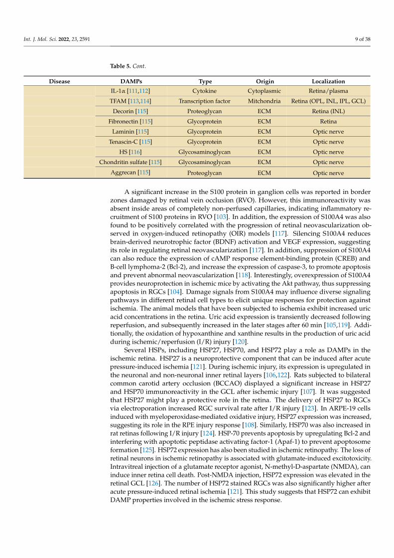
HMGB1 is a prototypic DAMP molecule localized to the GCL, inner nuclear layer(INL), and photoreceptor layer in the retina. It promotes inflammation, ganglion cell death,and photoreceptor degeneration in I/R-induced retinal damage [131]. Intravitreal injec-tion of recombinant HMGB1 has been known to result in a loss of RGCs [110]. In vitroaddition of HMGB1 to retinal glial cells also induced the production of pro-inflammatoryfactors [132]. However, the treatment of retinal ischemia with neutralizing anti-HMGB1monoclonal antibodies has been controversial. One study found that intraperitoneal injec-tion of a neutralizing anti-HMGB1 monoclonal antibody increased reactive oxygen species(ROS) production, resulting in retinal thinning and poor retinal function [133]. On thecontrary, another reported that the neutralization of HMGB1 can prevent retinal thinningand loss of ganglion cells, and reduce the number of irregular retinal capillaries [134]. Thedifferences in the neutralizing antibody concentrations could be a possible reason for thesediffering effects. IL-1α has also been shown to increase significantly in I/R-induced retinalinjury [111]. Blood plasma cytokine analysis of rats with I/R injury presented elevatedconcentrations of IL-1α, TNF-α, and MCP-1 [112]. IL-1α gene expression has also beenreported to rise rapidly, peaking at 3 to 12 h after rat retinal ischemia [135].
TFAM is a mitochondrial-DNA-binding protein crucial for mitochondrial gene expres-sion and essential for oxidative phosphorylation-mediated ATP synthesis. TFAM proteinexpression significantly increases in the ischemic retina [113] and is localized to the outerplexiform layer (OPL), INL, inner plexiform layer (IPL), and GCL [114]. An increasein TFAM expression can prevent the alteration of mitochondrial DNA in the ischemicretina [113]. Preservation of TFAM may also promote an endogenous repair mechanism toprotect RGCs against mitochondrial dysfunction during oxidative stress. In neonatal ratischemic brain injury, TFAM protein expression was rapidly elevated and mitochondrialdysfunction and ROS generation were reduced [136]. TFAM expression during retinalischemia may exhibit similar protective mechanisms. SAA, IL-6, and TGF-β are major pro-teins involved in the acute and chronic stages of inflammation. SAA is significantly higherin the aqueous humor of RVO patients with macular edema compared to controls [137].
The expression of extracellular glycoproteins decorin, fibronectin, laminin, tenascin-C,tenascin-R, and the chondroitin sulfate proteoglycans aggrecan, brevican, and phosphacanwere studied in an ischemia-reperfusion injury model. Interestingly, decorin expressionwas reduced in the inner retinal layers in the early stages but increased substantially in thelater stages of I/R, with strong immunoreactivity to damaged retinal layers. Fibronectinwas significantly elevated in the retina following ischemia, while laminin, tenascin-C andaggrecan showed enhanced immunoreactivity in the optic nerve after ischemia, indicatingtheir regulatory role during neurodegeneration [115]. Another proteoglycan, HS, cansuppress aberrant neovascularization by inhibiting VEGF-A from binding to VEGF-R2 [116].Fibronectin and tenascin-C expression were also increased, which localized to retinal
Int. J. Mol. Sci. 2022, 23, 2591 11 of 38
blood vessels in the inner layers of the ischemic retina [3]. Since ECM proteins playan important role in vascular development and neovascularization, the upregulation offibronectin in the ischemic retina could reflect its role in the remodeling of the retinalmicrovasculature. Elevation of tenascin-C concentrations can also contribute to retinaldegeneration observed in ischemic retinopathy. In tenascin-C-deficient ischemic mice, ERGa- and b-wave amplitudes were higher than in wild-type ischemic mice [138]. Less rodphotoreceptor degeneration was also observed in tenascin-C-deficient mice, suggesting thattenascin-C may be involved in ischemic retinal degeneration. Aggrecan and phosphacanare other extracellular DAMPs that have been studied in ischemic retinopathy. Proteinexpression of aggrecan and phophacan have been reported to be significantly reduced inthe ischemic rat retina [115]. Downregulation of these DAMPs could be associated withretinal gliosis, reorganization, or the retinal degenerative process.
2.6. DAMPs in Diabetic Retinopathy
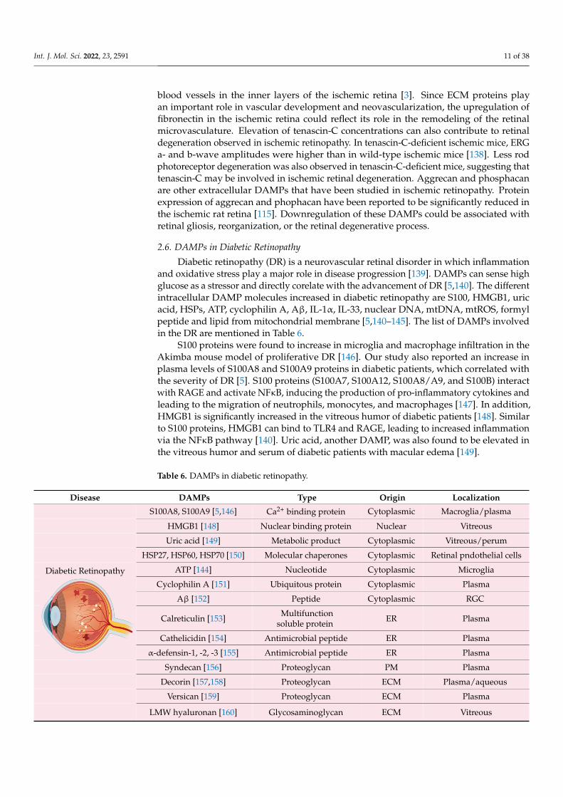
Diabetic retinopathy (DR) is a neurovascular retinal disorder in which inflammationand oxidative stress play a major role in disease progression [139]. DAMPs can sense highglucose as a stressor and directly corelate with the advancement of DR [5,140]. The differentintracellular DAMP molecules increased in diabetic retinopathy are S100, HMGB1, uricacid, HSPs, ATP, cyclophilin A, Aβ, IL-1α, IL-33, nuclear DNA, mtDNA, mtROS, formylpeptide and lipid from mitochondrial membrane [5,140–145]. The list of DAMPs involvedin the DR are mentioned in Table 6.
S100 proteins were found to increase in microglia and macrophage infiltration in theAkimba mouse model of proliferative DR [146]. Our study also reported an increase inplasma levels of S100A8 and S100A9 proteins in diabetic patients, which correlated withthe severity of DR [5]. S100 proteins (S100A7, S100A12, S100A8/A9, and S100B) interactwith RAGE and activate NFκB, inducing the production of pro-inflammatory cytokines andleading to the migration of neutrophils, monocytes, and macrophages [147]. In addition,HMGB1 is significantly increased in the vitreous humor of diabetic patients [148]. Similarto S100 proteins, HMGB1 can bind to TLR4 and RAGE, leading to increased inflammationvia the NFκB pathway [140]. Uric acid, another DAMP, was also found to be elevated inthe vitreous humor and serum of diabetic patients with macular edema [149].
Table 6. DAMPs in diabetic retinopathy.
Disease DAMPs Type Origin Localization
S100A8, S100A9 [5,146] Ca2+ binding protein Cytoplasmic Macroglia/plasma
HMGB1 [148] Nuclear binding protein Nuclear Vitreous
Uric acid [149] Metabolic product Cytoplasmic Vitreous/perum
HSP27, HSP60, HSP70 [150] Molecular chaperones Cytoplasmic Retinal pndothelial cells
ATP [144] Nucleotide Cytoplasmic Microglia
Cyclophilin A [151] Ubiquitous protein Cytoplasmic Plasma
Aβ [152] Peptide Cytoplasmic RGC
Calreticulin [153] Multifunctionsoluble protein ER Plasma
Cathelicidin [154] Antimicrobial peptide ER Plasma
α-defensin-1, -2, -3 [155] Antimicrobial peptide ER Plasma
Syndecan [156] Proteoglycan PM Plasma
Decorin [157,158] Proteoglycan ECM Plasma/aqueous
Versican [159] Proteoglycan ECM Plasma
Diabetic Retinopathy
Int. J. Mol. Sci. 2022, 23, x FOR PEER REVIEW 11 of 38
The expression of extracellular glycoproteins decorin, fibronectin, laminin, tenascin-C, tenascin-R, and the chondroitin sulfate proteoglycans aggrecan, brevican, and phos-phacan were studied in an ischemia-reperfusion injury model. Interestingly, decorin ex-pression was reduced in the inner retinal layers in the early stages but increased substan-tially in the later stages of I/R, with strong immunoreactivity to damaged retinal layers. Fibronectin was significantly elevated in the retina following ischemia, while laminin, tenascin-C and aggrecan showed enhanced immunoreactivity in the optic nerve after is-chemia, indicating their regulatory role during neurodegeneration [115]. Another proteo-glycan, HS, can suppress aberrant neovascularization by inhibiting VEGF-A from binding to VEGF-R2 [116]. Fibronectin and tenascin-C expression were also increased, which lo-calized to retinal blood vessels in the inner layers of the ischemic retina [3]. Since ECM proteins play an important role in vascular development and neovascularization, the up-regulation of fibronectin in the ischemic retina could reflect its role in the remodeling of the retinal microvasculature. Elevation of tenascin-C concentrations can also contribute to retinal degeneration observed in ischemic retinopathy. In tenascin-C-deficient ischemic mice, ERG a- and b-wave amplitudes were higher than in wild-type ischemic mice [138]. Less rod photoreceptor degeneration was also observed in tenascin-C-deficient mice, sug-gesting that tenascin-C may be involved in ischemic retinal degeneration. Aggrecan and phosphacan are other extracellular DAMPs that have been studied in ischemic retinopa-thy. Protein expression of aggrecan and phophacan have been reported to be significantly reduced in the ischemic rat retina [115]. Downregulation of these DAMPs could be asso-ciated with retinal gliosis, reorganization, or the retinal degenerative process.
2.6. DAMPs in Diabetic Retinopathy Diabetic retinopathy (DR) is a neurovascular retinal disorder in which inflammation
and oxidative stress play a major role in disease progression [139]. DAMPs can sense high glucose as a stressor and directly corelate with the advancement of DR [5,140]. The differ-ent intracellular DAMP molecules increased in diabetic retinopathy are S100, HMGB1, uric acid, HSPs, ATP, cyclophilin A, Aβ, IL-1α, IL-33, nuclear DNA, mtDNA, mtROS, formyl peptide and lipid from mitochondrial membrane [5,140–145]. The list of DAMPs involved in the DR are mentioned in Table 6.
S100 proteins were found to increase in microglia and macrophage infiltration in the Akimba mouse model of proliferative DR [146]. Our study also reported an increase in plasma levels of S100A8 and S100A9 proteins in diabetic patients, which correlated with the severity of DR [5]. S100 proteins (S100A7, S100A12, S100A8/A9, and S100B) interact with RAGE and activate NFκB, inducing the production of pro-inflammatory cytokines and leading to the migration of neutrophils, monocytes, and macrophages [147]. In addi-tion, HMGB1 is significantly increased in the vitreous humor of diabetic patients [148]. Similar to S100 proteins, HMGB1 can bind to TLR4 and RAGE, leading to increased in-flammation via the NFκB pathway [140]. Uric acid, another DAMP, was also found to be elevated in the vitreous humor and serum of diabetic patients with macular edema [149].
Table 6. DAMPs in diabetic retinopathy.
Disease DAMPs Type Origin Localization S100A8, S100A9 [5,146] Ca2+ binding protein Cytoplasmic Macroglia/plasma
HMGB1 [148] Nuclear binding protein Nuclear Vitreous Uric acid [149] Metabolic product Cytoplasmic Vitreous/perum
HSP27, HSP60, HSP70 [150]
Molecular chaperones Cytoplasmic Retinal pndothelial
cells ATP [144] Nucleotide Cytoplasmic Microglia
Cyclophilin A [151] Ubiquitous protein Cytoplasmic Plasma Aβ [152] Peptide Cytoplasmic RGC
LMW hyaluronan [160] Glycosaminoglycan ECM Vitreous

Int. J. Mol. Sci. 2022, 23, 2591 12 of 38
Table 6. Cont.
Disease DAMPs Type Origin Localization
HS [161] Glycosaminoglycan ECM Vitreous
Fibronectin [34,162] Glycoprotein ECM Plasma/vitreous/aqueous/retina
Laminin [163] Glycoprotein ECM Basementmembrane/retina
Fibrinogen [164] Glycoprotein ECM Plasma
Tenascin-C [165] Glycoprotein ECM Vitreous
High glucose levels with elevated uric acid causes an increase in TGF-β, which playsan important role in retinal fibrosis in proliferative DR [166]. Uric acid increases theexpression of Notch 1 receptors and ligands Dll1, Dll4, Jagged 1, and Jagged 2 in retinalendothelial cells, which promotes DR by increasing the activity of the Notch signalingpathway [141]. The overexpression and phosphorylation of HSPs affect vascular injuryand neovascularization in DR [150]. Extracellular HSP70 binds with CD40 and TLR3,resulting in endothelial proliferation and migration, which plays an important role inretinal neovascularization [167]. Moreover, ATP released from damaged neurons andactivated microglia acts as a pro-inflammatory molecule, initiating immunomodulatory,neurodegenerative, and hyperemic processes in the eye, which are mediated via activationof P2X7, P2Y1, and other ligand-gated P2X and G-protein-coupled receptor subtypesco-expressed in the retina [144]. Cyclophilin A is an important secreted oxidative-stress-induced factor, which is increased in the plasma levels of diabetic patients. It is secretedfrom endothelial cells and monocytes, and stimulates endothelial cell adhesion moleculeexpression to enhance the recruitment of circulating blood cells during the inflammatoryresponse [151]. The secreted Cyclophilin A may also interact with the CD147 receptorof macrophage and induce the production of matrix metallopeptidase (MMP)-9 and pro-inflammatory cytokines to promote cell migration [168]. It plays an important role inblood–brain barrier repair, though the role of Cyclophilin A in DR is not yet known [151].
The diabetic retina indicates increased deposition of Aβ in the ganglion cells [152]. Aβ
conciliates the RAGE-induced pro-inflammatory response via the TLR4 signaling pathwayin the retinal ganglion cell line RGC-5 [169]. Moreover, hyperglycemia increases the produc-tion of Aβ and damages the endothelial tight junction by inhibition of zonula occludens-1(ZO-1), claudin-5, occludin, and the junctional adhesion molecule (JAM)-C in endothelialcells [170]. In DR, there was no change in IL-1α expression, but there was upregulationof its receptor IL-1R in the diabetic retina. The nuclear translocation of IL-1α in the innernuclear layer was higher in the diabetic retina compared to the non-diabetic control [171].IL-1α is retained in the nucleus, tightly linked to chromatin, and released to the extracellularspace after necrosis, but not by apoptosis. It interacts with IL-1R and activates MAPKsand NFκB, leading to the expression of pro-inflammatory cytokines, chemokines, and sec-ondary mediators of the inflammatory response [172]. There was no observable significantdifference in the levels of IL-33 in the serum, vitreous, or aqueous humor of proliferativeDR patients. However, IL-33 is known to enhance M2 macrophage polarization in diabeticmice [173–175]. The nuclear and mtDNA released by the dead cells activate TLRs, NLRP3and other cytosolic immune response platforms, which activates caspase-1 and the secre-tion of IL-1β [145]. Endosomal and lysosomal membrane-associated TLR9 can also bindto mtDNA to activate absent melanoma (AIM)2 inflammasome and caspase-1 [1,145]. InDR, damaged mitochondria release various DAMP molecules including mtROS, mtDNA,formyl peptides, and lipid components. The endoplasmic reticulum-based DAMPs, suchas calreticulin, defensins and cathelicidin, are increased in plasma concentrations duringdiabetes [153–155], though only cathelicidin has been studied in DR. Under hyperglycemicconditions, calreticulin was observed to have higher expression in endothelial cells [176].The plasma membrane-based DAMPs such as syndecans are significantly increased in the

Int. J. Mol. Sci. 2022, 23, 2591 13 of 38
plasma of diabetic patients [156]. Syndecan-1 is known to inhibit leukostasis and angio-genesis by controlling leukocyte and endothelial cell interactions. Its increase in diabetesmight play a protective role [177].
The ECM molecules, such as biglycan, decorin, versican, aggrecan, phosphacan, LMWhyaluronan, HS, fibronectin, fibrinogen, laminin, tenascin-C, and tenascin-R, are cleavedfrom the ECM and turned into a host-derived non-microbial DAMP [1,178]. Though the ex-act role of biglycan is not defined in DR, preliminary data suggest its angiogenic and inflam-matory properties in DR [179,180]. Decorin concentrations have also been reported to beincreased in the plasma of diabetic patients and the aqueous humor of DR patients [157,158].Interestingly, decorin can be a multifunctional DAMP, acting on TLR2/TLR4 and TGF-β sig-naling pathways, deploying both pro- and anti-inflammatory effects [181]. In RPE, decorinprevents high glucose and hypoxia-induced epithelial barrier breakdown by suppressingp38 MAPK activation [182]. Plasma versican concentrations are also increased in diabeticpatients [159], though its role in DR is not known. The increase in versican is associatedwith the invasion of leukocytes early in the inflammatory process. In addition, versicaninteracts with inflammatory cells either via hyaluronan or via CD44; P-selectin glycoproteinligand-1 (PSGL-1); or TLRs present on the surface of immune and non-immune cells. Theseinteractions are important for the activation of signaling pathways that promote NFκB,resulting in the synthesis and secretion of inflammatory cytokines such as TNF-α andIL-6 [183]. Aggrecan is produced by proteolytic degradation of the aggrecan core protein,and activates macrophages in a TLR2/myeloid-differentiation primary-response protein 88(MyD88)-/NFκB-dependent manner, stimulating the expression of inducible nitric oxidesynthases (iNOS), CCL2, IL-1α, and IL-6 [178]. The role of aggrecan in DR is not known,though its presence is increased in other ischemic conditions, as mentioned earlier. LMWhyaluronan is generated by the effect of free radicals, AGE products, and hyaluronidaseenzyme activity, which leads to vitreous body liquefaction in DR [160,184]. These DAMPsstimulate endothelial cell proliferation, migration, and differentiation and may play a rolein angiogenesis in proliferative vitreoretinopathy (PVR); they might also be the reason forproliferative retinopathy in diabetes [184]. Furthermore, LMW hyaluronan acts on CD44,TLR2, and TLR4 receptors and plays an important role in inflammatory pathways [185].
Interestingly, the soluble HS in the aqueous humor acts as a DAMP and shows ananti-angiogenic property by inhibiting the binding of VEGF to vascular endothelial cells.It inhibits pathological retinal angiogenesis in mice by inhibition of VEGF-VEGFR2 bind-ing [116]. In younger individuals with diabetes, HS levels are low compared to olderdiabetic individuals, which provides a correlation between the higher susceptibility ofyounger subjects with diabetes mellitus and developing proliferative DR [161]. The intraoc-ular and plasma concentration of cellular fibronectin increases in diabetes patients withmacular edema [34,186]. In the early stages of DR, the deposition of fibronectin, collagenIV, and laminin, occurs in the endothelial basement membrane. The intravitreal injectionof diabetic rats with antisense oligonucleotides to fibronectin, collagen IV, and laminindecreases hyperglycemia-induced vascular leakage [163]. The overexpression of fibronectinand laminin γ1 in the diabetic retina could also be correlated with enhanced TLR4 andP2X7 receptor levels in diabetic rats. This is in line with the activation of transcriptionfactor NFκB, and histone H3 lysine 9 acetylation in diabetic retinas, which are implicatedin proinflammatory gene induction [162]. Microglia can recognize the integrins α5β1 andα5β5 of fibronectin and become activated [187]. Fibrinogen results in microglia activationand CX3CR1-mediated inflammation in DR pathogenesis [188]. Plasma fibrinogen con-centrations have also been reported to be directly corelated with the severity of DR [164].Additionally, fibrinogen activates macrophages through TLR4 signaling and stimulateschemokine secretion [189]. The vitreous concentration of tenascin-C is highly correlatedwith proliferative DR [165]. Tenascin-C enhanced the sprouting, migratory, and survivaleffects of angiogenic growth factors, and had distinct proliferative, migratory, and pro-tective capacities in vitro, and angiogenesis in vivo [190]. Tenascin-C activates TLR4 and

Int. J. Mol. Sci. 2022, 23, 2591 14 of 38
induces soluble proinflammatory mediators, such as IL-6, IL-8, and TNF-α in microglia,macrophage, and dendritic cells [191,192].
2.7. DAMPs in Age-Related Macular Degeneration
Age-related macular degeneration (AMD) is a neurodegenerative disorder character-ized by the accumulation of drusen (extracellular deposits) with the progressive destructionof photoreceptors and neural retina. AMD pathogenesis involves the metabolic abnormali-ties such as hypoxia, oxidative stress, and innate immunity responsible for the disease’sprogression, ultimately leading to the loss of vision. AMD occurs predominantly in twoforms, the atrophic or “dry” form and the neovascular or “wet” form. The various DAMPsinvolved in AMD pathogenesis are described in Table 7.
The activation of the innate immune system results in the release of DAMPs such asS100 proteins, uric acid, HSPs, ATP, Aβ, HMGB1, IL-1α, mtDNA, ET-1, and SLRPs. Thevitreous samples of AMD patients showed higher extracellular ATP levels. In wet AMDwith sub-retinal hemorrhage, the release of extracellular ATP induced severe photoreceptorcell death [193,194]. The extracellular ATP triggers an inflammatory cascade via TLRsand NLRs. Both TLRs and NLRs can trigger nuclear translocation of NFκB and subse-quent transcription of IL-1β and IL-18 proinflammatory components and activation of theNLRP3 inflammasome, leading to the proteolytic cleavage of precursors and the release ofinflammatory cytokines [195].
Table 7. DAMPs in age-related macular degeneration.
Disease DAMPs Type Origin Localization
S100A7, S100A8, S100A9 [196] Ca2+ binding protein Cytoplasmic Drusen/Retina
Uric acid [197] Metabolic product Cytoplasmic Serum
HSP40, HSP60, HSP70,HSP90 & small HSPs [198]
Molecular chaperones Cytoplasmic Retina
ATP [193] Nucleotide Cytoplasmic Vitreous
Aβ [199] Peptide Cytoplasmic RPE/photoreceptors
HMGB1 [7]HMGB2 [200] Nuclear binding protein Nuclear RPE
Photoreceptor
IL-1α [201] Cytokine Cytoplasmic RPE
dsRNA [202] Nucleic acid Nuclear Drusen, RPE
mtDNA [203] Nucleic acid Mitchondria RPE
ET-1 [204] Ribonuclease A ER Plasma
Perlecan [205] Proteoglycan Plasma membrane Retina
Syndecan-4 [205] Proteoglycan Plasma membrane Retina
Versican [206] Proteoglycan ECM RPE
Heparan sulfate [207] Glycosaminoglycan ECM Bruch’s membrane
Fibronectin [208] Glycoprotein ECM Basal deposits
Laminin [208] Glycoprotein ECM Basal deposits
AMD
Int. J. Mol. Sci. 2022, 23, x FOR PEER REVIEW 14 of 38
and CX3CR1-mediated inflammation in DR pathogenesis [188]. Plasma fibrinogen con-centrations have also been reported to be directly corelated with the severity of DR [164]. Additionally, fibrinogen activates macrophages through TLR4 signaling and stimulates chemokine secretion [189]. The vitreous concentration of tenascin-C is highly correlated with proliferative DR [165]. Tenascin-C enhanced the sprouting, migratory, and survival effects of angiogenic growth factors, and had distinct proliferative, migratory, and protec-tive capacities in vitro, and angiogenesis in vivo [190]. Tenascin-C activates TLR4 and in-duces soluble proinflammatory mediators, such as IL-6, IL-8, and TNF-α in microglia, macrophage, and dendritic cells [191,192].
2.7. DAMPs in Age-Related Macular Degeneration Age-related macular degeneration (AMD) is a neurodegenerative disorder character-
ized by the accumulation of drusen (extracellular deposits) with the progressive destruc-tion of photoreceptors and neural retina. AMD pathogenesis involves the metabolic ab-normalities such as hypoxia, oxidative stress, and innate immunity responsible for the disease’s progression, ultimately leading to the loss of vision. AMD occurs predominantly in two forms, the atrophic or “dry” form and the neovascular or “wet” form. The various DAMPs involved in AMD pathogenesis are described in Table 7.
The activation of the innate immune system results in the release of DAMPs such as S100 proteins, uric acid, HSPs, ATP, Aβ, HMGB1, IL-1α, mtDNA, ET-1, and SLRPs. The vitreous samples of AMD patients showed higher extracellular ATP levels. In wet AMD with sub-retinal hemorrhage, the release of extracellular ATP induced severe photorecep-tor cell death [193,194]. The extracellular ATP triggers an inflammatory cascade via TLRs and NLRs. Both TLRs and NLRs can trigger nuclear translocation of NFκB and subsequent transcription of IL-1β and IL-18 proinflammatory components and activation of the NLRP3 inflammasome, leading to the proteolytic cleavage of precursors and the release of inflammatory cytokines [195].
Table 7. DAMPs in age-related macular degeneration.
Disease DAMPs Type Origin Localization S100A7, S100A8, S100A9
[196] Ca2+ binding protein Cytoplasmic Drusen/Retina
Uric acid [197] Metabolic product Cytoplasmic Serum HSP40, HSP60, HSP70, HSP90 & small HSPs
[198] Molecular chaperones Cytoplasmic Retina
ATP [193] Nucleotide Cytoplasmic Vitreous Aβ [199] Peptide Cytoplasmic RPE/photoreceptors
HMGB1 [7] HMGB2[200]
Nuclear binding protein Nuclear RPE
Photoreceptor IL-1α [201] Cytokine Cytoplasmic RPE
dsRNA [202] Nucleic acid Nuclear Drusen, RPE mtDNA [203] Nucleic acid Mitchondria RPE
ET-1 [204] Ribonuclease A ER Plasma
Perlecan [205] Proteoglycan Plasma mem-
brane Retina
Syndecan-4 [205] Proteoglycan Plasma mem-
brane Retina
Versican [206] Proteoglycan ECM RPE Heparan sulfate [207] Glycosaminoglycan ECM Bruch’s membrane
Fibronectin [208] Glycoprotein ECM Basal deposits Tenascin-C [209] Glycoprotein ECM CNV membrane
LIF is reported to have a protective role in RPE. It has been characterized as a growthinhibitor and anti-angiogenic molecule, which acts by activating the STAT3 pathway to pro-tect choriocapillaris and possibly prevent atrophy associated with AMD [210]. The presenceof S100A7, S100A8, and S100A9 proteins in drusen of AMD patient retinas was confirmed byLC-MS/MS and immunohistochemistry; however, the role of S100 proteins in AMD has notbeen elucidated [196]. There is a strong relationship between hyperuricemia and AMD, andan increase in serum uric acid was significant in neovascular AMD [197,211]. Increased HSP
Int. J. Mol. Sci. 2022, 23, 2591 15 of 38
levels are also observed in the retina of AMD patients, as HSPs regulate protein turnoverin the RPE, and thus, provide protection in AMD [198]. However, HSP90 expressed fromnecrotic RPE cells may function to trigger inflammatory responses in adjacent healthyRPE cells in the retina [212]. HSP70 was proposed as an immunomodulatory protein, asthe overexpression of HSP70 significantly suppressed the production of proinflammatorycytokines associated with AMD, along with the elevation of anti-inflammatory cytokinesIL-10 and TGF-β1. The extracellular HSP70 exhibits an anti-inflammatory effect by actingon TLR2/TLR4-dependent inhibition of NFκB-driven nuclear translocation [213]. More-over, intravitreal injection of HSP70 inhibits choroidal neovascularization (CNV)-associatedsubretinal fibrosis by activation of IL-10 via TLR2/TLR4 receptors [214].
With aging, Aβ accumulates at the interface of the RPE and the photoreceptor outersegment in the retina. Subretinal injection of Aβ peptide (1–42) induces retinal inflamma-tion, followed by photoreceptor cell death via endoplasmic reticulum stress [199]. Aβ isone of the key constituents of drusen and causes RPE dysfunction leading to retinal de-generation. It is associated with the activation of microglia, astrocytes, and dendritic cellactivation; complement cascade; NFκB pathways; and cytokine production in retinal pig-ment epithelial cells [215,216]. In the in vitro model of AMD, the RPE cells treated withNaIO3, or H2O2, release HMGB1 from necrotic cells, which can enhance the generation ofIL-6 and TNF-α in macrophages and release inflammatory cytokines from RPE cells [7]. Fur-ther, HMGB1 activates calveolin-1 and plays an important role in cellular senescence [217].In the light-induced retinal degeneration animal model, HMGB2 causes photoreceptorcell death by down-regulating nuclear factor erythroid 2-related factor/heme oxygenase-1(Nrf2/HO-1) and up-regulating NFκB/NLRP3 signaling inflammatory pathways [200].IL-1α serum concentrations are significantly increased in AMD patients [218,219]. IL-1αreleased from stressed or dying RPE cells results in the secretion of other pro-inflammatorycytokines. IL-1α is also known to prime the assembly of the NLRP3 inflammasomes inthe retina and stimulates the alteration of the cell death profile of damaged RPE cells fromapoptosis to pyroptosis, an inflammatory cell death pathway [201]. There was a significantincrease in double-stranded (ds)RNA in drusen and RPE in the human eye with geographyatrophy [202]. dsRNA enhanced inflammation and neurodegeneration in the retina byreceptor-interacting protein (RIP) kinase-dependent necrosis [220].
mtDNA damage has been suggested to increase with aging and lesions in RPE cells,mainly from the macular region compared to the periphery. mtDNA damage was positivelycorrelated with the severity of AMD, contrary to the repair capacity. However, the role ofreleased mtDNA from damaged cells in AMD has not been described yet [203]. Althoughmitochondrial damage was reported in AMD, TFAM changes have not been reported.However, when human monocytic cell lines (THP-1) and human microglia were exposed torhTFAM, it induced the expression of pro-inflammatory cytokines IL-1β, IL-6, and IL-8 [221].The adeno-associated virus (AAV)-mediated delivery of calreticulin anti-angiogenic domain(CAD180), along with a functional 112-residue fragment CAD-like peptide 112 (CAD112),to a laser-induced CNV rodent model significantly attenuated neovascularization in mouseeyes. However, the role of calreticulin in AMD needs to be further evaluated [222]. ET-1significantly increased in the plasma of exudative and neovascular AMD [204].
A previous study suggested that the higher expression of HS proteoglycans (HSPGs)in CNV lesions may be linked to endothelial dysfunction and increased capillary per-meability [207]. Rat retinas with laser-induced CNV showed significant upregulation ofboth perlecan and syndecan-4 compared to the control retinas. The expression profiles ofthese proteoglycans were found not only to depend on the presence or absence of CNV,but also on the size of the CNV-lesion [205]. Intravitreal injection of decorin significantlyinhibits laser induced CNV in a rodent model [223]. Decorin might exhibit anti-angiogenicresponses by acting as an inhibitor for multiple receptor tyrosine kinases, such as theepidermal growth factor receptor (EGFR), the insulin-like growth factor receptor (IGFR),and the Met hepatocyte growth factor receptor [224]. It has also been shown to decreasehypoxia-induced VEGF expression by blocking the Met expression pathway and down-
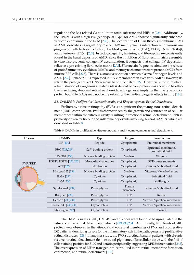
Int. J. Mol. Sci. 2022, 23, 2591 16 of 38
regulating the Ras-related C3 botulinum toxin substrate and HIF1-α [224]. Additionally,the RPE cells with a high-risk genotype at 10q26 for AMD showed significantly enhancedversican expression in the ECM [206]. The localization of HS in Bruch’s membrane (BM)in AMD describes its regulatory role of CNV mainly via its interaction with various an-giogenic growth factors, including fibroblast growth factor (FGF), VEGF, TNF-α, TGF-β,and interferon (IFN)-γ [207]. In fact, collagen IV, laminins, and fibronectin are consistentlyfound in the basal deposits of AMD. Since the inhibition of fibronectin matrix assemblyin vitro also prevents collagen IV accumulation, it suggests that collagen IV depositionrelies on a pre-existing fibronectin matrix [208]. Fibronectin fragments stimulate the releaseof proinflammatory cytokines, MMPs, and monocyte chemoattractant protein (MCP) frommurine RPE cells [225]. There is a strong association between plasma fibrinogen levels andAMD [226]. Tenascin-C is expressed in CNV membranes in eyes with AMD. However, itsrole in the pathogenesis of CNV remains to be elucidated [227]. Conversely, the intravitrealadministration of exogenous sulfated GAGs devoid of core protein was shown to be effec-tive in reducing abnormal retinal or choroidal angiogenesis, implying that the type of coreprotein bound to GAGs may not be important for their anti-angiogenic effects in vitro [116].
2.8. DAMPS in Proliferative Vitreoretinopathy and Rhegmatogenous Retinal Detachment
Proliferative vitreoretinopathy (PVR) is a significant rhegmatogenous retinal detach-ment (RRD) complication. PVR is characterized by the growth and contraction of cellularmembranes within the vitreous cavity resulting in tractional retinal detachment. PVR isprimarily driven by fibrotic and inflammatory events involving several DAMPs, which aredescribed in Table 8.
Table 8. DAMPs in proliferative vitreoretinopathy and rhegmatogenous retinal detachment.
Disease DAMPs Type Origin Localization
LIF [130] Peptide Cytoplasmic Pre-retinal membrane
S100 [228,229] Ca2+ binding protein Cytoplasmic Epiretinal membrane/subretinal fluid
HMGB1 [230] Nuclear binding protein Nuclear Vitreous
HSP47, HSP70 [231,232] Molecular chaperones Cytoplasmic RPE/inner segments
ATP [233] Nucleotide Cytoplasmic Vitreous/subretinal fluid
Histone-H3 [234] Nuclear binding protein Nuclear Vitreous/ detached retina
IL-1α [235] Cytokine Cytoplasmic Subretinal fluid
IL-33 [236] Cytokine Cytoplasmic Müller glia
Syndecan-1 [237] Proteoglycan Plasmamembrane Vitreous/subretinal fluid
Biglycan [238] Proteoglycan ECM Retina
Decorin [239,240] Proteoglycan ECM Vitreous/epiretinal membrane
Tenascin-C [240,241] Glycoprotein ECM Vitreous/epiretinal membrane
PVR/RRD
Int. J. Mol. Sci. 2022, 23, x FOR PEER REVIEW 16 of 38
further evaluated [222]. ET-1 significantly increased in the plasma of exudative and neo-vascular AMD [204].
A previous study suggested that the higher expression of HS proteoglycans (HSPGs) in CNV lesions may be linked to endothelial dysfunction and increased capillary perme-ability [207]. Rat retinas with laser-induced CNV showed significant upregulation of both perlecan and syndecan-4 compared to the control retinas. The expression profiles of these proteoglycans were found not only to depend on the presence or absence of CNV, but also on the size of the CNV-lesion [205]. Intravitreal injection of decorin significantly inhibits laser induced CNV in a rodent model [223]. Decorin might exhibit anti-angiogenic re-sponses by acting as an inhibitor for multiple receptor tyrosine kinases, such as the epi-dermal growth factor receptor (EGFR), the insulin-like growth factor receptor (IGFR), and the Met hepatocyte growth factor receptor [224]. It has also been shown to decrease hy-poxia-induced VEGF expression by blocking the Met expression pathway and downreg-ulating the Ras-related C3 botulinum toxin substrate and HIF1-α [224]. Additionally, the RPE cells with a high-risk genotype at 10q26 for AMD showed significantly enhanced versican expression in the ECM [206]. The localization of HS in Bruch’s membrane (BM) in AMD describes its regulatory role of CNV mainly via its interaction with various angi-ogenic growth factors, including fibroblast growth factor (FGF), VEGF, TNF-α, TGF-β, and interferon (IFN)-γ [207]. In fact, collagen IV, laminins, and fibronectin are consistently found in the basal deposits of AMD. Since the inhibition of fibronectin matrix assembly in vitro also prevents collagen IV accumulation, it suggests that collagen IV deposition relies on a pre-existing fibronectin matrix [208]. Fibronectin fragments stimulate the release of proinflammatory cytokines, MMPs, and monocyte chemoattractant protein (MCP) from murine RPE cells [225]. There is a strong association between plasma fibrinogen levels and AMD [226]. Tenascin-C is expressed in CNV membranes in eyes with AMD. However, its role in the pathogenesis of CNV remains to be elucidated [227]. Conversely, the intravi-treal administration of exogenous sulfated GAGs devoid of core protein was shown to be effective in reducing abnormal retinal or choroidal angiogenesis, implying that the type of core protein bound to GAGs may not be important for their anti-angiogenic effects in vitro [116].
2.8. DAMPS in Proliferative Vitreoretinopathy and Rhegmatogenous Retinal Detachment Proliferative vitreoretinopathy (PVR) is a significant rhegmatogenous retinal detach-
ment (RRD) complication. PVR is characterized by the growth and contraction of cellular membranes within the vitreous cavity resulting in tractional retinal detachment. PVR is primarily driven by fibrotic and inflammatory events involving several DAMPs, which are described in Table 8.
Table 8. DAMPs in proliferative vitreoretinopathy and rhegmatogenous retinal detachment.
Disease DAMPs Type Origin Localization
PVR/RRD
LIF [130] Peptide Cytoplasmic Pre-retinal membrane
S100 [228,229] Ca2+ binding protein Cytoplasmic Epiretinal mem-
brane/subretinal fluid HMGB1 [230] Nuclear binding protein Nuclear Vitreous
HSP47, HSP70 [231,232] Molecular chaperones Cytoplasmic RPE/inner segments
ATP [233] Nucleotide Cytoplasmic Vitreous/subretinal
fluid
Histone-H3 [234] Nuclear binding protein Nuclear Vitreous/ detached ret-
ina IL-1α [235] Cytokine Cytoplasmic Subretinal fluid IL-33 [236] Cytokine Cytoplasmic Müller glia
Fibrinogen [242] Glycoprotein ECM Plasma
The DAMPs such as S100, HMGB1, and histones were found to be upregulated in thevitreous of the retinal detachment patients [229,230,234]. Additionally, high levels of S100protein were observed in the vitreous and epiretinal membranes of PVR and proliferativeDR patients, describing its role for the inflammatory axis in the pathogenesis of proliferativeretinal disorders [228]. In another study, the PVR subretinal band in patients with chronicrecurrent retinal detachment demonstrated pigmented fibrocellular tissue with the foci ofcells staining positive for S100 and keratin peripherally, suggesting RPE differentiation [243].The overexpression of LIF in transgenic mice resulted in pre-retinal membrane formation,contraction, and retinal detachment [130].

Int. J. Mol. Sci. 2022, 23, 2591 17 of 38
HMGB1 plays an essential role in fibrosis in both proliferative DR and PVR. Thereis a significant increase in HMGB1 in the epiretinal membrane in proliferative DR andPVR [244]. There is also an increase in HMGB1 in the vitreous humor of patients withproliferative DR and retinal detachment compared to patients with retinal detachmentalone [245]. Under hypoxia, RPE cells secrete HMGB1. Additionally, HMGB1 can up-regulate the expression of angiogenic and fibrogenic factors in ARPE-19 cells, includingVEGF, basic FGF, TGF-β2, and connective tissue growth factor (CTGF), via TLR4 and theRAGE-dependent NFκB pathway [246]. HMGB1 released from the dying cells activates ERKphosphorylation and potentially promotes RPE proliferation and migration, contributingto retinal detachment [230]. HSP47 is linked to increased fibrosis in ARPE-19 cells [231] andsignificantly inhibits photoreceptor cell death in animal models of retinal detachment [232].There is significant increased HSP70 expression in the subretinal fibrosis model. HSP90was found in samples of idiopathic epiretinal membranes, and its expression appears tobe correlated with the presence of TGF-β receptor II and αSMA. HSP90 is involved inretinal fibrosis via the TGF-β1-induced transduction pathway in Müller glia [247]. Thereis a significant increase in extracellular ATP in the vitreous and subretinal space of RRDpatients compared to patients with macular holes and epiretinal membranes [233]. In theATP-induced retinal degeneration feline model, fibrotic tissue ultimately displaced theneural retina in the worst affected area [248]. However, the role of released ATP in fibrosisis not studied yet. Another DAMP member, histone H3, was found on the outer side ofthe detached retina and was associated with photoreceptor death in the rat model [234].Additionally, there is a significant increase in IL-1α concentrations in primary RRD subjectsdue to PVR. Since IL-1 induces RPE cell migration and its intravitreal injection leads tothe breakdown of the blood–ocular barrier, IL-1 has been suggested to be an importantcandidate in the activation processes that lead to PVR development [235]. Müller glia isa primary source of IL-33 in the retina. IL-33 is known for its profibrotic function andincreases retinal fibrosis after laser injury [236,249]. IL-33 deficiency enhanced retinal celldeath and gliosis after retinal detachment with sustained subretinal inflammation frominfiltrating macrophages [250,251].
Among proteoglycans, soluble syndecan-1 was significantly high in the vitreous andsubretinal fluid collected from RRD eyes. The increase in syndecan-1 concentrations inthe subretinal fluid was positively correlated with a longer duration of retinal detachmentand negatively correlated with younger age [237]. After retinal detachment, there was asignificant increase in biglycan gene expression after seven days of retinal detachment [238].However, the release of biglycan in the retina or vitreous has not been studied in RDpatients or animal models. Further, the hyaluronic acid concentration in the retinal detach-ment patient was significantly lower than in the control group. Hyaluronidase activity wassignificantly higher in the vitreous humor of patients with RRD. Contrarily, the vitreoushumor contained hyaluronic acid of high molecular mass in the control group [251]. Thereis also a significant increase in decorin in the epiretinal membrane of PVR and prolifera-tive DR patients [240]. The vitreal decorin concentrations significantly increased in RRDpatients who did develop PVR; however, they did not reliably predict the outcome [239].There is a significant increase in tenascin-C in the epiretinal membrane and vitreous ofboth proliferative DR and PVR patients [240,241,252]. Tenascin-C is expressed at lowerlevels in most adult tissues but is transiently upregulated during acute inflammation and iscontinuously expressed during chronic inflammation and tissue repair [227]. It was pre-dicted to play a role in fibrovascular membrane formation and angiogenesis in proliferativeDR [227]. Fibronectin also plays a vital role in retinal detachment. Intravitreal adminis-tration of fibronectin and platelet-derived growth factor (PDGF) was sufficient to inducethe resultant retinal detachment in the rabbit model [253]. There was a strong correlationbetween fibrinogen plasma levels and the clinical features of RRD, which supported therole of fibrinogen in retinal detachment [242].
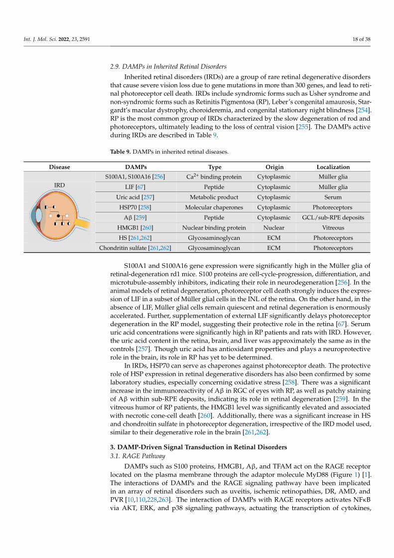
Int. J. Mol. Sci. 2022, 23, 2591 18 of 38
2.9. DAMPs in Inherited Retinal Disorders
Inherited retinal disorders (IRDs) are a group of rare retinal degenerative disordersthat cause severe vision loss due to gene mutations in more than 300 genes, and lead to reti-nal photoreceptor cell death. IRDs include syndromic forms such as Usher syndrome andnon-syndromic forms such as Retinitis Pigmentosa (RP), Leber’s congenital amaurosis, Star-gardt’s macular dystrophy, choroideremia, and congenital stationary night blindness [254].RP is the most common group of IRDs characterized by the slow degeneration of rod andphotoreceptors, ultimately leading to the loss of central vision [255]. The DAMPs activeduring IRDs are described in Table 9.
Table 9. DAMPs in inherited retinal diseases.
Disease DAMPs Type Origin Localization
S100A1, S100A16 [256] Ca2+ binding protein Cytoplasmic Müller glia
LIF [67] Peptide Cytoplasmic Müller glia
Uric acid [257] Metabolic product Cytoplasmic Serum
HSP70 [258] Molecular chaperones Cytoplasmic Photoreceptors
Aβ [259] Peptide Cytoplasmic GCL/sub-RPE deposits
HMGB1 [260] Nuclear binding protein Nuclear Vitreous
HS [261,262] Glycosaminoglycan ECM Photoreceptors
IRD
Int. J. Mol. Sci. 2022, 23, x FOR PEER REVIEW 18 of 38
RD patients or animal models. Further, the hyaluronic acid concentration in the retinal detachment patient was significantly lower than in the control group. Hyaluronidase ac-tivity was significantly higher in the vitreous humor of patients with RRD. Contrarily, the vitreous humor contained hyaluronic acid of high molecular mass in the control group [251]. There is also a significant increase in decorin in the epiretinal membrane of PVR and proliferative DR patients [240]. The vitreal decorin concentrations significantly increased in RRD patients who did develop PVR; however, they did not reliably predict the outcome [239]. There is a significant increase in tenascin-C in the epiretinal membrane and vitreous of both proliferative DR and PVR patients [240,241,252]. Tenascin-C is expressed at lower levels in most adult tissues but is transiently upregulated during acute inflammation and is continuously expressed during chronic inflammation and tissue repair [227]. It was pre-dicted to play a role in fibrovascular membrane formation and angiogenesis in prolifera-tive DR [227]. Fibronectin also plays a vital role in retinal detachment. Intravitreal admin-istration of fibronectin and platelet-derived growth factor (PDGF) was sufficient to induce the resultant retinal detachment in the rabbit model [253]. There was a strong correlation between fibrinogen plasma levels and the clinical features of RRD, which supported the role of fibrinogen in retinal detachment [242].
2.9. DAMPs in Inherited Retinal Disorders Inherited retinal disorders (IRDs) are a group of rare retinal degenerative disorders
that cause severe vision loss due to gene mutations in more than 300 genes, and lead to retinal photoreceptor cell death. IRDs include syndromic forms such as Usher syndrome and non-syndromic forms such as Retinitis Pigmentosa (RP), Leber’s congenital amauro-sis, Stargardt’s macular dystrophy, choroideremia, and congenital stationary night blind-ness [254]. RP is the most common group of IRDs characterized by the slow degeneration of rod and photoreceptors, ultimately leading to the loss of central vision [255]. The DAMPs active during IRDs are described in Table 9.
Table 9. DAMPs in inherited retinal diseases.
Disease DAMPs Type Origin Localization
IRD
S100A1, S100A16 [256] Ca2+ binding protein Cytoplasmic Müller glia LIF [67] Peptide Cytoplasmic Müller glia
Uric acid [257] Metabolic product Cytoplasmic Serum HSP70 [258] Molecular chaperones Cytoplasmic Photoreceptors
Aβ [259] Peptide Cytoplasmic GCL/sub-RPE depos-
its HMGB1 [260] Nuclear binding protein Nuclear Vitreous HS [261,262] Glycosaminoglycan ECM Photoreceptors
Chondritin sulfate [261,262]
Glycosaminoglycan ECM Photoreceptors
S100A1 and S100A16 gene expression were significantly high in the Müller glia of retinal-degeneration rd1 mice. S100 proteins are cell-cycle-progression, differentiation, and microtubule-assembly inhibitors, indicating their role in neurodegeneration [256]. In the animal models of retinal degeneration, photoreceptor cell death strongly induces the expression of LIF in a subset of Müller glial cells in the INL of the retina. On the other hand, in the absence of LIF, Müller glial cells remain quiescent and retinal degeneration is enormously accelerated. Further, supplementation of external LIF significantly delays photoreceptor degeneration in the RP model, suggesting their protective role in the retina [67]. Serum uric acid concentrations were significantly high in RP patients and rats with IRD. However, the uric acid content in the retina, brain, and liver was approximately the
Chondritin sulfate [261,262] Glycosaminoglycan ECM Photoreceptors
S100A1 and S100A16 gene expression were significantly high in the Müller glia ofretinal-degeneration rd1 mice. S100 proteins are cell-cycle-progression, differentiation, andmicrotubule-assembly inhibitors, indicating their role in neurodegeneration [256]. In theanimal models of retinal degeneration, photoreceptor cell death strongly induces the expres-sion of LIF in a subset of Müller glial cells in the INL of the retina. On the other hand, in theabsence of LIF, Müller glial cells remain quiescent and retinal degeneration is enormouslyaccelerated. Further, supplementation of external LIF significantly delays photoreceptordegeneration in the RP model, suggesting their protective role in the retina [67]. Serumuric acid concentrations were significantly high in RP patients and rats with IRD. However,the uric acid content in the retina, brain, and liver was approximately the same as in thecontrols [257]. Though uric acid has antioxidant properties and plays a neuroprotectiverole in the brain, its role in RP has yet to be determined.
In IRDs, HSP70 can serve as chaperones against photoreceptor death. The protectiverole of HSP expression in retinal degenerative disorders has also been confirmed by somelaboratory studies, especially concerning oxidative stress [258]. There was a significantincrease in the immunoreactivity of Aβ in RGC of eyes with RP, as well as patchy stainingof Aβ within sub-RPE deposits, indicating its role in retinal degeneration [259]. In thevitreous humor of RP patients, the HMGB1 level was significantly elevated and associatedwith necrotic cone-cell death [260]. Additionally, there was a significant increase in HSand chondroitin sulfate in photoreceptor degeneration, irrespective of the IRD model used,similar to their degenerative role in the brain [261,262].
3. DAMP-Driven Signal Transduction in Retinal Disorders3.1. RAGE Pathway
DAMPs such as S100 proteins, HMGB1, Aβ, and TFAM act on the RAGE receptorlocated on the plasma membrane through the adaptor molecule MyD88 (Figure 1) [1].The interactions of DAMPs and the RAGE signaling pathway have been implicatedin an array of retinal disorders such as uveitis, ischemic retinopathies, DR, AMD, andPVR [10,110,228,263]. The interaction of DAMPs with RAGE receptors activates NFκBvia AKT, ERK, and p38 signaling pathways, actuating the transcription of cytokines,
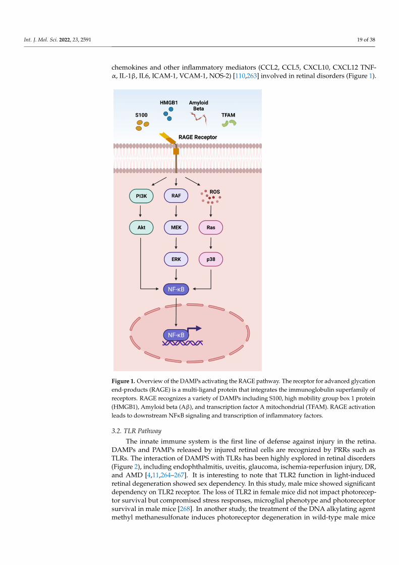
Int. J. Mol. Sci. 2022, 23, 2591 19 of 38
chemokines and other inflammatory mediators (CCL2, CCL5, CXCL10, CXCL12 TNF-α, IL-1β, IL6, ICAM-1, VCAM-1, NOS-2) [110,263] involved in retinal disorders (Figure 1).
Int. J. Mol. Sci. 2022, 23, x FOR PEER REVIEW 20 of 38
Figure 1. Overview of the DAMPs activating the RAGE pathway. The receptor for advanced gly-cation end-products (RAGE) is a multi-ligand protein that integrates the immunoglobulin super-family of receptors. RAGE recognizes a variety of DAMPs including S100, high mobility group box 1 protein (HMGB1), Amyloid beta (Aβ), and transcription factor A mitochondrial (TFAM). RAGE activation leads to downstream NFκB signaling and transcription of inflammatory factors.
3.2. TLR Pathway The innate immune system is the first line of defense against injury in the retina.
DAMPs and PAMPs released by injured retinal cells are recognized by PRRs such as TLRs. The interaction of DAMPS with TLRs has been highly explored in retinal disorders (Figure 2), including endophthalmitis, uveitis, glaucoma, ischemia-reperfusion injury, DR, and AMD [4,11,264–267]. It is interesting to note that TLR2 function in light-induced retinal degeneration showed sex dependency. In this study, male mice showed significant dependency on TLR2 receptor. The loss of TLR2 in female mice did not impact photore-ceptor survival but compromised stress responses, microglial phenotype and photorecep-tor survival in male mice [268]. In another study, the treatment of the DNA alkylating agent methyl methanesulfonate induces photoreceptor degeneration in wild-type male mice regulated by poly(ADP-ribose) polymerase 1 (PARP1) activation and cytoplasmic translocation of HMGB1, whereas wild-type female mice are partially protected. Addi-tionally, PARylation was significantly higher in methyl-methanesulfonate-treated male mice and muted in female mice, resulting in enhanced HMGB1 cytoplasmic translocation
Figure 1. Overview of the DAMPs activating the RAGE pathway. The receptor for advanced glycationend-products (RAGE) is a multi-ligand protein that integrates the immunoglobulin superfamily ofreceptors. RAGE recognizes a variety of DAMPs including S100, high mobility group box 1 protein(HMGB1), Amyloid beta (Aβ), and transcription factor A mitochondrial (TFAM). RAGE activationleads to downstream NFκB signaling and transcription of inflammatory factors.
3.2. TLR Pathway
The innate immune system is the first line of defense against injury in the retina.DAMPs and PAMPs released by injured retinal cells are recognized by PRRs such asTLRs. The interaction of DAMPS with TLRs has been highly explored in retinal disorders(Figure 2), including endophthalmitis, uveitis, glaucoma, ischemia-reperfusion injury, DR,and AMD [4,11,264–267]. It is interesting to note that TLR2 function in light-inducedretinal degeneration showed sex dependency. In this study, male mice showed significantdependency on TLR2 receptor. The loss of TLR2 in female mice did not impact photorecep-tor survival but compromised stress responses, microglial phenotype and photoreceptorsurvival in male mice [268]. In another study, the treatment of the DNA alkylating agentmethyl methanesulfonate induces photoreceptor degeneration in wild-type male mice
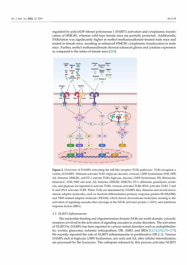
Int. J. Mol. Sci. 2022, 23, 2591 20 of 38
regulated by poly(ADP-ribose) polymerase 1 (PARP1) activation and cytoplasmic translo-cation of HMGB1, whereas wild-type female mice are partially protected. Additionally,PARylation was significantly higher in methyl-methanesulfonate-treated male mice andmuted in female mice, resulting in enhanced HMGB1 cytoplasmic translocation in malemice. Further, methyl methanesulfonate showed enhanced gliosis and cytokine expressionas compared to the retina of female mice [269].
Int. J. Mol. Sci. 2022, 23, x FOR PEER REVIEW 21 of 38
in male mice. Further, methyl methanesulfonate showed enhanced gliosis and cytokine expression as compared to the retina of female mice [269].
Figure 2. Overview of DAMPs activating the toll-like receptor (TLR) pathways. TLRs recognize a variety of DAMPs. Defensin activates TLR1; biglycan, decorin, versican, LMW hyaluronan, S100, HSP, Aβ, histones, HMGB1, and ET-1 activate TLR2; biglycan, decorin, LMW hyaluronan, HS, fi-bronectin, tenascin-C, S100, HSP, uric acid, Aβ, histones, HMGB1, HMGN1, ET-1, defensins, gran-ulysin, syndecan, and glypican are reported to activate TLR4; versican activates TLR6; RNA acti-vates TLR3, 7 and 8; and DNA activates TLR9. When TLRs are stimulated by DAMPs they dimerize and recruit downstream adaptor molecules, such as myeloid differentiation primary-response pro-tein 88 (MyD88), and TRIF-related adaptor molecule (TRAM), which directs downstream molecules, leading to the activation of signaling cascades that converge at the NFκB, activator protein 1 (AP1), and interferon response factors (IRFs).
3.3. NLRP3 Inflammasome
The nucleotide-binding and oligomerization domain NLRs are multi-domain, cyto-solic receptors involved in the activation of signaling cascades in ocular disorders. The activation of NLRP3 by DAMPs has been reported in various retinal disorders such as endophthalmitis, uveitis, glaucoma, ischemic retinopathies, DR, AMD, and IRDs [12,74,254,270–273]. We recently reported the role of NLRP3 inflammasome in prolifera-tive DR [12]. Various DAMPs such as biglycan, LMW hyaluronan, uric acid, and Aβ, after cellular internalization, are processed by the lysozyme. The cathepsin released by this pro-cess activates NLRP3 signaling, whereas biglycan, ATP, Aβ, and cathelicidin can directly activate NLRP3 via the P2X7 receptor, a purinergic receptor (Figure 3). DAMPs stimulate inflammasome formations, which are large intracellular multiprotein complexes (MRC) consisting of NLR family sensory proteins (NLRPs), apoptosis speck-like adaptor protein (ASC), and caspase-1 for the production and secretion of IL-1β, leading to further en-hancement in photoreceptor cell death by pyroptosis [274]. Additionally, LMW
Figure 2. Overview of DAMPs activating the toll-like receptor (TLR) pathways. TLRs recognize avariety of DAMPs. Defensin activates TLR1; biglycan, decorin, versican, LMW hyaluronan, S100, HSP,Aβ, histones, HMGB1, and ET-1 activate TLR2; biglycan, decorin, LMW hyaluronan, HS, fibronectin,tenascin-C, S100, HSP, uric acid, Aβ, histones, HMGB1, HMGN1, ET-1, defensins, granulysin, synde-can, and glypican are reported to activate TLR4; versican activates TLR6; RNA activates TLR3, 7 and8; and DNA activates TLR9. When TLRs are stimulated by DAMPs they dimerize and recruit down-stream adaptor molecules, such as myeloid differentiation primary-response protein 88 (MyD88),and TRIF-related adaptor molecule (TRAM), which directs downstream molecules, leading to theactivation of signaling cascades that converge at the NFκB, activator protein 1 (AP1), and interferonresponse factors (IRFs).
3.3. NLRP3 Inflammasome
The nucleotide-binding and oligomerization domain NLRs are multi-domain, cytosolicreceptors involved in the activation of signaling cascades in ocular disorders. The activationof NLRP3 by DAMPs has been reported in various retinal disorders such as endophthalmi-tis, uveitis, glaucoma, ischemic retinopathies, DR, AMD, and IRDs [12,74,254,270–273].We recently reported the role of NLRP3 inflammasome in proliferative DR [12]. VariousDAMPs such as biglycan, LMW hyaluronan, uric acid, and Aβ, after cellular internalization,are processed by the lysozyme. The cathepsin released by this process activates NLRP3

Int. J. Mol. Sci. 2022, 23, 2591 21 of 38
signaling, whereas biglycan, ATP, Aβ, and cathelicidin can directly activate NLRP3 via theP2X7 receptor, a purinergic receptor (Figure 3). DAMPs stimulate inflammasome forma-tions, which are large intracellular multiprotein complexes (MRC) consisting of NLR familysensory proteins (NLRPs), apoptosis speck-like adaptor protein (ASC), and caspase-1 forthe production and secretion of IL-1β, leading to further enhancement in photoreceptorcell death by pyroptosis [274]. Additionally, LMW hyaluronan interacts with CD44 andactivates the NLRP-3 inflammasome [1,74,198,254,270–273]. Furthermore, ATP is releasedby the apoptotic and necrotic cells and acts as a neurotransmitter and as a gliotransmitterin the retina to recruit macrophages and microglia. Once ATP binds P2X7, it activatesthe protein kinase C/MAP kinase pathway that leads to the release of chemokines andpro-inflammatory cytokines [275].
Int. J. Mol. Sci. 2022, 23, x FOR PEER REVIEW 22 of 38
hyaluronan interacts with CD44 and activates the NLRP-3 inflammasome [1,74,198,254,270–273]. Furthermore, ATP is released by the apoptotic and necrotic cells and acts as a neurotransmitter and as a gliotransmitter in the retina to recruit macrophages and microglia. Once ATP binds P2X7, it activates the protein kinase C/MAP kinase path-way that leads to the release of chemokines and pro-inflammatory cytokines [275].
Figure 3. Overview of DAMPs activating the NLRP3 inflammasome. The first step in the 2-step process involves activation and translocation of NFκB into the nucleus to regulate the transcription of the oligomerization-like receptor pyrin-domain-containing protein 3 (NLRP3) inflammasome genes. The second step is actuation of NLRP3 inflammasome mediated by (a) DAMPs such as bi-glycan, LMW hyaluronan, uric acid, and Aβ to release cathepsin from lysosomal degradation; (b) K+ efflux via P2X7 receptor activation by DAMPs such as biglycan, ATP, Aβ, and cathelicidin; and (c) CD44 activation by LMW hyaluronan caspase-1 signaling pathway leading to caspase-1 activity and the release of mature IL-1β.
3.4. Other Pathways DAMPs can also activate several other pathways in retinal disorders (Figure 4). The
activation of NFκB and AP-1 via CD14 receptors, along with TLR2 or 4, has been reported in various retinal disorders such as endophthalmitis, glaucoma, and DR [1,11,12,70]. Fur-ther, the regulation of NFκB and AP-1 by IL-33 and IL-1α via MYD88 has been reported
Figure 3. Overview of DAMPs activating the NLRP3 inflammasome. The first step in the 2-stepprocess involves activation and translocation of NFκB into the nucleus to regulate the transcription ofthe oligomerization-like receptor pyrin-domain-containing protein 3 (NLRP3) inflammasome genes.The second step is actuation of NLRP3 inflammasome mediated by (a) DAMPs such as biglycan,LMW hyaluronan, uric acid, and Aβ to release cathepsin from lysosomal degradation; (b) K+ effluxvia P2X7 receptor activation by DAMPs such as biglycan, ATP, Aβ, and cathelicidin; and (c) CD44activation by LMW hyaluronan caspase-1 signaling pathway leading to caspase-1 activity and therelease of mature IL-1β.
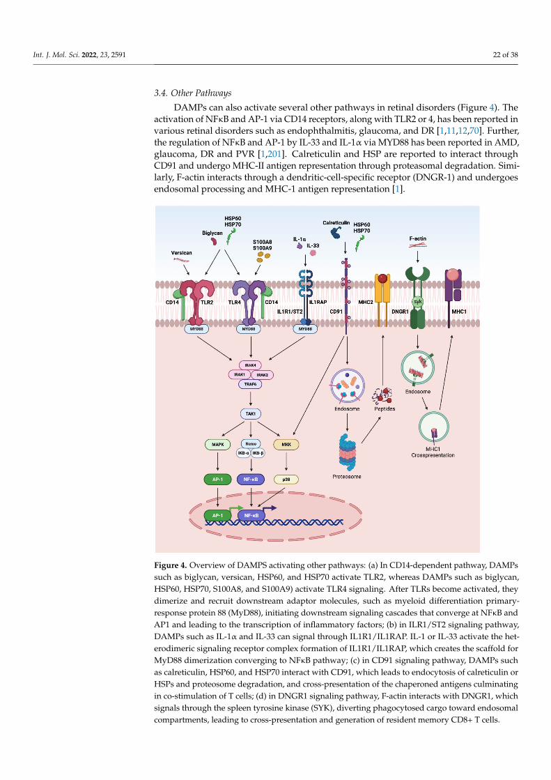
Int. J. Mol. Sci. 2022, 23, 2591 22 of 38
3.4. Other Pathways
DAMPs can also activate several other pathways in retinal disorders (Figure 4). Theactivation of NFκB and AP-1 via CD14 receptors, along with TLR2 or 4, has been reported invarious retinal disorders such as endophthalmitis, glaucoma, and DR [1,11,12,70]. Further,the regulation of NFκB and AP-1 by IL-33 and IL-1α via MYD88 has been reported in AMD,glaucoma, DR and PVR [1,201]. Calreticulin and HSP are reported to interact throughCD91 and undergo MHC-II antigen representation through proteasomal degradation. Simi-larly, F-actin interacts through a dendritic-cell-specific receptor (DNGR-1) and undergoesendosomal processing and MHC-1 antigen representation [1].
Int. J. Mol. Sci. 2022, 23, x FOR PEER REVIEW 23 of 38
in AMD, glaucoma, DR and PVR [1,201]. Calreticulin and HSP are reported to interact through CD91 and undergo MHC-II antigen representation through proteasomal degra-dation. Similarly, F-actin interacts through a dendritic-cell-specific receptor (DNGR-1) and undergoes endosomal processing and MHC-1 antigen representation [1].
Figure 4. Overview of DAMPS activating other pathways: (a) In CD14-dependent pathway, DAMPs such as biglycan, versican, HSP60, and HSP70 activate TLR2, whereas DAMPs such as biglycan, HSP60, HSP70, S100A8, and S100A9) activate TLR4 signaling. After TLRs become activated, they dimerize and recruit downstream adaptor molecules, such as myeloid differentiation primary-re-sponse protein 88 (MyD88), initiating downstream signaling cascades that converge at NFκB and AP1 and leading to the transcription of inflammatory factors; (b) in ILR1/ST2 signaling pathway, DAMPs such as IL-1α and IL-33 can signal through IL1R1/IL1RAP. IL-1 or IL-33 activate the heter-odimeric signaling receptor complex formation of IL1R1/IL1RAP, which creates the scaffold for MyD88 dimerization converging to NFκB pathway; (c) in CD91 signaling pathway, DAMPs such as calreticulin, HSP60, and HSP70 interact with CD91, which leads to endocytosis of calreticulin or HSPs and proteosome degradation, and cross-presentation of the chaperoned antigens culminating in co-stimulation of T cells; (d) in DNGR1 signaling pathway, F-actin interacts with DNGR1, which signals through the spleen tyrosine kinase (SYK), diverting phagocytosed cargo toward endosomal compartments, leading to cross-presentation and generation of resident memory CD8+ T cells.
4. Therapeutic Implications of DAMPs In retinal disorders, DAMPs are released by necrotic and apoptotic cells to elicit mul-
tiple downstream signaling effects to activate the innate immune system. The emerging evidence from preclinical and clinical studies suggests that DAMPs play both pathogenic
Figure 4. Overview of DAMPS activating other pathways: (a) In CD14-dependent pathway, DAMPssuch as biglycan, versican, HSP60, and HSP70 activate TLR2, whereas DAMPs such as biglycan,HSP60, HSP70, S100A8, and S100A9) activate TLR4 signaling. After TLRs become activated, theydimerize and recruit downstream adaptor molecules, such as myeloid differentiation primary-response protein 88 (MyD88), initiating downstream signaling cascades that converge at NFκB andAP1 and leading to the transcription of inflammatory factors; (b) in ILR1/ST2 signaling pathway,DAMPs such as IL-1α and IL-33 can signal through IL1R1/IL1RAP. IL-1 or IL-33 activate the het-erodimeric signaling receptor complex formation of IL1R1/IL1RAP, which creates the scaffold forMyD88 dimerization converging to NFκB pathway; (c) in CD91 signaling pathway, DAMPs suchas calreticulin, HSP60, and HSP70 interact with CD91, which leads to endocytosis of calreticulin orHSPs and proteosome degradation, and cross-presentation of the chaperoned antigens culminatingin co-stimulation of T cells; (d) in DNGR1 signaling pathway, F-actin interacts with DNGR1, whichsignals through the spleen tyrosine kinase (SYK), diverting phagocytosed cargo toward endosomalcompartments, leading to cross-presentation and generation of resident memory CD8+ T cells.
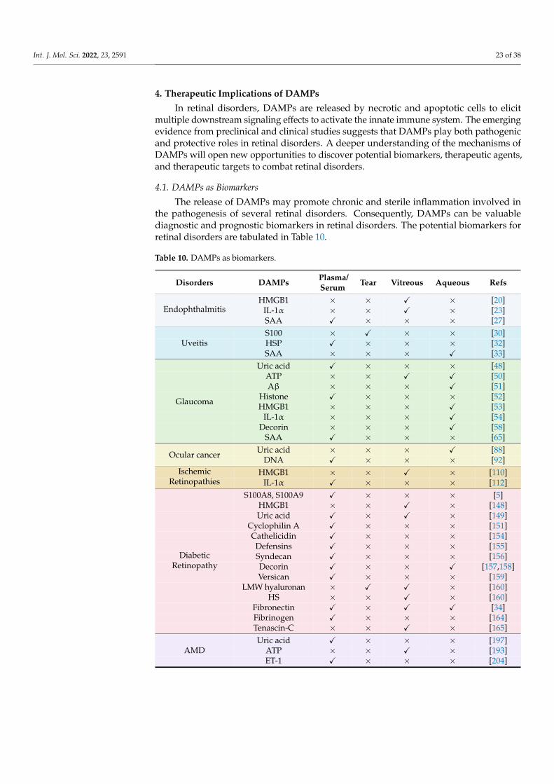
Int. J. Mol. Sci. 2022, 23, 2591 23 of 38
4. Therapeutic Implications of DAMPs
In retinal disorders, DAMPs are released by necrotic and apoptotic cells to elicitmultiple downstream signaling effects to activate the innate immune system. The emergingevidence from preclinical and clinical studies suggests that DAMPs play both pathogenicand protective roles in retinal disorders. A deeper understanding of the mechanisms ofDAMPs will open new opportunities to discover potential biomarkers, therapeutic agents,and therapeutic targets to combat retinal disorders.
4.1. DAMPs as Biomarkers
The release of DAMPs may promote chronic and sterile inflammation involved inthe pathogenesis of several retinal disorders. Consequently, DAMPs can be valuablediagnostic and prognostic biomarkers in retinal disorders. The potential biomarkers forretinal disorders are tabulated in Table 10.
Table 10. DAMPs as biomarkers.
Disorders DAMPs Plasma/Serum Tear Vitreous Aqueous Refs
HMGB1 × × X × [20]IL-1α × × X × [23]EndophthalmitisSAA X × × × [27]S100 × X × × [30]HSP X × × × [32]UveitisSAA × × × X [33]
Uric acid X × × × [48]ATP × × X X [50]Aβ × × × X [51]
Histone X × × × [52]HMGB1 × × × X [53]
IL-1α × × × X [54]Decorin × × × X [58]
Glaucoma
SAA X × × × [65]Uric acid × × × X [88]Ocular cancer
DNA X × × × [92]HMGB1 × × X × [110]Ischemic
Retinopathies IL-1α X × × × [112]S100A8, S100A9 X × × × [5]
HMGB1 × × X × [148]Uric acid X × X × [149]
Cyclophilin A X × × × [151]Cathelicidin X × × × [154]
Defensins X × × × [155]Syndecan X × × × [156]Decorin X × × X [157,158]Versican X × × × [159]
LMW hyaluronan × X X × [160]HS × × X × [160]
Fibronectin X × X X [34]Fibrinogen X × × × [164]
DiabeticRetinopathy
Tenascin-C × × X × [165]Uric acid X × × × [197]
ATP × × X × [193]AMDET-1 X × × × [204]
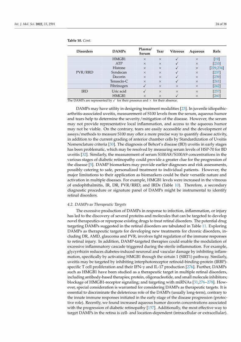
Int. J. Mol. Sci. 2022, 23, 2591 24 of 38
Table 10. Cont.
Disorders DAMPs Plasma/Serum Tear Vitreous Aqueous Refs
HMGB1 × × X × [19]ATP × × X × [233]
Histone × × X × [229,234]Syndecan × × X × [237]Decorin × × X × [239]
Tenascin-C × × X × [241]
PVR/RRD
Fibrinogen X × × × [242]IRD Uric acid X × × × [257]
HMGB1 × × X × [260]The DAMPs are represented by X for their presence and × for their absence.
DAMPs may have utility in designing treatment modalities [23]. In juvenile idiopathic-arthritis-associated uveitis, measurement of S100 levels from the serum, aqueous humorand tears help to determine the severity/mitigation of the disease. However, the serummay not provide representative local inflammation, and access to the aqueous humormay not be viable. On the contrary, tears are easily accessible and the development ofassays/methods to measure S100 may offer a more precise way to quantify disease activity,in addition to the current grading of anterior chamber cells by Standardization of UveitisNomenclature criteria [30]. The diagnosis of Behcet’s disease (BD) uveitis in early stageshas been problematic, which may be resolved by measuring serum levels of HSP-70 for BDuveitis [32]. Similarly, the measurement of serum S100A8/S100A9 concentrations in thevarious stages of diabetic retinopathy could provide a greater clue for the progression ofthe disease [5]. DAMP biomarkers may provide earlier diagnoses and risk assessments,possibly catering to safe, personalized treatment to individual patients. However, themajor limitations to their application as biomarkers could be their versatile nature andactivation in multiple diseases. For example, HMGB1 levels were increased in the vitreousof endophthalmitis, IR, DR, PVR/RRD, and IRDs (Table 10). Therefore, a secondarydiagnostic procedure or signature panel of DAMPs might be instrumental to identifyretinal disorders.
4.2. DAMPs as Therapeutic Targets
The excessive production of DAMPs in response to infection, inflammation, or injuryhas led to the discovery of several proteins and molecules that can be targeted to developnovel therapeutics or repurpose existing drugs to treat retinal disorders. The potential drugtargeting DAMPs suggested in the retinal disorders are tabulated in Table 11. ExploringDAMPs as therapeutic targets for developing new treatments for chronic disorders, in-cluding DR, AMD, glaucoma and PVR, involves tight regulation of the immune responsesto retinal injury. In addition, DAMP-targeted therapies could enable the modulation ofexcessive inflammatory cascade triggered during the sterile inflammation. For example,glycyrrhizin reduces diabetes-induced neuronal and vascular damage by inhibiting inflam-mation, specifically by activating HMGB1 through the sirtuin 1 (SIRT1) pathway. Similarly,uveitis may be targeted by inhibiting interphotoreceptor retinoid-binding-protein (IRBP)-specific T cell proliferation and their IFN-γ and IL-17 production [276]. Further, DAMPssuch as HMGB1 have been studied as a therapeutic target in multiple retinal disorders,including antibody-based therapies; protein, oligonucleotide, and small molecule inhibitors;blockage of HMGB1-receptor signaling; and targeting with miRNAs [31,276–278]. How-ever, special consideration is warranted for considering DAMPs as therapeutic targets. It isessential to discriminate the deleterious role of the DAMPs (usually long-term), contrary tothe innate immune responses initiated in the early stage of the disease progression (protec-tive role). Recently, we found increased aqueous humor decorin concentrations associatedwith the progression of diabetic retinopathy [157]. Additionally, the most effective way totarget DAMPs in the retina is cell- and location-dependent (intracellular or extracellular).

Int. J. Mol. Sci. 2022, 23, 2591 25 of 38
Thus, long-term targeting of DAMPs may prevent them from initiating regulatory T cells(Tregs) and promote immunosuppression [279].
Table 11. DAMPs as therapeutic targets.
Retinal Disorders Drug DAMPs Action Refs
Uveitis HMGB1 AbGlycyrrhizin HMGB1 Inhibition of IRBP-specific
T cell proliferation [31]
Glaucoma Brilliant Blue G andN-methyl-d-aspartic acid ATP Antagonists of the P2X7R [280]
Ocular cancer Ansamycins HSP90 G1 Arrest [281]
miR34A and miR-22 HMGB1 Act on autophagy, migration, andinvasion of RB cells [277,278]
Cyclosporin A TFAM Preserves TFAM [113]Coenzyme Q10 TFAM Preserves TFAM [282]
Ischemicretinopathies
Brimonidine TFAM Preserves TFAM [114]
Diabeticretinopathy
TasquinimodGlycyrrhizin
S100HMGB1
Inhibits angiogenesis via TSP-1,VEGF, ICAM-1 and ERK1/2
Acts on HMGB1 via SIRT1 andprovides neurovascular protection
[276,283]
AMD Geldanamycin HSP90 Inhibits VEGF and HIFα [284]Anti-histoneAntibodies Histone Reduced retinal damage [234]PVR/RRD
Geranylgeranylacetone HSP70 Activation of Akt pathway [232]
4.3. DAMPs as Therapeutic Agents
DAMPs such as decorin, LIF, defensins, IL-33, syndecan-1, and SAA have been de-scribed for their anti-inflammatory, anti-angiogenic or anti-fibrotic properties, and arepresented in Table 12. Harnessing these DAMPs as therapeutic agents or therapeuticsis an attractive novel therapeutic strategy against retinal disorders, which will perhapsfind its way into future routine treatment modalities. The availability of DAMPs duringinjury or infection is vital for providing the essential host defense system and restoringhomeostasis in the injured tissues. Cathelicidins act as an anti-microbial agent on manypathogens, including Gram-positive and Gram-negative bacteria, fungi, parasites, andenveloped viruses in vitro. Cationic cathelicidins can bind and disrupt negatively chargedmembranes, leading to microbial cell death. These peptides can also cross membranes andtarget intracellular processes such as RNA and DNA synthesis, impair the functions ofenzymes and chaperones, and can stimulate protein degradation [285]. However, underphysiological circumstances, most cathelicidins are impaired by high salt concentrations,sugars, and other host or microbial factors [285]. IL-33 is upregulated in the uveitis retinadepicting its anti-inflammatory role. However, its effects are model- and disease-specific.Thus, considering IL-33 as a therapeutic agent needs a complete understanding of its func-tion in the pathological microenvironment [45]. HSP70 binds to TLR2/TLR4 and exhibitsanti-inflammatory properties via secretion of IL-10 and TGF-β in AMD. Contrarily, theextracellular HSP70 has been suggested to have a pro-inflammatory effect. Hence, it is tooearly to predict the use of HSP-70 as an anti-inflammatory agent. Decorin binds to TGFβwith high affinity and is known for its anti-fibrotic role compared to traditional anti-fibroticadjuvants such as mitomycin-C and 5-fluorouracil [286,287]. Nevertheless, it has bothpro-and anti-angiogenic properties [224]. Similarly, anti-angiogenic therapeutic DAMPssuch as LIF, HS, and IL-33 are also in the early stages and their safety and efficacy profile isawaited for retinal disorders. Therefore, the application or inhibition of DAMPs must bedesigned under strict caveats and precautions using the therapeutic window during theprogression of retinal disorders.
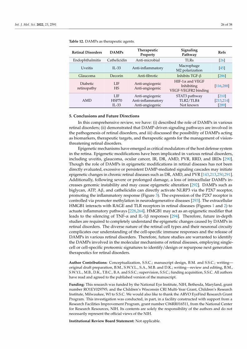
Int. J. Mol. Sci. 2022, 23, 2591 26 of 38
Table 12. DAMPs as therapeutic agents.
Retinal Disorders DAMPs TherapeuticProperty
SignalingPathway Refs
Endophthalmitis Cathelicidin Anti-microbial TLRs [26]
Uveitis IL-33 Anti-inflammatory MacrophageM2 polarization [45]
Glaucoma Decorin Anti-fibrotic Inhibits TGF-β [286]
Diabeticretinopathy
LIFHS
Anti-angiogenicAnti-angiogenic
HIF-1α and VEGFInhibiting
VEGF-VEGFR2 binding[116,288]
LIF Anti-angiogenic STAT3 pathway [210]HSP70 Anti-inflammatory TLR2/TLR4 [213,214]AMDIL-33 Anti-angiogenic Not known [289]
5. Conclusions and Future Directions
In this comprehensive review, we have: (i) described the role of DAMPs in variousretinal disorders; (ii) demonstrated that DAMP-driven signaling pathways are involved inthe pathogenesis of retinal disorders, and iii) discussed the possibility of DAMPs actingas biomarkers, therapeutic targets, and therapeutic agents for the management of vision-threatening retinal disorders.
Epigenetic mechanisms have emerged as critical modulators of the host defense systemin the retina. Epigenetic modifications have been implicated in various retinal disorders,including uveitis, glaucoma, ocular cancer, IR, DR, AMD, PVR, RRD, and IRDs [290].Though the role of DAMPs in epigenetic modifications in retinal diseases has not beendirectly evaluated, excessive or persistent DAMP-mediated signaling cascades may initiateepigenetic changes in chronic retinal diseases such as DR, AMD, and PVR [143,213,250,291].Additionally, following severe or prolonged damage, a loss of intracellular DAMPs in-creases genomic instability and may cause epigenetic alteration [292]. DAMPs such asbiglycan, ATP, Aβ, and cathelicidin can directly activate NLRP3 via the P2X7 receptor,promoting the inflammatory response (Figure 3). The expression of the P2X7 receptor iscontrolled via promoter methylation in neurodegenerative diseases [293]. The extracellularHMGB1 interacts with RAGE and TLR receptors in retinal diseases (Figures 1 and 2) toactuate inflammatory pathways [228,264]. HMGB1 may act as an epigenetic modifier thatleads to the silencing of TNF-α and IL-1β responses [294]. Therefore, future in-depthstudies are required to completely understand the epigenetic changes caused by DAMPs inretinal disorders. The diverse nature of the retinal cell types and their neuronal circuitycomplicates our understanding of the cell-specific immune responses and the release ofDAMPs in various retinal disorders. Therefore, future studies are warranted to identifythe DAMPs involved in the molecular mechanisms of retinal diseases, employing single-cell or cell-specific proteomic signatures to identify/design or repurpose next generationtherapeutics for retinal disorders.
Author Contributions: Conceptualization, S.S.C.; manuscript design, B.M. and S.S.C.; writing—original draft preparation, B.M., S.W.Y.L., S.A., M.B. and D.K.; writing—review and editing, B.M.,S.W.Y.L., M.B., D.K., T.B.C., B.A. and S.S.C.; supervision, S.S.C.; funding acquisition, S.S.C. All authorshave read and agreed to the published version of the manuscript.
Funding: This research was funded by the National Eye Institute, NIH, Bethesda, Maryland, grantnumber RO1EY029795; and the Children’s Wisconsin CRI Multi-Year Grant, Children’s ResearchInstitute, Milwaukee, WI to S.S.C. We would also like to thank the ARVO EyeFind Research GrantProgram. This investigation was conducted, in part, in a facility constructed with support from aResearch Facilities Improvement Program, grant number C06RR016511, from the National Centerfor Research Resources, NIH. Its contents are solely the responsibility of the authors and do notnecessarily represent the official views of the NIH.
Institutional Review Board Statement: Not applicable.
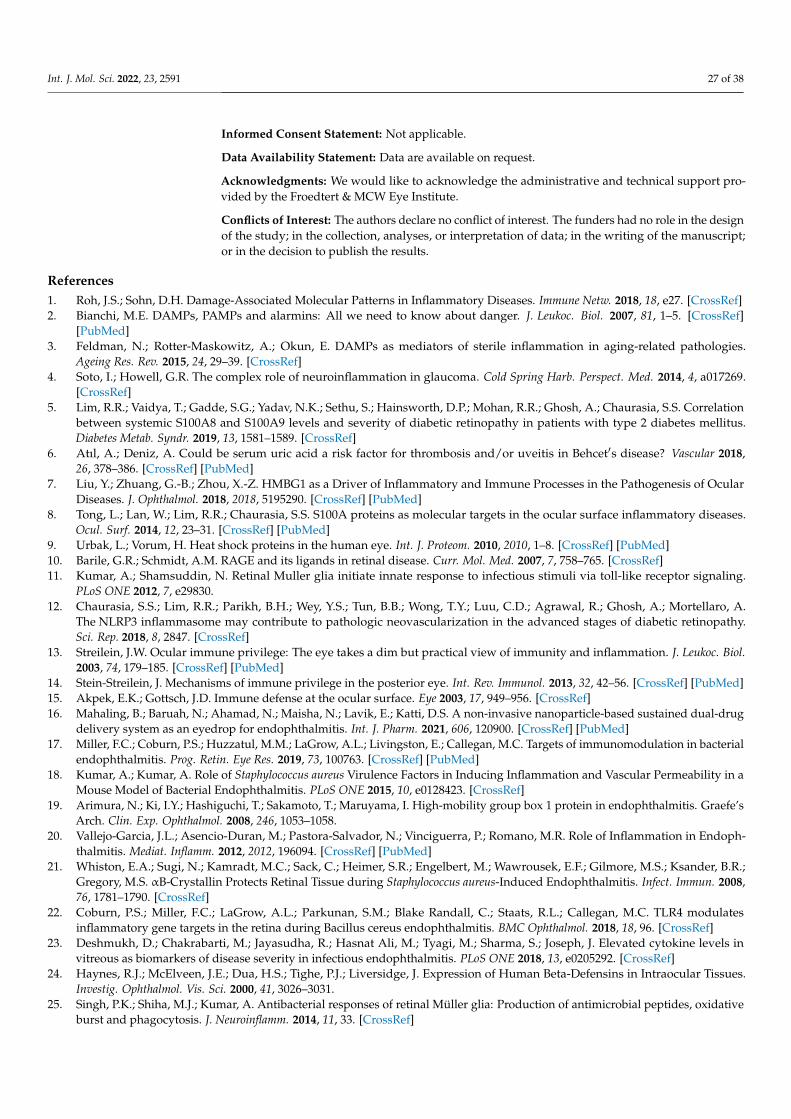
Int. J. Mol. Sci. 2022, 23, 2591 27 of 38
Informed Consent Statement: Not applicable.
Data Availability Statement: Data are available on request.
Acknowledgments: We would like to acknowledge the administrative and technical support pro-vided by the Froedtert & MCW Eye Institute.
Conflicts of Interest: The authors declare no conflict of interest. The funders had no role in the designof the study; in the collection, analyses, or interpretation of data; in the writing of the manuscript;or in the decision to publish the results.
References1. Roh, J.S.; Sohn, D.H. Damage-Associated Molecular Patterns in Inflammatory Diseases. Immune Netw. 2018, 18, e27. [CrossRef]2. Bianchi, M.E. DAMPs, PAMPs and alarmins: All we need to know about danger. J. Leukoc. Biol. 2007, 81, 1–5. [CrossRef]
[PubMed]3. Feldman, N.; Rotter-Maskowitz, A.; Okun, E. DAMPs as mediators of sterile inflammation in aging-related pathologies.
Ageing Res. Rev. 2015, 24, 29–39. [CrossRef]4. Soto, I.; Howell, G.R. The complex role of neuroinflammation in glaucoma. Cold Spring Harb. Perspect. Med. 2014, 4, a017269.
[CrossRef]5. Lim, R.R.; Vaidya, T.; Gadde, S.G.; Yadav, N.K.; Sethu, S.; Hainsworth, D.P.; Mohan, R.R.; Ghosh, A.; Chaurasia, S.S. Correlation
between systemic S100A8 and S100A9 levels and severity of diabetic retinopathy in patients with type 2 diabetes mellitus.Diabetes Metab. Syndr. 2019, 13, 1581–1589. [CrossRef]
6. Atıl, A.; Deniz, A. Could be serum uric acid a risk factor for thrombosis and/or uveitis in Behcet′s disease? Vascular 2018,26, 378–386. [CrossRef] [PubMed]
7. Liu, Y.; Zhuang, G.-B.; Zhou, X.-Z. HMBG1 as a Driver of Inflammatory and Immune Processes in the Pathogenesis of OcularDiseases. J. Ophthalmol. 2018, 2018, 5195290. [CrossRef] [PubMed]
8. Tong, L.; Lan, W.; Lim, R.R.; Chaurasia, S.S. S100A proteins as molecular targets in the ocular surface inflammatory diseases.Ocul. Surf. 2014, 12, 23–31. [CrossRef] [PubMed]
9. Urbak, L.; Vorum, H. Heat shock proteins in the human eye. Int. J. Proteom. 2010, 2010, 1–8. [CrossRef] [PubMed]10. Barile, G.R.; Schmidt, A.M. RAGE and its ligands in retinal disease. Curr. Mol. Med. 2007, 7, 758–765. [CrossRef]11. Kumar, A.; Shamsuddin, N. Retinal Muller glia initiate innate response to infectious stimuli via toll-like receptor signaling.
PLoS ONE 2012, 7, e29830.12. Chaurasia, S.S.; Lim, R.R.; Parikh, B.H.; Wey, Y.S.; Tun, B.B.; Wong, T.Y.; Luu, C.D.; Agrawal, R.; Ghosh, A.; Mortellaro, A.
The NLRP3 inflammasome may contribute to pathologic neovascularization in the advanced stages of diabetic retinopathy.Sci. Rep. 2018, 8, 2847. [CrossRef]
13. Streilein, J.W. Ocular immune privilege: The eye takes a dim but practical view of immunity and inflammation. J. Leukoc. Biol.2003, 74, 179–185. [CrossRef] [PubMed]
14. Stein-Streilein, J. Mechanisms of immune privilege in the posterior eye. Int. Rev. Immunol. 2013, 32, 42–56. [CrossRef] [PubMed]15. Akpek, E.K.; Gottsch, J.D. Immune defense at the ocular surface. Eye 2003, 17, 949–956. [CrossRef]16. Mahaling, B.; Baruah, N.; Ahamad, N.; Maisha, N.; Lavik, E.; Katti, D.S. A non-invasive nanoparticle-based sustained dual-drug
delivery system as an eyedrop for endophthalmitis. Int. J. Pharm. 2021, 606, 120900. [CrossRef] [PubMed]17. Miller, F.C.; Coburn, P.S.; Huzzatul, M.M.; LaGrow, A.L.; Livingston, E.; Callegan, M.C. Targets of immunomodulation in bacterial
endophthalmitis. Prog. Retin. Eye Res. 2019, 73, 100763. [CrossRef] [PubMed]18. Kumar, A.; Kumar, A. Role of Staphylococcus aureus Virulence Factors in Inducing Inflammation and Vascular Permeability in a
Mouse Model of Bacterial Endophthalmitis. PLoS ONE 2015, 10, e0128423. [CrossRef]19. Arimura, N.; Ki, I.Y.; Hashiguchi, T.; Sakamoto, T.; Maruyama, I. High-mobility group box 1 protein in endophthalmitis. Graefe’s
Arch. Clin. Exp. Ophthalmol. 2008, 246, 1053–1058.20. Vallejo-Garcia, J.L.; Asencio-Duran, M.; Pastora-Salvador, N.; Vinciguerra, P.; Romano, M.R. Role of Inflammation in Endoph-
thalmitis. Mediat. Inflamm. 2012, 2012, 196094. [CrossRef] [PubMed]21. Whiston, E.A.; Sugi, N.; Kamradt, M.C.; Sack, C.; Heimer, S.R.; Engelbert, M.; Wawrousek, E.F.; Gilmore, M.S.; Ksander, B.R.;
Gregory, M.S. αB-Crystallin Protects Retinal Tissue during Staphylococcus aureus-Induced Endophthalmitis. Infect. Immun. 2008,76, 1781–1790. [CrossRef]
22. Coburn, P.S.; Miller, F.C.; LaGrow, A.L.; Parkunan, S.M.; Blake Randall, C.; Staats, R.L.; Callegan, M.C. TLR4 modulatesinflammatory gene targets in the retina during Bacillus cereus endophthalmitis. BMC Ophthalmol. 2018, 18, 96. [CrossRef]
23. Deshmukh, D.; Chakrabarti, M.; Jayasudha, R.; Hasnat Ali, M.; Tyagi, M.; Sharma, S.; Joseph, J. Elevated cytokine levels invitreous as biomarkers of disease severity in infectious endophthalmitis. PLoS ONE 2018, 13, e0205292. [CrossRef]
24. Haynes, R.J.; McElveen, J.E.; Dua, H.S.; Tighe, P.J.; Liversidge, J. Expression of Human Beta-Defensins in Intraocular Tissues.Investig. Ophthalmol. Vis. Sci. 2000, 41, 3026–3031.
25. Singh, P.K.; Shiha, M.J.; Kumar, A. Antibacterial responses of retinal Müller glia: Production of antimicrobial peptides, oxidativeburst and phagocytosis. J. Neuroinflamm. 2014, 11, 33. [CrossRef]
Int. J. Mol. Sci. 2022, 23, 2591 28 of 38
26. Takahashi, T.; Kulkarni, N.N.; Lee, E.Y.; Zhang, L.-j.; Wong, G.C.L.; Gallo, R.L. Cathelicidin promotes inflammation by enablingbinding of self-RNA to cell surface scavenger receptors. Sci. Rep. 2018, 8, 4032. [CrossRef]
27. Yang, Y.-Y. Clinical value of serum amyloid A detection in the diagnosis of infective endophthalmitis. Int. Eye Sci. 2019,12, 1738–1740.
28. Chang, J.H.; McCluskey, P.J.; Wakefield, D. Recent advances in Toll-like receptors and anterior uveitis. Clin. Exp. Ophthalmol.2012, 40, 821–828. [CrossRef] [PubMed]
29. John Curnow, S.; Denniston, K.O.A.; Murray, P.I.; Wallace, G.R. Intraocular immune mechanisms in uveitis. Curr. Immunol. Rev.2011, 7, 350–359. [CrossRef]
30. Angeles-Han, S.T.; Miraldi Utz, V.; Thornton, S.; Schulert, G.; Rodriguez-Smith, J.; Kauffman, A.; Sproles, A.; Mwase, N.;Hennard, T.; Grom, A.; et al. S100 Proteins, Cytokines, and Chemokines as Tear Biomarkers in Children with Juvenile IdiopathicArthritis-associated Uveitis. Ocul. Immunol. Inflamm. 2020, 29, 1616–1620. [CrossRef]
31. Jiang, G.; Sun, D.; Yang, H.; Lu, Q.; Kaplan, H.J.; Shao, H. HMGB1 is an early and critical mediator in an animal model of uveitisinduced by IRBP-specific T cells. J. Leukoc. Biol. 2014, 95, 599–607. [CrossRef]
32. Sahebari, M.; Hashemzadeh, K.; Mahmoudi, M.; Saremi, Z.; Mirfeizi, Z. Diagnostic Yield of Heat Shock Protein 70 (HSP-70) andAnti-HSP-70 in Behcet-Induced Uveitis. Scand. J. Immunol. 2013, 77, 476–481. [CrossRef] [PubMed]
33. Bauer, D.; Kasper, M.; Walscheid, K.; Koch, J.M.; Müther, P.S.; Kirchhof, B.; Heiligenhaus, A.; Heinz, C. Multiplex CytokineAnalysis of Aqueous Humor in Juvenile Idiopathic Arthritis-Associated Anterior Uveitis with or without Secondary Glaucoma.Front. Immunol. 2018, 9, 708. [CrossRef]
34. Probst, K.; Fijnheer, R.; Schellekens, P.; Rothova, A. Intraocular and plasma levels of cellular fibronectin in patients with uveitisand diabetes mellitus. Br. J. Ophthalmol. 2004, 88, 667. [CrossRef]
35. Ni, M.; Chan, C.-C.; Nussenblatt, R.B.; Li, S.; Mao, W. Iris Inflammatory Cells, Fibronectin, Fibrinogen, and Immunoglobulin inVarious Ocular Diseases. Arch. Ophthalmol. 1988, 106, 392–395. [CrossRef]
36. Walscheid, K.; Heiligenhaus, A.; Holzinger, D.; Roth, J.; Heinz, C.; Tappeiner, C.; Kasper, M.; Foell, D. Elevated S100A8/A9 andS100A12 Serum Levels Reflect Intraocular Inflammation in Juvenile Idiopathic Arthritis-Associated Uveitis: Results from a PilotStudy. Investig. Ophthalmol. Vis. Sci. 2015, 56, 7653–7660. [CrossRef]
37. Wakefield, D.; Gray, P.; Chang, J.; Di Girolamo, N.; McCluskey, P. The role of PAMPs and DAMPs in the pathogenesis of acute andrecurrent anterior uveitis. Br. J. Ophthalmol. 2010, 94, 271. [CrossRef]
38. Poulaki, V.; Iliaki, E.; Mitsiades, N.; Mitsiades, C.S.; Paulus, Y.N.; Bula, D.V.; Gragoudas, E.S.; Miller, J.W. Inhibition of Hsp90attenuates inflammation in endotoxin-induced uveitis. FASEB J. 2007, 21, 2113–2123. [CrossRef] [PubMed]
39. Hu, W.; Hasan, A.; Wilson, A.; Stanford, M.R.; Li-Yang, Y.; Todryk, S.; Whiston, R.; Shinnick, T.; Mizushima, Y.; Van der Zee, R.;et al. Experimental mucosal induction of uveitis with the 60-kDa heat shock protein-derived peptide 336–351. Eur. J. Immunol.1998, 28, 2444–2455. [CrossRef]
40. Stanford, M.R.; Kasp, E.; Whiston, R.; Hasan, A.; Todryk, S.; Shinnick, T.; Mizushima, Y.; Dumonde, D.C.; van der Zee, R.; Lehner,T. Heat shock protein peptides reactive in patients with Behçet′s disease are uveitogenic in Lewis rats. Clin. Exp. Immunol. 1994,97, 226–231. [CrossRef]
41. Dai, M.-L.; Fan, S.; Li, Z.; Yu, X.; Lin, D.; Huang, X.-F.; Wang, Y. Correlation of serum amyloid A levels, clinical manifestations,treatment, and disease activity in patients with acute anterior uveitis. Eye 2020, 34, 1672–1678. [CrossRef] [PubMed]
42. Lopalco, G.; Lucherini, O.M.; Vitale, A.; Talarico, R.; Lopalco, A.; Galeazzi, M.; Lapadula, G.; Cantarini, L.; Iannone, F. PutativeRole of Serum Amyloid-A and Proinflammatory Cytokines as Biomarkers for Behcet′s Disease. Medicine 2015, 94, e1858.[CrossRef] [PubMed]
43. Niemi, K.; Teirilä, L.; Lappalainen, J.; Rajamäki, K.; Baumann, M.H.; Öörni, K.; Wolff, H.; Kovanen, P.T.; Matikainen, S.;Eklund, K.K. Serum amyloid A activates the NLRP3 inflammasome via P2X7 receptor and a cathepsin B-sensitive pathway.J. Immunol. 2011, 186, 6119–6128. [CrossRef] [PubMed]
44. Song, C.; Hsu, K.; Yamen, E.; Yan, W.; Fock, J.; Witting, P.K.; Geczy, C.L.; Freedman, S.B. Serum amyloid A induction of cytokinesin monocytes/macrophages and lymphocytes. Atherosclerosis 2009, 207, 374–383. [CrossRef] [PubMed]
45. Barbour, M.; Allan, D.; Xu, H.; Pei, C.; Chen, M.; Niedbala, W.; Fukada, S.Y.; Besnard, A.-G.; Alves-Filho, J.C.; Tong, X.; et al.IL-33 attenuates the development of experimental autoimmune uveitis. Eur. J. Immunol. 2014, 44, 3320–3329. [CrossRef]
46. Reinehr, S.; Kuehn, S.; Casola, C.; Koch, D.; Stute, G.; Grotegut, P.; Dick, H.B.; Joachim, S.C. HSP27 immunization reinforces AIIamacrine cell and synapse damage induced by S100 in an autoimmune glaucoma model. Cell Tissue Res. 2018, 371, 237–249.[CrossRef] [PubMed]
47. Hu, Q.; Huang, C.; Wang, Y.; Wu, R. Expression of leukemia inhibitory factor in the rat retina following acute ocular hypertension.Mol. Med. Rep. 2015, 12, 6577–6583. [CrossRef] [PubMed]
48. Yuki, K.; Murat, D.; Kimura, I.; Ohtake, Y.; Tsubota, K. Reduced-serum vitamin C and increased uric acid levels in normal-tensionglaucoma. Graefe’s Arch. Clin. Exp. Ophthalmol. 2010, 248, 243–248.
49. Luo, C.; Yang, X.; Kain, A.D.; Powell, D.W.; Kuehn, M.H.; Tezel, G. Glaucomatous tissue stress and the regulation of immuneresponse through glial Toll-like receptor signaling. Investig. Ophthalmol. Vis. Sci. 2010, 51, 5697–5707. [CrossRef] [PubMed]
50. Pulukool, S.K.; Bhagavatham, S.K.S.; Kannan, V.; Sukumar, P.; Dandamudi, R.B.; Ghaisas, S.; Kunchala, H.; Saieesh, D.; Naik, A.A.;Pargaonkar, A.; et al. Elevated dimethylarginine, ATP, cytokines, metabolic remodeling involving tryptophan metabolism andpotential microglial inflammation characterize primary open angle glaucoma. Sci. Rep. 2021, 11, 9766. [CrossRef] [PubMed]
Int. J. Mol. Sci. 2022, 23, 2591 29 of 38
51. Gupta, V.; Gupta, V.B.; Chitranshi, N.; Gangoda, S.; Vander Wall, R.; Abbasi, M.; Golzan, M.; Dheer, Y.; Shah, T.; Avolio, A.; et al.One protein, multiple pathologies: Multifaceted involvement of amyloid β in neurodegenerative disorders of the brain and retina.Cell. Mol. Life Sci. 2016, 73, 4279–4297. [CrossRef]
52. Reichelt, J.; Joachim, S.C.; Pfeiffer, N.; Grus, F.H. Analysis of autoantibodies against human retinal antigens in sera of patientswith glaucoma and ocular hypertension. Curr. Eye Res. 2008, 33, 253–261. [CrossRef]
53. He, W.; Xu, F.; Chen, L.; Huang, W.; Jiang, L.; Tang, F.; Yan, W.; Zhong, S.; Shen, C.; Huang, H.; et al. Association of High-MobilityGroup Box-1 with Inflammationrelated Cytokines in the Aqueous Humor with Acute Primary Angle-Closure Eyes. Curr. Mol.Med. 2021, 21, 237–245. [CrossRef]
54. Pantalon, A.; Obadă, O.; Constantinescu, D.; Feraru, C.; Chiselită, D. Inflammatory model in patients with primary open angleglaucoma and diabetes. Int. J. Ophthalmol. 2019, 12, 795–801. [PubMed]
55. Izzotti, A.; Saccà, S.C.; Cartiglia, C.; De Flora, S. Oxidative deoxyribonucleic acid damage in the eyes of glaucoma patients. Am. J.Med. 2003, 114, 638–646. [CrossRef]
56. Stowell, C.; Jang, G.-F.; Zhang, L.; Crabb, J.; Reynaud, J.; Gardiner, S.K.; Willard, b.; Marsh-Armstrong, N.; Crabb, J.W.;Burgoyne, C.F. Alterations in Optic Nerve Head (ONH) Endoplasmic Reticulum (ER) Stress Response Proteins in Non-HumanPrimate (NHP) Early Experimental Glaucoma (EG). Investig. Ophthalmol. Vis. Sci. 2018, 59, 3514.
57. Chauhan, B.C. Endothelin and its potential role in glaucoma. Can. J. Ophthalmol. 2008, 43, 356–360. [CrossRef]58. NikhalaShree, S.; Karthikkeyan, G.; George, R.; Shantha, B.; Vijaya, L.; Ratra, V.; Sulochana, K.N.; Coral, K. Lowered Decorin
with Aberrant Extracellular Matrix Remodeling in Aqueous Humor and Tenon′s Tissue from Primary Glaucoma Patients.Investig. Ophthalmol. Vis. Sci. 2019, 60, 4661–4669. [CrossRef]
59. Burgoyne, C.; Stowell, C.; Jang, G.-F.; Zhang, L.; Willard, B.; Crabb, J.S.; Crabb, J.W. Non-human primate (NHP) optic nerve head(ONH) proteomic change in early experimental glaucoma (EEG). Investig. Ophthalmol. Vis. Sci. 2014, 55, 4555.
60. Keller, K.E.; Bradley, J.M.; Vranka, J.A.; Acott, T.S. Segmental Versican Expression in the Trabecular Meshwork and Involvementin Outflow Facility. Investig. Ophthalmol. Vis. Sci. 2011, 52, 5049–5057. [CrossRef] [PubMed]
61. Patel, N.B.; Carter-Dawson, L.; Harwerth, R.S. Extracellular Matrix Tissue Content of the Neuroretinal Rim differs in Healthy andGlaucomatous Eyes. Investig. Ophthalmol. Vis. Sci. 2019, 60, 662.
62. Reinhard, J.; Wiemann, S.; Hildebrandt, S.; Faissner, A. Extracellular Matrix Remodeling in the Retina and Optic Nerve of a NovelGlaucoma Mouse Model. Biology 2021, 10, 169. [CrossRef]
63. Tezel, G.; Edward, D.P.; Wax, M.B. Serum autoantibodies to optic nerve head glycosaminoglycans in patients with glaucoma.Arch. Ophthalmol. 1999, 117, 917–924. [CrossRef]
64. Faralli, J.A.; Filla, M.S.; Peters, D.M. Role of Fibronectin in Primary Open Angle Glaucoma. Cells 2019, 8, 1518. [CrossRef][PubMed]
65. Wang, W.-H.; McNatt, L.G.; Pang, I.-H.; Hellberg, P.E.; Fingert, J.H.; McCartney, M.D.; Clark, A.F. Increased Expression of SerumAmyloid A in Glaucoma and Its Effect on Intraocular Pressure. Investig. Ophthalmol. Vis. Sci. 2008, 49, 1916–1923. [CrossRef][PubMed]
66. Casola, C.; Schiwek, J.E.; Reinehr, S.; Kuehn, S.; Grus, F.H.; Kramer, M.; Dick, H.B.; Joachim, S.C. S100 alone has the samedestructive effect on retinal ganglion cells as in combination with HSP 27 in an autoimmune glaucoma model. J. Mol. Neurosci.2015, 56, 228–236. [CrossRef]
67. Joly, S.; Lange, C.; Thiersch, M.; Samardzija, M.; Grimm, C. Leukemia Inhibitory Factor Extends the Lifespan of InjuredPhotoreceptors In Vivo. J. Neurosci. 2008, 28, 13765–13774. [CrossRef] [PubMed]
68. Lam, K.-W.; Liu, K.-M.; Yee, R.W.; Lee, P.-F. Detection of uric acid in aqueous humor by high pressure liquid chromatography.Curr. Eye Res. 1982, 2, 645–649. [CrossRef] [PubMed]
69. Li, S.; Shao, M.; Tang, B.; Zhang, A.; Cao, W.; Sun, X. The association between serum uric acid and glaucoma severity in primaryangle closure glaucoma: A retrospective case-control study. Oncotarget 2017, 8, 2816–2824. [CrossRef] [PubMed]
70. Tsai, T.; Grotegut, P.; Reinehr, S.; Joachim, S.C. Role of Heat Shock Proteins in Glaucoma. Int. J. Mol. Sci. 2019, 20, 5160. [CrossRef]71. Guo, L.; Salt, T.E.; Luong, V.; Wood, N.; Cheung, W.; Maass, A.; Ferrari, G.; Russo-Marie, F.; Sillito, A.M.; Cheetham, M.E.; et al.
Targeting amyloid-beta in glaucoma treatment. Proc. Natl. Acad. Sci. USA 2007, 104, 13444–13449. [CrossRef]72. Mohd Lazaldin, M.A.; Iezhitsa, I.; Agarwal, R.; Bakar, N.S.; Agarwal, P.; Mohd Ismail, N. Neuroprotective effects of brain-derived
neurotrophic factor against amyloid beta 1-40-induced retinal and optic nerve damage. Eur. J. Neurosci. 2020, 51, 2394–2411.[CrossRef] [PubMed]
73. Wang, L.; Mao, X. Role of Retinal Amyloid-β in Neurodegenerative Diseases: Overlapping Mechanisms and Emerging ClinicalApplications. Int. J. Mol. Sci. 2021, 22, 2360. [CrossRef]
74. Chi, W.; Chen, H.; Li, F.; Zhu, Y.; Yin, W.; Zhuo, Y. HMGB1 promotes the activation of NLRP3 and caspase-8 inflammasomes viaNF-κB pathway in acute glaucoma. J. Neuroinflamm. 2015, 12, 137. [CrossRef] [PubMed]
75. Wu, J.-H.; Zhang, S.-H.; Nickerson, J.M.; Gao, F.-J.; Sun, Z.; Chen, X.-Y.; Zhang, S.-J.; Gao, F.; Chen, J.-Y.; Luo, Y.; et al. CumulativemtDNA damage and mutations contribute to the progressive loss of RGCs in a rat model of glaucoma. Neurobiol. Dis. 2015,74, 167–179. [CrossRef]
76. Marola, O.J.; Syc-Mazurek, S.B.; Howell, G.R.; Libby, R.T. Endothelin 1-induced retinal ganglion cell death is largely mediated byJUN activation. Cell Death Dis. 2020, 11, 811. [CrossRef]
Int. J. Mol. Sci. 2022, 23, 2591 30 of 38
77. Hill, L.J.; Mead, B.; Blanch, R.J.; Ahmed, Z.; De Cogan, F.; Morgan-Warren, P.J.; Mohamed, S.; Leadbeater, W.; Scott, R.A.H.;Berry, M.; et al. Decorin Reduces Intraocular Pressure and Retinal Ganglion Cell Loss in Rodents Through Fibrolysis of theScarred Trabecular Meshwork. Investig. Ophthalmol. Vis. Sci. 2015, 56, 3743–3757. [CrossRef] [PubMed]
78. Reinehr, S.; Reinhard, J.; Wiemann, S.; Stute, G.; Kuehn, S.; Woestmann, J.; Dick, H.B.; Faissner, A.; Joachim, S.C. Early remodellingof the extracellular matrix proteins tenascin-C and phosphacan in retina and optic nerve of an experimental autoimmuneglaucoma model. J. Cell. Mol. Med. 2016, 20, 2122–2137. [CrossRef] [PubMed]
79. Eagle, R.C. The pathology of ocular cancer. Eye 2013, 27, 128–136. [CrossRef]80. Jovanovic, P.; Mihajlovic, M.; Djordjevic-Jocic, J.; Vlajkovic, S.; Cekic, S.; Stefanovic, V. Ocular melanoma: An overview of the
current status. Int. J. Clin. Exp. Pathol. 2013, 6, 1230–1244. [PubMed]81. Donato, R.; Cannon, B.R.; Sorci, G.; Riuzzi, F.; Hsu, K.; Weber, D.J.; Geczy, C.L. Functions of S100 proteins. Curr. Mol. Med. 2013,
13, 24–57. [CrossRef] [PubMed]82. Bresnick, A.R.; Weber, D.J.; Zimmer, D.B. S100 proteins in cancer. Nat. Rev. Cancer 2015, 15, 96–109. [CrossRef] [PubMed]83. Karim, M.M.; Itoh, H. Demonstration of S-100 protein immunoreactivity in normal human retina and retinoblastoma. Ophthalmo-
logica 1997, 211, 351–353. [CrossRef]84. He, W.; Inomata, H. Dual immunologic property of S-100 protein in normal eyes and eyes with retinoblastoma: A histopathological
and immunohistochemical study of 88 cases. Ophthalmologica 1993, 206, 133–138. [CrossRef] [PubMed]85. Kan-Mitchell, J.; Rao, N.; Albert, D.M.; Van Eldik, L.J.; Taylor, C.R. S100 immunophenotypes of uveal melanomas. Investig.
Ophthalmol. Vis. Sci. 1990, 31, 1492–1496.86. Keijser, S.; Missotten, G.S.; Bonfrer, J.M.; de Wolff-Rouendaal, D.; Jager, M.J.; de Keizer, R.J. Immunophenotypic markers to
differentiate between benign and malignant melanocytic lesions. Br. J. Ophthalmol. 2006, 90, 213–217. [CrossRef]87. Missotten, G.S.; Korse, C.M.; van Dehn, C.; Linders, T.C.; Keunen, J.E.; Jager, M.J.; Bonfrer, J.M. S-100B Protein and Melanoma
Inhibitory Activity Protein in Uveal Melanoma Screening. Tumor Biol. 2007, 28, 63–69. [CrossRef] [PubMed]88. Mendelsohn, M.E.; Abramson, D.H.; Senft, S.; Servodidio, C.A.; Gamache, P.H. Uric acid in the aqueous humor and tears of
retinoblastoma patients. J. Am. Assoc. Pediatr. Ophthalmol. Strabismus 1998, 2, 369–371. [CrossRef]89. Venkatesan, N.; Kanwar, J.R.; Deepa, P.R.; Navaneethakrishnan, S.; Joseph, C.; Krishnakumar, S. Targeting HSP90/Survivin
using a cell permeable structure based peptido-mimetic shepherdin in retinoblastoma. Chem.-Biol. Interact. 2016, 252, 141–149.[CrossRef] [PubMed]
90. Tsering, T.; Laskaris, A.; Abdouh, M.; Bustamante, P.; Parent, S.; Jin, E.; Ferrier, S.T.; Arena, G.; Burnier, J.V. Uveal Melanoma-Derived Extracellular Vesicles Display Transforming Potential and Carry Protein Cargo Involved in Metastatic Niche Preparation.Cancers 2020, 12, 2923. [CrossRef] [PubMed]
91. Pardo, M.; García, Á.; Thomas, B.; Piñeiro, A.; Akoulitchev, A.; Dwek, R.A.; Zitzmann, N. The characterization of the invasionphenotype of uveal melanoma tumour cells shows the presence of MUC18 and HMG-1 metastasis markers and leads to theidentification of DJ-1 as a potential serum biomarker. Int. J. Cancer 2006, 119, 1014–1022. [CrossRef]
92. Madic, J.; Piperno-Neumann, S.; Servois, V.; Rampanou, A.; Milder, M.; Trouiller, B.; Gentien, D.; Saada, S.; Assayag, F.;Thuleau, A.; et al. Pyrophosphorolysis-activated polymerization detects circulating tumor DNA in metastatic uveal melanoma.Clin. Cancer Res. 2012, 18, 3934–3941. [CrossRef]
93. Xu, J.; Zhao, Y.; Sun, H.; Xiao, Q.; Ye, P. Identification of Versican as an Independent Prognostic Factor in Uveal Melanoma. Int. J.Gen. Med. 2021, 14, 4639–4651. [CrossRef]
94. Albakova, Z.; Siam, M.K.S.; Sacitharan, P.K.; Ziganshin, R.H.; Ryazantsev, D.Y.; Sapozhnikov, A.M. Extracellular heat shockproteins and cancer: New perspectives. Transl. Oncol. 2021, 14, 100995. [CrossRef]
95. Missotten, G.S.; Journée-de Korver, J.G.; de Wolff-Rouendaal, D.; Keunen, J.E.; Schlingemann, R.O.; Jager, M.J. Heat shock proteinexpression in the eye and in uveal melanoma. Investig. Ophthalmol. Vis. Sci. 2003, 44, 3059–3065. [CrossRef]
96. Faingold, D.; Marshall, J.-C.; Antecka, E.; Di Cesare, S.; Odashiro, A.N.; Bakalian, S.; Fernandes, B.F.; Burnier, M.N. ImmuneExpression and Inhibition of Heat Shock Protein 90 in Uveal Melanoma. Clin. Cancer Res. 2008, 14, 847–855. [CrossRef] [PubMed]
97. Wang, L.-L.; Feng, Y.-Q.; Cheng, Y.-H. Effect on proliferation and apoptosis of retinoblastoma cell by RNA inhibiting high mobilitygroup protein box-1 expression. Int. J. Ophthalmol. 2017, 10, 30–34. [PubMed]
98. Singh, M.K.; Singh, L.; Pushker, N.; Sen, S.; Sharma, A.; Chauhan, F.A.; Kashyap, S. Correlation of High Mobility Group Box-1Protein (HMGB1) with Clinicopathological Parameters in Primary Retinoblastoma. Pathol. Oncol. Res. 2015, 21, 1237–1242.[CrossRef] [PubMed]
99. Pardo, M.; Piñeiro, A.; de la Fuente, M.; García, A.; Prabhakar, S.; Zitzmann, N.; Dwek, R.A.; Sánchez-Salorio, M.; Domínguez, F.;Capeans, C. Abnormal cell cycle regulation in primary human uveal melanoma cultures. J. Cell. Biochem. 2004, 93, 708–720.[CrossRef]
100. Berry, J.L.; Xu, L.; Murphree, A.L.; Krishnan, S.; Stachelek, K.; Zolfaghari, E.; McGovern, K.; Lee, T.C.; Carlsson, A.; Kuhn, P.; et al.Potential of Aqueous Humor as a Surrogate Tumor Biopsy for Retinoblastoma. JAMA Ophthalmol. 2017, 135, 1221–1230. [CrossRef][PubMed]
101. Kothari, P.; Marass, F.; Yang, J.L.; Stewart, C.M.; Stephens, D.; Patel, J.; Hasan, M.; Jing, X.; Meng, F.; Enriquez, J.; et al.Cell-free DNA profiling in retinoblastoma patients with advanced intraocular disease: An MSKCC experience. Cancer Med. 2020,9, 6093–6101. [CrossRef]
102. Jin, E.; Burnier, J.V. Liquid Biopsy in Uveal Melanoma: Are We There Yet? Ocul. Oncol. Pathol. 2021, 7, 1–16. [CrossRef]
Int. J. Mol. Sci. 2022, 23, 2591 31 of 38
103. Bek, T. Capillary closure secondary to retinal vein occlusion. A morphological, histopathological, and immunohistochemicalstudy. Acta Ophthalmol. Scand. 1998, 76, 643–648. [CrossRef]
104. Yang, J.; Yang, N.; Luo, J.; Cheng, G.; Zhang, X.; He, T.; Xing, Y. Overexpression of S100A4 protects retinal ganglion cells againstretinal ischemia-reperfusion injury in mice. Exp. Eye Res. 2020, 201, 108281. [CrossRef] [PubMed]
105. Sano, Y.; Kanematsu, E.; Yoshiura, M.; Iwamoto, T.; Takizawa, N.; Tokuhisa, T.; Mizuno, A. Uric acid as biochemical marker forretinal and optic nerve damage after occlusion and reperfusion of common carotid and vertebral arteries in rat. Jpn. J. Ophthalmol.1992, 36, 76–83. [PubMed]
106. Li, Y.; Roth, S.; Laser, M.; Ma, J.-x.; Crosson, C.E. Retinal preconditioning and the induction of heat-shock protein 27.Investig. Ophthalmol. Vis. Sci. 2003, 44, 1299–1304. [CrossRef]
107. Kalesnykas, G.; Tuulos, T.; Uusitalo, H.; Jolkkonen, J. Neurodegeneration and cellular stress in the retina and optic nerve in ratcerebral ischemia and hypoperfusion models. Neuroscience 2008, 155, 937–947. [CrossRef]
108. Strunnikova, N.; Baffi, J.; Gonzalez, A.; Silk, W.; Cousins, S.W.; Csaky, K.G. Regulated heat shock protein 27 expression in humanretinal pigment epithelium. Investig. Ophthalmol. Vis. Sci. 2001, 42, 2130–2138.
109. Arckens, L.; Van der Gucht, E.; Van den Bergh, G.; Massie, A.; Leysen, I.; Vandenbussche, E.; Eysel, U.T.; Huybrechts, R.;Vandesande, F. Differential display implicates cyclophilin A in adult cortical plasticity. Eur. J. Neurosci. 2003, 18, 61–75. [CrossRef]
110. Dvoriantchikova, G.; Hernandez, E.; Grant, J.; Santos, A.R.C.; Yang, H.; Ivanov, D. The high-mobility group box-1 nuclear factormediates retinal injury after ischemia reperfusion. Investig. Ophthalmol. Vis. Sci. 2011, 52, 7187–7194. [CrossRef]
111. Li, X.; Ye, Z.; Pei, S.; Zheng, D.; Zhu, L. Neuroprotective effect of minocycline on rat retinal ischemia-reperfusion injury. Mol. Vis.2021, 27, 438. [PubMed]
112. Ozacmak, H.S.; Ozacmak, V.H.; Barut, F.; Araslı, M.; Ucan, B.H. Pretreatment with mineralocorticoid receptor blocker reducesintestinal injury induced by ischemia and reperfusion: Involvement of inhibition of inflammatory response, oxidative stress,nuclear factor κB, and inducible nitric oxide synthase. J. Surg. Res. 2014, 191, 350–361. [CrossRef] [PubMed]
113. Kim, S.Y.; Shim, M.S.; Kim, K.Y.; Weinreb, R.N.; Wheeler, L.A.; Ju, W.K. Inhibition of cyclophilin D by cyclosporin A promotesretinal ganglion cell survival by preventing mitochondrial alteration in ischemic injury. Cell Death Dis. 2014, 5, e1105. [CrossRef][PubMed]
114. Lee, D.; Kim, K.-Y.; Noh, Y.H.; Chai, S.; Lindsey, J.D.; Ellisman, M.H.; Weinreb, R.N.; Ju, W.-K. Brimonidine blocks glutamateexcitotoxicity-induced oxidative stress and preserves mitochondrial transcription factor a in ischemic retinal injury. PLoS ONE2012, 7, e47098. [CrossRef]
115. Reinhard, J.; Renner, M.; Wiemann, S.; Shakoor, D.A.; Stute, G.; Dick, H.B.; Faissner, A.; Joachim, S.C. Ischemic injury leads toextracellular matrix alterations in retina and optic nerve. Sci. Rep. 2017, 7, 43470. [CrossRef] [PubMed]
116. Nishiguchi, K.M.; Kataoka, K.; Kachi, S.; Komeima, K.; Terasaki, H. Regulation of pathologic retinal angiogenesis in mice andinhibition of VEGF-VEGFR2 binding by soluble heparan sulfate. PLoS ONE 2010, 5, e13493. [CrossRef]
117. Cheng, G.; He, T.; Xing, Y. Silencing of S100A4, a metastasis-associated protein, inhibits retinal neovascularization via thedownregulation of BDNF in oxygen-induced ischaemic retinopathy. Eye 2016, 30, 877–887. [CrossRef] [PubMed]
118. Cheng, G.; Tian, K.; Zhang, L.; Yang, N.; Xing, Y.; He, T. S100A4 gene silencing in oxygen-induced ischemic retinopathy inhibitsretinal neovascularization via down-regulation of CREB expression. Graefe′s Arch. Clin. Exp. Ophthalmol. 2016, 254, 97–108.[CrossRef] [PubMed]
119. Inatani, M.; Tanihara, H.; Honjo, M.; Hangai, M.; Kresse, H. Expression of Proteoglycan Decorin in Neural Retina.Investig. Ophthalmol. Vis. Sci. 1999, 40, 1783–1791.
120. Wu, M.Y.; Yiang, G.T.; Liao, W.T.; Tsai, A.P.Y.; Cheng, Y.L.; Cheng, P.W.; Li, C.Y.; Li, C.J. Current Mechanistic Concepts in Ischemiaand Reperfusion Injury. Cell. Physiol. Biochem. 2018, 46, 1650–1667. [CrossRef]
121. Windisch, B.K.; LeVatte, T.L.; Archibald, M.L.; Chauhan, B.C. Induction of heat shock proteins 27 and 72 in retinal ganglion cellsafter acute pressure-induced ischaemia. Clin. Exp. Ophthalmol. 2009, 37, 299–307. [CrossRef]
122. Whitlock, N.A.; Agarwal, N.; Ma, J.-X.; Crosson, C.E. Hsp27 upregulation by HIF-1 signaling offers protection against retinalischemia in rats. Investig. Ophthalmol. Vis. Sci. 2005, 46, 1092–1098. [CrossRef] [PubMed]
123. Yokoyama, A.; Oshitari, T.; Negishi, H.; Dezawa, M.; Mizota, A.; Adachi-Usami, E. Protection of retinal ganglion cells fromischemia-reperfusion injury by electrically applied Hsp27. Investig. Ophthalmol. Vis. Sci. 2001, 42, 3283–3286.
124. Lewden, O.; Garcher, C.; Assem, M.; Morales, C.; Rochette, L.; Bron, A. Changes of the inducible heat shock protein 70 mRNAlevel in rat retina after ischemia and reperfusion. Ophthalmic Res. 1998, 30, 291–294. [CrossRef] [PubMed]
125. Yenari, M.A.; Liu, J.; Zheng, Z.; Vexler, Z.S.; Lee, J.E.; Giffard, R.G. Antiapoptotic and anti-inflammatory mechanisms of heat-shockprotein protection. Ann. N. Y. Acad. Sci. 2005, 1053, 74–83. [CrossRef] [PubMed]
126. Kwong, J.M.; Lam, T.T.; Caprioli, J. Hyperthermic pre-conditioning protects retinal neurons from N-methyl-D-aspartate (NMDA)-induced apoptosis in rat. Brain Res. 2003, 970, 119–130. [CrossRef]
127. Joachim, S.C.; Jehle, T.; Boehm, N.; Gramlich, O.W.; Lagreze, W.A.; Pfeiffer, N.; Grus, F.H. Effect of ischemia duration onautoantibody response in rats undergoing retinal ischemia-reperfusion. Ophthalmic Res. 2012, 48, 67–74. [CrossRef]
128. Yoneda, S.; Hara, H.; Hirata, A.; Fukushima, M.; Inomata, Y.; Tanihara, H. Vitreous fluid levels of β-amyloid (1–42) and tau inpatients with retinal diseases. Jpn. J. Ophthalmol. 2005, 49, 106–108. [CrossRef]
Int. J. Mol. Sci. 2022, 23, 2591 32 of 38
129. Yu, P.; Zhou, W.; Liu, L.; Tang, Y.-B.; Song, Y.; Lu, J.-J.; Hou, L.-N.; Chen, H.-Z.; Cui, Y.-Y. L-satropane Prevents Retinal NeuronDamage by Attenuating Cell Apoptosis and aβ Production via Activation of M1 Muscarinic Acetylcholine Receptor. Curr. EyeRes. 2017, 42, 1319–1326. [CrossRef] [PubMed]
130. Ash, J.; McLeod, D.S.; Lutty, G.A. Transgenic expression of leukemia inhibitory factor (LIF) blocks normal vascular developmentbut not pathological neovascularization in the eye. Mol. Vis. 2005, 11, 298–308.
131. Rivera-Pérez, J.; Martínez-Rosas, M.; Conde-Castañón, C.A.; Toscano-Garibay, J.D.; Ruiz-Pérez, N.J.; Flores, P.L.; Mera Jimenez, E.;Flores-Estrada, J. Epigallocatechin 3-Gallate has a neuroprotective effect in retinas of rabbits with ischemia/reperfusion throughthe activation of Nrf2/HO-1. Int. J. Mol. Sci. 2020, 21, 3716. [CrossRef] [PubMed]
132. He, C.; Sun, Y.; Ren, X.; Lin, Q.; Hu, X.; Huang, X.; Su, S.-B.; Liu, Y.; Liu, X. Angiogenesis mediated by toll-like receptor 4 inischemic neural tissue. Arterioscler. Thromb. Vasc. Biol. 2013, 33, 330–338. [CrossRef]
133. Yang, S.; Hirooka, K.; Liu, Y.; Fujita, T.; Fukuda, K.; Nakamutra, T.; Itano, T.; Zhang, J.; Nishibori, M.; Shiraga, F. Deleterious roleof anti-high mobility group box 1 monoclonal antibody in retinal ischemia-reperfusion injury. Curr. Eye Res. 2011, 36, 1037–1046.[CrossRef] [PubMed]
134. Liu, L.; Jiang, Y.; Steinle, J.J. Inhibition of HMGB1 protects the retina from ischemia-reperfusion, as well as reduces insulinresistance proteins. PLoS ONE 2017, 12, e0178236. [CrossRef] [PubMed]
135. Hangai, M.; Yoshimura, N.; Yoshida, M.; Yabuuchi, K.; Honda, Y. Interleukin-1 gene expression in transient retinal ischemia in therat. Investig. Ophthalmol. Vis. Sci. 1995, 36, 571–578.
136. Hokari, M.; Kuroda, S.; Kinugawa, S.; Ide, T.; Tsutsui, H.; Iwasaki, Y. Overexpression of mitochondrial transcription factorA (TFAM) ameliorates delayed neuronal death due to transient forebrain ischemia in mice. Neuropathology 2010, 30, 401–407.[CrossRef] [PubMed]
137. Feng, J.; Zhao, T.; Zhang, Y.; Ma, Y.; Jiang, Y. Differences in aqueous concentrations of cytokines in macular edema secondary tobranch and central retinal vein occlusion. PLoS ONE 2013, 8, e68149. [CrossRef] [PubMed]
138. Wiemann, S.; Yousf, A.; Joachim, S.C.; Peters, C.; Mueller-Buehl, A.M.; Wagner, N.; Reinhard, J. Knock-out of tenascin-Cameliorates ischemia-induced rod-photoreceptor degeneration and retinal dysfunction. Front. Neurosci. 2021, 15, 15. [CrossRef]
139. Mahaling, B.; Srinivasarao, D.A.; Raghu, G.; Kasam, R.K.; Bhanuprakash Reddy, G.; Katti, D.S. A non-invasive nanoparticlemediated delivery of triamcinolone acetonide ameliorates diabetic retinopathy in rats. Nanoscale 2018, 10, 16485–16498. [CrossRef][PubMed]
140. Steinle, J.J. Role of HMGB1 signaling in the inflammatory process in diabetic retinopathy. Cell. Signal. 2020, 73, 109687. [CrossRef][PubMed]
141. Zhu, D.-d.; Wang, Y.-z.; Zou, C.; She, X.-p.; Zheng, Z. The role of uric acid in the pathogenesis of diabetic retinopathy based onNotch pathway. Biochem. Biophys. Res. Commun. 2018, 503, 921–929. [CrossRef]
142. Joussen, A.M.; Huang, S.; Poulaki, V.; Camphausen, K.; Beecken, W.-D.; Kirchhof, B.; Adamis, A.P. In Vivo Retinal Gene Expressionin Early Diabetes. Investig. Ophthalmol. Vis. Sci. 2001, 42, 3047–3057.
143. Brucklacher, R.M.; Patel, K.M.; VanGuilder, H.D.; Bixler, G.V.; Barber, A.J.; Antonetti, D.A.; Lin, C.-M.; LaNoue, K.F.; Gardner, T.W.;Bronson, S.K.; et al. Whole genome assessment of the retinal response to diabetes reveals a progressive neurovascular inflamma-tory response. BMC Med. Genom. 2008, 1, 26. [CrossRef]
144. Zeiner, J.; Loukovaara, S.; Losenkova, K.; Zuccarini, M.; Korhonen, A.M.; Lehti, K.; Kauppinen, A.; Kaarniranta, K.; Müller, C.E.;Jalkanen, S.; et al. Soluble and membrane-bound adenylate kinase and nucleotidases augment ATP-mediated inflammation indiabetic retinopathy eyes with vitreous hemorrhage. J. Mol. Med. 2019, 97, 341–354. [CrossRef]
145. Singh, L.P.; Devi, T.S.; Yumnamcha, T. The Role of Txnip in Mitophagy Dysregulation and Inflammasome Activation in DiabeticRetinopathy: A New Perspective. JOJ Ophthalmol. 2017, 4, 10. [CrossRef]
146. Van Hove, I.; De Groef, L.; Boeckx, B.; Modave, E.; Hu, T.-T.; Beets, K.; Etienne, I.; Van Bergen, T.; Lambrechts, D.; Moons, L.; et al.Single-cell transcriptome analysis of the Akimba mouse retina reveals cell-type-specific insights into the pathobiology of diabeticretinopathy. Diabetologia 2020, 63, 2235–2248. [CrossRef]
147. Xia, C.; Braunstein, Z.; Toomey, A.C.; Zhong, J.; Rao, X. S100 Proteins as an Important Regulator of Macrophage Inflammation.Front. Immunol. 2018, 8, 1908. [CrossRef]
148. Abu El-Asrar, A.M.; Alam, K.; Garcia-Ramirez, M.; Ahmad, A.; Siddiquei, M.M.; Mohammad, G.; Mousa, A.; De Hertogh, G.;Opdenakker, G.; Simó, R. Association of HMGB1 with oxidative stress markers and regulators in PDR. Mol. Vis. 2017, 23, 853–871.
149. Krizova, L.; Kalousova, M.; Kubena, A.; Benakova, H.; Zima, T.; Kovarik, Z.; Kalvoda, J.; Kalvodova, B. Increased Uric Acidand Glucose Concentrations in Vitreous and Serum of Patients with Diabetic Macular Oedema. Ophthalmic Res. 2011, 46, 73–79.[CrossRef]
150. Bellini, S.; Barutta, F.; Mastrocola, R.; Imperatore, L.; Bruno, G.; Gruden, G. Heat Shock Proteins in Vascular Diabetic Complications:Review and Future Perspective. Int. J. Mol. Sci. 2017, 18, 2709. [CrossRef]
151. Ramachandran, S.; Venugopal, A.; Kutty, V.A.D.G.V.; Chitrasree, V.; Mullassari, A.; Pratapchandran, N.S.; Santosh, K.R.;Pillai, M.R.; Santosh, K.R.; Pillai, M.R.; et al. Plasma level of cyclophilin A is increased in patients with type 2 diabetes mellitusand suggests presence of vascular disease. Cardiovasc. Diabetol. 2014, 13, 38. [CrossRef]
152. Nagai, N.; Ito, Y.; Tanino, T. Effect of High Glucose Levels on Amyloid β Production in Retinas of Spontaneous Diabetes MellitusOtsuka Long-Evans Tokushima Fatty Rats. Biol. Pharm. Bull. 2015, 38, 601–610. [CrossRef] [PubMed]
Int. J. Mol. Sci. 2022, 23, 2591 33 of 38
153. Ma, J.; Shen, S.; Wang, J.; He, Z.; Poon, A.; Li, J.; Qu, J.; Zhang, S. Comparative Proteomic Analysis of the Mitochondria-associatedER Membrane (MAM) in a Long-term Type 2 Diabetic Rodent Model. Sci. Rep. 2017, 7, 2062. [CrossRef]
154. Uruska, A.; Michalska, A.; Ostrowska, J.; Skonieczna, P.; Lipski, D.; Uruski, P.; Pakuła, M.; Tykarski, A.; Zozulinska-Ziolkiewicz, D.Is cathelicidin a novel marker of diabetic microangiopathy in patients with type 1 diabetes? Clin. Biochem. 2017, 50, 1110–1114.[CrossRef] [PubMed]
155. Saraheimo, M.; Forsblom, C.; Pettersson-Fernholm, K.; Flyvbjerg, A.; Groop, P.H.; Frystyk, J. Increased levels of alpha-defensin(-1, -2 and -3) in type 1 diabetic patients with nephropathy. Nephrol. Dial. Transplant. 2008, 23, 914–918. [CrossRef] [PubMed]
156. Kolseth, I.B.M.; Reine, T.M.; Parker, K.; Sudworth, A.; Witczak, B.J.; Jenssen, T.G.; Kolset, S.O. Increased levels of inflammatorymediators and proinflammatory monocytes in patients with type I diabetes mellitus and nephropathy. J. Diabetes Its Complicat.2017, 31, 245–252. [CrossRef] [PubMed]
157. Low, S.W.Y.; Vaidya, T.; Gadde, S.G.K.; Mochi, T.B.; Kumar, D.; Kassem, I.S.; Costakos, D.M.; Ahmad, B.; Sethu, S.; Ghosh, A.; et al.Decorin Concentrations in Aqueous Humor of Patients with Diabetic Retinopathy. Life 2021, 11, 1421. [CrossRef]
158. Schaefer, L.; Raslik, I.; Gröne, H.-J.; Schönherr, E.; Macakova, K.; Ugorcakova, J.; Budny, S.; Schaefer, R.M.; Kresse, H.Small proteoglycans in human diabetic nephropathy: Discrepancy between glomerular expression and protein accumulation ofdecorin, biglycan, lumican, and fibromodulin. FASEB J. 2001, 15, 559–561. [CrossRef] [PubMed]
159. Osterbauer, K.J. Is There a Role for Myeloid Cell-Derived Versican in Diabetes-Accelerated Atherosclerosis? Master’s Thesis,University of Washington, Washington, DC, USA, 2018.
160. Leskova, W.; Pickett, H.; Eshaq, R.S.; Shrestha, B.; Pattillo, C.B.; Harris, N.R. Effect of diabetes and hyaluronidase on the retinalendothelial glycocalyx in mice. Exp. Eye Res. 2019, 179, 125–131. [CrossRef]
161. Nishiguchi, K.M.; Ushida, H.; Tomida, D.; Kachi, S.; Kondo, M.; Terasaki, H. Age-dependent alteration of intraocular solubleheparan sulfate levels and its implications for proliferative diabetic retinopathy. Mol. Vis. 2013, 19, 1125–1131.
162. Singh, L.P.; Devi, T.S. TXNIP Regulates Aberrant ECM Expression and Impaired Innate Immune Responses in Early DiabeticRetinopathy. Investig. Ophthalmol. Vis. Sci. 2011, 52, 3579.
163. Oshitari, T.; Polewski, P.; Chadda, M.; Li, A.-F.; Sato, T.; Roy, S. Effect of Combined Antisense Oligonucleotides Against High-Glucose–and Diabetes-Induced Overexpression of Extracellular Matrix Components and Increased Vascular Permeability. Diabetes2006, 55, 86. [CrossRef] [PubMed]
164. Le, D.S.; Miles, R.; Savage, P.J.; Cornell, E.; Tracy, R.P.; Knowler, W.C.; Krakoff, J. The association of plasma fibrinogen concentrationwith diabetic microvascular complications in young adults with early-onset of type 2 diabetes. Diabetes Res. Clin. Pract. 2008,82, 317–323. [CrossRef]
165. Kubo, Y.; Ishikawa, K.; Mori, K.; Kobayashi, Y.; Nakama, T.; Arima, M.; Nakao, S.; Hisatomi, T.; Haruta, M.; Sonoda, K.-H.; et al.Periostin and tenascin-C interaction promotes angiogenesis in ischemic proliferative retinopathy. Sci. Rep. 2020, 10, 9299.[CrossRef] [PubMed]
166. Zhu, D.; Mei, Z. Uric Acid Promotes Diabetic Retinopathy Progression through Transforming Growth Factor β2 (TGF-β2)Signaling and Beneficial Effects of Fenofibrate. 2021, preprint. [CrossRef]
167. Seclì, L.; Fusella, F.; Avalle, L.; Brancaccio, M. The dark-side of the outside: How extracellular heat shock proteins promote cancer.Cell. Mol. Life Sci. 2021, 78, 4069–4083. [CrossRef]
168. Qu, X.; Wang, C.; Zhang, J.; Qie, G.; Zhou, J. The Roles of CD147 and/or Cyclophilin A in Kidney Diseases. Mediat. Inflamm. 2014,2014, 728673. [CrossRef] [PubMed]
169. Lee, J.-J.; Wang, P.-W.; Yang, I.H.; Wu, C.-L.; Chuang, J.-H. Amyloid-beta mediates the receptor of advanced glycation endproduct-induced pro-inflammatory response via toll-like receptor 4 signaling pathway in retinal ganglion cell line RGC-5. Int. J.Biochem. Cell Biol. 2015, 64, 1–10. [CrossRef] [PubMed]
170. Chao, A.C.; Lee, T.-C.; Juo, S.-H.H.; Yang, D.-I. Hyperglycemia Increases the Production of Amyloid Beta-Peptide Leading toDecreased Endothelial Tight Junction. CNS Neurosci. Ther. 2016, 22, 291–297. [CrossRef]
171. Scuderi, S.; D’amico, A.G.; Federico, C.; Saccone, S.; Magro, G.; Bucolo, C.; Drago, F.; D’Agata, V. Different Retinal ExpressionPatterns of IL-1α, IL-1β, and Their Receptors in a Rat Model of Type 1 STZ-Induced Diabetes. J. Mol. Neurosci. 2015, 56, 431–439.[CrossRef]
172. Xu, D.; Mu, R.; Wei, X. The Roles of IL-1 Family Cytokines in the Pathogenesis of Systemic Sclerosis. Front. Immunol. 2019,10, 2025. [CrossRef]
173. Takeuchi, M.; Sato, T.; Tanaka, A.; Muraoka, T.; Taguchi, M.; Sakurai, Y.; Karasawa, Y.; Ito, M. Elevated Levels of CytokinesAssociated with Th2 and Th17 Cells in Vitreous Fluid of Proliferative Diabetic Retinopathy Patients. PLoS ONE 2015, 10, e0137358.[CrossRef]
174. Takeuchi, M.; Sato, T.; Sakurai, Y.; Taguchi, M.; Harimoto, K.; Karasawa, Y.; Ito, M. Association between aqueous humor andvitreous fluid levels of Th17 cell-related cytokines in patients with proliferative diabetic retinopathy. PLoS ONE 2017, 12, e0178230.[CrossRef] [PubMed]
175. He, R.; Yin, H.; Yuan, B.; Liu, T.; Luo, L.; Huang, P.; Dai, L.; Zeng, K. IL-33 improves wound healing through enhanced M2macrophage polarization in diabetic mice. Mol. Immunol. 2017, 90, 42–49. [CrossRef] [PubMed]
176. Lu, A.; Pallero, M.A.; Owusu, B.Y.; Borovjagin, A.V.; Lei, W.; Sanders, P.W.; Murphy-Ullrich, J.E. Calreticulin is important for thedevelopment of renal fibrosis and dysfunction in diabetic nephropathy. Matrix Biol. Plus 2020, 8, 100034. [CrossRef] [PubMed]
Int. J. Mol. Sci. 2022, 23, 2591 34 of 38
177. Götte, M.; Joussen, A.M.; Klein, C.; Andre, P.; Wagner, D.D.; Hinkes, M.T.; Kirchhof, B.; Adamis, A.P.; Bernfield, M. Role ofSyndecan-1 in Leukocyte–Endothelial Interactions in the Ocular Vasculature. Investig. Ophthalmol. Vis. Sci. 2002, 43, 1135–1141.
178. Frevert, C.W.; Felgenhauer, J.; Wygrecka, M.; Nastase, M.V.; Schaefer, L. Danger-Associated Molecular Patterns Derived fromthe Extracellular Matrix Provide Temporal Control of Innate Immunity. J. Histochem. Cytochem. 2018, 66, 213–227. [CrossRef][PubMed]
179. Myren, M.; Kirby, D.J.; Noonan, M.L.; Maeda, A.; Owens, R.T.; Ricard-Blum, S.; Kram, V.; Kilts, T.M.; Young, M.F.Biglycan potentially regulates angiogenesis during fracture repair by altering expression and function of endostatin. Matrix Biol.2016, 52–54, 141–150. [CrossRef]
180. Nastase, M.V.; Young, M.F.; Schaefer, L. Biglycan: A multivalent proteoglycan providing structure and signals. J. Histochem.Cytochem. 2012, 60, 963–975. [CrossRef]
181. Moreth, K.; Iozzo, R.V.; Schaefer, L. Small leucine-rich proteoglycans orchestrate receptor crosstalk during inflammation. Cell Cycle2012, 11, 2084–2091. [CrossRef]
182. Wang, S.; Du, S.; Wu, Q.; Hu, J.; Li, T. Decorin Prevents Retinal Pigment Epithelial Barrier Breakdown Under Diabetic Conditionsby Suppressing p38 MAPK Activation. Investig. Ophthalmol. Vis. Sci. 2015, 56, 2971–2979. [CrossRef]
183. Wight, T.N.; Kang, I.; Evanko, S.P.; Harten, I.A.; Chang, M.Y.; Pearce, O.M.T.; Allen, C.E.; Frevert, C.W. Versican-A CriticalExtracellular Matrix Regulator of Immunity and Inflammation. Front. Immunol. 2020, 11, 512. [CrossRef] [PubMed]
184. Ievdokimova, N. Hyaluronic acid, receptor CD44, and their role in diabetic complications. Ukr. Biokhim. Zh. 2008, 80, 5–44.185. Tavianatou, A.G.; Caon, I.; Franchi, M.; Piperigkou, Z.; Galesso, D.; Karamanos, N.K. Hyaluronan: Molecular size-dependent
signaling and biological functions in inflammation and cancer. FEBS J. 2019, 286, 2883–2908. [CrossRef] [PubMed]186. Kanters, S.D.J.M.; Banga, J.-D.; Algra, A.; Frijns, R.C.J.M.; Beutler, J.J.; Fijnheer, R. Plasma Levels of Cellular Fibronectin in
Diabetes. Diabetes Care 2001, 24, 323. [CrossRef]187. Milner, R.; Crocker, S.J.; Hung, S.; Wang, X.; Frausto, R.F.; del Zoppo, G.J. Fibronectin-and vitronectin-induced microglial
activation and matrix metalloproteinase-9 expression is mediated by integrins α5β1 and αvβ5. J. Immunol. 2007, 178, 8158–8167.[CrossRef] [PubMed]
188. Sarker, B.; Cardona, S.M.; Vanegas, D.; Church, K.; Mendiola, A.S.; Cardona, A. Fibrinogen in Microglia-Mediated Inflammationin Diabetic Retinopathy. Investig. Ophthalmol. Vis. Sci. 2021, 62, 418.
189. Smiley, S.T.; King, J.A.; Hancock, W.W. Fibrinogen stimulates macrophage chemokine secretion through toll-like receptor 4.J. Immunol. 2001, 167, 2887–2894. [CrossRef]
190. Castellon, R.; Caballero, S.; Hamdi, H.K.; Atilano, S.R.; Aoki, A.M.; Tarnuzzer, R.W.; Kenney, M.C.; Grant, M.B.; Ljubimov, A.V.Effects of Tenascin-C on Normal and Diabetic Retinal Endothelial Cells in Culture. Investig. Ophthalmol. Vis. Sci. 2002,43, 2758–2766.
191. Midwood, K.; Sacre, S.; Piccinini, A.M.; Inglis, J.; Trebaul, A.; Chan, E.; Drexler, S.; Sofat, N.; Kashiwagi, M.; Orend, G.; et al.Tenascin-C is an endogenous activator of Toll-like receptor 4 that is essential for maintaining inflammation in arthritic jointdisease. Nat. Med. 2009, 15, 774–780. [CrossRef]
192. Zuliani-Alvarez, L.; Marzeda, A.M.; Deligne, C.; Schwenzer, A.; McCann, F.E.; Marsden, B.D.; Piccinini, A.M.; Midwood, K.S.Mapping tenascin-C interaction with toll-like receptor 4 reveals a new subset of endogenous inflammatory triggers. Nat. Commun.2017, 8, 1595. [CrossRef] [PubMed]
193. Notomi, S.; Hisatomi, T.; Kanemaru, T.; Takeda, A.; Ikeda, Y.; Enaida, H.; Kroemer, G.; Ishibashi, T. Critical involvement ofextracellular ATP acting on P2RX7 purinergic receptors in photoreceptor cell death. Am. J. Pathol. 2011, 179, 2798–2809. [CrossRef][PubMed]
194. Notomi, S.; Hisatomi, T.; Murakami, Y.; Terasaki, H.; Sonoda, S.; Asato, R.; Takeda, A.; Ikeda, Y.; Enaida, H.; Sakamoto, T.; et al.Dynamic increase in extracellular ATP accelerates photoreceptor cell apoptosis via ligation of P2RX7 in subretinal hemorrhage.PLoS ONE 2013, 8, e53338. [CrossRef] [PubMed]
195. Ardeljan, C.P.; Ardeljan, D.; Abu-Asab, M.; Chan, C.C. Inflammation and Cell Death in Age-Related Macular Degeneration:An Immunopathological and Ultrastructural Model. J. Clin. Med. 2014, 3, 1542–1560. [CrossRef] [PubMed]
196. Crabb, J.W. The proteomics of drusen. Cold Spring Harb. Perspect. Med. 2014, 4, a017194. [CrossRef]197. Subramani, S.; Khor, S.E.; Livingstone, B.I.; Kulkarni, U.V. Serum uric acid levels and its association with age-related macular
degeneration (ARMD). Med. J. Malays. 2010, 65, 36–40.198. Kaarniranta, K.; Salminen, A.; Eskelinen, E.L.; Kopitz, J. Heat shock proteins as gatekeepers of proteolytic pathways-Implications
for age-related macular degeneration (AMD). Ageing Res. Rev. 2009, 8, 128–139. [CrossRef]199. Dinet, V.; Bruban, J.; Chalour, N.; Maoui, A.; An, N.; Jonet, L.; Buret, A.; Behar-Cohen, F.; Klein, C.; Tréton, J.; et al. Distinct
effects of inflammation on gliosis, osmohomeostasis, and vascular integrity during amyloid beta-induced retinal degeneration.Aging Cell 2012, 11, 683–693. [CrossRef]
200. Zhang, Y.; Zhao, Z.; Zhao, X.; Xie, H.; Zhang, C.; Sun, X.; Zhang, J. HMGB2 causes photoreceptor death via down-regulatingNrf2/HO-1 and up-regulating NF-κB/NLRP3 signaling pathways in light-induced retinal degeneration model. Free Radic. Biol.Med. 2022, 181, 14–28. [CrossRef]
201. Wooff, Y.; Man, S.M.; Aggio-Bruce, R.; Natoli, R.; Fernando, N. IL-1 Family Members Mediate Cell Death, Inflammation andAngiogenesis in Retinal Degenerative Diseases. Front. Immunol. 2019, 10, 1618. [CrossRef]
Int. J. Mol. Sci. 2022, 23, 2591 35 of 38
202. Kaneko, H.; Dridi, S.; Tarallo, V.; Gelfand, B.D.; Fowler, B.J.; Cho, W.G.; Kleinman, M.E.; Ponicsan, S.L.; Hauswirth, W.W.;Chiodo, V.A.; et al. DICER1 deficit induces Alu RNA toxicity in age-related macular degeneration. Nature 2011, 471, 325–330.[CrossRef]
203. Lin, H.; Xu, H.; Liang, F.Q.; Liang, H.; Gupta, P.; Havey, A.N.; Boulton, M.E.; Godley, B.F. Mitochondrial DNA damage and repairin RPE associated with aging and age-related macular degeneration. Investig. Ophthalmol. Vis. Sci. 2011, 52, 3521–3529. [CrossRef][PubMed]
204. Totan, Y.; Koca, C.; Erdurmus, M.; Keskin, U.; Yigitoglu, R. Endothelin-1 and Nitric Oxide Levels in Exudative Age-RelatedMacular Degeneration. J. Ophthalmic Vis. Res. 2015, 10, 151–154. [PubMed]
205. Regatieri, C.; Dreyfuss, J.L.; Melo, G.; Lavinsky, D.; Hossaka, S.; Rodrigues, E.B.; Farah, M.E.; Maia, M.; Nader, H. Quantitativeevaluation of experimental choroidal neovascularization by confocal scanning laser ophthalmoscopy: Fluorescein angiogramparallels heparan sulfate proteoglycan expression. Braz. J. Med. Biol. Res. 2010, 43, 627–633. [CrossRef]
206. Lin, M.K.; Yang, J.; Hsu, C.W.; Gore, A.; Bassuk, A.G.; Brown, L.M.; Colligan, R.; Sengillo, J.D.; Mahajan, V.B.; Tsang, S.H.HTRA1, an age-related macular degeneration protease, processes extracellular matrix proteins EFEMP1 and TSP1. Aging Cell2018, 17, e12710. [CrossRef] [PubMed]
207. Park, P.J.; Shukla, D. Role of heparan sulfate in ocular diseases. Exp. Eye Res. 2013, 110, 1–9. [CrossRef] [PubMed]208. Miller, C.G.; Budoff, G.; Prenner, J.L.; Schwarzbauer, J.E. Minireview: Fibronectin in retinal disease. Exp. Biol. Med. 2017, 242, 1–7.
[CrossRef]209. Kobayashi, Y.; Yoshida, S.; Zhou, Y.; Nakama, T.; Ishikawa, K.; Kubo, Y.; Arima, M.; Nakao, S.; Hisatomi, T.; Ikeda, Y.; et al.
Tenascin-C secreted by transdifferentiated retinal pigment epithelial cells promotes choroidal neovascularization via integrin αV.Lab. Investig. 2016, 96, 1178–1188. [CrossRef]
210. Li, P.; Li, Q.; Biswas, N.; Xin, H.; Diemer, T.; Liu, L.; Perez Gutierrez, L.; Paternostro, G.; Piermarocchi, C.; Domanskyi, S.; et al.LIF, a mitogen for choroidal endothelial cells, protects the choriocapillaris: Implications for prevention of geographic atrophy.EMBO Mol. Med. 2021, 14, e14511. [CrossRef]
211. Singh, J.A.; Cleveland, J.D. Gout and the risk of age-related macular degeneration in the elderly. PLoS ONE 2018, 13, e0199562.[CrossRef]
212. Aguilà, M.; Cheetham, M.E. Hsp90 as a Potential Therapeutic Target in Retinal Disease. Adv. Exp. Med. Biol. 2016, 854, 161–167.213. Kumar, R.; Soni, R.; Heindl, S.E.; Wiltshire, D.A.; Khan, S. Unravelling the Role of HSP70 as the Unexplored Molecular Target in
Age-Related Macular Degeneration. Cureus 2020, 12, e8960. [CrossRef] [PubMed]214. Yang, Y.; Takeda, A.; Yoshimura, T.; Oshima, Y.; Sonoda, K.H.; Ishibashi, T. IL-10 is significantly involved in HSP70-regulation of
experimental subretinal fibrosis. PLoS ONE 2013, 8, e80288.215. Hoh Kam, J.; Lenassi, E.; Jeffery, G. Viewing ageing eyes: Diverse sites of amyloid Beta accumulation in the ageing mouse retina
and the up-regulation of macrophages. PLoS ONE 2010, 5, e13127. [CrossRef]216. Liu, R.T.; Wang, A.; To, E.; Gao, J.; Cao, S.; Cui, J.Z.; Matsubara, J.A. Vinpocetine inhibits amyloid-beta induced activation
of NF-κB, NLRP3 inflammasome and cytokine production in retinal pigment epithelial cells. Exp. Eye Res. 2014, 127, 49–58.[CrossRef] [PubMed]
217. Sun, S.; Cai, B.; Li, Y.; Su, W.; Zhao, X.; Gong, B.; Li, Z.; Zhang, X.; Wu, Y.; Chen, C.; et al. HMGB1 and Caveolin-1 related to RPEcell senescence in age-related macular degeneration. Aging 2019, 11, 4323–4337. [CrossRef] [PubMed]
218. Nassar, K.; Grisanti, S.; Elfar, E.; Lüke, J.; Lüke, M.; Grisanti, S. Serum cytokines as biomarkers for age-related maculardegeneration. Graefe’s Arch. Clin. Exp. Ophthalmol. 2015, 253, 699–704. [CrossRef]
219. Sakurada, Y.; Nakamura, Y.; Yoneyama, S.; Mabuchi, F.; Gotoh, T.; Tateno, Y.; Sugiyama, A.; Kubota, T.; Iijima, H. Aqueoushumor cytokine levels in patients with polypoidal choroidal vasculopathy and neovascular age-related macular degeneration.Ophthalmic Res. 2015, 53, 2–7. [CrossRef] [PubMed]
220. Murakami, Y.; Matsumoto, H.; Roh, M.; Giani, A.; Kataoka, K.; Morizane, Y.; Kayama, M.; Thanos, A.; Nakatake, S.;Notomi, S.; et al. Programmed necrosis, not apoptosis, is a key mediator of cell loss and DAMP-mediated inflammation indsRNA-induced retinal degeneration. Cell Death Differ. 2014, 21, 270–277. [CrossRef] [PubMed]
221. Little, J.P.; Simtchouk, S.; Schindler, S.M.; Villanueva, E.B.; Gill, N.E.; Walker, D.G.; Wolthers, K.R.; Klegeris, A. Mitochondrialtranscription factor A (Tfam) is a pro-inflammatory extracellular signaling molecule recognized by brain microglia. Mol. Cell.Neurosci. 2014, 60, 88–96. [CrossRef] [PubMed]
222. Tu, L.; Wang, J.H.; Barathi, V.A.; Prea, S.M.; He, Z.; Lee, J.H.; Bender, J.; King, A.E.; Logan, G.J.; Alexander, I.E.; et al. AAV-mediated gene delivery of the calreticulin anti-angiogenic domain inhibits ocular neovascularization. Angiogenesis 2018, 21, 95–109.[CrossRef]
223. Wang, X.; Ma, W.; Han, S.; Meng, Z.; Zhao, L.; Yin, Y.; Wang, Y.; Li, J. TGF-β participates choroid neovascularization throughSmad2/3-VEGF/TNF-α signaling in mice with Laser-induced wet age-related macular degeneration. Sci. Rep. 2017, 7, 9672.[CrossRef]
224. Low, S.W.Y.; Connor, T.B.; Kassem, I.S.; Costakos, D.M.; Chaurasia, S.S. Small Leucine-Rich Proteoglycans (SLRPs) in the Retina.Int. J. Mol. Sci. 2021, 22, 7293. [CrossRef] [PubMed]
225. Austin, B.A.; Liu, B.; Li, Z.; Nussenblatt, R.B. Biologically Active Fibronectin Fragments Stimulate Release of MCP-1 and CatabolicCytokines from Murine Retinal Pigment Epithelium. Investig. Ophthalmol. Vis. Sci. 2009, 50, 2896–2902. [CrossRef] [PubMed]
Int. J. Mol. Sci. 2022, 23, 2591 36 of 38
226. Smith, W.; Mitchell, P.; Leeder, S.R.; Wang, J.J. Plasma fibrinogen levels, other cardiovascular risk factors, and age-relatedmaculopathy: The Blue Mountains Eye Study. Arch. Ophthalmol. 1998, 116, 583–587. [CrossRef]
227. Kobayashi, Y.; Yoshida, S.; Zhou, Y.; Nakama, T.; Ishikawa, K.; Arima, M.; Nakao, S.; Sassa, Y.; Takeda, A.; Hisatomi, T.; et al.Tenascin-C promotes angiogenesis in fibrovascular membranes in eyes with proliferative diabetic retinopathy. Mol. Vis. 2016,22, 436–445.
228. Pachydaki, S.I.; Tari, S.R.; Lee, S.E.; Ma, W.; Tseng, J.J.; Sosunov, A.A.; Cataldergirmen, G.; Scarmeas, N.; Caspersen, C.;Chang, S.; et al. Upregulation of RAGE and its ligands in proliferative retinal disease. Exp. Eye Res. 2006, 82, 807–815. [CrossRef]
229. Quintyn, J.C.; Pereira, F.; Hellot, M.F.; Brasseur, G.; Coquerel, A. Concentration of neuron-specific enolase and S100 protein in thesubretinal fluid of rhegmatogenous retinal detachment. Graefe’s Arch. Clin. Exp. Ophthalmol. 2005, 243, 1167–1174. [CrossRef][PubMed]
230. Arimura, N.; Ki-i, Y.; Hashiguchi, T.; Kawahara, K.-i.; Biswas, K.K.; Nakamura, M.; Sonoda, Y.; Yamakiri, K.; Okubo, A.;Sakamoto, T.; et al. Intraocular expression and release of high-mobility group box 1 protein in retinal detachment. Lab. Investig.2009, 89, 278–289. [CrossRef] [PubMed]
231. Swaney, J.S.; Moreno, K.M.; Gentile, A.M.; Sabbadini, R.A.; Stoller, G.L. Sphingosine-1-phosphate (S1P) is a novel fibrotic mediatorin the eye. Exp. Eye Res. 2008, 87, 367–375. [CrossRef]
232. Kayama, M.; Nakazawa, T.; Thanos, A.; Morizane, Y.; Murakami, Y.; Theodoropoulou, S.; Abe, T.; Vavvas, D.; Miller, J.W. Heatshock protein 70 (HSP70) is critical for the photoreceptor stress response after retinal detachment via modulating anti-apoptoticAkt kinase. Am. J. Pathol. 2011, 178, 1080–1091. [CrossRef] [PubMed]
233. Tachibana, T.; Hisatomi, T.; Notomi, S.; Nakatake, S.; Murakami, Y.; Sengoku, A.; Ikeda, Y.; Yoshida, S.; Ishibashi, T.; Sonoda, K.-H.Vitreous and subretinal fluid ATP concentrations in rhegmatogenous retinal detachment. Investig. Ophthalmol. Vis. Sci. 2016,57, 5376.
234. Kawano, H.; Ito, T.; Yamada, S.; Hashiguchi, T.; Maruyama, I.; Hisatomi, T.; Nakamura, M.; Sakamoto, T. Toxic effects ofextracellular histones and their neutralization by vitreous in retinal detachment. Lab. Investig. 2014, 94, 569–585. [CrossRef]
235. Ricker, L.J.A.G.; Kijlstra, A.; Kessels, A.G.H.; de Jager, W.; Liem, A.T.A.; Hendrikse, F.; La Heij, E.C. Interleukin and growth factorlevels in subretinal fluid in rhegmatogenous retinal detachment: A case-control study. PLoS ONE 2011, 6, e19141. [CrossRef][PubMed]
236. Adachi, K.; Hirakata, T.; Asada, Y.; Iwamoto, S.; Nakae, S.; Matsuda, A. The role of interleukin-33 in retinal tissue fibrosis afterlaser injury. Investig. Ophthalmol. Vis. Sci. 2018, 59, 344.
237. Wang, J.B.; Tian, C.W.; Guo, C.M.; Du, H.J.; Liu, H.L.; Zhang, Y.J.; Hui, Y.N. Increased levels of soluble syndecan-1 in the subretinalfluid and the vitreous of eyes with rhegmatogenous retinal detachment. Curr. Eye Res. 2008, 33, 101–107. [CrossRef]
238. Farjo, R.; Peterson, W.M.; Naash, M.I. Expression profiling after retinal detachment and reattachment: A possible role foraquaporin-0. Investig. Ophthalmol. Vis. Sci. 2008, 49, 511–521. [CrossRef]
239. Begum, G.; O′Neill, J.; Chaudhary, R.; Blachford, K.; Snead, D.R.J.; Berry, M.; Scott, R.A.H.; Logan, A.; Blanch, R.J. Altered DecorinBiology in Proliferative Vitreoretinopathy: A Mechanistic and Cohort Study. Investig. Ophthalmol. Vis. Sci. 2018, 59, 4929–4936.[CrossRef] [PubMed]
240. Hagedorn, M.; Esser, P.; Wiedemann, P.; Heimann, K. Tenascin and decorin in epiretinal membranes of proliferative vitreo-retinopathy and proliferative diabetic retinopathy. Ger. J. Ophthalmol. 1993, 2, 28–31.
241. Mitamura, Y.; Takeuchi, S.; Ohtsuka, K.; Matsuda, A.; Hiraiwa, N.; Kusakabe, M. Tenascin-C Levels in the Vitreous of Patientswith Proliferative Diabetic Retinopathy. Diabetes Care 2002, 25, 1899. [CrossRef]
242. Theocharis, I. Fibrinogen and rhegmatogenous retinal detachment: A pilot prospective study. Clin. Ophthalmol. 2010, 4, 73–76.[CrossRef] [PubMed]
243. Brown, K.R.; Dubovy, S.R.; Relhan, N.; Flynn, H.W., Jr. Clinicopathologic Correlation of a Subretinal Proliferative VitreoretinopathyBand in a Patient with Chronic Recurrent Retinal Detachment. Case Rep. Ophthalmol. 2018, 9, 279–282. [CrossRef]
244. El-Asrar, A.M.A.; Missotten, L.; Geboes, K. Expression of high-mobility groups box-1/receptor for advanced glycation endproducts/osteopontin/early growth response-1 pathway in proliferative vitreoretinal epiretinal membranes. Mol. Vis. 2011,17, 508–518.
245. Abu El-Asrar, A.M.; Imtiaz Nawaz, M.; Kangave, D.; Siddiquei, M.M.; Geboes, K. Osteopontin and Other Regulators ofAngiogenesis and Fibrogenesis in the Vitreous from Patients with Proliferative Vitreoretinal Disorders. Mediat. Inflamm. 2012,2012, 493043. [CrossRef]
246. Chang, Y.-C.; Lin, C.-W.; Hsieh, M.-C.; Wu, H.-J.; Wu, W.-S.; Wu, W.-C.; Kao, Y.-H. High mobility group B1 up-regulates angiogenicand fibrogenic factors in human retinal pigment epithelial ARPE-19 cells. Cell. Signal. 2017, 40, 248–257. [CrossRef]
247. Tosi, G.M.; Regoli, M.; Altera, A.; Galvagni, F.; Arcuri, C.; Bacci, T.; Elia, I.; Realini, G.; Orlandini, M.; Bertelli, E. Heat ShockProtein 90 Involvement in the Development of Idiopathic Epiretinal Membranes. Investig. Ophthalmol. Vis. Sci. 2020, 61, 34.[CrossRef]
248. Aplin, F.P.; Vessey, K.A.; Luu, C.D.; Guymer, R.H.; Shepherd, R.K.; Fletcher, E.L. Retinal Changes in an ATP-Induced Model ofRetinal Degeneration. Front. Neuroanat. 2016, 10, 46. [CrossRef]
249. Sugita, J.; Asada, Y.; Kawano, H.; Ebihara, N.; Murakami, A.; Nakae, S.; Matsuda, A. The role of interleukin-33 expression inretinal tissue. Investig. Ophthalmol. Vis. Sci. 2014, 55, 708.
Int. J. Mol. Sci. 2022, 23, 2591 37 of 38
250. Augustine, J.; Pavlou, S.; Ali, I.; Harkin, K.; Ozaki, E.; Campbell, M.; Stitt, A.W.; Xu, H.; Chen, M. IL-33 deficiency causes persistentinflammation and severe neurodegeneration in retinal detachment. J. Neuroinflamm. 2019, 16, 251. [CrossRef]
251. Kaprinis, K.; Symeonidis, C.; Papakonstantinou, E.; Tsinopoulos, I.; Dimitrakos, S.A. Decreased hyaluronan concentration duringprimary rhegmatogenous retinal detachment. Eur. J. Ophthalmol. 2016, 26, 633–638. [CrossRef]
252. Mitamura, Y.; Takeuchi, S.; Ohtsuka, K.; Matsuda, A.; Yamamoto, T.; Yamamoto, S.; Hiraiwa, N.; Kusakabe, M. Tenascin-C levelsin the vitreous of patients with proliferative vitreoretinopathy. Ophthalmologica 2003, 217, 422–425. [CrossRef]
253. Yeo, J.H.; Sadeghi, J.; Campochiaro, P.A.; Green, W.R.; Glaser, B.M. Intravitreous Fibronectin and Platelet-Derived Growth Factor:New Model for Traction Retinal Detachment. Arch. Ophthalmol. 1986, 104, 417–421. [CrossRef]
254. Olivares-González, L.; Velasco, S.; Campillo, I.; Rodrigo, R. Retinal inflammation, cell death and inherited retinal dystrophies.Int. J. Mol. Sci. 2021, 22, 2096. [CrossRef]
255. Campochiaro, P.A.; Mir, T.A. The mechanism of cone cell death in Retinitis Pigmentosa. Prog. Retin. Eye Res. 2018, 62, 24–37.[CrossRef]
256. Roesch, K.; Stadler, M.B.; Cepko, C.L. Gene expression changes within Müller glial cells in retinitis pigmentosa. Mol. Vis. 2012,18, 1197.
257. Chusova, G.; Ostapenko, I.; Shabanova, M.; Eliseeva, R. Changes in the blood uric acid levels in patients with retinitis pigmentosaand in rats with hereditary retinal degeneration. Biull.’ Eksp. Biol. Med. 1982, 94, 21–23.
258. Gawecki, M. Laser treatment in retinitis pigmentosa-a review. Lasers Med. Sci. 2020, 35, 1663–1670. [CrossRef]259. Löffler, K.U.; Edward, D.P.; Tso, M.O. Immunoreactivity against tau, amyloid precursor protein, and beta-amyloid in the human
retina. Investig. Ophthalmol. Vis. Sci. 1995, 36, 24–31.260. Murakami, Y.; Ikeda, Y.; Nakatake, S.; Tachibana, T.; Fujiwara, K.; Yoshida, N.; Notomi, S.; Nakao, S.; Hisatomi, T.; Miller, J.
Necrotic enlargement of cone photoreceptor cells and the release of high-mobility group box-1 in retinitis pigmentosa. Cell DeathDiscov. 2015, 1, 1–7. [CrossRef]
261. Landers, R.A.; Rayborn, M.E.; Myers, K.M.; Hollyfield, J.G. Increased retinal synthesis of heparan sulfate proteoglycan andHNK-1 glycoproteins following photoreceptor degeneration. J. Neurochem. 1994, 63, 737–750. [CrossRef]
262. Maïza, A.; Chantepie, S.; Vera, C.; Fifre, A.; Huynh, M.B.; Stettler, O.; Ouidja, M.O.; Papy-Garcia, D. The role of heparan sulfatesin protein aggregation and their potential impact on neurodegeneration. FEBS Lett. 2018, 592, 3806–3818. [CrossRef]
263. Yun, J.; Jiang, G.; Wang, Y.; Xiao, T.; Zhao, Y.; Sun, D.; Kaplan, H.J.; Shao, H. The HMGB1-CXCL12 Complex PromotesInflammatory Cell Infiltration in Uveitogenic T Cell-Induced Chronic Experimental Autoimmune Uveitis. Front. Immunol. 2017,8, 142. [CrossRef] [PubMed]
264. Kaplan, H.J.; Sun, D.; Shao, H. Damage-associated Molecular Patterns in Clinical and Animal Models of Uveitis. Ocul. Immunol.Inflamm. 2021, 3, 1–7. [CrossRef] [PubMed]
265. Minhas, G.; Sharma, J.; Khan, N. Cellular stress response and immune signaling in retinal ischemia–reperfusion injury.Front. Immunol. 2016, 7, 444. [CrossRef] [PubMed]
266. Mulfaul, K.; Rhatigan, M.; Doyle, S. Toll-Like Receptors and Age-Related Macular Degeneration. Adv. Exp. Med. Biol. 2018,1074, 19–28.
267. Sánchez-Cruz, A.; Méndez, A.C.; Lizasoain, I.; de la Villa, P.; de la Rosa, E.J.; Hernández-Sánchez, C. Tlr2 Gene Deletion DelaysRetinal Degeneration in Two Genetically Distinct Mouse Models of Retinitis Pigmentosa. Int. J. Mol. Sci. 2021, 22, 7815. [CrossRef]
268. Hooper, M.J.; Wang, J.; Browning, R.; Ash, J.D. Damage-associated molecular pattern recognition is required for induction ofretinal neuroprotective pathways in a sex-dependent manner. Sci. Rep. 2018, 8, 9115. [CrossRef]
269. Allocca, M.; Corrigan, J.J.; Mazumder, A.; Fake, K.R.; Samson, L.D. Inflammation, necrosis, and the kinase RIP3 are key mediatorsof AAG-dependent alkylation-induced retinal degeneration. Sci. Signal. 2019, 12, 568. [CrossRef]
270. Zhao, Y.; Jiang, S.; Zhang, J.; Guan, X.L.; Sun, B.G.; Sun, L. A virulent Bacillus cereus strain from deep-sea cold seep inducespyroptosis in a manner that involves NLRP3 inflammasome, JNK pathway, and lysosomal rupture. Virulence 2021, 12, 1362–1376.[CrossRef]
271. Willermain, F.; Rosenbaum, J.T.; Bodaghi, B.; Rosenzweig, H.L.; Childers, S.; Behrend, T.; Wildner, G.; Dick, A.D. Interplaybetween innate and adaptive immunity in the development of non-infectious uveitis. Prog. Retin. Eye Res. 2012, 31, 182–194.[CrossRef]
272. Qi, Y.; Zhao, M.; Bai, Y.; Huang, L.; Yu, W.; Bian, Z.; Zhao, M.; Li, X. Retinal ischemia/reperfusion injury is mediated by Toll-likereceptor 4 activation of NLRP3 inflammasomes. Investig. Ophthalmol. Vis. Sci. 2014, 55, 5466–5475. [CrossRef]
273. Lim, R.R.; Wieser, M.E.; Ganga, R.R.; Barathi, V.A.; Lakshminarayanan, R.; Mohan, R.R.; Hainsworth, D.P.; Chaurasia, S.S.NOD-like Receptors in the Eye: Uncovering Its Role in Diabetic Retinopathy. Int. J. Mol. Sci. 2020, 21, 899. [CrossRef]
274. Kataoka, K.; Matsumoto, H.; Kaneko, H.; Notomi, S.; Takeuchi, K.; Sweigard, J.; Atik, A.; Murakami, Y.; Connor, K.; Terasaki, H.Macrophage-and RIP3-dependent inflammasome activation exacerbates retinal detachment-induced photoreceptor cell death.Cell Death Dis. 2015, 6, e1731. [CrossRef]
275. Akhtar-Schäfer, I.; Wang, L.; Krohne, T.U.; Xu, H.; Langmann, T. Modulation of three key innate immune pathways for the mostcommon retinal degenerative diseases. EMBO Mol. Med. 2018, 10, e8259. [CrossRef] [PubMed]
276. Liu, L.; Jiang, Y.; Steinle, J.J. Epac1 and Glycyrrhizin Both Inhibit HMGB1 Levels to Reduce Diabetes-Induced Neuronal andVascular Damage in the Mouse Retina. J. Clin. Med. 2019, 8, 772. [CrossRef] [PubMed]
Int. J. Mol. Sci. 2022, 23, 2591 38 of 38
277. Liu, K.; Huang, J.; Xie, M.; Yu, Y.; Zhu, S.; Kang, R.; Cao, L.; Tang, D.; Duan, X. MIR34A regulates autophagy and apoptosis bytargeting HMGB1 in the retinoblastoma cell. Autophagy 2014, 10, 442–452. [CrossRef]
278. Liu, M.; Wang, S.M.; Jiang, Z.X.; Lauren, H.; Tao, L.M. Effects of miR-22 on viability, migration, invasion and apoptosis inretinoblastoma Y79 cells by targeting high-mobility group box 1. Int. J. Ophthalmol. 2018, 11, 1600–1607. [PubMed]
279. Land, W.G. Use of DAMPs and SAMPs as Therapeutic Targets or Therapeutics: A Note of Caution. Mol. Diagn. Ther. 2020,24, 251–262. [CrossRef]
280. Sakamoto, K.; Endo, K.; Suzuki, T.; Fujimura, K.; Kurauchi, Y.; Mori, A.; Nakahara, T.; Ishii, K. P2X7 receptor antagonists protectagainst N-methyl-d-aspartic acid-induced neuronal injury in the rat retina. Eur. J. Pharmacol. 2015, 756, 52–58. [CrossRef][PubMed]
281. Srethapakdi, M.; Liu, F.; Tavorath, R.; Rosen, N. Inhibition of Hsp90 function by ansamycins causes retinoblastoma geneproduct-dependent G1 arrest. Cancer Res. 2000, 60, 3940–3946. [PubMed]
282. Lee, D.; Kim, K.Y.; Shim, M.S.; Kim, S.Y.; Ellisman, M.H.; Weinreb, R.N.; Ju, W.K. Coenzyme Q10 ameliorates oxidative stress andprevents mitochondrial alteration in ischemic retinal injury. Apoptosis 2014, 19, 603–614. [CrossRef]
283. Jin, J.; Zhang, J.; Bu, S. Tasquinimod efficacy and S100A9 expression in glucose-treated HREC cells. Int. Ophthalmol. 2021, 1–16.[CrossRef] [PubMed]
284. Wu, W.C.; Kao, Y.H.; Hu, P.S.; Chen, J.H. Geldanamycin, a HSP90 inhibitor, attenuates the hypoxia-induced vascular endothelialgrowth factor expression in retinal pigment epithelium cells in vitro. Exp. Eye Res. 2007, 85, 721–731. [CrossRef]
285. Van Harten, R.M.; van Woudenbergh, E.; van Dijk, A.; Haagsman, H.P. Cathelicidins: Immunomodulatory Antimicrobials.Vaccines 2018, 6, 63. [CrossRef] [PubMed]
286. Grisanti, S.; Szurman, P.; Warga, M.; Kaczmarek, R.; Ziemssen, F.; Tatar, O.; Bartz-Schmidt, K.U. Decorin Modulates WoundHealing in Experimental Glaucoma Filtration Surgery: A Pilot Study. Investig. Ophthalmol. Vis. Sci. 2005, 46, 191–196. [CrossRef][PubMed]
287. Nassar, K.; Lüke, J.; Lüke, M.; Kamal, M.; Abd El-Nabi, E.; Soliman, M.; Rohrbach, M.; Grisanti, S. The novel use of decorin inprevention of the development of proliferative vitreoretinopathy (PVR). Graefe’s Arch. Clin. Exp. Ophthalmol. 2011, 249, 1649–1660.[CrossRef] [PubMed]
288. Wang, L.; Wu, Q.; Wang, R.Q.; Wang, R.Z.; Wang, J. Protection of leukemia inhibitory factor against high-glucose-induced humanretinal endothelial cell dysfunction. Arch. Physiol. Biochem. 2020, 1–8. [CrossRef] [PubMed]
289. Theodoropoulou, S.; Copland, D.A.; Liu, J.; Wu, J.; Gardner, P.J.; Ozaki, E.; Doyle, S.L.; Campbell, M.; Dick, A.D. Interleukin-33regulates tissue remodelling and inhibits angiogenesis in the eye. J. Pathol. 2017, 241, 45–56. [CrossRef]
290. He, S.; Li, X.; Chan, N.; Hinton, D.R. Review: Epigenetic mechanisms in ocular disease. Mol. Vis. 2013, 19, 665–674.291. Nanini, H.F.; Bernardazzi, C.; Castro, F.; de Souza, H.S.P. Damage-associated molecular patterns in inflammatory bowel disease:
From biomarkers to therapeutic targets. World J. Gastroenterol. 2018, 24, 4622–4634. [CrossRef] [PubMed]292. Huang, J.; Xie, Y.; Sun, X.; Zeh, H.J., 3rd; Kang, R.; Lotze, M.T.; Tang, D. DAMPs, ageing, and cancer: The ‘DAMP Hypothesis’.
Ageing Res. Rev. 2015, 24 Pt A, 3–16. [CrossRef]293. Jimenez-Mateos, E.M.; Smith, J.; Nicke, A.; Engel, T. Regulation of P2X7 receptor expression and function in the brain. Brain Res.
Bull. 2019, 151, 153–163. [CrossRef] [PubMed]294. Guo, Z.S.; Liu, Z.; Bartlett, D.L.; Tang, D.; Lotze, M.T. Life after death: Targeting high mobility group box 1 in emergent cancer
therapies. Am. J. Cancer Res. 2013, 3, 1–20. [PubMed]
Related Documents