REVIEW Cytoskeletal tropomyosins: choreographers of actin filament functional diversity Howard Vindin • Peter Gunning Received: 21 March 2013 / Accepted: 9 July 2013 Ó The Author(s) 2013. This article is published with open access at Springerlink.com Abstract The actin cytoskeleton plays a central role in many essential cellular processes. Its involvement requires actin filaments to form multiple populations with different structural and therefore functional properties in specific subcellular locations. This diversity is facilitated through the interaction between actin and a number of actin binding proteins. One family of proteins, the tropomyosins, are absolutely essential in regulating actin’s ability to form such diverse structures. In this review we integrate studies from different organisms and cell types in an attempt to provide a unifying view of tropomyosin dependent regu- lation of the actin cytoskeleton. Keywords Tropomyosin Á Cytoskeleton Á Actin Á Cytoskeletal regulation Abbreviations HMW High molecular weight LMW Low molecular weight ABPs Actin binding proteins Introduction The actin cytoskeleton is a diverse system involved in a plethora of cellular functions including adhesion, cytoki- nesis, cell motility, contractile force, signaling, intracellu- lar transport and apoptosis. There is now a mounting body of evidence demonstrating that the ability of one filament system to perform such a remarkable range of functions is facilitated through the functional specification of actin filaments by their associated tropomyosin isoform(s) (Gunning et al. 2005). Historically tropomyosin has been referred to as muscle or non-muscle. However, muscle has been shown to express tropomyosin localized to the actin cyto- skeleton that is distinct from those present in the contractile apparatus (Kee et al. 2009). Therefore, isoforms present in the contractile apparatus of muscle will be referred to as muscle tropomyosin, whilst cytoskeletal tropomyosin will be used to describe the isoforms present in the cytoskeleton of all cells. In mammalian cells tropomyosin is encoded by four genes, TPM1, 2, 3 and 4, which through use of multiple promoters and alternative splicing of exons lead to the expression of over 40 isoforms (Pittenger et al. 1994; Dufour et al. 1998; Cooley and Bergtrom 2001). These have historically been classified as either high molecular weight (HMW) (*284 amino acids) or low molecular weight (LMW) (*248 amino acids), that correspond to the use of either exons 1a plus 2 or exon 1b respectively to encode their N-termini (Pittenger et al. 1994). The molecular diversity seen in tropomyosin isoforms comes from the substantial differences seen in alternatively spliced exons (Fig. 1) (Schevzov et al. 2011). The specialized contractile systems of striated and smooth muscle utilize a total of four actin and only five tropomyosin isoforms (Herman 1993; Pittenger et al. 1994). In contrast, there are just two cytoskeletal actin isoforms and over 40 tropomyosin isoforms found in the cytoskeleton of mammalian cells which facilitate the functional diversity seen in the actin filament system of the cytoskeleton (Gunning et al. 2005, 2008). The role of tropomyosin has been extensively reviewed in Gunning et al. (2005, 2008). In this article we integrate the genetic, molecular cell biology and protein chemistry studies to H. Vindin Á P. Gunning (&) Oncology Research Unit, School of Medical Sciences, University of New South Wales, Sydney, NSW, Australia e-mail: [email protected] 123 J Muscle Res Cell Motil DOI 10.1007/s10974-013-9355-8

Welcome message from author
This document is posted to help you gain knowledge. Please leave a comment to let me know what you think about it! Share it to your friends and learn new things together.
Transcript
REVIEW
Cytoskeletal tropomyosins: choreographers of actin filamentfunctional diversity
Howard Vindin • Peter Gunning
Received: 21 March 2013 / Accepted: 9 July 2013
� The Author(s) 2013. This article is published with open access at Springerlink.com
Abstract The actin cytoskeleton plays a central role in
many essential cellular processes. Its involvement requires
actin filaments to form multiple populations with different
structural and therefore functional properties in specific
subcellular locations. This diversity is facilitated through
the interaction between actin and a number of actin binding
proteins. One family of proteins, the tropomyosins, are
absolutely essential in regulating actin’s ability to form
such diverse structures. In this review we integrate studies
from different organisms and cell types in an attempt to
provide a unifying view of tropomyosin dependent regu-
lation of the actin cytoskeleton.
Keywords Tropomyosin � Cytoskeleton � Actin �Cytoskeletal regulation
Abbreviations
HMW High molecular weight
LMW Low molecular weight
ABPs Actin binding proteins
Introduction
The actin cytoskeleton is a diverse system involved in a
plethora of cellular functions including adhesion, cytoki-
nesis, cell motility, contractile force, signaling, intracellu-
lar transport and apoptosis. There is now a mounting body
of evidence demonstrating that the ability of one filament
system to perform such a remarkable range of functions is
facilitated through the functional specification of actin
filaments by their associated tropomyosin isoform(s) (Gunning
et al. 2005). Historically tropomyosin has been referred to
as muscle or non-muscle. However, muscle has been
shown to express tropomyosin localized to the actin cyto-
skeleton that is distinct from those present in the contractile
apparatus (Kee et al. 2009). Therefore, isoforms present in
the contractile apparatus of muscle will be referred to as
muscle tropomyosin, whilst cytoskeletal tropomyosin will
be used to describe the isoforms present in the cytoskeleton
of all cells.
In mammalian cells tropomyosin is encoded by four
genes, TPM1, 2, 3 and 4, which through use of multiple
promoters and alternative splicing of exons lead to the
expression of over 40 isoforms (Pittenger et al. 1994;
Dufour et al. 1998; Cooley and Bergtrom 2001). These
have historically been classified as either high molecular
weight (HMW) (*284 amino acids) or low molecular
weight (LMW) (*248 amino acids), that correspond to the
use of either exons 1a plus 2 or exon 1b respectively to
encode their N-termini (Pittenger et al. 1994). The
molecular diversity seen in tropomyosin isoforms comes
from the substantial differences seen in alternatively
spliced exons (Fig. 1) (Schevzov et al. 2011).
The specialized contractile systems of striated and
smooth muscle utilize a total of four actin and only five
tropomyosin isoforms (Herman 1993; Pittenger et al.
1994). In contrast, there are just two cytoskeletal actin
isoforms and over 40 tropomyosin isoforms found in the
cytoskeleton of mammalian cells which facilitate the
functional diversity seen in the actin filament system of the
cytoskeleton (Gunning et al. 2005, 2008). The role of
tropomyosin has been extensively reviewed in Gunning
et al. (2005, 2008). In this article we integrate the genetic,
molecular cell biology and protein chemistry studies to
H. Vindin � P. Gunning (&)
Oncology Research Unit, School of Medical Sciences,
University of New South Wales, Sydney, NSW, Australia
e-mail: [email protected]
123
J Muscle Res Cell Motil
DOI 10.1007/s10974-013-9355-8
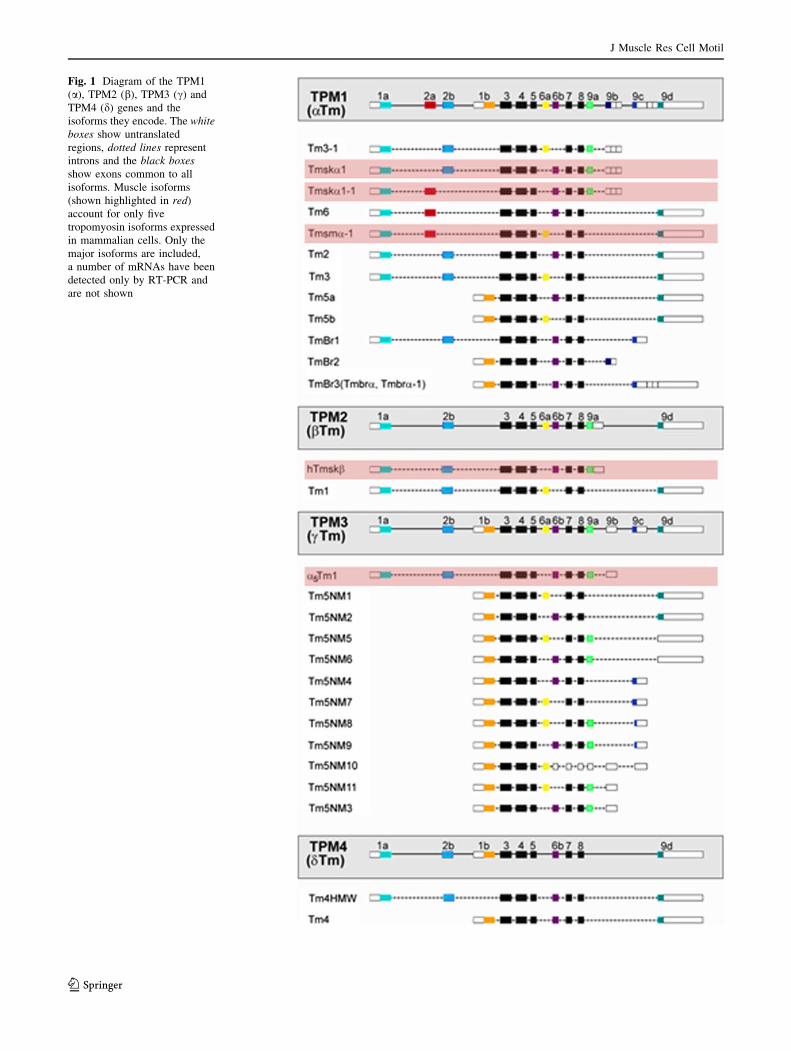
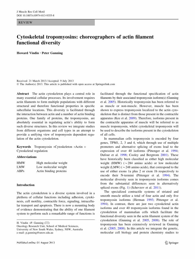
Fig. 1 Diagram of the TPM1
(a), TPM2 (b), TPM3 (c) and
TPM4 (d) genes and the
isoforms they encode. The white
boxes show untranslated
regions, dotted lines represent
introns and the black boxes
show exons common to all
isoforms. Muscle isoforms
(shown highlighted in red)
account for only five
tropomyosin isoforms expressed
in mammalian cells. Only the
major isoforms are included,
a number of mRNAs have been
detected only by RT-PCR and
are not shown
J Muscle Res Cell Motil
123
provide a unifying view of how tropomyosin isoforms act
as choreographers of the diversity of function of the animal
actin cytoskeleton.
We initially cover the experiments which establish the
lack of functional redundancy between tropomyosin
isoforms. This leads to consideration of the intracellular
spatial segregation of tropomyosin isoforms which provide
evidence that these isoforms perform spatially and func-
tionally distinct roles in the cell. The intracellular func-
tional specificity of tropomyosin isoforms is examined in a
range of cell types which leads to the conclusion that the
spatial segregation of tropomyosins has driven the evolu-
tion of functional specialisation. Mechanisms of functional
specialisation are then covered with respect to isoform
specific interactions with actin binding proteins. Finally, it
is proposed that for actin filaments containing tropomyosin,
it is the actin-tropomyosin co-polymer which should be
considered as the unit of function.
Tropomyosins are not functionally redundant
Tropomyosin is essential in yeast
The first work highlighting the essential nature of cyto-
skeletal tropomyosins was performed in Schizosaccharo-
myces pombe (Balasubramanian et al. 1992). They found
that haploid spores carrying the disrupted allele were still
able to germinate, though they die soon after as elongated
single cells (Balasubramanian et al. 1992). This study also
highlighted the essential role that the Cdc8 product, the
only tropomyosin isoform present in S. Pombe, has in
generating the contractile ring required for cytokinesis.
In Saccharomyces cerevisiae two tropomyosin isoforms
are present; Tpm1p and Tpm2p which are encoded by the
TPM1 and TPM2 genes respectively (Drees et al. 1995).
Previous work by Liu and Bretscher (1989) had shown that
whilst not lethal, the disruption of TPM1 gene expression
results in both the disappearance of actin cables and a
reduced growth rate. Interestingly whilst a loss of TPM2
gene expression shows no detectable phenotype, disruption
of both TPM1 and TPM2 expression results in lethality.
This illustrates that in yeast, the expression of at least one
isoform is essential for cell viability (Drees et al. 1995).
Furthermore it was shown that an elevated expression of
the TPM2 gene could not compensate for the loss of TPM1
expression in S. cerevisiae, providing evidence for their
functional differences (Drees et al. 1995). These results
illustrate that in yeast, tropomyosin is essential for cell
survival and in the case of S. cerevisiae, one isoform per-
forms a specific function that overexpression of the other
cannot compensate for.
Mammalian tropomyosin genes are not redundant
and perform essential functions
TPM1 (a-) and TPM2 (b-tropomyosin) genes
Homozygous knockout of the TPM1 gene in mice results in
embryonic lethality between embryonic day 8.5 and 11.5
(Rethinasamy et al. 1998). Furthermore, it was shown
using heterozygous knockout mice that despite a 50 %
decrease in mRNA from striated muscle a-Tm in the heart
there was no difference in total a-Tm or compensation
from other isoforms demonstrated by unaltered levels of
the b-Tm protein between heterozygous and control lit-
termates (Rethinasamy et al. 1998). A separate study found
that knockout of only the a-Tm striated muscle isoform
also results in embryonic lethality, however this occurred
between embryonic day 9.5 and 13.5 (Blanchard et al.
1997). These results when taken together suggest that the
a-Tm striated muscle isoform and one or more other iso-
forms encoded for by the TPM1 gene are critical for at least
two essential processes in embryonic development. Further
work by Wieczorek’s laboratory using transgenic mice
demonstrated that changes in relative levels of skeletal
tropomyosin in the heart by exchanging striated muscle
b-Tm with striated muscle a-Tm does not change the total
tropomyosin expression, however the ectopic expression of
b-Tm causes severe cardiac pathological abnormalities
(Muthuchamy et al. 1995, 1998; Palmiter et al. 1996). This
suggests that different tropomyosin isoforms can confer
different structural/functional information onto the actin
filaments they bind to, allowing them to perform specific
functions, and in the heart muscle only striated muscle
a-Tm is able to provide the structural/functional informa-
tion essential for normal cardiac function. It has also been
observed that homozygous knockout of the TPM2
gene results in a failure in early developmental processes
(Jagatheesan et al. 2010).
TPM3 (c-tropomyosin) gene
Knockout of the TPM3 gene which encodes for 11 cyto-
skeletal isoforms (Tm5NM1-11) has also been shown to be
embryonically lethal in mice by embryonic day 2.5 indi-
cating that at least one LMW product from this gene is
essential very early in embryonic development (Hook et al.
2004). This data taken with that from the TPM1 and TPM2
genes illustrates that these genes are not functionally
redundant and each is essential for survival. This study also
demonstrated that at least the TPM3 gene is essential for
embryonic stem cell viability. Given that all four tropo-
myosin genes are expressed in both embryos and embry-
onic stem cells, this data demonstrates that the loss in
J Muscle Res Cell Motil
123
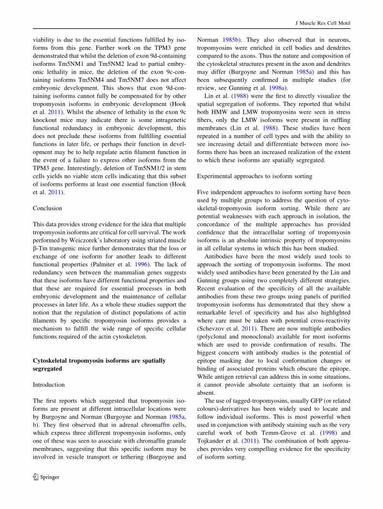
viability is due to the essential functions fulfilled by iso-
forms from this gene. Further work on the TPM3 gene
demonstrated that whilst the deletion of exon 9d-containing
isoforms Tm5NM1 and Tm5NM2 lead to partial embry-
onic lethality in mice, the deletion of the exon 9c-con-
taining isoforms Tm5NM4 and Tm5NM7 does not affect
embryonic development. This shows that exon 9d-con-
taining isoforms cannot fully be compensated for by other
tropomyosin isoforms in embryonic development (Hook
et al. 2011). Whilst the absence of lethality in the exon 9c
knockout mice may indicate there is some intragenetic
functional redundancy in embryonic development, this
does not preclude these isoforms from fulfilling essential
functions in later life, or perhaps their function in devel-
opment may be to help regulate actin filament function in
the event of a failure to express other isoforms from the
TPM3 gene. Interestingly, deletion of Tm5NM1/2 in stem
cells yields no viable stem cells indicating that this subset
of isoforms performs at least one essential function (Hook
et al. 2011).
Conclusion
This data provides strong evidence for the idea that multiple
tropomyosin isoforms are critical for cell survival. The work
performed by Weiczorek’s laboratory using striated muscle
b-Tm transgenic mice further demonstrates that the loss or
exchange of one isoform for another leads to different
functional properties (Palmiter et al. 1996). The lack of
redundancy seen between the mammalian genes suggests
that these isoforms have different functional properties and
that these are required for essential processes in both
embryonic development and the maintenance of cellular
processes in later life. As a whole these studies support the
notion that the regulation of distinct populations of actin
filaments by specific tropomyosin isoforms provides a
mechanism to fulfill the wide range of specific cellular
functions required of the actin cytoskeleton.
Cytoskeletal tropomyosin isoforms are spatially
segregated
Introduction
The first reports which suggested that tropomyosin iso-
forms are present at different intracellular locations were
by Burgoyne and Norman (Burgoyne and Norman 1985a,
b). They first observed that in adrenal chromaffin cells,
which express three different tropomyosin isoforms, only
one of these was seen to associate with chromaffin granule
membranes, suggesting that this specific isoform may be
involved in vesicle transport or tethering (Burgoyne and
Norman 1985b). They also observed that in neurons,
tropomyosins were enriched in cell bodies and dendrites
compared to the axons. Thus the nature and composition of
the cytoskeletal structures present in the axon and dendrites
may differ (Burgoyne and Norman 1985a) and this has
been subsequently confirmed in multiple studies (for
review, see Gunning et al. 1998a).
Lin et al. (1988) were the first to directly visualize the
spatial segregation of isoforms. They reported that whilst
both HMW and LMW tropomyosins were seen in stress
fibers, only the LMW isoforms were present in ruffling
membranes (Lin et al. 1988). These studies have been
repeated in a number of cell types and with the ability to
see increasing detail and differentiate between more iso-
forms there has been an increased realization of the extent
to which these isoforms are spatially segregated.
Experimental approaches to isoform sorting
Five independent approaches to isoform sorting have been
used by multiple groups to address the question of cyto-
skeletal-tropomyosin isoform sorting. While there are
potential weaknesses with each approach in isolation, the
concordance of the multiple approaches has provided
confidence that the intracellular sorting of tropomyosin
isoforms is an absolute intrinsic property of tropomyosins
in all cellular systems in which this has been studied.
Antibodies have been the most widely used tools to
approach the sorting of tropomyosin isoforms. The most
widely used antibodies have been generated by the Lin and
Gunning groups using two completely different strategies.
Recent evaluation of the specificity of all the available
antibodies from these two groups using panels of purified
tropomyosin isoforms has demonstrated that they show a
remarkable level of specificity and has also highlighted
where care must be taken with potential cross-reactivity
(Schevzov et al. 2011). There are now multiple antibodies
(polyclonal and monoclonal) available for most isoforms
which are used to provide confirmation of results. The
biggest concern with antibody studies is the potential of
epitope masking due to local conformation changes or
binding of associated proteins which obscure the epitope.
While antigen retrieval can address this in some situations,
it cannot provide absolute certainty that an isoform is
absent.
The use of tagged-tropomyosins, usually GFP (or related
colours)-derivatives has been widely used to locate and
follow individual isoforms. This is most powerful when
used in conjunction with antibody staining such as the very
careful work of both Temm-Grove et al. (1998) and
Tojkander et al. (2011). The combination of both approa-
ches provides very compelling evidence for the specificity
of isoform sorting.
J Muscle Res Cell Motil
123

Similarly, the use of in situ hybridization to localize
specific tropomyosin isoform mRNAs has provided com-
pelling evidence for the intracellular sorting of tropomyo-
sins. Hannan et al. (1995, 1998) demonstrated isoform
specific localization of tropomyosin isoform mRNAs
which was related to the localization of the corresponding
proteins revealed by isoform-specific antibodies. While
there was not a one-to-one correspondence of mRNA and
protein in neurons in vivo and in vitro, there was a clear
concordance of mRNA and protein polarity.
Biochemical sub-fractionation has been used in some
cases to detect specific tropomyosins associated with
specific intracellular structures/compartments. This was
originally used by Burgoyne and Norman (1985b) to
demonstrate the association of specific tropomyosins with
cromaffin granules and has also been used to confirm the
presence of Tm5NM1/2 with Golgi-derived structures
(Heimann et al. 1999). The use of sub-fractionation also
brings with it potential problems of contamination but is
powerful when combined with the other approaches.
Finally, gene knockout or siRNA knockdown experi-
ments have been used to show that removal of the isoform
removes antibody staining and/or impacts the function of
the compartment containing the isoform. Loss of function
can be problematic because of rescue by another isoform
but where loss of function is seen, it is most compelling.
Below we consider the wealth of experimental systems
and approaches which have unambiguously established the
generality of tropomyosin isoform intracellular sorting.
The studies documented below have used a range of dif-
ferent approaches or have been confirmed in multiple labs;
often using different antibodies or different approaches.
Differential sorting of Cdc8p in yeast
In fission yeast, the acetylation of the only tropomyosin
isoform expressed; Cdc8p has a significant impact on its
ability to bind and regulate actin filaments (Skoumpla et al.
2007). Coulton et al. (2010) found that acetylated Cdc8p
was strongly associated with actin filament bundles in the
cytokinetic actomyosin ring (CAR). In contrast, unacety-
lated Cdc8p was never seen within the CAR and was only
associated with filament bundles that extend throughout the
cell (Fig. 2a). Since tropomyosins have not been found in
either plants or amoebae (Pruyne 2008) these observations
demonstrate that sorting tropomyosin isoforms is an
intrinsic property that is as old as tropomyosin itself.
Neurons
Had et al. (1994) compared the localization of two iso-
forms Tm4 and TmBr3 both in cultured neurons and in the
mouse. They found that Tm4 was concentrated at the
growth cones of neurons whereas TmBr3 was notably
absent from these regions. In vivo, Tm4 was restricted to
postsynaptic regions whilst TmBr3 was concentrated at
presynaptic sites suggesting that these isoforms fulfill dif-
ferent functional roles in neurons (Had et al. 1994). This
spatial segregation has also been detected during neuronal
development. In developing neurons Tm5NM1/2 is
restricted to the developing axons. However in mature
neurons its localization is somatodendritic, and its loss
from the axon occurs coincident with the initial appearance
Fig. 2 a Distribution of tropomyosin in Schizosaccharomyces
pombe. Actin patches are found near the cell periphery and are not
associated with tropomyosin isoforms. The cables which run
throughout the cell are associated with unacetylated Cdc8p and
favour the binding of myosin-V. In contrast, the contractile ring actin
is associated with acetylated Cdc8p which favours the binding of
myosin-II. b Distribution of tropomyosin in Osteoclasts plated on
ivory. Tm4 (red) is associated with podosomes (represented as the
inner ring) and the interior of the cell. Tm5a/5b (green) is associated
with the F-actin ring (represented as the outer ring) and is slightly
enriched near the plasma membrane. Whilst some colocalization is
observed between Tm5a/5b and Tm4 (yellow), Tm5a/5b are notably
absent from the podosomes. Both Tm2/3 (orange) and Tm5NM1
(blue) are both found throughout the cell in different subcellular
pools, however the nature of these regions is not yet known
J Muscle Res Cell Motil
123

of TmBr3 in axons (Weinberger et al. 1996). This temporal
regulation has also been seen with Tm5a/5b where its
presence in the growth cones of primary neurons dimin-
ishes with time in culture (Schevzov et al. 1997).
Further experiments revealed that depolymerization of
actin filaments through the addition of cytochalasin B
resulted in a loss of spatial segregation of Tm5NM1/2.
After wash-out of the drug the spatial segregation of
tropomyosin isoforms was restored and Tm5NM1/2 was
again mostly absent from the growth cone (Schevzov et al.
1997). This clearly demonstrates that the tropomyosin
isoform composition of actin filaments is dependent on the
dynamic remodeling of the cytoskeleton.
Osteoclasts
Osteoclasts possess a highly dynamic cytoskeleton capable
of forming a number of distinct intracellular structures
which can be defined by the localization of specific
tropomyosin isoforms. At the podosomal attachment
structures, both Tm4 and Tm5a/5b are present although
their localization on these structures is mutually exclusive
(McMichael et al. 2006). Whilst Tm4 is found to associate
with the interior ends of podosomal actin cores and the top
half of F-actin rings in these cells, Tm5a/5b was enriched
at the base of podosomal cores and the outer edge of the
F-actin rings (McMichael et al. 2006). In contrast, staining
for Tm5NM1 and Tm2/3 showed that these isoforms are
excluded from attachment structures. Despite both these
isoforms being enriched in the cell interior, there was little
to no overlap between them (Fig. 2b). It was concluded
that they are localized to distinct internal structures in these
cells (McMichael et al. 2006). These results indicate that in
osteoclasts there are at least four cytoskeletal structures
which are associated with specific tropomyosin isoforms.
Skeletal muscles
Muscle fibers contain three skeletal muscle tropomyosin
isoforms which form part of the thin filament where they
are involved in the regulation of muscle contraction
(Huxley 1973). In addition to skeletal tropomyosin, two
cytoskeletal isoforms are expressed which sort to specific
compartments within the myofibril. Within the myofibril,
Tm5NM1 is specifically sorted to both a filament network
adjacent to the Z-line and a subsarcolemma filament sys-
tem found around the periphery of the myofibril (Kee et al.
2004). Tm4 is also expressed in muscle fibers where it is
sorted to two specific locations. Like Tm5NM1, this iso-
form is sorted to a filament network adjacent to the Z-line
where these two isoforms define distinct actin filament
populations (Vlahovich et al. 2009). Tm4 is also localized
to longitudinal filaments running perpendicular to the
Z-line which are associated with muscle fibers undergoing
remodeling and repair (Vlahovich et al. 2008).
Smooth muscle cells
More recently it was found that at least five tropomyosin
isoforms are expressed in vascular smooth muscle cells. In
addition to the smooth muscle tropomyosin isoforms Tm1
and Tm6 three cytoskeletal isoforms Tm2, Tm5NM1 and
Tm4 were also present (Gallant et al. 2011). In contrast to
previous work on chicken gizzard smooth muscle by
Sanders et al. (1986) in which heterodimer formation was
observed, not only did Tm1 and Tm6 not form heterodi-
mers in this cell type but these isoforms also sorted to
different intracellular regions and were associated with
different actin isoforms (Gallant et al. 2011).
Fibroblasts
Work in NIH 3T3 fibroblasts also demonstrated that spe-
cific tropomyosin isoforms are differentially sorted to
specific subcellular locations (Percival et al. 2000). One
hour after replating, products from the TPM1 gene were
incorporated into stress fiber structures, whereas those from
the TPM3 gene were localized to the perinuclear region.
This sorting becomes less distinct as the cells progress
through the cell cycle. After 8 h isoforms from both genes
were localized in stress fibers, however TPM3 isoforms
were still present in the central cytoplasm and TPM1 iso-
forms were more enriched at the cell periphery (Percival
et al. 2000). It was later shown that Tm5NM2 specifically
sorted to short actin filaments associated with the Golgi
complex (Percival et al. 2004).
In primary mouse embryo fibroblasts similar spatial
segregation is also seen. Schevzov and colleagues found
that the HMW isoforms from the TPM1 gene sorted pre-
dominately to stress fibers, whilst Tm5a/5b were the only
isoforms specifically located to the ruffling membranes
(Schevzov et al. 2005b, 2011).
Tropomyosin isoforms sort within stress fibers
More recently it has been shown that individual tropomy-
osin isoforms are further segregated into specific regions
along stress fibers. Tojkander et al. (2011) found that only
Tm2 was localized along entire stress fibers whilst Tm1,
Tm5NM1 and Tm5NM2 were concentrated at the distal
ends of filament bundles corresponding to focal adhesions.
Tm3 and Tm4 were found proximally to focal adhesions,
where they were seen either as short segments or as a
dotted pattern. Further results using live-cell imaging
demonstrate that tropomyosin isoforms are sequentially
recruited to both focal adhesions and dorsal stress fibers
J Muscle Res Cell Motil
123

and Tm4’s localization to dorsal stress fibers coincides
with the incorporation of myosin II into these structures
(Tojkander et al. 2011). Furthermore, it was shown that at
least four different tropomyosins are required for stress
fiber formation (Tojkander et al. 2011).
Mechanism of isoform sorting
The mechanism of isoform sorting has been the subject of
extensive reviews (Gunning et al. 1998a, b, 2005, 2008;
Martin and Gunning 2008) which can be summarized very
simply. The isoforms are locally assembled and held in
place by higher order structures (Martin and Gunning
2008). The site of protein synthesis of isoforms may aid in
sorting but does not absolutely determine isoform location
(Hannan et al. 1995, 1998). There is no evidence for
transport of isoforms to specific intracellular locations
(Martin et al. 2010). Hence, the mechanism of sorting most
likely occurs at the level of local assembly of the actin
filament by mechanism(s) as yet unknown.
Conclusion
It has been known for years that tropomyosin isoforms are
spatially segregated to distinct actin filament populations
and this extensive accumulation of data has established that
tropomyosin isoform sorting is a fundamental cellular
process which is shared across all animal cells.
Cytoskeletal tropomyosin function
The finely regulated spatial segregation of tropomyosin
isoforms is necessary as it ensures that individual isoforms
are in the correct locations to fulfill specific functions
critical for the normal functioning of the cell.
Neuronal morphogenesis
Given the fine spatial regulation of tropomyosin isoforms
seen in neurons, these cells have been more extensively
studied to determine the effects of tropomyosin on neuro-
nal morphogenesis. Primary neurons from transgenic mice
overexpressing Tm5NM1 were found to have increased
neuronal branching in both dendrites and axons and a
significant increase in growth cone size without any
noticeable change in its gross morphology (Schevzov et al.
2005a). In contrast, neurons from transgenic mice over-
expressing Tm3 had both significantly decreased numbers
and length of dendrites. These results indicate that
Tm5NM1 and Tm3 contain different structural informa-
tion, and their expression gives rise to filament populations
with different functional properties (Schevzov et al.
2005a). Consistent with the increase in dendritic length
seen with Tm5NM1 overexpression, neurons from mice
lacking Tm5NM1/2 were seen to have a significant
decrease in dendritic length, as well as a number of other
morphological changes compared to control neurons (Fath
et al. 2010). These results show that altering the tropo-
myosin composition of filaments in neurons leads to sig-
nificant changes in neuronal morphogenesis.
Trafficking
Organelle transport plays an essential part in many cellular
functions and is a process which relies on both actin fila-
ments and microtubules. Pelham et al. (1996) investigated
the role of tropomyosin in organelle transport through the
microinjection of Tm3 into NRK cells. They found that the
microinjection of Tm3 but not Tm5NM1 causes a
remarkable redistribution of membrane-bound organelles
into the perinuclear region (Pelham et al. 1996). These
results at the very least indicate that these two isoforms are
functionally distinct.
CFTR membrane levels
The actin cytoskeleton has also been shown to play a role
in the delivery of the cystic fibrosis transmembrane con-
ductance regulator (CFTR) into the apical membrane of
epithelial cells. It has been shown that reduced expression
of Tm5a and Tm5b result in an increased surface expres-
sion of CFTR in vitro indicating that these isoforms may be
associated with a subpopulation of actin filaments directly
involved in the removal of CFTR from the plasma mem-
brane (Dalby-Payne et al. 2003). It was concluded that
these isoforms play a role in the regulation of endocytosis.
Cytokinesis
A number of studies have shown that tropomyosin plays an
important part in cytokinesis. In yeast the role that tropo-
myosin plays in the regulation of cytokinesis has been
extensively studied. Balasubramanian et al. (1992) found
that the tropomyosin isoform Cdc8 was essential for cell
survival in S. pombe. Whilst Cdc8 was not required for
spore germination, cell growth or DNA replication it is
essential for cytokinesis. This indicates that the essential
role for this protein is to form part of the F-actin contractile
ring (Balasubramanian et al. 1992). Further work by
Mulvihill’s laboratory showed that the function and sorting
of Cdc8 to different cellular structures was dependent upon
its acetylation (Skoumpla et al. 2007; Coulton et al. 2010).
The cytokinetic deficit found in cells which lack the NatB
N-a-acetyltransferase regulatory subunit was also shown to
be as a result of a lack of tropomyosin acetylation (Coulton
J Muscle Res Cell Motil
123

et al. 2010). Stark et al. (2010) found that this regulation of
cytokinesis in S. pombe was through its role in stabilizing
actomyosin interactions.
The regulation of cytokinesis by tropomyosin has also
been seen in mammalian cells. Hughes et al. (2003) exam-
ined tropomyosin expression in developing and neoplastic
brain tissue. They found that in the embryonic brain HMW
tropomyosin expression was restricted to proliferative areas,
whereas in the adult brain, staining could only be seen in
blood vessels. They also noted that in rare proliferating
astrocytes HMW tropomyosins were found in the contractile
ring, but after withdrawal from the cell cycle HMW tropo-
myosin expression was down regulated (Hughes et al. 2003).
Forced expression of Tm5NM1 and a chimeric tropomyosin
Tm5/3 in Chinese hamster ovary cells resulted in faster cell
division which would suggest that tropomyosin is necessary
for the formation of the contractile ring (Eppinga et al.
2006). The abnormal division seen in Tm5NM1 over-
expressing cells may be due to the inability of other actin
binding proteins to disassemble the contractile ring. This is
supported by the observation that Tm5NM1 excludes the
association of ADF with filaments containing this tropo-
myosin isoform (Bryce et al. 2003).
Podosomes in osteoclasts
Osteoclasts express several cytoskeletal isoforms which
sort to specific regions. Tm4 was found to be associated
with the core of podosomes, suggesting that it may play a
role in regulating these structures (McMichael et al. 2006).
Knockdown and overexpression studies revealed a direct
role for Tm4 in regulating both podosomal and sealing
zone actin filaments. McMichael and Lee (2008) found that
either under- or overexpression of Tm4 disrupted these
attachment structures leading to impaired bone resorption
and cell motility. Further work examining the role of Tm2
and Tm3 in these cells revealed that despite a lack of
association with distinct actin structures in these cells,
these isoforms play a role in the regulation of osteoclast
morphology and function (Kotadiya et al. 2008). These
results taken together demonstrate that individual isoforms
decorate distinct actin filament populations in osteoclasts,
giving them specific functional properties necessary for
normal cellular function.
Stem cell viability
A number of tropomyosin isoforms play a critical role in the
regulation of stem cell viability and embryonic develop-
ment. Eliminating the cytoskeletal isoforms from the TPM3
gene (Tm5NM1-11) results in lethality prior to embryonic
day 2.5 and the inability to generate viable stem cells (Hook
et al. 2004). Embryonic stem cells deleted for exons 9a/9b of
the TPM3 gene (Tm5NM3, 5, 6, 8, 9, 11) are viable whereas
the failure to generate viable stem cells lacking exon 9d of
the TPM3 gene (Tm5NM1,2) indicates that at least one of
these isoforms is essential for cell growth in vitro (Hook
et al. 2011). Their role in cell growth is further supported by
the fact that these isoforms are expressed in most, if not all
cells and there is an increased reliance on these isoforms in
almost all forms of cancer (Stehn et al. 2006). These results
demonstrate that isoforms from the TPM3 gene are required
for the normal functioning of a cell and cannot be com-
pensated for by products from the other three genes.
Excitation contraction coupling in skeletal muscle
fibers
The importance of cytoskeletal tropomyosin isoforms in
cellular function is seen in vivo in skeletal muscle fibers. In
muscles from mice null for Tm5NM1 the level of T-tubule
dysmorphology was increased when compared to WT
muscles, and the Tm5NM1 KO mouse muscles had altered
contractile properties which were not due to fiber-type
changes (Vlahovich et al. 2009). Further experiments
revealed the altered contractile performance was a result of
dysregulation of T-tubule function due to the loss of
Tm5NM1. This demonstrates that the LMW Tm4 expres-
sed in an adjacent location in muscle cannot compensate
for the loss of Tm5NM1.
Conclusion
The specialized function of tropomyosin isoforms descri-
bed here provides clear evidence that tropomyosin has a
much underappreciated role in the functional regulation of
the actin cytoskeleton. In light of this, it would seem highly
likely that the binding of individual tropomyosin isoforms
to actin filaments confers specific functional properties
upon these filaments. This gives rise to distinct filament
populations localized to specific regions of a cell where
they perform different functions.
Mechanisms of specialized tropomyosin function
Historically, tropomyosin has been both studied and
understood in terms of its ability to regulate the myosin II
interaction with the actin filament in muscle (Murray and
Weber 1973). In vitro studies revealed that chicken gizzard
tropomyosin displays a greater cooperativity than rabbit
skeletal tropomyosin in terms of their effects on myosin
subfragment 1 activity (Lehrer and Morris 1984). Additional
protein chemistry studies also revealed that muscle tropo-
myosin could regulate muscle actin filament stability
(Fujime and Ishiwata 1971) and inhibit both DNase 1
J Muscle Res Cell Motil
123
(Hitchcock et al. 1976) and cofilin (Bernstein and Bamburg
1982) induced depolymerisation of muscle actin. The
implications of this work for the functional diversity of the
cytoskeleton required the development of molecular genetic
approaches to manipulate the composition of the cytoskel-
eton and visualization of different filament populations.
Evolutionary consequences of isoform sorting
The ability to sort isoforms to different spatial, and therefore
functional contexts, will inevitably result in the divergence
of their functional capacities due to the differing functional
constraints placed upon them. This sorting will lead to the
specialized functions of intracellular sites regulated by the
specific isoform population present. There is a clear lack of
functional redundancy seen between different tropomyosin
isoforms, and they show finely tuned spatial segregation to
specific subcellular locations (Martin and Gunning 2008).
This suggests that the creation of different tropomyosin
isoforms throughout evolution allows for the creation of a
range of actin filament populations which possess the
structural information required to fulfill a broad range of
unique functions (Gunning et al. 2008).
Regulate actin polymer levels
A number of studies have demonstrated the ability of
tropomyosin to regulate levels of F-actin within cells.
Schevzov et al. (2008) found that following the overex-
pression of Tm3 and Tm5NM1, the levels of other cyto-
skeletal tropomyosin isoforms and b- and c-actin levels were
unchanged in transgenic tissues. Interestingly in primary
hippocampal neurons from Tm5NM1 transgenic mice,
enrichment of Tm5NM1 staining in the growth cones was
associated with a significant increase in both total phalloidin
signal and mean pixel intensity (Schevzov et al. 2008).
Similar results were reported in osteoclasts where manipu-
lation of Tm4 expression resulted in changes in F-actin levels
at the site of Tm4 localization. Overexpression of Tm4
caused an increase in F-actin in podosomes, whereas the
knockdown of this isoform resulted in significant thinning of
F-actin in the actin ring and sealing zone (McMichael and
Lee 2008). These findings indicate that the levels of cyto-
skeletal tropomyosin are limiting for actin polymerization in
the subcellular regions where these isoforms are sorted.
Thus, tropomyosin isoform sorting regulates total actin
polymer levels at specific intracellular locations.
Regulate myosin motors
Whilst the regulation of myosin driven contraction by
tropomyosin has been extensively studied in striated mus-
cle, comparatively little is known about the interactions
between tropomyosin dependent regulation of actomyosin
interactions in cytoskeletal systems. Work by Fanning et al.
(1994) illustrated that the ability of tropomyosin to regulate
the ATPase activity and translocation of muscle myosin II
along actin filaments was dependent on the isoform pres-
ent. In contrast, all tropomyosin isoforms tested inhibited
the ATPase activity and translocation of myosin I to a
similar extent. This demonstrated that regulation of myosin
motor interactions with actin was isoform dependent and
that a single isoform can have opposing effects on different
myosin motors.
In fission yeast the tropomyosin isoform Cdc8p was
found to enhance myosin II motor activity, promoting the
formation of the contractile ring (Stark et al. 2010). Further
work by Lord’s laboratory has shown that Cdc8p also
regulates the activity of myosin V (Clayton et al. 2010).
Coulton et al. (2010) found that whilst acetylation of
Cdc8p was required for the regulation of myosin II, there
was no effect on the regulation of myosin I or V. Recent
work has demonstrated that tropomyosin allows for the
processive movement of class V myosins in S. cerevisiae.
Hodges et al. (2012) found whilst class V myosin motors
are unable to move processively along bare skeletal muscle
actin, supporting previous in vitro studies, the creation of a
more biologically relevant filament through the addition of
tropomyosin allowed for the processive movement of
Myo2p. This data strongly supports the idea that actomy-
osin interactions are sensitive to the presence of tropomy-
osin along the filament.
The regulation of myosin has also been seen in more
complex mammalian cells. Bryce et al. (2003) found that
myosin IIA, but not IIB, was recruited to stress fibers in
Tm5NM1 overexpressing B35 cells resulting in a sub-
stantial increase in myosin II activity. The recruitment of
myosin IIA was also seen in the dendrites of cortical
neurons from transgenic mice overexpressing Tm5NM1
(Bryce et al. 2003). However, in growth cones where
myosin IIA is absent, IIB was able to associate with
Tm5NM1 containing filament bundles demonstrating that
the preferential recruitment of myosin is dependent upon
the availability of specific myosin isoforms (Schevzov
et al. 2005a). Tang and Ostap (2001) provided further
evidence for the regulation of myosin I by tropomyosin.
They found that the exclusion of myosin I from actin
structures that contain tropomyosin was due to the regu-
lation of the actomyosin interaction by tropomyosin.
Regulate interactions of other actin binding proteins
Actin depolymerizing factor/cofilin
Actin depolymerizing factor (ADF)/cofilin depolymerizes
actin filaments and was initially found to compete with
J Muscle Res Cell Motil
123

tropoymosin for binding to the filament (Bernstein and
Bamburg 1982). Bryce et al. (2003) found that this
antagonistic interaction was isoform specific. Tm5NM1
expressing cells had an increased level of phosphorylated
ADF indicating that it was displaced from actin filaments,
however TmBr3 recruits ADF to the lamellapodia where
they colocalize on the same filaments. Thus, tropomyosins
can be seen as collaborators or competitors of ADF/cofilin
depending on the tropomyosin isoform (Kuhn and Bam-
burg 2008).
Fascin
Fascin is an actin bundling protein found in stress fibers
and filipodia which localizes with HMW but not LMW
tropomyosin isoforms (Yamashiro-Matsumura and Mat-
sumura 1986). In control B35 neuroblastoma cells there is
an association of fascin with Tm2. Creed et al. (2011)
found that in Tm3 overexpressing cells there was a
significant shift in fascin association from Tm2 to Tm3-
containing filaments. Furthermore they found that the
overexpression of Tm3 resulted in an increase in fascin
expression. This indicates that different tropomyosin iso-
forms can alter the expression of endogenous actin binding
proteins, possibly via changing the partitioning of these
proteins between the soluble and filament bound pools and
hence their turnover kinetics.
Formin homology proteins
Formins are a diverse family of actin nucleating proteins.
Whilst there have been several biochemical studies which
investigate the kinetics of the interaction between formins
and tropomyosin (Wawro et al. 2007; Ujfalusi et al. 2009,
2012), there has been little work describing this interaction
in a cell based system. In fission yeast, the formin Cdc12p
nucleates actin filaments which the tropomyosin isoform
Cdc8p binds to with diverse effects on Cdc12p-mediated
actin assembly (Skau et al. 2009). Cdc8p’s binding both
increases the rate of elongation and allows annealing of the
filaments before stopping Cdc12p-mediated elongation.
Interestingly, Cdc8p may then stop Cdc12p-mediated
elongation through either the trapping of Cdc12p or
dissociating it from the filament (Skau et al. 2009). This
intricate relationship is still not fully understood and it is
likely that the interactions which occur in mammalian
cells, which express multiple formin and tropomyosin
isoforms, would be far more complex. Whilst the conse-
quences of this are not known, this study suggests that
tropomyosin’s role in the regulation of the cytoskeleton is
far more complex and intricate than previously thought.
Responsive to availability of active ABPs
More recently it was shown that the HMW isoform Tm3
independently regulates the function of actin filaments at
specific intracellular sites (Creed et al. 2011). They found
that active ADF/cofilin was localized in the cell body and
within the base of the filipodia in Tm3 overexpressing
cells, whilst the inactive phosphorylated ADF/cofilin was
only found in the perinuclear region. This indicates that
only the active ADF/cofilin is able to be recruited to Tm3-
containing filaments. Furthermore they demonstrated that
inactivation or knockdown of ADF/cofilin caused a change
in cell morphology and cytoskeletal organization more
resembling the control B35 cells (Creed et al. 2011). This
indicates that whilst different tropomyosin isoforms will
preferentially recruit specific ABPs, their impact on the cell
is also determined by the local availability of active ABP’s.
The actin-tropomyosin copolymer as the unit
of function
Early research into understanding the cytoskeleton was
dominated by protein biochemistry and in vitro studies,
which have been critical in providing a basis for in vivo
studies. However, whilst in vitro work has been fundamental
in establishing many of the key concepts known today and
has driven the model building surrounding interactions of
cytoskeletal proteins, there have been numerous cases where
the biochemical data has not been supported by molecular
genetics. Interpretation of in vitro observations has estab-
lished a view that filaments are generic and the specific
functional outcomes observed within cells are due to the
chance interaction of a large number of actin binding
proteins with filament bundles at any given time. However,
many of these experiments were performed in a fixed envi-
ronment, under ideal conditions using a-skeletal, not cyto-
skeletal actin and in the absence of actin binding proteins
needed to assemble filaments into the biological structures
seen in vivo. In contrast, molecular genetics and in vivo data
have driven functional biology. The sorting of actin binding
proteins gives rise to filament bundles with unique functional
information that is critical for the functioning of a cell.
Whilst many biochemical studies have demonstrated both
polymerization of actin and subsequent binding of cyto-
skeletal actin binding proteins to skeletal actin in vitro, how
these processes occur in vivo is yet to be established. There
is an ever increasing body of evidence to suggest that there is
no active transport for the movement of tropomyosin to
specific spatial regions, and that its sorting relies on the
active formation of filaments. This suggests a ‘molecular
sink’ model whereby isoforms accumulate in structures
J Muscle Res Cell Motil
123
where they are most stable. This hypothesis has been sup-
ported by drug studies demonstrating that a loss of filaments
results in the abolishment of isoform sorting. Intrinsic to the
sorting of tropomyosin is that tropomyosin binds to poly-
merizing actin filaments, both providing a mechanism of
stabilizing single filaments and facilitating the interactions
with surrounding actin binding proteins.
Whilst the notion of gestalt-binding proposed by
Holmes and Lehman (2008) would accurately predict the
binding of tropomyosin to actin filaments in vitro, it fails to
take into account the complexity of the cellular environ-
ment. The formation of filaments in a cellular environment
is a continuous process that occurs throughout the whole
cell. Therefore, the notion that an actin filament forms in an
environment free from interactions with the plethora of
actin binding proteins present throughout the cytoplasm
would seem a highly inefficient process. On the other hand,
the binding of specific tropomyosin isoforms to nucleated
actin filaments whose conformational twist favors that
particular isoform, would allow not only for the stabiliza-
tion of the growing filament, but for the formation of a
functionally distinct filament population already equipped
with the structural information required to fulfill a specific
functional role. Given the dynamic nature of actin fila-
ments in an intracellular environment (for example, see
Tojkander et al. 2011) it seems more likely that specific
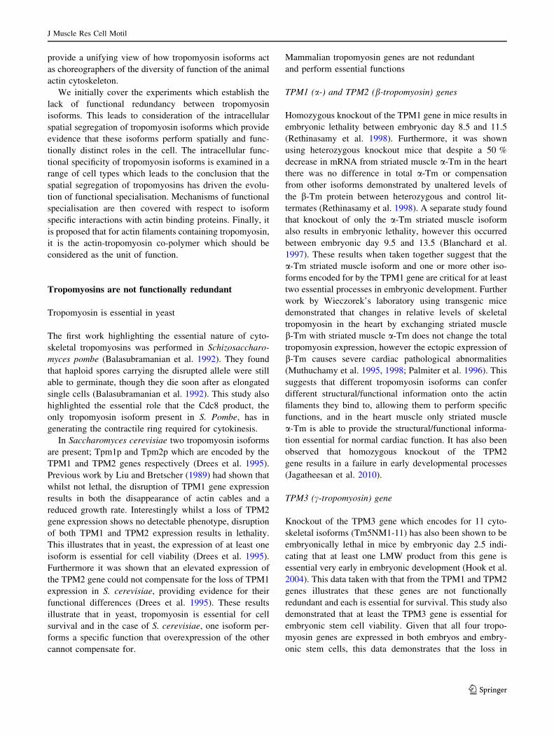
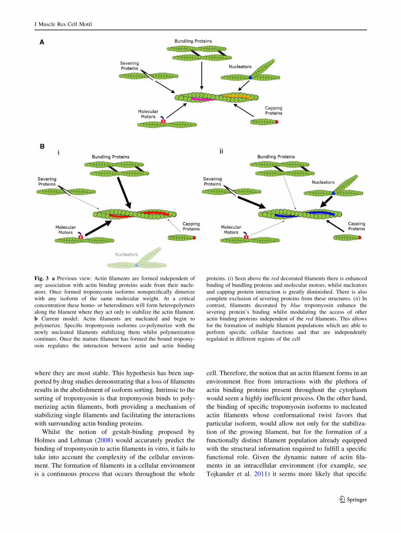
Fig. 3 a Previous view: Actin filaments are formed independent of
any association with actin binding proteins aside from their nucle-
ators. Once formed tropomyosin isoforms nonspecifically dimerize
with any isoform of the same molecular weight. At a critical
concentration these homo- or heterodimers will form heteropolymers
along the filament where they act only to stabilize the actin filament.
b Current model: Actin filaments are nucleated and begin to
polymerize. Specific tropomyosin isoforms co-polymerize with the
newly nucleated filaments stabilizing them whilst polymerization
continues. Once the mature filament has formed the bound tropomy-
osin regulates the interaction between actin and actin binding
proteins. (i) Seen above the red decorated filaments there is enhanced
binding of bundling proteins and molecular motors, whilst nucleators
and capping protein interaction is greatly diminished. There is also
complete exclusion of severing proteins from these structures. (ii) In
contrast, filaments decorated by blue tropomyosin enhance the
severing protein’s binding whilst modulating the access of other
actin binding proteins independent of the red filaments. This allows
for the formation of multiple filament populations which are able to
perform specific cellular functions and that are independently
regulated in different regions of the cell
J Muscle Res Cell Motil
123
actin and tropomyosin isoforms co-assemble to form
functionally distinct filaments. The mechanism of assembly
may be influenced by the nature of filament nucleation in a
cell which differs from that used in most in vitro studies.
The proposal that tropomyosin plays a somewhat
redundant role in cytoskeletal regulation where its binding
can be so easily disrupted by other binding proteins is
questioned by the presence of a number of essential
tropomyosin isoforms within the cell. The finely regulated
sorting of tropomyosin to distinct filament populations,
highly regulated both in time and space would indicate that
its role is far more important than a ‘parking attendant’ for
actin filaments. Instead the overwhelming accumulation of
data now proposes a more biologically relevant model for
the tropomyosin dependent regulation of the actin cyto-
skeleton (Gunning et al. 2008).
From the results discussed in this review, it would seem
obvious in retrospect, that tropomyosin must play a key
role in the regulation of the actin cytoskeleton. The two
cytoskeletal actin isoforms; b- and c-actin vary by only a
few amino acids, presumably as further changes to the
highly conserved amino acid sequence would result in an
inability to provide adequate structural support. In contrast,
there are over 40 tropomyosin isoforms that are extensively
regulated both spatially and temporally. The differential
sorting of these structurally distinct isoforms to specific
subcellular locations allows for the formation of function-
ally distinct actin filament populations, each possessing
unique structural information.
Here the various tropomyosin isoforms act primarily as
choreographers regulating the dynamic interaction between
actin and all its binding proteins. This dynamic system
gives rise to the formation of unique and functionally
distinct filament systems largely based on one gene family.
From an evolutionary perspective, cells which were able to
utilize multiple filament systems independently regulated
by single tropomyosin isoforms would be given a distinct
advantage. This would likely have driven the evolution of
the tropomyosin dependent regulation of the actin cyto-
skeleton and the basis for the functional diversity that has
become characteristic of the actin filament system. This is
described graphically in Fig. 3.
Acknowledgments We’d like to thank Dr. Justine Stehn and Ms.
Ashleigh Swain for critical reading of this manuscript. This work has
been supported by two funding bodies; generous donations from The
Kids Cancer Project and a National Health and Medical Research
Council (NHMRC) Project Grant (APP1004188). Howard Vindin is a
recipient of an Australian Postgraduate Award.
Open Access This article is distributed under the terms of the
Creative Commons Attribution License which permits any use, dis-
tribution, and reproduction in any medium, provided the original
author(s) and the source are credited.
References
Balasubramanian MK, Helfman DM, Hemmingsen SM (1992) A new
tropomyosin essential for cytokinesis in the fission yeast S.
pombe. Nature 360(6399):84–87
Bernstein BW, Bamburg JR (1982) Tropomyosin binding to F-actin
protects the F-actin from disassembly by brain actin-depolymerizing
factor (ADF). Cell Motil 2(1):1–8. doi:10.1002/cm.970020102
Blanchard EM, Iizuka K, Christe M, Conner DA, Geisterfer-
Lowrance A, Schoen FJ, Maughan DW, Seidman CE, Seidman
JG (1997) Targeted ablation of the murine a-tropomyosin gene.
Circ Res 81(6):1005–1010. doi:10.1161/01.res.81.6.1005
Bryce NS, Schevzov G, Ferguson V, Percival JM, Lin JJ-C,
Matsumura F, Bamburg JR, Jeffrey PL, Hardeman EC, Gunning
P, Weinberger RP (2003) Specification of actin filament function
and molecular composition by tropomyosin isoforms. Mol Biol
Cell 14(3):1002–1016. doi:10.1091/mbc.E02-04-0244
Burgoyne RD, Norman K-M (1985a) Immunocytochemical localiza-
tion of tropomyosin in rat cerebellum. Brain Res 361(1–2):
178–184. doi:10.1016/0006-8993(85)91287-9
Burgoyne RD, Norman K-M (1985b) Presence of tropomyosin in
adrenal chromaffin cells and its association with chromaffin
granule membranes. FEBS Lett 179(1):25–28. doi:10.1016/
0014-5793(85)80183-6
Clayton JE, Sammons, Stark BC, Hodges AR, Lord M (2010)
Differential regulation of unconventional fission yeast myosins
via the actin track. Curr Biol 20(16):1423–1431. doi:10.1016/
j.cub.2010.07.026
Cooley BC, Bergtrom G (2001) Multiple combinations of alterna-
tively spliced exons in rat tropomyosin-a gene MRNA: evidence
for 20 new isoforms in adult tissues and cultured cells. Arch
Biochem Biophys 390(1):71–77. doi:10.1006/abbi.2001.2347
Coulton AT, East DA, Galinska-Rakoczy A, Lehman W, Mulvihill DP
(2010) The recruitment of acetylated and unacetylated tropomy-
osin to distinct actin polymers permits the discrete regulation of
specific myosins in fission yeast. J Cell Sci 123(19):
3235–3243. doi:10.1242/jcs.069971
Creed SJ, Desouza M, Bamburg JR, Gunning P, Stehn J (2011)
Tropomyosin isoform 3 promotes the formation of filopodia by
regulating the recruitment of actin-binding proteins to actin
filaments. Exp Cell Res 317(3):249–261. doi:10.1016/j.yexcr.
2010.10.019
Dalby-Payne JR, O’Loughlin EV, Gunning P (2003) Polarization of
specific tropomyosin isoforms in gastrointestinal epithelial cells
and their impact on CFTR at the apical surface. Mol Biol Cell
14(11):4365–4375. doi:10.1091/mbc.E03-03-0169
Drees B, Brown C, Barrell BG, Bretscher A (1995) Tropomyosin is
essential in yeast, yet the TPM1 and TPM2 products perform
distinct functions. J Cell Biol 128(3):383–392. doi:10.1083/jcb.
128.3.383
Dufour C, Weinberger RP, Schevzov G, Jeffrey PL, Gunning P
(1998) Splicing of two internal and four carboxyl-terminal
alternative exons in nonmuscle tropomyosin 5 pre-mRNA is
independently regulated during development. J Biol Chem
273(29):18547–18555. doi:10.1074/jbc.273.29.18547
Eppinga RD, Li Y, Lin JLC, Lin JJC (2006) Tropomyosin and
caldesmon regulate cytokinesis speed and membrane stability
during cell division. Arch Biochem Biophys 456(2):161–174.
doi:10.1016/j.abb.2006.06.015
Fanning AS, Wolenski JS, Mooseker MS, Izant JG (1994) Differential
regulation of skeletal muscle myosin-II and brush border
myosin-I enzymology and mechanochemistry by bacterially
produced tropomyosin isoforms. Cell Motil Cytoskelet
29(1):29–45. doi:10.1002/cm.970290104
J Muscle Res Cell Motil
123
Fath T, Agnes Chan Y-K, Vrhovski B, Clarke H, Curthoys N, Hook J,
Lemckert F, Schevzov G, Tam P, Watson CM, Khoo P-L,
Gunning P (2010) New aspects of tropomyosin-regulated
neuritogenesis revealed by the deletion of Tm5NM1 and 2.
Eur J Cell Biol 89(7):489–498. doi:10.1016/j.ejcb.2009.11.028
Fujime S, Ishiwata S (1971) Dynamic study of F-actin by quasielastic
scattering of laser light. J Mol Biol 62(1):251–265
Gallant C, Appel S, Graceffa P, Leavis P, Lin JJ-C, Gunning PW,
Schevzov G, Chaponnier C, DeGnore J, Lehman W, Morgan KG
(2011) Tropomyosin variants describe distinct functional sub-
cellular domains in differentiated vascular smooth muscle cells.
Am J Physiol Cell Physiol 300(6):1356–1365. doi:10.1152/
ajpcell.00450.2010
Gunning P, Hardeman E, Jeffrey P, Weinberger R (1998a) Creating
intracellular structural domains: spatial segregation of actin and
tropomyosin isoforms in neurons. BioEssays 20:892–900
Gunning P, Weinberger R, Jeffrey P, Hardeman E (1998b) Isoform
sorting and the creation of intracellular compartments. Annu Rev
Cell Dev Biol 14:339–372
Gunning PW, Schevzov G, Kee AJ, Hardeman EC (2005) Tropomy-
osin isoforms: divining rods for actin cytoskeleton function.
Trends Cell Biol 15(6):333–341. doi:10.1016/j.tcb.2005.04.007
Gunning P, O’Neill G, Hardeman E (2008) Tropomyosin-based
regulation of the actin cytoskeleton in time and space. Physiol
Rev 88(1):1–35. doi:10.1152/physrev.00001.2007
Had L, Faivre-Sarrailh C, Legrand C, Mery J, Brugidou J, Rabie A
(1994) Tropomyosin isoforms in rat neurons: the different devel-
opmental profiles and distributions of TM-4 and TMBr-3 are
consistent with different functions. J Cell Sci 107(10):2961–2973
Hannan AJ, Schevzov G, Gunning P, Jeffrey PL, Weinberger RP
(1995) Intracellular localization of tropomyosin mRNA and
protein is associated with development of neuronal polarity. Mol
Cell Neurosci 6(5):397–412
Hannan AJ, Gunning P, Jeffrey PL, Weinberger RP (1998) Structural
compartments within neurons: developmentally regulated orga-
nization of microfilament isoform mRNA and protein. Mol Cell
Neurosci 11(5–6):289–304
Heimann K, Percival JM, Weinberger R, Gunning P, Stow JL (1999)
Specific isoforms of actin-binding proteins on distinct populations
of golgi-derived vesicles. J Biol Chem 274(16):10743–10750
Herman IM (1993) Actin isoforms. Curr Opin Cell Biol 5(1):48–55.
doi:10.1016/S0955-0674(05)80007-9
Hitchcock SE, Carisson L, Lindberge U (1976) Depolymerization of
F-actin by deoxyribonuclease I. Cell 7(4):531–542
Hodges Alex R, Krementsova Elena B, Bookwalter Carol S, Fagnant
Patricia M, Sladewski Thomas E, Trybus Kathleen M (2012)
Tropomyosin is essential for processive movement of a class V
myosin from budding yeast. Curr Biol 22(15):1410–1416.
doi:10.1016/j.cub.2012.05.035
Holmes K, Lehman W (2008) Gestalt-binding of tropomyosin to actin
filaments. J Muscle Res Cell Motil 29(6–8):213–219. doi:10.
1007/s10974-008-9157-6
Hook J, Lemckert F, Qin H, Schevzov G, Gunning P (2004) Gamma
tropomyosin gene products are required for embryonic develop-
ment. Mol Cell Biol 24(6):2318–2323. doi:10.1128/mcb.24.6.
2318-2323.2004
Hook J, Lemckert F, Schevzov G, Fath T, Gunning P (2011)
Functional identity of the gamma tropomyosin gene: implica-
tions for embryonic development, reproduction and cell viabil-
ity. BioArch 1(1):49–59
Hughes JAI, Cooke-Yarborough CM, Chadwick NC, Schevzov G,
Arbuckle SM, Gunning P, Weinberger RP (2003) High-molec-
ular-weight tropomyosins localize to the contractile rings of
dividing CNS cells but are absent from malignant pediatric and
adult CNS tumors. Glia 42(1):25–35. doi:10.1002/glia.10174
Huxley HE (1973) Structural changes in the actin- and myosin-
containing filaments during contraction. Cold Spring Harbor
Symp Quant Biol 37:361–376. doi:10.1101/sqb.1973.037.01.046
Jagatheesan G, Rajan S, Wieczorek DF (2010) Investigations into
tropomyosin function using mouse models. J Mol Cell Cardiol
48(5):893–898. doi:10.1016/j.yjmcc.2009.10.003
Kee AJ, Schevzov G, Nair-Shalliker V, Robinson CS, Vrhovski B,
Ghoddusi M, Qiu MR, Lin JJ-C, Weinberger R, Gunning PW,
Hardeman EC (2004) Sorting of a nonmuscle tropomyosin to a
novel cytoskeletal compartment in skeletal muscle results in
muscular dystrophy. J Cell Biol 166(5):685–696. doi:10.1083/
jcb.200406181
Kee A, Gunning P, Hardeman E (2009) Diverse roles of the actin
cytoskeleton in striated muscle. J Muscle Res Cell Motil 30:
187–197. doi:10.1007/s10974-009-9193-x
Kotadiya P, McMichael BK, Lee BS (2008) High molecular weight
tropomyosins regulate osteoclast cytoskeletal morphology. Bone
43(5):951–960. doi:10.1016/j.bone.2008.06.017
Kuhn T, Bamburg J (2008) Tropomyosin and ADF/Cofilin as
Collaborators and Competitors. In: Gunning P (ed) Tropomyosin,
vol 644. Adv Exp Med Biol. Springer, New York, pp 232–249
Lehrer SS, Morris EP (1984) Comparison of the effects of smooth and
skeletal tropomyosin on skeletal actomyosin subfragment 1
ATPase. J Biol Chem 257(14):8073–8080
Lin JJ, Hegmann TE, Lin JL (1988) Differential localization of
tropomyosin isoforms in cultured nonmuscle cells. J Cell Biol
107(2):563–572. doi:10.1083/jcb.107.2.563
Liu H, Bretscher A (1989) Disruption of the single tropomyosin gene
in yeast results in the disappearance of actin cables from the
cytoskeleton. Cell 57(2):233–242
Martin C, Gunning P (2008) Isoform sorting of tropomyosins. In:
Gunning P (ed) Tropomyosin, vol 644. Adv Exp Med Biol. Springer,
New York, pp 187–200. doi:10.1007/978-0-387-85766-4_15
Martin C, Schevzov G, Gunning P (2010) Alternatively spliced
N-terminal exons in tropomyosin isoforms do not act as
autonomous targeting signals. J Struct Biol 170(2):286–293
McMichael BK, Lee BS (2008) Tropomyosin 4 regulates adhesion
structures and resorptive capacity in osteoclasts. Exp Cell Res
314(3):564–573. doi:10.1016/j.yexcr.2007.10.018
McMichael BK, Kotadiya P, Singh T, Holliday LS, Lee BS (2006)
Tropomyosin isoforms localize to distinct microfilament popu-
lations in osteoclasts. Bone 39(4):694–705
Murray JM, Weber A (1973) Molecular control mechanisms in
muscle contraction. Physiol Rev 53:612–673
Muthuchamy M, Grupp IL, Grupp G, O’Toole BA, Kier AB, Boivin
GP, Neumann J, Wieczorek DF (1995) Molecular and physio-
logical effects of overexpressing striated muscle b-tropomyosin
in the adult murine heart. J Biol Chem 270(51):30593–30603.
doi:10.1074/jbc.270.51.30593
Muthuchamy M, Boivin GP, Grupp IL, Wieczorek DF (1998)
b-Tropomyosin overexpression induces severe cardiac abnormal-
ities. J Mol Cell Cardiol 30(8):1545–1557. doi:10.1006/jmcc.
1998.0720
Palmiter KA, Kitada Y, Muthuchamy M, Wieczorek DF, Solaro RJ
(1996) Exchange of a- for b-tropomyosin in hearts of transgenic
mice induces changes in thin filament response to Ca, strong
cross-bridge binding, and protein phosphorylation. J Biol Chem
271(20):11611–11614. doi:10.1074/jbc.271.20.11611
Pelham RJ, Lin JJ, Wang YL (1996) A high molecular mass non-
muscle tropomyosin isoform stimulates retrograde organelle
transport. J Cell Sci 109(5):981–989
Percival JM, Thomas G, Cock T-A, Gardiner EM, Jeffrey PL, Lin
JJC, Weinberger RP, Gunning P (2000) Sorting of tropomyosin
isoforms in synchronised NIH 3T3 fibroblasts: evidence for
distinct microfilament populations. Cell Motil Cytoskelet
J Muscle Res Cell Motil
123

47(3):189–208. doi:10.1002/1097-0169(200011)47:3\189:aid-
cm3[3.0.co;2-c
Percival JM, Hughes JAI, Brown DL, Schevzov G, Heimann K,
Vrhovski B, Bryce N, Stow JL, Gunning PW (2004) Targeting of
a tropomyosin isoform to short microfilaments associated with
the golgi complex. Mol Biol Cell 15(1):268–280. doi:10.1091/
mbc.E03-03-0176
Pittenger MF, Kazzaz JA, Helfman DM (1994) Functional properties
of non-muscle tropomyosin isoforms. Curr Opin Cell Biol
6(1):96–104. doi:10.1016/0955-0674(94)90122-8
Pruyne D (2008) Tropomyosin function in yeast. Adv Exp Med Biol
664:168–186
Rethinasamy P, Muthuchamy M, Hewett T, Boivin G, Wolska BM,
Evans C, Solaro RJ, Wieczorek DF (1998) Molecular and
physiological effects of a-tropomyosin ablation in the mouse.
Circ Res 82(1):116–123. doi:10.1161/01.res.82.1.116
Sanders C, Burtnick LD, Smillie LB (1986) Native chicken gizzard
tropomyosin is predominantly a beta gamma-heterodimer. J Biol
Chem 261(27):12774–12778
Schevzov G, Gunning P, Jeffrey PL, Temm-Grove C, Helfman DM,
Lin JJC, Weinberger RP (1997) Tropomyosin localization
reveals distinct populations of microfilaments in neurites and
growth cones. Mol Cell Neurosci 8(6):439–454. doi:10.1006/
mcne.1997.0599
Schevzov G, Bryce NS, Almonte-Baldonado R, Joya J, Lin JJ-C,
Hardeman E, Weinberger R, Gunning P (2005a) Specific
features of neuronal size and shape are regulated by tropomyosin
isoforms. Mol Biol Cell 16(7):3425–3437. doi:10.1091/mbc.
E04-10-0951
Schevzov G, Vrhovski B, Bryce NS, Elmir S, Qiu MR, O’Neill GM,
Yang N, Verrills NM, Kavallaris M, Gunning PW (2005b)
Tissue-specific tropomyosin isoform composition. J Histochem
Cytochem 53(5):557–570. doi:10.1369/jhc.4A6505.2005
Schevzov G, Fath T, Vrhovski B, Vlahovich N, Rajan S, Hook J, Joya
JE, Lemckert F, Puttur F, Lin JJ-C, Hardeman EC, Wieczorek
DF, O’Neill GM, Gunning PW (2008) Divergent regulation of
the sarcomere and the cytoskeleton. J Biol Chem 283(1):
275–283. doi:10.1074/jbc.M704392200
Schevzov G, Whittaker SP, Fath T, Lin JJC, Gunning PW (2011)
Tropomyosin isoforms and reagents. BioArch 1(4):135–164
Skau CT, Neidt EM, Kovar DR (2009) Role of tropomyosin in
formin-mediated contractile ring assembly in fission yeast. Mol
Biol Cell 20(8):2160–2173. doi:10.1091/mbc.E08-12-1201
Skoumpla K, Coulton AT, Lehman W, Geeves MA, Mulvihill DP
(2007) Acetylation regulates tropomyosin function in the fission
yeast Schizosaccharomyces pombe. J Cell Sci 120(9):
1635–1645. doi:10.1242/jcs.001115
Stark BC, Sladewski TE, Pollard LW, Lord M (2010) Tropomyosin
and myosin-ii cellular levels promote actomyosin ring assembly
in fission yeast. Mol Biol Cell 21(6):989–1000. doi:10.1091/
mbc.E09-10-0852
Stehn J, Schevzov G, O‘Neill G, Gunning P (2006) Specialisation of
the tropomyosin composition of actin filaments provides new
potential targets for chemotherapy. Curr Cancer Drug Targets
6(3):245–256. doi:10.2174/156800906776842948
Tang N, Ostap EM (2001) Motor domain-dependent localization of
myo1b (myr-1). Curr Biol 11(14):1131–1135. doi:10.1016/S0960-
9822(01)00320-7
Temm-Grove CJ, Jockusch BM, Weinberger RP, Schevzov G,
Helfman DM (1998) Distinct localizations of tropomyosin
isoforms in LLC-PK1 epithelial cells suggests specialized func-
tion at cell–cell adhesions. Cell Motil Cytoskelet 40(4):393–407
Tojkander S, Gateva G, Schevzov G, Hotulainen P, Naumanen P,
Martin C, Gunning Peter W, Lappalainen P (2011) A molecular
pathway for myosin ii recruitment to stress fibers. Curr Biol
21(7):539–550
Ujfalusi Z, Vig A, Hild G, Nyitrai M (2009) Effect of tropomyosin on
formin-bound actin filaments. Biophys J 96(1):162–168. doi:10.
1529/biophysj.108.138420
Ujfalusi Z, Kovacs M, Nagy NT, Barko S, Hild G, Lukacs A, Nyitrai
M, Bugyi B (2012) Myosin and tropomyosin stabilize the
conformation of formin-nucleated actin filaments. J Biol Chem
287(38):31894–31904. doi:10.1074/jbc.M112.341230
Vlahovich N, Schevzov G, Nair-Shaliker V, Ilkovski B, Artap ST,
Joya JE, Kee AJ, North KN, Gunning PW, Hardeman EC (2008)
Tropomyosin 4 defines novel filaments in skeletal muscle
associated with muscle remodelling/regeneration in normal and
diseased muscle. Cell Motil Cytoskelet 65(1):73–85. doi:10.
1002/cm.20245
Vlahovich N, Kee AJ, Van der Poel C, Kettle E, Hernandez-Deviez
D, Lucas C, Lynch GS, Parton RG, Gunning PW, Hardeman EC
(2009) Cytoskeletal tropomyosin Tm5NM1 is required for
normal excitation–contraction coupling in skeletal muscle. Mol
Biol Cell 20(1):400–409. doi:10.1091/mbc.E08-06-0616
Wawro B, Greenfield NJ, Wear MA, Cooper JA, Higgs HN,
Hitchcock-DeGregori SE (2007) Tropomyosin regulates elonga-
tion by formin at the fast-growing end of the actin filament.
Biochemistry 46(27):8146–8155. doi:10.1021/bi700686p
Weinberger R, Schevzov G, Jeffrey P, Gordon K, Hill M, Gunning P
(1996) The molecular composition of neuronal microfilaments is
spatially and temporally regulated. J Neurosci 16(1):238–252
Yamashiro-Matsumura S, Matsumura F (1986) Intracellular localiza-
tion of the 55-kD actin-bundling protein in cultured cells: spatial
relationships with actin, alpha-actinin, tropomyosin, and fimbrin.
J Cell Biol 103(2):631–640. doi:10.1083/jcb.103.2.631
J Muscle Res Cell Motil
123
Related Documents