NON-THEMA TIC REVIEW Cytochrome P450-derived eicosanoids: the neglected pathway in cancer Dipak Panigrahy & Arja Kaipainen & Emily R. Greene & Sui Huang Published online: 13 October 2010 # The Author(s) 2010. This article is published with open access at Springerlink.com Abstract Endogenously produced lipid autacoids are local- ly acting small molecule mediat ors that play a central role in the regulation of inflammation and tissue homeostasis. A well-studied group of autacoids are the products of arach- idonic acid metabol ism, among which the prostag landin s and leukotrienes are the best known. They are generated by two pat hwa ys control led by the enzyme sys tems cyclo- oxygenase and lipoxygenase, respectively. However, arach- idonic acid is also substrate for a third enzymatic pathway, the cytochrome P450 (CYP) system. This third eicosanoid pa thway consist s of two mai n bra nch es: ω-hydroxylases convert arachidonic acid to hydroxyeicosatetraenoic acids (HETEs) and epoxygenases convert it to epoxyeicosatrie- noic acids (EETs). This third CYP pathway was originally studied in conjunction with inflammatory and ca rdiovascular disease. Arachidonic acid and its metabolites have recently sti mul ate d gr eat inter est in cancer bio log y; but, unlike prostagla ndins and leukotrienes the link betwee n cyto- chome P450 met abolites and cancer has received litt le attention. In this review, the emerging role in cancer of cyt ochrome P450 met abolite s, not abl y 20- HETE and EETs, are discussed. Keywords Cytochrome P450 . Arachid onic acid . HETEs . EETs . Cancer . Metastasis Abbreviations CYP and P450 Cyto chro me P450 COX Cyclooxygenase LOX Lipoxygenase EET Epoxyeicosatrienoic acid HETE Hydroxyeic osate tr aenoic acid sEH Soluble epoxide hydrolase DHET Dih ydroxyeic osa tri enoic acid 14,15-EEZE 14,15- epoxye icosa-5(Z)- enoicacid PGE 2 Prostag landin E 2 L TB 4 Leuko triene B 4 VEGF Va s cu la r en dot he li al gr owth fa c to r FGF-2 Fibrobla st growth facto r-2 EGF Epidermal growth factor EGFR Epiderm a l growt h factor recepto r MAPK Mi togen -a ct ivat ed pr ot ei n ki n as e NF-κB Nuclear factor-kappaB HIF-1α Hypoxia-inducible factor-1α NO Nitric oxide eNOS Endotheli al nitr ic oxid e synth ase PI 3K/Ak t Phos patid yl ino sit ol -3-ki nase/Ak t PPAR Pe roxi some -p roli fera tor- ac ti va te d re ce pt or 1 Introduction Products of arachidonic acid metabolism, including prosta- glandins and leukotrienes are potent mediators of inflam- mation [1]. The se lipid mediat ors, col lect ively call ed D. Panigrahy ( *) : E. R. Greene Vascular Biology Program, Children ’s Hospital Boston, Boston, MA, USA e-mail: [email protected] E. R. Greene e-mail: [email protected] D. Panigrahy : E. R. Greene Division of Pediatr ic Oncolog y, Dana-Far ber Cancer Institute, Harvar d Medica l School , Boston, MA, USA A. Kaipain en Depart ment of Bioche mistr y and Molecu lar Biology , University of Calgary, Calgary, Canada e-mail: [email protected] S. Huang Institute for Biocomplexity and Informatics, University of Calgary, Calgary, Canada e-mail: [email protected] Cancer Metastas is Rev (2010) 29:723 – 735 DOI 10.1007/s10555-010-9264-x

Welcome message from author
This document is posted to help you gain knowledge. Please leave a comment to let me know what you think about it! Share it to your friends and learn new things together.
Transcript
8/7/2019 Cytochrome P450 derived eicosanoids
http://slidepdf.com/reader/full/cytochrome-p450-derived-eicosanoids 1/13
NON-THEMATIC REVIEW
Cytochrome P450-derived eicosanoids: the neglected
pathway in cancer
Dipak Panigrahy & Arja Kaipainen & Emily R. Greene &
Sui Huang
Published online: 13 October 2010# The Author(s) 2010. This article is published with open access at Springerlink.com
Abstract Endogenously produced lipid autacoids are local-
ly acting small molecule mediators that play a central role in
the regulation of inflammation and tissue homeostasis. A
well-studied group of autacoids are the products of arach-idonic acid metabolism, among which the prostaglandins
and leukotrienes are the best known. They are generated by
two pathways controlled by the enzyme systems cyclo-
oxygenase and lipoxygenase, respectively. However, arach-
idonic acid is also substrate for a third enzymatic pathway,
the cytochrome P450 (CYP) system. This third eicosanoid
pathway consists of two main branches: ω-hydroxylases
convert arachidonic acid to hydroxyeicosatetraenoic acids
(HETEs) and epoxygenases convert it to epoxyeicosatrie-
noic acids (EETs). This third CYP pathway was originally
studied in conjunction with inflammatory and cardiovascular
disease. Arachidonic acid and its metabolites have recentlystimulated great interest in cancer biology; but, unlike
prostaglandins and leukotrienes the link between cyto-
chome P450 metabolites and cancer has received little
attention. In this review, the emerging role in cancer of
cytochrome P450 metabolites, notably 20-HETE andEETs, are discussed.
Keywords Cytochrome P450 . Arachidonic acid . HETEs .
EETs . Cancer . Metastasis
Abbreviations
CYP and P450 Cytochrome P450
COX Cyclooxygenase
LOX Lipoxygenase
EET Epoxyeicosatrienoic acid
HETE Hydroxyeicosatetraenoic acid
sEH Soluble epoxide hydrolaseDHET Dihydroxyeicosatrienoic acid
14,15-EEZE 14,15-epoxyeicosa-5(Z)-enoicacid
PGE2 Prostaglandin E2
LTB4 Leukotriene B4
VEGF Vascular endothelial growth factor
FGF-2 Fibroblast growth factor-2
EGF Epidermal growth factor
EGFR Epidermal growth factor receptor
MAPK Mitogen-activated protein kinase
NF-κB Nuclear factor-kappaB
HIF-1α Hypoxia-inducible factor-1α
NO Nitric oxideeNOS Endothelial nitric oxide synthase
PI3K/Akt Phospatidylinositol-3-kinase/Akt
PPAR Peroxisome-proliferator-activated receptor
1 Introduction
Products of arachidonic acid metabolism, including prosta-
glandins and leukotrienes are potent mediators of inflam-
mation [1]. These lipid mediators, collectively called
D. Panigrahy (*) : E. R. Greene
Vascular Biology Program, Children’s Hospital Boston,
Boston, MA, USA
e-mail: [email protected]
E. R. Greene
e-mail: [email protected]
D. Panigrahy : E. R. Greene
Division of Pediatric Oncology, Dana-Farber Cancer Institute,
Harvard Medical School,
Boston, MA, USA
A. Kaipainen
Department of Biochemistry and Molecular Biology,
University of Calgary,
Calgary, Canada
e-mail: [email protected]
S. Huang
Institute for Biocomplexity and Informatics,
University of Calgary,
Calgary, Canada
e-mail: [email protected]
Cancer Metastasis Rev (2010) 29:723 – 735
DOI 10.1007/s10555-010-9264-x
8/7/2019 Cytochrome P450 derived eicosanoids
http://slidepdf.com/reader/full/cytochrome-p450-derived-eicosanoids 2/13
eicosanoids, play critical roles in diverse physiological and
pathological processes such as pulmonary fibrosis and
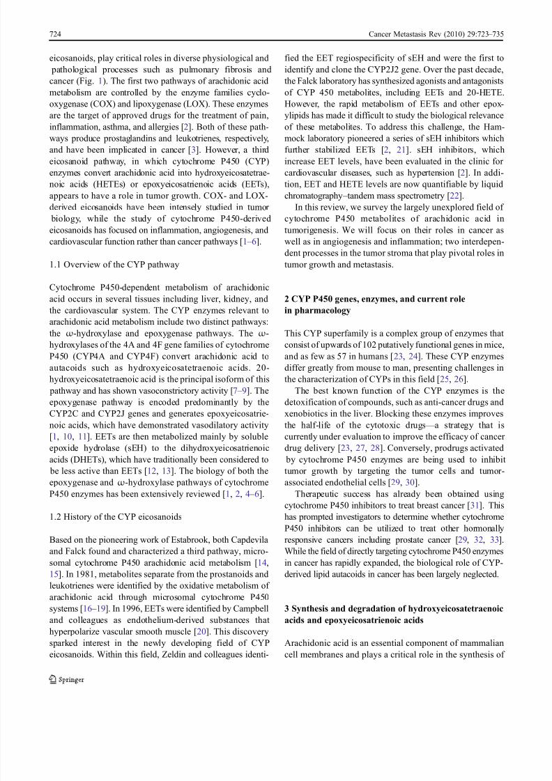
cancer (Fig. 1). The first two pathways of arachidonic acid
metabolism are controlled by the enzyme families cyclo-
oxygenase (COX) and lipoxygenase (LOX). These enzymes
are the target of approved drugs for the treatment of pain,
inflammation, asthma, and allergies [2]. Both of these path-
ways produce prostaglandins and leukotrienes, respectively,and have been implicated in cancer [3]. However, a third
eicosanoid pathway, in which cytochrome P450 (CYP)
enzymes convert arachidonic acid into hydroxyeicosatetrae-
noic acids (HETEs) or epoxyeicosatrienoic acids (EETs),
appears to have a role in tumor growth. COX- and LOX-
derived eicosanoids have been intensely studied in tumor
biology, while the study of cytochrome P450-derived
eicosanoids has focused on inflammation, angiogenesis, and
cardiovascular function rather than cancer pathways [1 – 6].
1.1 Overview of the CYP pathway
Cytochrome P450-dependent metabolism of arachidonic
acid occurs in several tissues including liver, kidney, and
the cardiovascular system. The CYP enzymes relevant to
arachidonic acid metabolism include two distinct pathways:
the ω-hydroxylase and epoxygenase pathways. The ω-
hydroxylases of the 4A and 4F gene families of cytochrome
P450 (CYP4A and CYP4F) convert arachidonic acid to
autacoids such as hydroxyeicosatetraenoic acids. 20-
hydroxyeicosatetraenoic acid is the principal isoform of this
pathway and has shown vasoconstrictory activity [7 – 9]. The
epoxygenase pathway is encoded predominantly by the
CYP2C and CYP2J genes and generates epoxyeicosatrie-
noic acids, which have demonstrated vasodilatory activity
[1, 10, 11]. EETs are then metabolized mainly by soluble
epoxide hydrolase (sEH) to the dihydroxyeicosatrienoic
acids (DHETs), which have traditionally been considered to
be less active than EETs [12, 13]. The biology of both the
epoxygenase and ω-hydroxylase pathways of cytochrome
P450 enzymes has been extensively reviewed [1, 2, 4 – 6].
1.2 History of the CYP eicosanoids
Based on the pioneering work of Estabrook, both Capdevila
and Falck found and characterized a third pathway, micro-
somal cytochrome P450 arachidonic acid metabolism [14,
15]. In 1981, metabolites separate from the prostanoids and
leukotrienes were identified by the oxidative metabolism of
arachidonic acid through microsomal cytochrome P450
systems [16 – 19]. In 1996, EETs were identified by Campbell
and colleagues as endothelium-derived substances that
hyperpolarize vascular smooth muscle [20]. This discovery
sparked interest in the newly developing field of CYP
eicosanoids. Within this field, Zeldin and colleagues identi-
fied the EET regiospecificity of sEH and were the first to
identify and clone the CYP2J2 gene. Over the past decade,
the Falck laboratory has synthesized agonists and antagonists
of CYP 450 metabolites, including EETs and 20-HETE.
However, the rapid metabolism of EETs and other epox-
ylipids has made it difficult to study the biological relevance
of these metabolites. To address this challenge, the Ham-
mock laboratory pioneered a series of sEH inhibitors whichfurther stabilized EETs [2, 21]. sEH inhibitors, which
increase EET levels, have been evaluated in the clinic for
cardiovascular diseases, such as hypertension [2]. In addi-
tion, EET and HETE levels are now quantifiable by liquid
chromatography – tandem mass spectrometry [22].
In this review, we survey the largely unexplored field of
cytochrome P450 metabolites of arachidonic acid in
tumorigenesis. We will focus on their roles in cancer as
well as in angiogenesis and inflammation; two interdepen-
dent processes in the tumor stroma that play pivotal roles in
tumor growth and metastasis.
2 CYP P450 genes, enzymes, and current role
in pharmacology
This CYP superfamily is a complex group of enzymes that
consist of upwards of 102 putatively functional genes in mice,
and as few as 57 in humans [23, 24]. These CYP enzymes
differ greatly from mouse to man, presenting challenges in
the characterization of CYPs in this field [25, 26].
The best known function of the CYP enzymes is the
detoxification of compounds, such as anti-cancer drugs and
xenobiotics in the liver. Blocking these enzymes improves
the half-life of the cytotoxic drugs — a strategy that is
currently under evaluation to improve the efficacy of cancer
drug delivery [23, 27, 28]. Conversely, prodrugs activated
by cytochrome P450 enzymes are being used to inhibit
tumor growth by targeting the tumor cells and tumor-
associated endothelial cells [29, 30].
Therapeutic success has already been obtained using
cytochrome P450 inhibitors to treat breast cancer [31]. This
has prompted investigators to determine whether cytochrome
P450 inhibitors can be utilized to treat other hormonally
responsive cancers including prostate cancer [29, 32, 33].
While the field of directly targeting cytochrome P450 enzymes
in cancer has rapidly expanded, the biological role of CYP-
derived lipid autacoids in cancer has been largely neglected.
3 Synthesis and degradation of hydroxyeicosatetraenoic
acids and epoxyeicosatrienoic acids
Arachidonic acid is an essential component of mammalian
cell membranes and plays a critical role in the synthesis of
724 Cancer Metastasis Rev (2010) 29:723 – 735
8/7/2019 Cytochrome P450 derived eicosanoids
http://slidepdf.com/reader/full/cytochrome-p450-derived-eicosanoids 3/13
bioactive eicosanoids [1]. Eicosanoids are generated via the
oxidation of the 20-carbon chain present on arachidonic acid or
other related fatty acids [2]. During processes, such as
inflammation, arachidonic acid is released from the cell
membrane through the activation of phospholipase A2 [1].
Arachidonic acid is metabolized by the CYP ω-hydroxylases
to 7-, 10-, 12-, 13-, 15-, 16-, 17-, 18-, 19-, and 20-HETEs, the
principal metabolite being the pro-inflammatory 20-HETE [4].
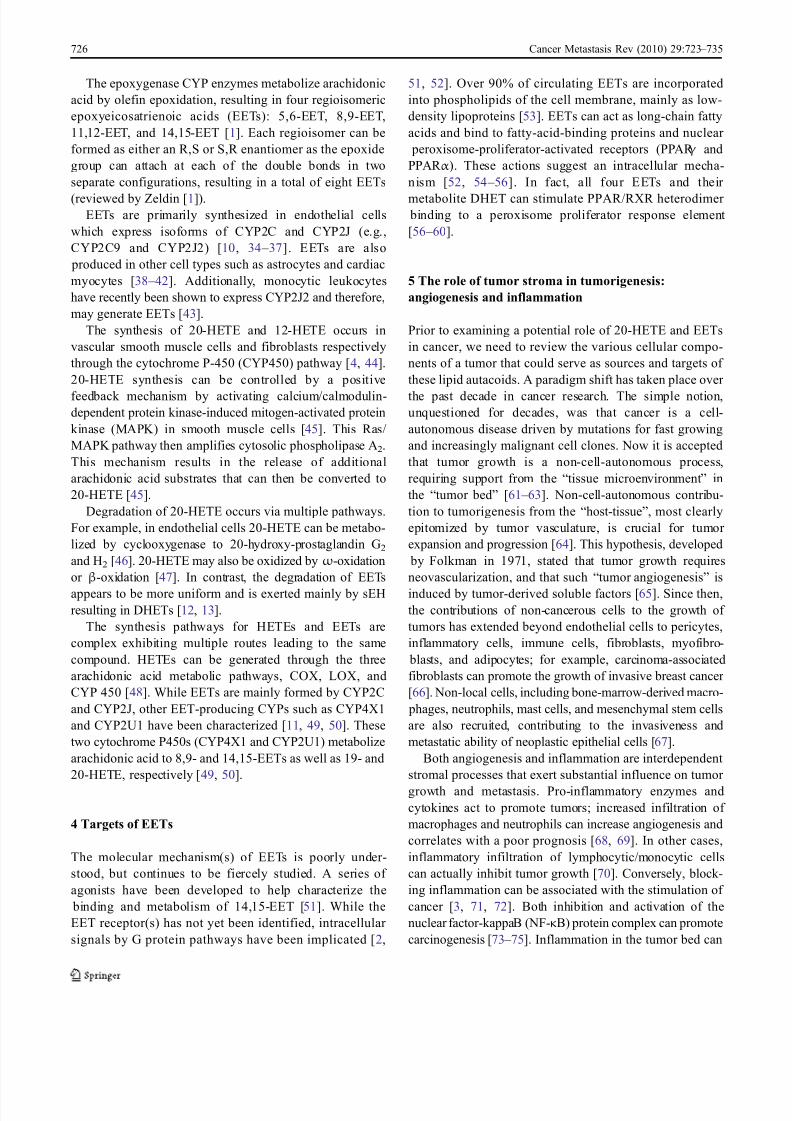
Stimulus Phospholipid
Arachidonic acid
COX1COX2COX3
5-LOX8-LOX12-LOX15-LOX
Epoxygenases
CYP2CCYP2J
CYP4A
n-HETELEUKOTRIENES“PROSTAGLANDINS” EPOXYEICOSATRIENOIC ACID
MSPPOH
HET0016
ProstacyclinsPGI2
ProstaglandinsPGE2
PGD2
PGF2 ThromboxanesTXA2
Platelet aggregation
Inflammation Pain Proliferation
LT A4LT B4
LT C4LT D4
12-HETE19-HETE
20-HETE
Inflammation Vascular
function COX1
20-OH PGE2
20-OH PGI2
5,6-EET8,9-EET
11,12-EET14,15-EET
EET agonist
14, 15-EEZE
sEH sEH inhibitor
Vasorelaxation
Cardioprotection Proliferation
Inflammation
DHET
Inflammation Allergy Bronchoconstriction
Cyclooxgenase (COX) Lipooxygenases (LOX)Cytochrome P450 (CYP)
(87 genes in humans)
O
O
OH
HO
HO
Prostacyclins Leukotrienes C4
OH
S
COOH
CONHCH2COOH
NHCO(CH2)2CHCOOH
NH2
OH
COOH
20-HETE
O O
HO
11,12-EET
A
B
Fig. 1 a, b Bioactive eicosanoids derived from the arachidonic acid
cascade. Arachidonic acid is metabolized by three pathways —
thecyclooxygenase (COX ), lipoxygenase ( LOX ), and cytochrome P450
(CYP ) pathways. Schematic overview of major mediators and their
metabolites (blue); enzymes (black , boxed ) and biological role ( green).
Inhibitors (red ovals) and agonists ( green ovals). HETEs Hydroxyeico-
satetraenoic acids, EETs epoxyeicosatrienoic acids, CYP cytochrome
P450 enzymes. MS-PPOH is a selective inhibitor of a subset of
epoxygenases. HET0016 is a selective inhibitor of the ω-hydroxlaseCYP4A. The sEH inhibitor (soluble epoxide hydrolase inhibitor)
increases EET levels by acting as an agonist of the EET pathway.
14,15-EEZE is a putative EET receptor antagonist. PGE 2 prostaglandin
E2, PGI 2 prostacyclin, LTA4 leukotriene A4, DHET dihydroxyeicosa-
trienoic acid, 20-OH PGE 2 20-hydroxy-prostaglandin E2
Cancer Metastasis Rev (2010) 29:723 – 735 725
8/7/2019 Cytochrome P450 derived eicosanoids
http://slidepdf.com/reader/full/cytochrome-p450-derived-eicosanoids 4/13
The epoxygenase CYP enzymes metabolize arachidonic
acid by olefin epoxidation, resulting in four regioisomeric
epoxyeicosatrienoic acids (EETs): 5,6-EET, 8,9-EET,
11,12-EET, and 14,15-EET [1]. Each regioisomer can be
formed as either an R,S or S,R enantiomer as the epoxide
group can attach at each of the double bonds in two
separate configurations, resulting in a total of eight EETs
(reviewed by Zeldin [1]).EETs are primarily synthesized in endothelial cells
which express isoforms of CYP2C and CYP2J (e.g.,
CYP2C9 and CYP2J2) [10, 34 – 37]. EETs are also
produced in other cell types such as astrocytes and cardiac
myocytes [38 – 42]. Additionally, monocytic leukocytes
have recently been shown to express CYP2J2 and therefore,
may generate EETs [43].
The synthesis of 20-HETE and 12-HETE occurs in
vascular smooth muscle cells and fibroblasts respectively
through the cytochrome P-450 (CYP450) pathway [4, 44].
20-HETE synthesis can be controlled by a positive
feedback mechanism by activating calcium/calmodulin-dependent protein kinase-induced mitogen-activated protein
kinase (MAPK) in smooth muscle cells [45]. This Ras/
MAPK pathway then amplifies cytosolic phospholipase A2.
This mechanism results in the release of additional
arachidonic acid substrates that can then be converted to
20-HETE [45].
Degradation of 20-HETE occurs via multiple pathways.
For example, in endothelial cells 20-HETE can be metabo-
lized by cyclooxygenase to 20-hydroxy-prostaglandin G2
and H2 [46]. 20-HETE may also be oxidized by ω-oxidation
or β-oxidation [47]. In contrast, the degradation of EETs
appears to be more uniform and is exerted mainly by sEH
resulting in DHETs [12, 13].
The synthesis pathways for HETEs and EETs are
complex exhibiting multiple routes leading to the same
compound. HETEs can be generated through the three
arachidonic acid metabolic pathways, COX, LOX, and
CYP 450 [48]. While EETs are mainly formed by CYP2C
and CYP2J, other EET-producing CYPs such as CYP4X1
and CYP2U1 have been characterized [11, 49, 50]. These
two cytochrome P450s (CYP4X1 and CYP2U1) metabolize
arachidonic acid to 8,9- and 14,15-EETs as well as 19- and
20-HETE, respectively [49, 50].
4 Targets of EETs
The molecular mechanism(s) of EETs is poorly under-
stood, but continues to be fiercely studied. A series of
agonists have been developed to help characterize the
binding and metabolism of 14,15-EET [51]. While the
EET receptor(s) has not yet been identified, intracellular
signals by G protein pathways have been implicated [2,
51, 52]. Over 90% of circulating EETs are incorporated
into phospholipids of the cell membrane, mainly as low-
density lipoproteins [53]. EETs can act as long-chain fatty
acids and bind to fatty-acid-binding proteins and nuclear
peroxisome-proliferator-activated receptors (PPAR γ and
PPAR α ). These actions suggest an intracellular mecha-
nism [52, 54 – 56]. In fact, all four EETs and their
metabolite DHET can stimulate PPAR/RXR heterodimer binding to a peroxisome proliferator response element
[56 – 60].
5 The role of tumor stroma in tumorigenesis:
angiogenesis and inflammation
Prior to examining a potential role of 20-HETE and EETs
in cancer, we need to review the various cellular compo-
nents of a tumor that could serve as sources and targets of
these lipid autacoids. A paradigm shift has taken place over
the past decade in cancer research. The simple notion,unquestioned for decades, was that cancer is a cell-
autonomous disease driven by mutations for fast growing
and increasingly malignant cell clones. Now it is accepted
that tumor growth is a non-cell-autonomous process,
requiring support from the “tissue microenvironment ” in
the “tumor bed” [61 – 63]. Non-cell-autonomous contribu-
tion to tumorigenesis from the “host-tissue”, most clearly
epitomized by tumor vasculature, is crucial for tumor
expansion and progression [64]. This hypothesis, developed
by Folkman in 1971, stated that tumor growth requires
neovascularization, and that such “tumor angiogenesis” is
induced by tumor-derived soluble factors [65]. Since then,
the contributions of non-cancerous cells to the growth of
tumors has extended beyond endothelial cells to pericytes,
inflammatory cells, immune cells, fibroblasts, myofibro-
blasts, and adipocytes; for example, carcinoma-associated
fibroblasts can promote the growth of invasive breast cancer
[66]. Non-local cells, including bone-marrow-derived macro-
phages, neutrophils, mast cells, and mesenchymal stem cells
are also recruited, contributing to the invasiveness and
metastatic ability of neoplastic epithelial cells [67].
Both angiogenesis and inflammation are interdependent
stromal processes that exert substantial influence on tumor
growth and metastasis. Pro-inflammatory enzymes and
cytokines act to promote tumors; increased infiltration of
macrophages and neutrophils can increase angiogenesis and
correlates with a poor prognosis [68, 69]. In other cases,
inflammatory infiltration of lymphocytic/monocytic cells
can actually inhibit tumor growth [70]. Conversely, block-
ing inflammation can be associated with the stimulation of
cancer [3, 71, 72]. Both inhibition and activation of the
nuclear factor-kappaB (NF-κB) protein complex can promote
carcinogenesis [73 – 75]. Inflammation in the tumor bed can
726 Cancer Metastasis Rev (2010) 29:723 – 735
8/7/2019 Cytochrome P450 derived eicosanoids
http://slidepdf.com/reader/full/cytochrome-p450-derived-eicosanoids 5/13
then either stimulate or inhibit tumor growth [72, 76, 77].
Thus, pharmacological modulation of inflammation in cancer
treatments must be evaluated with the notion that inflamma-
tion may be a double-edged sword in tumor growth.
6 Lipid autacoids in cancer
It has been recognized that tumor growth is a complex
process involving many cell types. The intercellular
communication that takes place between these cells is
conducted by an array of soluble factors such as:
proteinaceous growth factors and chemokines, vascular
endothelial growth factor (VEGF), FGF-2, TGF-β, TNF-
α , interleukin (IL)-1, and oxygen radicals [78].
Little attention has been paid to small molecule
mediators, such as lipid autacoids, whose role in cancer
has only recently emerged. Given that a tumor consists of
both cancerous and non-cancerous cells, the role of
autacoids in tumor growth can be separated into their direct effects on neoplastic growth and their effects on inflamma-
tion, angiogenesis, and stromal cells.
The pro-inflammatory prostaglandins and leukotrienes
directly induce epithelial tumor cell proliferation, survival,
migration, and invasion in an autocrine and paracrine
manner [3]. Lipid autacoids, such as prostaglandin E2
(PGE2) and leukotriene B4 (LTB4), stimulate both epithelial
cells and stromal cells to produce VEGF and FGF-2. These
angiogenic growth factors induce COX2 and in turn
produce PGE2 and PGI2 in endothelial cells [3, 79]. Other
studies have linked eicosanoids to stroma inflammation in
epithelial ovarian cancer [80]. Levels of eicosanoid metab-
olites, such as PGE2, 5-HETE, and 12-HETE, increase
progressively in patients with benign pelvic disease to those
with epithelial ovarian cancer. This demonstrates the
involvement of lipid autacoids in the inflammatory envi-
ronment of cancer [80]. However, the role of lipid autacoids
derived from the third eicosanoid pathway of arachidonic
acid remains poorly characterized in cancer.
7 HETEs effects on inflammation and the vasculature
Lipoxygenase-derived HETEs inhibit apoptosis, stimulate
angiogenesis, and enhance proliferation and migration of
cancer cells [48]. 20-HETE, the principal metabolite of the
ω-hydroxylation pathway, is a pro-inflammatory mediator
that markedly stimulates the production of inflammatory
cytokines/chemokines in endothelial cells, including IL-8, IL-
13, IL-4, and prostaglandin E2 [81]. 20-HETE stimulates NF-
κB activation and MAPK/ERK pathways, which suggests
that HETE’s pro-inflammatory effect may be mediated by
the central inflammatory pathway of NF-κB [81].
In addition to its pro-inflammatory activity, 20-HETE
has pro-angiogenic activity including the stimulation of
endothelial cell proliferation, migration, and cell survival
[82 – 85]. 20-HETE has an important role in VEGF-
dependent angiogenesis [86] (reviewed in [85]). While
VEGF seems to be the primary mediator of 20-HETE –
induced endothelial cell proliferation, inhibition with a
VEGF antibody does not completely abrogate the mitogen-ic effect of 20-HETE [82]. This suggests other pathways are
involved in 20-HETE-mediated angiogenesis [82].
The pro-angiogenic factor fibroblast growth factor-2
(FGF-2) can activate cytosolic phospholipase A2 (the
enzyme which releases arachidonic acid from cell mem-
branes) in endothelial cells [87]. FGF-2 increases arach-
idonic acid production, potentially stimulating CYP4A and
production of 20-HETE [85]. The overexpression of
CYP4A1, which increases 20-HETE production, results in
increased neovessel formation [88].
HET0016, a selective inhibitor of CYP4A, suppresses
the formation of 20-HETE at a concentration <10 nM, andhas no effect on epoxygenase, cyclooxygenase, or lip-
oxygenase activity at concentrations up to 1 μ M [4, 89].
HET0016 inhibits VEGF-induced endothelial cell prolifer-
ation in vitro and corneal neovascularization in vivo when
administered locally with pellets containing VEGF [84].
When administered locally into the cornea, HET0016
inhibited tumor-induced (U251 glioblastoma cells) angio-
genesis by 70% [84]. Furthermore, the administration of the
stable 20-HETE agonist, 20-hydroxyeicosa-6(Z) 15(Z)-
dienoic acid (WIT003), induced mitogenesis in endothelial
cells and corneal neovascularization in vivo [84]. These
studies provide experimental evidence that inhibiting 20-
HETE may offer a strategy to reduce pathological angio-
genesis not only in tumors but in angiogenic diseases such
as diabetic retinopathy, macular degeneration and chronic
inflammatory diseases, such as psoriasis [84]. However,
these studies did not determine whether 20-HETE was
produced by the cornea or endothelial cells and, therefore,
further studies are needed [90].
In the systemic circulation, 20-HETE produced by
vascular smooth muscle cells acts as a vasoconstrictor [4].
However, in pulmonary arteries, 20-HETE contributes to
VEGF-induced relaxation of the lungs [91]. VEGF, a nitric
oxide (NO)-dependent dilator of systemic arteries, plays a
key role in maintaining the integrity of the pulmonary
vasculature [91].
8 20-HETE effects in cancer
In 2008, U251 glioblastoma cells were genetically altered
(transfected with rat CYP4A1 cDNA) to increase the
formation of 20-HETE [92]. This stimulated proliferation
Cancer Metastasis Rev (2010) 29:723 – 735 727
8/7/2019 Cytochrome P450 derived eicosanoids
http://slidepdf.com/reader/full/cytochrome-p450-derived-eicosanoids 6/13
in culture. When these transfected U251 glioblastoma cells
were implanted into the brain of rats, a tenfold increase in
tumor volume was observed when compared to animals
receiving mock-transfected U251 cells [92].
Conversely, Guo et al. demonstrated that HET0016
significantly inhibited human U251 glioblastoma cell
proliferation in a dose-dependent manner [90]. HET0016
inhibited the phosphorylation of the epidermal growthfactor receptor (EGFR) and the subsequent phosphorylation
of p42/p44 MAPK [90]. While U251 cells expressed
CYP4A11 mRNA and protein, HPLC and mass spectrom-
etry analysis of U251 cell extracts revealed that they did not
appear to synthesize 20-HETE [90]. Thus, HET0016 has
other effects independent of suppressing 20-HETE. Subse-
quently, the same group demonstrated that 9L gliosarcoma
proliferation and tumor growth in rats are suppressed by
HET0016 [93]. Systemic administration of HET0016
inhibited the tumor growth of 9L gliosarcomas by 80%,
and tumor angiogenesis by roughly 50%. In a separate
study, HET0016 and a 20-HETE antagonist (WIT002) bothinhibited the proliferation of a renal adenocarcinoma. This
cell type expressed CYP4F isoforms and produced 20-
HETE [94].
Little is known about 20-HETE in cancer patients. In
one study, 12-HETE and 20-HETE concentrations were
shown to be elevated in the urine of patients with benign
prostatic hypertrophy and prostate cancer patients as
compared to normal subjects [95]. Further analysis did not
establish a correlation between the concentrations of
HETEs and prostatic specific antigen level, gland size, or
tumor grade [95].
9 EETs and angiogenesis
EETs are mainly secreted by endothelial cells and play critical
roles in cellular proliferation, migration, and inflammation;
their major target is blood vessels [6, 37]. EETs may act in an
autocrine fashion on the endothelium inducing vasodilatory
and anti-inflammatory effects in blood vessels [96]. As a
result of these effects, EETs lower blood pressure and protect
the myocardium and brain from ischemia [56, 97 – 99].
The initial finding that linked EETs to angiogenesis was
shown by an increase in proliferation of cerebral capillary
endothelial cells by astrocyte conditioned media [40]. In
contrast, an inhibitor of cytochrome P450, 17-octadecynoic
acid (17-ODYA), suppressed the formation of capillary
tubes in a co-culture of astrocytes and endothelial cells.
Both EETs secreted by astrocytes and synthetic EETs
stimulated endothelial cell proliferation, tube formation,
and angiogenesis in a matrigel plug in vivo [40, 100, 101].
Angiogenesis is critically dependent on endothelial cell
migration [102]. The development of synthetic EETs has
provided insight into the angiogenic functions and path-
ways of the various EETs. For instance, EETs have been
shown to promote endothelial cell migration via endothelial
NO synthase, MEK/MAPK, and PI3K [103]. Another assay
to evaluate angiogenesis is the chick chorioallantoic
membrane assay, which uses the chorioallantoic membrane
(CAM) of a chicken embryo [104]. Michaelis et al.
employed this assay to demonstrate that 11,12-EETstimulates vessel formation [105]. Importantly, this CAM-
mediated angiogenesis was suppressed by either an EGF
receptor-neutralizing antibody or an inhibitor of the EGF
receptor. Thus, 11,12-EET may stimulate angiogenesis
through the activation of the EGF receptor [105].
Several other pathways have been implicated in 11,12-
EET- and 14,15-EET-mediated angiogenesis. Sphingosine
kinase-1 (SK1) is one important mediator of 11,12-EET-
induced angiogenic effects [106]. The expression of a
dominant-negative SK1 or knockdown of SK1 by siRNA,
inhibited 11,12-EET-induced endothelial cell proliferation,
migration, tube formation, and matrigel plug vessel forma-tion [106]. In other studies, EphB4 is a critical component
of the CYP2C9-activated signaling cascade [107]. Both
CYP2C9 overexpression or the administration of 11,12-
EET showed increased expression of EphB4 in endothelial
cells. The availability of these synthetic EETs has made it
possible to evaluate another regioisomer, 14,15-EET. 14,15-
EET was shown to induce angiogenesis via several path-
ways including: Src, phospatidylinositol-3-kinase/Akt
(PI3K/Akt) signaling in parallel with mTOR-S6K1 activa-
tion and Src-dependent STAT-3-mediated VEGF expression
[108, 109].
Other groups have studied CYP 450-derived metabolites,
utilizing the strategy of overexpressing CYP epoxygenases.
In lieu of EETs, this system inhibited endothelial cell
apoptosis through activation of the PI3K/Akt pathway
[110]. The overexpression of CYP epoxygenases, including
CYP2J2, also increased muscle capillary density in a rat
ischemic hind limb model [103]. Thus, CYP 450-derived
metabolites may stimulate the development of collateral
circulation in ischemic tissue [103].
While most investigators have focused on 11,12-EET
and 14,15-EET, Pozzi et al. identified 5,6- and 8,9-EET as
pro-angiogenic lipids [36]. These regioisomers increased
blood vessel density and formed functionally intact vessels
in a subcutaneous sponge model in mice. This neovascula-
rization was enhanced by the co-administration of an
epoxide hydrolase inhibitor, which elevates the levels of
EETs [36]. This study corroborates the critical role that
EETs plays in angiogenesis.
It is known that hypoxia stimulates angiogenesis via
transcriptional VEGF induction, a response that is mediated
by the hypoxia-inducible factor-1α (HIF-1α ) [111]. It was
shown by the Fleming laboratory that hypoxia also
728 Cancer Metastasis Rev (2010) 29:723 – 735
8/7/2019 Cytochrome P450 derived eicosanoids
http://slidepdf.com/reader/full/cytochrome-p450-derived-eicosanoids 7/13
stimulates CYP 2C8 and 2C9 expression [5, 112, 113].
Consistently, the CYP inhibitor (MS-PPOH) and the
putative EET receptor antagonist (14,15-EEZE), inhibited
hypoxia-induced endothelial tube formation [112]. Further-
more, the angiogenic effect of EETs is partially dependent
on HIF-1α -mediated VEGF induction [114]. This may have
implications in cancer beyond angiogenesis, since HIF-1α
can provide a growth and survival advantage to tumor cells,especially under metabolic stress [72].
The effects of EETs and VEGF regulation are closely
intertwined. EETs can enhance the effects of VEGF-
induced angiogenesis [115]. In turn, VEGF can increase
CYP2C promoter activity in endothelial cells and induce
the expression of CYP2C8, resulting in increased intracel-
lular EET levels [115]. The putative EET receptor antago-
nist, 14,15-EEZE, inhibits VEGF-induced endothelial cell
tube formation. However, 14,15-EEZE does not affect
VEGF-induced phosphorylation of its receptor or FGF-2-
stimulated tube formation [115]. In a parallel study,
CYP2C44 epoxygenase appears to be an important com- ponent in the VEGF signaling pathway [116]. For example,
in cultured lung endothelial cells that express VEGF-
inducible CYP2C44 epoxygenase, resulting in increased
levels of 11,12- and 14,15-EET, angiogenesis was stimu-
lated in vitro. Taken together, these studies suggest that the
pro-angiogenic activity of EETs is mediated at least, in part,
by VEGF [115, 116].
10 CYP 450 epoxygenases and cancer
While the pro-angiogenic activity of EETs has extensively
been investigated [36, 103, 117], the role of EETs in cancer
remains poorly characterized. Although two decades ago,
14,15-EET was shown to stimulate mesangial and renal
epithelial cell proliferation [118, 119], only in the last
5 years has evidence, supporting cytochrome P450 epox-
ygenases as a potential tumor-promoting enzyme, begun to
emerge [120]. The role of CYP2J2 epoxygenase in cancer
was first shown by Jiang et al. In this study, CYP2J2 was
upregulated in 77% of human carcinoma tissues and eight
different human carcinoma cell lines [120]. Furthermore,
the transfection of tumor cells with cytochrome epoxyge-
nase 2J2 enhanced tumor formation [120]. Subsequent
studies, in which CYP epoxygenase levels were manipu-
lated, by either overexpression of CYP2J2 or antisense in
the xenotransplanted tumor cell, suggest EETs may play a
role in cancer metastasis [121].
EETs also appear to be important for cancer cell
survival. Specific CYP2J2 inhibitors suppress human tumor
cell proliferation [122]. These inhibitors activate caspase-3,
which leads to reduced tumor cell adhesion, migration,
invasion, and suppressed murine xenograft tumor growth
[122]. It is often difficult to distinguish a direct effect on the
tumor cells or the stromal processes. It is likely that both
mechanisms synergize to account for the potential pro-
tumorigenic activity of EETs.
There are few pharmacological studies using drugs
which can non-specifically affect EETs. In one study
conducted by Pozzi et al., mice treated with PPAR α ligands
exhibited a reduction of tumor growth, vascularization, and plasma EETs [123]. In a separate study, two mechanistically
different synthetic inhibitors of cytochrome P450, 17-
ODYA, and miconazole significantly reduced tumor size
and capillary formation in intracranial glial tumors, and
prolonged survival of treated rats [124]. Interestingly, these
inhibitors had no effect on EETs in the tumor tissue
suggesting that the tumor endothelium may be the target
of these CYP inhibitors [124]. It has recently been reported
that EET antagonists inhibit prostate carcinoma cell motility
[125]. This may represent a novel mechanism of EET
antagonists acting directly on the tumor cell [125].
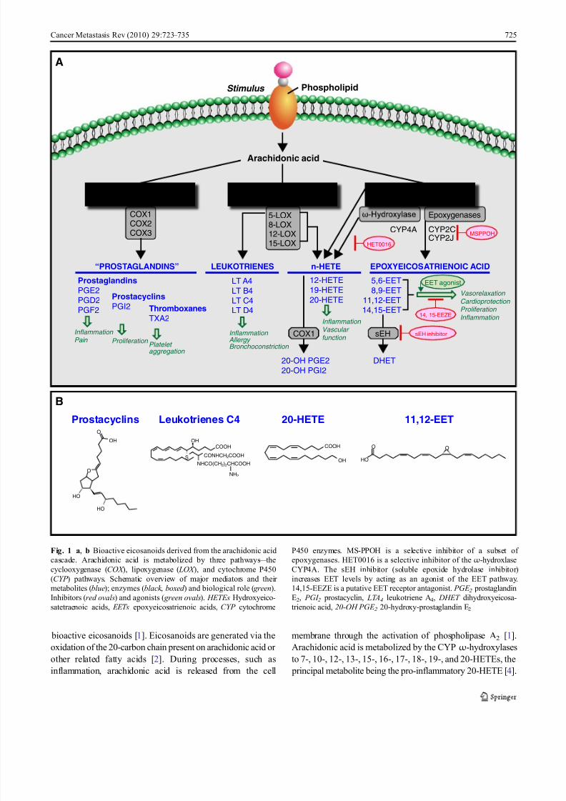
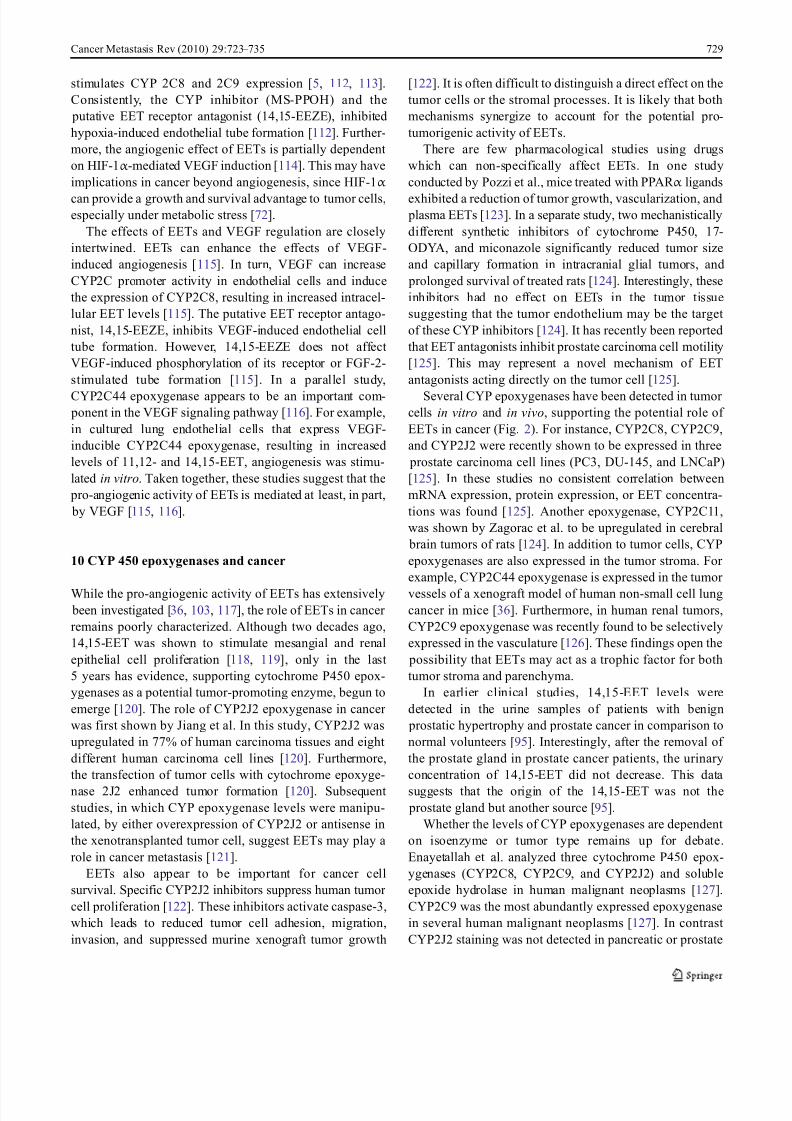
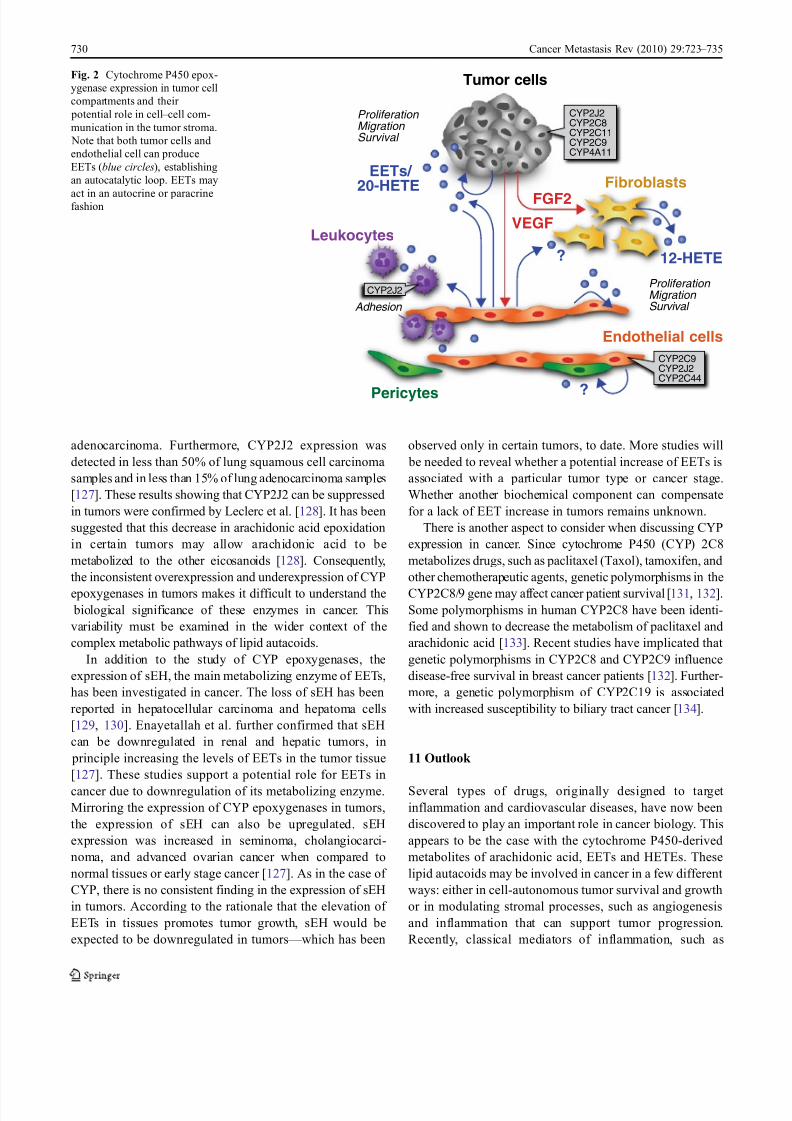
Several CYP epoxygenases have been detected in tumor cells in vitro and in vivo, supporting the potential role of
EETs in cancer (Fig. 2). For instance, CYP2C8, CYP2C9,
and CYP2J2 were recently shown to be expressed in three
prostate carcinoma cell lines (PC3, DU-145, and LNCaP)
[125]. In these studies no consistent correlation between
mRNA expression, protein expression, or EET concentra-
tions was found [125]. Another epoxygenase, CYP2C11,
was shown by Zagorac et al. to be upregulated in cerebral
brain tumors of rats [124]. In addition to tumor cells, CYP
epoxygenases are also expressed in the tumor stroma. For
example, CYP2C44 epoxygenase is expressed in the tumor
vessels of a xenograft model of human non-small cell lung
cancer in mice [36]. Furthermore, in human renal tumors,
CYP2C9 epoxygenase was recently found to be selectively
expressed in the vasculature [126]. These findings open the
possibility that EETs may act as a trophic factor for both
tumor stroma and parenchyma.
In earlier clinical studies, 14,15-EET levels were
detected in the urine samples of patients with benign
prostatic hypertrophy and prostate cancer in comparison to
normal volunteers [95]. Interestingly, after the removal of
the prostate gland in prostate cancer patients, the urinary
concentration of 14,15-EET did not decrease. This data
suggests that the origin of the 14,15-EET was not the
prostate gland but another source [95].
Whether the levels of CYP epoxygenases are dependent
on isoenzyme or tumor type remains up for debate.
Enayetallah et al. analyzed three cytochrome P450 epox-
ygenases (CYP2C8, CYP2C9, and CYP2J2) and soluble
epoxide hydrolase in human malignant neoplasms [127].
CYP2C9 was the most abundantly expressed epoxygenase
in several human malignant neoplasms [127]. In contrast
CYP2J2 staining was not detected in pancreatic or prostate
Cancer Metastasis Rev (2010) 29:723 – 735 729
8/7/2019 Cytochrome P450 derived eicosanoids
http://slidepdf.com/reader/full/cytochrome-p450-derived-eicosanoids 8/13
adenocarcinoma. Furthermore, CYP2J2 expression was
detected in less than 50% of lung squamous cell carcinoma
samples and in less than 15% of lung adenocarcinoma samples
[127]. These results showing that CYP2J2 can be suppressed
in tumors were confirmed by Leclerc et al. [128]. It has been
suggested that this decrease in arachidonic acid epoxidation
in certain tumors may allow arachidonic acid to be
metabolized to the other eicosanoids [128]. Consequently,
the inconsistent overexpression and underexpression of CYP
epoxygenases in tumors makes it difficult to understand the
biological significance of these enzymes in cancer. This
variability must be examined in the wider context of the
complex metabolic pathways of lipid autacoids.
In addition to the study of CYP epoxygenases, the
expression of sEH, the main metabolizing enzyme of EETs,
has been investigated in cancer. The loss of sEH has been
reported in hepatocellular carcinoma and hepatoma cells
[129, 130]. Enayetallah et al. further confirmed that sEH
can be downregulated in renal and hepatic tumors, in
principle increasing the levels of EETs in the tumor tissue
[127]. These studies support a potential role for EETs in
cancer due to downregulation of its metabolizing enzyme.
Mirroring the expression of CYP epoxygenases in tumors,
the expression of sEH can also be upregulated. sEH
expression was increased in seminoma, cholangiocarci-
noma, and advanced ovarian cancer when compared to
normal tissues or early stage cancer [127]. As in the case of
CYP, there is no consistent finding in the expression of sEH
in tumors. According to the rationale that the elevation of
EETs in tissues promotes tumor growth, sEH would be
expected to be downregulated in tumors — which has been
observed only in certain tumors, to date. More studies will
be needed to reveal whether a potential increase of EETs is
associated with a particular tumor type or cancer stage.
Whether another biochemical component can compensate
for a lack of EET increase in tumors remains unknown.
There is another aspect to consider when discussing CYP
expression in cancer. Since cytochrome P450 (CYP) 2C8
metabolizes drugs, such as paclitaxel (Taxol), tamoxifen, and
other chemotherapeutic agents, genetic polymorphisms in the
CYP2C8/9 gene may affect cancer patient survival [131, 132].
Some polymorphisms in human CYP2C8 have been identi-
fied and shown to decrease the metabolism of paclitaxel and
arachidonic acid [133]. Recent studies have implicated that
genetic polymorphisms in CYP2C8 and CYP2C9 influence
disease-free survival in breast cancer patients [132]. Further-
more, a genetic polymorphism of CYP2C19 is associated
with increased susceptibility to biliary tract cancer [134].
11 Outlook
Several types of drugs, originally designed to target
inflammation and cardiovascular diseases, have now been
discovered to play an important role in cancer biology. This
appears to be the case with the cytochrome P450-derived
metabolites of arachidonic acid, EETs and HETEs. These
lipid autacoids may be involved in cancer in a few different
ways: either in cell-autonomous tumor survival and growth
or in modulating stromal processes, such as angiogenesis
and inflammation that can support tumor progression.
Recently, classical mediators of inflammation, such as
Tumor cells
Leukocytes
EETs/ 20-HETE Fibroblasts
?
FGF2
Endothelial cells
Pericytes ?
Proliferation Migration Survival
Adhesion
Proliferation Migration Survival
VEGF
12-HETE
CYP2J2
CYP2C8
CYP2C11
CYP2C9
CYP4A11
CYP2C9
CYP2J2
CYP2C44
CYP2J2
Fig. 2 Cytochrome P450 epox-
ygenase expression in tumor cell
compartments and their
potential role in cell – cell com-
munication in the tumor stroma.
Note that both tumor cells and
endothelial cell can produce
EETs (blue circles), establishing
an autocatalytic loop. EETs may
act in an autocrine or paracrinefashion
730 Cancer Metastasis Rev (2010) 29:723 – 735
8/7/2019 Cytochrome P450 derived eicosanoids
http://slidepdf.com/reader/full/cytochrome-p450-derived-eicosanoids 9/13
prostaglandins, have received new attention as potential targets
in cancer treatment. The EET and HETE pathways should be
evaluated as potential targets in cancer therapy, directed both
against tumor cells and their surrounding stroma. Today, the
role of cytochrome P450 metabolites in cancer is still poorly
characterized, in part because of their biochemical complexity.
The increasing availability of research tools, such as novel
synthetic agonists, antagonists, and enzyme inhibitors, nowoffer a reasonable platform for dissecting the role of EETs and
HETEs family autacoids in cancer.
Acknowledgments We thank Catherine Butterfield for suggestions
in preparing the manuscript; Kristin Johnson preparation of the figures
and cover; and Tadanori Mammoto for confocal imaging in the cover.
This work was supported by National Cancer Institute grant
RO1CA148633-O1A1 (DP).
Open Access This article is distributed under the terms of the Creative
Commons Attribution Noncommercial License which permits any
noncommercial use, distribution, and reproduction in any medium,
provided the original author(s) and source are credited.
References
1. Zeldin, D. C. (2001). Epoxygenase pathways of arachidonic acid
metabolism. The Journal of Biological Chemistry, 276 , 36059 – 36062.
2. Imig, J. D., & Hammock, B. D. (2009). Soluble epoxide
hydrolase as a therapeutic target for cardiovascular diseases.
Nature Reviews. Drug Discovery, 8, 794 – 805.
3. Wang, D., & Dubois, R. N. (2010). Eicosanoids and cancer.
Nature Reviews. Cancer, 10, 181 – 193.
4. Roman, R. J. (2002). P-450 metabolites of arachidonic acid in the
control of cardiovascular function. Physiological Reviews, 82, 131 –
185.
5. Fleming, I. (2007). Epoxyeicosatrienoic acids, cell signaling and
angiogenesis. Prostaglandins & Other Lipid Mediators, 82, 60 – 67.
6. Spector, A. A., & Norris, A. W. (2007). Action of epoxyeicosa-
trienoic acids on cellular function. American Journal of
Physiology. Cell Physiology, 292, C996 – C1012.
7. Hardwick, J. P., Song, B. J., Huberman, E., & Gonzalez, F. J.
(1987). Isolation, complementary DNA sequence, and regulation
of rat hepatic lauric acid omega-hydroxylase (cytochrome P-
450LA omega). Identification of a new cytochrome P-450 gene
family. The Journal of Biological Chemistry, 262, 801 – 810.
8. Powell, P. K., Wolf, I., Jin, R., & Lasker, J. M. (1998).
Metabolism of arachidonic acid to 20-hydroxy-5, 8, 11, 14-
eicosatetraenoic acid by P450 enzymes in human liver: Involve-
ment of CYP4F2 and CYP4A11. The Journal of Pharmacology
and Experimental Therapeutics, 285, 1327 – 1336.
9. Miyata, N.,& Roman,R. J. (2005). Role of 20-hydroxyeicosatetraenoic
acid (20-HETE) in vascular system. Journal of Smooth Muscle
Research, 41, 175 – 193.
10. Fisslthaler, B., Popp, R., Kiss, L., Potente, M., Harder, D. R., et
al. (1999). Cytochrome P450 2 C is an EDHF synthase in
coronary arteries. Nature, 401, 493 – 497.
11. Kaspera, R., & Totah, R. A. (2009). Epoxyeicosatrienoic acids:
Formation, metabolism and potential role in tissue physiology
and pathophysiology. Expert Opinion on Drug Metabolism &
Toxicology, 5, 757 – 771.
12. Zeldin, D. C., Kobayashi, J., Falck, J. R., Winder, B. S.,
Hammock, B. D., et al. (1993). Regio- and enantiofacial
selectivity of epoxyeicosatrienoic acid hydration by cytosolic
epoxide hydrolase. The Journal of Biological Chemistry, 268,
6402 – 6407.
13. Yu, Z., Xu, F., Huse, L. M., Morisseau, C., Draper, A. J., et al.
(2000). Soluble epoxide hydrolase regulates hydrolysis of
vasoactive epoxyeicosatrienoic acids. Circulation Research, 87 ,
992 – 998.
14. Chacos, N., Capdevila, J., Falck, J. R., Manna, S., Martin-
Wixtrom, C., et al. (1983). The reaction of arachidonic acid
epoxides (epoxyeicosatrienoic acids) with a cytosolic epoxidehydrolase. Archives of Biochemistry and Biophysics, 223, 639 –
648.
15. Capdevila, J. H., Harris, R. C., & Falck, J. R. (2002).
Microsomal cytochrome P450 and eicosanoid metabolism.
Cellular and Molecular Life Sciences, 59, 780 – 789.
16. Capdevila, J., Chacos, N., Werringloer, J., Prough, R. A., &
Estabrook, R. W. (1981). Liver microsomal cytochrome P-450
and the oxidative metabolism of arachidonic acid. Proceedings
of the National Academy of Sciences of the United States of
America, 78, 5362 – 5366.
17. Capdevila, J., Marnett, L. J., Chacos, N., Prough, R. A., &
Estabrook, R. W. (1982). Cytochrome P-450-dependent oxygen-
ation of arachidonic acid to hydroxyicosatetraenoic acids.
Proceedings of the National Academy of Sciences of the United
States of America, 79, 767 – 770.18. Morrison, A. R., & Pascoe, N. (1981). Metabolism of arach-
idonate through NADPH-dependent oxygenase of renal cortex.
Proceedings of the National Academy of Sciences of the United
States of America, 78, 7375 – 7378.
19. Oliw, E. H., Lawson, J. A., Brash, A. R., & Oates, J. A. (1981).
Arachidonic acid metabolism in rabbit renal cortex. Formation of
two novel dihydroxyeicosatrienoic acids. The Journal of Biolog-
ical Chemistry, 256 , 9924 – 9931.
20. Campbell, W. B., Gebremedhin, D., Pratt, P. F., & Harder, D. R.
(1996). Identification of epoxyeicosatrienoic acids as
endothelium-derived hyperpolarizing factors. Circulation Re-
search, 78, 415 – 423.
21. Morisseau, C., Goodrow, M. H., Dowdy, D., Zheng, J., Greene,
J. F., et al. (1999). Potent urea and carbamate inhibitors of
soluble epoxide hydrolases. Proceedings of the National Acad-
emy of Sciences of the United States of America, 96 , 8849 – 8854.
22. Newman, J. W., Watanabe, T., & Hammock, B. D. (2002). The
simultaneous quantification of cytochrome P450 dependent
linoleate and arachidonate metabolites in urine by HPLC-MS/
MS. Journal of Lipid Research, 43, 1563 – 1578.
23. Nelson, D. R., Zeldin, D. C., Hoffman, S. M., Maltais, L. J.,
Wain, H. M., et al. (2004). Comparison of cytochrome P450
(CYP) genes from the mouse and human genomes, including
nomenclature recommendations for genes, pseudogenes and
alternative-splice variants. Pharmacogenetics, 14, 1 – 18.
24. Nebert, D. W., & Russell, D. W. (2002). Clinical importance of
the cytochromes P450. Lancet, 360, 1155 – 1162.
25. Wang, H., Zhao, Y., Bradbury, J. A., Graves, J. P., Foley, J., et al.
(2004). Cloning, expression, and characterization of three new
mouse cytochrome p450 enzymes and partial characterization of
their fatty acid oxidation activities. Molecular Pharmacology,
65, 1148 – 1158.
26. Finta, C., & Zaphiropoulos, P. G. (2000). The human CYP2C
locus: A prototype for intergenic and exon repetition splicing
events. Genomics, 63, 433 – 438.
27. Waxman, D. J., Chen, L., Hecht, J. E., & Jounaidi, Y. (1999).
Cytochrome P450-based cancer gene therapy: Recent advances
and future prospects. Drug Metabolism Reviews, 31, 503 – 522.
28. Nebert, D. W., & Dalton, T. P. (2006). The role of cytochrome
P450 enzymes in endogenous signalling pathways and environ-
mental carcinogenesis. Nature Reviews. Cancer, 6 , 947 – 960.
Cancer Metastasis Rev (2010) 29:723 – 735 731
8/7/2019 Cytochrome P450 derived eicosanoids
http://slidepdf.com/reader/full/cytochrome-p450-derived-eicosanoids 10/13
29. Swanson, H. I., Njar, V. C., Yu, Z., Castro, D. J., Gonzalez, F. J.,
et al. (2010). Targeting drug-metabolizing enzymes for effective
chemoprevention and chemotherapy. Drug Metabolism and
Disposition, 38, 539 – 544.
30. Lu, H., Chen, C. S., & Waxman, D. J. (2009). Potentiation of
methoxymorpholinyl doxorubicin antitumor activity by P450
3A4 gene transfer. Cancer Gene Therapy, 16 , 393 – 404.
31. Jordan, V. C., & Brodie, A. M. (2007). Development and
evolution of therapies targeted to the estrogen receptor for the
treatment and prevention of breast cancer. Steroids, 72, 7 – 25.32. Bruno, R. D., & Njar, V. C. (2007). Targeting cytochrome P450
enzymes: A new approach in anti-cancer drug development.
Bioorganic & Medicinal Chemistry, 15, 5047 – 5060.
33. Moreira, V. M., Salvador, J. A., Vasaitis, T. S., & Njar, V. C.
(2008). CYP17 inhibitors for prostate cancer treatment — An
update. Current Medicinal Chemistry, 15, 868 – 899.
34. Node, K., Huo, Y., Ruan, X., Yang, B., Spiecker, M., et al.
(1999). Anti-inflammatory properties of cytochrome P450
epoxygenase-derived eicosanoids. Science, 285, 1276 – 1279.
35. Rosolowsky, M., & Campbell, W. B. (1996). Synthesis of
hydroxyeicosatetraenoic (HETEs) and epoxyeicosatrienoic acids
(EETs) by cultured bovine coronary artery endothelial cells.
Biochimica et Biophysica Acta, 1299, 267 – 277.
36. Pozzi, A., Macias-Perez, I., Abair, T., Wei, S., Su, Y., et al.
(2005). Characterization of 5, 6- and 8, 9-epoxyeicosatrienoicacids (5, 6- and 8, 9-EET) as potent in vivo angiogenic lipids.
The Journal of Biological Chemistry, 280, 27138 – 27146.
37. Fleming, I. (2007). DiscrEET regulators of homeostasis: Epox-
yeicosatrienoic acids, cytochrome P450 epoxygenases and
vascular inflammation. Trends in Pharmacological Sciences,
28, 448 – 452.
38. Alkayed, N. J., Narayanan, J., Gebremedhin, D., Medhora, M.,
Roman, R. J., et al. (1996). Molecular characterization of an
arachidonic acid epoxygenase in rat brain astrocytes. Stroke, 27 ,
971 – 979.
39. Amruthesh, S. C., Boerschel, M. F., McKinney, J. S., Willoughby,
K. A., & Ellis, E. F. (1993). Metabolism of arachidonic acid to
epoxyeicosatrienoic acids, hydroxyeicosatetraenoic acids, and
prostaglandins in cultured rat hippocampal astrocytes. Journal of
Neurochemistry, 61, 150 – 159.
40. Munzenmaier, D. H., & Harder, D. R. (2000). Cerebral
microvascular endothelial cell tube formation: Role of astrocytic
epoxyeicosatrienoic acid release. American Journal of Physiol-
ogy. Heart and Circulatory Physiology, 278, H1163 – H1167.
41. Wu, S., Moomaw, C. R., Tomer, K. B., Falck, J. R., & Zeldin,
D. C. (1996). Molecular cloning and expression of CYP2J2, a
human cytochrome P450 arachidonic acid epoxygenase highly
expressed in heart. The Journal of Biological Chemistry, 271,
3460 – 3468.
42. Wu, S., Chen, W., Murphy, E.,Gabel, S., Tomer, K. B.,et al.(1997).
Molecular cloning, expression, and functional significance of a
cytochrome P450 highly expressed in rat heart myocytes. The
Journal of Biological Chemistry, 272, 12551 – 12559.
43. Nakayama, K., Nitto, T., Inoue, T., & Node, K. (2008).
Expression of the cytochrome P450 epoxygenase CYP2J2 in
human monocytic leukocytes. Life Sciences, 83, 339 – 345.
44. Nieves, D., & Moreno, J. J. (2006). Hydroxyeicosatetraenoic
acids released through the cytochrome P-450 pathway regulate
3 T6 fibroblast growth. Journal of Lipid Research, 47 , 2681 –
2689.
45. Muthalif, M. M., Benter, I. F., Karzoun, N., Fatima, S., Harper,
J., et al. (1998). 20-Hydroxyeicosatetraenoic acid mediates
calcium/calmodulin-dependent protein kinase II-induced
mitogen-activated protein kinase activation in vascular smooth
muscle cells. Proceedings of the National Academy of Sciences
of the United States of America, 95, 12701 – 12706.
46. Schwartzman, M. L., Falck, J. R., Yadagiri, P., & Escalante, B.
(1989). Metabolism of 20-hydroxyeicosatetraenoic acid by cyclo-
oxygenase. Formation and identification of novel endothelium-
dependent vasoconstrictor metabolites. The Journal of Biological
Chemistry, 264, 11658 – 11662.
47. Kaduce, T. L., Fang, X., Harmon, S. D., Oltman, C. L.,
Dellsperger, K. C., et al. (2004). 20-hydroxyeicosatetraenoic
acid (20-HETE) metabolism in coronary endothelial cells. The
Journal of Biological Chemistry, 279, 2648 – 2656.
48. Moreno, J. J. (2009). New aspects of the role of hydroxyeico-satetraenoic acids in cell growth and cancer development.
Biochemical Pharmacology, 77 , 1 – 10.
49. Stark, K., Dostalek, M., & Guengerich, F. P. (2008). Expression
and purification of orphan cytochrome P450 4×1 and oxidation
of anandamide. The FEBS Journal, 275, 3706 – 3717.
50. Chuang, S. S., Helvig, C., Taimi, M., Ramshaw, H. A., Collop, A.
H., et al. (2004). CYP2U1, a novel human thymus- and brain-
specific cytochrome P450, catalyzes omega- and (omega-1)-
hydroxylation of fatty acids. The Journal of Biological Chemistry,
279, 6305 – 6314.
51. Yang, W., Holmes, B. B., Gopal, V. R., Kishore, R. V., Sangras,
B., et al. (2007). Characterization of 14, 15-epoxyeicosatrienoyl-
sulfonamides as 14, 15-epoxyeicosatrienoic acid agonists: Use
for studies of metabolism and ligand binding. The Journal of
Pharmacology and Experimental Therapeutics, 321, 1023 – 1031.52. Spector, A. A. (2009). Arachidonic acid cytochrome P450
epoxygenase pathway. Journal of Lipid Research, 50(Suppl),
S52 – S56.
53. Karara, A., Wei, S., Spady, D., Swift, L., Capdevila, J. H., et al.
(1992). Arachidonic acid epoxygenase: Structural characteriza-
tion and quantification of epoxyeicosatrienoates in plasma.
Biochemical and Biophysical Research Communications, 182,
1320 – 1325.
54. Spector, A. A., Fang, X., Snyder, G. D., & Weintraub, N. L.
(2004). Epoxyeicosatrienoic acids (EETs): Metabolism and
biochemical function. Progress in Lipid Research, 43, 55 – 90.
55. Widstrom, R. L., Norris, A. W., Van Der Veer, J., & Spector, A.
A. (2003). Fatty acid-binding proteins inhibit hydration of
epoxyeicosatrienoic acids by soluble epoxide hydrolase. Bio-
chemistry, 42, 11762 – 11767.
56. Liu, Y., Zhang, Y., Schmelzer, K., Lee, T. S., Fang, X., et al.
(2005). The antiinflammatory effect of laminar flow: The role of
PPARgamma, epoxyeicosatrienoic acids, and soluble epoxide
hydrolase. Proceedings of the National Academy of Sciences of
the United States of America, 102, 16747 – 16752.
57. Deng, Y., Theken, K. N., & Lee, C. R. (2010). Cytochrome P450
epoxygenases, soluble epoxide hydrolase, and the regulation of
cardiovascular inflammation. Journal of Molecular and Cellular
Cardiology, 48, 331 – 341.
58. Cowart, L. A., Wei, S., Hsu, M. H., Johnson, E. F., Krishna, M.
U., et al. (2002). The CYP4A isoforms hydroxylate epoxyeico-
satrienoic acids to form high affinity peroxisome proliferator-
activated receptor ligands. The Journal of Biological Chemistry,
277 , 35105 – 35112.
59. Fang, X., Hu, S., Watanabe, T., Weintraub, N. L., Snyder, G. D.,
et al. (2005). Activation of peroxisome proliferator-activated
receptor alpha by substituted urea-derived soluble epoxide
hydrolase inhibitors. The Journal of Pharmacology and Exper-
imental Therapeutics, 314, 260 – 270.
60. Fang, X., Hu, S., Xu, B., Snyder, G. D., Harmon, S., et al.
(2006). 14, 15-Dihydroxyeicosatrienoic acid activates peroxi-
some proliferator-activated receptor-alpha. American Journal of
Physiology. Heart and Circulatory Physiology, 290, H55 – H63.
61. Folkman, J. (1990). What is the evidence that tumors are
angiogenesis-dependent? Journal of the National Cancer Insti-
tute, 82, 4 – 6.
732 Cancer Metastasis Rev (2010) 29:723 – 735
8/7/2019 Cytochrome P450 derived eicosanoids
http://slidepdf.com/reader/full/cytochrome-p450-derived-eicosanoids 11/13
62. McAllister, S. S., & Weinberg, R. A. (2010). Tumor-host
interactions: A far-reaching relationship. Journal of Clinical
Oncology, 28, 4022 – 4028.
63. Panigrahy, D., Huang, S., Kieran, M. W., & Kaipainen, A.
(2005). PPARgamma as a therapeutic target for tumor angiogen-
esis and metastasis. Cancer Biology & Therapy, 4, 687 – 693.
64. Bhowmick, N. A., Neilson, E. G., & Moses, H. L. (2004).
Stromal fibroblasts in cancer initiation and progression. Nature,
432, 332 – 337.
65. Folkman, J. (1971). Tumor angiogenesis: Therapeutic implica-tions. The New England Journal of Medicine, 285, 1182 – 1186.
66. Orimo, A., Gupta, P. B., Sgroi, D. C., Arenzana-Seisdedos, F.,
Delaunay, T., et al. (2005). Stromal fibroblasts present in invasive
human breast carcinomas promote tumor growth and angiogenesis
through elevated SDF-1/CXCL12 secretion. Cell, 121, 335 – 348.
67. Joyce, J. A., & Pollard, J. W. (2009). Microenvironmental
regulation of metastasis. Nature Reviews. Cancer, 9, 239 – 252.
68. Lin, E. Y., & Pollard, J. W. (2004). Role of infiltrated leucocytes
in tumour growth and spread. British Journal of Cancer, 90,
2053 – 2058.
69. de Visser, K. E., Eichten, A., & Coussens, L. M. (2006).
Paradoxical roles of the immune system during cancer develop-
ment. Nature Reviews. Cancer, 6 , 24 – 37.
70. Zhang, L., Conejo-Garcia, J. R., Katsaros, D., Gimotty, P. A.,
Massobrio, M., et al. (2003). Intratumoral T cells, recurrence,and survival in epithelial ovarian cancer. The New England
Journal of Medicine, 348, 203 – 213.
71. Clevers, H. (2004). At the crossroads of inflammation and
cancer. Cell, 118, 671 – 674.
72. Aggarwal, B. B., Shishodia, S., Sandur, S. K., Pandey, M. K., &
Sethi, G. (2006). Inflammation and cancer: How hot is the link?
Biochemical Pharmacology, 72, 1605 – 1621.
73. Seitz, C. S., Lin, Q., Deng, H., & Khavari, P. A. (1998).
Alterations in NF-kappaB function in transgenic epithelial tissue
demonstrate a growth inhibitory role for NF-kappaB. Proceed-
ings of the National Academy of Sciences of the United States of
America, 95, 2307 – 2312.
74. Dajee, M., Lazarov, M., Zhang, J. Y., Cai, T., Green, C. L., et al.
(2003). NF-kappaB blockade and oncogenic Ras trigger invasive
human epidermal neoplasia. Nature, 421, 639 – 643.
75. Karin, M. (2009). NF-kappaB as a critical link between
inflammation and cancer. Cold Spring Harbor Perspectives in
Biology, 1, a000141.
76. Kaipainen, A., Kieran, M. W., Huang, S., Butterfield, C.,
Bielenberg, D., et al. (2007). PPARalpha deficiency in inflam-
matory cells suppresses tumor growth. PLoS ONE, 2, e260.
77. Panigrahy, D., Kaipainen, A., Kieran, M.W., Huang, S. (2008).
PPARs: A Double-Edged Sword in Cancer Therapy? PPAR Res
2008: 350351.
78. Ono, M. (2008). Molecular links between tumor angiogenesis
and inflammation: Inflammatory stimuli of macrophages and
cancer cells as targets for therapeutic strategy. Cancer Science,
99, 1501 – 1506.
79. Salcedo, R., Zhang, X., Young, H. A., Michael, N., Wasserman,
K., et al. (2003). Angiogenic effects of prostaglandin E2 are
mediated by up-regulation of CXCR4 on human microvascular
endothelial cells. Blood, 102, 1966 – 1977.
80. Freedman, R. S., Wang, E., Voiculescu, S., Patenia, R., Bassett,
R. L., Jr., et al. (2007). Comparative analysis of peritoneum and
tumor eicosanoids and pathways in advanced ovarian cancer.
Clinical Cancer Research, 13, 5736 – 5744.
81. Ishizuka, T., Cheng, J., Singh, H., Vitto, M. D., Manthati, V. L.,
et al. (2008). 20-Hydroxyeicosatetraenoic acid stimulates nuclear
factor-kappaB activation and the production of inflammatory
cytokines in human endothelial cells. The Journal of Pharma-
cology and Experimental Therapeutics, 324, 103 – 110.
82. Guo, A. M., Arbab, A. S., Falck, J. R., Chen, P., Edwards, P. A., et
al. (2007). Activation of vascular endothelial growth factor through
reactive oxygen species mediates 20-hydroxyeicosatetraenoic acid-
induced endothelial cell proliferation. The Journal of Pharmacol-
ogy and Experimental Therapeutics, 321, 18 – 27.
83. Dhanasekaran, A., Bodiga, S., Gruenloh, S., Gao, Y., Dunn, L.,
et al. (2009). 20-HETE increases survival and decreases
apoptosis in pulmonary arteries and pulmonary artery endothelial
cells. American Journal of Physiology. Heart and Circulatory
Physiology, 296 , H777 – H786.84. Chen, P., Guo, M., Wygle, D., Edwards, P. A., Falck, J. R., et al.
(2005). Inhibitors of cytochrome P450 4A suppress angiogenic
responses. The American Journal of Pathology, 166 , 615 – 624.
85. Ljubimov, A. V., & Grant, M. B. (2005). P450 in the
angiogenesis affair: The unusual suspect. The American Journal
of Pathology, 166 , 341 – 344.
86. Amaral, S. L., Maier, K. G., Schippers, D. N., Roman, R. J., &
Greene, A. S. (2003). CYP4A metabolites of arachidonic acid
and VEGF are mediators of skeletal muscle angiogenesis.
American Journal of Physiology. Heart and Circulatory Physi-
ology, 284, H1528 – H1535.
87. Sa, G., Murugesan, G., Jaye, M., Ivashchenko, Y., & Fox, P. L.
(1995). Activation of cytosolic phospholipase A2 by basic
fibroblast growth factor via a p42 mitogen-activated protein
kinase-dependent phosphorylation pathway in endothelial cells.
The Journal of Biological Chemistry, 270, 2360 – 2366.
88. Jiang, M., Mezentsev, A., Kemp, R., Byun, K., Falck, J. R., et al.
(2004). Smooth muscle-specific expression of CYP4A1 induces
endothelial sprouting in renal arterial microvessels. Circulation
Research, 94, 167 – 174.
89. Miyata, N., Taniguchi, K., Seki, T., Ishimoto, T., Sato-Watanabe,
M., et al. (2001). HET0016, a potent and selective inhibitor of
20-HETE synthesizing enzyme. British Journal of Pharmacolo-
gy, 133, 325 – 329.
90. Guo, M., Roman, R. J., Falck, J. R., Edwards, P. A., & Scicli, A.
G. (2005). Human U251 glioma cell proliferation is suppressed
by HET0016 [N-hydroxy-N’-(4-butyl-2-methylphenyl)formami-
dine], a selective inhibitor of CYP4A. The Journal of Pharma-
cology and Experimental Therapeutics, 315, 526 – 533.
91. Jacobs, E. R., Zhu, D., Gruenloh, S., Lopez, B., & Medhora, M.
(2006). VEGF-induced relaxation of pulmonary arteries is
mediated by endothelial cytochrome P-450 hydroxylase. Amer-
ican Journal of Physiology. Lung Cellular and Molecular
Physiology, 291, L369 – L377.
92. Guo, A. M., Sheng, J., Scicli, G. M., Arbab, A. S., Lehman, N.
L., et al. (2008). Expression of CYP4A1 in U251 human glioma
cell induces hyperproliferative phenotype in vitro and rapidly
growing tumors in vivo. The Journal of Pharmacology and
Experimental Therapeutics, 327 , 10 – 19.
93. Guo, M., Roman, R. J., Fenstermacher, J. D., Brown, S. L.,
Falck, J. R., et al. (2006). 9 L gliosarcoma cell proliferation and
tumor growth in rats are suppressed by N-hydroxy-N’-(4-butyl-
2-methylphenol) formamidine (HET0016), a selective inhibitor
of CYP4A. The Journal of Pharmacology and Experimental
Therapeutics, 317 , 97 – 108.
94. Alexanian, A., Rufanova, V. A., Miller, B., Flasch, A., Roman,
R. J., et al. (2009). Down-regulation of 20-HETE synthesis and
signaling inhibits renal adenocarcinoma cell proliferation and
tumor growth. Anticancer Research, 29, 3819 – 3824.
95. Nithipatikom, K., Isbell, M. A., See, W. A., & Campbell, W.
B. (2006). Elevated 12- and 20-hydroxyeicosatetraenoic acid
in urine of patients with prostatic diseases. Cancer Letters,
233, 219 – 225.
96. Fleming, I. (2008). Vascular cytochrome p450 enzymes: Phys-
iology and pathophysiology. Trends in Cardiovascular Medicine,
18, 20 – 25.
Cancer Metastasis Rev (2010) 29:723 – 735 733

8/7/2019 Cytochrome P450 derived eicosanoids
http://slidepdf.com/reader/full/cytochrome-p450-derived-eicosanoids 12/13
97. Gross, G. J., Falck, J. R., Gross, E. R., Isbell, M., Moore, J., et
al. (2005). Cytochrome P450 and arachidonic acid metabolites:
Role in myocardial ischemia/reperfusion injury revisited. Car-
diovascular Research, 68, 18 – 25.
98. Chaudhary, K. R., Batchu, S. N., Das, D., Suresh, M. R.,
Falck, J. R., et al. (2009). Role of B-type natriuretic peptide
in epoxyeicosatrienoic acid-mediated improved post-ischaemic
recovery of heart contractile function. Cardiovascular Research,
83, 362 – 370.
99. Simpkins, A. N., Rudic, R. D., Schreihofer, D. A., Roy, S.,Manhiani, M., et al. (2009). Soluble epoxide inhibition is
protective against cerebral ischemia via vascular and neural
protection. The American Journal of Pathology, 174, 2086 – 2095.
100. Zhang, C., & Harder, D. R. (2002). Cerebral capillary endothelial
cell mitogenesis and morphogenesis induced by astrocytic
epoxyeicosatrienoic acid. Stroke, 33, 2957 – 2964.
101. Medhora, M., Daniels, J., Mundey, K., Fisslthaler, B., Busse, R.,
et al. (2003). Epoxygenase-driven angiogenesis in human lung
microvascular endothelial cells. American Journal of Physiology.
Heart and Circulatory Physiology, 284, H215 – H224.
102. Ausprunk, D. H., & Folkman, J. (1977). Migration and
proliferation of endothelial cells in preformed and newly formed
blood vessels during tumor angiogenesis. Microvascular Re-
search, 14, 53 – 61.
103. Wang, Y., Wei, X., Xiao, X., Hui, R., Card, J. W., et al. (2005).Arachidonic acid epoxygenase metabolites stimulate endothelial
cell growth and angiogenesis via mitogen-activated protein
kinase and phosphatidylinositol 3-kinase/Akt signaling path-
ways. The Journal of Pharmacology and Experimental Thera-
peutics, 314, 522 – 532.
104. Ribatti, D., Vacca, A., Roncali, L., & Dammacco, F. (2000). The
chick embryo chorioallantoic membrane as a model for in vivo
research on antiangiogenesis. Current Pharmaceutical Biotech-
nology, 1, 73 – 82.
105. Michaelis, U. R., Fisslthaler, B., Medhora, M., Harder, D.,
Fleming, I., et al. (2003). Cytochrome P450 2 C9-derived
epoxyeicosatrienoic acids induce angiogenesis via cross-talk
with the epidermal growth factor receptor (EGFR). The FASEB
Journal, 17 , 770 – 772.
106. Yan, G., Chen, S., You, B., & Sun, J. (2008). Activation of
sphingosine kinase-1 mediates induction of endothelial cell
proliferation and angiogenesis by epoxyeicosatrienoic acids.
Cardiovascular Research, 78, 308 – 314.
107. Webler, A. C., Popp, R., Korff, T., Michaelis, U. R., Urbich, C.,
et al. (2008). Cytochrome P450 2 C9-induced angiogenesis is
dependent on EphB4. Arteriosclerosis, Thrombosis, and Vascu-
lar Biology, 28, 1123 – 1129.
108. Zhang, B., Cao, H., & Rao, G. N. (2006). Fibroblast growth
factor-2 is a downstream mediator of phosphatidylinositol 3-
kinase-Akt signaling in 14, 15-epoxyeicosatrienoic acid-induced
angiogenesis. The Journal of Biological Chemistry, 281, 905 – 914.
109. Cheranov, S. Y., Karpurapu, M., Wang, D., Zhang, B., Venema,
R. C., et al. (2008). An essential role for SRC-activated STAT-3
in 14, 15-EET-induced VEGF expression and angiogenesis.
Blood, 111, 5581 – 5591.
110. Yang, S., Lin, L., Chen, J. X., Lee, C. R., Seubert, J. M., et al.
(2007). Cytochrome P-450 epoxygenases protect endothelial cells
from apoptosis induced by tumor necrosis factor-alpha via MAPK
and PI3K/Akt signaling pathways. American Journal of Physiol-
ogy. Heart and Circulatory Physiology, 293, H142 – H151.
111. Tsuzuki, Y., Fukumura, D., Oosthuyse, B., Koike, C., Carmeliet,
P., et al. (2000). Vascular endothelial growth factor (VEGF)
modulation by targeting hypoxia-inducible factor-1alpha – >hyp-
oxia response element – >VEGF cascade differentially regulates
vascular response and growth rate in tumors. Cancer Research,
60, 6248 – 6252.
112. Michaelis, U. R., Fisslthaler, B., Barbosa-Sicard, E., Falck, J. R.,
Fleming, I., et al. (2005). Cytochrome P450 epoxygenases 2 C8
and 2 C9 are implicated in hypoxia-induced endothelial cell
migration and angiogenesis. Journal of Cell Science, 118, 5489 –
5498.
113. Earley, S., Pastuszyn, A., & Walker, B. R. (2003). Cytochrome
p-450 epoxygenase products contribute to attenuated vasocon-
striction after chronic hypoxia. American Journal of Physiology.
Heart and Circulatory Physiology, 285, H127 – H136.
114. Suzuki, S., Oguro, A., Osada-Oka, M., Funae, Y., & Imaoka, S.(2008). Epoxyeicosatrienoic acids and/or their metabolites
promote hypoxic response of cells. Journal of Pharmacological
Sciences, 108, 79 – 88.
115. Webler, A. C., Michaelis, U. R., Popp, R., Barbosa-Sicard, E.,
Murugan, A., et al. (2008). Epoxyeicosatrienoic acids are part of
the VEGF-activated signaling cascade leading to angiogenesis.
American Journal of Physiology. Cell Physiology, 295, C1292 –
C1301.
116. Yang, S., Wei, S., Pozzi, A., & Capdevila, J. H. (2009). The
arachidonic acid epoxygenase is a component of the signaling
mechanisms responsible for VEGF-stimulated angiogenesis.
Archives of Biochemistry and Biophysics, 489, 82 – 91.
117. Dunn, L. K., Gruenloh, S. K., Dunn, B. E., Reddy, D. S.,
Falck, J. R., et al. (2005). Chick chorioallantoic membrane as
an in vivo model to study vasoreactivity: Characterization of development-dependent hyperemia induced by epoxyeicosa-
trienoic acids (EETs). The Anatomical Record. Part A:
Discoveries in Molecular, Cellular, and Evolutionary Biology,
285, 771 – 780.
118. Harris, R. C., Homma, T., Jacobson, H. R., & Capdevila, J.
(1990). Epoxyeicosatrienoic acids activate Na+/H+exchange and
are mitogenic in cultured rat glomerular mesangial cells. Journal
of Cellular Physiology, 144, 429 – 437.
119. Sellmayer, A., Uedelhoven, W. M., Weber, P. C., & Bonventre, J.
V. (1991). Endogenous non-cyclooxygenase metabolites of
arachidonic acid modulate growth and mRNA levels of
immediate-early response genes in rat mesangial cells. The
Journal of Biological Chemistry, 266 , 3800 – 3807.
120. Jiang, J. G., Chen, C. L., Card, J. W., Yang, S., Chen, J. X., et al.
(2005). Cytochrome P450 2 J2 promotes the neoplastic pheno-
type of carcinoma cells and is up-regulated in human tumors.
Cancer Research, 65, 4707 – 4715.
121. Jiang, J. G., Ning, Y. G., Chen, C., Ma, D., Liu, Z. J., et al.
(2007). Cytochrome p450 epoxygenase promotes human cancer
metastasis. Cancer Research, 67 , 6665 – 6674.
122. Chen, C., Li, G., Liao, W., Wu, J., Liu, L., et al. (2009).
Selective inhibitors of CYP2J2 related to terfenadine exhibit
strong activity against human cancers in vitro and in vivo. The
Journal of Pharmacology and Experimental Therapeutics, 329,
908 – 918.
123. Pozzi, A., Ibanez, M. R., Gatica, A. E., Yang, S., Wei, S., et al.
(2007). Peroxisomal proliferator-activated receptor-alpha-
dependent inhibition of endothelial cell proliferation and tumori-
genesis. The Journal of Biological Chemistry, 282, 17685 – 17695.
124. Zagorac, D., Jakovcevic, D., Gebremedhin, D., & Harder, D. R.
(2008). Antiangiogenic effect of inhibitors of cytochrome P450
on rats with glioblastoma multiforme. Journal of Cerebral Blood
Flow and Metabolism, 28, 1431 – 1439.
125. Nithipatikom, K., Brody, D.M., Tang, A.T., Manthati, V.L.,
Falck, J.R., et al. (2010). Inhibition of carcinoma cell motility by
epoxyeicosatrienoic acid (EET) antagonists. Cancer Sci.
126. Pozzi, A., Popescu, V., Yang, S., Mei, S., Shi, M., et al. (2010).
The anti-tumorigenic properties of peroxisomal proliferator-
activated receptor alpha are arachidonic acid epoxygenase-
mediated. The Journal of Biological Chemistry, 285, 12840 –
12850.
734 Cancer Metastasis Rev (2010) 29:723 – 735
8/7/2019 Cytochrome P450 derived eicosanoids
http://slidepdf.com/reader/full/cytochrome-p450-derived-eicosanoids 13/13
127. Enayetallah, A. E., French, R. A., & Grant, D. F. (2006).
Distribution of soluble epoxide hydrolase, cytochrome P450
2 C8, 2 C9 and 2 J2 in human malignant neoplasms. Journal of
Molecular Histology, 37 , 133 – 141.
128. Leclerc, J., Tournel, G., Courcot-Ngoubo Ngangue, E., Pottier,
N., Lafitte, J. J., et al. (2010). Profiling gene expression of whole
cytochrome P450 superfamily in human bronchial and peripheral
lung tissues: Differential expression in non-small cell lung
cancers. Biochimie, 92, 292 – 306.
129. Yang, M. D., Wu, C. C., Chiou, S. H., Chiu, C. F., Lin, T. Y., et al.(2003). Reduction of dihydrodiol dehydrogenase expression in
resected hepatocellular carcinoma. Oncology Reports, 10, 271 – 276.
130. Roques, M., Bagrel, D., Magdalou, J., & Siest, G. (1991).
Expression of arylhydrocarbon hydroxylase, epoxide hydrolases,
glutathione S-transferase and UDP-glucuronosyltransferases in
H5-6 hepatoma cells. General Pharmacology, 22, 677 – 684.
131. Rahman, A., Korzekwa, K. R., Grogan, J., Gonzalez, F. J., &
Harris, J. W. (1994). Selective biotransformation of taxol to 6
alpha-hydroxytaxol by human cytochrome P450 2C8. Cancer
Research, 54, 5543 – 5546.
132. Jernstrom, H., Bageman, E., Rose, C., Jonsson, P. E., & Ingvar,
C. (2009). CYP2C8 and CYP2C9 polymorphisms in relation to
tumour characteristics and early breast cancer related events
among 652 breast cancer patients. British Journal of Cancer,
101, 1817 – 1823.
133. Dai, D., Zeldin, D. C., Blaisdell, J. A., Chanas, B., Coulter, S. J.,et al. (2001). Polymorphisms in human CYP2C8 decrease
metabolism of the anticancer drug paclitaxel and arachidonic
acid. Pharmacogenetics, 11, 597 – 607.
134. Isomura, Y., Yamaji, Y., Ohta, M., Seto, M., Asaoka, Y., et al.
(2010). A genetic polymorphism of CYP2C19 is associated with
susceptibility to biliary tract cancer. J Gastroenterol .
Cancer Metastasis Rev (2010) 29:723 – 735 735
Related Documents