Cystic bone lesions DR.ABHINAV KESARKAR

Cystic lesions of bone
Aug 07, 2015
Welcome message from author
This document is posted to help you gain knowledge. Please leave a comment to let me know what you think about it! Share it to your friends and learn new things together.
Transcript

Cystic bone lesions
DR.ABHINAV KESARKAR

Cyst ???
• Cysts are closed capsule or sac-like structures, typically filled with liquid, semisolid or gaseous material - very much like a blister.
• A cyst is not a normal part of the tissue where it is located. It has a distinct membrane and division on nearby tissue - the outer or capsular portion of a cyst is called the cyst wall. If the sac is filled with pus it is not a cyst, it is an abscess
Cystic lesions of the bone.
1. Solitary bone cyst (unicameral bone cyst)2. Aneurysmal bone cyst3. Intraosseous ganglion cyst4. Epidermoid cyst
conditions resembling cystic bone pathology
1. Fibrous dysplasia2. Chondromyxoid fibroma3. Chondroblastoma4. Giant cell tumour5. Fibrous cortical defect6. Brown tumor7. Hydatid cyst (rare)
Unicameral bone cyst• This is the lesion of the rowing bone annd considered to be
more a developmental or a reactive lesion.• Age : first 2 decades of life• sex preponderence : M > F [2:1]• Location : proximal humerus and femur in growin bones. In
adults , ilium and calcaneum most common sites. But can occur in any of the extremity.
• The lesion are most active in maturing bones and heal spontaneously at maturity.

Types of UBC
1. Active cystic lesion - within 1cm of physis
2. Benign latent cysts – towards diaphysis

Pathophysiology• Still elusive
• Focal defect in metaphyseal remodeling blocks interstitial fluid drainage which leads to increased pressure in conduits and thus cell necrosis and accumulation of fluid.
• Recent studies showed that Unicameral bone cyst fluid possesses N-acetyl-beta-D-glucosaminidase, beta-glucuronidase, PZ-peptidase, cathepsin D, acid phosphatase, N-acetyl-beta-D galactosaminidase, and beta-galactosidase activities. The activities of lysosomal enzymes in the cyst fluid are, as a rule, higher than in the serum, whereas the total protein content is lower

• The presence in the cyst cavity of extracellular lysosomal enzymes and collagen degradation products testifies to the permanent corrosion of the cyst cavity walls from the inside as well as to the increase in the osmotic pressure of the cyst fluid. Lysosome destruction should be regarded as an important pathogenetic factor that requires surgical or pharmacologic correction or both in the course of bone cyst management.
The role of lysosomes in the pathogenesis of unicameral bone cysts (central institute of traumatology and
orthopedics ,USSR)

Unicameral bone cyst is filled with straw colored fluid rich in prostaglandins, GAGs, mucopolysaccharides and lysosomal enzymes. This fluid contributes to bone resorption.

UBC HISTOLOGYLINED BY FIBROUS MEMBRANE < 1MM THICK COMPOSED OF FIBROBLAST,IMMATURE BONE,MESENCHYMAL CELLS AND LYMPHOCYTES

RADIOLOGY• lytic, centrally located , purely lytic lesion•No periosteal reaction•Fallen leaf sign•Prominent osseous ridges give it a multiloculated appearance

UBC IN CALCANEUM

MRI
Fluid level seen on MRI.

TREATMENT
• Small asymptomatic lesion in upper limbs – wait n watch with serial x rays
• Larger lesions , especially located at stress points prone to pathological fractures – CURETTAGE (with or without bone graft and internal fixation)
• Pathological fractures in upper limb can be managed conservatively as they can promote healing.

• Intra lesional methyl prednisolone (80 – 200 mg) has shown good results.
• Exact MOA not known but is hypothesised to be due to antiprostaglandin effect and reduction of intracavitory pressure.
• If no radiographic signs of healing , repeat doses at 2 months.

Other materials used for percutaneous treatment of UBC
1. Autogenous bone matrix mixed with allograft2. High porosity hydroxypatite3. Calcium sulfate [STIMULAN]4. Calcium phosphate bone substitute5. Prp ( ?)

UBCs resistant to percutaneous treatment
• Multiloculated• Large size• Age less than 10 years• Radiographically active lesion

Aneurysmal bone cyst
• Locally destructive blood filled reactive lesions of bone .
• Site – proximal humerus , distal femur , proximal tibia and spine.
• Usually occurs in second decade of life and with slight female preponderance.

pathogenesis
• Uncertain• Postulated to occur due to local circulatory
disturbance leading to increased venous pressure and production of local haemorrhage
• based on angiographic, immunohistochemical and electron microscopic studies, dilated and tortous efferent veins became visible in the late venous phase. Due to the impedance of venous flow, the intracystic pressure increases and the small veins become dilated causing formation of aneurysmal slits.

MICROSCOPY
HAEMORRHAGIC TISSUE WITH CAVERNOUS SPACE SEPARATED BY STROMA . CAVITY LINED BY COMPRESSED FIBROBLASTS AND HISTIOCYTES.
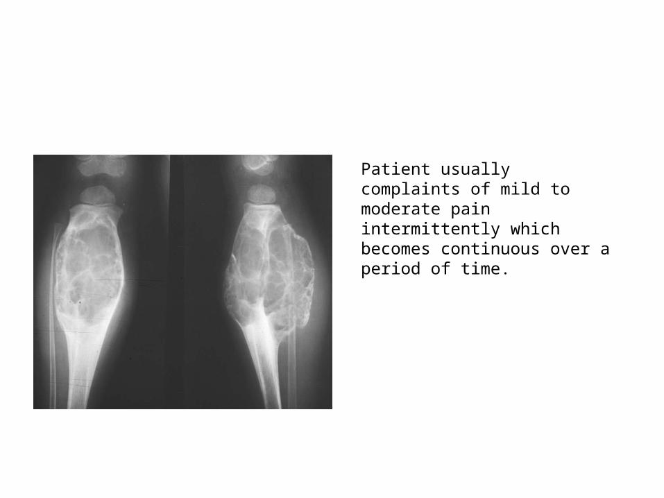
Patient usually complaints of mild to moderate pain intermittently which becomes continuous over a period of time.
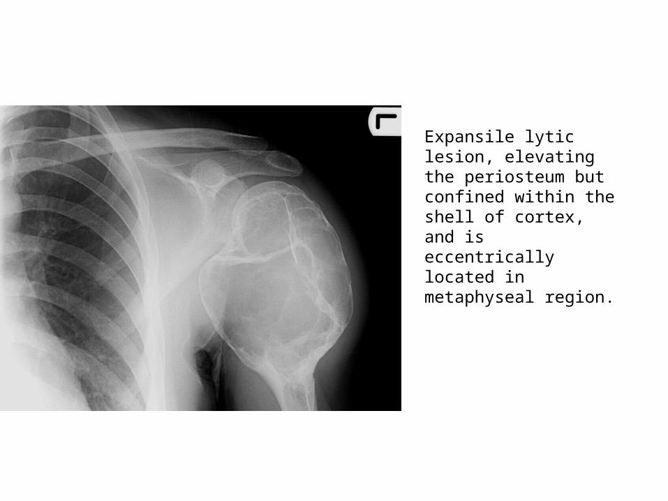
Expansile lytic lesion, elevating the periosteum but confined within the shell of cortex, and is eccentrically located in metaphyseal region.

CT - ABC
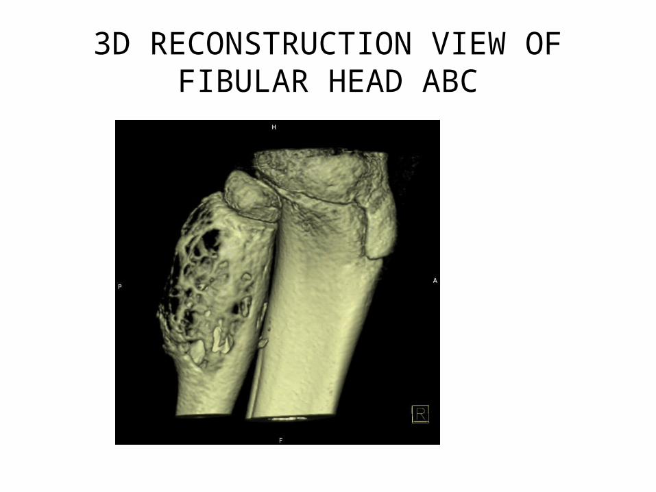
3D RECONSTRUCTION VIEW OF FIBULAR HEAD ABC

TREATMENT
• SURGICAL INTERVENTION IS MAINSTAY AND INCLUDES1. EXTENDED CURETTAGE AND GRAFTING2. MARGINAL RESECTION3. PREOPERATIVE EMBOLISATION IN LESONS OF SPINE AND
PELVIS TO REDUCE RISK OF BLEEDING.4. LOW DOSE RADIATION
• RECURRENCE RATE AFTER CURETTAGE IS 10 -20 %• RECURRENCE HAVE BEEN CO RELATED WITH Age less than 15 yrs centrally located cysts incomplete resection

Intraosseous ganglion cyst
They are intra osseous extension of ganglion of soft tissues.

Intraosseous ganglion is a benign, nonneoplastic bone lesion with histological similarity to that in soft tissue . Intraosseous ganglion contains mucoid viscous material with no epithelial or synovial lining . Peak incidence of intraosseous ganglion is in the 4th and 5th decades of life, and it is rare in children
Treatment
• Symptomatic lesion treated by local excision of overlying soft tissue and curettage.
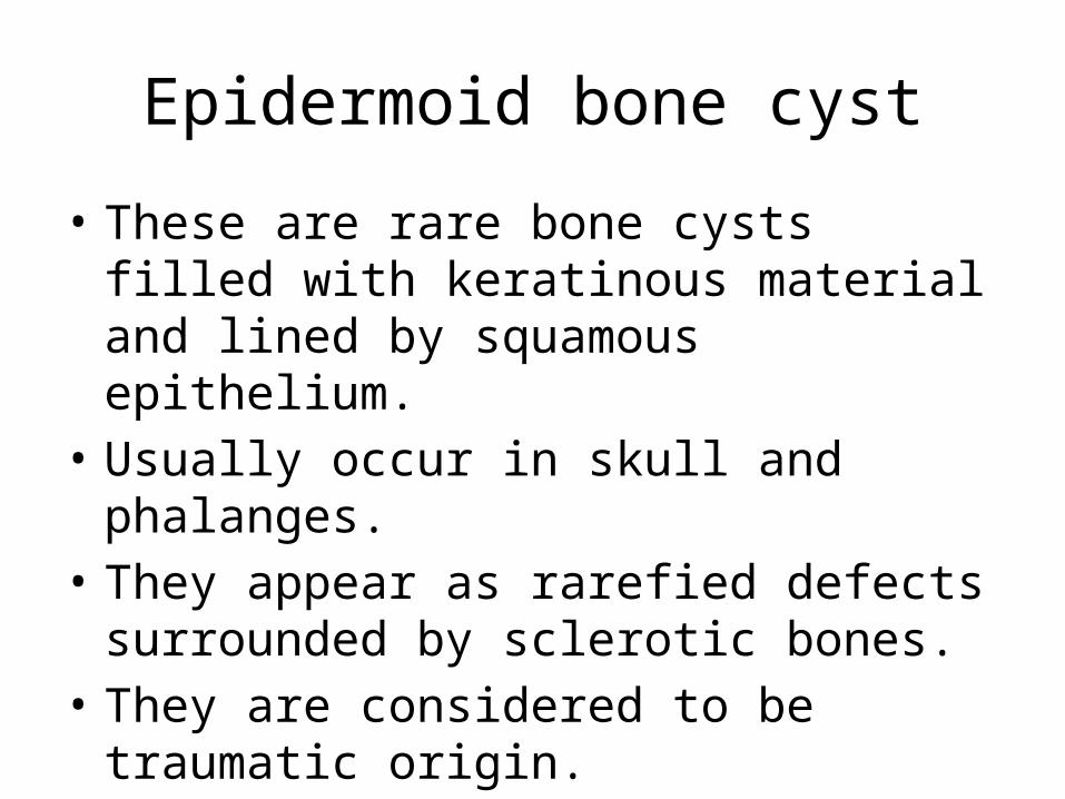
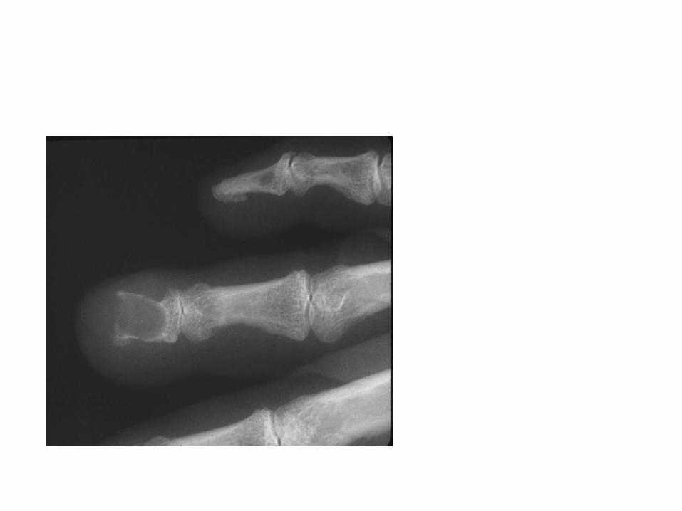
Epidermoid bone cyst
• These are rare bone cysts filled with keratinous material and lined by squamous epithelium.
• Usually occur in skull and phalanges.• They appear as rarefied defects surrounded by
sclerotic bones.• They are considered to be traumatic origin.
pathogenesis
• Not known• Theory 1 – metaplasia of cells
• Theory 2 – implantation of ectodermal cells in periosteum due to trauma. Still a topic of debate.

treatment
• Excision for smooth walled cells.• Friedlanders and sontag suggested
amputation of the affected part more wise.

THANK YOU
Related Documents