Mini-review Cyclooxygenases in cancer: progress and perspective Shan Zha b , Vasan Yegnasubramanian c , William G. Nelson a,b,c , William B. Isaacs b,c , Angelo M. De Marzo a,b,c, * a Department of Pathology, The Johns Hopkins University, Baltimore, MD, USA b Brady Urological Institute, The Johns Hopkins University, Baltimore, MD, USA c The Sidney Kimmel Comprehensive Cancer Center, The Johns Hopkins University, Baltimore, MD, USA Received 1 June 2004; accepted 7 June 2004 Abstract Aspirin has been used to control pain and inflammation for over a century. Epidemiological studies first associated a decreased incidence of colorectal cancer with the long-term use of aspirin in the early 1980s. Near the same time the first reports showing regression of colorectal adenomas in response to the non-steroidal anti-inflammatory drug (NSAID) sulindac were reported. In subsequent years, the use of other NSAIDs, which inhibit cyclooxygenase (COX) enzymes, was linked to reduced cancer risk in multiple tissues including those of the breast, prostate, and lung. Together these studies resulted in the identification of a new cancer preventive and/or therapeutic target-COX enzymes, especially COX-2. Meanwhile, the overexpression of COX-2, and less consistently, the upstream and downstream enzymes of the prostaglandin synthesis pathway, was demonstrated in multiple cancer types and some pre-neoplastic lesions. Direct interactions of prostaglandins with their receptors through autocrine or paracrine pathways to enhance cellular survival or stimulate angiogenesis have been proposed as the molecular mechanisms underlying the pro-carcinogenic functions of COX-2. The rapid development of safe and effective inhibitors targeting individual COX enzymes not only dramatically improved our understanding of the function of COX-2, but also resulted in discovery of COX independent functions of NSAIDs, providing important hints for future drug design. Here we review the fundamental features of COX enzymes, especially as related to carcinogenesis, their expression and function in both animal tumor models and clinical cancers and the proposed mechanisms behind their roles in cancer. q 2004 Published by Elsevier Ireland Ltd. Keywords: Cyclooxygenase; Cancer; Angiogenesis; Non-steroid anti-inflammation drug; Review 1. Introduction Aspirin was introduced as an anti-pyretic, anti- inflammatory and analgesic drug at the end of nineteenth century. Soon after, a family of drugs with similar properties were discovered and collectively termed non-steroidal anti-inflammatory drugs (NSAIDs). In the late 1960s work from 0304-3835/$ - see front matter q 2004 Published by Elsevier Ireland Ltd. doi:10.1016/j.canlet.2004.06.014 Cancer Letters 215 (2004) 1–20 www.elsevier.com/locate/canlet * Corresponding author. Address: Department of Pathology, The Johns Hopkins University, Room 153, Bunting-Blaustein Cancer Research Building, 1650 Orleans Street, Baltimore, MD 21231- 1000, USA. Tel.: C1-410-614-5686; fax: C1-410-502-9817. E-mail address: [email protected] (A.M. De Marzo).

Welcome message from author
This document is posted to help you gain knowledge. Please leave a comment to let me know what you think about it! Share it to your friends and learn new things together.
Transcript
Mini-review
Cyclooxygenases in cancer: progress and perspective
Shan Zhab, Vasan Yegnasubramanianc, William G. Nelsona,b,c,William B. Isaacsb,c, Angelo M. De Marzoa,b,c,*
aDepartment of Pathology, The Johns Hopkins University, Baltimore, MD, USAbBrady Urological Institute, The Johns Hopkins University, Baltimore, MD, USA
cThe Sidney Kimmel Comprehensive Cancer Center, The Johns Hopkins University, Baltimore, MD, USA
Received 1 June 2004; accepted 7 June 2004
Abstract
Aspirin has been used to control pain and inflammation for over a century. Epidemiological studies first associated a
decreased incidence of colorectal cancer with the long-term use of aspirin in the early 1980s. Near the same time the first reports
showing regression of colorectal adenomas in response to the non-steroidal anti-inflammatory drug (NSAID) sulindac were
reported. In subsequent years, the use of other NSAIDs, which inhibit cyclooxygenase (COX) enzymes, was linked to reduced
cancer risk in multiple tissues including those of the breast, prostate, and lung. Together these studies resulted in the
identification of a new cancer preventive and/or therapeutic target-COX enzymes, especially COX-2. Meanwhile, the
overexpression of COX-2, and less consistently, the upstream and downstream enzymes of the prostaglandin synthesis pathway,
was demonstrated in multiple cancer types and some pre-neoplastic lesions. Direct interactions of prostaglandins with their
receptors through autocrine or paracrine pathways to enhance cellular survival or stimulate angiogenesis have been proposed as
the molecular mechanisms underlying the pro-carcinogenic functions of COX-2. The rapid development of safe and effective
inhibitors targeting individual COX enzymes not only dramatically improved our understanding of the function of COX-2,
but also resulted in discovery of COX independent functions of NSAIDs, providing important hints for future drug design.
Here we review the fundamental features of COX enzymes, especially as related to carcinogenesis, their expression and
function in both animal tumor models and clinical cancers and the proposed mechanisms behind their roles in cancer.
q 2004 Published by Elsevier Ireland Ltd.
Keywords: Cyclooxygenase; Cancer; Angiogenesis; Non-steroid anti-inflammation drug; Review
0304-3835/$ - see front matter q 2004 Published by Elsevier Ireland Ltd
doi:10.1016/j.canlet.2004.06.014
* Corresponding author. Address: Department of Pathology, The
Johns Hopkins University, Room 153, Bunting-Blaustein Cancer
Research Building, 1650 Orleans Street, Baltimore, MD 21231-
1000, USA. Tel.: C1-410-614-5686; fax: C1-410-502-9817.
E-mail address: [email protected] (A.M. De Marzo).
1. Introduction
Aspirin was introduced as an anti-pyretic, anti-
inflammatory and analgesic drug at the end of
nineteenth century. Soon after, a family of drugs
with similar properties were discovered and
collectively termed non-steroidal anti-inflammatory
drugs (NSAIDs). In the late 1960s work from
Cancer Letters 215 (2004) 1–20
www.elsevier.com/locate/canlet
.
S. Zha et al. / Cancer Letters 215 (2004) 1–202
Samuelsson and Bergstrom revealed the prosta-
glandin synthesis pathways [1–3] and a few years
later, J.R. Vane and his colleagues identified the
therapeutic target of NSAIDs as the cyclooxygenase
(COX) enzyme [4]. The Noble Prize for Physiology or
Medicine was awarded to Drs. Vane, Samuelsson and
Bergstrom in 1982 ‘for their discoveries concerning
prostaglandins and related biologically active
substances’ [5]. Both epidemiological and random-
ized clinical trials have indicated efficacy, albeit not
uniformly, in the ability of aspirin and/or NSAIDs to
decrease colorectal cancer [6–9].
A number of epidemiological studies have
indicated that long term aspirin/NSAID use is
associated with 30–50% reduction in risk of colorectal
cancer or adenomatous polyps or death from
colorectal cancer [10]. In addition, these studies
suggest that the duration and the consistency of
NSAID use are more important than the dosage. Other
epidemiologic studies also found associations
between NSAID use and a lower death rate from
cancers of the esophagus, stomach, breast, lung,
prostate, urinary bladder and ovary [11,12].
Meanwhile, Dr. William Waddell reported the
regression of rectal polyps in a small number of
familial adenomatous polyposis (FAP) patients in
response to the NSAID sulindac [13,14]. This work
has been extended by a number of epidemiological
studies as well as clinical trials. The results from
the completed randomized double-blind placebo
controlled trial on FAP patients suggest that sulindac
and celecoxib cause adenoma regression in some
polyposis patients, and in some cases, a complete
regression is seen [15–19]. Clinical trials on other
high risk populations have generally shown a
beneficial reduction in adenoma number and/or size,
although the effects are inconsistent [9,20–24].
In young FAP patients who were entered into a
randomized clinical trial prior to the development of
colorectal adenomas, there was no significant effect of
sulindac on preventing de novo adenoma formation
[25]. In a large scale randomized clinical trial to
determine the ability of aspirin to prevent myocardial
infarction, there was no reduction in colorectal cancer
in the patients receiving aspirin in a secondary
analysis [26]. Taken together, despite the early very
promising results, currently there is not sufficient
evidence to recommend wide-spread use of any of
these agents for primary prevention of colon cancer.
More clinical trials are ongoing with aspirin, sulindac,
celecoxib and refocoxib and we await the results of
these trials to provide a more complete estimate of the
chemo-preventative value of NSAIDs.
2. Cyclooxygenase genes and enzymes
In 1988, three different groups cloned a gene
encoding cyclooxygenase, which later turned out to be
the constitutive isoform—COX-1 [27–29]. Subse-
quently, the inducible isoform of COX was discovered
and named—COX-2 [30–33]. The human gene
encoding the COX-1 enzyme (PTGS1) is located on
chromosome 9 (9q32–9q33.3), contains 11 exons and
spreads across 40 kb; its mRNA is approximately
2.8 kb [34]. The gene encoding COX-2 (PTGS2) is
located on chromosome 1 (1q25.2–25.3), contains 10
exons and encompasses 7.5 kb with a 4.5 kb transcript
[35]. Despite the difference in genomic structure and
transcript size, the proteins of both COX enzymes are
about 600 amino acids with the calculated molecular
weight as 68 kDa unmodified and about 75–80 kDa
after post-translational modifications, which mainly
consist of glycosylation [36].
Despite their similarities, the expression pattern
and regulation of these two isomers are different [37].
While there are notable exceptions, a simplified view
is that COX-1 is constitutively expressed with near
constant levels and activity in many tissues, whereas
COX-2 is an inducible or early-response gene. COX-2
expression is low or negative in most tissues;
however, a few hours after a single stimulation, the
mRNA, protein and enzymatic activity of COX-2
increase more than 10-fold and then return promptly
back to the basal level. Exceptions to this include
portions of the central nervous system (CNS),
the kidney and the seminal vesicles, which contain
constitutively high levels of COX-2. The best studied
inducers of COX-2 are bacterial lipopolysacc-
haride (LPS), pro-inflammatory cytokines-interleukin
(IL)-1b, IL-2 and tumor necrosis factor (TNF)-a[38–41]. Growth factors (e.g. epidermal growth factor
(EGF), platelet derived growth factors (PDGF)) and
some tumor promoters such as phorbol-12-myristate-
13-acetate (PMA) also stimulate COX-2 expression
[42,43]. On the other hand, anti-inflammatory
S. Zha et al. / Cancer Letters 215 (2004) 1–20 3
molecules such as corticosteroids, IL-13, IL-10 and
IL-4 suppress the expression of COX-2 [44]. Finally,
COX-1 protein can also be induced in certain cell
types by either phorbol esters or dexamethazone [37].
Therefore, the simplistic notion that COX-2 is the
inducible form and COX-1 is the constitutive form is
probably an oversimplification.
3. Functions of cyclooxygenases
Prostaglandins were first discovered in semen or in
the extract of prostate as lipid soluble compounds with
potent vasodepressor and smooth muscle-stimulating
activity. They were named based on the fact that they
were believed to be derived from the prostate [45,46].
Now it is clear that the normal human prostate itself is
not the major source of prostaglandins. The large
amounts of prostaglandins in the semen are derived
from the nearby seminal vesicles, which are one of the
most abundant sources of prostaglandins in the body.
Prostaglandins and leukotrienes compose a large
family of regulatory molecules termed eicosanoids,
which include almost all long-chain oxygenated
polyunsaturated fatty acids derived from arachidonic
acid (20:4u6) [2,47]. Prostaglandins, which are also
referred to as prostanoids, are composed of the cyclic
oxidized members of the eicosanoid family.
Prostaglandins can be produced in almost every
human cell type and act as autocrine and/or paracrine
mediators through their specific receptors. De novo
prostaglandin synthesis starts with the oxidative
cyclization of the five carbons at the center of
arachidonic acid, which is released by phospholipase
A2 (PLA2) from the cell membrane. The free
arachidonic acid is then presented to the endoplasmic
reticulum (ER) and nuclear membrane, where the
COX enzymes catalyze the rate-limiting step for
prostaglandin synthesis—the generation of the bio-
cyclic endoperoxide intermediate—prostaglandin G2
(PGG2) and the reduction to prostaglandin H2 (PGH2)
[48]. In different cell types and under different
physiological conditions, the down stream meta-
bolism of PGH2 can be dramatically different.
Prostaglandin D (PGD) synthase is usually found in
mast cells and in the brain; prostaglandin F (PGF)
synthase is expressed in the uterus; prostaglandin I
synthase (also called prostacyclin synthase, PGI) is
found in endothelial cells; thromboxane synthase is
commonly seen in platelets and macrophages; and
prostaglandin E isomerase appears in most cell types.
There are also non-enzymatic mechanisms involved
in the transformation of PGH2 into primary prosta-
glandins. In some cases, the COX enzymes and the
subsequent prostaglandin synthase(s) are coordinately
regulated. For example, during inflammatory cell
activation, macrophages increase the expression of
both COX-2 and prostaglandin E isomerase [49].
4. Structure of cyclooxygenases
COX-1 and COX-2 share the same substrates,
generate the same products, and catalyze the same
reaction using identical catalytic mechanisms.
When the X-ray crystal structures of these two
enzymes were solved, both human and murine
COX-2 could be largely superimposed on that of
COX-1, with the amino acids serving as the substrate
binding pocket and catalytic site being nearly
identical to each other. One exception with profound
implications is that the isoleucine 590 around the
substrate channel of COX-1 is replaced by valine in
COX-2 [50–52], which gives COX-2 a larger
substrate binding pocket and consequently a broader
substrate spectrum. For arachidonic acid and dihomo-
g-linolenate, COX-1 and COX-2 are equally effec-
tive, but for other fatty acids such as linoleic acid and
eicosapentaenoic acid, COX-2 is significantly more
efficient than COX-1 [53]. The isoleucine/valine
substitution is also the structural basis for the
COX-2 selective inhibitors. Co-crystals of either
COX-1 or COX-2 with its selective inhibitor showed
that the smaller valine in COX-2 allows the bulk
structure of COX-2 selective inhibitors to access the
substrate-binding site, while the larger isoleucine in
COX-1 prevents their binding [50,51]. It also explains
the different degrees of inhibition that aspirin
possesses towards COX-1 and COX-2. As an
irreversible inhibitor, aspirin acetylates serine 530 in
COX-1, completely abolishing its ability to oxidize
arachidonic acid; while after similar acetylation,
COX-2 can still oxidize arachidonic acid, but to
15R-hydroxyeicosatertraenoic acid (HETE) instead of
PGG2 [54–56]. The retention of oxygenase activity in
COX-2 has been attributed to the larger overall space
S. Zha et al. / Cancer Letters 215 (2004) 1–204
available in the COX-2 active site than that in COX-1.
Therefore, the acetylation of serine 530—critical for
controlling the configuration of prostaglandins at the
15-carbon—can better be accommodated in COX-2.
Another important structural difference between these
two enzymes is that COX-2 contains an insertion of 18
additional amino acids towards its C-terminus and is
missing 17 amino acids from its N-terminus in
comparison to COX-1 [31]. It is known that the
C-terminal insertion in COX-2 does not alter the last
four amino acids, which are believed to serve as the
ER-targeting signal for both proteins. COX-2 is
localized to both the ER and the nuclear envelope,
while COX-1 is only found in the ER. It has been
suggested that the C-terminal insertion might
contribute to the nuclear membrane localization of
COX-2 [31,57,58]. When the X-ray crystal structures
were published, the last 18 amino acids of COX-1 and
the last 30 amino acids of COX-2 were unsolved,
presumably due to the high flexibility of these regions
even in the crystalline forms [50,52]. Further
investigation is needed to elucidate the functional
significance of the different termini.
5. Genetic evidence for an association
between COX-2 and cancer
The studies from a murine model of FAP
(mice carrying APCD716) provided the first genetic
evidence for a link between COX-2 and carcinogen-
esis. When APCD716 mice were crossed with mice
containing targeted mutations that inactivate the Pgst2
gene (homozygous or heterozygous), the size and
number of small intestinal and colonic polyps,
especially the number of large polyps were reduced
in a dose-dependent manner in comparison with the
Pgst2 wide-type littermates [59]. Deletion of the gene
(Pla2g4), encoding the upstream enzyme PLA2—in
the same mouse model for colorectal cancer resulted in
a significant decrease of the size but not the number of
polyps in small intestine, and neither size or number of
polyps in the colon [60]. The authors attributed the
discrepancy in Pgst2 and Pla2g4 knockout mouse
models to the fact that the arachidonic acid might be
potentially provided by other PLA2 isoforms other
than that encoded by Pla2g4 in the colon. Genetic
disruption of Pla2g4 in another mouse model for colon
cancer—APCMin, confirmed the protective effects of
Pla2g4 deletion in the small intestine [61]. Among the
various downstream prostaglandins, PGE2 has long
been suggested as the key player for the following
reasons: (i) PGE2 concentration is increased in colon
cancer tissues where COX-2 is overexpressed [62]; (ii)
PGE2 can induce angiogenesis in vitro and increases
cellular resistance to apoptosis which fits into the
proposed mechanisms for COX-2 to promote carcino-
genesis [63]; (iii) only the prostaglandin E receptor
(Ptger) knockout mice, but not other single prosta-
glandin receptor (prostaglandin D, F, I and thrombox-
ane receptor) knockout mice, show a significant
decrease in the number of aberrant crypt foci when
compared with the wild type controls (Table 1). There
are four subtypes of prostaglandin E receptors (EP
1–4), whose genes are designated Ptger1–4.
The results on Ptger1 and Ptger2 knockouts were not
consistent among different colon cancer animal
models. Sonoshita et al. reported that knockout of
Ptger2, but not Ptger1 decreased the number and size
of polyposis in APCD716 mouse through blocking
angiogenesis [64]. The decrease is parallel to the Pgst2
knockout [59,64]. They also showed that the
expression of Ptger2, but not Ptger1, 3 or 4 was
elevated in polyps with reference to normal tissue from
the small intestine and colon. But in the azoxymethane
(AOM) induced rodent colon cancer model, Ptger1 but
not Ptger2 knockout mice showed decreased numbers
of aberrant crypt foci, which could also be recapitu-
lated in the Min mouse model by the Ptger1
antagonist—ONO-8711 [65]. Knockout of Ptger3
showed no effect in either the AOM-induced colon
cancer model or APCD716 models [65,66]. Ptger4 was
implicated as a key player in the AOM induced colon
cancer model (not tested in APCD716) and the Ptger4
antagonist—ONO-AE2-227 decreased the polyp num-
ber, especially the number of polyps larger than
1.5 mm in APCMin mice [67]. Pai et al. reported that
in addition to acting on its own receptor, PGE2 could
also activate the epidermal growth factor receptor,
providing another potential mechanism for the tumor
promotion effect of COX enzymes [68]. These results
not only further support the role of COX-2 as a tumor
promoter in the intestine, but also point to PGE2 as the
key mediator of the COX-2 related susceptibility to
colon cancer. These findings suggest the PGE
isomerase might be a more specific target for colorectal

Table 1
Prostaglandins and their implication in cancer
Prostaglandin Receptor/G protein Second messenger Implication in cancer
PGD2 DP/Gs [cAMP DP1 null do not change the number of AOM induced
aberrant crypt foci
PGE2 EP1/unknown [Ca2C EP1-null-Ynumber of AOM induced aberrant
crypt foci
EP1 antagonist-Ythe number of polyps in APCMin
EP1-null-do not change the number of polyps
APCD716 mice
EP2/Gs [cAMP EP2 null-Ynumber of large polyps in APCD716 mice
EP2 null-do not change AOM induced aberrant
crypt foci
EP3/Gs or Gi [cAMP or YcAMP EP3 null do not change polyps number in APCD716
and AOM induced aberrant crypt foci
EP4/Gs [cAMP EP4 null-YAOM-induced colon cancer
EP4 antagonist-Ynumber of large polyps in APCMin
EP4-null-do not change polyp number
in APCD716 mice
Promotes proliferation through Akt pathway
PGF2 FP/Gq PI response Counteracts indomethacin to restore DMBA/TPA
induced skin tumors
PGI2 IP/Gs, Gq [cAMP, PI response Overexpression of PGI synthase inhibits neovacular
formation in colon cancer xenograft model
Transgenic over expression of PGI syn protect mice
from carcinogen induced lung cancer
TXA2 TPa/Gi, Gq YcAMP, [Ca2C TP agonists-restore neovascular formation blocked
by COX-2 inhibitors
TPb/Gs, Gq [cAMP, [Ca2C Overexpression of TXA synthase promotes vascular
formation in colon cancer xenograft model
S. Zha et al. / Cancer Letters 215 (2004) 1–20 5
cancer prevention in comparison with COX-2. Various
PGE receptor antagonists have been developed and are
being tested in animal models.
Is constitutive expression of COX-2 sufficient to
transform cells? When the Pgst2 gene was placed
downstream of the murine mammary tumor virus
(MMTV) promoter, its expression was induced in the
mammary gland during pregnancy and lactation.
This high level of COX-2 expression causes mam-
mary gland hyperplasia, carcinoma and even-
tually metastatic breast cancer in multiparous mice
but not virgin mice [69]. Pregnancy and lactation
associated COX-2 expression was strong in mammary
gland epithelial cells and weak in surrounding stromal
cells, which correlated with increased PGE2 and PGF2
levels. Interestingly, the increased expression of the
anti-apoptotic molecule—Bcl-2 and decreased
expression of its counterparts—Bax and Bcl-xL
were only seen in tumor tissues, but not in the
adjacent normal from the transgenic mouse. It is not
clear whether this reflects an effect of transformation
or actually contributes to the transformation. Given
that the surrounding normal mammary gland
epithelial cells with COX-2 expression did not have
the altered expression of Bax and Bcl-xL, changing of
apoptotic balance might not be a direct consequence
of COX-2 overexpression.
Recently two transgenic mouse models have been
generated to study the role of COX-2 in skin tumor
initiation and promotion. Pgst2 cDNA was inserted
downstream of the keratin 5 and keratin 14 promoters
to achieve constitutive COX-2 expression in the basal
region of the interfollicular epidermis and the
pilosebaceous unit [70,71]. Both transgenic strains
developed significant alopecia, which was success-
fully corrected in the K14.COX2 mice by adminis-
tration of the COX-2 specific inhibitor—celecoxib
(not tested in K5.COX2 mice). Some K5.COX2 mice
displayed spontaneous hyperplasia in the scale
epidermis of the tail with focal signs of dysplasia.
S. Zha et al. / Cancer Letters 215 (2004) 1–206
No spontaneous hyperplasia was reported from
K14.COX2 mice. When skin tumors were initiated
in the K14.COX2 mice by topical application of
7,12-dimethylbenz [a] anthracene (DMBA) and
subsequently promoted by PMA, the tumor incidence
and multiplicity decreased dramatically on two
different genetic backgrounds. This surprising result
was bolstered by the administration of celecoxib
before DMBA induction, which increased the tumor
incidence in K14.COX2 mice, further suggesting a
protective role of COX-2 in the DMBA/PMA tumor
model. Different results were obtained, however, with
the K5.COX-2 mice. While these mice also develop
alopecia, they are prone to develop hyperplasia and
focal dysplasia in tail skin [72]. In tumor initiation-
promotion experiments, these mice readily developed
tumors (squamous papillomas, squamous carcinomas,
and sebaceous gland adenomas), in response to
DMBA alone—they did not require subsequent
PMA administration, as needed for tumor develop-
ment in wild type mice [71]. However, there was a
change in the proportion of the different tumor types
in the DMBA alone induction experiments, with a
higher proportion than usual of sebaceous adenomas.
Administration of celecoxib before DMBA appli-
cation or between DMBA and PMA applications both
decreased the tumor multiplicity in comparison to
control group, with no difference between these two
treatment schedules. The authors of this study con-
cluded that these data support a role of COX-2 in tumor
promotion, but not in initiation [71]. The discrepant
results found in these two different transgenic mouse
models are difficult to reconcile. The different promo-
ters (keratin 5 vs. keratin 14) used and the diverse
genetic backgrounds might both contribute to the
discrepancies in the results, given that strain-dependent
responses by skin tumors to COX-2 inhibitors have
been reported previously [73,74]. Previous studies
showed that the non-specific COX inhibitor, indo-
methacin, was able to reduce the multiplicity of tumors
induced by DMBA/PMA; furthermore, topical appli-
cation of PGF2 but not PGE2 counteracted indometha-
cin [74]. Taken together, the effects of COX-2 on skin
carcinogenesis in mouse models is certainly unclear at
this time. Some experiments clearly show that
expression or overexpression of COX-2 promotes
carcinogenesis, and that inhibition of COX-2 prevents
tumorigenesis. By contrast, other experiments, not only
refute this concept, but actually strongly suggest the
opposite—COX-2 expression may protect skin from
carcinogenesis.
In an attempt to dissect the contribution of COX-2
from different cell types, Williams et al., implanted
COX-1 and -2 positive Lewis lung carcinoma (LCC)
grafts into genetically compatible C57/BL6 mice that
were either wild type, or containing targeted disrup-
tions of either Pgts2 or Pgts1. Seven days after the
engraftment, LLC tumors grown in the Pgts2K/K
hosts started to show a statistically significant smaller
size in comparison with the tumors in either wild type
or Pgts1K/K hosts and this decrease correlated with
decreased levels of VEGF and vascular density in the
tumors [75]. These results implicated non-tumori-
genic host cells as potential key factors of COX-2
mediated tumor growth.
Disruption of COX-2 itself or its upstream or
downstream genes by means of gene knockout is not
sufficient to stop the initiation of polyps in either
APCD716 mice or AOM induced colon cancer model,
which suggests that COX-2 and its related pathways
serve as modulators for tumor growth, but not single
agent initiators. In keeping with this, in APCD716 mice
COX-2 expression only becomes obvious when the
size of polyps is larger than 1 mm in diameter and
positive staining cells are mostly stroma cells, not the
epithelial cells in the polyps. Furthermore, COX-2
expression in APCD716 mice correlated with the
expression of angiogenesis factors (e.g. VEGF and
bFGF). Together these results suggested that COX-2
and the related prostaglandin pathways affect colon
polyp growth beyond 1 mm through modulating
angiogenesis. In contrast to this, in all the transgenic
mouse models of skin carcinogenesis mentioned
above, the ectopic expression of COX-2 was in the
epithelial cell components. Yet spontaneous tumor
formation is only seen in the MMTV driven COX2
expressing mammary glands of multiparous mice, but
in none of the K14.COX2 or K5.COX2 mice. This
suggests that the expression level of COX-2 needed to
transform cells is very high and other initiation factors
are most likely needed to achieve the transformation.
Second, the alteration in the spectrum of tumor types
occurring in K5.COX2 mice treated with DMBA/PMA
or DMBA alone suggests different sensitivity to COX-
2 mediated tumor promotion in different cell popu-
lations. Finally, high levels of COX-2 protein are not

S. Zha et al. / Cancer Letters 215 (2004) 1–20 7
sufficient to induce cancer when physiologically
expressed, such as in the seminal vesicles, which
virtually never develop cancer.
While most of the focus has been on COX-2,
APCMin mice carrying inactivated Pgts1 genes
also had a 80% reduction of tumor multiplicity
in comparison to the Pgts1C/C littler mates [76].
In addition, inhibitors that preferentially block COX-1
(e.g. piroxican) have protective effects against colon
cancer in both animal experiments and epidemiologi-
cal studies. On one hand, these data reinforce the role
of prostaglandins as common mediators for COX
enzymes related to tumor promotion, yet also raise
the question regarding how much each isoform
contributes to tumor promotion.
When examining the data regarding the function of
cyclooxygenase genes in mouse models, some of the
surprising phenotypes of these mice are of interest.
First, despite the long standing belief of COX-2’s
primary function in the inflammatory response, Ptgs2
knockout mice show a normal response to acute
inflammation induced by arachidonic acid or PMA in
an ear-edema test [77,78]. Second, although inhibition
of COX-1, not COX-2, has been proposed to be
responsible for the renal deficiency associated with
using non-specific COX inhibitors, especially aspirin,
Ptgs1 knockout mice showed no defect in kidney
function, unless the kidneys were compromised
by other disease (e.g. diabetes, hypertension etc.)
[78,79]. However, Ptgs2 knockout mice showed
unexpected developmental abnormalities in the
kidney and eventually led to reduced life span [80].
Third, long term use of aspirin causes gastric ulcer
formation and bleeding in patients due to the
inhibition of COX-1, which is a protective factor for
the gastric mucosal layer. But Ptgs1 knockout mice
showed no spontaneous gastric erosion or injury [77].
One possible explanation is that the early loss of
COX-1 might cause an adaptive increase of other
protective mechanisms (e.g. calcitonin gene related
peptide, NO), which is different from losing COX-1
function due to NSAID intake in adulthood. This is
not to say that the knockout models have no features
consistent with presumed functions of COX-1 or
COX-2. The female reproductive deficiency and pain
sensation loss are consistent with the predictions
based on the known functions of COX-2 [77,81].
6. Proposed mechanism for the role
of COX-2 in carcinogenesis
6.1. Role of COX-2 in angiogenesis
The ability to induce angiogenesis is essential for
most solid tumors to grow beyond 2–3 mm in
diameter. Angiogenesis may also provide an import-
ant path for metastasis. Tumor angiogenesis, as with
other neovascular formations, includes destabilization
of pre-existent blood vessels, proliferation of vascular
endothelial cells, invasion by endothelial cells into the
extracellular matrix (ECM) and finally the migration
and positioning of endothelial cells. One of the
earliest observations regarding COX-2 and angiogen-
esis was made while studying the anti-tumor effect of
existing COX inhibitors. In a study published on
1997, Seed et al. noticed that a non-selective COX
inhibitor, diclofenac suppressed the growth of COX-2
positive colon-26 cells in nude mice through blocking
angiogenesis [82]. Subsequently, studies on corneal
models indicated that COX-2 specific inhibitors block
new vessel formation and this effect is reversed by
adding a TXA2 receptor agonist [83]. Numerous
studies showed co-localization of angiogenesis
factors, such as VEGF, PDGF, basic fibroblast growth
factor (bFGF) and tumor growth factor-b (TGF-b)
with COX-2 by immunohistochemical staining in
different cancer types [84]. In breast and cervical
cancers, enhanced COX-2 expression has been further
associated with increased micro-vascular density
(MCD) and with poor prognosis [85,86].
To further explore COX-2 related angiogenesis,
using colon cancer cell lines co-cultured with vascular
endothelial cells Tsujii et al. demonstrated that COX-2
supported angiogenesis at multiple steps both directly
and indirectly [87]. First, COX-2 up-regulation leads
to prostaglandin production. Since each prostaglandin
has distinct roles for angiogenesis, the profile is
important to determine the end effects on different
cell types and under different circumstances [88].
For example, TXA2 is particularly efficient at
promoting endothelial cell migration [83]. Second,
overexpression of COX-2 in tumor cells directly
stimulates the production of angiogenic factors from
these cells. Overexpression of COX-2 in a colon
cancer cell line induced the production of VEGF,
PDGF, bFGF and TGF-b. Through these angiogenesis
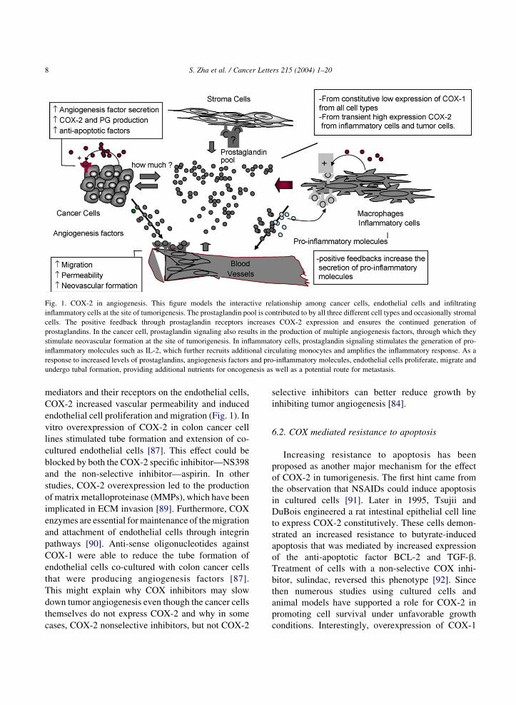
Fig. 1. COX-2 in angiogenesis. This figure models the interactive relationship among cancer cells, endothelial cells and infiltrating
inflammatory cells at the site of tumorigenesis. The prostaglandin pool is contributed to by all three different cell types and occasionally stromal
cells. The positive feedback through prostaglandin receptors increases COX-2 expression and ensures the continued generation of
prostaglandins. In the cancer cell, prostaglandin signaling also results in the production of multiple angiogenesis factors, through which they
stimulate neovascular formation at the site of tumorigenesis. In inflammatory cells, prostaglandin signaling stimulates the generation of pro-
inflammatory molecules such as IL-2, which further recruits additional circulating monocytes and amplifies the inflammatory response. As a
response to increased levels of prostaglandins, angiogenesis factors and pro-inflammatory molecules, endothelial cells proliferate, migrate and
undergo tubal formation, providing additional nutrients for oncogenesis as well as a potential route for metastasis.
S. Zha et al. / Cancer Letters 215 (2004) 1–208
mediators and their receptors on the endothelial cells,
COX-2 increased vascular permeability and induced
endothelial cell proliferation and migration (Fig. 1). In
vitro overexpression of COX-2 in colon cancer cell
lines stimulated tube formation and extension of co-
cultured endothelial cells [87]. This effect could be
blocked by both the COX-2 specific inhibitor—NS398
and the non-selective inhibitor—aspirin. In other
studies, COX-2 overexpression led to the production
of matrix metalloproteinase (MMPs), which have been
implicated in ECM invasion [89]. Furthermore, COX
enzymes are essential for maintenance of the migration
and attachment of endothelial cells through integrin
pathways [90]. Anti-sense oligonucleotides against
COX-1 were able to reduce the tube formation of
endothelial cells co-cultured with colon cancer cells
that were producing angiogenesis factors [87].
This might explain why COX inhibitors may slow
down tumor angiogenesis even though the cancer cells
themselves do not express COX-2 and why in some
cases, COX-2 nonselective inhibitors, but not COX-2
selective inhibitors can better reduce growth by
inhibiting tumor angiogenesis [84].
6.2. COX mediated resistance to apoptosis
Increasing resistance to apoptosis has been
proposed as another major mechanism for the effect
of COX-2 in tumorigenesis. The first hint came from
the observation that NSAIDs could induce apoptosis
in cultured cells [91]. Later in 1995, Tsujii and
DuBois engineered a rat intestinal epithelial cell line
to express COX-2 constitutively. These cells demon-
strated an increased resistance to butyrate-induced
apoptosis that was mediated by increased expression
of the anti-apoptotic factor BCL-2 and TGF-b.
Treatment of cells with a non-selective COX inhi-
bitor, sulindac, reversed this phenotype [92]. Since
then numerous studies using cultured cells and
animal models have supported a role for COX-2 in
promoting cell survival under unfavorable growth
conditions. Interestingly, overexpression of COX-1
S. Zha et al. / Cancer Letters 215 (2004) 1–20 9
or just simply adding PGE2 into the culture medium
could also increase the resistance to apoptosis. These
results suggested that increased prostaglandin pro-
duction itself might account for the resistance to
apoptosis [93,94]. COX-2 but not COX-1 is usually
upregulated in tumors. Multiple NF-kB binding sites,
Sp-1 sites and a cAMP-response element are located
in the PGTS2 promoter and enhancer region, which
provide target DNA binding sites for transcription
factors to rapidly induce mRNA expression under
stress conditions. These features are not present in
the PGTS1 gene. The COX-2 transcript also contains
multiple repeats of a sequence within its 3 0 un-
translated region (AUUUA) that mediates rapid
mRNA degradation [95].
The notion that the anti-apoptotic effects of
selective or non-selective COX inhibitors are always
mediated through the COX enzymes themselves has
been challenged recently. Given that there are now
known COX-2 independent functions (see below) of
those inhibitors, it is not clear if inhibition of COX-2
enzymatic function alone is responsible for the
increased apoptosis in each case. It will be interes-
ting to test the effect of NSAIDs on Ptgs2K/K and
Ptgs1K/K animal tumor models or even double
knockouts, such as APCMin/K Ptgs2K/K compound
mice, to tease out the COX independent function of
NSAIDs. Recently Song et al. showed that cells
lacking PGTS1 or PGTS2 were viable and sensitive to
celecoxib-induced apoptosis. In addition, a derivative
of celecoxib, which is incapable of inhibiting COX-2,
also induced apoptosis in these cells at a similar
concentration [96].
7. Expression of COX enzymes in human normaltissues and in cancer
7.1. Expression in normal tissues
Although COX-2 protein is undetectable by
immunohistochemistry in many human tissues
under normal physiological conditions, there are
several known exceptions. The seminal vesicles are
known to have the high levels of constitutive
expression of COX-2. PGE2 and its 19-hydroxy
metabolites are the major components of primate
semen [97]. COX-2 is also constitutively expressed
in the kidney with positive staining in glomeruli and
small blood vessels. The limited evidence on human
subjects suggests that COX-2 is involved in
sodium regulation and kidney perfusion under
stress, but not in maintaining basal renal blood
flow [98,99]. The CNS contains both constitutive
and inducible COX-2 expression in both neuronal
and non-neuronal cells in the cortex, hippocampus,
hypothalamus and spinal cord, where COX-2 is
involved in the establishment of pain sensation and
body temperature control [100]. COX-2 is also
expressed in ovarian follicles upon gonadotrophin
stimulation, in uterine epithelial cells and surround-
ing stromal cells at the site of blastocyst attachment
during implantation and decidualization [81].
7.2. Colorectal cancer
In 1994, Eberhart et al. first reported COX-2
overexpression in human colon cancer, followed by
two other groups in the next year [101–103]. In their
papers, they described that COX-1 expression was
weak, universal and unchanged in both normal and
cancerous colon, while COX-2 expression was only
seen in tumors. COX-2 overexpression was also
reported in the tumors generated from APCMin,
APCD716 and the AOM-induced colon cancer models
[59,104]. These results in combination with the
encouraging information from APCMin or D716
Ptgs2K/K mice have bolstered motivation for clinical
trials on COX-2 selective inhibitors for colon cancer
prevention. There are still, however, several areas that
remain somewhat unclear. First, the percentage of
COX-2 positive cells among clinical colon cancer
samples tested varied from 40 to 100% between
different studies. Even though most studies reported
that colon cancers occurring in FAP patients often
express COX-2, there was a great deal of variation
among sporadic cases. Second, it is not clear at what
time during carcinogenesis COX-2 expression is
induced and how it changes during tumor progression.
In general COX-2 overexpression has been con-
sidered to be an early event in colon cancer
development, which correlates well with the
prophylactic effect of NSAIDs. But how early it is
and its temporal relationship with other early events,
in particular the loss of the wild type APC allele, is
undetermined. While studying APCD716/C mice,
S. Zha et al. / Cancer Letters 215 (2004) 1–2010
Oshima et al. reported that COX-2 expression was
only seen in the large established adenomas, not in the
uninvolved colon nor in the adenomas smaller than
2–3 mm diameter [59], while all the adenomas
genotyped had already lost the wild type allele of
APC. However, COX-2 upregulation was described in
uninvolved colon epithelium from Min mice [104].
In the clinical setting, the distal non-involved polyps
from FAP patients showed minimal COX-2 staining,
but the cancer from corresponding cases showed
strong staining for COX-2. Increased COX-2 staining
correlated with larger polyp size and progression to
invasive carcinomas as well [105,106]. Third, the
actual cell-type expressing COX-2 within colon
cancer is largely debatable. Many published studies
suggested that the carcinoma cells themselves express
COX-2, especially in the early studies. Others
suggested that most of the expression was found in
infiltrating macrophages within the tumors [107,108].
Other cancer types expression of COX-2 by vascular
endothelial cells [109], fibroblasts [110] and smooth
muscle cells around the cancer, and even neuroendo-
crine cells [111] has all been reported. Oshima et al.
replaced one allele of the Ptgs2 gene with the bacterial
b-galactosidase (lacZ) to generate gene knockout
mice in which the lacZ expression was under the
control of endogenous Ptgs2 promoter. When
the mice were crossed with APCD716 mice, only the
interstitial cells with large ovoid and light stained
nuclei were lacZ positive, but the epithelium itself
was negative [59]. Although it was not directly
shown, the identity of many of these cells was
consistent with that of lamina propia macrophages.
This is also consistent with the result from studying
clinical samples in which the vast majority of strong
COX-2 immunoreactivity was present in the lamina
propia macrophages directly subjacent to the surface
adenomatous epithelial cells [107]. Genetic
differences between the study groups, artifacts
introduced during sample handling and storage, and
variations between the antibodies and staining
protocols all could potentially lead to the discrepan-
cies. The generation of tissue and cell-type specific
PGTS2 knockout mice might provide some insights
regarding these questions, and shed some light on
either paracrine or autocrine mechanisms contributing
to tumorigenic function of COX-2.
7.3. Breast cancer
Increased prostaglandin concentration in breast
cancer, especially PGE2 and TxA2 was reported in
the early 1980s [112]. Long-term use of NSAIDs has
also been associated with reduced risk of breast cancer
[113]. In the initial study, Kargman et al. did not
find expression of COX-2 in any of the three breast
tumor/normal pairs by immunohistochemistry, but
they did detect significant expression of COX-2 in
colon cancer samples [102]. In 1998, the first study that
focused on COX-2 expression in breast cancer was
published using both immunohistochemistry and
Western blotting. Only two out of the forty-four
cases studied had strong, definitive COX-2 expression,
mainly in the tumor epithelial cells. Meanwhile among
these cases, thirty of them had elevated COX-1
expression, but mainly in the stromal cells [114].
In another study, Costa et al. reported that COX-2 was
expressed in eight out of forty-six carcinomas studied,
and the expression of COX-2 staining correlated with
microvessel density, lymph node metastasis, apoptotic
index, and shorter disease-free survival time [115].
Furthermore, Half et al. reported COX-2 expression in
the epithelial cells of 43% of invasive breast cancers,
63% of ductal carcinoma in situ and 80% normal
appearing breast tissues that were adjacent to cancer
[116]. RT-PCR revealed an average ninefold increase
of COX-2 mRNA in cancer vs. proximal normal
tissues. From this, the authors proposed that COX-2
upregulation might be an early event in mammary
gland tumorigenesis, but the continued expression
might become less important after an invasive tumor
was formed.
7.4. Prostate cancer
The expression and function of COX-2 in
prostate tissues and prostate cancer has been the
subject of multiple reports [117–122]. In general the
results of these studies suggest that COX-2
expression in normal prostate tissue is either weak
or negative and prostate cancer tissue has an
elevated level of COX-2 protein. Based upon these
data, it was hypothesized that that the effects of
NSAIDs on prostate cancer are mediated by
inhibition of the enzymatic activity of COX-2 in
the prostate cancer cells. COX-2 expression was not
S. Zha et al. / Cancer Letters 215 (2004) 1–20 11
seen in the normal prostatic cells in mice, but
appeared in prostate tumors from TRAMP mice—a
probasin-SV40 large T antigen transgenic prostate
cancer model [123], where the established tumors
are largely neuroendocrine in phenotype. Never-
theless, a consensus has not been reached regarding
expression of COX-2 in prostate cancer. A recent
study from our group confirmed that COX-2
expression is very low or undetectable in the normal
prostate [124]. However, in contrast to the previous
reports, we found that the expression of COX-2 was
not elevated in prostatic intraepithelial neoplasia—
the proposed precursor lesions, or in established
prostate cancers studied (nZ144 cases) [124]
(Fig. 2). In limited cases, when staining for
COX-2 was observed in prostate cancer, the extent
of positive staining did not correlate with estab-
lished clinical and/or pathological risk factors—
Gleason score or pathological stage. By contrast to
the neoplastic tissue, we did find consistent
expression of COX-2 protein in proliferative inflam-
matory atrophy lesions, which have been proposed
as an important etiological factor for prostate cancer
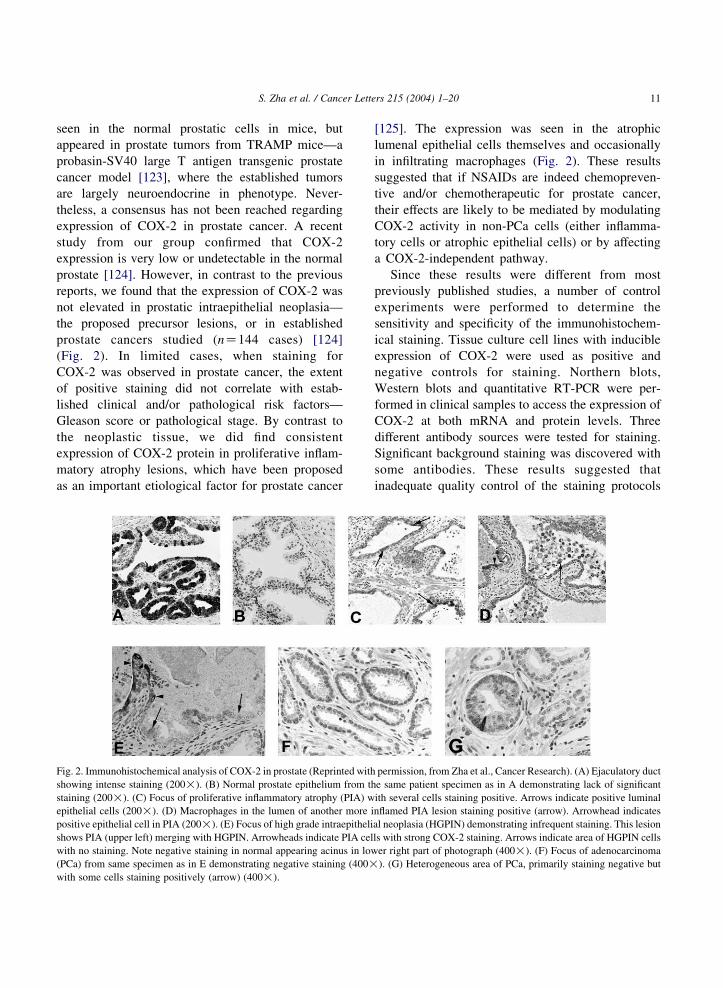
Fig. 2. Immunohistochemical analysis of COX-2 in prostate (Reprinted with
showing intense staining (200!). (B) Normal prostate epithelium from th
staining (200!). (C) Focus of proliferative inflammatory atrophy (PIA) w
epithelial cells (200!). (D) Macrophages in the lumen of another more i
positive epithelial cell in PIA (200!). (E) Focus of high grade intraepitheli
shows PIA (upper left) merging with HGPIN. Arrowheads indicate PIA cel
with no staining. Note negative staining in normal appearing acinus in low
(PCa) from same specimen as in E demonstrating negative staining (400!
with some cells staining positively (arrow) (400!).
[125]. The expression was seen in the atrophic
lumenal epithelial cells themselves and occasionally
in infiltrating macrophages (Fig. 2). These results
suggested that if NSAIDs are indeed chemopreven-
tive and/or chemotherapeutic for prostate cancer,
their effects are likely to be mediated by modulating
COX-2 activity in non-PCa cells (either inflamma-
tory cells or atrophic epithelial cells) or by affecting
a COX-2-independent pathway.
Since these results were different from most
previously published studies, a number of control
experiments were performed to determine the
sensitivity and specificity of the immunohistochem-
ical staining. Tissue culture cell lines with inducible
expression of COX-2 were used as positive and
negative controls for staining. Northern blots,
Western blots and quantitative RT-PCR were per-
formed in clinical samples to access the expression of
COX-2 at both mRNA and protein levels. Three
different antibody sources were tested for staining.
Significant background staining was discovered with
some antibodies. These results suggested that
inadequate quality control of the staining protocols
permission, from Zha et al., Cancer Research). (A) Ejaculatory duct
e same patient specimen as in A demonstrating lack of significant
ith several cells staining positive. Arrows indicate positive luminal
nflamed PIA lesion staining positive (arrow). Arrowhead indicates
al neoplasia (HGPIN) demonstrating infrequent staining. This lesion
ls with strong COX-2 staining. Arrows indicate area of HGPIN cells
er right part of photograph (400!). (F) Focus of adenocarcinoma
). (G) Heterogeneous area of PCa, primarily staining negative but
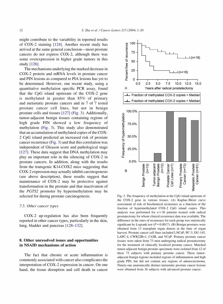
Fig. 3. The frequency of methylation at the CpG island upstream of
the COX-2 gene in various tissues. (A) Kaplan–Meier curve
assessment of risk of biochemical recurrence as a function of the
fraction of hypermethylated COX-2 CpG island copies. This
analysis was performed for nZ36 patients treated with radical
prostatectomy for whom clinical recurrence data was available. The
difference in the rates of recurrence for each group was statistically
significant by Logrank test (PZ0.0017). (B) Benign prostates were
S. Zha et al. / Cancer Letters 215 (2004) 1–2012
might contribute to the variability in reported results
of COX-2 staining [124]. Another recent study has
arrived at the same general conclusion—most prostate
cancers do not express COX-2, although there was
some overexpression in higher grade tumors in this
study [126].
The mechanisms underlying the marked decrease in
COX-2 protein and mRNA levels in prostate cancer
and PIN lesions as compared to PIA lesions has yet to
be determined. However, one recent study, using a
quantitative methylation specific PCR assay, found
that the CpG island upstream of the COX-2 gene
is methylated in greater than 85% of primary
and metastatic prostate cancers and in 7 of 7 tested
prostate cancer cell lines, but not in benign
prostate cells and tissues [127] (Fig. 3). Additionally,
tumor-adjacent benign tissues containing regions of
high grade PIN showed a low frequency of
methylation (Fig. 3). This study also demonstrated
that an accumulation of methylated copies of the COX-
2 CpG island predicted an increased risk of prostate
cancer recurrence (Fig. 3) and that this correlation was
independent of Gleason score and pathological stage
[127]. These data suggest that DNA methylation may
play an important role in the silencing of COX-2 in
prostate cancers. In addition, along with the results
from the transgenic K14.COX2 mice suggesting that
COX-2 expression may actually inhibit carcinogenesis
(see above description), these results suggest that
maintenance of COX-2 may be protective against
transformation in the prostate and that inactivation of
the PGTS2 promoter by hypermethylation may be
selected for during prostate carcinogenesis.
7.5. Other cancer types
COX-2 up-regulation has also been frequently
reported in other cancer types, particularly in the skin,
lung, bladder and pancreas [128–132].
obtained from 13 transplant organ donors at the time of organharvest. Prostate cancer cell lines included LNCaP, PC-3, DU-145,
LAPC-4, CWR22Rv1, C42B, and VCaP. Primary prostate cancer
tissues were taken from 73 men undergoing radical prostatectomy
for the treatment of clinically localized prostate cancer. Matched
tumor-adjacent benign prostate specimens were isolated from 12 of
these 73 subjects with primary prostate cancer. These tumor-
adjacent benign regions included regions of inflammation and high
grade PIN, but did not contain any regions of adenocarcinoma.
Ninety-one anatomically distinct metastatic prostate cancer lesions
were obtained from 36 subjects with advanced prostate cancer.
8. Other unresolved issues and opportunities
in NSAID mechanisms of action
The fact that chronic or acute inflammation is
commonly associated with cancer also complicates the
interpretation of COX-2 expression in cancer. On one
hand, the tissue disruption and cell death in cancer

S. Zha et al. / Cancer Letters 215 (2004) 1–20 13
recruit pro-inflammatory cells and lead to inflam-
mation. On the other hand, some types of infections or
chronic inflammation are causative for the initiation of
certain cancers, such as chronic hepatitis, chronic
gastritis and chronic ulcerative colitis. Prostaglandins
generated as a result of COX-2 overexpression can also
act as paracrine as well as autocrine growth regulators
(Fig. 4). Prostaglandin receptors are expressed in most
endothelial cells, macrophages, stroma and epithelial
cell types. It is known that at least some prostaglandin-
receptor interactions (e.g. PGE2–PTGER2) can send
positive feedback signals to increase COX-2 mRNA
levels. If this is the case, regardless of the initial trigger,
once COX-2 expression begins, prostaglandins could
mediate a wave of COX-2 expression not only in
cancer cells but also in the surrounding stroma,
macrophages and endothelial cells. At any given
time, one particular cell or cell type may or may not
express COX-2, but specific prostaglandins may be
present. This may explain why prostaglandin level
elevation is relatively consistently observed between
studies. Thus it may be very difficult to separate COX-2
expression caused by inflammation and that caused by
transformation. If tracking the expression of COX-2
longitudinally in a particular cell type becomes
possible, it would clarify some of the confusion. The
development of tissue-specific COX-2 knockouts
would be an excellent tool to study the effects of
COX-2 expression on the initiation and progression
of cancer. Macrophage-specific loss of COX-2
expression would be an especially powerful way to
address the relationship between inflammation and
cancer.
In tissue culture settings, NSAIDs induce apoptosis
in multiple tumor cell lines and suppress the expression
of angiogenesis factors [133]. However, the ability of
NSAIDs to induce apoptosis does not always correlate
with their ability to inhibit the COX enzymes.
Therefore several COX independent mechanisms
have been proposed in the past years. The first hint
came from the study of sulindac metabolites. Sulindac
is usually given as the parental drug and it is
metabolized to sulindac sulfide (an active COX
inhibitor) and sulindac sulfone (not an inhibitor of
COX). But both metabolites induced apoptosis with
similar efficiency in cell culture models [134,135].
This result indicated the existence of a COX
independent mechanism of apoptosis induction.
Recently Song et al. generated PC3 prostate cancer
cells with varying levels of COX-2 protein expression.
The sensitivity to apoptosis induced by both celecoxib
and its non-COX-2 inhibiting derivatives was similar
regardless of the levels of COX-2 protein, which
support the COX independent function of NSAIDs,
even the COX-2 selective groups [96].
Peroxisome proliferation activating receptors
(PPARs) could serve as the intracellular receptors
for some prostaglandins as well as some NSAIDs
[136]. Reduced PPARg and over activation of
PPARd/b have been associated with colorectal cancer.
He et al. suggested that sulindac could interfere with
the DNA-binding of PPARd/b, and other groups
proposed the possibility for NSAIDs to cause accumu-
lation of an endogenous as yet undiscovered PPARgligand [136]. In terms of another potential mechanism,
sulindac has also been reported to reduce the levels of
the anti-apoptotic factor BCL-xL, tilting the balance
between the pro-apoptotic factor BAX and BCL-xL
and subsequent programmed cell death. Therefore
cells containing inactive BAX gene are resistant to
sulindac induced apoptosis [137]. Aspirin and salicy-
lates might also suppress NF-kB related survival
signaling by inhibiting IkKa activation, leading to
apoptosis. Sulindac sulfide can inhibit both IkKa and b[138]. Yet, other NSAIDs, such as indomethacin or
ibuprofen, did not interfere with NF-kB signaling in
the colon cancer cell line tested (HCT-115) [139].
These results suggested additional COX-2 indepen-
dent mechanisms that contribute to the apoptosis
resulting from NSAID treatment. Another COX
independent mechanism may involve inhibiting
cGMP-specific phophodiesterases PDE2 and PDE5
[140]. In most cases, the COX independent effects of
NSAIDs are relatively specific for each individual
inhibitor and have been tested only in limited samples.
Further investigations are called for to elucidate the
particular structural feature of each group of NSAIDs.
Other results confirm the importance of COX in
NSAID action, but introduce different explanations
for effects on apoptosis. Cao et al. suggested that an
increase in the concentration of unesterified
arachidonic may be responsible for NSAID induced
apoptosis [141]. In support of this, introduction of
fatty acid-CoA ligase—another enzyme that uses free
arachidonic acid as its substrate—can produce
NSAID resistance. Also multiple studies suggest
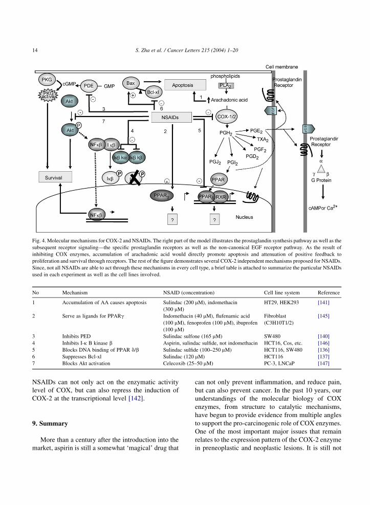
Fig. 4. Molecular mechanisms for COX-2 and NSAIDs. The right part of the model illustrates the prostaglandin synthesis pathway as well as the
subsequent receptor signaling—the specific prostaglandin receptors as well as the non-canonical EGF receptor pathway. As the result of
inhibiting COX enzymes, accumulation of arachadonic acid would directly promote apoptosis and attenuation of positive feedback to
proliferation and survival through receptors. The rest of the figure demonstrates several COX-2 independent mechanisms proposed for NSAIDs.
Since, not all NSAIDs are able to act through these mechanisms in every cell type, a brief table is attached to summarize the particular NSAIDs
used in each experiment as well as the cell lines involved.
No Mechanism NSAID (concentration) Cell line system Reference
1 Accumulation of AA causes apoptosis Sulindac (200 mM), indomethacin
(300 mM)
HT29, HEK293 [141]
2 Serve as ligands for PPARg Indomethacin (40 mM), flufenamic acid
(100 mM), fenoprofen (100 mM), ibuprofen
(100 mM)
Fibroblast
(C3H10T1/2)
[145]
3 Inhibits PED Sulindac sulfone (165 mM) SW480 [140]
4 Inhibits I-k B kinase b Aspirin, sulindac sulfide, not indomethacin HCT16, Cos, etc. [146]
5 Blocks DNA binding of PPAR d/b Sulindac sulfide (100–250 mM) HCT116, SW480 [136]
6 Suppresses Bcl-xl Sulindac (120 mM) HCT116 [137]
7 Blocks Akt activation Celecoxib (25–50 mM) PC-3, LNCaP [147]
S. Zha et al. / Cancer Letters 215 (2004) 1–2014
NSAIDs can not only act on the enzymatic activity
level of COX, but can also repress the induction of
COX-2 at the transcriptional level [142].
9. Summary
More than a century after the introduction into the
market, aspirin is still a somewhat ‘magical’ drug that
can not only prevent inflammation, and reduce pain,
but can also prevent cancer. In the past 10 years, our
understandings of the molecular biology of COX
enzymes, from structure to catalytic mechanisms,
have begun to provide evidence from multiple angles
to support the pro-carcinogenic role of COX enzymes.
One of the most important major issues that remain
relates to the expression pattern of the COX-2 enzyme
in preneoplastic and neoplastic lesions. It is still not
S. Zha et al. / Cancer Letters 215 (2004) 1–20 15
clear, for example, exactly which cells the inhibitors
are acting on since there is often controversy regard-
ing which cells express the enzyme. Further studies of
the expression and function of COX-2 in clinical
samples and animal models, with emphasis on proper
control experiments, are needed to further clarify this
important issue. In addition, we submit that the most
interpretations of why NSAIDs prevent cancer have
perhaps underemphasized the importance of chronic
inflammation in cancer development—most reports
have ignored the strong possibility that the mechan-
ism of action of NSAIDs in cancer prevention may
often proceed via inhibition of the inflammatory
response. Another potential issue regarding COX-2 is
that in at least one animal model, the K.14-COX-2
transgenic mouse, ectopic overexpression of COX-2
dramatically prevented cancer. In addition, the finding
that PGST2 is apparently silenced during prostate
carcinogenesis by hypermethylation of the CpG island
in its promoter region raises the question that this gene
is targeted for inactivation during prostate carcino-
genesis [127]. Thus the simple view that COX-2
expression is always acting to increase cancer risk
may have to be revised. This is also bolstered by the
very high levels of constitutive COX-2 expression in
the seminal vesicles, which have an extremely low
rate of cancer development.
Future work using cell-specific gene knockout and
transgenic animals may help elucidate specific
temporal and spatial relationships between COX-2
expression, the particular prostaglandin profile and
tumor initiation and progression in various organ
systems. These types of studies may also help to
address the specific functions of each of the COX
enzymes: COX-1, COX-2 and COX-3—the newly
identified isoform of COX-1 [143,144], and their
contribution to NSAID mediated tumor regression.
Chemical approaches with the effects of various
structural derivatives on these animal models, and
models with alterations in the prostaglandin receptors,
should further refine the specific and non-specific
effects of NSAIDs. Finally, results of ongoing and
future prospective placebo-controlled double blind
studies of various inhibitors in human studies are
needed to provide definitive information regarding
what types of patients can benefit from the various
types of inhibitors.
Note added in proof
A recent study (Y.G. Crawford, M.L. Gauthier, A.
Joubel, K. Mantei, K. Kozakiewicz, C.A. Afshari,
T.D. Tlsty, Histologically normal human mammary
epithelia with silenced P16INK4a over express COX-2,
promoting a premalignant program, Cancer Cell 5
(2004) 263–273.) provides new evidence for a role of
COX-2 in breast carcinogenesis.
Acknowledgements
Funded by Public Health Services NIH/NCI
#R01CA084997, NIH/#R01CA70196 and NIH/NCI
Specialized Program in Research Excellence (SPORE)
in Prostate Cancer #P50CA58236.
References
[1] E. Anggard, F.M. Matschinsky, B. Samuelsson, Prostaglan-
dins: enzymatic analysis, Science 163 (1969) 479–480.
[2] S. Bergstrom, B. Samuelsson, The prostaglandins, Endea-
vour 27 (1968) 109–113.
[3] D.H. Nugteren, D.A. Van Dorp, S. Bergstrom, M. Hamberg,
B. Samuelsson, Absolute configuration of the prostaglan-
dins, Nature 212 (1966) 38–39.
[4] J.R. Vane, Inhibition of prostaglandin synthesis as a
mechanism of action for aspirin-like drugs, Nat. New
Biol. 231 (1971) 232–235.
[5] Noble e-Museum, The Nobel Prize in Physiology or
Medicine for 1982 is jointly to Sune K. Bergstrom, Bengt
I. Samuelsson, and John R. Vane for their discoveries
concerning ‘prostaglandins and related biologically active
substances’, 10-11-1982.
[6] J.P. Collet, C. Sharpe, E. Belzile, J.F. Boivin, J. Hanley,
L. Abenhaim, Colorectal cancer prevention by non-steroidal
anti-inflammatory drugs: effects of dosage and timing, Br.
J. Cancer 81 (1999) 62–68.
[7] L.J. Marnett, R.N. DuBois, COX-2: a target for colon
cancer prevention, Annu. Rev. Pharmacol. Toxicol. 42
(2002) 55–80.
[8] M.M. Bertagnolli, The potential of non-steroidal anti-
inflammatory drugs (NSAIDs) for colorectal cancer
prevention, J. Surg. Oncol. 84 (2003) 113–119.
[9] G. Huls, J.J. Koornstra, J.H. Kleibeuker, Non-steroidal anti-
inflammatory drugs and molecular carcinogenesis of color-
ectal carcinomas, Lancet 362 (2003) 230–232.
[10] L.A. Garcia Rodriguez, C. Huerta-Alvarez, Reduced inci-
dence of colorectal adenoma among long-term users of
S. Zha et al. / Cancer Letters 215 (2004) 1–2016
nonsteroidal antiinflammatory drugs: a pooled analysis of
published studies and a new population-based study,
Epidemiology 11 (2000) 376–381.
[11] E.M. Moran, Epidemiological and clinical aspects of
nonsteroidal anti-inflammatory drugs and cancer risks,
J. Environ. Pathol. Toxicol. Oncol. 21 (2002) 193–201.
[12] M.J. Thun, S.J. Henley, C. Patrono, Nonsteroidal anti-
inflammatory drugs as anticancer agents: mechanistic,
pharmacologic, and clinical issues, J. Natl Cancer Inst. 94
(2002) 252–266.
[13] W.R. Waddell, R.W. Loughry, Sulindac for polyposis of the
colon, J. Surg. Oncol. 24 (1983) 83–87.
[14] W.R. Waddell, G.F. Ganser, E.J. Cerise, R.W. Loughry,
Sulindac for polyposis of the colon, Am. J. Surg. 157 (1989)
175–179.
[15] D. Labayle, D. Fischer, P. Vielh, F. Drouhin, A. Pariente,
C. Bories, et al., Sulindac causes regression of rectal polyps
in familial adenomatous polyposis, Gastroenterology 101
(1991) 635–639.
[16] F.M. Giardiello, S.R. Hamilton, A.J. Krush, S. Piantadosi,
L.M. Hylind, P. Celano, et al., Treatment of colonic and
rectal adenomas with sulindac in familial adenomatous
polyposis, N. Engl. J. Med. 328 (1993) 1313–1316.
[17] K.P. Nugent, K.C. Farmer, A.D. Spigelman, C.B. Williams,
R.K. Phillips, Randomized controlled trial of the effect of
sulindac on duodenal and rectal polyposis and cell prolifer-
ation in patients with familial adenomatous polyposis, Br. J.
Surg. 80 (1993) 1618–1619.
[18] W.E. Smalley, R.N. DuBois, Colorectal cancer and
nonsteroidal anti-inflammatory drugs, Adv. Pharmacol. 39
(1997) 1–20.
[19] G. Steinbach, P.M. Lynch, R.K. Phillips, M.H. Wallace,
E. Hawk, G.B. Gordon, et al., The effect of celecoxib, a
cyclooxygenase-2 inhibitor, in familial adenomatous poly-
posis, N. Engl. J. Med. 342 (2000) 1946–1952.
[20] R. Calaluce, D.L. Earnest, D. Heddens, J.G. Einspahr,
D. Roe, C.L. Bogert, et al., Effects of piroxicam on
prostaglandin E2 levels in rectal mucosa of adenomatous
polyp patients: a randomized phase IIb trial, Cancer
Epidemiol. Biomarkers Prev. 9 (2000) 1287–1292.
[21] J.A. Baron, B.F. Cole, R.S. Sandler, R.W. Haile, D. Ahnen,
R. Bresalier, et al., A randomized trial of aspirin to prevent
colorectal adenomas, N. Engl. J. Med. 348 (2003) 891–899.
[22] R.S. Sandler, S. Halabi, J.A. Baron, S. Budinger, E. Paskett,
R. Keresztes, et al., A randomized trial of aspirin to prevent
colorectal adenomas in patients with previous colorectal
cancer, N. Engl. J. Med. 348 (2003) 883–890.
[23] H.H. Chow, D.L. Earnest, D. Clark, N. Mason-Liddil,
C.B. Kramer, J.G. Einspahr, et al., Effect of subacute
ibuprofen dosing on rectal mucosal prostaglandin E2 levels
in healthy subjects with a history of resected polyps, Cancer
Epidemiol. Biomarkers Prev. 9 (2000) 351–356.
[24] M.T. Ruffin, K. Krishnan, C.L. Rock, D. Normolle,
M.A. Vaerten, M. Peters-Golden, et al., Suppression of
human colorectal mucosal prostaglandins: determining the
lowest effective aspirin dose, J. Natl Cancer Inst. 89 (1997)
1152–1160.
[25] F.M. Giardiello, V.W. Yang, L.M. Hylind, A.J. Krush,
G.M. Petersen, J.D. Trimbath, et al., Primary chemopreven-
tion of familial adenomatous polyposis with sulindac,
N. Engl. J. Med. 346 (2002) 1054–1059.
[26] P.H. Gann, J.E. Manson, R.J. Glynn, J.E. Buring,
C.H. Hennekens, Low-dose aspirin and incidence of color-
ectal tumors in a randomized trial, J. Natl Cancer Inst. 85
(1993) 1220–1224.
[27] D.L. DeWitt, W.L. Smith, Primary structure of prostaglandin
G/H synthase from sheep vesicular gland determined from
the complementary DNA sequence, Proc. Natl Acad. Sci.
USA 85 (1988) 1412–1416.
[28] J.P. Merlie, D. Fagan, J. Mudd, P. Needleman, Isolation and
characterization of the complementary DNA for sheep
seminal vesicle prostaglandin endoperoxide synthase
(cyclooxygenase), J. Biol. Chem. 263 (1988) 3550–3553.
[29] C. Yokoyama, T. Takai, T. Tanabe, Primary structure of
sheep prostaglandin endoperoxide synthase deduced from
cDNA sequence, Fed. Eur. Biochem. Soc. Lett. 231 (1988)
347–351.
[30] T. Hla, K. Neilson, Human cyclooxygenase-2 cDNA, Proc.
Natl Acad. Sci. USA 89 (1992) 7384–7388.
[31] D.A. Jones, D.P. Carlton, T.M. McIntyre, G.A. Zimmerman,
S.M. Prescott, Molecular cloning of human prostaglandin
endoperoxide synthase type II and demonstration of
expression in response to cytokines, J. Biol. Chem. 268
(1993) 9049–9054.
[32] M.K. O’Banion, H.B. Sadowski, V. Winn, D.A. Young, A
serum- and glucocorticoid-regulated 4-kilobase mRNA
encodes a cyclooxygenase-related protein, J. Biol. Chem.
266 (1991) 23261–23267.
[33] M.K. O’Banion, V.D. Winn, D.A. Young, cDNA cloning and
functional activity of a glucocorticoid-regulated inflamma-
tory cyclooxygenase, Proc. Natl Acad. Sci. USA 89 (1992)
4888–4892.
[34] C.D. Funk, L.B. Funk, M.E. Kennedy, A.S. Pong,
G.A. Fitzgerald, Human platelet/erythroleukemia cell pros-
taglandin G/H synthase: cDNA cloning, expression, and
gene chromosomal assignment, Fed. Am. Soc. Exp. Biol. J. 5
(1991) 2304–2312.
[35] A. Tay, J.A. Squire, H. Goldberg, K. Skorecki, Assignment
of the human prostaglandin-endoperoxide synthase 2
(PTGS2) gene to 1q25 by fluorescence in situ hybridization,
Genomics 23 (1994) 718–719.
[36] J.C. Otto, D.L. DeWitt, W.L. Smith, N-glycosylation of
prostaglandin endoperoxide synthases-1 and -2 and their
orientations in the endoplasmic reticulum, J. Biol. Chem. 268
(1993) 18234–18242.
[37] D.W. Gilroy, P.R. Colville-Nash, New insights into the
role of COX 2 in inflammation, J. Mol. Med. 78 (2000)
121–129.
[38] C.C. Chen, Y.T. Sun, J.J. Chen, Y.J. Chang, Tumor
necrosis factor-alpha-induced cyclooxygenase-2 expression
via sequential activation of ceramide-dependent mitogen-
activated protein kinases, and IkappaB kinase 1/2 in
human alveolar epithelial cells, Mol. Pharmacol. 59
(2001) 493–500.
S. Zha et al. / Cancer Letters 215 (2004) 1–20 17
[39] C.Y. Fong, L. Pang, E. Holland, A.J. Knox, TGF-beta1
stimulates IL-8 release, COX-2 expression, and PGE(2)
release in human airway smooth muscle cells, Am. J. Physiol.
Lung Cell Mol. Physiol. 279 (2000) L201–L207.
[40] S.L. Hempel, M.M. Monick, G.W. Hunninghake, Lipopoly-
saccharide induces prostaglandin H synthase-2 protein and
mRNA in human alveolar macrophages and blood mono-
cytes, J. Clin. Invest. 93 (1994) 391–396.
[41] J.D. Laporte, P.E. Moore, T. Lahiri, I.N. Schwartzman,
R.A. Panettieri, S.A. Shore, p38 MAP kinase regulates IL-1
beta responses in cultured airway smooth muscle cells, Am.
J. Physiol. Lung Cell Mol. Physiol. 279 (2000) L932–L941.
[42] M.P. Peppelenbosch, L.G. Tertoolen, W.J. Hage, S.W. de
Laat, Epidermal growth factor-induced actin remodeling is
regulated by 5- lipoxygenase and cyclooxygenase products,
Cell 74 (1993) 565–575.
[43] M. Goerig, A.J. Habenicht, R. Heitz, W. Zeh, H. Katus,
B. Kommerell, et al., sn-1,2-Diacylglycerols and phorbol
diesters stimulate thromboxane synthesis by de novo
synthesis of prostaglandin H synthase in human promyelo-
cytic leukemia cells, J. Clin. Invest. 79 (1987) 903–911.
[44] H. Niiro, T. Otsuka, E. Ogami, K. Yamaoka, S. Nagano,
M. Akahoshi, et al., MAP kinase pathways as a route for
regulatory mechanisms of IL-10 and IL-4 which inhibit
COX-2 expression in human monocytes, Biochem. Biophys.
Res. Commun. 250 (1998) 200–205.
[45] M.M. Burr, G.O. Burr, On the nature and role of the fatty acids
essential in nutrition, J. Biol. Chem. 86 (1930) 587–621.
[46] U.S. von Euler, J. Physiol. 81 (1934) 102.
[47] B. Samuelsson, Leukotrienes: mediators of immediate
hypersensitivity reactions and inflammation, Science 220
(1983) 568–575.
[48] C.D. Funk, Prostaglandins and leukotrienes: advances in
eicosanoid biology, Science 294 (2001) 1871–1875.
[49] M. Murakami, H. Naraba, T. Tanioka, N. Semmyo,
Y. Nakatani, F. Kojima, et al., Regulation of prostaglandin
E2 biosynthesis by inducible membrane-associated prosta-
glandin E2 synthase that acts in concert with cyclooxygen-
ase-2, J. Biol. Chem. 275 (2000) 32783–32792.
[50] R.G. Kurumbail, A.M. Stevens, J.K. Gierse, J.J. McDonald,
R.A. Stegeman, J.Y. Pak, et al., Structural basis for selective
inhibition of cyclooxygenase-2 by anti-inflammatory
agents (published erratum appears in Nature 385 (6616)
(1997 Feb 6) 555), Nature 384 (1996) 644–648.
[51] C. Luong, A. Miller, J. Barnett, J. Chow, C. Ramesha,
M.F. Browner, Flexibility of the NSAID binding site in the
structure of human cyclooxygenase-2, Nat. Struct. Biol. 3
(1996) 927–933.
[52] D. Picot, P.J. Loll, R.M. Garavito, The X-ray crystal structure
of the membrane protein prostaglandin H2 synthase-1,
Nature 367 (1994) 243–249.
[53] G.P. O’Neill, J.A. Mancini, S. Kargman, J. Yergey,
M.Y. Kwan, J.P. Falgueyret, et al., Overexpression of
human prostaglandin G/H synthase-1 and -2 by recombinant
vaccinia virus: inhibition by nonsteroidal anti-inflammatory
drugs and biosynthesis of 15-hydroxyeicosatetraenoic acid,
Mol. Pharmacol. 45 (1994) 245–254.
[54] M. Lecomte, O. Laneuville, C. Ji, D.L. DeWitt, W.L. Smith,
Acetylation of human prostaglandin endoperoxide synthase-
2 (cyclooxygenase-2) by aspirin, J. Biol. Chem. 269 (1994)
13207–13215.
[55] J.A. Mancini, G.P. O’Neill, C. Bayly, P.J. Vickers, Mutation
of serine-516 in human prostaglandin G/H synthase-2 to
methionine or aspirin acetylation of this residue stimulates
15-R-HETE synthesis, Fed. Eur. Biochem. Soc. Lett. 342
(1994) 33–37.
[56] E.A. Meade, W.L. Smith, D.L. DeWitt, Differential
inhibition of prostaglandin endoperoxide synthase (cycloox-
ygenase) isozymes by aspirin and other non-steroidal anti-
inflammatory drugs, J. Biol. Chem. 268 (1993) 6610–6614.
[57] M.K. Reiger, D.L. DeWitt, M.S. Schindler, W.L. Smith,
Subcellular localization of prostaglandin endoperoxide
synthase-2 in murine 3T3 cells, Arch. Biochem. Biophys.
301 (1993) 439–444.
[58] Y. Ren, D.S. Loose-Mitchell, R.J. Kulmacz, Prostaglandin H
synthase-1: evaluation of C-terminus function, Arch. Bio-
chem. Biophys. 316 (1995) 751–757.
[59] M. Oshima, J.E. Dinchuk, S.L. Kargman, H. Oshima,
B. Hancock, E. Kwong, et al., Suppression of intestinal
polyposis in Apc delta716 knockout mice by inhibition of
cyclooxygenase 2 (COX-2), Cell 87 (1996) 803–809.
[60] K. Takaku, M. Sonoshita, N. Sasaki, N. Uozumi, Y. Doi,
T. Shimizu, M.M. Taketo, Suppression of intestinal poly-
posis in Apc(delta 716) knockout mice by an additional
mutation in the cytosolic phospholipase A(2) gene, J. Biol.
Chem. 275 (2000) 34013–34016.
[61] K.H. Hong, J.C. Bonventre, E. O’Leary, J.V. Bonventre,
E.S. Lander, Deletion of cytosolic phospholipase A(2)
suppresses Apc(Min)-induced tumorigenesis, Proc. Natl
Acad. Sci. USA 98 (2001) 3935–3939.
[62] V.W. Yang, J.M. Shields, S.R. Hamilton, E.W. Spannhake,
W.C. Hubbard, L.M. Hylind, et al., Size-dependent increase
in prostanoid levels in adenomas of patients with familial
adenomatous polyposis, Cancer Res. 58 (1998) 1750–1753.
[63] R. Fukuda, B. Kelly, G.L. Semenza, Vascular endothelial
growth factor gene expression in colon cancer cells exposed
to prostaglandin E2 is mediated by hypoxia-inducible factor
1, Cancer Res. 63 (2003) 2330–2334.
[64] M. Sonoshita, K. Takaku, N. Sasaki, Y. Sugimoto,
F. Ushikubi, S. Narumiya, et al., Acceleration of intestinal
polyposis through prostaglandin receptor EP2 in Apc(Delta
716) knockout mice, Nat. Med. 7 (2001) 1048–1051.
[65] K. Watanabe, T. Kawamori, S. Nakatsugi, T. Ohta,
S. Ohuchida, H. Yamamoto, et al., Role of the prostaglandin
E receptor subtype EP1 in colon carcinogenesis, Cancer Res.
59 (1999) 5093–5096.
[66] F. Ushikubi, Y. Sugimoto, A. Ichikawa, S. Narumiya, Roles of
prostanoids revealed from studies using mice lacking specific
prostanoid receptors, Jpn. J. Pharmacol. 83 (2000) 279–285.
[67] M. Mutoh, K. Watanabe, T. Kitamura, Y. Shoji,
M. Takahashi, T. Kawamori, et al., Involvement of
prostaglandin E receptor subtype EP(4) in colon carcino-
genesis, Cancer Res. 62 (2002) 28–32.

S. Zha et al. / Cancer Letters 215 (2004) 1–2018
[68] R. Pai, B. Soreghan, I.L. Szabo, M. Pavelka, D. Baatar,
A.S. Tarnawski, Prostaglandin E2 transactivates EGF
receptor: a novel mechanism for promoting colon cancer
growth and gastrointestinal hypertrophy, Nat. Med. 8 (2002)
289–293.
[69] C.H. Liu, S.H. Chang, K. Narko, O.C. Trifan, M.T. Wu,
E. Smith, et al., Overexpression of cyclooxygenase-2 is
sufficient to induce tumorigenesis in transgenic mice, J. Biol.
Chem. 276 (2001) 18563–18569.
[70] D.K. Bol, R.B. Rowley, C.P. Ho, B. Pilz, J. Dell, M. Swerdel,
et al., Cyclooxygenase-2 overexpression in the skin of
transgenic mice results in suppression of tumor development,
Cancer Res. 62 (2002) 2516–2521.
[71] K. Muller-Decker, G. Neufang, I. Berger, M. Neumann,
F. Marks, G. Furstenberger, Transgenic cyclooxygenase-2
overexpression sensitizes mouse skin for carcinogenesis,
Proc. Natl Acad. Sci. USA 99 (2002) 12483–12488.
[72] G. Neufang, G. Furstenberger, M. Heidt, F. Marks,
K. Muller-Decker, Abnormal differentiation of epidermis in
transgenic mice constitutively expressing cyclooxygenase-2
in skin, Proc. Natl Acad. Sci. USA 98 (2001) 7629–7634.
[73] S.M. Fischer, G.L. Gleason, G.D. Mills, T.J. Slaga, Indo-
methacin enhancement of TPA tumor promotion in mice,
Cancer Lett. 10 (1980) 343–350.
[74] G. Furstenberger, M. Gross, F. Marks, Eicosanoids and
multistage carcinogenesis in NMRI mouse skin: role of
prostaglandins E and F in conversion (first stage of
tumor promotion) and promotion (second stage of tumor
promotion), Carcinogenesis 10 (1989) 91–96.
[75] C.S. Williams, M. Tsujii, J. Reese, S.K. Dey, R.N. DuBois,
Host cyclooxygenase-2 modulates carcinoma growth, J. Clin.
Invest. 105 (2000) 1589–1594.
[76] P.C. Chulada, M.B. Thompson, J.F. Mahler, C.M. Doyle,
B.W. Gaul, C. Lee, et al., Genetic disruption of Ptgs-1, as
well as Ptgs-2, reduces intestinal tumorigenesis in Min mice,
Cancer Res. 60 (2000) 4705–4708.
[77] R. Langenbach, S.G. Morham, H.F. Tiano, C.D. Loftin,
B.I. Ghanayem, P.C. Chulada, et al., Prostaglandin synthase
1 gene disruption in mice reduces arachidonic acid-induced
inflammation and indomethacin-induced gastric ulceration,
Cell 83 (1995) 483–492.
[78] R. Langenbach, C.D. Loftin, C. Lee, H. Tiano, Cycloox-
ygenase-deficient mice. A summary of their characteristics
and susceptibilities to inflammation and carcinogenesis, Ann.
N.Y. Acad. Sci. 889 (1999) 52–61.
[79] J.E. Dinchuk, B.D. Car, R.J. Focht, J.J. Johnston, B.D. Jaffee,
M.B. Covington, et al., Renal abnormalities and an altered
inflammatory response in mice lacking cyclooxygenase II,
Nature 378 (1995) 406–409.
[80] S.G. Morham, R. Langenbach, C.D. Loftin, H.F. Tiano,
N. Vouloumanos, J.C. Jennette, et al., Prostaglandin synthase
2 gene disruption causes severe renal pathology in the mouse,
Cell 83 (1995) 473–482.
[81] H. Lim, B.C. Paria, S.K. Das, J.E. Dinchuk, R. Langenbach,
J.M. Trzaskos, S.K. Dey, Multiple female reproductive
failures in cyclooxygenase 2-deficient mice, Cell 91 (1997)
197–208.
[82] M.P. Seed, J.R. Brown, C.N. Freemantle, J.L. Papworth,
P.R. Colville-Nash, D. Willis, et al., The inhibition of
colon-26 adenocarcinoma development and angiogenesis by
topical diclofenac in 2.5% hyaluronan, Cancer Res. 57
(1997) 1625–1629.
[83] T.O. Daniel, H. Liu, J.D. Morrow, B.C. Crews, L.J. Marnett,
Thromboxane A2 is a mediator of cyclooxygenase-2-
dependent endothelial migration and angiogenesis, Cancer
Res. 59 (1999) 4574–4577.
[84] E. Fosslien, Review: molecular pathology of cyclooxygen-
ase-2 in cancer-induced angiogenesis, Ann. Clin. Lab Sci. 31
(2001) 325–348.
[85] S. Hockel, K. Schlenger, P. Vaupel, M. Hockel, Association
between host tissue vascularity and the prognostically
relevant tumor vascularity in human cervical cancer, Int.
J. Oncol. 19 (2001) 827–832.
[86] D.W. Visscher, S. Smilanetz, S. Drozdowicz, S.M. Wykes,
Prognostic significance of image morphometric microvessel
enumeration in breast carcinoma, Anal. Quant. Cytol. Histol.
15 (1993) 88–92.
[87] M. Tsujii, S. Kawano, S. Tsuji, H. Sawaoka, M. Hori,
R.N. DuBois, Cyclooxygenase regulates angiogenesis
induced by colon cancer cells (published erratum appears
in Cell 94 (2) (1998 Jul 24) following 271), Cell 93 (1998)
705–716.
[88] P. Pradono, R. Tazawa, M. Maemondo, M. Tanaka, K. Usui,
Y. Saijo, et al., Gene transfer of thromboxane A(2) synthase
and prostaglandin I(2) synthase antithetically altered
tumor angiogenesis and tumor growth, Cancer Res. 62
(2002) 63–66.
[89] Y. Takahashi, F. Kawahara, M. Noguchi, K. Miwa, H. Sato,
M. Seiki, et al., Activation of matrix metalloproteinase-2 in
human breast cancer cells overexpressing cyclooxygenase-1
or -2, Fed. Eur. Biochem. Soc. Lett. 460 (1999) 145–148.
[90] O. Dormond, A. Foletti, C. Paroz, C. Ruegg, NSAIDs inhibit
alpha beta 3 integrin-mediated and Cdc41Rac-dependent
endothelial cell spreading, migration and angiogenesis, Nat.
Med. 7 (2001) 1041–1047.
[91] X. Lu, W. Xie, W.S. Bradshaw, D.L. Simmons, Nonsteroidal
anti-inflammatory drugs cause apoptosis and induce cyclo-
oxygenases in chicken embryo fibroblasts, Pro. Natl. Acad.
Sci. USA 92 (1995) 7961–7965.
[92] M. Tsujii, R.N. DuBois, Alterations in cellular adhesion and
apoptosis in epithelial cells overexpressing prostaglandin
endoperoxide synthase 2, Cell 83 (1995) 493–501.
[93] H. Sheng, J.D. Morrow, R.D. Beauchamp, R.N. Dubois,
Modulation of apoptosis and Bcl-2 expression by prosta-
glandin E2 in human colon cancer cells, Can. Res. 58 (1998)
362–366.
[94] K. Narko, A. Ristimaki, M. MacPhee, E. Smith,
C.C. Haudenschild, T. Hla, Tumorigenic transformation of
immortalized ECV endothelial cells by cyclooxygenase-1
overexpression, J. Biol. Chem. 272 (1997) 21455–21460.
[95] S.J. Cok, A.R. Morrison, The 3 0-untranslated region of
murine cyclooxygenase-2 contains multiple regulatory
elements that alter message stability and translational
efficiency, J. Biol. Chem. 276 (2001) 23179–23185.
S. Zha et al. / Cancer Letters 215 (2004) 1–20 19
[96] X. Song, H.P. Lin, A.J. Johnson, P.H. Tseng, Y.T. Yang,
S.K. Kulp, C.S. Chen, Cyclooxygenase-2, player or spectator
in cyclooxygenase-2 inhibitor-induced apoptosis in prostate
cancer cells, J. Natl Cancer Inst. 94 (2002) 585–591.
[97] P.L. Taylor, R.W. Kelly, 19-Hydroxylated E prostaglandins
as the major prostaglandins of human semen, Nature 250
(1974) 665–667.
[98] K.N. Khan, K.M. Stanfield, A. Dannenberg, S.V. Seshan,
R.N. Baergen, D.A. Baron, R.A. Soslow, Cyclooxygenase-2
expression in the developing human kidney, Pediatr. Dev.
Pathol. 4 (2001) 461–466.
[99] K.N. Khan, K.M. Stanfield, R.K. Harris, D.A. Baron,
Expression of cyclooxygenase-2 in the macula densa of
human kidney in hypertension, congestive heart failure, and
diabetic nephropathy, Ren. Fail. 23 (2001) 321–330.
[100] M. Ek, D. Engblom, S. Saha, A. Blomqvist,
P.J. Jakobsson, A. Ericsson-Dahlstrand, Inflammatory
response: pathway across the blood-brain barrier, Nature
410 (2001) 430–431.
[101] C.E. Eberhart, R.J. Coffey, A. Radhika, F.M. Giardiello,
S. Ferrenbach, R.N. DuBois, Up-regulation of cyclooxygen-
ase 2 gene expression in human colorectal adenomas and
adenocarcinomas, Gastroenterology 107 (1994) 1183–1188.
[102] S.L. Kargman, G.P. O’Neill, P.J. Vickers, J.F. Evans,
J.A. Mancini, S. Jothy, Expression of prostaglandin G/H
synthase-1 and -2 protein in human colon cancer, Cancer
Res. 55 (1995) 2556–2559.
[103] H. Sano, Y. Kawahito, R.L. Wilder, A. Hashiramoto,
S. Mukai, K. Asai, et al., Expression of cyclooxygenase-1
and -2 in human colorectal cancer, Cancer Res. 55 (1995)
3785–3789.
[104] C.S. Williams, C. Luongo, A. Radhika, T. Zhang,
L.W. Lamps, L.B. Nanney, et al., Elevated cyclooxygen-
ase-2 levels in Min mouse adenomas, Gastroenterology 111
(1996) 1134–1140.
[105] B. Humar, O. Giovanoli, A. Wolf, M. Attenhofer, I. Bendik,
R. Meier, et al., Germline alterations in the cyclooxygenase-2
gene are not associated with the development of extracolonic
manifestations in a large swiss familial adenomatous
polyposis kindred, Int. J. Cancer 87 (2000) 812–817.
[106] K.N. Khan, J.L. Masferrer, B.M. Woerner, R. Soslow,
A.T. Koki, Enhanced cyclooxygenase-2 expression in
sporadic and familial adenomatous polyposis of the human
colon, Scand. J. Gastroenterol. 36 (2001) 865–869.
[107] H. Bamba, S. Ota, A. Kato, A. Adachi, S. Itoyama,
F. Matsuzaki, High expression of cyclooxygenase-2 in
macrophages of human colonic adenoma, Int. J. Cancer 83
(1999) 470–475.
[108] K.S. Chapple, E.J. Cartwright, G. Hawcroft, A. Tisbury,
C. Bonifer, N. Scott, et al., Localization of cyclooxygenase-2
in human sporadic colorectal adenomas, Am. J. Pathol. 156
(2000) 545–553.
[109] E.T. Goluboff, A. Shabsigh, J.A. Saidi, I.B. Weinstein,
N. Mitra, D. Heitjan, et al., Exisulind (sulindac sulfone)
suppresses growth of human prostate cancer in a nude mouse
xenograft model by increasing apoptosis, Urology 53 (1999)
440–445.
[110] R.C. Mifflin, J.I. Saada, J.F. Di Mari, P.A. Adegboyega,
J.D. Valentich, D.W. Powell, Regulation of COX-2
expression in human intestinal myofibroblasts: mechanisms
of IL-1-mediated induction, Am. J. Physiol. Cell Physiol. 282
(2002) C824–C834.
[111] T. Nakajima, K. Hamanaka, T. Fukuda, T. Oyama,
K. Kashiwabara, T. Sano, Why is cyclooxygenase-2
expressed in neuroendocrine cells of the human alimentary
tract?, Pathol. Int. 47 (1997) 889–891.
[112] P.H. Rolland, P.M. Martin, J. Jacquemier, A.M. Rolland,
M. Toga, Prostaglandin in human breast cancer: evidence
suggesting that an elevated prostaglandin production is a
marker of high metastatic potential for neoplastic cells,
J. Natl Cancer Inst. 64 (1980) 1061–1070.
[113] A.F. Badawi, M.Z. Badr, Chemoprevention of breast cancer
by targeting cyclooxygenase-2 and peroxisome proliferator-
activated receptor-gamma (Review), Int. J. Oncol. 20 (2002)
1109–1122.
[114] D. Hwang, D. Scollard, J. Byrne, E. Levine, Expression of
cyclooxygenase-1 and cyclooxygenase-2 in human breast
cancer, J. Natl Cancer Inst. 90 (1998) 455–460.
[115] C. Costa, R. Soares, J.S. Reis-Filho, D. Leitao,
I. Amendoeira, F.C. Schmitt, Cyclo-oxygenase 2 expression
is associated with angiogenesis and lymph node metastasis in
human breast cancer, J. Clin. Pathol. 55 (2002) 429–434.
[116] E. Half, X.M. Tang, K. Gwyn, A. Sahin, K. Wathen,
F.A. Sinicrope, Cyclooxygenase-2 expression in human
breast cancers and adjacent ductal carcinoma in situ, Cancer
Res. 62 (2002) 1676–1681.
[117] S. Gupta, M. Srivastava, N. Ahmad, D.G. Bostwick,
H. Mukhtar, Over-expression of cyclooxygenase-2 in
human prostate adenocarcinoma, Prostate 42 (2000) 73–78.
[118] A. Kirschenbaum, D.R. Liotta, S. Yao, X.H. Liu,
A.P. Klausner, P. Unger, et al., Immunohistochemical
localization of cyclooxygenase-1 and cyclooxygenase-2 in
the human fetal and adult male reproductive tracts, J. Clin.
Endocrinol. Metab. 85 (2000) 3436–3441.
[119] A. Kirschenbaum, A.P. Klausner, R. Lee, P. Unger, S. Yao,
X.H. Liu, A.C. Levine, Expression of cyclooxygenase-1 and
cyclooxygenase-2 in the human prostate, Urology 56 (2000)
671–676.
[120] S. Madaan, P.D. Abel, K.S. Chaudhary, R. Hewitt,
M.A. Stott, G.W. Stamp, E.N. Lalani, Cytoplasmic induction
and over-expression of cyclooxygenase-2 in human
prostate cancer: implications for prevention and treatment,
Br. J. Urol. Int. 86 (2000) 736–741.
[121] N. Tanji, T. Kikugawa, M. Yokoyama, Immunohistochem-
ical study of cyclooxygenases in prostatic adenocarcinoma;
relationship to apoptosis and Bcl-2 protein expression,
Anticancer Res. 20 (2000) 2313–2319.
[122] R. Yoshimura, H. Sano, C. Masuda, M. Kawamura,
Y. Tsubouchi, J. Chargui, et al., Expression of cycloox-
ygenase-2 in prostate carcinoma, Cancer 89 (2000) 589–596.
[123] W.J. Wechter, D.D. Leipold, E.D. Murray Jr., D. Quiggle,
J.D. McCracken, R.S. Barrios, N.M. Greenberg, E-7869 (R-
flurbiprofen) inhibits progression of prostate cancer in the
TRAMP mouse, Cancer Res. 60 (2000) 2203–2208.
S. Zha et al. / Cancer Letters 215 (2004) 1–2020
[124] S. Zha, W.R. Gage, J. Sauvageot, E.A. Saria, M.J. Putzi,
C.M. Ewing, et al., Cyclooxygenase-2 is up-regulated in
proliferative inflammatory atrophy of the prostate, but not in
prostate carcinoma, Cancer Res. 61 (2001) 8617–8623.
[125] A.M. De Marzo, V.L. Marchi, J.I. Epstein, W.G. Nelson,
Proliferative inflammatory atrophy of the prostate:
implications for carcinogenesis, Am. J. Pathol. 155 (1999)
1985–1992.
[126] S.B. Shappell, S. Manning, W.E. Boeglin, Y. Guan,
R.L. Robert, L. Davis, et al., Alteration in lipoxygenase and
cyclooxygenase-2 catalytic activity and mRNA expression
in prostate carcinoma, Neoplasia 3 (2001) 287–303.
[127] S. Yegnasubramanian, M.L. Gonzalgo, J. Kowalski,
M. Zahurak, S. Piantadosi, P.C. Walsh, et al., Hypermethy-
lation of CpG islands in primary and metastatic human
prostate cancer, Cancer Res. 64 (2004) 1975–1986.
[128] S.Y. Buckman, A. Gresham, P. Hale, G. Hruza, J. Anast,
J. Masferrer, A.P. Pentland, COX-2 expression is induced by
UVB exposure in human skin: implications for the develop-
ment of skin cancer, Carcinogenesis 19 (1998) 723–729.
[129] S. Hasturk, B. Kemp, S.K. Kalapurakal, J.M. Kurie,
W.K. Hong, J.S. Lee, Expression of cyclooxygenase-1 and
cyclooxygenase-2 in bronchial epithelium and nonsmall cell
lung carcinoma, Cancer 94 (2002) 1023–1031.
[130] M. Kagoura, M. Toyoda, C. Matsui, M. Morohashi, Immu-
nohistochemical expression of cyclooxygenase-2 in skin
cancers, J. Cutan. Pathol. 28 (2001) 298–302.
[131] M. Komhoff, Y. Guan, H.W. Shappell, L. Davis, G. Jack,
Y. Shyr, et al., Enhanced expression of cyclooxygenase-2 in
high grade human transitional cell bladder carcinomas, Am.
J. Pathol. 157 (2000) 29–35.
[132] A. Maitra, A. Ashfag, C.R. Gunn, A. Rahman, C.J. Yeo,
J.L. Cameron, R.H. Hruban, R.E. Wilentz, Cyclooxygenase 2
expression in pancreatic adenocarcinoma and pancreatic
intraepithelial neoplasia: an immunohistochemical analysis
with automated cellular imaging, Am. J. Clin. Pathol. (2002)
194–201.
[133] P. Ricchi, R. Zarrilli, A. Di Palma, A.M. Acquaviva,
Nonsteroidal anti-inflammatory drugs in colorectal cancer:
from prevention to therapy, Br. J. Cancer 88 (2003) 803–807.
[134] J.T. Lim, G.A. Piazza, E.K. Han, T.M. Delohery, H. Li,
T.S. Finn, et al., Sulindac derivatives inhibit growth and
induce apoptosis in human prostate cancer cell lines,
Biochem. Pharmacol. 58 (1999) 1097–1107.
[135] G.A. Piazza, A.L. Rahm, M. Krutzsch, G. Sperl,
N.S. Paranka, P.H. Gross, et al., Antineoplastic drugs
sulindac sulfide and sulfone inhibit cell growth by inducing
apoptosis, Cancer Res. 55 (1995) 3110–3116.
[136] T.C. He, T.A. Chan, B. Vogelstein, K.W. Kinzler, PPARdelta
is an APC-regulated target of nonsteroidal anti-inflammatory
drugs, Cell 99 (1999) 335–345.
[137] L. Zhang, J. Yu, B.H. Park, K.W. Kinzler, B. Vogelstein,
Role of BAX in the apoptotic response to anticancer agents,
Science 290 (2000) 989–992.
[138] K.S. Berman, U.N. Verma, G. Harburg, J.D. Minna,
M.H. Cobb, R.B. Gaynor, Sulindac enhances tumor necrosis
factor-alpha-mediated apoptosis of lung cancer cell lines by
inhibition of nuclear factor-kappaB, Clin. Cancer Res. 8
(2002) 354–360.
[139] C.M. Bradbury, S. Markovina, S.J. Wei, L.M. Rene,
I. Zoberi, N. Horikoshi, D. Gius, Indomethacin-induced
radiosensitization and inhibition of ionizing radiation-
induced NF-kappaB activation in HeLa cells occur via a
mechanism involving p38 MAP kinase, Cancer Res. 61
(2001) 7689–7696.
[140] W.J. Thompson, G.A. Piazza, H. Li, L. Liu, J. Fetter, B. Zhu,
et al., Exisulind induction of apoptosis involves guanosine
3 0,5 0-cyclic monophosphate phosphodiesterase inhibition,
protein kinase G activation, and attenuated beta-catenin,
Cancer Res. 60 (2000) 3338–3342.
[141] Y. Cao, A.T. Pearman, G.A. Zimmerman, T.M. McIntyre,
S.M. Prescott, Intracellular unesterified arachidonic acid
signals apoptosis, Proc. Natl Acad. Sci. USA 97 (2000)
11280–11285.
[142] Y. Cao, S.M. Prescott, Many actions of cyclooxygenase-2 in
cellular dynamics and in cancer, J. Cell Physiol. 190 (2002)
279–286.
[143] T.D. Warner, J.A. Mitchell, Cyclooxygenase-3 (COX-3):
filling in the gaps toward a COX continuum?, Proc. Natl
Acad. Sci. USA 99 (2002) 13371–13373.
[144] N.V. Chandrasekharan, H. Dai, K.L. Roos, N.K. Evanson,
J. Tomsik, T.S. Elton, D.L. Simmons, COX-3, a cycloox-
ygenase-1 variant inhibited by acetaminophen and
other analgesic/antipyretic drugs: cloning, structure,
and expression, Proc. Natl Acad. Sci. USA 99 (2002)
13926–13931.
[145] J.M. Lehmann, J.M. Lenhard, B.B. Oliver, G.M. Ringold,
S.A. Kliewer, Peroxisome proliferator-activated receptors
alpha and gamma are activated by indomethacin and other
non-steroidal anti-inflammatory drugs, J. Biol. Chem. 272
(1997) 3406–3410.
[146] Y. Yamamoto, R.B. Gaynor, Therapeutic potential of
inhibition of the NF-kappaB pathway in the treatment
of inflammation and cancer, J. Clin. Invest. 107 (2001)
135–142.
[147] A.L. Hsu, T.T. Ching, D.S. Wang, X. Song, V.M. Rangnekar,
C.S. Chen, The cyclooxygenase-2 inhibitor celecoxib
induces apoptosis by blocking Akt activation in human
prostate cancer cells independently of Bcl-2, J. Biol. Chem.
275 (2000) 11397–11403.
Related Documents