CXCR3/CCR5 pathways in metastatic melanoma patients treated with adoptive therapy and interleukin-2 D Bedognetti * ,1,2,3 , T L Spivey 1,4,5 , Y Zhao 6 , L Uccellini 1,7 , S Tomei 1 , M E Dudley 8 , M L Ascierto 1,3,9 , V De Giorgi 1 , Q Liu 1 , L G Delogu 10 , M Sommariva 1,11 , M R Sertoli 2,3 , R Simon 6 , E Wang 1 , S A Rosenberg 8 and F M Marincola * ,1,12 1 Infectious Disease and Immunogenetics Section (IDIS), Department of Transfusion Medicine, Clinical Center and trans-NIH Center for Human Immunology (CHI), National Institutes of Health, Bethesda, MD 20892, USA; 2 Department of Oncology, Biology, Genetics (DOBIG), University of Genoa and National Cancer Research Institute, 16132 Genoa, Italy; 3 Department of Internal Medicine (DiMI), University of Genoa, 16132 Genoa, Italy; 4 Clinical Research Training Program (CRTP), National Institutes of Health, Bethesda, MD 20892, USA; 5 Rush University Medical Center, Rush Medical College, Chicago, IL 60612, USA; 6 Biometric Research Branch, Division of Cancer Treatment and Diagnosis, National Cancer Institute, National Institutes of Health, Bethesda, MD 20892, USA; 7 Infectious Diseases and Tropical Medicine Section, Department of Clinical Science, University of Milan, Milan, Italy; 8 Surgery Branch, National Cancer institute, National Institutes of Health, Bethesda, MD 20892, USA; 9 Center of Excellence for Biomedical Research (CEBR), University of Genoa, 16132 Genoa, Italy; 10 Department of Chemistry and Pharmacy, University of Sassari, 07100 Sassari, Italy; 11 Department of Biomedical Sciences for Health, University of Milan, 20133 Milan, Italy and 12 Research Branch, Sidra Medical and Research Centre, Doha, Qatar Background: Adoptive therapy with tumour-infiltrating lymphocytes (TILs) induces durable complete responses (CR) in B20% of patients with metastatic melanoma. The recruitment of T cells through CXCR3/CCR5 chemokine ligands is critical for immune- mediated rejection. We postulated that polymorphisms and/or expression of CXCR3/CCR5 in TILs and the expression of their ligands in tumour influence the migration of TILs to tumours and tumour regression. Methods: Tumour-infiltrating lymphocytes from 142 metastatic melanoma patients enrolled in adoptive therapy trials were genotyped for CXCR3 rs2280964 and CCR5-D32 deletion, which encodes a protein not expressed on the cell surface. Expression of CXCR3/CCR5 in TILs and CXCR3/CCR5 and ligand genes in 113 available parental tumours was also assessed. Tumour-infiltrating lymphocyte data were validated by flow cytometry (N ¼ 50). Results: The full gene expression/polymorphism model, which includes CXCR3 and CCR5 expression data, CCR5-D32 polymorphism data and their interaction, was significantly associated with both CR and overall response (OR; P ¼ 0.0009, and P ¼ 0.007, respectively). More in detail, the predicted underexpression of both CXCR3 and CCR5 according to gene expression and polymorphism data (protein prediction model, PPM) was associated with response to therapy (odds ratio ¼ 6.16 and 2.32, for CR and OR, respectively). Flow cytometric analysis confirmed the PPM. Coordinate upregulation of CXCL9, CXCL10, CXCL11, and CCL5 in pretreatment tumour biopsies was associated with OR. Conclusion: Coordinate overexpression of CXCL9, CXCL10, CXCL11, and CCL5 in pretreatment tumours was associated with responsiveness to treatment. Conversely, CCR5-D32 polymorphism and CXCR3/CCR5 underexpression influence downregulation of the corresponding receptors in TILs and were associated with likelihood and degree of response. *Correspondence: Dr D Bedognetti; E-mail: [email protected] or Dr FM Marincola; E-mail: [email protected] Received 22 April 2013; revised 19 August 2013; accepted 20 August 2013; published online 15 October 2013 & 2013 Cancer Research UK. All rights reserved 0007 – 0920/13 FULL PAPER Keywords: immunotherapy; interlukin-2; chemokines; melanoma; tumour microenvironment British Journal of Cancer (2013) 109, 2412–2423 | doi: 10.1038/bjc.2013.557 2412 www.bjcancer.com | DOI:10.1038/bjc.2013.557

Welcome message from author
This document is posted to help you gain knowledge. Please leave a comment to let me know what you think about it! Share it to your friends and learn new things together.
Transcript

CXCR3/CCR5 pathways in metastaticmelanoma patients treated with adoptivetherapy and interleukin-2D Bedognetti*,1,2,3, T L Spivey1,4,5, Y Zhao6, L Uccellini1,7, S Tomei1, M E Dudley8, M L Ascierto1,3,9,V De Giorgi1, Q Liu1, L G Delogu10, M Sommariva1,11, M R Sertoli2,3, R Simon6, E Wang1, S A Rosenberg8
and F M Marincola*,1,12
1Infectious Disease and Immunogenetics Section (IDIS), Department of Transfusion Medicine, Clinical Center and trans-NIHCenter for Human Immunology (CHI), National Institutes of Health, Bethesda, MD 20892, USA; 2Department of Oncology, Biology,Genetics (DOBIG), University of Genoa and National Cancer Research Institute, 16132 Genoa, Italy; 3Department of InternalMedicine (DiMI), University of Genoa, 16132 Genoa, Italy; 4Clinical Research Training Program (CRTP), National Institutesof Health, Bethesda, MD 20892, USA; 5Rush University Medical Center, Rush Medical College, Chicago, IL 60612, USA; 6BiometricResearch Branch, Division of Cancer Treatment and Diagnosis, National Cancer Institute, National Institutes of Health, Bethesda,MD 20892, USA; 7Infectious Diseases and Tropical Medicine Section, Department of Clinical Science, University of Milan, Milan,Italy; 8Surgery Branch, National Cancer institute, National Institutes of Health, Bethesda, MD 20892, USA; 9Center of Excellence forBiomedical Research (CEBR), University of Genoa, 16132 Genoa, Italy; 10Department of Chemistry and Pharmacy, University ofSassari, 07100 Sassari, Italy; 11Department of Biomedical Sciences for Health, University of Milan, 20133 Milan, Italy and 12ResearchBranch, Sidra Medical and Research Centre, Doha, Qatar
Background: Adoptive therapy with tumour-infiltrating lymphocytes (TILs) induces durable complete responses (CR) in B20% ofpatients with metastatic melanoma. The recruitment of T cells through CXCR3/CCR5 chemokine ligands is critical for immune-mediated rejection. We postulated that polymorphisms and/or expression of CXCR3/CCR5 in TILs and the expression oftheir ligands in tumour influence the migration of TILs to tumours and tumour regression.
Methods: Tumour-infiltrating lymphocytes from 142 metastatic melanoma patients enrolled in adoptive therapy trials weregenotyped for CXCR3 rs2280964 and CCR5-D32 deletion, which encodes a protein not expressed on the cell surface.Expression of CXCR3/CCR5 in TILs and CXCR3/CCR5 and ligand genes in 113 available parental tumours was also assessed.Tumour-infiltrating lymphocyte data were validated by flow cytometry (N¼ 50).
Results: The full gene expression/polymorphism model, which includes CXCR3 and CCR5 expression data, CCR5-D32polymorphism data and their interaction, was significantly associated with both CR and overall response (OR; P¼ 0.0009,and P¼ 0.007, respectively). More in detail, the predicted underexpression of both CXCR3 and CCR5 according to geneexpression and polymorphism data (protein prediction model, PPM) was associated with response to therapy (odds ratio¼ 6.16and 2.32, for CR and OR, respectively). Flow cytometric analysis confirmed the PPM. Coordinate upregulation of CXCL9, CXCL10,CXCL11, and CCL5 in pretreatment tumour biopsies was associated with OR.
Conclusion: Coordinate overexpression of CXCL9, CXCL10, CXCL11, and CCL5 in pretreatment tumours was associated withresponsiveness to treatment. Conversely, CCR5-D32 polymorphism and CXCR3/CCR5 underexpression influence downregulationof the corresponding receptors in TILs and were associated with likelihood and degree of response.
*Correspondence: Dr D Bedognetti; E-mail: [email protected] or Dr FM Marincola; E-mail: [email protected]
Received 22 April 2013; revised 19 August 2013; accepted 20 August 2013; published online 15 October 2013
& 2013 Cancer Research UK. All rights reserved 0007 – 0920/13
FULL PAPER
Keywords: immunotherapy; interlukin-2; chemokines; melanoma; tumour microenvironment
British Journal of Cancer (2013) 109, 2412–2423 | doi: 10.1038/bjc.2013.557
2412 www.bjcancer.com | DOI:10.1038/bjc.2013.557

Adoptive therapy with autologous tumour-infiltrating T lymphocytes(TILs) represents a promising treatment for patients withmetastatic melanoma. In phase II trials, where TILs are expanded,activated ex vivo, and then adoptively transferred into patients incombination with systemic high-dose interleukin-(IL)-2 followinglymphodepletion, durable complete responses (CR) were observedin B20% of patients (Rosenberg et al, 2011). However, theadministration of preparative chemotherapy and high-dose IL-2 isassociated with significant toxicity and no pretreatment biomar-kers of responsiveness useful for patient selection are currentlyavailable.
Several studies in humans and animal models suggest that therecruitment of activated T lymphocytes through CXCR3 and CCR5chemokine ligands has a key role immune-mediated tissuedestruction, including tumour rejection (Wang et al, 2008;Bedognetti et al, 2010; Gajewski et al, 2011; Spivey et al, 2011;Gonzalez-Martin et al, 2012). We observed that systemicallyadministered IL-2 induces inflammation within tumours leading toproduction of lymphocyte chemoattractants including CXCR3 andCCR5 ligands (Panelli et al, 2002; Panelli et al, 2004a; Bedognettiet al, 2010; Weiss et al, 2011). Indeed, the localisation of TILswithin tumours is a necessary, although not sufficient, conditionfor tumour rejection (Pockaj et al, 1994). Thus, we postulated thatpolymorphisms and/or expression of CXCR3 and CCR5 mayquantitatively and/or qualitatively influence chemokine receptorexpression, and therefore influence migration of TILs to tumoursand their regression.
The CCR5 polymorphism D32 consists of a 32-base deletionencoding a protein not expressed on the cell surface. Consequently,heterozygosity results in decreased and homozygosity absentreceptor expression (Huang et al, 1996; Carrington et al, 1999).A retrospective study reported decreased survival of patients withmetastatic melanoma carrying the CCR5-D32 polymorphismtreated with immunotherapy (Ugurel et al, 2008). A commonsingle nucleotide polymorphism (rs2280964) of CXCR3 has beenrecently associated with variation in receptor expression,
lymphocyte chemotactic activity, and risk of developing asthma(Choi et al, 2008).
Here, in a prospective-retrospective hypothesis-driven analysis(Simon et al, 2009), we investigated the role of CXCR3 and CCR5expression and polymorphisms (i.e., rs2280964 for CXCR3 andD32 for CCR5) by sequencing and profiling infused TILs frompatients with metastatic melanoma undergoing adoptive therapy(N¼ 142). Results were validated using flow cytometry (N¼ 50).The role of CXCR3/CCR5 ligand gene expression was assessedby profiling matched pretreatment tumour biopsies (N¼ 113).
MATERIALS AND METHODS
Patient samples. Samples from 142 patients were available:142 TILs and 113 paired pretreatment tumour biopsies for RNAextraction, and 141 TILs for DNA extraction. Fifty pairedTIL samples were also analysed using flow cytometry. Tumour-infiltrating lymphocytes were frozen just before the infusion.Samples were collected during five consecutive trials at the SurgeryBranch, National Cancer Institute (Dudley et al, 2010; Rosenberget al, 2011; Uccellini et al, 2012; http://www.clinicaltrials.gov/ct2/show/NCT00513604?term=07C0176&rank=1), codified as:TNMA, T200, T1200, TYT, and TCD8 (Table 1). All patientshad progressive disease and had previously received standard orexperimental regimens. Fifty percent of the patients (71 out of 142)achieved an objective response (OR), of which 25 (18%)experienced a durable CR and 46 (32%) a partial response (PR).Tumour biopsy samples included 24 CR (21%), 34 PR (30%) and55 (49%) non-response (NR) samples.
Total body irradiation (TBI; 2 or 12 Gy) was administered inconjunction with chemotherapy in T200 and T1200 trials,respectively (Rosenberg et al, 2011). A day following lymphodeple-tion, TILs were infused into patients and high-dose IL-2 therapywas started. Protocols employed to generate TILs are describedelsewhere (Dudley et al, 2003; Tran et al, 2008; Dudley et al,
Table 1. Patient and treatment characteristics
CR NR NRAll, n¼142
n (%) Objective response (OR vs NR)*Complete response
(CR vs non-CR)*
SexFemale vs male
Odds ratio (95% CI)Female vs male
Odds ratio (95% CI)
Male n (%) 15 (17) 29 (33) 45 (51) 89 (63) 1.06 (0.54–2.10) 1.15 (0.47–2.78)Female n (%) 10 (19) 17 (32) 26 (49) 53 (37) P-value P-value
0.86 0.76
Age (median: 46; range: 18–64)Age
P-valueAge
P-value
16–30 n (%) 3 (20) 3 (20) 9 (60) 15 (11) 0.43 0.5031–45 n (%) 9 (18) 17 (33) 25 (49) 51 (36)46–60 n (%) 13 (20) 23 (35) 30 (46) 66 (47)61–75 n (%) 0 (0) 3 (30) 7 (70) 10 (7)
TreatmentTreatment
P-valueTreatment
P-value
TNMA n (%) 5 (13) 13 (33) 21 (54) 39 (28) 0.015 0.017T200 n (%) 5 (21) 8 (33) 11 (46) 24 (17)T1200 n (%) 10 (40) 8 (32) 7 (28) 25 (18)TYT n (%) 2 (8) 4 (16) 19 (76) 25 (18)TCD8 n (%) 3 (10) 13 (45) 13 (45) 29 (20)
Abbreviations: CI¼ confidence interval; CR¼ complete response; NR¼ no response; Non-CR¼PRþNR; OR¼objective response (CRþPR); PR¼partial response; T200¼ 2 Gy total bodyirradiation protocol; T1200¼ 12 Gy total body irradiation protocol; TCD8¼ young TIL CD8-enriched protocol; TNMA¼ no total body irradiation protocol; TYT¼ young TIL protocol. *P-valuesare from the w2-test or the Fisher’s exact test, as appropriate.
CXCR3/CCR5 pathways in adoptive therapy BRITISH JOURNAL OF CANCER
www.bjcancer.com | DOI:10.1038/bjc.2013.557 2413

2010; Rosenberg et al, 2011). Before TIL administration, patientsreceived a non-myeloablative lymphodepleting regimen consistingof cyclophosphamide at 60 mg kg� 1 per day for 2 days andfludarabine at 25 mg m� 2 per day for 5 days (Dudley et al, 2010;Rosenberg et al, 2011). A day following lymphodepletion,TILs were infused into patients and high-dose IL-2 therapy wasstarted (720 000 IU kg� 1 intravenously every 8 h to tolerance).Major inclusion criteria included minimum age of 18 years,measurable disease, good clinical performance and life expectancygreater than 3 months. Detailed study protocol informationare available elsewhere (http://www.clinicaltrials.gov/ct2/show/NCT00513604?term=07C0176&rank=1), (Dudley et al, 2010;Rosenberg et al, 2011).
All patients signed an informed consent approved by theInstitutional Review Board of the National Cancer Institute.Data for this analysis are updated as of 11 January 2012. Response(CR, PR, or NR) was rated according to the Response EvaluationCriteria in Solid Tumors guidelines 4 weeks following TILadministration and at regular intervals thereafter. A CR or PR wasconsidered an OR and a PR or NR was considered a non-CR.
Sequencing. DNA was successfully extracted from 141 TILsamples from frozen material (cell pellet) preserved at � 80 1C.One sample was unavailable for DNA extraction. Total DNA wasisolated with the Nucleic Acid Isolation System QuickGene-810using the QuickGene DNA whole blood Mini Kit (Fujifilm, Tokyo,Japan).
Amplification PCR was carried out in a reaction mixturecontaining 4ml (40 ng) of DNA, 10 ml of HotStar Taq Master Mix(Qiagen, Germantown, MD, USA), 1 ml (10 pmol) of each of thefollowing primers: forward 50-CTGGCCATCTCTGACCTGTT-30
and reverse 50-CCCTGTGCCTCTTCTTCTCA-30 for CCR5 gene(D32 polymorphism) and forward 50-CTCCCTACACTGAGGCTTGC-30 and reverse 50-AGCCATGGTCCTTGAGGTAA-30 forCXCR3 gene (rs2280964 polymorphisms). Primers were designedusing the Primer3 software (http://www.frodo.wi.mit.edu/).For CCR5 analysis, the reaction mixture was denatured at 95 1Cfor 10 min and cycled 35 times at 94 1C for 30 s, 58 1C for 30 s, and72 1C for 60 s, with final extension at 72 1C for 10 min. For CXCR3analysis, the reaction mixture was denatured at 95 1C for 10 minand cycled 32 times at 94 1C for 45 s, 58 1C for 45 s, and 72 1C for60 s, with the final extension at 72 1C for 10 min.
The PCR product was purified with Exosap-IT (USB Corpora-tion, Cleveland, OH, USA) according to the manufacturer’sprotocol. PCR products were visualised on ultraviolet transillumi-nated, ethidium bromide 2% Agarose gel (Invitrogen, Carlsbad,CA, USA).
Sequencing reactions were performed using 3.5 ml of purifiedDNA product, 2 ml of Big Dye terminator (Big Dye Terminatorv3.1 cycle sequencing reaction kit, Applied Biosystems, Foster City,CA, USA) and 4 pmol of forward primer. The following conditionswere used: initial temperature of 96 1C for 60 s, followed by 30cycles at 96 1C for 30 s, 50 1C for 30 s, and 60 1C for 4 min. Excessdye terminators were removed using DyeEx 96 Kit columns as perthe manufacturer’s instructions (Qiagen).
The Sanger sequencing was performed on an ABI Prism 3730XL instrument (Applied Biosystems). Data were analysed usingSequencher software v.4.9 (Gene Codes Corporation, Ann Arbor,MI, USA).
Samples for gene expression and sequencing experiments wereprocessed in parallel during the same time period.
Gene expression. Total RNA was isolated from 142 TILs(cryopreserved just before the infusion into the patients) and 113snap frozen tumour samples used for TIL generation. Total RNAwas extracted with the Qiagen miRNeasy Mini kit and its qualitywas tested with the Agilent Bioanalyzer 2000 (Agilent Techno-logies, Palo Alto, CA, USA). Three hundred nanograms of total
RNA were used for RNA amplification according to the manu-facturer’s instructions (WT Expression Kit; Ambion, Austin, TX,USA). Amplified RNA was reverse transcribed into cDNAs followedby fragmentation. After hybridisation to the GeneChip HumanGene 1.0 ST Arrays, the chips were labelled with a WT TerminalLabeling Kit (Affymetrix, Santa Clara, CA, USA) and scanned on aGeneChip Scanner 3000 7G (Affymetrix). Data were normalisedusing the Robust Multi-Chip Average method and Log2-transformedusing Partek Genomics Suite 6.4 (Partek Inc., St Louis, MO, USA).Data analyses were based on the whole transcripts.
Flow cytometry. Flow cytometry was performed in 50 TILsamples (cryopreserved just before the infusion into the patients)to validate the accuracy of genomic data in predicting cell surfaceprotein expression. Tumour-infiltrating lymphocytes were selectedas follows: (a) all available CRs; (2) samples concomitantly under-or overexpressing CXCR3 and CCR5; (3) CCR5-D32 homozygoussamples; and (4) CCR5-D32 heterozygous samples included in theprotein prediction model (PPM; described in the Results section).Because of the non-random sampling (selection bias), a correlationanalysis between receptor expression and response was neitherplanned nor performed. Tumour-infiltrating lymphocyte samplesincluded 18 CR (11 CXCR3/CCR5 low and 7 CXCR3/CCR5 high),3 PR (CXCR3/CCR5 high) and 29 NR (14 CXCR3/CCR5 low and15 CXCR3/CCR5 high) cases. Ten CCR5-D32 heterozygous (six PR,two CR and two NR) and one CCR5-D32 homozygous sampleswere selected, including six samples that were reclassified as thelow group according to the PPM. Tumour-infiltrating lymphocytespreserved in liquid nitrogen were thawed, washed, immediatelystained and analysed. FITC-labelled anti-CCR5 and PE-labelledanti-CXCR3 antibodies were used for chemokine receptor expres-sion analyses. 7AAD antibodies were used to identify dead cellsallowing for gating and analysis of live cells only. Samples were alsostained with the appropriate isotype controls. Cells were analysedon a FACSCalibur flow cytometer (Becton Dickinson & Co, SanJose, CA, USA). Thirty thousand events were acquired on theFACSCalibur from each of the TIL samples for analysis. Dailyfluctuations of the FACSCalibur flow cytometer were controlled byusing Sphero 8 peak Rainbow Calibration Particles (BectonDickinson & Co) and specific fluorescencent signal were calculatedon the live cells by normalising to the appropriate isotype control,as described elsewhere (Pos et al, 2010). All of the antibodies werefrom Becton Dickinson & Co. Samples were stained according tothe manufacturer’s protocols.
Statistical analysis. The difference of the overall outcomedistribution (CR, PR, and NR) among CXCR3 and CCR5polymorphism classes was assessed using the Kruskal–Wallis testthat accounts for the ordered nature of the outcomes (i.e., degree ofresponse: CR better than PR better than NR). The distribution ofOR and CR between CXCR3 and CCR5 classes was then assessedby the w2-test or Fisher’s exact test, as appropriate (i.e., if anycategory has a cell count o5). Odds ratios with their 95% CI werealso reported, as appropriate. To minimise ‘data overfitting biases’,statistical significance levels and confidence intervals are notreported for data-derived hypotheses. As for the CXCR3 rs2280964polymorphism, the four G carriers were not included in thegenotype-response and genotype-transcript level correlationanalysis. The associations between the expression of CXCR3 andCCR5 in TILs and clinical responses were first assessed individuallyby the Spearman rank correlation coefficient (r) tests, whichaccount for the ordered nature of the outcomes. One sample wasunavailable for genotyping. Logistic regression models were used toassess the joint association between the expression of CXCR3 andCCR5, the CCR5-D32 polymorphism, and their interactions withthe clinical outcome, defined as either CR or OR. The loglikelihood ratio test to was used to compare the fit of two models,one of which (the null model) is a special case of the other
BRITISH JOURNAL OF CANCER CXCR3/CCR5 pathways in adoptive therapy
2414 www.bjcancer.com | DOI:10.1038/bjc.2013.557

(the alternative model). The test is based on the log likelihood ratio,which expresses how many times more likely the data are under onemodel than the other. The null distribution of log likelihood isgenerated by calculating the log likelihood for each permutation.The variables included in the model are defined as follows: X1, X2,X3 are binary variables; X1¼ 1 when Log2 gene expression valueof CCR54median; X1¼ 0 when Log2 gene expression value ofCCR5pmedian; X2¼ 1 when Log2 base gene expression valueof CXCR34median; X2¼ 0 when Log2 base gene expression valueof CXCR3pmedian; X3¼ 0 when CCR5 polymorphism D32 ispresent; wild-type (WT)¼ 1; Rx¼ treatment (TNMA, T200,T1200, TYT, and TCD8); Y: end point (CR or OR). During themodel fitting, the sample with missing genotype data was omitted.
The gene expression model included CXCR3 and CCR5expression and their interaction (model A). The gene expression/polymorphism model included CXCR3 and CCR5 expression,CCR5-D32 polymorphism, and their interaction (model B).Likelihood ratio tests were used to evaluate the statisticalsignificance of effects in the models compared. The geneexpression and the gene expression/polymorphism models werealso controlled for treatment effect by including treatmentvariables in the two models (model C and model D, respectively).
In order to facilitate interpretation of the statistically significantinteractions found in the logistic regression models, CXCR3 andCCR5 expression data were then dichotomised (low vs high)according to their respective median values and integrated with eachother and with CCR5-D32 polymorphism data, resulting in a PPM.
Univariate associations between TIL and tumour CXCR3 andCCR5 expression and their polymorphisms were assessed byStudent’s t-test (CCR5-D32 polymorphism) after checking fornormal distribution (the D0Agostino–Pearson normality test) or bythe Spearman rank correlation coefficient (r) test (CXCR3rs2280964 polymorphism). Association between sex, age andclinical outcomes (CR or OR), as well as cluster enrichmentanalysis was assessed by the w2- or Fisher exact test, as appropriate.
Correlation between flow cytometry and gene expression datawas assessed by the Spearman rank correlation coefficient (r) testas flow cytometry data were not normally distributed.
The correlation matrix of tumour biopsy gene expression datawas based on the Spearman rank correlation coefficient (r) testbecause not all the data were normally distributed. Hierarchicalclustering was based on the Euclidean distances, and wasperformed with Partek Genomics Suite 6.4. Probability (P) valueso0.05 (two-sided) were considered to be statistically significant.All statistical tests were two sided. Analyses were performed withSPSS version 18.0 (SPSS, Chicago, IL, USA), Partek Genomics Suite6.4 (Partek Inc., and R (Bedognetti et al, 2011).
Single subject CXCR3 rs2280964 genotypes (HapMap and 1000Genome Projects, CEU populations) were downloaded fromensembl (http://www.useast.ensembl.org/index.html).
RESULTS
Tumour-infiltrating lymphocyte sequencing. Demographiccharacteristics of the study population are shown in Table 1. Asfor CXCR3 (located on the X chromosome), no association wasobserved between response and rs2280964 genotype (Table 2). Thefrequencies of T allele were 0.24 and 0.30 in men and women,respectively. These frequencies are similar to those reported inindividuals with European ancestry (T-allele frequency in men:0.26 and 0.20, CEU HapMap, and CEU 1000 Genomes Project,respectively; T-allele frequency in women: 0.22 and 0.27, CEUHapMap, and CEU 1000 Genomes Project, respectively). Interest-ingly, we found four men (one CR, two PR, and one NR) carryingthe G allele (electopherograms in Supplementary Figure S1).
Although rs2280964 is annotated as bi-allelic (C/T) polymorphismin the dbSNP database (http://www.ncbi.nlm.nih.gov/snp/?term=2280964), HapMap project and genome-wide associationstudies (both array based) might miss the identification of tri-allelicvariants (Ellinghaus et al, 2009). In fact, array-based genotypingneed the knowledge of a third allele being present for the assaysdesign, otherwise the third allele cannot be detected without usingappropriate imputation algorithms (Huebner et al, 2007). However,programs used to genotype the current phase 1 release of 1000Genomes Projects (sequencing based) are unable to genotype siteswith more than two alleles. (http://www.1000genomes.org/faq/are-all-genotype-calls-current-release-vcf-files-bi-allelic).
As for CCR5, the frequency of the D32 genotype is B20% inCaucasians (Carrington et al, 1999). Similarly, we found 26patients (18.4%) carrying the D32 polymorphism (1 homozygousand 25 heterozygous). The difference in the overall distribution ofthe outcomes for the WT and D32 patients was borderlinesignificant (P¼ 0.058) when checked with the Kruskal–Wallis testthat assumes a progressive enrichment of one genotype over theother in three ordered response classes (i.e., CR4PR4NR).Although the CR rate was similar between the two groups (24% vs16%), 17 of 25 patients (68%) carrying the D32 polymorphism(including the homozygous patients) experienced an OR, whereasonly 53 of 116 (46%) WT patients experienced an OR (oddsratio¼ 2.53, CI¼ 1.01–6.32, D32 vs WT patients, respectively,P¼ 0.043, Table 2).
Tumour-infiltrating lymphocyte gene expression and integratedgene expression and sequencing analysis. No differences inCXCR3 and CCR5 expression in TILs among the response classeswas observed when data were compared by the Spearman rankcorrelation coefficient (r) test, which assumes a progressivechange in gene expression in the three ordered response classes(i.e., CR4PR4NR or CRoPRoNR), having CR and PR samplesthe lowest and the highest transcript levels (CXCR3 and CCR5 geneexpression data distribution is shown in Supplementary Figure S2).
To evaluate the weight on response of the coordinate expressionof CXCR3 and CCR5, which have redundant functions, we utilisedlogistic regression analyses (Table 3). The gene expression modelwith interaction (full A model), included as predictor variables theexpression of the two genes in TILs and the two-way interactionbetween the individual gene expressions. This model wassignificantly associated with CR (P¼ 0.032) but not significantlyassociated with OR (P¼ 0.15). The gene expression/polymorphismmodel with interaction (full B model) includes the variables of thefull A model as well as an indicator variable for D32 polymorphismand the two-way and three way interaction of the CCR5-D32polymorphism with the gene expression variables. By adding theDNA dimension (CCR5-D32 polymorphism), the model wassignificantly associated with both CR (P¼ 0.0009) and OR(P¼ 0.007). The gene expression/polymorphism model withinteraction (full B model) performed significantly better than thegene expression model with interaction (full A model) for both CR(P¼ 0.003) and OR (P¼ 0.007). As we detected a significantdifference in OR and CR rates across the cohorts (see demographiccharacteristics in Table 1), we also evaluated models containing atreatment group factor, as well as the other variables. With thisadjustment (model C and D), the gene expression model withinteraction (full C model) remained significantly associated withCR (P¼ 0.002), and became also significantly associated with OR(P¼ 0.010). The gene expression/polymorphism model withinteraction (full D model) remained significantly associated withCR (P¼ 0.0001) and with OR (P¼ 0.003) and, again, wassignificantly better than the gene expression model with interactionfor CR and OR (P¼ 0.005 and P¼ 0.036, respectively). A detaileddescription of the results of the logistic regression models,including intermediate models, is reported in Supplementary
CXCR3/CCR5 pathways in adoptive therapy BRITISH JOURNAL OF CANCER
www.bjcancer.com | DOI:10.1038/bjc.2013.557 2415

Tables S1 and S2. Despite the possible intrinsic data overfitting, theemployment of logistic models, which assess all the combinationamong the variables analysed, reduce biases related to the selectionof group comparisons. Overall, these models clearly (andconsistently) show that the addition of new dimensions to data(e.g., combinatorial gene expression analysis and integration withgenetic polymorphisms) can increase the chances to identifypatients more probably to respond to treatment.
To understand the significant interactions identified by thelogistic modelling, samples were grouped according to theexpression of chemokine receptor genes. The CXCR3-lowgroup was enriched in CR samples (CR rate: 25% vs 10%,odds ratio¼ 3.11, CXCR3 low vs high, respectively, Figure 1A).The CCR5-low group was also slightly enriched in CR samples(CR rate: 21% vs 14%, odds ratio¼ 1.63, CCR5 low vs high,respectively). Similar OR rate was observed in CXCR3-low group(as compared with CXCR3 high group), and in CCR5-low group(as compared with CCR5-high group, Figure 1B).
We then explored the coordinate underexpression of bothtranscripts. The samples with CXCR3 and CCR5 expression valuesbelow the median were grouped in the category CXCR3/CCR5 low(n¼ 41) and those with at least one above the median weregrouped as CXCR3/CCR5 high (n¼ 101). Co-underexpression ofboth genes was strongly associated with achievement of CR: 32% ofpatients in the CXCR3/CCR5-low group achieved a CR comparedwith 12% in the CXCR3/CCR5-high group (odds ratio¼ 3.44,Figure 1A). A similar trend was observed in the OR analysis(Figure 1B).
As the protein expression of CCR5 on TIL cell surface wasexpected to be reduced in CCR5-D32 patients (partially inheterozygous, complete in homozygous), we integrated thisvariable (i.e., CCR5-D32 polymorphism) with gene expressionvalues to build a PPM. In the PPM (rational explained in detail inFigure 2), CCR5-D32 carriers with high CCR5 transcript levels(CCR5 high) were included in the CCR5-low (PPM) group and inthe CXCR3/CCR5-low (PPM) group if CXCR3 was low. By adding
Table 2. Correlation between CXCR3 and CCR5 polymorphisms and outcome
CXCR3rs2280964 CR PR NR
All, n¼141n (%)
Responsea
(CR4PR4NR)Objective responseb
(OR vs NR)Complete responseb
(CR vs non-CR)
C (CC or C/-) C/T vs TþCC/T vs TþC
Odds ratio (95% CI)C/T vs TþC
Odds ratio (95% CI)
All n (%) 15 (17) 26 (30) 46 (53) 87 (62) All¼1.28 (0.54–3.01) All¼ 0.83 (0.26–2.67)Male n (%) 9 (14) 21 (33) 33 (52) 63 (45) Female¼ 1.26 (0.43–3.70) Female¼ 0.64 (0.16–2.58)Female n (%) 6 (25) 5 (21) 13 (54) 24 (17)
P-value P-value P-value
All¼ 0.75 All¼0.58 All¼0.75Female¼ 0.94 Female¼0.68 Female¼0.52
CT Cþ C/T vs TCþ C/T vs T
Odds ratio (95% CI)CþC/T vs T
Odds ratio (95% CI)
All (female) n (%) 4 (15) 10 (39) 12 (46) 26 (18) All¼0.95 (0.39–2.29) All¼ 0.77 (0.26–2.31)Male¼1.00 (0.37–2.27) Male¼0.53 (0.16–1.82)
Female¼ 0.50 (0.04–5.87) Female¼ 1.81 (0.08–37.93)
T (TT or T/-) P-value P-value P-value
All n (%) 5 (21) 7 (29) 12 (50) 24 (17) All¼ 0.80 All¼0.91 All¼0.77Male¼0.73 Male¼1.00 Male¼ 0.32
Female¼ 0.92 Female¼1.00 Female¼1.00
Male n (%) 5 (24) 5 (24) 11 (52) 21 (15) C vs TþT/C C vs TþT/CFemale n (%) 0 (0) 2 (67) 1 (33) 3 (2) C vs Tþ T/C Odds ratio (95% CI) Odds ratio (95% CI)
All¼0.82 (0.41–1.65) All¼ 0.95 (0.38–2.36)Male¼1.00 (0.37–2.69) Male¼0.53 (0.16–1.82)
Female¼ 0.69 (0.23–2.04) Female¼ 2.08 (0.51–8.47)
P-value P-value P-value
All¼ 0.64 All¼0.59 All¼1.00Male¼0.73 Male¼1.00 Male¼ 0.32
Female¼ 0.90 Female¼0.49 Female¼0.48
CCR5-D32 CR PR NRAll, n¼141
n (%)Response
(CR4PR4NR)Objective response
(OR vs NR)Complete response
(CR vs non-CR)
D32D32 vs WT
Odds ratio (95% CI)D32 vs WT
Odds ratio (95% CI)
All n (%) 6 (24) 11c (44) 8 (32) 25 (18) 2.53 (1.01–6.32) 1.61 (0.57–4.57)
WT P-value P-value P-value
All n (%) 19 (16) 34 (29) 63 (54) 116 (82) 0.058 0.043 0.37
Abbreviations: CI¼ confidence interval; CR¼ complete response; Non-CR¼PRþNR; NR¼ no response; OR¼objective response (CRþPR); PR¼partial response; WT¼wild-type. One sample(PR) was not available for genotyping. As the CXCR3 gene is located on the X chromosome, the association between CXCR3 rs2280964 genotype and response was also separately evaluated inmen and women. The four (men) patients carrying rs2280964 G allele ( one CR, two PR, one NR) were not included in the correlative analysis.aP-values are from the Kruskall–Wallis test.bP-values are from the w2-test or Fisher’s exact test, as appropriate.cIncluded one patient homozygous for D32 polymorphism.
BRITISH JOURNAL OF CANCER CXCR3/CCR5 pathways in adoptive therapy
2416 www.bjcancer.com | DOI:10.1038/bjc.2013.557

this dimension, the disproportion of CR and OR samples betweenthe CXCR3/CCR5-low vs -high group (PPM) increased (CR rate:36% vs 8%, odds ratio¼ 6.16; OR rate 64% vs 43%, oddsratio¼ 2.32, CXCR3/CCR5 low vs high (PPM), respectively).
No association was observed between expression of CXCR3 orCCR5 and the corresponding polymorphism in TILs or in tumoursamples (Supplementary Figure S3).
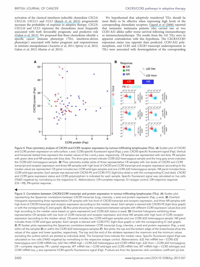
Tumour-infiltrating lymphocyte flow cytometry. We validatedthe correlation between transcript and protein level in 50 availableTIL samples (Figure 3). All TIL samples displayed strong,although variable, expression of CXCR3. CCR5 expression wasweaker. The fluorescence intensity curve of the two chemokinereceptors assumed a skewed Gaussian distribution without theevidence of well-defined subpopulations. Protein expression ofCXCR3 correlated with the expression of their respectivetranscripts (Figure 3A). CCR5 transcript expression and CCR5protein expression correlated in both WT and D32 samples(Figure 3E and F). CCR5-D32 strongly influenced the expressionof CCR5 protein. The lower expression of CCR5 protein inCCR5-D32 in monocytes and T cells from CCR5-D32 carriers hasbeen previously demonstrated (Carrington et al, 1999), but nodata are available (to the best of our knowledge) on TILs. In fact,TILs used for adoptive therapy are cultured and expandedin vitro with consequent possible modulation of surface markers.
We observed that the level of CCR5 receptor in CCR5-D32heterozygous samples expressing high levels of CCR5 transcriptswas approximately the half of that detected in CCR5 WT samples(Figure 3D). No expression of CCR5 receptor on cell surface wasobserved (as expected) in the CCR5-D32 homozygous sample.Therefore, even if post-transcriptional changes could influencethe expression of CCR5 receptor (Shimizu et al, 2009), thepresence of D32 polymorphism has a major role in lowering theexpression of CCR5 protein in presence of high CCR5 transcriptsin TILs.
Despite the bias selection (see Materials and Methods section), itis noteworthy that only 1 out of 18 CR samples overexpressed bothchemokine receptors (Figure 4). Thus, flow cytometry confirmedthe PPM suggesting that estimates based on germ line andtranscriptional data could be extended to the remaining samplesfor which TILs were no longer available.
Pretreatment tumour biopsy gene expression. We investigatedthe role of the coordinate intratumoral expression of CXCR3/CXCR3 ligand gene expression (i.e., CXCL9, CXCL10, andCXCL11) and CCR5/CCR5 ligands (i.e., CCL3, CCL4, and CCL5)in 113 available pretreatment tumour biopsies used for TILgeneration.
CXCL9, CXCL10, CXCL11 and CCL5 were co-expressed(Figure 5A) and, when used for hierarchical clustering analysis
Table 3. Association between TIL CXCR3, CCR5 expression, CCR5-D32 polymorphism, and their interaction, with clinical outcome
Model A (gene expression model): expression of CXCR3 and CCR5, and their interactions
Full model definition Association with clinical outcome
ComparatorOutcomeP-valuea
Full model A CR OR
YBuþ x1þ x2þ x1x2 YBu 0.032 0.15
Model B (gene expression/polymorphism model): expression of CXCR3 and CCR5, and CCR5-D32 polymorphism
Full model definition Association with clinical outcome
ComparatorOutcomeP-valuea
Full model B CR OR
YBuþ x1þ x2þ x3þ x1x2þ x1x3þ x2x3þ x1x2x3 YBu 0.0009 0.007Full model A 0.003 0.007
Model C (gene expression and treatment model): treatment, expression of CCR5and CXCR3, and their interactions
Full model definition Association with clinical outcome
ComparatorOutcomeP-valuea
Full model C CR OR
YBuþRxþ x1þ x2þ x1x2 YBuþRx 0.002 0.010
Model D (gene expression/polymorphism and treatment model): treatment, expression of CXCR3 and CCR5, CCR5-D32 polymorphism,and their interactions
Full model definition Association with clinical outcome
ComparatorOutcomeP-valuea
Full model D CR OR
YBuþRxþ x1þ x2þ x3þ x1x2þ x1x3þ x2x3þ x1x2x3 YBuþRx 0.0001 0.003Full Model C 0.005 0.036
Abbreviations: CR¼ complete response; OR¼overall response; Rx¼ treatment (TNMA, T200, T1200, TYT, and TCD8); TIL¼ tumour-infiltrating lymphocyte; Y¼ end point (CR or OR);YBu¼ null model; X1¼ expression of CCR5; X2¼expression of CXCR3; X3¼CCR5-D32 polymorphism.aThe log likelihood ratio test to was used to compare the fit of two models, one of which (the null model) is a special case of the other (the alternative model). The test is based on the loglikelihood ratio, which expresses how many times more likely the data are under one model than the other. During the model fitting, the sample with missing genotype data was omitted.
CXCR3/CCR5 pathways in adoptive therapy BRITISH JOURNAL OF CANCER
www.bjcancer.com | DOI:10.1038/bjc.2013.557 2417

based on mean-centered expression values (Figure 5B), segregatedmelanoma metastases into three main groups: cluster 1 includedsamples with low CXCR3 and CCR5 ligand gene expression;cluster 2 and cluster 3 with intermediate and high expression,respectively. Cluster 1 was disproportionately enriched withsamples from NR patients (OR rate: 38%, P¼ 0.030) and cluster3 with samples from OR patients (OR rate: 65%, P¼ 0.031),whereas cluster 2 included an intermediate proportion of ORsamples (52%), suggesting an association between expression ofCXCL9, CXCL10, CXCL11 and CCL5 and immune responsiveness.CXCL9, CXCL10, CXCL11 and CCL5 are the classical interferoninducible chemokines and are coordinately regulated by STAT1signalling (Chandrasekar et al, 2013; Rauch et al, 2013). Nocorrelation with CR or OR was observed when samples wereclustered according to the other markers (i.e., CCL3, CCL4,CXCR3, and CCR5, Supplementary Figure S4).
DISCUSSION
Activated cytotoxic and helper-1 T cells express CXCR3 and CCR5on cell surface (Franciszkiewicz et al, 2012). The intratumoraloverexpression of CXCR3 and/or CCR5 ligand genes in primary
VariableOR Rate
OR vs NRn = 142
Odds ratio
CCR5-Δ32 vs WT (DNA) 24% (6/25) vs 16%(19/116)
1.61
CXCR3/CCR5 low vs high(PPM)
36% (17/47) vs 8%(8/95)
6.16
CCR5 low vs high (mRNA) 21% (15/71) vs 14%(10/71)
1.63
CXCR3 low vs high (mRNA) 25% (18/71) vs 10%(7/71)
3.11
CXCR3/CCR5 low vs high(mRNA)
32% (13/41) vs 12%(12/101)
3.44
CCR5 low vs high (mRNA) 48% (34/71) vs 52%(37/71)
0.84
CCR5-Δ32 vs WT (DNA) 68% (17/25) vs 46%(53/116)
2.53
CXCR3 low vs high (mRNA) 54% (38/71) vs 47%(33/71)
1.33
CXCR3/CCR5 low vs high(mRNA)
59% (24/41) vs 47%(47/101)
1.62
CXCR3/CCR5 low vs high(PPM)
64% (30/47) vs 43%(41/95)
2.32
DNA
RNA
RNA
RNA
PPM
VariableCR rate
CR vs non-CRn = 142
Odds ratio
RNA
RNA
RNA
PPM
DNA
Objective response (OR vs NR)
CCR5 low vs high (PPM) 22% (19/85) vs 11%(6/57)
2.45 PPM
CCR5 low vs high (PPM) 54% (46/85) vs 44%(25/57)
1.51PPM
0 1 2 3 4 5 6 7
0 1 2 3 4 5 6 7
Complete response (CR vs non-CR)
Figure 1. Effect of CXCR3/CCR5 expression status of tumor-infiltrating lymphocytes (TILs) on complete response (A) and objective response (B).Odds ratios are represented. CCR5-D32 (DNA): presence of D32 polymorphism; CCR5-D32 low (PPM): CCR5 low according to the PPM (CCR5below the median value according to the gene expression data or presence of CCR5-D32 polymorphism); CCR5 low (mRNA): CCR5 below themedian value according to the gene expression data; CXCR3 low (RNA): CXCR3 below the median value according to the gene expression data;CXCR3/CCR5 low (mRNA): CXCR3 and CCR5 below the median value according to the gene expression data; CXCR3/CCR5 low (PPM): CXCR3and CCR5 below the median value according to the gene expression data or CXCR3 low and CCR5 high in presence of CCR5-D32 polymorphism.P-values are from w2-test. Abbreviations: CI¼ confidence interval; CR¼ complete response; non-CR¼PRþNR; OR¼objective response (CRþPR);PPM¼protein prediction model; PR¼partial response; WT¼wild-type.
CCR5 high transcript expression CCR5 high protein expressio
CCR5 high transcript expression CCR5 low protein expressio
Δ32 Polymorphism
Wild-type
A
B
Figure 2. Protein prediction model. In wild-type subjects (A) weassume that high levels of CCR5 transcript are associated with highexpression of CCR5 on cell surface. However, because D32polymorphism encodes a protein not expressed on the cell surface (B),CCR5-D32 carriers were expected to have a low receptor expression(decreased in heterozygous individuals and absent in homozygousindividuals) despite the high transcript expression.
BRITISH JOURNAL OF CANCER CXCR3/CCR5 pathways in adoptive therapy
2418 www.bjcancer.com | DOI:10.1038/bjc.2013.557

tumours has been correlated with high density of T cells andreduced risk of relapse or death in the lung (CCL5; Moran et al,2002), melanoma (CCL5, CXCL9, and CXCL10), ovarian, (CXCL9and CCL5; Zhang et al, 2003; Leffers et al, 2010) colorectal (CCL5,CXCL9, and CXCL10; Mlecnik et al, 2010; Tosolini et al, 2011), andbreast cancer (CCL5, CXCL9, and CXCL10; Ascierto et al, 2012;Curtis et al, 2012). Consistently, Messina et al (2012) recentlyreported a prolonged survival in metastatic melanoma patients
bearing tumours overexpressing CCL3, CCL4, CCL5, CXCL9,CXCL10, and CXCL11 (Messina et al, 2012). In addition, thesechemokine genes have been associated with a higher likelihood torespond to ipilimumab (CCL4, CCL5, CXCL9, CXCL10, andCXCL11; Ji et al, 2012) and MAGE-A3 vaccination (CXCL9, CXCL10and CCL5; Ulloa-Montoya et al, 2013) (Wang et al, 2013a).
Testing pretreatment biopsies from 113 melanoma patientsundergoing adoptive therapy, we observed that the coordinated
� = 0.44
P = 0.001
CR
% o
f max
mRNA high mRNA high mRNA high
mRNA low mRNA low
S04 S05 S35
S14S50S43
mRNA low
NR
Ratio=31.3 Ratio=44.2
Ratio=15.2
Ratio=27.4
Ratio=13.7Ratio=17.7
CXCR3
CX
CR
3 pr
otei
n (F
sp)
CXCR3 mRNA (log2 intensity)
NR
CR
% o
f max
WT mRNA high WT mRNA high WT mRNA high
WT mRNA low WT mRNA low Δ32 Het mRNAlow
S04 S05 S23
S15S50S43
� = 0.65
WT Δ32 Het
Ratio=1.5Ratio=2.5Ratio=2.1
Ratio=8.0 Ratio=6.3 Ratio=7.1
CCR5
� = 0.69
CC
R5
pro
tein
(F
sp)
CC
R5
pro
tein
(F
sp)
CC
R5
pro
tein
(F
sp)
CCR5 mRNA (Log2 intensity) CCR5 mRNA (Log2 intensity)
CCR5CCR5
0.00
7.75 8.00 8.25 8.50 8.75 9.00
10.00
20.00
30.00
40.00
50.00
Response
CRPRNR
Response
CRPR
NR
Response
CRPRNR
100
80
60
40
20
0100 101 102 103 104
0.0
Δ32Hom
Δ32 HetmRNA
low
Δ32 HetmRNAhigh
WTmRNA
low
WTmRNAhigh
2.0
4.0
6.0
8.0
2.000.50
1.00
1.50
2.00
2.50
6.00 6.50 7.00 7.50 8.00 6.00 6.50 7.00 7.50 8.008.50
4.00
6.00
8.00
100
80
60
40
20
0100 101 102 103 104
100
80
60
40
20
0100 101 102 103 104
100 101 102 103 104
100
80
60
40
20
0100 101 102 103 104
100
80
60
40
20
0100 101 102 103 104
100
80
60
40
20
0100 101 102 103 104
100
80
60
40
20
0100 101 102 103 104
100
80
60
40
20
0100 101 102 103 104
100
80
60
40
20
0100 101 102 103 104
100
80
60
40
20
0100 101 102 103 104
100
80
60
40
20
0100 101 102 103 104
P < 0.001 P = 0.04
FITC: IC/CXCR3
FITC: IC/CCR5
Figure 3. For caption see next page.
CXCR3/CCR5 pathways in adoptive therapy BRITISH JOURNAL OF CANCER
www.bjcancer.com | DOI:10.1038/bjc.2013.557 2419
activation of the classical interferon inducible chemokine CXCL9,CXCL10, CXCL11 and CCL5 (Rauch et al, 2013) progressivelyincreases the probability of response to adoptive therapy. CXCL9,CXCL10 and CCL5 represent the chemokines most frequentlyassociated with both favourable prognostic and predictive role(Galon et al, 2013). We proposed that these chemokines identify aspecific cancer immune phenotype (‘Th1, interferon-drivenphenotype’) associated with better prognosis and responsivenessto immune manipulations (Ascierto et al, 2011; Spivey et al, 2012;Galon et al, 2013; Murtas et al, 2013).
We hypothesised that adoptively transferred TILs should bemost likely to be effective when expressing high levels of thecorresponding chemokine receptors. Ugurel et al (2008) reportedthat metastatic melanoma patients who carried one or twoCCR5-D32 alleles suffer worse survival following immunotherapyor immunochemotherapy. The results from the 142 TILs were inapparent contradiction with this hypothesis. The CXCR3/CCR5expression status was opposite than predicted. CCR5-D32 poly-morphism, and CCR5 and CXCR3 transcript underexpression inTILs were associated with downregulation of the corresponding
Figure 3. Correlation between CXCR3/CCR5 transcript and protein expression in tumour-infiltrating lymphocytes (TILs). (A) Scatter plotrepresenting the Spearman correlations between CXCR3 transcript (Log2 intensity, x axis) and protein expression (Fsp, y axis). (B) Overlaidhistograms representing three representative CR samples with low level of CXCR3 transcript and receptor expression, and three NR samples withhigh level of CXCR3 transcript and receptor expression (according to the median value). Each sample is stained with CXCR3-PE (light blue graph)or with the corresponding IC (red graph). (C) Box plots showing the comparisons between samples with different CCR5 expression values (low andhigh according to the median value based on gene expression) and CCR5-D32 status (x axes): (D) Overlaid histograms exhibiting threerepresentative CR samples with low level of CCR5 transcript and receptor expression and three NR samples with high level of CCR5 receptorexpression (according to the median value). CR panel includes two CCR5 wild-type samples and one CCR5-D32 heterozygous sample. NR panelincludes three CCR5 wild-type samples. Each sample is stained with CCR5-FITC (light blue graph) or with the corresponding IC (red graph).(E, F) Scatter plots representing the Spearman correlations between CCR5 transcript (Log2 intensity, x axis) and protein expression (Fsp, y axis)within all the samples (E) or within the CCR5-D32 heterozygous samples (F). Box plots: the top and the bottom edge of the tinted boxes show thevalues of the upper and lower quartiles, respectively. The top and the end of the whiskers represent the maximum and the minimum valuesexcluding the outliers (which are plotted as individual dots). The horizontal lines indicate the median value. Specific fluorescent signal (Fsp) wascalculated on live cells (7AAD negative) by normalising to the respective isotype control. Abbreviations: D32 Het mRNA low¼CCR5-D32heterozygous and CCR5 mRNA low; D32 Het mRNA high¼CCR5-D32 heterozygous and CCR5 mRNA high; D32 Hom¼CCR5-D32 homozygous;CR¼ complete response; PR¼partial response; WT mRNA low¼CCR5 wild-type and CCR5 mRNA low; WT mRNA high¼CCR5 wild-type andCCR5 mRNA low; y axis represents CCR5-specific fluorescence signal (Fsp). P-values are from the Spearman rank correlation coefficient (r) test.
NRCR
FIT
C: IC
- CC
R5
PE IC - CXCR3
mR
NA
CX
CR
3/CC
R5
high (WT
)
mR
NA
CX
CR
3/CC
R5
high (WT
)
mR
NA
C
XC
R3/C
CR
5high (W
T)
mR
NA
C
XC
R3/C
CR
5low
(Δ32 )
mR
NA
CX
CR
3/CC
R5
low (W
T)
mR
NA
CX
CR
3/CC
R5
low (W
T)
Δ32 Het
Δ32 Hom
CX
CR
3 pr
otei
n (F
sp)
CCR5 protein (Fsp)
50Response
CRPRNR
40
30
20
10
0
0 2 4 6 8
Figure 4. Flow cytometry analysis of CXCR3 and CCR5 receptor expression by tumour-infiltrating lymphocytes (TILs). (A) Scatter plot of CXCR3and CCR5 protein expression on cells surface. x axis: CCR5-specific fluorescent signal (Fsp); y axis: CXCR3-specific fluorescent signal (Fsp). Verticaland horizontal dotted lines represent the median values of the x and y axes, respectively. CR samples are represented with red dots, PR sampleswith green dots and NR samples with blue dots. The short grey arrows indicate CCR5-D32 heterozygous sample and the long grey arrow indicatesthe CCR5-D32 homozygous sample. (B) Flow cytometry scatter plots of three representative CR samples with low levels of CXCR3 and CCR5transcript and receptor expression and three NR samples with high level of CXCR3 and CCR5 transcript and receptor expression (according to themedian value) are represented. CR panel includes two CCR5 wild-type samples and one CCR5-D32 heterozygous sample. NR panel includes threeCCR5 wild-type samples. Each sample was stained with CXCR3-PE and CCR5-FITC (light blue dots) or with the corresponding IC (red dots). CXCR3and CCR5 gene expression status and CCR5 polymorphism is indicated for each sample. Specific fluorescent signal was calculated on live cells(7AAD negative) by normalising to the respective IC. Abbreviations: CR=complete response; IC=isotype control; OR=objective response(CRþPR); PR=partial response.
BRITISH JOURNAL OF CANCER CXCR3/CCR5 pathways in adoptive therapy
2420 www.bjcancer.com | DOI:10.1038/bjc.2013.557

receptors and higher likelihood (OR rate) and degree (CR rate) ofresponse. The association with achievement of a CR is intriguingand particularly relevant as CR patients treated with adoptivetherapy usually experience a extremely durable remission (up to100% of overall survival at 3 years (Rosenberg et al, 2011)). Thestrength of the association with response progressively increased asCCR5 and CXCR3 expression data were combined and wereintegrated with CCR5 sequencing data, as indicated by the loglikelihood test for models.
These counterintuitive and surprising results could be explainedby the dynamics of TIL migration to the tumour in relation toconcomitant IL-2 administration. Localisation of TILs to thetumour site is necessary for their antitumour activity (Pockaj et al,1994). However, soon (2 h) after high-dose IL-2 administration, theinfused TILs rapidly disappear from the circulation. They do notlocalise to tumour tissue and rather migrate to normal liver, lung,and spleen (Fisher et al, 1989). Migration to the tumour begins 24or 48 h after IL-2 administration and increases over time duringthe following days (Fisher et al, 1989). However, concentrations ofCCR5 and CXCR3 ligands (e.g., CCL3, CCL4, CXCL9, CXCL10and CXCL11) increase immediately after IL-2 administration(Panelli et al, 2004b). Our previous genomic and proteomic studiessuggest that IL-2 seems to exert its effect on the tumour siteindirectly by inducing monocyte activation, followed by a delayed,dose-dependent release of chemoattractants such as CXCR3 andCCR5 ligands that recruit TILs only in the later stages of treatment(Panelli et al, 2002; Wang et al, 2002; Panelli et al, 2004b; Weiss
et al, 2011). In fact, administration of IL-2 is followed by a rapidclearance of peripheral mononuclear cells, without any evidence oftheir migration to tumour metastases at early time points (Panelliet al, 2002), consistently with results from adoptive therapy studies(Pockaj et al, 1994). Several richly perfused organs including thelung and liver, populated with IL-2-receptor carrying cells, areprobably the primary target of systemic IL-2 administration, and itis there where the inflammatory cytokine storm is probablyinitiated leading, among others, to the production of massivequantities of CXCR3 and/or CCR5 ligands. A significant associa-tion was reported with increased persistence of TILs in thecirculation 1 month following treatment and likelihood of clinicalresponse (Dudley et al, 2002; Rosenberg et al, 2011). It is temptingto hypothesise that a low expression of chemokine receptors byTILs in the acute phases of treatment might prevent theirsequestration by extratumoral tissues and paradoxically allow theirsubsequent localisation to the tumour when the cytokine storm hassubsided and the tumour remains the only tissue maintainingexpression of chemokines. This hypothesis would be consistentwith the favourable predictive weight that higher expression ofCXCL9, CXCL10, CXCL11, and CCL5 in steady state conditionsconfers. The enhancement of the expression of these chemokinesin tumour/tumour microenvironment could identify novelstrategies for the improvement of current immunotherapies(Muthuswamy et al, 2012; Pescatori et al, 2013).
These ligands could modulate the tumour microenvironment inother ways, independently of the recruitment of T cells. CCR5
CC
L3
CX
CR
3
CC
L4
CC
L5
CC
R5
CX
CL11
CX
CL10
CX
CL9
CXCL10
CCL5
CXCL11
CXCL9
Cluster 1 (low):OR: 38% (16/42)
CR
NR
PR
Cluster 2 (MID)OR: 52% (16/31)
Cluster 3 (high)OR: 65% (26/40)
OR enrichment
Cluster 3 vs cluster 1 + cluster 2 P = 0.031Odds ratio (95% CI) = 2.13 (1.07–5.28)
Cluster 1 vs cluster 2 + cluster 3 P = 0.030Odds ratio (95% CI) = 0.43 (0.19–0.93)
CR enrichmentNS
OR rate: cluster 1 < cluster 2 < cluster 3
CXCL9/10/11, CCL5
CCL3
CXCR3
CCL4
CCR5
CCL5
CXCL9
CXCL11
CXCL10
Cluster 2 vs cluster 1 + cluster 3 P = 0.97Odds ratio (95% CI) = 1.02 (0.44–2.32)
Figure 5. CXCR3/CCR5 ligands gene expression in pretreatment biopsies. (A) Hierarchical clustering of gene–gene correlation from 113pretreatment tumours. The Spearman rank correlation coefficient (r) was calculated between combination of CXCR3, CCR5, CXCR3 ligands(CXCL9, CXCL10, and CXCL11) and CCR5 ligands (CCL3, CCL4, and CCL5). CXCL9, CXCL10, CXCL11 and CCL5 clustered together and wereselected for hierarchical clustering analysis based on the mean-centered gene expression values (B). Patients with clinical complete responses (CR)are shown in red, patients with partial remission (PR) are shown in green and non-responders (NR) are shown in blue. Abbreviations: non-CR¼PRþNR; OR¼objective response (CRþPR). P-values are from the w2-test.
CXCR3/CCR5 pathways in adoptive therapy BRITISH JOURNAL OF CANCER
www.bjcancer.com | DOI:10.1038/bjc.2013.557 2421

blockade, for example, has been demonstrated to enhanceproliferation of xenografts from breast cancer cells bearing WTp53, and the presence of D32 polymorphism has been associatedwith higher risk of relapse in breast cancer patients with p53 WTtumours (Manes et al, 2003). Thus, the aforementioned observa-tion of Ugurel et al (2008) only apparently clashes with ourfindings. As response rate was not assessed in that study, the D32polymorphism mutation may have enhanced tumour growththrough a direct effect on tumour biology (Manes et al, 2003),resulting in the observed poor survival in these patients (Ugurelet al, 2008). As in our study the presence of CCR5-D32 mutationwas not associated with worse response (and rather, whenconsidered as single parameter, it was marginally associated withbetter OR, P¼ 0.043), it is likely that the direct modulation ofCCR5 in cancer tumour cells has a little (if any) impact on theclinical outcome of patients undergoing adoptive therapy.
However, it is possible that aside from the adoptive transfermodel, when activated CXCR3þ /CCR5þ TILs are infusedin combination with high-dose IL-2, during other treatments(e.g., combination of immunochemotherapy (Ugurel et al, 2008)),the proportional induction of the CXCR3 and CCR5 ligandsfollowing treatment is not as unbalanced as it is in our study.
Although we stress that our findings must be prospectivelyvalidated, the significance of our observation is supported by thefact that it does not come from a merely retrospectiveinvestigation. Retrospective investigations performed on archivedspecimens are often conducted independent of a specified protocoland are unfocused, with numerous patient subsets and end points(Simon et al, 2009). Our prospective-retrospective (Simon et al,2009) investigation was rather designed to study specimenscollected during prospective trials and specifically stored topreserve DNAs, mRNAs and proteins in order to perform thededicated assays.
Animal models can provide important mechanistic insights intohuman diseases. Studies in mouse models employing adoptivelytransferred lymphocytes have emphasised a key role of theupregulation of CCR5 in TILs in mediating tumour rejection(Gonzalez-Martin et al, 2012). Our present study, however, fails todemonstrate that the downregulation of CCR5 in TIL is adetrimental factor in this setting in humans.
However, in view of the intrinsic limitations of the investigation(e.g., the possible data overfitting because of the use of regressionmodels), our results should be considered exploratory and need tobe validated by integrated quantitative PCR, sequencing, and flowcytometric analyses in an independent cohort of patients.
Considering the complexity of tumour–host interactions, webelieve that only a prospective clinical translation study employingserial biopsies (i.e., pre- and post treatment) in different tissues andspecifically designed to asses T-cell tumour trafficking in humans canconclusively confirm the hypotheses generated by our present report.
In conclusion, our study identified candidate biomarkers ofimmune responsiveness in this setting by applying a combinatorialmultifactorial approach. In fact, none of the individual parametersassessed had predictive value alone, but the coordinated analysis ofthe two molecules corrected for the predicted function according tothe genetic polymorphism yielded strong predictive value. Thispoint proposes a paradigm shift for the identification of clinicallyrelevant biomarkers in complex biological problems. The optimi-sation of this approach in genomic-scale models represents a futurechallenge (Wang et al, 2013b).
ACKNOWLEDGEMENTS
This study was supported by the Intramural Research program ofthe US National Institutes of Health and by the Conquer Cancer
Foundation of the American Society of Clinical Oncology (2011Young Investigator Award Grant, granted to Davide Bedognetti).Dr Bedognetti’s fellowship was initially supported by the ItalianAssociation of Medical Oncology (AIOM) Foundation.
CONFLICT OF INTEREST
The authors declare no conflict of interest.
REFERENCES
Ascierto ML, De Giorgi V, Liu QZ, Bedognetti D, Spivey TL, Murtas D, Uccellini L,Ayotte BD, Stroncek DF, Chouchane L, Manjili MH, Wang E, Marincola FM(2011) An immunologic portrait of cancer. J Transl Med 9: 146.
Ascierto ML, Kmieciak M, Idowu MO, Manjili R, Zhao Y, Grimes M, Dumur C,Wang E, Ramakrishnan V, Wang XY, Bear HD, Marincola FM, Manjili MH(2012) A signature of immune function genes associated with recurrence-free survival in breast cancer patients. Breast Cancer Res Treat 131(3):871–880.
Bedognetti D, Balwit JM, Wang E, Disis ML, Britten CM, Delogu LG, Tomei S,Fox BA, Gajewski TF, Marincola FM, Butterfield LH (2011) SITC/iSBTccancer immunotherapy biomarkers resource document: online resourcesand useful tools—a compass in the land of biomarker discovery.J Transl Med 9: 155.
Bedognetti D, Wang E, Sertoli MR, Marincola FM (2010) Gene-expressionprofiling in vaccine therapy and immunotherapy for cancer. Expert RevVaccines 9(6): 555–565.
Carrington M, Dean M, Martin MP, O0Brien SJ (1999) Genetics of HIV-1infection: chemokine receptor CCR5 polymorphism and its consequences.Hum Mol Genet 8(10): 1939–1945.
Chandrasekar B, Deobagkar-Lele M, Victor ES, Nandi D (2013) Regulation ofchemokines, CCL3 and CCL4, by interferon gamma and nitric oxidesynthase 2 in mouse macrophages and during Salmonella enterica serovartyphimurium infection. J Infect Dis 207(10): 1556–1568.
Choi JW, Park CS, Hwang M, Nam HY, Chang HS, Park SG, Han BG, Kimm K,Kim HL, Oh B, Kim Y (2008) A common intronic variant of CXCR3 isfunctionally associated with gene expression levels and the polymorphicimmune cell responses to stimuli. J Allergy Clin Immunol 122(6):1119–1126, e7.
Curtis C, Shah SP, Chin SF, Turashvili G, Rueda OM, Dunning MJ, Speed D,Lynch AG, Samarajiwa S, Yuan Y, Graf S, Ha G, Haffari G, Bashashati A,Russell R, McKinney S, Langerod A, Green A, Provenzano E, Wishart G,Pinder S, Watson P, Markowetz F, Murphy L, Ellis I, Purushotham A,Borresen-Dale AL, Brenton JD, Tavare S, Caldas C, Aparicio S (2012) Thegenomic and transcriptomic architecture of 2,000 breast tumours revealsnovel subgroups. Nature 486(7403): 346–352.
Dudley ME, Gross CA, Langhan MM, Garcia MR, Sherry RM, Yang JC, Phan GQ,Kammula US, Hughes MS, Citrin DE, Restifo NP, Wunderlich JR, Prieto PA,Hong JJ, Langan RC, Zlott DA, Morton KE, White DE, Laurencot CM,Rosenberg SA (2010) CD8þ enriched ‘young’ tumor infiltrating lymphocytescan mediate regression of metastatic melanoma. Clin Cancer Res 16(24):6122–6131.
Dudley ME, Wunderlich JR, Robbins PF, Yang JC, Hwu P, Schwartzentruber DJ,Topalian SL, Sherry R, Restifo NP, Hubicki AM, Robinson MR, Raffeld M,Duray P, Seipp CA, Rogers-Freezer L, Morton KE, Mavroukakis SA, White DE,Rosenberg SA (2002) Cancer regression and autoimmunity in patients afterclonal repopulation with antitumor lymphocytes. Science 298(5594): 850–854.
Dudley ME, Wunderlich JR, Shelton TE, Even J, Rosenberg SA (2003)Generation of tumor-infiltrating lymphocyte cultures for use in adoptivetransfer therapy for melanoma patients. J Immunother 26(4): 332–342.
Ellinghaus D, Schreiber S, Franke A, Nothnagel M (2009) Current software forgenotype imputation. Hum Genomics 3(4): 371–380.
Fisher B, Packard BS, Read EJ, Carrasquillo JA, Carter CS, Topalian SL, Yang JC,Yolles P, Larson SM, Rosenberg SA (1989) Tumor localization of adoptivelytransferred indium-111 labeled tumor infiltrating lymphocytes in patientswith metastatic melanoma. J Clin Oncol 7(2): 250–261.
Franciszkiewicz K, Boissonnas A, Boutet M, Combadiere C, Mami-Chouaib F(2012) Role of chemokines and chemokine receptors in shaping theeffector phase of the antitumor immune response. Cancer Res 72(24):6325–6332.
BRITISH JOURNAL OF CANCER CXCR3/CCR5 pathways in adoptive therapy
2422 www.bjcancer.com | DOI:10.1038/bjc.2013.557

Gajewski TF, Fuertes M, Spaapen R, Zheng Y, Kline J (2011) Molecularprofiling to identify relevant immune resistance mechanisms in the tumormicroenvironment. Curr Opin Immunol 23(2): 286–292.
Galon J, Angell Helen K, Bedognetti D, Marincola Francesco M (2013) Thecontinuum of cancer immunosurveillance: prognostic, predictive, andmechanistic signatures. Immunity 39(1): 11–26.
Gonzalez-Martin A, Mira E, Manes S (2012) CCR5 in cancer immunotherapy:More than an ‘attractive’ receptor for T cells. Oncoimmunology 1(1): 106–108.
Huang Y, Paxton WA, Wolinsky SM, Neumann AU, Zhang L, He T, Kang S,Ceradini D, Jin Z, Yazdanbakhsh K, Kunstman K, Erickson D, Dragon E,Landau NR, Phair J, Ho DD, Koup RA (1996) The role of a mutant CCR5 allelein HIV-1 transmission and disease progression. Nat Med 2(11): 1240–1243.
Huebner C, Petermann I, Browning BL, Shelling AN, Ferguson LR (2007)Triallelic single nucleotide polymorphisms and genotyping error in geneticepidemiology studies: MDR1 (ABCB1) G2677/T/A as an example. CancerEpidemiol Biomarkers Prev 16(6): 1185–1192.
Ji RR, Chasalow SD, Wang L, Hamid O, Schmidt H, Cogswell J, Alaparthy S,Berman D, Jure-Kunkel M, Siemers NO, Jackson JR, Shahabi V (2012) Animmune-active tumor microenvironment favors clinical response toipilimumab. Cancer Immunol Immunother 61(7): 1019–1031.
Leffers N, Fehrmann RS, Gooden MJ, Schulze UR, Ten Hoor KA, Hollema H,Boezen HM, Daemen T, de Jong S, Nijman HW, van der Zee AG (2010)Identification of genes and pathways associated with cytotoxic T lymphocyteinfiltration of serous ovarian cancer. Br J Cancer 103(5): 685–692.
Manes S, Mira E, Colomer R, Montero S, Real LM, Gomez-Mouton C,Jimenez-Baranda S, Garzon A, Lacalle RA, Harshman K, Ruiz A, Martinez AC(2003) CCR5 expression influences the progression of human breast cancer ina p53-dependent manner. J Exper Med 198(9): 1381–1389.
Messina JL, Fenstermacher DA, Eschrich S, Qu X, Berglund AE, Lloyd MC,Schell MJ, Sondak VK, Weber JS, Mule JJ (2012) 12-chemokine genesignature identifies lymph node-like structures in melanoma: potential forpatient selection for immunotherapy? Sci Rep 2: 765.
Mlecnik B, Tosolini M, Charoentong P, Kirilovsky A, Bindea G, Berger A,Camus M, Gillard M, Bruneval P, Fridman WH, Pages F, Trajanoski Z,Galon J (2010) Biomolecular network reconstruction identifies T-cellhoming factors associated with survival in colorectal cancer.Gastroenterology 138(4): 1429–1440.
Moran CJ, Arenberg DA, Huang CC, Giordano TJ, Thomas DG, Misek DE,Chen G, Iannettoni MD, Orringer MB, Hanash S, Beer DG (2002)RANTES expression is a predictor of survival in stage I lungadenocarcinoma. Clin Cancer Res 8(12): 3803–3812.
Murtas D, Maric D, De Giorgi V, Reinboth J, Worschech A, Fetsch P, Filie A,Ascierto ML, Bedognetti D, Liu Q, Uccellini L, Chouchane L, Wang E,Marincola FM, Tomei S (2013) IRF-1 responsiveness to IFN-gammapredicts different cancer immune phenotypes. Br J Cancer 109(1): 76–82.
Muthuswamy R, Berk E, Junecko BF, Zeh HJ, Zureikat AH, Normolle D,Luong TM, Reinhart TA, Bartlett DL, Kalinski P (2012) NF-kappaBhyperactivation in tumor tissues allows tumor-selective reprogramming ofthe chemokine microenvironment to enhance the recruitment of cytolyticT Effector cells. Cancer Res 72(15): 3735–3743.
Panelli MC, Martin B, Nagorsen D, Wang E, Smith K, Monsurro V, Marincola FM(2004a) A genomic- and proteomic-based hypothesis on the eclectic effects ofsystemic interleukin-2 administration in the context of melanoma-specificimmunization. Cells Tissues Organs 177(3): 124–131.
Panelli MC, Wang E, Phan G, Puhlmann M, Miller L, Ohnmacht GA, Klein HG,Marincola FM (2002) Gene-expression profiling of the response of peripheralblood mononuclear cells and melanoma metastases to systemic IL-2administration. Genome Biol 3(7): RESEARCH0035.
Panelli MC, White R, Foster M, Martin B, Wang E, Smith K, Marincola FM(2004b) Forecasting the cytokine storm following systemic interleukin(IL)-2 administration. J Transl Med 2(1): 17.
Pescatori M, Bedognetti D, Venturelli E, Menard-Moyon C, Bernardini C,Muresu E, Piana A, Maida G, Manetti R, Sgarrella F, Bianco A, Delogu LG(2013) Functionalized carbon nanotubes as immunomodulator systems.Biomaterials 34(18): 4395–4403.
Pockaj BA, Sherry RM, Wei JP, Yannelli JR, Carter CS, Leitman SF, CarasquilloJA, Steinberg SM, Rosenberg SA, Yang JC (1994) Localization of111indium-labeled tumor infiltrating lymphocytes to tumor in patientsreceiving adoptive immunotherapy. Augmentation with cyclophosphamideand correlation with response. Cancer 73(6): 1731–1737.
Pos Z, Selleri S, Spivey TL, Wang JK, Liu H, Worschech A, Sabatino M,Monaco A, Leitman SF, Falus A, Wang E, Alter HJ, Marincola FM (2010)Genomic scale analysis of racial impact on response to IFN-alpha. ProcNatl Acad Sci USA 107(2): 803–808.
Rauch I, Muller M, Decker T (2013) The regulation of inflammation byinterferons and their STATs. JAK-STAT 2(1): e23820–13.
Rosenberg SA, Yang JC, Sherry RM, Kammula US, Hughes MS, Phan GQ,Citrin DE, Restifo NP, Robbins PF, Wunderlich JR, Morton KE,Laurencot CM, Steinberg SM, White DE, Dudley ME (2011) Durablecomplete responses in heavily pretreated patients with metastatic melanomausing T-cell transfer immunotherapy. Clin Cancer Res 17(13): 4550–4557.
Shimizu S, Kamata M, Kittipongdaja P, Chen KN, Kim S, Pang S, Boyer J, Qin FX,An DS, Chen IS (2009) Characterization of a potent non-cytotoxic shRNAdirected to the HIV-1 co-receptor CCR5. Genet Vaccines Ther 7: 8.
Simon RM, Paik S, Hayes DF (2009) Use of archived specimens in evaluation ofprognostic and predictive biomarkers. J Natl Cancer Inst 101(21): 1446–1452.
Spivey TL, De Giorgi V, Zhao YD, Bedognetti D, Pos Z, Liu QZ, Tomei S,Ascierto ML, Uccellini L, Reinboth J, Chouchane L, Stroncek DF, Wang E,Marincola FM (2012) The stable traits of melanoma genetics: an alternateapproach to target discovery. BMC Genomics 13: 156.
Spivey TL, Uccellini L, Ascierto ML, Zoppoli G, De Giorgi V, Delogu LG,Engle AM, Thomas JM, Wang E, Marincola FM, Bedognetti D (2011)Gene expression profiling in acute allograft rejection: challenging theimmunologic constant of rejection hypothesis. J Transl Med 9: 174.
Tosolini M, Kirilovsky A, Mlecnik B, Fredriksen T, Mauger S, Bindea G, Berger A,Bruneval P, Fridman WH, Pages F, Galon J (2011) Clinical impact of differentclasses of infiltrating T cytotoxic and helper cells (Th1, th2, treg, th17) inpatients with colorectal cancer. Cancer Res 71(4): 1263–1271.
Tran KQ, Zhou J, Durflinger KH, Langhan MM, Shelton TE, Wunderlich JR,Robbins PF, Rosenberg SA, Dudley ME (2008) Minimally cultured tumor-infiltrating lymphocytes display optimal characteristics for adoptive celltherapy. J Immunother 31(8): 742–751.
Uccellini L, De Giorgi V, Zhao Y, Tumaini B, Erdenebileg N, Dudley ME, Tomei S,Bedognetti D, Ascierto ML, Liu Q, Simon R, Kottyan L, Kaufman KM,Harley JB, Wang E, Rosenberg SA, Marincola FM (2012) IRF5 genepolymorphisms in melanoma. J Transl Med 10(1): 170.
Ugurel S, Schrama D, Keller G, Schadendorf D, Brocker EB, Houben R, Zapatka M,Fink W, Kaufman HL, Becker JC (2008) Impact of the CCR5 genepolymorphism on the survival of metastatic melanoma patients receivingimmunotherapy. Cancer Immunol Immunother 57(5): 685–691.
Ulloa-Montoya F, Louahed J, Dizier B, Gruselle O, Spiessens B, Lehmann FF,Suciu S, Kruit WH, Eggermont AM, Vansteenkiste J, Brichard VG (2013)Predictive gene signature in MAGE-A3 antigen-specific cancerimmunotherapy. J Clin Oncol 31(19): 2388–2395.
Wang E, Bedognetti D, Marincola FM (2013a) Prediction of response toanticancer immunotherapy using gene signatures. J Clin Oncol 31(19):2369–2371.
Wang E, Bedognetti D, Tomei S, Marincola FM (2013b) Common pathwaysto tumor rejection. Ann N Y Acad Sci 1284(1): 75–79.
Wang E, Miller LD, Ohnmacht GA, Mocellin S, Perez-Diez A, Petersen D,Zhao Y, Simon R, Powell JI, Asaki E, Alexander HR, Duray PH, Herlyn M,Restifo NP, Liu ET, Rosenberg SA, Marincola FM (2002) Prospectivemolecular profiling of melanoma metastases suggests classifiers of immuneresponsiveness. Cancer Res 62(13): 3581–3586.
Wang E, Worschech A, Marincola FM (2008) The immunologic constant ofrejection. Trends Immunol 29(6): 256–262.
Weiss GR, Grosh WW, Chianese-Bullock KA, Zhao Y, Liu H, Slingluff Jr CL,Marincola FM, Wang E (2011) Molecular insights on the peripheral andintratumoral effects of systemic high-dose rIL-2 (aldesleukin) administrationfor the treatment of metastatic melanoma. Clin Cancer Res 17(23): 7440–7450.
Zhang L, Conejo-Garcia JR, Katsaros D, Gimotty PA, Massobrio M,Regnani G, Makrigiannakis A, Gray H, Schlienger K, Liebman MN,Rubin SC, Coukos G (2003) Intratumoral T cells, recurrence, and survivalin epithelial ovarian cancer. N Engl J Med 348(3): 203–213.
This work is published under the standard license to publish agree-ment. After 12 months the work will become freely available andthe license terms will switch to a Creative Commons Attribution-NonCommercial-Share Alike 3.0 Unported License.
Supplementary Information accompanies this paper on British Journal of Cancer website (http://www.nature.com/bjc)
CXCR3/CCR5 pathways in adoptive therapy BRITISH JOURNAL OF CANCER
www.bjcancer.com | DOI:10.1038/bjc.2013.557 2423
Related Documents