©2016 MFMER | slide-1 Cutaneous Lymphoma Demystified September 24, 2016 Jason Sluzevich MD Assistant Professor of Dermatology Mayo Clinic Florida

Welcome message from author
This document is posted to help you gain knowledge. Please leave a comment to let me know what you think about it! Share it to your friends and learn new things together.
Transcript

©2016 MFMER | slide-1
Cutaneous Lymphoma Demystified September 24, 2016 Jason Sluzevich MD Assistant Professor of Dermatology Mayo Clinic Florida

©2016 MFMER | slide-2
Main Difficulties • Less Common
• Diagnostic Uncertainty • Therapeutic Uncertainty
• Muddled Source Literature

©2016 MFMER | slide-3
Main Difficulties • Less Common
• Complexity ≠ Lack of Familiarity
• Diagnostic Uncertainty • Therapeutic Uncertainty
• Muddled Source Literature

©2016 MFMER | slide-4
Main Difficulties • Less Common
• Diagnostic Uncertainty
• Non-diagnostic histopathology • Non-specific molecular typing
• Therapeutic Uncertainty
• Muddled Source Literature

©2016 MFMER | slide-5
Main Difficulties • Less Common
• Diagnostic Uncertainty • Therapeutic Uncertainty
• What To Do and When
• Muddled Source Literature

©2016 MFMER | slide-6
Main Difficulties • Less Common
• Diagnostic Uncertainty • Therapeutic Uncertainty
• Muddled Source Literature
• Terminology : imprecise, “PARAPSORIASIS” ; “TUMOUR D’EMBLEE” • Classification : inconsistent, divergent w/molecular techniques • Endpoints : variable, so hard to compare outcomes

©2016 MFMER | slide-7
WHO Classification (2006)
Primary (1˚) Cutaneous Lymphoma
Revision upcoming…
LIST OF DISEASES
ORGANIZED BY CELL TYPE

©2016 MFMER | slide-8
Very helpful if you are a pathologist
but not so much as a clinician.

©2016 MFMER | slide-9
Very helpful if you are a pathologist
but not so much as a clinician.

©2016 MFMER | slide-10
Translate “approximate”
↓ Classification
System Into
Diagnostic Method

©2016 MFMER | slide-11
Group These Conditions Into
4 Buckets
A bucket will be defined by: KEY HISTORY
KEY MORPHOLOGY

©2016 MFMER | slide-12
The 4 Buckets of Cutaneous Lymphoma Key History & Morphology
Progressive Pruritic
Generalized Erythema
~
Usually: Erythroderma Sometimes: Morbilliform
Chronic Pruritic Patches
~ Later:
Plaques Nodules
Acute Persistent Eruptive
Papulonodules
~
Waxing & Waning Papulonodules
~

©2016 MFMER | slide-13
First Two Buckets of Cutaneous Lymphoma Chronic Patches
~ Later Plaques
Nodules
Waxing & Waning Papulonodules
~ Persist Sometimes
Often Ulcerate
Mycosis Fungoides MF ≠ all CTCL
CD30+ Lymphoproliferative
Disorders Lymphomatoid
Papulosis Anaplastic Large Cell
Lymphoma

©2016 MFMER | slide-14
Main Actors : 80% of Cutaneous Lymphoma Chronic Patches
~ Later Plaques
Nodules
Waxing & Waning Papulonodules
~ Persist Sometimes
Often Ulcerate
Mycosis Fungoides 50%
CD30+ Lymphoproliferative
Disorders 30%

©2016 MFMER | slide-15
Main Buckets of Cutaneous Lymphoma Chronic Patches
~ Later Plaques
Nodules
Waxing & Waning Papulonodules
~ Persist Sometimes
Often Ulcerate
Mycosis Fungoides
CD30+ Lymphoproliferative
Disorders
May Proceed Or
Occur Together

©2016 MFMER | slide-16
Mycosis Fungoides • Chronic • Starts As Patches • Progresses From Patches • Usually Indolent

©2016 MFMER | slide-17
Mycosis Fungoides • Chronic
• If acute (<1 year) and progressive must consider diagnostic alternatives, including inflammatory disorders
• Starts As Patches • Progresses From Patches • Usually Indolent

©2016 MFMER | slide-18
Mycosis Fungoides • Chronic • Starts As Patches
• Pruritic • Sun Covered Areas • Often Annular • Ddx: Dermatitis
• Progresses From Patches • Usually Indolent

©2016 MFMER | slide-19
Mycosis Fungoides • Chronic • Starts As Patches
• Pruritic • Sun Covered Areas • Often Annular • Ddx: Dermatitis
• Progresses From Patches • Usually Indolent

©2016 MFMER | slide-20
Mycosis Fungoides • Chronic • Starts As Patches
• Pruritic • Sun Covered Areas • Often Annular • Ddx: Dermatitis
• Progresses From Patches • Usually Indolent

©2016 MFMER | slide-21
Mycosis Fungoides • Chronic • Starts As Patches • Progresses From
Patches • Plaques and nodules
from patches • If not, re-reconsider
the diagnosis • Usually Indolent

©2016 MFMER | slide-22
Mycosis Fungoides • Chronic • Starts As Patches • Progresses From
Patches • Plaques and nodules
from patches • If not, re-reconsider
the diagnosis • Usually Indolent

©2016 MFMER | slide-23
Mycosis Fungoides • Chronic • Starts As Patches • Progresses From
Patches • Plaques and nodules
from patches • If not, re-reconsider
the diagnosis • Usually Indolent

©2016 MFMER | slide-24
Mycosis Fungoides • Chronic • Starts As Patches • Progresses From
Patches • Plaques and nodules
from patches • If not, re-reconsider
the diagnosis • Usually Indolent

©2016 MFMER | slide-25
Mycosis Fungoides • Chronic • Starts As Patches • Progresses From Patches • Usually Indolent
• 80% with skin-limited patch-plaque disease • Skin-directed therapy for most

©2016 MFMER | slide-26
Mycosis Fungoides Pitfalls • Early Stage Disease • Molecular False Positives • Mimics

©2016 MFMER | slide-27
Mycosis Fungoides Pitfalls • Early Stage Disease
• Frequent Non-Diagnostic Biopsies • Neoplastic disorder with features of inflammatory
dermatosis • Diagnosis Is Largely Clinical • No Rush to Make a Diagnosis
• Immunosuppression Accidents • Molecular False Positives • Mimics

©2016 MFMER | slide-28
Mycosis Fungoides Pitfalls • Early Stage Disease
• Frequent Non-Diagnostic Biopsies • Diagnosis Is Largely Clinical • No Rush to Make a Diagnosis
• Molecular False Positives • Mimics
4 POINTS FOR MF

©2016 MFMER | slide-29
Mycosis Fungoides Pitfalls • Early Stage Disease
• Frequent Non-Diagnostic Biopsies • Diagnosis Is Largely Clinical • No Rush to Make a Diagnosis
• No Evidence Early Treatment Changes Outcome • Diagnostic Zen : “Chronic Superficial Dermatitis”

©2016 MFMER | slide-30
Mycosis Fungoides Pitfalls • Molecular False Positives (and negatives….)
• A positive T-cell gene rearrangement in isolation does not confirm a diagnosis of MF
• Inflammatory disorders can be clonal.
• A negative T-cell gene rearrangement in isolation does not exclude a diagnosis of MF.
• Likelihood of positive result : patch < plaque < nodule
©2016 MFMER | slide-31
Mycosis Fungoides Pitfalls • Mimics
• Psoriasiform Dermatitis • Adult T-Cell Leukemia/Lymphoma

©2016 MFMER | slide-32
Mycosis Fungoides Pitfalls • Mimics
• Psoriasiform Dermatitis • Especially in the Elderly • Psoriasis-like eruption (without h/o of MF)
• New onset or Flaring • Chronic inflammatory dermatosis clonal T-cell
expansion MF late • Skeptics say ‘no’ : all had MF to begin with.
• Adult T-Cell Leukemia/Lymphoma

©2016 MFMER | slide-33
Psoriasiform Dermatitis
CHIEF COMPLAINT “WORSENING PSORASIS”
15 year history
No longer responding to topicals.
R/O MF

©2016 MFMER | slide-34
Psoriasiform Dermatitis
PRIOR DX “PSORIASIFORM
DERMATITIS”
Psoriasis-like distribution
Topicals unhelpful
R/O MF

©2016 MFMER | slide-35
Mycosis Fungoides Pitfalls • Mimics
• Psoriasiform Dermatitis • Adult Onset T-Cell Leukemia/Lymphoma
• HTLV-1 positive • Caribbean & South Pacific demographic • 50% with MF-like skin lesions
• Key Difference : Lesions rapidly erupt and progress • Histology may be MF-like
• Key Difference : Epidermotropic cells CD25+/FoxP3+

©2016 MFMER | slide-36
Adult Onset T-Cell Leukemia/Lymphoma
“FLOWER CELL” Note that flow cytometry of the peripheral blood
would also be abnormal.

©2016 MFMER | slide-37
The 2nd Bucket Of Cutaneous Lymphoma
Waxing & Waning Papulonodules
CD30+ LYMPHOPROLIFERATIVE
DISORDERS ~
Two types:
Lymphomatoid Papulosis (LYP)
Anaplastic Large Cell Lymphoma (ALCL)

©2016 MFMER | slide-38
CD30+ Lymphoproliferative Disorders
Original description by a community dermatologist in 1968.
Recognized a discordance
between clinical behavior and an atypical histology.

©2016 MFMER | slide-39
CD30+ Lymphoproliferative Disorders
Lymphomatoid Papulosis
~ Multiple Lesions
Smaller (“Papular”) Often Symmetric
100% Spontaneous
Resolution

©2016 MFMER | slide-40
CD30+ Lymphoproliferative Disorders
Anaplastic Large Cell Lymphoma
~ Few Lesions
Larger (“Nodular”) Localized
50% Spontaneous
Resolution

©2016 MFMER | slide-41
CD30+ Lymphoproliferative Disorders ALCL In Involution
• Ulceration is common
• Involution is slower and may be incomplete.

©2016 MFMER | slide-42
CD30+ Lymphoproliferative Spectrum
LYP Papular
More Lesions Fully Resolve
ALCL Nodular
Fewer Lesions May Persist
Unified By A Common Histologic Feature: Variable Numbers of CD30+ Cells
A Clinical Spectrum with Two Poles

©2016 MFMER | slide-43
CD30+ Lymphoproliferative Spectrum
LYP Papular
More Lesions Fully Resolve
ALCL Nodular
Fewer Lesions May Persist
A Clinical Spectrum with Two Poles
And everything else in between…
One intuitive consequence is that more than 2 biopsy
patterns may be seen.

©2016 MFMER | slide-44
CD30+ Lymphoproliferative Disorders
5 BIOPSY PATTERNS A: Classic B: Mycosis Fungoides C: ALCL (cutaneous or systemic) D: Cytotoxic Lymphoma E: Atypical Lymphocytic Vasculitis
Only with clinical correlation can the correct diagnosis be rendered.
Lymphomatoid Papulosis

©2016 MFMER | slide-45
CD30+ Lymphoproliferative Disorders
HISTOPATHOLOGY IDENTICAL TO: 1: Systemic ALCL 2: LYP (Type C pattern) 3: Mycosis Fungoides (CD30+ transformation)
Once again clinical correlation is essential to the correct diagnosis, workup, and treatment.
Anaplastic Large Cell Lymphoma

©2016 MFMER | slide-46
The 4 Buckets of Cutaneous Lymphoma Key History + Morphology
Progressive Pruritic
Generalized Erythema
~ Usually:
Erythroderma Sometimes: Morbilliform
Chronic Pruritic Patches
~
MYCOSIS FUNGOIDES
Acute Persistent Eruptive
Papulonodules ~
Waxing & Waning Papulonodules
~ LYP
ALCL

©2016 MFMER | slide-47
The 3rd Bucket of Cutaneous Lymphoma Heterogeneous in Etiology
Acute Persistent Eruptive Papulonodules
~ LEUKEMIA CUTIS LYMPHOMA CUTIS
~ Primary Cutaneous B Cell Lymphoma Other Peripheral T-Cell Lymphomas Other Extranodal Skin Lymphomas
©2016 MFMER | slide-48
The 3rd Bucket of Cutaneous Lymphoma More Clinical Variability
Acute Persistent Eruptive Papulonodules
~ Primary Cutaneous B Cell Lymphoma Other Peripheral T-Cell Lymphomas Other Extranodal Skin Lymphomas
With this group especially… The pathologist is your best friend or your foe.

©2016 MFMER | slide-49
The 3rd Bucket of Cutaneous Lymphoma Three Conceptual Groups
Acute Persistent Eruptive
Papulonodules
(Excluding conventional leukemia cutis and lymphoma cutis)
1˚ Cutaneous B-Cell Lymphoma
Other Peripheral T-Cell Lymphomas
Other Extranodal Lymphomas

©2016 MFMER | slide-50
The 3rd Bucket of Cutaneous Lymphoma Three Conceptual Groups
Acute Persistent Eruptive
Papulonodules
(Excluding conventional leukemia cutis and lymphoma cutis)
1˚ Cutaneous B-Cell Lymphoma
Other Peripheral T-Cell Lymphomas
Other Extranodal Lymphomas

©2016 MFMER | slide-51
Cutaneous B-Cell Lymphoma
• Constitute 10% of all primary cutaneous lymphomas • However…
LYMPHOMA CUTIS (cutaneous involvement by systemic B-lymphoma) is far more common overall.
• Main diagnostic pitfall:
• Each primary cutaneous B-cell lymphomas has a corresponding systemic (nodal) B-cell counterpart
• This can lead to under or over treatment without appropriate staging studies

©2016 MFMER | slide-52
Primary Cutaneous B-Cell Lymphoma
• Three Major 1˚ Cutaneous B-Cell Lymphomas: • Diffuse Large, Leg-Type : Aggressive (5-year survival 65%) • Follicular Cell : Caution (5-year survival 95%) • Marginal Zone : Outstanding (5-year survival 98%)
• Role of Clonality Studies in Diagnosis:
• IgH gene arrangement by PCR No false positives

©2016 MFMER | slide-53
Marginal Zone Lymphoma
• Younger (middle-aged) • Favors trunk and extremities
• Solitary or multi-focal involvement • Red-purple (“Plum”) plaques,
papules, or nodules. • Ulceration rare

©2016 MFMER | slide-54
Marginal Zone Lymphoma
• “Barely” lymphoma : extraordinarily indolent
• Skin-directed Rx is the norm • Excision • Local radiation • IL Kenalog
• Immunophenotype: • CD79A >> CD20 • Bcl-6 & CD10 negative • λ/ϰ restriction often

©2016 MFMER | slide-55
Follicular Cell Lymphoma
• Most common 1˚ PCBL • Favors Head & Neck
• Upper torso also common
• Solitary or Multi-focal involvement
• Very good prognosis • Rarely, extra-cutaneous
involvement develops
©2016 MFMER | slide-56
Follicular Cell Lymphoma
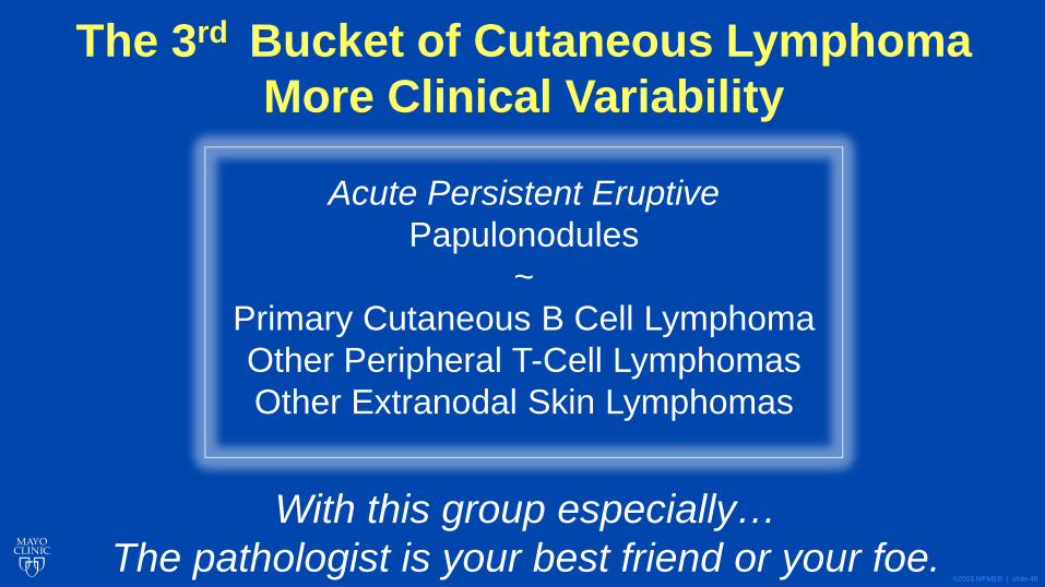
• Red-purple (“Plum”) papules, plaques, papules, or nodules.
• Growth rate variable • Ulceration rare

©2016 MFMER | slide-57
Follicular Cell Lymphoma
• Red-purple (“Plum”) papules, plaques, papules, or nodules.
• Growth rate variable • Ulceration rare
• Sometimes arcuate plaques
involving the torso.

©2016 MFMER | slide-58
Follicular Cell Lymphoma
• Extensive involvement of the head and neck may necessitate systemic treatment
• Extent of multi-focal disease does not necessarily correlate with prognosis
• Rituximab is often very helpful in this setting

©2016 MFMER | slide-59
Follicular Cell Lymphoma
• Histopathology • Follicular pattern : good prognosis; vast majority of cases • Diffuse pattern : more aggressive disease; uncommon.
• Why prognostic caution compared to marginal zone type
• Immunophenotype: • CD20+ • Bcl-6 & CD10 positive (follicular markers) • Bcl-2 negative (excludes systemic follicular B-cell lymphoma)

©2016 MFMER | slide-60
Diffuse Large B-Cell Lymphoma, Leg Type
• Elderly (80+) • Female predominance • Lower legs but not exclusively so • Aggressive
• Always extra-cutaneous spread • Always systemic treatment
• Immunophenotype: • CD20+, CD10- • MUM-1+, FoxP3+ (“activated” B-cell
markers)

©2016 MFMER | slide-61
The 3rd Bucket of Cutaneous Lymphoma Three Conceptual Groups
Acute Persistent Eruptive
Papulonodules
(Excluding conventional leukemia cutis and lymphom acutis)
1˚ Cutaneous B-Cell Lymphoma
Other Peripheral T-Cell Lymphomas
Other Extranodal Lymphoma

©2016 MFMER | slide-62
Other Peripheral T-Cell Lymphomas
• “Peripheral” • Means the T-cells are mature (post-thymic origin) • Does not refer to the clinical distribution of lesions
• Clinically, T-Cell lymphomas that are:
• Not Mycosis Fungoides • Not CD30+ Lymphoproliferative Disorder
• May be indolent or aggressive

©2016 MFMER | slide-63
Other Peripheral T-Cell Lymphomas
• Acute eruptive nodule on cheek x 1 month • Can exclude MF by history alone:
• No precursor patch/plaque

©2016 MFMER | slide-64
• 1 month later: persists, increasing in size • By history less likely, ALCL
• Pathology confirms CD30 negative
Other Peripheral T-Cell Lymphomas

©2016 MFMER | slide-65
• Staging workup unremarkable • Treated with radiotherapy:
• 6 years of follow up with no recurrence or systemic involvement.
Other Peripheral T-Cell Lymphomas

©2016 MFMER | slide-66
CD4+ Small/Medium-sized Pleomorphic T-cell Lymphoma
• Solitary lesion • Head and Neck typically
• But also the trunk • Immunophenotype:
• CD4+,CXCL13+ • Indolent
• Skin-directed treatment

©2016 MFMER | slide-67
Other Peripheral T-Cell Lymphomas • Ill-defined deep erythematous
nodular-plaques with minimal surface change
• Chronic and recurrent over months
• R/O MF : no patches • R/O CD30+ disorder: possible
given recurrence but morphology atypical

©2016 MFMER | slide-68
Subcutaneous Panniculitis-Like T-Cell Lymphoma
• Clinically resembles panniculitis • Recurrent often • Confused with lupus panniculitis
• Immunophenotype: • CD8+,TIA-1+, granzyme-B+ • CD56-; EBV negative
• Outcome: • T-cells α/ß : indolent, easy to treat, low risk of systemic involvement • T-cells γ/∆ : aggressive, systemic spread inevitable, poor prognosis
40x

©2016 MFMER | slide-69
Other Peripheral T-Cell Lymphomas
• Sometimes see presentations with: • Multi-focal lesions • Rapid enlargement • Rapid extra-cutaneous spread
• Phenotypically are often: • Cytotoxic (CD8+) T-cells • Gamma-delta (γ/∆) T-cells
• Poor prognosis : mean survival 15-20 months

©2016 MFMER | slide-70
Other Peripheral T-Cell Lymphomas
• Sometimes see presentations with: • Multi-focal lesions • Rapid enlargement • Rapid extra-cutaneous spread
• Phenotypically are often: • Cytotoxic (CD8+) T-cells • Gamma-delta (γ/∆) T-cells
• Poor prognosis : mean survival 15-20 months
• Incompletely characterized to date
40x
2x

©2016 MFMER | slide-71
The 3rd Bucket of Cutaneous Lymphoma Three Conceptual Groups
Acute Persistent Eruptive
Papulonodules
(Excluding conventional leukemia cutis and lymphoma cutis)
1˚ Cutaneous B-Cell Lymphoma
Other Peripheral T-Cell Lymphomas
Other Extranodal Lymphoma

©2016 MFMER | slide-72
Other Extranodal Lymphomas
• Extranodal • The lymphoma originates outside a lymph node
• Hybrid morphologic or phenotypic properties:
• NK/T Cell • Hybrid of natural killer (NK) cell and a T-cell
• Plasmacytoid Dendritic Cell; conceptually, it is: • Hybrid of a plasma cell and a macrophage
• Presents antigen instead of making antibodies • Not present in non-inflamed skin

©2016 MFMER | slide-73
Extranodal NK/T Cell Lymphoma, nasal type
• 60-90% present in nose and nasopharynx • Poor prognosis: 5-year survival rate 38-62%
“lethal midline granuloma,” “destructive midline lymphoma,” “angiocentric lymphoma,” “malignant granuloma,” “polymorphic reticulosis”

©2016 MFMER | slide-74
Extranodal NK/T Cell Lymphoma, nasal type
• Most common in Asians and Hispanics • Ulceration (“infarctive”) is a prominent clinical feature

©2016 MFMER | slide-75
Extranodal NK/T Cell Lymphoma, nasal type
• 40% of cases have extra-nasal skin involvement
• May occur before or after primary nasal involvement
• Some presentations are purely cutaneous without nasal involvement

©2016 MFMER | slide-76
Extranodal NK/T Cell Lymphoma, nasal type
• 40% of cases have extra-nasal skin involvement
• May occur before or after primary nasal involvement
• Some presentations are purely cutaneous without nasal involvement

©2016 MFMER | slide-77
Extranodal NK/T Cell Lymphoma, nasal type
CD3 Granzyme B
CD56 EBV
CD56(+/-), cytoplasmic CD3-ε(+), cytotoxic markers(+), βF1(−). NO TCR CLONE.

©2016 MFMER | slide-78
Blastic Plasmacytoid Dendritic Cell Neoplasm
• Aggressive leukemia that presents as a primary cutaneous skin lymphoma
• In most cases (80%) the disease manifestations are seen exclusively in the skin, and then the later leukemic phase develops.

©2016 MFMER | slide-79
Blastic Plasmacytoid Dendritic Cell Neoplasm
• Elderly (60–70 years) • Male predominance • Solitary or multiple purpuric
nodules and plaques. • Plasmacytoid Dendritic Cell Immunophenotype:
• CD4+, CD56+ • CD123+ • Negative for all other B, T, and myeloid markers

©2016 MFMER | slide-80
The 4 Buckets of Cutaneous Lymphoma Key History + Morphology
Progressive Pruritic
Generalized Erythema
~ Usually:
Erythroderma Sometimes: Morbilliform
Chronic Pruritic Patches
~
MYCOSIS FUNGOIDES
Acute Persistent Eruptive
Papulonodules ~
1˚ CUTANEOUS B-CELL LYMPHOMA OTHER PERIPHERAL T-CELL LYMPHOMAS OTHER EXTRANODAL
LYMPHOMAS
Waxing & Waning Papulonodules
~ LYP
ALCL

©2016 MFMER | slide-81
4th Bucket of Cutaneous Lymphoma More Inflammatory Appearance
• Eruptions resemble an inflammatory dermatosis rather than conventional lymphoma
• Two morphologic patterns of generalized erythema • Erythroderma:
• Sézary Syndrome • Erythrodermic MF
• Morbilliform: • Angioimmunoblastic T-cell Lymphoma

©2016 MFMER | slide-82
Erythrodermic Cutaneous Lymphoma
• Pruritic • Whole-body erythema with fine scale

©2016 MFMER | slide-83
Erythrodermic Cutaneous Lymphoma
• Pruritic • Whole-body erythema with fine scale • Keratoderma common

©2016 MFMER | slide-84
Erythrodermic Cutaneous Lymphoma
• Pruritic • Whole-body erythema with fine scale • Keratoderma common • Prednisone responsive early

©2016 MFMER | slide-85
• Pruritic • Erythema with fine scale • Keratoderma common • Prednisone responsive early
Leukemic Cutaneous Lymphoma: Variable numbers of atypical T-cells in skin and blood.
SÉZARY CELL SIZE > 12 µM
CEREBRIFORM NUCLEUS CD4+, CD7-, CD26-
500X Erythrodermic Cutaneous Lymphoma

©2016 MFMER | slide-86
Erythrodermic Cutaneous Lymphoma
Sézary Syndrome
Nodal Memory T Cells
Erythrodermic MF
Skin Memory T Cells
• Erythrodermic MF • Progression of MF
• (TNM) B • Low blood burden (<20%)
• Sézary Syndrome (SS)
• Abrupt and de Novo • No MF, but often non-specific
dermatitis and/or pruritus • High blood burden (>20%)
While often viewed synonymously, Sézary Syndrome and Erythrodermic MF have clinical and etiologic distinctions that
may be important for therapy.

©2016 MFMER | slide-87
Erythrodermic Cutaneous Lymphoma
Sézary Syndrome
Nodal Memory T Cells
Erythrodermic MF
Skin Memory T Cells
• Erythrodermic MF • Progression of MF
• (TNM) B • Low blood burden (<20%)
• Sézary Syndrome (SS)
• Abrupt and de Novo • No MF, but often non-specific
dermatitis and/or pruritus • High blood burden (>20%)
While often viewed synonymously, Sézary Syndrome and Erythrodermic MF have clinical and etiologic distinctions that
may be important for therapy.

©2016 MFMER | slide-88
Erythrodermic Cutaneous Lymphoma
“Sézary Preceded by MF” Rarely patients with MF develop a degree blood involvement which meets the pathologic criteria for
Sézary Syndrome
• 6 year history of limited patch-plaque MF (<15% BSA)
• Well controlled with PUVA • Over 3 months developed
• Generalized pruritus • Rapid onset erythroderma
HELPER : SUPPRESSOR T-CELL RATIO > 10

©2016 MFMER | slide-89
Morbilliform Cutaneous Lymphoma • Rare • Morbilliform (“macular-papular”)
• May not persistent initially • Pruritic
• Secondary dermatitis possible
• Often low index of suspicion versus: • Drug Eruption • Viral Exanthem

©2016 MFMER | slide-90
Morbilliform Cutaneous Lymphoma • Seen once in the ED, 3 times by PMD, 4
times by dermatology, and twice by rheumatology… • Diagnosed with “dermatitis” and “lupus” • Prior biopsies “superficial perivasicular
lymphocytic dermatitis” • No change with topical steroids • IM Kenalog decreased itch but
otherwise no change

©2016 MFMER | slide-91
Angioimmunoblastic T-Cell Lymphoma • Middle-aged elderly • Early: Malaise, weakness,
pruritus, morbilliform +/-dermatitic
• Later: B-symptoms, diffuse adenopathy
• Labs: ANA+, eosinophilia, MGUS
• Poor prognosis • Mean survival 15-36
months
T-Cell Lymphoma “Triggered” By Aberrant B-Cells
Activated Nodal B (EBV+) Cell = Exaggerated Follicular Helper T-Cell Recruitment =
“Chronic Inflammation with Auto-Amplification” = Clonal Follicular Helper T-Cell (TFH) Emerges
T-Cell Lymphoma skin & other sites

©2016 MFMER | slide-92
Angioimmunoblastic T-Cell Lymphoma • Lymph node biopsy is
diagnostic
• Skin biopsies can be a pitfall as atypia can be mild • May resemble
inflammatory disorder • Immunophenotype is
quite characteristic
Peripheral T-Cell Lymphoma with Follicular Helper T Cell (TFH) Phenotype
CD4+, CD279+ (PD1),
CXCR5+, CXCL13+, bcl-6+

©2016 MFMER | slide-93
The 4 Buckets of Cutaneous Lymphoma In Summary...
Chronic Pruritic Patches
~
MYCOSIS FUNGOIDES
Acute Persistent Eruptive
Papulonodules ~
1˚ CUTANEOUS B-CELL LYMPHOMA OTHER PERIPHERAL T-CELL LYMPHOMAS OTHER EXTRANODAL
LYMPHOMAS
Waxing & Waning Papulonodules
~ LYP
ALCL
Progressive Pruritic
Generalized Erythema
~ SÉZARY
SYNDROME ERYTHRODERMIC
MF AITL

Related Documents