Cytotoxic/Natural Killer Cell Cutaneous Lymphomas Report of the EORTC Cutaneous Lymphoma Task Force Workshop Marco Santucci, M.D. 1 Nicola Pimpinelli, M.D., Ph.D. 2 Daniela Massi, M.D., Ph.D. 1 Marshall E. Kadin, M.D. 3 Chris J. L. M. Meijer, M.D. 4 Hans Konrad Mu ¨ ller-Hermelink, M.D. 5 Marco Paulli, M.D. 6 Janine Wechsler, M.D. 7 Rein Willemze, M.D. 8 Heike Audring, M.D. 9 Maria Grazia Bernengo, M.D. 10 Lorenzo Cerroni, M.D. 11 Sergio Chimenti, M.D. 12 Andreas Chott, M.D. 13 Jose ´ L. Dı ´az-Pe ´ rez, M.D. 14 Edgard Dippel, M.D. 15 Lyn M. Duncan, M.D. 16 Alfred C. Feller, M.D. 17 Marie-Louise Geerts, M.D. 18 Christian Hallermann, M.D. 19 Werner Kempf, M.D. 20 Robin Russell-Jones, M.D. 21 Christian Sander, M.D. 22 Emilio Berti, M.D. 23 1 Department of Human Pathology and Oncology, University of Florence Medical School, Florence, Italy. 2 Department of Dermatological Sciences, Univer- sity of Florence Medical School, Florence, Italy. 3 Section of Hematopathology, Department of Pa- thology, Beth Israel Deaconess Medical Center, Harvard Medical School, Boston, Massachusetts. 4 Department of Pathology, Free University Hospi- tal, Amsterdam, The Netherlands. 5 Institute of Pathology, University of Würzburg, Würzburg, Germany. 6 Department of Pathology, IRCCS San Matteo Hospital, Pavia, Italy. 7 Department of Pathology, Henry Mondor Hospi- tal, Paris, France. 8 Department of Dermatology, University of Leiden, Leiden, The Netherlands. 9 Department of Dermatology, Charité Humboldt University, Berlin, Germany. 10 Department of Dermatology, University of Turin, Turin, Italy. 11 Department of Dermatology, University of Graz, Graz, Austria. 12 Department of Dermatology, University of Rome “Tor Vergata,” Rome, Italy. 13 Institute for Clinical Pathology, University of Vi- enna, Vienna, Austria. 14 Department of Dermatology, Cruces Hospital, University of Bilbao, Bilbao, Spain. 15 Department of Dermatology, “Benjamin Frank- lin” University Hospital, Berlin, Germany. 16 Dermatopathology Unit, Massachusetts General Hospital, Harvard Medical School, Boston, Massa- chusetts. 17 Institute of Pathology, Medical University of Lü- beck, Lübeck, Germany. 18 Department of Dermatology, University Hospital of Gent, Gent Belgium. 19 Department of Dermatology, “Georg August” University of Göttingen, Go ¨ ttingen, Germany. 20 Department of Dermatology, University Hospital of Zurich, Zurich, Switzerland. 21 Skin Tumour Unit, St. John’s Institute of Der- matology, St. Thomas’ Hospital, London, United Kingdom. 22 Department of Dermatology, “Ludwig Maximil- ians” University of Munich, Munich, Germany. 23 Institute of Dermatological Sciences, University of Milan, Milan, Italy. Address for reprints: Marco Santucci, Dipartimento di Patologia Umana ed Oncologia, Universita ´ degli Studi di Firenze, Viale G.B. Morgagni, 85 50134 Firenze, Italy; Fax: 011-39-055-4379868; E-mail: [email protected] Received July 30, 2002; revision received August 14, 2002; accepted August 27, 2002. BACKGROUND. Cutaneous lymphomas expressing a cytotoxic or natural killer (NK) cell phenotype represent a group of lymphoproliferative disorders for which there is currently much confusion and little consensus regarding the best nomenclature and classification. METHODS. This study analyzes 48 cases of primary cutaneous lymphoma express- ing cytotoxic proteins and/or the NK cell marker, CD56. These cases were collected for a workshop of the European Organization for Research and Treatment of Cancer Cutaneous Lymphoma Task Force, to better clarify the clinical, morpho- logic, and phenotypic features of these uncommon tumors. RESULTS. Several categories with different clinical and pathologic features were delineated: 1) aggressive, CD8, epidermotropic, cytotoxic T-cell lymphoma; 2) mycosis fungoides, cytotoxic immunophenotype variant; 3) subcutaneous pannic- ulitis-like T-cell lymphoma; 4) NK/T-cell lymphoma, nasal type; 5) CD4, NK cell lymphoma; 6) blastoid NK cell lymphoma; (7) intravascular NK-like lymphoma; and 8) cytotoxic, peripheral T-cell lymphoma. CONCLUSIONS. Our data show that primary cutaneous cytotoxic/NK cell lympho- mas include distinct groups of diseases, clinically, histologically, and biologically. Because the finding of a cytotoxic phenotype often has prognostic significance, the routine use of cytotoxic markers in the diagnosis and classification of cutaneous lymphomas should be expanded. Cancer 2003;97:610 –27. © 2003 American Cancer Society. DOI 10.1002/cncr.11107 KEYWORDS: cytotoxic/NK cell lymphomas, skin, classification, CD56, TIA-1, prog- nosis, therapy. 610 © 2003 American Cancer Society

Welcome message from author
This document is posted to help you gain knowledge. Please leave a comment to let me know what you think about it! Share it to your friends and learn new things together.
Transcript
Cytotoxic/Natural Killer Cell Cutaneous LymphomasReport of the EORTC Cutaneous Lymphoma Task Force Workshop
Marco Santucci, M.D.1
Nicola Pimpinelli, M.D., Ph.D.2
Daniela Massi, M.D., Ph.D.1
Marshall E. Kadin, M.D.3
Chris J. L. M. Meijer, M.D.4
Hans Konrad Muller-Hermelink, M.D.5
Marco Paulli, M.D.6
Janine Wechsler, M.D.7
Rein Willemze, M.D.8
Heike Audring, M.D.9
Maria Grazia Bernengo, M.D.10
Lorenzo Cerroni, M.D.11
Sergio Chimenti, M.D.12
Andreas Chott, M.D.13
Jose L. Dıaz-Perez, M.D.14
Edgard Dippel, M.D.15
Lyn M. Duncan, M.D.16
Alfred C. Feller, M.D.17
Marie-Louise Geerts, M.D.18
Christian Hallermann, M.D.19
Werner Kempf, M.D.20
Robin Russell-Jones, M.D.21
Christian Sander, M.D.22
Emilio Berti, M.D.23
1 Department of Human Pathology and Oncology,University of Florence Medical School, Florence,Italy.2 Department of Dermatological Sciences, Univer-sity of Florence Medical School, Florence, Italy.3 Section of Hematopathology, Department of Pa-thology, Beth Israel Deaconess Medical Center,Harvard Medical School, Boston, Massachusetts.4 Department of Pathology, Free University Hospi-tal, Amsterdam, The Netherlands.5 Institute of Pathology, University of Würzburg,Würzburg, Germany.6 Department of Pathology, IRCCS San MatteoHospital, Pavia, Italy.7 Department of Pathology, Henry Mondor Hospi-tal, Paris, France.8 Department of Dermatology, University of Leiden,Leiden, The Netherlands.9 Department of Dermatology, Charité HumboldtUniversity, Berlin, Germany.
10 Department of Dermatology, University of Turin,Turin, Italy.11 Department of Dermatology, University of Graz,Graz, Austria.12 Department of Dermatology, University of Rome“Tor Vergata,” Rome, Italy.13 Institute for Clinical Pathology, University of Vi-enna, Vienna, Austria.14 Department of Dermatology, Cruces Hospital,University of Bilbao, Bilbao, Spain.15 Department of Dermatology, “Benjamin Frank-lin” University Hospital, Berlin, Germany.16 Dermatopathology Unit, Massachusetts GeneralHospital, Harvard Medical School, Boston, Massa-chusetts.17 Institute of Pathology, Medical University of Lü-beck, Lübeck, Germany.18 Department of Dermatology, University Hospitalof Gent, Gent Belgium.
19 Department of Dermatology, “Georg August”University of Göttingen, Gottingen, Germany.20 Department of Dermatology, University Hospitalof Zurich, Zurich, Switzerland.21 Skin Tumour Unit, St. John’s Institute of Der-matology, St. Thomas’ Hospital, London, UnitedKingdom.22 Department of Dermatology, “Ludwig Maximil-ians” University of Munich, Munich, Germany.23 Institute of Dermatological Sciences, Universityof Milan, Milan, Italy.
Address for reprints: Marco Santucci, Dipartimentodi Patologia Umana ed Oncologia, Universita degliStudi di Firenze, Viale G.B. Morgagni, 85 50134Firenze, Italy; Fax: 011-39-055-4379868; E-mail:[email protected]
Received July 30, 2002; revision received August14, 2002; accepted August 27, 2002.
BACKGROUND. Cutaneous lymphomas expressing a cytotoxic or natural killer (NK)
cell phenotype represent a group of lymphoproliferative disorders for which there
is currently much confusion and little consensus regarding the best nomenclature
and classification.
METHODS. This study analyzes 48 cases of primary cutaneous lymphoma express-
ing cytotoxic proteins and/or the NK cell marker, CD56. These cases were collected
for a workshop of the European Organization for Research and Treatment of
Cancer Cutaneous Lymphoma Task Force, to better clarify the clinical, morpho-
logic, and phenotypic features of these uncommon tumors.
RESULTS. Several categories with different clinical and pathologic features were
delineated: 1) aggressive, CD8�, epidermotropic, cytotoxic T-cell lymphoma; 2)
mycosis fungoides, cytotoxic immunophenotype variant; 3) subcutaneous pannic-
ulitis-like T-cell lymphoma; 4) NK/T-cell lymphoma, nasal type; 5) CD4�, NK cell
lymphoma; 6) blastoid NK cell lymphoma; (7) intravascular NK-like lymphoma;
and 8) cytotoxic, peripheral T-cell lymphoma.
CONCLUSIONS. Our data show that primary cutaneous cytotoxic/NK cell lympho-
mas include distinct groups of diseases, clinically, histologically, and biologically.
Because the finding of a cytotoxic phenotype often has prognostic significance, the
routine use of cytotoxic markers in the diagnosis and classification of cutaneous
lymphomas should be expanded. Cancer 2003;97:610 –27.
© 2003 American Cancer Society.
DOI 10.1002/cncr.11107
KEYWORDS: cytotoxic/NK cell lymphomas, skin, classification, CD56, TIA-1, prog-nosis, therapy.
610
© 2003 American Cancer Society
Several subtypes of cutaneous lymphomas (al-though reported in the current literature and re-
cently included in the World Health Organization[WHO] classification1) are not yet mentioned, or areincluded as provisional entities, in the CutaneousLymphoma Study Group of the European Organiza-tion for Research and Treatment of Cancer (EORTC)classification.2 This is the result of the absence of adistinctive clinicopathologic presentation. Among thecategories of this “gray” area, cutaneous lymphomasexpressing a cytotoxic or natural killer (NK) cell phe-notype represent a group of lymphoproliferative dis-orders for which there is currently much confusionand little consensus regarding the best nomenclatureand classification. In particular, the rarity of tumorsexpressing the NK cell phenotype, especially in West-ern countries, represents a major limitation to a com-plete understanding. The recognition of lymphoidneoplasms expressing a cytotoxic or an NK-like phe-notype may have relevant clinical and therapeutic im-plications because it has been reported that many ofthem follow an aggressive clinical course.3– 6
The aim of this report is to describe the spectrumof primary cutaneous lymphomas expressing cyto-toxic proteins and/or the NK cell marker, CD56, and tobetter clarify their clinical, morphologic, and pheno-typic features.
MATERIALS AND METHODSA workshop of the EORTC Cutaneous Lymphoma TaskForce was held on July 3–5, 1998, at the Study Center“I Cappuccini,” S. Miniato (Pisa, Italy). Cases weresolicited on the following topics: 1) lymphomas inwhich tumor cells expressed at least one of the follow-ing molecules, namely, CD8, TIA-1, or CD56; 2) lym-phoma showing features of subcutaneous or angio-centric lymphoma; and 3) lymphoma expressing the�/� phenotype. Sixty examples of primary cutaneouslymphoma exhibiting one the aforementioned fea-tures were provided.
During the workshop, there was a plenary discus-sion on the definition and differential diagnosis ofcytotoxic and nonnasal NK/T-cell primary cutaneouslymphomas, but some gray areas still remained. Tosolve residual problems, a committee composed of 10experienced dermatopathologists or hematopatholo-gists (EB, MEK, DM, CJLMM, HKM-H, MP, NP, MS,JW, RW) was appointed. They met on January 16 –18,1999, at the Department of Human Pathology andOncology, University of Florence Medical School, Flo-rence, (Italy). This report summarizes the results ofboth the plenary and committee discussions. Forty-eight cases of appropriate immunophenotype wereidentified and included in the present study. Four of
these cases have been included in previous studies onvarious lymphoma types.7–10
Clinical records and follow-up data through De-cember 1999 were obtained from patients’ charts. Theprincipal parameters evaluated included age and gen-der, extent of disease, spontaneous regression, symp-toms at presentation, bone marrow involvement, cu-taneous recurrences, extracutaneous progression ofdisease, treatment, and follow-up duration (Table 1).
The TIA-1 and CD56 status was determined im-munohistochemically on paraffin sections using theantibodies TIA-1 (Coulter, Miami, FL) and 123C3 (Lab-Vision, Fremont, CA), respectively. In previous studies,other authors have shown that 123C3 reliably detectsCD56 expression in lymphomas on paraffin-embed-ded tissues.5 Additional immunohistochemical datawere provided by the source institutions. Epstein–Barrvirus (EBV) infection was demonstrated using noniso-topic in situ hybridization for EBV-encoded smallRNAs (EBERs) on paraffin-embedded material, as pre-viously described,11 as well as by immunohistochem-istry with antibodies against LMP-1 or by polymerasechain reaction (PCR). The DNA used for gene rear-rangement studies was extracted from frozen or par-affin-embedded tumor tissue. T-cell receptor (TCR)gene rearrangement was evaluated by a PCR assaycoupled with nondenaturing polyacrylamide gel elec-trophoresis according to a method previously de-scribed.12 Amplification of the TCR-� chain locus V-Jjunctional region was performed by using oligonucle-otide primers specific for J1/2 paired with V2a, V9, andV10. In some cases, DNA was digested with restrictionendonuclease BamHI, EcoRI, or HindIII, subjected toelectrophoresis on a 0.8% agarose gel, and transferredto a nitrocellulose filter for Southern analysis usinghybridization with 32P-labeled DNA probes, accordingto standard protocols.13,14 Rearrangements of the TCRand immunoglobulin genes were evaluated usingprobes that include a 1.0-kb germline Pst-EcoRI frag-ment containing the first region (J�1), the constantregion of TCR-� gene (C�), the TCR-� gene, and JH(heavy-chain joining region).15
RESULTSSeveral groups with different clinical (Table 1) andpathologic features were delineated among these 48cases.
Cases 1–4: Aggressive, CD8�, Epidermotropic, CytotoxicT-cell Lymphoma WHO: Not RecognizedClinical featuresThis group included four male patients, 33– 82 years ofage (mean age, 62 years; median age, 76.5 years).Three of the four patients presented with widespread,
Cytotoxic/NK Cell Cutaneous Lymphomas/Santucci et al. 611
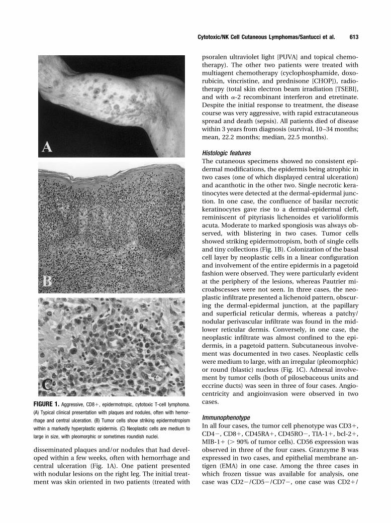
TABLE 1Clinical Data and Follow-Up Information
Caseno.
Gender/age Type of lesion
Sites of disease atpresentation Initial treatment Subsequent treatment Progression Outcome (mos)
1 M/33 Plaques, nodules Widespread PUVA-PR IFN-NR, TSEBI-NR, CT(CHOP)-NR Skin, oral mucosa, and systemicinvolvement (sepsis)
DOD, 34
2 M/82 Nodules Widespread CT (COP) and RT-CR CT (unspecified) Skin and systemic involvement DOD, 173 M/59 Plaques Widespread Topical nitrogen mustard-CR CT (CHOP), RT, topical nitrogen mustard Skin and systemic involvement DOD, 284 M/74 Nodules Lower limb IFN and retinoids-PR CT (CHOP)-PR Skin and systemic involvement DOD, 105 M/16 Patches, plaques Widespread PUVA-CR N.A. Skin AWD, 246 M/30 Patches Lower limb PUVA-PR Topical steroids-NR CT (miltefusine)-PR Skin AWD, 367 F/77 Plaques Widespread PUVA-PR RT (TSEBI)-CR Skin AWD, 328 F/30 Patches Trunk PUVA-CR Nil Nil NED, 309 F/59 Patches, plaques Widespread PUVA-CR PUVA-PR Skin AWD, 14410 F/54 Subcutaneous nodules Limbs Systemic steroids-CR CT(cyclophosphamide)-PR Skin AWD, 9611 M/14 Subcutaneous nodules Limbs, buttocks CT (ACVBP)-CR Nil Nil NED, 1512 F/55 Subcutaneous nodules Limbs Nil (spontaneous remission) Nil (spontaneous remission) Skin AWD, 6013 M/36 Subcutaneous nodules Limbs Topical steroids-CR CT (CHOP, CDA, ara-C, others)-PR, RT
(TSEBI)-PRSkin and systemic involvement
(hemophagocytic syndrome)AWD, 24
14 M/53 Subcutaneous nodules Limbs Steroids andhydroxychlorokine-NR
CT (mitoxantrone, etoposide, vincristine,cyclophosphamide)-NR
Skin and systemic involvement(pancytopenia, sepsis)
DOD, 9
15 F/56 Subcutaneous nodules Head, trunk, upperlimb
Steroids-NR CT (cyclophosphamide, CEOP)-NR Skin and systemic involvement(pancytopenia, sepsis)
DOD, 24
16 M/10 Subcutaneous nodules Trunk, upper limb Steroids-NR CT (cyclophosphamide, CEOP)-NR Skin and systemic involvement(pancytopenia, sepsis)
DOD, 5
17 M/76 Subcutaneous nodules Limbsa N.A. N.A. Skin AWD, 2418 F/44 Subcutaneous nodules Lower limb CT (CHOP)-NR CT (VIM2, ara-C)-NR Skin and systemic involvement (liver
and lung)DOD, 17
19 F/8 Subcutaneous nodules Trunk Cyclosporine-PR CT (unspecified)-NR Skin and systemic involvement DOD, 2320 F/73 Subcutaneous nodules Lower limb, buttocks CT (CHOP)-PR RT-PR, CT (methotrexate)-PR Skin and systemic involvement (liver) DOD, 1221 F/33 Subcutaneous nodules Limbs, buttocks Steroids and retinoids-PR N.A. Skin and systemic symptoms (malaise,
fatigue, and fever)AWD, 12
22 M/53 Nodule Upper limba N.A. N.A. N.A. N.A.23 F/27 Nodules Head, trunk Nil (spontaneous remission) N.A. N.A. N.A.24 F/43 Nodules Upper limb CT (CHOP)-CR CT (unspecified)-PR Skin,b AWD, 1025 M/71 Nodules Trunk, limbs CT (CHOP)-CR CT (methotrexate)-CR CNS DOC (stroke), 326 M/60 Nodules Head CT (VICOP-B)-NR CT (unspecified)-NR Skin and systemic involvement (bone
marrow)DOD, 8
27 M/60 Papules, plaques Widespread CT (fludarabine)-CR Nil Nil NED, 1628 M/57 Plaques, nodules Widespread CT (unspecified)-PR CT (unspecified)-PR Skin N.A.29 F/47 Plaques Head, lower limb RT and CT (CHOP)-CR IFN and CT (vincristine, chlorambucil)-
PRSkin and systemic involvement (lymph
nodes, CNS)DOD, 31
30 M/41 Plaques Lower limba RT-CR Nil Nil NED, 4431 M/38 Plaques, nodules Widespread Nil CT (PROMACE-CYTABOM) and
allogeneic BMT-CRSkin and systemic involvement (bone
marrow and blood)DOD, 22
32 M/83 Plaques, nodules Trunk CT (mini-CEOP)-CR CT (unspecified)-NR Skin and systemic involvement (bonemarrow and blood)b, c
DOD, 10
33 M/80 Plaques, nodules Head, trunk CT (COP)-NR CT (unspecified)-PR Skin and systemic involvement (bonemarrow)
DOD, 20
34 M/51 Nodules Head, trunk N.A. N.A. N.A. N.A.35 M/55 Plaques, nodules Head, trunk CT (MACOP-B)-CR IFN, CT (chlorambucil), and steroids-NR,
autologous BMT-CRSkin and systemic involvement N.A.
36 M/61 Plaques, nodules Trunk, limbs CT (CEOP)-CR RT and CT (MACOP-B)-CR Skin and systemic involvement (bonemarrow, blood, and lymph nodes)
DOD, 37
37 M/58 Plaques, nodules Widespread CT (CHOP)-PR CT (unspecified)-NR Skin and systemic involvement (testisand bone marrow)
DOD, 16
38 M/54 Plaques Trunk, lower limb Antibioticd CT (CHOP)-CR Skin and systemic involvement (CNS) DOD, 1739 F/72 Nodule Head RT-CR Nil Nil NED, 3640 M/43 Nodule Upper limb N.A. N.A. N.A. N.A.41 M/63 Nodules Limbs, trunk Nil (spontaneous remission) Nil Skin NED, 1242 F/60 Nodules Limbs, trunk Steroids and clofazimine-PR N.A. Skin AWD, 6043 F/63 Plaques Lower limb RT and CT
(cyclophosphamide)-CRNil Nil NED, 36
44 M/60 Nodules Limbse RT-NR, CT (CDA, CHOP)-PR Skin and systemic involvement (lymphnodes)
DOD, 18
45 F/38 Plaques, nodules Trunk, limbsf CT (unspecified)-CR Nil N.A.46 F/76 Plaques Limbs Nil (spontaneous remission) RT-PR Skin and systemic involvement (lymph
nodes, soft tissues, CNS)DOD, 11
47 M/77 Nodule Head Surgery and RT-CR N.A. Skin and systemic involvement(pharyngeal mass)
DOD, 11
48 M/39 Nodule Lower limb RT-CR RT � CT (CHOP)-PR Skin and systemic involvement (softtissues and bone marrow)
DOD, 12
AWD: Alive with disease; BMT: bone marrow transplantation; CNS: central nervous system; CR: complete remission; CT: chemotherapy; DOD: dead of disease; NR: no response; PR: partial remission; RT: radiotherapy;
N.A.: information not available; NED: no evidence of disease; IFN�: -2 recombinant interferon; TSEBI: total skin electron beam irradiation; ara-C: cytosine arabinoside; PUVA: psoralen ultraviolet light; CHOP:
cyclophosphamide, doxorubicin, vincristine, and prednisone.a Myelodysplastic syndrome.b Polypoidal thickening of the nasal/paranasal mucosa demonstrated at computed tomographic scan; biopsy not performed.c Gastric carcinoma.d Treatment for concomitant Borrelia Burgdorferi infection.e Hairy cell leukemia.f Lymphomatoid granulomatosis Epstein–Barr virus infection.
612 CANCER February 1, 2003 / Volume 97 / Number 3
disseminated plaques and/or nodules that had devel-oped within a few weeks, often with hemorrhage andcentral ulceration (Fig. 1A). One patient presentedwith nodular lesions on the right leg. The initial treat-ment was skin oriented in two patients (treated with
psoralen ultraviolet light [PUVA] and topical chemo-therapy). The other two patients were treated withmultiagent chemotherapy (cyclophosphamide, doxo-rubicin, vincristine, and prednisone [CHOP]), radio-therapy (total skin electron beam irradiation [TSEBI],and with �-2 recombinant interferon and etretinate.Despite the initial response to treatment, the diseasecourse was very aggressive, with rapid extracutaneousspread and death (sepsis). All patients died of diseasewithin 3 years from diagnosis (survival, 10 –34 months;mean, 22.2 months; median, 22.5 months).
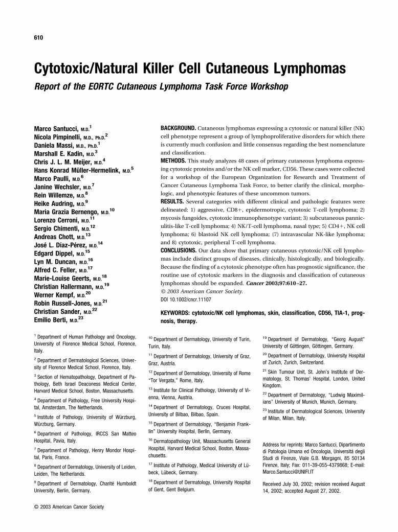
Histologic featuresThe cutaneous specimens showed no consistent epi-dermal modifications, the epidermis being atrophic intwo cases (one of which displayed central ulceration)and acanthotic in the other two. Single necrotic kera-tinocytes were detected at the dermal-epidermal junc-tion. In one case, the confluence of basilar necrotickeratinocytes gave rise to a dermal-epidermal cleft,reminiscent of pityriasis lichenoides et varioliformisacuta. Moderate to marked spongiosis was always ob-served, with blistering in two cases. Tumor cellsshowed striking epidermotropism, both of single cellsand tiny collections (Fig. 1B). Colonization of the basalcell layer by neoplastic cells in a linear configurationand involvement of the entire epidermis in a pagetoidfashion were observed. They were particularly evidentat the periphery of the lesions, whereas Pautrier mi-croabscesses were not seen. In three cases, the neo-plastic infiltrate presented a lichenoid pattern, obscur-ing the dermal-epidermal junction, at the papillaryand superficial reticular dermis, whereas a patchy/nodular perivascular infiltrate was found in the mid-lower reticular dermis. Conversely, in one case, theneoplastic infiltrate was almost confined to the epi-dermis, in a pagetoid pattern. Subcutaneous involve-ment was documented in two cases. Neoplastic cellswere medium to large, with an irregular (pleomorphic)or round (blastic) nucleus (Fig. 1C). Adnexal involve-ment by tumor cells (both of pilosebaceous units andeccrine ducts) was seen in three of four cases. Angio-centricity and angioinvasion were observed in twocases.
ImmunophenotypeIn all four cases, the tumor cell phenotype was CD3�,CD4�, CD8�, CD45RA�, CD45RO�, TIA-1�, bcl-2�,MIB-1� (� 90% of tumor cells). CD56 expression wasobserved in three of the four cases. Granzyme B wasexpressed in two cases, and epithelial membrane an-tigen (EMA) in one case. Among the three cases inwhich frozen tissue was available for analysis, onecase was CD2�/CD5�/CD7�, one case was CD2�/
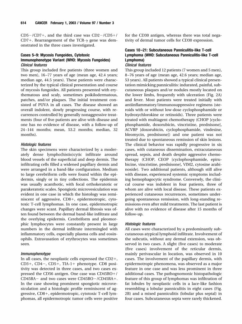
FIGURE 1. Aggressive, CD8�, epidermotropic, cytotoxic T-cell lymphoma.
(A) Typical clinical presentation with plaques and nodules, often with hemor-
rhage and central ulceration. (B) Tumor cells show striking epidermotropism
within a markedly hyperplastic epidermis. (C) Neoplastic cells are medium to
large in size, with pleomorphic or sometimes roundish nuclei.
Cytotoxic/NK Cell Cutaneous Lymphomas/Santucci et al. 613
CD5�/CD7�, and the third case was CD2�/CD5�/CD7�. Rearrangement of the TCR-� gene was dem-onstrated in the three cases investigated.
Cases 5–9: Mycosis Fungoides, CytotoxicImmunophenotype Variant (WHO: Mycosis Fungoides)Clinical featuresThis group included five patients (three women andtwo men), 16 –77 years of age (mean age, 42.4 years;median age, 44.5 years). These patients were charac-terized by the typical clinical presentation and courseof mycosis fungoides. All patients presented with ery-thematous and scaly, sometimes poikilodermatous,patches, and/or plaques. The initial treatment con-sisted of PUVA in all cases. The disease showed anoverall indolent, slowly progressing course, with re-currences controlled by generally nonaggressive treat-ments (four of five patients are alive with disease andone has no evidence of disease, with a follow-up of24 –144 months; mean, 53.2 months; median, 32months).
Histologic featuresThe skin specimens were characterized by a moder-ately dense lympho(histio)cytic infiltrate aroundblood vessels of the superficial and deep dermis. Theinfiltrating cells filled a widened papillary dermis andwere arranged in a band-like configuration. Mediumto large cerebriform cells were found within the epi-dermis, singly or in tiny collections. The epidermiswas usually acanthotic, with focal orthokeratotic orparakeratotic scales. Spongiotic microvesiculation wasevident in one case in which the histology was remi-niscent of aggressive, CD8�, epidermotropic, cyto-toxic T-cell lymphomas. In one case, epidermotropicchanges were scant. Papillary dermal fibrosis was of-ten found between the dermal band-like infiltrate andthe overlying epidermis. Cerebriform and pleomor-phic lymphocytes were constantly present in largenumbers in the dermal infiltrate intermingled withinflammatory cells, especially plasma cells and eosin-ophils. Extravasation of erythrocytes was sometimesseen.
ImmunophenotypeIn all cases, the neoplastic cells expressed the CD2�,CD3�, CD4�, CD5�, TIA-1� phenotype. CD8 posi-tivity was detected in three cases, and two cases ex-pressed the CD56 antigen. One case was CD45RO�/CD45RA� and two cases were CD45RO�/CD45RA�.In the case showing prominent spongiotic microve-siculation and a histologic profile reminiscent of ag-gressive, CD8�, epidermotropic, cytotoxic T-cell lym-phomas, all epidermotropic tumor cells were positive
for the CD30 antigen, whereas there was total nega-tivity of dermal tumor cells for CD30 expression.
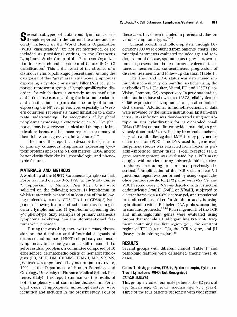
Cases 10–21: Subcutaneous Panniculitis-like T-cellLymphoma (WHO: Subcutaneous Panniculitis-like T-cellLymphoma)Clinical featuresThis group included 12 patients (7 women and 5 men),8 –76 years of age (mean age, 42.6 years; median age,53 years). All patients showed a typical clinical presen-tation mimicking panniculitis: indurated, painful, sub-cutaneous plaques and/or nodules mostly located onthe lower limbs, frequently with ulceration (Fig. 2A)and fever. Most patients were treated initially withantiinflammatory/immunosuppressive regimens (ste-roids with or without low-dose cyclophosphamide orhydroxychlorokine or retinoids). Three patients weretreated with multiagent chemotherapy (CHOP [cyclo-phosphamide, doxorubicin, vincristine, prednisone],ACVBP [doxorubicin, cyclophosphamide, vindesine,bleomycin, prednisone]) and one patient was nottreated due to spontaneous remission of skin lesions.The clinical behavior was rapidly progressive in sixcases, with cutaneous dissemination, extracutaneousspread, sepsis, and death despite aggressive chemo-therapy (CHOP, CEOP [cyclophosphamide, epiru-bicine, vincristine, prednisone], VIM2, cytosine arabi-noside). Two additional patients, although still alivewith disease, experienced systemic symptoms includ-ing hemophagocytic syndrome. Conversely, the clini-cal course was indolent in four patients, three ofwhom are alive with local disease. These patients ex-perienced cutaneous recurrences, sometimes under-going spontaneous remission, with long-standing re-missions even after mild treatments. The last patient isalive with no evidence of disease after 15 months offollow-up.
Histologic featuresAll cases were characterized by a predominantly sub-cutaneous atypical lymphoid infiltrate. Involvement ofthe subcutis, without any dermal extension, was ob-served in two cases. A slight (five cases) to moderate(five cases) involvement of the reticular dermis,mainly perivascular in location, was observed in 10cases. The involvement of the papillary dermis, withepidermotropic phenomena, was observed as a majorfeature in one case and was less prominent in threeadditional cases. The pathognomonic histopathologicfeature of this group of lymphomas was infiltration offat lobules by neoplastic cells in a lace-like fashionresembling a lobular panniculitis in eight cases (Fig.2B) and a mixed panniculitis (lobular plus septal) infour cases. Subcutaneous septa were rarely thickened.
614 CANCER February 1, 2003 / Volume 97 / Number 3
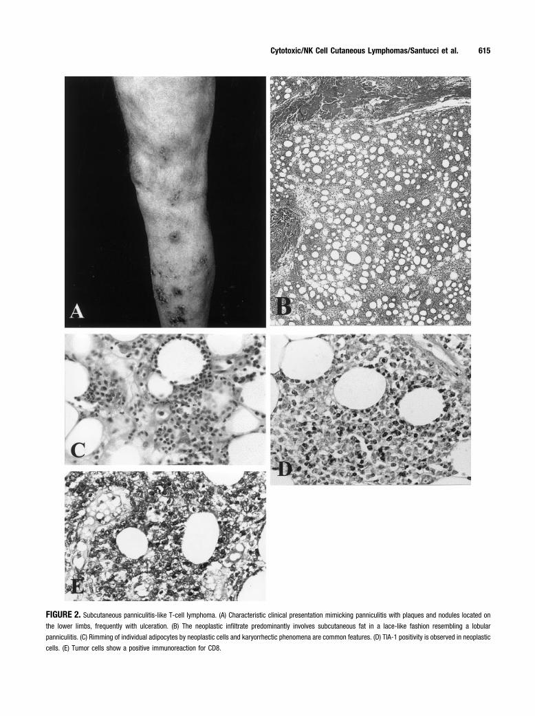
FIGURE 2. Subcutaneous panniculitis-like T-cell lymphoma. (A) Characteristic clinical presentation mimicking panniculitis with plaques and nodules located on
the lower limbs, frequently with ulceration. (B) The neoplastic infiltrate predominantly involves subcutaneous fat in a lace-like fashion resembling a lobular
panniculitis. (C) Rimming of individual adipocytes by neoplastic cells and karyorrhectic phenomena are common features. (D) TIA-1 positivity is observed in neoplastic
cells. (E) Tumor cells show a positive immunoreaction for CD8.
Cytotoxic/NK Cell Cutaneous Lymphomas/Santucci et al. 615
The rimming of individual adipocytes by neoplasticcells was a common feature (Fig. 2C). The neoplasticinfiltrate was composed of pleomorphic lymphocytesof variable size, with an irregular, hyperchromatic nu-cleus. A predominance of small to medium cells wasobserved in all cases and large transformed lymphoidcells were seen in six cases. A characteristic featurewas the presence of karyorrhectic phenomena, whichwere seen in every case and were prominent in 10cases. Fat necrosis was frequent with foamy or finelyvacuolated histiocytes. Small cysts lined by amor-phous eosinophilic material were documented in fivecases. Multinucleated giant cells were seen in fivecases. Scattered plasma cells were rarely seen. Neu-trophils and eosinophils were absent, except for twocases in which neutrophilic microabscesses werefound within necrotic foci. A slight to moderate angio-centricity occurred in eight cases and angioinvasion ofsmall blood vessels was present in five cases.
ImmunophenotypeIn all cases, the tumor cell phenotype was CD2�, CD3�,CD45RO�, CD43�, and TIA-1� (Fig. 2D). Seven casesexpressed the T-suppressor cell antigen, CD8 (Fig. 2E),and possible coexpression of CD4 was seen in two ofthese cases. Two additional cases were CD4�/CD8�,whereas the neoplastic cells did not express either CD4or CD8 in three cases. CD56 was positive in 7 of 12 cases.Among the five cases with frozen tissue available forimmunohistochemistry, three were positive for CD5 an-tigen and all cases were negative for CD7. Perforin andGranzyme B (GrB) cytoplasmic staining was observed inall four tested cases. Weak focal staining for CD30 wasnoted in two of seven cases. All nine cases tested ex-pressed TCR antigens. Two cases proved to be �/�
(�F1�) T-cell lymphomas and the other seven were de-rived from �/� T cells (TCR�1�). All �/�� cases wereCD56�, whereas none of the two �/� lymphomas ex-pressed CD56�. A significant difference in the tumor cellphenotype was observed according to the exclusive sub-cutaneous or subcutaneous and dermal involvement byneoplastic cells. Among cases with dermal extensiontested for TCRs, seven were TCR�1� and one was �F1�.Clonal TCR gene rearrangements were documented inthe six cases investigated. Clonal rearrangement ofTCR-� was shown in five lymphomas, including 1 �/�
and 4 �/� lymphomas. Clonal rearrangements of theTCR-�gene were confirmed by PCR analysis in 1 �/�
lymphoma showing a clonal V�2 rearrangement. None ofthe three cases tested for EBER or LMP-1 was positive forEBV.
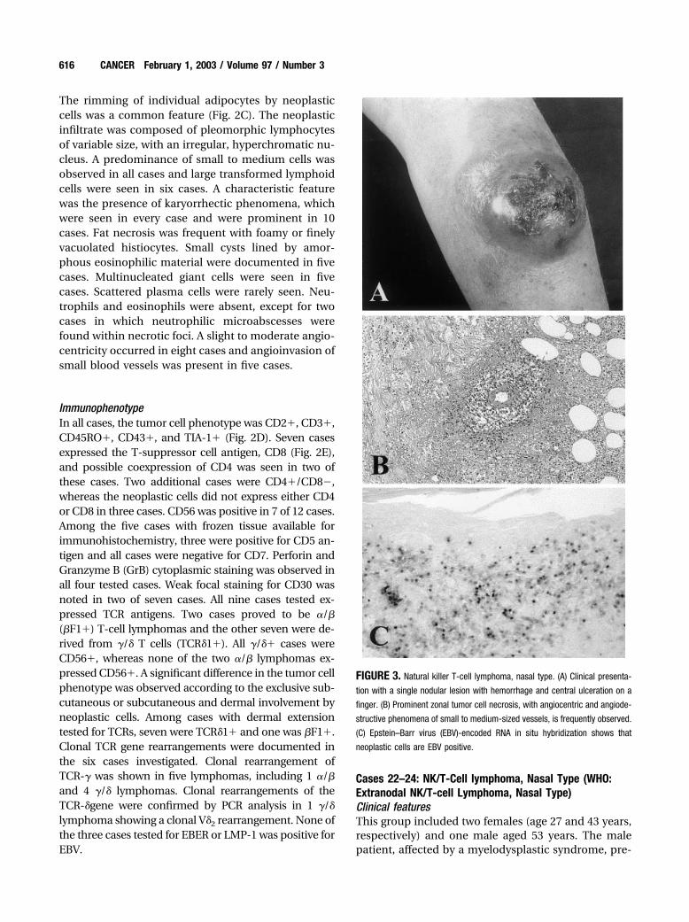
Cases 22–24: NK/T-Cell lymphoma, Nasal Type (WHO:Extranodal NK/T-cell Lymphoma, Nasal Type)Clinical featuresThis group included two females (age 27 and 43 years,respectively) and one male aged 53 years. The malepatient, affected by a myelodysplastic syndrome, pre-
FIGURE 3. Natural killer T-cell lymphoma, nasal type. (A) Clinical presenta-
tion with a single nodular lesion with hemorrhage and central ulceration on a
finger. (B) Prominent zonal tumor cell necrosis, with angiocentric and angiode-
structive phenomena of small to medium-sized vessels, is frequently observed.
(C) Epstein–Barr virus (EBV)-encoded RNA in situ hybridization shows that
neoplastic cells are EBV positive.
616 CANCER February 1, 2003 / Volume 97 / Number 3
sented with a single nodular lesion on the left ringfinger (Fig. 3A). Another patient developed multiplecutaneous nodules that underwent spontaneous re-mission. Staging was negative. These two patientswere lost to follow-up. The third patient presentedwith multiple, rapidly growing cutaneous nodules onthe left arm and other lesions appeared on the legs 6months later, some of which underwent spontaneousremission. Approximately 6 months after diagnosis, acomputed tomographic (CT) scan documented apolypoidal thickening of the nasal and paranasal(frontal sinus) mucosa. Bone CT scan and bone mar-row biopsy were negative. Following aggressive mul-tiagent chemotherapy courses, the lesions underwentalmost complete remission, but the disease recurred.The patient died with disseminated disease 18 monthsfrom diagnosis.
Histologic featuresIn two cases, the lymphomatous infiltrate was diffuse,involving the entire dermis and the subcutis. In thelast case, tumor cells were localized mainly to the deepreticular dermis and subcutis. The neoplastic infiltratewas composed of small to medium-sized pleomorphiclymphocytes with irregular nuclei, inconspicuous nu-cleoli, and scant cytoplasm. Neither polymorphonu-clear leukocytes nor plasma cells were seen, whereassmall numbers of reactive lymphocytes, intermingledwith tumor cells, were observed in all cases. In onecase, reactive lymphocytes were seen in the epidermisand skin appendages. Apoptosis was prominent in twocases. Zonal tumor cell necrosis, focal or confluent,with angiocentric and angiodestructive phenomena ofsmall to medium-sized vessels was a prominent fea-ture in all specimens (Fig. 3B). The vessels of the deepreticular dermis were massively infiltrated by atypicallymphoid cells and surrounded by extensive areas ofcoagulative necrosis, with only cuffs of surviving lym-phoma cells around the blood vessels. The involvedvessels showed endothelial swelling and onion skinthickening of the wall, with fibrin deposition and nu-clear debris. Extensive epidermal ulceration was ob-served in one case.
ImmunophenotypeAll cases were CD3�, CD3��, CD56�, CD57�, CD4�,CD45RO�, CD45RA�, and TIA-1�. Two of three caseswere CD8�. In one case in which frozen tissue wasavailable, tumor cells were CD2�/CD94�/NKp46�/CD5�/CD7�. CD30 expression was found in a minor-ity of tumor cells (�30%) in two of three cases. In allcases, B-lineage markers (CD20, CD79a) were nega-tive. None of the cases showed clonal TCR rearrange-ment by PCR for TCR-�. In the two cases tested, neo-
plastic cells were EBER� (in situ hybridization andPCR; Fig. 3C) and LMP�1�.
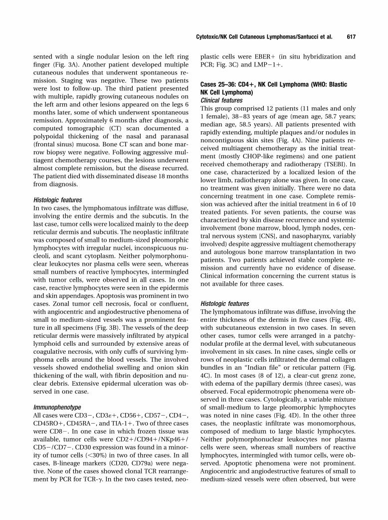
Cases 25–36: CD4�, NK Cell Lymphoma (WHO: BlasticNK Cell Lymphoma)Clinical featuresThis group comprised 12 patients (11 males and only1 female), 38 – 83 years of age (mean age, 58.7 years;median age, 58.5 years). All patients presented withrapidly extending, multiple plaques and/or nodules innoncontiguous skin sites (Fig. 4A). Nine patients re-ceived multiagent chemotherapy as the initial treat-ment (mostly CHOP-like regimens) and one patientreceived chemotherapy and radiotherapy (TSEBI). Inone case, characterized by a localized lesion of thelower limb, radiotherapy alone was given. In one case,no treatment was given initially. There were no dataconcerning treatment in one case. Complete remis-sion was achieved after the initial treatment in 6 of 10treated patients. For seven patients, the course wascharacterized by skin disease recurrence and systemicinvolvement (bone marrow, blood, lymph nodes, cen-tral nervous system [CNS], and nasopharynx, variablyinvolved) despite aggressive multiagent chemotherapyand autologous bone marrow transplantation in twopatients. Two patients achieved stable complete re-mission and currently have no evidence of disease.Clinical information concerning the current status isnot available for three cases.
Histologic featuresThe lymphomatous infiltrate was diffuse, involving theentire thickness of the dermis in five cases (Fig. 4B),with subcutaneous extension in two cases. In sevenother cases, tumor cells were arranged in a patchy-nodular profile at the dermal level, with subcutaneousinvolvement in six cases. In nine cases, single cells orrows of neoplastic cells infiltrated the dermal collagenbundles in an “Indian file” or reticular pattern (Fig.4C). In most cases (8 of 12), a clear-cut grenz zone,with edema of the papillary dermis (three cases), wasobserved. Focal epidermotropic phenomena were ob-served in three cases. Cytologically, a variable mixtureof small-medium to large pleomorphic lymphocyteswas noted in nine cases (Fig. 4D). In the other threecases, the neoplastic infiltrate was monomorphous,composed of medium to large blastic lymphocytes.Neither polymorphonuclear leukocytes nor plasmacells were seen, whereas small numbers of reactivelymphocytes, intermingled with tumor cells, were ob-served. Apoptotic phenomena were not prominent.Angiocentric and angiodestructive features of small tomedium-sized vessels were often observed, but were
Cytotoxic/NK Cell Cutaneous Lymphomas/Santucci et al. 617
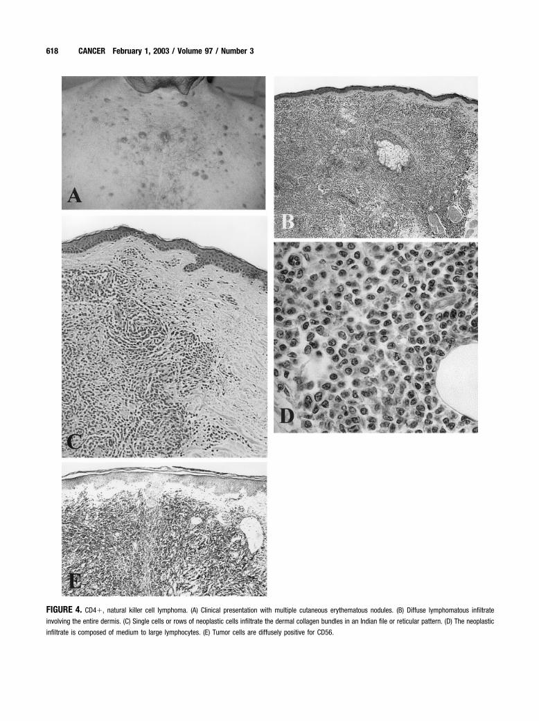
FIGURE 4. CD4�, natural killer cell lymphoma. (A) Clinical presentation with multiple cutaneous erythematous nodules. (B) Diffuse lymphomatous infiltrate
involving the entire dermis. (C) Single cells or rows of neoplastic cells infiltrate the dermal collagen bundles in an Indian file or reticular pattern. (D) The neoplastic
infiltrate is composed of medium to large lymphocytes. (E) Tumor cells are diffusely positive for CD56.
618 CANCER February 1, 2003 / Volume 97 / Number 3
not prominent. There were no obvious areas of intra-tumoral necrosis.
ImmunophenotypeAll cases were CD4�, CD56� (Fig. 4E), bcl-2�,CD43�, CD3�, CD8�, �F1�, TCR�1�, CD30�. All butone case were CD45RA�/CD45RO�. CD3� (poly-clonal anti-CD3) was expressed in four of seven testedcases. On frozen sections, all eight cases were CD123/IL3R��, CD94�, NKp46�. Eight of 12 cases wereCD2�/CD5�/CD7�, 3 cases were CD2�/CD5�/CD7�, whereas the last case was CD2�/CD5�/CD7�.The CD57 antigen was found in two of the six testedcases. In eight of the nine cases tested, a high prolif-erative activity of tumor cells (MIB-1 �50%) wasfound. TIA-1 was negative in all but two cases. Amongthe 10 cases in which CD68 expression was evaluated,7 cases gave a positive reaction in a high percentage oftumor cells. CD34 was negative in the eight testedcases. B-cell–associated antigens (CD20, CD21, CD22,CD79a) were negative in all cases. None of the 10 casestested for EBER and LMP-1 was positive for EBV. Noneof the nine cases tested showed TCR gene rearrange-ments.
Case 37: Blastoid NK Cell Lymphoma (WHO: Blastic NKCell Lymphoma)Clinical featuresThis patient was a 58-year-old man who presentedwith multiple plaques and nodules disseminated onthe skin. At presentation, systemic symptoms wereabsent and the patient’s staging workup was negative.Following aggressive chemotherapy courses (CHOP),the patient experienced partial remission. Shortlythereafter, the disease pursued a fulminant coursewith bone marrow and testicular involvement. Thepatient died 16 months after diagnosis.
Histologic featuresThe tumor cells were located mainly in the middle anddeep dermis, sparing the subepidermal region, andinfiltrated the collagen bundles in an interstitial pat-tern of growth. The infiltrate was monomorphous andcomposed of medium to large-sized cells with roundnuclei and fine diffuse chromatin.
ImmunophenotypeThe neoplastic cells were CD3�, CD4�, CD8�,CD45RO�, CD45RA�, CD56�, TIA-1�, GrB�, �F1�,TCR�1�, EBER�, and LMP-1�. A focal positivity forCD68 was also demonstrated. B-cell markers (CD20,CD79a) were negative.
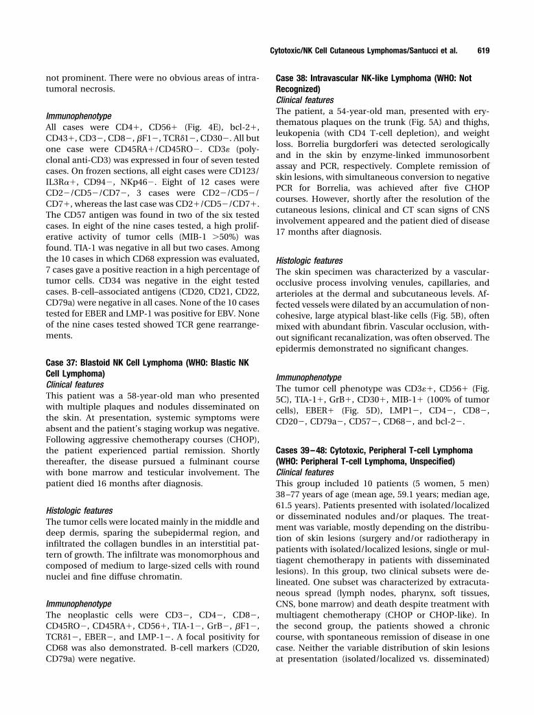
Case 38: Intravascular NK-like Lymphoma (WHO: NotRecognized)Clinical featuresThe patient, a 54-year-old man, presented with ery-thematous plaques on the trunk (Fig. 5A) and thighs,leukopenia (with CD4 T-cell depletion), and weightloss. Borrelia burgdorferi was detected serologicallyand in the skin by enzyme-linked immunosorbentassay and PCR, respectively. Complete remission ofskin lesions, with simultaneous conversion to negativePCR for Borrelia, was achieved after five CHOPcourses. However, shortly after the resolution of thecutaneous lesions, clinical and CT scan signs of CNSinvolvement appeared and the patient died of disease17 months after diagnosis.
Histologic featuresThe skin specimen was characterized by a vascular-occlusive process involving venules, capillaries, andarterioles at the dermal and subcutaneous levels. Af-fected vessels were dilated by an accumulation of non-cohesive, large atypical blast-like cells (Fig. 5B), oftenmixed with abundant fibrin. Vascular occlusion, with-out significant recanalization, was often observed. Theepidermis demonstrated no significant changes.
ImmunophenotypeThe tumor cell phenotype was CD3��, CD56� (Fig.5C), TIA-1�, GrB�, CD30�, MIB-1� (100% of tumorcells), EBER� (Fig. 5D), LMP1�, CD4�, CD8�,CD20�, CD79a�, CD57�, CD68�, and bcl-2�.
Cases 39–48: Cytotoxic, Peripheral T-cell Lymphoma(WHO: Peripheral T-cell Lymphoma, Unspecified)Clinical featuresThis group included 10 patients (5 women, 5 men)38 –77 years of age (mean age, 59.1 years; median age,61.5 years). Patients presented with isolated/localizedor disseminated nodules and/or plaques. The treat-ment was variable, mostly depending on the distribu-tion of skin lesions (surgery and/or radiotherapy inpatients with isolated/localized lesions, single or mul-tiagent chemotherapy in patients with disseminatedlesions). In this group, two clinical subsets were de-lineated. One subset was characterized by extracuta-neous spread (lymph nodes, pharynx, soft tissues,CNS, bone marrow) and death despite treatment withmultiagent chemotherapy (CHOP or CHOP-like). Inthe second group, the patients showed a chroniccourse, with spontaneous remission of disease in onecase. Neither the variable distribution of skin lesionsat presentation (isolated/localized vs. disseminated)
Cytotoxic/NK Cell Cutaneous Lymphomas/Santucci et al. 619
nor the aggressiveness of the initial treatment corre-lated with the clinical course.
Histologic featuresThis group was characterized by a proliferation of pleo-morphic lymphocytes of variable size (small to mediumcells in three cases, medium to large cells in six cases,and large, blast-like cells in one case). The pattern ofinfiltration was heterogenous, ranging from patchyperivascular (three cases) to diffuse dermal involvement(two cases), to diffuse in the dermis with extensive sub-cutaneous involvement (three cases), to mainly subcu-taneous with slight dermal involvement (two cases).
ImmunophenotypeAll cases expressed CD3, all but two cases were TIA-1�,and 5 of 10 were CD56� (including the 2 TIA-1� cases).Three cases were CD4�, three cases CD8� and the fiveCD4�, CD8� cases stained with �F1 (two cases) or with
TCR-�1 (two cases). The CD45RO antigen was diffuselyexpressed in one-half of the cases. Among the six casesin which frozen material was available, three cases ex-pressed either CD2, CD5, or CD7. Two cases showed adiffuse positivity (� 70% of tumor cells) for the CD30antigen. One of these two patients experienced sponta-neous remission and is alive with no evidence of disease1 year from diagnosis. All but one case was negative forB-cell–associated markers. The remaining case was im-munoreactive for CD79a, even though it lacked immu-noreactivity for CD20, was positive for �F1, and lackedclonal rearrangement for Ig heavy chains by PCR. Noneof the five cases tested for EBER and/or LMP-1 waspositive for EBV.
DISCUSSIONThe spectrum of cutaneous cytotoxic lymphomas washeterogenous with regard to clinical presentation,morphologic and immunophenotypic features, asso-
FIGURE 5. Intravascular natural killer-like lymphoma. (A) The patient presented with erythematous plaques on the trunk. (B) Affected vessels are dilated by an
accumulation of noncohesive, large atypical blast-like cells. (C) Tumor cells are positive for CD56. (D) Epstein–Barr virus (EBV)-encoded RNA in situ hybridization
shows that neoplastic cells are EBV positive.
620 CANCER February 1, 2003 / Volume 97 / Number 3
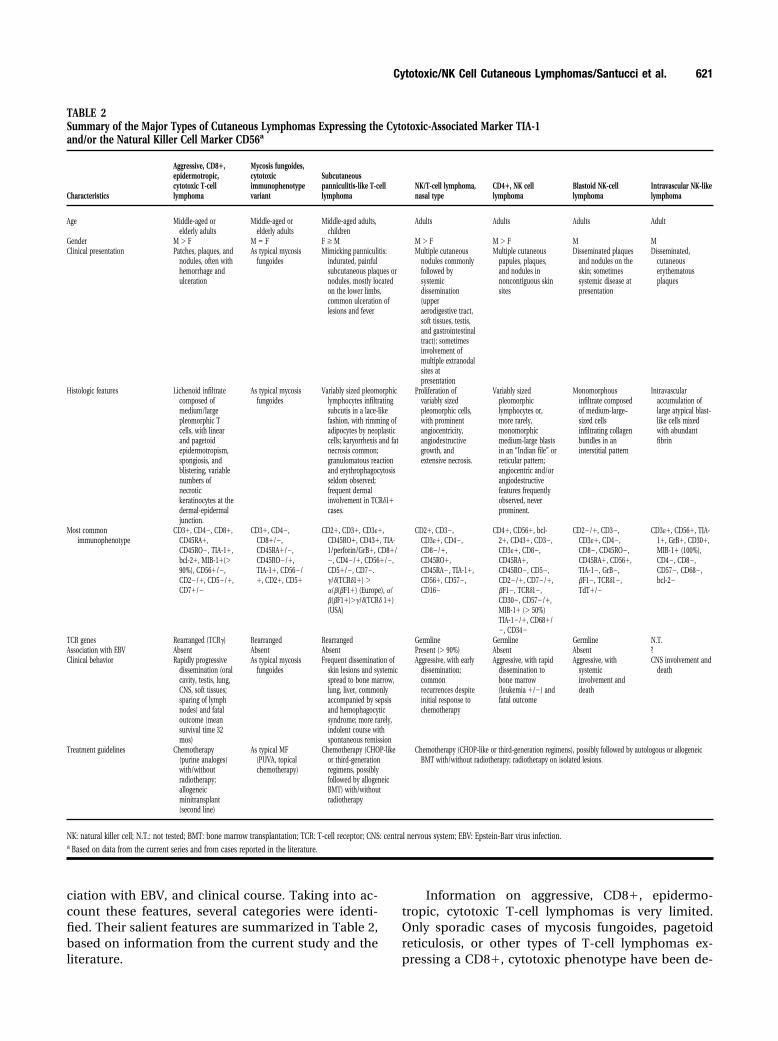
ciation with EBV, and clinical course. Taking into ac-count these features, several categories were identi-fied. Their salient features are summarized in Table 2,based on information from the current study and theliterature.
Information on aggressive, CD8�, epidermo-tropic, cytotoxic T-cell lymphomas is very limited.Only sporadic cases of mycosis fungoides, pagetoidreticulosis, or other types of T-cell lymphomas ex-pressing a CD8�, cytotoxic phenotype have been de-
TABLE 2Summary of the Major Types of Cutaneous Lymphomas Expressing the Cytotoxic-Associated Marker TIA-1and/or the Natural Killer Cell Marker CD56a
Characteristics
Aggressive, CD8�,epidermotropic,cytotoxic T-celllymphoma
Mycosis fungoides,cytotoxicimmunophenotypevariant
Subcutaneouspanniculitis-like T-celllymphoma
NK/T-cell lymphoma,nasal type
CD4�, NK celllymphoma
Blastoid NK-celllymphoma
Intravascular NK-likelymphoma
Age Middle-aged orelderly adults
Middle-aged orelderly adults
Middle-aged adults,children
Adults Adults Adults Adult
Gender M � F M � F F � M M � F M � F M MClinical presentation Patches, plaques, and
nodules, often withhemorrhage andulceration
As typical mycosisfungoides
Mimicking panniculitis:indurated, painfulsubcutaneous plaques ornodules, mostly locatedon the lower limbs,common ulceration oflesions and fever
Multiple cutaneousnodules commonlyfollowed bysystemicdissemination(upperaerodigestive tract,soft tissues, testis,and gastrointestinaltract); sometimesinvolvement ofmultiple extranodalsites atpresentation
Multiple cutaneouspapules, plaques,and nodules innoncontiguous skinsites
Disseminated plaquesand nodules on theskin; sometimessystemic disease atpresentation
Disseminated,cutaneouserythematousplaques
Histologic features Lichenoid infiltratecomposed ofmedium/largepleomorphic Tcells, with linearand pagetoidepidermotropism,spongiosis, andblistering, variablenumbers ofnecrotickeratinocytes at thedermal-epidermaljunction.
As typical mycosisfungoides
Variably sized pleomorphiclymphocytes infiltratingsubcutis in a lace-likefashion, with rimming ofadipocytes by neoplasticcells; karyorrhexis and fatnecrosis common;granulomatous reactionand erythrophagocytosisseldom observed;frequent dermalinvolvement in TCR�1�cases.
Proliferation ofvariably sizedpleomorphic cells,with prominentangiocentricity,angiodestructivegrowth, andextensive necrosis.
Variably sizedpleomorphiclymphocytes or,more rarely,monomorphicmedium-large blastsin an “Indian file” orreticular pattern;angiocentric and/orangiodestructivefeatures frequentlyobserved, neverprominent.
Monomorphousinfiltrate composedof medium-large–sized cellsinfiltrating collagenbundles in aninterstitial pattern
Intravascularaccumulation oflarge atypical blast-like cells mixedwith abundantfibrin
Most commonimmunophenotype
CD3�, CD4�, CD8�,CD45RA�,CD45RO�, TIA-1�,bcl-2�, MIB-1�(�90%), CD56�/�,CD2�/�, CD5�/�,CD7�/�
CD3�, CD4�,CD8�/�,CD45RA�/�,CD45RO�/�,TIA-1�, CD56�/�, CD2�, CD5�
CD2�, CD3�, CD3��,CD45RO�, CD43�, TIA-1/perforin/GrB�, CD8�/�, CD4�/�, CD56�/�,CD5�/�, CD7�.�/�(TCR�1�) ��/�(�F1�) (Europe), �/�(�F1�)��/�(TCR� 1�)(USA)
CD2�, CD3�,CD3��, CD4�,CD8�/�,CD45RO�,CD45RA�, TIA-1�,CD56�, CD57�,CD16�
CD4�, CD56�, bcl-2�, CD43�, CD3�,CD3��, CD8�,CD45RA�,CD45RO�, CD5�,CD2�/�, CD7�/�,�F1�, TCR�1�,CD30�, CD57�/�,MIB-1� (� 50%)TIA-1�/�, CD68�/�, CD34�
CD2�/�, CD3�,CD3��, CD4�,CD8�, CD45RO�,CD45RA�, CD56�,TIA-1�, GrB�,�F1�, TCR�1�,TdT�/�
CD3��, CD56�, TIA-1�, GrB�, CD30�,MIB-1� (100%),CD4�, CD8�,CD57�, CD68�,bcl-2�
TCR genes Rearranged (TCR�) Rearranged Rearranged Germline Germline Germline N.T.Association with EBV Absent Absent Absent Present (� 90%) Absent Absent ?Clinical behavior Rapidly progressive
dissemination (oralcavity, testis, lung,CNS, soft tissues;sparing of lymphnodes) and fataloutcome (meansurvival time 32mos)
As typical mycosisfungoides
Frequent dissemination ofskin lesions and systemicspread to bone marrow,lung, liver, commonlyaccompanied by sepsisand hemophagocyticsyndrome; more rarely,indolent course withspontaneous remission
Aggressive, with earlydissemination;commonrecurrences despiteinitial response tochemotherapy
Aggressive, with rapiddissemination tobone marrow(leukemia �/�) andfatal outcome
Aggressive, withsystemicinvolvement anddeath
CNS involvement anddeath
Treatment guidelines Chemotherapy(purine analoges)with/withoutradiotherapy;allogeneicminitransplant(second line)
As typical MF(PUVA, topicalchemotherapy)
Chemotherapy (CHOP-likeor third-generationregimens, possiblyfollowed by allogeneicBMT) with/withoutradiotherapy
Chemotherapy (CHOP-like or third-generation regimens), possibly followed by autologous or allogeneicBMT with/without radiotherapy; radiotherapy on isolated lesions.
NK: natural killer cell; N.T.: not tested; BMT: bone marrow transplantation; TCR: T-cell receptor; CNS: central nervous system; EBV: Epstein-Barr virus infection.a Based on data from the current series and from cases reported in the literature.
Cytotoxic/NK Cell Cutaneous Lymphomas/Santucci et al. 621
scribed.16 –33 Even the EORTC classification in its cur-rent form does not include CD8� cytotoxic T-celllymphomas neither in a well defined nor provisionalcategory.2 However, Berti et al.8 drew attention tothese tumors and suggested that CD8� cytotoxic T-cell lymphomas represent a distinctive type of cuta-neous T-cell lymphoma with an aggressive clinicalbehavior.8 The results of our study confirmed those ofBerti et al.8 concerning the distinctive clinical presen-tation and course, histology, and immunophenotypicfeatures of neoplastic cells of this peculiar type ofcutaneous T-cell lymphoma. In addition, we demon-strated that these cutaneous T-cell lymphomas alsoexpress the CD56 antigen, a finding not previouslyreported. The T-cell origin and the clonality of thisneoplasm were confirmed by the rearrangement of theTCR-� gene in the three cases investigated. The courseof the disease is characterized by rapidly progressivedissemination and death, despite the use of aggressivemultiagent chemotherapy regimens. It is noteworthythat the systemic spread did not involve the lymphnodes, but involved the oral cavity (one case), the softtissues of the centrofacial region (one case), and un-usual sites (lung, testis, or CNS) accompanied by sep-sis in one case. This may be attributed to the CD56�phenotype, which is associated with homing to ex-tranodal sites.
Aggressive therapeutic modalities were ineffectivein the management of these patients. Therefore, newstrategies are needed. In this respect, two main pointsshould be taken into account. First, the course of thedisease is frequently associated with severe immuno-deficiency. Therefore, neither TSEBI nor aggressivepolychemotherapy regimens are likely to achievelong-standing clinical responses. When possible, anallogeneic minitransplant instead of autologous bonemarrow transplantation may be suggested. Alterna-tively, the use of purine analogs devoid of high immu-nosuppressive capacity (like gemcitabine, which wasused in aggressive cutaneous T-cell lymphoma cas-es34) may be proposed, possibly associated with localradiotherapy. Second, as suggested by Berti et al.,8 theputative Th1-like cytokine profile of these aggressiveCD8� cutaneous T-cell lymphoma should discouragetreatments that increase Th1 responses (e.g., retinoidsor interferon alpha). Extreme caution should be usedfor such regimens.
Concerning mycosis fungoides, the cytotoxic im-munophenotype variant, these cases were character-ized by the typical clinical evolution, histology, andcourse of mycosis fungoides. If there are immunophe-notypic similarities with CD8� aggressive lympho-mas, cytotoxic mycosis fungoides has to be regardedas a phenotypic, and not a clinicopathologic, variant
of mycosis fungoides and should be treated nonag-gressively according to well established guidelines(PUVA, topical chemotherapy).
Subcutaneous panniculitis-like T-cell lymphoma(SPTCL), currently included as a provisional entity inboth the REAL classification35 and the EORTC classi-fication2 for primary cutaneous lymphomas, has beenincorporated as an entity in the WHO classification.2
The 12 cases in the current series exhibited the typicalfeatures as described in the more than 60 cases re-ported to date.36 –59 In our series, two phenotypic sub-sets can be delineated: TCR-�/��/CD56� and �/��/CD56�. In agreement with previous observations,58
these phenotypic subsets correlated with the presenceor absence of dermal invasion. All but one case withdermal extension were �/��/CD56�. Although a lim-ited number of SPTCLs have been investigated for TCRexpression, earlier studies from Europe and Easterncountries documented a prevalent �/� TCR expressionsimilar to that found in the current, study.40,46,55,60 – 62
American studies have emphasized that the majorityof SPTCL cases expressed �/� TCRs.57,58 We do notknow the reasons for the discrepancy between theAmerican and European/Eastern studies. In agree-ment with previous observations,39,49 most cases havean aggressive clinical behavior. In a few cases, thecourse of the disease is indolent, with spontaneousremission of skin lesions and long periods of stabledisease without associated systemic symptoms. Nei-ther the age of the patients nor the aggressiveness ofthe initial treatment was related to the course of thedisease. In agreement with previous observations,57 acorrelation was found between the expression of TCR-�/��/CD56� phenotype by tumor cells and an ag-gressive course. Therefore, the presence of this pecu-liar immunophenotypic profile may be indicative of adismal clinical course and may require aggressivetherapeutic modalities (CHOP-like or third generationregimens, possibly followed by allogeneic bone mar-row transplantation). Conversely, the finding of aTCR-�/��/CD56� phenotype may be indicative of anindolent course and favor less aggressive treatments.However, further studies are needed to confirmwhether the origin (�/� or �/�) and CD56 expressionhave a prognostic significance.
Increasing attention has been given to the clini-copathologic spectrum of the CD56� lymphomas,possibly due to the availability of a paraffin section-reactive CD56 antibody, which has greatly facilitatedthe recognition and further characterization of theserare tumors.4 –5,63– 69 CD56� lymphomas show a poly-morphous clinical presentation, a wide morphologicspectrum, and a variable immunophenotypic profile.For this reason, there is much confusion and little
622 CANCER February 1, 2003 / Volume 97 / Number 3
consensus regarding the best nomenclature for thesetumors. A review of the literature on this topic showsthat similar cases have been designated with differentterms, such as “aggressive natural killer cell leukemia/lymphoma”,70,71 “large granular lymphocytic lympho-ma”,72,73 “angiocentric T-cell lymphoma”,74 “CD56�T-”cell lymphoma,”75,76 “natural killer cell lymphoma”and “CD56 angiocentric lymphoma”.78 A comprehen-sive study provided a more complete picture of theserare neoplasms and better clarified the clinicopatho-logic spectrum of these uncommon tumors.5 In thecurrent study, we focused on NK and NK-like lympho-mas with primary presentation of disease in the skin.It should be noted that many of our cases had histo-logic/immunophenotypic patterns that were difficultto assign unambiguously to one or another of theexisting categories. In addition, this preliminary re-view gave us reason to doubt that histopathologicfeatures by themselves were invariably specific. Thatis, we frequently found it impossible to unequivocallyclassify single cases on morphologic grounds alone.
The three cases of NK/T-cell lymphoma, nasaltype, showed the characteristic morphology (dermalproliferation of small to medium pleomorphic cells,with prominent angiocentricity and angiodestructivegrowth as well as extensive necrosis), immunopheno-type (CD3�, CD3��, CD56�, CD45RO�, TIA-1�,TCRs�), and association with EBV, as previously de-scribed.4,5,79 In addition, in the only patient with avail-able follow-up information, the development of skinlesions was rapidly followed by involvement of thefrontal paranasal sinuses and nasal cavity, in agree-ment with the highly aggressive clinical behavior re-ported in previous studies.4,5 An extensive immuno-histochemical study on frozen section also showed anNK CD2�/CD94�/NKp46� phenotype.
Clinically, in the group of CD4� NK cell lympho-mas, a striking male preponderance was observed, inagreement with previous observations.5,67 Histopatho-logically, these cases showed a dermal infiltrate ofvariably sized pleomorphic cells or monomorphic,medium to large blasts. The neoplastic cells showedan Indian file or reticular pattern of infiltration. Theangiocentric/angiodestructive features, although of-ten observed, were never prominent as in the nasaltype NK/T-cell lymphoma. The immunophenotypewas CD3��/�, CD4�, CD43�, CD45RA�/�, CD56�,CD68�/�, CD123/IL3R��, CD3�, CD57�, TCRs�,TIA-1�, EBER� and LMP-1�, resembling the immu-nophenotypic profile described by Petrella et al.6 as“CD4�, CD56� cutaneous lymphomas.” Possible ex-amples were published by others.7,80,81 Therefore, wepreferred to retain this terminology instead of using
the one proposed by the WHO classification (blasticNK-cell lymphoma).
The expression of CD4, CD56, CD68, and CD123and the negativity of NKp46 and CD94 (in seven casestested), the striking male preponderance, and the sub-sequent rapid spread to bone marrow, with or withoutleukemic evolution, raise doubts concerning the ori-gin/differentiation of tumor cells and the relationshipof this entity with other NK/T-cell lymphomas andleukemias.4,5,70,82– 85 The CD4�/CD68�/CD123� phe-notype was mainly expressed by immature monocyticand dendritic cell precursors. The cases belonging tothis group, although primary cutaneous at presenta-tion according to the EORTC definition2 and charac-terized by initial response to the treatment with ag-gressive polychemotherapy, experienced rapid spreadto the bone marrow, with or without leukemia, anddeath notwithstanding aggressive second-line treat-ments including bone marrow transplantation. Werecommend aggressive polychemotherapy possiblyfollowed by autologous or heterologous bone marrowtransplantation in patients presenting with dissemi-nated cutaneous diseases and local radiotherapy andclose follow-up in patients who present with single,isolated skin lesions.
The patient with blastoid NK cell lymphoma pre-sented with the clinical symptoms (extranodal diseaseat diagnosis, histopathologic features (monomor-phous proliferation of medium to large cells in a reti-form pattern reminiscent of leukemia), immunophe-notypic profile (CD3�, CD4�, CD8�, CD56�, �F1�,TCR�1�), the lack of association with EBV, and theaggressive clinical course recently described by Chanet al.5 as typical of this entity. For this reason, andbecause of the lack of CD4 expression, we believe thatthis case is phenotypically different from CD4�NK-like lymphoma. Therefore, we retained the originalterminology instead of using the one proposed by theWHO classification, which lumps the two categoriesunder the heading “blastic NK cell lymphoma.” How-ever, because few cases of blastoid NK cell lymphomahave been reported, the proper nosology of these rarecases and their relationship with other types of NK/T-cell lymphomas/leukemias remain to be determined.In particular, it cannot be excluded that both blastoidNK cell lymphoma and CD4�/CD56� primary cuta-neous lymphoma partly overlap with or may be iden-tical to the entity called blastic NK cell lymphomaaccording to the WHO classification.1
The only case of intravascular, NK-like lymphomahad such a distinctive morphoimmunophenotypicprofile that it deserves a separate category. We pre-ferred to use NK-like instead of NK cell because wewere not able to investigate the TCR gene status. On
Cytotoxic/NK Cell Cutaneous Lymphomas/Santucci et al. 623
histopathologic examination, our case exhibited thetypical intravascular accumulation of large atypicalcells, which dilated and occluded the vascular luminaand were associated with fibrinous thrombi.86 To thebest of our knowledge, this is the first case reported ofan intravascular NK-like lymphoma. In fact, most ofthe previously described cases showed a B-cell phe-notype and a concurrent predilection for lung andskin,87,88 whereas fewer reported cases of T-cell– de-rived cases showed a clear-cut predilection for theskin.88 –100 The current case characterized by completeremission after initial aggressive polychemotherapy(CHOP), metastasis to the CNS, and death 17 monthsafter diagnosis, has to be considered exceptional asthat putatively histiocytic variant reported.101
The group of cytotoxic, peripheral T-cell lympho-mas by Snowden et al. represented a hodgepodge ofcases that did not have unifying features and werelumped together because they did not conform to anyof the other categories. These cases were composed ofpleomorphic lymphocytes of variable size or blast-likecells with the features of pleomorphic small to medi-um-sized cutaneous T-cell lymphoma or large cellcutaneous T-cell lymphoma, CD30�, or large cell cu-taneous T-cell lymphoma CD30�/lymphomatoidpapulosis according to the EORTC classification. Al-though the small number of cases does not us allow todraw definite conclusions, it seems that the cytotoxicphenotype does not significantly affect the clinicalevolution and prognosis of the single entities, similarlyto cytotoxic mycosis fungoides.
In conclusion, primary cutaneous lymphomas ex-pressing the cytotoxic granule-associated protein(TIA-1) and/or the NK cell marker (CD56) includedistinct groups of diseases, both clinically and biolog-ically. Our objective was to clarify the clinical, mor-phologic, and phenotypic features of these distinctcategories, which are often characterized by a highlyaggressive behavior. Because the finding of a cytotoxicphenotype often has prognostic significance, the rou-tine use of cytotoxic markers in the diagnosis andclassification of cutaneous lymphomas should be ex-panded.
REFERENCES1. Jaffe ES, Harris NL, Stein H, Vardiman JW. Tumours of
haematopoietic and lymphoid tissues. World Health Orga-nization classification of tumours. Lyon: IARC Press, 2001.
2. Willemze R, Kerl H, Sterry W, et al. EORTC classification forprimary cutaneous lymphomas: a proposal from the cuta-neous lymphoma study group of the European Organiza-tion for Research and Treatment of Cancer. Blood. 1997;90:354 –374.
3. Felgar RE, Macon WR, Kinney MC, Roberts S, Pasha T,Salhany KE. TIA-1 expression in lymphoid neoplasms:
identification of subsets with cytotoxic T lymphocyte ornatural killer differentiation. Am J Pathol. 1997;150:1893–1900.
4. Jaffe ES, Chan JKC, Su IJ, et al. Report of the workshop onnasal and related extranodal angiocentric T/natural killercell lymphomas. Am J Surg Pathol. 1996;20:103–111.
5. Chan JKC, Sin VC, Wong KF, et al. Nonnasal lymphomaexpressing the natural killer cell marker CD56: a clinico-pathologic study of 49 cases of an uncommon aggressiveneoplasm. Blood. 1997;89:4501– 4513.
6. Petrella T, Dalac S, Maynadie M, et al. CD4� CD56� cu-taneous neoplasms: a distinct hematological entity? Am JSurg Pathol. 1999;23:137–146.
7. Dummer R, Potoczna N, Haffner AC, Zimmermann DR,Gilardi S, Burg G. A primary cutaneous non-T, non-BCD4�, CD56� lymphoma. Arch Dermatol. 1996;132:550 –553.
8. Berti E, Tomasini D, Vermeer M, Meijer CJLM, Alessi E,Willemze R. Primary cutaneous CD8-positive epidermo-tropic cytotoxic T-cell lymphomas: a distinct clinicopath-ologic entity with an aggressive clinical behavior. Am JPathol. 1999;155:483– 492.
9. Blakolmer K, Vesely M, Kummer JA, Jurecka W, MannhalterC, Chott A. Immunoreactivity of B-cell markers (CD79a,L26) in rare cases of extranodal cytotoxic peripheral T-(NK/T-) cell lymphomas. Mod Pathol. 2000;13:766 –772.
10. Kamarashev J, Burg G, Mingari MC, Kempf W, Hofbauer G,Dummer R. Differential expression of cytotoxic moleculesand killer cell inhibitory receptors in CD8� and CD56�cutaneous lymphomas. Am J Pathol. 2001;158:1593–1598.
11. Montone KT, Brigati DJ. In situ molecular pathology: in-strumentation, oligonucleotides, and viral nucleic acid de-tection. J Histotechnol. 1994;17:195.
12. Bottaro M, Berti E, Biondi A, Migone N, Crosti L. Hetero-duplex analysis of T-cell receptor � gene rearrangementsfor diagnosis and monitoring of cutaneous T-cell lympho-mas. Blood. 1994;83:3271–3278.
13. Arnold A, Cossman J, Bakhshi A, Jaffe ES, Waldmann TA,Korsmeyer SJ. Immunoglobulin-gene rearrangements asunique clonal markers in human lymphoid neoplasms.N Engl J Med. 1983;309:1593–1599.
14. Minden MD, Toyonaga B, Ha K, et al. Somatic rearrange-ment of T-cell antigen receptor gene in human T-cell ma-lignancies. Proc Natl Acad Sci USA. 1985;82:1224 –1227.
15. Kimura N, Toyonaga B, Yoshikai Y, Du RP, Mak T. Se-quences and repertoire of the human T cell receptor � and� chain variable region genes in thymocytes. Eur J Immu-nol. 1987;17:375–383.
16. Jensen JR, Thestrup-Pedersen K. Subpopulations of T-lym-phocytes in a patient with fulminant mycosis fungoides.Acta Derm Venereol (Stockh). 1980;60:159 –161.
17. Haynes BF, Hensley LL, Jegasothy BV. Phenotypic charac-terization of skin-infiltrating T cells in cutaneous T-celllymphoma: comparison with benign cutaneous T-cell in-filtrates. Blood. 1982;60:463– 473.
18. Caputo R, Monti M, Berti E, Cavicchini S. A verrucoidepidermotropic OKT8 positive lymphoma. Am J Dermato-pathol. 1983;5:159 –161.
19. Buechner SA, Winkelmann RK, Banks PM. T cells and T cellsubsets in mycosis fungoides and parapsoriasis. A study of18 cases with anti-human T cell monoclonal antibodiesand histochemical techniques. Arch Dermatol. 1984;120:897–905.
624 CANCER February 1, 2003 / Volume 97 / Number 3
20. Bennet SR, Greer JP, Stein RS, Glick AD, Cousar JB, CollinsRD. Death due to splenic rupture in suppressor cell myco-sis fungoides: a case report. Am J Clin Pathol. 1984;82:104 –109.
21. Mackie RM, Turbitt ML. A case of pagetoid reticulosisbearing the cytotoxic suppressor surface marker on thelymphoid infiltrate: further evidence that pagetoid reticu-losis is not a variant of mycosis fungoides. Br J Dermatol.1984;110:89 –94.
22. Gonzalez M, Martin-Pascual MM, Miguel JS, Caballero MD,Lopez Borrasca A. Phenotypic characterization of skin in-filtrating cells in pagetoid reticulosis by monoclonal anti-bodies. Acta Derm Venereol (Stockh). 1984;64:421– 424.
23. Nasu K, Said J, Vonderheid E, Olerud J, Sako D, Kadin M.Immunopathology of cutaneous T-cell lymphomas. Am JPathol. 1985;119:436 – 447.
24. Ralfkiaer E, Lange-Wantzin G, Mason DY, Hou Jensen K,Stein H, Thomsen K. Phenotypic characterization of lym-phocyte subsets in mycosis fungoides. Comparison withlarge plaque parapsoriasis and benign chronic dermatoses.Am J Clin Pathol. 1985;84:610 – 619.
25. Quanterman MJ, Lesher JL, Davis LS, Pantazis CG, MullinsS. Rapidly progressive CD8-positive cutaneous T-cell lym-phoma with tongue involvement. Am J Dermatopathol.1995;17:287–291.
26. Jimbow K, Maeda K, Ito Y, Ishida O, Takami T. Heteroge-neity of cutaneous T-cell lymphoma. Phenotypic and ul-trastructural characterization of four unusual cases. Can-cer. 1985;56:2458 –2469.
27. Ohkolhchi K, Aiba S, Tagami H. OKT8-reactive cell mycosisfungoides. Arch Dermatol. 1986;122:20 –22.
28. Fujiwara Y, Abe Y, Kuyama M, et al. CD8� cutaneous T-celllymphoma with pagetoid epidermotropism, and angiocen-tric, and angiodestructive infiltration. Arch Dermatol. 1990;126:801– 804.
29. Urrutia S, Piris MA, Orradre JL, Martinez B, Cruz MA,Garcia-Almagro D. Cytotoxic/suppressor (CD8�, CD4�)cutaneous T-cell lymphoma with aggressive course. Am JDermatopathol. 1990;12:603– 606.
30. Agnarsson BA, Vonderheid EC, Kadin ME. Cutaneous T celllymphoma with suppressor/cytotoxic (CD8) phenotype:identification of rapidly progressive and chronic subtypes.J Am Acad Dermatol. 1990;22:569 –577.
31. Smoller BR, Stewart M, Warnke R. A case of Woringer-Kolopp disease (localized pagetoid reticulosis) or unile-sional mycosis fungoides? Arch Dermatol. 1992;128:526 –529.
32. Marti RM, Estrach T, Palou J, et al. Specific cutaneouslesions in a CD8� peripheral T-cell lymphoma. Int J Der-matol. 1992;31:624 – 628.
33. Kikuchi A, Sakuraoka K, Kurihara S, Akiyama M, Shimizu H,Nishikawa T. CD8� cutaneous anaplastic large-cell lym-phoma: report of two cases with immunophenotyping, T-cell-receptor gene rearrangement and electron micro-scopic studies. Br J Dermatol. 1992;126:404 – 408.
34. Zinzani PL, Magagnoli M, Bendandi M, et al. Therapy withgemcitabine in pretreated peripheral T-cell lymphoma pa-tients. Ann Oncol. 1998;9:1351–1353.
35. Harris NL, Jaffe ES, Stein H, et al. A revised European-American classification of lymphoid neoplasms: a proposalfrom the International Lymphoma Study Group. Blood.1994;84:1361–1392.
36. Wick MR, Sanchez NP, Crotty CP, Winkelmann RK. Cuta-neous malignant histiocytosis: a clinical and histologic
study of eight cases, with an immunohistochemical analy-sis. J Am Acad Dermatol. 1983;8:50 – 62.
37. Ashworth J, Coady AT, Guy R, Breathnach SM. Brawnycutaneous induration and granulomatous panniculitis inlarge cell non-Hodgkin’s (T suppressor/cytotoxic cell) lym-phoma. Br J Dermatol. 1989;120:563–569.
38. Tanaka K, Hagari Y, Sano Y, Shimano S, Namba K. A case ofT-cell lymphoma associated with panniculitis, progressivepancytopenia and hyperbilirubunemia. Br J Dermatol.1990;123:649 – 652.
39. Gonzalez CL, Medeiros J, Braziel RM, Jaffe ES. T-cell lym-phoma involving subcutaneous tissue. A clinicopathologicentity commonly associated with hemophagocytic syn-drome. Am J Surg Pathol. 1991;15:17–27.
40. Burg G, Dummer R, Wilhelm M, et al. A subcutaneousdelta-positive T-cell lymphoma that produces interferongamma. N Engl J Med. 1991;325:1078 –1081.
41. Smith KJ, Skelton HG, Giblin WL, James WD. Cutaneouslesions of hemophagocytic syndrome in a patient withT-cell lymphoma and active Epstein-Barr virus infection.J Am Acad Dermatol. 1991;25:919 –924.
42. Grange F, Avril MF, Duvillard P, et al. Lymphome a grandescellules anaplasiques Ki1� en tissu sous-cutane, avec as-pect clinique de panniculite. Ann Dermatol Venereol. 1992;119:890 – 892.
43. Hytiroglou P, Phelps RG, Wattenberg DJ, Strauchen JA.Histiocytic cytophagic panniculitis: molecular evidence fora clonal T-cell disorder. J Am Acad Dermatol. 1992;27:333–336.
44. Prescott RJ, Banerjee SS, Cross PA. Subcutaneous T-celllymphoma with florid granulomatous panniculitis. Histo-pathology. 1992;20:535–537.
45. Kaplan MA, Jacobson JO, Ferry JA, Harris NL. T-cell lym-phoma of the vulva in a renal allograft recipient with as-sociated hemophagocytosis. Am J Surg Pathol. 1993;17:842– 849.
46. Avinoach I, Halevy S, Argov S, Sacks M. �/� T-cell lym-phoma involving the subcutaneous tissue and associatedwith a hemophagocytic syndrome. Am J Dermatopathol.1994;16:426 – 433.
47. Chan YF, Lee KC, Llewellyn H. Subcutaneous T-cell lym-phoma presenting as panniculitis in children: report of twocases. Pediatr Pathol. 1994;14:595– 608.
48. Mehregan DA, Su D, Kurtin PJ. Subcutaneous T-cell lym-phoma: a clinical, histopathologic and immunohistochem-ical study of six cases. J Cutan Pathol. 1994;21:110 –117.
49. Perniciaro C, Winkelmann RK, Ehrhardt D. Fatal systemiccytophagic histiocytic panniculitis: a histopathologic andimmunohistochemical study of multiple organ sites. J AmAcad Dermatol. 1994;31:901–905.
50. Harada H, Iwatsuki K, Kaneko F. Detection of Epstein-Barrvirus genes in malignant lymphoma with clinical and his-tologic features of cytophagic histiocytic panniculitis. J AmAcad Dermatol. 1994;31:379 –383.
51. Cho KH, Oh JK, Kim CW, Heo DS, Kim ST. Peripheral T-celllymphoma involving subcutaneous tissue. Br J Dermatol.1995;132:290 –295.
52. Wang CY, Su WP, Kurtin PJ. Subcutaneous panniculitisT-cell lymphoma. Int J Dermatol. 1996;35:1– 8.
53. Romero LS, Goltz RW, Nagi C, Shin SS, Ho A. SubcutaneousT-cell lymphoma with associated hemophagocytic syn-drome and terminal leukemic transformation. J Am AcadDermatol. 1996;34:904 –910.
Cytotoxic/NK Cell Cutaneous Lymphomas/Santucci et al. 625
54. Sajben FP, Schmidt C. Subcutaneous T-cell lymphoma: acase report and additional observations. Cutis. 1996;58:297–302.
55. von den Driesch P, Staib G, Simon M, Sterry W. SubcutaneousT-cell lymphoma. J Am Acad Dermatol. 1997;36:285–289.
56. Dargent JL, Roufosse C, Delville JP, et al. Subcutaneouspanniculitis-like T-cell lymphoma: further evidence for adistinct neoplasm originating from large granular lympho-cytes of T/NK phenotype. J Cutan Pathol. 1998;25:394 – 400.
57. Salhany KE, Macon WR, Choi JK, et al. Subcutaneous pan-niculitis-like T-cell lymphoma. Clinicopathologic, immu-nophenotypic, and genotypic analysis of alpha/beta andgamma/delta subtypes. Am J Surg Pathol. 1998;22:881– 893.
58. Kumar S, Krenacs L, Medeiros J, et al. Subcutaneous pan-niculitic T-cell lymphoma is a tumor of cytotoxic T lym-phocytes. Hum Pathol. 1998;29:397– 403.
59. Craig AJ, Cualing H, Thomas G, Lamerson C, Smith R.Cytophagic histiocytic panniculitis—a syndrome associ-ated with benign and malignant panniculitis: case compar-ison and review of the literature. J Am Acad Dermatol.1998;39:721–736.
60. Ralfkiaer E, Wollf-Sneedorff A, Thomsen K, Geisler C, Ve-jlsgaard GL. T-cell receptor �/� -positive peripheral T-celllymphomas presenting in the skin: a clinical, histologicaland immunophenotypic study. Exp Dermatol. 1992;1:31–36.
61. Fujita M, Miyachi Y, Furukawa F, et al. A case of cutaneousT-cell lymphoma expressing �/� T-cell receptors. J AmAcad Dermatol. 1993;28:355–360.
62. Munn SE, McGregor JM, Jones A, et al. Clinical and patho-logical heterogeneity in cutaneous gamma-delta T-celllymphoma: a report of three cases and a review of theliterature. Br J Dermatol. 1996;135:976 –981.
63. Tsang WYW, Chan JKC, Pau MY. Utility of a paraffin sec-tion-reactive CD56 antibody (123C3) for characterizationand diagnosis of lymphomas. Am J Surg Pathol. 1996;20:202–210.
64. Jaffe ES. Classification of natural killer (NK) cell and NK-like T-cell malignancies. Blood. 1996;87:1207–1210.
65. Jaffe ES, Krenacs L, Raffeld M. Classification of T-cell andNK-cell neoplasms based on the REAL classification. AnnOncol. 1997;8(Suppl 2):S17.
66. Chan JKC, Tsang WYW, Lau WH, et al. Aggressive T/naturalkiller lymphoma presenting as testicular tumor. Cancer.1996;77:1198 –1205.
67. Ansai SI, Maeda K, Yamakawa M, et al. CD56-positive (na-sal-type T/NK cell) lymphoma arising on the skin. Reportof two cases and review of the literature. J Cutan Pathol.1997;24:468 – 476.
68. Kwong YL, Chan ACL, Liang R, et al. CD56� NK lympho-mas: clinicopathologic features and prognosis. Br J Haema-tol. 1997;97:821– 829.
69. Natkunam Y, Smoller BR, Zehnder JL, Dorfman RF, WarnkeRA. Aggressive cutaneous NK and NK-like T-cell lympho-mas. Am J Surg Pathol. 1999;23:571–581.
70. Imamura N, Kusunikoi Y, Kawa-Ha K, et al. Aggressivenatural killer cell leukemia/lymphoma: report of four casesand review of the literature. Possible existence of a newclinical entity originated from the third lineage of lym-phoid cells. Br J Haematol. 1990;75:49 –59.
71. Sun T, Brody J, Susin M, et al. Aggressive natural killer celllymphoma/leukemia: a recently recognized clinico-patho-logic entity. Am J Surg Pathol. 1993;17:1289 –1299.
72. Berceanu S, Roman S, Butoianu E, et al. A particular case of
large granular lymphocytes lymphoma. Haematologica.1989;22:43–53.
73. Sun T, Schulman P, Kolitz J, et al. A study of lymphoma oflarge granular lymphocytes with modern modalities: reportof two cases and review of the literature. Am J Hematol.1992;40:135–142.
74. Chan JKC, Ng CS, Ngan KC, Hui PK, Lo STH, Lau WH.Angiocentric T-cell lymphoma of the skin: an aggressivelymphoma distinct from mycosis fungoides. Am J SurgPathol. 1988;12:861– 876.
75. Hayashi K, Nakamura S, Koshikawa T, et al. A case ofneural cell adhesion molecule-positive peripheral T-celllymphoma associated with human T-cell lymphotrophicvirus type 1 showing an unusual involvement of the gas-trointestinal tract during the course of the disease. HumPathol. 1994;25:1251–1253.
76. Tsang WYW, Chan JKC, Yip TTC, et al. In situ localization ofEpstein-Barr virus encoded RNA in non-nasal/nasopha-ryngeal CD56-positive and CD56-negative T-cell lym-phoma. Hum Pathol. 1994;25:758 –765.
77. Takayama A, Ochiai T, Yoshizawa K, Fujisawa S, MorishimaT. Cutaneous natural killer cell lymphoma. Skin Cancer.1996;7:376.
78. Nakamura S, Suchi T, Koshikawa T, et al. Clinicopathologicstudy of CD56 (NCAM)-positive angiocentric lymphomaoccurring in sites other than the upper and lower respira-tory tract. Am J Surg Pathol. 1995;19:284 –296.
79. Wong KF, Chan JKC, Ng CS, Lee KC, Tsang WYW, CheungMMC. CD56 (NKH1)-positive hematolymphoid malignan-cies: an aggressive neoplasm featuring frequent cutaneous-mucosal involvement, cytoplasmic azurophilic granulesand angiocentricity. Hum Pathol. 1992;23:798 – 804.
80. Adachi M, Maeda K, Takekawa M, et al. High expression ofCD56 (N-CAM) in a patient with cutaneous CD4-positivelymphoma. Am J Hematol. 1994;47:278 –282.
81. Wasik MA, Sackstein R, Novick D, et al. Cutaneous CD56�large T-cell lymphoma associated with high serum concen-tration of IL-2. Hum Pathol. 1996;27:738 –744.
82. Imamura N, Kusunoki Y, Kajihara H, Okada K, Kuramoto A.Aggressive natural killer cell leukemia/lymphoma withN901-positive surface phenotype: evidence for the exis-tence of a third lineage of lymphoid cells. Acta Haematol.1988;80:121–128.
83. Gattei V, Carbone A, Marotta G, et al. Expression of thenatural killer antigens in malignant histiocytosis and asubset of acute myelomonocytic leukemias. Bone MarrowTransplant. 1989;4:22.
84. Gattei V, Carbone A, Zagonel V, Pinto A. Expression ofnatural killer antigens in a subset of non-T, non-B lympho-ma/leukemia with histiocytic features. Br J Haematol. 1990;76:444 – 448.
85. DiGiuseppe JA, Louie DC, Williams JE, et al. Blastic naturalkiller cell leukemia/lymphoma: a clinicopathologic study.Am J Surg Pathol. 1997;21:1223–1230.
86. Wick MR, Mills SE, Scheithauer BW, Cooper PH, DavitzMA, Parkinson K. Reassessment of malignant endothelio-matosis. Evidence in favour of its reclassification as “intra-vascular lymphoma.” Am J Surg Pathol. 1986;10:112–123.
87. Carroll TJ Jr., Schelper RL, Goeken JA, Kemp JD. Neoplasticangioendotheliomatosis: immunopathologic and morpho-logic evidence for intravascular malignant lymphomatosis.Am J Clin Pathol. 1986;85:169 –175.
626 CANCER February 1, 2003 / Volume 97 / Number 3
88. Stroup RM, Sheibani K, Moncada A, Purdy LJ, Battifora H.Angiotropic (intravascular) large cell lymphoma: a clinico-pathologic study of seven cases with unique clinical pre-sentations. Cancer. 1990;66:1781–1788.
89. Sheibani K, Battifora H, Winberg CD, et al. Further evi-dence that “malignant angioendotheliomatosis” is an an-giotropic large-cell lymphoma. N Engl J Med. 1986;314:943–948.
90. Maruyama N, Ishida Y, Sato H, Koike T, Nagao K. Expres-sion of lymphocyte-associated antigens on neoplastic an-gioendotheliosis. Appl Pathol. 1986;5:246.
91. Sepp N, Schuler G, Romani N, et al. “Intravascular lympho-matosis” (angioendotheliomatosis): evidence for a T-cellorigin in two cases. Hum Pathol. 1990;21:1051–1058.
92. Tateyama H, Eimoto T, Tada T, Kamiya M, Fujiyoshi Y,Kajiura S. Congenital angiotropic lymphoma (intravascularlymphomatosis) of the T-cell type. Cancer. 1991;67:2131–2136.
93. Clark WC, Dohan FC, Moss T, Schweitzer JB. Immunocy-tochemical evidence of lymphocytic derivation of neoplas-tic cells in malignant endotheliomatosis. J Neurosurg. 1991;74:757–762.
94. Shimokawa I, Higami Y, Sakai H, Moriuchi Y, Murase K,Ikeda T. Intravascular malignant lymphomatosis: a case ofT-cell lymphoma probably associated with human T-celllymphotropic virus. Hum Pathol. 1991; 22:200 –202.
95. Sangueza O, Hyder DM, Sangueza P. Intravascular lympho-matosis: report of an unusual case with T-cell phenotypeoccurring in an adolescent male. J Cutan Pathol. 1992;19:226 –231.
96. Lopez-Gil F, Roura M, Umbert I, Umbert P. Malignantproliferative angioendotheliomatosis or angiotropic lym-phoma associated with a soft-tissue lymphoma. J Am AcadDermatol. 1992;26:101–104.
97. Glass J, Hochberg FH, Miller DC. Intravascular lymphoma-tosis: a systemic disease with neurologic manifestations.Cancer. 1993;71:3156 –3164.
98. DiGiuseppe JA, Nelson WG, Seifter EJ, Boitnott JK, MannRB. Intravascular lymphomatosis: a clinicopathologicstudy of 10 cases and assessment of response to chemo-therapy. J Clin Oncol. 1994;12:2573–2579.
99. Ghorbani RP, Shokouh-Amiri H, Gaber LW. Intragraft an-giotropic large-cell lymphoma of T-cell type in a long-termrenal allograft recipient. Mod Pathol. 1996;9:671– 676.
100. Au WY, Shek WH, Nicholls J, Tse KM, Todd D, Kwong YL.T-cell intravascular lymphomatosis (angiotropic large celllymphoma): association with Epstein-Barr viral infection.Histopathology. 1997;31:563–567.
101. Snowden JA, Angel CA, Winfield DA, Pringle JH, West KP.Angiotropic lymphoma: report of a case with histiocyticfeatures. J Clin Pathol. 1997;50:67–70.
Cytotoxic/NK Cell Cutaneous Lymphomas/Santucci et al. 627
Related Documents


