CT and MR: Concepts & Application of CT and MR: Concepts & Application of These Complementary Techniques These Complementary Techniques 3 3 rd rd Annual Imaging & Physiology Summit Annual Imaging & Physiology Summit November 20 November 20 - - 21, 2009 Seoul, Korea 21, 2009 Seoul, Korea Wm. Guy Wm. Guy Weigold Weigold , MD, FACC , MD, FACC Washington Hospital Center Washington Hospital Center Cardiovascular Research Institute Cardiovascular Research Institute Washington, DC Washington, DC

Welcome message from author
This document is posted to help you gain knowledge. Please leave a comment to let me know what you think about it! Share it to your friends and learn new things together.
Transcript

CT and MR: Concepts & Application of CT and MR: Concepts & Application of These Complementary TechniquesThese Complementary Techniques
33rdrd Annual Imaging & Physiology SummitAnnual Imaging & Physiology SummitNovember 20November 20--21, 2009 Seoul, Korea21, 2009 Seoul, Korea
Wm. Guy Wm. Guy WeigoldWeigold, MD, FACC, MD, FACC
Washington Hospital CenterWashington Hospital CenterCardiovascular Research InstituteCardiovascular Research Institute
Washington, DCWashington, DC

Cardiac CT Technique: ECGCardiac CT Technique: ECG--GatingGating
75% of RR
75% of RR
RetrospectiveReconstruction
001011010101011001101100001110101010101010010100101010101010101000101101010101100110110000111010101010101001010010101010101010101101001010101010101010101010101010101010101010101010001011010101010100101010101010101010101010101010101010101010101000101101010101001001010101010101010101110100110101010101010101011010010010110010100101010101010101010111010011010101010101010101101001001011001010101010101010101010101010111001011010101010101001101010110101010111101010101010101010101011100101101010101010100110101011010101011100001010101100110101010010101101010101010110010011101010101010101010101010110011010101001010110101010101011001001110101010101010101010101001011010101011001101100001110101010101010010100101010101010101100101101010101100110110000111010101010101001010010101010101010101010100101010101010101010101010101010101010101010101000101101010101010010101010101010101010101010101010101010101010100010110101010100100101010101010101010111010011010101010101010101101001001011001010010101010101010101011101001101010101010101010110100100101100101010101010101010101010101011100101101010101010100110101011010101011110101010101010101010101110010110101010101010011010101101010101110000101010110011010101001010110101010101011001001110101010101010101010101011001101010100101011010101010101100100111010101010101010101010
001011010101011001101100001110101010101010010100101010101010101010100101010101010101010101010101010101010101010101000101101010101010010101010101010101011101001101010101010101010110100100101100101010101010101010101010101110010110101010101010011010101101010101110010101011001101010100101011010101010101100100111010101010101010101010010110101010110011011000011101010101010100101001010101010101010101001010101010101010101010101010101010101010101010001011010101010100101010101010101010111010011010101010101010101101001001011001010101010101010101010101011100101101010101010100110101011010101011100101010110011010101001010110101010101011001001110101010101010101010

Prospective Cardiac CT: XProspective Cardiac CT: X--Ray Tube Mostly OffRay Tube Mostly Off
ProspectiveECG-Triggered Acquisition
00101101010101100110110000111010101010101001010010101010101010100010110101010110011011000011101010101010100101001010101010101010110100101010101010101010101010101010101010101010101000101101010101010010101010101010101010101010101010101010101010100010110101010100100101010101010101010111010011010101010101010101101001001011001010010101010101010101011101001101010101010101010110100100101100101010001011010101011001101100001110101010101010010100101010101010101000101101010101100110110000111010101010101001010010101010101010101101001010101010101010101010101010101010101010101010001011010101010100101010101010101010101010101010101010101010101000101101010101001001010101010101010101110100110101010101010101011010010010110010100101010101010101010111010011010101010101010101101001001011001010100010110101010110011011000011101010101010100101001010101010101010001011010101011001101100001110101010101010010100101010101010101011010010101010101010101010101010101010101010101010100010110101010101001010101010101010101010101010101010101010101010001011010101010010010101010101010101011101001101010101010101010110100100101100101001010101010101010101110100110101010101010101011010010010110010101000101101010101100110110000111010101010101001010010101010101010100010110101010110011011000011101010101010100101001010101010101010110100101010101010101010101010101010101010101010101000101101010101010010101010101010101010101010101010101010101010100010110101010100100101010101010101010111010011010101010101010101101001001011001010010101010101010101011101001101010101010101010110100100101100101010

Same Image Quality…Much Lower Radiation Exposure
2-4 mSv(lower than standard
chest CT)
General Pros and Cons of CTGeneral Pros and Cons of CT
•• CCT Pros:CCT Pros:•• Fast: single 5Fast: single 5--10 second acquisition10 second acquisition•• Excellent spatial resolution (0.4mm)Excellent spatial resolution (0.4mm)•• True volume acquisition & isotropic True volume acquisition & isotropic voxelsvoxels
•• CCT Cons:CCT Cons:•• Requires intravenous contrastRequires intravenous contrast•• RadiationRadiation•• ““OneOne--shotshot”” (one(one--chance) acquisitionchance) acquisition

MR: MethodMR: Method
•• Uses magnetic dipole moment of protonsUses magnetic dipole moment of protons•• Application of external magnetic field aligns axes of dipole Application of external magnetic field aligns axes of dipole
moments of moments of protons(longitudinalprotons(longitudinal magnetization)magnetization)•• An electromagnetic wave (radiofrequency pulse) forces the An electromagnetic wave (radiofrequency pulse) forces the
longitudinal magnetization of the protons into the longitudinal magnetization of the protons into the xx--yy planeplane•• After the pulse, recovery of longitudinal magnetization, and After the pulse, recovery of longitudinal magnetization, and
decay of transverse magnetization, occursdecay of transverse magnetization, occurs

General Pros and Cons of CMRGeneral Pros and Cons of CMR
•• CMR Pros:CMR Pros:•• (Usually) no significant contrast toxicity(Usually) no significant contrast toxicity•• No ionizing radiationNo ionizing radiation•• Can repeatedly reacquire dataCan repeatedly reacquire data•• Excellent tissue characterizationExcellent tissue characterization
•• CMR Cons:CMR Cons:•• Longer total acquisition timeLonger total acquisition time•• Some patients claustrophobicSome patients claustrophobic•• Metal implantsMetal implants•• Typically not a volume acquisitionTypically not a volume acquisition

Capabilities of Cardiac CT and MRCapabilities of Cardiac CT and MR
•• General cardiovascular structuresGeneral cardiovascular structures•• Aorta (dissection, aneurysm)Aorta (dissection, aneurysm)•• Pulmonary arteries (PE) and veinsPulmonary arteries (PE) and veins•• Heart chambers (enlargement, hypertrophy, Heart chambers (enlargement, hypertrophy,
masses) and pericardiummasses) and pericardium•• Congenital heart disease (cardiac chambers Congenital heart disease (cardiac chambers
and great vessels)and great vessels)
•• MyocardiumMyocardium•• Ventricular contractilityVentricular contractility•• PerfusionPerfusion•• Scar / ViabilityScar / Viability
•• Coronary arteries (and veins)Coronary arteries (and veins)

Congenital Heart Disease ImagingCongenital Heart Disease Imaging
Greil GF et al JACC 2002;39:335-41 Greil GF et al JACC 2002;39:335-41
Lim DS…Kramer CM. JCMR 2008;10:34
••Good results from either CT or MRGood results from either CT or MR••Flow quantification by MRFlow quantification by MR••Simultaneous coronary anatomy from CTSimultaneous coronary anatomy from CT••Radiation concerns in pediatric populationRadiation concerns in pediatric population••ICDICD’’ss a concern for MRa concern for MR

IntraIntra--cardiac massescardiac masses

Normal vs. Calcified (Constrictive) PericardiumNormal vs. Calcified (Constrictive) Pericardium(contrast is not required)(contrast is not required)

Ischemia Detection: Adenosine Cardiac MRIschemia Detection: Adenosine Cardiac MRInferolateral adenosine-induced perfusion defect
Small inferior inducible defect
Large (multi-vessel) defect

Sensitivity and Specificity of MRPerfusion to detect >50% stenosis by QCA.
Schwitter et al. Circulation 2001;103:2230-35


NonNon--transmural Myocardial Infarcttransmural Myocardial Infarct
http://dcmrc.mc.duke.edu/mahrholdt/fig1/.

Sub endocardial hyperenhancement
Viable myocardium


Myocardial Infarct by first pass CTMyocardial Infarct by first pass CT

CE-MDCT DE-MDCT
90 min occlusion/reperfusion model

Delayed MDCT for MI DetectionDelayed MDCT for MI Detection
Author Patients /segments
Reference Sensitivity (%)
Specificity (%)
Paul 34/578 SPECT 78 91
Mahnken 28/448 CMR 97 98
Gerber 16/256 CMR 85 90
Habis 36/576 DSE 92 100

DEDE--MDCTMDCTPatient study Patient study -- reperfused AMIreperfused AMI
Mahnken, JACC 2005.
DE-MDCTDE-CMR
Infarct size = 31.2%/slice
Infarct size = 33.3%/slice

Infiltrative Cardiomyopathies:Infiltrative Cardiomyopathies:Amyloidosis Amyloidosis SarcoidosisSarcoidosis

Bright T2 and Epicardial Gadolinium Bright T2 and Epicardial Gadolinium Enhancement in MyocarditisEnhancement in Myocarditis

Evaluation of Chest PainEvaluation of Chest PainExclusion of CAD in Suspected False Abnormal Stress TestExclusion of CAD in Suspected False Abnormal Stress Test
•• A robust application of CTA robust application of CT•• Detailed anatomy of the entire coronary tree Detailed anatomy of the entire coronary tree
from a single 5from a single 5--10 second acquisition10 second acquisition•• Either as firstEither as first--line test or followline test or follow--up to up to
equivocal stress testequivocal stress test•• Detects nonDetects non--obstructive CADobstructive CAD

Chest Pain: PreChest Pain: Pre--test Probability of Significant CAD test Probability of Significant CAD
Diamond et al. NEJM. 1979

Detection of Coronary StenosisDetection of Coronary Stenosis

n Sens. Spec. Not evaluable64 SLICE CTHerzog Radiology 2007 50 89% 92% --Mühlenbruch Eur Radiol 2007 51 87% 95% --Shabestari Am J Cardiol 2007 143 94% 97% 2%Cademartiri Radiol Med 2007 72 100% 99% --Hausleiter Eur Heart J 207 114 100% 92% 8%Sheth Am Heart J 2008 80 90% 96% 3%Bayrak Acta Cardiol 2008 100 91% 97% --Brodoefel Eur J Radiol 2008 102 91% 99% 11%Meijboom JACC 2008 245 88% 94% --
DUAL SOURCE CTWeustink JACC 2007 100 95% 95% --Johnson Invest Radiol 2007 35 88% 98% 2% Leber Eur Heart J 2007 90 90% 98% --Scheffel Eur Radiol 2006 30 96% 98% --Ropers JACC 2007 100 90% 98% 4%Achenbach iJACC 2008 50 97% 97% 2%Brodoefel Radiology 2008 100 91% 92% 10%Alkadhi Eur Heart J 2008 150 97% 95% 2%
320 ROW CTDewey Circulation 2009 30 89% 96% --
Accuracy
Acute Chest PainAcute Chest Pain(Emergency Dept.)(Emergency Dept.)
•• CT good for lowCT good for low--risk patientsrisk patients•• Low or intermediate preLow or intermediate pre--test probability test probability
of significant CADof significant CAD•• No STNo ST--elevation or depressionelevation or depression•• Normal cardiac markers (troponin)Normal cardiac markers (troponin)
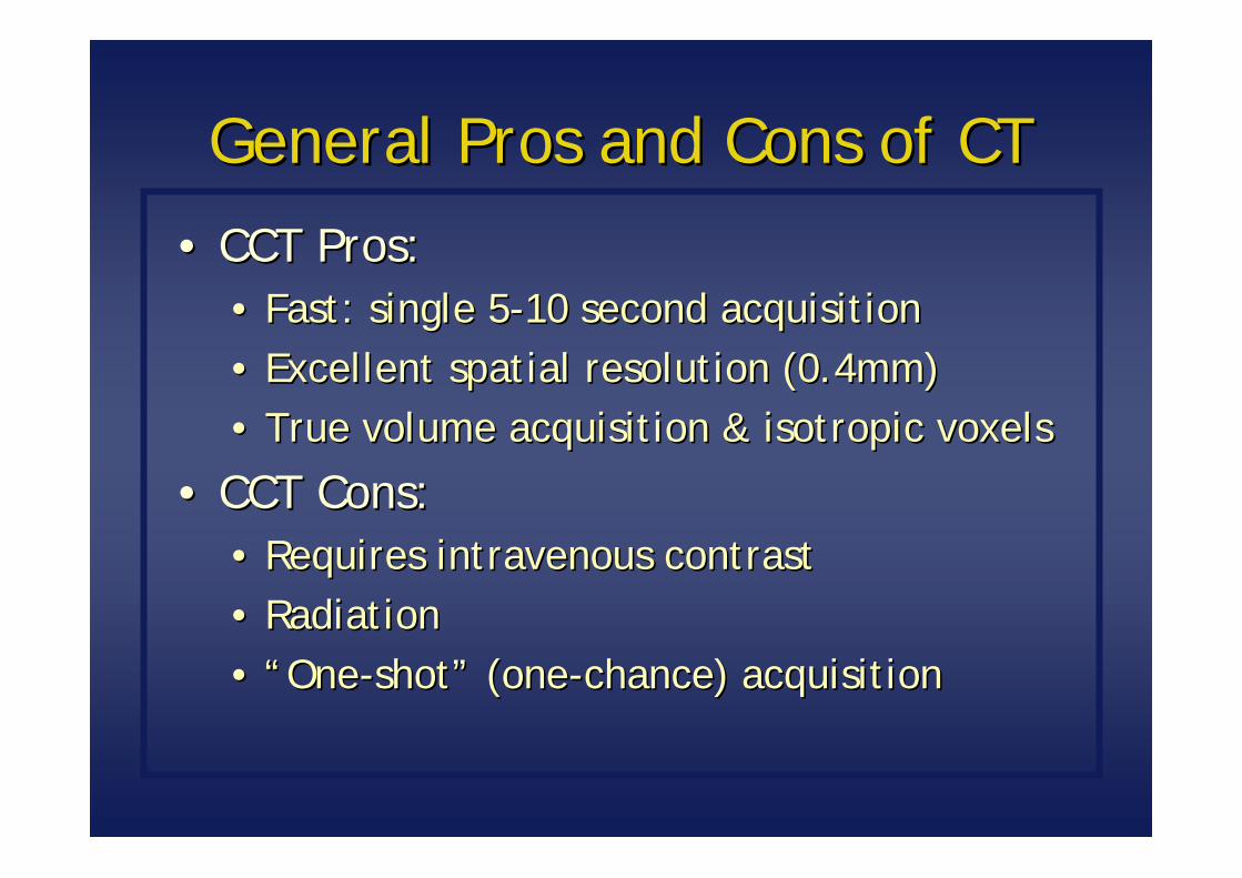
CT-STAT trial: recently discussed at American Heart Assoc. annual meeting (Nov 18th)
Compared to serial ECG’s/enzymes followed by SPECT…CT cut time to diagnosis by 54%CT cut cost by 38%With no difference in 6-month MACE
82% of patients had minimal or no CAD

Anomalous LMCAAnomalous LMCA

Persistent Vertical VeinPersistent Vertical Vein

Aneurysm
Coronary Artery AneurysmCoronary Artery Aneurysm

Left Main Aneurysm

Coronary FistulaCoronary Fistula

Author not Sens. Spec. PPVevaluable
Rixe 42% 86% 98% 86%
Oncel 0% 89% 95% 90%
Rist 2% 75% 92% 67%
Ehara 12% 91% 93% 54% 90% >= 3.0 mm
Cademartiri 7% 95% 93% 63%
Manghat 10% 85% 86% 61% Mean: 3.3 mm
Hecht 0% 94% 87% 39%
Schuijf 14% 100% 100% 71% Mean: 3.4 mm
Pugliese 0% 94% 92% 77%
Pflederer 8% 87% 95% 73% Only > 3.0 mm
Accuracy: In-stent Stenosis
Stents?

PrePre--op: Repeat Cardiac Surgeryop: Repeat Cardiac Surgery

Appropriateness Criteria for CCT and CMRAppropriateness Criteria for CCT and CMRJ Am Coll Cardiol. Oct 2006;48(7):1475J Am Coll Cardiol. Oct 2006;48(7):1475--9797
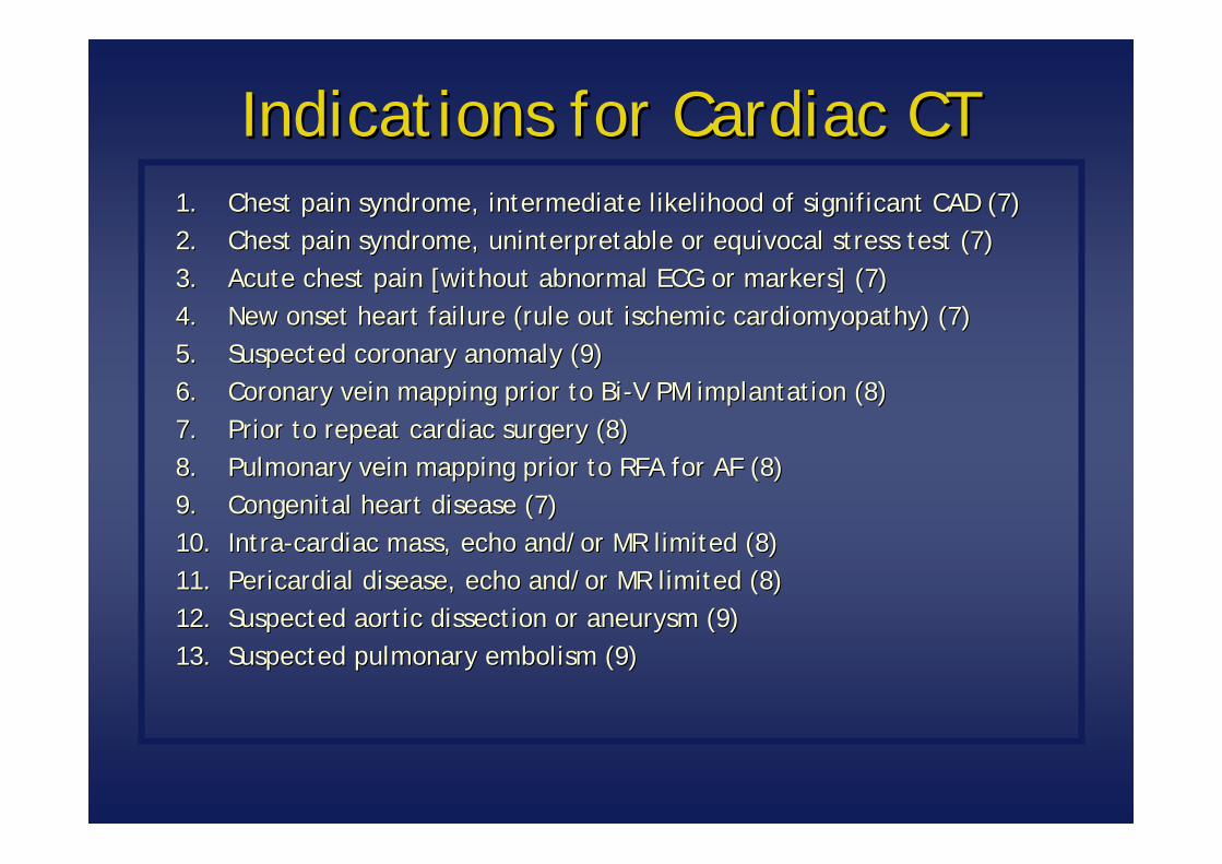
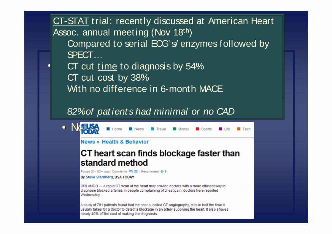
Indications for Cardiac CTIndications for Cardiac CT1.1. Chest pain syndrome, intermediate likelihood of significant CAD Chest pain syndrome, intermediate likelihood of significant CAD (7)(7)2.2. Chest pain syndrome, uninterpretable or equivocal stress test (7Chest pain syndrome, uninterpretable or equivocal stress test (7))3.3. Acute chest pain [without abnormal ECG or markers] (7)Acute chest pain [without abnormal ECG or markers] (7)4.4. New onset heart failure (rule out ischemic cardiomyopathy) (7)New onset heart failure (rule out ischemic cardiomyopathy) (7)5.5. Suspected coronary anomaly (9)Suspected coronary anomaly (9)6.6. Coronary vein mapping prior to BiCoronary vein mapping prior to Bi--V PM implantation (8)V PM implantation (8)7.7. Prior to repeat cardiac surgery (8)Prior to repeat cardiac surgery (8)8.8. Pulmonary vein mapping prior to RFA for AF (8)Pulmonary vein mapping prior to RFA for AF (8)9.9. Congenital heart disease (7)Congenital heart disease (7)10.10. IntraIntra--cardiac mass, echo and/or MR limited (8)cardiac mass, echo and/or MR limited (8)11.11. Pericardial disease, echo and/or MR limited (8)Pericardial disease, echo and/or MR limited (8)12.12. Suspected aortic dissection or aneurysm (9)Suspected aortic dissection or aneurysm (9)13.13. Suspected pulmonary embolism (9)Suspected pulmonary embolism (9)

Indications for Cardiac MRIndications for Cardiac MR1.1. Chest pain syndrome and intermediate preChest pain syndrome and intermediate pre--test likelihood of test likelihood of
significant CAD (vasodilator perfusion CMR or dobutamine stress significant CAD (vasodilator perfusion CMR or dobutamine stress function CMR) (7)function CMR) (7)
2.2. Suspected coronary anomaly (coronary MRA) (8)Suspected coronary anomaly (coronary MRA) (8)3.3. Congenital heart disease (9)Congenital heart disease (9)4.4. LV function when echo limited or discordant results (8)LV function when echo limited or discordant results (8)5.5. Evaluation of specific nonEvaluation of specific non--ischemic cardiomyopathies (amyloid, ischemic cardiomyopathies (amyloid,
sarcoid, HCM, cardiotoxin, myocarditis) (8)sarcoid, HCM, cardiotoxin, myocarditis) (8)6.6. Native and prosthetic valves when echo limited (8)Native and prosthetic valves when echo limited (8)7.7. Arrhythmogenic right ventricular dysplasia (ARVD) (9)Arrhythmogenic right ventricular dysplasia (ARVD) (9)8.8. Intracardiac mass (9)Intracardiac mass (9)9.9. Pericardial disease (8)Pericardial disease (8)10.10. Aortic dissection (8)Aortic dissection (8)11.11. Pulmonary vein mapping prior to RFA for AF (8)Pulmonary vein mapping prior to RFA for AF (8)12.12. Infarct detection and viability (9)Infarct detection and viability (9)

Comparison of IndicationsComparison of Indications2006 Appropriateness Criteria2006 Appropriateness Criteria
CT More Appropriate Either Appropriate MR More Appropriate
Chest pain workup(coronary CTA or adenosine or dobutamine MR)
Coronary anomaly
Congenital heart disease
Cardiac mass
Pericardial disease
Aortic dissection
Acute chest pain eval Infarct / Viability Imaging
Prior to repeat cardiac surgery
ARVD / specific cardiomyopathies
Rule out pulmonary embolism
Native or prosthetic valves

CT and MR: ConclusionsCT and MR: Conclusions
•• Choosing the best test for the right patient for Choosing the best test for the right patient for the right clinical scenario:the right clinical scenario:•• Know the capabilities and limitations of the testsKnow the capabilities and limitations of the tests•• Know the Appropriate Use CriteriaKnow the Appropriate Use Criteria
•• Updates coming in 2010Updates coming in 2010
•• Know your institutions Know your institutions ““local expertiselocal expertise””
Test Test FactorsFactors
Patient Patient FactorsFactors
Clinical Clinical ScenarioScenario
Best Best TestTest
Related Documents