Citation: Heo, Y.; Lee, I.; Moon, S.; Yun, J.-H.; Kim, E.Y.; Park, S.-Y.; Park, J.-H.; Kim, W.T.; Lee, W. Crystal Structures of the Plant Phospholipase A1 Proteins Reveal a Unique Dimerization Domain. Molecules 2022, 27, 2317. https://doi.org/10.3390/ molecules27072317 Academic Editors: Kunihiro Kuwajima, Yuko Okamoto, Tuomas Knowles and Michele Vendruscolo Received: 13 March 2022 Accepted: 31 March 2022 Published: 2 April 2022 Publisher’s Note: MDPI stays neutral with regard to jurisdictional claims in published maps and institutional affil- iations. Copyright: © 2022 by the authors. Licensee MDPI, Basel, Switzerland. This article is an open access article distributed under the terms and conditions of the Creative Commons Attribution (CC BY) license (https:// creativecommons.org/licenses/by/ 4.0/). molecules Article Crystal Structures of the Plant Phospholipase A1 Proteins Reveal a Unique Dimerization Domain Yunseok Heo 1 , Inhwan Lee 1 , Sunjin Moon 1 , Ji-Hye Yun 1,2 , Eun Yu Kim 3 , Sam-Yong Park 4 , Jae-Hyun Park 1 , Woo Taek Kim 3, * and Weontae Lee 1,2, * 1 Structural Biochemistry & Molecular Biophysics Laboratory, Department of Biochemistry, College of Life Science and Biotechnology, Yonsei University, Seoul 03722, Korea; [email protected] (Y.H.); [email protected] (I.L.); [email protected] (S.M.); [email protected] (J.-H.Y.); [email protected] (J.-H.P.) 2 PCG-Biotech, Ltd., 508 KBIZ DMC Tower, Sangam-ro, Seoul 03929, Korea 3 Department of Systems Biology, College of Life Science and Biotechnology, Yonsei University, Seoul 03722, Korea; [email protected] 4 Drug Design Laboratory, Graduate School of Medical Life Science, Yokohama City University, Tsurumi, Yokohama 230-0045, Japan; [email protected] * Correspondence: [email protected] (W.T.K.);[email protected] (W.L.) Abstract: Phospholipase is an enzyme that hydrolyzes various phospholipid substrates at specific ester bonds and plays important roles such as membrane remodeling, as digestive enzymes, and the regulation of cellular mechanism. Phospholipase proteins are divided into following the four major groups according to the ester bonds they cleave off: phospholipase A1 (PLA1), phospholipase A2 (PLA2), phospholipase C (PLC), and phospholipase D (PLD). Among the four phospholipase groups, PLA1 has been less studied than the other phospholipases. Here, we report the first molecular struc- tures of plant PLA1s: AtDSEL and CaPLA1 derived from Arabidopsis thaliana and Capsicum annuum, respectively. AtDSEL and CaPLA1 are novel PLA1s in that they form homodimers since PLAs are generally in the form of a monomer. The dimerization domain at the C-terminal of the AtDSEL and CaPLA1 makes hydrophobic interactions between each monomer, respectively. The C-terminal domain is also present in PLA1s of other plants, but not in PLAs of mammals and fungi. An activity assay of AtDSEL toward various lipid substrates demonstrates that AtDSEL is specialized for the cleavage of sn-1 acyl chains. This report reveals a new domain that exists only in plant PLA1s and suggests that the domain is essential for homodimerization. Keywords: X-ray crystallography; phospholipase A1; homodimer; dimerization domain; catalytic triad; plant protein 1. Introduction Phospholipase hydrolyzes phospholipids at specific sites. The phospholipases have roles as digestive enzymes, for maintaining and remodeling of a membrane, and for the regulation of cellular mechanisms [1]. The phospholipases are classified into four groups according to the cleavage site; phospholipase A1 (PLA1) cleaves sn-1 acyl chain, phospholipase A2 (PLA2) cleaves sn-2 acyl chain, phospholipase C (PLC) cleaves before the phosphate, and phospholipase D (PLD) cleaves after the phosphate [2]. Therefore, PLA1 and PLA2 are the acylhydrolase family that catalyzes the substrates containing acyl group, and PLC and PLD belong to phosphodiesterase. Triacylglycerol lipase (TGL), lipoprotein lipase (LPL), and monoacylglycerol lipase (MGL) belong to PLA because they also cleave the sn-1 and/or sn-2 acyl chain. PLA1 (EC 3.1.1.32) is present in many different organisms including mammals, fungi, insects, bacteria, metazoans, protozoan parasites, venoms, and plants [1,3]. To date, three- dimensional structures of various PLAs have been elucidated [4–10]. Although these Molecules 2022, 27, 2317. https://doi.org/10.3390/molecules27072317 https://www.mdpi.com/journal/molecules

Welcome message from author
This document is posted to help you gain knowledge. Please leave a comment to let me know what you think about it! Share it to your friends and learn new things together.
Transcript
�����������������
Citation: Heo, Y.; Lee, I.; Moon, S.;
Yun, J.-H.; Kim, E.Y.; Park, S.-Y.; Park,
J.-H.; Kim, W.T.; Lee, W. Crystal
Structures of the Plant Phospholipase
A1 Proteins Reveal a Unique
Dimerization Domain. Molecules 2022,
27, 2317. https://doi.org/10.3390/
molecules27072317
Academic Editors:
Kunihiro Kuwajima, Yuko Okamoto,
Tuomas Knowles and
Michele Vendruscolo
Received: 13 March 2022
Accepted: 31 March 2022
Published: 2 April 2022
Publisher’s Note: MDPI stays neutral
with regard to jurisdictional claims in
published maps and institutional affil-
iations.
Copyright: © 2022 by the authors.
Licensee MDPI, Basel, Switzerland.
This article is an open access article
distributed under the terms and
conditions of the Creative Commons
Attribution (CC BY) license (https://
creativecommons.org/licenses/by/
4.0/).
molecules
Article
Crystal Structures of the Plant Phospholipase A1 ProteinsReveal a Unique Dimerization DomainYunseok Heo 1 , Inhwan Lee 1, Sunjin Moon 1, Ji-Hye Yun 1,2 , Eun Yu Kim 3 , Sam-Yong Park 4,Jae-Hyun Park 1, Woo Taek Kim 3,* and Weontae Lee 1,2,*
1 Structural Biochemistry & Molecular Biophysics Laboratory, Department of Biochemistry, College of LifeScience and Biotechnology, Yonsei University, Seoul 03722, Korea; [email protected] (Y.H.);[email protected] (I.L.); [email protected] (S.M.); [email protected] (J.-H.Y.);[email protected] (J.-H.P.)
2 PCG-Biotech, Ltd., 508 KBIZ DMC Tower, Sangam-ro, Seoul 03929, Korea3 Department of Systems Biology, College of Life Science and Biotechnology, Yonsei University,
Seoul 03722, Korea; [email protected] Drug Design Laboratory, Graduate School of Medical Life Science, Yokohama City University, Tsurumi,
Yokohama 230-0045, Japan; [email protected]* Correspondence: [email protected] (W.T.K.); [email protected] (W.L.)
Abstract: Phospholipase is an enzyme that hydrolyzes various phospholipid substrates at specificester bonds and plays important roles such as membrane remodeling, as digestive enzymes, and theregulation of cellular mechanism. Phospholipase proteins are divided into following the four majorgroups according to the ester bonds they cleave off: phospholipase A1 (PLA1), phospholipase A2(PLA2), phospholipase C (PLC), and phospholipase D (PLD). Among the four phospholipase groups,PLA1 has been less studied than the other phospholipases. Here, we report the first molecular struc-tures of plant PLA1s: AtDSEL and CaPLA1 derived from Arabidopsis thaliana and Capsicum annuum,respectively. AtDSEL and CaPLA1 are novel PLA1s in that they form homodimers since PLAs aregenerally in the form of a monomer. The dimerization domain at the C-terminal of the AtDSELand CaPLA1 makes hydrophobic interactions between each monomer, respectively. The C-terminaldomain is also present in PLA1s of other plants, but not in PLAs of mammals and fungi. An activityassay of AtDSEL toward various lipid substrates demonstrates that AtDSEL is specialized for thecleavage of sn-1 acyl chains. This report reveals a new domain that exists only in plant PLA1s andsuggests that the domain is essential for homodimerization.
Keywords: X-ray crystallography; phospholipase A1; homodimer; dimerization domain; catalytic triad;plant protein
1. Introduction
Phospholipase hydrolyzes phospholipids at specific sites. The phospholipases haveroles as digestive enzymes, for maintaining and remodeling of a membrane, and forthe regulation of cellular mechanisms [1]. The phospholipases are classified into fourgroups according to the cleavage site; phospholipase A1 (PLA1) cleaves sn-1 acyl chain,phospholipase A2 (PLA2) cleaves sn-2 acyl chain, phospholipase C (PLC) cleaves before thephosphate, and phospholipase D (PLD) cleaves after the phosphate [2]. Therefore, PLA1and PLA2 are the acylhydrolase family that catalyzes the substrates containing acyl group,and PLC and PLD belong to phosphodiesterase. Triacylglycerol lipase (TGL), lipoproteinlipase (LPL), and monoacylglycerol lipase (MGL) belong to PLA because they also cleavethe sn-1 and/or sn-2 acyl chain.
PLA1 (EC 3.1.1.32) is present in many different organisms including mammals, fungi,insects, bacteria, metazoans, protozoan parasites, venoms, and plants [1,3]. To date, three-dimensional structures of various PLAs have been elucidated [4–10]. Although these
Molecules 2022, 27, 2317. https://doi.org/10.3390/molecules27072317 https://www.mdpi.com/journal/molecules
Molecules 2022, 27, 2317 2 of 13
lipases have little sequence identity, they show a typical α/β hydrolase scaffold comprisingof a core of seven (or more) β-strands flanked by several α-helices [11]. Other commonstructural features of lipases are a GXSXG motif, lid domain, and catalytic triad in the α/βhydrolase scaffold [12]. The GXSXG motif is composed of catalytic Ser and two residuesbefore and after the Ser, and the lid domain plays a role for capping the active site; thecatalytic triad consists of His, Asp, and the Ser of the GXSXG motif [13–16].
Plant PLAs have important roles in metabolism and lipid biosynthesis, as well asapplications in food and biotechnology industries [17,18]. In Arabidopsis thaliana, DAD1(At2g44810) was identified as an sn-1 specific acylhydrolase, and several studies havesuggested that AtDSEL (Arabidopsis thaliana DAD1-like seedling establishment-relatedlipase), one of the homologues of DAD1, plays significant roles in the regulation of cellularprocesses such as seed germination, tissue growth, and seedling establishment [19,20].In the early roots of hot pepper (Capsicum annuum), a cDNA encoding PLA1 homolog(CaPLA1) was identified [21]. CaPLA1 was selectively expressed in young roots of the hotpepper and hydrolyzed phospholipids at the sn-1 position [21]. CaPLA1 is presumed to beinvolved in the development of the roots of hot pepper, considering that the expression ofCaPLA1 rapidly declined after germination [21].
We have determined the three-dimensional structures of AtDSEL and CaPLA1. Theyare the first elucidated structures of PLA1 derived from plants. Here, we revealed that theserepresentative plant PLA1s are different from previously reported PLAs; only the plantPLA1s have a domain for homodimerization. Hydrophobic residues in the dimerizationdomain form a strong dimeric interface. This report broadens our knowledge of PLA bypresenting the plant PLA1 structures with novel features.
2. Results and Discussion2.1. Structural Features of AtDSEL and CaPLA1
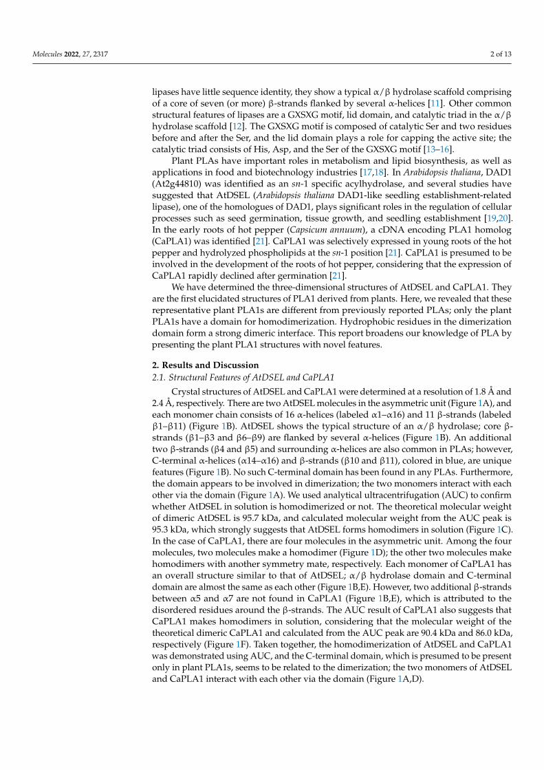
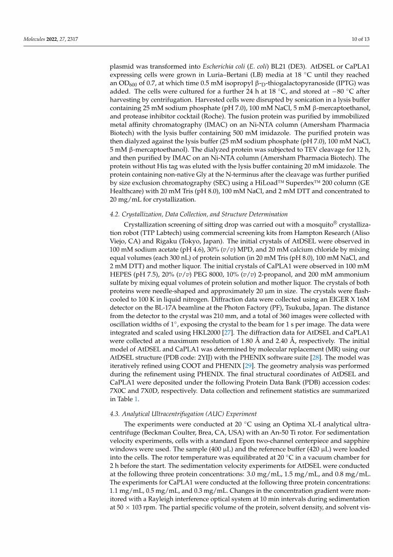
Crystal structures of AtDSEL and CaPLA1 were determined at a resolution of 1.8 Å and2.4 Å, respectively. There are two AtDSEL molecules in the asymmetric unit (Figure 1A), andeach monomer chain consists of 16 α-helices (labeled α1–α16) and 11 β-strands (labeledβ1–β11) (Figure 1B). AtDSEL shows the typical structure of an α/β hydrolase; core β-strands (β1–β3 and β6–β9) are flanked by several α-helices (Figure 1B). An additionaltwo β-strands (β4 and β5) and surrounding α-helices are also common in PLAs; however,C-terminal α-helices (α14–α16) and β-strands (β10 and β11), colored in blue, are uniquefeatures (Figure 1B). No such C-terminal domain has been found in any PLAs. Furthermore,the domain appears to be involved in dimerization; the two monomers interact with eachother via the domain (Figure 1A). We used analytical ultracentrifugation (AUC) to confirmwhether AtDSEL in solution is homodimerized or not. The theoretical molecular weightof dimeric AtDSEL is 95.7 kDa, and calculated molecular weight from the AUC peak is95.3 kDa, which strongly suggests that AtDSEL forms homodimers in solution (Figure 1C).In the case of CaPLA1, there are four molecules in the asymmetric unit. Among the fourmolecules, two molecules make a homodimer (Figure 1D); the other two molecules makehomodimers with another symmetry mate, respectively. Each monomer of CaPLA1 hasan overall structure similar to that of AtDSEL; α/β hydrolase domain and C-terminaldomain are almost the same as each other (Figure 1B,E). However, two additional β-strandsbetween α5 and α7 are not found in CaPLA1 (Figure 1B,E), which is attributed to thedisordered residues around the β-strands. The AUC result of CaPLA1 also suggests thatCaPLA1 makes homodimers in solution, considering that the molecular weight of thetheoretical dimeric CaPLA1 and calculated from the AUC peak are 90.4 kDa and 86.0 kDa,respectively (Figure 1F). Taken together, the homodimerization of AtDSEL and CaPLA1was demonstrated using AUC, and the C-terminal domain, which is presumed to be presentonly in plant PLA1s, seems to be related to the dimerization; the two monomers of AtDSELand CaPLA1 interact with each other via the domain (Figure 1A,D).
Molecules 2022, 27, 2317 3 of 13Molecules 2022, 27, x FOR PEER REVIEW 3 of 13
Figure 1. Overall structures of AtDSEL and CaPLA1. (A) Crystal structure of full-length AtDSEL. AtDSEL homodimer is shown as a ribbon diagram. Each monomer is colored in cyan and orange, respectively. (B) Topology diagram of AtDSEL monomer. α/β hydrolase domain and C-terminal domain are colored in red and blue, respectively. (C) Analytical ultracentrifugation (AUC) result of AtDSEL. The experiments were performed at following three protein concentrations: 3.0 mg/mL (colored in red), 1.5 mg/mL (green), and 0.8 mg/mL (blue). Calculated molecular weight of AtDSEL from the AUC peak is 95.3 ± 0.3 kDa, which strongly suggests that AtDSEL forms homodimers in solution, considering that the theoretical molecular weight of dimeric AtDSEL is 95.7 kDa. (D) Crys-tal structure of full-length CaPLA1. CaPLA1 homodimer is shown as a ribbon diagram. (E) Topol-ogy diagram of CaPLA1 monomer. (F) AUC result of CaPLA1. The experiments were performed at following three protein concentrations: 1.1 mg/mL (colored in red), 0.5 mg/mL (green), and 0.3 mg/mL (blue). Calculated molecular weight of CaPLA1 from the AUC peak is 86.0 ± 2.0 kDa, which strongly suggests that CaPLA1 forms homodimers in solution, considering that the theoretical mo-lecular weight of dimeric CaPLA1 is 90.4 kDa.
2.2. Conserved Motifs and Domains of AtDSEL and CaPLA1 AtDSEL and CaPLA1 are divided into the following two domains: the α/β hydrolase
domain containing the lid domain, GXSXG motif, and catalytic triad and the C-terminal
Figure 1. Overall structures of AtDSEL and CaPLA1. (A) Crystal structure of full-length AtDSEL.AtDSEL homodimer is shown as a ribbon diagram. Each monomer is colored in cyan and orange,respectively. (B) Topology diagram of AtDSEL monomer. α/β hydrolase domain and C-terminaldomain are colored in red and blue, respectively. (C) Analytical ultracentrifugation (AUC) result ofAtDSEL. The experiments were performed at following three protein concentrations: 3.0 mg/mL(colored in red), 1.5 mg/mL (green), and 0.8 mg/mL (blue). Calculated molecular weight of AtDSELfrom the AUC peak is 95.3 ± 0.3 kDa, which strongly suggests that AtDSEL forms homodimers insolution, considering that the theoretical molecular weight of dimeric AtDSEL is 95.7 kDa. (D) Crystalstructure of full-length CaPLA1. CaPLA1 homodimer is shown as a ribbon diagram. (E) Topologydiagram of CaPLA1 monomer. (F) AUC result of CaPLA1. The experiments were performedat following three protein concentrations: 1.1 mg/mL (colored in red), 0.5 mg/mL (green), and0.3 mg/mL (blue). Calculated molecular weight of CaPLA1 from the AUC peak is 86.0 ± 2.0 kDa,which strongly suggests that CaPLA1 forms homodimers in solution, considering that the theoreticalmolecular weight of dimeric CaPLA1 is 90.4 kDa.
Molecules 2022, 27, 2317 4 of 13
2.2. Conserved Motifs and Domains of AtDSEL and CaPLA1
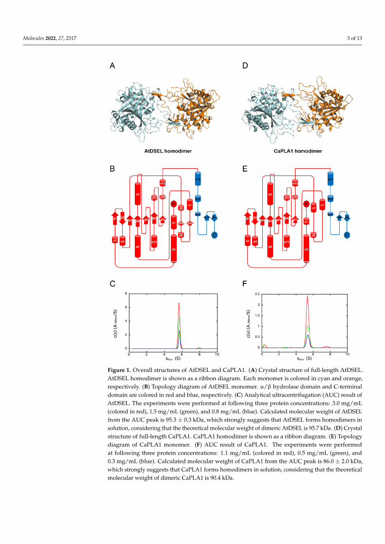
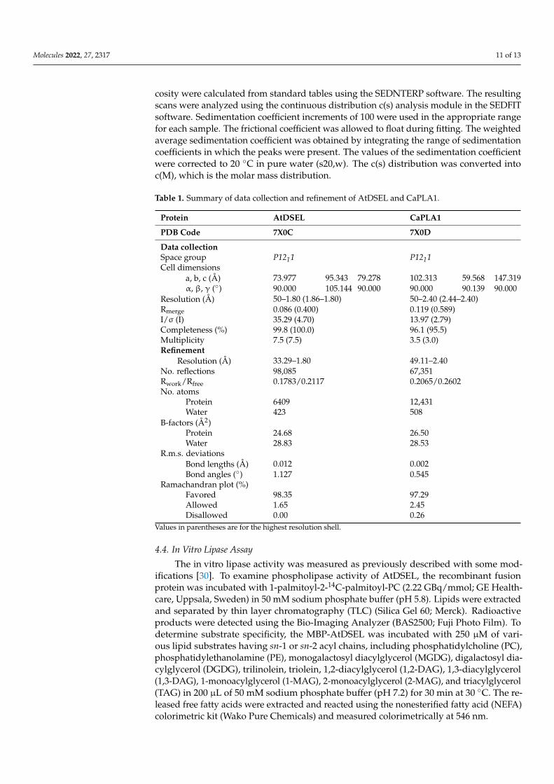
AtDSEL and CaPLA1 are divided into the following two domains: the α/β hydrolasedomain containing the lid domain, GXSXG motif, and catalytic triad and the C-terminaldomain containing the dimerization domain (Figure 2A–D). The α/β hydrolase domainsof AtDSEL and CaPLA1 are composed of 340 and 323 residues, respectively, and thetheir C-terminal domains consist of 79 and 74 residues, respectively (Figure 2B,D). MostPLA structures reported to date have only the α/β hydrolase domain and are composedof approximately 300 residues [4–6]. They do not make any multimers [4–6]. On theother hand, mammal PLAs (human LPL and human pancreatic TGL) have another C-terminal domain in addition to the α/β hydrolase domain; the α/β hydrolase domain iscomposed of about 340 residues, and the other C-terminal domain consists of about 130residues [7–9]. The C-terminal domain of mammals shows a typical β-barrel structure [7–9],which is totally different from the C-terminal domains of AtDSEL and CaPLA1. HumanMGL is composed of 303 residues and has only the α/β hydrolase domain; however, thelipase makes homodimers [10]. The two monomers of human MGL makes a dimer withan up–down orientation [10]. Taken together, PLAs in fungi and venom have only theα/β hydrolase domain [4–6], and some PLAs in mammals and our plant PLA1s have anadditional C-terminal domain as well as the α/β hydrolase domain [7–9] (Figure 2B,D).However, the overall structures and functions of the C-terminal domains of the PLAs inmammals and plants are completely different from each other. The C-terminal domain ofAtDSEL and CaPLA1 is composed of three α-helices and one pair of β-strands (Figure 1B,E),while the C-terminal domains of PLAs in mammals are composed of 8–12 β-strands makingup the β-barrel structure [7–9]. Moreover, the C-terminal domain of AtDSEL and CaPLA1is involved in the homodimerization, while the β-barrel of the PLA is related to theinteractions with lipids [7]. On the other hand, human MGL can make homodimers thoughthe proteins consisting of only the α/β hydrolase domain [10]. Therefore, only the PLAs inplants have the C-terminal domain for homodimerization.
AtDSEL has two disconnected loops caused by the disordered residues; Arg120 andGlu121 between β1 and β2 and from Pro159 to Leu160 between β3 and α5 could notbe assigned due to the poor electron density map of the residues (Figures 1B and 2A).Pro159 and Leu160 belong to the lid domain, which determines the substrate specificities(Figure 2A,B). CaPLA1 also has two disconnected loops resulting from the disorderedresidues; Pro91 and Asp92 between β1 and β2 and from Asp160 to Phe167 between α6and α7 could not be refined because of the deficient electron density map of the residues(Figures 1E and 2C). The disordered residues from Asp160 to Phe167 affected the surround-ing secondary structures; a pair of β-strands (β4 and β5 in the case of AtDSEL) were notfound in CaPLA1 (Figure 1B,E and Figure 2A,C). It has been reported that the lengthof the lid domain is involved in the type of substrate that the enzyme can catalyze [3].AtDSEL and CaPLA1 have a lid domain with the proper length for catalyzing the sn-1 acylgroups [3] (Figure 2B,D).
The α/β hydrolase domains of AtDSEL and CaPLA1 have a conserved GXSXG motif(Figure 2A,C). In the AtDSEL, the GXSXG motif consists of the residues from Gly234 toGly238, while the motif is composed of the residues from Gly219 to Gly223 in the case ofCaPLA1 (Figure 2B,D). The GXSXG motif is found in various lipases and esterases [22].The central Ser of the motif functions as a nucleophile [23]. In AtDSEL, the motif existsbetween β6 and α9 and has the following sequence: GHSLG (Figures 1B and 2A). The motifof CaPLA1 is also located in the same position and has the same sequence as in AtDSEL(Figure 2A,C). It has been reported that the two Gly residues of the GXSXG motif are alsoessential for the lipase activity [24]. The lipase activities of MGLs in Arabidopsis thalianawere completely lost when the researchers applied the following mutagenesis to the lipases:GXAXG, GXSXS, or SXSXG [24], which demonstrates that Gly to the Ser mutant as well asSer to the Ala mutant causes severe deactivation of the lipases.
Molecules 2022, 27, 2317 5 of 13
Molecules 2022, 27, x FOR PEER REVIEW 5 of 13
highly diminished activity [26]. Therefore, the catalytic triad could not be replaced with other residues.
Figure 2. Conserved catalytic triad and domains of AtDSEL and CaPLA1. (A) Three residues (Ser236, Asp302, and His339) that compose the catalytic triad of AtDSEL are shown as a stick model. 2mFO−DFC electron density map at a level of 2.0 σ is superimposed to the residues. Lid domain is colored in salmon. Pro159 and Leu160 of the lid domain were disordered, causing the disconnected lid domain in this figure. Conserved GXSXG motif is colored in green, and dimerization domain is colored in purple. (B) The construct map of AtDSEL. The lid domain and GXSXG motif belong to α/β hydrolase domain. C-terminal domain containing dimerization domain exists only in plant phospholipase A1 (PLA1). (C) Three residues (Ser221, Asp285, and His322), which compose the catalytic triad of CaPLA1, are shown as a stick model. 2mFO−DFC electron density map at a level of 1.8 σ is superimposed to the residues. (D) The construct map of CaPLA1.
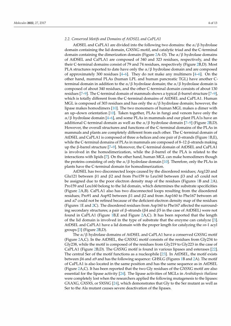
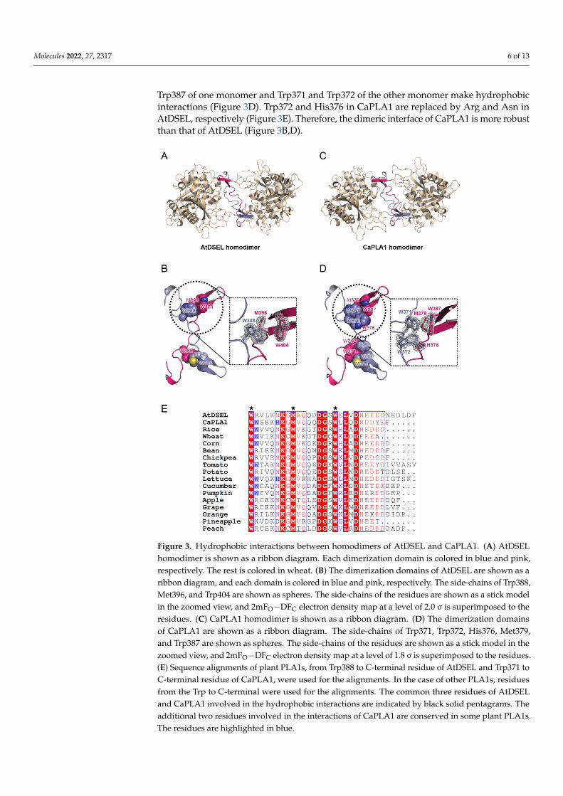
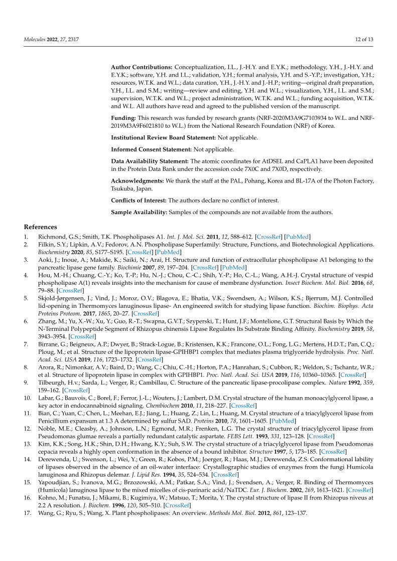
2.3. Interactions between Homodimers of AtDSEL and CaPLA1 The dimerization domains of the AtDSEL homodimer cross each other and stretch in
opposite directions (Figure 3a). Met396 and Trp404 of one monomer and Trp388 of the other monomer interact with each other (Figure 3b). The hydrophobic interactions are main driving force behind the formation of the dimerization of AtDSEL. Interactions be-tween the monomers occur throughout the loops (Figure 3b). The length from a sulfur atom of Met396 of one monomer to a sulfur atom of the other Met396 is 23.3 Å (Figure 3b). The dimerization interface of AtDSEL is extremely long to form robust homodimers. The dimerization domain of CaPLA1 is similar to that of AtDSEL; however, more residues are involved in the dimerization (Figure 3c,d). The length from a sulfur atom of one Met379 to that of the other Met379 is 22.1 Å (Figure 3d). In CaPLA1, His376, Met379, and Trp387 of one monomer and Trp371 and Trp372 of the other monomer make hydrophobic inter-actions (Figure 3d). Trp372 and His376 in CaPLA1 are replaced by Arg and Asn in
Figure 2. Conserved catalytic triad and domains of AtDSEL and CaPLA1. (A) Three residues(Ser236, Asp302, and His339) that compose the catalytic triad of AtDSEL are shown as a stick model.2mFO−DFC electron density map at a level of 2.0 σ is superimposed to the residues. Lid domain iscolored in salmon. Pro159 and Leu160 of the lid domain were disordered, causing the disconnectedlid domain in this figure. Conserved GXSXG motif is colored in green, and dimerization domainis colored in purple. (B) The construct map of AtDSEL. The lid domain and GXSXG motif belongto α/β hydrolase domain. C-terminal domain containing dimerization domain exists only in plantphospholipase A1 (PLA1). (C) Three residues (Ser221, Asp285, and His322), which compose thecatalytic triad of CaPLA1, are shown as a stick model. 2mFO−DFC electron density map at a level of1.8 σ is superimposed to the residues. (D) The construct map of CaPLA1.
The catalytic triad of AtDSEL is Ser236, Asp302, and His339, while that of CaPLA1 isSer221, Asp285, and His322 (Figure 2A,C). The lid domain, which is conserved in lipaseproteins, plays the role of capping this active site [25] (Figure 2A,C). All human pancreaticTGL mutants of the catalytic triad (Ser, Asp, and His) showed no detectable activity orhighly diminished activity [26]. Therefore, the catalytic triad could not be replaced withother residues.
2.3. Interactions between Homodimers of AtDSEL and CaPLA1
The dimerization domains of the AtDSEL homodimer cross each other and stretchin opposite directions (Figure 3A). Met396 and Trp404 of one monomer and Trp388 ofthe other monomer interact with each other (Figure 3B). The hydrophobic interactionsare main driving force behind the formation of the dimerization of AtDSEL. Interactionsbetween the monomers occur throughout the loops (Figure 3B). The length from a sulfuratom of Met396 of one monomer to a sulfur atom of the other Met396 is 23.3 Å (Figure 3B).The dimerization interface of AtDSEL is extremely long to form robust homodimers. Thedimerization domain of CaPLA1 is similar to that of AtDSEL; however, more residuesare involved in the dimerization (Figure 3C,D). The length from a sulfur atom of oneMet379 to that of the other Met379 is 22.1 Å (Figure 3D). In CaPLA1, His376, Met379, and
Molecules 2022, 27, 2317 6 of 13
Trp387 of one monomer and Trp371 and Trp372 of the other monomer make hydrophobicinteractions (Figure 3D). Trp372 and His376 in CaPLA1 are replaced by Arg and Asn inAtDSEL, respectively (Figure 3E). Therefore, the dimeric interface of CaPLA1 is more robustthan that of AtDSEL (Figure 3B,D).
Molecules 2022, 27, x FOR PEER REVIEW 6 of 13
AtDSEL, respectively (Figure 3e). Therefore, the dimeric interface of CaPLA1 is more ro-bust than that of AtDSEL (Figure 3b,d).
Figure 3. Hydrophobic interactions between homodimers of AtDSEL and CaPLA1. (A) AtDSEL ho-modimer is shown as a ribbon diagram. Each dimerization domain is colored in blue and pink, respectively. The rest is colored in wheat. (B) The dimerization domains of AtDSEL are shown as a ribbon diagram, and each domain is colored in blue and pink, respectively. The side-chains of Trp388, Met396, and Trp404 are shown as spheres. The side-chains of the residues are shown as a stick model in the zoomed view, and 2mFO−DFC electron density map at a level of 2.0 σ is superim-posed to the residues. (C) CaPLA1 homodimer is shown as a ribbon diagram. (D) The dimerization domains of CaPLA1 are shown as a ribbon diagram. The side-chains of Trp371, Trp372, His376, Met379, and Trp387 are shown as spheres. The side-chains of the residues are shown as a stick model in the zoomed view, and 2mFO−DFC electron density map at a level of 1.8 σ is superimposed to the residues. (E) Sequence alignments of plant PLA1s, from Trp388 to C-terminal residue of AtDSEL and Trp371 to C-terminal residue of CaPLA1, were used for the alignments. In the case of other PLA1s, residues from the Trp to C-terminal were used for the alignments. The common three resi-dues of AtDSEL and CaPLA1 involved in the hydrophobic interactions are indicated by black solid pentagrams. The additional two residues involved in the interactions of CaPLA1 are conserved in some plant PLA1s. The residues are highlighted in blue.
Figure 3. Hydrophobic interactions between homodimers of AtDSEL and CaPLA1. (A) AtDSELhomodimer is shown as a ribbon diagram. Each dimerization domain is colored in blue and pink,respectively. The rest is colored in wheat. (B) The dimerization domains of AtDSEL are shown as aribbon diagram, and each domain is colored in blue and pink, respectively. The side-chains of Trp388,Met396, and Trp404 are shown as spheres. The side-chains of the residues are shown as a stick modelin the zoomed view, and 2mFO−DFC electron density map at a level of 2.0 σ is superimposed to theresidues. (C) CaPLA1 homodimer is shown as a ribbon diagram. (D) The dimerization domainsof CaPLA1 are shown as a ribbon diagram. The side-chains of Trp371, Trp372, His376, Met379,and Trp387 are shown as spheres. The side-chains of the residues are shown as a stick model in thezoomed view, and 2mFO−DFC electron density map at a level of 1.8 σ is superimposed to the residues.(E) Sequence alignments of plant PLA1s, from Trp388 to C-terminal residue of AtDSEL and Trp371 toC-terminal residue of CaPLA1, were used for the alignments. In the case of other PLA1s, residuesfrom the Trp to C-terminal were used for the alignments. The common three residues of AtDSELand CaPLA1 involved in the hydrophobic interactions are indicated by black solid pentagrams. Theadditional two residues involved in the interactions of CaPLA1 are conserved in some plant PLA1s.The residues are highlighted in blue.
Molecules 2022, 27, 2317 7 of 13
The C-terminal residues of AtDSEL, CaPLA1, and 15 PLA1s from various plants arealigned (Figure 3E). The two Trp residues and one Met of AtDSEL are conserved in the17 PLA1s, indicating that the dimerization is commonplace in plant PLA1s (Figure 3E).Among the 17 PLA1s, eight PLA1s containing CaPLA1 have the two consecutive Trpresidues, while the other PLA1s have positively charged residue instead of the second Trp(Figure 3E). His of CaPLA1 is conserved only in lettuce PLA1; the other PLA1s have Asninstead of the His, except for pineapple PLA1 (Figure 3E). The 17 PLA1s have a number ofnegatively charged residues in the C-terminus (Figure 3E); the reason for this has not yetbeen clearly elucidated. Taken together, the sequence alignments of the C-terminal regionsof the plant PLA1s suggest that they would form homodimers.
2.4. Structural Comparison among PLAs
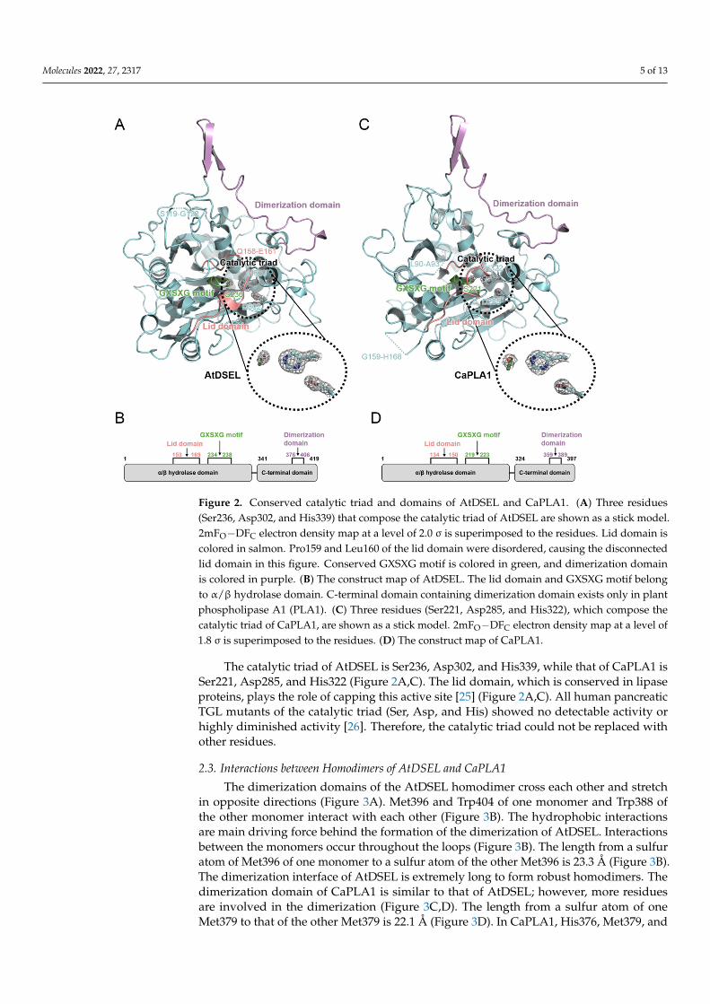
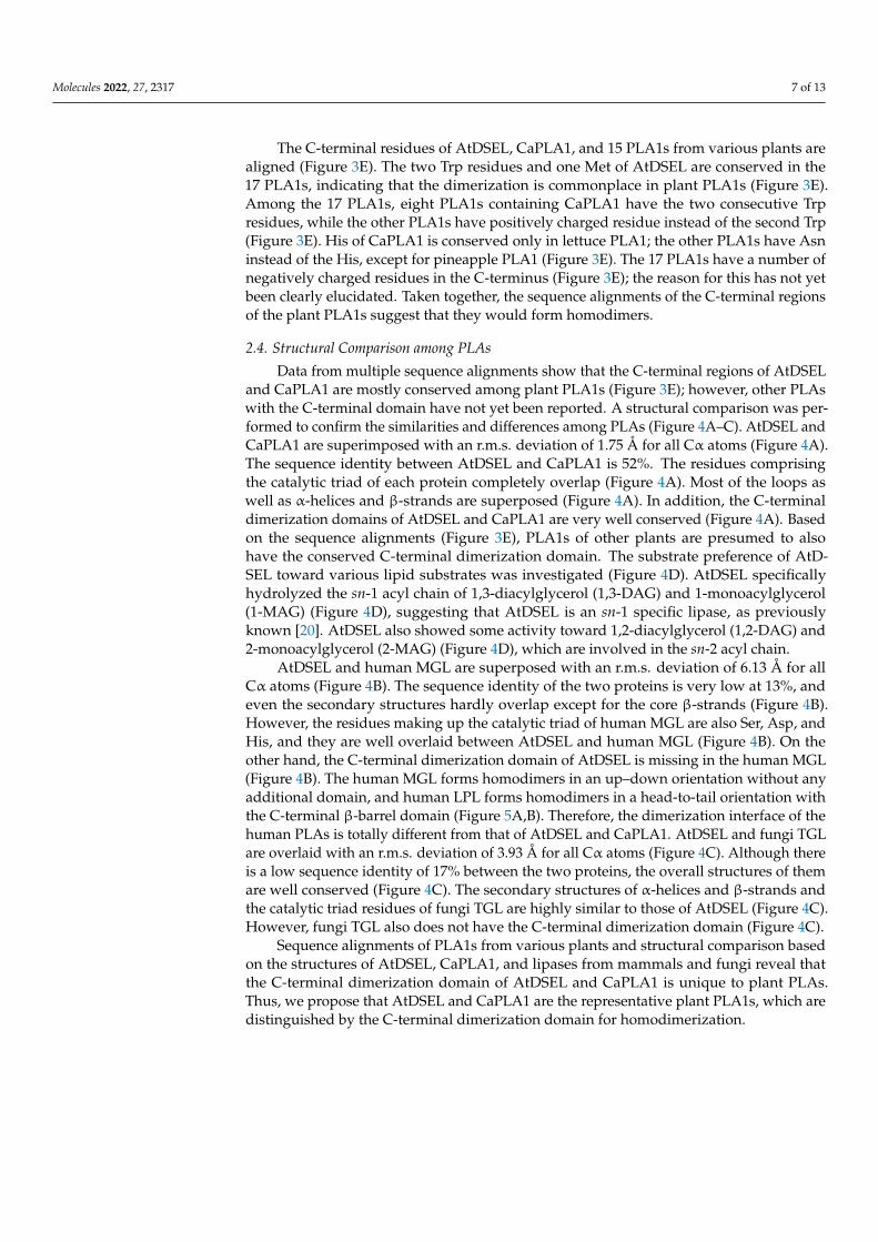
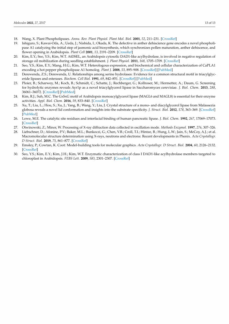
Data from multiple sequence alignments show that the C-terminal regions of AtDSELand CaPLA1 are mostly conserved among plant PLA1s (Figure 3E); however, other PLAswith the C-terminal domain have not yet been reported. A structural comparison was per-formed to confirm the similarities and differences among PLAs (Figure 4A–C). AtDSEL andCaPLA1 are superimposed with an r.m.s. deviation of 1.75 Å for all Cα atoms (Figure 4A).The sequence identity between AtDSEL and CaPLA1 is 52%. The residues comprisingthe catalytic triad of each protein completely overlap (Figure 4A). Most of the loops aswell as α-helices and β-strands are superposed (Figure 4A). In addition, the C-terminaldimerization domains of AtDSEL and CaPLA1 are very well conserved (Figure 4A). Basedon the sequence alignments (Figure 3E), PLA1s of other plants are presumed to alsohave the conserved C-terminal dimerization domain. The substrate preference of AtD-SEL toward various lipid substrates was investigated (Figure 4D). AtDSEL specificallyhydrolyzed the sn-1 acyl chain of 1,3-diacylglycerol (1,3-DAG) and 1-monoacylglycerol(1-MAG) (Figure 4D), suggesting that AtDSEL is an sn-1 specific lipase, as previouslyknown [20]. AtDSEL also showed some activity toward 1,2-diacylglycerol (1,2-DAG) and2-monoacylglycerol (2-MAG) (Figure 4D), which are involved in the sn-2 acyl chain.
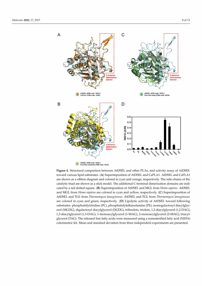
AtDSEL and human MGL are superposed with an r.m.s. deviation of 6.13 Å for allCα atoms (Figure 4B). The sequence identity of the two proteins is very low at 13%, andeven the secondary structures hardly overlap except for the core β-strands (Figure 4B).However, the residues making up the catalytic triad of human MGL are also Ser, Asp, andHis, and they are well overlaid between AtDSEL and human MGL (Figure 4B). On theother hand, the C-terminal dimerization domain of AtDSEL is missing in the human MGL(Figure 4B). The human MGL forms homodimers in an up–down orientation without anyadditional domain, and human LPL forms homodimers in a head-to-tail orientation withthe C-terminal β-barrel domain (Figure 5A,B). Therefore, the dimerization interface of thehuman PLAs is totally different from that of AtDSEL and CaPLA1. AtDSEL and fungi TGLare overlaid with an r.m.s. deviation of 3.93 Å for all Cα atoms (Figure 4C). Although thereis a low sequence identity of 17% between the two proteins, the overall structures of themare well conserved (Figure 4C). The secondary structures of α-helices and β-strands andthe catalytic triad residues of fungi TGL are highly similar to those of AtDSEL (Figure 4C).However, fungi TGL also does not have the C-terminal dimerization domain (Figure 4C).
Sequence alignments of PLA1s from various plants and structural comparison basedon the structures of AtDSEL, CaPLA1, and lipases from mammals and fungi reveal thatthe C-terminal dimerization domain of AtDSEL and CaPLA1 is unique to plant PLAs.Thus, we propose that AtDSEL and CaPLA1 are the representative plant PLA1s, which aredistinguished by the C-terminal dimerization domain for homodimerization.
Molecules 2022, 27, 2317 8 of 13
Molecules 2022, 27, x FOR PEER REVIEW 8 of 13
Figure 4. Structural comparison between AtDSEL and other PLAs, and activity assay of AtDSEL toward various lipid substrates. (A) Superimposition of AtDSEL and CaPLA1. AtDSEL and CaPLA1 are shown as a ribbon diagram and colored in cyan and orange, respectively. The side-chains of the catalytic triad are shown as a stick model. The additional C-terminal dimerization domains are in-dicated by a red dotted square. (B) Superimposition of AtDSEL and MGL from Homo sapiens. AtDSEL and MGL from Homo sapiens are colored in cyan and yellow, respectively. (C) Superimpo-sition of AtDSEL and TGL from Thermomyces lanuginosus. AtDSEL and TGL from Thermomyces lanu-ginosus are colored in cyan and green, respectively. (D) Lipolytic activity of AtDSEL toward follow-ing substrates: phosphatidylcholine (PC), phosphatidylethanolamine (PE), monogalactosyl diacyl-glycerol (MGDG), digalactosyl diacylglycerol (DGDG), trilinolein, triolein, 1,2-diacylglycerol (1,2-DAG), 1,3-diacylglycerol (1,3-DAG), 1-monoacylglycerol (1-MAG), 2-monoacylglycerol (2-MAG), triacylglycerol (TAG). The released free fatty acids were measured using a nonesterified fatty acid (NEFA) colorimetric kit. Mean and standard deviation from three independent experiments are pre-sented.
Figure 4. Structural comparison between AtDSEL and other PLAs, and activity assay of AtDSELtoward various lipid substrates. (A) Superimposition of AtDSEL and CaPLA1. AtDSEL and CaPLA1are shown as a ribbon diagram and colored in cyan and orange, respectively. The side-chains of thecatalytic triad are shown as a stick model. The additional C-terminal dimerization domains are indi-cated by a red dotted square. (B) Superimposition of AtDSEL and MGL from Homo sapiens. AtDSELand MGL from Homo sapiens are colored in cyan and yellow, respectively. (C) Superimposition ofAtDSEL and TGL from Thermomyces lanuginosus. AtDSEL and TGL from Thermomyces lanuginosusare colored in cyan and green, respectively. (D) Lipolytic activity of AtDSEL toward followingsubstrates: phosphatidylcholine (PC), phosphatidylethanolamine (PE), monogalactosyl diacylglyc-erol (MGDG), digalactosyl diacylglycerol (DGDG), trilinolein, triolein, 1,2-diacylglycerol (1,2-DAG),1,3-diacylglycerol (1,3-DAG), 1-monoacylglycerol (1-MAG), 2-monoacylglycerol (2-MAG), triacyl-glycerol (TAG). The released free fatty acids were measured using a nonesterified fatty acid (NEFA)colorimetric kit. Mean and standard deviation from three independent experiments are presented.
Molecules 2022, 27, 2317 9 of 13Molecules 2022, 27, x FOR PEER REVIEW 9 of 13
Figure 5. Dimerization interface of human PLAs. (A) Dimeric structure of human MGL. Each mon-omer of human MGL are colored in cyan and orange, respectively. (B) Dimeric structure of human LPL.
3. Conclusions Crystal structures of AtDSEL and CaPLA1, which are the first PLA1 structures de-
rived from plants, reveal the unique features of the plant PLA1s. In addition to the con-served α/β hydrolase scaffold, GXSXG motif, and catalytic triad, plant PLA1s have an ex-tended C-terminal domain composed of approximately 70 residues. In AtDSEL and CaPLA1, these C-terminal regions form a homodimer. Trp388, Met396, and Trp404 in AtDSEL and Trp371, Trp372, His376, Met379, and Trp387 in CaPLA1 play as hydrophobic anchors tethering each monomer of the proteins. A data search by BLAST indicates that the homologous C-terminal regions of AtDSEL and CaPLA1 are only found in plants. Se-quence alignments of the C-terminal regions of the plant PLA1s revealed that the two Trp residues and one Met, which are the common residues between the C-terminal domains of AtDSEL and CaPLA1, are conserved. The homodimerizations of AtDSEL and CaPLA1 were supported by our AUC experiments. However, it is still unknown as to why the plant PLA1s form homodimers. The dimerization domain identified by the structures of AtDSEL and CaPLA1 can serve as the structural templates for understanding the plant phospholipase function at the molecular level. Furthermore, we found that some plant PLA1s have two Trp residues in the C-terminal region, while others have one more Trp in the C-terminal region. AtDSEL has two Trp residues and CaPLA1 has three Trp resi-dues in the C-terminal regions, and all Trp residues in the C-terminal region play an im-portant role as the hydrophobic anchors. Whether the difference in the number of Trp in the C-terminal region can characterize the two types of plant PLA1 dimerization domains should be further explained.
4. Materials and Methods 4.1. Cloning, Expression, and Purification
The full-length AtDSEL (Uniprot entry: O49523) and CaPLA1 (Uniprot entry: A5YW95) genes were amplified by polymerase chain reaction (PCR), respectively. The
Figure 5. Dimerization interface of human PLAs. (A) Dimeric structure of human MGL. Eachmonomer of human MGL are colored in cyan and orange, respectively. (B) Dimeric structure ofhuman LPL.
3. Conclusions
Crystal structures of AtDSEL and CaPLA1, which are the first PLA1 structures derivedfrom plants, reveal the unique features of the plant PLA1s. In addition to the conservedα/β hydrolase scaffold, GXSXG motif, and catalytic triad, plant PLA1s have an extendedC-terminal domain composed of approximately 70 residues. In AtDSEL and CaPLA1, theseC-terminal regions form a homodimer. Trp388, Met396, and Trp404 in AtDSEL and Trp371,Trp372, His376, Met379, and Trp387 in CaPLA1 play as hydrophobic anchors tetheringeach monomer of the proteins. A data search by BLAST indicates that the homologousC-terminal regions of AtDSEL and CaPLA1 are only found in plants. Sequence alignmentsof the C-terminal regions of the plant PLA1s revealed that the two Trp residues and oneMet, which are the common residues between the C-terminal domains of AtDSEL andCaPLA1, are conserved. The homodimerizations of AtDSEL and CaPLA1 were supportedby our AUC experiments. However, it is still unknown as to why the plant PLA1s formhomodimers. The dimerization domain identified by the structures of AtDSEL and CaPLA1can serve as the structural templates for understanding the plant phospholipase function atthe molecular level. Furthermore, we found that some plant PLA1s have two Trp residuesin the C-terminal region, while others have one more Trp in the C-terminal region. AtDSELhas two Trp residues and CaPLA1 has three Trp residues in the C-terminal regions, and allTrp residues in the C-terminal region play an important role as the hydrophobic anchors.Whether the difference in the number of Trp in the C-terminal region can characterize thetwo types of plant PLA1 dimerization domains should be further explained.
4. Materials and Methods4.1. Cloning, Expression, and Purification
The full-length AtDSEL (Uniprot entry: O49523) and CaPLA1 (Uniprot entry: A5YW95)genes were amplified by polymerase chain reaction (PCR), respectively. The amplifiedcDNA fragments were subcloned into the pET21b expression vector (Novagen) with afused 6× His tag and TEV cleavage site (ENLYFQG) at the N-terminus. The resulting
Molecules 2022, 27, 2317 10 of 13
plasmid was transformed into Escherichia coli (E. coli) BL21 (DE3). AtDSEL or CaPLA1expressing cells were grown in Luria–Bertani (LB) media at 18 ◦C until they reachedan OD600 of 0.7, at which time 0.5 mM isopropyl β-D-thiogalactopyranoside (IPTG) wasadded. The cells were cultured for a further 24 h at 18 ◦C, and stored at −80 ◦C afterharvesting by centrifugation. Harvested cells were disrupted by sonication in a lysis buffercontaining 25 mM sodium phosphate (pH 7.0), 100 mM NaCl, 5 mM β-mercaptoethanol,and protease inhibitor cocktail (Roche). The fusion protein was purified by immobilizedmetal affinity chromatography (IMAC) on an Ni-NTA column (Amersham PharmaciaBiotech) with the lysis buffer containing 500 mM imidazole. The purified protein wasthen dialyzed against the lysis buffer (25 mM sodium phosphate (pH 7.0), 100 mM NaCl,5 mM β-mercaptoethanol). The dialyzed protein was subjected to TEV cleavage for 12 h,and then purified by IMAC on an Ni-NTA column (Amersham Pharmacia Biotech). Theprotein without His tag was eluted with the lysis buffer containing 20 mM imidazole. Theprotein containing non-native Gly at the N-terminus after the cleavage was further purifiedby size exclusion chromatography (SEC) using a HiLoad™ Superdex™ 200 column (GEHealthcare) with 20 mM Tris (pH 8.0), 100 mM NaCl, and 2 mM DTT and concentrated to20 mg/mL for crystallization.
4.2. Crystallization, Data Collection, and Structure Determination
Crystallization screening of sitting drop was carried out with a mosquito® crystalliza-tion robot (TTP Labtech) using commercial screening kits from Hampton Research (AlisoViejo, CA) and Rigaku (Tokyo, Japan). The initial crystals of AtDSEL were observed in100 mM sodium acetate (pH 4.6), 30% (v/v) MPD, and 20 mM calcium chloride by mixingequal volumes (each 300 nL) of protein solution (in 20 mM Tris (pH 8.0), 100 mM NaCl, and2 mM DTT) and mother liquor. The initial crystals of CaPLA1 were observed in 100 mMHEPES (pH 7.5), 20% (v/v) PEG 8000, 10% (v/v) 2-propanol, and 200 mM ammoniumsulfate by mixing equal volumes of protein solution and mother liquor. The crystals of bothproteins were needle-shaped and approximately 20 µm in size. The crystals were flash-cooled to 100 K in liquid nitrogen. Diffraction data were collected using an EIGER X 16Mdetector on the BL-17A beamline at the Photon Factory (PF), Tsukuba, Japan. The distancefrom the detector to the crystal was 210 mm, and a total of 360 images were collected withoscillation widths of 1◦, exposing the crystal to the beam for 1 s per image. The data wereintegrated and scaled using HKL2000 [27]. The diffraction data for AtDSEL and CaPLA1were collected at a maximum resolution of 1.80 Å and 2.40 Å, respectively. The initialmodel of AtDSEL and CaPLA1 was determined by molecular replacement (MR) using ourAtDSEL structure (PDB code: 2YIJ) with the PHENIX software suite [28]. The model wasiteratively refined using COOT and PHENIX [29]. The geometry analysis was performedduring the refinement using PHENIX. The final structural coordinates of AtDSEL andCaPLA1 were deposited under the following Protein Data Bank (PDB) accession codes:7X0C and 7X0D, respectively. Data collection and refinement statistics are summarizedin Table 1.
4.3. Analytical Ultracentrifugation (AUC) Experiment
The experiments were conducted at 20 ◦C using an Optima XL-I analytical ultra-centrifuge (Beckman Coulter, Brea, CA, USA) with an An-50 Ti rotor. For sedimentationvelocity experiments, cells with a standard Epon two-channel centerpiece and sapphirewindows were used. The sample (400 µL) and the reference buffer (420 µL) were loadedinto the cells. The rotor temperature was equilibrated at 20 ◦C in a vacuum chamber for2 h before the start. The sedimentation velocity experiments for AtDSEL were conductedat the following three protein concentrations: 3.0 mg/mL, 1.5 mg/mL, and 0.8 mg/mL.The experiments for CaPLA1 were conducted at the following three protein concentrations:1.1 mg/mL, 0.5 mg/mL, and 0.3 mg/mL. Changes in the concentration gradient were mon-itored with a Rayleigh interference optical system at 10 min intervals during sedimentationat 50 × 103 rpm. The partial specific volume of the protein, solvent density, and solvent vis-
Molecules 2022, 27, 2317 11 of 13
cosity were calculated from standard tables using the SEDNTERP software. The resultingscans were analyzed using the continuous distribution c(s) analysis module in the SEDFITsoftware. Sedimentation coefficient increments of 100 were used in the appropriate rangefor each sample. The frictional coefficient was allowed to float during fitting. The weightedaverage sedimentation coefficient was obtained by integrating the range of sedimentationcoefficients in which the peaks were present. The values of the sedimentation coefficientwere corrected to 20 ◦C in pure water (s20,w). The c(s) distribution was converted intoc(M), which is the molar mass distribution.
Table 1. Summary of data collection and refinement of AtDSEL and CaPLA1.
Protein AtDSEL CaPLA1
PDB Code 7X0C 7X0D
Data collectionSpace group P1211 P1211Cell dimensions
a, b, c (Å) 73.977 95.343 79.278 102.313 59.568 147.319α, β, γ (◦) 90.000 105.144 90.000 90.000 90.139 90.000
Resolution (Å) 50–1.80 (1.86–1.80) 50–2.40 (2.44–2.40)Rmerge 0.086 (0.400) 0.119 (0.589)I/σ (I) 35.29 (4.70) 13.97 (2.79)Completeness (%) 99.8 (100.0) 96.1 (95.5)Multiplicity 7.5 (7.5) 3.5 (3.0)Refinement
Resolution (Å) 33.29–1.80 49.11–2.40No. reflections 98,085 67,351Rwork/Rfree 0.1783/0.2117 0.2065/0.2602No. atoms
Protein 6409 12,431Water 423 508
B-factors (Å2)Protein 24.68 26.50Water 28.83 28.53
R.m.s. deviationsBond lengths (Å) 0.012 0.002Bond angles (◦) 1.127 0.545
Ramachandran plot (%)Favored 98.35 97.29Allowed 1.65 2.45Disallowed 0.00 0.26
Values in parentheses are for the highest resolution shell.
4.4. In Vitro Lipase Assay
The in vitro lipase activity was measured as previously described with some mod-ifications [30]. To examine phospholipase activity of AtDSEL, the recombinant fusionprotein was incubated with 1-palmitoyl-2-14C-palmitoyl-PC (2.22 GBq/mmol; GE Health-care, Uppsala, Sweden) in 50 mM sodium phosphate buffer (pH 5.8). Lipids were extractedand separated by thin layer chromatography (TLC) (Silica Gel 60; Merck). Radioactiveproducts were detected using the Bio-Imaging Analyzer (BAS2500; Fuji Photo Film). Todetermine substrate specificity, the MBP-AtDSEL was incubated with 250 µM of vari-ous lipid substrates having sn-1 or sn-2 acyl chains, including phosphatidylcholine (PC),phosphatidylethanolamine (PE), monogalactosyl diacylglycerol (MGDG), digalactosyl dia-cylglycerol (DGDG), trilinolein, triolein, 1,2-diacylglycerol (1,2-DAG), 1,3-diacylglycerol(1,3-DAG), 1-monoacylglycerol (1-MAG), 2-monoacylglycerol (2-MAG), and triacylglycerol(TAG) in 200 µL of 50 mM sodium phosphate buffer (pH 7.2) for 30 min at 30 ◦C. The re-leased free fatty acids were extracted and reacted using the nonesterified fatty acid (NEFA)colorimetric kit (Wako Pure Chemicals) and measured colorimetrically at 546 nm.
Molecules 2022, 27, 2317 12 of 13
Author Contributions: Conceptualization, I.L., J.-H.Y. and E.Y.K.; methodology, Y.H., J.-H.Y. andE.Y.K.; software, Y.H. and I.L.; validation, Y.H.; formal analysis, Y.H. and S.-Y.P.; investigation, Y.H.;resources, W.T.K. and W.L.; data curation, Y.H., J.-H.Y. and J.-H.P.; writing—original draft preparation,Y.H., I.L. and S.M.; writing—review and editing, Y.H. and W.L.; visualization, Y.H., I.L. and S.M.;supervision, W.T.K. and W.L.; project administration, W.T.K. and W.L.; funding acquisition, W.T.K.and W.L. All authors have read and agreed to the published version of the manuscript.
Funding: This research was funded by research grants (NRF-2020M3A9G7103934 to W.L. and NRF-2019M3A9F6021810 to W.L.) from the National Research Foundation (NRF) of Korea.
Institutional Review Board Statement: Not applicable.
Informed Consent Statement: Not applicable.
Data Availability Statement: The atomic coordinates for AtDSEL and CaPLA1 have been depositedin the Protein Data Bank under the accession code 7X0C and 7X0D, respectively.
Acknowledgments: We thank the staff at the PAL, Pohang, Korea and BL-17A of the Photon Factory,Tsukuba, Japan.
Conflicts of Interest: The authors declare no conflict of interest.
Sample Availability: Samples of the compounds are not available from the authors.
References1. Richmond, G.S.; Smith, T.K. Phospholipases A1. Int. J. Mol. Sci. 2011, 12, 588–612. [CrossRef] [PubMed]2. Filkin, S.Y.; Lipkin, A.V.; Fedorov, A.N. Phospholipase Superfamily: Structure, Functions, and Biotechnological Applications.
Biochemistry 2020, 85, S177–S195. [CrossRef] [PubMed]3. Aoki, J.; Inoue, A.; Makide, K.; Saiki, N.; Arai, H. Structure and function of extracellular phospholipase A1 belonging to the
pancreatic lipase gene family. Biochimie 2007, 89, 197–204. [CrossRef] [PubMed]4. Hou, M.-H.; Chuang, C.-Y.; Ko, T.-P.; Hu, N.-J.; Chou, C.-C.; Shih, Y.-P.; Ho, C.-L.; Wang, A.H.-J. Crystal structure of vespid
phospholipase A(1) reveals insights into the mechanism for cause of membrane dysfunction. Insect Biochem. Mol. Biol. 2016, 68,79–88. [CrossRef]
5. Skjold-Jørgensen, J.; Vind, J.; Moroz, O.V.; Blagova, E.; Bhatia, V.K.; Swendsen, A.; Wilson, K.S.; Bjerrum, M.J. Controlledlid-opening in Thermomyces lanuginosus lipase- An engineered switch for studying lipase function. Biochim. Biophys. ActaProteins Proteom. 2017, 1865, 20–27. [CrossRef]
6. Zhang, M.; Yu, X.-W.; Xu, Y.; Guo, R.-T.; Swapna, G.V.T.; Szyperski, T.; Hunt, J.F.; Montelione, G.T. Structural Basis by Which theN-Terminal Polypeptide Segment of Rhizopus chinensis Lipase Regulates Its Substrate Binding Affinity. Biochemistry 2019, 58,3943–3954. [CrossRef]
7. Birrane, G.; Beigneux, A.P.; Dwyer, B.; Strack-Logue, B.; Kristensen, K.K.; Francone, O.L.; Fong, L.G.; Mertens, H.D.T.; Pan, C.Q.;Ploug, M.; et al. Structure of the lipoprotein lipase-GPIHBP1 complex that mediates plasma triglyceride hydrolysis. Proc. Natl.Acad. Sci. USA 2019, 116, 1723–1732. [CrossRef]
8. Arora, R.; Nimonkar, A.V.; Baird, D.; Wang, C.; Chiu, C.-H.; Horton, P.A.; Hanrahan, S.; Cubbon, R.; Weldon, S.; Tschantz, W.R.;et al. Structure of lipoprotein lipase in complex with GPIHBP1. Proc. Natl. Acad. Sci. USA 2019, 116, 10360–10365. [CrossRef]
9. Tilbeurgh, H.v.; Sarda, L.; Verger, R.; Cambillau, C. Structure of the pancreatic lipase-procolipase complex. Nature 1992, 359,159–162. [CrossRef]
10. Labar, G.; Bauvois, C.; Borel, F.; Ferror, J.-L.; Wouters, J.; Lambert, D.M. Crystal structure of the human monoacylglycerol lipase, akey actor in endocannabinoid signaling. Chembiochem 2010, 11, 218–227. [CrossRef]
11. Bian, C.; Yuan, C.; Chen, L.; Meehan, E.J.; Jiang, L.; Huang, Z.; Lin, L.; Huang, M. Crystal structure of a triacylglycerol lipase fromPenicillium expansum at 1.3 A determined by sulfur SAD. Proteins 2010, 78, 1601–1605. [PubMed]
12. Noble, M.E.; Cleasby, A.; Johnson, L.N.; Egmond, M.R.; Frenken, L.G. The crystal structure of triacylglycerol lipase fromPseudomonas glumae reveals a partially redundant catalytic aspartate. FEBS Lett. 1993, 331, 123–128. [CrossRef]
13. Kim, K.K.; Song, H.K.; Shin, D.H.; Hwang, K.Y.; Suh, S.W. The crystal structure of a triacylglycerol lipase from Pseudomonascepacia reveals a highly open conformation in the absence of a bound inhibitor. Structure 1997, 5, 173–185. [CrossRef]
14. Derewenda, U.; Swenson, L.; Wei, Y.; Green, R.; Kobos, P.M.; Joerger, R.; Haas, M.J.; Derewenda, Z.S. Conformational labilityof lipases observed in the absence of an oil-water interface: Crystallographic studies of enzymes from the fungi Humicolalanuginosa and Rhizopus delemar. J. Lipid Res. 1994, 35, 524–534. [CrossRef]
15. Yapoudjian, S.; Ivanova, M.G.; Brzozowski, A.M.; Patkar, S.A.; Vind, J.; Svendsen, A.; Verger, R. Binding of Thermomyces(Humicola) lanuginosa lipase to the mixed micelles of cis-parinaric acid/NaTDC. Eur. J. Biochem. 2002, 269, 1613–1621. [CrossRef]
16. Kohno, M.; Funatsu, J.; Mikami, B.; Kugimiya, W.; Matsuo, T.; Morita, Y. The crystal structure of lipase II from Rhizopus niveus at2.2 A resolution. J. Biochem. 1996, 120, 505–510. [CrossRef]
17. Wang, G.; Ryu, S.; Wang, X. Plant phospholipases: An overview. Methods Mol. Biol. 2012, 861, 123–137.
Molecules 2022, 27, 2317 13 of 13
18. Wang, X. Plant Phospholipases. Annu. Rev. Plant Physiol. Plant Mol. Biol. 2001, 52, 211–231. [CrossRef]19. Ishiguro, S.; Kawai-Oda, A.; Ueda, J.; Nishida, I.; Okada, K. The defective in anther dehiscience gene encodes a novel phospholi-
pase A1 catalyzing the initial step of jasmonic acid biosynthesis, which synchronizes pollen maturation, anther dehiscence, andflower opening in Arabidopsis. Plant Cell 2001, 13, 2191–2209. [CrossRef]
20. Kim, E.Y.; Seo, Y.S.; Kim, W.T. AtDSEL, an Arabidopsis cytosolic DAD1-like acylhydrolase, is involved in negative regulation ofstorage oil mobilization during seedling establishment. J. Plant Physiol. 2011, 168, 1705–1709. [CrossRef]
21. Seo, Y.S.; Kim, E.Y.; Mang, H.G.; Kim, W.T. Heterologous expression, and biochemical and cellular characterization of CaPLA1encoding a hot pepper phospholipase A1 homolog. Plant J. 2008, 53, 895–908. [CrossRef] [PubMed]
22. Derewenda, Z.S.; Derewenda, U. Relationships among serine hydrolases: Evidence for a common structural motif in triacylglyc-eride lipases and esterases. Biochem. Cell Biol. 1991, 69, 842–851. [CrossRef] [PubMed]
23. Ploier, B.; Scharwey, M.; Koch, B.; Schmidt, C.; Schatte, J.; Rechberger, G.; Kollroser, M.; Hermetter, A.; Daum, G. Screeningfor hydrolytic enzymes reveals Ayr1p as a novel triacylglycerol lipase in Saccharomyces cerevisiae. J. Biol. Chem. 2013, 288,36061–36072. [CrossRef] [PubMed]
24. Kim, R.J.; Suh, M.C. The GxSxG motif of Arabidopsis monoacylglycerol lipase (MAGL6 and MAGL8) is essential for their enzymeactivities. Appl. Biol. Chem. 2016, 59, 833–840. [CrossRef]
25. Xu, T.; Liu, L.; Hou, S.; Xu, J.; Yang, B.; Wang, Y.; Liu, J. Crystal structure of a mono- and diacylglycerol lipase from Malasseziaglobosa reveals a novel lid conformation and insights into the substrate specificity. J. Struct. Biol. 2012, 178, 363–369. [CrossRef][PubMed]
26. Lowe, M.E. The catalytic site residues and interfacial binding of human pancreatic lipase. J. Biol. Chem. 1992, 267, 17069–17073.[CrossRef]
27. Otwinowski, Z.; Minor, W. Processing of X-ray diffraction data collected in oscillation mode. Methods Enzymol. 1997, 276, 307–326.28. Liebschner, D.; Afonine, P.V.; Baker, M.L.; Bunkoczi, G.; Chen, V.B.; Croll, T.I.; Hintze, B.; Hung, L.W.; Jain, S.; McCoy, A.J.; et al.
Macromolecular structure determination using X-rays, neutrons and electrons: Recent developments in Phenix. Acta Crystallogr.D Struct. Biol. 2019, 75, 861–877. [CrossRef]
29. Emsley, P.; Cowtan, K. Coot: Model-building tools for molecular graphics. Acta Crystallogr. D Struct. Biol. 2004, 60, 2126–2132.[CrossRef]
30. Seo, Y.S.; Kim, E.Y.; Kim, J.H.; Kim, W.T. Enzymatic characterization of class I DAD1-like acylhydrolase members targeted tochloroplast in Arabidopsis. FEBS Lett. 2009, 583, 2301–2307. [CrossRef]
Related Documents