Review Bacterial phospholipase A: structure and function of an integral membrane phospholipase H.J. Snijder, B.W. Dijkstra * Laboratory of Biophysical Chemistry, BIOSON Research Institute, University of Groningen, Nijenborgh 4, 9747 AG Groningen, The Netherlands Abstract Within the large family of lipolytic enzymes, phospholipases constitute a very diverse subgroup with physiological functions such as digestion and signal transduction. Most phospholipases may associate with membranes at the lipid-water interface. However, in many Gram-negative bacteria, a phospholipase is present which is located integrally in the bacterial outer membrane. This phospholipase (outer membrane phospholipase A or OMPLA) is involved in transport across the bacterial outer membrane and has been implicated in bacterial virulence. OMPLA is calcium dependent and its activity is strictly regulated by reversible dimerisation. Recently the crystal structure of this integral membrane enzyme has been elucidated. In this review, we summarise the implications of these structural data for the understanding of the function and regulation of OMPLA, and discuss a mechanism for phospholipase dependent colicin release in Escherichia coli. ß 2000 Elsevier Science B.V. All rights reserved. Keywords : Phospholipase ; Membrane protein ; Dimerisation ; Colicin secretion ; Bacterial pathogenesis 1. Introduction 1.1. Phospholipases Lipolytic enzymes form a large family of enzymes which catalyse the hydrolysis of lipids and phospho- lipids. Phospholipases constitute a very diverse sub- group of enzymes belonging to this family, compris- ing enzymes with phosphodiesterase as well as acyl hydrolase activity. Of the acyl hydrolases, the 14 kDa disulphide bond rich pancreatic and snake ven- om phospholipases have been studied in most detail during the past 40 years. More recently, it was found that this group of enzymes, however, is much larger and has a much broader cellular distribution than originally expected [1]. Moreover, other soluble phospholipases, not homologous to the disulphide bond rich phospholipases, have been discovered. Amongst them, the mammalian 85 kDa cytosolic phospholipase [2] has been implicated in in£amma- tory responses. Yet another member, which has nei- ther sequence homology to the disulphide bond rich phospholipases nor to the cytosolic phospholipases, is the outer membrane phospholipase A from Esche- richia coli. It is an integral membrane protein located in the outer membrane of many Gram-negative bac- teria. Here we review the current biochemical and structural knowledge of this enzyme, outer mem- brane phospholipase A (OMPLA). 1388-1981 / 00 / $ ^ see front matter ß 2000 Elsevier Science B.V. All rights reserved. PII:S1388-1981(00)00113-X * Corresponding author. Fax: +31 (50) 3634800; E-mail : [email protected] Biochimica et Biophysica Acta 1488 (2000) 91^101 www.elsevier.com/locate/bba

Welcome message from author
This document is posted to help you gain knowledge. Please leave a comment to let me know what you think about it! Share it to your friends and learn new things together.
Transcript

Review
Bacterial phospholipase A: structure and function of an integralmembrane phospholipase
H.J. Snijder, B.W. Dijkstra *Laboratory of Biophysical Chemistry, BIOSON Research Institute, University of Groningen,
Nijenborgh 4, 9747 AG Groningen, The Netherlands
Abstract
Within the large family of lipolytic enzymes, phospholipases constitute a very diverse subgroup with physiologicalfunctions such as digestion and signal transduction. Most phospholipases may associate with membranes at the lipid-waterinterface. However, in many Gram-negative bacteria, a phospholipase is present which is located integrally in the bacterialouter membrane. This phospholipase (outer membrane phospholipase A or OMPLA) is involved in transport across thebacterial outer membrane and has been implicated in bacterial virulence. OMPLA is calcium dependent and its activity isstrictly regulated by reversible dimerisation. Recently the crystal structure of this integral membrane enzyme has beenelucidated. In this review, we summarise the implications of these structural data for the understanding of the function andregulation of OMPLA, and discuss a mechanism for phospholipase dependent colicin release in Escherichia coli. ß 2000Elsevier Science B.V. All rights reserved.
Keywords: Phospholipase; Membrane protein; Dimerisation; Colicin secretion; Bacterial pathogenesis
1. Introduction
1.1. Phospholipases
Lipolytic enzymes form a large family of enzymeswhich catalyse the hydrolysis of lipids and phospho-lipids. Phospholipases constitute a very diverse sub-group of enzymes belonging to this family, compris-ing enzymes with phosphodiesterase as well as acylhydrolase activity. Of the acyl hydrolases, the 14kDa disulphide bond rich pancreatic and snake ven-
om phospholipases have been studied in most detailduring the past 40 years. More recently, it was foundthat this group of enzymes, however, is much largerand has a much broader cellular distribution thanoriginally expected [1]. Moreover, other solublephospholipases, not homologous to the disulphidebond rich phospholipases, have been discovered.Amongst them, the mammalian 85 kDa cytosolicphospholipase [2] has been implicated in in£amma-tory responses. Yet another member, which has nei-ther sequence homology to the disulphide bond richphospholipases nor to the cytosolic phospholipases,is the outer membrane phospholipase A from Esche-richia coli. It is an integral membrane protein locatedin the outer membrane of many Gram-negative bac-teria. Here we review the current biochemical andstructural knowledge of this enzyme, outer mem-brane phospholipase A (OMPLA).
1388-1981 / 00 / $ ^ see front matter ß 2000 Elsevier Science B.V. All rights reserved.PII: S 1 3 8 8 - 1 9 8 1 ( 0 0 ) 0 0 1 1 3 - X
* Corresponding author. Fax: +31 (50) 3634800;E-mail : [email protected]
BBAMCB 55687 3-11-00
Biochimica et Biophysica Acta 1488 (2000) 91^101www.elsevier.com/locate/bba
1.2. History and enzyme characteristics
In 1971 Doi et al. [3] reported a membrane-asso-ciated phospholipase A and lysophospholipase activ-ity in E. coli. In the same year Scandella and Korn-berg [4] isolated and puri¢ed a membrane boundphospholipase A1 from E. coli. Some years later,these di¡erent activities were attributed to one en-zyme located integrally in the outer membrane[5,6]. This 31 kDa enzyme, outer membrane phos-pholipase A (or detergent-resistant phospholipase,pldA protein) appeared to contain 269 amino acidsresidues preceded by a 20 amino acid signal sequencewhich targets the protein across the inner membrane[7]. The enzyme is strictly calcium dependent [6] anddisplays a broad substrate speci¢city. Besides phos-pholipase A1 and A2 activity the enzyme also har-bours lysophospholipase A1 and A2 activity, andmono- and diacylglyceride lipase activity. The mini-mal substrate requirements of the enzyme are a moreor less polar head group esteri¢ed to an acyl chain ofat least 14 carbon atoms [8].
1.3. Activation
OMPLA activity and phospholipid turnover aretriggered by such diverse events as phage-inducedlysis [9], temperature shift [10], spheroplast formation[11], EDTA treatment [12], polymixin B exposure[13], heat shock [14] and colicin release [15^17]. Fur-thermore, increased levels of phospholipase A activ-ity have been observed in E. coli variants which dis-play decreased stability of the cell envelope (B fadcells [18] and envC mutations [19]). All these diverseconditions and triggers have in common that the out-er membrane integrity is perturbed, indicating thatthe activity of OMPLA is correlated with loss ofenvelope integrity.
1.4. Physiological function
The pldA gene is widespread among Gram-nega-
tive bacteria (Fig. 1), indicative of an importantfunction of the gene product. The role of OMPLAhas been most thoroughly studied in E. coli, where itparticipates in the secretion of bacteriocins. Bacter-iocin release is triggered by a lysis protein (bacterio-cin release protein or BRP), followed by a phospho-lipase dependent accumulation of lysophospholipidsand free fatty acids in the outer membrane. The re-action products enhance the permeability of the out-er membrane, which allows the semispeci¢c secretionof bacteriocins. [15,17,20,21]. In pldA3 strains, bac-teriocin secretion is perturbed and the bacteriocinsare accumulated in the cytoplasm [17].
In pathogenic bacteria, phospholipases have beenimplicated as virulence factors. Grant et al. [22] dem-onstrated that the pldA product is a major haemo-lytic factor in Campylobacter coli [22]. Bukholm et al.[23] identi¢ed a Helicobacter pylori variant with ahigh lysophospholipid content in its outer mem-brane. This variant showed an increased invasive ca-pacity, and an increased VacA and urease release.From these results it has been suggested that releaseof VacA and urease could be mechanistically similarto bacteriocin release in E. coli and that phospholi-pase A activation is responsible for the increasedlysophospholipid levels [23].
The constitutive expression of OMPLA, even in E.coli strains that do not possess bacteriocin encodingplasmids, suggests that there must be additionalphysiological roles for the phospholipase. One spec-ulative function of OMPLA is related to organic sol-vent tolerance in bacteria. Pedrotta and Witholt [24]postulated that outer membrane perturbationscaused by organic solvents would activate OMPLA,resulting in an increased hydrolysis of outer mem-brane phospholipids and increased release of fattyacids. The fatty acids are a substrate for the periplas-mic cis-trans isomerase (CTI), an enzyme that iso-merises cis fatty acids to the trans conformation.Phospholipids containing trans fatty acids decreasethe membrane £uidity and permeability. Such a de-crease is part of the solvent tolerance of bacteria.
C
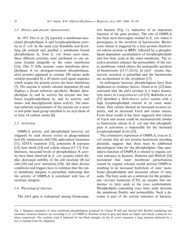
Fig. 1. Sequence alignment of outer membrane phospholipases produced by Clustal W [42] and Alscript [43]. Residue numbering andsecondary structure elements are according to E. coli OMPLA. Residues boxed in grey and black are highly and strictly conserved res-idues, respectively. The catalytic triad is indicated by the black triangles. In the H. pylori sequences a long insertion indicated by abox is omitted from the alignment.
BBAMCB 55687 3-11-00
H.J. Snijder, B.W. Dijkstra / Biochimica et Biophysica Acta 1488 (2000) 91^10192

BBAMCB 55687 3-11-00
H.J. Snijder, B.W. Dijkstra / Biochimica et Biophysica Acta 1488 (2000) 91^101 93

The trans fatty acids have to be reincorporated inphospholipids, by an as yet unknown mechanism.Whereas the involvement of CTI in solvent toleranceis demonstrated [25], the role of OMPLA in this pro-cess still needs to be clari¢ed.
2. Structure
2.1. Fold
Structural studies on OMPLA started with the de-termination of the nucleotide sequence of the pldAgene. The rather hydrophilic character and the lackof long hydrophobic amino acid stretches, indicatedthat the interaction of OMPLA with the membranedi¡ered from integral membrane proteins such asbacteriorhodopsin [7]. Circular dichroism (CD) mea-surements indicated a high L-strand content [26] andthis led Brok et al. [27] to propose a molecular modelwith a L-barrel architecture. Electron crystallographycon¢rmed the L-barrel character; projection mapsshowed an oval ring-like density large enough to ac-commodate 12 L-strands. An elongated domain pro-trudes from the L-barrel with a size of about 10U25Aî [28]. Overexpression and a protocol for refoldingof inclusion bodies [26] paved the way for detailedstructural analysis by X-ray crystallography. Crystalssuitable for X-ray analysis were grown using deter-gent solubilised protein and an organic solvent asprecipitant [29]. The X-ray structure was publishedrecently [30].
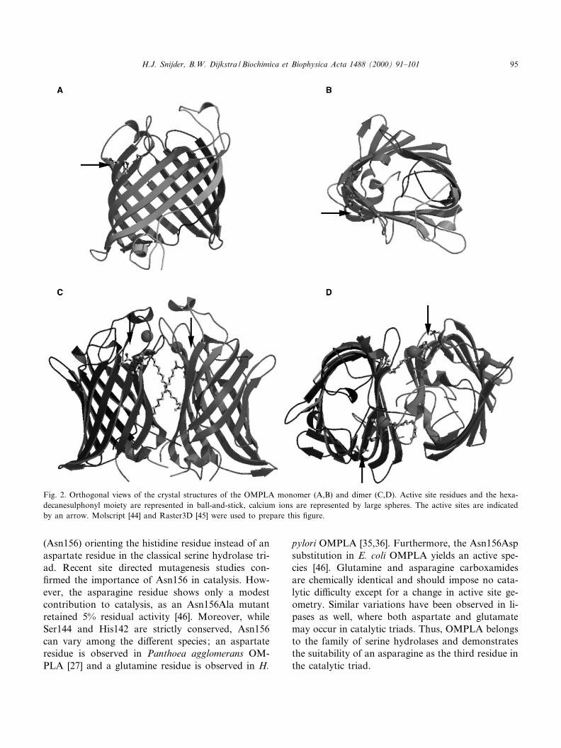
OMPLA has a 12 stranded antiparallel L-barrelcomposed of a convex and a £at side with approx-imate dimensions of 20U30U45 Aî 3 (Fig. 2A,B) [30].Amphipathic L-strands traverse the membrane, pre-senting a cylindrical hydrophobic protein surface. Oneither side of the hydrophobic cylinder, regions occurwhich are rich in aromatic residues. NeighbouringL-strands are joined by loops and turns which havea polar character. The enzyme's interior is polar andcontains an intricate hydrogen bonding network pro-viding a rigid structure. Two internal cavities existthat contain water molecules, which are capped byhydrophobic residues. Epitope insertion studies [31]identi¢ed the orientation of OMPLA in the mem-brane; the loop region is located at the extracellular
side while the termini and turns are oriented towardsthe periplasm.
2.2. Active site
First hints for the active site residues were circum-stantial, when it was found that trans-esteri¢cationwith methanol resulted in fatty acid methyl esters [3],similar to the lipases from the serine hydrolase fam-ily. This suggested that a serine might be a catalyticresidue. Later, the ¢nding of an inactive Ser152Phevariant of OMPLA suggested that residue 152 wasthe active site serine [27]. This hypothesis was re-jected, however, when site directed mutagenesis dem-onstrated some tolerance at this position (Ser152Asnand Ser152Thr) [32]. De¢nite evidence for a serine ascatalytic nucleophile came from inhibition studieswith hexadecanesulphonyl £uoride. A stoichiometricamount of hexadecanesulphonyl £uoride led to anirreversible loss of enzyme activity and resulted insulphonylation of serine 144 [33]. Site directed muta-genesis of Ser144 supported this ¢nding; only theSer144Cys mutant had some residual activity (ap-prox. 1%). This has been observed more often forserine-cysteine mutations in the serine hydrolase fam-ily [32].
The ¢nding of the active site serine led to the beliefthat OMPLA could be a member of the class ofserine hydrolases and could have a classical Ser-His-Asp triad. Inactivation experiments using p-bro-mophenacyl bromide suggested that one out of the¢ve histidines was involved in catalysis [7]. Later, thisinhibition proved to be rather non-speci¢c. Never-theless, site directed mutagenesis unequivocally dem-onstrated that one histidine, His142, was catalyticallyessential, with the His142Gly variant displaying afour orders of magnitude lower activity than thewild-type enzyme [34].
The active site residues His142 and Ser144 are lo-cated at the exterior of the L-barrel, at the outerlea£et side of the membrane. This location indicatesthat under normal conditions the substrate and theactive site are physically separated, since in E. coliphospholipids are exclusively located in the innerlea£et of the outer membrane. The constellation ofactive site residues resembles that of classical serinehydrolases, however, with an asparagine residue
BBAMCB 55687 3-11-00
H.J. Snijder, B.W. Dijkstra / Biochimica et Biophysica Acta 1488 (2000) 91^10194
(Asn156) orienting the histidine residue instead of anaspartate residue in the classical serine hydrolase tri-ad. Recent site directed mutagenesis studies con-¢rmed the importance of Asn156 in catalysis. How-ever, the asparagine residue shows only a modestcontribution to catalysis, as an Asn156Ala mutantretained 5% residual activity [46]. Moreover, whileSer144 and His142 are strictly conserved, Asn156can vary among the di¡erent species; an aspartateresidue is observed in Panthoea agglomerans OM-PLA [27] and a glutamine residue is observed in H.
pylori OMPLA [35,36]. Furthermore, the Asn156Aspsubstitution in E. coli OMPLA yields an active spe-cies [46]. Glutamine and asparagine carboxamidesare chemically identical and should impose no cata-lytic di¤culty except for a change in active site ge-ometry. Similar variations have been observed in li-pases as well, where both aspartate and glutamatemay occur in catalytic triads. Thus, OMPLA belongsto the family of serine hydrolases and demonstratesthe suitability of an asparagine as the third residue inthe catalytic triad.
Fig. 2. Orthogonal views of the crystal structures of the OMPLA monomer (A,B) and dimer (C,D). Active site residues and the hexa-decanesulphonyl moiety are represented in ball-and-stick, calcium ions are represented by large spheres. The active sites are indicatedby an arrow. Molscript [44] and Raster3D [45] were used to prepare this ¢gure.
BBAMCB 55687 3-11-00
H.J. Snijder, B.W. Dijkstra / Biochimica et Biophysica Acta 1488 (2000) 91^101 95
3. Activation and regulation
3.1. Dimerisation
3.1.1. Evidence for dimersStrict regulation of the phospholipase activity is
necessary since uncontrolled activity can have lethalconsequences for the bacterium [16]. Regulationmust occur at the protein level, since OMPLA isconstitutively expressed and overproduction causesno phenotypic variations [14,37]. In vitro, the phos-pholipase activity is modulated in a calcium depen-dent manner by reversible dimerisation. Inhibition byhexadecanesulphonyl £uoride stabilises the dimericform of OMPLA [38]. In vivo, chemical cross-linkingstudies indicate the presence of the monomeric formof OMPLA in the outer membrane [38]. Soni¢cationor induction of bacteriocin release protein activatesOMPLA which is a process concurring with dimer-isation [39].
Fluorescence resonance energy transfer (FRET)and chemical cross-linking experiments showed thatdimerisation is dependent on the presence of calciumand substrate [40]. However, these experiments alsosupplied evidence for another form of regulation,modulated by the physical state of the outer mem-brane lipids. In a bilayer environment, dimeric OM-PLA displays low activity even in excess of calcium.Perturbation of these bilayers by detergents results ina 70-fold increase in activity.
3.1.2. Dimer structureOMPLA dimers are formed by the association of
two enzyme monomers interacting via the £at barrelside (Fig. 2C,D) [30]. The association mode observedin the crystal structure is supported by cross-linkingexperiments using H26C single cysteine mutants.Residue 26 is located close to the £at side of the L-barrel OMPLA, and cysteine residues at this positioncan be e¡ectively cross-linked to produce an activedimer complex [40]. Dimeric and monomeric OM-PLA hardly exhibit structural di¡erences, only theN-terminal loop, which precedes the ¢rst strand,lacks interpretable density in dimeric OMPLA. Com-parison of the X-ray crystal structure and electronmicroscopy projection maps of monomeric OMPLAindicate a high £exibility of this loop [28] (Snijder etal., manuscript in preparation).
3.1.3. Formation of substrate binding pocketsDimerisation creates two extended clefts along the
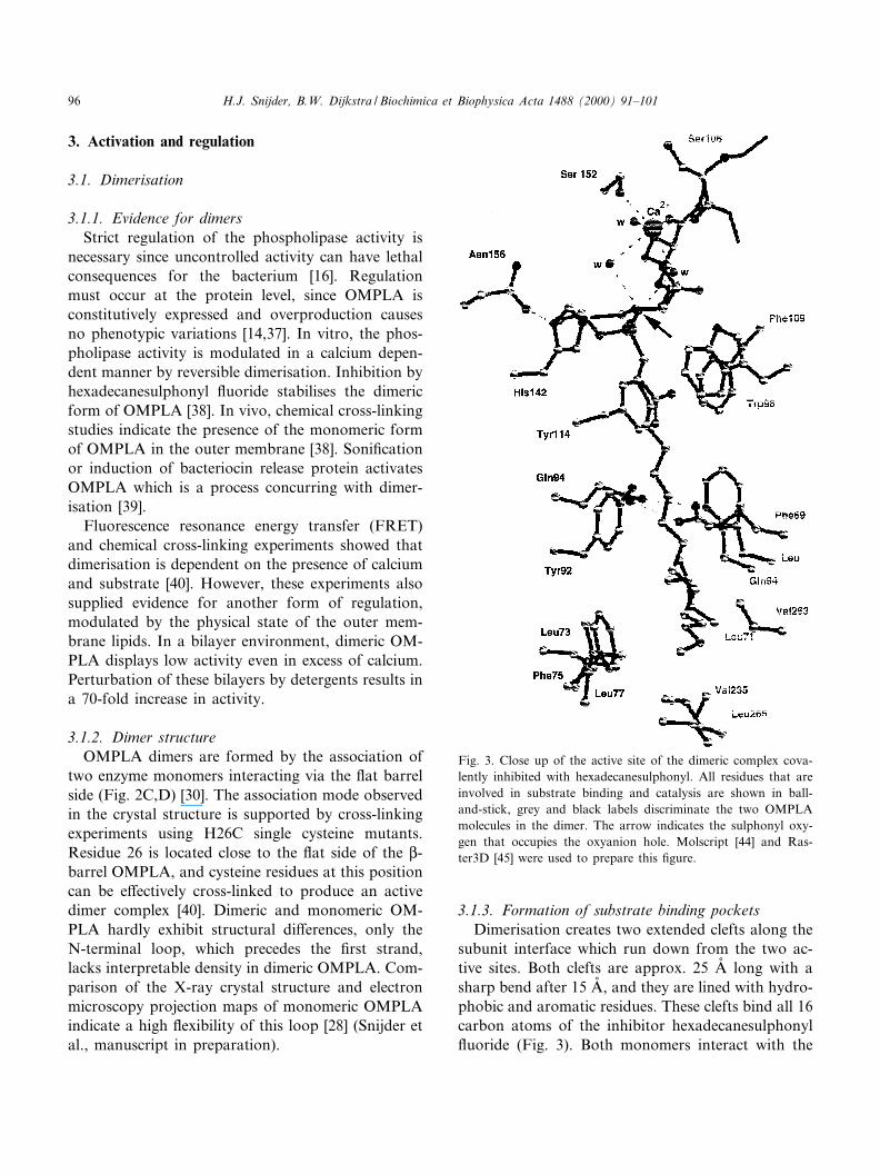
subunit interface which run down from the two ac-tive sites. Both clefts are approx. 25 Aî long with asharp bend after 15 Aî , and they are lined with hydro-phobic and aromatic residues. These clefts bind all 16carbon atoms of the inhibitor hexadecanesulphonyl£uoride (Fig. 3). Both monomers interact with the
Fig. 3. Close up of the active site of the dimeric complex cova-lently inhibited with hexadecanesulphonyl. All residues that areinvolved in substrate binding and catalysis are shown in ball-and-stick, grey and black labels discriminate the two OMPLAmolecules in the dimer. The arrow indicates the sulphonyl oxy-gen that occupies the oxyanion hole. Molscript [44] and Ras-ter3D [45] were used to prepare this ¢gure.
BBAMCB 55687 3-11-00
H.J. Snijder, B.W. Dijkstra / Biochimica et Biophysica Acta 1488 (2000) 91^10196
inhibitors and this explains the observed stabilisationof the dimer upon inhibition. The clefts bind onlyone acyl chain from the substrate. The residuesthat line these pockets are not strictly conserved,but most mutations are conservative and maintainthe hydrophobic character of the pockets. Thus,although a monomer contains a catalytic triad, themonomeric enzyme form lacks catalytic activity dueto absence of substrate binding pockets, which areformed only in the dimeric complex [30].
The architecture of the catalytic site and substratebinding clefts allow a broad range of substrates tobind. The active sites are exposed to the solvent andcan accommodate a large variety of polar headgroups, varying from as small as a glycol unit to acomplete Tween 20 head group (polyoxyethylene (20)sorbitan). As a consequence of the one rather non-speci¢c acyl binding site, substrates with either oneor two acyl chains of various lengths can be produc-tively bound. Long hydrocarbon chains can extendin the substrate binding pocket and into the hydro-phobic membrane core. Short chain length substrates(912 carbon atoms) are less e¡ectively hydrolysed[8], which is probably related to their lesser e¤ciencyto induce dimerisation as only the carbon atoms atthe end of the acyl chain (from C12) have interactionwith the neighbouring molecule.
3.1.4. Protein-protein interactionsThe dimer interaction surface is almost exclusively
con¢ned to the hydrophobic membrane embeddedpart. Only three main chain hydrogen bonds formthe interactions in the polar loops and turns regionof OMPLA [30]. In the hydrophobic part, the inter-face contains a patch of four leucines on both mono-mers which have a knob-and-hole interaction.Although these are not fully conserved, the hydro-phobic character of the interaction is maintained.Additional stabilisation of the dimer complex occursvia aromatic stacking, conserved in all but theH. pylori OMPLA. However, the key feature in di-merisation is a double hydrogen bond formed be-tween the Gln94 residue side chain of both mono-mers, a residue which is embedded in thehydrophobic interior of the dimer. The importanceof this polar protein-protein interaction is exempli-¢ed by the evolutionary conservation of Gln94 in allpldA sequences.
3.2. Calcium dependence
3.2.1. BiochemistryCalcium is an essential cofactor for activity of
OMPLA [6]. It is rather strongly bound, with a dis-sociation constant of 10^15 WM. Calcium can be re-placed by strontium, albeit with a 10-fold higher dis-sociation constant and a substantially lower activity.Magnesium and barium cannot replace calcium asthe catalytic cofactor. They are in fact competitiveinhibitors with dissociation constants in the millimo-lar range [8]. In vitro, calcium is required for dimer-isation [38]. Kinetic experiments indicate the in£u-ence of the detergent on the calcium a¤nity. A 70-fold lower a¤nity (millimolar range) is observed inessays with non-ionic detergents compared to essayswith alkylphosphocholine detergents. Various directbinding techniques (gel ¢ltration, £uorescence spec-troscopy and isothermal titration calorimetry) char-acterised calcium binding in detail [41]. Two bindingsites are present per monomer, one high a¤nity cal-cium binding site (KdW36 WM) and a second bindingsite with a 10-fold lower a¤nity (KdW358 WM). Asdimerisation occurs at low Ca2� concentration, thissuggests that the ¢rst calcium ion plays a role indimer stabilisation. Sulphonylation of OMPLA (byhexadecanesulphonyl £uoride) changes the calciumbinding behaviour and two high a¤nity (KdW48WM) calcium sites were detected in the inhibited en-zyme [41].
3.2.2. Structural evidenceOne calcium binding site was identi¢ed in the
structure of monomeric OMPLA. The calcium ionin this site is located approx. 10 Aî away from theactive site and is bound between loops L3 and L4(L3L4 site). Calcium could be substituted for thelanthanide ion Sm3�. Two aspartate side chain car-boxylates act as ligands (Asp149 and Asp184). Theseresidues show some evolutionary variation. Aspar-tate 149 is replaced by arginine in the Neisseria men-ingitides and Neisseria gonorrhoeae OMPLA. In H.pylori and Bordetella pertussis OMPLA this aspartateis replaced by a glycine, while aspartate 184 is re-placed by a serine and a glutamic acid residue re-spectively. Such variation strongly suggests that en-zymes from the di¡erent sources will show a di¡erentcalcium dependence, most likely changing the dimer-
BBAMCB 55687 3-11-00
H.J. Snijder, B.W. Dijkstra / Biochimica et Biophysica Acta 1488 (2000) 91^101 97
isation behaviour as well as the calcium dependenceof activity (Snijder et al., manuscript in preparation).
After dimerisation a second Ca2� binding site ispresent. It is located in the active site at the dimerinterface (catalytic calcium site). The calcium ion isligated by the side chain of Ser152 and by one mainchain carbonyl oxygen atom from each monomer.Water molecules complete the ligation sphere result-ing in a binding site with a bipyramidal octahedralgeometry. The contribution of both monomers to thebinding of calcium correlates with the biochemicalevidence that calcium is needed for dimerisation.
The structures supply only relative information onthe binding a¤nities of the two di¡erent calciumbinding sites. In the dimeric crystal structure, thea¤nity of the catalytic calcium site is higher thanthat of the L3L4 binding site, since only the catalyticcalcium ion could be modelled with con¢dence in theexperimental electron density maps. The opposite sit-uation occurs in the crystal structure of monomericOMPLA, where the calcium ion could only be ob-served in the L3L4 binding site.
Calcium has an essential catalytic role, which isillustrated by the dimeric hexadecanesulphonyl-in-hibited structure. This hexadecanesulphonyl inhibitormimics the negatively charged tetrahedral intermedi-ate of the acylation reaction of serine hydrolases. Inclassical serine hydrolases, stabilisation of the nega-tive charge during catalysis is supplied by donationof two or three hydrogen bonds by a structural fea-ture, which is called the oxyanion hole. In OMPLA,the sulphonyl oxygen atom which proposedly woulddevelop a negative charge during catalysis, formshydrogen bonds with the main chain amide fromresidue glycine 146 and two water molecules. Thecalcium ion positions these water molecules in theactive site and polarises them. Thus, the calciumion stabilises negatively charged intermediates duringcatalysis via water molecules.
In the OMPLA Michaelis-Menten complex, thesubstrate would be oriented with the carbonyl oxy-gen atom of the ester towards the calcium ion. Thisorientation causes the carbonyl bond to become po-larised and increases the electrophilicity of the car-bonyl carbon atom, which will facilitate the nucleo-philic attack by serine 144. Thus, the contribution ofcalcium to catalysis stems (i) from the polarisation ofthe substrate ester carbonyl group and (ii) from sta-
bilisation of negatively charged reaction intermedi-ate. These roles of the calcium ion suggest that thecatalytic importance of the strictly conserved Ser152is related to its calcium ligating capacity and its con-tribution to correctly positioning of the calcium ion[30].
3.2.3. Binding modelThe various parameters that in£uence calcium
binding and that modulate the a¤nity of OMPLAfor calcium, hamper a full understanding of the com-plex calcium binding process. Nevertheless, a work-ing model for calcium binding emerges from thecombined structural and biochemical data. In thenatural environment of OMPLA, calcium is boundto monomeric phospholipase with high a¤nity. Onlyafter dimerisation, the catalytic calcium site isformed. Active site occupation and dimerisation re-sults in an increased calcium a¤nity of the active sitecalcium binding site. Subsequent transfer of calciumto the catalytic site results in the active complex. Asecond calcium can be now bound to the emptybinding site between loops L3 and L4 yielding adimer complex with two calcium ions per monomer.Although the role of calcium in catalysis seems clear,this is much less so for its role in the regulation ofthe activity of OMPLA. As calcium ions are abun-dant in the bacterial outer membrane environment, itis questionable whether the in vitro observed calciumdependent dimerisation is of any physiological rele-vance.
4. Mode of action
All biochemical and structural data lead to a mod-el for the action of OMPLA, which is graphicallysummarised in Fig. 4. Normally, OMPLA is presentin the outer membrane as a monomer with a calciumion bound at the L3L4 binding site. This form isinactive owing to the absence of substrate bindingpockets, lack of oxyanion stabilisation and physicalseparation of substrate and active site. External orinternal triggers (e.g. bacteriocin release protein in-duction, temperature shock, polymixin B exposure)perturb the outer membrane, introducing phospho-lipids in the outer lea£et. The presence of the phos-pholipids in the outer lea£et increases the £uidity of
BBAMCB 55687 3-11-00
H.J. Snijder, B.W. Dijkstra / Biochimica et Biophysica Acta 1488 (2000) 91^10198

the outer membrane, which will facilitate lateral dif-fusion of its embedded components. Besides that,substrates are presented to the active site of OM-PLA, which presumably triggers dimerisation. Thecalcium ion possibly moves from the L3L4 site tothe active site of OMPLA, thus creating an activedimer-substrate-cofactor complex.
The active complex hydrolyses the phospholipidthrough a mechanism analogous to that of serinehydrolases. Ser144 performs a nucleophilic attackon the carbonyl carbon of the ester, facilitated bypolarisation of the ester carbonyl bond by electro-static interactions with the calcium ion. A negativelycharged tetrahedral intermediate is formed which isstabilised by the tetrahedral arrangement of hydro-gen bonds and by the remote in£uence of the calciumion. The transient intermediate collapses to give theenzyme-acyl intermediate. The lysophospholipid canleave by lateral di¡usion in the membrane. Deacyla-tion will occur with a water molecule acting as the
nucleophile. The fatty acid product can leave theactive site either by lateral di¡usion into the mem-brane via the opening between the two monomers oralternatively by dissociation of the dimer into mono-mers.
In addition to the membrane perturbing trigger,release of fatty acids and lysophospholipids destabil-ises the membrane, possibly resulting in non-bilayerstructures. In vitro the OMPLA activity is stronglyenhanced by non-bilayer structures, resulting in morerapid generation of fatty acids and lysophospholip-ids. Hence, the membrane perturbing signal is ampli-¢ed and the reaction products further permeabilisethe membrane. Thus, semispeci¢c excretion of coli-cins and other e¡ector molecules is facilitated.
5. Conclusion and perspective
The recently solved structures of OMPLA have
Fig. 4. Model for involvement of OMPLA in bacteriocin release. Under normal conditions OMPLA is in a dormant monomeric formand the outer lea£et of the membrane consists solely of LPS (A). Under the in£uence of a trigger, for example the bacteriocin releaseprotein, the membrane bilayer is perturbed and phospholipids are presented in the outer lea£et (B). OMPLA dimers are formed, sub-strate and calcium are bound in the active site (C). Phospholipids are hydrolysed, lysophospholipids and fatty acids perturb the lipidbilayer (D). Eventually the outer membrane becomes permeable and bacteriocins can be semispeci¢cally secreted (E).
BBAMCB 55687 3-11-00
H.J. Snijder, B.W. Dijkstra / Biochimica et Biophysica Acta 1488 (2000) 91^101 99

yielded a better understanding of membrane proteininteractions, phospholipid metabolism, and function-ing of this speci¢c phospholipase in particular. Itrevealed an entirely new fold of phospholipases andsheds light on its catalytic properties. However, ourunderstanding is far from complete. What is the bio-logical relevance of the L3L4 calcium binding site?How can the physical state of the lipidic surroundingmodulate enzyme activity? What is the exact biolog-ical function of OMPLA? How is OMPLA involvedin bacterial pathogenesis? What mechanism arrestsenzymatic activity and averts complete bacterial ly-sis? In the future, continued biochemical and struc-tural research are expected to unravel the remainingsecrets and to obtain a full understanding of themolecular mechanism of this intriguing enzyme.
Acknowledgements
This research was supported by the NetherlandsFoundation for Chemical Research (CW) with ¢nan-cial aid from the Netherlands Organisation for Sci-enti¢c Research (NWO).
References
[1] E.A. Dennis, Trends Biochem. Sci. 22 (1997) 1^2.[2] J.D. Clark, N. Milona, J.L. Knopf, Proc. Natl. Acad. Sci.
USA 87 (1990) 7708^7712.[3] O. Doi, M. Ohki, S. Nojima, Biochim. Biophys. Acta 260
(1972) 244^258.[4] C.J. Scandella, A. Kornberg, Biochemistry 10 (1971) 4447^
4456.[5] F.R. Albright, D.A. White, W.J. Lennarz, J. Biol. Chem.
248 (1973) 3968^3977.[6] M. Nishijima, S. Nakaike, Y. Tamori, S. Nojima, Eur. J.
Biochem. 73 (1977) 115^124.[7] H. Homma, T. Kobayashi, N. Chiba, K. Karasawa, H. Ike-
da, M. Sekiguchi, S. Nojima, J. Biochem. 96 (1984) 1655^1664.
[8] A.J.G. Horrevoets, T.M. Hackeng, H.M. Verheij, R. Dijk-man, G.H. de Haas, Biochemistry 28 (1989) 1139^1147.
[9] J.E. Cronan Jr., D.L. Wul¡, Virology 38 (1969) 241^246.[10] H. Okuyama, Biochim. Biophys. Acta 176 (1969) 125^134.[11] P. Patriarca, S. Beckerdite, P. Elsbach, Biochim. Biophys.
Acta 260 (1972) 593^600.[12] K.L. Hardaway, C.S. Buller, J. Bacteriol. 137 (1979) 62^68.[13] J. Weiss, S. Beckerdite-Quagliata, P. Elsbach, J. Biol. Chem.
254 (1979) 11010^11014.
[14] P. de Geus, I. van Die, H. Bergmans, J. Tommassen, G.H.de Haas, Mol. Gen. Genet. 190 (1983) 150^155.
[15] A.P. Pugsley, M. Schwartz, EMBO J. 3 (1984) 2393^2397.[16] J. Luirink, C. van der Sande, J. Tommassen, E. Veltkamp,
F.K. de Graaf, B. Oudega, J. Gen. Microbiol. 132 (1986)825^834.
[17] D. Cavard, D. Baty, S.P. Howard, H.M. Verheij, C. Laz-dunski, J. Bacteriol. 169 (1987) 2187^2194.
[18] A. Audet, G. Nantel, P. Proulx, Biochim. Biophys. Acta 348(1974) 334^343.
[19] G.P.F. Michel, J. Starka, FEBS Lett. 108 (1979) 261^265.[20] F.J. Van der Wal, J. Luirink, B. Oudega, FEMS Microb.
Rev. 17 (1995) 381^399.[21] C.J. Lazdunski, D. Baty, V. Geli, D. Cavard, J. Morlon, R.
Lloubes, S.P. Howard, M. Knibiehler, M. Chartier, S. Var-enne, M. Frenette, J.L. Dasseux, F. Pattus, Biochim. Bio-phys. Acta 947 (1988) 445^464.
[22] K.A. Grant, I. Ubarretxena-Belandia, N. Dekker, P.T. Ri-chardson, S.F. Park, Infect. Immun. 65 (1997) 1172^1180.
[23] G. Bukholm, T. TannEs, P. Nedenskov, Y. Esbensen, H.J.Grav, T. Hovig, S. Ariansen, I. Guldvog, Scan. J. Gastro-enterol. 32 (1997) 445^454.
[24] V. Pedrotta, B. Witholt, J. Bacteriol. 181 (1999) 3256^3261.
[25] F. Junker, J.L. Ramos, J. Bacteriol. 181 (1999) 5693^5700.[26] N. Dekker, K. Merck, J. Tommassen, H.M. Verheij, Eur. J.
Biochem. 232 (1995) 214^219.[27] R.G.P.M. Brok, E. Brinkman, R. van Boxtel, A.C. Bekkers,
H.M. Verheij, J. Tommassen, J. Bacteriol. 176 (1994) 861^870.
[28] E.J. Boekema, M. Stuart, R.I. Koning, W. Keegstra, A.Brisson, H.M. Verheij, N. Dekker, J. Struct. Biol. 123(1998) 67^71.
[29] M. Blaauw, N. Dekker, H.M. Verheij, K.H. Kalk, B.W.Dijkstra, FEBS Lett. 373 (1995) 10^12.
[30] H.J. Snijder, I. Ubarretxena-Belandia, M. Blaauw, K.H.Kalk, H.M. Verheij, M.R. Egmond, N. Dekker, B.W. Dijk-stra, Nature 401 (1999) 717^721.
[31] K.B. Merck, H. de Cock, H.M. Verheij, J. Tommassen,J. Bacteriol. 179 (1997) 3443^3450.
[32] R.G.P.M. Brok, I. Ubarretxena-Belandia, N. Dekker, J.Tommassen, H.M. Verheij, Biochemistry 35 (1996) 7787^7793.
[33] A.J. Horrevoets, H.M. Verheij, G.H. de Haas, Eur. J. Bio-chem. 198 (1991) 247^253.
[34] R.G.P.M. Brok, N. Dekker, N. Gerrits, H.M. Verheij, J.Tommassen, Eur. J. Biochem. 234 (1995) 934^938.
[35] J.F. Tomb, O. White, A.R. Kerlavage, R.A. Clayton, G.G.Sutton, R.D. Fleischmann, K.A. Ketchum, H.P. Klenk, S.Gill, B.A. Dougherty, K. Nelson, J. Quackenbush, L.X.Zhou, E.F. Kirkness, S. Peterson, B. Loftus, D. Richardson,R. Dodson, H.G. Khalak, A. Glodek, K. McKenney, L.M.Fitzegerald, N. Lee, M.D. Adams, E.K. Hickey, D.E. Berg,J.D. Gocayne, T.R. Utterback, J.D. Peterson, J.M. Kelley,M.D. Cotton, J.M. Weidman, C. Fujii, C. Bowman, L. Wat-they, E. Wallin, W.S. Hayes, M. Borodovsky, P.D. Karp,
BBAMCB 55687 3-11-00
H.J. Snijder, B.W. Dijkstra / Biochimica et Biophysica Acta 1488 (2000) 91^101100
H.O. Smith, C.M. Fraser, J.C. Venter, Nature 389 (1997)412^412.
[36] R.G.P.M. Brok, A.P. Boots, N. Dekker, H.M. Verheij, J.Tommassen, Res. Microbiol. 149 (1998) 703^710.
[37] H. Homma, N. Chiba, T. Kobayashi, I. Kudo, K. Inoue, H.Ikeda, M. Sekiguchi, S. Nojima, J. Biochem. 96 (1984) 1645^1653.
[38] N. Dekker, J. Tommassen, A. Lustig, J.P. Rosenbusch,H.M. Verheij, J. Biol. Chem. 272 (1997) 3179^3184.
[39] N. Dekker, J. Tommassen, H.M. Verheij, J. Bacteriol. 181(1999) 3281^3283.
[40] I. Ubarretxena-Belandia, L. Hozeman, E. van der Brink-vander Laan, E.H.M. Pap, M.R. Egmond, H.M. Verheij, N.Dekker, Biochemistry 38 (1999) 7398^7405.
[41] I. Ubarretxena-Belandia, J.W.P. Boots, H.M. Verheij, N.Dekker, Biochemistry 37 (1998) 16011^16018.
[42] J.D. Thompson, D.G. Higgins, T.J. Gibson, Nucleic AcidsRes. 22 (1994) 4673^4680.
[43] G.J. Barton, Protein Eng. 6 (1993) 37^40.[44] P.J. Kraulis, J. Appl. Crystallogr. 24 (1991) 946^950.[45] E.A. Merritt, J.A. Bacon, Methods Enzymol. 277 (1997)
505^524.[46] R.L. Kingma, M. Fragiathaki, H.J. Snijder, B.W. Dijkstra,
H.M. Verheij, N. Dekker, M.R. Egmond, Biochemistry 39(2000) 10017^10022.
BBAMCB 55687 3-11-00
H.J. Snijder, B.W. Dijkstra / Biochimica et Biophysica Acta 1488 (2000) 91^101 101
Related Documents