Crystal Structures of Intermediates in the Nitroalkane Oxidase Reaction † Annie Héroux ⊥ , Dragana M. Bozinovski § , Michael P. Valley § , Paul F. Fitzpatrick §,∥,‡,* , and Allen M. Orville ⊥,* ⊥ Department of Biology, Brookhaven National Laboratory, Upton, NY 11973 § Departments of Biochemistry and Biophysics, Texas A&M University, College Station, TX 77843-2128 ∥ Departments of Chemistry, Texas A&M University, College Station, TX 77843-2128 Abstract The flavoenzyme nitroalkane oxidase is a member of the acyl-CoA dehydrogenase superfamily. Nitroalkane oxidase catalyzes the oxidation of neutral nitroalkanes to nitrite and the corresponding aldehydes or ketones. Crystal structures to 2.2 Å resolution or better are described of enzyme complexes with bound substrates and of a trapped substrate-flavin adduct. The D402N enzyme has no detectable activity with neutral nitroalkanes (Valley, M. P., and Fitzpatrick, P. F. (2003) J. Am. Chem. Soc. 23, 8738–8739). The structure of the D402N enzyme crystallized in the presence of 1- nitrohexane or 1-nitrooctane shows the presence of the substrate in the binding site. The aliphatic chain of the substrate extends into a tunnel leading to the enzyme surface. The oxygens of the substrate nitro group interact both with amino acid residues and with the 2’-hydroxyl of the FAD. When nitroalkane oxidase oxidizes nitroalkanes in the presence of cyanide, an electrophilic flavin imine intermediate can be trapped (Valley, M. P., Tichy, S. E., and Fitzpatrick, P. F. (2005) J. Am. Chem. Soc. 127, 2062–2066). The structure of the enzyme trapped with cyanide during oxidation of 1- nitrohexane shows the presence of the modified flavin. A continuous hydrogen bond network connects the nitrogen of the CN-hexyl-FAD through the FAD 2’-hydroxyl to a chain of water molecules extending to the protein surface. Together, our complementary approaches provide strong evidence that the flavin cofactor is in the appropriate oxidation state and correlates well with the putative intermediate state observed within each of the crystal structures. Consequently, these results provide important structural descriptions of several steps along the nitroalkane oxidase reaction cycle. Nitroalkane oxidase (NAO 1 ) from the soil fungus Fusarium oxysporum catalyzes the oxidation of nitroalkanes to the corresponding aldehydes or ketones with the release of nitrite and the † This research was supported in part by grants to PFF from the NIH (GM058698) and The Welch Foundation (A-1245) and to AMO from the Offices of Biological and Environmental Research US Department of Energy, the National Center for Research Resources (2 P41 RR012408) of the NIH and from the of the US Department of Energy. Use of the National Synchrotron Light Source at Brookhaven National Laboratory was supported by the U.S. Department of Energy Office of Basic Energy Sciences, under Contract DE- AC02-98CH10886. *Corresponding authors. AMO: phone 631-344-4739; e-mail: [email protected]; PFF: phone, 210-567-8264; fax, 210-567-8778; e-mail, [email protected]. ‡ Current address: Department of Biochemistry, MC 7760, University of Texas Health Science Center at San Antonio, San Antonio, TX 78229-3900 The atomic coordinates and structure factors have been deposited with the Protein Data Bank with the corresponding file names: a) D402N NAO plus 1-nitrohexane, 3D9D; b) D402N NAO plus 1-nitrooctane, 3D9E; c) S276A NAO plus 1-nitrohexane, 3D9F; and d) wild-type NAO containing the N5-cyanohexyl FAD, 3D9G. 1 Abbreviations: NAO, nitroalkane oxidase; ACO, acyl-CoA oxidase; ACAD, acyl-CoA dehydrogenase; K ne , steady-state K m for nitroethane. NIH Public Access Author Manuscript Biochemistry. Author manuscript; available in PMC 2010 April 21. Published in final edited form as: Biochemistry. 2009 April 21; 48(15): 3407–3416. doi:10.1021/bi8023042. NIH-PA Author Manuscript NIH-PA Author Manuscript NIH-PA Author Manuscript

Welcome message from author
This document is posted to help you gain knowledge. Please leave a comment to let me know what you think about it! Share it to your friends and learn new things together.
Transcript

Crystal Structures of Intermediates in the Nitroalkane OxidaseReaction†
Annie Héroux⊥, Dragana M. Bozinovski§, Michael P. Valley§, Paul F. Fitzpatrick§,∥,‡,*, andAllen M. Orville⊥,*⊥Department of Biology, Brookhaven National Laboratory, Upton, NY 11973§Departments of Biochemistry and Biophysics, Texas A&M University, College Station, TX77843-2128∥Departments of Chemistry, Texas A&M University, College Station, TX 77843-2128
AbstractThe flavoenzyme nitroalkane oxidase is a member of the acyl-CoA dehydrogenase superfamily.Nitroalkane oxidase catalyzes the oxidation of neutral nitroalkanes to nitrite and the correspondingaldehydes or ketones. Crystal structures to 2.2 Å resolution or better are described of enzymecomplexes with bound substrates and of a trapped substrate-flavin adduct. The D402N enzyme hasno detectable activity with neutral nitroalkanes (Valley, M. P., and Fitzpatrick, P. F. (2003) J. Am.Chem. Soc. 23, 8738–8739). The structure of the D402N enzyme crystallized in the presence of 1-nitrohexane or 1-nitrooctane shows the presence of the substrate in the binding site. The aliphaticchain of the substrate extends into a tunnel leading to the enzyme surface. The oxygens of the substratenitro group interact both with amino acid residues and with the 2’-hydroxyl of the FAD. Whennitroalkane oxidase oxidizes nitroalkanes in the presence of cyanide, an electrophilic flavin imineintermediate can be trapped (Valley, M. P., Tichy, S. E., and Fitzpatrick, P. F. (2005) J. Am. Chem.Soc. 127, 2062–2066). The structure of the enzyme trapped with cyanide during oxidation of 1-nitrohexane shows the presence of the modified flavin. A continuous hydrogen bond networkconnects the nitrogen of the CN-hexyl-FAD through the FAD 2’-hydroxyl to a chain of watermolecules extending to the protein surface. Together, our complementary approaches provide strongevidence that the flavin cofactor is in the appropriate oxidation state and correlates well with theputative intermediate state observed within each of the crystal structures. Consequently, these resultsprovide important structural descriptions of several steps along the nitroalkane oxidase reaction cycle.
Nitroalkane oxidase (NAO1) from the soil fungus Fusarium oxysporum catalyzes the oxidationof nitroalkanes to the corresponding aldehydes or ketones with the release of nitrite and the
†This research was supported in part by grants to PFF from the NIH (GM058698) and The Welch Foundation (A-1245) and to AMOfrom the Offices of Biological and Environmental Research US Department of Energy, the National Center for Research Resources (2P41 RR012408) of the NIH and from the of the US Department of Energy. Use of the National Synchrotron Light Source at BrookhavenNational Laboratory was supported by the U.S. Department of Energy Office of Basic Energy Sciences, under Contract DE-AC02-98CH10886.*Corresponding authors. AMO: phone 631-344-4739; e-mail: [email protected]; PFF: phone, 210-567-8264; fax, 210-567-8778; e-mail,[email protected].‡Current address: Department of Biochemistry, MC 7760, University of Texas Health Science Center at San Antonio, San Antonio, TX78229-3900The atomic coordinates and structure factors have been deposited with the Protein Data Bank with the corresponding file names: a)D402N NAO plus 1-nitrohexane, 3D9D; b) D402N NAO plus 1-nitrooctane, 3D9E; c) S276A NAO plus 1-nitrohexane, 3D9F; and d)wild-type NAO containing the N5-cyanohexyl FAD, 3D9G.1Abbreviations: NAO, nitroalkane oxidase; ACO, acyl-CoA oxidase; ACAD, acyl-CoA dehydrogenase; Kne, steady-state Km fornitroethane.
NIH Public AccessAuthor ManuscriptBiochemistry. Author manuscript; available in PMC 2010 April 21.
Published in final edited form as:Biochemistry. 2009 April 21; 48(15): 3407–3416. doi:10.1021/bi8023042.
NIH
-PA Author Manuscript
NIH
-PA Author Manuscript
NIH
-PA Author Manuscript
consumption of molecular oxygen to yield hydrogen peroxide (1). This reaction occurs indistinct reductive and oxidative half-reactions (Scheme 1) (2,3). The reductive half-reaction isinitiated by abstraction of the α-proton from the neutral form of the nitroalkane by Asp402,the active site base (3,4). The resulting anion attacks the N5 position of the FAD to form acovalent adduct (5,6); the subsequent elimination of nitrite results in a cationic, electrophilicflavin imine that can be attacked by hydroxide (7). Completion of the reductive half reactionoccurs with formation of the aldehyde or ketone product and the reduced FAD. In the oxidativehalf-reaction, the reduced FAD reacts with molecular oxygen to regenerate the oxidized flavin,producing hydrogen peroxide. This second-order reaction exhibits no detectable intermediates(3) and is typical for a flavoprotein oxidase (8). Product release from the oxidized enzymelimits the rate of turnover with primary nitroalkanes, the best substrates (9).
NAO is a structural member of the flavoenzyme acyl-CoA dehydrogenase (ACAD)superfamily (6,10,11). However, NAO does not utilize acyl-CoAs as substrates, nor doesACAD oxidize nitroalkanes (10). Indeed, the two types of enzymes appear to bind substratesfrom opposite orientations, despite the similarity between the thioester-acyl portion of acyl-CoA and the nitro group of nitroalkanes. While the sequences of ACAD family members areonly 13–23% identical to that of NAO, the identities are distributed throughout the proteinsequence (1,6), and the FAD and active site base are in analogous locations in the active sitesof NAO and ACAD (6). The initial steps of the catalytic reactions of these enzymes are similar,in that a protein carboxylate abstracts an acidic proton from the substrate (1,12). However,flavin reduction in ACAD occurs by transfer of a hydride from the substrate concerted with orsoon after proton abstraction rather than via attack of the substrate anion on the flavin (12).Thus, similar protein environments yield very different outcomes from a similar intermediatein members of the superfamily.
Structural analyses of enzyme-substrate complexes and catalytic intermediates are necessaryto explain these differences. Obtaining crystal structures of such intermediates poses significantchallenges because of the transient nature of most intermediates and the stability necessary forcrystallographic order. In NAO, as in other flavoprotein oxidases, the substrate is rapidlyoxidized even in the absence of oxygen; simply adding the substrate to the enzyme will yieldthe reduced enzyme, from which the products of the reductive half-reaction dissociate.Moreover, substrate binding or proton abstraction is rate-limiting for the reductive half-reactionof NAO (9), so that subsequent catalytic intermediates do not accumulate during turnover.Mutagenesis of Asp402 to asparagine or alanine yields an enzyme that will turn over anionicnitroalkane substrates at rates similar to the wild-type enzyme reacting with neutral substratesbut does not appear to catalyze reactions with the neutral substrate (4). Therefore, it wasexpected that D402N NAO would bind a neutral nitroalkane to form a Michaelis complex butproceed no further in the reaction cycle. Asp402, Arg409, and Ser276 constitute a catalytictriad in NAO (6,13). Kinetic and structural analyses of the effects of mutating Asp402 andArg409 suggest that both Ser276 and Arg409 are required to position the side chain of Asp402properly for proton abstraction. We describe here kinetic analyses of the S276A mutantenzyme, confirming the importance of Ser276 in proton abstraction. Since this mutation alsogreatly decreases the rate of proton abstraction, the S276A enzyme provides another optionfor obtaining a structure of the Michaelis complex. Further along the catalytic cycle, the cationicflavin imine intermediate does not accumulate; however, it can be trapped with either anitroalkane anion (5,6) or cyanide (7). An N5-cyanoalkyl-FAD is isosteric with the tetrahedralintermediate formed upon hydroxide attack on the imine intermediate (Scheme 1). We describehere structures of the D402N and S276A enzymes with bound nitroalkane as models for theenzyme-substrate complex in NAO and the structure of NAO trapped with cyanide duringturnover as a model for this critical intermediate in the NAO reaction.
Héroux et al. Page 2
Biochemistry. Author manuscript; available in PMC 2010 April 21.
NIH
-PA Author Manuscript
NIH
-PA Author Manuscript
NIH
-PA Author Manuscript
EXPERIMENTAL PROCEDURESMaterials
All chemicals were purchased from Sigma-Aldrich Chemical Corp. (Milwaukee, WI), unlessotherwise specified. S276A NAO was generated with the QuikChange Site-DirectedMutagenesis Kit (Stratagene) as previously described for other NAO mutant proteins (3). DNAsequencing of the entire coding sequence of each mutant plasmid was performed at theLaboratory for Plant Genome Technologies of the Texas A&M University to confirm themutation and the lack of undesired mutations. The wild-type and mutant enzymes wereexpressed in E. coli and purified as previously described (3,10). Protein concentrations weredetermined using an ε446 value of 14.2 mM−1cm−1. The cyanide-trapped enzyme was formedby allowing NAO to oxidize 1-nitrohexane in the presence of cyanide as previously described(7).
KineticsEnzyme activity was measured in air-saturated 100 mM Hepes buffer and 0.1 mM FAD at pH8.0 and 30 °C by monitoring oxygen consumption with a computer-interfaced Hansatech Clarkoxygen electrode (Hansatech Instruments, Pentney King's Lynn, U.K.) as described previously(13). To prevent the formation of the substrate anion, stock solutions of nitroethane wereprepared in DMSO and assays were initiated by the addition of substrate. Rapid reactionexperiments were carried out using an Applied Photophysics SX-20MV stopped-flowspectrophotometer, mixing ~30 µM NAO with 1–100 mM nitroethane and following thereaction at 455 nm. Enzyme samples were made anaerobic by repeated cycles of vacuum andargon. Glucose oxidase (36 nM) and glucose (5 mM) were added to enzyme solutions toscavenge any residual oxygen. Kinetic data were analyzed using the programs KaleidaGraph(Adelbeck Software, Reading, PA) and Igor Pro (WaveMetrics, Inc., Lake Oswego, OR).Steady-state kinetic parameters were determined by fitting the data to the Michaelis-Mentenequation or equation 1, where Ki is the inhibition constant for substrate inhibition and the otherterms have their usual definitions. Steady-state kinetic isotope effects were calculated fromequation 2. Here Fi is the fraction of deuterium in the substrate, and Dkcat and D(kcat/Km) arethe isotope effects for kcat and kcat/Km, respectively. Rapid reaction data were fit to equation3. At is the absorbance at 455 nm at time t, ΔAi is the absorbance change associated with agiven phase, ki is the apparent rate constant for that phase, and A∞ is the absorbance at infinitetime. The rate constants as a function of substrate concentration were fit to equation 4 to obtainkred, the limiting first-order rate constant for flavin reduction, and Kd, the apparent dissociationconstant.
(1)
(2)
(3)
(4)
Héroux et al. Page 3
Biochemistry. Author manuscript; available in PMC 2010 April 21.
NIH
-PA Author Manuscript
NIH
-PA Author Manuscript
NIH
-PA Author Manuscript

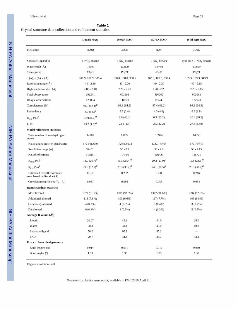
Structure determinationCrystals were obtained using hanging drop vapor-diffusion methods similar to those describedpreviously (6,13,14), with the exception that spermine was omitted. For the D402N and S276ANAO structures, 1 µL of 1-nitrohexane or 1-nitrooctane was added to 60 µL stock enzymesolution (~10 mg/mL) immediately prior to setting up the drops. The crystals were grown at277 K from a mother liquor solution containing 24–30% (w/v) PEG3350, 20% (v/v) glycerol,100 mM sodium cacodylate, pH 7.5. In some cases yellow crystals started appearing withintwo hours of setting up the 5 µL hanging drops (1:1 protein, mother liquor). Under similarconditions, colorless crystals of the cyanide-trapped enzyme grew within two days; these wereharvested immediately since they gradually turned yellow after approximately one week. Eachcrystal was mounted in a nylon loop without further cryoprotection and flash frozen by quick-submersion in liquid nitrogen. X-Ray diffraction data were collected at beamline X12-B, X25or X29 of the National Synchrotron Light Source with crystals held at 100 K. The data wereintegrated and scaled with HKL2000 (15). The structures were solved by molecularreplacement with MolRep from the CCP4 suit of programs (16) to resolve the polarity of thetrigonal space group. Initially, the search models comprised a homotetramer extracted fromthe wild-type NAO structure (PDB code 2c0u) (6) from which all solvent and FAD atoms wereremoved and Asp402 or Ser276 mutated to alanine as appropriate. The solutions indicated thatthe crystals were in space group p3221 with one α4 holoenzyme molecule in the asymmetricunit. Model refinement was done with REFMAC 5.2 coupled with COOT (17) to mutateresidues and assess the refinement cycles. Noncrystallographic symmetry restraints were notapplied during the model refinement. Substrate ligands were added in the last stages of modelrefinement. We also tested several alternative atomic models. For example, atomic modelscontaining several solvent molecules within the electron density for the putative substratemolecules did not refine well. The data collection and model refinement statistics are given inTable 1. Each structure was confirmed with independent datasets from at least two crystals.Only the highest quality structure for each is presented here.
Optical spectra of single crystalsOptical absorption spectra were collected from 34 µM wild type NAO in 500 mM Na cacodylicacid, pH 7.6, in a 1 cm quartz cuvette with a Perkin Elmer Lambda 35 spectrometer at roomtemperature (~23 °C). Single crystal optical absorption spectra were collected at the SingleCrystal μ-Spectroscopy Facility at beamline X26-C of the National Synchrotron Light Sourceat Brookhaven National Laboratory (18). This dedicated facility is available for the generaluser population. The microspectrophotometer is comprised of components from anXSPECTRA instrument (4DX-ray Systems AB, Sweden). It can be used for benchtopmeasurements or aligned with the crystal rotation axes and coincident with the X-ray beam atX26-C. The microscope objectives use parabolic mirrors to achieve 15x magnification andminimize spherical or chromatic aberration. The objectives provide a 24 mm working distancethrough a 0.4 numerical aperture, which allows for cryocooling and access for othercomponents. The microscope objective was coupled to a 50 µm quartz optical fiber, whichyields an incident spot size of approximately 25 µm in diameter focused on the cryocooledcrystal. The transmitted light was collected from approximately a 75 µm spot size. The incidentlight (350 – 850 nm) was from a 75W Xe research arc lamp (Newport Corp.). An Ocean OpticsUSB 4000 spectrophotometer (Dunedin, Florida) containing a 3648-element Toshiba linearCCD detector was used to collect the optical absorption spectra. The data were processed withthe SpectraSuite software on either a Windows XP or LINUX operating system. Themicrospectrophotometer was calibrated with a Hg-Ar calibration laser. The reference spectrumin air was collected in the absence of the sample. Typically, optical absorption spectra wereobtained by averaging 3–10 spectra, each of which was collected with an integration timebetween 70 and 200 ms, and with a 3–10-pixel “box car” setting of the CCD detector array.Each crystal was held at 100 K during data collection. We observed that the optical spectra
Héroux et al. Page 4
Biochemistry. Author manuscript; available in PMC 2010 April 21.
NIH
-PA Author Manuscript
NIH
-PA Author Manuscript
NIH
-PA Author Manuscript

from single crystals are frequently anisotropic and, therefore, collected optical spectra atseveral crystal angles. The best spectra were usually obtained from needles or plates alignedsuch that the optical path was normal to the observable flat feature of the particular crystalhabit and to the cryoloop. To reduce unusual baseline excursions, no part of the cryoloopintersected the optical spectroscopic axis.
RESULTSKinetic characterization of S276A NAO
In order to probe the role of the interaction between the hydroxyl group of Ser276 and thecarboxylate of the active site base Asp402 in the NAO reaction, Ser276 was changed to alanineby site-directed mutagenesis. The purified recombinant enzyme was characterized usingsteady-state and rapid-reaction kinetics (Table 2). While the kcat/Km values are greater forlonger chain primary nitroalkanes (19), with nitroethane as substrate the rate-limiting step insubstrate oxidation is formation of the nitroethane anion by Asp402 (9). Thus, the kinetics withthis substrate directly probe cleavage of the substrate CH bond. Wild-type NAO exhibitssubstrate inhibition with nitroethane as substrate, but no substrate inhibition was detected withthe S276A enzyme. The kcat and the kcat/Km values for nitroethane are both significantlysmaller in the mutant enzyme. Two approaches were taken to determine more directly the effectof the mutation on abstraction of the substrate proton. The deuterium kinetic isotope effectson the kcat and kcat/Km values for nitroethane were determined (Table 2). The D(kcat/Km) valueis unchanged from the wild-type value. With the wild-type enzyme the D(kcat/Km) value equalsthe intrinsic isotope effect on the CH bond cleavage step, reflecting the fact that CH bondcleavage is fully rate-limiting for flavin reduction. The identical value for the mutant enzymeestablishes that CH bond cleavage is similarly rate-limiting in the reductive half-reaction forthe mutant enzyme with nitroethane as substrate. The Dkcat value for the wild-type enzyme isclose to one because product release is substantially slower than the reductive half-reactionand consequently limits the overall turnover (9); this isotope effect increases in the S276Aenzyme. Since the kcat value for NAO is simply a combination of the rate constants for flavinreduction and product release (3), the kcat and Dkcat values can be used to calculate the rateconstants for these two steps (13). The data in Table 2 yield a value of 6.3 ±1.2 s−1 for the rateconstant for flavin reduction and a value of 1.2 ± 0.4 s−1 for the rate constant for product release.Both values are significantly less than the wild-type values of 247 s−1 and 17 s−1 (3). As analternative approach to determining the effect of the S276A mutation on the rate constant forCH bond cleavage, the rate constant for flavin reduction by nitroethane was determined directlyin the stopped-flow spectrophotometer. The decrease in absorbance at 455 nm was composedof two phases of comparable amplitude; only the first phase showed a dependence on theconcentration of nitroethane (results not shown). Fitting the apparent first-order rate constantsfor reduction at different concentrations of nitroethane to equation 4 yielded a value of 3.4 ±0.4 s−1 for the limiting rate constant for flavin reduction, in reasonable agreement with thevalue calculated from steady-state kinetics. The rate constant for the second phase was thesame at all concentrations of nitroethane, with an average value of 0.072 ± 0.017 s−1, and islikely due to dissociation of product from the reduced enzyme, a step not on the normal catalyticpathway (3). Thus, the mutation decreases the rate constant for removal of the substrate protonby Asp402 by two orders of magnitude.
The effects of the S276A mutation on the steady-state kinetics with 1-nitrohexane and 1-nitrooctane are also shown in Table 2. With the wild-type enzyme these values primarily reflectthe rate constants for substrate binding and product release (3,9). Even so, both kineticparameters decrease significantly for these substrates in the S276A mutant enzyme, with thevalues for 1-nitrooctane being affected more. While the Km values for 1-nitrohexane and 1-
Héroux et al. Page 5
Biochemistry. Author manuscript; available in PMC 2010 April 21.
NIH
-PA Author Manuscript
NIH
-PA Author Manuscript
NIH
-PA Author Manuscript

nitrooctane are increased in the mutant protein, they are still significantly lower than the valuefor nitroethane.
Overall description of the protein crystalsIn order to obtain structures of mutant enzymes containing a nitroalkane substrate in the activesite, 1-nitrohexane or 1-nitrooctane was added to the protein just before setting up the crystals.To obtain the cyanide-trapped intermediate, the wild-type enzyme was incubated with 1-nitrohexane in the presence of 1 mM cyanide prior to crystallization. Nitrohexane andnitrooctane were chosen because the enzyme exhibits the largest kcat/Km values with primarynitroalkanes of four or more carbons as substrates (Table 2) (19). This suggests that thesetighter-binding substrates should give better occupancy, while the longer aliphatic chain shouldalso better define the binding pocket. High quality crystals were obtained of D402N NAOcocrystallized with nitrohexane or nitrooctane, S276A NAO cocrystallized with nitrohexane,and wild-type NAO trapped with cyanide during turnover with nitrohexane. The data collectionand refinement statistics are collected in Table 1. All of the diffraction data were to 2.2 Åresolution or better, and the structures were determined by molecular replacement with thewild-type enzyme. In all cases, the proteins crystallized in space group P3221, with foursubunits in the asymmetric unit, consistent with the tetrameric structure of the wild-typeenzyme (6,20). The overall coordinate error for each structure is estimated at approximately0.2 Å. Pair-wise overlays of the various structures with those of the other mutants or the wild-type enzyme indicated that the RMS difference between structures ranges between 0.17 and0.25 Å. Thus, the overall structure of the enzyme is not perturbed by either mutation, bysubstrate complex formation, or by formation of the trapped cyanohexyl intermediate.
Single crystal optical spectra of enzyme-substrate complexesThe greatly reduced catalytic efficiency of the D402N and S276A enzymes make these mutantenzymes good candidates to crystallize in the presence of neutral nitroalkanes. The substrateshould bind near the FAD, but the FAD should remain oxidized in the crystal. The opticalspectrum of the enzyme-bound FAD provides a ready measure of its oxidation state;accordingly, visible absorption spectra were obtained of the protein while in the crystals. Figure12 shows representative spectra at 100 K of a single crystal of D402N NAO obtained by aerobiccocrystallization with nitrooctane. For comparison, the optical spectrum of oxidized wild typeNAO in two orientations at 100 K, as well as that of oxidized NAO in solution at 295 K arealso shown. The spectrum of the D402N-nitrooctane complex in the crystal is clearly that ofoxidized enzyme. Moreover, the spectra from the single crystals at low temperature aresomewhat better resolved than the enzyme in solution, with distinct peaks at 448 and 474 nm,consistent with previous low-temperature spectra of flavoproteins (21). Similar spectra wereobtained from the D402N and the S276A mutant enzymes cocrystallized with nitrohexane (datanot shown), establishing that the predominant species in these crystals contain oxidized FAD,despite the presence of saturating concentrations of nitroalkane substrates.
In contrast to the crystals of the mutant proteins, those of the wild-type enzyme trapped withcyanide during turnover with 1-nitrohexane were colorless (7). Consistent with thisobservation, the optical spectra of single crystals at 100 K show a single, broad peak with aλmax at approximately 350 nm, and a small fraction of oxidized FAD with shoulders at 448and 474 nm (data not shown). This is consistent with the presence of the cyanohexyl-flavinadduct as the vast majority species in the crystals (7). Over the course of several days at ambienttemperature, these crystals turn yellow, indicating formation of the oxidized flavin. The
2The data reported here are for samples that were aligned such that the nylon loop and the flat face of the crystal habit were approximatelyperpendicular to the optical spectroscopy axis. The optical spectra obtained from single crystal NAO at low temperature are anisotropic.Consequently, they depend on the rotation angle of the sample with respect to the spectroscopic axis.
Héroux et al. Page 6
Biochemistry. Author manuscript; available in PMC 2010 April 21.
NIH
-PA Author Manuscript
NIH
-PA Author Manuscript
NIH
-PA Author Manuscript

simplest rationale for this observation is that the formation of the cyanohexyl-FAD isreversible. Loss of cyanide from the adduct would reform the electrophilic imine cation, whichcould proceed further through catalysis (Scheme 1). This would eventually deplete the 1-nitrohexane, yielding oxidized enzyme.
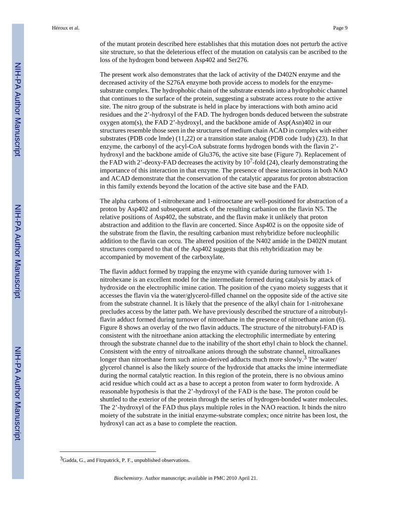
Crystal structures of D402N and S276A NAO with bound substratesFigure 2 shows representative electron density maps of the active sites in the aerobic complexesof the mutant enzymes with nitroalkanes. In both the D402N and the S276A NAO structures,the electron densities for FAD, the active site residues, and an exogenous ligand are allreasonably well resolved. These are all are in comparable positions in the different structuresdescribed here (Figure 3), establishing that the mutations do not significantly perturb the activesite structure. The electron density observed in each active site is consistent with the particularnitroalkane being present. This supports our conclusion from optical spectroscopy that we havetrapped an oxidized enzyme-substrate complex with both mutant proteins. The ability to trapthis complex with the S276A enzyme, despite the presence of Asp402, confirms the importanceof Ser276 in catalysis.
Figure 4 shows the typical interatomic distances between active site residues, the FAD, andthe substrates deduced from the structures described here. We note that each crystal structureexhibits a small degree of variability with respect to the substrate binding orientation withinthe four independent active sites of each holoenzyme. This variability is consistent with themoderate resolution of the structures reported here. The position of Arg409 is not significantlyaltered in any of the mutant structures, with its NH2 hydrogen-bonded to the backbone carbonylof the residue at position 402 and NH1 interacting with the carboxylate or amide of the sidechain at that position. Similarly, the side chain of Ser276 is not affected by the D402N mutation.In contrast, the amide moiety of asparagine in the D402N NAO structures is shifted slightlyfrom the position of the aspartate carboxylate in the wild-type and S276A enzymes, to a positioncloser to the flavin (Figure 3). The distance from the FAD N5 to Asp402 is similar in the wild-type and S276A enzymes (~6.8 Å); it is about 1 Å less in the D402N structures (~6.0 Å). Thereplacement of Asp402 with an asparagine also alters the interaction with Arg409, in that theasparagine side chain is ~0.4 Å closer to the arginine NH2 in the D402N structures than inS276A or wild-type NAO. While the altered position of Asn402 may indicate that the activesite base moves slightly when the substrate is bound, the difference may instead be due to thereplacement of a carboxylate with an amide and the resulting weaker interaction with Ser276.
The nitroalkane substrate occupies the same position in the structures of all three mutantproteins (Figure 3), clearly defining the binding mode of the substrate. In the structures ofD402N NAO, the alpha carbons of both 1-nitrohexane and 1-nitrooctane are 3.4 – 3.5 Å fromthe side chain of Asn402, an appropriate placement for proton abstraction. This distance isslightly longer, 3.7 Å, in the S276A enzyme structure. The alpha carbon of the nitroalkane isabout 3 Å from the flavin N 5 position in the D402N enzymes; this distance increases ~0.5 Åin the S276A enzyme. The two oxygen atoms of the nitro group of the substrates form multipleinteractions. In the D402N structures one substrate oxygen atom is 2.8 – 2.9 Å from both theamide moiety of Asn402 and the 2’-OH of the FAD. Both distances are increased significantlyin the S276A enzyme, to 3.2 Å and 3.6 Å, respectively, due to the different position of Asp402in this structure. The other oxygen atom of the substrate forms one good hydrogen bond (2.8– 2.9 Å) with the side chain of the residue at position 402 in all three mutant structures; thereis an additional interaction with the 2’OH of the FAD which is much stronger (2.7 Å) in theS276A enzyme than in either D402N structure (3.2 – 3.3 Å). In structures of all the mutantproteins and the wild-type enzyme the flavin 2’-hydroxyl is ~3.1 Å from a backbone amidenitrogen.
Héroux et al. Page 7
Biochemistry. Author manuscript; available in PMC 2010 April 21.
NIH
-PA Author Manuscript
NIH
-PA Author Manuscript
NIH
-PA Author Manuscript

The substrate alkyl moieties extend away from the flavin into a channel that eventually emergesfrom the protein surface. In crystals of the wild-type enzyme, this channel contained electrondensity consistent with a spermine molecule in some of the subunits (6). The obviousconclusion drawn from that earlier structure was that this channel is probably the substratebinding site. Spermine was omitted from the crystallization conditions in the present structures,and the observed substrate binding orientations confirm this hypothesis. Indeed, thenitroalkanes occupy the same position in the channel as did spermine in the earlier structure(Figure 5), clearly suggesting a substrate access path to the active site. As we reported earlier(6), the residues surrounding the hexyl and octyl chains are all hydrophobic: Leu80, I1e92,Val95, Ala96, Ala98, Leu99, Leu279, Leu279, Val280, Met283, Phe401. Thus, thehydrophobicity of this channel complements the aliphatic portion of the substrate ligands andis consistent with the higher kcat/KM values for longer chain aliphatic substrates. We also notethat in crystal structures of the wild-type enzyme and these mutants that are devoid of substrateand spermine, we observe electron density features in the channel of some subunits that canbe interpreted as a partially ordered PEG molecule (data not shown).
Crystal structure of wild-type NAO trapped as the cyanohexyl adductHigh resolution mass spectrometry was previously used to demonstrate formation of acyanohexyl-FAD during NAO turnover in the presence of cyanide (7). The electron density ofthe active site of the wild-type enzyme trapped by cyanide during turnover with 1-nitrohexaneis shown in Figure 2D. There is continuous electron density from the hexyl chain to the flavinN5, confirming the presence of a flavin adduct in these colorless crystals. The electron densityfor the CN projects above the re-face of the adduct; the hexyl side chain occupies the samespace as the substrate side chains in the structures of the mutant proteins, with the mostsignificant change being that of the position of the alpha carbon (Figure 3). The nitrogen atomof the cyano moiety is within hydrogen-bonding distance from the backbone amide of Asp402and the 2’-hydroxyl of the FAD. It is likely that the hydroxyl group in the tetrahedralintermediate formed during normal turnover is in an analogous position as the cyano moietyin the present structure. As noted previously for the wild-type enzyme (6), there is a secondchannel from the active site to the enzyme surface on the opposite face from the substratechannel. Although the side chain of Phe273 in NAO causes a constriction of the channel nearthe FAD, the overall channel is similar to that of the CoA binding site in acyl-CoAdehydrogenase family members. In the structures of NAO described previously and in thepresent structure, this channel contains several water molecules and a glycerol molecule, whichwas added as a cryoprotectant. In all of the structures reported here, there is a continuoushydrogen bond network of solvent molecules and glycerol from the FAD 2’-hydroxyl to theprotein surface (Figure 6).
DISCUSSIONThe role of Asp402 as the active site base in NAO was previously established by site-directedmutagenesis (3,4). Critically, the D402N enzyme is essentially inactive with neutralnitroalkanes, but has nearly wild-type activity when the substrate carbanion is used. In theactive site of NAO, Asp402 interacts with two other residues, Arg409 and Ser276. Theimportance of Arg409 for proton abstraction from the substrate by Asp402 has been confirmedby site-directed mutagenesis (13). The rate constant for CH bond cleavage by the R409Kenzyme is only 1% of the wild-type value. Direct structure determination of the mutant proteinestablished that this can be explained by the side chain of the lysyl residue being too short tointeract with the carboxylate of the aspartate, altering the position of the active site base. Thecharacterization of the S276A enzyme described here establishes that this residue is alsoimportant for proton abstraction by Asp402, in that the rate constant for removal of the substrateproton is also ~100-fold smaller for the mutant protein than the wild-type value. The structure
Héroux et al. Page 8
Biochemistry. Author manuscript; available in PMC 2010 April 21.
NIH
-PA Author Manuscript
NIH
-PA Author Manuscript
NIH
-PA Author Manuscript
of the mutant protein described here establishes that this mutation does not perturb the activesite structure, so that the deleterious effect of the mutation on catalysis can be ascribed to theloss of the hydrogen bond between Asp402 and Ser276.
The present work also demonstrates that the lack of activity of the D402N enzyme and thedecreased activity of the S276A enzyme both provide access to models for the enzyme-substrate complex. The hydrophobic chain of the substrate extends into a hydrophobic channelthat continues to the surface of the protein, suggesting a substrate access route to the activesite. The nitro group of the substrate is held in place by interactions with both amino acidresidues and the 2’-hydroxyl of the FAD. The hydrogen bonds deduced between the substrateoxygen atom(s), the FAD 2’-hydroxyl, and the backbone amide of Asp(Asn)402 in ourstructures resemble those seen in the structures of medium chain ACAD in complex with eithersubstrates (PDB code lmde) (11,22) or a transition state analog (PDB code 1udy) (23). In thatenzyme, the carbonyl of the acyl-CoA substrate forms hydrogen bonds with the flavin 2’-hydroxyl and the backbone amide of Glu376, the active site base (Figure 7). Replacement ofthe FAD with 2’-deoxy-FAD decreases the activity by 107-fold (24), clearly demonstrating theimportance of this interaction in that enzyme. The presence of these interactions in both NAOand ACAD demonstrate that the conservation of the catalytic apparatus for proton abstractionin this family extends beyond the location of the active site base and the FAD.
The alpha carbons of 1-nitrohexane and 1-nitrooctane are well-positioned for abstraction of aproton by Asp402 and subsequent attack of the resulting carbanion on the flavin N5. Therelative positions of Asp402, the substrate, and the flavin make it unlikely that protonabstraction and addition to the flavin are concerted. Since Asp402 is on the opposite side ofthe substrate from the flavin, the resulting carbanion must rehybridize before nucleophilicaddition to the flavin can occu. The altered position of the N402 amide in the D402N mutantstructures compared to that of the Asp402 suggests that this rehybridization may beaccompanied by movement of the carboxylate.
The flavin adduct formed by trapping the enzyme with cyanide during turnover with 1-nitrohexane is an excellent model for the intermediate formed during catalysis by attack ofhydroxide on the electrophilic imine cation. The position of the cyano moiety suggests that itaccesses the flavin via the water/glycerol-filled channel on the opposite side of the active sitefrom the substrate channel. It is likely that the presence of the alkyl chain for 1-nitrohexaneprecludes access by the latter path. We have previously described the structure of a nitrobutyl-flavin adduct formed during turnover of nitroethane in the presence of nitroethane anion (6).Figure 8 shows an overlay of the two flavin adducts. The structure of the nitrobutyl-FAD isconsistent with the nitroethane anion attacking the electrophilic intermediate by enteringthrough the substrate channel due to the inability of the short ethyl chain to block the channel.Consistent with the entry of nitroalkane anions through the substrate channel, nitroalkaneslonger than nitroethane form such anion-derived adducts much more slowly.3 The water/glycerol channel is also the likely source of the hydroxide that attacks the imine intermediateduring the normal catalytic reaction. In this region of the protein, there is no obvious aminoacid residue which could act as a base to accept a proton from water to form hydroxide. Areasonable hypothesis is that the 2’-hydroxyl of the FAD is the base. The proton could beshuttled to the exterior of the protein through the series of hydrogen-bonded water molecules.The 2’-hydroxyl of the FAD thus plays multiple roles in the NAO reaction. It binds the nitromoiety of the substrate in the initial enzyme-substrate complex; once nitrite has been lost, thehydroxyl can act as a base to complete the reaction.
3Gadda, G., and Fitzpatrick, P. F., unpublished observations.
Héroux et al. Page 9
Biochemistry. Author manuscript; available in PMC 2010 April 21.
NIH
-PA Author Manuscript
NIH
-PA Author Manuscript
NIH
-PA Author Manuscript

REFERENCES1. Fitzpatrick PF, Orville AM, Nagpal A, Valley MP. Nitroalkane oxidase, a carbanion-forming
flavoprotein homologous to Acyl-CoA dehydrogenase. Arch. Bicohem. Biophys 2005;433:157–165.2. Heasley CJ, Fitzpatrick PF. Kinetic mechanism and substrate specificity of nitroalkane oxidase.
Biochem. Biophys. Res. Commun 1996;225:6–10. [PubMed: 8769086]3. Valley MP, Fitzpatrick PF. Reductive half-reaction of nitroalkane oxidase: effect of mutation of the
active site aspartate to glutamate. Biochemistry 2003;42:5850–8566. [PubMed: 12741843]4. Valley MP, Fitzpatrick PF. Inactivation of nitroalkane oxidase upon mutation of the active site base
and rescue with a deprotonated substrate. J. Am. Chem. Soc 2003;23:8738–8739. [PubMed: 12862464]5. Gadda G, Edmondson RD, Russel DH, Fitzpatrick PF. Identification of the naturally occurring flavin
of nitroalkane oxidase from Fusarium oxysporum as a 5-nitrobutyl-FAD and conversion of the enzymeto the active FAD-containing form. J. Biol. Chem 1997;272:5563–5570. [PubMed: 9038163]
6. Nagpal A, Valley MP, Fitzpatrick PF, Orville AM. Crystal structures of nitroalkane oxidase: insightsinto the reaction mechanism from a covalent complex of the flavoenzyme trapped during turnover.Biochemistry 2006;45:1138–1150. [PubMed: 16430210]
7. Valley MP, Tichy SE, Fitzpatrick PF. Establishing the kinetic competency of the cationic imineintermediate in nitroalkane oxidase. J. Am. Chem. Soc 2005;127:2062–2066. [PubMed: 15713081]
8. Massey V. Activation of molecular oxygen by flavins and flavoproteins. J.Biol.Chem 1994;269:22459–22462. [PubMed: 8077188]
9. Gadda G, Choe DY, Fitzpatrick PF. Use of pH and kinetic isotope effects to dissect the effects ofsubstrate size on binding and catalysis by nitroalkane oxidase. Arch. Bicohem. Biophys 2000;382:138–144.
10. Daubner SC, Gadda G, Valley MP, Fitzpatrick PF. Cloning of nitroalkane oxidase from Fusariumoxysporum identifies a new member of the acyl-CoA dehydrogenase superfamily. Proc. Nat. Acad.Sci 2002;99:2702–2707. [PubMed: 11867731]
11. Kim JJ, Miura R. Acyl-CoA dehydrogenases and acyl-CoA oxidases. Structural basis for mechanisticsimilarities and differences. Eur. J. Biochem 2004;271:483–493. [PubMed: 14728675]
12. Ghisla S, Thorpe C. Acyl-CoA dehydrogenases. A mechanistic overview. Eur. J. Biochem2004;271:494–508. [PubMed: 14728676]
13. Fitzpatrick PF, Valley MP, Bozinovski DM, Shaw PG, Héroux A, Orville AM. Mechanistic andStructural Analyses of the Roles of Arg409 and Asp402 in the Reaction of the FlavoproteinNitroalkane Oxidase. Biochemistry 2007;46:13800–13808. [PubMed: 17994768]
14. Nagpal A, Valley MP, Fitzpatrick PF, Orville AM. Crystallization and preliminary analysis of activenitroalkane oxidase in three crystal forms. Acta Crystallogr D Biol Crystallogr 2004;60:1456–1460.[PubMed: 15272176]
15. Otwinowski Z, Minor W. Processing of X-ray diffraction data collected in oscillation mode. MethodsEnzymol 1997;276:307–326.
16. CollaborativeComputationalProject. The CCP4 suite: Programs for protein crystallography. ActaCryst.allogr. D Biol. Crystallogr 1994;50:760–763.
17. Emsley P, Cowtan K. Coot: model-building tools for molecular graphics. Acta Crystallogr D BiolCrystallogr 2004;60:2126–2132. [PubMed: 15572765]
18. Orville AM, Lountos GT, Finnegan S, Gadda G, Prabhakar R. Crystallographic, spectroscopic, andcomputational analysis of a flavin C4a-oxygen adduct in choline oxidase. Biochemistry2009;48:720–728. [PubMed: 19133805]
19. Gadda G, Fitzpatrick PF. Substrate specificity of a nitroalkane oxidizing enzyme. Arch. Bicohem.Biophys 1999;363:309–313.
20. Gadda G, Fitzpatrick PF. Biochemical and physical characterization of the active FAD-containingform of nitroalkane oxidase from Fusarium oxysporum. Biochemistry 1998;37:6154–6164.[PubMed: 9558355]
21. Siddiqui MSU, Stanley RJ. A cryogenic optical waveguide spectrometer for the measurement of low-temperature absorption spectra of dilute biological samples. Anal. Bioch 2005;337:121–129.
Héroux et al. Page 10
Biochemistry. Author manuscript; available in PMC 2010 April 21.
NIH
-PA Author Manuscript
NIH
-PA Author Manuscript
NIH
-PA Author Manuscript
22. Kim J-JP, Wang M, Paschke R. Crystal structures of medium-chain acyl-CoA dehydrogenase frompig liver mitochondria with and without substrate. Proc. Nat. Acad. Sci 1993;90:7523–7527.[PubMed: 8356049]
23. Satoh A, Nakajima Y, Miyahara I, Hirotsu K, Tanaka T, Nishina Y, Shiga K, Tamaoki H, SetoyamaC, Miura R. Structure of the transition state analog of medium-chain acyl-CoA dehydrogenase.Crystallographic and molecular orbital studies on the charge-transfer complex of medium-chain acyl-CoA dehydrogenase with 3-thiaoctanoyl-CoA. J. Biochem. (Tokyo) 2003;134:297–304. [PubMed:12966080]
24. Engst S, Vock P, Wang M, Kim J-JP, Ghisla S. Mechanism of activation of acyl-CoA substrates bymedium chain acyl-CoA dehydrogenase: interaction of the thioester carbonyl with the flavin adeninedinucleotide ribityl side chain. Biochemistry 1999;38:257–267. [PubMed: 9890906]
Héroux et al. Page 11
Biochemistry. Author manuscript; available in PMC 2010 April 21.
NIH
-PA Author Manuscript
NIH
-PA Author Manuscript
NIH
-PA Author Manuscript

Figure 1.Optical absorbance spectra of NAO in solution or from single crystals. The bottom blackspectrum is of 280 µM wild-type NAO in 500 mM cacodylic acid buffer, pH 7.6, at roomtemperature. The top black spectrum was obtained at 100 K from a single crystal of D402NNAO cocrystallized with 1-nitrooctane. The red and blue spectra in the center are from oxidizedwild-type NAO at 100 K obtained from opposite orientations of the cryoloop with respect tothe spectroscopic axis. To better facilitate comparisons, the three single crystal spectra havebeen normalized for the same absorbance at 600 nm, The oxidized wild-type NAO crystal inthe orientation from which the red spectrum was obtained is shown in the inset. In this viewthe crystal measures approximately 250 µm in the horizontal and 75 µm in the verticaldirections. It is approximately in the center of the nylon loop, which measures approximately235 µm in the vertical direction. The spectroscopic spot size was focused to approximately 25µm at the intersection of the dashed cross-hairs.
Héroux et al. Page 12
Biochemistry. Author manuscript; available in PMC 2010 April 21.
NIH
-PA Author Manuscript
NIH
-PA Author Manuscript
NIH
-PA Author Manuscript
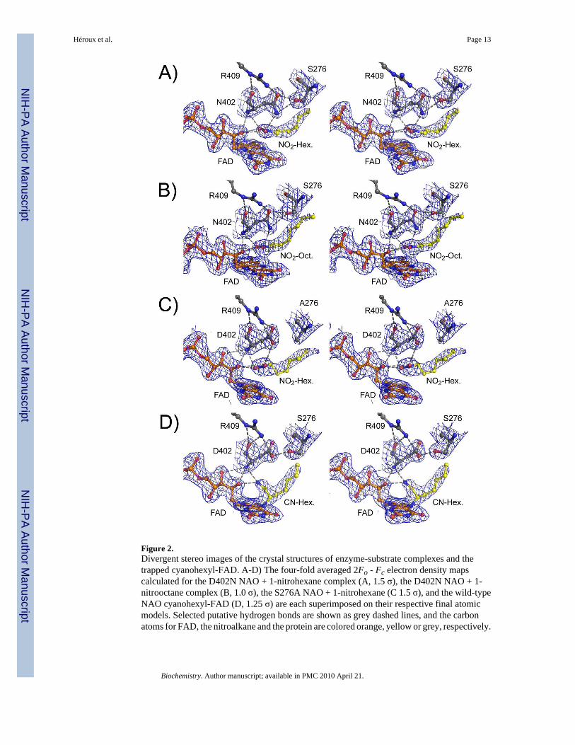
Figure 2.Divergent stereo images of the crystal structures of enzyme-substrate complexes and thetrapped cyanohexyl-FAD. A-D) The four-fold averaged 2Fo - Fc electron density mapscalculated for the D402N NAO + 1-nitrohexane complex (A, 1.5 σ), the D402N NAO + 1-nitrooctane complex (B, 1.0 σ), the S276A NAO + 1-nitrohexane (C 1.5 σ), and the wild-typeNAO cyanohexyl-FAD (D, 1.25 σ) are each superimposed on their respective final atomicmodels. Selected putative hydrogen bonds are shown as grey dashed lines, and the carbonatoms for FAD, the nitroalkane and the protein are colored orange, yellow or grey, respectively.
Héroux et al. Page 13
Biochemistry. Author manuscript; available in PMC 2010 April 21.
NIH
-PA Author Manuscript
NIH
-PA Author Manuscript
NIH
-PA Author Manuscript
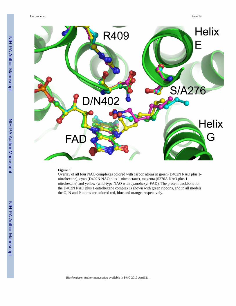
Figure 3.Overlay of all four NAO complexes colored with carbon atoms in green (D402N NAO plus 1-nitrohexane), cyan (D402N NAO plus 1-nitrooctane), magenta (S276A NAO plus 1-nitrohexane) and yellow (wild-type NAO with cyanohexyl-FAD). The protein backbone forthe D402N NAO plus 1-nitrohexane complex is shown with green ribbons, and in all modelsthe O, N and P atoms are colored red, blue and orange, respectively.
Héroux et al. Page 14
Biochemistry. Author manuscript; available in PMC 2010 April 21.
NIH
-PA Author Manuscript
NIH
-PA Author Manuscript
NIH
-PA Author Manuscript

Héroux et al. Page 15
Biochemistry. Author manuscript; available in PMC 2010 April 21.
NIH
-PA Author Manuscript
NIH
-PA Author Manuscript
NIH
-PA Author Manuscript
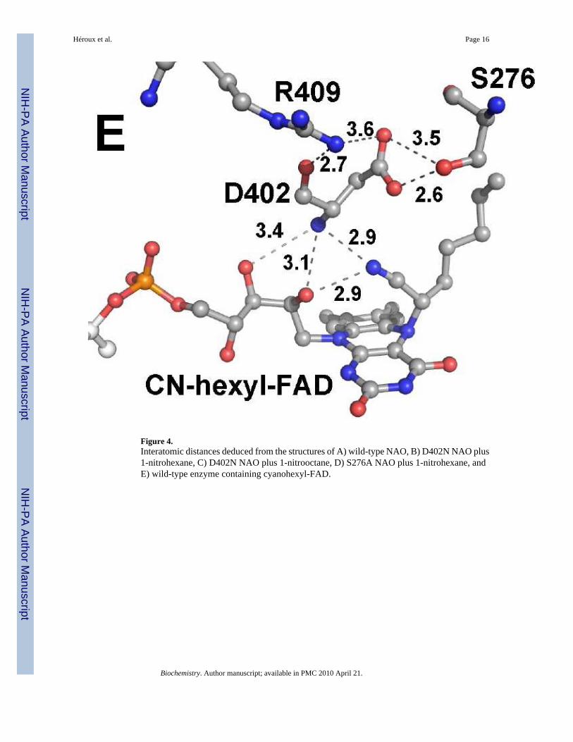
Figure 4.Interatomic distances deduced from the structures of A) wild-type NAO, B) D402N NAO plus1-nitrohexane, C) D402N NAO plus 1-nitrooctane, D) S276A NAO plus 1-nitrohexane, andE) wild-type enzyme containing cyanohexyl-FAD.
Héroux et al. Page 16
Biochemistry. Author manuscript; available in PMC 2010 April 21.
NIH
-PA Author Manuscript
NIH
-PA Author Manuscript
NIH
-PA Author Manuscript
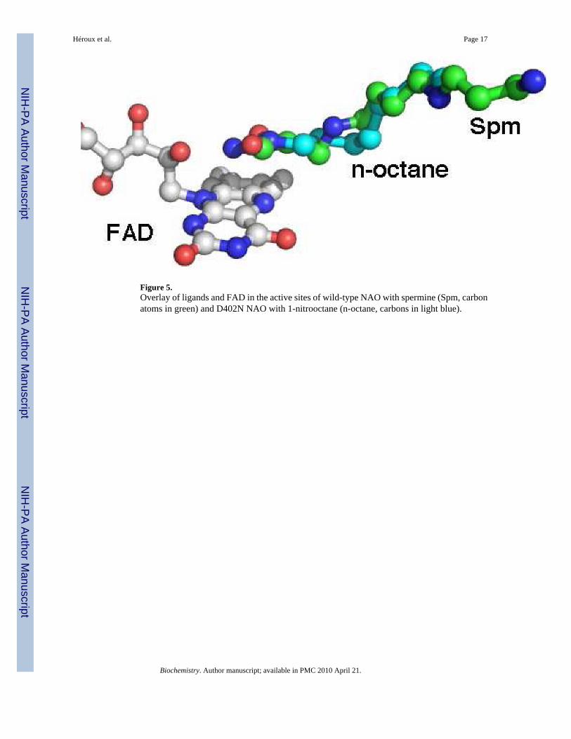
Figure 5.Overlay of ligands and FAD in the active sites of wild-type NAO with spermine (Spm, carbonatoms in green) and D402N NAO with 1-nitrooctane (n-octane, carbons in light blue).
Héroux et al. Page 17
Biochemistry. Author manuscript; available in PMC 2010 April 21.
NIH
-PA Author Manuscript
NIH
-PA Author Manuscript
NIH
-PA Author Manuscript

Figure 6.Channel of water molecules from the active site to the surface from the cyanohexyl-FADstructure: left, deduced hydrogen bonding interactions, with the carbon atoms from the hexylmoiety in yellow; right, surface rendering to illustrate the channel. The orientation of this viewis rotated approximately 180 degrees relative to all other figures. The view of the surfacerendering is from the exterior toward the glycerol molecule.
Héroux et al. Page 18
Biochemistry. Author manuscript; available in PMC 2010 April 21.
NIH
-PA Author Manuscript
NIH
-PA Author Manuscript
NIH
-PA Author Manuscript
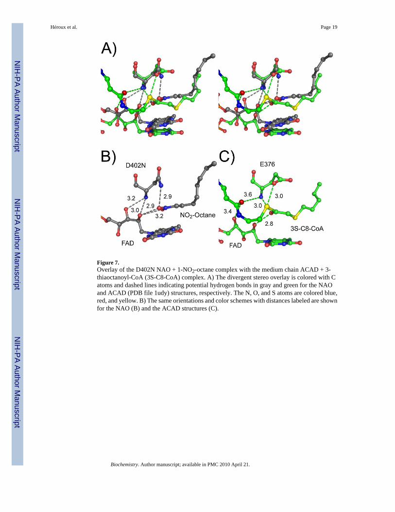
Figure 7.Overlay of the D402N NAO + 1-NO2-octane complex with the medium chain ACAD + 3-thiaoctanoyl-CoA (3S-C8-CoA) complex. A) The divergent stereo overlay is colored with Catoms and dashed lines indicating potential hydrogen bonds in gray and green for the NAOand ACAD (PDB file 1udy) structures, respectively. The N, O, and S atoms are colored blue,red, and yellow. B) The same orientations and color schemes with distances labeled are shownfor the NAO (B) and the ACAD structures (C).
Héroux et al. Page 19
Biochemistry. Author manuscript; available in PMC 2010 April 21.
NIH
-PA Author Manuscript
NIH
-PA Author Manuscript
NIH
-PA Author Manuscript

Figure 8.Overlay of NAO flavin adducts formed during turnover of nitrohexane in the presence ofcyanide (light gray carbon atoms) and from a mixture of neutral and anionic nitroethane (lightblue carbon atoms, pdb file 2cou).
Héroux et al. Page 20
Biochemistry. Author manuscript; available in PMC 2010 April 21.
NIH
-PA Author Manuscript
NIH
-PA Author Manuscript
NIH
-PA Author Manuscript

Scheme 1.
Héroux et al. Page 21
Biochemistry. Author manuscript; available in PMC 2010 April 21.
NIH
-PA Author Manuscript
NIH
-PA Author Manuscript
NIH
-PA Author Manuscript
NIH
-PA Author Manuscript
NIH
-PA Author Manuscript
NIH
-PA Author Manuscript
Héroux et al. Page 22
Table 1Crystal structure data collection and refinement statistics
D402N NAO D402N NAO S276A NAO Wild-type NAO
PDB code 3D9D 3D9E 3D9F 3D9G
Substrate Ligand(s) 1-NO2-hexane 1-NO2-octane 1-NO2-hexane cyanide + 1-NO2-hexane
Wavelength (Å) 1.1000 1.0809 0.9798 1.0809
Space group P3221 P3221 P3221 P3221
a (Å), b (Å), c (Å) 107.9, 107.9, 338.4 108.0, 108.0, 338.6 108.3, 108.3, 339.4 109.3, 109.3, 343.9
Resolution range (Å) 49 - 2.10 49 - 2.20 49 - 2.20 49 - 2.15
High resolution shell (Å) 2.80 - 2.10 2.28 - 2.20 2.28 - 2.20 2.23 - 2.15
Total observations 391271 602598 490262 803662
Unique observations 123094 110228 115243 122023
Completeness (%) 91.9 (62.1)a 93.8 (64.9) 97.4 (83.2) 94.2 (64.9)
Redundancy 3.2 (1.6)a 5.5 (2.4) 4.3 (4.0) 6.6 (1.8)
Rsym (%)b 8.9 (44.7)a 6.0 (43.4) 6.9 (55.2) 10.4 (28.5)
I / σ I 13.7 (1.3)a 23.2 (1.4) 26.3 (2.2) 27.4 (1.92)
Model refinement statistics
Total number of non-hydrogenatoms
14165 13772 13974 14353
No. residues protein/ligand/water 1724/16/650 1723/12/273 1722/16/468 1721/6/840
Resolution range (Å) 50 - 2.1 50 - 2.2 50 - 2.2 50 - 2.15
No. of reflections 116861 104709 109423 115721
Rcryst (%)c 18.0 (26.7)a 19.3 (27.4)a 20.2 (27.0)a 19.6 (24.3)a
Rfree (%)d 21.9 (32.7)a 23.3 (33.7)a 24.1 (30.5)a 23.3 (30.2)a
Estimated overall coordinateerror based on R-value (Å)
0.192 0.232 0.231 0.216
Correlation coefficient (Fo - Fc) 0.957 0.956 0.955 0.954
Ramachandran statistics
Most favored 1377 (91.5%) 1399 (92.8%) 1377 (91.6%) 1394 (92.6%)
Additional allowed 118 (7.8%) 100 (6.6%) 117 (7.7%) 103 (6.8%)
Generously allowed 4 (0.3%) 4 (0.3%) 6 (0.4%) 3 (0.2%)
Disallowed 6 (0.4%) 4 (0.3%) 4 (0.3%) 5 (0.3%)
Average B values (Å2)
Protein 36.07 42.2 44.0 38.0
Water 38.8 38.4 43.0 40.8
Substrate ligand 50.2 60.2 55.2 --
FAD 29.7 34.4 38.7 32.2
R.m.s.d. from ideal geometry
Bond lengths (Å) 0.014 0.011 0.012 0.010
Bond angles (˚) 1.53 1.35 1.35 1.30
aHighest resolution shell.
Biochemistry. Author manuscript; available in PMC 2010 April 21.
NIH
-PA Author Manuscript
NIH
-PA Author Manuscript
NIH
-PA Author Manuscript
Héroux et al. Page 23
bΣ(|Ihkl - <Ihkl>|) / Σ Ihkl
cR-value calculated with the working and the test set inclusive.
dFree R-value test set calculated with 5% of the data.
Biochemistry. Author manuscript; available in PMC 2010 April 21.

NIH
-PA Author Manuscript
NIH
-PA Author Manuscript
NIH
-PA Author Manuscript
Héroux et al. Page 24
Table 2Kinetic parameters for wild-type and S276A nitroalkane oxidase.
Substrate Kinetic parameter Wild-type NAO S276A NAO
Nitroethanea kcat(nitroethane) (s−1) 15 ± 1 1.0 ± 0.01
kcat/K(nitroethane) (mM−1s−1) 6.3 ± 0.4 0.30 ± 0.02
K(nitroethane) (mM) 2.3 ± 0.2 3.3 ± 0.2
Ki(nitroethane) (mM) 25 ± 3 -D(kcat/Km) 9.2 ± 1.1 9.7 ± 2.0Dkcat(nitroethane) 1.4 ± 0.2 2.1 ± 0.4
kred (s−1) 247 ± 5 3.4 ± 0.4
1-nitrohexane kcat(nitrohexane) (s−1) 2.0 ± 0.1 0.30 ± 0.006
kcat/Knitrohexane (mM−1s−1) 47 ± 10 1.5 ± 0.2
Knitrohexane (mM) 0.04 ± 0.01 0.20 ± 0.02
Ki(nitrohexane) (mM) - 130 ± 25
1-nitrooctane kcat(nitrooctane) (s−1) 4.4 ± 0.4 0.2 ± 0.01
kcat/K nitrooctane (mM−1 s−1) 150 ± 46 1.3 ± 0.2
K nitrooctane (mM) 0.03 ± 0.01 0.3 ± 0.02
Ki(nitrooctane) (mM) 8.3 ± 3.7 26 ± 5
aData for the wild-type enzyme are from Valley and Fitzpatrick (3).
Biochemistry. Author manuscript; available in PMC 2010 April 21.
Related Documents