Cell, Vol. 91, 973–983, December 26, 1997, Copyright 1997 by Cell Press Molecular Basis of Sulfite Oxidase Deficiency from the Structure of Sulfite Oxidase enzymes have been sequenced. The enzyme is located in the mitochondrial intermembrane space and was found to be a homodimer with a molecular mass be- Caroline Kisker,* § Hermann Schindelin,* Andrew Pacheco, ² William A. Wehbi, ² Robert M. Garrett, ‡ K.V. Rajagopalan, ‡§ tween 101 and 110 kDa. The rat enzyme can be proteo- John H. Enemark, ²§ and D.C. Rees* lytically cleaved by trypsin into two domains (Johnson * Howard Hughes Medical Institute and Rajagopalan, 1977), a smaller N-terminal domain and Division of Chemistry and Chemical with a molecular mass of about 10 kDa containing a b5 Engineering cytochrome and a larger C-terminal domain of about 147-75CH 42 kDa, which retains the dimeric structure and har- California Institute of Technology bors the molybdenum cofactor. The sequence identity Pasadena, California 91125 among the eukaryotic sulfite oxidases is very high, with ² Department of Chemistry 68% identity observed between the chicken and human University of Arizona enzymes, and 88% between the rat and human en- Tucson, Arizona 85721 zymes. ‡ Department of Biochemistry In humans, genetic deficiency of sulfite oxidase can Duke University Medical Center occur for either of two reasons (Johnson and Wadman, Durham, North Carolina 27710 1989). The first is a defect in the synthesis of the Mo- co, which also affects the two other Mo-co-containing enzymes in humans, xanthine dehydrogenase and alde- Summary hyde oxidase. The second affects sulfite oxidase activity alone and is caused by mutations in the gene encoding The molybdenum-containing enzyme sulfite oxidase sulfite oxidase. Molybdenum cofactor deficiency and catalyzes the conversion of sulfite to sulfate, the termi- isolated sulfite oxidase deficiency have been described nal step in the oxidative degradation of cysteine and in several patients. Individuals suffering from either methionine. Deficiency of this enzyme in humans usu- genetic defect exhibit identical symptoms, including ally leads to major neurological abnormalities and severe neurological abnormalities, dislocated ocular early death. The crystal structure of chicken liver sul- lenses, mental retardation, and in several cases, attenu- fite oxidase at 1.9 A ˚ resolution reveals that each mono- ated growth of the brain. This observation led to the mer of the dimeric enzyme consists of three domains. conclusion that the pathophysiology of cofactor defi- At the active site, the Mo is penta-coordinated by three ciency is due to the absence of sulfite oxidase activity. sulfur ligands, one oxo group, and one water/hydroxo. It is not clear whether the brain damage occurs as a A sulfate molecule adjacent to the Mo identifies the result of toxic levels of sulfite, the absence of sulfate, substrate binding pocket. Four variants associated or a combination of both. with sulfite oxidase deficiency have been identified: Amino acid sequence comparisons have revealed that two mutations are near the sulfate binding site, while sulfite oxidase is related to the heme-containing assimi- the other mutations occur within the domain mediating latory nitrate reductases. Nitrate reductases catalyze dimerization. the reduction of nitrate to nitrite in algae, fungi, and higher plants, which is a critical step in the global nitro- Introduction gen cycle. As observed with sulfite oxidase, nitrate re- ductases form homodimers with a molecular weight of Sulfite oxidase catalyzes the physiologically vital oxida- z220 kDa, but contain three cofactors within each sub- tion of sulfite to sulfate, the terminal reaction in the unit. In the case of spinach nitrate reductase, the oxidative degradation of the sulfur containing amino N-terminal domain with a molecular mass of z59 kDa acids cysteine and methionine. Sulfite oxidase belongs contains the Mo-co, a small central domain (z14 kDa) to the molybdenum cofactor (Mo-co) containing family contains a b-type heme, and the C-terminal domain (z30 of enzymes that are characterized by the presence of a kDa) contains FAD and a NAD(P) 1 binding site (Kubo mononuclear Mo atom coordinated to the sulfur atoms et al., 1988). Sequence comparisons between nitrate of a pterin derivative, named molybdopterin. Enzymes reductase and sulfite oxidase revealed 31% identity containing the Mo-co catalyze important transforma- (Wootton et al., 1991) between the N-terminal domain tions in the global sulfur, nitrogen, and carbon cycles of chicken liver sulfite oxidase (Neame and Barber, 1989) (Hille, 1996). The overall reaction catalyzed by most Mo- and the heme domain of assimilatory nitrate reductase co containing enzymes, including sulfite oxidase, con- from Arabidopsis thaliana (Crawford et al., 1988). Be- sists of the net transfer of an oxygen atom between tween the Mo-co-containing domains, the sequence ho- the substrate and water in a two-electron oxidation- mology was found to be even more significant, with 38% reduction reaction. identity observed between chicken liver sulfite oxidase Sulfite oxidase is ubiquitous among animals, and the and nitrate reductases from higher plants (Neame and human (Garrett et al., 1995), rat (Garrett and Rajagopa- Barber, 1989; Wootton et al., 1991). lan, 1994), and chicken liver (Neame and Barber, 1989) In an attempt to understand the architecture of the eukaryotic active site harboring the molybdenum co- factor in sulfite oxidase and nitrate reductase and to § To whom correspondence should be addressed.

Welcome message from author
This document is posted to help you gain knowledge. Please leave a comment to let me know what you think about it! Share it to your friends and learn new things together.
Transcript

Cell, Vol. 91, 973–983, December 26, 1997, Copyright 1997 by Cell Press
Molecular Basis of Sulfite Oxidase Deficiencyfrom the Structure of Sulfite Oxidase
enzymes have been sequenced. The enzyme is locatedin the mitochondrial intermembrane space and wasfound to be a homodimer with a molecular mass be-
Caroline Kisker,*§ Hermann Schindelin,*Andrew Pacheco,† William A. Wehbi,†Robert M. Garrett,‡ K.V. Rajagopalan,‡§
tween 101 and 110 kDa. The rat enzyme can be proteo-John H. Enemark,†§ and D.C. Rees*lytically cleaved by trypsin into two domains (Johnson*Howard Hughes Medical Instituteand Rajagopalan, 1977), a smaller N-terminal domainand Division of Chemistry and Chemicalwith a molecular mass of about 10 kDa containing a b5Engineeringcytochrome and a larger C-terminal domain of about147-75CH42 kDa, which retains the dimeric structure and har-California Institute of Technologybors the molybdenum cofactor. The sequence identityPasadena, California 91125among the eukaryotic sulfite oxidases is very high, with†Department of Chemistry68% identity observed between the chicken and humanUniversity of Arizonaenzymes, and 88% between the rat and human en-Tucson, Arizona 85721zymes.‡Department of Biochemistry
In humans, genetic deficiency of sulfite oxidase canDuke University Medical Centeroccur for either of two reasons (Johnson and Wadman,Durham, North Carolina 277101989). The first is a defect in the synthesis of the Mo-co, which also affects the two other Mo-co-containingenzymes in humans, xanthine dehydrogenase and alde-Summaryhyde oxidase. The second affects sulfite oxidase activityalone and is caused by mutations in the gene encodingThe molybdenum-containing enzyme sulfite oxidasesulfite oxidase. Molybdenum cofactor deficiency andcatalyzes the conversionof sulfite to sulfate, the termi-isolated sulfite oxidase deficiency have been describednal step in the oxidative degradation of cysteine andin several patients. Individuals suffering from eithermethionine. Deficiency of this enzyme in humans usu-genetic defect exhibit identical symptoms, includingally leads to major neurological abnormalities andsevere neurological abnormalities, dislocated ocularearly death. The crystal structure of chicken liver sul-lenses, mental retardation, and in several cases, attenu-fite oxidase at1.9 A resolution reveals that each mono-ated growth of the brain. This observation led to themer of the dimeric enzyme consists of three domains.conclusion that the pathophysiology of cofactor defi-At theactive site, the Mois penta-coordinated by threeciency is due to the absence of sulfite oxidase activity.sulfur ligands, one oxo group, and one water/hydroxo.It is not clear whether the brain damage occurs as aA sulfate molecule adjacent to the Mo identifies theresult of toxic levels of sulfite, the absence of sulfate,substrate binding pocket. Four variants associatedor a combination of both.with sulfite oxidase deficiency have been identified:
Amino acid sequence comparisons have revealed thattwo mutations are near the sulfate binding site, whilesulfite oxidase is related to the heme-containing assimi-theother mutations occur within the domain mediatinglatory nitrate reductases. Nitrate reductases catalyzedimerization.the reduction of nitrate to nitrite in algae, fungi, andhigher plants, which is a critical step in the global nitro-
Introductiongen cycle. As observed with sulfite oxidase, nitrate re-ductases form homodimers with a molecular weight of
Sulfite oxidase catalyzes the physiologically vital oxida- z220 kDa, but contain three cofactors within each sub-tion of sulfite to sulfate, the terminal reaction in the
unit. In the case of spinach nitrate reductase, theoxidative degradation of the sulfur containing amino
N-terminal domain with a molecular mass of z59 kDaacids cysteine and methionine. Sulfite oxidase belongs
contains the Mo-co, a small central domain (z14 kDa)to the molybdenum cofactor (Mo-co) containing family contains a b-type heme, and the C-terminal domain (z30of enzymes that are characterized by the presence of a kDa) contains FAD and a NAD(P)1 binding site (Kubomononuclear Mo atom coordinated to the sulfur atoms et al., 1988). Sequence comparisons between nitrateof a pterin derivative, named molybdopterin. Enzymes reductase and sulfite oxidase revealed 31% identitycontaining the Mo-co catalyze important transforma- (Wootton et al., 1991) between the N-terminal domaintions in the global sulfur, nitrogen, and carbon cycles of chicken liver sulfite oxidase (Neame and Barber, 1989)(Hille, 1996). The overall reaction catalyzed by most Mo- and the heme domain of assimilatory nitrate reductaseco containing enzymes, including sulfite oxidase, con- from Arabidopsis thaliana (Crawford et al., 1988). Be-sists of the net transfer of an oxygen atom between tween the Mo-co-containing domains, the sequence ho-the substrate and water in a two-electron oxidation- mology was found to be even more significant, with 38%reduction reaction. identity observed between chicken liver sulfite oxidase
Sulfite oxidase is ubiquitous among animals, and the and nitrate reductases from higher plants (Neame andhuman (Garrett et al., 1995), rat (Garrett and Rajagopa- Barber, 1989; Wootton et al., 1991).lan, 1994), and chicken liver (Neame and Barber, 1989) In an attempt to understand the architecture of the
eukaryotic active site harboring the molybdenum co-factor in sulfite oxidase and nitrate reductase and to§To whom correspondence should be addressed.

Cell974
Table 1. Data Collection and MIR Statistics
NAT1 NAT2 EMTS PIP AU LU SM
dmin (A) 3.0 1.9 2.8 2.8 3.0 2.8 3.0Completeness (Last Shell) 0.818 (0.371) 0.892 (0.662) 0.910 (0.804) 0.897 (0.760) 0.870 (0.528) 0.968 (0.840) 0.845 (0.560)Redundancy (Last Shell) 2.5 (2.1) 2.1 (1.8) 2.1 (1.9) 1.8 (1.5) 2.2 (1.7) 2.1 (1.7) 1.7 (1.6)Rsym (Last Shell) 0.077 (0.212) 0.060 (0.160) 0.056 (0.115) 0.060 (0.117) 0.088 (0.164) 0.057 (0.120) 0.082 (0.198)Rderiv — — 0.166 0.237 0.142 0.140 0.153No. of Sites — — 4 8 4 1 3Sites — — A–D A–H A–D I A, B, IPP — — 1.25 1.19 1.16 0.35 1.02FOM — — 0.490 (to 3.0 A resolution)
Rsym 5 ShklSi|Ii 2 ,I.|/ShklSi,I. where Ii is the ith measurement and ,I. is the weighted mean of all measurements of I. Rderiv 5 Shkl||FPH|2|FP||/Shkl|FP| where FP and FPH are the native and heavy atom derivative structure factor amplitude. Phasing power (PP) is the mean value of theheavy atom structure factor amplitude divided by the residual lack of closure error.
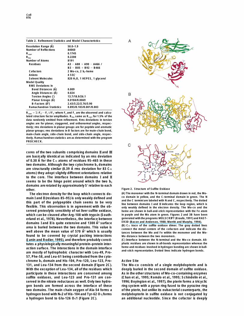
characterize the molecular basis of sulfite oxidase defi- Fold of the MonomerThe fold of sulfite oxidase can be described as a mixedciency, we have determined the three-dimensionala 1 b structure and is clearly divided into three domainsstructure of chicken liver sulfite oxidase at 1.9 A resolu-(Figure 2A). The N-terminal domain (residues 3–84) istion and identified the sites of substitutions in four vari-structurally similar to the fold of bovine cytochrome b5ants with reduced activity.and comprises a three-stranded antiparallel b sheet (b3–b5) and six a helices. The three-stranded b sheet isflanked by two very short b strands, b1 and b2. Five aResults and Discussionhelices (a2–a6) and the b sheet form a hydrophobiccrevice for the heme group. Although not covalentlyStructure Determinationattached to the protein, the heme is deeply buried inSulfite oxidase was isolated from chicken liver in its fullythis crevice with the vinyl groups pointing toward the boxidized state (Mo[VI]/Fe[III]) and crystallized by vaporsheet and the propionate side chains solvent-exposed.diffusion methods and microseeding techniques (seeA superposition of 59 Ca atoms of bovine cytochromeExperimental Procedures). The structure was solved byb5 (pdb entry 3b5c) (Mathews et al., 1972) and domainmultiple isomorphous replacement with five heavy atomI of sulfite oxidase results in a root-mean-square devia-derivatives (Table 1) and subsequent two-fold noncrys-tion of 0.86 A. The heme iron atom in sulfite oxidase istallographic symmetry (NCS) averaging and solvent flat-symmetrically coordinated by the Ne2 atoms of His-40tening. The electron density at 3.0 A resolution was ofand His-65, and the four heme pyrrole nitrogen atomsmoderate quality and showed as highest peaks the posi-(average metal-ligand distance of 2.0 A), resulting intions of both molybdenum atoms and only one of theapproximately octahedral coordination geometry.two heme iron atoms. Iterative rounds of model building,
Domain II (residues 96–323) contains a total of thirteenaveraging, and solvent flattening yielded a model com-b strands organized in three b sheets and nine a helices.prising residues 3–88 and 90–466 for one monomer andThe molybdenum cofactor is bound at the center of thisresidues 3–85 and 92–466 for the second monomer. Thedomain and is contacted by discontinous stretches ofelectron density for residue 89 in the first monomer andthe polypeptide chain. In order to analyze the fold of thisfor residues 86–91 in the second monomer was verydomain, a comparison to a total of 872 protein structures
weakly defined, and therefore, these residues were notwas performed using the program DALI (Holm and
included in the refinement. The model of each monomerSander, 1995). None of the structures had a Z-score
contains two cofactors, one b5-type heme and one Mo-greater than 2, indicating that this domain comprises a
co. During the refinement, several discrepancies werefold that has not been observed previously. At the N
observed between the electron density map and the terminus of this domain, a three-stranded antiparallel bpublished amino acid sequence, which was determined sheet is formed (b6–b8), which leads into a mixed five-by Edman degradation (Neame and Barber, 1989), and stranded b sheet (b9, b10, b13, b14, and b18) locatedchanges in the sequence were made accordingly (Figure on the opposite side of the domain. This five-stranded1). One difference, a deletion of an Arg residue at posi- b sheet is flanked by five short a helices. The center oftion 153 is noteworthy since it decreases all following the domain comprises a long b hairpin (b11 and b12),residue numbers by one. After Arg-459, seven additional which together with b15 and b16/b17 forms a four-residues were observed. These residues are part of a b stranded antiparallel b sheet. Residues 186–196, whichstrand (b26), and their insertion reveals a highly con- are located between b11 and b12 and include a shortserved stretch of nine residues that is now present at a helix (a10), grasp the third domain and are responsiblethe C terminus of all sulfite oxidases. The structure was for several polar and hydrophobic interactions betweenrefined to a working and free R factor of 17.5% and the two domains.22.0%, respectively, including all data between 50.0 A The core of the C-terminal domain (residues 324–466)and 1.9 A resolution (Table 2). The resulting model has is characterized by seven b strands arranged in twogood stereochemistry with 89.3% of all residues in the antiparallel b sheets. The first sheet contains strandsmost favored regions of the Ramachandran diagram and b19, b21, and b24, and the second b22, b23, b25, and
b26. The topology of the two sheets can be describedonly two outliers, Arg-21 in both monomers.
Crystal Structure of Sulfite Oxidase975
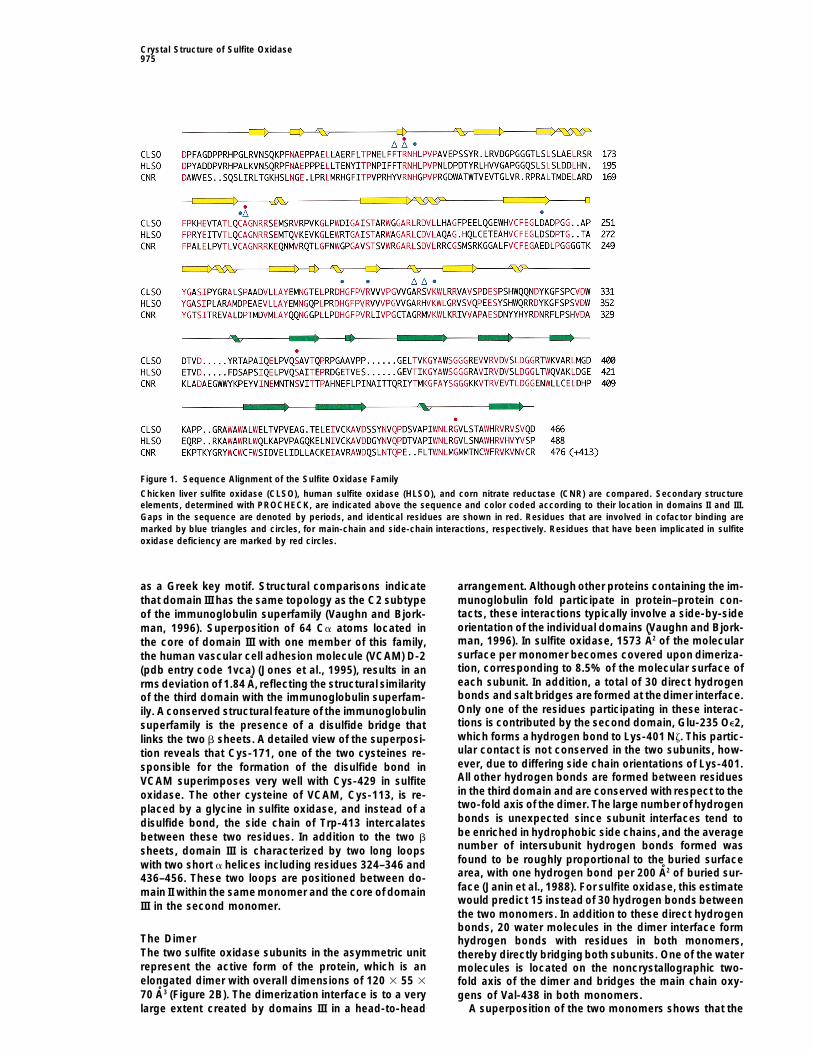
Figure 1. Sequence Alignment of the Sulfite Oxidase Family
Chicken liver sulfite oxidase (CLSO), human sulfite oxidase (HLSO), and corn nitrate reductase (CNR) are compared. Secondary structureelements, determined with PROCHECK, are indicated above the sequence and color coded according to their location in domains II and III.Gaps in the sequence are denoted by periods, and identical residues are shown in red. Residues that are involved in cofactor binding aremarked by blue triangles and circles, for main-chain and side-chain interactions, respectively. Residues that have been implicated in sulfiteoxidase deficiency are marked by red circles.
as a Greek key motif. Structural comparisons indicate arrangement. Although other proteins containing the im-munoglobulin fold participate in protein–protein con-that domain III has the same topology as the C2 subtypetacts, these interactions typically involve a side-by-sideof the immunoglobulin superfamily (Vaughn and Bjork-orientation of the individual domains (Vaughn and Bjork-man, 1996). Superposition of 64 Ca atoms located inman, 1996). In sulfite oxidase, 1573 A2 of the molecularthe core of domain III with one member of this family,surface per monomer becomes covered upon dimeriza-the human vascular cell adhesion molecule (VCAM) D-2tion, corresponding to 8.5% of the molecular surface of(pdb entry code 1vca) (Jones et al., 1995), results in aneach subunit. In addition, a total of 30 direct hydrogenrms deviation of 1.84 A, reflecting the structural similaritybonds and salt bridges are formed at thedimer interface.of the third domain with the immunoglobulin superfam-Only one of the residues participating in these interac-ily. A conserved structural feature of the immunoglobulintions is contributed by the second domain, Glu-235 Oe2,superfamily is the presence of a disulfide bridge thatwhich forms a hydrogen bond to Lys-401 Nz. This partic-links the two b sheets. A detailed view of the superposi-ular contact is not conserved in the two subunits, how-tion reveals that Cys-171, one of the two cysteines re-ever, due to differing side chain orientations of Lys-401.sponsible for the formation of the disulfide bond inAll other hydrogen bonds are formed between residuesVCAM superimposes very well with Cys-429 in sulfitein the thirddomain and are conserved with respect to theoxidase. The other cysteine of VCAM, Cys-113, is re-two-fold axis of the dimer. The large number of hydrogenplaced by a glycine in sulfite oxidase, and instead of abonds is unexpected since subunit interfaces tend todisulfide bond, the side chain of Trp-413 intercalatesbe enriched in hydrophobic side chains,and the averagebetween these two residues. In addition to the two bnumber of intersubunit hydrogen bonds formed wassheets, domain III is characterized by two long loopsfound to be roughly proportional to the buried surfacewith two short a helices including residues 324–346 andarea, with one hydrogen bond per 200 A2 of buried sur-436–456. These two loops are positioned between do-face (Janin et al., 1988). For sulfite oxidase, this estimatemain II within the same monomer and the core of domainwould predict 15 instead of 30 hydrogen bonds betweenIII in the second monomer.the two monomers. In addition to these direct hydrogenbonds, 20 water molecules in the dimer interface form
The Dimer hydrogen bonds with residues in both monomers,The two sulfite oxidase subunits in the asymmetric unit thereby directly bridging both subunits. One of the waterrepresent the active form of the protein, which is an molecules is located on the noncrystallographic two-elongated dimer with overall dimensions of 120 3 55 3 fold axis of the dimer and bridges the main chain oxy-70 A3 (Figure 2B). The dimerization interface is to a very gens of Val-438 in both monomers.
A superposition of the two monomers shows that thelarge extent created by domains III in a head-to-head
Cell976
Table 2. Refinement Statistics and Model Characteristics
Resolution Range (A) 50.0–1.9Number of Reflections 84060Rcryst 0.1745Rfree 0.2200Number of Atoms 8191
Residues A3 2 A88 1 A90 2 A466 /B3 2 B85 1 B92 2 B466
Cofactors 2 Mo-co, 2 b5-hemeAnions 4 SO22
4
Solvent Molecules 820 H2O, 1 HEPES, 1 glycerolModel Quality
RMS Deviations inBond Distances (A) 0.009Angle Distances (A) 0.024Torsion Angles (8) 13.7/18.9/26.1Planar Groups (A) 0.0184/0.0069B-Factors (A2) 2.43/3.22/2.76/3.90
Ramachandran Statistics 0.893/0.103/0.001/0.003
Rcryst 5 S||Fo|2|Fc||/|Fo| where Fo and Fc are the observed and calcu-lated structure factor amplitudes. Rfree same as Rcryst for 1.5% of thedata randomly omitted from refinement. Rms deviations in torsionangles are for planar, staggered, and orthonormal angles, respec-tively; rms deviations in planar groups are for peptide and aromaticplanar groups; rms deviations in B factors are for main-chain bond,main-chain angle, side-chain bond, and side-chain angle, respec-tively. Ramachandran statistics are as determined with the programPROCHECK.
cores of the two subunits comprising domains II and IIIare basically identical as indicated by an rms deviationof 0.30 A for the Ca atoms of residues 95–465 in thesetwo domains. Although the two cytochrome b5 domainsare structurally similar (0.39 A rms deviation for 83 Ca
atoms) they adopt slightly different orientations relativeto the core. The interface between domains I and IIseems to be the hinge point around which the two b5
domains are rotated by approximately 58 relative to eachFigure 2. Structure of Sulfite Oxidaseother.(A) The monomer with the N-terminal domain drawn in red, the Mo-The electron density for the loop which connects do-co domain in yellow, and the C-terminal domain in green. The Nmain I and II (residues 85–95) is only weakly defined andand the C termini are labeled with N and C, respectively. The dotted
this part of the polypeptide chain seems to be very line between domains I and II indicates the loop region, which isflexible. This observation is in agreement with the ob- only weakly defined in the electron density. The Mo-co and theserved proteolytic sensitivity of rat liver sulfite oxidase, heme are shown in ball-and-stick representation with the Fe atom
in purple and the Mo atom in green. Figures 2 and 3B have beenwhich can be cleaved after Arg-108 with trypsin (South-generated with the programs MOLSCRIPT (Kraulis, 1991) and RAST-erland et al., 1978). Nevertheless, the interface betweenER3D (Bacon and Anderson, 1988; Merritt and Murphy, 1994).domains I and II is quite extensive and 1300 A2 surface(B) Ca trace of the sulfite oxidase dimer. The gray dotted lines
area is buried between the two domains. This value is connect the metal centers of the cofactors and indicate the dis-well above the mean value of 570 A2 which is usually tances between the Mo and Fe within the monomer and the Mo-found to be covered by crystal packing interactions Mo distance between the two monomers.
(C) Interface between the N-terminal and the Mo-co domain. Ali-(Janin and Rodier, 1995), and therefore probably consti-phatic residues are shown in all-bonds representation whereas thetutes a physiologically meaningful protein–protein inter-heme and residues involved in hydrogen bonding are shown in ball-action surface. The interactions in the domain interfaceand-stick representation. Dotted lines indicate hydrogen bonds.
are mostly of hydrophobic character with Leu-49, Pro-57, Phe-58, and Leu-61 being contributed from the cyto-chrome b5 domain and His-104, Pro-120, Leu-123, Pro- Active Site
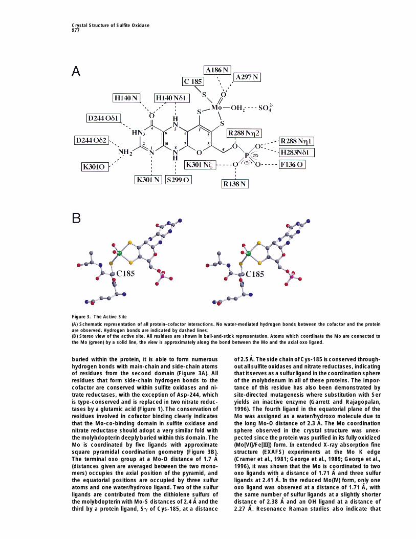
The Mo-co consists of a single molybdopterin and is131, and Leu-134 from the second domain (Figure 2C).With the exception of Leu-134, all of the residues which deeply buried in the second domain of sulfite oxidase.
As in the other structures of Mo-co-containing enzymesparticipate in these interactions are conserved amongsulfite oxidases, and Leu-123 and Pro-131 are con- (Chan et al., 1995; Romao et al., 1995; Schindelin et al.,
1996; Boyington et al., 1997), the pterin forms a tricyclicserved in the nitrate reductases as well. Only two hydro-gen bonds are formed across the interface of these ring system with a pyran ring fused to the pyrazine ring
of the pterin, but unlike its eubacterial counterparts, thetwo domains. The main chain oxygen of Ala-54 forms ahydrogen bond with Ne2 of His-104 and Tyr-62 Oh forms molybdopterin in sulfite oxidase is not conjugated by
an additional nucleotide. Since the cofactor is deeplya hydrogen bond to Glu-126 Oe1 (Figure 2C).
Crystal Structure of Sulfite Oxidase977
Figure 3. The Active Site
(A) Schematic representation of all protein–cofactor interactions. No water-mediated hydrogen bonds between the cofactor and the proteinare observed. Hydrogen bonds are indicated by dashed lines.(B) Stereo view of the active site. All residues are shown in ball-and-stick representation. Atoms which coordinate the Mo are connected tothe Mo (green) by a solid line, the view is approximately along the bond between the Mo and the axial oxo ligand.
buried within the protein, it is able to form numerous of 2.5 A. The side chain of Cys-185 isconserved through-out all sulfite oxidases and nitrate reductases, indicatinghydrogen bonds with main-chain and side-chain atoms
of residues from the second domain (Figure 3A). All that it serves as a sulfur ligand in thecoordination sphereof the molybdenum in all of these proteins. The impor-residues that form side-chain hydrogen bonds to the
cofactor are conserved within sulfite oxidases and ni- tance of this residue has also been demonstrated bysite-directed mutagenesis where substitution with Sertrate reductases, with the exception of Asp-244, which
is type-conserved and is replaced in two nitrate reduc- yields an inactive enzyme (Garrett and Rajagopalan,1996). The fourth ligand in the equatorial plane of thetases by a glutamic acid (Figure 1). The conservation of
residues involved in cofactor binding clearly indicates Mo was assigned as a water/hydroxo molecule due tothe long Mo-O distance of 2.3 A. The Mo coordinationthat the Mo-co-binding domain in sulfite oxidase and
nitrate reductase should adopt a very similar fold with sphere observed in the crystal structure was unex-pected since the protein was purified in its fully oxidizedthe molybdopterin deeply buried within this domain. The
Mo is coordinated by five ligands with approximate (Mo[VI]/Fe[III]) form. In extended X-ray absorption finestructure (EXAFS) experiments at the Mo K edgesquare pyramidal coordination geometry (Figure 3B).
The terminal oxo group at a Mo-O distance of 1.7 A (Cramer et al., 1981; George et al., 1989; George et al.,1996), it was shown that the Mo is coordinated to two(distances given are averaged between the two mono-
mers) occupies the axial position of the pyramid, and oxo ligands with a distance of 1.71 A and three sulfurligands at 2.41 A. In the reduced Mo(IV) form, only onethe equatorial positions are occupied by three sulfur
atoms and one water/hydroxo ligand. Two of the sulfur oxo ligand was observed at a distance of 1.71 A, withthe same number of sulfur ligands at a slightly shorterligands are contributed from the dithiolene sulfurs of
the molybdopterin with Mo-S distances of 2.4 A and the distance of 2.38 A and an OH ligand at a distance of2.27 A. Resonance Raman studies also indicate thatthird by a protein ligand, Sg of Cys-185, at a distance
Cell978
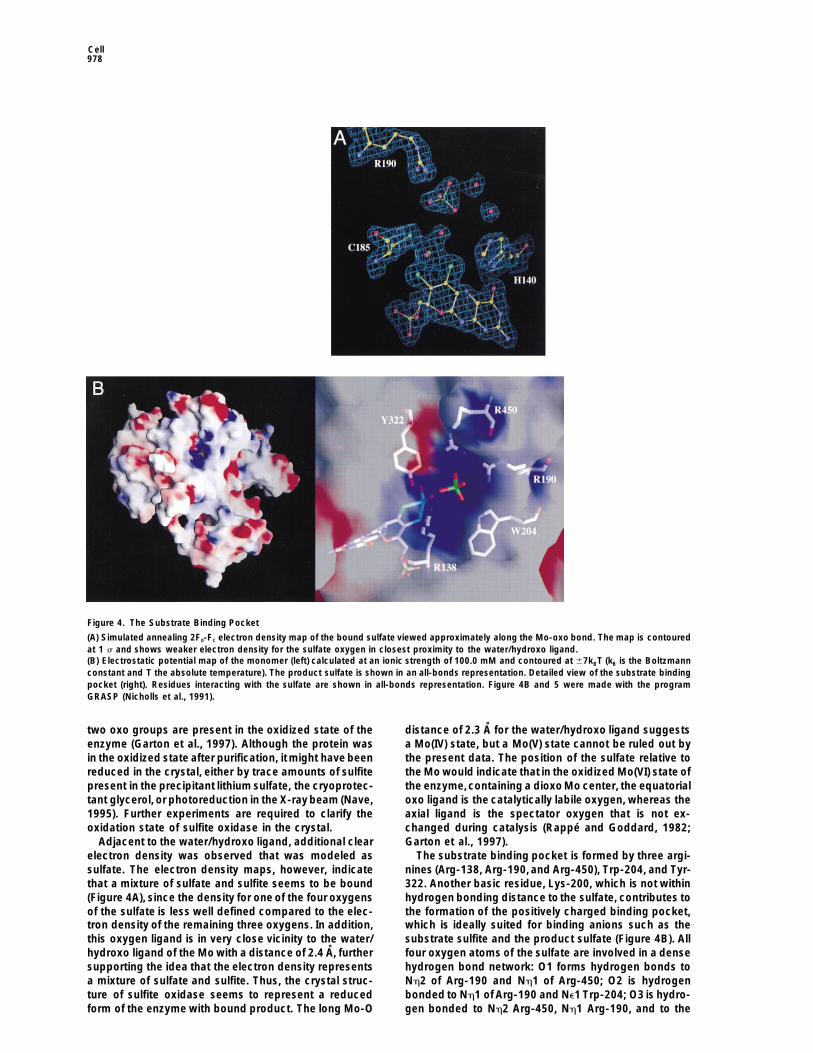
Figure 4. The Substrate Binding Pocket
(A) Simulated annealing 2Fo-Fc electron density map of the bound sulfate viewed approximately along the Mo-oxo bond. The map is contouredat 1 s and shows weaker electron density for the sulfate oxygen in closest proximity to the water/hydroxo ligand.(B) Electrostatic potential map of the monomer (left) calculated at an ionic strength of 100.0 mM and contoured at 67kBT (kB is the Boltzmannconstant and T the absolute temperature). The product sulfate is shown in an all-bonds representation. Detailed view of the substrate bindingpocket (right). Residues interacting with the sulfate are shown in all-bonds representation. Figure 4B and 5 were made with the programGRASP (Nicholls et al., 1991).
two oxo groups are present in the oxidized state of the distance of 2.3 A for the water/hydroxo ligand suggestsa Mo(IV) state, but a Mo(V) state cannot be ruled out byenzyme (Garton et al., 1997). Although the protein was
in the oxidized state after purification, itmight have been the present data. The position of the sulfate relative tothe Mo would indicate that in the oxidized Mo(VI) state ofreduced in the crystal, either by trace amounts of sulfite
present in the precipitant lithium sulfate, the cryoprotec- the enzyme,containing a dioxo Mo center, the equatorialoxo ligand is the catalytically labile oxygen, whereas thetant glycerol, or photoreduction in the X-raybeam (Nave,
1995). Further experiments are required to clarify the axial ligand is the spectator oxygen that is not ex-changed during catalysis (Rappe and Goddard, 1982;oxidation state of sulfite oxidase in the crystal.
Adjacent to the water/hydroxo ligand, additional clear Garton et al., 1997).The substrate binding pocket is formed by three argi-electron density was observed that was modeled as
sulfate. The electron density maps, however, indicate nines (Arg-138, Arg-190,and Arg-450), Trp-204, and Tyr-322. Another basic residue, Lys-200, which is not withinthat a mixture of sulfate and sulfite seems to be bound
(Figure 4A), since the density for one of the four oxygens hydrogen bonding distance to the sulfate, contributes tothe formation of the positively charged binding pocket,of the sulfate is less well defined compared to the elec-
tron density of the remaining three oxygens. In addition, which is ideally suited for binding anions such as thesubstrate sulfite and the product sulfate (Figure 4B). Allthis oxygen ligand is in very close vicinity to the water/
hydroxo ligand of the Mo with a distance of 2.4 A, further four oxygen atoms of the sulfate are involved in a densehydrogen bond network: O1 forms hydrogen bonds tosupporting the idea that the electron density represents
a mixture of sulfate and sulfite. Thus, the crystal struc- Nh2 of Arg-190 and Nh1 of Arg-450; O2 is hydrogenbonded to Nh1 of Arg-190 and Ne1 Trp-204; O3 is hydro-ture of sulfite oxidase seems to represent a reduced
form of the enzyme with bound product. The long Mo-O gen bonded to Nh2 Arg-450, Nh1 Arg-190, and to the

Crystal Structure of Sulfite Oxidase979
Table 3. Point Mutants of Human Sulfite Oxidase Identified in Fibroblasts from Patients with Isolated Sulfite Oxidase Deficiency
Amino Acid Distance from Oligomeric KMa,b kcat kcat/KM
Cell Line Nucleotide Change Substitution Active Site (A)f State (mM) (s21) (M21s21)
1b g479a R160Q 4.3 Dimer 1.7 2.4 1.4 3 103
2c c623a A208D 3.8 NDe ND ND ND3c g1022a R340Q 16.3 ND ND ND ND
g1109a S370Y4d c1110a S370S 10.2 Monomer 6.6 2.7 4 3 102
g1418a G473D
a KM, kcat and kcat/KM for recombinant native human sulfite oxidase are 17 mM, 16 s21, and 9.4 3 105 M21s21, respectively.b Garrett et al., submitted.c T. Graf, personal communication.d R. M. G., unpublished data.e ND—not determined.f Distances in chicken liver sulfite oxidase.
main chain N of Leu-202; and O4 is hydrogen bonded the cytochrome b5 domain and from there are passedto cytochrome c. The electron transfer and the concomi-to Nh2 Arg-138, Ne Arg-138, and Oh Tyr-322. The onetant deprotonation of the Mo-bound hydroxo are neces-oxygen in the bound sulfate that has weaker electronsary to reestablish the resting Mo(VI) state. A surprisingdensity indicates that a bound sulfite would be orientedresult of this structure analysis is the long distance ofsuch that its sulfite lone pair is pointing roughly toward32 A between the Mo and the heme Fe (Figure 2B) withinthe water/hydroxo ligand, which in turn is hydrogenthe same monomer. Due to the elongated shape of thebonded to S19 of the cofactor, to the side chains of Cys-dimer and the location of the two cytochrome domains185, Arg-138, and Tyr-322. This coordination geometryon opposite ends of the dimer, a shorter pathway acrossmight allow a nucleophilic attack of the sulfite lone-the dimer interface does not exist. Based on currentpair electrons on the equatorial oxo group, as has beenmodels for long range electron transfer (Gray and Wink-proposed previously (Hille, 1994). According to the pos-ler, 1996), the observed rates for electron transfer andtulated mechanism, a bidentate intermediate would beturnover of the substrate are quite fast (Sullivan et al.,formed after the nucleophilic attack, with the fifth ligand1993), considering the large intermetallic distance. Thisbeing coordinated to the Mo and the substrate. Subse-implies that the two redox centers should be connectedquently, sulfate would be released from the active site,by a very efficient electron-transfer pathway. One possi-and a hydroxide from the solvent would be coordinatedble pathway involves Arg-138, which forms a hydrogento the Mo. This completes the reductive half-cycle ofbond to the water/hydroxo ligand. From Arg 138, thethe reaction, in which Mo is reduced from (VI) to (IV),electron could be transferred via themain chain atoms ofwhile sulfite is oxidized to sulfate. In the crystal struc-b strand b8 to Leu-134, which is located at the interfaceture, the separation between the sulfur and the Mo isbetween domains I and II and is in van der Waals contact5.2 A, and a bidentate intermediate would require theto Phe-58, which itself is in close proximity to the heme.sulfite to move z1.5–2 A closer to the Mo. However, thisThe pterin ring does not seem to be part of this electronmovement can only be accomplished if conformationaltransfer pathway, since it is not oriented toward thechanges occur at the active site. In the nitrate reduc-heme. This is another surprising result, since in other
tases, only two of the three arginines, which correspondMo-co containing enzymes, such as aldehyde oxidore-
to Arg-190 and Arg-138, are conserved. Arg-450 is re-ductase from Pyrococcus furiosus (Chan et al., 1995),
placed by a methionine, which is conserved among thealdehyde oxidoreductase from Desulfovibrio gigas (Ro-
nitrate reductases. The presence of only two argininesmaoet al., 1995) and formate dehydrogenase from Esch-
in the substrate binding pocket is reasonable since theerichia coli (Boyington et al., 1997), all of which contain
substrate nitrate has to be bound with one of the oxy- additional redox centers, the pterin is oriented towardgens positioned toward the Mo, which is subsequently this additional redox center, and the two cofactors areremoved to form nitrite. The residue corresponding to connected by only a small number of hydrogen bonds.Tyr-322 is replaced by an Asn in the nitrate reductases. The unusually long separation between the two redoxAn additional sulfate molecule was identified at a posi- centers and the orientation of the molybdopterin ringtion 10.6 A from the Mo and 5.5 A from Arg-138 Nh2. system away from the second cofactor raises the possi-While close to the substrate binding pocket, this sul- bility that in solution the cytochrome b5 domain mightfate is positioned toward the solvent compared to the adopt a different conformation, closer to the Mo-co.first sulfate molecule. The second sulfate is hydrogen As described above, the relatively extensive interfacebonded by the side chain of Trp-317 and several water between domains I and II seems to argue against thismolecules. Furthermore, Arg-337 of a symmetry-related idea, but additional experiments are necessary to ad-molecule in the crystal contributes hydrogen bonds, and dress this point.thus the significance of this sulfate binding site is notclear. However, it is possible that the sulfate molecule Structural Characterization of Mutationsdenotes the entry/exit path of the substrate/product to Causing Sulfite Oxidase Deficiencyor from the active site. So far, four point mutations have been characterized
In the oxidative half cycle, two electrons are trans- from patients suffering from isolated sulfite oxidase de-ficiency (Table 3). In the coding sequence of clonedferred sequentially from the Mo center to the heme in
Cell980
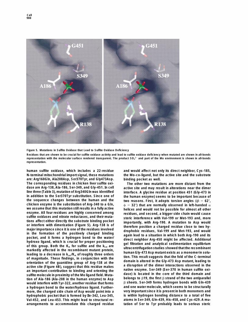
Figure 5. Mutations in Sulfite Oxidase that Lead to Sulfite Oxidase Deficiency
Residues that are shown to be crucial for sulfite oxidase activity and lead to sulfite oxidase deficiency when mutated are shown in all-bondsrepresentation with the molecular surface rendered transparent. The product SO4
22 and part of the Mo environment is shown in all-bondsrepresentation.
human sulfite oxidase, which includes a 22-residue and would affect not only its direct neighbor, Cys-185,N-terminal mitochondrial import signal, these mutations the Mo-co-ligand, but the active site and the substrateare: Arg160Gln, Ala208Asp, Ser370Tyr, and Gly473Asp. binding pocket as well.The corresponding residues in chicken liver sulfite oxi- The other two mutations are more distant from thedase are Arg-138, Ala-186, Ser-349, and Gly-451. In cell active site and may result in alterations near the dimerline three (Table 3), mutation of Arg340Gln was identified interface. A glycine residue at position 451 (Gly-473 inin addition to the Ser370Tyr substitution. Since one of the human enzyme) seems to be important because ofthe sequence changes between the human and the two reasons. First, it adopts torsion angles (φ 5 628,chicken enzyme is the substitution of Arg-340 to a Gln, c 5 328) that are normally observed in left-handed awe assume that this mutation still results in a fully active helices and would not be possible for almost all otherenzyme. All four residues are highly conserved among residues, and second, a bigger side chain would causesulfite oxidases and nitrate reductases, and their muta- steric interference with Val-199 or Met-193 and, moretions affect either directly the substrate binding pocket importantly, with Arg-190. A mutation to Asp wouldor interfere with dimerization (Figure 5). Arg-138 is of therefore position a charged residue close to two hy-major importance since it is one of the residues involved drophobic residues, Val-199 and Met-193, and wouldin the formation of the positively charged binding again lead to a situation in which both Arg-190 and itspocket, and it forms a hydrogen bond to the water/ direct neighbor Arg-450 might be affected. Additionalhydroxo ligand, which is crucial for proper positioning
gel filtration and analytical sedimentation equilibriumof this group. Both the Km for sulfite and the kcat are
ultracentrifugation studies showed that the recombinantmarkedly affected in the recombinant mutant protein,
human Gly-473 Asp mutant exists as a monomer in solu-leading to a decrease in kcat/Km of roughly three orderstion. This result suggests that the fold of the C-terminalof magnitude. These findings, in conjunction with thedomain is altered in the Gly-473 Asp mutant, leading toorientation of the guanidino group of Arg-138 at thea disruption of the dimer interactions observed in theactive site (Figure 4B), suggest that this residue makesnative enzyme. Ser-349 (Ser-370 in human sulfite oxi-an important contribution to binding and orienting thedase) is located in the core of the third domain andsulfite molecule in proximity of the Mo ligand field. Muta-belongs to b19, the first b strand of the two antiparalleltion of Ala-186 (Ala-208 in the human enzyme) to Aspb sheets. Ser-349 forms hydrogen bonds with Gln-439would interfere with Tyr-322, another residue that formsand one water molecule, which seems to be structurallya hydrogen bond to the water/hydroxo ligand. Further-very important since it is present in both monomers andmore, the charged side chain of Asp would point into ais within hydrogen bonding distance to a total of fivehydrophobic pocket that is formed by Ile-342, Leu-345,atoms in Ser-349, Gln-439, His-458, and Cys-429. A mu-Val-452, and Leu-453. This might lead to structural re-
arrangements to accommodate this charged residue tation of Ser to Tyr probably leads to serious steric
Crystal Structure of Sulfite Oxidase981
in centricon microconcentrators and prior to crystallization appliedinterference in the core of the immunoglobulin-like fold,to a Mono Q column. The protein was stored in 10 ml aliquots atresulting in perturbation of the dimer interface.2808C at a concentration of 75 mg/ml or 1.5 mM. For crystallizationIn the cases of glucose 6-phosphate dehydrogenasesetups, the protein was diluted with water to yield a concentration
(Rowland et al., 1994; Naylor et al., 1996) and superoxide of 10 mg/ml. Crystals were grownat 228C by vapor diffusion methodsdismutase (Deng et al., 1993), mappings of point muta- and microseeding techniques against a reservoir containing 0.65–
0.9 M Li2SO4 and 100 mM HEPES (pH 7.5). Crystals grew normallytions identified in human variants onto the crystal struc-within 2 weeks and reached their final size of about 0.5 by 0.1 bytures of these enzymes have shown that many substitu-0.05 mm3 in 2–3 months. They belong to the monoclinic spacegrouptions are located at the interfaces between subunits.P21 with a 5 55.1 A, b 5 197.7 A, c 5 56.0 A, and b 5 94.28 andSubstitutions in this region may impair activity by disrup-contain two molecules or one dimer per asymmetric unit. With two
tion of the dimeric interactions and general destabiliza- molecules in the asymmetric unit, the Mathews coefficient is VM 5tion of the protein structure, and represent a mechanism 2.9 A3Da21, which corresponds to a solvent content of 58%.by which defective subunits may perturb the activities of
Data Collectionwild-type subunits in heterozygous systems. In contrast,Due to radiation sensitivity of the crystals, all data collection wasamino acid replacements at or near the active site ofcarried out at cryogenic temperatures. Crystals were transferredthese enzymes have not been observed. Consequently,into mother liquor containing 20% glycerol by stepwise increase
while the number of mutations causing sulfite oxidase in glycerol concentration and then flash frozen in liquid nitrogen.deficiency is presently limited, it is somewhat surprising Datasets Nat2, EMTS, PIP,and LU (Table 1) were collected on beam-that two of the four mutations found in sulfite oxidase line 7.1 at the Stanford Synchrotron Radiation Laboratory (SSRL)
with a MAR research image plate detector at a wavelength of 1.08(Arg-138 and Ala-186) are within 5 A of the Mo and couldA. All other data sets were collected on an R-Axis IIC imaging platetherefore reduce activity through direct perturbation ofusing monochromatized CuKa radiation produced by a Rigaku RUsubstrate binding and/or oxidation. The remaining two200 rotating anode generator operating at 50 kV and 100 mA. All
substitutions (Ser-349 and Gly-451) could disturb activ- data sets wereprocessed with DENZO (Otwinowski andMinor, 1996)ity through more global effects onstability and alteration and scaled with SCALEPACK. For all subsequent calculations, pro-of the sulfite oxidase dimer. grams of the CCP4 suite (Bailey, 1994) were used, with exceptions
as indicated.On thebasis of sequence comparisons, enzymes con-taining a Mo-co center are currently classified into four
Heavy Atom Derivatives and Phase Determinationdifferent families (Kisker et al., 1997). The sulfite oxidaseAll heavy atom derivatives were prepared by soaking the crystals
structure now joins the other three classes in having in mother liquor containing either 2.5 mM di-m-iodo-bis-(ethylenedi-structurally characterized members (Chan et al., 1995; amine) di-platinum II nitrate for 60 hr (PIP), 1.5 mM sodium ethyl-Romao et al., 1995; Schindelin et al., 1996; Boyington mercurithiosalicylate for 35 hr (EMTS), 2.5 mM K[Au(CN)2] for 43 hr
(AU), 10 mM SmCl3 for 1 week (SM), or 10 mM Lu-acetate for 3 dayset al., 1997). The present studies on chicken liver sulfite(LU). In order to keep the heavy atom bound to the crystalline protein,oxidase provide a structural framework for this entirethe solutions with cryoprotectant contained the same heavy atomenzyme family. Most importantly, due to the high se-concentration as used during the soaking procedure. Heavy atomquence identity between the chicken and the humansites for the EMTS derivative were determined by direct methods
enzyme, it was possible to analyze mutations that lead using SHELX-86 (Sheldrick, 1990) and difference Patterson meth-to sulfite oxidase deficiency in humans. Two of the four ods, and the remaining derivatives were solved by difference Fourier
methods. Heavy atoms were refined, and phases were calculatedcurrently identified mutations seem to directly perturbwith MLPHARE (Otwinowski, 1991) to a resolution of 3.0 A, resultingthe substrate binding pocket and might interfere within a figure of merit of 0.49. Initial maps were of poor quality due tosubstrate binding or catalysis. The structure of sulfitethe fact that the heavy atom derivatives shared the same majoroxidase thus represents an important step toward un-heavy atom sites, with the exception of the Lu derivative, which
derstanding the metabolic disease sulfite oxidase defi- contributed significant phase information only to a resolution of 6 Aciency at the atomic level. (Table 1).
Two-Fold Noncrystallographic AveragingExperimental ProceduresInitial NCS relationships were determined by the heavy atom posi-tions of the EMTS and PIP derivatives. Masks were calculated inProtein Purification and CrystallizationNCSMASK and the NCS matrix was improved in DM. Averaging ofThe purification of sulfite oxidase was based on several existingthe electron density maps was carried out against data set NAT1methods (Kessler and Rajagopalan, 1972; Kipke et al., 1989; Sullivanto a resolution of 3.0 A. After 250 cycles of averaging, the correlationet al., 1993). Chicken liver (1.5 kg) was combined with 1.6 l of 10coefficient for the electron density of the two monomers related bymM phosphate buffer (pH 7.8) containing 1 mM EDTA and 0.125the NCS transformation improved from 0.32 to 0.83 with an RfreemM PMSF. The mixture was homogenized in a large Waring blender,0.311. The resulting electron density was of reasonable quality andafter which the pH was immediately readjusted to 7.8 with 30%showed as highest peaks both molybdenum sites and one of theNH4OH. The homogenate was centrifuged, and the supernatant wastwo Fe sites. A clear distinction between the cytochrome b5 domainssubmitted to two (NH4)2SO4 fractionation steps (20% and 55%), withand the core of the protein was possible. An atomic model was builtan intervening heat denaturation step. The above procedure wasusing O (Jones et al., 1991). For the cytochrome b5 domain, therepeated for a total of about 9 kg of chicken liver, after which themodel of bovine cytochrome b5 was positioned into the electronpellets from the second (NH4)2SO4 fractionation were dissolved in 2 ldensity with the necessary exchanges for different residues andof 250 mM Tris buffer (pH 7.8) and loaded onto a phenyl-sepharosedomain II and III were built using the skeleton obtained with thecolumn equilibrated with 25 mM Tris (pH 7.8), 1.5 mM EDTA, andprogram MAPMAN (Kleywegt and Jones, 1994).0.5 M NaCl. Proteins were eluted by decreasing the column salt
concentration with deionized water, dialyzed against 2.5 mM Tris(pH 7.8), then further purified by ion exchange chromatography. Refinement
The structure has been refined against data set Nat2, initially atSubsequently, the phenyl-sepharose chromatography was repeatedas described above, but using a much smaller column. The eluant 2.5 A resolution and incorporating tight NCS restraints, with the
simulated annealing protocol in X-PLOR (Brunger, 1992) using thewas concentrated in an Amicon concentrator and applied to a Seph-acryl S-200 size exclusion column. The enzyme was concentrated stereochemical parameters of Engh and Huber (Engh and Huber,

Cell982
1991). After refinement, model phases and MIRAS phases were Chan, M.K., Mukund, S., Kletzin, A., Adams, M.W.W., and Rees, D.C.(1995). Structure of a hyperthermophilic tungstopterin, aldehyde fer-combined, and the resulting electron density maps were improvedredoxin oxidoreductase. Science 267, 1463–1469.by two-fold averaging and solvent flattening. The resolution was
extended to 1.9 A, and after several rounds of model building, refine- Cramer, S.P., Wahl, R., and Rajagopalan, K.V. (1981). Molybdenumment, and averaging, the NCS restraints were removed. A bulk sol- sites of sulfite oxidase and xanthine dehydrogenase. A comparisonvent correction was applied throughout refinement and allowed the by EXAFS. J. Am. Chem. Soc. 103, 7721–7727.inclusion of all data between 50 and 1.9 A resolution. Refinement Crawford, N.M., Smith, M., Bellissimo, D., and Davis, R.W. (1988).was then continued with REFMAC (Murshudov et al., 1997) utilizing Sequence and nitrate regulation of the arabidopsis thaliana messen-the maximum likelihood approach. Partial structure factors for the ger RNA encoding nitrate reductase, a metalloflavoprotein with 3bulk solvent contribution were calculated with XPLOR and applied functional domains. Proc. Natl. Acad. Sci. USA 85, 5006–5010.in REFMAC. Iterative rounds of refinement with REFMAC and model
Deng, H.-X., Hentati, A., Tainer, J.A., Iqbal, Z., Cayabyab, A., Hung,building were performed. After the last cycle, an anisotropic B-factorW.-Y., Getzoff, E.D., Hu, P., Herzfeldt, B., Roos, R.P., et al. (1993).correction was calculated with X-PLOR yielding the current model,Amyotrophic lateral sclerosis and structural defects in Cu, Zn super-
which has a crystallographic and free R factor of 17.45% and 22.0%,oxide dismutase. Science 261, 1047–1051.
respectively. The model contains a total of 8191 nonhydrogenEngh, R.A., and Huber, R. (1991). Accurate bond and angle parame-atoms, with residues 3–88 and 90–466 in the first monomer andters for X-ray protein structure refinement. Acta Crystallogr. A47,residues 3–85 and 92–466 in the second monomer, one Mo-co and392–400.one heme per monomer, two SO4
22 per monomer, 820 water mole-Garrett, R.M., Bellissimo, D.B., and Rajagopalan, K.V. (1995). Molec-cules, one HEPES, and one glycerol. Residue 90 in the first monomerular cloning of human liver sulfite oxidase. Biochim. Biophys. Actaand residues 86–91 in the second monomer are only very weakly1262, 147–149.defined in the electron density and were therefore not included in
refinement. The stereochemistry of the model is good with rms Garrett, R.M., and Rajagopalan, K.V. (1994). Molecular cloning ofdeviations of 0.009 A in bond distances and 0.024 A in angle dis- rat liver sulfite oxidase. J. Biol. Chem. 269, 272–276.tances. The structure has been analyzed with PROCHECK (Laskow- Garrett, R.M., and Rajagopalan, K.V. (1996). Site-directed mutagene-ski et al., 1993), revealing that more than 89% of all residues are in sis of recombinant sulfite oxidase. J. Biol. Chem. 271, 7387–7391.the most favored regions of the Ramachandran diagram, and only
Garton, S.D., Garrett, R.M., Rajagopalan, K.V., and Johnson, M.K.Arg-21, although well-defined in the electron density maps in both
(1997). Resonance raman characterization of the molybdenum cen-monomers, adopts torsion angles that are in disallowed regions.
ter in sulfite oxidase—identification of Mo5O stretching modes. J.Am. Chem. Soc. 119, 2590–2591.
Identification of Inborn Point Mutations in HumanGeorge, G.N., Kipke, C.A., Prince, R.C., Sunde, R.A., Enemark, J.H.,
Sulfite Oxidaseand Cramer, S.P. (1989). Structure of the active site of sulfite oxi-
Details of the isolation of mutant human sulfite oxidase genes willdase. X-ray absorption spectroscopy of the Mo(IV), Mo(V), and
be presented elsewhere (Garrett at al., submitted). Messenger RNA Mo(VI) oxidation states. Biochemistry 28, 5075–5080.isolated from fibroblasts patients with sulfite oxidase deficiency was
George, G.N., Garrett, R.M., Prince, R.C., and Rajagopalan, K.V.used to synthesize cDNA. The mutant sulfite oxidase gene was(1996). The molybdenum site of sulfite oxidase: a comparison ofamplified by PCR using primers flanking the start and stop sites ofwild-type and the Cysteine 207 to Serine mutant using X-ray absorp-the coding sequence (Garrett and Rajagopalan, 1996). The PCRtion spectroscopy. J. Am. Chem. Soc. 118, 8588–8592.amplification products were cloned into a sequencing vector andGray, H.B., and Winkler, J.R. (1996). Electron transfer in proteins.transformed into TA One Shot competent cells (Invitrogen). TheAnnu. Rev. Biochem. 65, 537–561.inserts were sequenced from several clones to rule out PCR arti-
facts. The base substitutions and the resulting amino acid replace- Hille, R. (1994). The reaction mechanism of oxomolybdenum en-ments in the mutants are listed in Table 3. The R160Q and G473D zymes. Biochim. Biophys. Acta 1184, 143–169.mutations were introduced into the cloned human sulfite oxidase Hille, R. (1996). The mononuclear molybdenum enzymes. Chem.gene, andthe recombinant protein expressed in E. coli, as described Rev. 96, 2757–2816.before (Garrett and Rajagopalan, 1996). Holm, L., and Sander, C. (1995). Dali: a network tool for protein
structure comparison. Trends Biochem. Sci. 20, 478–480.Acknowledgments
Janin, J., and Rodier, F. (1995). Protein–protein interaction at crystalcontacts. Proteins 23, 580–587.
This work was supported by Deutsche ForschungsgemeinschaftJanin, J., Miller, S., and Chothia, C. (1988). Surface, subunit inter-postdoctoral fellowships (to C. K. and H. S.) and by USPHS grantsfaces and interior of oligomeric proteins. J. Mol. Biol. 204, 155–164.(GM50775 to D. C. R., GM37773 to J. H. E., and GM00091 andJohnson, J.L., and Rajagopalan, K.V. (1977). Tryptic cleavage of ratGM44283 to K. V. R.). The rotation camera facility at SSRL is sup-liver sulfite oxidase. J. Biol. Chem. 252, 2017–2025.ported by DOE and NIH. The mutant studies could not have been
carried out without the tireless effort of Dr. Jean Johnson, who Johnson, J.L., and Wadman, S.K. (1989). Molybdenum cofactor defi-secured all the patient fibroblast lines from various clinical investi- ciency. In The Metabolic Basis of Inherited Disease, 6th Edition,gators. C.R. Scriver, A.L. Beaudet, W.S. Sly, and D.L. Valle, eds. (New York:
McGraw-Hill), pp.1463–1475.Received September 2, 1997; revised November 6, 1997. Jones, T.A., Zou, J.Y., Cowan, S.W., and Kjeldgaard, M. (1991).
Improved methods for building protein models in electron densitymaps and the location of errors in these models. Acta Crystallogr.ReferencesA47, 110–119.
Bacon, D.J., and Anderson, W.F. (1988). A fast algorithm for render- Jones, E.Y., Harlos, K., Bottomley, M.J., Robinson, R.C., Driscoll,ing space-filling molecule pictures. J. Mol. Graph. 6, 219–220. P.C., Edwards, R.M., Clements, J.M., Dudgeon, T.J., and Stuart, D.I.
(1995). Crystal structure of an integrin-binding fragment of vascularBailey, S. (1994). The CCP4suite—programs for protein crystallogra-cell adehesion molecule 1 at 1.8 A resolution. Nature 373, 539–544.phy. Acta Crystallogr. D50, 760–763.Kessler, D.L., and Rajagopalan, K.V. (1972). Purification and proper-Boyington, J.C., Gladyshev, V.N., Khangulov, S.V., Stadtman, T.C.,ties of sulfite oxidase from chicken liver. Presence of molybdenum inand Sun, P.D. (1997). Crystal structure of formate dehydrogenasesulfite oxidase from diverse sources. J. Biol. Chem. 247, 6566–6573.H: catalysis involving Mo, molybdopterin, selenocysteine, and an
Fe4S4 cluster. Science 275, 1305–1308. Kipke, C.A., Enemark, J.H., and Sunde, R.A. (1989). Purification ofprosthetically intact sulfite oxidase from chicken liver using a modi-Brunger, A.T. (1992). X-PLOR Version 3.1—A System for X-RayCrys-fied procedure. Arch. Biochem. Biophys. 270, 383–390.tallography and NMR (New Haven, CT and London: Yale University
Press). Kisker, C., Schindelin, H., and Rees, D.C. (1997). Molybdenum-

Crystal Structure of Sulfite Oxidase983
cofactor-containing enzymes: structure and mechansim. Annu. Rev. the pterin molybdenum cofactor: sequence families, spectroscopicproperties and possible cofactor-binding domains. Biochim. Bio-Biochem. 66, 233–267.phys. Acta 1057, 157–185.Kleywegt, G.J., and Jones, T.A. (1994). Halloween . . . masks and
bones. In CCP4 Study Weekend From First Map to Final Model,PBID Protein Data Bank CoordinatesS. Bailey, R. Hubbard, and D. Waller, eds. (Warrington, UK: SERC
Daresbury Laboratory), pp. 59–66.The accession number for the sequence reported in this paper isKraulis, P.J. (1991). MOLSCRIPT—a program to produce both de-BNL-20210; coordinates can also be requested by e-mail to kisker@tailed and schematic plots of protein structures. J. Appl. Crystallogr.citray.caltech.edu.24, 946–950.
Kubo, Y., Ogura, N., and Nakagawa, H. (1988). Limited proteolysisof the nitrate reductase from spinach leaves. J. Biol. Chem. 263,19684–19689.
Laskowski, R.A., McArthur, M.W., Moss, D.S., and Thornton, J.M.(1993). PROCHECK—a program to check the stereochemical qualityof protein structures. J. Appl. Crystallogr. 26, 283–291.
Mathews, F.S., Argos, P., and Levine, M. (1972). The structure ofcytochrome b5 at 2.0 A resolution. Cold Spring Harbor Symp. 36,387.
Merritt, E.A., and Murphy, M.E.P. (1994). Raster3D Version 2.0—aprogram for photorealistic molecular graphics. Acta Crystallogr.D50, 869–873.
Murshudov, G., Vagin, A., and Dodson, E. (1997). Refinement ofmacromolecular structures by the maximum likelihood method.ActaCrystallogr. D53, 240–255.
Nave, C. (1995). Radiation damage in protein crystallography. Rad.Phys. Chem. 45, 483–490.
Naylor, C.E., Rowland, P., Basak, A.K., Gover, S., Mason, P.J.,Bautista, J.M., Vulliamy, T.J., Luzzatto, L., and Adams, M.J. (1996).Glucose 6-phosphate dehydrogenase mutations causing enzymedeficiency in a model of the tertiary structure of the human enzyme.Blood 87, 2974–2982.
Neame, P.J., and Barber, M.J. (1989). Conserved domains in molyb-denum hydroxylases. J. Biol. Chem. 264, 20894–20901.
Nicholls, A., Sharp, K.A., and Honig, B. (1991). Protein folding andassociation: insights from the interfacial and thermodynamic prop-erties of hydrocarbons. Proteins 11, 281–296.
Otwinowski, Z. (1991). Isomorphous replacement and anomalousscattering. In CCP4 Study Weekend Isomorphous Replacement andAnomalous Scattering, W. Wolf, P. Evans, and A. Leslie, eds. (War-rington, UK: SERC Daresbury Laboratory), pp. 80–86.
Otwinowski, Z., and Minor, W. (1996). Processing of X-ray diffractiondata collected in oscillation mode. In Macromolecular Crystallogra-phy, Part A, C.W. Carter, Jr. and R.M. Sweet, eds. (New York: Aca-demic Press), pp. 307–326.
Rappe, A.K., and Goddard, W.A. (1982). Olefin metathesis—a mech-anistic study of high valent group26 catalysts. J. Am. Chem. Soc.104, 448–456.
Romao, M.J., Archer, M., Moura, I., LeGall, J., Engh, R., Schneider,M., Hof, P., and Huber, R. (1995). Crystal structure of the xanthineoxidase-related aldehyde oxido-reductase from Desulfovibrio gigas.Science 270, 1170–1176.
Rowland, P., Basak, A.K., Gover, S., Levy, H.R., and Adams, M.J.(1994). The three-dimensional structure of glucose 6-phosphate de-hydrogenase from Leuconostoc mesenteroides refined at 2.0 A res-olution. Structure 2, 1073–1087.
Schindelin, H., Kisker, C., Hilton, J., Rajagopalan, K.V., and Rees,D.C. (1996). Crystal structure of DMSO reductase: redox-linkedchanges in molybdopterin coordination. Science 272, 1615–1621.
Sheldrick, G.M. (1990). Phase annealing in SHELX-90—direct meth-ods for larger structures. Acta Crystallogr. A46, 467–473.
Southerland, W.M., Wings, D.R., and Rajagopalan, K.V. (1978). Thedomains of rat liver sulfite oxidase. J. Biol. Chem. 253, 8747–8752.
Sullivan, E.P., Jr., Hazzard, J.T., Tollin, G., and Enemark, J.H. (1993).Electron transfer in sulfite oxidase: effects of pH and anions ontransient kinetics. Biochemistry 32, 12465–12470.
Vaughn, D.E., and Bjorkman, P.J. (1996). The (Greek) Key to struc-tures of neural adhesion molecules. Neuron 16, 261–273.
Wootton, J.C., Nicholson, R.E., Cock, J.M., Walters, D.E., Burke,J.F., Doyle, W.A., and Bray, R.C. (1991). Enzymes depending on
Related Documents