CRTAM: A Molecule Involved in Epithelial Cell Adhesion Erika Garay, 1 Genaro Patin ˜ o-Lo ´pez, 2 Socorro Islas, 1 Lourdes Alarco ´n, 1 Elsy Canche-Pool, 2 Ricardo Valle-Rios, 2 Oscar Medina-Contreras, 2 Giovana Granados, 3 Bibiana Cha ´vez-Munguı ´a, 4 Eusebio Juaristi, 3 Vianney Ortiz-Navarrete, 2 and Lorenza Gonza ´lez-Mariscal 1 * 1 Department of Physiology, Biophysics and Neuroscience, Center for Research and Advanced Studies (Cinvestav), Mexico City, Mexico 2 Department of Biomedicine, Center for Research and Advanced Studies (Cinvestav), Mexico City, Mexico 3 Department of Chemistry, Center for Research and Advanced Studies (Cinvestav), Mexico City, Mexico 4 Department of Infectomics and Molecular Pathogenesis, Center for Research and Advanced Studies (Cinvestav), Mexico City, Mexico ABSTRACT Class I-restricted T cell associated molecule (CRTAM) is a member of the immunoglobulin superfamily that complies with the structural characteristics of the JAM family of proteins and is phylogenetically more closely related to nectin-like proteins. Here we demonstrate for the first time, that CRTAM is expressed in epithelial cells along the lateral membrane and is important for early cell–cell contacts and cell– substrate interactions. CRTAM is sensitive to intermediate filament disruption and treatment of monolayers with soluble CRTAM enhances cell–cell dissociation and lowers transepithelial electrical resistance. Incubation of newly plated cells with anti-CRTAM antibody decreases the formation of cell aggregates and promotes cell detachment. Co-cultures of epithelial cells and fibroblasts that lack CRTAM expression and in vitro binding assays, demonstrate the participation of CRTAM in homotypic and heterotypic trans-interactions. Hence we conclude that CRTAM is a molecule involved in epithelial cell adhesion. J. Cell. Biochem. 111: 111–122, 2010. ß 2010 Wiley-Liss, Inc. KEY WORDS: CRTAM; JAM; CELL ADHESION; TIGHT JUNCTION; EPITHELIA T he class I-restricted T cell associated molecule (CRTAM) received its name for its restricted expression pattern in T cells [Kennedy et al., 2000]. This protein was originally found to be highly expressed in activated CD8 þ T and NKT cells which are class I major histocompatibility complex (MHC) restricted [Kennedy et al., 2000]. More recently however, CRTAM expression has been detected in natural killer (NK) cells and in a minor population of activated CD4 T cells [Arase et al., 2005; Yeh et al., 2008]. In mouse, CRTAM mRNA is detectable in spleen, brain and testis, and in humans is present in spleen, thymus, intestine, lymph nodes, lung, testis, ovary and colon [Kennedy et al., 2000]. In human CNS, CRTAM mRNA is strongly expressed in cerebellum and immunocytochemistry studies have revealed that the protein is mainly localized to Purkinje neurons and granule cells [Patino- Lopez et al., 2006]. CRTAM belongs to the immunoglobulin superfamily (Ig-SF) due to the presence of two Ig-like domains at the extracellular region [Kennedy et al., 2000]. CRTAM exhibits at its carboxyl terminal end the class I PDZ binding motif ESIV [Arase et al., 2005; Patino-Lopez et al., 2006]. Previously we and others suggested a relationship between CRTAM and the nectin-like (necl) family of proteins [Boles et al., 2005; Patino-Lopez et al., 2006], although CRTAM shows below 20% amino acid identity to necl proteins. Nectins and necls are Ca 2þ -independent cell adhesion molecules with three extra- cellular Ig-like domains, a single transmembrane region, and a PDZ binding motif at their carboxyl terminal ends which allows nectins but not necl proteins to bind afadin [Ogita and Takai, 2006]. The fact that CRTAM has two and not three Ig-like domains, prompted us to revisit our previous analysis and to explore the relationship of CRTAM to the JAM protein family. Journal of Cellular Biochemistry ARTICLE Journal of Cellular Biochemistry 111:111–122 (2010) 111 Additional Supporting Information may be found in the online version of this article. Grant sponsor: Mexican Council for Science and Technology (CONACYT); Grant numbers: 45691-Q, CO1-139/A-1; Grant sponsor: Cinvestav Multidisciplinary Project. *Correspondence to: Dr. Lorenza Gonza ´lez-Mariscal, PhD, Department of Physiology, Biophysics and Neuroscience, Center for Research and Advanced Studies (CINVESTAV), Ave. IPN 2508, Mexico 07360, D.F., Mexico. E-mail: lorenza@fisio.cinvestav.mx Received 18 December 2009; Accepted 16 April 2010 DOI 10.1002/jcb.22673 ß 2010 Wiley-Liss, Inc. Published online 12 May 2010 in Wiley Online Library (wileyonlinelibrary.com).

Welcome message from author
This document is posted to help you gain knowledge. Please leave a comment to let me know what you think about it! Share it to your friends and learn new things together.
Transcript
Journal of CellularBiochemistry
ARTICLEJournal of Cellular Biochemistry 111:111–122 (2010)
CRTAM: A Molecule Involved in Epithelial Cell Adhesion
A
GG
*CE
R
P
Erika Garay,1 Genaro Patino-Lopez,2 Socorro Islas,1 Lourdes Alarcon,1 Elsy Canche-Pool,2
Ricardo Valle-Rios,2 Oscar Medina-Contreras,2 Giovana Granados,3
Bibiana Chavez-Munguıa,4 Eusebio Juaristi,3 Vianney Ortiz-Navarrete,2 andLorenza Gonzalez-Mariscal1*1Department of Physiology, Biophysics and Neuroscience, Center for Research and Advanced Studies (Cinvestav),Mexico City, Mexico
2Department of Biomedicine, Center for Research and Advanced Studies (Cinvestav), Mexico City, Mexico3Department of Chemistry, Center for Research and Advanced Studies (Cinvestav), Mexico City, Mexico4Department of Infectomics and Molecular Pathogenesis, Center for Research and Advanced Studies (Cinvestav),Mexico City, Mexico
ABSTRACTClass I-restricted T cell associated molecule (CRTAM) is a member of the immunoglobulin superfamily that complies with the structural
characteristics of the JAM family of proteins and is phylogenetically more closely related to nectin-like proteins. Here we demonstrate for the
first time, that CRTAM is expressed in epithelial cells along the lateral membrane and is important for early cell–cell contacts and cell–
substrate interactions. CRTAM is sensitive to intermediate filament disruption and treatment of monolayers with soluble CRTAM enhances
cell–cell dissociation and lowers transepithelial electrical resistance. Incubation of newly plated cells with anti-CRTAM antibody decreases the
formation of cell aggregates and promotes cell detachment. Co-cultures of epithelial cells and fibroblasts that lack CRTAM expression and in
vitro binding assays, demonstrate the participation of CRTAM in homotypic and heterotypic trans-interactions. Hence we conclude that
CRTAM is a molecule involved in epithelial cell adhesion. J. Cell. Biochem. 111: 111–122, 2010. � 2010 Wiley-Liss, Inc.
KEY WORDS: CRTAM; JAM; CELL ADHESION; TIGHT JUNCTION; EPITHELIA
T he class I-restricted T cell associated molecule (CRTAM)
received its name for its restricted expression pattern in
T cells [Kennedy et al., 2000]. This protein was originally found to be
highly expressed in activated CD8þ T and NKT cells which are class I
major histocompatibility complex (MHC) restricted [Kennedy et al.,
2000].
More recently however, CRTAM expression has been detected
in natural killer (NK) cells and in a minor population of activated
CD4 T cells [Arase et al., 2005; Yeh et al., 2008].
In mouse, CRTAM mRNA is detectable in spleen, brain and
testis, and in humans is present in spleen, thymus, intestine, lymph
nodes, lung, testis, ovary and colon [Kennedy et al., 2000]. In
human CNS, CRTAM mRNA is strongly expressed in cerebellum
and immunocytochemistry studies have revealed that the protein
is mainly localized to Purkinje neurons and granule cells [Patino-
Lopez et al., 2006].
dditional Supporting Information may be found in the online version o
rant sponsor: Mexican Council for Science and Technology (CONACYT);rant sponsor: Cinvestav Multidisciplinary Project.
Correspondence to: Dr. Lorenza Gonzalez-Mariscal, PhD, Department of Penter for Research and Advanced Studies (CINVESTAV), Ave. IPN 2508,-mail: [email protected]
eceived 18 December 2009; Accepted 16 April 2010 � DOI 10.1002/jcb.
ublished online 12 May 2010 in Wiley Online Library (wileyonlinelibrar
CRTAM belongs to the immunoglobulin superfamily (Ig-SF) due
to the presence of two Ig-like domains at the extracellular region
[Kennedy et al., 2000]. CRTAM exhibits at its carboxyl terminal end
the class I PDZ binding motif ESIV [Arase et al., 2005; Patino-Lopez
et al., 2006].
Previously we and others suggested a relationship between
CRTAM and the nectin-like (necl) family of proteins [Boles et al.,
2005; Patino-Lopez et al., 2006], although CRTAM shows below
20% amino acid identity to necl proteins. Nectins and necls
are Ca2þ-independent cell adhesion molecules with three extra-
cellular Ig-like domains, a single transmembrane region, and a PDZ
binding motif at their carboxyl terminal ends which allows nectins
but not necl proteins to bind afadin [Ogita and Takai, 2006].
The fact that CRTAM has two and not three Ig-like domains,
prompted us to revisit our previous analysis and to explore the
relationship of CRTAM to the JAM protein family.
111
f this article.
Grant numbers: 45691-Q, CO1-139/A-1;
hysiology, Biophysics and Neuroscience,Mexico 07360, D.F., Mexico.
22673 � � 2010 Wiley-Liss, Inc.
y.com).
JAMs are proteins of the Ig-SF that exhibit two Ig-like domains
with intramolecular disulfide bonds and two or more N-glycosyla-
tion sites in their extracellular domain. JAMs A, B and C exhibit a
short cytoplasmic tail of 45–50 residues that ends with a type II PDZ
binding motif, while others like CAR, ESAM, JAM4, and CLMP have
longer tails of 105–165 amino acids ending in type I PDZ binding
motifs [Hirabayashi and Hata, 2006].
JAMs are present at the intercellular junctions and lateral
membrane of epithelial and endothelial cells as well as on the
surface of leukocytes [Cunningham et al., 2000; Ostermann et al.,
2002] and platelets [Kornecki et al., 1990; Santoso et al., 2002].
JAMs appear to be involved in tethering specific proteins to the tight
junction (TJ) [Bazzoni et al., 2000; Ebnet et al., 2001; Itoh et al.,
2001; Martinez-Estrada et al., 2001]. Besides their role in TJ
assembly, JAMs participate in leukocyte transmigration [Martin-
Padura et al., 1998; Johnson-Leger et al., 2002; Zen et al., 2004],
platelet activation [Sobocka et al., 2000; Santoso et al., 2002],
angiogenesis [Naik et al., 2003] and virus binding [Barton et al.,
2001; Forrest et al., 2003].
Here we have analyzed the relationship between CRTAM, JAMs,
nectin and necl proteins, studied the expression of CRTAM in
epithelial cells and tested its role in epithelial cell–cell adhesion.
MATERIALS AND METHODS
BIOINFORMATICS
Multiple sequence alignments were performed using the T-Coffee
method based on ClustalW pairwise pre-alignment approach with
progressive optimization of the alignments using Geneious Soft-
ware. Amino acid identities of sequences were determined after their
pairwise alignment using CrustalW with the Gonnet protein weight
matrix and settings of a gap open penalty of 10, a gap extension
penalty of 0.1 and no gap separation penalty. Phylogenetic analysis
and their plot as cladogram-like unrooted trees were conducted with
PHYML method [Guindon and Gascuel, 2003] based on multiple
bootstrapped data sets using standard settings.
CELL CULTURE
Epithelial MDCK cells (Madin Darby canine kidney cells) between
the 60th and 90th passage were grown as previously described [Islas
et al., 2002].
Total peripheral blood mononuclear cells (PBMCs), obtained from
healthy donors at the Hospital Central Norte de Pemex blood bank,
were isolated from blood samples using Ficoll-Paque Plus according
to the manufacturer’s protocol (Amersham, Bioscience, AB Uppsala,
Sweden). Human CD8 T cells were purified from PBMCs thorough
negative selection using magnetic beads (Miltenyi Biotec, Auburn,
CA, Cat. No. 130-091-154). PBMCs or CD8þ T cells were activated
with PMA (2 ng/ml) and Ionomycin (200 ng/ml) as previously
described [Patino-Lopez et al., 2006].
Chinese hamster ovary fibroblasts (CHO) obtained from the
American Type Culture Collection, were cultured in a mixture of
F12/DMEM. Cells were harvested with trypsin–EDTA and plated
on dishes with or without coverslips. For the co-culture, CHO cells
were pre-labeled with 10mM 5-(and 6-)[(4-chloromethyl)benzoyl]
112 CRTAM: AN EPITHELIAL CELL ADHESION MOLECULE
amino-tetramethylrhodamine (C-2927, CellTracker Orange CMTMR;
Molecular Probes, Eugene, OR) as described by the manufacturer.
For the co-culture, instead of doing a mixture of CHO and MDCK
cell suspensions, CHO cells were first plated at sparse density on
glass coverslips and 1 h later the MDCK suspension was plated at
sparse density on the same coverslips. This procedure was followed
in order to guarantee the presence of both types of cells in the
culture, since MDCK cells adhere much faster to the glasscoverslips
than CHO cells. Two days after plating the cultures were processed
for immunofluoresence.
RT-PCR
Total RNA was isolated from MDCK cells using the Trizol method
(Invitrogen. Cat. No. 15596). To measure CRTAM mRNA a
semiquantitative RT-PCR was performed using The One Step
System (Invitrogen). One microgram of total RNA was used in
each analysis. RT-PCR amplification conditions for were as follows:
60 min at 558C (RT), 5 min at 948C, 30 cycles of amplification 30 s at
948C, 30 s at 608C, finally 30 s at 728C for a final extension. Primers
used for amplification were the following: CRTAM forward 50-
CCAAATACCAGCTTCTTCACCA-30 and CRTAM reverse 50-
CTTCAAATCGGAAAGGTGCT-30.
CELLULAR LYSATES AND PROTEIN BLOTTING
MDCK and CHO cells as well as pellets from resting or PMA/
Ionomycin activated CD8þ T cells were lysed with RIPA buffer
as previously described [Hernandez et al., 2007]. Blotting was
performed with the following antibodies— (1) Monoclonals: mouse
IgG anti-human CRTAM (Catalog No. MAB16951, R&D Systems,
Minneapolis, MN; dilution 1:200); rat IgG anti-E-cadherin (DECMA-
1; Sigma U-3254, dilution 1:500); rat IgG against ZO-1 (R26.4C,
Developmental Studies Hybridoma bank, University of Iowa, IA,
USA, dilution 1:500); mouse IgG anti-actin (generously provided by
Dr. Jose Manuel Hernandez from the Department of Cell Biology, at
Cinvestav, Mexico, dilution 1:500). (2) Polyclonals: rabbit IgG
against claudin-1 (Invitrogen 51-9000, dilution 1:1500); rabbit IgG
anti-occludin (Invitrogen 71-1500; dilution 1:1,000); and goat
IgG anti-JAM-A (R&D Systems AF1077, dilution 1:200).
As secondary antibodies the following peroxidase-conjugated
antibodies were employed: goat IgG against mouse (Catalog No.
62-6520; dilution 1:3,000; Zymed Laboratories, San Francisco, CA);
rabbit IgG anti-goat (Invitrogen 61-1620, dilution 1:3,000); goat
IgG against rabbit (Sigma-Aldrich A9169, dilution 1:10,000). Then
the ECLþPlus chemiluminescence detection Kit (GE Healthcare,
Buckinghamshire, UK) was employed.
IMMUNOFLUORESCENCE
Monolayers grown on glass coverslips were fixed and processed
as previously described [Hernandez et al., 2007]. The monolayers
were incubated for 2-h with a rabbit polyclonal antiserum against
the human and mouse CRTAM antigenic peptide 257DKEEKE262
(dilution 1:10 in 0.5% of BSA), or else incubated with the anti-
CRTAM polyclonal anti-serum pre-absorbed for 30 min with 5mg/
ml of antigenic peptide, or incubated with any of the following
antibodies: (1) a rat monoclonal anti-E-cadherin (dilution 1:50 in
0.5% of BSA); (2) a mouse monoclonal anti-Desmoplakin (dilution
JOURNAL OF CELLULAR BIOCHEMISTRY
1:50 in 0.5% of BSA); (3) a mouse monoclonal anti-occludin (Zymed
33-1500, dilution 1:50); (4) a rat monoclonal against ZO-1 (dilution
1:50); (5) a goat polyclonal anti-JAM-A (dilution 1:20), (6) a rabbit
polyclonal anti-occludin (dilution 1:50); and (7) a mouse mono-
clonal anti-cytokeratin-8 (1238-817; Boehringer, Mannheim,
Germany; dilution 1:10).
As secondary antibodies we employed FITC-conjugated goat
anti-rabbit IgG (Zymed 62-6111; dilution 1:100) and goat anti-rat
IgG (Zymed 62-9511; dilution 1:100), a TRITC conjugated goat anti-
mouse IgG (Zymed T2762; dilution 1:100) and a goat anti-rabbit IgG
(Zymed 81-6114; dilution 1:100); CY5 conjugated goat anti-rat
(Zymed 629516; dilution 1:75) and goat anti-mouse (Zymed
81-6516; dilution 1:75), an Alexa Fluor 488 donkey anti-goat
(Molecular Probes A-11055; dilution 1:200), and Alexa Fluor 568
donkey anti-rabbit (Molecular Probes A-11042; dilution 1:200).
The fluorescence of the monolayers was examined using a
confocal microscope (Leica SP2) with argon and helium-neon lasers
and employing the Leica confocal software.
Immunofluorescence of rat kidney frozen sections was done
as previously described [Gonzalez-Mariscal et al., 2000]. Rabbit
pre-immune serum was employed for a negative control.
ACTIN AND INTERMEDIATE FILAMENTS DISRUPTION
Confluent monolayers grown on glass coverslips were washed twice
with PBS and incubated: (A) for 2-h at 378C with culture medium
containing 5 mM acrylamide, previously prepared as a 100 mM
stock in H2O; or (B) for 1-h at 378C with cytochalasin B (Sigma, Cat.
No. C6762) in Hepes buffer saline with glucose (HBSG): 10 mM
Hepes, pH 7.4, 5.4 mM KCl, 137 mM NaCl, 1.3 mM CaCl2, 0.5 mM
MgCl2 and 5.6 mM glucose. Cytochalasin B was prepared as a 5mg/
ml stock in DMSO and added to HBSG buffer to a final concentration
of 5mg/ml. DMSO final concentration in medium (0.1%) had no
effect in control experiments. Monolayers were then fixed and
processed for immunofluorescence.
IMMUNOELECTRON MICROSCOPY
For the localization of CRTAM on confluent MDCK cells, monolayers
were fixed with 4% paraformaldehyde and 0.1% glutaraldehyde and
dehydrated in graded ethanols. The monolayers were detached from
the culture dish by cutting the border with a knife, and then carefully
lifting the monolayer from the Transwell insert by hand with
the help of a razor blade. The samples were next embedded in LR
White resin (Polysciences Inc., Warrington, PA; Cat. No. 17411) as
indicated by the manufacturer. Thin sections of 60 nm were cut with
a diamond knife and picked upon 200-mesh nickel grids coated with
formvar films. Blockade of unspecific sites was done by first floating
these grids for 1 h in 6% pig serum. Next the grids were incubated
overnight in drops of mouse monoclonal anti-human CRTAM
antibody (dilution 1:2). Grids were then washed 5 times in PBS,
blocked again with 6% pig serum and incubated for 1 h in 20 nm
gold conjugate coupled to goat anti-mouse IgG (Ted Pella Redding,
CA; Cat. No. 15753; dilution 1:20 in 3% pig serum). Thin sections
were contrasted with uranyl acetate and lead citrate and observed in
a Jeol JEM-1011 electron microscope.
JOURNAL OF CELLULAR BIOCHEMISTRY
CALCIUM SWITCH ASSAY
This protocol was done as previously described by us [Gonzalez-
Mariscal et al., 1985]. Briefly, trypsinized cells are plated at
confluent density (5� 105 cells/cm2) in the presence of Ca2þ and
with trypsin inhibitor. One hour later, when the cells have adhered
to the substrate, the culture is gently washed 3 times with PBS
without Ca2þ and left for 20 hr in low calcium media (1–5mM Ca2þ).
Then the monolayers are transferred to normal calcium medium
(1.8 mM Ca2þ) for different periods of time in the absence
(Supplemental Fig. 5) or presence of: 500mg/ml of CRTAM-Fc
and BSA (Figs. 6B and 7), or 2 mM of E-cadherin CAR peptide
SHAVAS and scramble peptide AAHSSV (Supplemental Fig. 6).
PEPTIDE SYNTHESIS AND PURIFICATION
Peptides Ac-SHAVAS-NH2 and Ac-AAHSSV-NH2 with acetylated
amine termini were synthesized employing standard Fmoc/tBu
amino acid chemistry, using 0.1 mmol Rink Amide MBHA resin
(100–200 mesh, substitution: 0.56 mmol/g). Protected amino acids
were incorporated using N0,N-diisopropylcabodiimide/1-hydroxy-
benzotriazole as coupling agent. Fmoc group removal was
performed using 37% piperidine in N,N-dimethylformamide with
0.07% triton1 X-100. Amine termini was acetylated by treatment
with 15% acetic anhydride, and N,N-diisopropylethylamine in N,N-
dimethylformamide(2:1:16, v/v/v). Subsequent side chain protect-
ing group removal and cleavage was performed by slow addition of
3 ml of a solution of trifluoroacetic acid, water and triisopropylsi-
lane (94:4:2, v/v/v) for 1 h. The crude peptide was then precipitated
with diethyl ether. The crude lyophilized peptides were purified by
reverse phase HPLC using a Waters SymmetryPrep1, C18, 7mm,
7.8 mm� 300 mm column, with a gradient of solvent system A
(0.1% trifluoroacetic acid in H2O) and solvent B (0.1% trifluoroacetic
acid in acetonitrile). The final products were characterized by HPLC
and Electrospray mass spectrometry. Mass spectra were obtained in
a Agilent Accurate-Mass Time of Flight (TOF) MS spectrometer and
the chromatograms were acquired using a Waters Symmetry3001
column (C18, 5mm, 4.6 mm� 150 mm column) with a gradient
(0–100% B, 15 min, flow: 1 ml/min) of solvent A (0.05%
trifluoroacetic acid in H2O) and solvent B (0.01% trifluoroacetic
acid in acetonitrile).
CELL AGGREGATION ASSAY
MDCK cells were trypsinized and sparsely plated (6.8� 104 cells/
cm2) on glass coverslips. After 1-h incubation in CDMEM, non-
adherent cells were removed by rinsing with fresh medium. Next
the monolayers were incubated at 378C with CDMEM alone or
containing antibodies against CRTAM (dilution 1:10), or E-cadherin
(dilution 1:50). The medium was removed 5-h later and the
monolayers were fixed for 30 min with 2% p-formaldehyde.
Monolayers were observed with a 20� objective in a light
microscope (Nikon Eclipse E600, Japan) and photographed with a
digital camera (Nikon Coolpix, Japan). The number of isolated cells
and of cells forming aggregates was counted in photographs taken
from seven different fields of the coverslip of each experimental
condition. Two independent experiments were done.
CRTAM: AN EPITHELIAL CELL ADHESION MOLECULE 113
PREPARATION OF RECOMBINANT PROTEINS
The CRTAM-glutathione transferase (GST) construct was previously
described by us [Patino-Lopez et al., 2006]. This construct was used
to transform competent Escherichia coli (Cat. No. 230196, Arctic
Express RP competent cells; Stratagene, La Jolla, CA). Protein
expression was induced for 24 h at 108C with 0.5 mM isopropyl b-D-
tiogalactoside. Fusion proteins were purified by standard methods.
Soluble recombinant proteins were generated by fusing the
extracellular domain of hCRTAM or Nectin-like2 protein to the Fc
domain of human IgG1 through linkage by the thrombin recognition
site, employing methods recently described by us [Medina-Contreras
et al., 2010]. Ig fusion constructs were transfected by electroporation
in Jurkat cells. Stable transfected cells were selected with G-418
(Invitrogen). CRTAM-Fc and Fc-Nectin-like2 proteins were purified
from culture supernatants by protein G sepharose (Invitrogen).
INHIBITION OF TIGHT JUNCTION FORMATION WITH
SOLUBLE CRTAM
Recombinant protein CRTAM-Fc was used to compete CRTAM
mediated cell–cell adhesion in the following assays: In the first, done
as previously described [Cereijido et al., 1981], confluent mono-
layers cultured on Transwell inserts were treated for 19 min at 378Cwith 2.4 mM EGTA. Next the monolayers were transferred to normal
calcium containing medium (NC; 1.8 mM CaCl2) or NC medium with
500mg/ml of CRTAM-Fc. TER was measured before and after EGTA
addition and at different time points after the transfer to NC medium.
In the second named Ca2þ switch assay [Gonzalez-Mariscal et al.,
1985], after 20-h of incubation in LC medium, the monolayers were
next cultured for 6-h in NC medium alone or containing 500mg/ml
of soluble CRTAM. TER was measured at different time points, with
the EVOM epithelial voltohmmeter (World Precision Instruments,
Sarasota, FL).
DISPASE-BASED DISSOCIATION ASSAY
This assay previously reported [Huen et al., 2002], was done on
MDCK cells undergoing a Ca2þ-switch experiment. Briefly, after
20-h of incubation in LC medium, the monolayers were transferred
for 6-h to normal calcium medium in the presence of 500mg/ml of
CRTAM-Fc or BSA. Next 2.4 U/ml of dispase II (Roche 11276921001)
were employed for 1 h and 50 min at 378C to release the monolayers
from the Transwell inserts. The liberated monolayers were then
carefully transferred with a P100 pipette tip cut at the tip to a
12-well dish containing PBS with 1 mM CaCl2. The monolayers
were then subjected to agitation at 300 rpm for 30 min in an orbital
shaker (LABNET Orbit 1000). The strength of cell–cell adhesion
was determined by counting under a dissecting microscope
(Nikon SMZ1500) at 0.63� magnification, the number of fragments
dissociated/well from the dispase-released monolayers.
RECOMBINANT PROTEIN BINDING ASSAY
Soluble proteins CRTAM (CRTAM-Fc) and Nectin-like-2 (Necl2-Fc)
(2.5mg/ml of each one) were immobilized overnight at 48C in
96-well microtiter plates (Costar 3590, Corning Incorporated, New
York). The plates were then blocked with 1% BSA for 2 h at room
temperature. GST-CRTAM (5mg/ml) or GST (negative control), were
114 CRTAM: AN EPITHELIAL CELL ADHESION MOLECULE
next added to the wells and incubated for 1 h at 378C. After three
washes with 0.05% Tween 20 in PBS, binding of GST or GST-CRTAM
was detected by addition of a monoclonal antibody against GST
(Zymed 13-6700; dilution 1:500) followed by a HRP conjugated goat
anti-mouse IgG (Zymed 62-6520; dilution 1:3,000), and by the
addition of the substrate o-phenylenediamine (OPD). Assessment of
color development at 490 nm was evaluated in a microtiter plate
reader (Bio-Rad Microplate Reader Benchmark, CA).
RESULTS
CRTAM HAS A STRUCTURAL ORGANIZATION SIMILAR TO THAT OF
JAM PROTEINS ALTHOUGH ITS SEQUENCE IS MORE CLOSELY
RELATED TO NECTIN-LIKE PROTEINS
First we analyzed with a cladogram-like unrooted tree the
relationship of CRTAM with diverse members of the Ig-SF. CRTAM
has in common with JAM proteins the presence of two Ig-like
repeats and a PDZ binding motif at the carboxyl terminal end.
Therefore in this analysis we included six proteins that have been
identified as members of the JAM family: JAM-A, JAM-B, JAM-C,
CAR, CLMP, and BT-IgSF [Hirabayashi and Hata, 2006]. We also
included others proteins like A33 and CTH that belong to the cortical
thymocyte Xenopus (CTX) family of proteins characterized for
having two Ig-like repeats with intramolecular disulfide bonds, but
whose cytoplasmic region is devoid of a PDZ binding motif [Weber
et al., 2007]. We have also included the nectin and nectin-like (necl)
proteins in our analysis since a previous bioinformatic study
suggested a relationship with CRTAM [Patino-Lopez et al., 2006].
The cladogram-like tree shown in Figure S1A indicates that
hCRTAM is more closely related to the necl proteins 1, 2, 3, and 4
than to JAM proteins A, B and C, and is phylogenetically more
distant to the group of JAM proteins with long cytoplasmic tails than
to nectins 1–4 and necl 5. Since the overall percentage of identity
between CRTAM and nectins, necl proteins [Boles et al., 2005] and
JAMs is below 20%, we next decided to do the bioinformatics
analysis only with the V type distal Ig-like repeat which is present in
all the analyzed proteins. Figure S1B cladogram-like unrooted tree
reveals that this segment of hCRTAM is more closely related to that
of necl proteins 1–4, than to the other members of the Ig-SF
analyzed.
CRTAM IS PRESENT IN THE LATERAL MEMBRANE OF
EPITHELIAL CELLS
Next we asked if CRTAM is expressed in the MDCK epithelial cell
line. Figure 1A shows that CRTAM mRNA is present in MDCK cells
and as previously described in peripheral blood mononuclear cells
(PBMCs) activated with phorbol myristate acetate (PMA) and
ionomycin [Kennedy et al., 2000]. The Western blot (Fig. 1B), done
with a monoclonal antibody against CRTAM, reveals a 93 kDa band
in MDCK cells and in activated CD8þ T cells. This band is also found
in non-activated CD8þ T cells, but at a lower level.
We next analyzed by confocal microscopy the localization of
CRTAM in MDCK cells. For these experiments we employed a rabbit
polyclonal antiserum generated by us against the human and mouse
JOURNAL OF CELLULAR BIOCHEMISTRY
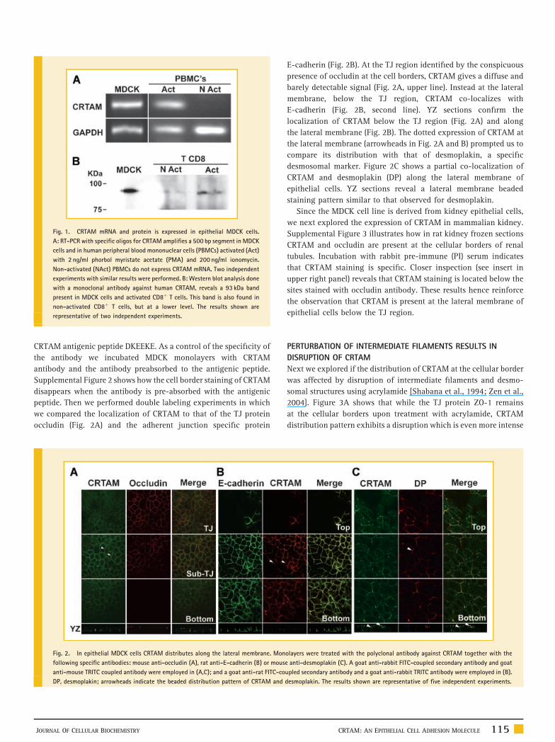
Fig. 1. CRTAM mRNA and protein is expressed in epithelial MDCK cells.
A: RT-PCR with specific oligos for CRTAM amplifies a 500 bp segment in MDCK
cells and in human peripheral blood mononuclear cells (PBMCs) activated (Act)
with 2 ng/ml phorbol myristate acetate (PMA) and 200 ng/ml ionomycin.
Non-activated (NAct) PBMCs do not express CRTAM mRNA. Two independent
experiments with similar results were performed. B: Western blot analysis done
with a monoclonal antibody against human CRTAM, reveals a 93 kDa band
present in MDCK cells and activated CD8þ T cells. This band is also found in
non-activated CD8þ T cells, but at a lower level. The results shown are
representative of two independent experiments.
CRTAM antigenic peptide DKEEKE. As a control of the specificity of
the antibody we incubated MDCK monolayers with CRTAM
antibody and the antibody preabsorbed to the antigenic peptide.
Supplemental Figure 2 shows how the cell border staining of CRTAM
disappears when the antibody is pre-absorbed with the antigenic
peptide. Then we performed double labeling experiments in which
we compared the localization of CRTAM to that of the TJ protein
occludin (Fig. 2A) and the adherent junction specific protein
Fig. 2. In epithelial MDCK cells CRTAM distributes along the lateral membrane. Mon
following specific antibodies: mouse anti-occludin (A), rat anti-E-cadherin (B) or mous
anti-mouse TRITC coupled antibody were employed in (A,C); and a goat anti-rat FITC-co
DP, desmoplakin; arrowheads indicate the beaded distribution pattern of CRTAM and
JOURNAL OF CELLULAR BIOCHEMISTRY
E-cadherin (Fig. 2B). At the TJ region identified by the conspicuous
presence of occludin at the cell borders, CRTAM gives a diffuse and
barely detectable signal (Fig. 2A, upper line). Instead at the lateral
membrane, below the TJ region, CRTAM co-localizes with
E-cadherin (Fig. 2B, second line). YZ sections confirm the
localization of CRTAM below the TJ region (Fig. 2A) and along
the lateral membrane (Fig. 2B). The dotted expression of CRTAM at
the lateral membrane (arrowheads in Fig. 2A and B) prompted us to
compare its distribution with that of desmoplakin, a specific
desmosomal marker. Figure 2C shows a partial co-localization of
CRTAM and desmoplakin (DP) along the lateral membrane of
epithelial cells. YZ sections reveal a lateral membrane beaded
staining pattern similar to that observed for desmoplakin.
Since the MDCK cell line is derived from kidney epithelial cells,
we next explored the expression of CRTAM in mammalian kidney.
Supplemental Figure 3 illustrates how in rat kidney frozen sections
CRTAM and occludin are present at the cellular borders of renal
tubules. Incubation with rabbit pre-immune (PI) serum indicates
that CRTAM staining is specific. Closer inspection (see insert in
upper right panel) reveals that CRTAM staining is located below the
sites stained with occludin antibody. These results hence reinforce
the observation that CRTAM is present at the lateral membrane of
epithelial cells below the TJ region.
PERTURBATION OF INTERMEDIATE FILAMENTS RESULTS IN
DISRUPTION OF CRTAM
Next we explored if the distribution of CRTAM at the cellular border
was affected by disruption of intermediate filaments and desmo-
somal structures using acrylamide [Shabana et al., 1994; Zen et al.,
2004]. Figure 3A shows that while the TJ protein ZO-1 remains
at the cellular borders upon treatment with acrylamide, CRTAM
distribution pattern exhibits a disruption which is even more intense
olayers were treated with the polyclonal antibody against CRTAM together with the
e anti-desmoplakin (C). A goat anti-rabbit FITC-coupled secondary antibody and goat
upled secondary antibody and a goat anti-rabbit TRITC antibody were employed in (B).
desmoplakin. The results shown are representative of five independent experiments.
CRTAM: AN EPITHELIAL CELL ADHESION MOLECULE 115
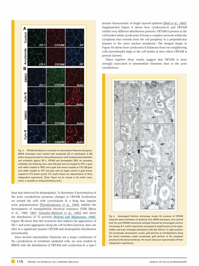
Fig. 4. Immunogold electron microscopy reveals the presence of CRTAM
along the lateral membrane of epithelial cells. MDCK monolayers were stained
with the anti-CRTAM monoclonal antibody followed by immunogold electron
microscopy. B, C, and D, respectively, correspond to amplifications of the upper,
middle, and lower rectangles delineated in (A). Bar 200 nm. TJ, tight junction;
full arrowheads, desmosomes; arrows, gold particles at interdigitations along
the lateral membrane; empty arrowheads, gold particles at the cytoplasm
proximal to the lateral membrane. The results shown are representative of three
independent experiments.
Fig. 3. CRTAM distribution is sensitive to intermediate filaments disruption.
MDCK monolayers were treated with acrylamide (A) or cytochalasin B (B)
before being processed for immunofluorescence with rhodaminated phalloidin
and antibodies against ZO-1, CRTAM and desmoplakin (DP). As secondary
antibodies the following were used: (A) goat anti-rat coupled to FITC, a goat
anti-rabbit coupled to TRITC and a goat anti-mouse coupled to CY5; (B) goat
anti-rabbit coupled to FITC and goat anti-rat (upper panel) or goat-mouse
coupled to CY5 (lower panel). The results shown are representative of three
independent experiments. [Color figure can be viewed in the online issue,
which is available at wileyonlinelibrary.com.]
than that observed for desmoplakin. To determine if perturbation of
the actin cytoskeleton promotes changes in CRTAM localization
we treated the cells with cytochalasin B, a drug that impairs
actin polymerization [Theodoropoulos et al., 1994], inhibits the
development of transepithelial electrical resistance (TER) [Meza
et al., 1980, 1982; Gonzalez-Mariscal et al., 1985] and alters
the distribution of TJ proteins [Nybom and Magnusson, 1996].
Figure 3B shows that this treatment that induces the appearance of
ZO-1 and actin aggregates along the cell borders (arrows), does not
alter in a significant manner CRTAM and desmoplakin distribution
(arrowheads).
Since keratin intermediate filaments are a major constituent of
the cytoskeleton of vertebrate epithelial cells, we next studied in
MDCK cells the distribution of CRTAM and cytokeratin-8, a type I
116 CRTAM: AN EPITHELIAL CELL ADHESION MOLECULE
keratin characteristic of single layered epithelia [Moll et al., 1982].
Supplemental Figure 4 shows how cytokeratin-8 and CRTAM
exhibit very different distribution patterns: CRTAM is present at the
cell borders while cytokeratin-8 forms a complex network within the
cytoplasm that extends from the cell periphery in a perpendicular
manner to the outer nuclear membrane. The merged image in
Figure S4 shows how cytokeratin 8 filaments from two neighboring
cells (arrowheads) align at the cell border at sites where CRTAM is
present (arrow).
Taken together these results suggest that CRTAM is more
strongly associated to intermediate filaments than to the actin
cytoskeleton.
JOURNAL OF CELLULAR BIOCHEMISTRY
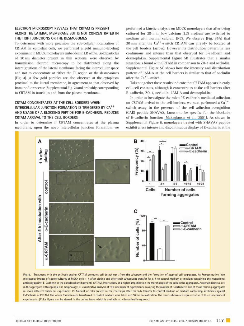
ELECTRON MICROSCOPY REVEALS THAT CRTAM IS PRESENT
ALONG THE LATERAL MEMBRANE BUT IS NOT CONCENTRATED IN
THE TIGHT JUNCTIONS OR THE DESMOSOMES
To determine with more precision the sub-cellular localization of
CRTAM in epithelial cells, we performed a gold immuno-labeling
experiment in MDCK monolayers embedded in LR white. Gold particles
of 20nm diameter present in thin sections, were observed by
transmission electron microscopy to be distributed along the
interdigitations of the lateral membrane facing the intercellular space
and not to concentrate at either the TJ region or the desmosomes
(Fig. 4). A few gold particles are also observed at the cytoplasm
proximal to the lateral membrane, in agreement to that observed by
immunofuorescence (Supplemental Fig. 2) and probably corresponding
to CRTAM in transit to and from the plasma membrane.
CRTAM CONCENTRATES AT THE CELL BORDERS WHEN
INTERCELLULAR JUNCTION FORMATION IS TRIGGERED BY CA2R
AND USAGE OF A BLOCKING PEPTIDE FOR E-CADHERIN, REDUCES
CRTAM ARRIVAL TO THE CELL BORDERS
In order to determine if CRTAM concentrates at the plasma
membrane, upon the novo intercellular junction formation, we
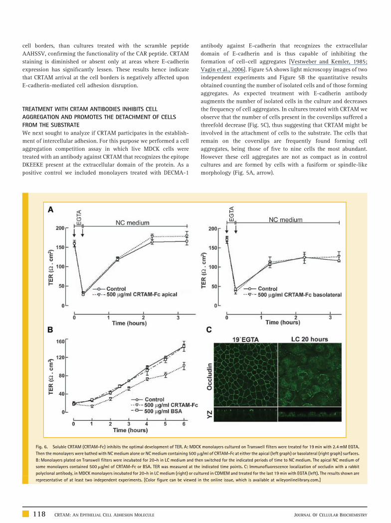
Fig. 5. Treatment with the antibody against CRTAM promotes cell detachment from
microscopy images of sparse cultures of MDCK cells 1-h after plating and after their su
antibody against E-Cadherin or the polyclonal antibody anti-CRTAM. Inserts show at a hig
in the aggregate with a spindle like morphology. B: Quantitative analysis of two independ
in seven different fields per experiment. C: Amount of cells present in the coverslips
E-Cadherin or CRTAM. The values found in cells transferred to control medium were take
experiments. [Color figure can be viewed in the online issue, which is available at wil
JOURNAL OF CELLULAR BIOCHEMISTRY
performed a kinetic analysis on MDCK monolayers that after being
cultured for 20-h in low calcium (LC) medium are switched to
medium with normal calcium (NC). We observe (Fig. S5A) that
20 min after the Ca2þ-switch CRTAM can already be located at
the cell borders (arrow). However its distribution pattern is less
continuous and intense than that observed for E-cadherin and
desmoplakin. Supplemental Figure 5B illustrates that a similar
situation is found with CRTAM in comparison to ZO-1 and occludin.
Supplemental Figure 5C shows how the intensity and distribution
pattern of JAM-A at the cell borders is similar to that of occludin
after the Ca2þ-switch.
Taken together these results indicate that CRTAM appears in early
cell–cell contacts, although it concentrates at the cell borders after
E-cadherin, ZO-1, occludin, JAM-A and desmoplakin.
In order to investigate the role of E-cadherin-mediated adhesion
on CRTAM arrival to the cell borders, we next performed a Ca2þ-
switch assay in the presence of the cell adhesion recognition
(CAR) peptide SHAVAS, known to be specific for the blockade
of E-cadherin function [Makagiansar et al., 2001]. As shown in
Supplemental Figure 6, monolayers treated with SHAVAS peptide
exhibit a less intense and discontinuous display of E-cadherin at the
the substrate and the formation of atypical cell aggregates. A: Representative light
bsequent transfer for 5-h to control medium or medium containing the monoclonal
her amplification the morphology of the cells in the aggregates. Arrows indicates a cell
ent experiments, counting the number of isolated cells and of those forming aggregates
after the 5-h transfer to control medium or medium containing antibodies against
n as 100 for normalization. The results shown are representative of three independent
eyonlinelibrary.com.]
CRTAM: AN EPITHELIAL CELL ADHESION MOLECULE 117
cell borders, than cultures treated with the scramble peptide
AAHSSV, confirming the functionality of the CAR peptide. CRTAM
staining is diminished or absent only at areas where E-cadherin
expression has significantly lessen. These results hence indicate
that CRTAM arrival at the cell borders is negatively affected upon
E-cadherin-mediated cell adhesion disruption.
TREATMENT WITH CRTAM ANTIBODIES INHIBITS CELL
AGGREGATION AND PROMOTES THE DETACHMENT OF CELLS
FROM THE SUBSTRATE
We next sought to analyze if CRTAM participates in the establish-
ment of intercellular adhesion. For this purpose we performed a cell
aggregation competition assay in which live MDCK cells were
treated with an antibody against CRTAM that recognizes the epitope
DKEEKE present at the extracellular domain of the protein. As a
positive control we included monolayers treated with DECMA-1
Fig. 6. Soluble CRTAM (CRTAM-Fc) inhibits the optimal development of TER. A: MDCK
Then the monolayers were bathed with NC medium alone or NC medium containing 500m
B: Monolayers plated on Transwell filters were incubated for 20-h in LC medium and the
some monolayers contained 500mg/ml of CRTAM-Fc or BSA. TER was measured at the
polyclonal antibody, in MDCK monolayers incubated for 20-h in LC medium (right) or cul
representative of at least two independent experiments. [Color figure can be viewed i
118 CRTAM: AN EPITHELIAL CELL ADHESION MOLECULE
antibody against E-cadherin that recognizes the extracellular
domain of E-cadherin and is thus capable of inhibiting the
formation of cell–cell aggregates [Vestweber and Kemler, 1985;
Vagin et al., 2006]. Figure 5A shows light microscopy images of two
independent experiments and Figure 5B the quantitative results
obtained counting the number of isolated cells and of those forming
aggregates. As expected treatment with E-cadherin antibody
augments the number of isolated cells in the culture and decreases
the frequency of cell aggregates. In cultures treated with CRTAM we
observe that the number of cells present in the coverslips suffered a
threefold decrease (Fig. 5C), thus suggesting that CRTAM might be
involved in the attachment of cells to the substrate. The cells that
remain on the coverslips are frequently found forming cell
aggregates, being those of five to nine cells the most abundant.
However these cell aggregates are not as compact as in control
cultures and are formed by cells with a fusiform or spindle-like
morphology (Fig. 5A, arrow).
monolayers cultured on Transwell filters were treated for 19 min with 2.4 mM EGTA.
g/ml of CRTAM-Fc at either the apical (left graph) or basolateral (right graph) surfaces.
n switched for the indicated periods of time to NC medium. The apical NC medium of
indicated time points. C: Immunofluorescence localization of occludin with a rabbit
tured in CDMEM and treated for the last 19 min with EGTA (left). The results shown are
n the online issue, which is available at wileyonlinelibrary.com.]
JOURNAL OF CELLULAR BIOCHEMISTRY
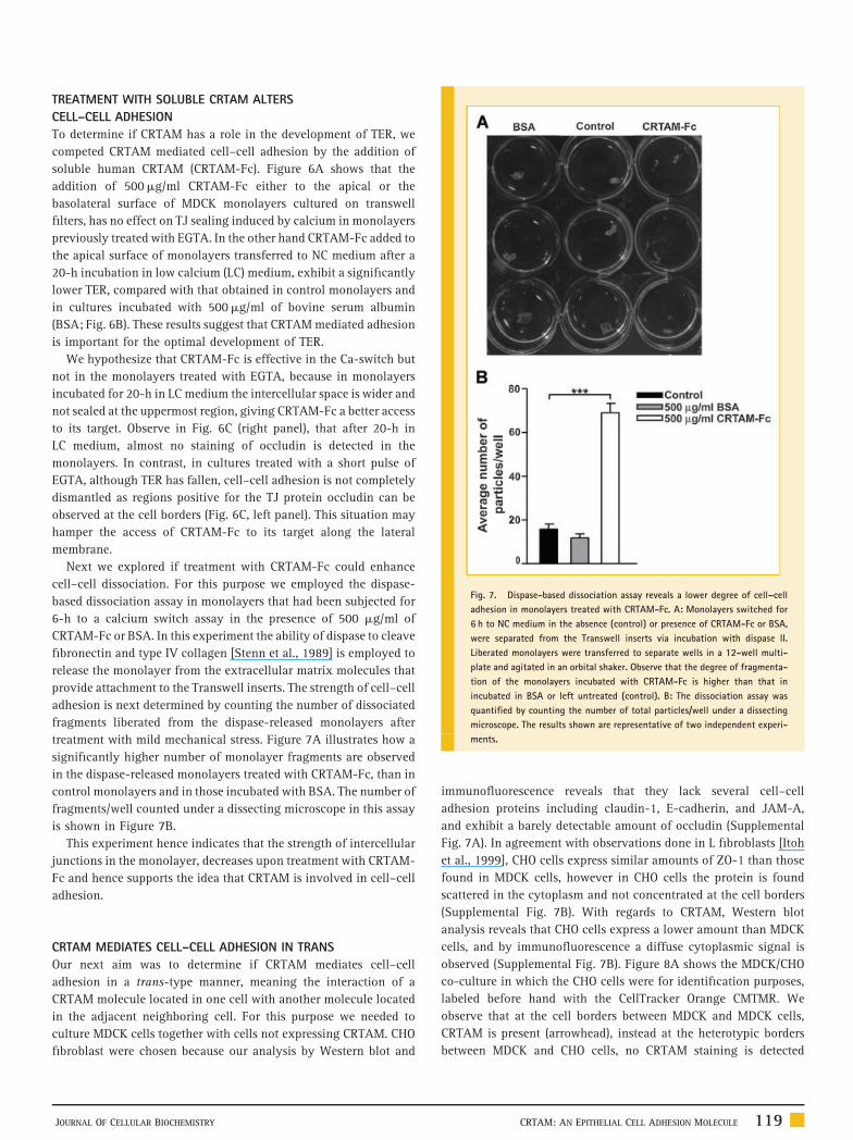
Fig. 7. Dispase-based dissociation assay reveals a lower degree of cell–cell
adhesion in monolayers treated with CRTAM-Fc. A: Monolayers switched for
6 h to NC medium in the absence (control) or presence of CRTAM-Fc or BSA,
were separated from the Transwell inserts via incubation with dispase II.
Liberated monolayers were transferred to separate wells in a 12-well multi-
plate and agitated in an orbital shaker. Observe that the degree of fragmenta-
tion of the monolayers incubated with CRTAM-Fc is higher than that in
incubated in BSA or left untreated (control). B: The dissociation assay was
quantified by counting the number of total particles/well under a dissecting
microscope. The results shown are representative of two independent experi-
ments.
TREATMENT WITH SOLUBLE CRTAM ALTERS
CELL–CELL ADHESION
To determine if CRTAM has a role in the development of TER, we
competed CRTAM mediated cell–cell adhesion by the addition of
soluble human CRTAM (CRTAM-Fc). Figure 6A shows that the
addition of 500mg/ml CRTAM-Fc either to the apical or the
basolateral surface of MDCK monolayers cultured on transwell
filters, has no effect on TJ sealing induced by calcium in monolayers
previously treated with EGTA. In the other hand CRTAM-Fc added to
the apical surface of monolayers transferred to NC medium after a
20-h incubation in low calcium (LC) medium, exhibit a significantly
lower TER, compared with that obtained in control monolayers and
in cultures incubated with 500mg/ml of bovine serum albumin
(BSA; Fig. 6B). These results suggest that CRTAM mediated adhesion
is important for the optimal development of TER.
We hypothesize that CRTAM-Fc is effective in the Ca-switch but
not in the monolayers treated with EGTA, because in monolayers
incubated for 20-h in LC medium the intercellular space is wider and
not sealed at the uppermost region, giving CRTAM-Fc a better access
to its target. Observe in Fig. 6C (right panel), that after 20-h in
LC medium, almost no staining of occludin is detected in the
monolayers. In contrast, in cultures treated with a short pulse of
EGTA, although TER has fallen, cell–cell adhesion is not completely
dismantled as regions positive for the TJ protein occludin can be
observed at the cell borders (Fig. 6C, left panel). This situation may
hamper the access of CRTAM-Fc to its target along the lateral
membrane.
Next we explored if treatment with CRTAM-Fc could enhance
cell–cell dissociation. For this purpose we employed the dispase-
based dissociation assay in monolayers that had been subjected for
6-h to a calcium switch assay in the presence of 500 mg/ml of
CRTAM-Fc or BSA. In this experiment the ability of dispase to cleave
fibronectin and type IV collagen [Stenn et al., 1989] is employed to
release the monolayer from the extracellular matrix molecules that
provide attachment to the Transwell inserts. The strength of cell–cell
adhesion is next determined by counting the number of dissociated
fragments liberated from the dispase-released monolayers after
treatment with mild mechanical stress. Figure 7A illustrates how a
significantly higher number of monolayer fragments are observed
in the dispase-released monolayers treated with CRTAM-Fc, than in
control monolayers and in those incubated with BSA. The number of
fragments/well counted under a dissecting microscope in this assay
is shown in Figure 7B.
This experiment hence indicates that the strength of intercellular
junctions in the monolayer, decreases upon treatment with CRTAM-
Fc and hence supports the idea that CRTAM is involved in cell–cell
adhesion.
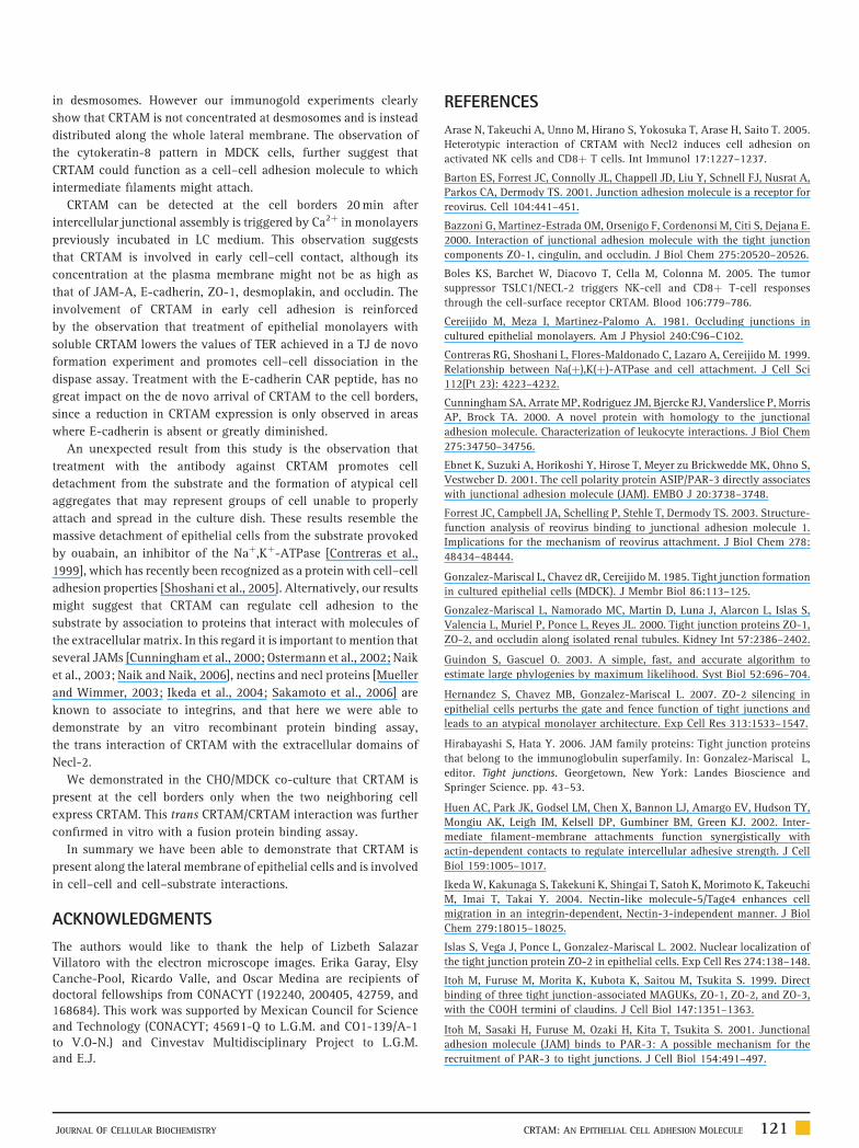
CRTAM MEDIATES CELL–CELL ADHESION IN TRANS
Our next aim was to determine if CRTAM mediates cell–cell
adhesion in a trans-type manner, meaning the interaction of a
CRTAM molecule located in one cell with another molecule located
in the adjacent neighboring cell. For this purpose we needed to
culture MDCK cells together with cells not expressing CRTAM. CHO
fibroblast were chosen because our analysis by Western blot and
JOURNAL OF CELLULAR BIOCHEMISTRY
immunofluorescence reveals that they lack several cell–cell
adhesion proteins including claudin-1, E-cadherin, and JAM-A,
and exhibit a barely detectable amount of occludin (Supplemental
Fig. 7A). In agreement with observations done in L fibroblasts [Itoh
et al., 1999], CHO cells express similar amounts of ZO-1 than those
found in MDCK cells, however in CHO cells the protein is found
scattered in the cytoplasm and not concentrated at the cell borders
(Supplemental Fig. 7B). With regards to CRTAM, Western blot
analysis reveals that CHO cells express a lower amount than MDCK
cells, and by immunofluorescence a diffuse cytoplasmic signal is
observed (Supplemental Fig. 7B). Figure 8A shows the MDCK/CHO
co-culture in which the CHO cells were for identification purposes,
labeled before hand with the CellTracker Orange CMTMR. We
observe that at the cell borders between MDCK and MDCK cells,
CRTAM is present (arrowhead), instead at the heterotypic borders
between MDCK and CHO cells, no CRTAM staining is detected
CRTAM: AN EPITHELIAL CELL ADHESION MOLECULE 119
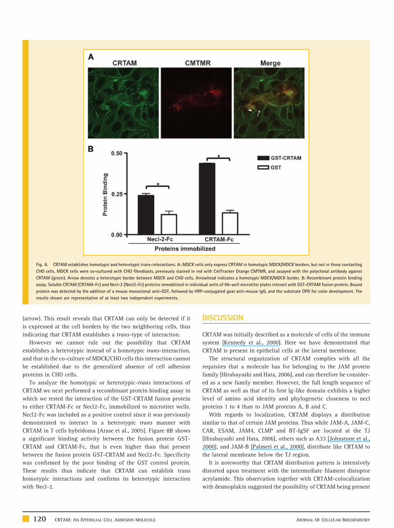
Fig. 8. CRTAM establishes homotypic and heterotypic trans-interactions. A: MDCK cells only express CRTAM in homotypic MDCK/MDCK borders, but not in those contacting
CHO cells. MDCK cells were co-cultured with CHO fibroblasts, previously stained in red with CellTracker Orange CMTMR, and assayed with the polyclonal antibody against
CRTAM (green). Arrow denotes a heterotypic border between MDCK and CHO cells. Arrowhead indicates a homotypic MDCK/MDCK border. B: Recombinant protein binding
assay. Soluble CRTAM (CRTAM-Fc) and Necl-2 (Necl2-Fc) proteins immobilized in individual wells of 96-well microtiter plates interact with GST-CRTAM fusion protein. Bound
protein was detected by the addition of a mouse monoclonal anti-GST, followed by HRP-conjugated goat anti-mouse IgG, and the substrate OPD for color development. The
results shown are representative of at least two independent experiments.
(arrow). This result reveals that CRTAM can only be detected if it
is expressed at the cell borders by the two neighboring cells, thus
indicating that CRTAM establishes a trans-type of interaction.
However we cannot rule out the possibility that CRTAM
establishes a heterotypic instead of a homotypic trans-interaction,
and that in the co-culture of MDCK/CHO cells this interaction cannot
be established due to the generalized absence of cell adhesion
proteins in CHO cells.
To analyze the homotypic or heterotypic-trans interactions of
CRTAM we next performed a recombinant protein binding assay in
which we tested the interaction of the GST-CRTAM fusion protein
to either CRTAM-Fc or Necl2-Fc, immobilized to microtiter wells.
Necl2-Fc was included as a positive control since it was previously
demonstrated to interact in a heterotypic trans manner with
CRTAM in T cells hybridoma [Arase et al., 2005]. Figure 8B shows
a significant binding activity between the fusion protein GST-
CRTAM and CRTAM-Fc, that is even higher than that present
between the fusion protein GST-CRTAM and Necl2-Fc. Specificity
was confirmed by the poor binding of the GST control protein.
These results thus indicate that CRTAM can establish trans
homotypic interactions and confirms its heterotypic interaction
with Necl-2.
120 CRTAM: AN EPITHELIAL CELL ADHESION MOLECULE
DISCUSSION
CRTAM was initially described as a molecule of cells of the immune
system [Kennedy et al., 2000]. Here we have demonstrated that
CRTAM is present in epithelial cells at the lateral membrane.
The structural organization of CRTAM complies with all the
requisites that a molecule has for belonging to the JAM protein
family [Hirabayashi and Hata, 2006], and can therefore be consider-
ed as a new family member. However, the full length sequence of
CRTAM as well as that of its first Ig-like domain exhibits a higher
level of amino acid identity and phylogenetic closeness to necl
proteins 1 to 4 than to JAM proteins A, B and C.
With regards to localization, CRTAM displays a distribution
similar to that of certain JAM proteins. Thus while JAM-A, JAM-C,
CAR, ESAM, JAM4, CLMP and BT-IgSF are located at the TJ
[Hirabayashi and Hata, 2006], others such as A33 [Johnstone et al.,
2000], and JAM-B [Palmeri et al., 2000], distribute like CRTAM to
the lateral membrane below the TJ region.
It is noteworthy that CRTAM distribution pattern is intensively
distorted upon treatment with the intermediate filament disruptor
acrylamide. This observation together with CRTAM-colocalization
with desmoplakin suggested the possibility of CRTAM being present
JOURNAL OF CELLULAR BIOCHEMISTRY
in desmosomes. However our immunogold experiments clearly
show that CRTAM is not concentrated at desmosomes and is instead
distributed along the whole lateral membrane. The observation of
the cytokeratin-8 pattern in MDCK cells, further suggest that
CRTAM could function as a cell–cell adhesion molecule to which
intermediate filaments might attach.
CRTAM can be detected at the cell borders 20 min after
intercellular junctional assembly is triggered by Ca2þ in monolayers
previously incubated in LC medium. This observation suggests
that CRTAM is involved in early cell–cell contact, although its
concentration at the plasma membrane might not be as high as
that of JAM-A, E-cadherin, ZO-1, desmoplakin, and occludin. The
involvement of CRTAM in early cell adhesion is reinforced
by the observation that treatment of epithelial monolayers with
soluble CRTAM lowers the values of TER achieved in a TJ de novo
formation experiment and promotes cell–cell dissociation in the
dispase assay. Treatment with the E-cadherin CAR peptide, has no
great impact on the de novo arrival of CRTAM to the cell borders,
since a reduction in CRTAM expression is only observed in areas
where E-cadherin is absent or greatly diminished.
An unexpected result from this study is the observation that
treatment with the antibody against CRTAM promotes cell
detachment from the substrate and the formation of atypical cell
aggregates that may represent groups of cell unable to properly
attach and spread in the culture dish. These results resemble the
massive detachment of epithelial cells from the substrate provoked
by ouabain, an inhibitor of the Naþ,Kþ-ATPase [Contreras et al.,
1999], which has recently been recognized as a protein with cell–cell
adhesion properties [Shoshani et al., 2005]. Alternatively, our results
might suggest that CRTAM can regulate cell adhesion to the
substrate by association to proteins that interact with molecules of
the extracellular matrix. In this regard it is important to mention that
several JAMs [Cunningham et al., 2000; Ostermann et al., 2002; Naik
et al., 2003; Naik and Naik, 2006], nectins and necl proteins [Mueller
and Wimmer, 2003; Ikeda et al., 2004; Sakamoto et al., 2006] are
known to associate to integrins, and that here we were able to
demonstrate by an vitro recombinant protein binding assay,
the trans interaction of CRTAM with the extracellular domains of
Necl-2.
We demonstrated in the CHO/MDCK co-culture that CRTAM is
present at the cell borders only when the two neighboring cell
express CRTAM. This trans CRTAM/CRTAM interaction was further
confirmed in vitro with a fusion protein binding assay.
In summary we have been able to demonstrate that CRTAM is
present along the lateral membrane of epithelial cells and is involved
in cell–cell and cell–substrate interactions.
ACKNOWLEDGMENTS
The authors would like to thank the help of Lizbeth SalazarVillatoro with the electron microscope images. Erika Garay, ElsyCanche-Pool, Ricardo Valle, and Oscar Medina are recipients ofdoctoral fellowships from CONACYT (192240, 200405, 42759, and168684). This work was supported by Mexican Council for Scienceand Technology (CONACYT; 45691-Q to L.G.M. and CO1-139/A-1to V.O-N.) and Cinvestav Multidisciplinary Project to L.G.M.and E.J.
JOURNAL OF CELLULAR BIOCHEMISTRY
REFERENCES
Arase N, Takeuchi A, Unno M, Hirano S, Yokosuka T, Arase H, Saito T. 2005.Heterotypic interaction of CRTAM with Necl2 induces cell adhesion onactivated NK cells and CD8þ T cells. Int Immunol 17:1227–1237.
Barton ES, Forrest JC, Connolly JL, Chappell JD, Liu Y, Schnell FJ, Nusrat A,Parkos CA, Dermody TS. 2001. Junction adhesion molecule is a receptor forreovirus. Cell 104:441–451.
Bazzoni G, Martinez-Estrada OM, Orsenigo F, Cordenonsi M, Citi S, Dejana E.2000. Interaction of junctional adhesion molecule with the tight junctioncomponents ZO-1, cingulin, and occludin. J Biol Chem 275:20520–20526.
Boles KS, Barchet W, Diacovo T, Cella M, Colonna M. 2005. The tumorsuppressor TSLC1/NECL-2 triggers NK-cell and CD8þ T-cell responsesthrough the cell-surface receptor CRTAM. Blood 106:779–786.
Cereijido M, Meza I, Martinez-Palomo A. 1981. Occluding junctions incultured epithelial monolayers. Am J Physiol 240:C96–C102.
Contreras RG, Shoshani L, Flores-Maldonado C, Lazaro A, Cereijido M. 1999.Relationship between Na(þ),K(þ)-ATPase and cell attachment. J Cell Sci112(Pt 23): 4223–4232.
Cunningham SA, Arrate MP, Rodriguez JM, Bjercke RJ, Vanderslice P, MorrisAP, Brock TA. 2000. A novel protein with homology to the junctionaladhesion molecule. Characterization of leukocyte interactions. J Biol Chem275:34750–34756.
Ebnet K, Suzuki A, Horikoshi Y, Hirose T, Meyer zu Brickwedde MK, Ohno S,Vestweber D. 2001. The cell polarity protein ASIP/PAR-3 directly associateswith junctional adhesion molecule (JAM). EMBO J 20:3738–3748.
Forrest JC, Campbell JA, Schelling P, Stehle T, Dermody TS. 2003. Structure-function analysis of reovirus binding to junctional adhesion molecule 1.Implications for the mechanism of reovirus attachment. J Biol Chem 278:48434–48444.
Gonzalez-Mariscal L, Chavez dR, Cereijido M. 1985. Tight junction formationin cultured epithelial cells (MDCK). J Membr Biol 86:113–125.
Gonzalez-Mariscal L, Namorado MC, Martin D, Luna J, Alarcon L, Islas S,Valencia L, Muriel P, Ponce L, Reyes JL. 2000. Tight junction proteins ZO-1,ZO-2, and occludin along isolated renal tubules. Kidney Int 57:2386–2402.
Guindon S, Gascuel O. 2003. A simple, fast, and accurate algorithm toestimate large phylogenies by maximum likelihood. Syst Biol 52:696–704.
Hernandez S, Chavez MB, Gonzalez-Mariscal L. 2007. ZO-2 silencing inepithelial cells perturbs the gate and fence function of tight junctions andleads to an atypical monolayer architecture. Exp Cell Res 313:1533–1547.
Hirabayashi S, Hata Y. 2006. JAM family proteins: Tight junction proteinsthat belong to the immunoglobulin superfamily. In: Gonzalez-Mariscal L,editor. Tight junctions. Georgetown, New York: Landes Bioscience andSpringer Science. pp. 43–53.
Huen AC, Park JK, Godsel LM, Chen X, Bannon LJ, Amargo EV, Hudson TY,Mongiu AK, Leigh IM, Kelsell DP, Gumbiner BM, Green KJ. 2002. Inter-mediate filament-membrane attachments function synergistically withactin-dependent contacts to regulate intercellular adhesive strength. J CellBiol 159:1005–1017.
Ikeda W, Kakunaga S, Takekuni K, Shingai T, Satoh K, Morimoto K, TakeuchiM, Imai T, Takai Y. 2004. Nectin-like molecule-5/Tage4 enhances cellmigration in an integrin-dependent, Nectin-3-independent manner. J BiolChem 279:18015–18025.
Islas S, Vega J, Ponce L, Gonzalez-Mariscal L. 2002. Nuclear localization ofthe tight junction protein ZO-2 in epithelial cells. Exp Cell Res 274:138–148.
Itoh M, Furuse M, Morita K, Kubota K, Saitou M, Tsukita S. 1999. Directbinding of three tight junction-associated MAGUKs, ZO-1, ZO-2, and ZO-3,with the COOH termini of claudins. J Cell Biol 147:1351–1363.
Itoh M, Sasaki H, Furuse M, Ozaki H, Kita T, Tsukita S. 2001. Junctionaladhesion molecule (JAM) binds to PAR-3: A possible mechanism for therecruitment of PAR-3 to tight junctions. J Cell Biol 154:491–497.
CRTAM: AN EPITHELIAL CELL ADHESION MOLECULE 121
Johnson-Leger CA, Aurrand-Lions M, Beltraminelli N, Fasel N, Imhof BA.2002. Junctional adhesion molecule-2 (JAM-2) promotes lymphocyte trans-endothelial migration. Blood 100:2479–2486.
Johnstone CN, Tebbutt NC, Abud HE, White SJ, Stenvers KL, Hall NE, CodySH, Whitehead RH, Catimel B, Nice EC, Burgess AW, Heath JK. 2000.Characterization of mouse A33 antigen, a definitive marker for basolateralsurfaces of intestinal epithelial cells. Am J Physiol Gastrointest Liver Physiol279:G500–G510.
Kennedy J, Vicari AP, Saylor V, Zurawski SM, Copeland NG, Gilbert DJ,Jenkins NA, Zlotnik A. 2000. A molecular analysis of NKT cells: Identifica-tion of a class-I restricted T cell-associated molecule (CRTAM). J Leukoc Biol67:725–734.
Kornecki E, Walkowiak B, Naik UP, Ehrlich YH. 1990. Activation of humanplatelets by a stimulatory monoclonal antibody. J Biol Chem 265:10042–10048.
Makagiansar IT, Avery M, Hu Y, Audus KL, Siahaan TJ. 2001. Improving theselectivity of HAV-peptides in modulating E-cadherin-E-cadherin interac-tions in the intercellular junction of MDCK cell monolayers. Pharm Res18:446–453.
Martin-Padura I, Lostaglio S, Schneemann M, Williams L, Romano M,Fruscella P, Panzeri C, Stoppacciaro A, Ruco L, Villa A, Simmons D, DejanaE. 1998. Junctional adhesion molecule, a novel member of the immunoglo-bulin superfamily that distributes at intercellular junctions and modulatesmonocyte transmigration. J Cell Biol 142:117–127.
Martinez-Estrada OM, Villa A, Breviario F, Orsenigo F, Dejana E, Bazzoni G.2001. Association of junctional adhesion molecule with calcium/calmodu-lin-dependent serine protein kinase (CASK/LIN-2) in human epithelial caco-2cells. J Biol Chem 276:9291–9296.
Medina-Contreras O, Soldevila G, Patino-Lopez G, Canche-Pool E, Valle-RiosR, Ortiz-Navarrete V. 2010. Role of CRTAM during mouse early T lympho-cytes development. Dev Comp Immunol 34:196–202.
Meza I, Ibarra G, Sabanero M, Martinez-Palomo A, Cereijido M. 1980.Occluding junctions and cytoskeletal components in a cultured transportingepithelium. J Cell Biol 87:746–754.
Meza I, Sabanero M, Stefani E, Cereijido M. 1982. Occluding junctions inMDCK cells: Modulation of transepithelial permeability by the cytoskeleton.J Cell Biochem 18:407–421.
Moll R, Franke WW, Schiller DL, Geiger B, Krepler R. 1982. The catalog ofhuman cytokeratins: Patterns of expression in normal epithelia, tumors andcultured cells. Cell 31:11–24.
Mueller S, Wimmer E. 2003. Recruitment of nectin-3 to cell-cell junctionsthrough trans-heterophilic interaction with CD155, a vitronectin and polio-virus receptor that localizes to alpha(v)beta3 integrin-containing membranemicrodomains. J Biol Chem 278:31251–31260.
Naik MU, Naik UP. 2006. Junctional adhesion molecule-A-induced endothe-lial cell migration on vitronectin is integrin alpha v beta 3 specific. J Cell Sci119:490–499.
Naik MU, Mousa SA, Parkos CA, Naik UP. 2003. Signaling through JAM-1and alphavbeta3 is required for the angiogenic action of bFGF: Dissociationof the JAM-1 and alphavbeta3 complex. Blood 102:2108–2114.
Nybom P, Magnusson KE. 1996. Modulation of the junctional integrity bylow or high concentrations of cytochalasin B and dihydrocytochalasin B isassociated with distinct changes in F-actin and ZO-1. Biosci Rep 16:313–326.
122 CRTAM: AN EPITHELIAL CELL ADHESION MOLECULE
Ogita H, Takai Y. 2006. Nectins and nectin-like molecules: Roles in celladhesion, polarization, movement, and proliferation. IUBMB Life 58:334–343.
Ostermann G, Weber KS, Zernecke A, Schroder A, Weber C. 2002. JAM-1 is aligand of the beta(2) integrin LFA-1 involved in transendothelial migrationof leukocytes. Nat Immunol 3:151–158.
Palmeri D, van Zante A, Huang CC, Hemmerich S, Rosen SD. 2000. Vascularendothelial junction-associated molecule, a novel member of the immuno-globulin superfamily, is localized to intercellular boundaries of endothelialcells. J Biol Chem 275:19139–19145.
Patino-Lopez G, Hevezi P, Lee J, Willhite D, Verge GM, Lechner SM,Ortiz-Navarrete V, Zlotnik A. 2006. Human class-I restricted T cellassociated molecule is highly expressed in the cerebellum and is a markerfor activated NKT and CD8þ T lymphocytes. J Neuroimmunol 171:145–155.
Sakamoto Y, Ogita H, Hirota T, Kawakatsu T, Fukuyama T, Yasumi M,Kanzaki N, Ozaki M, Takai Y. 2006. Interaction of integrin alpha(v)beta3with nectin. Implication in cross-talk between cell-matrix and cell-celljunctions. J Biol Chem 281:19631–19644.
Santoso S, Sachs UJ, Kroll H, Linder M, Ruf A, Preissner KT, Chavakis T. 2002.The junctional adhesion molecule 3 (JAM-3) on human platelets is acounterreceptor for the leukocyte integrin Mac-1. J Exp Med 196:679–691.
Shabana AH, Oboeuf M, Forest N. 1994. Cytoplasmic desmosomes andintermediate filament disturbance following acrylamide treatment in cul-tured rat keratinocytes. Tissue Cell 26:43–55.
Shoshani L, Contreras RG, Roldan ML, Moreno J, Lazaro A, Balda MS, MatterK, Cereijido M. 2005. The polarized expression of Naþ,Kþ-ATPase inepithelia depends on the association between beta-subunits located inneighboring cells. Mol Biol Cell 16:1071–1081.
Sobocka MB, Sobocki T, Banerjee P, Weiss C, Rushbrook JI, Norin AJ,Hartwig J, Salifu MO, Markell MS, Babinska A, Ehrlich YH, Kornecki E.2000. Cloning of the human platelet F11 receptor: A cell adhesion moleculemember of the immunoglobulin superfamily involved in platelet aggrega-tion. Blood 95:2600–2609.
Stenn KS, Link R, Moellmann G, Madri J, Kuklinska E. 1989. Dispase, aneutral protease from Bacillus polymyxa, is a powerful fibronectinase andtype IV collagenase. J Invest Dermatol 93:287–290.
Theodoropoulos PA, Gravanis A, Tsapara A, Margioris AN, Papadogiorgaki E,Galanopoulos V, Stournaras C. 1994. Cytochalasin B may shorten actinfilaments by a mechanism independent of barbed end capping. BiochemPharmacol 47:1875–1881.
Vagin O, Tokhtaeva E, Sachs G. 2006. The role of the beta1 subunit of theNa,K-ATPase and its glycosylation in cell-cell adhesion. J Biol Chem281:39573–39587.
Vestweber D, Kemler R. 1985. Identification of a putative cell adhesiondomain of uvomorulin. EMBO J 4:3393–3398.
Weber C, Fraemohs L, Dejana E. 2007. The role of junctional adhesionmolecules in vascular inflammation. Nat Rev Immunol 7:467–477.
Yeh JH, Sidhu SS, Chan AC. 2008. Regulation of a late phase of T cell polarityand effector functions by Crtam. Cell 132:846–859.
Zen K, Babbin BA, Liu Y, Whelan JB, Nusrat A, Parkos CA. 2004. JAM-C is acomponent of desmosomes and a ligand for CD11b/CD18-mediated neu-trophil transepithelial migration. Mol Biol Cell 15:3926–3937.
JOURNAL OF CELLULAR BIOCHEMISTRY
Related Documents