INFLAMMATORY BOWEL DISEASE Critical role of the CD40–CD40-ligand pathway in regulating mucosal inflammation-driven angiogenesis in inflammatory bowel disease S Danese, F Scaldaferri, S Vetrano, T Stefanelli, C Graziani, A Repici, R Ricci, G Straface, A Sgambato, A Malesci, C Fiocchi, S Rutella ................................................................................................................................... See end of article for authors’ affiliations ........................ Correspondence to: Silvio Danese, MD, Division of Gastroenterology, Istituto Clinico Humanitas, Via Manzoni 56, 20089 Rozzano, Milan, Italy; [email protected] Revised 13 February 2007 Accepted 13 February 2007 Published Online First 22 February 2007 ........................ Gut 2007;56:1248–1256. doi: 10.1136/gut.2006.111989 Background and aims: Angiogenesis is a novel component in inflammatory bowel disease (IBD) pathogenesis. We have previously shown that immune–nonimmune interactions through the CD40–CD40- ligand (CD40L) pathway might sustain gut inflammation, although their effect on regulating inflammation- driven angiogenesis is unknown. The present study evaluated the role of the CD40–CD40L interaction in the promotion of immune-mediated angiogenesis in IBD. Methods: Human nonimmune cells of colonic origin—namely, human intestinal fibroblasts (HIFs) and human intestinal microvascular endothelial cells (HIMECs)—were activated with either soluble CD40L (sCD40L), or CD40 + D1.1 cells or CD40L-activated lamina propria T (LPT) cells before measuring pro-angiogenic cytokine release. Blocking antibodies to either CD40 or CD40L were used to disrupt the CD40–CD40L interaction. The dextran sodium sulphate (DSS) model of experimental colitis in CD40 and CD40L knockout mice was established to assess whether the CD40–CD40L pathway was implicated in controlling inflammation-driven angiogenesis in vivo. Results: Engagement of CD40 on HIFs promoted the release of vascular endothelial growth factor (VEGF), interleukin-8 (IL-8) and hepatocyte growth factor (HGF). LPT cells were potent inducers of pro-angiogenic cytokine secretion by HIFs. Supernatants from sCD40L-activated HIFs induced migration of HIMECs and tubule formation, both of which were inhibited by blocking antibodies to either VEGF, IL-8 or HGF. Both CD40- and CD40L-deficient mice were protected from DSS-induced colitis and displayed a significant impairment of gut inflammation-driven angiogenesis, as assessed by microvascular density. Conclusions: The CD40–CD40L pathway appears to be crucially involved in regulating inflammation-driven angiogenesis, suggesting that strategies aimed at blocking CD40–CD40L interactions might be beneficial in acute and chronic intestinal injury. A ngiogenesis is a complex process whereby novel vessels can be generated from pre-existing ones; it involves endothelial cell (EC) division, degradation of vascular basement membrane and surrounding extracellular matrix, and EC migration. 1 Angiogenesis is tightly regulated by a balance between the expression and function of pro-angiogenic and anti-angiogenic mediators. Vascular endothelial growth factor (VEGF) is recognised as a potent pro-angiogenic cytokine and is secreted by monocytes/macrophages, ECs and a variety of other cell types. 2 Similarly, hepatocyte growth factor (HGF) possesses pro-angiogenic properties and promotes tumour cell motility, invasiveness and proliferation. 34 Interleukin (IL)-8, initially identified as a pro-inflammatory cytokine, is significantly upregulated in the colonic mucosa of patients with inflamma- tory bowel disease (IBD), in direct proportion with the degree of inflammation. 5 In addition to being a chemoattractant for neutrophils, IL-8 exerts effects on nonimmune cells, including human intestinal microvascular ECs (HIMECs), is a potent chemoattractant and promotes in vitro tube formation in HIMECs by binding IL-8 cognate receptor CXCR2. 6 Both colonic epithelial cells and HIMECs secrete IL-8 in a highly regulated fashion in vitro. 7 Lymphocytes and monocytes might mediate inflammatory- driven angiogenesis, which plays a key pathogenic role in several chronic inflammatory diseases, including rheumatoid arthritis, psoriasis and IBD. 8 In such disorders, leukocytes sustain inflammation by producing pro-angiogenic factors and stimulating angiogenesis. 9 10 We have previously shown that inflammation-driven angiogenesis actively occurs in both Crohn’s disease (CD) and ulcerative colitis (UC), being directly correlated with leukocyte infiltrate. Although many of the pathways that trigger inflammatory angiogenesis are unknown at present, the CD40–CD40-ligand (CD40L) system crucially mediates leukocyte–endothelial interactions in the inflamed intestine. 11 12 In particular, both molecules are highly upregu- lated in IBD, and are functional in the inflamed mucosa by triggering inflammatory cytokine production, cell-adhesion molecule expression and by fostering mucosal inflammation. To date, the role of the CD40–CD40L pathway in mediating inflammatory angiogenesis in the gut remains to be elucidated. Lymphocytes and monocytes produce pro-angiogenic factors and stimulate angiogenesis during inflammatory responses. 9 10 Excessive production of angiogenesis factors by activated T cells and/or activated macrophages might occur in delayed-type hypersensitivity and in chronic inflammatory disorders. 13 Abbreviations: APCs, antigen-presenting cells; CD, Crohn’s disease; CD40L, CD40 ligand; DAI, disease activity index; DSS, dextran sodium sulphate; ECs, endothelial cells; FBS, foetal bovine serum; FGF, fibroblast growth factor; HGF, hepatocyte growth factor; HIFs, human intestinal fibroblasts; HIMECs, human intestinal microvascular endothelial cells; HUVECs, human umbilical-vein endothelial cells; IBD, inflammatory bowel disease; IL-8, interleukin-8; KO, knockout; LP, lamina propria; LPT, lamina propria T cells; sCD40, soluble CD40; TNF, tumour necrosis factor; VEGF, vascular endothelial growth factor; WT, wild type 1248 www.gutjnl.com

Welcome message from author
This document is posted to help you gain knowledge. Please leave a comment to let me know what you think about it! Share it to your friends and learn new things together.
Transcript
INFLAMMATORY BOWEL DISEASE
Critical role of the CD40–CD40-ligand pathway in regulatingmucosal inflammation-driven angiogenesis in inflammatorybowel diseaseS Danese, F Scaldaferri, S Vetrano, T Stefanelli, C Graziani, A Repici, R Ricci, G Straface, A Sgambato,A Malesci, C Fiocchi, S Rutella. . . . . . . . . . . . . . . . . . . . . . . . . . . . . . . . . . . . . . . . . . . . . . . . . . . . . . . . . . . . . . . . . . . . . . . . . . . . . . . . . . . . . . . . . . . . . . . . . . . . . . . . . . . . . . . . . . . . . . . . . . . . . . . . . . .
See end of article forauthors’ affiliations. . . . . . . . . . . . . . . . . . . . . . . .
Correspondence to:Silvio Danese, MD, Divisionof Gastroenterology, IstitutoClinico Humanitas, ViaManzoni 56, 20089Rozzano, Milan, Italy;[email protected]
Revised 13 February 2007Accepted 13 February 2007Published Online First22 February 2007. . . . . . . . . . . . . . . . . . . . . . . .
Gut 2007;56:1248–1256. doi: 10.1136/gut.2006.111989
Background and aims: Angiogenesis is a novel component in inflammatory bowel disease (IBD)pathogenesis. We have previously shown that immune–nonimmune interactions through the CD40–CD40-ligand (CD40L) pathway might sustain gut inflammation, although their effect on regulating inflammation-driven angiogenesis is unknown. The present study evaluated the role of the CD40–CD40L interaction in thepromotion of immune-mediated angiogenesis in IBD.Methods: Human nonimmune cells of colonic origin—namely, human intestinal fibroblasts (HIFs) and humanintestinal microvascular endothelial cells (HIMECs)—were activated with either soluble CD40L (sCD40L), orCD40+ D1.1 cells or CD40L-activated lamina propria T (LPT) cells before measuring pro-angiogenic cytokinerelease. Blocking antibodies to either CD40 or CD40L were used to disrupt the CD40–CD40L interaction. Thedextran sodium sulphate (DSS) model of experimental colitis in CD40 and CD40L knockout mice wasestablished to assess whether the CD40–CD40L pathway was implicated in controlling inflammation-drivenangiogenesis in vivo.Results: Engagement of CD40 on HIFs promoted the release of vascular endothelial growth factor (VEGF),interleukin-8 (IL-8) and hepatocyte growth factor (HGF). LPT cells were potent inducers of pro-angiogeniccytokine secretion by HIFs. Supernatants from sCD40L-activated HIFs induced migration of HIMECs andtubule formation, both of which were inhibited by blocking antibodies to either VEGF, IL-8 or HGF. BothCD40- and CD40L-deficient mice were protected from DSS-induced colitis and displayed a significantimpairment of gut inflammation-driven angiogenesis, as assessed by microvascular density.Conclusions: The CD40–CD40L pathway appears to be crucially involved in regulating inflammation-drivenangiogenesis, suggesting that strategies aimed at blocking CD40–CD40L interactions might be beneficial inacute and chronic intestinal injury.
Angiogenesis is a complex process whereby novel vesselscan be generated from pre-existing ones; it involvesendothelial cell (EC) division, degradation of vascular
basement membrane and surrounding extracellular matrix, andEC migration.1 Angiogenesis is tightly regulated by a balancebetween the expression and function of pro-angiogenic andanti-angiogenic mediators. Vascular endothelial growth factor(VEGF) is recognised as a potent pro-angiogenic cytokine and issecreted by monocytes/macrophages, ECs and a variety of othercell types.2 Similarly, hepatocyte growth factor (HGF) possessespro-angiogenic properties and promotes tumour cell motility,invasiveness and proliferation.3 4 Interleukin (IL)-8, initiallyidentified as a pro-inflammatory cytokine, is significantlyupregulated in the colonic mucosa of patients with inflamma-tory bowel disease (IBD), in direct proportion with the degreeof inflammation.5 In addition to being a chemoattractant forneutrophils, IL-8 exerts effects on nonimmune cells, includinghuman intestinal microvascular ECs (HIMECs), is a potentchemoattractant and promotes in vitro tube formation inHIMECs by binding IL-8 cognate receptor CXCR2.6 Both colonicepithelial cells and HIMECs secrete IL-8 in a highly regulatedfashion in vitro.7
Lymphocytes and monocytes might mediate inflammatory-driven angiogenesis, which plays a key pathogenic role inseveral chronic inflammatory diseases, including rheumatoidarthritis, psoriasis and IBD.8 In such disorders, leukocytessustain inflammation by producing pro-angiogenic factors and
stimulating angiogenesis.9 10 We have previously shown thatinflammation-driven angiogenesis actively occurs in bothCrohn’s disease (CD) and ulcerative colitis (UC), being directlycorrelated with leukocyte infiltrate. Although many of thepathways that trigger inflammatory angiogenesis are unknownat present, the CD40–CD40-ligand (CD40L) system cruciallymediates leukocyte–endothelial interactions in the inflamedintestine.11 12 In particular, both molecules are highly upregu-lated in IBD, and are functional in the inflamed mucosa bytriggering inflammatory cytokine production, cell-adhesionmolecule expression and by fostering mucosal inflammation.To date, the role of the CD40–CD40L pathway in mediatinginflammatory angiogenesis in the gut remains to be elucidated.
Lymphocytes and monocytes produce pro-angiogenic factorsand stimulate angiogenesis during inflammatory responses.9 10
Excessive production of angiogenesis factors by activated T cellsand/or activated macrophages might occur in delayed-typehypersensitivity and in chronic inflammatory disorders.13
Abbreviations: APCs, antigen-presenting cells; CD, Crohn’s disease;CD40L, CD40 ligand; DAI, disease activity index; DSS, dextran sodiumsulphate; ECs, endothelial cells; FBS, foetal bovine serum; FGF, fibroblastgrowth factor; HGF, hepatocyte growth factor; HIFs, human intestinalfibroblasts; HIMECs, human intestinal microvascular endothelial cells;HUVECs, human umbilical-vein endothelial cells; IBD, inflammatory boweldisease; IL-8, interleukin-8; KO, knockout; LP, lamina propria; LPT, laminapropria T cells; sCD40, soluble CD40; TNF, tumour necrosis factor; VEGF,vascular endothelial growth factor; WT, wild type
1248
www.gutjnl.com

Interactions between CD40 and CD40L (or CD154) playpluripotent functions in inflammation, predominantly in theeffector phase of the immune response.11 14 15 CD40, a 50-kDtype-I transmembrane-glycoprotein member of the tumournecrosis factor (TNF) receptor gene family, is expressed bynumerous cell types, including professional antigen-presentingcells (APCs), monocytes and ECs.11 16 17 CD40L, a 33-kD type-IImembrane-protein member of the TNF family, is predomi-nantly expressed by activated CD4+ T cells and platelets.18 CD40triggering mediates several biological responses, includingimmunoglobulin isotype switching in B cells, expression of B7family molecules on APCs, adhesion-molecule expression byECs, and expression of cytokines and chemokines that arecharacteristic of the effector phases of the immune response.
Insights have recently been provided into the role of CD40–CD40L interactions in the induction of VEGF expression and in
VEGF-stimulated angiogenesis in vitro and in vivo.14 19 Humanumbilical-vein endothelial cells (HUVECs) and monocytes, butnot CD4+ T cells, release high amounts of VEGF after CD40triggering by soluble CD40L (sCD40L).14 Consistent with thesefindings, injection of human CD40L into SCID mice bearinghuman skin transplants translates into a significant enhance-ment of VEGF and fibroblast growth factor (FGF) expression inskin grafts.19 Finally, blockade of CD40L–CD40 interactionsprevents acute and chronic inflammation, including allograftrejection and atherosclerosis.20 21
CD40 and CD40L are overexpressed in IBD, suggesting apossible involvement of the CD40–CD40L pathway in diseasepathogenesis.11 Previously, we demonstrated that the CD40–CD40L pathway amplifies intestinal inflammation and links
Figure 1 VEGF production by CD40L-stimulated HIFs. Panel A: Humanintestinal fibroblasts (HIFs) were activated with soluble CD40 ligand(sCD40L) or were co-cultured either with CD40+ D1.1 cells or with CD402
Jurkat cells for 24 hours, before measuring vascular endothelial growthfactor (VEGF) release in culture supernatants. In selected experiments,blocking antibodies (Ab) to either CD40 (M2) or CD40L (M90) were usedto interfere with the CD40–CD40L interaction. Results are representative ofeight independent experiments performed in duplicate and are expressedas mean and standard deviation. *p,0.01 compared with VEGF releaseby HIFs not activated with sCD40L; **p,0.001 compared with VEGFrelease by HIFs cultured in the absence of CD40+ D1.1 cells; 1p,0.001compared with VEGF release in the absence of blocking antibodies toeither CD40 or CD40L. Panel B: HIFs were co-cultured either withunstimulated lamina propria T (LPT) cells or with CD40L+-activated LPT cells.In selected experiments, blocking antibodies to either CD40 (M2) or CD40L(M90) were used to interfere with the CD40–CD40L interaction.**p,0.001 compared with VEGF release by HIFs cultured in the absence ofCD40+ LPT; 1p,0.001 compared with VEGF release in the absence ofblocking antibodies to either CD40 or CD40L.
Figure 2 IL-8 production by CD40L-stimulated HIFs. Panel A: Humanintestinal fibroblasts (HIFs) were activated with soluble CD40 ligand(sCD40L) or were co-cultured either with CD40+ D1.1 cells or with CD402
Jurkat cells for 24 hours, before measuring interleukin-8 (IL-8) release inculture supernatants. In selected experiments, blocking antibodies (Ab) toeither CD40 (M2) or CD40L (M90) were used to interfere with the CD40–CD40L interaction. Results are representative of eight independentexperiments performed in duplicate and are expressed as mean andstandard deviation. *p,0.001 compared with IL-8 release by HIFs notactivated with sCD40L; **p,0.001 compared with IL-8 release by HIFscultured in the absence of CD40+ D1.1 cells; 1p,0.001 compared with IL-8release in the absence of blocking antibodies to either CD40 or CD40L.Panel B: HIFs were co-cultured either with unstimulated lamina propria T(LPT) cells or with CD40L+ activated LPT cells. In selected experiments,blocking antibodies to either CD40 (M2) or CD40L (M90) were used tointerfere with the CD40–CD40L interaction. **p,0.001 compared with IL-8release by HIFs cultured in the absence of CD40+ LPT cells; 1p,0.001compared with IL-8 release in the absence of blocking antibodies to eitherCD40 or CD40L
CD40–CD40-ligand pathway in IBD 1249
www.gutjnl.com
activated CD40L-expressing T cells with nonimmune cells that,in turn, upregulate cell-adhesion molecules and secrete solublemediators that recruit T cells, thus sustaining a cycle ofimmune–nonimmune interactions.12 Accordingly, disruptionof the CD40–CD40L pathway by antibodies to CD40L mightfavourably affect disease course, as suggested by studies in amurine model of hapten-induced colitis and by studies inpatients with IBD.22–25 It is demonstrated here that CD40engagement on human nonimmune cells—namely, humanintestinal fibroblasts (HIFs) and HIMECs—translates into thepromotion of angiogenesis. Furthermore, we provide evidence
that the CD40–CD40L pathway might be involved in themaintenance of inflammation-driven angiogenesis in experi-mental colitis.
MATERIALS AND METHODSIsolation and culture of HIFs and HIMECsSurgical specimens used to isolate HIFs and HIMECs were ofcolonic origin, as reported elsewhere.12 HIFs were obtained asexplants of surgically resected intestinal mucosa, grown tosubconfluence in Dulbecco’s minimal essential medium sup-plemented with 10% foetal bovine serum (FBS) and antibiotics,and then established as long-term cultures that were fed twicea week and split at confluence.12 Isolation of HIMECs consistedof enzymatic digestion of intestinal mucosal strips, followed bygentle compression to extrude EC clumps, which adhered tofibronectin-coated plates and were subsequently cultured inMCDB131 medium (Sigma Aldrich, St Louis, MO) supplemen-ted with 20% FBS, antibiotics, heparin and EC growth factor.For HIF activation, cells were plated onto 24-well cluster plates;after 24–48 hours, the resulting monolayer was fed with freshmedium alone or containing 500 U/mL IFN-c (R&D Systems,Oxon, UK). Cultures of HIFs and HIMECs were maintained at37 C̊ in 5% CO2. HIFs and HIMECs were used between passage3 and 10.12
T-cell isolation and cultureDifferent T-cell preparations were used, including transformedT-cell lines (Jurkat cells; American Type Culture Collection;Manassas, VA) and lamina propria (LP) T cells (LPTs). LPmononuclear cells were isolated as previously detailed.12 26
Adherent cells were removed by plating 30 6 106 cells inRPMI 1640 (Biowhittaker, Walkersville, MD) with 5% FBS ontotissue culture dishes at 37 C̊ and 5% CO2 for 1 hour.Nonadherent cells were collected; they were incubated for30 minutes at 4 C̊ with magnetically labelled anti-CD19, CD14and CD16 antibodies (Miltenyi Biotec, Gladbach, Germany)against B cells, monocytes and neutrophils, respectively. T cellswere negatively selected by magnetic activated cell sorting.
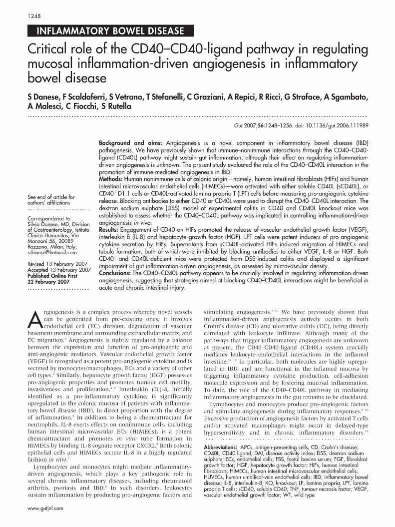
Figure 3 HGF production by CD40L-stimulated HIFs. Panel A: Humanintestinal fibroblasts (HIFs) were activated with soluble CD40 ligand(sCD40L) or were co-cultured either with CD40+ D1.1 cells or with CD402
Jurkat cells for 24 hours, before measuring hepatocyte growth factor (HGF)release in culture supernatants. In selected experiments, blockingantibodies to either CD40 (M2) or CD40L (M90) were used to interfere withthe CD40–CD40L interaction. Results are representative of eightindependent experiments performed in duplicate and are expressed asmean and standard deviation. *p,0.001 compared with HGF release byHIFs not activated with sCD40L; **p,0.001 compared with HGF release byHIFs cultured in the absence of CD40+ D1.1 cells; 1p,0.001 comparedwith HGF release in the absence of blocking antibodies to either CD40 orCD40L. Panel B: HIFs were co-cultured either with unstimulated laminapropria T (LPT) cells or with CD40L+ activated LPT cells. In selectedexperiments, blocking antibodies to either CD40 (M2) or CD40L (M90)were used to interfere with the CD40–CD40L interaction. **p,0.001compared with HGF release by HIFs cultured in the absence of CD40+ LPTcells; 1p,0.001 compared with HGF release in the absence of blockingantibodies to either CD40 or CD40L.
Figure 4 Effects of CD40 engagement on HIMEC chemotaxis. Migrationof human intestinal microvascular endothelial cells (HIMECs) was measuredafter activation with either soluble CD40 ligand (sCD40L) or exogenouslyadded vascular endothelial growth factor (VEGF). Additionally, cellmigration was assessed after culturing with supernatants from sCD40L-activated human intestinal fibroblasts (HIFs), either in the absence or in thepresence of blocking antibodies to VEGF, interleukin-8 (IL-8) or hepatocytegrowth factor (HGF). *p,0.001 compared with baseline HIMEC migration.Results are representative of six independent experiments performed induplicate and are expressed as mean and standard deviation. 1p,0.001compared with cultures performed in the absence of blocking antibodies toVEGF, IL-8 or HGF.
1250 Danese, Scaldaferri, Vetrano, et al
www.gutjnl.com

Flow cytometry analysis showed that purified T cells were.97% CD3+. To induce CD40L expression, LPTs were activatedwith polystyrene beads coated with murine monoclonalantibodies to human CD3 (10 mg/ml) and CD28 (1 mg/ml;DYNAL, Lake Success, NY), as previously reported.12
Measurement of pro-angiogenic cytokine andchemokine production by HIFsConfluent HIFs were either left alone or stimulated with 1 mg/ml of sCD40L, or with irradiated D1.1 cells (CD40L+ acute T-cellleukaemia) or Jurkat cells (CD402 acute T-cell leukaemia; 10:1ratio of T cells to HIFs) for 24 hours. In other wells, a blockingantibody to CD40L (M90 clone; Immunex, Seattle, WA) wasused to hinder the CD40–CD40L interaction.12 27 TrimericsCD40L and antibodies against CD40 (M2 clone) were providedby Immunex.
VEGF, HGF and IL-8 levels in HIF-conditioned medium weremeasured in triplicate with commercially available ELISAs,following the manufacturer’s instructions. The limits ofdetection were as follows: ,9 pg/ml VEGF, 3.5 pg/ml IL-8and ,40 pg/ml HGF.
HIMEC chemotaxis assayChemotaxis was assessed as previously detailed.28 Briefly,fluorescence-blocked polycarbonate filters (8 mm pore size; BDBiosciences, Palo Alto, CA) were coated with human fibronectin(10 mg/ml) for 1 hour at room temperature. By using atranswell system, 35 6 104 HIMECs were plated in the upperchamber in MCDB-131 medium (Sigma Aldrich), whereas thelower chamber contained chemotaxis buffer with phorbolmyristate acetate (10 ng/ml), basic FGF (bFGF; 50 ng/ml) orVEGF (50 ng/ml) as positive controls or 1 ml of supernatantsfrom sCD40L-activated HIFs. In selected wells, 25 mg/l of
blocking antibodies to VEGF, IL-8 or HGF (R&D Systems) orcontrol antibodies were added to the chemotaxis buffer. After4 hours, buffer was removed from both chambers, and HIMECsmigrated onto the lower surface of the porous membrane werewashed twice in phosphate-buffered saline and stained withcalcein for 15 min at 37 C̊. Duplicates of migrated cells wereobserved with an inverted fluorescence microscope and countedin six random high-power fields. Quantitative analysis of datawas performed by using the Ima Pro Plus software (MediaCybernetics, Silver Spring, MD) connected to an OptronicsColor digital camera (Olympus Corporation, Tokyo, Japan).
Immunostaining and morphometric analysis ofintestinal microvasculatureImmunostaining was performed as previously described.28
Briefly, paraffin-embedded intestinal sections were cut at3 mm thickness, deparaffinized then hydrated, blocked forendogenous peroxidase using 3% H2O2/H2O, and subsequentlysubjected to microwave epitope enhancement using a targetretrieval solution (Dako, Carpenteria, CA) at pH 10.0.Incubation with anti-CD31 antibodies (Dako) was performedat 1:200 dilution for 30 min at room temperature. Detectionwas achieved with a standard streptavidin-biotin system(Dako), and antigen localization was visualized with 39-3-diamino benzidene (Sigma Aldrich). Computerized morpho-metric analysis was performed using an international con-sensus method for quantification of angiogenesis.29 Briefly,stained colonic sections were scanned at low power to detectthe most vascularized area, after which at least five micro-photographs of the mucosa at high magnification were taken,defined as the area above the muscularis mucosae (ie, thelamina propria), as well as the submucosa, defined as the areabetween the muscularis mucosae and muscularis propria. The
Figure 5 Supernatants of CD40L-activatedHIF promote in vitro tube formation. Tubeformation assays were performed withMatrigelTM, as detailed in Materials andMethods. Inverted-phase contrastmicroscopy was used to assess the formationof endothelia cell (EC)-like tube structures.Either vascular endothelial growth factor(VEGF) or soluble CD40 ligand (sCD40L)were used as positive controls for ECformation. Results are representative of threeindependent experiments. Five high-powerfields per culture condition were examined.Magnification 640. HIFs, human intestinalfibroblasts; HIMECs, human intestinalmicrovascular endothelial cells.
CD40–CD40-ligand pathway in IBD 1251
www.gutjnl.com

number of vessels/field (vascular density) was obtained with anOptronics Color digital camera (Olympus). Quantitative analy-sis of the data was performed with the Image Pro Plus system(Media Cybernetics).
In vitro tube formation assayEC tube formation was assessed using MatrigelTM, a solubilisedextracellular basement membrane matrix extracted from theEngelbreth-Holm-Swarm mouse sarcoma, as detailed else-where.6 Briefly, multi-well dishes were coated with 250 ml ofcomplete medium containing 5 mg/ml MatrigelTM, andHIMECs resuspended in complete growth medium were seededat a density of 5 6 104. Cells were cultured on MatrigelTM for16 hours and inverted phase-contrast microscopy was usedto assess formation of endothelial tube-like structures. Five
high-power fields per condition were examined and experi-ments were performed in duplicate.
Induction and evaluation of colonic inflammationCD40 knockout (CD40KO) and CD40L knockout (CD40LKO)mice were kindly provided by Mario P. Colombo(Immunotherapy and Gene Therapy Unit, Istituto NazionaleTumori, Milan, Italy).30 For the dextran sodium sulphate (DSS)model of colitis, acute inflammation was induced by adminis-tration of 3% DSS (40 kDa molecular mass; ICN Biomedicals,Aurora, OH) in filter-purified drinking water for 8 days(Millipore, Bedford, MA). For induction of chronic colitis, micereceived three cycles of 5-day 3% DSS followed by 10-day water,as described previously.31 Mice were sacrificed 10 days aftercompletion of the last cycle. Grading of intestinal inflammationwas measured in a blinded fashion by three readers: noinflammation (0); modest numbers of infiltrating cells in theLP (1); infiltration of mononuclear cells leading to separation ofcrypts and mild mucosal hyperplasia (2); massive infiltrationwith inflammatory cells accompanied by disrupted mucosalarchitecture, loss of goblet cells, and marked mucosal hyper-plasia (3); all of the above plus crypt abscesses or ulceration (4),with a histological score from 0 to 15.32 The disease activityindex (DAI) was calculated by scoring 1 point for theappearance of each of the following: ruffled fur, occult foecalblood as determined on a Hemoccult Sensa card (Smith KlineDiagnostics, San Jose, CA), rectal prolapse ,1 mm, and softstool. The mice were scored an additional point for diarrhoea orsevere rectal prolapse .1 mm.
Statistical analysisThe approximation of data distribution to normality waspreliminarily tested with statistics for kurtosis and symmetry.Results were presented as mean and SD. All comparisons wereperformed with the Student’s t-test for paired or unpaireddeterminations or with the analysis of variance, as appropriate.The criterion for statistical significance was defined as p 0(.05.
RESULTSHIFs release pro-angiogenic cytokines on CD40engagementIn a preliminary set of experiments, we investigated the abilityof human CD40-expressing HIFs to release the pro-angiogeniccytokines VEGF, IL-8 and HGF on CD40 triggering. Baselineexpression levels and kinetics of CD40 induction in HIFs wereextensively described elsewhere.28 Both sCD40L and CD40L+
D1.1 cells induced significant VEGF release by HIFs after24 hours of culture (fig 1A). Conversely, CD402 Jurkat cellsfailed to upregulate VEGF production by co-cultured HIFs, andVEGF levels were comparable to those measured in unstimu-lated cultures. Blocking antibodies to either CD40L (M90) orCD40 (M2) abrogated VEGF production in response to co-culture with CD40L+ D1.1 cells, suggesting the involvement ofthe CD40–CD40L pathway in the promotion of VEGF release inthis system model (fig 1A). Interestingly, activated, CD40L-expressing LPT cells, but not resting LPT cells, from healthysubjects promoted VEGF production by HIFs, a response thatwas significantly inhibited by the addition of antibodies toCD40L or CD40 to the co-cultures (figure 1B). Controlantibodies failed to inhibit VEGF production by HIFs (datanot shown).
As shown in figure 2A, IL-8 production by HIFs alsoincreased in response to sCD40L and, even more so, toCD40L+ D1.1 cells. It must be noted that IL-8 was present inthe supernatants of co-cultures established with CD402 Jurkatcells, although at significantly lower levels compared withcultures supplemented with sCD40L (fig 2A). The addition of
Figure 6 Protection from DSS-induced colitis in CD40KO and CD40LKOmice. Experimental colitis was induced with dextran sodium sulphate (DSS)in either wild-type (WT) mice or CD40 knockout (CD40KO) or CD40Lknockout (CD40LKO) mice, in accordance with previously publishedprotocols.33 The percentage of weight loss (panel A) and disease activityindex (DAI; panel B) were monitored as indicators of disease severity.Results are representative of independent experiments (eight animals pergroup) and are expressed as mean and standard deviation. *p,0.01when comparing either CD40KO or CD40LKO mice with their WTcounterpart.
1252 Danese, Scaldaferri, Vetrano, et al
www.gutjnl.com

antibodies to either CD40L or CD40 to the co-cultures of HIFand D1.1 cells translated into a significant reduction of IL-8, thelevels of which approached those measured in the presence ofCD402 Jurkat cells. Similar results were obtained withactivated LPT cells (fig 2B). It should be emphasised that bothJurkat cells (fig 2A) and resting LPT cells (fig 2B) elicitedmodest IL-8 production, reinforcing the concept that IL-8production by HIFs appears to be only partly dependent onCD40–CD40L interactions.
Finally, sCD40L-stimulated HIFs released significantamounts of HGF and interference with the CD40–CD40Linteraction by antibodies specific to either CD40L or CD40resulted in abrogation of HGF production (fig 3A). In line withthe above results on VEGF and IL-8, activated LPT cells wereinducers of HGF release by HIFs, and antibodies to CD40 orCD40L significantly, albeit not completely, inhibited HGFsecretion (fig 3B). Unexpectedly, bFGF production by HIFswas unaffected by CD40 engagement (data not shown),indicating that the CD40–CD40L pathway might be involvedin controlling the release of a distinctive spectrum of pro-angiogenic factors. Collectively, these results pointed to acrucial role for CD40–CD40L interactions in promoting pro-angiogenic cytokine release.
CD40 engagement on HIFs promotes EC migrationthrough an indirect mechanism involving pro-angiogenic cytokine releaseWe next asked whether pro-angiogenic cytokine production byHIFs on CD40 engagement was functional and promoted in vitroand in vivo angiogenesis. To this end, we transferred culturesupernatants of either resting or sCD40L-activated HIFs ontoHIMECs before assessing their migratory behaviour. In controlcultures, exogenous VEGF was provided as a cytokine stimulusfor optimal HIMEC migration. Soluble CD40L induced migra-tion of HIMECs at levels that were comparable to thosemeasured in response to exogenous VEGF (fig 4). Supernatantsof HIFs activated with sCD40L for 24 hours induced significantHIMEC migration, which was in part inhibited by blocking
antibodies to either VEGF, IL-8 or HGF, and was completelyabrogated by antibodies directed against CD40L (fig 4). Thesefindings suggest that CD40 engagement is capable of promotingHIMEC migration both directly and through the activation ofpro-angiogenic cytokine release by HIFs. This conclusion isfurther corroborated by in vitro tube formation experiments. Asshown in figure 5, either sCD40L (panel C) or supernatantsfrom CD40L-activated HIFs (panel D) promoted the formationof tube-like structures to a degree comparable to that inducedby exogenously added VEGF (panel B). The provision ofblocking antibodies to CD40L to cultures of HIMECs andsupernatants from sCD40L-activated HIFs inhibited tubeformation, indicating a pivotal role for sCD40L in the describedphenomena (data not shown).
CD40KO and CD40L KO mice manifest diminishedangiogenesis and are protected from intestinalinflammationWe next took advantage of the DSS model of experimentalcolitis to assess whether the CD40–CD40L system might beimplicated in controlling colonic inflammation in vivo througheffects on angiogenesis. As expected, DSS-treated wild-type(WT) mice developed intestinal inflammation and wastingdisease.33 Measurements of both weight loss and DAI clearlyindicated that, beginning on day 6 from colitis induction, bothCD40KO and CD40LKO mice were significantly protected fromDSS-induced colitis compared with the control group of WTmice (fig 6). These findings were underpinned by thehistological evaluation and grading of inflammation-inducedtissue damage. As shown in figure 7, colonic tissue sectionsfrom CD40KO and CD40LKO mice displayed a diminishedinfiltration with inflammatory cells, and this translated into alower histological score compared with WT mice. In addition,studies of microvascular density and tissue staining withantibodies to CD31, a well-recognised EC marker, providedobjective evidence of diminished angiogenesis in both CD40KOand CD40LKO mice (fig 8). These findings were also confirmedin the chronic model of DSS-induced colitis. As shown in
Figure 7 Protection from histologicalinflammation after colitis induction inCD40KO and CD40LKO mice. Colonicsections were established from either wild-type (WT) or CD40 knockout (CD40KO) orCD40L knockout (CD40LKO) mice, afterdevelopment of dextran sodium sulphate(DSS)-induced colitis. Histological damagescores were measured as detailed inMaterials and Methods. Results arerepresentative of independent experiments(eight animals per group) and are expressedas mean and standard deviation. *p,0.01when comparing either CD40KO orCD40LKO mice with their WT counterpart.Magnification 610.
CD40–CD40-ligand pathway in IBD 1253
www.gutjnl.com

figure 9, microvascular density was significantly lower incolonic tissue sections of either CD40KO or CD40LKO micecompared with their WT counterpart.
DISCUSSIONAngiogenesis is a critical component of several chronicinflammatory disorders, including human IBD. However, thepathways that mediate inflammation-associated angiogenesisare not established in the gut. Both CD40 and CD40L areoverexpressed in CD and UC, suggesting an intimate involve-ment in disease pathogenesis and progression.11 From atherapeutic standpoint, disruption of the CD40–CD40L pathwaywith subsequent attenuation of gut inflammation could haveimportant implications for human IBD. Indeed, CD40–CD40L-dependent immune–nonimmune interactions favour an influxof pathogenic T cells in the gut mucosa, thus contributing tochronic inflammation.12 We have recently shown that IBDmucosal extracts induce HIMEC migration and that thisresponse is mostly dependent on IL-8.28 In addition, IBDmucosal extracts induce potent angiogenic responses in boththe corneal and chorioallantoic membrane assays, indicatingthat the local microvasculature undergoes an intense process ofinflammation-dependent angiogenesis.28 Ligation of CD40 onhuman pancreatic cells induces secretion of pro-inflammatorychemokines, including IL-8 and MIP-1a, indicating a potentialrole of immune–nonimmune interactions in contributing topancreatic-islet inflammation or graft loss after transplanta-tion.34 Finally, increased CD40 expression with subsequentinduction of IL-8 release has been reported in inflamed humanrespiratory epithelial cells, further substantiating the view thatnonimmune cells can behave as effector cells of the inflamma-tion process following activation of the CD40–CD40L pathway.35
The present study was designed to address the role of theCD40–CD40L pathway in inflammation-induced angiogenesis.CD40 engagement on HIFs by either sCD40L or CD40-expressing D1.1 cells induced the release of prototypical pro-angiogenic factors such as VEGF, IL-8 and HGF, whereas CD40-nonexpressing Jurkat cells failed to elicit measurable cytokineproduction in culture supernatants. Expression of VEGF
following CD40 ligation has been reported in different experi-mental models, including human umbilical ECs and mono-cytes.14 In this study, CD40-dependent VEGF expression wasattributed to transcriptional control mechanisms and translatedinto marked angiogenesis in vivo.14 Specifically, CD40-inducedoverexpression of VEGF in ECs has been correlated with theactivation of the Ras pathway and its effector molecules,providing a mechanism by which the immune system,represented by CD40–CD40L interactions, can regulate angio-genesis.36
In our study, LPT cells isolated from the colonic mucosa ofhealthy subjects and activated in vitro through the T-cellreceptor to express CD40L, were potent inducers of pro-angiogenic cytokine production by HIFs, suggesting a potentialinvolvement of T cells from inflamed target tissues in thepromotion of inflammation-induced angiogenesis. Accordingly,fibroblast-like synovial cells from rheumatoid arthritis patientshave been shown to release VEGF after culturing with CD40L-transfected L cells, further supporting the hypothesis thatnonimmune cells from inflamed tissues might represent animportant source of pro-angiogenic cytokines in a variety ofdisease models.37
Mechanistically, CD40 occupancy on cells of nonimmuneorigin, such as HIFs, induced pro-angiogenic factor release,which was significantly reduced by neutralizing antibodiesdirected against either CD40L or CD40. Furthermore, super-natants of HIF cultures were capable of transferring theangiogenesis-inducing activity of CD40-stimulated HIFs, sug-gesting that cytokine production by HIFs has functionalconsequences. Blocking antibodies to either VEGF, IL-8 orHGF significantly inhibited HIMEC migration in response toHIF supernatants, unravelling a potential role for theseprototypical angiogenic factors in inflammation-driven angio-genic responses. Thus, these experiments point out that theCD40–CD40L pathway can regulate angiogenesis by differentmechanisms. First, ECs might be activated by CD40 triggeringand this could be the case in CD and UC, in which the elevatedlevels of sCD40L could induce HIMEC angiogenic activity.8 18
The second indirect mechanism might include the stimulation
Figure 8 Reduction of inflammatoryangiogenesis after acute colitis induction inCD40KO and CD40LKO mice. Colonicsections were established from either wild-type (WT) or CD40 knockout (CD40KO) orCD40L knockout (CD40LKO) mice, afterdevelopment of acute dextran sodiumsulphate (DSS)-induced colitis. Microvesseldensity was used as an indicator ofangiogenesis. *p,0.01 when comparingeither CD40KO or CD40LKO mice with theirWT counterpart. Magnification 610.
1254 Danese, Scaldaferri, Vetrano, et al
www.gutjnl.com

of angiogenic cytokine production by CD40-expressing HIFs,with subsequent activation of HIMECs. Tube-formation experi-ments substantiated these findings by demonstrating theappearance of tube-like structures after culture of HIMECs inthe presence of supernatants from CD40-activated HIFs.
In a final set of experiments, we took advantage of CD40KOand CD40LKO mice to explore the role, if any, of the CD40–CD40L pathway in promoting inflammation-induced angiogen-esis in vivo. Serial determinations of DAI in CD40KO/CD40LKOmice indicated a significant protection from DSS-induced acuteand chronic colonic inflammation when compared with WTanimals. Improved intestinal histology and reduced microvas-cular density in sections of inflamed colons from CD40KO/CD40LKO mice further strengthened the role of CD40–CD40Linteractions in inflammation-driven angiogenesis in this IBDexperimental model.
In summary, these findings support the notion that immune–nonimmune interactions are important for the maintenanceand propagation of inflammation-induced mucosal angiogen-esis. We suggest that the modulation of the CD40–CD40Lpathway deserves further attention as a future target fortherapies to inhibit nonimmune cell-dependent angiogenesis.In mice, administration of CD40L antibodies has translated intoa clinical benefit for autoimmune and inflammatory conditions,such as collagen-induced arthritis, lung inflammation andfibrosis, experimental allergic encephalomyelitis, lupus nephri-tis, atherosclerosis and hapten-induced colitis.11 Studies withchimeric antagonistic monoclonal antibodies to CD40L (IDEC-131) have been initiated in CD.25 The administration of IDEC-131 to patients with refractory immune thrombocytopenicpurpura has been associated with mild adverse events but notwith infections or thromboembolic events,38 suggesting thatthis treatment modality might be safe and well tolerated inpatients with IBD. Our study also suggests that activated LPTcells bearing CD40L as a consequence of mucosal inflammationmight trigger HIF activation and angiogenic cytokine release.Such soluble mediators might, in turn, activate HIMECangiogenesis. In addition, sCD40L fosters directly mucosal
angiogenesis, pointing to a dual mechanism responsible forCD40-dependent angiogenesis in the inflamed gut. Our studiesindicate that therapeutic inhibition of the CD40–CD40L path-way can impact both on nonimmune cell activation and oninflammation-driven angiogenesis in IBD.
ACKNOWLEDGEMENTSThe authors are indebted to Mario P. Colombo (Immunotherapy andGene Therapy Unit, Istituto Nazionale Tumori, Milan, Italy) forproviding CD40KO and CD40LKO mice.The authors wish to thank Mr. Renato Zocchi for figure editing.The authors’ studies were supported by grants from the Broad MedicalResearch Program and by Associazione Italiana per la Ricerca sulCancro (AIRC) grant (to SD).
Authors’ affiliations. . . . . . . . . . . . . . . . . . . . . . .
S Danese, S Vetrano, T Stefanelli, A Repici, A Malesci, Division ofGastroenterology, Istituto Clinico Humanitas, IRCCS in Gastroenterology,Milan, ItalyF Scaldaferri, C Graziani, G Straface, Department of Internal Medicine,Catholic University Medical School, Rome, ItalyR Ricci, Department of Pathology, Catholic University Medical School,Rome, ItalyA Sgambato, Institute of General Pathology, Catholic University MedicalSchool, Rome, ItalyA Malesci, Division of Gastroenterology, University of Milan, Milan, ItalyC Fiocchi, The Cleveland Clinic Foundation, Lerner Research Institute,Cleveland, OH, USAS Rutella, Department of Haematology, Catholic University Medical School,Rome, Italy
Competing interest statement: None declared.
REFERENCES1 Folkman J. Seminars in Medicine of the Beth Israel Hospital, Boston. Clinical
applications of research on angiogenesis. N Engl J Med 1995;333:1757–63.2 Leung DW, Cachianes G, Kuang WJ, et al. Vascular endothelial growth factor is
a secreted angiogenic mitogen. Science 1989;246:1306–9.3 Rutella S, Bonanno G, Procoli A, et al. Hepatocyte growth factor favors monocyte
differentiation into regulatory interleukin (IL)-10++IL-12low/neg accessory cells withdendritic-cell features. Blood 2006;108:218–27.
Figure 9 Reduction of inflammatoryangiogenesis after chronic colitis induction inCD40KO and CD40LKO mice. Colonicsections were established from either wild-type (WT) or CD40 knockout (CD40KO) orCD40L knockout (CD40LKO) mice, afterdevelopment of chronic dextran sodiumsulphate (DSS)-induced colitis.31 Microvesseldensity was used as an indicator ofangiogenesis. *p,0.01 when comparingeither CD40KO (n = 8) or CD40LKO (n = 7)mice with their WT counterpart (n = 7).Magnification 610.
CD40–CD40-ligand pathway in IBD 1255
www.gutjnl.com
4 Jiang WG, Martin TA, Parr C, et al. Hepatocyte growth factor, its receptor, andtheir potential value in cancer therapies. Crit Rev Oncol Hematol2005;53:35–69.
5 Mazzucchelli L, Hauser C, Zgraggen K, et al. Expression of interleukin-8 gene ininflammatory bowel disease is related to the histological grade of activeinflammation. Am J Pathol 1994;144:997–1007.
6 Heidemann J, Ogawa H, Dwinell MB, et al. Angiogenic effects of interleukin 8(CXCL8) in human intestinal microvascular endothelial cells are mediated byCXCR2. J Biol Chem 2003;278:8508–15.
7 Utgaard JO, Jahnsen FL, Bakka A, et al. Rapid secretion of prestored interleukin8 from Weibel-Palade bodies of microvascular endothelial cells. J Exp Med1998;188:1751–6.
8 Koutroubakis IE, Tsiolakidou G, Karmiris K, et al. Role of angiogenesis ininflammatory bowel disease. Inflamm Bowel Dis 2006;12:515–23.
9 Polverini PJ, Cotran PS, Gimbrone MA Jr, et al. Activated macrophages inducevascular proliferation. Nature 1977;269:804–6.
10 Auerbach R, Sidky YA. Nature of the stimulus leading to lymphocyte-inducedangiogenesis. J Immunol 1979;123:751–4.
11 Danese S, Sans M, Fiocchi C. The CD40/CD40L costimulatory pathway ininflammatory bowel disease. Gut 2004;53:1035–43.
12 Vogel JD, West GA, Danese S, et al. CD40-mediated immune-nonimmune cellinteractions induce mucosal fibroblast chemokines leading to T-celltransmigration. Gastroenterology 2004;126:63–80.
13 Ferrara N. VEGF: an update on biological and therapeutic aspects. Curr OpinBiotechnol 2000;11:617–24.
14 Melter M, Reinders ME, Sho M, et al. Ligation of CD40 induces the expression ofvascular endothelial growth factor by endothelial cells and monocytes andpromotes angiogenesis in vivo. Blood 2000;96:3801–8.
15 Danese S, Fiocchi C. Platelet activation and the CD40/CD40 ligand pathway:mechanisms and implications for human disease. Crit Rev Immunol2005;25:103–21.
16 Rutella S, Danese S, Leone G. Tolerogenic dendritic cells: Cytokine modulationcomes of age. Blood 2006;108:1435–40.
17 Lederman S, Yellin MJ, Krichevsky A, et al. Identification of a novel surfaceprotein on activated CD4+ T cells that induces contact-dependent B celldifferentiation (help). J Exp Med 1992;175:1091–101.
18 Danese S, Katz JA, Saibeni S, et al. Activated platelets are the source of elevatedlevels of soluble CD40 ligand in the circulation of inflammatory bowel diseasepatients. Gut 2003;52:1435–41.
19 Reinders ME, Sho M, Robertson SW, et al. Proangiogenic function of CD40ligand-CD40 interactions. J Immunol 2003;171:1534–41.
20 Larsen CP, Elwood ET, Alexander DZ, et al. Long-term acceptance of skin andcardiac allografts after blocking CD40 and CD28 pathways. Nature1996;381:434–8.
21 Schonbeck U, Sukhova GK, Shimizu K, et al. Inhibition of CD40 signaling limitsevolution of established atherosclerosis in mice. Proc Natl Acad Sci USA2000;97:7458–63.
22 Stuber E, Strober W, Neurath M. Blocking the CD40L–CD40 interaction in vivospecifically prevents the priming of T helper 1 cells through the inhibition ofinterleukin 12 secretion. J Exp Med 1996;183:693–8.
23 Danese S, Sans M, Gasbarrini A. CD40 antisense based strategy forinflammatory bowel disease: shutting down multiple cellular communicationsystems. Gut 2005;54:891–2.
24 Maerten P, Liu Z, Ceuppens JL. Targeting of costimulatory molecules as a therapeuticapproach in inflammatory bowel disease. BioDrugs 2003;17:395–411.
25 Dumont FJ. IDEC-131. IDEC/Eisai. Curr Opin Investig Drugs 2002;3:725–34.26 Fiocchi C, Battisto JR, Farmer RG. Gut mucosal lymphocytes in inflammatory
bowel disease: isolation and preliminary functional characterization. Dig Dis Sci1979;24:705–17.
27 Danese S, de la Motte C, Reyes BM, et al. Cutting edge: T cells trigger CD40-dependent platelet activation and granular RANTES release: a novel pathway forimmune response amplification. J Immunol 2004;172:2011–5.
28 Danese S, Sans M, de la Motte C, et al. Angiogenesis as a novel component ofinflammatory bowel disease pathogenesis. Gastroenterology2006;130:2060–73.
29 Vermeulen PB, Gasparini G, Fox SB, et al. Quantification of angiogenesis insolid human tumours: an international consensus on the methodology and criteriaof evaluation. Eur J Cancer 1996;32A:2474–84.
30 Guiducci C, Valzasina B, Dislich H, et al. CD40/CD40L interaction regulatesCD4+CD25+ Treg homeostasis through dendritic cell-produced IL-2. Eur J Immunol2005;35:557–67.
31 Obermeier F, Dunger N, Strauch UG, et al. CpG motifs of bacterial DNAessentially contribute to the perpetuation of chronic intestinal inflammation.Gastroenterology 2005;129:913–27.
32 Spencer DM, Veldman GM, Banerjee S, et al. Distinct inflammatory mechanismsmediate early versus late colitis in mice. Gastroenterology 2002;122:94–105.
33 Krieglstein CF, Cerwinka WH, Sprague AG, et al. Collagen-binding integrinalpha1beta1 regulates intestinal inflammation in experimental colitis. J Clin Invest2002;110:1773–82.
34 Barbe-Tuana FM, Klein D, Ichii H, et al. CD40–CD40 ligand interaction activatesproinflammatory pathways in pancreatic islets. Diabetes 2006;55:2437–45.
35 Cagnoni F, Oddera S, Giron-Michel J, et al. CD40 on adult human airwayepithelial cells: expression and proinflammatory effects. J Immunol2004;172:3205–14.
36 Flaxenburg JA, Melter M, Lapchak PH, et al. The CD40-induced signalingpathway in endothelial cells resulting in the overexpression of vascularendothelial growth factor involves Ras and phosphatidylinositol 3-kinase.J Immunol 2004;172:7503–9.
37 Cho CS, Cho ML, Min SY, et al. CD40 engagement on synovial fibroblast up-regulates production of vascular endothelial growth factor. J Immunol2000;164:5055–61.
38 Kuwana M, Nomura S, Fujimura K, et al. Effect of a single injection ofhumanized anti-CD154 monoclonal antibody on the platelet-specificautoimmune response in patients with immune thrombocytopenic purpura. Blood2004;103:1229–36.
EDITOR’S QUIZ: GI SNAPSHOT . . . . . . . . . . . . . . . . . . . . . . . . . . . . . . . . . . . . . . . . . . . . . . . . . . . . . . . . . . . . . . . . . . . . . .
AnswerFrom question on page 1247
The findings in this case are typical of ovarian endometrioma and suspicious for endometriosisof the bowel wall. Various symptoms, such as dysmenorrhea, dyspareunia, chronic pelvic pain,diarrhoea, constipation, cyclic rectal bleeding, colic abdominal pain up to intestinal occlusionmay characterise this pathology.
The value of imaging in the diagnosis of endometriosis is limited. Small implants are usuallynot detectable with ultrasound and are also difficult to identify with MRI. Endometrioma is theonly form of endometriosis readily diagnosed with ultrasound (generally transvaginal).
The diagnosis of intestinal endometriosis can be difficult to establish. Endometrial deposits donot invade the mucosa, therefore colonoscopy with biopsies is frequently non-diagnostic. In thiscase, intestinal ultrasound allowed an endometrial implant in the sigmoid colon to be identified.
It should be noted that endometriosis of the bowel most often involves the sigmoid andrectosigmoid, even though it is a relatively uncommon cause of obstruction. In the event ofrectosigmoid strictures of unknown aetiology in premenopausal women, endometriosis shouldbe included in the differential diagnosis. In this case, surgery, which confirmed the diagnosis,was the treatment of choice.
doi: 10.1136/gut.2006.106583a
1256 Danese, Scaldaferri, Vetrano, et al
www.gutjnl.com
Related Documents